
Signalling of the BCR is regulated by a lipid rafts-localised transcription factor, Bright
14
Signalling of the BCR is regulated by a lipid rafts-localised transcription factor, Bright Christian Schmidt 1 , Dongkyoon Kim 1 , Gregory C Ippolito 1 , Hassan R Naqvi 2 , Loren Probst 1 , Shawn Mathur 1 , German Rosas-Acosta 3 , Van G Wilson 3 , Athenia L Oldham 4 , Martin Poenie 2 , Carol F Webb 4 and Philip W Tucker 1, * 1 Institute for Cellular and Molecular Biology, The University of Texas at Austin, Austin, TX, USA, 2 Department of Molecular Cell and Developmental Biology, The University of Texas at Austin, Austin, TX, USA, 3 Department of Microbial and Molecular Pathogenesis, Texas A&M Health Science Center, College Station, TX, USA and 4 Immunobiology and Cancer Program, Oklahoma Medical Research Foundation, University of Oklahoma Health Sciences Center, Oklahoma City, OK, USA Regulation of BCR signalling strength is crucial for B-cell development and function. Bright is a B-cell-restricted factor that complexes with Bruton’s tyrosine kinase (Btk) and its substrate, transcription initiation factor-I (TFII-I), to activate immunoglobulin heavy chain gene transcription in the nucleus. Here we show that a palmi- toylated pool of Bright is diverted to lipid rafts of resting B cells where it associates with signalosome components. After BCR ligation, Bright transiently interacts with su- moylation enzymes, blocks calcium flux and phosphoryla- tion of Btk and TFII-I and is then discharged from lipid rafts as a Sumo-I-modified form. The resulting lipid raft concentration of Bright contributes to the signalling threshold of B cells, as their sensitivity to BCR stimulation decreases as the levels of Bright increase. Bright regulates signalling independent of its role in IgH transcription, as shown by specific dominant-negative titration of rafts- specific forms. This study identifies a BCR tuning mechan- ism in lipid rafts that is regulated by differential post- translational modification of a transcription factor with implications for B-cell tolerance and autoimmunity. The EMBO Journal (2009) 28, 711–724. doi:10.1038/ emboj.2009.20; Published online 12 February 2009 Subject Categories: membranes & transport; immunology Keywords: B cell; immunity; signal transduction Introduction B-cell development and response to antigen depend on signalling through the B-cell antigen receptor (BCR) complex (Gauld et al, 2002; Meyer-Bahlburg et al, 2008). BCR signal- ling directs positive and negative selection of immature B cells and their progression through transitional (T) stages into mature B cells. Surface markers allow the resolution of three non-proliferative immature B-cell subpopulations: T1, T2 and T3 (Allman et al, 2001; Sims et al, 2005). The lineage origins and signalling requirements of these intermediate stages of B cells are the subject of considerable interest and debate (Matthias and Rolink, 2005; Teague et al, 2007; Welner et al, 2008). It is generally agreed that sequential progression requires an increasingly higher threshold level of BCR signal- ling; that is, low or ‘tonic’ threshold signals promote T1 to T2, whereas relatively higher levels of signalling are needed for T2 to progress to FO or MZB (Petro et al, 2002; Su and Rawlings, 2002; Hoek et al, 2006). Strong BCR signalling also is required to direct non-transitional, fetal progenitors to B-1 fate (Loder et al, 1999; Cariappa et al, 2001; Casola et al, 2004). The amplitude of BCR signalling is positively and negatively regulated by coreceptors (Carter and Fearon, 1992; Cherukuri et al, 2001; Ravetch and Bolland, 2001) and crosstalk between the antigen receptors and other path- ways, particularly BAFF (Guo and Rothstein, 2005; Venkatesh et al, 2006). A spatially continuous but mobile unit of critical size within the plasma membrane is required for efficient initiation of BCR activation by multivalent antigen (Dintzis et al, 1976). Engagement of the antigen receptor yields ‘microclusters’ that can be found in highly ordered domains within the plasma membrane, known as lipid rafts (Dykstra et al, 2003; Saeki et al, 2003; Harwood and Batista, 2008). Size and composition of these platforms of BCR signalling are dynamic and respon- sive to signalling events mediated by the actin cytoskeleton through plasma membrane linker proteins, such as Ezrin (Stoddart et al, 2002; Gupta et al, 2006; Sohn et al, 2006). Bright ( B-cell regulator of IgH transcription/Dril1/ ARID3A) is the founder of the AT-rich interaction domain (ARID) super-family of DNA-binding proteins (Herrscher et al, 1995; Wilsker et al, 2005). Bright shuttles between the cytoplasm and the nucleus in a Crm1- and cell cycle-depen- dent fashion (Kim and Tucker, 2006). Bright transactivates the IgH intronic enhancer (Em) and certain IgH promoters by binding as a tetramer to ATC motifs within nuclear matrix associating regions (Webb et al, 1999; Kim et al, 2007; Lin et al, 2007). DNA binding and IgH transcriptional activities of Bright are stimulated by its interaction with Btk and tran- scription initiation factor-II (TFII-I), a direct substrate of Btk (Webb et al, 2000; Rajaiya et al, 2005, 2006). TFII-I also undergoes nucleocytoplasmic shuttling (Hakre et al, 2006), and, within the cytoplasm, it associates with PLCg to inhibit Ca 2 þ mobilisation (Caraveo et al, 2006). Bright is lineage and stage-specifically expressed with high basal levels in immature B cells and in mitogen or cytokine- induced mature B cells (Webb et al, 1991a,b, 1998; Nixon et al, 2004a, b). Shankar et al (2007) recently demonstrated Received: 30 July 2008; accepted: 9 January 2009; published online: 12 February 2009 *Corresponding author. Institute for Cellular and Molecular Biology, Molecular Genetics and Microbiology, The University of Texas at Austin, 1 University Station A5000, Austin, TX 78712, USA. Tel.: þ 1 512 475 7705; Fax: þ 1 512 475 7707; E-mail: [email protected] The EMBO Journal (2009) 28, 711–724 | & 2009 European Molecular Biology Organization | All Rights Reserved 0261-4189/09 www.embojournal.org & 2009 European Molecular Biology Organization The EMBO Journal VOL 28 | NO 6 | 2009 EMBO THE EMBO JOURNAL THE EMBO JOURNAL 711
Transcript of Signalling of the BCR is regulated by a lipid rafts-localised transcription factor, Bright

Signalling of the BCR is regulated by a lipidrafts-localised transcription factor, Bright
Christian Schmidt1, Dongkyoon Kim1,Gregory C Ippolito1, Hassan R Naqvi2,Loren Probst1, Shawn Mathur1,German Rosas-Acosta3, Van G Wilson3,Athenia L Oldham4, Martin Poenie2,Carol F Webb4 and Philip W Tucker1,*1Institute for Cellular and Molecular Biology, The University of Texas atAustin, Austin, TX, USA, 2Department of Molecular Cell andDevelopmental Biology, The University of Texas at Austin, Austin, TX,USA, 3Department of Microbial and Molecular Pathogenesis, Texas A&MHealth Science Center, College Station, TX, USA and 4Immunobiologyand Cancer Program, Oklahoma Medical Research Foundation,University of Oklahoma Health Sciences Center, Oklahoma City,OK, USA
Regulation of BCR signalling strength is crucial for B-cell
development and function. Bright is a B-cell-restricted
factor that complexes with Bruton’s tyrosine kinase
(Btk) and its substrate, transcription initiation factor-I
(TFII-I), to activate immunoglobulin heavy chain gene
transcription in the nucleus. Here we show that a palmi-
toylated pool of Bright is diverted to lipid rafts of resting B
cells where it associates with signalosome components.
After BCR ligation, Bright transiently interacts with su-
moylation enzymes, blocks calcium flux and phosphoryla-
tion of Btk and TFII-I and is then discharged from lipid
rafts as a Sumo-I-modified form. The resulting lipid raft
concentration of Bright contributes to the signalling
threshold of B cells, as their sensitivity to BCR stimulation
decreases as the levels of Bright increase. Bright regulates
signalling independent of its role in IgH transcription, as
shown by specific dominant-negative titration of rafts-
specific forms. This study identifies a BCR tuning mechan-
ism in lipid rafts that is regulated by differential post-
translational modification of a transcription factor with
implications for B-cell tolerance and autoimmunity.
The EMBO Journal (2009) 28, 711–724. doi:10.1038/
emboj.2009.20; Published online 12 February 2009
Subject Categories: membranes & transport; immunology
Keywords: B cell; immunity; signal transduction
Introduction
B-cell development and response to antigen depend on
signalling through the B-cell antigen receptor (BCR) complex
(Gauld et al, 2002; Meyer-Bahlburg et al, 2008). BCR signal-
ling directs positive and negative selection of immature B
cells and their progression through transitional (T) stages
into mature B cells. Surface markers allow the resolution of
three non-proliferative immature B-cell subpopulations: T1,
T2 and T3 (Allman et al, 2001; Sims et al, 2005). The lineage
origins and signalling requirements of these intermediate
stages of B cells are the subject of considerable interest and
debate (Matthias and Rolink, 2005; Teague et al, 2007; Welner
et al, 2008). It is generally agreed that sequential progression
requires an increasingly higher threshold level of BCR signal-
ling; that is, low or ‘tonic’ threshold signals promote T1 to T2,
whereas relatively higher levels of signalling are needed for
T2 to progress to FO or MZB (Petro et al, 2002; Su and
Rawlings, 2002; Hoek et al, 2006). Strong BCR signalling also
is required to direct non-transitional, fetal progenitors to B-1
fate (Loder et al, 1999; Cariappa et al, 2001; Casola et al,
2004). The amplitude of BCR signalling is positively and
negatively regulated by coreceptors (Carter and Fearon,
1992; Cherukuri et al, 2001; Ravetch and Bolland, 2001)
and crosstalk between the antigen receptors and other path-
ways, particularly BAFF (Guo and Rothstein, 2005; Venkatesh
et al, 2006).
A spatially continuous but mobile unit of critical size within
the plasma membrane is required for efficient initiation of
BCR activation by multivalent antigen (Dintzis et al, 1976).
Engagement of the antigen receptor yields ‘microclusters’ that
can be found in highly ordered domains within the plasma
membrane, known as lipid rafts (Dykstra et al, 2003; Saeki
et al, 2003; Harwood and Batista, 2008). Size and composition
of these platforms of BCR signalling are dynamic and respon-
sive to signalling events mediated by the actin cytoskeleton
through plasma membrane linker proteins, such as Ezrin
(Stoddart et al, 2002; Gupta et al, 2006; Sohn et al, 2006).
Bright (B-cell regulator of IgH transcription/Dril1/
ARID3A) is the founder of the AT-rich interaction domain
(ARID) super-family of DNA-binding proteins (Herrscher
et al, 1995; Wilsker et al, 2005). Bright shuttles between the
cytoplasm and the nucleus in a Crm1- and cell cycle-depen-
dent fashion (Kim and Tucker, 2006). Bright transactivates
the IgH intronic enhancer (Em) and certain IgH promoters by
binding as a tetramer to ATC motifs within nuclear matrix
associating regions (Webb et al, 1999; Kim et al, 2007; Lin
et al, 2007). DNA binding and IgH transcriptional activities of
Bright are stimulated by its interaction with Btk and tran-
scription initiation factor-II (TFII-I), a direct substrate of Btk
(Webb et al, 2000; Rajaiya et al, 2005, 2006). TFII-I also
undergoes nucleocytoplasmic shuttling (Hakre et al, 2006),
and, within the cytoplasm, it associates with PLCg to inhibit
Ca2þ mobilisation (Caraveo et al, 2006).
Bright is lineage and stage-specifically expressed with high
basal levels in immature B cells and in mitogen or cytokine-
induced mature B cells (Webb et al, 1991a, b, 1998; Nixon
et al, 2004a, b). Shankar et al (2007) recently demonstratedReceived: 30 July 2008; accepted: 9 January 2009; published online:12 February 2009
*Corresponding author. Institute for Cellular and Molecular Biology,Molecular Genetics and Microbiology, The University of Texas at Austin,1 University Station A5000, Austin, TX 78712, USA.Tel.: þ 1 512 475 7705; Fax: þ 1 512 475 7707;E-mail: [email protected]
The EMBO Journal (2009) 28, 711–724 | & 2009 European Molecular Biology Organization | All Rights Reserved 0261-4189/09
www.embojournal.org
&2009 European Molecular Biology Organization The EMBO Journal VOL 28 | NO 6 | 2009
EMBO
THE
EMBOJOURNAL
THE
EMBOJOURNAL
711

the pathological consequences of loss of this tight control.
Transgenic (TG) mice that over-express wild-type (WT)
Bright specifically within the B lineage display spontaneous
autoimmunity. This intrinsic B-cell autoreactivity was not
accompanied by global increase in serum Ig. Instead, a
markedly expanded population of T1 and MZB cells was
observed.
These observations, along with the extranuclear expres-
sion of Bright, TFII-I and their functional association with
Btk, prompted us to examine whether Bright is used in BCR
signal transduction. We show here that a pool of Bright acts
within lipid rafts as a ‘brake’ to set a signalling threshold on
the BCR.
Results
Association of Bright with mIgM on B-cell membranes
is reduced after antigen receptor stimulation
Immunostaining of murine B splenocytes indicated that a
fraction of the non-nuclear Bright pool colocalised with mIgM,
suggesting cortical and/or membrane-associated localisation
(Figure 1A and readdressed below). This observation was
confirmed by computerised 3D reconstructions of the immuno-
fluorescence data (Figure 1A0 and Supplementary Video 1).
To determine whether this colocalisation remains intact
after engagement of the BCR, cells were stimulated for 5 min
with a-m. Only modest colocalisation of Bright and IgM
was retained, as assessed by computerised 3D reconstruc-
tions of the immunofluorescence data (Figure 1A00 and
Supplementary Video 2). Inspection of these and additional
images (data not shown) indicated that the observed redis-
tribution of mIgM-associated Bright in stimulated B cells was
not accompanied by significant alteration in either its nuclear
or its cytoplasmic levels (data not shown).
Bright accumulates within lipid rafts of resting but not
stimulated B cells
Because lipid rafts serve as platforms for BCR signalling,
we assayed purified plasma membranes and lipid rafts
(Supplementary Figure 1A) for the presence of Bright. A
small pool of Bright resides in lipid rafts purified from
unstimulated CD43� B cells (Figure 1B, upper panel).
Consistent with the imaging results, Bright was not detected
within lipid rafts after BCR engagement that was sufficient to
elicit a phosphotyrosine (pY) response (Figure 1B, lower
panel). This suggested that the presence or absence of
Bright within lipid rafts might influence BCR signalling.
Levels of Bright within lipid rafts determine BCR
signalling threshold
Normal B cells and mature B-cell lines were examined semi-
quantitatively for lipid raft content of Bright using the B cell-
specific lipid rafts component, Raflin (Saeki et al, 2003), as an
internal control (Supplementary Figure 1B and data not
shown). We estimated that raft-localised Bright accounted
for 1–10% of total cellular Bright, consistent with percentages
previously estimated for mIgM concentrations in lipid rafts
(Sproul et al, 2000; Putnam et al, 2003). Lipid rafts of Raji and
Daudi cells contained B10-fold less Bright than those of CL01
or Ramos (Figure 2A). However, no significant differences
were observed in other subcellular fractions among these
lines (Supplementary Figure 1B).
To achieve maximal BCR responses under minimal anti-
body concentrations and minimal receptor internalisation, an
approach using an anti-IgM mAb in the absence of secondary
cross-linking was optimised (Supplementary Figures 2B and
4B; Materials and methods; data not shown). Lipid rafts from
resting and stimulated cell lines were purified on sucrose
A BBright IgM DNA Merge
A'
A''
Bright
IgM
DNA
Bright
IgM
DNA
Bright
IgM
Untreated
Bright
IgM
α-CD19α-μ – +
– –++
pY
γ-Tubulin
100 -75 -
50 -
200 -150 -
BrightRaftlin
Bright
75 -100 -
75 -50 -
WCL
1 2 3
Lipid rafts
α-μ α-μ
Untreated
Figure 1 Bright accumulates within lipid rafts of resting but not stimulated B cells. (A) Association of Bright with mIgM on B-cell membranesis reduced after antigen receptor stimulation. CD43� B cells from spleens of BALB/c adult mice were fixed and stained for Bright (red), mIgM(green) and DNA (blue). Arrows point to areas (yellow) where Bright colocalises with membrane IgM. (A0, A00) Engagement of the antigenreceptor reduces the colocalisation between Bright and mIgM. CD43� B cells (B1�104) from spleens of BALB/c adult mice were left untreated(A0) or stimulated for 5 min (A00) with 10 pg a-m, followed by immunostaining as described above. Deconvoluted images are shown with arrowspointing to areas (yellow) where Bright colocalises with mIgM. (B) BCR engagement leads to a discharge of Bright from lipid rafts. CD43� Bcells (B2�106) were stimulated with either 2 ng a-m or 2 ng a-mþ 2 ng a-CD19 for 5 min. Lipid rafts or whole cell lysates (WCL) were preparedfrom half of each sample. Proteins from each fraction were analysed by SDS–PAGE/western blot using the antibodies indicated.
Rafts-localised Bright regulates BCR signallingC Schmidt et al
The EMBO Journal VOL 28 | NO 6 | 2009 &2009 European Molecular Biology Organization712
gradients, and fractions were analysed for Bright and other
signalosome occupants (Figure 2B). In agreement with pub-
lished reports (Saeki et al, 2003; Depoil et al, 2008), levels of
mIgM, CD19, and the Bright-interacting partner Btk increased
in lipid rafts after a-m stimulation (Figure 2B). As observed
for normal B cells, Bright moved in the opposite manner.
Fraction
Btk
CL-01
Daudi
Raftlin
Untreated
Bright
CD19
Btk
Bright
CD19
Btk
Bright
CD19Btk
Ramos
Raji
B
Bright
BrightCD19Btk
Raftlin
Raftlin
Raftlin
Raftlin
75 -
150 -
75 -
100 -
75 -
150 -
75 -
100 -
75 -
150 -
75 -
100 -
100 -
75 -150 -75 -
100 -100
12 3 4 5 67 8 9101
910
– – – ++ ++ – + –+ –– + – +– ++ + – –+ +
– – + +– –
+ ++ ++ +– –– –+ ++ –+ –– +– +
– –– –+ +– –– –+ ++ –+ –– +– +
11 121314
2 3 4 5 67 8 910
1 2 3 4 5 6 7 8910 1 2 3 4 5 6 7 8910
-
75 -150 -
75 -100 -
75 -150 -
75 -100 -
100 -100 -
75 -150 -
75 -100 -
100 -
75 -
150 -
75 -
100 -100 -
IP α BrightIP preimmune
BrightBtk
3 41 2
DaudiCL-01
75 -
100 -75 -
IP pre-immune
BrightBtk
CD197 85 6
DaudiCL-01
150 -100 -
75 -75 -
IP α-μ
IP α-μ
IP α-histoneH1
RajiRamos
150 -
100 -
75 -
75 -
Raj
iR
amos
Dau
diC
L-01
Bright
Raftlin
1 2 3 4
Lipid raft 100 -
75 -
-
C
A
250 -
75 -
37 -25 -
– 105 60
Raji Ramos
1 2 3 7 8654
– 105 60
p-Y
F
G
E
Raji Ramos Daudi CL-01
Bright
Sumo-1
BrightSumo-1
3 54 1097 861 2 11 12 15 16 17 22211918 2013 14 23 24
3 54 1097 861 2 11 12 15 16 17 22211918 2013 14 23 24
3 54 1097 861 2 11 12 15 16 17 22211918 2013 14 23 24
WCLLipid raftsPlasma
membrane WCLLipidrafts
Plasmamembrane
WCLLipidrafts
Plasmamembrane
WCLLipidrafts
Plasmamembrane
WCLLipid raftsPlasma
membrane WCLLipidrafts
Plasmamembrane
WCLLipidrafts
Plasmamembrane
WCLLipidrafts
Plasmamembrane
WCLLipid raftsPlasma
membrane WCLLipidrafts
Plasmamembrane
WCLLipidrafts
Plasmamembrane
WCLLipidrafts
Plasmamembrane
α-CD19 + α-μ
Bright++
+–
++
+–
++
+–
++
+–
++
+–
++
+–
++
+–
++
+–
++–
++
+–
++
+–
++
+–
Sumo-1
+IP α-Bright
75 -
75 -
-
-
75 -
75
α-CD19IP α-Bright
IP α-Bright
7575
-
D– 5 – 5
CL-01 Daudi
250 -100 -
37 -25 -
50 -
1211109
p-Y
++
+–
++
+–
++
+–
++
+–
++
+–
++
+–
++
+–
++
+–
++
+–
++
+–
++
+–
++
+–
++
+–
++
+–
++
+–
++
+–
++
+–
++
+–
++
+–
++
+–
++
+–
++
+–
++
+–
++
+–α-μ
α-μ
α-μ
α-μ
α-μ
α-μ
α-μ treated
α-μ treated
CD19
μ
Btk
Raftlin
Bright
CD19
μBtk
Bright
CD19
μ
μ
μ
μ
μ
BtkBtk
Bright
Bright
CD19
CD19
μ
μ
μ
μ
5%5% 40%40%
5% 5%40% 40%
Untreated
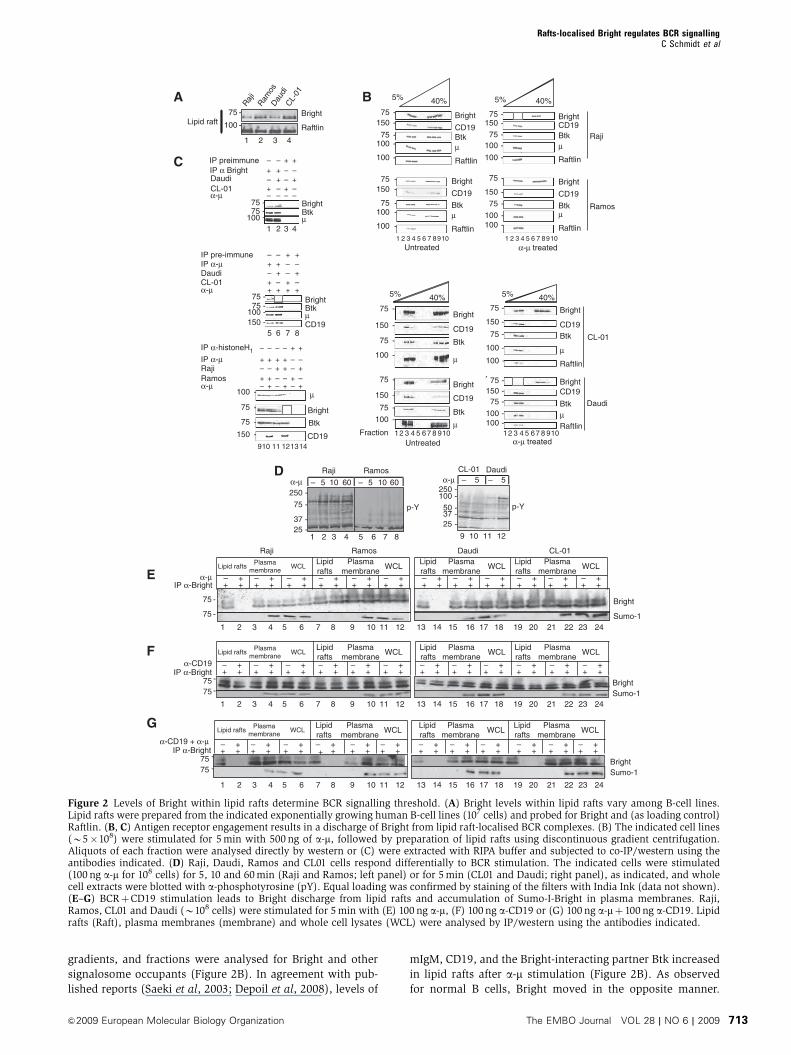
Figure 2 Levels of Bright within lipid rafts determine BCR signalling threshold. (A) Bright levels within lipid rafts vary among B-cell lines.Lipid rafts were prepared from the indicated exponentially growing human B-cell lines (107 cells) and probed for Bright and (as loading control)Raftlin. (B, C) Antigen receptor engagement results in a discharge of Bright from lipid raft-localised BCR complexes. (B) The indicated cell lines(B5�108) were stimulated for 5 min with 500 ng of a-m, followed by preparation of lipid rafts using discontinuous gradient centrifugation.Aliquots of each fraction were analysed directly by western or (C) were extracted with RIPA buffer and subjected to co-IP/western using theantibodies indicated. (D) Raji, Daudi, Ramos and CL01 cells respond differentially to BCR stimulation. The indicated cells were stimulated(100 ng a-m for 108 cells) for 5, 10 and 60 min (Raji and Ramos; left panel) or for 5 min (CL01 and Daudi; right panel), as indicated, and wholecell extracts were blotted with a-phosphotyrosine (pY). Equal loading was confirmed by staining of the filters with India Ink (data not shown).(E–G) BCRþCD19 stimulation leads to Bright discharge from lipid rafts and accumulation of Sumo-I-Bright in plasma membranes. Raji,Ramos, CL01 and Daudi (B108 cells) were stimulated for 5 min with (E) 100 ng a-m, (F) 100 ng a-CD19 or (G) 100 ng a-mþ 100 ng a-CD19. Lipidrafts (Raft), plasma membranes (membrane) and whole cell lysates (WCL) were analysed by IP/western using the antibodies indicated.
Rafts-localised Bright regulates BCR signallingC Schmidt et al
&2009 European Molecular Biology Organization The EMBO Journal VOL 28 | NO 6 | 2009 713
However, its discharge from lipid rafts was complete only in
cell lines in which its starting levels in lipid rafts were low
(Daudi and Raji, Figure 2B, fractions 3 and 4; boxed in red).
These differences in trafficking could reflect differences in
composition of raft-localised complexes, or artifacts resulting
from increased resistance to solubilisation, as BCR ligation is
known to induce coalescence of lipid rafts (Gupta et al, 2006).
Therefore, we compared profiles obtained from RIPA-solubi-
lised versus non-soluble lipid rafts immunoprecipitated (IP)
with a-m, a-Raftlin and a-Btk (Supplementary Figure 2E). We
observed that, as previously published (Saeki et al, 2003), a
complex containing Raflin and IgM was seen only in non-
solubilised rafts (Supplementary Figure 2E); this indicated
that our solubilisation conditions were sufficient. However,
unexpectedly, Raflin did IP with Btk under both conditions,
suggesting that an IP complex containing Btk and Raflin is not
disrupted by RIPA solubilisation of lipid rafts (readdressed
below). Importantly, Bright remained in a complex with
mIgM and Btk in solubilised lipid rafts of all unstimulated
cells (Figure 2C) but was lost only in a-m stimulated cells
(Daudi and Raji) that contained lower starting levels in their
lipid rafts (Figure 2C, lanes 6 and 12; boxed in red).
These results suggested that B cells that contain more lipid
rafts-associated Bright (Ramos and CL01) would be less
sensitive (higher threshold) to BCR ligation. This was con-
firmed by the pY responses of these cell lines to a-m stimula-
tion (Figure 2D). Ramos and CL01 also responded less
vigorously to pro-apoptotic signals shown previously (Chen
et al, 1999) to result from long-term stimulation by a-m(Supplementary Figure 2C).
Ligation of the BCR coreceptor, CD19, is known to
synergistically enhance antigen receptor-mediated signalling
(Carter and Fearon, 1992; Cherukuri et al, 2001; Depoil et al,
2008). Accordingly, all cell lines responded to a-mþ a-CD19
costimulation with robust responses (Supplementary Figure
2B). BCR costimulation was required to expel Bright from
lipid rafts of the less sensitive (higher threshold) cell lines
Ramos and CL01 (Figure 2E–G). That Bright migrates as a
doublet is apparent in these experiments (addressed below in
the context of the sumoylation observations).
Thus, engagement of the BCR results in a significant and
specific reduction of the small pool of lipid rafts-localised
Bright. This pool is lost from lipid rafts, as Btk and other
signalosome components accumulate there, in proportion to
BCR signalling strength.
Entry of Bright into lipid rafts does not require
interaction with Btk but does require palmitoylation
Bright was readily detected in lipid rafts prepared from
retrovirally transduced NIH/3T3 fibroblasts and other non-B
cells (Figure 3B; data not shown). This indicated that even
though Bright associates (at least transiently) with Btk and
other signalsome components in a-m stimulated lipid rafts
(Figure 2C and addressed further below), these B-cell-re-
stricted proteins are not required to designate or retain
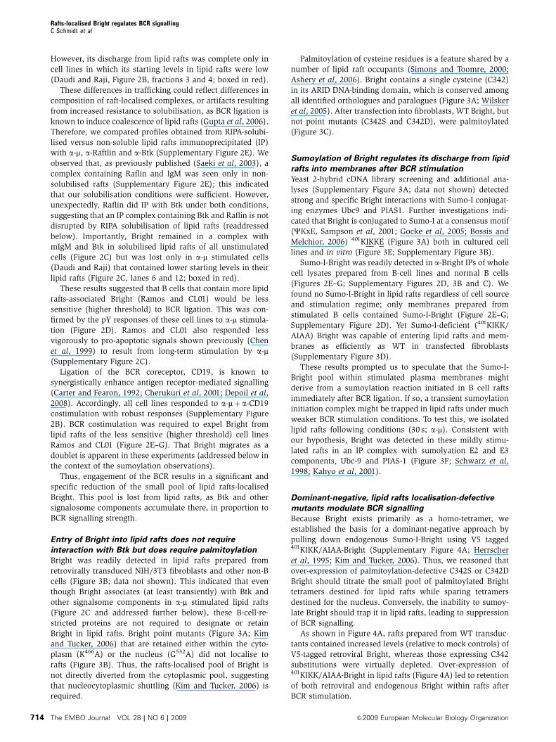
Bright in lipid rafts. Bright point mutants (Figure 3A; Kim
and Tucker, 2006) that are retained either within the cyto-
plasm (K466A) or the nucleus (G532A) did not localise to
rafts (Figure 3B). Thus, the rafts-localised pool of Bright is
not directly diverted from the cytoplasmic pool, suggesting
that nucleocytoplasmic shuttling (Kim and Tucker, 2006) is
required.
Palmitoylation of cysteine residues is a feature shared by a
number of lipid raft occupants (Simons and Toomre, 2000;
Ashery et al, 2006). Bright contains a single cysteine (C342)
in its ARID DNA-binding domain, which is conserved among
all identified orthologues and paralogues (Figure 3A; Wilsker
et al, 2005). After transfection into fibroblasts, WT Bright, but
not point mutants (C342S and C342D), were palmitoylated
(Figure 3C).
Sumoylation of Bright regulates its discharge from lipid
rafts into membranes after BCR stimulation
Yeast 2-hybrid cDNA library screening and additional ana-
lyses (Supplementary Figure 3A; data not shown) detected
strong and specific Bright interactions with Sumo-I conjugat-
ing enzymes Ubc9 and PIAS1. Further investigations indi-
cated that Bright is conjugated to Sumo-I at a consensus motif
(CKxE, Sampson et al, 2001; Gocke et al, 2005; Bossis and
Melchior, 2006) 401KIKKE (Figure 3A) both in cultured cell
lines and in vitro (Figure 3E; Supplementary Figure 3B).
Sumo-I-Bright was readily detected in a-Bright IPs of whole
cell lysates prepared from B-cell lines and normal B cells
(Figures 2E–G; Supplementary Figures 2D, 3B and C). We
found no Sumo-I-Bright in lipid rafts regardless of cell source
and stimulation regime; only membranes prepared from
stimulated B cells contained Sumo-I-Bright (Figure 2E–G;
Supplementary Figure 2D). Yet Sumo-I-deficient (401KIKK/
AIAA) Bright was capable of entering lipid rafts and mem-
branes as efficiently as WT in transfected fibroblasts
(Supplementary Figure 3D).
These results prompted us to speculate that the Sumo-I-
Bright pool within stimulated plasma membranes might
derive from a sumoylation reaction initiated in B cell rafts
immediately after BCR ligation. If so, a transient sumoylation
initiation complex might be trapped in lipid rafts under much
weaker BCR stimulation conditions. To test this, we isolated
lipid rafts following conditions (30 s; a-m). Consistent with
our hypothesis, Bright was detected in these mildly stimu-
lated rafts in an IP complex with sumolyation E2 and E3
components, Ubc-9 and PIAS-1 (Figure 3F; Schwarz et al,
1998; Kahyo et al, 2001).
Dominant-negative, lipid rafts localisation-defective
mutants modulate BCR signalling
Because Bright exists primarily as a homo-tetramer, we
established the basis for a dominant-negative approach by
pulling down endogenous Sumo-I-Bright using V5 tagged401KIKK/AIAA-Bright (Supplementary Figure 4A; Herrscher
et al, 1995; Kim and Tucker, 2006). Thus, we reasoned that
over-expression of palmitoylation-defective C342S or C342D
Bright should titrate the small pool of palmitoylated Bright
tetramers destined for lipid rafts while sparing tetramers
destined for the nucleus. Conversely, the inability to sumoy-
late Bright should trap it in lipid rafts, leading to suppression
of BCR signalling.
As shown in Figure 4A, rafts prepared from WT transduc-
tants contained increased levels (relative to mock controls) of
V5-tagged retroviral Bright, whereas those expressing C342
substitutions were virtually depleted. Over-expression of401KIKK/AIAA-Bright in lipid rafts (Figure 4A) led to retention
of both retroviral and endogenous Bright within rafts after
BCR stimulation.
Rafts-localised Bright regulates BCR signallingC Schmidt et al
The EMBO Journal VOL 28 | NO 6 | 2009 &2009 European Molecular Biology Organization714
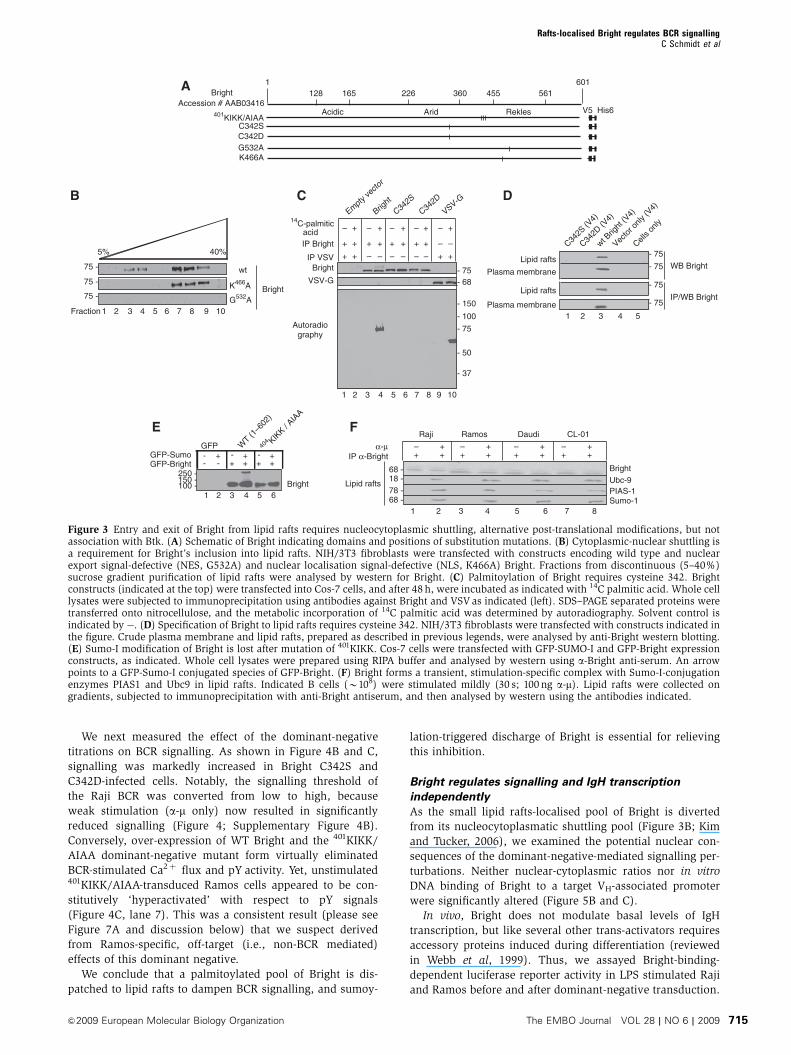
We next measured the effect of the dominant-negative
titrations on BCR signalling. As shown in Figure 4B and C,
signalling was markedly increased in Bright C342S and
C342D-infected cells. Notably, the signalling threshold of
the Raji BCR was converted from low to high, because
weak stimulation (a-m only) now resulted in significantly
reduced signalling (Figure 4; Supplementary Figure 4B).
Conversely, over-expression of WT Bright and the 401KIKK/
AIAA dominant-negative mutant form virtually eliminated
BCR-stimulated Ca2þ flux and pY activity. Yet, unstimulated401KIKK/AIAA-transduced Ramos cells appeared to be con-
stitutively ‘hyperactivated’ with respect to pY signals
(Figure 4C, lane 7). This was a consistent result (please see
Figure 7A and discussion below) that we suspect derived
from Ramos-specific, off-target (i.e., non-BCR mediated)
effects of this dominant negative.
We conclude that a palmitoylated pool of Bright is dis-
patched to lipid rafts to dampen BCR signalling, and sumoy-
lation-triggered discharge of Bright is essential for relieving
this inhibition.
Bright regulates signalling and IgH transcription
independently
As the small lipid rafts-localised pool of Bright is diverted
from its nucleocytoplasmatic shuttling pool (Figure 3B; Kim
and Tucker, 2006), we examined the potential nuclear con-
sequences of the dominant-negative-mediated signalling per-
turbations. Neither nuclear-cytoplasmic ratios nor in vitro
DNA binding of Bright to a target VH-associated promoter
were significantly altered (Figure 5B and C).
In vivo, Bright does not modulate basal levels of IgH
transcription, but like several other trans-activators requires
accessory proteins induced during differentiation (reviewed
in Webb et al, 1999). Thus, we assayed Bright-binding-
dependent luciferase reporter activity in LPS stimulated Raji
and Ramos before and after dominant-negative transduction.
ReklesArid
BrightAccession # AAB03416
1 601455 561226 360128 165
Acidic
A
B C D
401KIKK/AIAAV5 His6
C342SC342D
K466AG532A
wt
K466A
40%5%
3 4 5 10976 81 2FractionG532A
Bright
75 -
75 -
75 -
41 2 3 9 1085 6 7
+– + – +– + –+–
C342S
C342D
Bright
Empty ve
ctor
14C-palmitic acid
VSV-G
Autoradiography
- 75
- 50
- 37
- 150
- 100
- 75- 68
Bright
VSV-G
IP Bright
IP VSV
++ + + –+ + –++
–+ + – +– – +––
Cells o
nly
C342S
(V4)
C342D
(V4)
1 2 3 4 5
wt Brig
ht (V
4)
Vecto
r only
(V4)
IP/WB Bright
WB BrightLipid rafts
Plasma membrane
- 75
- 75
- 75
- 75
E
GFP-Sumo - + - +- +GFP W
T (1–6
02)
404 KIK
K / AIA
A
GFP-Bright + + + +- -
1 2 3 4 5 6100 -150 -250 -
Bright
Fα-μ
Raji Ramos Daudi CL-01
– + – +– + – ++ + + ++ + + +IP α-Bright
41 2 3 85 6 7
BrightUbc-9PIAS-1Sumo-1
68 -18 -78 -68 -
Lipid rafts
Lipid rafts
Plasma membrane
Figure 3 Entry and exit of Bright from lipid rafts requires nucleocytoplasmic shuttling, alternative post-translational modifications, but notassociation with Btk. (A) Schematic of Bright indicating domains and positions of substitution mutations. (B) Cytoplasmic-nuclear shuttling isa requirement for Bright’s inclusion into lipid rafts. NIH/3T3 fibroblasts were transfected with constructs encoding wild type and nuclearexport signal-defective (NES, G532A) and nuclear localisation signal-defective (NLS, K466A) Bright. Fractions from discontinuous (5–40%)sucrose gradient purification of lipid rafts were analysed by western for Bright. (C) Palmitoylation of Bright requires cysteine 342. Brightconstructs (indicated at the top) were transfected into Cos-7 cells, and after 48 h, were incubated as indicated with 14C palmitic acid. Whole celllysates were subjected to immunoprecipitation using antibodies against Bright and VSV as indicated (left). SDS–PAGE separated proteins weretransferred onto nitrocellulose, and the metabolic incorporation of 14C palmitic acid was determined by autoradiography. Solvent control isindicated by�. (D) Specification of Bright to lipid rafts requires cysteine 342. NIH/3T3 fibroblasts were transfected with constructs indicated inthe figure. Crude plasma membrane and lipid rafts, prepared as described in previous legends, were analysed by anti-Bright western blotting.(E) Sumo-I modification of Bright is lost after mutation of 401KIKK. Cos-7 cells were transfected with GFP-SUMO-I and GFP-Bright expressionconstructs, as indicated. Whole cell lysates were prepared using RIPA buffer and analysed by western using a-Bright anti-serum. An arrowpoints to a GFP-Sumo-I conjugated species of GFP-Bright. (F) Bright forms a transient, stimulation-specific complex with Sumo-I-conjugationenzymes PIAS1 and Ubc9 in lipid rafts. Indicated B cells (B108) were stimulated mildly (30 s; 100 ng a-m). Lipid rafts were collected ongradients, subjected to immunoprecipitation with anti-Bright antiserum, and then analysed by western using the antibodies indicated.
Rafts-localised Bright regulates BCR signallingC Schmidt et al
&2009 European Molecular Biology Organization The EMBO Journal VOL 28 | NO 6 | 2009 715
As shown in Figure 5D, LPS-induced increase in reporter
activity above endogenous levels (lanes 5 and 29) was
equally enhanced by over-expression of WT (lanes 11 and
35) or substitution-mutant forms of Bright (lanes 17, 23, 41
and 47). Five minutes of stimulation using a-m sufficient to
influence BCR signalling (Figure 4) failed to elevate reporter
activity above background levels (Figure 5D, lanes 6, 12, 18,
24, 30, 36, 42 and 48). We conclude that the dominant-
negative effects of C342S/D and 401KIKK/AIAA are limited
to the rafts-destined pool, and that Bright functions indepen-
dently as both an inducible transactivator of IgH and a BCR
signalling regulator.
Over-expression of Bright impairs BCR signalling of
normal B-cell subpopulations
What is the consequence of manipulating Bright levels within
lipid rafts of normal B cells? Splenic B cells purified from
Bright-TG mice express 3–5-fold higher levels of Bright within
lipid rafts and whole cells lysates (Figure 6A). TG B cells were
markedly reduced relative to WT in Ca2þ and pY responses
over a wild range of a-m doses with concomitant depletion
kinetics of Bright from stimulated rafts (Figure 6A;
Supplementary Figure 5A).
MZB and immature B-cell populations are significantly
elevated in Bright TG, leading to development of sponta-
neously autoimmunity during aging (Shankar et al, 2007). TG
and WT B splenocytes were sorted under conditions that
avoid BCR activation into immature (T1 and T2), MZB, and
FO populations (Figure 6B). Elevated levels of Bright (B2–5-
fold) were observed in whole cell lysates and in lipid rafts
(Figure 6C and D) prepared from all resting TG populations
except FO. We confirmed that our a-m stimulation conditions
induced no changes in proliferation or differentiation of these
subpopulations, such as that observed by others under
prolonged stimulation (data not shown; Petro et al, 2002;
readdressed in Discussion).
The movement of mIgM, Btk and CD19 into lipid rafts was
unaffected by the starting levels of Bright, as all TG and WT
populations were indistinguishable (Figure 6D). Likewise, all
WT and TG populations responded to strong (a-mþa-CD19)
BCR costimulation with a complete discharge of Bright from
lipid rafts (Figure 6D) and a loss of membrane colocalisation
with mIgM (Supplementary Figure 5B and data not shown).
However, the BCR signalling threshold of immature and MZB
populations, as judged by their response to weak (a-m)
stimulation, correlated inversely with their lipid raft content
of Bright. As shown in Figure 6C, global pY responses of T1,
T2 and MZB TG B cells were reduced relative to WT controls,
consistent with the fact that weak stimulation was insuffi-
cient to discharge Bright from their lipid rafts (Figure 6D). pY
responses of FO WT B cells were, as expected (Li et al, 2001),
relatively less robust (Figure 6C). Resting FO B cells con-
tained slightly lower levels of total or lipid raft-localised
Bright (Figure 6C and D; Shankar et al, 2007) and displayed
less colocalisation between Bright and mIgM than the other
subpopulations (Supplementary Figure 5B). Nonetheless,
weak TG FO signalling was consistently dampened in
response to a-m stimulation, and Bright was not fully
discharged from their lipid rafts (Figure 6C and D).
We conclude that lipid rafts-localised Bright increases the
signalling threshold of MZB, immature, and, to a lower
extent, FO B cells. We further suggest that, as BCR signalling
41 2 3 9 1085 6 7
α-CD19α-μ
75 -
100 -
75 -
Raji (low threshold)41 2 3 9
++–
–++–
–++–
–++–
–++–
–++–
–++–
–++–
–++–
–++–
–
1085 6 7
RaftlinBrightV5
Ramos (high threshold)
A
0
1
1.61.8
2
Raji—empty vector
2 μM Ionomycin
40 μM Digitonin10 mM EGTA
20 mM Tris
100 s
Flu
ores
cenc
e ra
tio
Raji—C342S Bright
2 μM Ionomycin
40 μM Digitonin10 mM EGTA
20 mM Tris
Raji—wild-type Bright
2 μM Ionomycin
40 μM Digitonin10mM EGTA
20 mM Tris
Raji—401KIKK/AIAA Bright
2 μM Ionomycin
40 μM Digitonin10mM EGTA
20 mM Tris
B
WCL
100 -75 -
50 -
37 -25 -
200 -
41 2 3 121110985 6 7 131415
α−CD19α−μ
50 -Bright
41 2 3 121110985 6 7 1314 15
p-Y
γ-Tubulin
C
75 -
Raji (low threshold) Ramos (high threshold)
Lipid rafts
C342D
C342S
Bright
Bright
Empt
y vec
tor
Empt
y vec
tor
C342D
C342S
401 KIK
K/AIA
A
C342D
C342S
Bright
Empt
y vec
tor
401 KIK
K/AIA
A
C342D
C342S
Bright
Empt
y vec
tor
401 KIK
K/AIA
A40
1 KIKK/A
IAA
++
+––
–++
+––
–++
+––
–++
+––
–++
+––
–++
+––
–++
+––
–++
+––
–++
+––
–++
+––
–
1.41.2
0.20.40.60.8
Figure 4 Dominant-negative, lipid rafts localisation-defective mutants modulate BCR signalling. Raji and Ramos cells (5�108) were infectedwith retroviruses encoding wild type and mutant forms of Bright and were then stimulated for 5 min with 500 ng a-m, 500 ng a-CD19 or 500 nga-mþ 500 ng a-CD19. (A) Levels of Bright in lipid rafts are altered by dominant-negative forms. Lipid rafts levels of total Bright(endogenousþ ectopic) were measured by anti-Bright western. Levels of ectopic V5-tagged wild-type Bright, a palmitoylation-defective(C342S/D) form, which is unable to enter lipid rafts, and a Sumo-I-mutant form (401KIKK/AIAA), unable to be discharged from rafts, weredetected by anti-V5 western. Raftlin was used as a loading control. (B) Intracellular free [Ca2þ ] is increased by palmitoylation-deficient anddecreased by wild type or Sumo-I-deficient titration of endogenous Bright. Transduced Raji B cells (1�106 cells/ml) were loaded with 2mMIndo-1 and stimulated with 1 ng or 40mg of a-m (indicated as low and high concentrations, respectively, by triangles). Fluorescence signal wasplotted against time (scale bar¼ 100 s). Internal calibration was performed as detailed in Materials and methods. Downward arrows indicatetimes of reagent addition. (C) Dominant-negative Bright mutants alter global phosphotyrosine responses. Whole cell lysates prepared from celllines established and stimulated in (A) were analysed by anti-pY, Bright and tubulin (loading control) western.
Rafts-localised Bright regulates BCR signallingC Schmidt et al
The EMBO Journal VOL 28 | NO 6 | 2009 &2009 European Molecular Biology Organization716

strength contributes to B-cell subset development (Loder
et al, 1999; Cariappa et al, 2001; Niiro and Clark, 2002;
Petro et al, 2002; Su and Rawlings, 2002; Casola et al, 2004;
Su et al, 2004; Hoek et al, 2006), the skewed T1 and MZB
populations in Bright-over-expressing B-cells derived, at least
in part, from impaired signalling.
Phosphorylation of Btk and TFII-I within lipid rafts is
inhibited by Bright
Btk interacts with Bright (Webb et al, 2000; Rajaiya et al,
2005) to modulate its transcriptional activity in the nucleus
(Rajaiya et al, 2006). Thus, it seemed particularly informative
to determine how Bright levels outside the nucleus affect Btk
activation. First, we examined whole cell extracts prepared
from the dominant-negative transduced cell lines (Figure 7A).
When Bright entry into lipid rafts was blocked by over-
expression of the palmitoylation-defective C342S/D mutant,
pY-Btk was robustly detected after BCR stimulation of either
the less sensitive (Ramos) or the more sensitive (Raji) cell
line. Under conditions in which lipid rafts levels of
Bright were increased (by either WT over-expression or
Bright-401KIKK/AIAA retention), pY of Btk was inhibited
(Figure 7A). Note that the constitutive hyperactivation
phenotype observed for global pTyr (Figure 4C, lane 7) was
confirmed in this independent set of 401-KIKK/AIAA Ramos
transductants.
These results suggested that Btk activation by pY in lipid
rafts is inhibited by Bright. Consistent with this hypothesis,
1 2 3 4
C342S
C342Dwild-type
IVT
IVT
IVT
–––
–
––
––
–
––
–
Bright
S107 VH 1-MAR
401KIKK/AIAAIVT
A
C342SC342Dwt Bright
9 10 136 7 8 11 1254321 1514 16
CY NP CH NM CY NP CH NM CY NP CH NM CY NP CH NM
ReklesArid
BrightAccession # AAB03416
1 601455 561226 360128 165
Acidic
C342SC342D
Raji
Bright
401KIKK/AIAA
401KIKK/AIAA
75 -
C
B
D
0
2
4
6
Fire
fly/R
enill
a
8
10
12
14
LPS
α-μ
+–– – +
– +–– – –
– +–– – +
– +–– – +
– +–– – +
– +–– – +
– +–– – +
– +–– – +
– +–– – +
– +–– – +
– +–– – +
– +–– – +
– +–– – +
– +–– – +
– +–– – +
– +–– – +
–
pGL3-btppGL3 pGL3-btppGL3 pGL3-btppGL3 pGL3-btppGL3 pGL3-btppGL3 pGL3-btppGL3 pGL3-btppGL3 pGL3-btppGL3
Empty vector Bright 401KIKK/AIAA C342S C342DEmpty vector Bright 401KIKK/AIAA
Raji Ramos
1 2 3 4 5 6 7 8 9 10 11 12 13 14 15 16 17 18 19 20 21 22 23 24 25 26 27 28 29 30 31 32 33 34 35 36 37 38 39 40 41 42 43 44 45 46 47 48
Figure 5 Wild type and dominant-negative mutant forms of Bright are indistinguishable in DNA-binding, subcellular localisation andtranscriptional activity. (A) Schematic illustration of Bright amino acid substitution mutations. (B) Subcellular fractionation of Bright isunaltered by dominant-negative transduction. Raji (107 cells), expressing wild type and the indicated mutant forms of Bright, were subjected tofractionation into cytoplasm (CY), nucleoplasm (NP), chromatin (CH) and nuclear matrix (NM) and analysed by western using a-Bright anti-serum. (C) Substitution mutants bind indistinguishably to IgH promoter sites. The indicated forms of Bright were prepared by in vitrotranscription/translation (IVT) and subjected to electrophoretic mobility shift assays using 32P labelled S107 VH1-MAR probe. Specificity ofbinding was demonstrated by a-Bright super-shift and cold probe competition (not shown) as previously described (Herrscher et al, 1995; Zonget al, 2000; Kaplan et al, 2001). (D) Bright transactivation of inducible IgH promoter activity is not changed by dominant-negative transduction.Exponentially growing Raji and Ramos cells, stably transduced with retroviruses encoding empty vector (control) or wild type and mutantforms of Bright (as indicated), were transfected by electroporation with either a Firefly luciferase reporter (pGL3) or a pGL3-derived Bright-responsive (Kim and Tucker, 2006) reporter (VH1-MAR-pGCL3) driven by the S107 VH1-MAR-containing promoter plus Renilla luciferase. Afterculture for 2 days, cells were either untreated or stimulated for 5 days with 20mg/ml LPS or stimulated for 5 min with anti-IgM F(ab0)2 fragment,as described in the Materials and methods. Dual luciferase activity was then measured as described (Rajaiya et al, 2006) and expressed as theFirefly/Renilla ratio.
Rafts-localised Bright regulates BCR signallingC Schmidt et al
&2009 European Molecular Biology Organization The EMBO Journal VOL 28 | NO 6 | 2009 717

pY-Btk was detected in a-Btk IPs prepared from lipid rafts of
all a-m stimulated WT B-cell populations but in none of the
corresponding Bright-over-expressing TG B cells (Figure 7B).
As predicted by the global pY results, all WT and TG B-cells
responded with robust pY-Btk after strong (a-mþa-CD19)
BCR ligation (Figure 6C).
Btk pY is required for TFII-I function as a nuclear tran-
scription factor (Rajaiya et al, 2005, 2006) as well as for its
cytoplasmic interaction with PLCg and subsequent inhibition
of PLCg-mediated Ca2þ mobilisation (Guo et al, 2004;
Caraveo et al, 2006). As observed for Btk, TFII-I moved into
lipid rafts of TG and WT B-cell subsets after a-m stimulation
C
D
Btk
75 -
50 -
Raftlin
Bright
γ-Tubulin
μ
CD-19
TFII-I
Bright
100 -
100 -
150 -
75 -
100 -
75 -
A
p-Y
2–5x Brighttransgene wt
2–5x Brighttransgene wt
––
–+ +
+––
–+ +
+––
–+ +
+––
–+ +
+––
–+ +
+––
–+ +
+––
–+ +
+––
–+ +
+
MZ B cells FO B cells
100 -
75 -
150 -
α-CD19α-μ
2–5x Brighttransgene wt
2–5x Brighttransgene wt
T1 B cells T2 B cells
W
C
L
Lipid rafts
0
1
1.82
2 μM Ionomycin
40 μM Digitonin10 mM EGTA
20 mM Tris
2 μM Ionomycin
40 μM Digitonin10mM EGTA
20 mM Trisα-IgM F(ab’)2 α-IgM F(ab’)2
Flu
ores
cenc
e ra
tio
100 s Bright transgenic CD43– splenocytes Wild-type CD43– splenocytes
B
FSC
SS
C
B220+ fraction
B220
CD
93
CD23
CD
21
T 2
T 1
F O BM Z B
31 2 118 9 1074 5 6 12 13 14 15 16 17 18 19 20 21 22 23 24
1.61.41.2
0.80.60.40.2
Figure 6 Transgenic over-expression of Bright decreases BCR signalling of normal B cells. (A) Mobilisation of intracellular Ca2þ is reduced byBright over-expression. Wild type and Bright transgenic mice were sacrificed, splenic CD43� B cells were prepared by negative selection,loaded with Indo-1 and then subjected to measurements of intracellular Ca2þ using 1 ng (low), 500 ng (medium) or 40 mg a-m (high) tostimulate 106 cells; Ionomycin, Digitonin, EGTA and Tris were used for internal calibration. (B) Purification of T1, T2, FO and MZ B-cellpopulations. B cells were prepared from single cell suspensions of transgenic (shown here) and wild-type (not shown) splenocytes. B220þ
B-cell subpopulations were defined as T1 (CD93þ CD23� CD21�), T2 (CD93þ CD23þ CD21þ ), FO (CD93� CD23þ CD21þ ) and MZ (CD93�
CD23� CD21þ ). (C) Over-expression of Bright inhibits global phosphotyrosine responses of isolated B-cell populations. Each indicatedsubpopulation (B106 cells) was stimulated for 5 min using 1 ng a-m, 1 ng a-CD19 or 1 ng a-mþ 1 ng a-CD19. Whole cell lysates (WCL) wereprepared from each and then were subjected to SDS–PAGE/western blotting using a-pY, a-Bright and a-Tubulin (loading control) anti-sera.(D) Trafficking of BCR signalling components in and out of lipid rafts is not disturbed in transgenic B-cell populations. The indicatedsubpopulations (B2�106 cells each) were stimulated for 5 min using 2 ng a-m, 2 ng a-CD19 or 2 ng a-mþ 2 ng a-CD19. The entire preparation ofeach was then used for the isolation of lipid rafts. Fractions from the gradient centrifugation were collected and divided into two equal parts.One part was analysed directly by SDS–PAGE/western (lipid rafts from B106 cells per lane) using the antibodies indicated to serve as an inputcontrol for the IP performed with the other half of the RIPA extracted samples (shown in Figure 7B). Here, increased levels of coprecipitated m,Raftlin and CD19 indicated coalescence of lipid rafts upon BCR engagement (Depoil et al, 2008).
Rafts-localised Bright regulates BCR signallingC Schmidt et al
The EMBO Journal VOL 28 | NO 6 | 2009 &2009 European Molecular Biology Organization718

(Figures 6D and 7B). However, while stronger stimulation
(a-mþ a-CD19) further concentrated Btk and other signalo-
some components there, TFII-I was codischarged with Bright
(Figures 6D and 7B). Notably, phosphorylation of TFII-I and
Btk was inhibited in a-m stimulated rafts of Bright-over-
expressing TG B cells (Figure 7B).
These results indicate that lipid rafts-localised Bright con-
tributes to dampening of BCR responses by decoupling Btk
activation and downstream immediate early events, such as
tyrosine phosphorylation of TFII-I.
Discussion
We have shown that 1–10% of Bright, a B-cell-specific
transcriptional activator of IgH transcription, associates
with the BCR complex in lipid rafts prepared from trans-
formed mature B-cell lines or from primary B-cell populations
purified from mouse splenocytes or from human PBL. We
demonstrated that Bright is palmitoylated at a single cysteine
residue and that this modification is required for its localisa-
tion in lipid rafts. Our data indicate that the lipid raft
concentration of Bright is used for BCR threshold signalling,
as the sensitivity to BCR stimulation, as measured by calcium
flux and transmission of global and Btk-mediated pTyr sig-
nals, depends on the amount of Bright discharged from the
rafts. An inducible, rafts-specific association between Bright
and Sumo-I E2/Ubc-9 and E3/PIAS-1 enzymes, along with
accumulation of sumoylated Bright in the plasma membrane
only after BCR stimulation, led us to test whether this post-
translational modification triggered Bright discharge.
Signalling alterations observed in TG B cells that over-express
Bright, or in B-cells transduced with sumoylation-insensitive
and rafts localisation-defective dominant-negative Bright ret-
roviruses, support this notion. The data suggest a model in
which Bright acts as a ‘brake’ to set a signalling threshold that
is regulated by alternative post-translational modification.
The strength of BCR-derived signals determines the se-
quential development of immature to mature B cells and their
subsequent fates in the spleen (Casola et al, 2004; Gazumyan
et al, 2006; Patterson et al, 2006; Pao et al, 2007). Shankar
et al reported that T1 and MZB cells are statistically increased
relative to other B-cell populations in Bright-over-expressing
TG mice. Although serum Ig levels were increased only
modestly, spontaneous autoimmunity ensued (Shankar
et al, 2007). Petro et al (2002) showed that under conditions
of prolonged (24 h) BCR engagement with high concentra-
tions of a-m (B10 mg/ml/105 cells), normal T1 B cells undergo
apoptosis, whereas T2 B cells proliferate. Under the same
conditions, T2 B cells display a higher threshold for a-m-
elicited signals than T1 cells (Petro et al, 2002; Hoek et al,
Btk
Raftlin
μ
CD-19
TFII-I
Bright
p-Y
75 -
100 -
100 -
150 -
75 -
100 -
75 -
100 -
31 2 118 9 1074 5 6 12 13 14 15 16 17 18 19 20 21 22 23 24
B
IP α-Btk
2–5x Brighttransgene wt 2–5x Bright
transgene wt
++
+––
–++
+––
–++
+––
–++
+––
–++
+––
–++
+––
–++
+––
–++
+––
–
MZ B cells FO B cells
α-CD19α-μ
2–5x Brighttransgene wt 2–5x Bright
transgene wt
T1 B cells T2 B cells
α-CD19
α-μ
75 -
IP α-Btk
75 - p-Y
Btk
C242D
C242S
Bright
Empt
y vec
tor
401 KIK
K/AIA
A
++
–
–
+
+
++
–
–
+
+
++
–
–
+
+
++
–
–
+
+
++
–
–
+
+
Ramos (high threshold)75 -
41 2 3 9 1085 6 7
75 - p-Y
Btk
Raji (low threshold)
A
W
C
L
Lipid rafts
Figure 7 BCR-mediated activation of Btk and TFII-I depends on the levels of Bright in lipid rafts. (A) Dominant-negative Bright mutants alterBtk phosphorylation in B-cell lines. Whole cell lysates prepared from retrovirally transduced cell lines established in Figure 4A were stimulatedfor 5 min using either 500 ng a-m or 500 ng a-mþ 500 ng a-CD19 for 5�108 cells as indicated. After anti-Btk immunoprecipitation, westerns wereperformed with anti-Btk (loading control) and anti-pY anti-sera. (B) Transgenic Bright over-expression inhibits Btk and TFII-I phosphorylationin B-cell subpopulations. Lipid raft extracts prepared using RIPA buffer (described in Figure 6D; equivalent of B106 cells per lane) weresubjected to a-Btk IP, followed by western blotting using the antibodies indicated.
Rafts-localised Bright regulates BCR signallingC Schmidt et al
&2009 European Molecular Biology Organization The EMBO Journal VOL 28 | NO 6 | 2009 719
2006; Meyer-Bahlburg et al, 2008). To focus on early signal-
ling events and to circumvent apoptotic/proliferation com-
plications, we used B103-fold lower concentrations of F(ab0)2
in most of our stimulation assays. We found that over-
expression of Bright led to an elevated level of Bright in
lipid raft-associated signalosomes of all B-cell populations
except FO and increased signalling thresholds to our low
concentrations of a-m. In contrast to the imaging results of
Chung et al (2001), we found that this level of stimulation
was adequate to induce translocation of BCR components
(mIgM, CD19, and Btk) into rafts from all subpopulations.
However, our biochemical approach did not allow us to
address their contention that there are fewer lipid rafts in
immature B cells. That T1 and MZB were particularly sensi-
tive suggests that, in Bright TG mice, these populations would
be compromised in their ability to undergo appropriate
apoptotic responses to self-antigens in vivo. This would
account for the elevated numbers of TG T1 and MZB—
established predecessors for autoreactive B cells (Atencio
et al, 2004; Samuels et al, 2005; Yurasov et al, 2005a, b;
Quinn et al, 2006)—and provide a unique mechanism by
which a B-cell-restricted transcription factor could contribute
intrinsically to B-cell tolerance. On the other hand, and not
mutually exclusive, the selective production of antibodies
associated with the autoimmune syndromes of Bright TGs
(Shankar et al, 2007) could be a direct result of as yet
unidentified, non-IgH transcriptional targets of Bright.
Bright interacts with a well-defined signalling molecule,
Btk (Webb et al, 2000; Rajaiya et al, 2005, 2006). Previously
we hypothesised (Webb et al, 1999) that their interaction in
the cytoplasm allowed Bright to deliver Btk to nuclear IgH
promoters. There, Btk could phosphorylate and activate TFII-
I, which at the time, was the only defined substrate for Btk
(Novina et al, 1999). Subsequent studies by Webb and
colleagues (Webb et al, 2000; Rajaiya et al, 2005, 2006)
support the notion that Bright delivers Btk to places of active
TFII-I transcription in the nucleus.
Extension of the hypothesis would predict that Btk is
required for Bright’s inclusion into lipid rafts and, taken to
the extreme, into lipid raft-localised BCR signalosomes.
However, in Btk-deficient non-B cells, exogenously expressed
Bright accumulated within lipid rafts, indicating that its
localisation is independent of Btk or other B-cell-specific
factors. This led us to hypothesise that Bright might function
in lipid raft-localised BCR complexes to limit or increase the
concentration of Btk. Bright–Btk association was, indeed,
observed in lipid raft-localised BCR complexes. BCR engage-
ment resulted in reversed trafficking patterns in and out of
lipid rafts; that is, Btk accumulated in lipid raft-localised BCR
complexes as Bright was being depleted. Thus, Bright does
not function to limit signalosome-associated Btk.
Alternatively and in contrast to their kinase-dependent
collaboration in the nucleus (Rajaiya et al, 2006), we rea-
soned that Bright–Btk complexes in lipid rafts may be cata-
lytically inactive or compromised, such that Bright has to be
removed from Btk in order for the Tec kinase to achieve full
activity. Consistent with this notion, activation of Btk by
tyrosine phosphorylation is stimulated when Bright’s entry
to lipid rafts is blocked by palmitoylation-defective mutants
and inhibited when Bright is trapped in lipid rafts by a
sumoylation-defective mutant. In further support, we found
that loss of Btk activation occurs in lipid rafts, and inactive
Btk (non-phosphorylated) is associated with Bright there.
This suggested that Bright-containing BCR complexes are
signalling-impaired because the presence of Bright in lipid
rafts raises the threshold of Btk-dependent BCR signalling.
Accordingly, Btk phosphorylation of a direct downstream
substrate, TFII-I, was inhibited within Bright-rich lipid rafts.
The lineage relationships between FO and MZB and the
role that Btk plays in this are controversial (Matthias and
Rolink, 2005; Teague et al, 2007; Welner et al, 2008). MZB
cells have been ascribed to develop either directly from T1
cells (Debnath et al, 2007) or from a subpopulation of CD21int
T2 B cells (Meyer-Bahlburg et al, 2008). Others contend that
both MZB and FO derive from a long lived, post-transitional
follicular B-cell subset described as Follicular Type II
(Cariappa et al, 2007; Allman and Pillai, 2008). Although
Xid phenotypic CBA/N mice show significantly greater loss of
FO (Hardy et al, 1982), their MZB numbers are also reduced
(Liu et al, 1988; Cariappa et al, 2001). The enrichment of
certain Ag specificities into MZB requires functional Btk
(Martin and Kearney, 2000; Kanayama et al, 2005). Our
findings support a common progenitor model and suggest
that if MZB require an intact Btk signalling pathway, Bright
has a function in this regulation.
Bright is the first transcription factor shown to function in
lipid rafts, but its residence there is not unprecedented. Small
cytoplasmic fractions of Stat1 and Stat3 constitutively localise
to lipid rafts and have been suggested to function there
during early stages of cytokine signalling (Sehgal et al,
2002). An isoform of OCA-B, an IgH transcriptional coacti-
vator, localises as a myristoylated form to the cytoplasm and
to plasma membranes but not to lipid rafts per se (Yu et al,
2001, 2006). Similarly, TFII-I was previously detected in
cytoplasmic complexes with PLCg (Caraveo et al, 2006).
Btk-dependent phosphorylation of TFII-I was required for
its interaction with and concomitant inhibition of PLCg-
catalysed Ca2þ mobilisation (Guo et al, 2004). Both TFII-I
and Bright are regulated by nucleocytoplasmic shuttling
(Novina et al, 1999; Nore et al, 2000; Kim and Tucker,
2006). Lipid rafts-designated Bright derives from a palmitoy-
lated pool that requires continual shuttling, as neither NLS
nor NES mutants accumulate in rafts. That nucleocytopla-
smic shuttling is also required for Bright transcriptional
activity (Kim and Tucker, 2006) raises the possibility that its
occupancy in lipid rafts is prerequisite for assembly of and
subsequent transfer of Bright–Btk–TFII-I complexes to the
nucleus. Consistent with this notion, Bright is codischarged
with activated TFII-I from lipid rafts after BCR ligation as a
Sumo-I-modified form. Although sumoylation has been as-
cribed to mediate nuclear import/export and activity of
transcription factors (Liu et al, 2006), the membrane-loca-
lised metabotropic glutamate receptor is targeted by the
sumoylation machinery (Tang et al, 2005). Similarly, the
sumo pathway was shown to control the activity of the
potassium channel K2P1 (Rajan et al, 2005).
Our observations suggest a new avenue for signal propa-
gation between the membrane and the nucleus. They em-
phasise the need to describe the properties of the
compartmentalised pools, their temporal and spatial regula-
tion and the molecular requirement(s) for the signalling
networks involved. Our results extend and enrich the notion
that mice with an artificially altered BCR threshold are more
likely to display features of autoimmunity (Grimaldi et al,
Rafts-localised Bright regulates BCR signallingC Schmidt et al
The EMBO Journal VOL 28 | NO 6 | 2009 &2009 European Molecular Biology Organization720

2005; Goodnow, 2007). We identify the transcription factor
Bright as an unsuspected component of such a network and
implicate it as regulator of an early event in BCR signalling
and immunologic tolerance.
Materials and methods
CellsRaji (EBVþ; McConnell et al, 1992; Miller et al, 1993; Caldwellet al, 1998; Cerimele et al, 2005; Bernasconi et al, 2006), Daudi(EBVþ ; Wang et al, 1990), Ramos (EBV�; Cerimele et al, 2005) andCL01 (EBV�; Laskov et al, 2006) were obtained from ATCC(Manassas, Virginia) and maintained as described (Fell et al,1986). CD43-B cells were prepared by negative selection of wholehuman blood (Gulf Coast Regional Blood Center, Houston, Texas) orfrom B10-wk-old BALB/c murince splenocytes (Webb et al, 1998).Preparative sorts were executed according to Webb et al (1998) andShankar et al (2007). B cells were stained as described by Kim andTucker (2006), and deconvolution was performed according toKuhn and Poenie (2002) and Combs et al (2006).
Molecular and cellular biologyMutant forms of Bright were generated using the site directedmutagenesis kit (Stratagene, CA) and transferred into the retroviralconstruct pVxy (Ngo et al, 2006).
In vitro translation, sumoylation assays and transduction of Bcells were performed as described (Kienker et al, 1998; Rosas-Acosta et al, 2005a, b; Kim and Tucker, 2006). Specificity ofsumoylation reactions was confirmed by cleavage of modifiedBright by Ulp-1 (Li and Hochstrasser (2003). Stabilisation of Sumo-1modified Bright was achieved by alkylation with iodoacetic acidsodium salt (Byrd and Hruby, 2005).
To assay for palmitoylation, WT and mutant forms of Bright aswell as VSV-G were transfected into Cos-7 cells and processed asdescribed (Rose et al, 1984; a-VSV was kindly provided by DrMichael G Roth, U.T. Southwestern Medical Center, Dallas; Yu andRoth, 2002).
Preparation of stable retrovirally transduceddominant-negative B-cell linesStable transductants were established by employment of thePhoenix-A retroviral system. We plated 3�105 amphitrophicPhoenix-A packaging cells in 4 ml of DMEM supplemented with10% fetal bovine serum (FBS) in 60-mm plates. After one day ofculture, cells were transfected using pBabe constructs usingFuGene6, and viral supernatant was harvested 2 days post-transfection, centrifuged, and filtered to remove live cells anddebris. Target cells (3�105) were plated into 60-mm plates andgrowth medium was replaced with viral mixture. Stable cell lineswere established by selection with 2mg/ml of puromycin from day 2post-infection.
Transcriptional analysisThe transcriptional activity of WT Bright and mutant forms wereassayed by luciferase assays (Kim and Tucker, 2006; Rajaiya et al,2006). Raji or Ramos cells (B5�105) stably transduced with emptyvector, WT or one of the mutant forms of Bright (401KIKK/AIAA,C342S or C342D) were mixed with 125 ng of pRL (Renilla) and750 ng of either pGL3 or pGL3btp in 300ml of RPMI. Cells wereincubated for 15 min at room temperature, transferred intoelectroporation cuvettes and subjected to electroporation at 975mFand 260 V. Electroporated cells were left in the cuvette at roomtemperature for 15 min and cultured in RPMI complete growthmedium for 5 h. Cells were then treated with LPS (20mg/ml) for 3days in complete growth medium to stimulate cofactors required forBright transactivation (Nixon et al, 2004a). Anti-IgM stimulationwas performed for 5 min using 500pg a-m for 5�105 cells.Luciferase activity was then measured and normalised accordingto the Dual Luciferase Reporter Assay Kit from Promega.
B-cell stimulationTo measure signalling effects at low doses of anti-IgM wherereceptor internalisation is minimised, we used monoclonal anti-IgMantibodies in the absence of secondary cross-linking (please seeSupplementary Figures 2B, 4B and 5 for optimisation and further
considerations). To stimulate B cells, 500 ng of F(ab’)2 fragments ofa-m (clone JDC-15; Dako [a-human]; OB1022; Southern Biotech[a-mouse]) and a-CD19 (clone HD37; Dako [a-human]; clone SJ25-C1 [a-mouse]) were added to 5�108 cells for 5 min at 371C (or othertimes as indicated in the figures). We determined by FACS analysis(data not shown) and semi-quantitative western of lipid raft-associated mIgM (Supplementary Figure 5A) that under theseconditions B1–5% of mIgM in rafts and membranes are engaged.
Measurement of free intracellular calciumApproximately 1�106 cells/ml were loaded with 2mM Indo-1(Molecular Probes) in HEPES buffered RPMI/1% FBS (pH 7.1) for30 min at 371C and stimulated successively with 1 ng or 40 mg(indicated in Figures 4B and 6A as low or high a-m, respectively.Fluorescence was excited at 340 and 380 nm, and the resultingsignal at each excitation wavelength was plotted against time.Internal calibration was performed using final concentrations of2 mM Ionomycin, 40mM Digitonin, 10 mM EGTA/pH 7 and 20 mMTris exactly as described (Vorndran et al, 1995). At the end of eachcalcium trace, dye calibration to insure comparable loading and dyeresponses were performed by treatment with 2mM ionomycinfollowed by 40mM digitonin to release Indo-1 into the medium thatcontains 1 mM calcium. These levels of calcium saturate the Indo-1to give the apparent Rmax. Subsequently, excess EGTA/pH7 (10 mM)is added, followed by 20 mM Tris base to convert EGTA from the -2or -1 form at pH 7 (and below) to the -4 form, greatly increasing itschelating ability. That this procedure was sufficient to achieve Rmin
was confirmed by the fact that, in the presence of a large excess ofEGTA (10 mM), further additions of Tris base (i.e., beyond twoequivalents) did not lower Indo-1 ratios.
Preparation of lipid raftsApproximately 500 mg of wet cell pellet were washed twice in ice-cold phosphate buffered solution (PBS) and homogenised in 5 ml of10 mM Tris/Cl (pH 7.4), 1 mM EDTA, 250 mM sucrose, 1 mMphenylmethylsulfonyl fluoride and 1mg/ml leupeptin (all fromSigma, St Louis, Montana) in a tightly fitted Dounce homogeniserusing five strokes (Shelton et al, 1982; Short and Barr, 2000). Theresulting homogenate was centrifuged at 900 g for 10 min at 41C, theresulting supernatant was then subjected to centrifugation at110 000 g for 90 min at 41C (Nagamatsu et al, 1992). The resultingmembrane pellet was resuspended in ice cold 500ml TNE buffer(10 mM Tris/Cl [pH 7.4], 150 mM NaCl, 5 mM EDTA, 1% TritonX-100 [Sigma], 10� protease inhibitors [Complete tablets, Roche,Indianapolis, IN]). Sucrose gradients for the preparation of lipidrafts were assembled exactly as described in an earlier publication(Fuentes-Panana et al, 2005). Lipid rafts were isolated by flotationon discontinuous sucrose gradients. Membrane pellets wereextracted for 30 min on ice in TNE buffer. For the discontinuoussucrose gradient, 1 ml of cleared supernatant was mixed with 1 mlof 85% sucrose in TNE and transferred to the bottom of anultracentrifugation tube, followed by overlay with 6 ml of 35%sucrose in TNE and 3.5 ml of 5% sucrose in TNE. Samples werespun at 200 000 g for 30 h at 41C; fractions were collected from thetop of the gradient and analysed using western blotting and/orcoimmunoprecipitation, as described in an earlier publication (Kimand Tucker, 2006).
Immunoprecipitation/western analysesUnabridged BCR complexes have been successfully immunopreci-pitated using either stringent RIPA-based buffers (Indraccolo et al,2002; Gazumyan et al, 2006) or mild conditions, such as Digitoninor NP-40 (Hombach et al, 1988; Batista et al, 1996). We used astringent RIPA formulation of 500 mM NaCl; 10 mM Tris–Cl pH 8;0.1% SDS; 5 mM EDTA, pH 8; 10� protease inhibitor (Completetablet, Roche) to solubilise lipid rafts for subsequent immunopre-cipitation experiments. Briefly, buoyant fractions, taken from thediscontinuous gradient centrifugation, were pooled and incubatedwith the same volume of RIPA buffer on ice for 15 min. Resultingextracts were pre-cleared by rocking with 1 ml of a 5% slurry ofRIPA equilibrated Protein A beads CL-4B (Amersham Pharmacia,Uppsala) for 4 h at 41C and removal of the precipitate. The resultingsupernatant was then subjected to IP/western assays as described(Kim and Tucker, 2006). The following antibodies were used:a-CD19 (clone 6D5, Dako), a-Bright, a-IgM (BD Pharmingen), a-V5(Sigma), a-Raftlin (Dr Akihiko Yoshimura; Fukuoka, Japan; Saekiet al, 2003), a-Sumo-1 (Sigma), a-phosphotyrosine (Sigma),
Rafts-localised Bright regulates BCR signallingC Schmidt et al
&2009 European Molecular Biology Organization The EMBO Journal VOL 28 | NO 6 | 2009 721

a-caspase-3 (Cell Signalling) and anti-TFII-I (kindly provided by DrCarol Webb; Rajaiya et al, 2005).
ApoptosisTo assay for DNA fragmentation, 1�108 cells were treated with100 ng a-m for 72 h and processed as described by Duke et al (1983).In sum, cells were harvested by centrifugation (200 g; 10 min atroom temperature), washed twice with PBS and resuspended in2 ml lysis buffer (100 mM NaCl, 10 mM Tris, 1 mM EDTA, 0.5%NP-40, and 0.5% SDS, pH 7.4) for 10 min at room temperature. DNAwas extracted twice with an equal volume of phenol followed by asingle extraction with an equal volume of chloroform. DNAfragments were resolved in a 0.75% agarose gel in TBE runningbuffer (90 mM Tris, 90 mM boric acid, 1.5 mM EDTA pH 8.4) andvisualised by staining with ethidium bromide and photographyunder UV illumination.
Note added in proofNixon et al (2008) recently reported that dominant inhibition ofBright DNA binding activity in vivo inhibits antibody response tophosphorylcholine of B1 B cells-a subset whose development and
function are highly dependent on Btk signaling. These data provideadditional support for a mechanistic link between Btk and bothnuclear and lipid rafts-localized Bright.
Supplementary dataSupplementary data are available at The EMBO Journal Online(http://www.embojournal.org).
Acknowledgements
The authors are indebted to Paul Das for administrative assistance.We thank Chhaya Das, Maya Ghosh and June V Harriss for experttechnical assistance. We thank Martin P Kracklauer and Dr Mark ABrown for critically reading the manuscript and all members of theTucker lab for comments on the paper. SM is the recipient of anUndergraduate Research Fellowship from UT Austin. Financial aidfrom the NIH is acknowledged by GCI (CA110624), CFW(AI044215), MP (AA015437) and PWT (Marie Betzner MorrowCentennial Endowment, NIH grants AI64886 and CA031534).
References
Allman D, Lindsley RC, DeMuth W, Rudd K, Shinton SA, Hardy RR(2001) Resolution of three nonproliferative immature splenic Bcell subsets reveals multiple selection points during peripheral Bcell maturation. J Immunol 167: 6834–6840
Allman D, Pillai S (2008) Peripheral B cell subsets. Curr OpinImmunol 20: 149–157
Ashery U, Yizhar O, Rotblat B, Kloog Y (2006) Nonconventionaltrafficking of Ras associated with Ras signal organization. Traffic7: 119–126
Atencio S, Amano H, Izui S, Kotzin BL (2004) Separation of the NewZealand Black genetic contribution to lupus from New ZealandBlack determined expansions of marginal zone B and B1a cells.J Immunol 172: 4159–4166
Batista FD, Anand S, Presani G, Efremov DG, Burrone OR (1996)The two membrane isoforms of human IgE assemble intofunctionally distinct B cell antigen receptors. J Exp Med 184:2197–2205
Bernasconi M, Berger C, Sigrist JA, Bonanomi A, Sobek J, Niggli FK,Nadal D (2006) Quantitative profiling of housekeeping andEpstein-Barr virus gene transcription in Burkitt lymphoma celllines using an oligonucleotide microarray. Virol J 3: 43
Bossis G, Melchior F (2006) Regulation of SUMOylation byreversible oxidation of SUMO conjugating enzymes. Mol Cell21: 349–357
Byrd CM, Hruby DE (2005) Development of an in vitro cleavageassay system to examine vaccinia virus I7L cysteine proteinaseactivity. Virol J 2: 63
Caldwell RG, Wilson JB, Anderson SJ, Longnecker R (1998) Epstein-Barr virus LMP2A drives B cell development and survival in theabsence of normal B cell receptor signals. Immunity 9: 405–411
Caraveo G, van Rossum DB, Patterson RL, Snyder SH, Desiderio S(2006) Action of TFII-I outside the nucleus as an inhibitor ofagonist-induced calcium entry. Science 314: 122–125
Cariappa A, Boboila C, Moran ST, Liu H, Shi HN, Pillai S (2007) Therecirculating B cell pool contains two functionally distinct, long-lived, posttransitional, follicular B cell populations. J Immunol179: 2270–2281
Cariappa A, Tang M, Parng C, Nebelitskiy E, Carroll M,Georgopoulos K, Pillai S (2001) The follicular versus marginalzone B lymphocyte cell fate decision is regulated by Aiolos, Btk,and CD21. Immunity 14: 603–615
Carter RH, Fearon DT (1992) CD19: lowering the threshold forantigen receptor stimulation of B lymphocytes. Science 256:105–107
Casola S, Otipoby KL, Alimzhanov M, Humme S, Uyttersprot N,Kutok JL, Carroll MC, Rajewsky K (2004) B cell receptor signalstrength determines B cell fate. Nat Immunol 5: 317–327
Cerimele F, Battle T, Lynch R, Frank DA, Murad E, Cohen C,Macaron N, Sixbey J, Smith K, Watnick RS, Eliopoulos A,Shehata B, Arbiser JL (2005) Reactive oxygen signaling andMAPK activation distinguish Epstein-Barr Virus (EBV)-positive
versus EBV-negative Burkitt’s lymphoma. Proc Natl Acad Sci USA102: 175–179
Chen W, Wang HG, Srinivasula SM, Alnemri ES, Cooper NR (1999)B cell apoptosis triggered by antigen receptor ligation proceedsvia a novel caspase-dependent pathway. J Immunol 163:2483–2491
Cherukuri A, Cheng PC, Sohn HW, Pierce SK (2001) The CD19/CD21complex functions to prolong B cell antigen receptor signalingfrom lipid rafts. Immunity 14: 169–179
Chung JB, Baumeister MA, Monroe JG (2001) Cutting edge: differ-ential sequestration of plasma membrane-associated B cell anti-gen receptor in mature and immature B cells intoglycosphingolipid-enriched domains. J Immunol 166: 736–740
Combs J, Kim SJ, Tan S, Ligon LA, Holzbaur EL, Kuhn J, Poenie M(2006) Recruitment of dynein to the Jurkat immunologicalsynapse. Proc Natl Acad Sci USA 103: 14883–14888
Debnath I, Roundy KM, Weis JJ, Weis JH (2007) Defining in vivotranscription factor complexes of the murine CD21 and CD23genes. J Immunol 178: 7139–7150
Depoil D, Fleire S, Treanor BL, Weber M, Harwood NE, MarchbankKL, Tybulewicz VL, Batista FD (2008) CD19 is essential for B cellactivation by promoting B cell receptor-antigen microclusterformation in response to membrane-bound ligand. NatImmunol 9: 63–72
Dintzis HM, Dintzis RZ, Vogelstein B (1976) Molecular determi-nants of immunogenicity: the immunon model of immuneresponse. Proc Natl Acad Sci USA 73: 3671–3675
Duke RC, Chervenak R, Cohen JJ (1983) Endogenous endonuclease-induced DNA fragmentation: an early event in cell-mediatedcytolysis. Proc Natl Acac Sci USA 80: 6361–6365
Dykstra M, Cherukuri A, Sohn HW, Tzeng SJ, Pierce SK (2003)Location is everything: lipid rafts and immune cell signaling.Annu Rev Immunol 21: 457–481
Fell HP, Smith RG, Tucker PW (1986) Molecular analysis of thet(2;14) translocation of childhood chronic lymphocytic leukemia.Science 232: 491–494
Fuentes-Panana EM, Bannish G, van der Voort D, King LB, MonroeJG (2005) Iga/Igb complexes generate signals for B cell develop-ment independent of selective plasma membrane compartmenta-lization. J Immunol 174: 1245–1252
Gauld SB, Dal Porto JM, Cambier JC (2002) B cell antigen receptorsignaling: roles in cell development and disease. Science 296:1641–1642
Gazumyan A, Reichlin A, Nussenzweig MC (2006) Igb tyrosineresidues contribute to the control of B cell receptor signaling byregulating receptor internalization. J Exp Med 203: 1785–1794
Gocke CB, Yu H, Kang J (2005) Systematic identification andanalysis of mammalian small ubiquitin-like modifier substrates.J Biol Chem 280: 500–512
Goodnow CC (2007) Multistep pathogenesis of autoimmunedisease. Cell 130: 25–35
Rafts-localised Bright regulates BCR signallingC Schmidt et al
The EMBO Journal VOL 28 | NO 6 | 2009 &2009 European Molecular Biology Organization722
Grimaldi CM, Hill L, Xu X, Peeva E, Diamond B (2005) Hormonalmodulation of B cell development and repertoire selection. MolImmunol 42: 811–820
Guo B, Rothstein TL (2005) B cell receptor (BCR) cross-talk: IL-4creates an alternate pathway for BCR-induced ERK activation thatis phosphatidylinositol 3-kinase independent. J Immunol 174:5375–5381
Guo S, Ferl GZ, Deora R, Riedinger M, Yin S, Kerwin JL, Loo JA,Witte ON (2004) A phosphorylation site in Bruton’s tyrosinekinase selectively regulates B cell calcium signaling efficiencyby altering phospholipase C-g activation. Proc Natl Acad Sci USA101: 14180–14185
Gupta N, Wollscheid B, Watts JD, Scheer B, Aebersold R, DeFrancoAL (2006) Quantitative proteomic analysis of B cell lipid raftsreveals that ezrin regulates antigen receptor-mediated lipid raftdynamics. Nat Immunol 7: 625–633
Hakre S, Tussie-Luna MI, Ashworth T, Novina CD, Settleman J,Sharp PA, Roy AL (2006) Opposing functions of TFII-I splicedisoforms in growth factor-induced gene expression. Mol Cell 24:301–308
Hardy RR, Hayakawa K, Haaijman J, Herzenberg LA (1982) B-cellsubpopulations identified by two-colour fluorescence analysis.Nature 297: 589–591
Harwood NE, Batista FD (2008) New insights into the earlymolecular events underlying B cell activation. Immunity 28:609–619
Herrscher RF, Kaplan MH, Lelsz DL, Das C, Scheuermann R, TuckerPW (1995) The immunoglobulin heavy-chain matrix-associatingregions are bound by Bright: a B cell-specific trans-activatorthat describes a new DNA-binding protein family. Genes Dev 9:3067–3082
Hoek KL, Antony P, Lowe J, Shinners N, Sarmah B, Wente SR, WangD, Gerstein RM, Khan WN (2006) Transitional B cell fate isassociated with developmental stage-specific regulation of dia-cylglycerol and calcium signaling upon B cell receptor engage-ment. J Immunol 177: 5405–5413
Hombach J, Leclercq L, Radbruch A, Rajewsky K, Reth M (1988) Anovel 34kD protein co-isolated with the IgM molecule in surfaceIgM-expressing cells. EMBO J 7: 3451–3456
Indraccolo S, Minuzzo S, Zamarchi R, Calderazzo F, Piovan E,Amadori A (2002) Alternatively spliced forms of Iga and Igbprevent B cell receptor expression on the cell surface. Eur JImmunol 32: 1530–1540
Kahyo T, Nishida T, Yasuda H (2001) Involvement of PIAS1 in thesumoylation of tumor suppressor p53. Mol Cell 8: 713–718
Kanayama N, Cascalho M, Ohmori H (2005) Analysis of marginalzone B cell development in the mouse with limited B celldiversity: role of the antigen receptor signals in the recruitmentof B cells to the marginal zone. J Immunol 174: 1438–1445
Kaplan MH, Zong RT, Herrscher RF, Scheuermann RH, Tucker PW(2001) Transcriptional activation by a matrix associating region-binding protein. contextual requirements for the function ofbright. J Biol Chem 276: 21325–21330
Kienker LJ, Ghosh MR, Tucker PW (1998) Regulatory elements inthe promoter of a murine TCRD V gene segment. J Immunol 161:791–804
Kim D, Probst L, Das C, Tucker PW (2007) REKLES is an ARID3-restricted multifunctional domain. J Biol Chem 282: 15768–15777
Kim D, Tucker PW (2006) A regulated nucleocytoplasmic shuttlecontributes to Bright’s function as a transcriptional activator ofimmunoglobulin genes. Mol Cell Biol 26: 2187–2201
Kuhn JR, Poenie M (2002) Dynamic polarization of the microtubulecytoskeleton during CTL-mediated killing. Immunity 16:111–121
Laskov R, Berger N, Scharff MD, Horwitz MS (2006) Tumor necrosisfactor-a and CD40L modulate cell surface morphology and induceaggregation in Ramos Burkitt’s lymphoma cells. Leuk Lymphoma47: 507–519
Li SJ, Hochstrasser M (2003) The Ulp1 SUMO isopeptidase: distinctdomains required for viability, nuclear envelope localization, andsubstrate specificity. J Cell Biol 160: 1069–1081
Li X, Martin F, Oliver AM, Kearney JF, Carter RH (2001) Antigenreceptor proximal signaling in splenic B-2 cell subsets. J Immunol166: 3122–3129
Lin D, Ippolito GC, Zong RT, Bryant J, Koslovsky J, Tucker P (2007)Bright/ARID3A contributes to chromatin accessibility of theimmunoglobulin heavy chain enhancer. Mol Cancer 6: 23
Liu H, Ippolito GC, Wall JK, Niu T, Probst L, Lee BS, Pulford K,Banham AH, Stockwin L, Shaffer AL, Staudt LM, Das C, Dyer MJ,Tucker PW (2006) Functional studies of BCL11A: characterizationof the conserved BCL11A-XL splice variant and its interactionwith BCL6 in nuclear paraspeckles of germinal center B cells. MolCancer 5: 18
Liu YJ, Lortan JE, Oldfield S, MacLennan IC (1988) CBA/N micehave marginal zone B cells with normal surface immunoglobulinphenotype. Adv Exp Med Biol 237: 105–111
Loder F, Mutschler B, Ray RJ, Paige CJ, Sideras P, Torres R, LamersMC, Carsetti R (1999) B cell development in the spleen takesplace in discrete steps and is determined by the quality of B cellreceptor-derived signals. J Exp Med 190: 75–89
Martin F, Kearney JF (2000) Positive selection from newly formed tomarginal zone B cells depends on the rate of clonal production,CD19, and btk. Immunity 12: 39–49
Matthias P, Rolink AG (2005) Transcriptional networks in develop-ing and mature B cells. Nat Rev Immunol 5: 497–508
McConnell FM, Shears SB, Lane PJ, Scheibel MS, Clark EA(1992) Relationships between the degree of cross-linking ofsurface immunoglobulin and the associated inositol 1,4,5-trisphosphate and Ca2+ signals in human B cells. Biochem J284: 447–455
Meyer-Bahlburg A, Andrews SF, Yu KO, Porcelli SA, Rawlings DJ(2008) Characterization of a late transitional B cell populationhighly sensitive to BAFF-mediated homeostatic proliferation.J Exp Med 205: 155–168
Miller CL, Longnecker R, Kieff E (1993) Epstein-Barr virus latentmembrane protein 2A blocks calcium mobilization in B lympho-cytes. J Virol 67: 3087–3094
Nagamatsu S, Kornhauser JM, Burant CF, Seino S, Mayo KE, Bell GI(1992) Glucose transporter expression in brain. cDNA sequenceof mouse GLUT3, the brain facilitative glucose transporter iso-form, and identification of sites of expression by in situ hybridi-zation. J Biol Chem 26: 467–472
Ngo VN, Davis RE, Lamy L, Yu X, Zhao H, Lenz G, Lam LT, Dave S,Yang L, Powell J, Staudt LM (2006) A loss-of-function RNAinterference screen for molecular targets in cancer. Nature 441:106–110
Niiro H, Clark EA (2002) Regulation of B-cell fate by antigen-receptor signals. Nat Rev Immunol 2: 945–956
Nixon JC, Rajaiya J, Webb CF (2004a) Mutations in the DNA-binding domain of the transcription factor Bright act as dominantnegative proteins and interfere with immunoglobulin transacti-vation. J Biol Chem 279: 52465–52472
Nixon JC, Ferrell S, Miner C, Oldham AL, Hochgeschwender U,Webb CF (2008) Transgenic mice expressing dominant-negativebright exhibit defects in B1 B cells. J Immunol 181: 6913–6922
Nixon JC, Rajaiya JB, Ayers N, Evetts S, Webb CF (2004b) Thetranscription factor, Bright, is not expressed in all human Blymphocyte subpopulations. Cell Immunol 228: 42–53
Nore BF, Vargas L, Mohamed AJ, Branden LJ, Backesjo CM, IslamTC, Mattsson PT, Hultenby K, Christensson B, Smith CI (2000)Redistribution of Bruton’s tyrosine kinase by activation of phos-phatidylinositol 3-kinase and Rho-family GTPases. Eur J Immunol30: 145–154
Novina CD, Kumar S, Bajpai U, Cheriyath V, Zhang K, Pillai S,Wortis HH, Roy AL (1999) Regulation of nuclear localization andtranscriptional activity of TFII-I by Bruton’s tyrosine kinase. MolCell Biol 19: 5014–5024
Pao LI, Lam KP, Henderson JM, Kutok JL, Alimzhanov M, NitschkeL, Thomas ML, Neel BG, Rajewsky K (2007) B cell-specificdeletion of protein-tyrosine phosphatase Shp1 promotes B-1acell development and causes systemic autoimmunity. Immunity27: 35–48
Patterson HC, Kraus M, Kim YM, Ploegh H, Rajewsky K (2006) TheB cell receptor promotes B cell activation and proliferationthrough a non-ITAM tyrosine in the Iga cytoplasmic domain.Immunity 25: 55–65
Petro JB, Gerstein RM, Lowe J, Carter RS, Shinners N, Khan WN(2002) Transitional type 1 and 2 B lymphocyte subsets aredifferentially responsive to antigen receptor signaling. J BiolChem 277: 48009–48019
Putnam MA, Moquin AE, Merrihew M, Outcalt C, Sorge E, CaballeroA, Gondre-Lewis TA, Drake JR (2003) Lipid raft-independent Bcell receptor-mediated antigen internalization and intracellulartrafficking. J Immunol 170: 905–912
Rafts-localised Bright regulates BCR signallingC Schmidt et al
&2009 European Molecular Biology Organization The EMBO Journal VOL 28 | NO 6 | 2009 723

Quinn WJ, Noorchashm N, Crowley JE, Reed AJ, Noorchashm H,Naji A, Cancro MP (2006) Cutting edge: impaired transitional Bcell production and selection in the nonobese diabetic mouse.J Immunol 176: 7159–7164
Rajaiya J, Hatfield M, Nixon JC, Rawlings DJ, Webb CF (2005)Bruton’s tyrosine kinase regulates immunoglobulin promoteractivation in association with the transcription factor Bright.Mol Cell Biol 25: 2073–2084
Rajaiya J, Nixon JC, Ayers N, Desgranges ZP, Roy AL, Webb CF(2006) Induction of immunoglobulin heavy-chain transcriptionthrough the transcription factor Bright requires TFII-I. Mol CellBiol 26: 4758–4768
Rajan S, Plant LD, Rabin ML, Butler MH, Goldstein SA (2005)Sumoylation silences the plasma membrane leak K+ channelK2P1. Cell 121: 37–47
Ravetch JV, Bolland S (2001) IgG Fc receptors. Annu Rev Immunol19: 275–290
Rosas-Acosta G, Langereis MA, Deyrieux A, Wilson VG (2005a)Proteins of the PIAS family enhance the sumoylation of thepapillomavirus E1 protein. Virology 331: 190–203
Rosas-Acosta G, Russell WK, Deyrieux A, Russell DH, Wilson VG(2005b) A universal strategy for proteomic studies of SUMO andother ubiquitin-like modifiers. Mol Cell Proteomics 4: 56–72
Rose JK, Adams GA, Gallione CJ (1984) The presence of cysteine inthe cytoplasmic domain of the vesicular stomatitis virus glyco-protein is required for palmitate addition. Proc Natl Acad Sci USA81: 2050–2054
Saeki K, Miura Y, Aki D, Kurosaki T, Yoshimura A (2003) The B cell-specific major raft protein, Raftlin, is necessary for the integrity oflipid raft and BCR signal transduction. EMBO J 22: 3015–3026
Sampson DA, Wang M, Matunis MJ (2001) The small ubiquitin-likemodifier-1 (SUMO-1) consensus sequence mediates Ubc9 bindingand is essential for SUMO-1 modification. J Biol Chem 276:21664–21669
Samuels J, Ng YS, Coupillaud C, Paget D, Meffre E (2005) Impairedearly B cell tolerance in patients with rheumatoid arthritis. J ExpMed 201: 1659–1667
Schwarz SE, Matuschewski K, Liakopoulos D, Scheffner M, JentschS (1998) The ubiquitin-like proteins SMT3 and SUMO-1 areconjugated by the UBC9 E2 enzyme. Proc Natl Acad Sci USA 95:560–564
Sehgal PB, Guo GG, Shah M, Kumar V, Patel K (2002) Cytokinesignaling: STATS in plasma membrane rafts. J Biol Chem 277:12067–12074
Shankar M, Nixon JC, Maier S, Workman J, Farris AD, Webb CF(2007) Antinuclear antibody production and autoimmunity intransgenic mice that overexpress the transcription factor Bright.J Immunol 178: 2996–3006
Shelton KR, Guthrie VH, Cochran DL (1982) Oligomeric structure ofthe major nuclear envelope protein lamin B. J Biol Chem 257:4328–4332
Short B, Barr FA (2000) The Golgi apparatus. Curr Biol 10:R583–R585
Simons K, Toomre D (2000) Lipid rafts and signal transduction.Nat Rev Mol Cell Biol 1: 31–39
Sims GP, Ettinger R, Shirota Y, Yarboro CH, Illei GG, Lipsky PE(2005) Identification and characterization of circulating humantransitional B cells. Blood 105: 4390–4398
Sohn HW, Tolar P, Jin T, Pierce SK (2006) Fluorescence resonanceenergy transfer in living cells reveals dynamic membrane changesin the initiation of B cell signaling. Proc Natl Acad Sci USA 103:8143–8148
Sproul TW, Malapati S, Kim J, Pierce SK (2000) Cutting edge: B cellantigen receptor signaling occurs outside lipid rafts in immature Bcells. J Immunol 165: 6020–6023
Stoddart A, Dykstra ML, Brown BK, Song W, Pierce SK, Brodsky FM(2002) Lipid rafts unite signaling cascades with clathrin toregulate BCR internalization. Immunity 17: 451–462
Su TT, Guo B, Wei B, Braun J, Rawlings DJ (2004) Signaling intransitional type 2 B cells is critical for peripheral B-cell devel-opment. Immunol Rev 197: 161–178
Su TT, Rawlings DJ (2002) Transitional B lymphocyte subsetsoperate as distinct checkpoints in murine splenic B cell develop-ment. J Immunol 168: 2101–2110
Tang Z, El Far O, Betz H, Scheschonka A (2005) Pias1 interactionand sumoylation of metabotropic glutamate receptor 8. J BiolChem 280: 38153–38159
Teague BN, Pan Y, Mudd PA, Nakken B, Zhang Q, Szodoray P,Kim-Howard X, Wilson PC, Farris AD (2007) Cutting edge:Transitional T3 B cells do not give rise to mature B cells, haveundergone selection, and are reduced in murine lupus. J Immunol178: 7511–7515
Venkatesh J, Peeva E, Xu X, Diamond B (2006) Cutting edge:hormonal milieu, not antigenic specificity, determines the maturephenotype of autoreactive B cells. J Immunol 176: 3311–3314
Vorndran C, Minta A, Poenie M (1995) New fluorescent calciumindicators designed for cytosolic retention or measuring calciumnear membranes. Biophys J 69: 2112–2124
Wang F, Gregory C, Sample C, Rowe M, Liebowitz D, Murray R,Rickinson A, Kieff E (1990) Epstein-Barr virus latent membraneprotein (LMP1) and nuclear proteins 2 and 3C are effectors ofphenotypic changes in B lymphocytes: EBNA-2 and LMP1 co-operatively induce CD23. J Virol 64: 2309–2318
Webb C, Zong RT, Lin D, Wang Z, Kaplan M, Paulin Y, Smith E,Probst L, Bryant J, Goldstein A, Scheuermann R, Tucker P (1999)Differential regulation of immunoglobulin gene transcription vianuclear matrix-associated regions. Cold Spring Harb Symp QuantBiol 64: 109–118
Webb CF, Das C, Eaton S, Calame K, Tucker PW (1991a) Novelprotein-DNA interactions associated with increased immunoglo-bulin transcription in response to antigen plus interleukin-5. MolCell Biol 11: 5197–5205
Webb CF, Das C, Eneff KL, Tucker PW (1991b) Identification of amatrix-associated region 50 of an immunoglobulin heavy chainvariable region gene. Mol Cell Biol 11: 5206–5211
Webb CF, Smith EA, Medina KL, Buchanan KL, Smithson G, Dou S(1998) Expression of bright at two distinct stages of B lymphocytedevelopment. J Immunol 160: 4747–4754
Webb CF, Yamashita Y, Ayers N, Evetts S, Paulin Y, Conley ME,Smith EA (2000) The transcription factor Bright associates withBruton’s tyrosine kinase, the defective protein in immunodefi-ciency disease. J Immunol 165: 6956–6965
Welner RS, Pelayo R, Kincade PW (2008) Evolving views on thegenealogy of B cells. Nat Rev Immunol 8: 95–106
Wilsker D, Probst L, Wain HM, Maltais L, Tucker PW, Moran E(2005) Nomenclature of the ARID family of DNA-binding pro-teins. Genomics 86: 242–251
Yu S, Roth MG (2002) Casein kinase I regulates membrane bindingby ARF GAP1. Mol Biol Cell 13: 2559–2570
Yu X, Wang L, Luo Y, Roeder RG (2001) Identification and char-acterization of a novel OCA-B isoform. Implications for a role in Bcell signaling pathways. Immunity 14: 157–167
Yu X, Siegel R, Roeder RG (2006) Interaction of the B cell-specifictranscriptional coactivator OCA-B and Galectin-1 and a possiblerole in regulating BCR-mediated B cell proliferation. J Biol Chem281: 15505–15516
Yurasov S, Hammersen J, Tiller T, Tsuiji M, Wardemann H (2005a)B-cell tolerance checkpoints in healthy humans and patients withsystemic lupus erythematosus. Ann NY Acad Sci 1062: 165–174
Yurasov S, Wardemann H, Hammersen J, Tsuiji M, Meffre E, PascualV, Nussenzweig MC (2005b) Defective B cell tolerance check-points in systemic lupus erythematosus. J Exp Med 201: 703–711
Zong RT, Das C, Tucker PW (2000) Regulation of matrix attachmentregion-dependent, lymphocyte-restricted transcription throughdifferential localization within promyelocytic leukemia nuclearbodies. EMBO J 19: 4123–4133
Rafts-localised Bright regulates BCR signallingC Schmidt et al
The EMBO Journal VOL 28 | NO 6 | 2009 &2009 European Molecular Biology Organization724