Extracellular SOD-Derived H2O2 Promotes VEGF Signaling in Caveolae/Lipid Rafts and Post-Ischemic...
14
Extracellular SOD-Derived H 2 O 2 Promotes VEGF Signaling in Caveolae/Lipid Rafts and Post-Ischemic Angiogenesis in Mice Jin Oshikawa 1,3 , Norifumi Urao 1,3 , Ha Won Kim 2,3 , Nihal Kaplan 1,3 , Masooma Razvi 1,3 , Ronald McKinney 1,2,3 , Leslie B. Poole 4 , Tohru Fukai 2,3 , Masuko Ushio-Fukai 1,3 * 1 Center for Lung and Vascular Biology, Department of Pharmacology, University of Illinois at Chicago, Chicago, Illinois, United States of America, 2 Department of Medicine and Pharmacology, University of Illinois at Chicago, Chicago, Illinois, United States of America, 3 Center for Cardiovascular Research, University of Illinois at Chicago, Chicago, Illinois, United States of America, 4 Department of Biochemistry, Wake Forest University School of Medicine, Winston-Salem, North Carolina, United States of America Abstract Reactive oxygen species (ROS), in particular, H 2 O 2 , is essential for full activation of VEGF receptor2 (VEGFR2) signaling involved in endothelial cell (EC) proliferation and migration. Extracellular superoxide dismutase (ecSOD) is a major secreted extracellular enzyme that catalyzes the dismutation of superoxide to H 2 O 2 , and anchors to EC surface through heparin- binding domain (HBD). Mice lacking ecSOD show impaired postnatal angiogenesis. However, it is unknown whether ecSOD- derived H 2 O 2 regulates VEGF signaling. Here we show that gene transfer of ecSOD, but not ecSOD lacking HBD (ecSOD- DHBD), increases H 2 O 2 levels in adductor muscle of mice, and promotes angiogenesis after hindlimb ischemia. Mice lacking ecSOD show reduction of H 2 O 2 in non-ischemic and ischemic limbs. In vitro, overexpression of ecSOD, but not ecSOD-DHBD, in cultured medium in ECs enhances VEGF-induced tyrosine phosphorylation of VEGFR2 (VEGFR2-pY), which is prevented by short-term pretreatment with catalase that scavenges extracellular H 2 O 2 . Either exogenous H 2 O 2 (,500 mM), which is diffusible, or nitric oxide donor has no effect on VEGF-induced VEGFR2-pY. These suggest that ecSOD binding to ECs via HBD is required for localized generation of extracellular H 2 O 2 to regulate VEGFR2-pY. Mechanistically, VEGF-induced VEGFR2-pY in caveolae/lipid rafts, but non-lipid rafts, is enhanced by ecSOD, which localizes at lipid rafts via HBD. One of the targets of ROS is protein tyrosine phosphatases (PTPs). ecSOD induces oxidation and inactivation of both PTP1B and DEP1, which negatively regulates VEGFR2-pY, in caveolae/lipid rafts, but not non-lipid rafts. Disruption of caveolae/lipid rafts, or PTPs inhibitor orthovanadate, or siRNAs for PTP1B and DEP1 enhances VEGF-induced VEGFR2-pY, which prevents ecSOD- induced effect. Functionally, ecSOD promotes VEGF-stimulated EC migration and proliferation. In summary, extracellular H 2 O 2 generated by ecSOD localized at caveolae/lipid rafts via HBD promotes VEGFR2 signaling via oxidative inactivation of PTPs in these microdomains. Thus, ecSOD is a potential therapeutic target for angiogenesis-dependent cardiovascular diseases. Citation: Oshikawa J, Urao N, Kim HW, Kaplan N, Razvi M, et al. (2010) Extracellular SOD-Derived H 2 O 2 Promotes VEGF Signaling in Caveolae/Lipid Rafts and Post- Ischemic Angiogenesis in Mice. PLoS ONE 5(4): e10189. doi:10.1371/journal.pone.0010189 Editor: Krisztian Stadler, Louisiana State University, United States of America Received March 2, 2010; Accepted March 25, 2010; Published April 21, 2010 Copyright: ß 2010 Oshikawa et al. This is an open-access article distributed under the terms of the Creative Commons Attribution License, which permits unrestricted use, distribution, and reproduction in any medium, provided the original author and source are credited. Funding: This research was supported by National Institutes of Health (NIH) R01 Heart and Lung (HL)077524 and HL077524-S1 (to M.U.-F.), HL070187 (to T.F.) and Cancer (CA)126659 (to L.B.P.), American Heart Association (AHA) Grant-In-Aid 0755805Z (to M.U.-F.) and AHA National Center Research Program (NCRP) Innovative Research Grant 0970336N (to M.U.-F), AHA Post-doctoral Fellowship 09POST2250151 (to N.U.), Ruth L. Kirschstein-National Service Research Award (Kirschstein- NRSA) T32 Training Grant (to N.K. and M.R.), Uehara Memorial Foundation and Naito Foundation (to J.O.). The funders had no role in study design, data collection and analysis, decision to publish, or preparation of the manuscript. Competing Interests: The authors have declared that no competing interests exist. * E-mail: [email protected] Introduction Angiogenesis is involved in physiological process such as development and wound healing as well as pathophysiologies such as ischemic heart and limb diseases, atherosclerosis and cancer. In endothelial cells (ECs), vascular endothelial growth factor (VEGF) induces angiogenesis by stimulating EC proliferation and migration primarily through the VEGF receptor type2 (VEGFR2, KDR/ Flk1) [1]. VEGF binding initiates autophosphorylation of VEGFR2, which is followed by activation of diverse downstream signaling events linked to angiogenesis in ECs [2,3]. Reactive oxygen species (ROS), in particular H 2 O 2 , function as key signaling molecules to mediate various biological responses including angiogenesis. Reversible oxidative inactivation of reactive (low pKa) cysteinyl residues (Cys-SH) at active sites in protein tyrosine phosphatases (PTPs) is important mechanism by which ROS stimulate tyrosine phosphorylation-dependent redox signaling events [4,5]. We and others reported that ROS derived from NADPH oxidase play an important role in VEGFR2-mediated signaling linked to EC migration and proliferation [6,7,8] as well as post-ischemic angiogenesis in vivo [9,10]. Evidence reveals that extracellular redox state regulates intracellular signaling [11] or tumor growth [12] by modulating plasma membrane-associated proteins. Exogenous H 2 O 2 induces expression of both VEGF and VEGFR2 [13] and pro-angiogenic responses in ECs [8]. Since H 2 O 2 is diffusible molecule, we have posited that generating extracellular H 2 O 2 at site PLoS ONE | www.plosone.org 1 April 2010 | Volume 5 | Issue 4 | e10189
-
Upload
northwestern -
Category
Documents
-
view
1 -
download
0
Transcript of Extracellular SOD-Derived H2O2 Promotes VEGF Signaling in Caveolae/Lipid Rafts and Post-Ischemic...

Extracellular SOD-Derived H2O2 Promotes VEGFSignaling in Caveolae/Lipid Rafts and Post-IschemicAngiogenesis in MiceJin Oshikawa1,3, Norifumi Urao1,3, Ha Won Kim2,3, Nihal Kaplan1,3, Masooma Razvi1,3, Ronald
McKinney1,2,3, Leslie B. Poole4, Tohru Fukai2,3, Masuko Ushio-Fukai1,3*
1 Center for Lung and Vascular Biology, Department of Pharmacology, University of Illinois at Chicago, Chicago, Illinois, United States of America, 2 Department of
Medicine and Pharmacology, University of Illinois at Chicago, Chicago, Illinois, United States of America, 3 Center for Cardiovascular Research, University of Illinois at
Chicago, Chicago, Illinois, United States of America, 4 Department of Biochemistry, Wake Forest University School of Medicine, Winston-Salem, North Carolina, United
States of America
Abstract
Reactive oxygen species (ROS), in particular, H2O2, is essential for full activation of VEGF receptor2 (VEGFR2) signalinginvolved in endothelial cell (EC) proliferation and migration. Extracellular superoxide dismutase (ecSOD) is a major secretedextracellular enzyme that catalyzes the dismutation of superoxide to H2O2, and anchors to EC surface through heparin-binding domain (HBD). Mice lacking ecSOD show impaired postnatal angiogenesis. However, it is unknown whether ecSOD-derived H2O2 regulates VEGF signaling. Here we show that gene transfer of ecSOD, but not ecSOD lacking HBD (ecSOD-DHBD), increases H2O2 levels in adductor muscle of mice, and promotes angiogenesis after hindlimb ischemia. Mice lackingecSOD show reduction of H2O2 in non-ischemic and ischemic limbs. In vitro, overexpression of ecSOD, but not ecSOD-DHBD,in cultured medium in ECs enhances VEGF-induced tyrosine phosphorylation of VEGFR2 (VEGFR2-pY), which is prevented byshort-term pretreatment with catalase that scavenges extracellular H2O2. Either exogenous H2O2 (,500 mM), which isdiffusible, or nitric oxide donor has no effect on VEGF-induced VEGFR2-pY. These suggest that ecSOD binding to ECs viaHBD is required for localized generation of extracellular H2O2 to regulate VEGFR2-pY. Mechanistically, VEGF-inducedVEGFR2-pY in caveolae/lipid rafts, but non-lipid rafts, is enhanced by ecSOD, which localizes at lipid rafts via HBD. One of thetargets of ROS is protein tyrosine phosphatases (PTPs). ecSOD induces oxidation and inactivation of both PTP1B and DEP1,which negatively regulates VEGFR2-pY, in caveolae/lipid rafts, but not non-lipid rafts. Disruption of caveolae/lipid rafts, orPTPs inhibitor orthovanadate, or siRNAs for PTP1B and DEP1 enhances VEGF-induced VEGFR2-pY, which prevents ecSOD-induced effect. Functionally, ecSOD promotes VEGF-stimulated EC migration and proliferation. In summary, extracellularH2O2 generated by ecSOD localized at caveolae/lipid rafts via HBD promotes VEGFR2 signaling via oxidative inactivation ofPTPs in these microdomains. Thus, ecSOD is a potential therapeutic target for angiogenesis-dependent cardiovasculardiseases.
Citation: Oshikawa J, Urao N, Kim HW, Kaplan N, Razvi M, et al. (2010) Extracellular SOD-Derived H2O2 Promotes VEGF Signaling in Caveolae/Lipid Rafts and Post-Ischemic Angiogenesis in Mice. PLoS ONE 5(4): e10189. doi:10.1371/journal.pone.0010189
Editor: Krisztian Stadler, Louisiana State University, United States of America
Received March 2, 2010; Accepted March 25, 2010; Published April 21, 2010
Copyright: � 2010 Oshikawa et al. This is an open-access article distributed under the terms of the Creative Commons Attribution License, which permitsunrestricted use, distribution, and reproduction in any medium, provided the original author and source are credited.
Funding: This research was supported by National Institutes of Health (NIH) R01 Heart and Lung (HL)077524 and HL077524-S1 (to M.U.-F.), HL070187 (to T.F.) andCancer (CA)126659 (to L.B.P.), American Heart Association (AHA) Grant-In-Aid 0755805Z (to M.U.-F.) and AHA National Center Research Program (NCRP) InnovativeResearch Grant 0970336N (to M.U.-F), AHA Post-doctoral Fellowship 09POST2250151 (to N.U.), Ruth L. Kirschstein-National Service Research Award (Kirschstein-NRSA) T32 Training Grant (to N.K. and M.R.), Uehara Memorial Foundation and Naito Foundation (to J.O.). The funders had no role in study design, data collectionand analysis, decision to publish, or preparation of the manuscript.
Competing Interests: The authors have declared that no competing interests exist.
* E-mail: [email protected]
Introduction
Angiogenesis is involved in physiological process such as
development and wound healing as well as pathophysiologies such
as ischemic heart and limb diseases, atherosclerosis and cancer. In
endothelial cells (ECs), vascular endothelial growth factor (VEGF)
induces angiogenesis by stimulating EC proliferation and migration
primarily through the VEGF receptor type2 (VEGFR2, KDR/
Flk1) [1]. VEGF binding initiates autophosphorylation of VEGFR2,
which is followed by activation of diverse downstream signaling
events linked to angiogenesis in ECs [2,3]. Reactive oxygen species
(ROS), in particular H2O2, function as key signaling molecules to
mediate various biological responses including angiogenesis.
Reversible oxidative inactivation of reactive (low pKa) cysteinyl
residues (Cys-SH) at active sites in protein tyrosine phosphatases
(PTPs) is important mechanism by which ROS stimulate tyrosine
phosphorylation-dependent redox signaling events [4,5]. We and
others reported that ROS derived from NADPH oxidase play an
important role in VEGFR2-mediated signaling linked to EC
migration and proliferation [6,7,8] as well as post-ischemic
angiogenesis in vivo [9,10]. Evidence reveals that extracellular redox
state regulates intracellular signaling [11] or tumor growth [12] by
modulating plasma membrane-associated proteins. Exogenous
H2O2 induces expression of both VEGF and VEGFR2 [13] and
pro-angiogenic responses in ECs [8]. Since H2O2 is diffusible
molecule, we have posited that generating extracellular H2O2 at site
PLoS ONE | www.plosone.org 1 April 2010 | Volume 5 | Issue 4 | e10189

of VEGFR2 activation in the specific subcellular compartment is
important therapeutic approach to promote VEGF signaling linked
to angiogenesis.
Extracellular superoxide dismutase (ecSOD, SOD3) is the
major SOD in the vascular extracellular space that catalyzes
dismutation of superoxide anion (O22) to H2O2 [14]. ecSOD is
highly expressed in blood vessels and lung, and synthesized and
secreted by a variety of fibroblasts [15]. Importantly, ecSOD is
anchored to EC surface via binding with heparan sulfate
proteoglycans (HSPGs) through a heparin-binding domain
(HBD) [16]. In vivo, ecSOD has been implicated in protecting
endothelial function in various cardiovascular diseases by
controlling the levels of extracellular O22 and nitric oxide (NO)
bioactivity in the vasculature [14]. We showed that ecSOD
expression is markedly increased in ischemic tissues in response to
hindlimb ischemia, and that mice lacking ecSOD show impaired
post-ischemic neovascularization [17]. However, a role of ecSOD-
derived extracellular H2O2 in VEGF signaling and postnatal
angiogenesis remains unknown.
Caveolae and lipid rafts are cholesterol- and sphingolipid-rich
plasma membrane microdomains, in which multiple signaling
molecules and receptors are assembled to provide the molecular
proximity for rapid, efficient, and specific activation of down-
stream signaling [18]. VEGF-induced VEGFR2 autophosphoryla-
tion initially occurs in caveolin-enriched lipid rafts [19,20] where
NADPH oxidase subunits [21] are localized in ECs. Several PTPs,
which are targets of ROS, as described above, are also found in
caveolae/lipid rafts in non-vascular systems [22,23]. Among PTPs,
PTP1B [24] and density-enhanced phosphatase-1 (DEP-1)/
CD148 [25] are major endogenous negative regulator for
VEGFR2 tyrosine phosphorylation in ECs. However, their
presence in lipid rafts and oxidation in ECs have not been
demonstrated. VEGFR2 signaling is also regulated by HSPGs
[26,27], and some core proteins of HSPGs localize in caveolae/
lipid rafts [28,29]. Given that ecSOD binds to HSPGs via HBD,
we hypothesized that ecSOD-derived extracellular H2O2 may
locally regulate VEGFR2 signaling to promote angiogenesis.
Here we demonstrate that gene transfer of ecSOD increases
H2O2 levels in adductor muscle, and promotes angiogenesis after
hindlimb ischemia, in a HBD-dependent manner. Mice lacking
ecSOD show reduction of H2O2 production in both non-ischemic
and ischemic limbs. In vitro, H2O2 generated extracellularly by
ecSOD anchored to ECs surface via HBD enhances VEGF-
induced VEGFR2 autophosphorylation in caveolin-enriched lipid
rafts, but not in non-lipid rafts. HBD of ecSOD is required for
localization of ecSOD at plasma membrane lipid rafts where
VEGFR2 and PTP1B/DEP-1 are found. ecSOD promotes
oxidative inactivation of PTP1B and DEP1 in caveolae/lipid rafts
as well as VEGF-induced EC migration and proliferation. These
findings suggest that localization of ecSOD in caveolae/lipid rafts
via HBD can serve as an important mechanism by which ecSOD-
derived extracellular H2O2 efficiently promotes VEGFR2 signal-
ing in ECs and postnatal angiogenesis.
Methods
AnimalsStudy protocols were approved by the Animal Care and
Institutional Biosafety Committee of University of Illinois at
Chicago (ACC: 09-066).
MaterialsAdenovirus expressing wild-type human ecSOD (Ad.ecSOD)
and human ecSOD lacking heparin binding domain (Ad.ecSOD-
DHBD) were from adenovirus core at University of Iowa [16].
Anti-human ecSOD antibody was kindly provided by Dr. David
Harrison at Emory University [30]. Anti-mouse ecSOD antibody
has been described previously [31]. Antibodies to VEGFR2,
phosphotyrosine (pY99) and paxillin were from Santa Cruz.
Antibodies to phospho-VEGFR2 (pY1175) were from Cell
Signaling. Anti-PTP1B antibody was from Calbiochem. Anti-
caveolin-1 antibody was from BD Biosciences. Anti-DEP-1
antibody was from R&D systems. Human recombinant VEGF165
was from R&D Systems. Oligofectamine, and Opti-MEMI
Reduced-Serum Medium, were from Invitrogen Corp. Catalase
was from Calbiochem. Other materials were purchased from
Sigma.
Cell CultureHuman umbilical vein ECs (HUVECs) were grown in
endothelial basal medium2 (EBM2, Clonetics) containing 5%
fetal bovine serum (FBS) as described [7].
Immunoprecipitation and ImmunoblottingGrowth-arrested HUVECs were stimulated with VEGF (20 ng/
ml) and cells were lysed in lysis buffer, pH 7.4 (in mM) 50 HEPES,
5 EDTA, 100 NaCl), 1% Triton X-100, protease inhibitors
(10 mg/ml aprotinin, 1 mmol/L phenylmethylsulfonyl fluoride,
10 mg/ml leupeptin) and phosphatase inhibitors ((in mmol/L) 50
sodium fluoride, 1 sodium orthovanadate, 10 sodium pyrophos-
phate). Cell lysates were used for immunoprecipitation and
immunoblotting, as described previously [32].
Adenovirus TransductionHUVECs were incubated with 5 multiples of infection (MOI) of
either Ad.ecSOD or Ad.ecSOD-DHBD or Ad.LacZ (control) in
5% FBS containing culture medium for 24 hr, followed by
incubation in 0.5% FBS containing culture medium without virus
for 24 hr before experiments, as we described previously [20]. For
experiments using conditioned medium, 0.5% FBS containing
culture medium obtained from ECs infected with Ad.LacZ or
Ad.ecSOD for 24 hr was applied to other HUVECs without
infection.
H2O2 measurementH2O2 production was detected by incubating the cells with
20 mM 5-(and-6)-chloromethyl-29,79-dichlorodihydrofluorescein
diacetate, acetyl ester (CM-H2DCFDA, Invitrogen) for 15 min
at 37uC and observed by confocal microscopy using same
exposure condition in each experiment. Relative DCF-DA
fluorescence intensity was recorded and analyzed using ImageJ
as we reported previously [32].
Superoxide Dismutase Activity AssaysTo isolate ecSOD from conditioned media, Con A-Sepharose
chromatography (Pharmacia Biotech) was used, as described
previously [33]. Unlike Cu/Zn SOD and Mn SOD, the
glycoprotein in ecSOD binds to the lectin concanavalin A.
Conditioned media were applied to a Con A-Sepharose column
equilibrated with 50 mM potassium phosphate buffer (pH 7.4)
in 120 mM NaCl. ecSOD fraction was eluted with 150 mM a-
methyl mannoside in 50 mM potassium phosphate buffer
(pH 7.4). SOD activity was measured in 50 mM phosphate
buffer by inhibition of the reduction of cytochrome C (50 mM)
by superoxide generated by xantine oxidase (0.01 U/ml) at
pH 7.4.
ecSOD and VEGF Signaling
PLoS ONE | www.plosone.org 2 April 2010 | Volume 5 | Issue 4 | e10189

Amplex Red assayH2O2 formation in non-ischemic and ischemic adductor muscle
(1–2 mg) was measured by Amplex Red assay, which predomi-
nantly detects extracellular H2O2, according to manufacturer’s
instruction (Invitrogen). The values were standardized with tissue
weights.
Sucrose Gradient FractionationCaveolae/lipid rafts fractions were separated, as described
previously [34]. Briefly, HUVECs (5.06107 cells) or mouse lung
(400 mg) were homogenized in a solution containing 0.5 M
sodium carbonate (pH 11), 1 mM sodium orthovanadate and
protease inhibitors. The homogenates were adjusted to 45%
sucrose by adding 90% sucrose in a buffer containing 25 mM Mes
(pH 6.5) and 0.15 M NaCl. A 5–35% discontinuous sucrose
gradient was formed above and centrifuged at 39,000 rpm for 16–
20 hrs in a Beckman SW-40Ti rotor. From the top of the tube, 13
fractions were collected, and an equal volume from each fraction
was subjected to immunoblotting. To quantify the protein
expression levels in caveolae/lipid rafts and non-caveolae/lipid
rafts fractions, equal volume of fractions 4–6 were combined for
caveolae/lipid rafts and fractions 9–13 were combined for non-
caveolae/lipid rafts. In some experiments, HUVECs were lysed in
25 mM Mes (pH 6.5), 0.15 M sodium orthovanadate, 0.1%
Triton X-100 and protease inhibitors, and used for PTPs activity
and oxidation assays.
siRNA TransfectionRNA oligonucleotides were obtained from Sigma or Ambion.
siRNA against PTP1B is described previously and siRNA against
DEP-1 is from SilencerH Select Pre-designed siRNA (Ambion).
HUVECs were grown to 40% confluence in 100 mm dishes and
transfected with 10 nM siRNA using Oligofectamine (Invitrogen),
as described previously [35]. Cells were used for experiments at
48 hr after transfection.
PTP Activity AssaySpecific PTP1B and DEP-1 PTPs activities were measured by
the hydrolysis of p-nitrophenyl phosphate (pNPP; Sigma). Briefly,
PTP1B and DEP1 immunoprecipitates from sucrose gradient-
fractionated samples were incubated in a final volume of 100 ml at
37uC for 30 min in reaction buffer containing 10 mM pNPP. The
reaction was stopped by the addition of 200 ml of 5 M NaOH, and
the absorption was determined at 410 nm [24].
Sulfenic Acid (Cys-SOH) labelingFor the labeling of Cys-SOH in proteins, HUVEC were lysed
in de-oxygenized ice-cold lysis buffer containing 0.1 mM Cys-
SOH trapping reagent, [36,37], 200 U/ml Catalase, 100 mM
DTPA, 5 mM iodoacetamide. In order to affinity enrich for
biotin labeled proteins modified by the Cys-SOH probe, lysates
were incubated overnight with Streptavidin beads (Thermo-
scientific), and precipitated samples were subjected for
immunoblotting.
Cell Proliferation AssayHUVECs (105 cells) were seeded in 6-well plates in EBM2
containing 5% FBS overnight, and incubated in EBM2 containing
0.5% FBS for 24 hours and then incubated with or without
stimulants in EBM containing 0.2% FBS for 48 hours. After
trypsinization, the cell number was determined by counting with a
hemocytometer as described before [7].
Modified Boyden Chamber Migration AssayMigration assays using a Modified Boyden Chamber method
were conducted in 24-well transwell chambers as described
previously [7].
Mouse Ischemic Hindlimb ModelFemale C57BL/6J mice (8–9 weeks of age) were obtained from
The Jackson Laboratory. The ecSOD-deficient mice in a C57Blk/
6 background were described previously [17]. The superficial
femoral artery was ligated proximally and distally with 5–0 silk
ligatures, and excised. After surgery, adenovirus expressing human
ecSOD or ecSOD-DHBD or LacZ was injected into adductor
muscle at 16109 pfu. To measure hind limb blood flow we used a
laser Doppler blood flow (LDBF) analyzer (Lisca AB, Sweden) as
described previously [9]. At 7 days after ischemia, thigh adductor
muscles in ischemic hindlimbs were used for immunohistochem-
istry as described previously [9,10,20].
Statistical AnalysisResults are expressed as mean 6 S.E. Statistical significance was
assessed by Student’s paired two-tailed t-test or analysis of variance
on untransformed data, followed by comparison of group averages
by contrast analysis using the Super ANOVA statistical program
(Abacus Concepts, Berkeley, CA). A p value of ,0.05 was
considered to be statistically significant.
Results
ecSOD increases H2O2 level and promotes angiogenesisin ischemic hindlimbs, in a HBD-dependent manner
To determine if ecSOD serves as a source of H2O2 and
promotes angiogenesis in vivo, we injected Ad.ecSOD into
adductor muscle immediately after hindlimb ischemia. Figure 1A
shows that gene transfer of ecSOD, but not ecSOD-DHBD,
improved limb blood flow recovery at day 14 after ischemia, as
measured by laser Doppler blood flow analysis. Human specific
ecSOD antibody confirmed the expression of both human ecSOD
and ecSOD-DHBD proteins in adductor muscles at the similar
extent (Fig. 1B). Figure 1C shows that Ad.ecSOD, but not
Ad.ecSOD-DHBD, promoted ischemia-induced increase in cap-
illary density, as detected by lectin staining. Figure 2A shows that
Ad.ecSOD, but not Ad.ecSOD-DHBD, increased H2O2 levels in
adductor muscle with or without hindlimb ischemia, as measured
by Amplex Red, which predominantly detects extracellular H2O2.
These suggest that ecSOD binding to tissue via HBD is required
for its effects to increase H2O2 and promote angiogenesis in
ischemic hindlimbs. We next examined whether endogenous
ecSOD functions as a generator of H2O2 in ischemia hindlimb
model. We previously demonstrated that ecSOD2/2 mice showed
impaired ischemia-induced blood flow recovery and angiogenesis
[17]. As shown in Figure 2B, hindlimb ischemia significantly
increased H2O2 levels in adductor muscle of wild type (WT) mice,
and that H2O2 levels in both non-ischemic and ischemic muscles
were markedly reduced in ecSOD2/2 mice. These suggest that
ecSOD is a predominant source of H2O2 in basal and after
hindlimb ischemia, which may contribute to post-ischemic
neovascularization.
Extracellular H2O2 generated by ecSOD enhancesVEGF-induced VEGFR2 autophosphorylation, in aHBD-dependent manner, in Ecs
Since ecSOD anchoring to ECs surface via HBD is required for
its EC protective function [16], we next examined the role of
ecSOD and VEGF Signaling
PLoS ONE | www.plosone.org 3 April 2010 | Volume 5 | Issue 4 | e10189

Figure 1. ecSOD gene transfer promotes blood flow recovery and angiogenesis in hindlimb ischemia model. A. C57BL/6J mice weresubjected to unilateral hindlimb ischemic surgery and adenoviral injection (Ad.LacZ or Ad.ecSOD or Ad.ecSOD-DHBD, 16109 pfu) into adductormuscle was performed at immediately after surgery. Hindlimb blood flow recovery was measured by relative values of laser Doppler perfusionbetween ischemic and non-ischemic legs at day14 (n = 5–6). B. Representative Western blots of adductor muscle lysates obtained after adenoviralinjection at day 3 probed by anti-human ecSOD or anti-a-tubulin antibodies. C. Mouse adductor muscle tissues were stained by simplicifolia lectin todetect capillaries at day7 after ischemia. Capillary density was quantitated as the number of capillaries per muscle fibers. (n = 4). Bar indicates 50 mm.*p,0.05 vs. Ad.LacZ.doi:10.1371/journal.pone.0010189.g001
ecSOD and VEGF Signaling
PLoS ONE | www.plosone.org 4 April 2010 | Volume 5 | Issue 4 | e10189

ecSOD-derived H2O2 in VEGF signaling in ECs. Figure 3A shows
that infection of HUVECs with Ad.ecSOD significantly enhanced
VEGF-induced VEGFR2 autophosphorylation without affecting
basal phosphorylation. By contrast, Ad.ecSOD-DHBD had no
effects on this response under the condition in which both ecSOD
and ecSOD-DHBD were expressed in cell lysates to similar extent
(Fig. 3B). We also verified the protein expression and activity of
both ecSOD and ecSOD-DHBD in cultured media (Fig. S1).
These suggest that newly synthesized ecSOD proteins pass
through intracellular secretory pathway to the extracellular space,
and that ecSOD bound to ECs surface via HBD, but not ecSOD
inside the cells, is required for facilitating VEGF-induced
VEGFR2-pY. Consistently, conditioned media of Ad.ecSOD-
infected ECs also augmented VEGF-induced receptor phosphor-
ylation (Fig. 3D). Of note, short-term pretreatment with the H2O2-
detoxifying enzyme catalase that does not enter the cells prevented
Figure 2. ecSOD increases H2O2 levels in non-ischemic and ischemic limbs in hindlimb ischemia model. H2O2 levels in non-ischemic andischemic adductor muscles were measured by Amplex Red from WT mice after adenoviral injection (Ad.LacZ or Ad.ecSOD or Ad.ecSOD-DHBD, 16109
pfu) (A), or from WT and ecSOD2/2 mice (B) at day 3 (n = 4–6). The values were normalized by tissue weights and expressed as fold change over LacZ(A) or WT (B) of non-ischemic sites. *p,0.05 vs. LacZ (A) or WT (B).doi:10.1371/journal.pone.0010189.g002
ecSOD and VEGF Signaling
PLoS ONE | www.plosone.org 5 April 2010 | Volume 5 | Issue 4 | e10189

Figure 3. Extracellular H2O2 generated by ecSOD enhances VEGF-induced VEGFR2 autophosphorylation, in a HBD-dependentmanner, in ECs. A and B. HUVECs were infected with Ad.ecSOD or Ad.LacZ (A and B) or Ad.ecSOD-DHBD (B), and stimulated with VEGF (20 ng/ml)for 5 min. Lysates were immunoprecipitated (IP) with anti-VEGFR2 antibody (Ab), followed by immunoblotted (IB) with anti-phospho-tyrosine (pTyr)Ab to measure VEGFR2-pY. The same lysates were IB with anti-VEGFR2 or ecSOD Abs (n = 3–4). C. HUVECs infected with Ad.LacZ or Ad.ecSOD werepretreated with catalase (500 U/ml) for 1 hr to scavenge extracellular H2O2, and then stimulated with VEGF (20 ng/ml) for 5 min. Lysates were used formeasurement of VEGFR2-pY or total VEGFR2 or ecSOD expression (n = 4). D. HUVECs were incubated with conditioned media (CM) obtained fromAd.ecSOD or Ad.LacZ-infected HUVECs for 15 min, and stimulated with VEGF (20 ng/ml) for 5 min. Some cells were pretreated with catalase (500 U/ml) for 15 min to scavenge extracellular H2O2 before CM addition. Lysates were used for measurement of VEGFR2-pY or total VEGFR2 (n = 3). Bottompanel shows averaged data; expressed as fold change over basal (means 6 S.E.). *p,0.05 vs. Ad.LacZ+VEGF. # p,0.05.doi:10.1371/journal.pone.0010189.g003
ecSOD and VEGF Signaling
PLoS ONE | www.plosone.org 6 April 2010 | Volume 5 | Issue 4 | e10189
the effects induced by Ad.ecSOD (Fig. 3C) and conditioned media
of Ad.ecSOD-infected ECs (Fig. 3D). By contrast, this exogenous
catalase treatment had no effects on VEGF-induced VEGFR2
phosphorylation in LacZ-infected ECs. Either exogenous applica-
tion of H2O2 (,500 mM) which is diffusible (Fig. S2), or NO donor
DETA-NO (Fig. S3) had no effects on both basal and VEGF-
induced VEGFR2-pY, while higher concentration of H2O2 (at
500 mM) only enhanced VEGF-induced this response (Fig. S2).
We found that concentration of H2O2 in culture medium in
Ad.ecSOD-infected ECs was at around 1 mM, as measured by
Amplex Red. These suggest that extracellular H2O2 derived from
ecSOD anchored to ECs surface via HBD is produced locally to
promote VEGFR2 phosphorylation.
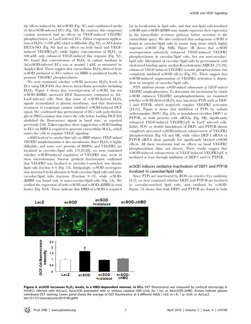
We next examined whether ecSOD increases H2O2 levels in
ECs using DCF-DA that detects intracellular peroxides including
H2O2. Figure 4 shows that overexpression of ecSOD, but not
ecSOD-DHBD, increased DCF fluorescence compared to Ad.-
LacZ-infected cells. Note that some of ecSOD-derived H2O2
signals accumulated at plasma membrane, and that short-term
treatment of exogenous catalase inhibited ecSOD-induced DCF
signal. We confirmed that pretreatment of ECs with polyethylene
glycol (PEG)-catalase that enters the cells before loading DCF-DA
abolished the fluorescence signals in basal state, as reported
previously [38]. Taken together, these suggest that ecSOD binding
to ECs via HBD is required to generate extracellular H2O2, which
enters the cells to regulate VEGF signaling.
ecSOD localized in caveolae/lipid rafts via HBD enhances VEGF-induced
VEGFR2 autophoshorylation in these microdomains. Since H2O2 is highly
diffusible, and some core proteins of HSPGs and VEGFR2 are
localized in caveolae/lipid rafts [19,20,28], we next examined
whether ecSOD-induced regulation of VEGFR2 may occur in
these microdomains. Sucrose gradient fractionation confirmed
that VEGFR2 was localized in caveolin-1-enriched, low density
lipid rafts fraction 4–6 (Fig. 5A). Intriguingly, ecSOD overexpres-
sion increased its localization in both caveolae/lipid rafts and non-
caveolae/lipid rafts fractions (Fraction 9–13), while ecSOD-
DHBD was found only in non-caveolae/lipid rafts (Fig. 5A). We
verified the expression of both ecSOD and ecSOD-DHBD in total
lysates (Fig. S4A). These indicate that HBD of ecSOD is required
for its localization in lipid rafts, and that non-lipid rafts-localized
ecSOD and ecSOD-DHBD may mainly represent their expression
in the intracellular secretory pathway before secretion to the
extracellular space. We also confirmed that endogenous ecSOD is
found in caveolae/lipid rafts in mouse lung tissue which highly
expresses ecSOD (Fig. S4B). Figure 5B shows that ecSOD
overexpression selectively enhanced VEGF-induced VEGFR2
phosphorylation in caveolae/lipid rafts, but not non-caveolae/
lipid rafts. Disruption of caveolae/lipid rafts by pretreatment with
cholesterol-binding agent, methyl-b-cyclodextrin (MbCD) [19,39],
enhanced VEGF-induced VEGFR2 tyrosine phosphorylation, but
completely inhibited ecSOD effects (Fig. S5). These suggest that
ecSOD-induced augmentation of VEGFR2 activation is depen-
dent on integrity of caveolae/lipid rafts.
PTPs inhibition prevents ecSOD-induced enhancement of VEGF-induced
VEGFR2 autophosphorylation. To determine the mechanism by which
ecSOD enhances VEGFR2 autophosphorylation, we examined
whether ecSOD-derived H2O2 may inactivate PTPs such as DEP-
1 and PTP1B, which negatively regulate VEGFR2 activation
[24,25]. Figure 6 shows that inhibition of PTPs by sodium
orthovanadate (SOV) (Fig. 6A); or knockdown of either DEP-1 or
PTP1B, or both proteins with siRNAs (Fig. 6B), significantly
enhanced VEGF-induced VEGFR2-pY in LacZ infected cells.
Either SOV or double knockdown of DEP1 and PTP1B almost
completely prevented ecSOD-induced enhancement of VEGFR2
phosphorylation (Fig. 6A and 6B), while either DEP-1 siRNA or
PTP1B siRNA alone partially but significantly blocked ecSOD
effects. All these treatments had no effects on basal VEGFR2
phosphorylation (data not shown). These results suggest that
ecSOD-induced enhancement of VEGF-induced VEGFR2-pY is
mediated at least through inhibition of DEP-1 and/or PTP1B.
ecSOD induces oxidative inactivation of DEP1 and PTP1Blocalized in caveolae/lipid rafts
Since PTPs are inactivated by ROS via reactive Cys oxidation
[4,5], we next examined whether DEP1 and PTP1B are localized
in caveolin-enriched lipid rafts, and oxidized by ecSOD.
Figure 7A shows that both DEP1 and PTP1B are found in both
Figure 4. ecSOD increases H2O2 levels, in a HBD-dependent manner, in ECs. DCF fluorescence was measured by confocal microscopy inHUVECs infected with Ad.LacZ, Ad.ecSOD pretreated with or without catalase (500 U/ml, for 1 hr), or Ad.ecSOD-DHBD. Arrows indicate plasmamembrane DCF staining. Lower panel shows the average of DCF fluorescence at 4 different fields (663) (n = 4). * p,0.05 vs. Ad.LacZ.doi:10.1371/journal.pone.0010189.g004
ecSOD and VEGF Signaling
PLoS ONE | www.plosone.org 7 April 2010 | Volume 5 | Issue 4 | e10189

caveolae/lipid rafts and non-caveolae/lipid rafts fractions, and
that ecSOD overexpression decreased their PTP activity in
caveolae/lipid rafts, but not non-caveolae/lipid rafts (Fig. 7B).
Furthermore, newly-developed Cys-SOH trapping reagent [36]
revealed that Ad.ecSOD increased Cys-OH formation of DEP1
and PTP1B in lipid rafts fraction. These suggest that extracellular
H2O2 generated by ecSOD induces oxidative inactivation of
DEP1/PTP1B in caveolae/lipid rafts, thereby promoting VEGF-
induced VEGFR2 autophosphorylation in these specialized
microdomains.
Figure 5. ecSOD localized in caveolae/lipid rafts via HBD enhances VEGF-induced VEGFR2 autophoshorylation in thesemicrodomains. A. After sucrose gradient centrifugation to isolate caveolae/lipid rafts, equal volume of each fraction from top to bottom (total 13fractions) was IB with anti-VEGFR2, caveolin-1, or paxillin Abs (upper panel). In lower panel, Ad.LacZ or Ad.ecSOD or Ad.ecSOD-DHBD-infectedHUVECs were used for caveolae/lipid rafts isolation, and each fraction was IB with anti-ecSOD or caveolin-1 Abs. B. Ad.LacZ or Ad.ecSOD-infectedHUVECs were stimulated with VEGF (20 ng/ml) for 5 min, and followed by caveolae/lipid rafts fractionation. Equal amounts of proteins from pooledFraction 4–6 (caveolae/lipid rafts) and Fraction 9–13 (non-caveolae/lipid rafts) were IB with anti-VEGFR2-pY1175, total VEGFR2 or caveolin-1 Abs(n = 3). *p,0.05.doi:10.1371/journal.pone.0010189.g005
ecSOD and VEGF Signaling
PLoS ONE | www.plosone.org 8 April 2010 | Volume 5 | Issue 4 | e10189

ecSOD promotes VEGF-induced EC migrationWe next examined the functional consequence of enhancement
of VEGFR2 activation by ecSOD-derived extracellular H2O2 in
VEGF-induced EC migration and proliferation. Figure 8 using
modified Boyden chamber assay shows that ecSOD, but not
ecSOD-DHBD, significantly enhanced VEGF-induced migration
without affecting sphingosine-1-phosphate (S1P)-induced response.
Thus, ecSOD-induced effect is specific for VEGFR2 signaling.
Importantly, ecSOD-induced enhancement of VEGF-induced EC
migration was prevented by catalase, supporting the role of ecSOD-
derived H2O2. VEGF-induced EC proliferation was also augment-
ed by Ad.ecSOD (Fig. S6). These effects of ecSOD were associated
with an enhancement of VEGFR2 downstream signaling such as
PLCc and p38MAPK phosphorylation (Fig. S7).
Figure 6. Inhibition of PTPs or knockdown of DEP1 and PTP1B prevents ecSOD-induced enhancement of VEGFR2 autopho-sphorylation. A. HUVECs infected with Ad.LacZ or Ad.ecSOD were pretreated with 0.3 mM sodium orthovanadate (SOV) for 30 min, and stimulatedwith VEGF (20 ng/ml) for 5 min. Lysates were used for measurement of VEGFR2-pY or total VEGFR2 or ecSOD expression (n = 3). B. HUVECs weretransfected with DEP1 or/and PTP1B siRNA, and then infected Ad.LacZ or Ad.ecSOD. Cells were stimulated with VEGF (20 ng/ml) for 5 min and lysateswere used for measurement of VEGFR2-pY and expression of proteins indicated (n = 4). * p,0.05.doi:10.1371/journal.pone.0010189.g006
ecSOD and VEGF Signaling
PLoS ONE | www.plosone.org 9 April 2010 | Volume 5 | Issue 4 | e10189

Discussion
The present study provides novel evidence that ecSOD
functions as a generator of extracellular H2O2 in specific
subcellular compartments to promote VEGF signaling linked to
angiogenesis. Here we show that: 1) gene transfer of ecSOD, but
not ecSOD-DHBD, increases H2O2 levels in adductor muscle, and
promotes angiogenesis in response to hindlimb ischemia; 2) H2O2
levels in both non-ischemic and ischemic hindlimbs are markedly
reduced in ecSOD2/2 mice; 3) In vitro, overexpression of ecSOD,
but not ecSOD-DHBD, in cultured medium in ECs enhances
VEGF-induced VEGFR2-pY through generation of extracellular
H2O2; 4) HBD of ecSOD is required for localization of ecSOD at
plasma membrane caveolin-enriched lipid rafts where VEGFR2
Figure 7. ecSOD induces inactivation and oxidation of DEP-1 and PTP1B localized in caveolae/lipid rafts. A. After sucrose gradientcentrifugation, equal volumes of each fraction from top to bottom (total 13 fractions) were IB with anti-DEP-1, PTP1B, VEGFR2 or caveolin-1 Abs. B.DEP-1 and PTP1B activities in pooled Fraction 4–6 (caveolae/lipid rafts) and Fraction 9–13 (non-caveolae/lipid rafts) in Ad.LacZ and Ad.ecSOD-infectedHUVECs were measured using pNPP as a substrate after IP with anti-DEP-1, PTP1B Abs. The values were expressed as a ratio to Ad.LacZ infected PTPactivity (defined as 1.0) in each fraction (n = 3) *p,0.05. C. Ad.LacZ or Ad.ecSOD-infected HUVECs were extracted in the presence of biotin-labeledCys-SOH trapping reagent DCP-Bio1. After sucrose gradient centrifugation, pooled Fraction 4–6 and Fraction 9–13 were affinity captured withstreptavidin beads to purify the Cys-SOH formed protein, followed by IB with anti-DEP-1 or PTP1B Abs.doi:10.1371/journal.pone.0010189.g007
ecSOD and VEGF Signaling
PLoS ONE | www.plosone.org 10 April 2010 | Volume 5 | Issue 4 | e10189

and PTP1B/DEP-1 are found; 5) endogenous ecSOD is also
found in caveolae/lipid rafts in tissues enriched with ecSOD; 6)
VEGF-induced VEGFR2-pY in caveolae/lipid rafts, but not non-
lipid rafts, is selectively enhanced by ecSOD, which is at least due
to oxidative inactivation of PTP1B and DEP1 in caveolae/lipid
rafts; 7) ecSOD-derived H2O2 promotes VEGF-induced EC
migration in a HBD-dependent manner.
Exogenous H2O2 can increase angiogenic gene expression and
promote pro-angiogenesis responses in ECs [8,13]. However, since
H2O2 is diffusible and short-lived, its application for therapeutic
neovascularization in vivo is difficult and not efficient. ecSOD is the
enzyme that catalyzes dismutation of O22 to produce H2O2 in the
extracellular space by anchoring to ECs surface or extracellular
matrix through HBD [14]. We previously reported that ecSOD
expression is increased in response to hindlimb ischemia, and that
post-ischemic revascularization is impaired in ecSOD2/2 mice
[17]. However, a role of ecSOD-derived H2O2 in VEGF signaling
and ischemia-induced angiogenesis was virtually unexplored. Here
we show that gene transfer of Ad.ecSOD, but not Ad.ecSOD-
DHBD, increases H2O2 production in adductor muscles, as
measured by Amplex Red assay, which predominantly detects
extracellular H2O2, as well as promotes blood flow recovery and
capillary formation in response to hindlimb ischemia. Further-
more, ecSOD2/2 mice show significant reduction of H2O2 levels
in both non-ischemic and ischemic hindlimbs. These results
strongly suggest that ecSOD bound to tissue via HBD plays an
important to role as a generator of extracellular H2O2 to promote
angiogenesis in vivo. To determine the underlying mechanisms, we
examined the effects of ecSOD-derived H2O2 on VEGF signaling
in ECs. The present study demonstrates for the first time that
overexpression of ecSOD, but not ecSOD-DHBD, in ECs or its
conditioned media enhances VEGF-induced VEGFR2 autopho-
sphorylation. Moreover, these ecSOD-induced effects on
VEGFR2, but not VEGF-induced VEGFR2 autophosphorylation,
are inhibited by short-term pretreatment with catalase that
scavenges extracellular H2O2. Thus, these findings indicate that
extracellular H2O2 derived from ecSOD promotes VEGF-induced
VEGFR2-pY in ECs in a HBD-dependent manner.
In this study, we found that H2O2 concentration in culture
media of Ad.ecSOD-infected ECs is around 1 mM, while
exogenous H2O2 requires at least 500 mM to enhance VEGF-
induced receptor phosphorylation. These results support the
possibility that ecSOD binding to ECs surface via HBD may
provide the microenvironment in which extracellular H2O2
generated by ecSOD is more compartmentalized than exogenous-
ly-applied H2O2. Of note, either high concentration of exogenous
H2O2 or Ad.ecSOD has no effects on basal VEGFR2-pY. These
suggest that ligand-induced pre-assembly of VEGFR2 containing
signaling complexes and/or their specific localization might be
required for promoting effect of extracellular H2O2 derived from
ECs-bound ecSOD on VEGFR2-pY. It has been shown that
VEGF-induced VEGFR2 autophosphorylation is regulated by
‘‘intracellular’’ H2O2 derived from Nox2-based NADPH oxidase
in ECs [7,8]. NADPH oxidase-dependent O22 production occurs
both intracellularly and extracellularly [12,40]. Thus, NADPH
oxidase-derived O22 produced extracellularly may be rapidly
dismutated by ecSOD to generate H2O2 in close proximity to the
VEGFR2 to facilitate its phosphorylation efficiently. Of note,
Figure 8. ecSOD promotes VEGF-induced EC migration in a HBD-dependent manner. HUVECs infected with Ad.LacZ or Ad.ecSOD orAd.ecSOD-DHBD were stimulated with 20 ng/ml VEGF or 10 mmol/L sphingosine-1-phosphate (S1P) for 6 hours, and cell migration was measured bythe modified Boyden chamber method. Some cells were pretreated with 500 U/ml catalase for 15 min and performed migration assay in the presenceof catalase. Bar graph represents averaged data, expressed as cell number counted per 10 fields (x200) and fold change over that in unstimulatedcells (control). * p,0.05 vs. Ad.LacZ+VEGF.doi:10.1371/journal.pone.0010189.g008
ecSOD and VEGF Signaling
PLoS ONE | www.plosone.org 11 April 2010 | Volume 5 | Issue 4 | e10189

classical role of ecSOD is to scavenge O22 to increase NO
bioactivity; however, NO donor has no effect on VEGF-induced
phosphorylation of the VEGFR2. Thus, it is H2O2 rather than
NO, which mediates ecSOD-induced augmentation of VEGFR2
activation in ECs.
ecSOD binds to cell surface HSPGs via HBD, and some cell
surface core proteins of HSPGs are localized in caveolae/lipid
rafts in ECs [28,41]. We thus examined whether ecSOD-induced
modulation of VEGFR2 might occur in these specialized
microdomains. Sucrose gradient fractionation reveals that ecSOD
is localized in both caveolae/lipid rafts and non-caveolae/lipid
rafts fractions in Ad.ecSOD-infected ECs, while ecSOD-DHBD is
found only in non-caveolae/lipid rafts fraction. Of note,
endogenous ecSOD protein is also found in caveolae/lipid rafts
in lung tissue in which ecSOD is abundantly expressed. We show
that VEGF-induced VEGFR2-pY in caveolae/lipid rafts, but not
in non-caveolae/lipid rafts, is enhanced by ecSOD. Disruption of
caveolae/lipid rafts by cholesterol-binding reagent increases
VEGF-induced VEGFR2 autophosphorylation, but prevents
ecSOD-induced effect. Mechanism by which cholesterol depletion
increases VEGF-induced phosphorylation of VEGFR2 in ECs
seems to be due to dissociation of VEGFR2 from caveolin [19].
Thus, these results suggest that ecSOD localization at caveolin-
enriched lipid rafts via HBD is required for ecSOD-induced
enhancement of ligand-induced VEGFR2 phosphorylation in
these specific plasma membrane compartments.
Reversible oxidative inactivation of PTPs by ROS [42,43,44]
and their specific localization are important for ROS to increase
tyrosine phosphorylation signaling events [4,5]. The initial product
of Cys oxidation is Cys-SOH, a key intermediate involved in redox
signaling [45]. The present study shows that inhibition of PTPs or
knockdown of DEP-1 and/or PTP1B increases VEGF-induced
VEGFR2-pY, which prevents ecSOD-induced effect on
VEGFR2. These suggest that both DEP1 and PTP1B function
as a negative regulator for VEGFR2-pY, as reported previously
[24,25], and that ecSOD-derived H2O2 inhibits their PTPs
activity to promote VEGFR2 phosphorylation. Intriguingly, we
found that both DEP1 and PTP1B are localized in both caveolae/
lipid rafts and non-lipid rafts in ECs. Moreover, newly-developed
cell permeable Cys-SOH trapping probe [36,37] reveals that
ecSOD increases Cys-SOH formation of DEP-1 and PTP1B as
well as decreases their PTP activity in caveolin-enriched lipid rafts,
but not in non-lipid rafts. NADPH oxidase is localized in lipid rafts
to generate O22 in ECs [21]. These suggest that extracellular
H2O2 generated by ecSOD locally oxidizes and inactivates DEP-1
and/or PTP1B in caveolae/lipid rafts where NADPH oxidase and
VEGFR2 are found, which in turn promotes VEGF-induced
VEGFR2 phosphorylation in these specific microdomains. Other
possible PTPs that are regulated by ecSOD cannot be ruled out in
the current study.
Functionally, ecSOD, but not ecSOD-DHBD, promotes
VEGF-induced EC migration in vitro, which is prevented by
exogenous application of catalase. This is consistent with ecSOD-
induced augmentation of ischemia-induced angiogenesis in vivo. Of
note, S1P-induced migration was not affected by Ad.ecSOD,
supporting our conclusion that localizing ecSOD, VEGFR2, and
DEP-1/PTP1B in lipid rafts as important mechanism by which
ecSOD-derived H2O2 enhances VEGFR2 signaling lined to
angiogenic responses. We previously reported that ecSOD
functions to preserve NO bioactivity by scavenging O22 in the
ischemic tissues, thereby promoting angiogenesis [17]. Similarly,
HBD-dependent protective endothelial function of ecSOD via
decreasing extracellular O22 has been reported in animal model
with hypertension [16]. The R213G polymorphism in the ecSOD
gene, which reduces binding to endothelium surface and increases
serum ecSOD levels, is associated with increased risk of
cardiovascular diseases [46]. The present study uncovers a novel
mechanism by which ecSOD promotes endothelial functions such
as EC migration and proliferation by generating extracellular
H2O2 at the specific membrane compartment, and thus facilitating
VEGF signaling linked to angiogenesis. In contrast, ecSOD
overexpression inhibits, instead of increase, tumor angiogenesis
and tumor invasion [47,48]. In pro-oxidant pathological condi-
tions such as atherosclerosis and hypertension, ecSOD seems to be
inactivated by H2O2 derived from ecSOD due to its peroxidase
activity [49,50]. Thus, ecSOD gene transfer effect on angiogenesis
Figure 9. Proposed model for role of ecSOD-derived H2O2 in VEGFR2 signaling linked to angiogenesis. Extracellular H2O2 generated byecSOD localized at caveolae/lipid rafts via HBD induces oxidative inactivation of DEP1 and PTP1B in these microdomains, thereby promoting VEGF-induced VEGFR2 phosphorylation, which may contribute to EC migration and proliferation in vitro as well as angiogenesis in vivo.doi:10.1371/journal.pone.0010189.g009
ecSOD and VEGF Signaling
PLoS ONE | www.plosone.org 12 April 2010 | Volume 5 | Issue 4 | e10189

in vivo seems to be varied with cell types and context specific
[17,47,48,51].
In summary, extracellular H2O2 generated by ecSOD localized
at caveolin-enriched lipid rafts via HBD efficiently facilitates
VEGFR2 signaling via oxidative inactivation of DEP-1/PTP1B in
these microdomains, which may contribute to promoting postnatal
angiogenesis (Fig. 9). Our previous and present studies may
uncover novel mechanism whereby increased ecSOD expression
in ischemic tissues promotes reparative neovascularization in vivo.
It is likely that ecSOD may serve as a potent generator of
extracellular H2O2 in the plasma membrane specific compart-
ments to promote angiogenesis growth factor signaling. The
present findings also imply that ecSOD gene transfer may
represent an important therapeutic approach for treatment of
angiogenesis-dependent diseases including ischemic heart and
limb diseases.
Supporting Information
Figure S1 ecSOD and ecSOD-DHBD protein expression and
activity in culture medium in adenovirus infected HUVECs.
Conditioned media obtained from HUVECs infected with
Ad.LacZ or Ad.ecSOD or Ad.ecSOD-DHBD was used for
Western analysis with anti-human ecSOD antibody (A) or
measurement of ecSOD activity (B).
Found at: doi:10.1371/journal.pone.0010189.s001 (0.03 MB
PDF)
Figure S2 Exogenous H2O2 at physiological concentration
cannot enhance VEGF-induced VEGFR2 autophosphorylation.
HUVECs were pretreated with indicated concentration of H2O2
for 15 min, and stimulated with VEGF (20 ng/ml) for 5 min.
Lysates were immunoprecipitated (IP) with anti-VEGFR2 Ab and
followed by immunoblotted (IB) with anti-pTyr Ab for measure-
ment of VEGFR2-pY (n = 3).
Found at: doi:10.1371/journal.pone.0010189.s002 (0.05 MB
PDF)
Figure S3 Exogenous application of NO donor has no effect on
VEGF-induced VEGFR2 autophosphorylation. HUVECs were
pretreated with indicated concentration of No donor, diethylene-
tetraamine-NONOate (DETA-NO) for 30 min, and stimulated
with VEGF (20 ng/ml) for 5 min. Lysates were used for
measurement of VEGFR2-pY.
Found at: doi:10.1371/journal.pone.0010189.s003 (0.05 MB
PDF)
Figure S4 Endogenous ecSOD is localized in caveolae/lipid
rafts in mouse lung in which ecSOD is highly expressed. A. Total
lysates from HUVECs infected Ad.LacZ or Ad.ecSOD or
Ad.ecSOD-DHBD for caveolae isolation were IB with anti-
ecSOD to confirm the expression of ecSOD and ecSOD-DHBD.
B. Mouse lung (400 mg) was fractionated to isolate caveolae/lipid
rafts and IB with anti-mouse ecSOD or caveolin-1 antibodies.
Found at: doi:10.1371/journal.pone.0010189.s004 (0.05 MB
PDF)
Figure S5 Intact caveolae/lipid rafts are required for ecSOD-
induced enhancement of VEGFR2 autophosphorylation. HU-
VECs were pretreated with or without 10 mM methyl-b-
cyclodextrin (MbCD) for 1 hr, and stimulated with VEGF
(20 ng/ml) for 5 min. Lysates were used for measurement of
VEGFR2-pY or total VEGFR2 or ecSOD expression (n = 3).
* p,0.05.
Found at: doi:10.1371/journal.pone.0010189.s005 (0.09 MB
PDF)
Figure S6 ecSOD promotes VEGF-induced EC proliferation.
Ad.LacZ or Ad.ecSOD-infected HUVECs were cultured in 0.5%
FBS containing medium with or without VEGF (20 ng/ml) for
48 hours, and cell number was counted with a hemocytometer
(n = 8). * p,0.05.
Found at: doi:10.1371/journal.pone.0010189.s006 (0.01 MB
PDF)
Figure S7 ecSOD enhances VEGFR2 downstream signaling in
HUVECs. Cell lysates from Ad.LacZ and Ad.ecSOD infected
HUVECs with or without VEGF stimulation (20 ng/ml, 5 min)
were IB with anti-p-PLCc or PLCc (A) or p-p38MAPK or
p38MAPK (B) antibodies (n = 3). *p,0.05
Found at: doi:10.1371/journal.pone.0010189.s007 (0.08 MB
PDF)
Author Contributions
Conceived and designed the experiments: JO NK MR TF MUF.
Performed the experiments: JO NU HWK. Analyzed the data: JO.
Contributed reagents/materials/analysis tools: NU NK MR RM LBP.
Wrote the paper: JO TF MUF.
References
1. Matsumoto T, Claesson-Welsh L (2001) VEGF receptor signal transduction. Sci
STKE 2001: RE21.
2. Takahashi T, Yamaguchi S, Chida K, Shibuya M (2001) A single autopho-
sphorylation site on KDR/Flk-1 is essential for VEGF-A-dependent activation of
PLC-gamma and DNA synthesis in vascular endothelial cells. Embo J 20:
2768–2778.
3. Lamalice L, Houle F, Jourdan G, Huot J (2004) Phosphorylation of tyrosine
1214 on VEGFR2 is required for VEGF-induced activation of Cdc42 upstream
of SAPK2/p38. Oncogene 23: 434–445.
4. Finkel T (1999) Signal transduction by reactive oxygen species in non-phagocytic
cells. J Leukoc Biol 65: 337–340.
5. Tonks NK (2005) Redox redux: revisiting PTPs and the control of cell signaling.
Cell 121: 667–670.
6. Colavitti R, Pani G, Bedogni B, Anzevino R, Borrello S, et al. (2002) Reactive
oxygen species as downstream mediators of angiogenic signaling by vascular
endothelial growth factor receptor-2/KDR. J Biol Chem 277: 3101–3108.
7. Ushio-Fukai M, Tang Y, Fukai T, Dikalov SI, Ma Y, et al. (2002) Novel role of
gp91(phox)-containing NAD(P)H oxidase in vascular endothelial growth factor-
induced signaling and angiogenesis. Circ Res 91: 1160–1167.
8. Ushio-Fukai M (2006) Redox signaling in angiogenesis: role of NADPH oxidase.
Cardiovasc Res 71: 226–235.
9. Tojo T, Ushio-Fukai M, Yamaoka-Tojo M, Ikeda S, Patrushev NA, et al. (2005)
Role of gp91phox (Nox2)-containing NAD(P)H oxidase in angiogenesis in
response to hindlimb ischemia. Circulation 111: 2347–2355.
10. Urao N, Inomata H, Razvi M, Kim HW, Wary K, et al. (2008) Role of nox2-
based NADPH oxidase in bone marrow and progenitor cell function involved in
neovascularization induced by hindlimb ischemia. Circ Res 103: 212–220.
11. Go YM, Park H, Koval M, Orr M, Reed M, et al. (2009) A key role for
mitochondria in endothelial signaling by plasma cysteine/cystine redox
potential. Free Radic Biol Med.
12. Chaiswing L, Oberley TD (2009) Extracellular/Microenvironmental Redox
State. Antioxid Redox Signal.
13. Gonzalez-Pacheco FR, Deudero JJ, Castellanos MC, Castilla MA, Alvarez-
Arroyo MV, et al. (2006) Mechanisms of endothelial response to oxidative
aggression: protective role of autologous VEGF and induction of VEGFR2 by
H2O2. Am J Physiol Heart Circ Physiol 291: H1395–1401.
14. Fukai T, Folz RJ, Landmesser U, Harrison DG (2002) Extracellular superoxide
dismutase and cardiovascular disease. Cardiovasc Res 55: 239–249.
15. Marklund SL (1990) Expression of extracellular superoxide dismutase by human
cell lines. Biochem J 266: 213–219.
16. Chu Y, Iida S, Lund DD, Weiss RM, DiBona GF, et al. (2003) Gene transfer of
extracellular superoxide dismutase reduces arterial pressure in spontaneously
hypertensive rats: role of heparin-binding domain. Circ Res 92: 461–468.
17. Kim HW, Lin A, Guldberg RE, Ushio-Fukai M, Fukai T (2007) Essential role of
extracellular SOD in reparative neovascularization induced by hindlimb
ischemia. Circ Res 101: 409–419.
18. Insel PA, Patel HH (2009) Membrane rafts and caveolae in cardiovascular
signaling. Curr Opin Nephrol Hypertens 18: 50–56.
ecSOD and VEGF Signaling
PLoS ONE | www.plosone.org 13 April 2010 | Volume 5 | Issue 4 | e10189

19. Labrecque L, Royal I, Surprenant DS, Patterson C, Gingras D, et al. (2003)
Regulation of vascular endothelial growth factor receptor-2 activity by caveolin-1 and plasma membrane cholesterol. Mol Biol Cell 14: 334–347.
20. Ikeda S, Ushio-Fukai M, Zuo L, Tojo T, Dikalov S, et al. (2005) Novel role of
ARF6 in vascular endothelial growth factor-induced signaling and angiogenesis.Circ Res 96: 467–475.
21. Ushio-Fukai M (2009) Compartmentalization of redox signaling throughNADPH oxidase-derived ROS. Antioxid Redox Signal 11: 1289–1299.
22. Caselli A, Mazzinghi B, Camici G, Manao G, Ramponi G (2002) Some protein
tyrosine phosphatases target in part to lipid rafts and interact with caveolin-1.Biochem Biophys Res Commun 296: 692–697.
23. Oshikawa J, Otsu K, Toya Y, Tsunematsu T, Hankins R, et al. (2004) Insulinresistance in skeletal muscles of caveolin-3-null mice. Proc Natl Acad Sci U S A
101: 12670–12675.24. Nakamura Y, Patrushev N, Inomata H, Mehta D, Urao N, et al. (2008) Role of
protein tyrosine phosphatase 1B in vascular endothelial growth factor signaling
and cell-cell adhesions in endothelial cells. Circ Res 102: 1182–1191.25. Grazia Lampugnani M, Zanetti A, Corada M, Takahashi T, Balconi G, et al.
(2003) Contact inhibition of VEGF-induced proliferation requires vascularendothelial cadherin, beta-catenin, and the phosphatase DEP-1/CD148. J Cell
Biol 161: 793–804.
26. Gitay-Goren H, Soker S, Vlodavsky I, Neufeld G (1992) The binding of vascularendothelial growth factor to its receptors is dependent on cell surface-associated
heparin-like molecules. J Biol Chem 267: 6093–6098.27. Jakobsson L, Kreuger J, Holmborn K, Lundin L, Eriksson I, et al. (2006)
Heparan sulfate in trans potentiates VEGFR-mediated angiogenesis. Dev Cell10: 625–634.
28. Tkachenko E, Simons M (2002) Clustering induces redistribution of syndecan-4
core protein into raft membrane domains. J Biol Chem 277: 19946–19951.29. Baljinnyam E, Iwatsubo K, Kurotani R, Wang X, Ulucan C, et al. (2009) Epac
increases melanoma cell migration by a heparan sulfate-related mechanism.Am J Physiol Cell Physiol 297: C802–813.
30. Mavromatis K, Fukai T, Tate M, Chesler N, Ku DN, et al. (2000) Early effects
of arterial hemodynamic conditions on human saphenous veins perfused ex vivo.Arterioscler Thromb Vasc Biol 20: 1889–1895.
31. Fukai T, Galis ZS, Meng XP, Parthasarathy S, Harrison DG (1998) Vascularexpression of extracellular superoxide dismutase in atherosclerosis. J Clin Invest
101: 2101–2111.32. Ushio-Fukai M, Alexander RW, Akers M, Griendling KK (1998) p38 Mitogen-
activated protein kinase is a critical component of the redox-sensitive signaling
pathways activated by angiotensin II. Role in vascular smooth muscle cellhypertrophy. J Biol Chem 273: 15022–15029.
33. Qin Z, Gongora MC, Ozumi K, Itoh S, Akram K, et al. (2008) Role of MenkesATPase in angiotensin II-induced hypertension: a key modulator for
extracellular superoxide dismutase function. Hypertension 52: 945–951.
34. Song KS, Li S, Okamoto T, Quilliam LA, Sargiacomo M, et al. (1996) Co-purification and direct interaction of Ras with caveolin, an integral membrane
protein of caveolae microdomains. Detergent-free purification of caveolaemicrodomains. J Biol Chem 271: 9690–9697.
35. Yamaoka-Tojo M, Ushio-Fukai M, Hilenski L, Dikalov SI, Chen YE, et al.(2004) IQGAP1, a novel vascular endothelial growth factor receptor binding
protein, is involved in reactive oxygen species-dependent endothelial migration
and proliferation. Circ Res 95: 276–283.
36. Poole LB, Klomsiri C, Knaggs SA, Furdui CM, Nelson KJ, et al. (2007)
Fluorescent and affinity-based tools to detect cysteine sulfenic acid formation in
proteins. Bioconjug Chem 18: 2004–2017.
37. Michalek RD, Nelson KJ, Holbrook BC, Yi JS, Stridiron D, et al. (2007) The
requirement of reversible cysteine sulfenic acid formation for T cell activation
and function. J Immunol 179: 6456–6467.
38. Ikeda S, Yamaoka-Tojo M, Hilenski L, Patrushev NA, Anwar GM, et al. (2005)
IQGAP1 regulates reactive oxygen species-dependent endothelial cell migration
through interacting with Nox2. Arterioscler Thromb Vasc Biol 25: 2295–2300.
39. Ushio-Fukai M, Hilenski L, Santanam N, Becker PL, Ma Y, et al. (2001)
Cholesterol depletion inhibits epidermal growth factor receptor transactivation
by angiotensin II in vascular smooth muscle cells: Role of cholesterol-rich
microdomains and focal adhesions in angiotensin II signaling. J Biol Chem 276:
48269–48275.
40. Souchard JP, Barbacanne MA, Margeat E, Maret A, Nepveu F, et al. (1998)
Electron spin resonance detection of extracellular superoxide anion released by
cultured endothelial cells. Free Radic Res 29: 441–449.
41. Buczek-Thomas JA, Chu CL, Rich CB, Stone PJ, Foster JA, et al. (2002)
Heparan sulfate depletion within pulmonary fibroblasts: implications for
elastogenesis and repair. J Cell Physiol 192: 294–303.
42. Rhee SG, Bae YS, Lee SR, Kwon J (2000) Hydrogen peroxide: a key messenger
that modulates protein phosphorylation through cysteine oxidation. Sci STKE
2000: PE1.
43. Ostman A, Bohmer FD (2001) Regulation of receptor tyrosine kinase signaling
by protein tyrosine phosphatases. Trends Cell Biol 11: 258–266.
44. Chiarugi P, Cirri P (2003) Redox regulation of protein tyrosine phosphatases
during receptor tyrosine kinase signal transduction. Trends Biochem Sci 28:
509–514.
45. Poole LB, Nelson KJ (2008) Discovering mechanisms of signaling-mediated
cysteine oxidation. Curr Opin Chem Biol 12: 18–24.
46. Juul K, Tybjaerg-Hansen A, Marklund S, Heegaard NH, Steffensen R, et al.
(2004) Genetically reduced antioxidative protection and increased ischemic
heart disease risk: The Copenhagen City Heart Study. Circulation 109: 59–65.
47. Wheeler MD, Smutney OM, Samulski RJ (2003) Secretion of extracellular
superoxide dismutase from muscle transduced with recombinant adenovirus
inhibits the growth of B16 melanomas in mice. Mol Cancer Res 1: 871–881.
48. Chaiswing L, Zhong W, Cullen JJ, Oberley LW, Oberley TD (2008)
Extracellular redox state regulates features associated with prostate cancer cell
invasion. Cancer Res 68: 5820–5826.
49. Hink HU, Santanam N, Dikalov S, McCann L, Nguyen AD, et al. (2002)
Peroxidase properties of extracellular superoxide dismutase: role of uric acid in
modulating in vivo activity. Arterioscler Thromb Vasc Biol 22: 1402–1408.
50. Jung O, Marklund SL, Xia N, Busse R, Brandes RP (2007) Inactivation of
extracellular superoxide dismutase contributes to the development of high-
volume hypertension. Arterioscler Thromb Vasc Biol 27: 470–477.
51. Laurila JP, Castellone MD, Curcio A, Laatikainen LE, Haaparanta-Solin M,
et al. (2009) Extracellular superoxide dismutase is a growth regulatory mediator
of tissue injury recovery. Mol Ther 17: 448–454.
ecSOD and VEGF Signaling
PLoS ONE | www.plosone.org 14 April 2010 | Volume 5 | Issue 4 | e10189