Structure-Function Analysis of Lyn Kinase Association with Lipid Rafts and Initiation of Early...
12
10.1128/MCB.21.24.8318-8328.2001. 2001, 21(24):8318. DOI: Mol. Cell. Biol. Dráber Arudchandran, Lubica Dráberová, Juan Rivera and Petr Martina Kovárová, Pavel Tolar, Ramachandran Receptor I Aggregation of Early Signaling Events after Fc? Association with Lipid Rafts and Initiation Structure-Function Analysis of Lyn Kinase http://mcb.asm.org/content/21/24/8318 Updated information and services can be found at: These include: REFERENCES http://mcb.asm.org/content/21/24/8318#ref-list-1 at: This article cites 53 articles, 31 of which can be accessed free CONTENT ALERTS more» articles cite this article), Receive: RSS Feeds, eTOCs, free email alerts (when new http://journals.asm.org/site/misc/reprints.xhtml Information about commercial reprint orders: http://journals.asm.org/site/subscriptions/ To subscribe to to another ASM Journal go to: on May 5, 2014 by guest http://mcb.asm.org/ Downloaded from on May 5, 2014 by guest http://mcb.asm.org/ Downloaded from
-
Upload
independent -
Category
Documents
-
view
0 -
download
0
Transcript of Structure-Function Analysis of Lyn Kinase Association with Lipid Rafts and Initiation of Early...

10.1128/MCB.21.24.8318-8328.2001.
2001, 21(24):8318. DOI:Mol. Cell. Biol. DráberArudchandran, Lubica Dráberová, Juan Rivera and Petr Martina Kovárová, Pavel Tolar, Ramachandran Receptor I Aggregationof Early Signaling Events after Fc?Association with Lipid Rafts and Initiation Structure-Function Analysis of Lyn Kinase
http://mcb.asm.org/content/21/24/8318Updated information and services can be found at:
These include:
REFERENCEShttp://mcb.asm.org/content/21/24/8318#ref-list-1at:
This article cites 53 articles, 31 of which can be accessed free
CONTENT ALERTS more»articles cite this article),
Receive: RSS Feeds, eTOCs, free email alerts (when new
http://journals.asm.org/site/misc/reprints.xhtmlInformation about commercial reprint orders: http://journals.asm.org/site/subscriptions/To subscribe to to another ASM Journal go to:
on May 5, 2014 by guest
http://mcb.asm
.org/D
ownloaded from
on M
ay 5, 2014 by guesthttp://m
cb.asm.org/
Dow
nloaded from

MOLECULAR AND CELLULAR BIOLOGY,0270-7306/01/$04.00�0 DOI: 10.1128/MCB.21.24.8318–8328.2001
Dec. 2001, p. 8318–8328 Vol. 21, No. 24
Copyright © 2001, American Society for Microbiology. All Rights Reserved.
Structure-Function Analysis of Lyn Kinase Association with LipidRafts and Initiation of Early Signaling Events after
Fcε Receptor I AggregationMARTINA KOVAROVA,1,2 PAVEL TOLAR,1 RAMACHANDRAN ARUDCHANDRAN,2
LUBICA DRABEROVA,1 JUAN RIVERA,2* AND PETR DRABER1
Institute of Molecular Genetics, Academy of Sciences of the Czech Republic, 14220 Prague, Czech Republic,1 andMolecular Inflammation Section, National Institute of Arthritis and Musculoskeletal and Skin Diseases,
National Institutes of Health, Bethesda, Maryland 20892-18202
Received 23 July 2001/Accepted 14 September 2001
The first step in immunoreceptor signaling is represented by ligand-dependent receptor aggregation, fol-lowed by receptor phosphorylation mediated by tyrosine kinases of the Src family. Recently, sphingolipid- andcholesterol-rich plasma membrane microdomains, called lipid rafts, have been identified and proposed tofunction as platforms where signal transduction molecules may interact with the aggregated immunoreceptors.Here we show that aggregation of the receptors with high affinity for immunoglobulin E (Fc�RI) in mast cellsis accompanied by a co-redistribution of the Src family kinase Lyn. The co-redistribution requires Lyn dualfatty acylation, Src homology 2 (SH2) and/or SH3 domains, and Lyn kinase activity, in cis or in trans.Palmitoylation site-mutated Lyn, which is anchored to the plasma membrane but exhibits reduced sublocal-ization into lipid rafts, initiates the tyrosine phosphorylation of Fc�RI subunits, Syk protein tyrosine kinase,and the linker for activation of T cells, along with an increase in the concentration of intracellular Ca2�.However, Lyn mutated in both the palmitoylation and myristoylation sites does not anchor to the plasmamembrane and is incapable of initiating Fc�RI phosphorylation and early signaling events. These data,together with our finding that a constitutively tyrosine-phosphorylated Fc�RI does not exhibit an increasedassociation with lipid rafts, suggest that Fc�RI phosphorylation and early activation events can be initiatedoutside of lipid rafts.
The high-affinity immunoglobulin E (IgE) receptor (FcεRI)-mediated activation of mast cells and basophils triggers a cas-cade of intracellular biochemical events that ultimately lead tothe secretion of preformed pharmacological agents and thetranscription of cytokine genes. This process is initiated byaggregation of the receptor by means of multivalent antigen(Ag)-IgE complexes, followed by tyrosine phosphorylation ofthe receptor subunits by Src family protein tyrosine kinases(14, 31).
FcεRI has a tetrameric structure comprised of an IgE-bind-ing � subunit, a � subunit, and a disulfide-bonded � dimer (32).The � and � subunits possess immunoreceptor tyrosine-basedactivation motifs (ITAMs), which are rapidly phosphorylatedby protein tyrosine kinase Lyn. Tyrosine-phosphorylatedITAMs of the the � subunits serve as novel binding sites for Srchomology 2 (SH2) domains of Syk kinase (6, 22, 28), leading tophosphorylation and activation of Syk. Thereafter, a number ofother signaling and adaptor molecules become phosphorylatedand recruited into the regions of activated FcεRI/Syk com-plexes. These include PLC�1 (26), the proto-oncogene productVav (41), PKC-� (18), and the linker for activation of T cells(LAT) (37, 52).
Detailed molecular mechanisms of the initial engagement ofthe Lyn kinase and FcεRI are not completely understood, buttwo different models have been proposed. One model, based
on protein-protein interactions, postulates that a small fractionof Lyn is constitutively associated with the � subunit of theFcεRI prior to activation. FcεRI aggregation effected by mul-tivalent antigen-IgE complexes or by other means facilitatesthe transphosphorylation of one FcεRI by Lyn bound to ajuxtaposed receptor (33). The tyrosine phosphorylation of �and � subunits of FcεRI supports the recruitment of additionalLyn to � subunit and of the Syk kinase to � subunits, promotingfurther propagation of the activation signal.
The alternative model postulates that the initial coupling ofLyn with aggregated FcεRI is mediated by protein-lipid inter-actions. According to this model, Lyn is anchored to the innerleaflet of the plasma membrane via myristate and palmitatechains that localize it in lipid rafts enriched in glycosphingo-lipids, cholesterol, and glycosylphosphatidylinositol-anchoredproteins. These domains, which are also referred to as deter-gent-insoluble glycosphingolipid domains, have been found innumerous cell types (9, 40). Upon aggregation, the FcεRIrapidly translocates into lipid rafts, where it is phosphorylatedby Lyn kinase (15, 39).
Although the model based on the sequestration of signaltransduction molecules into lipid rafts is very attractive andwas recently supported by analyses of activation via the T-cellreceptor (51, 23, 29) and B-cell receptor (10), it is not com-pletely clear how the receptor aggregation leads to its inclusionin lipid rafts and whether this is important for early activationevents. For example, aggregation of FcεRI on rat basophilicleukemia (RBL) cells (a mast cell tumor analog) by divalentmonoclonal antibodies (MAbs), which induce little visible re-
* Corresponding author. Mailing address: NIAMS/NIH, Building10, Room 9N228, MSC 1820, Bethesda, MD 20892-1820. Phone: (301)496-7592. Fax: (301) 402-0012. E-mail: [email protected].
8318
on May 5, 2014 by guest
http://mcb.asm
.org/D
ownloaded from

ceptor aggregation, as determined by light and scanning elec-tron microscopy (30), results in a cellular response similar tothat caused by aggregation induced by a multivalent Ag thatcauses extensive FcεRI aggregation (42).
To investigate the localization of Lyn kinase and its func-tional consequences, we tagged Lyn with green fluorescenseprotein (GFP) and followed its distribution in RBL cells,mouse bone marrow-derived mast cells (BMMC), and BMMCfrom a mouse with a genetically disrupted Lyn gene (BMMC-Lyn�/�) (19) before and after FcεRI engagement. VariousLyn-GFP constructs with defects in palmitoylation, myristoyl-ation, or both acylation sites, as well as mutations in the SH1and SH2/SH3 domains, and constitutively tyrosine-phosphory-lated FcεRI were employed to determine the factors affectingthe early stages of FcεRI-mediated activation. These experi-ments suggest that FcεRI phosphorylation and early activationevents can be initiated outside of lipid rafts.
MATERIALS AND METHODS
Antibodies, reagents, and cell cultures. Mouse MAbs specific for Lyn, Syk, �subunit of FcεRI, 2,4,6-trinitrophenyl (TNP)-specific IgE (IGEL b4 1), and2,4-dinitrophenyl (DNP)-specific IgE have been described previously (13, 35, 36,45, 46). Rabbit polyclonal Ab specific for Syk, Lyn, LAT, GFP, and IgE wereprepared by immunization with the recombinant fragments of Syk (2), Lyn (13),LAT, and GFP (unpublished) or IGEL b4 1 MAb, respectively. A chickenantibody to FcR� has been described previously (45). Horseradish peroxidase(HRP)-conjugated mouse antiphosphotyrosine MAb PY-20, goat anti-mouseIgG, and goat anti-rabbit IgG were purchased from Transduction Laboratories.Antiphosphotyrosine MAb (4G10) and anti-Shc were obtained from UpstateBiotechnology, and anti-c-Src MAb (B 12) was from Santa Cruz Biotechnology.RBL-2H3 cells and their culture conditions have been described (11). RBL-2H3cells defective in FcεRI� and transfected with wild-type FcεRI� (�wt) or withmutated FcεRI� chain in which a threonine at position 52 was substituted foralanine (�T52A) have been described (45). BMMC and BMMC-Lyn�/� (19)were kindly provided by M. Hibbs (Ludwig Institute for Cancer Research, Mel-bourne, Australia).
DNA constructs, recombinant SFV, and infection. The Semliki Forest virus(SFV) gene expression system, pSFV1 and helper pSFV2, was purchased fromLife Technologies. pSFV1 was further modified to contain a multiple cloning siteand a GFP expression cassette as previously described (4). Lyn mutants weregenerated by PCR from cDNA encoding wild-type rat LynA and LynB (48).
In the primers described below, the mutation sites are marked by double lines,AvrII restriction sites by bold letters, and the optimized ribosome-binding sites bya single line. The following 5� primers were used for the construction of wild-typeLynA-GFP (LynA-WT) and wild-type LynB-GFP (LynB-WT): 1, 5�-AAA CCTAGG GCC ACC ATG GGA TGT ATT AAA TCA AAA AGG AAA GAC-3�(5�Lyn-WT primer); 2, LynB in which Cys3 was replaced by Ala (LynB-CA),5�-AAA CCT AGG GCC ACC ATG GGA GCT ATT AAA TCA AAA AGGAAA GAC-3�; 3, LynB in which Gly2 was replaced by Ala (LynB-GA), 5�-AAACCT AGG GCC ACC ATG GCA TGT ATT AAA TCA AAA AGG AAAGAC-3�; and 4, LynB in which both Gly2 and Cys3 were replaced by Ala(LynB-GCA), 5�-AAA CCT AGG GCC ACC ATG GCA GCT ATT AAA TCAAAA AGG AAA GAC-3�. As a 3� primer, the oligonucleotide 5�-AAA CCTAGG TGG CTG CTG CTG ATA CTG CCC TTC CGT GGC AGT GTA-3� wasused.
Unique domains of LynA (LynA-UNI) and LynB (LynB-UNI) were preparedby combining the 5�Lyn-WT primer and Lyn 3� primer: 5�-AAA CCT AGG GTCCCC TTG CTC CTC TGG ATC TTT TGC-3�. For construction of LynAwithout a catalytic domain (LynA-SH2), the 5�Lyn-WT primer was combinedwith 3� primer: 5�-AAA CCT AGG TTT CAC CAG CTT AAT GGA CTCCCG-3�.
The PCR-generated DNA fragments were digested with AvrII restriction en-zyme and inserted into AvrII-digested pSFV1-EGFP (4). A construct with amutation in the catalytic domain (LynB-CI) was prepared from Lyn CI (48). Aschematic representation of the Lyn constructs used in this study is shown in Fig.1. For all PCRs, the proofreading Pfu DNA polymerase (Stratagene) was used.Fidelity of all PCR products was confirmed by direct sequencing. Generation ofSFV and infection of cells with recombinant viruses were done as described (4).
SFV infectivity ranged from 60 to 90% for RBL-2H3 cells and 20 to 60% forBMMC and BMMC-Lyn�/�.
Confocal microscopy. The cells were sensitized with biotinylated IgE (1.5�g/ml) immediately after infection. After incubation for 3 h at 37°C, the cellswere washed twice in buffered saline solution (BSS) containing 20 mM HEPES(pH 7.4), 135 mM NaCl, 5 mM KCl, 1.8 mM CaCl2, 1 mM MgCl2, 5.6 mMglucose, and 1 mg of bovine serum albumin (BSA) per ml and suspended at aconcentration of 106 cells/ml in indocarbocyanine (Cy3)-conjugated streptavidin(Pierce; 2 �g/ml) in BSS/BSA. After 2 min at 37°C, the cells were washed inphosphate-buffered saline (PBS), transferred onto cover slips, pretreated withpoly-L-lysine in PBS (Sigma; 100 �g/ml) in a 24-well plate, and centrifuged for 1min at 200 g. Attached cells were fixed with 4% paraformaldehyde for 15 minat room temperature, washed twice in PBS, dried in air, and mounted in ProLongantifade reagent (Molecular Probes). Control cells were fixed with paraformal-dehyde before exposure to Cy3-streptavidin.
The confocal fluorescence images were taken using a Leica TCS NT/SP con-focal system in conjunction with a Leica DM R microscope (Leica Microsys-tems). Achromatically corrected objective 100 (NA 1.4) was used to simulta-neously collect green and red images of the cells. Fluorescence bleedthrough wasevaluated by separate excitation with blue (488 nm) or yellow (568 nm) laser lines(argon-krypton mix gas laser). Cross-correlation analysis of the co-redistributionof Lyn-GFP constructs with aggregated FcεRI complexes was carried out onequatorial images of individual cells using the quantification mode of the LeicaTCS NT software (Leica Microsystems). The correlation coefficients were cal-culated from this analysis as described (38). These values were averaged for 12to 16 cells from each sample for numerical comparison of the degree of co-redistribution.
Isolation of plasma membranes and lipid rafts. Cell membranes were isolated4 h after infection. The cells were harvested and washed twice with ice-cold PBS,and 5 106 cells were resuspended in 500 �l of ice-cold homogenizing buffer (10mM Tris-HCl [pH 8.0], 0.5 mM MgCl2) containing protease inhibitors (1 mMphenylmethylsulfonyl fluoride [PMSF] plus 0.5 U of aprotinin and 0.5 U ofleupeptin per ml). After 10 min of incubation on ice, the cells were homogenizedby passing them 10 times through a 27-gauge needle, followed by an addition of150 �l of tonicity restoration buffer (10 mM Tris-HCl [pH 8.0] 0.5 mM MgCl2,0.6 M NaCl). Insoluble material was removed by centrifugation, and the post-nuclear supernatant was supplemented with 6.5 �l of 0.5 M EDTA and thencentrifuged at 100,000 g for 45 min. The membrane pellet was resuspended in
FIG. 1. Schematic representation of Lyn constructs. All constructswere tagged at the C terminus with enhanced GFP (solid circle).LynA-WT and LynB-WT are constructs with two forms of wild-typeLyn kinase that differ in a 20-amino-acid-long insert in the uniquedomain (shaded box). LynB-CA, LynB-GA, and LynB-GCA are con-structs mutated in the palmitoylation, myristoylation, or both acylationsites, respectively. LynA-UNI and LynB-UNI contain only the N-ter-minal unique domains of LynA and LynB, respectively. LynB-CI iscatalytically inactive Lyn B (mutation of Lys279 to Arg). LynA-SH2 isLynA without the catalytic domain. Arrows mark sites of introducedpoint mutations. The kinase activity of all Lyn constructs is indicated asfull (�) and null (�) kinase activity.
VOL. 21, 2001 STRUCTURE-FUNCTION ANALYSIS OF Lyn-FcεRI INTERACTION 8319
on May 5, 2014 by guest
http://mcb.asm
.org/D
ownloaded from

ice-cold 1% Triton X-100 lysis buffer (20 mM Tris-HCl [pH 8.0], 140 mM NaCl,2 mM EDTA, 1 mM Na3VO4, 1 mM PMSF, and 0.5 U of aprotinin and 0.5 U ofleupeptin per ml). After 20 min on ice, the insoluble material was removed bycentrifugation for 10 min at 4°C at 10,000 g, and the supernatant was used forfurther analysis.
Lipid rafts were isolated by sucrose density gradient ultracentrifugation asdescribed (43). For analysis of density distribution of FcεRI and its aggregatedforms as well as Lyn and Src kinases, the cells (2 107) were sensitized insuspension with 125I-labeled TNP-specific IgE and activated or not as describedin Results. The cells were lysed in 0.8 ml of lysis buffer containing 10 mMTris-HCl (pH 8.0), 50 mM NaCl, 10 mM EDTA, 1 mM Na3VO4, 10 mMglycerophosphate, 1 mM PMSF, 0.5 U of aprotinin and 0.5 U of leupeptin per ml,and 0.06, 0.1, or 0.2% (vol/vol) Triton X-100. After 15 min, the lysate washomogenized by passing 10 times through a 27-gauge needle and adjusted to40% (wt/vol) sucrose by adding an equal amount of 80% sucrose. A gradient wasformed by successive addition of 0.2 ml of 80% sucrose stock to the bottom of apolyallomer tube (13 by 51 mm), followed by 0.5 ml of 60% sucrose, 1.5 ml of40% sucrose (containing the cell lysate), 0.8 ml of 35% sucrose, and 0.5-mlaliquots of 30, 25, 20, and 15% sucrose.
Sucrose solutions were prepared by mixing the appropriate amount of thegradient buffer (25 mM Tris-HCl [pH 7.5], 125 mM NaCl, 2 mM EDTA) and80% sucrose. Tubes were centrifuged at 210,000 g for 4 h at 4°C using an SW55Ti rotor (Beckman Instruments). Gradients were fractionated into 0.2-ml ali-quots withdrawn from the top of the tube, and radioactivity in each fraction wasdetermined. The exact sucrose concentration in each fraction was determinedwith an Abbe refractometer. Fractions 1 to 10 (15 to 30% sucrose) containeddetergent-insoluble lipid rafts.
For analysis of association of FcεRI with lipid rafts from �wt and �T52A RBLcells, the cells (15 106) were sensitized with 125I-labeled DNP-specific IgE andactivated or not with DNP-human serum albumin (HSA) conjugate (Sigma).Cells were lysed on ice in 1.0 ml of lysis buffer (0.1% Triton X-100, 20 mMTris-HCl [pH 8.0], 140 mM NaCl, 2 mM EDTA, 1 mM Na3VO4, 1 mM PMSF,0.5 U of aprotinin and 0.5 U of leupeptin per ml) as above. The gradient wasformed by addition of 1 ml of 80% sucrose on the bottom of the tube followedby 2 ml of 40% sucrose containing the cell lysate and successive addition of 6 mlof 30% and 2.5 ml of 5% sucrose, and then centrifuged for 12 h at 200,000 gin an L8-70 Beckman centrifuge. Eleven fractions were collected from the top ofthe gradient.
Cell activation, immunoprecipitation, immunoblotting, and kinase assay.Transfected cells were sensitized with TNP-specific IgE in suspension. Afterincubation for 30 min at 37°C, the cells were washed twice in BSS/BSA andactivated for 5 min with TNP-BSA at a final concentration of 0.5 �g/ml. Towardsthe end of the activation period, the cells were briefly centrifuged and the pelletwas resuspended in ice-cold 0.5 % Triton X-100 lysis buffer. After 20 min on ice,the lysate was centrifuged at 12,000 g for 10 min to remove nuclei andinsoluble remnants. IgE-FcεRI complexes, Syk, or LAT was immunoprecipitatedfrom samples equivalent to 107 cells with rabbit anti-IgE, anti-Syk, or anti-LATbound to UltraLink-immobilized protein A (Pierce), and immunoblotting withthe corresponding antibodies was performed as described (2). Kinase activity ofall Lyn-GFP constructs was determined by in vitro kinase assay as described (3).
Intracellular calcium measurements. Changes in the concentration of freeintracellular Ca2� ([Ca2�]i) in transfected cells expressing GFP or GFP-contain-ing constructs were monitored using Fura Red AM probe (Molecular Probes).GFP and Fura Red have similar excitation but different emission maxima, al-lowing determination of [Ca2�]i only in the transfected GFP-expressing cells.The transfected cells were sensitised with IgE and at 3 h postinfection werewashed and loaded with Fura Red AM (4 �g/2 106 cells). After incubation for1 h at 37°C, the cells were washed twice in PBS and transferred to tubes for flowcytometry. TNP-BSA at a final concentration of 0.5 �g/ml was added 60 s afterthe start of fluorescence-activated cell sorting (FACS) analysis, and thapsigargin(Sigma; 1 �M final concentration) was added at 180 s. The measurements wereperformed with a FACSscan flow cytometer (Becton Dickinson) equipped witha single 488-nm argon laser used as the excitation source. GFP fluorescence wascollected at 515 to 535 nm, and Fura Red emission at 665 to 685 nm using linearamplification. GFP-negative cells were gated out, and Fura Red fluorescence wascollected at the indicated time intervals.
RESULTS
N-terminal myristoylation but not palmitoylation is re-quired for Lyn kinase anchoring to the plasma membrane.The unique domain of Lyn kinase contains specific sequences
for myristoylation and palmitoylation at positions Gly2 andCys3, respectively. It has been shown that a mutation in theacylation sites of other members of the Src kinase family, Lckand Fyn, leads to their inability to associate with the plasmamembrane (7, 39). To analyze the significance of N-terminalacylation and SH1 and SH2/SH3 domains for the anchoring ofLyn to the plasma membrane and partitioning in lipid rafts andthe in vivo functional consequences thereof, we prepared var-ious Lyn constructs with defects in one or both acylation sitesas well as in SH domains. In all constructs GFP was added atthe C terminus to allow fluorescence visualization of the con-structs and to distinguish them from endogenous Lyn. Thekinase activities of all constructs are shown in Fig. 1. Theconstructs were transfected into RBL cells using an SFV ex-pression system, and the subcellular distribution of the con-structs was analyzed at 4 h postinfection.
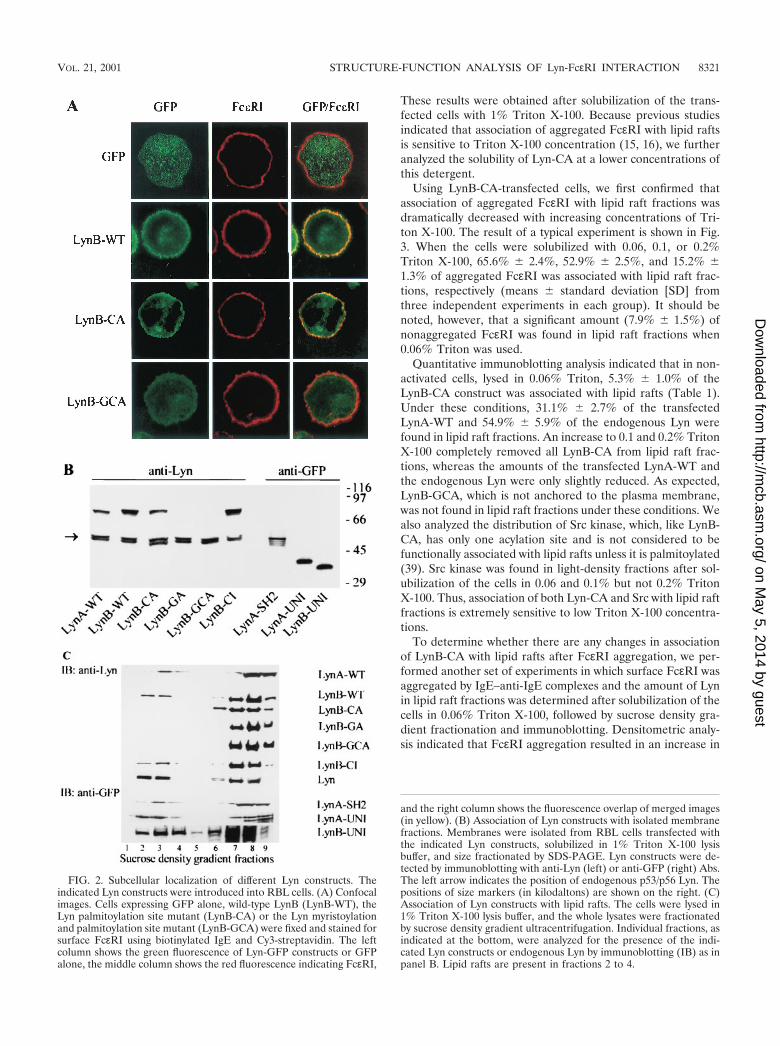
Confocal microscopy indicated that the constructs differedin their ability to associate with the nucleus and plasma mem-brane. As shown in Fig. 2A, GFP alone was evenly distributedthroughout the cell and was included in the nucleus. It was notassociated with the plasma membrane, as indicated by theabsence of fluorescence overlap (yellow) with FcεRI. LynA-UNI and LynB-UNI were also found in the nucleus (notshown), whereas all other constructs were excluded from thenucleus. Wild-type LynB was mainly found associated with theplasma membrane, similar to the distribution of endogenousLyn, as determined by biochemical means or indirect immu-nofluorescence of detergent-permeabilized cells (12, 15), dem-onstrating that the GFP tag did not affect the distribution ofthe transfected Lyn. It should also be noted that both GFP-tagged Lyn (Fig. 2A) and endogenous Lyn (12) accumulated inthe perinuclear region. LynB-CA also localized to the plasmamembrane, but LynB-GCA was found predominantly in thecytoplasm (Fig. 2A). Similar results were obtained when vari-ous Lyn constructs were analyzed in transfected BMMC andBMMC-Lyn�/� (not shown).
In order to verify the results of confocal microscopy, weanalyzed the distribution of Lyn constructs by immunoblotting.As can be seen in Fig. 2B, endogenous Lyn was detectable asa double band of 53 and 56 kDa. Wild-type Lyn constructs(LynA-WT and LynB-WT), as well as a palmitoylation sitemutant (LynB-CA) and the catalytically inactive Lyn (LynB-CI) were detected as bands of approximately 75 kDa. As ex-pected, the myristoylation site-mutated Lyn (LynB-GA) andLyn with mutations in both acylation sites (LynB-GCA) werenot found in the plasma membrane fractions. LynA-SH2 (55kDa) as well as LynA-UNI (40 kDa) and LynB-UNI (35kDa) were also associated with the plasma membrane, as de-tected by immunoblotting with anti-GFP antibody. Thus, asso-ciation of Lyn with the plasma membrane is dependent onmyristoylation of the N-terminal Gly in the unique domain,while other domains have no significant role in this anchoring.
It has been suggested that the Lyn localized in lipid raftsplays an important role in the phosphorylation of FcεRI andsubsequent events (15, 16, 20, 38). To test this postulate, wefirst examined whether various Lyn constructs differed in theirlocalization in lipid rafts. The data presented in Fig. 2C indi-cate that all Lyn constructs found in the plasma membranefractions (see Fig. 2B) also partitioned to lipid rafts, with theexception of the palmitoylation-mutated Lyn (LynB-CA).
8320 KOVAROVA ET AL. MOL. CELL. BIOL.
on May 5, 2014 by guest
http://mcb.asm
.org/D
ownloaded from
These results were obtained after solubilization of the trans-fected cells with 1% Triton X-100. Because previous studiesindicated that association of aggregated FcεRI with lipid raftsis sensitive to Triton X-100 concentration (15, 16), we furtheranalyzed the solubility of Lyn-CA at a lower concentrations ofthis detergent.
Using LynB-CA-transfected cells, we first confirmed thatassociation of aggregated FcεRI with lipid raft fractions wasdramatically decreased with increasing concentrations of Tri-ton X-100. The result of a typical experiment is shown in Fig.3. When the cells were solubilized with 0.06, 0.1, or 0.2%Triton X-100, 65.6% � 2.4%, 52.9% � 2.5%, and 15.2% �1.3% of aggregated FcεRI was associated with lipid raft frac-tions, respectively (means � standard deviation [SD] fromthree independent experiments in each group). It should benoted, however, that a significant amount (7.9% � 1.5%) ofnonaggregated FcεRI was found in lipid raft fractions when0.06% Triton was used.
Quantitative immunoblotting analysis indicated that in non-activated cells, lysed in 0.06% Triton, 5.3% � 1.0% of theLynB-CA construct was associated with lipid rafts (Table 1).Under these conditions, 31.1% � 2.7% of the transfectedLynA-WT and 54.9% � 5.9% of the endogenous Lyn werefound in lipid raft fractions. An increase to 0.1 and 0.2% TritonX-100 completely removed all LynB-CA from lipid raft frac-tions, whereas the amounts of the transfected LynA-WT andthe endogenous Lyn were only slightly reduced. As expected,LynB-GCA, which is not anchored to the plasma membrane,was not found in lipid raft fractions under these conditions. Wealso analyzed the distribution of Src kinase, which, like LynB-CA, has only one acylation site and is not considered to befunctionally associated with lipid rafts unless it is palmitoylated(39). Src kinase was found in light-density fractions after sol-ubilization of the cells in 0.06 and 0.1% but not 0.2% TritonX-100. Thus, association of both Lyn-CA and Src with lipid raftfractions is extremely sensitive to low Triton X-100 concentra-tions.
To determine whether there are any changes in associationof LynB-CA with lipid rafts after FcεRI aggregation, we per-formed another set of experiments in which surface FcεRI wasaggregated by IgE–anti-IgE complexes and the amount of Lynin lipid raft fractions was determined after solubilization of thecells in 0.06% Triton X-100, followed by sucrose density gra-dient fractionation and immunoblotting. Densitometric analy-sis indicated that FcεRI aggregation resulted in an increase in
FIG. 2. Subcellular localization of different Lyn constructs. Theindicated Lyn constructs were introduced into RBL cells. (A) Confocalimages. Cells expressing GFP alone, wild-type LynB (LynB-WT), theLyn palmitoylation site mutant (LynB-CA) or the Lyn myristoylationand palmitoylation site mutant (LynB-GCA) were fixed and stained forsurface FcεRI using biotinylated IgE and Cy3-streptavidin. The leftcolumn shows the green fluorescence of Lyn-GFP constructs or GFPalone, the middle column shows the red fluorescence indicating FcεRI,
and the right column shows the fluorescence overlap of merged images(in yellow). (B) Association of Lyn constructs with isolated membranefractions. Membranes were isolated from RBL cells transfected withthe indicated Lyn constructs, solubilized in 1% Triton X-100 lysisbuffer, and size fractionated by SDS-PAGE. Lyn constructs were de-tected by immunoblotting with anti-Lyn (left) or anti-GFP (right) Abs.The left arrow indicates the position of endogenous p53/p56 Lyn. Thepositions of size markers (in kilodaltons) are shown on the right. (C)Association of Lyn constructs with lipid rafts. The cells were lysed in1% Triton X-100 lysis buffer, and the whole lysates were fractionatedby sucrose density gradient ultracentrifugation. Individual fractions, asindicated at the bottom, were analyzed for the presence of the indi-cated Lyn constructs or endogenous Lyn by immunoblotting (IB) as inpanel B. Lipid rafts are present in fractions 2 to 4.
VOL. 21, 2001 STRUCTURE-FUNCTION ANALYSIS OF Lyn-FcεRI INTERACTION 8321
on May 5, 2014 by guest
http://mcb.asm
.org/D
ownloaded from

association of LynB-CA with lipid rafts from 5.3% � 1.0% (seeTable 1) to 10.3% � 2.5% (three experiments). However, thesignificance of this increase is unclear, especially because un-der the same experimental conditions the fraction of LynB-WTin lipid rafts was similar in activated (28.9% � 3.6%; threeexperiments) and resting cells (31.1% � 2.7%; see Table 1).
Co-redistribution of Lyn kinase with aggregated Fc�RI isdependent on Lyn N-terminal acylation, kinase activity, andconformation. FcεRI and fully acylated Lyn are uniformly dis-tributed in the plasma membrane (Fig. 2A). If the FcεRI isaggregated with IgE and multivalent Ag or by other means, thecomplexes redistribute into small patches. As can be seen inthe representative images in Fig. 4A, the aggregation of sur-face FcεRI in RBL cells results in co-redistribution of wild-type Lyn kinase (LynB-WT), as reflected by the formation ofyellow patches from the overlay of green and red fluorescenceprofiles. An even more extensive co-redistribution was ob-served when Lyn with a deleted SH1 domain (LynA-SH2) wastested. The palmitoylation-defective Lyn construct (LynB-CA)showed decreased co-redistribution, indicating that a firm an-chor into lipid rafts contributes to this process. GFP alone didnot show any co-redistribution.
Quantitative analysis of co-redistribution of various Lyn con-structs with aggregated FcεRI in RBL cells is shown in Fig. 4B.To determine the role of endogenous Lyn in the co-redistri-bution of the transfected Lyn constructs with aggregatedFcεRI, we also used wild-type BMMC and BMMC-Lyn�/�.The wild-type Lyn showed a co-redistribution which was sim-ilar in all three cell lines used. Palmitoylation-defective Lynexhibited a decreased co-redistribution which was not depen-dent on endogenous Lyn, as reflected by the same extent of
co-redistribution in BMMC and BMMC-Lyn�/�. A defect inboth acylation sites (LynB-GCA) and, therefore, the absenceof anchoring in the plasma membrane resulted in the absenceof co-redistribution.
Interestingly, the absence of co-redistribution was also ob-served in LynA-UNI (Fig. 4B) and LynB-UNI (not shown),indicating that anchoring to the plasma membrane and lipidrafts is not sufficient for the co-redistribution and that possiblythe kinase activity and SH2/SH3 domains play a significantrole. This assumption was confirmed by examination of theproperties of Lyn with a mutation in the catalytic domain(LynB-CI). This construct exhibited co-redistribution with ag-gregated FcεRI comparable to the wild-type Lyn in RBL cellsand BMMC. However, when LynB-CI was transfected intoBMMC-Lyn�/�, no co-redistribution of LynB-CI with aggre-gated FcεRI was observed, indicating that the endogenous Lynactivity was necessary to mediate the co-redistribution. Addi-tionally, the highest level of co-redistribution was observedwhen the LynA-SH2 construct was used. In RBL cells andBMMC, the cross-correlation coefficients attained were 0.78 �0.07 and 0.81 � 0.07, respectively. However, in the absence ofendogenous Lyn (BMMC-Lyn�/�), the co-redistribution ofLynA-SH2 dropped to 0.31 � 0.09.
Palmitoylation-defective Lyn kinase is able to initiate earlystages of Fc�RI-mediated activation. To find out whether ornot Lyn kinase must be anchored in the plasma membrane andlipid rafts to initiate early FcεRI-mediated activation events,we transfected various Lyn constructs into BMMC-Lyn�/� andfollowed the tyrosine phosphorylation of FcεRI� and � sub-units, Syk kinase, and the LAT adaptor. For these experiments,we used constructs with Lyn kinase activity, namely, LynB-WT,palmitoylation-deficient LynB-CA, and myristoylation- andpalmitoylation-deficient Lyn-B-GCA. In all experiments, aconstruct with GFP alone served as the negative control. Thetransfected cells were sensitized with IgE and stimulated withTNP-BSA. Five minutes later the cells were lysed, and FcεRI,Syk, or LAT was precipitated using the appropriate Abs. Theimmunoprecipitates were size fractionated by sodium dodecylsulfate-polyacrylamide gel electrophoresis (SDS-PAGE) andanalyzed by immunoblotting.
FIG. 3. Detergent-sensitive association of aggregated and unaggre-gated FcεRI with lipid rafts as detected by sucrose density gradientanalysis. RBL-2H3 cells were sensitized with [125I]IgE (1 �g/ml), and[125I]IgE-FcεRI complexes were aggregated (open symbols) or not(solid symbols) with rabbit anti-IgE antibody (10 �g/ml) for 5 min. Thecells were solubilized on ice in a lysis buffer containing 0.06% (circles),0.1% (squares), or 0.2% (diamonds) Triton X-100. Total cell lysateswere loaded within 40% sucrose fractions of the sucrose step gradientand fractionated by ultracentrifugation for 4 h. Points show the per-centage of total cpm present in individual fractions (left axis). Thepercentage of the FcεRI found in the lipid raft fractions was 65.6% �2.4%, 52.9% � 2.5%, and 15.2% � 1.3% for detergent concentrationsof 0.06, 0.1, and 0.2%, respectively. Sucrose concentrations are alsoindicated (�, right axis).
TABLE 1. Quantitative immunoblotting analysis of various Lynconstructs and endogenous Lyn and Src kinase in lipid raft fractions
after solubilization of transfected RBL cells with differentconcentrations of Triton X-100a
Kinase analyzed
Mean % of kinase in lipid raft fraction � SD(no. of expt) at Triton X-100 concn:
0.06% 0.1% 0.2%
Transfected kinaseLynA-WT 31.1 � 2.7 (3) 30.4 � 3.2 (3) 28.2 � 4.0 (3)LynB-CA 5.3 � 1.0 (4) 0.0 � 0.0 (2) 0.0 � 0.0 (2)LynB-GCA 0.0 � 0.0 (2) NT NT
Endogenous kinaseLyn 54.9 � 5.9 (7) 51.2 � 1.9 (3) 47.2 � 5.2 (3)Src 18.8 � 3.3 (4) 14.4 � 2.0 (3) 0.0 � 0.0 (2)
a Values indicate means � SD of the percentage of kinases in lipid raftfractions (15 to 30% sucrose) after solubilization of the cells in the indicatedconcentrations of Triton X-100 and fractionation on sucrose density gradients.Numbers in parentheses indicate number of independent experiments per-formed. NT, not tested.
8322 KOVAROVA ET AL. MOL. CELL. BIOL.
on May 5, 2014 by guest
http://mcb.asm
.org/D
ownloaded from

Representative data in Fig. 5A from three experiments in-dicate that following the FcεRI engagement, both the LynB-WT and LynB-CA transfectants exhibited an increased tyro-sine phosphorylation of FcεRI � and � subunits and an asso-
ciated protein of about 70 kDa. In contrast, the engagement ofFcεRI in cells transfected with LynB-GCA or GFP alone didnot result in increased tyrosine phosphorylation of FcεRI sub-units and the associated 70-kDa protein. The absence of thesignal was not due to the absence of the receptor in immuno-precipitated material, as indicated by the results of immuno-blotting with an MAb specific for the FcεRI-� subunit.
Similar results were obtained when Syk and LAT immuno-precipitates were analyzed. Thus, the engagement of FcεRIresulted in increased Syk and LAT tyrosine phosphorylation incells transfected with LynB-WT and LynB-CA (Fig. 5B and C).Quantitative analysis of the immunoblots, which took into ac-count the infection efficiences, indicated that the level ofFcεRI, Syk, and LAT tyrosine phosphorylation increased to asimilar extent in LynB-WT and LynB-CA cells (Fig. 5D). Noincrease in Syk and LAT tyrosine phosphorylation was ob-served in activated cells with the GFP construct. It should benoted that in LynB-GCA-transfected cells, some increase intyrosine phosphorylation of Syk and LAT was observed. Thiswas apparently due to the Lyn kinase activity of the LynB-GCA construct.
An increase in [Ca2�]i is one of the early signs of cell acti-vation. In further experiments we measured [Ca2�]i in BMMC-Lyn�/� transfected with various constructs following activationvia FcεRI (Fig. 6A). For these measurements, the cells wereloaded with Fura Red AM, a specific indicator of free Ca2�,and [Ca2�]i was only determined in transfected cells, i.e., thoseexpressing GFP. As seen in Fig. 6B, cells transfected withwild-type Lyn (LynB-WT) showed a significant increase in[Ca2�]i after FcεRI engagement, 49% � 6% of the maximumincrease in [Ca2�]i observed in cells exposed to thapsigargin. Incells transfected with the palmitoylation-deficient LynB-CA,the FcεRI engagement also resulted in a significant increase in[Ca2�]i. However, this increase was slightly lower (41% � 7%)and reached its maximum with some delay. FcεRI engagementin cells expressing the myristoylation- and palmitoylation-de-ficient LynB-GCA induced only a small increase in [Ca2�]i
(15% � 3% of maximal release) that was comparable to that innegative controls transfected with GFP alone (10% � 4%).
Is constitutively phosphorylated Fc�RI associated with lipidrafts? In the experiments described above, we showed thatLynB-CA is able to phosphorylate FcεRI and trigger earlysignaling events. Because LynB-CA seems to be excluded fromlipid rafts, at least those defined by resistance to �0.1% TritonX-100, the above data suggested that FcεRI could be phos-phorylated outside the lipid rafts. To further analyze this pos-sibility, we studied the lipid raft association of a constitutivelytyrosine-phosphorylated FcεRI that results from the expres-sion of a mutant � chain (Thr52 to Ala) in RBL-2H3-�� cells(45). This mutation, resulting in constitutive tyrosine phos-phorylation of FcεRI, could cause a conformational change ofFcεRI�, allowing its tyrosine phosphorylation cis- or trans-molecularly. Alternatively, the T52A mutation could cause theconstitutive formation of FcεRI aggregates that would then betargeted to lipid rafts, where they would become phosphory-lated. In this case, an increased amount of FcεRI�T52A shouldbe detected in lipid rafts.
First, we wanted to confirm, using a slightly different exper-imental setup, that FcεRI from �T52A is constitutively tyrosinephosphorylated. Tyrosine phosphorylation of FcεRI� was an-
FIG. 4. Co-redistribution of Lyn constructs with aggregated FcεRI.The indicated Lyn constructs or GFP alone was introduced into RBLcells, BMMC, or BMMC-Lyn�/�. Cells were sensitized with biotinyl-ated IgE, and surface FcεRI-IgE complexes were aggregated withCy3-conjugated streptavidin for 2 min. The cells were fixed and ana-lyzed by confocal microscopy. (A) Confocal images of RBL cells.Images are arranged as in Fig. 2A. (B) Quantitative analysis of co-redistribution of the indicated Lyn constructs with aggregated FcεRI inRBL cells, BMMC, and BMMC-Lyn�/�. Means � SD of correlationcoefficients were calculated from at least two independent experi-ments, with 12 to 16 cells analyzed in each experiment.
VOL. 21, 2001 STRUCTURE-FUNCTION ANALYSIS OF Lyn-FcεRI INTERACTION 8323
on May 5, 2014 by guest
http://mcb.asm
.org/D
ownloaded from

alyzed by immunoprecipitation of tyrosine-phosphorylatedproteins with 4G10 antibody followed by immunoblotting withan antibody to FcεRI�. The data presented in Fig. 7 indicatethat FcεRI� from resting �T52A cells exhibits tyrosine phos-phorylation similar to that of FcεRI� from antigen-activated�wt cells. Antigen aggregation of FcεRI�T52A led to a furtherincrease in its tyrosine phosphorylation, which correlated withits inclusion in lipid rafts (see below). The observed differencesin FcεRI� tyrosine phosphorylation were not due to differentamounts of proteins immunoprecipitated, as indicated by thesame amount of Shc, which is tyrosine phosphorylated in bothresting and activated cells exposed to serum (47).
The fraction of FcεRI in lipid rafts was measured as thepercentage of radioactivity in the light-density fractions (frac-tions 2 to 5; 5 to 30% sucrose) after solubilization of 125I-sensitized cells in 0.1% Triton X-100 and fractionation onsucrose density gradient. When �wt cells were used, only a
FIG. 5. Antigen-induced tyrosine phosphorylation of FcεRI, Syk,and LAT in cells expressing palmitoylation site-mutated Lyn. BMMC-Lyn�/� were transfected with LynB-WT, LynB-CA, LynB-GCA, or GFPalone. The transfected and IgE-sensitized cells were activated or not withTNP-BSA for 5 min. The cells were lysed, and FcεRI complexes, Syk, orLAT was immunoprecipitated from the postnuclear supernatants usingAbs specific for IgE (A), Syk (B), or LAT (C). Proteins were resolved bySDS-PAGE and analyzed by immunoblotting (IB) with antiphosphoty-rosine MAb PY-20-HRP. After stripping, MAbs specific for FcεRI �subunit, Syk, and LAT were used to estimate the relative amounts of theimmunoprecipitated (IP) protein. Arrows indicate the positions of thephosphorylated � and � subunits of the FcεRI (A), Syk (B), and LAT (C).(A) Positions of size markers are shown on the right (in kilodaltons). (D)Quantitative analysis of tyrosine phosphorylation of immunoprecipitatedFcεRI, Syk, and LAT from cells transfected with LynB-WT (WT),LynB-CA (CA), LynB-GCA (GCA), or GFP alone. Tyrosine phosphor-ylation of FcεRI, Syk and LAT from cells transfected with LynB-WTwas taken as 100%. Means � SD were calculated from four indepen-dent experiments in each group. Data were normalized for differenttransfections efficiencies as determined by flow cytofluorometry.
FIG. 6. Antigen-induced increase in [Ca2�]i in cells expressing pal-mitoylation site-mutated Lyn. (A) BMMC-Lyn�/� were transfectedwith LynB-WT, LynB-CA, LynB-GCA, or GFP alone. The cells weresensitized with IgE and loaded with Fura Red AM. At 3 h postinfec-tion, the cells were stimulated with TNP-BSA, and [Ca2�]i was deter-mined only in transfected cells by double-color FACS analysis. Theinverse of the quenching of Fura Red fluorescence intensity (rise inintracellular calcium) in transfected cells is reported as a function oftime. The long arrow and short arrow indicate time of addition of theantigen and thapsigargin, respectively. (B) The extent of activation isexpressed as a ratio between the decrease in Fura Red fluorescence inFcεRI-activated cells and that observed after addition of thapsigargin,which was taken as 100%. Means � SD were calculated from six toeight experiments in each group.
8324 KOVAROVA ET AL. MOL. CELL. BIOL.
on May 5, 2014 by guest
http://mcb.asm
.org/D
ownloaded from

small fraction of FcεRI (4.0% � 0.7%; three experiments) wasassociated with light-density fractions. Under the same exper-imental conditions, a similar amount of FcεRI from �T52Awas found in lipid raft fractions (3.9% � 0.6%). After antigen-mediated aggregation, a significant increase in the amount of�wt-derived FcεRI in lipid raft fractions was observed (41.9%� 5.0%), but again similar values where attained when FcεRIfrom �T52A was analyzed (43.1% � 4.1%). These data indi-cate that constitutive tyrosine phosphorylation of FcεRI�T52Ais not due to its higher association with lipid rafts and supportthe notion that it can be phosphorylated outside lipid rafts.
DISCUSSION
The results presented in this study show that Lyn kinasemust be anchored in the plasma membrane, but not necessarilyfirmly in lipid rafts, to be able to initiate early stages of cellactivation mediated by FcεRI. The first step in this process isaggregation of the FcεRI by multivalent ligand and subsequenttyrosine phosphorylation of its � and � subunits by Lyn kinase(14, 31). This is followed by tyrosine phosphorylation of othersignaling molecules (see the introduction). Interestingly, mostof these molecules have been shown to transiently co-redistrib-ute with the aggregated FcεRI (5, 42). As shown in this study,as well as in a previous report (20), Lyn exhibits a homogenousdistribution on the plasma membrane before activation, asdetected by fluorescence microsocopy, and moves after FcεRIaggregation into regions of aggregated FcεRI. Quantitativeanalysis revealed that the co-redistribution of Lyn and aggre-gated FcεRI depends on several structural and functionalproperties of the Lyn kinase.
First, Lyn must be fatty acylated in the N-terminal end of themolecule. In the absence of both palmitoylation and myristoyl-ation, no anchoring of Lyn into the plasma membrane is ob-served and there is no co-redistribution of Lyn with aggregatedFcεRI. The absence of membrane anchoring and co-redistri-bution with FcεRI was found not only in the Lyn-GCA con-struct, with both acylation sites mutated, but also in theLyn-GA construct, with a mutation in the myristoylation site.
This is consistent with previous data indicating that N-myris-toylation is a necessary prerequisite for palmitoylation (7). Theabsence of palmitoylation in the LynB-CA mutant resulted inits decreased ability to localize into lipid rafts but did notpreclude its association with the plasma membrane. The LynB-CA mutant exhibited a decreased co-redistribution with aggre-gated FcεRI. This inhibition was similar in RBL, BMMC, andBMMC-Lyn�/�, indicating that it is independent of the pres-ence of the endogenous Lyn. Importantly, the decreased abilityto associate with lipid rafts and the decreased co-redistributionwith aggregated FcεRI did not interfere with the ability of theLyn-CA mutant to initiate early FcεRI phosphorylation andactivation of early events (see below).
Second, the inhibition of kinase activity caused by mutatingLys279 to Arg in full-length Lyn B kinase (LynB-CI) had noeffect on anchoring of this mutant kinase to the plasma mem-brane and lipid rafts, but completely suppressed its co-redis-tribution with aggregated FcεRI. This suppression was ob-served after transfection of LynB-CI into BMMC-Lyn�/� butnot into BMMC and RBL cells, which express an endogenousLyn. These data, together with the comparable co-redistribu-tion of wild-type Lyn with aggregated FcεRI in both BMMCand BMMC-Lyn�/�, indicated that Lyn kinase activity in cis ortrans was necessary for proper Lyn-FcεRI interaction.
Third, consistent with these data, the removal of the catalyticdomain in the LynA-SH2 construct had no effect on Lyn’sincorporation in the plasma membrane and lipid rafts butinhibited co-redistribution of this construct with aggregatedFcεRI in BMMC-Lyn�/�. Compared to LynB-CI, the extent ofinhibition was smaller, suggesting that the absence of the ki-nase domain and/or the regulatory C-terminal tyrosine induceda conformational change improving its co-redistribution. Inaccord with this interpretation, LynA-SH2, exhibited a higherdegree of co-redistribution with FcεRI in RBL and BMMCthan wild-type Lyn.
Fourth, LynA-UNI and LynB-UNI, which lack the SH1,SH2, and SH3 domains but retain both acylation sites, local-ized properly in the plasma membrane and in lipid rafts butfailed to show any co-redistribution with aggregated FcεRI.The absence of recruitment of the Lyn unique domains intoregions of aggregated FcεRI was observed after transfection ofthe Lyn constructs not only into BMMC-Lyn�/�, but also intoBMMC and RBL cells, i.e., cells expressing endogenous wild-type Lyn. These data, together with the finding of normalcolocalization of LynB-CI in cells expressing endogenous Lyn(see above), suggest that the SH2 and SH3 domains are in-volved in the recruitment of Lyn to the phosphorylated recep-tors. Our findings are consistent with in vitro studies (25)demonstrating that the SH2 domain of Lyn interacts with thephosphorylated ITAM of the FcεRI� subunit; consequently,we assume that this interaction is likely involved in the LynB-CI recruitment.
The relationship between patches of aggregated FcεRI, asdetermined by confocal microscopy, and lipid rafts with aggre-gated FcεRI, as determined by biochemical analysis of deter-gent-solubilized cells, is not completely clear and will requirefurther study. However, our confocal microscopy findings thatLyn constructs exhibited different potentials to colocalize withpatches of aggregated FcεRI and that this association corre-sponded to the activation potential of the constructs indicated
FIG. 7. Tyrosine phosphorylation of FcεRI from �wt and �T52Acells. The cells (5 106) were sensitized with IgE and stimulated (�)or not (�) with 0.1 �g of DNP-HSA (Ag). After 5 min the cells werelysed in 1% Triton X-100-containing lysis buffer, and tyrosine-phos-phorylated proteins were immunoprecipitated (IP) with MAb 4G10.Immunoblots (IB) were probed with chicken antibody to FcεRI�,followed by stripping and sequential immunoblotting with an antibodyrecognizing constitutively phosphorylated p46 and p52 isoforms of Shcas a loading control.
VOL. 21, 2001 STRUCTURE-FUNCTION ANALYSIS OF Lyn-FcεRI INTERACTION 8325
on May 5, 2014 by guest
http://mcb.asm
.org/D
ownloaded from

that association of Lyn and FcεRI in patches has a physiolog-ical significance. Furthermore, because most of the aggregatedFcεRI was located in small patches on the cell surface and atthe same time was found associated with lipid rafts, it is verylikely that microscopic patches of aggregated FcεRI are foundin lipid raft fractions of the sucrose gradient, especially whenanalysis is performed at early stages of receptor activation.
Thus, the collective data favor the co-redistribution of Lynwith FcεRI as reflecting an SH2 domain-dependent redistribu-tion of Lyn that follows the initial phosphorylation of theFcεRI by a constitutive but weakly associated Lyn kinase. Thisview is supported by the requirement of Lyn kinase activity forco-redistribution of Lyn-CI in the BMMC-Lyn�/� by the en-hanced co-redistribution of a Lyn-SH2 construct, and by ourinability to observe the previously described weak interactionof Lyn-UNI with FcεRI (48) in the co-redistribution assay.Regardless, the confocal studies demonstrated the unique fail-ure of the SH2 domain-containing and catalytically activeLyn-CA to co-redistribute with the FcεRI in the presence orabsence of endogenous Lyn. As this Lyn construct is not ef-fectively recruited to lipid rafts (based on our biochemicaldata), our findings argue the importance of Lyn residence inlipid rafts for the SH2-dependent redistribution of Lyn withFcεRI and suggest that this takes place in lipid rafts.
The most important finding of this work is the evidence thatLynB-CA, unlike LynB-GCA, was able to initiate early stagesof mast cell activation, including tyrosine phosphorylation ofFcεRI � and � subunits, Syk kinase, and LAT and an increasein [Ca2�]i. These data support the concept that Lyn kinaseanchored to the plasma membrane but not necessarily to lipidrafts (defined by resistance to detergents) is important for earlystages of mast cell activation. Thus, our data are more consis-tent with the model suggesting that Lyn kinase in resting cellsis somehow weakly associated with a small fraction of FcεRI.This association requires Lyn myristoylation and its anchoringinto the plasma membrane. The initial phosphorylation of thereceptor subunits but not their extensive aggregation promotesa transient association of more Lyn, thereby shifting the bal-ance between phosphorylation and dephosphorylation state(see protein-protein model in the introduction).
Several recent findings support this notion. Using a yeasttwo-hybrid system, direct interaction was detected betweenLyn or its unique domain and the C-terminal cytoplasmic do-main of the FcεRI � subunit (48). Next, immunoelectron mi-croscopic studies of nonactivated cells indicated that FcεRIand Lyn are localized in the plasma membrane in small clustersand that approximately 25% of the FcεRI clusters containedLyn (50). Furthermore, functional studies indicated that cata-lytically active or inactive Lyn chimeric constructs, which didnot localize in lipid rafts, were able to potentiate or inhibit,respectively, aggregation-induced phosphorylation of recep-tors (49). Finally, our data indicating that constitutively ty-rosine-phosphorylated FcεRI from resting �T52A cells andunphosphorylated FcεRI from resting �wt cells exhibit thesame density on sucrose gradients suggest that FcεRI can betyrosine phosphorylated in the absence of aggregation andincreased association with lipid rafts.
Although our data indicate that Lyn must be anchored in theplasma membrane for its proper signaling, they do not supportthe model suggesting that Lyn kinase sequestered in lipid rafts
is necessary for FcεRI-induced phosphorylation (see the intro-duction). That concept was inferred from observations that theaggregated and tyrosine-phosphorylated FcεRI, derived fromnonionic-detergent-solubilized activated cells, was found inthe buoyant fraction of lipid rafts, together with Lyn kinaseand glycosyl phosphatidylinositol (GPI)-anchored proteins(15–17). Furthermore, cholesterol depletion induced by a 60-min incubation of RBL cells with 10 mM methyl-�-cyclodex-trin inhibited the co-redistribution of Lyn with aggregatedFcεRI, association of aggregated FcεRI with lipid rafts, andtyrosine phosphorylation of the FcεRI subunits (20, 38). How-ever, newer data indicate that a shorter treatment of RBL cellswith 10 mM methyl-�-cyclodextrin, which removed approxi-mately 60% of cholesterol and led to almost complete solubi-lization of Lyn kinase in Triton X-100 with only a negligibleeffect on tyrosine phosphorylation of Syk kinase, could dra-matically potentiate the FcεRI-mediated secretory response(44).
However, it should be noted that all studies to date, includ-ing the present one, do not rule out a role for lipid rafts inFcεRI signaling. In fact, we observed an additional increase inphosphorylation of the constitutively tyrosine-phosphorylatedFcεRI�T52A upon antigen aggregation, an event which en-hanced its association with lipid rafts. This suggests the possi-bility that Lyn in lipid rafts may function to sustain the activa-tion state of the FcεRI in an SH2 domain-dependent manner.Nevertheless, we also observed the normal phosphorylation ofLAT, a lipid raft-resident protein (53), and normal calciumsignals independent of Lyn residence in lipid rafts but depen-dent on Lyn anchoring to the plasma membrane. Thus, be-cause we observed normal calcium signals and LAT is requiredfor this event in mast cells (37), its phosphorylation in lipidrafts can occur independently of Lyn residence in lipid rafts.
Data seemingly contradictory to ours were obtained byHonda and coworkers, who found that only the palmitoylatedSrc kinases were able to physically interact with FcεRI and tomediate signal transduction (21). However, in their experi-ments the activity of endogenous Src family kinases was firstsuppressed by introduction of a membrane-anchored C-termi-nal Src kinase (m-Csk) and then reconstituted with Src familykinases whose C-terminal negative regulatory sequence wasreplaced with a c-Myc epitope. Thus, as the authors themselvesadmit, in this system one cannot exclude the possibility that thetransfected Src kinases competed for the inhibitory activity ofm-Csk and that FcεRI signaling was in fact initiated by theendogenous Src kinases instead of the transfected ones. Re-cent findings that a Csk binding protein, Cbp (24) or PAG (8),is associated with lipid rafts, and therefore only lipid raft-anchored kinases could compete with the substrate, make thisscenario plausible.
Taken together, the combined data suggest that the consti-tutive and functional association between Lyn and FcεRI canoccur outside the lipid rafts. Nevertheless, membrane lipidsseem to be involved in the proper positioning of Lyn in theplasma membrane of both resting and activated cells. Theformation of large receptor aggregates in the course of activa-tion by multivalent antigen could lead to changes in lipidssurrounding the FcεRI, and this could explain previous obser-vations of a redistribution of some lipids in FcεRI-activatedcells (20, 42). Lipids involved not just in anchoring Lyn in the
8326 KOVAROVA ET AL. MOL. CELL. BIOL.
on May 5, 2014 by guest
http://mcb.asm
.org/D
ownloaded from

plasma membrane but also in co-localization of Lyn withFcεRI in resting cells could conceivably be responsible for thesignaling capacity of FcεRI or of chimeras lacking the � sub-unit (1, 27), by stabilizing or promoting weak interactions withthe � cytoplasmic tail (34, 49).
ACKNOWLEDGMENTS
We thank H. Metzger and B. Vonakis for Lyn and Lyn-CI cDNAand Hanka Mrazova for technical assistance.
This work was supported by grants 312/96/K205, 204/00/0204, and310/00/205 from the Grant Agency of the Czech Republic, by grantsA5052005/00 and A7052006/00 from the Grant Agency of the Acad-emy of Sciences of the Czech Republic, and by grant LN00A026 fromthe Ministry of Education, Youth and Sports of the Czech Republic.The research of P. Draber was supported in part by an InternationalResearch Scholar’s award from Howard Hughes Medical Institute.
REFERENCES
1. Alber, G. L., L. Miller, C. L. Jelsema, N. Varin-Blank, and H. Metzger. 1991.Structure-function relationships in the mast cell high affinity receptor forIgE. J. Biol. Chem. 266:22613–22620.
2. Amoui, M., L. Draberova, P. Tolar, and P. Draber. 1997. Direct interactionof Syk and Lyn protein tyrosine kinases in rat basophilic leukemia cellsactivated via type I Fcε receptor. Eur. J. Immunol. 27:321–328.
3. Amoui, M., P. Draber, and L. Draberova. 1997. Src family-selective tyrosinekinase inhibitor, PP1, inhibits both FcεRI- and Thy-1-mediated activation ofrat basophilic leukemia cells. Eur. J. Immunol. 27:1881–1886.
4. Arudchandran, R., M. J. Brown, J. S. Song, S. A. Wank, H. Haleem-Smith,and J. Rivera. 1999. Polyethylene glycol-mediated infection of non-permis-sive mammalian cells with Semliki Forest virus: application to signal trans-duction studies. J. Immunol. Methods 222:197–208.
5. Arudchandran, R., M. J. Brown, M. J. Peirce, J. S. Song, J. Zhang, R. P.Siraganian, U. Blank, and J. Rivera. 2000. The Src homology 2 domain ofVav is required for its compartmentation to the plasma membrane andactivation of c-Jun NH2-terminal kinase 1. J. Exp. Med. 191:47–59.
6. Benhamou, M., N. J. P. Ryba, H. Kihara, H. Nishikata, and R. P. Siraganian.1993. Protein-tyrosine kinase p72syk in high affinity IgE receptor signaling.J. Biol. Chem. 268:23318–233324.
7. Bijlmakers, M. J., M. Isobe-Nakamura, L. J. Ruddock, and M. Marsh. 1997.Intrinsic signals in the unique domain target p56lck to the plasma membraneindependently of CD4. J. Cell Biol. 137:1029–1040.
8. Brdicka, T., D. Pavlistova, A. Leo, E. Bruyns, V. Korınek, P. Angelisova, J.Scherer, A. Shevchenko, I. Hilgert, J. Cerny, K. Drbal, Y. Kuramitsu, B.Kornacker, V. Horejsı, and B. Schraven. 2000. Phosphoprotein associatedwith glycosphingolipid-enriched microdomains (PAG), a novel ubiquitouslyexpressed transmembrane adaptor protein, binds the protein tyrosine kinasecsk and is involved in regulation of T cell activation. J. Exp. Med. 191:1591–1604.
9. Brown, D. A., and E. London. 1998. Functions of lipid rafts in biologicalmembranes. Annu. Rev. Cell Dev. Biol. 14:111–136.
10. Cheng, P. C., M. L. Dykstra, R. N. Mitchell, and S. K. Pierce. 1999. A rolefor lipid rafts in B cell antigen receptor signaling and antigen targeting. J.Exp. Med. 190:1549–1560.
11. Draberova, L., and P. Draber. 1991. Functional expression of the endoge-nous Thy-1 gene and the transfected murine Thy-1.2 gene in rat basophilicleukemia cells. Eur. J. Immunol. 21:1583–1590.
12. Draberova, L., E. Draberova, Z. Surviladze, P. Draber, and Pa. Draber.1999. Protein tyrosine kinase p53/p56lyn forms complexes with �-tubulin inrat basophilic leukemia cells. Int. Immunol. 11:1829–1839.
13. Draberova, L., M. Amoui, and P. Draber. 1996. Thy-1-mediated activation ofrat mast cells: the role of Thy-1 membrane microdomains. Immunology87:141–148.
14. Eiseman, E., and J. B. Bolen. 1992. Engagement of the high-affinity IgEreceptor activates src protein-related tyrosine kinases. Nature 355:78–80.
15. Field, K. A., D. Holowka, and B. Baird. 1995. FcεRI-mediated recruitment ofp53/56lyn to detergent-resistant membrane domains accompanies cellularsignaling. Proc. Natl. Acad. Sci. USA 92:9201–9205.
16. Field, K. A., D. Holowka, and B. Baird. 1997. Compartmentalized activationof the high affinity immunoglobulin E receptor within membrane domains.J. Biol. Chem. 272:4276–4280.
17. Field, K. A., D. Holowka, and B. Baird. 1999. Structural aspects of theassociation of FcεRI with detergent-resistant membranes. J. Biol. Chem.274:1753–1758.
18. Haleem-Smith, H., E. Y. Chang, Z. Szallasi, P. M. Blumberg, and J. Rivera.1995. Tyrosine phosphorylation of protein kinase C-� in response to theactivation of the high-affinity receptor for immunoglobulin E modifies itssubstrate recognition. Proc. Natl. Acad. Sci. USA 92:9112–9116.
19. Hibbs, M. L., D. M. Tarlinton, J. Armes, D. Grail, G. Hodgson, R. Maglitto,S. A. Stacker, and A. R. Dunn. 1995. Multiple defects in the immune systemof Lyn-deficient mice, culminating in autoimmune disease. Cell 83:301–311.
20. Holowka, D., E. D. Sheets, and B. Baird. 2000. Interactions between FcεRIand lipid raft components are regulated by the actin cytoskeleton. J. Cell Sci.113:1009–1019.
21. Honda, Z., T. Suzuki, H. Kono, M. Okada, T. Yamamoto, C. Ra, Y. Morita,and K. Yamamoto. 2000. Sequential requirements of the N-terminal palmi-toylation site and SH2 domain of Src family kinases in the initiation andprogression of FcεRI signaling. Mol. Cell. Biol. 20:1759–1771.
22. Hutchcroft, J. E., R. L. Geahlen, G. G. Deanin, and J. M. Oliver. 1992.FcεRI-mediated tyrosine phosphorylation and activation of the 72-kD pro-tein-tyrosine kinase, PTK72, in RBL-2H3 rat tumor mast cells. Proc. Natl.Acad. Sci. USA 89:9107–9111.
23. Janes, P. W., S. C. Ley, and A. I. Magee. 1999. Aggregation of lipid raftsaccompanies signaling via the T cell antigen receptor. J. Cell Biol. 147:447–461.
24. Kawabuchi, M., Y. Satomi, T. Takao, Y. Shimonishi, S. Nada, K. Nagai, A.Tarakhovsky, and M. Okada. 2000. Transmembrane phosphoprotein Cbpregulates the activities of Src-family tyrosine kinases. Nature 404:999–1003.
25. Kihara, H., and R. P. Siraganian. 1994. Src homology 2 domains of Syk andLyn bind to tyrosine-phosphorylated subunit of the high affinity IgE receptor.J. Biol. Chem. 269:22427–22432.
26. Li, W., G. G. Deanin, B. Margolis, J. Schlessinger, and J. M. Oliver. 1992.FcεRI-mediated tyrosine phosphorylation of multiple proteins, includingphospholipase C�1 and the receptor ��2 complex, in RBL-2H3 rat baso-philic leukemia cells. Mol. Cell. Biol. 12:3176–3182.
27. Lin, S., C. Cicala, A. M. Scharenberg, and J. P. Kinet. 1996. The FcεRI�subunit functions as an amplifier of FcεRI�-mediated cell activation signals.Cell 85:985–995.
28. Minoguchi, K., M. Benhamou, W. D. Swaim, Y. Kawakami, T. Kawakami,and R. P. Siraganian. 1994. Activation of protein tyrosine kinase p72syk byFcεRI aggregation in rat basophilic leukemia cells. J. Biol. Chem. 269:16902–16908.
29. Montixi, C., C. Langlet, A. M. Bernard, J. Thimonier, C. Dubois, M. A.Wurbel, J. P. Chauvin, M. Pierres, and H. T. He. 1998. Engagement of T cellreceptor triggers its recruitment to low-density detergent-insoluble mem-brane domains. EMBO J. 17:5334–5348.
30. Ortega, E., M. Lara, I. Lee, C. Santana, A. M. Martinez, J. R. Pfeiffer, R. J.Lee, B. S. Wilson, and J. M. Oliver. 1999. Lyn dissociation from phosphor-ylated FcεRI subunits: A new regulatory step in the FcεRI signaling cascaderevealed by studies of FcεRI dimer signaling activity. J. Immunol. 162:176–185.
31. Paolini, R., M. H. Jouvin, and J. P. Kinet. 1991. Phosphorylation and de-phosphorylation of the high-affinity receptor for immunoglobulin E imme-diately after receptor engagement and disengagement. Nature 353:855–858.
32. Perez-Montfort, R., J. P. Kinet, and H. Metzger. 1983. A previously unrec-ognized subunit of the receptor for immunoglobulin E. Biochemistry 22:5722–5728.
33. Pribluda, V. S., C. Pribluda, and H. Metzger. 1994. Transphosphorylation asthe mechanism by which the high-affinity receptor for IgE is phosphorylatedupon aggregation. Proc. Natl. Acad. Sci. USA 91:11246–11250.
34. Repetto, B., G. Bandara, H. Kado Fong, J. D. Larigan, G. A. Wiggan, D.Pocius, M. Basu, A. M. Gilfillan, and J. P. Kochan. 1996. Functional con-tributions of the FcεRI� and FcεRI� subunit domains in FcεRI-mediatedsignaling in mast cells. J. Immunol. 156:4876–4883.
35. Rivera, J., J. P. Kinet, J. Kim, C. Pucillo, and H. Metzger. 1988. Studies witha monoclonal antibody to the � subunit of the receptor with high affinity forimmunoglobulin E. Mol. Immunol. 25:647–661.
36. Rudolph, A. K., P. D. Burrows, and M. R. Wabl. 1981. Thirteen hybridomassecreting hapten-specific immunoglobulin E from mice with Iga or Igb heavychain haplotype. Eur. J. Immunol. 11:527–529.
37. Saitoh, S., R. Arudchandran, T. S. Manetz, W. Zhang, C. L. Sommers, P. E.Love, J. Rivera, and L. E. Samelson. 2000. LAT is essential for FcεRI-mediated mast cell activation. Immunity 12:525–535.
38. Sheets, E. D., D. Holowka, and B. Baird. 1999. Critical role for cholesterol inLyn-mediated tyrosine phosphorylation of FcεRI and their association withdetergent-resistant membranes. J. Cell Biol. 145:877–887.
39. Shenoy-Scaria, A. M., L. K. Gauen, J. Kwong, A. S. Shaw, and D. M. Lublin.1993. Palmitylation of an amino-terminal cysteine motif of protein tyrosinekinases p56lck and p59fyn mediates interaction with glycosyl-phosphatidylino-sitol-anchored proteins. Mol. Cell. Biol. 13:6385–6392.
40. Simons, K., and E. Ikonen. 1997. Functional rafts in cell membranes. Nature387:569–572.
41. Song, J. S., J. Gomez, L. F. Stancato, and J. Rivera. 1996. Association of ap95 Vav-containing signaling complex with the FcεRI � chain in the RBL-2H3 mast cell line. Evidence for a constitutive in vivo association of Vav withGrb2, Raf-1, and ERK2 in an active complex. J. Biol. Chem. 271:26962–26970.
42. Stauffer, T. P., and T. Meyer. 1997. Compartmentalized IgE receptor-medi-ated signal transduction in living cells. J. Cell Biol. 139:1447–1454.
43. Surviladze, Z., L. Draberova, L. Kubınova, and P. Draber. 1998. Functional
VOL. 21, 2001 STRUCTURE-FUNCTION ANALYSIS OF Lyn-FcεRI INTERACTION 8327
on May 5, 2014 by guest
http://mcb.asm
.org/D
ownloaded from

heterogeneity of Thy-1 membrane microdomains in rat basophilic leukemiacells. Eur. J. Immunol. 28:1847–1858.
44. Surviladze, Z., L. Draberova, M. Kovarova, M. Boubelık, and P. Draber.2001. Differential sensitivity to acute cholesterol lowering of activation me-diated via the high-affinity IgE receptor and Thy-1 glycoprotein. Eur. J. Im-munol. 31:1–10.
45. Swann, P. G., S. Odom, Y.-J. Zhou, Z. Szallasi, P. M. Blumberg, P. Draber,and J. Rivera. 1999. Requirement for a negative charge at threonine 60 ofthe FcR� for complete activation of Syk. J. Biol. Chem. 274:23068–23077.
46. Tolar, P., L. Draberova, and P. Draber. 1997. Protein tyrosine kinase Syk isinvolved in Thy-1 signaling in rat basophilic leukemia cells. Eur. J. Immunol.27:3389–3397.
47. Turner, H., K. Reif, J. Rivera, and D. A. Cantrell. 1995. Regulation of theadapter molecule Grb2 by FcεR1 in the mast cell line RBL2H3. J. Biol.Chem. 270:9500–9506.
48. Vonakis, B. M., H. Chen, H. Haleem-Smith, and H. Metzger. 1997. Theunique domain as the site on Lyn kinase for its constitutive association with
the high affinity receptor for IgE. J. Biol. Chem. 272:24072–24080.49. Vonakis, B. M., H. Haleem-Smith, P. Benjamin, and H. Metzger. 2001.
Interaction between the unphosphorylated receptor with high affinity for IgEand Lyn kinase. J. Biol. Chem. 276:1041–1050.
50. Wilson, B. S., J. R. Pfeiffer, and J. M. Oliver. 2000. Observing FcεRI sig-naling from the inside of the mast cell membrane. J. Cell Biol. 149:1131–1142.
51. Xavier, R., T. Brennan, Q. Li, C. McCormack, and B. Seed. 1998. Membranecompartmentation is required for efficient T cell activation. Immunity 8:723–732.
52. Zhang, W., J. Sloan-Lancaster, J. Kitchen, R. P. Trible, and L. E. Samelson.1998. LAT: the ZAP-70 tyrosine kinase substrate that links T cell receptor tocellular activation. Cell 92:83–92.
53. Zhang, W., R. P. Trible, and L. E. Samelson. 1998. LAT palmitoylation: itsessential role in membrane microdomain targeting and tyrosine phosphory-lation during T cell activation. Immunity 9:239–246.
8328 KOVAROVA ET AL. MOL. CELL. BIOL.
on May 5, 2014 by guest
http://mcb.asm
.org/D
ownloaded from