Hyaluronan Cell Surface Binding Is Induced by Type I Collagen and Regulated by Caveolae in Glioma...
10
Hyaluronan Cell Surface Binding Is Induced by Type I Collagen and Regulated by Caveolae in Glioma Cells* Received for publication, December 15, 2003, and in revised form, March 4, 2004 Published, JBC Papers in Press, March 10, 2004, DOI 10.1074/jbc.M313694200 Borhane Annabi‡§¶, Se ´ bastien Thibeault¶, Robert Moumdjian**, and Richard Be ´ liveau‡‡ From the ‡Laboratoire d’Oncologie Mole ´culaire, Chemistry Department, Universite ´ du Que ´bec a ` Montre ´al, the Laboratoire de Me ´decine Mole ´culaire, Centre de Cance ´rologie Charles-Bruneau, Ho ˆpital Sainte-Justine-Universite ´ du Que ´bec a Montre ´al, and the **Department of Surgery, Ho ˆpital Notre-Dame, Montreal, Quebec H2L 4M1, Canada Hyaluronan (HA) is a component of the brain extra- cellular matrix environment that is synthesized and se- creted by glioma cells. The primary cell surface receptor for HA is CD44, a membrane glycoprotein that is func- tionally regulated by a membrane type 1 matrix metal- loproteinase (MT1-MMP). Both CD44 and MT1-MMP are partially located in Triton X-100-insoluble domains, but no functional link has yet been established between them. In the present study, we studied the regulation of HA cell surface binding in U-87 glioma cells. We show that an MMP-dependent mechanism regulates the in- trinsic cell surface binding of HA as ilomastat, a broad MMP inhibitor, increased HA binding to glioma cells. HA binding was also rapidly and specifically up-regu- lated by 3-fold by type I collagen in U-87 cells, which also induced a significant morphological reorganization as- sociated with the activation of a latent form of MMP-2 through a MT1-MMP-mediated mechanism. Interest- ingly, caveolae depletion with a cell surface cholesterol- depleting agent -cyclodextrin triggered an additional increase (9-fold) in the binding of HA, in synergy with type I collagen. On the other hand, HA cell surface bind- ing was diminished by the MEK inhibitor PD98059 and by the overexpression of a recombinant, wild type MT1- MMP, whereas its cytoplasmic-deleted form had no ef- fect. Taken together, our results suggest that MT1-MMP regulates, through its cytoplasmic domain, the cell sur- face functions of CD44 in a collagen-rich pericellular environment. Additionally, we describe a new molecular mechanism regulating the invasive potential of glioma cells involving a MT1-MMP/CD44/caveolin interaction, which could represent a potential target for anti-cancer therapies. The principal molecules that have been identified in the normal brain extracellular matrix (ECM) 1 are hyaluronan (HA; also known as hyaluronic acid, hyaluronate) and chondroitin sulfate (1, 2). HA is an important glycosaminoglycan constitu- ent believed to be implicated in angiogenesis, the formation of new blood vessels from preexisting vasculature. Although the serum level of HA is already used as an indicator of progressive malignant disease (3), its effects on in vivo angiogenesis and endothelial cell (EC) function are complex and have been re- ported to depend on HA concentration and molecular size (4, 5). Accordingly, whereas high molecular weight HA was shown to inhibit EC functions (6), low molecular weight HA stimulated EC proliferation, tubulogenesis (6, 7), and neovascularization (8). Moreover, small HA polymers efficiently regulated CD44 cell surface functional expression and promoted tumor cell migration (9). Astrocytic tumors of the central nervous system express CD44 among other cell adhesion receptors of the integrin or immunoglobulin superfamily. Although HA is the principal ligand of CD44, other CD44 ligands include the ECM compo- nents collagen, fibronectin, laminin, and chondroitin sulfate, whereas mucosal addressin, serglycin, osteopontin, and the class II invariant chain represent ECM-unrelated ligands of the molecule. CD44 is also implicated in the promotion of tumor growth, invasiveness, and metastatic potential in exper- imental and human cancers (10, 11). Recent findings suggest that CD44 provides a docking site for matrix metalloproteinase (MMP)-9 on the surface of melanoma and carcinoma cells and thus can indirectly contribute to pericellular proteolysis of types IV and V but not type I collagen (12). Although several other cell surface receptors for HA have been reported, it has been shown that gliomas express significant levels of CD44 and that CD44 expression could be relevant in determining their highly invasive behavior (13, 14). Several studies have revealed different molecular and cellu- lar mechanisms regulating CD44-mediated processes. The most recent studies evaluated the role of CD44 in the rolling interaction of lymphoid cells with HA (15), the role of E-cad- herin in CD44-mediated tumor invasion (16), and the role of intracellular Rho-mediated signaling leading to cytokine pro- duction and breast tumor progression (17). Moreover, molecu- lar paradigms of cell migration that may be involved in tumor invasion and metastasis have also recently been highlighted by the common cell surface localization of CD44 with a membrane type (MT) 1-MMP at the leading edge of motile cells (lamelli- podia) (18, 19). These studies elegantly demonstrated that CD44 directed MT1-MMP to lamellipodia by associating with * This work was supported by a grant from the Canadian Institutes of Health Research (CIHR) (to R. B.). The costs of publication of this article were defrayed in part by the payment of page charges. This article must therefore be hereby marked “advertisement” in accordance with 18 U.S.C. Section 1734 solely to indicate this fact. § Holder of a Canada Research Chair in Molecular Oncology (Tier II) from the CIHR. ¶ These authors contributed equally to this work. ‡‡ To whom correspondence and reprint requests should be ad- dressed: Laboratoire de Me ´decine Mole ´culaire, Universite ´ du Que ´bec a ` Montre ´al C. P. 8888, Succursalle centre-ville, Montre ´al, Que ´bec H3C 3P8, Canada. Tel.: 514-987-3000 (ext. 8551); Fax: 514-987-0246; E-mail: [email protected]. 1 The abbreviations used are: ECM, extracellular matrix; EC, endo- thelial cell; EGCg, epigallocatechin-(3)-gallate; HA, hyaluronan (hyalu- ronic acid, hyaluronate); MMP, matrix metalloproteinase; MT1-MMP, membrane type 1 matrix metalloproteinase; MEK, mitogen-activated protein kinase/extracellular signal-regulated kinase kinase; MAPK, mi- togen-activated protein kinase; PBS, phosphate-buffered saline; BSA, bovine serum albumin; FITC, fluorescein isothiocyanate; Wt, wild type; TBST, Tris-buffered saline plus Tween 20; ERK, extracellular signal- regulated kinase; Mes, 4-morpholineethanesulfonic acid. THE JOURNAL OF BIOLOGICAL CHEMISTRY Vol. 279, No. 21, Issue of May 21, pp. 21888 –21896, 2004 © 2004 by The American Society for Biochemistry and Molecular Biology, Inc. Printed in U.S.A. This paper is available on line at http://www.jbc.org 21888 by guest on May 10, 2016 http://www.jbc.org/ Downloaded from
Transcript of Hyaluronan Cell Surface Binding Is Induced by Type I Collagen and Regulated by Caveolae in Glioma...

Hyaluronan Cell Surface Binding Is Induced by Type I Collagenand Regulated by Caveolae in Glioma Cells*
Received for publication, December 15, 2003, and in revised form, March 4, 2004Published, JBC Papers in Press, March 10, 2004, DOI 10.1074/jbc.M313694200
Borhane Annabi‡§¶, Sebastien Thibeault¶�, Robert Moumdjian**, and Richard Beliveau�‡‡
From the ‡Laboratoire d’Oncologie Moleculaire, Chemistry Department, Universite du Quebec a Montreal, the �Laboratoirede Medecine Moleculaire, Centre de Cancerologie Charles-Bruneau, Hopital Sainte-Justine-Universite du Quebec a�Montreal, and the **Department of Surgery, Hopital Notre-Dame, Montreal, Quebec H2L 4M1, Canada
Hyaluronan (HA) is a component of the brain extra-cellular matrix environment that is synthesized and se-creted by glioma cells. The primary cell surface receptorfor HA is CD44, a membrane glycoprotein that is func-tionally regulated by a membrane type 1 matrix metal-loproteinase (MT1-MMP). Both CD44 and MT1-MMP arepartially located in Triton X-100-insoluble domains, butno functional link has yet been established betweenthem. In the present study, we studied the regulation ofHA cell surface binding in U-87 glioma cells. We showthat an MMP-dependent mechanism regulates the in-trinsic cell surface binding of HA as ilomastat, a broadMMP inhibitor, increased HA binding to glioma cells.HA binding was also rapidly and specifically up-regu-lated by 3-fold by type I collagen in U-87 cells, which alsoinduced a significant morphological reorganization as-sociated with the activation of a latent form of MMP-2through a MT1-MMP-mediated mechanism. Interest-ingly, caveolae depletion with a cell surface cholesterol-depleting agent �-cyclodextrin triggered an additionalincrease (9-fold) in the binding of HA, in synergy withtype I collagen. On the other hand, HA cell surface bind-ing was diminished by the MEK inhibitor PD98059 andby the overexpression of a recombinant, wild type MT1-MMP, whereas its cytoplasmic-deleted form had no ef-fect. Taken together, our results suggest that MT1-MMPregulates, through its cytoplasmic domain, the cell sur-face functions of CD44 in a collagen-rich pericellularenvironment. Additionally, we describe a new molecularmechanism regulating the invasive potential of gliomacells involving a MT1-MMP/CD44/caveolin interaction,which could represent a potential target for anti-cancertherapies.
The principal molecules that have been identified in thenormal brain extracellular matrix (ECM)1 are hyaluronan (HA;
also known as hyaluronic acid, hyaluronate) and chondroitinsulfate (1, 2). HA is an important glycosaminoglycan constitu-ent believed to be implicated in angiogenesis, the formation ofnew blood vessels from preexisting vasculature. Although theserum level of HA is already used as an indicator of progressivemalignant disease (3), its effects on in vivo angiogenesis andendothelial cell (EC) function are complex and have been re-ported to depend on HA concentration and molecular size (4, 5).Accordingly, whereas high molecular weight HA was shown toinhibit EC functions (6), low molecular weight HA stimulatedEC proliferation, tubulogenesis (6, 7), and neovascularization(8). Moreover, small HA polymers efficiently regulated CD44cell surface functional expression and promoted tumor cellmigration (9).
Astrocytic tumors of the central nervous system expressCD44 among other cell adhesion receptors of the integrin orimmunoglobulin superfamily. Although HA is the principalligand of CD44, other CD44 ligands include the ECM compo-nents collagen, fibronectin, laminin, and chondroitin sulfate,whereas mucosal addressin, serglycin, osteopontin, and theclass II invariant chain represent ECM-unrelated ligands ofthe molecule. CD44 is also implicated in the promotion oftumor growth, invasiveness, and metastatic potential in exper-imental and human cancers (10, 11). Recent findings suggestthat CD44 provides a docking site for matrix metalloproteinase(MMP)-9 on the surface of melanoma and carcinoma cells andthus can indirectly contribute to pericellular proteolysis oftypes IV and V but not type I collagen (12). Although severalother cell surface receptors for HA have been reported, it hasbeen shown that gliomas express significant levels of CD44 andthat CD44 expression could be relevant in determining theirhighly invasive behavior (13, 14).
Several studies have revealed different molecular and cellu-lar mechanisms regulating CD44-mediated processes. Themost recent studies evaluated the role of CD44 in the rollinginteraction of lymphoid cells with HA (15), the role of E-cad-herin in CD44-mediated tumor invasion (16), and the role ofintracellular Rho-mediated signaling leading to cytokine pro-duction and breast tumor progression (17). Moreover, molecu-lar paradigms of cell migration that may be involved in tumorinvasion and metastasis have also recently been highlighted bythe common cell surface localization of CD44 with a membranetype (MT) 1-MMP at the leading edge of motile cells (lamelli-podia) (18, 19). These studies elegantly demonstrated thatCD44 directed MT1-MMP to lamellipodia by associating with
* This work was supported by a grant from the Canadian Institutes ofHealth Research (CIHR) (to R. B.). The costs of publication of thisarticle were defrayed in part by the payment of page charges. Thisarticle must therefore be hereby marked “advertisement” in accordancewith 18 U.S.C. Section 1734 solely to indicate this fact.
§ Holder of a Canada Research Chair in Molecular Oncology (Tier II)from the CIHR.
¶ These authors contributed equally to this work.‡‡ To whom correspondence and reprint requests should be ad-
dressed: Laboratoire de Medecine Moleculaire, Universite du Quebec aMontreal C. P. 8888, Succursalle centre-ville, Montreal, Quebec H3C3P8, Canada. Tel.: 514-987-3000 (ext. 8551); Fax: 514-987-0246; E-mail:[email protected].
1 The abbreviations used are: ECM, extracellular matrix; EC, endo-thelial cell; EGCg, epigallocatechin-(3)-gallate; HA, hyaluronan (hyalu-ronic acid, hyaluronate); MMP, matrix metalloproteinase; MT1-MMP,membrane type 1 matrix metalloproteinase; MEK, mitogen-activated
protein kinase/extracellular signal-regulated kinase kinase; MAPK, mi-togen-activated protein kinase; PBS, phosphate-buffered saline; BSA,bovine serum albumin; FITC, fluorescein isothiocyanate; Wt, wild type;TBST, Tris-buffered saline plus Tween 20; ERK, extracellular signal-regulated kinase; Mes, 4-morpholineethanesulfonic acid.
THE JOURNAL OF BIOLOGICAL CHEMISTRY Vol. 279, No. 21, Issue of May 21, pp. 21888–21896, 2004© 2004 by The American Society for Biochemistry and Molecular Biology, Inc. Printed in U.S.A.
This paper is available on line at http://www.jbc.org21888
by guest on May 10, 2016
http://ww
w.jbc.org/
Dow
nloaded from

its hemopexin-like domain, and that cell surface MT1-MMP-mediated cleavage of CD44 subsequently played a critical rolein promoting tumor cell migration. In addition, one other com-mon feature between MT1-MMP and CD44 is that both par-tially localize within Triton X-100-insoluble and cholesterol-enriched membrane domains (20–23). Accordingly, thecaveolar location of MT1-MMP has been suggested to provide aregulatory mechanism of glioma cells invasiveness by caveo-lin-1. Whether MT1-MMP, aside from its classical roles in cellmigration, tubulogenesis, and activation of proMMP-2, alsoregulates CD44-mediated binding to HA is unknown. More-over, very little is known about the common MT1-MMP/CD44caveolar regulation of ECM protein recognition.
In the present study, we investigated the mechanisms in-volved in the regulation of CD44 function in cells derived froma highly aggressive and vascularized brain tumor glioblastoma.Specifically, we addressed the importance of caveolae withrespect to the potential MT1-MMP-dependent functional regu-lation of HA recognition/binding at the cell surface of gliomacells. More importantly, we provide the first evidence of a newregulatory cell surface functional cross-talk between MT1-MMP and CD44 that impacts on the ability of gliomas to bindHA. Finally, we show that type I collagen triggers an increasedcell surface binding to HA in glioma cells, and that the cyto-plasmic domain and caveolar location of MT1-MMP may inpart regulate such effect through MAPK-dependent intracellu-lar signaling. Collectively, our results demonstrate a new po-tential MT1-MMP/CD44/caveolin cross-talk that could regulatethe invasive potential of glioma cells through their interactionwith the brain ECM environment and that could represent anew potential target for anti-cancer therapies.
EXPERIMENTAL PROCEDURES
Reagents—Agarose, (�)-epigallocatechin 3-gallate (EGCg), sodiumdodecyl sulfate (SDS), gelatin, �-cyclodextrin, bovine serum albumin(BSA), laminin-1, and Triton X-100 were purchased from Sigma(Oakville, Ontario, Canada). ilomastat (GM-6001) was from BIOMOL(Plymouth Meeting, PA), TRIzol reagent was from Invitrogen. Fu-GENE-6 transfection reagent and fibronectin were from Roche Diag-nostics Canada (Laval, Quebec, Canada). Type I collagen was extractedfrom rat tail tendon according to classical protocols (24). The anti-CD44R-phycoerythrin-conjugated mouse anti-human monoclonal antibody(G44–26) and mouse IgG2b� (clone 27–35) were from BD Pharmingen(Franklin Lakes, NJ). The anti-MT1-MMP polyclonal antibody AB-815,the anti-caveolin-1 monoclonal antibody, the anti-ERK and anti-phos-pho-ERK (Thr202/Tyr204) antibodies were from Chemicon (Temecula,CA). Fluorescein isothiocyanate-labeled hyaluronic acid (HA-FITC) andhyaluronic acid sodium salt were from CarboMer (San Diego, CA). TheMEK inhibitor PD98059 was from Calbiochem (La Jolla, CA).
Cell Culture and cDNA Transfection Method—The U-87 glioma cellline was purchased from American Type Culture Collection and main-tained in Eagle’s minimum essential medium containing 10% (v/v) fetalbovine serum (HyClone Laboratories, Logan, UT), 2 mM glutamine, 100units/ml penicillin, 100 �g/ml streptomycin, and were cultured at 37 °Cunder a humidified atmosphere containing 5% CO2. The MT1-MMPcDNA constructs were previously generated and validated by us (25) asfollows. Wt encodes the full-length MT1-MMP protein (Met1–Val582); �1encodes a protein, which lacks the entire C-terminal 20-amino acidcytoplasmic domain (Met1–Phe562); �TM encodes a soluble secretedform of MT1-MMP, which lacks the entire transmembrane and cyto-plasmic domain (Met1–Cys508). U-87 cells were transiently transfectedwith cDNA constructs using the nonliposomal formulation FuGENE-6transfection reagent. Transfection efficiency was confirmed by Westernblotting and zymography. All experiments involving these cells wereperformed 36 h after transfection. Mock transfections of U-87 cultureswith pcDNA (3.1�) expression vector alone were used as controls.
Total RNA Isolation and Reverse Transcriptase-Polymerase ChainReaction (RT-PCR) Analysis—Total RNA was extracted from monolay-ers of cultured U-87 cells using TRIzol reagent. One microgram of totalRNA was used for first strand cDNA synthesis, followed by specific geneproduct amplification with the One-Step RT-PCR kit (Invitrogen).Primers for CD44s (forward, 5�-TTTGCCTCTTACAGTTGAGCCTG-3�;reverse, 5�-GGTGCCATCACGGTTGACAATAG-3�) were derived from
human sequences, and PCR conditions optimized so that the geneproducts were in the exponential phase of the amplification (94 °C for 2min; then run for 30 cycles at 94 °C for 30 s, 55 °C for 1 min, and 72 °Cfor 1 min; followed by a 7-min final elongation at 72 °C). PCR productswere resolved on 1.5% agarose gels containing 1 �g/ml ethidiumbromide.
Immunoblotting Procedures—Proteins were separated by SDS-poly-acrylamide gel electrophoresis (PAGE). After electrophoresis, proteinswere electrotransferred to polyvinylidene difluoride membranes, whichwere then blocked overnight at 4 °C with 5% nonfat dry milk in Tris-buffered saline (150 mM Tris, 20 mM Tris-HCl, pH 7.5) containing 0.3%Tween 20 (TBST). Membranes were further washed in TBST and incu-bated with primary antibodies (1/1,000 dilution) in TBST containing 3%bovine serum albumin, washed again in TBST, and followed by a 1-hincubation with horseradish peroxidase-conjugated anti-rabbit IgG(1/10,000 dilution) in TBST containing 5% nonfat dry milk. Immunore-active material was visualized by enhanced chemiluminescence (Amer-sham Biosciences, Baie d’Urfe, Quebec, Canada). Note that immunode-tection of the recombinant MT1-MMP proteins was performed after a30-s exposure to ECL, whereas that of the endogenous MT1-MMPrequired at least 2 min to obtain a significant signal.
Gelatin Zymography—To assess the extent of functional recombinantMT1-MMP expression in transfected U-87 cells, we measured the acti-vation of a latent, exogenous source of proMMP-2 by gelatin zymogra-phy as described previously (20, 26). Briefly, an aliquot (20 �l) of theculture medium was subjected to SDS-PAGE in a gel containing 0.1
FIG. 1. HA binding to U-87 glioma cell surface is regulatedthrough an MMP-dependent mechanism. Control U-87 glioma cells(left panels) were resuspended and incubated for 1 h at 4 °C as de-scribed under “Experimental Procedures” with 1 mg/ml unlabeled HA(A), IgG (C), or anti-CD44 antibody (D). U-87 cells were also incubatedfor 18 h at 37 °C with 20 �M ilomastat (B). After the respective treat-ments, cells were analyzed by flow cytometry for their ability to bind 20�g/ml FITC-labeled HA (right panels). Filled tracings represent eitherthe intrinsic cell fluorescence (left panels) or the cell surface fluores-cence in the presence of HA-FITC (right panels). Bold line tracingsrepresent the treatments described above in the absence (control) orpresence of HA-FITC. The results are representative of three independ-ent experiments.
MT1-MMP Regulates HA Binding in Glioma Cells 21889
by guest on May 10, 2016
http://ww
w.jbc.org/
Dow
nloaded from

mg/ml gelatin. The gels were then incubated in 2.5% Triton X-100 andrinsed in double-distilled H2O. Gels were further incubated at 37 °C for20 h in 20 mM NaCl, 5 mM CaCl2, 0.02% Brij-35, 50 mM Tris-HCl buffer,pH 7.6, then stained with 0.1% Coomassie Brilliant Blue R-250 anddestained in 10% acetic acid, 30% methanol in H2O. Gelatinolytic ac-tivity was detected as unstained bands on a blue background. Allexperiments were carried out with cells that had been serum-deprivedby overnight incubation.
Flow Cytometry Analysis and Fluorescein Isothiocyanate-labeledHA Binding Assay—Serum-deprived cells were preincubated for 1 hat 37 °C under a humidified atmosphere containing 5% CO2 with orwithout 5 mM �-cyclodextrin, 20 �M ilomastat, and 10 �M EGCg. Forovernight treatments, serum-deprived cells were preincubated for18 h at 37 °C under a humidified atmosphere containing 5% CO2 with20 �M ilomastat. Cells were dislodged after brief trypsinization,washed extensively, resuspended in 10% fetal bovine serum/Dulbec-co’s modified Eagle’s medium at a concentration of 105 cells/ml,washed once with 0.1% PBS plus 0.1% BSA, and then incubated with100 �g/ml HA-FITC for 1 h on ice with or without 100 �g/ml type Icollagen. After washing with PBS/BSA, the cells were suspended in 1ml of PBS/BSA, and analyzed on a FACSCalibur flow cytometer withthe CellQuestPro software (BD Biosciences, Mississauga, Ontario,Canada). For assessment of cell surface CD44 expression, cells weredetached from plates as described above and resuspended in 10%fetal bovine serum/Dulbecco’s modified Eagle’s medium at a concen-tration of 106 cells/ml, washed two times, and blocked for 15 min atroom temperature in PBS containing 5% inactivated fetal calf serum(PBS/FCS). The cells were then incubated in PBS plus 0.5% FCS with0.5 �g/ml CD44 monoclonal antibody or mouse IgG2b� at room tem-perature for 30 min, washed once, and resuspended in PBS plus 0.5%FCS. Results are expressed as the ratio of relative geometric mean
values from the HA-treated cells to their untreated controls and arerepresentative of three independent experiments.
Extraction of Caveolae-enriched Membrane Fractions with AlkalineCarbonate, and Purification—Carbonate extraction was performed asdescribed previously (27). In brief, cells were grown to confluence in aF-75 dish and were washed twice in cold PBS. After aspiration of thePBS solution, 2 ml of 500 mM Na2CO3, pH 11.0, was used to scrape thecells off the dish. The sample was transferred to a 5-ml polycarbonatetube and was homogenized with a Polytron instrument, followed bysonication. The resulting homogenate was mixed with an equal volumeof 90% (w/v) sucrose prepared in 25 mM Mes, 0.15 M NaCl (pH 6.5)(MBS). The sample was then transferred to a 12-ml ultracentrifugetube and overlaid with a discontinuous sucrose gradient (4 ml of 35%(w/v) sucrose, 4 ml of 5% (w/v) sucrose, both prepared in MBS lackingdetergent). The samples were centrifuged at 200,000 � g (39,000 rpm ina Beckman SWT-1 rotor) for 18 h at 4 °C. A light-scattering band wasobserved at the 5–35% sucrose interface. Twelve 1-ml fractions werecollected, and 20-�l aliquots from fractions 2–10 were subjected toSDS-PAGE and immunoblot analysis.
Statistical Data Analysis—Data are representative of three or moreindependent experiments. Statistical significance was assessed usingStudent’s unpaired t test and was used to compare the relative HA cellsurface binding of transfected or treated cells to untreated (mock orcontrol) U-87 cells. Probability values of less than 0.05 were consideredsignificant, and an asterisk (*) identifies such significance in eachfigure.
RESULTS
HA Binding to U-87 Glioma Cells Is an MMP-regulatedEvent—Using flow cytometry, we first evaluated whether HA
FIG. 2. Type I collagen specifically triggers HA binding to the cell surface of U-87 glioma cells. A, U-87 glioma cells were trypsinizedand co-incubated for 1 h at 4 °C with HA-FITC in the presence of increasing concentrations of fibronectin, type I collagen, or laminin (no ECMprotein, control (shaded plot), 20 �g/ml ECM proteins (dashed line), 200 �g/ml ECM proteins (full lines)). HA binding was monitored by flowcytometry as described under “Experimental Procedures,” and the quantified data are shown in B (open circles, collagen; closed circles, fibronectin;closed triangles, laminin). Cell morphology of adherent U-87 glioma cells was observed under visible light in response to type I collagen, ilomastat,or combined collagen/ilomastat treatment (C). The heavy network correlated with type I collagen-induced proMMP-2 activation (D), as assessedby gelatin zymography.
MT1-MMP Regulates HA Binding in Glioma Cells21890
by guest on May 10, 2016
http://ww
w.jbc.org/
Dow
nloaded from

binding to U-87 glioma cells could be assessed. Because HAuptake/degradation was shown to occur at higher tempera-tures, we performed the FITC-labeled HA (HA-FITC) bindingassay at 4 °C, at which no active internalization occurs (28, 29).HA-FITC bound to the cell surface of glioma cells detached bytrypsinization, as was demonstrated by the shift in fluores-cence intensity (Fig. 1, right panels, shaded plots). Similarlevels of HA binding were obtained with cells detached bytreatment with EDTA (data not shown). Excess nonfluorescentunlabeled HA is shown to compete for cell surface HA bindingsites, as the shift in fluorescence is significantly diminished(Fig. 1A). Interestingly, an overnight (18 h) incubation of thecells with ilomastat, a broad spectrum MMP inhibitor reportedto block the activation of MT1-MMP (30), and to tightly bindthe recombinant catalytic domain of MT1-MMP (31, 32), re-sulted in an increase in basal HA binding (Fig. 1B). Thisobservation suggests that a membrane-anchored MMP activityis involved in the long term regulation of the functional cellsurface binding of HA because this effect could not be observedat a shorter time (1 h, see Fig. 5). Because such MMP-mediatedregulation of HA binding has recently been shown to involveCD44 (19), we pre-incubated some U-87 cells with control IgG(Fig. 1C) or a blocking anti-CD44 antibody (Fig. 1D). The latterspecific antibody inhibited CD44-mediated cell surface HAbinding, but suggests that other alternate HA-binding cell sur-face molecules could also be involved because only partial in-hibition was observed. Interestingly, whereas IgG had no effecton the ilomastat-induced HA cell surface binding, the anti-CD44 blocking antibody antagonized that increase in HA bind-ing (data not shown). This further supports the cross-talk that
links membrane-bound MMP activity to CD44 functions inbinding HA.
Type I Collagen Specifically Triggers HA Binding to the CellSurface of U-87 Glioma Cells—To examine the effect and spec-ificity of several matrix molecules on the binding of HA, weincubated glioma cells in the presence of several ECM proteins.Incubation of the cells with type I collagen triggered a signifi-cant, 2.5-fold increase in cell surface-associated HA binding(Fig. 2A), whereas neither fibronectin nor laminin had such aneffect (Fig. 2B). Type I collagen treatment of U-87 cells alsoinduced profound morphological changes (Fig. 2C). This cellmorphology perturbation was accompanied by activation of alatent, secreted, soluble form of proMMP-2 into its activeMMP-2 form, as assessed by gelatin zymography, and whichactivation was inhibited by ilomastat (Fig. 2D). Such activationby type I collagen was recently suggested to be mediatedthrough an MT1-MMP process in cardiac fibroblasts (33).Whether a similar MT1-MMP-dependent mechanism couldfunctionally regulate HA cell surface binding in glioma cellswas next investigated.
MT1-MMP Overexpression Antagonizes HA Binding throughCD44 Cell Surface Down-regulation—Aside from its well doc-umented effect on proMMP-2 activation, MT1-MMP is alsothought to be involved in CD44 regulation at the cell surface.However, the structure-function relationships of MT1-MMP tocellular HA binding are unknown. We transfected U-87 cellswith cDNA constructs to overexpress three MT1-MMP recom-binant forms. These were the full-length Wt MT1-MMP, thecytoplasmic-truncated domain (�1) MT1-MMP, and a completecytoplasmic/transmembrane-domain truncated (�TM) MT1-
FIG. 3. The cytoplasmic domain of MT1-MMP regulates the cell surface expression level of CD44 and the HA binding function. A,U-87 glioblastoma cells were transfected with cDNA plasmids encoding the full-length (Wt) MT1-MMP, cytoplasmic domain-deleted (�1)MT1-MMP, and soluble (�TM) MT1-MMP recombinant proteins (25) as described under “Experimental Procedures.” Cell lysates (20 �g) weremigrated through 9% SDS-PAGE gels followed by Western blotting and immunodetection with anti-MT1-MMP antibody as described under“Experimental Procedures.” B, the recombinant soluble MT1-MMP secretion was monitored in the conditioned media of transfected U-87glioblastoma cells. Conditioned media were resolved on 9% SDS-PAGE and immunoblotted for MT1-MMP. NS represents nonspecific immuno-reactive proteins. Conditioned medium was also used to monitor membrane-bound MT1-MMP-dependent proMMP-2 activation by gelatinzymography as described under “Experimental Procedures.” C, total RNA was isolated from U-87 glioblastoma cells transfected (or not) with thedifferent MT1-MMP cDNAs as described under “Experimental Procedures.” RT-PCR was performed to amplify a 480 bp DNA fragment for CD44.D, flow cytometry was used to monitor HA-FITC cell surface binding in transfected cells as well as CD44 cell surface protein expression asdescribed under “Experimental Procedures.”
MT1-MMP Regulates HA Binding in Glioma Cells 21891
by guest on May 10, 2016
http://ww
w.jbc.org/
Dow
nloaded from

MMP. All the MT1-MMP recombinant forms were overex-pressed in cell lysates (Fig. 3A), but only the soluble �TM wassecreted into the conditioned media (Fig. 3B). Moreover, onlythe plasma membrane-anchored Wt and �1 recombinant MT1-MMP forms were able to activate proMMP-2 (Fig. 3B), confirm-ing that the cytoplasmic domain of MT1-MMP is not requiredfor this function, as previously documented by us (25, 34).Interestingly, whereas CD44 gene expression was not affectedby overexpression of any of the recombinant MT1-MMP forms(Fig. 3C), only the Wt MT1-MMP recombinant protein wascapable of down-regulating both the cell surface binding of HAas well as the CD44 cell surface protein expression. This wasassessed by flow cytometry, quantified, and expressed as theratio of relative geometric mean values from the respective cellconditions (Fig. 3D). Although total CD44 protein expressionwas not assessed in cell lysates, Wt-MT1-MMP expression didtrigger, as previously demonstrated (18, 19), CD44 sheddinginto the conditioned media of transfected U-87 cells (data notshown). The sum of these observations definitively proves thatthe intracellular portion of MT1-MMP, although not requiredfor proMMP-2 activation, plays a crucial role in mediating theintracellular signaling necessary for CD44 cell surface expres-sion and function.
Cholesterol Depletion Increases the Cell Surface Binding ofHA to U-87 Glioma Cells in Synergy with Type I Collagen—Therecent finding that MT1-MMP and CD44 were partially local-ized within cholesterol-enriched plasma membrane domains
(20, 22) prompted us to evaluate the importance of this local-ization. Using �-cyclodextrin, we first showed that cholesteroldepletion from U-87 cell plasma membranes did not modulatecell surface CD44 expression, as assessed by flow cytometry(Fig. 4A). Caveolae depletion of the cells did not modulate theexpression of total plasma membrane-associated caveolin-1 ineither Cos-7 epithelial or U-87 glioma cells, nor did it modulatetotal membrane-associated MT1-MMP in glioma cells (Fig. 4B).Cos-7 cells do not express endogenous MT1-MMP, and CD44gene expression was not affected by either type I collagen or�-cyclodextrin treatments of U-87 cells (data not shown). Adiscontinuous sucrose gradient fractionation further confirmedthat, although total caveolin-1 or MT1-MMP protein expressionwas unaffected by a �-cyclodextrin treatment, cholesterol de-pletion triggered a redistribution of both MT1-MMP and caveo-lin-1 to the higher density fractions associated with low cho-lesterol content (Fig. 4C). More importantly, subsequent type Icollagen treatment of caveolae-depleted glioma cells resulted inincreased cell surface HA binding (Fig. 4D).
The Type I Collagen-mediated Increase in HA Cell SurfaceBinding Is Regulated through a Caveolar Cell Surface-associ-ated MMP Activity—We next evaluated whether the rapid,type I collagen-induced HA cell surface binding in glioma cellsdepleted of caveolae involved any caveolar cell surface-associ-ated MMP activity. Glioma cells were thus co-incubated witheither ilomastat or EGCg, a naturally occurring green tea cat-echin for which we have recently documented several crucial
FIG. 4. Cholesterol depletion increases the cell surface binding of HA to U-87 glioma cells in synergy with collagen. Cell surfacecholesterol depletion using 5 mM �-cyclodextrin was performed on serum-deprived cells as described under “Experimental Procedures.” A, flowcytometry was used to assess the cell surface CD44 protein levels in untreated (filled tracings) or �-cyclodextrin-treated U-87 cells (bold lines). B,cholesterol depletion effects were monitored in total membrane preparations from COS-7 and U-87 cells. Twenty micrograms of protein/well wereresolved by SDS-PAGE. Immunoblotting for caveolin-1 and MT1-MMP protein expression in total membrane preparations are shown. C, carbonateextraction was performed as described under “Experimental Procedures”; lysates were fractionated in a discontinuous sucrose density gradient.Fractions 2–10 were analyzed for caveolin-1 and MT1-MMP immunoreactivity in untreated or �-cyclodextrin-treated U-87 glioma cells. D, theeffect of caveolae depletion (5 mM �-cyclodextrin) was evaluated on cell surface HA binding in untreated (open bars) or type I collagen-treated(closed bars) glioma cells.
MT1-MMP Regulates HA Binding in Glioma Cells21892
by guest on May 10, 2016
http://ww
w.jbc.org/
Dow
nloaded from

anti-angiogenic and anti-MMP inhibitory activities (35–37).We demonstrated that HA cell surface binding in control gli-oma cells treated for 1 h with either ilomastat or EGCg had noeffect (Fig. 5). However, whereas a 1-h treatment with type Icollagen had no effect on MT1-MMP gene expression (data notshown), the type I collagen-induced HA binding was foundincreased in �-cyclodextrin-treated cells and both type I colla-gen stimulation and �-cyclodextrin stimulation were inhibited
by ilomastat and EGCg (Fig. 5). These observations confirmthat some caveolae-associated protein location potentiallydown-regulates HA cell surface binding, and that depletion ofthe cholesterol-enriched plasma membrane domains releasescryptic sites that become accessible to the subsequent rapidaction of type I collagen.
The Cytoplasmic Domain of MT1-MMP Regulates HA Bind-ing through the Activation of the Extracellular Signal-regulatedProtein Kinase ERK Cascade—We have previously reportedthat MT1-MMP triggered cellular signal transduction eventsleading to cell migration, and that these signals originatedfrom its cytoplasmic domain (34). We now investigated whetherany signaling from the MAPK pathway would regulate HA cellsurface binding. Mock-, Wt-, or �1-MT1-MMP-transfected U-87cells were treated with PD98059, a selective and cell-perme-able inhibitor of MAPK kinase (MEK) (Fig. 6A, bold line trac-ings). We show that basal HA cell surface binding was dimin-ished by �50% in PD98059-treated mock-transfected cells (Fig.6, A (upper right panel) and B), suggesting that the MAPKpathway regulated HA cell surface binding in resting cells.When U-87 cells overexpressed recombinant Wt-MT1-MMP,HA binding accordingly diminished by 74% (Fig. 6A, shadedplot in middle right panel) and B) as reported in Fig. 3D. Thatreduced HA binding was, however, partially reversed by theaddition of the MEK inhibitor (Fig. 6, A (bold line tracing inmiddle right panel) and B) to levels still lower than untreatedmock cells but comparable with that of the PD98059-treatedmock cells. The overexpression of the recombinant cytoplasmictruncated domain-(�1)-MT1-MMP had, however, no effect onHA cell surface binding, which was accordingly inhibited byPD98059 to the same level as the control (Fig. 6A, compare theshaded plot of the lower right panel with that of the upper rightpanel) suggesting that crucial MT1-MMP-mediated intracellu-
FIG. 5. MMP-dependent mechanisms regulate the cholesteroldepletion-induced up-regulation of HA cell surface binding inU-87 glioma cells. Serum-deprived U-87 glioma cells were preincu-bated for 1 h at 37 °C with or without 5 mM �-cyclodextrin to depletedplasma membrane cholesterol. Then 20 �M ilomastat or 10 �M EGCgwere added to the cells in the presence or absence of 100 �g/ml type Icollagen. Cells were then trypsinized and 106 cells incubated for 1 h at4 °C with HA-FITC. Flow cytometry was used to assess HA cell surfacebinding. The results are representative of three independentexperiments.
FIG. 6. The cytoplasmic domain of MT1-MMP regulates HA cell surface binding through the MAPK pathway. A, U-87 cells weretransfected with cDNA vector alone (Mock, upper panels), or cDNA encoding Wt-MT1-MMP (middle panels), or cytoplasmic domain-truncated(�1)-MT1-MMP (bottom panels). Left panels represent cell autofluorescence, whereas right panels reflect the extent of HA-FITC cell surfacebinding. In addition, the effect of the MEK inhibitor PD98059 (50 �M, 30 min at 30 °C) (bold line tracings) was also assessed on the capacity of thedifferent cell lines to bind HA-FITC in comparison to untreated cells (shaded plots) as described under “Experimental Procedures,” and quantifiedin B. The double asterisk (**) identifies a statistical significant difference (p � 0.05) between Wt-MT1-MMP and PD98059 treatment of U-87 cells.The respective cell lysates were then further analyzed for the extent of ERK phosphorylation. Twenty �g were loaded on a 9% SDS-PAGE and theactivation of ERK visualized using anti-phosphospecific ERK antibodies (upper panel), whereas total ERK was detected using a monoclonal ERKantibody (lower panel).
MT1-MMP Regulates HA Binding in Glioma Cells 21893
by guest on May 10, 2016
http://ww
w.jbc.org/
Dow
nloaded from
lar signaling regulates HA cell surface binding. These effectswere all quantified in Fig. 6B. Furthermore, we show that thecytoplasmic domain of MT1-MMP clearly transduces HA cellsurface binding signaling through the MAPK pathway. Indeed,ERK phosphorylation is increased by the overexpression of thefull-length Wt-MT1-MMP protein and is reflected by the de-crease in HA cell surface binding. In contrast, the overexpres-sion of the cytoplasmic truncated domain-(�1)-MT1-MMP pro-tein did not trigger ERK phosphorylation and did not modulateHA cell surface binding differently than in mock cells. TotalERK expression remained unaffected (Fig. 6c). Finally, theinhibition of the MAPK pathway by PD98059 significantlyreduced Wt-MT1-MMP-induced ERK phosphorylation, whichsubsequent effect was to bring back HA cell surface binding tolevels similar to that of PD98059-treated mock cells (Fig. 6, Band C). Collectively, this MT1-MMP-mediated regulation of theMAPK pathway correlates with the subsequent effects leadingto HA cell surface binding.
DISCUSSION
Malignant gliomas are the most common and aggressiveprimary tumors of the adult central nervous system. The in-teraction between glioma and brain ECM components is, how-ever, extremely complex and remains poorly understood. The
recent finding that the expression of CD44 directly correlateswith the highly invasive behavior of gliomas (14) establishesCD44 protein expression and functions as potential centralnervous system tumor markers (38, 39). Accordingly, CD44-mediated HA binding at the cell surface of gliomas is thought toinvolve multivalent binding events affected by the size of theHA ligand, the quantity and density of cell surface CD44, andthe activation state of CD44 (40). Moreover, high grade gliomasin the brain become more aggressive when they co-express HAsynthase and hyaluronidases, further emphasizing the crucialimplication of HA cell surface recognition and subsequent me-tabolism in the growing tumor (41).
HA, which represents �35% of the total glycosaminoglycansin the adult mammalian brain (42), was recently found endo-cytosed in membrane regions termed lipid rafts (43), in whicha significant portion of one of its receptor CD44 molecules wereidentified (22, 44). Because specialized plasma membrane do-mains such as lamellipodia and caveolae dictate some of thecharacteristics of glioma invasion (45, 46), the identificationand functional characterization of specific molecular playerslocated within these domains should provide further insightinto their role in interacting with ECM proteins. For instance,it has been hypothesized that lipids or accessory proteins as-
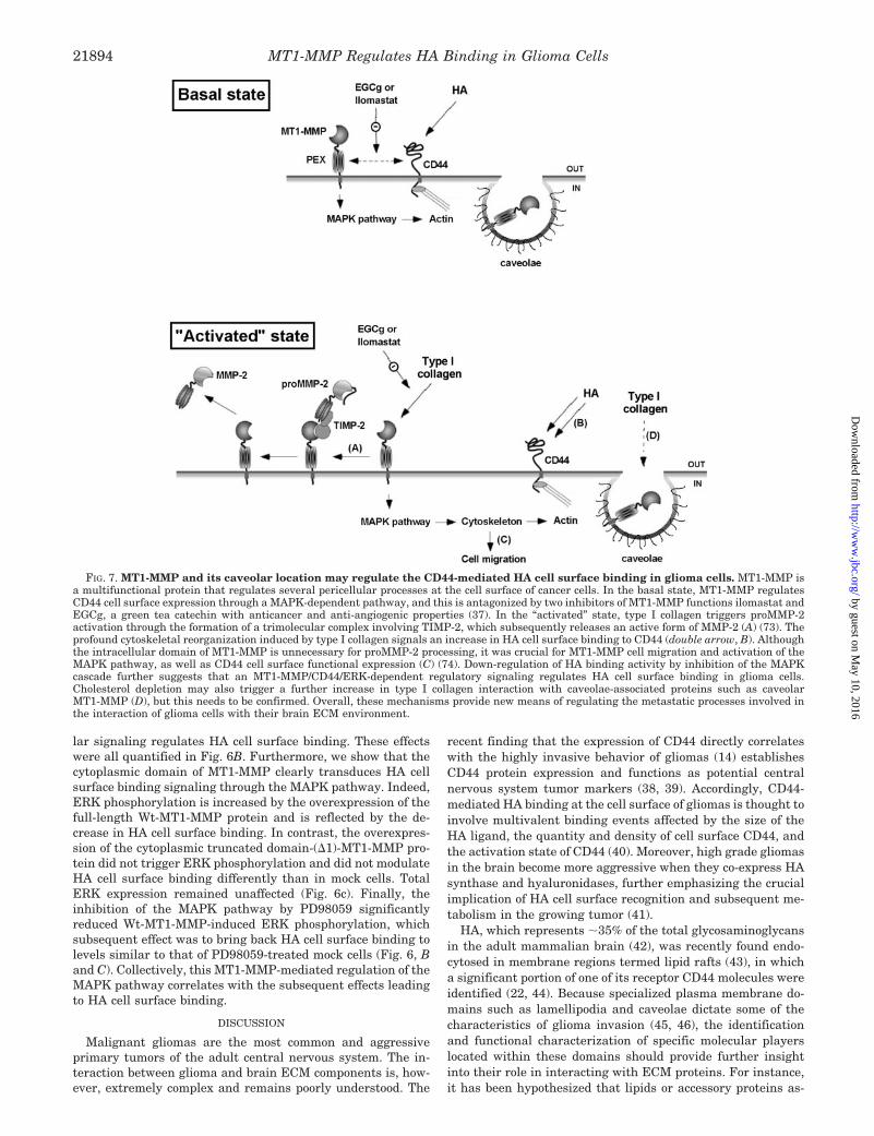
FIG. 7. MT1-MMP and its caveolar location may regulate the CD44-mediated HA cell surface binding in glioma cells. MT1-MMP isa multifunctional protein that regulates several pericellular processes at the cell surface of cancer cells. In the basal state, MT1-MMP regulatesCD44 cell surface expression through a MAPK-dependent pathway, and this is antagonized by two inhibitors of MT1-MMP functions ilomastat andEGCg, a green tea catechin with anticancer and anti-angiogenic properties (37). In the “activated” state, type I collagen triggers proMMP-2activation through the formation of a trimolecular complex involving TIMP-2, which subsequently releases an active form of MMP-2 (A) (73). Theprofound cytoskeletal reorganization induced by type I collagen signals an increase in HA cell surface binding to CD44 (double arrow, B). Althoughthe intracellular domain of MT1-MMP is unnecessary for proMMP-2 processing, it was crucial for MT1-MMP cell migration and activation of theMAPK pathway, as well as CD44 cell surface functional expression (C) (74). Down-regulation of HA binding activity by inhibition of the MAPKcascade further suggests that an MT1-MMP/CD44/ERK-dependent regulatory signaling regulates HA cell surface binding in glioma cells.Cholesterol depletion may also trigger a further increase in type I collagen interaction with caveolae-associated proteins such as caveolarMT1-MMP (D), but this needs to be confirmed. Overall, these mechanisms provide new means of regulating the metastatic processes involved inthe interaction of glioma cells with their brain ECM environment.
MT1-MMP Regulates HA Binding in Glioma Cells21894
by guest on May 10, 2016
http://ww
w.jbc.org/
Dow
nloaded from

sociated within such lipidic bilayer may significantly modulatethe functional activity of CD44 (47, 48). However, the biologicalsignificance of such regulation in Triton X-100-insolubleplasma membrane domains still remains unknown. These ob-servations strongly suggest that caveolae-associated proteinsmust regulate HA binding to the cell surface, and that one suchcandidate could be caveolar MT1-MMP. Accordingly, a veryrecent study provides supportive evidence toward the newfunctional role that would be attributable to caveolar MT1-MMP (49). That study suggests that caveolin-enriched lipidrafts represent a new modality in regulating the functionalactivity of MT1-MMP in malignant cells, and that the yet-to-beidentified mechanisms involving the cytoplasmic tail peptidesequence are likely to facilitate the enzyme translocation tothat distinct membrane compartment.
Our study supports the fact that MT1-MMP-mediated CD44cell surface cleavage may indeed play a critical role in efficientcell detachment from HA substrate and could consequentlypromote glioma cell migration. Structure-function studies havedemonstrated that the PEX domain of MT1-MMP was respon-sible for the formation of a complex with CD44 (50), whereas itsshort 20-amino acid cytoplasmic tail was found to mediateinternalization of the enzyme (51, 52). Interestingly, a completedeletion or point mutation within the MT1-MMP cytoplasmicdomain retains its proteolytic activity on the cell surface at alevel comparable with that of the Wt MT1-MMP (25). Ourstudy provides new evidence that the MT1-MMP cytoplasmicdomain further regulates cell surface CD44 function of bindingHA through intracellular signaling that involves the MAPKpathway (summarized in Fig. 7). The activation of this pathwayhas already been shown to regulate MT1-MMP-mediated cellinvasion processes (34). Whether that regulatory CD44/MT1-MMP-dependent functions could affect other documented pro-cesses involving MT1-MMP, such as its potent fibrinolytic ac-tivity or its role in endothelial cell tubulogenesis, is currentlyunder investigation. Interestingly, a very recent study showedfor the first time that a new 19-kDa protein, which has beenidentified as MTCBP-1 and which belongs to the newly pro-posed Cupin superfamily, associated with the cytoplasmic do-main of MT1-MMP and regulated its function in cell invasionand ECM recognition (53). Whether this new intracellular pro-tein regulates other MT1-MMP-mediated signaling and/orfunctions remains to be investigated. Because for most cancersthe precise role of CD44 in the diffusion of tumors is stillunclear, CD44 interaction with MT1-MMP, HA, and otherECM molecules thus becomes obviously relevant to variousprocesses in metastasis, a complex multistep process that isknown to vary from tumor to tumor and site to site.
CD44 is also known to bind other ECM components. The soleeffect of type I collagen that we report on cell surface HAbinding, combined with the lack of effect of the other ECMproteins tested in the present study, thus strongly suggests therequirement for new cell surface mechanisms regulating CD44-mediated HA binding function. However, the diversity of re-sponses that has been reported from CD44 ligation indicatesthat downstream events following ligand binding by CD44 mayvary depending on the cell type expressing CD44 and on theenvironment of that cell (54). Among the intracellular path-ways, CD44 mediates phosphorylation of ZAP-70 and activa-tion of phospholipase C�, RAS, protein kinase C�, and NF-�Bbinding activity (55). Low molecular mass HA fragments, onthe other hand, enhance tumor cell migration (56) and activatethe Ras-mitogen-activated protein kinase pathway as well asthe phosphoinositide 3-kinase pathway (57). One effect of CD44“outside-in” signaling is up-regulation and activation of inte-grins (58) and the expression of MMP (59). It is possible that
CD44 thus works in concert with another cell surface moleculeto efficiently bind to type I collagen and to subsequently up-regulate cell signaling pathways. In fact, one candidate that wecannot exclude at this point is �1 integrin, which has beenreported to mediate type I collagen induction of proMMP-2processing in normal fibroblasts (60), and to affect the expres-sion, processing, and activity of MT1-MMP in ovarian carci-noma cells (61). One common intracellular pathway may alsoinvolve Ca2�/calmodulin-dependent signaling, which has beenshown to regulate MT1-MMP cell surface processing (25), aswell as the phosphorylation of CD44 required for cell migrationon HA (62). Finally, we also report that EGCg, an effectiveinhibitor of MT1-MMP-mediated functions (36, 63), also antag-onized MT1-MMP-mediated cell surface HA-binding.
CD44 and several isoforms have been characterized on avariety of tumor cell surfaces and have been suggested to beprognostic indicators of malignant melanoma (64, 65). How-ever, although CD44 binds to types I, IV, VI, and XIV collagen,it is not a primary receptor for cell adhesion to collagen (66–69). Thus, other HA receptors, such as the receptor for HA-mediated motility and brain-enriched HA binding/brevican,could also play a role in glioma cell migration (70–72). Thesecould also be involved in the metastatic processes throughinteraction with, and movement through, collagen, most oftentype I and/or basement membrane (type IV) collagen. Ourstudy thus provides a new look at a fundamental mechanism ofglioma invasion that depicts a unique interaction between col-lagen and HA cell surface binding. Manipulation of this inter-action could ultimately serve as a target in the development ofnew anticancer therapeutic strategy.
REFERENCES
1. Asher, R., Perides, G., Vanderhaeghen, J. J., and Bignami, A. (1991) J. Neu-rosci. Res. 28, 410–421
2. Bertolotto, A., Rocca, G., and Schiffer, D. (1990) J. Neurol. Sci. 100, 113–1233. Thylen, A., Wallin, J., and Martensson, G. (1999) Cancer 86, 2000–20054. Rooney, P., Kumar, S., Ponting, J., and Wang, M. (1995) Int. J. Cancer 60,
632–6365. Savani, R. C., Cao, G., Pooler, P. M., Zaman, A., Zhou, Z., and DeLisser, H. M.
(2001) J. Biol. Chem. 276, 36770–367786. West, D. C., and Kumar, S. (1989) Ciba Found. Symp. 143, 187–2017. Rahmanian, M., Pertoft, H., Kanda, S., Christofferson, R., Claesson-Welsh, L.,
and Heldin, P. (1997) Exp. Cell Res. 237, 223–2308. Sattar, A., Rooney, P., Kumar, S., Pye, D., West, D. C., Scott, I., and Ledger, P.
(1998) J. Invest. Dermatol. 103, 576–5799. Sugahara, K. N., Murai, T., Nishinakamura, H., Kawashima, H., Saya, H., and
Miyasaka, M. (2003) J. Biol. Chem. 278, 32259–3226510. Gunthert, U., Schwarzler, C., Wittig, B., Laman, J., Ruiz, P., Stauder, R.,
Bloem, A., Smadja-Joffe, F., Zoller, M., and Rolink, A. (1998) Adv. Exp. Med.Biol. 451, 43–49
11. Naot, D., Sionov, R. V., and Ish-Shalom, D. (1997) Adv. Cancer Res. 71,241–319
12. Yu, Q., and Stamenkovic, I. (1999) Genes Dev. 13, 35–4813. Akiyama, Y., Jung, S., Salhia, B., Lee, S., Hubbard, S., Taylor, M., Mainprize,
T., Akaishi, K., van Furth, W., and Rutka, J. T. (2001) J. Neurooncol. 53,115–127
14. Ranuncolo, S. M., Ladeda, V., Specterman, S., Varela, M., Lastiri, J., Morandi,A., Matos, E., Bal de Kier Joffe, E., Puricelli, L., and Pallotta, M. G. (2002)J. Surg. Oncol. 79, 30–35
15. Gal, I., Lesley, J., Ko, W., Gonda, A., Stoop, R., Hyman, R., and Mikecz, K.(2003) J. Biol. Chem. 278, 11150–11158
16. Xu, Y., and Yu, Q. (2003) J. Biol. Chem. 278, 8661–866817. Bourguignon, L. Y., Singleton, P. A., Zhu, H., and Diedrich, F. (2003) J. Biol.
Chem. 278, 29420–2943418. Okamoto, I., Kawano, Y., Tsuiki, H., Sasaki, J., Nakao, M., Matsumoto, M.,
Suga, M., Ando, M., Nakajima, M., and Saya, H. (1999) Oncogene 18,1435–1446
19. Kajita, M., Itoh, Y., Chiba, T., Mori, H., Okada, A., Kinoh, H., and Seiki, M.(2001) J. Cell Biol. 153, 893–904
20. Annabi, B., Lachambre, M. P., Bousquet-Gagnon, N., Page, M., Gingras, D.,and Beliveau, R. (2001) Biochem. J. 353, 547–553
21. Puyraimond, A., Fridman, R., Lemesle, M., Arbeille, B., and Menashi, S. (2001)Exp. Cell Res. 262, 28–36
22. Perschl, A., Lesley, J., English, N., Hyman, R., and Trowbridge, I. S. (1995)J. Cell Sci. 108, 1033–1041
23. McCurdy, L. H., and Graham, B. S. (2003) J. Virol. 77, 1747–175624. Silver, F. H., and Trelstad, R. L. (1980) J. Biol. Chem. 255, 9427–943325. Annabi, B., Pilorget, A., Bousquet-Gagnon, N., Gingras, D., and Beliveau, R.
(2001) Biochem. J. 359, 325–33326. Beaulieu, E., Kachra, Z., Mousseau, N., Delbecchi, L., Hardy, J., and Beliveau,
R. (1999) Neurosurgery 45, 1432–1440
MT1-MMP Regulates HA Binding in Glioma Cells 21895
by guest on May 10, 2016
http://ww
w.jbc.org/
Dow
nloaded from

27. Gingras, D., Gauthier, F., Lamy, S., Desrosiers, R. R., and Beliveau, R. (1998)Biochem. Biophys. Res. Commun. 247, 888–893
28. Gustafson, S., and Forsberg, N. (1991) Biochim. Biophys. Acta 1091, 36–4029. Samuelsson, C., and Gustafson, S. (1998) Glycoconj. J. 15, 169–17530. Rozanov, D. V., Deryugina, E. I., Ratnikov, B. I., Monosov, E. Z., Marchenko,
G. N., Quigley, J. P., and Strongin A. Y. (2001) J. Biol. Chem. 276,25705–25714
31. Galardy, R. E., Grobelny, D., Foellmer, H. G., and Fernandez, L. A. (1994)Cancer Res. 54, 4715–4718
32. Bernardo, M. M., Brown, S., Li, Z. H., Fridman, R., and Mobashery, S. (2002)J. Biol. Chem. 277, 11201–11207
33. Guo, C., and Piacentini, L. (2003) J. Biol. Chem. 278, 46699–4670834. Gingras, D., Bousquet-Gagnon, N., Langlois, S., Lachambre, M. P., Annabi, B.,
and Beliveau, R. (2001) FEBS Lett. 507, 231–23635. Lamy, S., Gingras, D., and Beliveau, R. (2002) Cancer Res. 62, 381–38536. Annabi, B., Lachambre M. P., Bousquet-Gagnon, N., Page, M., Gingras, D.,
and Beliveau, R. (2002) Biochim. Biophys. Acta 1542, 209–22037. Demeule, M., Michaud-Levesque, J., Annabi, B., Gingras, D., Boivin, D.,
Jodoin, J., Lamy, S., Bertrand, Y., and Beliveau, R. (2002) Curr. Med.Chem. Anti-Canc. Agents 2, 441–463
38. Kuppner, M. C., Van Meir, E., Gauthier, T., Hamou, M. F., and de Tribolet, N.(1992) Int. J. Cancer 50, 572–577
39. Nagasaka, S., Tanabe, K. K., Bruner, J. M., Saya, H., Sawaya, R. E., andMorrison, R. S. (1995) J. Neurosurg. 82, 858–863
40. Lesley, J., Hascall, V. C., Tammi, M., and Hyman, R. (2000) J. Biol. Chem. 275,26967–26975
41. Enegd, B., King, J. A., Stylli, S., Paradiso, L., Kaye, A. H., and Novak, U.(2002) Neurosurgery 50, 1311–1318
42. Maleski, M., and Hockfield, S. (1997) Glia 20, 193–20243. Zajchowski, L. D., and Robbins, S. M. (2002) Eur. J. Biochem. 269, 737–75244. Oliferenko, S., Paiha, K., Harder, T., Gerke, V., Schwarzler, C., Schwarz, H.,
Beug, H., Gunthert, U., and Huber, L. A. (1999) J. Cell Biol. 146, 843–85445. Maidment, S. L. (1997) Anticancer Res. 17, 4145–414946. Silva, W. I., Maldonado, H. M., Lisanti, M. P., Devellis, J., Chompre, G., Mayol,
N., Ortiz, M., Velazquez, G., Maldonado, A., and Montalvo, J. (1999) Int. J.Dev. Neurosci. 17, 705–714
47. Isacke, C. M. (1994) J. Cell Sci. 107, 2353–235948. Liu, D., and Sy, M. S. (1996) J. Exp. Med. 183, 1987–199449. Uekita, T., Gotoh, I., Kinoshita, T., Itoh, Y., Sato, H., Shiomi, T., Okada, Y.,
and Seiki, M. (2004) J. Biol. Chem. 279, 12734–1274350. Seiki, M., Koshikawa, N., and Yana, I. (2003) Cancer Metastasis Rev. 22,
129–14351. Uekita, T., Itoh, Y., Yana, I., Ohno, H., and Seiki, M. (2001) J. Cell Biol. 155,
1345–1356
52. Jiang, A., Lehti, K., Wang, X., Weiss, S. J., Keski-Oja, J., and Pei, D. (2001)Proc. Natl. Acad. Sci. U. S. A. 98, 13693–13698
53. Rozanov, D. V., Deryugina, E. I., Monosov, E. Z., Marchenko, N. D., andStrongin, A. Y. (2004) Exp. Cell Res. 293, 81–95
54. Lesley, J., Hyman, R., and Kincade, P. W. (1993) Adv. Immunol. 54, 271–33555. Pure, E., and Cuff, C. A. (2001) Trends Mol. Med. 7, 213–22156. Lokeshwar, V., Schroeder, G., Hautmann, S., and Selzer, M. (2001) Sci. World
J. 1, 10657. Sohara, Y., Ishiguro, N., Machida, K., Kurata, H., Thant, A. A., Senga, T.,
Matsuda, S., Kimata, K., Iwata, H., and Hamaguchi, M. (2001) Mol. Biol.Cell 12, 1859–1868
58. Fujisaki, T., Tanaka, Y., Fujii, K., Mine, S., Saito, K., Yamada, S., Yamashita,U., Irimura, T., and Eto, S. (1999) Cancer Res. 59, 4427–4434
59. Takahashi, K., Eto, H., and Tanabe, K. K. (1999) Int. J. Cancer 80, 387–39560. Seltzer, J. L., Lee, A. Y., Akers, K. T., Sudbeck, B., Southon, E. A., Wayner,
E. A., and Eisen, A. Z. (1994) Exp. Cell Res. 213, 365–37461. Ellerbroek, S. M., Fishman, D. A., Kearns, A. S., Bafetti, L. M., and Stack,
M. S. (1999) Cancer Res. 59, 1635–164162. Lewis, C. A., Townsend, P. A., and Isacke, CM. (2001) Biochem. J. 357,
843–85063. Oku, N., Matsukawa, M., Yamakawa, S., Asai, T., Yahara, S., Hashimoto, F.,
and Akizawa, T. (2003) Biol. Pharm. Bull. 26, 1235–123864. Leigh, C. J., Palechek, P. L., Knutson, J. R., McCarthy, J. B., Cohen, M. B., and
Argenyi, Z. B. (1996) Hum. Pathol. 27, 1288–129465. Dome, B., Somlai, B., Ladanyi, A., Fazekas, K., Zoller, M., and Timar, J. (2001)
Virchows Arch. 439, 628–63566. Knutson, J. R., Iida, J., Fields, G. B., and McCarthy, J. B. (1996) Mol. Biol. Cell
7, 383–39667. Carter, W. G., and Wayner, E. A. (1988) J. Biol. Chem. 263, 4193–420168. Ehnis, T., Dieterich, W., Bauer, M., Lampe, B., and Schuppan, D. (1996) Exp.
Cell Res. 229, 388–39769. Faassen, A. E., Schrager, J. A., Klein, D. J., Oegema, T. R., Couchman, J. R.,
and McCarthy, J. B. (1992) J. Cell Biol. 116, 521–53170. Jaworski, D. M., Kelly, G. M., Piepmeier, J. M., and Hockfield, S. (1996) Cancer
Res. 56, 2293–229871. Zhang, H., Kelly, G., Zerillo, C., Jaworski, D. M., and Hockfield, S. (1998)
J. Neurosci. 18, 2370–237672. Nutt, C. L., Zerillo, C. A., Kelly, G. M., and Hockfield, S. (2001) Cancer Res. 61,
7056–705973. Itoh, Y., Takamura, A., Ito, N., Maru, Y., Sato, H., Suenaga, N., Aoki, T., and
Seiki, M. (2001) EMBO J. 20, 4782–479374. Mori, H., Tomari, T., Koshikawa, N., Kajita, M., Itoh, Y., Sato, H., Tojo, H.,
Yana, I., and Seiki, M. (2002) EMBO J. 21, 3949–3959
MT1-MMP Regulates HA Binding in Glioma Cells21896
by guest on May 10, 2016
http://ww
w.jbc.org/
Dow
nloaded from

Borhane Annabi, Sébastien Thibeault, Robert Moumdjian and Richard BéliveauCaveolae in Glioma Cells
Hyaluronan Cell Surface Binding Is Induced by Type I Collagen and Regulated by
doi: 10.1074/jbc.M313694200 originally published online March 10, 20042004, 279:21888-21896.J. Biol. Chem.
10.1074/jbc.M313694200Access the most updated version of this article at doi:
Alerts:
When a correction for this article is posted•
When this article is cited•
to choose from all of JBC's e-mail alertsClick here
http://www.jbc.org/content/279/21/21888.full.html#ref-list-1
This article cites 74 references, 33 of which can be accessed free at
by guest on May 10, 2016
http://ww
w.jbc.org/
Dow
nloaded from