Identification of Novel Genetic Alterations in Samples of Malignant Glioma Patients
11
Identification of Novel Genetic Alterations in Samples of Malignant Glioma Patients Vedrana Milinkovic 1 , Jasna Bankovic 1 , Miodrag Rakic 2 , Tijana Stankovic 1 , Milica Skender-Gazibara 3 , Sabera Ruzdijic 1 , Nikola Tanic 1 * 1 University of Belgrade, Institute for Biological Research ‘‘Sinisa Stankovic’’, Department of Neurobiology, Belgrade, Republic of Serbia, 2 Clinical Center of Serbia, Clinic for Neurosurgery, Belgrade, Republic of Serbia, 3 University of Belgrade, School of Medicine, Institute of Pathology, Belgrade, Republic of Serbia Abstract Glioblastoma is the most frequent and malignant human brain tumor. High level of genomic instability detected in glioma cells implies that numerous genetic alterations accumulate during glioma pathogenesis. We investigated alterations in AP- PCR DNA profiles of 30 glioma patients, and detected specific changes in 11 genes not previously associated with this disease: LHFPL3, SGCG, HTR4, ITGB1, CPS1, PROS1, GP2, KCNG2, PDE4D, KIR3DL3, and INPP5A. Further correlations revealed that 8 genes might play important role in pathogenesis of glial tumors, while changes in GP2, KCNG2 and KIR3DL3 should be considered as passenger mutations, consequence of high level of genomic instability. Identified genes have a significant role in signal transduction or cell adhesion, which are important processes for cancer development and progression. According to our results, LHFPL3 might be characteristic of primary glioblastoma, SGCG, HTR4, ITGB1, CPS1, PROS1 and INPP5A were detected predominantly in anaplastic astrocytoma, suggesting their role in progression of secondary glioblastoma, while alterations of PDE4D seem to have important role in development of both glioblastoma subtypes. Some of the identified genes showed significant association with p53, p16, and EGFR, but there was no significant correlation between loss of PTEN and any of identified genes. In conclusion our study revealed genetic alterations that were not previously associated with glioma pathogenesis and could be potentially used as molecular markers of different glioblastoma subtypes. Citation: Milinkovic V, Bankovic J, Rakic M, Stankovic T, Skender-Gazibara M, et al. (2013) Identification of Novel Genetic Alterations in Samples of Malignant Glioma Patients. PLoS ONE 8(12): e82108. doi:10.1371/journal.pone.0082108 Editor: Marta M. Alonso, University Hospital of Navarra, Spain Received August 22, 2013; Accepted October 25, 2013; Published December 16, 2013 Copyright: ß 2013 Milinkovic et al. This is an open-access article distributed under the terms of the Creative Commons Attribution License, which permits unrestricted use, distribution, and reproduction in any medium, provided the original author and source are credited. Funding: This study was supported by Grant III41031 from the Ministry of Education, Science and Technological Development, Republic of Serbia. The funders had no role in study design, data collection and analysis, decision to publish, or preparation of the manuscript. Competing Interests: The authors have declared that no competing interests exist. * E-mail: [email protected] Introduction Cancer is a genetic disease characterized by DNA sequence changes, copy number aberrations, chromosomal rearrangements and modification in DNA methylation leading to compromised regulatory mechanisms governing cell proliferation and homeo- stasis. Studies carried out over the past three decades suggest that malignant gliomas, like other cancers, represent a consequence of the accumulation of genetic alterations, although their nature and exact number required for tumorigenesis remain unclear. Glioblastoma is the most frequent and aggressive brain tumor. Majority of glioblastomas (GBM, WHO grade IV) develop de novo (primary glioblastomas) without clinical or histological evidence of a less malignant precursor lesion, while others, progressing from low-grade diffuse astrocytoma or anaplastic astrocytoma (AA), represent rare secondary glioblastomas. Despite a similar histo- logical appearance, primary and secondary glioblastomas are distinct tumor entities that originate from different precursor cells containing different genetic alterations [1]. Epidermal growth factor receptor (EGFR) amplification and PTEN mutations are genetic alterations typical of primary glioblastomas, whereas p53 mutations are early and frequent genetic alterations in the pathway leading to secondary glioblastomas. Furthermore, muta- tions in the active site of isocitrate dehydrogenase 1 (IDH1) were associated with secondary GBMs [2]. On the other hand, LOH 10q and alterations of p16 INK4a /RB1 pathway seem to be important in the development of both primary and secondary glioblastomas [3]. Besides these frequently altered pathways, there is evidence of large number of genetic alterations in glioblastoma samples, reported in The Cancer Genome Atlas (TCGA) database. TCGA project enabled the integrated analyses of multi-dimensional genomic data collected from different platforms with the aim to better characterize and understand tumor origin, behavior and treatment [4]. However, further analyses are needed to identify additional potentially useful genetic alterations for the classification and targeted therapy of GBMs. Besides, significant level of genomic instability and heterogeneity detected in glial tumors confirm that they evolve along a multitude of pathways rather than along a single defined pathway [5,6,7]. Even though only a few specific genetic pathways are consistently highlighted, there are undoubt- edly complex interactions among them as well as with additional yet unknown factors. Reliable molecular markers are needed to enable the identification of patients at risk for developing GBM, improve the early detection and appropriate diagnosis, as well as to provide molecular profile for better prediction of patient outcome and response to therapy. PLOS ONE | www.plosone.org 1 December 2013 | Volume 8 | Issue 12 | e82108
-
Upload
independent -
Category
Documents
-
view
1 -
download
0
Transcript of Identification of Novel Genetic Alterations in Samples of Malignant Glioma Patients

Identification of Novel Genetic Alterations in Samples ofMalignant Glioma PatientsVedrana Milinkovic1, Jasna Bankovic1, Miodrag Rakic2, Tijana Stankovic1, Milica Skender-Gazibara3,
Sabera Ruzdijic1, Nikola Tanic1*
1 University of Belgrade, Institute for Biological Research ‘‘Sinisa Stankovic’’, Department of Neurobiology, Belgrade, Republic of Serbia, 2 Clinical Center of Serbia, Clinic
for Neurosurgery, Belgrade, Republic of Serbia, 3 University of Belgrade, School of Medicine, Institute of Pathology, Belgrade, Republic of Serbia
Abstract
Glioblastoma is the most frequent and malignant human brain tumor. High level of genomic instability detected in gliomacells implies that numerous genetic alterations accumulate during glioma pathogenesis. We investigated alterations in AP-PCR DNA profiles of 30 glioma patients, and detected specific changes in 11 genes not previously associated with thisdisease: LHFPL3, SGCG, HTR4, ITGB1, CPS1, PROS1, GP2, KCNG2, PDE4D, KIR3DL3, and INPP5A. Further correlations revealedthat 8 genes might play important role in pathogenesis of glial tumors, while changes in GP2, KCNG2 and KIR3DL3 should beconsidered as passenger mutations, consequence of high level of genomic instability. Identified genes have a significantrole in signal transduction or cell adhesion, which are important processes for cancer development and progression.According to our results, LHFPL3 might be characteristic of primary glioblastoma, SGCG, HTR4, ITGB1, CPS1, PROS1 andINPP5A were detected predominantly in anaplastic astrocytoma, suggesting their role in progression of secondaryglioblastoma, while alterations of PDE4D seem to have important role in development of both glioblastoma subtypes. Someof the identified genes showed significant association with p53, p16, and EGFR, but there was no significant correlationbetween loss of PTEN and any of identified genes. In conclusion our study revealed genetic alterations that were notpreviously associated with glioma pathogenesis and could be potentially used as molecular markers of differentglioblastoma subtypes.
Citation: Milinkovic V, Bankovic J, Rakic M, Stankovic T, Skender-Gazibara M, et al. (2013) Identification of Novel Genetic Alterations in Samples of MalignantGlioma Patients. PLoS ONE 8(12): e82108. doi:10.1371/journal.pone.0082108
Editor: Marta M. Alonso, University Hospital of Navarra, Spain
Received August 22, 2013; Accepted October 25, 2013; Published December 16, 2013
Copyright: � 2013 Milinkovic et al. This is an open-access article distributed under the terms of the Creative Commons Attribution License, which permitsunrestricted use, distribution, and reproduction in any medium, provided the original author and source are credited.
Funding: This study was supported by Grant III41031 from the Ministry of Education, Science and Technological Development, Republic of Serbia. The fundershad no role in study design, data collection and analysis, decision to publish, or preparation of the manuscript.
Competing Interests: The authors have declared that no competing interests exist.
* E-mail: [email protected]
Introduction
Cancer is a genetic disease characterized by DNA sequence
changes, copy number aberrations, chromosomal rearrangements
and modification in DNA methylation leading to compromised
regulatory mechanisms governing cell proliferation and homeo-
stasis. Studies carried out over the past three decades suggest that
malignant gliomas, like other cancers, represent a consequence of
the accumulation of genetic alterations, although their nature and
exact number required for tumorigenesis remain unclear.
Glioblastoma is the most frequent and aggressive brain tumor.
Majority of glioblastomas (GBM, WHO grade IV) develop de novo
(primary glioblastomas) without clinical or histological evidence of
a less malignant precursor lesion, while others, progressing from
low-grade diffuse astrocytoma or anaplastic astrocytoma (AA),
represent rare secondary glioblastomas. Despite a similar histo-
logical appearance, primary and secondary glioblastomas are
distinct tumor entities that originate from different precursor cells
containing different genetic alterations [1]. Epidermal growth
factor receptor (EGFR) amplification and PTEN mutations are
genetic alterations typical of primary glioblastomas, whereas p53
mutations are early and frequent genetic alterations in the
pathway leading to secondary glioblastomas. Furthermore, muta-
tions in the active site of isocitrate dehydrogenase 1 (IDH1) were
associated with secondary GBMs [2]. On the other hand, LOH
10q and alterations of p16INK4a/RB1 pathway seem to be
important in the development of both primary and secondary
glioblastomas [3]. Besides these frequently altered pathways, there
is evidence of large number of genetic alterations in glioblastoma
samples, reported in The Cancer Genome Atlas (TCGA)
database. TCGA project enabled the integrated analyses of
multi-dimensional genomic data collected from different platforms
with the aim to better characterize and understand tumor origin,
behavior and treatment [4].
However, further analyses are needed to identify additional
potentially useful genetic alterations for the classification and
targeted therapy of GBMs. Besides, significant level of genomic
instability and heterogeneity detected in glial tumors confirm that
they evolve along a multitude of pathways rather than along a
single defined pathway [5,6,7]. Even though only a few specific
genetic pathways are consistently highlighted, there are undoubt-
edly complex interactions among them as well as with additional
yet unknown factors. Reliable molecular markers are needed to
enable the identification of patients at risk for developing GBM,
improve the early detection and appropriate diagnosis, as well as
to provide molecular profile for better prediction of patient
outcome and response to therapy.
PLOS ONE | www.plosone.org 1 December 2013 | Volume 8 | Issue 12 | e82108

A number of techniques based on PCR, hybridization, and
conformation changes, as well as modern high-throughput
genome wide techniques can be employed for the detection of
specific mutations in cancer cells. AP-PCR DNA fingerprinting
method has numerous advantages over conventional methods,
mainly because no prior knowledge of the genome under
investigation is required and because it allows the screening of
the whole genome including non coding DNA regions. Further-
more, AP-PCR is a reliable, inexpensive method that does not
require complex equipment, does providing highly reproducible
patterns of amplified fragments which faithfully reflect differences
in DNA sequences and/or relative abundance of the templates
and enable detection of genetic alterations in tumor tissue [8].
Finally, the possibility of further analysis of variant bands by
reamplification, cloning, and sequencing enables rapid identifica-
tion of genes probably linked to the development and progression
of malignant tumor [9]. AP-PCR has already proven to be highly
informative for analysis of cancer associated somatic mutations
since it has been implemented in the analyzes of various cancers,
including pancreatic and colorectal carcinomas [10,11], as well as
lung [12,13], and breast cancers [14].
We applied AP-PCR for the detection of anonymous multiple
genetic alterations in 30 patients with AA and GBM. Following
our previous study of genomic instability in glioma patients [15]
we analyzed altered sequences in tumor DNA profiles with the aim
to identify genes specific for glioma pathogenesis. Furthermore, we
examined identified genes in relation to genomic instability,
clinicopathological parameters, and patients’ survival. Also, we
tested the association of the most frequently present genetic
alterations in primary and secondary glioblastomas with newly
identified genes with the aim to recognize potential molecular
markers of different glioblastoma subtypes.
Materials and Methods
Ethics StatementThe samples were collected and used after obtaining informed
consents and approval from the Ethical Committee of Clinical
Center of Serbia (approval number 3672/1), in accordance with
the ethical standards laid down in the 1964 Declaration of
Helsinki, Laws of Republic of Serbia, as well as GCP guidelines.
All participants provided their written informed consent to
participate in this study. The form of informed consent was
approved by the Ethical Committee of Clinical Center of Serbia.
Tissue samplesPaired cancer tissue and blood samples were collected from 30
patients who underwent surgery at Neurosurgery Clinic, Clinical
Center of Serbia. The specimens were frozen in liquid nitrogen,
where they were kept until DNA extraction. All patients had a
histologically confirmed diagnosis of anaplastic astrocytoma
(WHO grade III; n = 8) or glioblastoma multiforme (WHO grade
IV; n = 22) according to the 2007 WHO classification [16]. All
grade IV tumors were considered primary (de novo) because the
glioblastoma diagnosis was made at the first biopsy, without
clinical or histopathological evidence of a less malignant precursor
lesion. The 30 patients included 19 men and 11 women, with a
median age of 56.9 years (within the range of 20 to 84 years). The
median overall survival was 11 months. Patients received neither
radio- nor chemotherapy before surgery.
Immunohistochemical analysisFollowing routine hematoxylin-eosin method for staining of
paraffin tissue sections [15], we performed immunohistochemical
staining for p53 to further characterize our samples. Sections of
tissue were subsequently heated in phosphate buffered saline
(PBS), and stained with the streptavidin-biotin technique using
antibody against p53 protein (Dako, Monoclonal Rabbit Anti-
Human Antibody, dilution 1:50), according to the manufacturer’s
instructions.
Figure 1. Immunohistochemical characterization of samples. Example of anaplastic astrocytoma (WHO grade III). (A) Cellular tumor withincreased mitotic activity (HE 6200) (B) Diffuse nuclear p53 positivity of tumor cells (immunostaining 6250).doi:10.1371/journal.pone.0082108.g001
Table 1. Distribution of samples according to the level ofgenomic instability.
Level of genomic instability Number of patients
30
Genomic instability total
low#0,3 11
high.0,3 19
Microsatellite instability
low#0,15 14
high.0,15 16
Chromosomal instability
low#0,16 13
high.0,16 17
doi:10.1371/journal.pone.0082108.t001
Novel Genetic Alterations in Glioma Samples
PLOS ONE | www.plosone.org 2 December 2013 | Volume 8 | Issue 12 | e82108
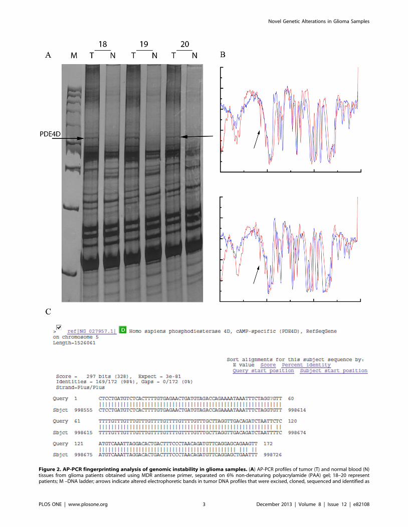
Figure 2. AP-PCR fingerprinting analysis of genomic instability in glioma samples. (A) AP-PCR profiles of tumor (T) and normal blood (N)tissues from glioma patients obtained using MDR antisense primer, separated on 6% non-denaturing polyacrylamide (PAA) gel; 18–20 representpatients; M –DNA ladder; arrows indicate altered electrophoretic bands in tumor DNA profiles that were excised, cloned, sequenced and identified as
Novel Genetic Alterations in Glioma Samples
PLOS ONE | www.plosone.org 3 December 2013 | Volume 8 | Issue 12 | e82108

DNA extractionDNA was extracted using the phenol/chloroform/isoamyl
alcohol method [17]. The quality of the DNA was verified by
electrophoresis on 0.8% agarose gel. The DNA concentration was
assessed spectrophotometrically.
AP-PCR DNA fingerprintingAP-PCR DNA fingerprinting, was used to compare DNA
profiles of paired tumor and blood samples of the same patient
[15]. In short, seven primers were tested for the ability to generate
informative fingerprints that distinguish tumor from normal tissue.
Optimization of AP-PCR reactions was done for each primer
according to Cobb [18] and included the search for conditions
that would yield profiles of moderate complexity in order to
simplify the analysis [19]. Primer sequences, reaction conditions,
and amplification profiles were described previously [15].
AP-PCR products were separated on 6–8% non-denaturing
polyacrylamide (PAA) gels and visualized by silver staining. Gel
images were acquired with the Multi-Analyst/PC Software Image
Analysis System (Bio Rad Gel Doc 1000). Digitized images were
loaded into the specialized public software Image J (National
Institute of Health, USA, www.rsb.info.nih.gov/ij) and analyzed
by the image enhancement function ‘adapthisteq’ as previously
described [15].
Isolation, cloning and sequencing of variantelectrophoretic bands
Twenty selected DNA bands with altered mobility were further
characterized. The PCR amplicons resolved on the silver stained
PAA gels were gently removed with a hypodermic 22-gauge
needle pre-wetted with the PCR master mix solution. The needle
was dipped in the PCR master mix for 2 min and then discarded.
The PCR products were reamplified with the same primers used
for AP-PCR reactions at high-stringency conditions specific for
each particular primer [20]. The reamplified material was
administrated on 1.5% agarose gels, purified using DNA
Extraction Kit (Fermentas Life Sciences, Lithuania) and cloned
with GeneJetTM PCR Cloning Kit (Fermentas Life Sciences,
Lithuania) according to manufacturer’s instructions. Plasmids were
purified using GeneJetTM Plasmid Miniprep Kit (Fermentas Life
Sciences, Lithuania). Sequences were determined on ABI Prism
3130 Genetic Analyzer automated sequencer (Applied Biosystems,
Foster City, CA, USA) using BigDye Terminator v3.1 Cycle
Sequencing Kit (Applied Biosystems, Foster City, CA, USA).
Sequencing was performed in both directions on several clones for
each selected DNA band. The obtained sequences were analyzed
and identified using BLAST software in the NCBI GenBank and
EBI (Sanger Institute) database.
Analysis of alterations in p53, PTEN, and p16 tumorsuppressor genes
Alterations of p53 tumor suppressor gene were analyzed in our
previous study [15]. Frequently mutated exons of p53 gene (5–9)
were amplified and screened for mutations by PCR-SSCP (Single
Strand Conformation Polymorphism) analysis according to Orita
et al. [21]. Detected mutations were confirmed by sequencing with
Applied Biosystems Incorporated dye terminator sequencing kit
according to the manufacturer’s specifications on an ABI Prism
3130 automated sequencer (Applied Biosystems, Foster City,
USA).
Tumor and blood DNA obtained from all 30 patients was used
to study the loss of heterozygosity (LOH) of p53, PTEN and p16
tumor suppressors. As described previously, LOH analysis of p53
was performed by fragment analysis with highly polymorphic
microsatellite markers (TP53pentanucleotide, TP53dinucleotide,
D17S1537 and D17S786) specific for chromosomal region
spanning p53 gene locus [15]. Five polymorphic microsatellite
markers lying within or flanking PTEN gene (D10S579,
D10S1765, D10S215, AFM086wg9, and D10S541) were selected
to cover deletions at the whole PTEN locus on chromosome
10q23. All forward primers were 59-labeled with Fam, Vic, Ned,
Pet, and Fam fluorescent dyes, respectively. The choice of the
microsatellite markers and locus-specific PCR conditions was
determined from published sources [22,23].
Another set of 3 polymorphic microsatellite markers spanning
the INK4a/ARF locus (D9S171, D9S1748 and D9S162) were
selected to cover deletions on chromosome 9p21–23 [24]. All
forward primers were 59-labeled with Ned, Pet and Vic fluorescent
dyes, respectively. PCR products for all LOH analyses were
separated by capillary electrophoresis on an ABI Prism 3130
automated sequencer and sized using GeneScan 2500 LIZ size
standard (Applied Biosystems, Foster City, USA). The obtained
data were analyzed with the GeneMapper software (Applied
Biosystems). DNA extracted from peripheral blood of each patient
was used as a reference. A marker was defined as noninformative
(homozygote) when only 1 allelic peak was detected in the
reference sample. Contrary to this, a marker was considered
informative (heterozygote) when two major allelic peaks occurred
in a blood specimen. The LOH score was calculated automatically
by GeneMapper software according to the following equation:
(peak height of normal allele 2)/(peak height of normal allele 1)
divided by (peak height of tumor allele 2)/(peak height of tumor
allele 1). A sample was considered to be a LOH candidate for
particular locus if the ratio values were less than 0.67 and higher
than 1.35. p16 was also tested for the presence of homozygous
deletions by a differential PCR method, according to conditions
determined from published sources [12]. Briefly, a 199-bp
fragment of INK4a/ARF locus from exon 2 was co-amplified
with a 131-bp fragment of the adenine phosphoribosyltransferase
(APRT) gene, which was used as internal control. Primer
sequences were previously described by Hayashi et al. [25].
Forward primers were 59-labeled with Fam fluorescent dye.
Fluorescent PCR products were separated by capillary electro-
phoresis on an ABI Prism 3130 automated sequencer and sized
using GeneScan 2500 LIZ size standard (Applied Biosystems,
Foster City, CA, USA). Obtained data were analyzed by fragment
analysis with the GeneMapper software (Applied Biosystems,
Foster City, CA, USA). The presence of homozygous deletions was
determined according to ratio of peak intensities of INK4a/ARF
and APRT in tumor samples relative to the same ratio in normal
samples. A sample had homozygous deletion of INK4a/ARF locus
and consequently p16 tumor suppressor gene if the value of this
proportion was higher than 2.
Amplification status of EGFR geneFor differential PCR analysis, a 110-bp fragment of the EGFR
gene on chromosome 7 was co-amplified with a 168-bp fragment
PDE4D. (B) contrast-limited adaptive histograms obtained using image enhancement function ‘adapthisteq’ of the specialized public software ImageJ, arrows indicate alterations; (C) the identity of the altered AP-PCR bands determined by BLAST homology search in the NCBI genBank and EBISanger databases.doi:10.1371/journal.pone.0082108.g002
Novel Genetic Alterations in Glioma Samples
PLOS ONE | www.plosone.org 4 December 2013 | Volume 8 | Issue 12 | e82108

of b-actin (ACTB) gene on the same chromosome. The primer
sequences were as follows: 59-AGC CAT GCC CGC ATT AGC
TC-39 (sense) and 59-AAA GGA ATG CAA CTT CCC AA-39
(antisense) for EGFR, and 59-CTC TTT TCT TTC CCG ATA
GGT-39 (sense) and 59-CTG GGA TGC TCT TCG ACC TC-39
(antisense) for the ACTB. Genomic DNA (100 ng) was amplified
with 0.8 mM of each primer, 16KCl Buffer, 1.5 mM of MgCl2,
0.25 mM of dNTPs, and 1 U Taq polymerase (Fermentas,
Thermo Scientific) in reaction volume of 25 ml. The PCR reaction
was performed on the GeneAmpH PCR System 9700 (Applied
Bioscience) under the following conditions: initial denaturation at
95uC for 10 minutes was followed by 30 cycles at 95uC for 1
minute, at 58uC for 1 minute and at 72uC for 1 minute, with final
elongation at 72uC for 10 minutes. The PCR products were
loaded on 2% agarose gels and stained with ethidium bromide.
Multi-Analyst/PC Software Image Analysis System (Bio-Rad
GelDoc 1000) was employed for densitometric analysis.
Ratio of the EGFR/ACTB score from tumor and blood tissue of
each patient was calculated and values higher than 2 indicated
presence of EGFR gene amplification.
Statistical analysisSignificant differences between the data sets were determined by
STATISTICA 6.0 software (StatSoft, Inc., Tulsa, USA). The
correlations between identified genes and genomic instability,
clinicopathological parameters and alterations of p53, PTEN, p16,
and EGFR were evaluated using Fisher exact test. Survival analyses
were performed using Kaplan and Meier product-limit method.
The log rank test was used to assess the significance of the
difference between pairs of survival probabilities. Overall survival
was calculated from the day after surgery to the last follow-up
examination or death of the patient. Statistical differences were
considered significant when p was #0.05 (*), p#0.01 (**) and
p#0.005 (***).
Results
Histopathological classification of tumor samplesAfter staining of surgically removed tissues by routine hema-
toxylin-eosin method and immunohistochemical testing for p53
positivity (Fig. 1) we confirmed diagnosis of 8 anaplastic
astrocytoma (WHO grade III) and 22 glioblastoma multiforme
samples (WHO grade IV) according to WHO criteria (2007).
Analysis of variant DNA fragmentsSeven AP-PCR primers were used to discriminate normal from
tumor tissue. Four of them produced informative AP-PCR DNA
profiles containing explicit and countable differences between
tumor and blood in all 30 patients that were analyzed. Observed
differences were further classified as qualitative (mobility shifts in
the banding pattern due to mutations at the primer-template
interaction sites) and quantitative (altered band intensities repre-
senting amplifications or deletions of existing chromosomal
material) and were used as a measurement of the level of total,
microsatellite and chromosomal instability, as described in our
previous paper [15]. Significant level of genomic instability was
present in all samples, and based on the distribution of the
Figure 3. Identification of genetic alterations in tumor DNAfingerprints. The identity of the AP-PCR bands was determined byBLAST homology search in the NCBI GenBank and EBI Sanger databases.For each gene identified, numbers indicating beginning and the end ofthe region of homology in the GenBank sequence, overall sequenceidentity of the clone (%) and number of gaps are represented.doi:10.1371/journal.pone.0082108.g003
Novel Genetic Alterations in Glioma Samples
PLOS ONE | www.plosone.org 5 December 2013 | Volume 8 | Issue 12 | e82108

frequency of DNA alterations, patients were assorted into two
groups, with low or high level of genomic instability (Table 1).
The next step in our work was to identify some of the aberrant
bands in DNA AP-PCR profiles common to more than 5 patients.
Hence, twenty aberrant bands were retrieved from the PAA gels
and cloned (Fig. 2A). Bands (amplicons) with the same electro-
phoretic mobility were isolated and characterized from at least two
patients in order to confirm that they represent the same DNA
sequence (Fig. 2B). Two clones of each band were sequenced.
Obtained sequences were submitted for homology or identity
search in NCBI GenBank and EBI (Sanger Institute) databases.
Some of the sequences matched known genes and were easily
identified, while others represented parts of certain contigs
mapped on chromosomes 1, 2, 8q21–q23, 9, and 12, and their
identification is yet to be determined (Fig. 2C).
The following 11 genes were identified: lipoma HMGIC fusion
partner-like 3 (LHFPL3); sarcoglycan, gamma (SGCG); 5-hydroxy-
tryptamine (serotonin) receptor 4 (HTR4); integrin beta 1 (ITGB1);
mitochondrial carbamoyl-phosphate synthetase 1 (CPS1); protein S
(alpha) (PROS1); glycoprotein 2 (zymogen granule membrane)
(GP2, ZAP75); potassium voltage-gated channel, subfamily G,
member 2 (KCNG2); cAMP-specific 39,59-cyclic phosphodiesterase
4D (PDE4D); killer cell immunoglobulin-like receptor, three
domains, long cytoplasmic tail, 3 (KIR3DL3); inositol polypho-
sphate-5-phosphatase (INPP5A). We were also able to identify
types of mutations in these genes. Namely, we identified nucleotide
substitutions in KIR3DL3, INPP5A and KCNG2, multiple nucleo-
tide substitutions in SGCG, PDE4D and LHFPL3, while HTR4,
ITGB1, CPS1, PROS1 and GP2 were carrying both multiple
substitutions and 2 nucleotide deletions (Fig. 3). We then analyzed
eight out of 11 identified genes regarding the expression profile of
these genes in glial cells as a basic criterion.
Association of identified DNA alterations with genomicinstability and clinicopathological parameters
Alterations of eight out of eleven identified genes were further
examined in relation to the level of total, microsatellite and
chromosomal genomic instability, tumor grade (grade III AA or
grade IV GBM), age and sex (Tables 2 and 3). There was no
statistical significance in correlation of identified DNA alterations
with age and sex of the patients.
LHFPL3 was altered in 10 out of 30 patients (33.3%),
predominantly in grade IV glioblastoma samples (36.4% vs.
25% in grade III anaplastic astrocytoma). It was detected in
significantly higher percentage in samples with high level of total
genomic instability (52.9% vs. 7.7% of samples with low level of
total instability, p = 0.005). The same trend was observed for
microsatellite (37.5% of samples with high vs. 28.6% of samples
with low level) and chromosomal instability (47.1% of samples
with high vs. 15.4% of samples with low level), but without
statistical significance.
Alterations of SGCG, HTR4, ITGB1, CPS1 and PROS1 were
detected in 26.6% of samples (8 out of 30 patients) and were
slightly increased in patients with anaplastic astrocytoma (37.5%)
compared to GBM (22.7%), as well as in the samples with low level
of total genomic instability (38.5% vs. 17.64% with high
Table 2. Association between the frequency of altered genes, clinicopathological parameters and genomic instability.
LHFPL3 SGCG PDE4D HTR4
Total cNP NP % p NP % p NP % p NP % p
Parameter
Total 30 10 33.3 8 26.6 9 30 8 26.6
Glioma subtype
AAa 8 2 25.0 0.45 3 37.5 0.36 2 25.0 0.55 3 37.5 0.36
GBM 22 8 36.4 5 22.7 7 31.8 5 22.7
Sex
Male 19 8 42.1 0.18 5 26.3 0.64 5 26.3 0.43 5 26.3 0.64
Female 11 2 18.2 3 27.3 4 36.4 3 27.3
Age
$50y 24 10 41.7 0.06 5 20.8 0.17 7 29.2 0.42 5 20.8 0.17
,50y 6 0 00.0 3 50.0 2 33.3 3 50.0
Genomic instability total
Low 13 1 7.7 0.005b 5 38.5 0.19 3 23.1 0.38 5 38.5 0.19
High 17 9 52.9 3 17.6 6 35.3 3 17.6
Microsatellite instability
Low 14 4 28.6 0.45 3 21.4 0.42 6 42.8 0.15 3 21.4 0.42
High 16 6 37.5 5 31.2 3 18.8 5 31.2
Chromosomal instability
Low 13 2 15.4 0.07 7 53.8 0.005 1 7.7 0.02 7 53.8 0.005
High 17 8 47.1 1 5.9 8 47.1 1 5.9
aAA, anaplastic astrocytoma; GBM, glioblastoma multiforme;bBold indicates statistically significant values;cNP, number of patients per group.; LHFPL3- lipoma HMGIC fusion partner-like 3; SGCG- sarcoglycan, gamma; PDE4D- cAMP-specific 39,59-cyclic phosphodiesterase 4D;HTR4-5-hydroxytryptamine (serotonin) receptor 4.doi:10.1371/journal.pone.0082108.t002
Novel Genetic Alterations in Glioma Samples
PLOS ONE | www.plosone.org 6 December 2013 | Volume 8 | Issue 12 | e82108

Table 4. Association of the frequency of identified genes with p53, PTEN, p16 and EGFR alterations.
LHFPL3 SGCG PDE4D HTR4
Total bNP NP % p NP % p NP % p NP % p
Parameter
p53
YES 12 5 41.7 0.34 4 33.3 0.40 1 8.3 0.04a 4 33.3 0.40
NO 18 5 27.8 4 22.2 8 44.4 4 22.2
PTEN
YES 20 7 35.0 0.56 4 20.0 0.23 6 30.0 0.67 4 20.0 0.23
NO 10 3 30.0 4 40.0 3 30.0 4 40.0
p16
YES 18 5 27.8 0.34 6 33.3 0.28 3 16.7 0.05 6 33.3 0.28
NO 12 5 41.7 2 16.7 6 50.0 2 16.7
EGFR
YES 13 5 38.4 0.45 5 27.8 0.19 3 23.1 0.38 5 27.8 0.19
NO 17 5 29.4 3 25.0 6 35.3 3 25.0
aBold indicates statistically significant values;bNP, number of patients per group; LHFPL3- lipoma HMGIC fusion partner-like 3; SGCG- sarcoglycan, gamma; PDE4D- cAMP-specific 39,59-cyclic phosphodiesterase 4D;HTR4-5-hydroxytryptamine (serotonin) receptor 4.doi:10.1371/journal.pone.0082108.t004
Table 3. Association between the frequency of altered genes, clinicopathological parameters and genomic instability.
ITGB1 INPP5A CPS1 PROS1
Total cNP NP % p NP % p NP % p NP % p
Parameter
Total 30 8 26.6 13 43.3 8 26.6 8 26.6
Glioma subtype
AAa 8 3 37.5 0.36 5 62.5 0.19 3 37.5 0.36 3 37.5 0.36
GBM 22 5 22.7 8 36.4 5 22.7 5 22.7
Sex
Male 19 5 26.3 0.64 10 52.6 0.17 5 26.3 0.64 5 26.3 0.64
Female 11 3 27.3 3 27.3 3 27.3 3 27.3
Age
$50y 24 5 20.8 0.17 12 50.0 0.16 5 20.8 0.17 5 20.8 0.17
,50y 6 3 50.0 1 16.7 3 50.0 3 50.0
Genomic instability total
Low 13 5 38.5 0.19 5 38.5 0.46 5 38.5 0.19 5 38.5 0.19
High 17 3 17.6 8 47.0 3 17.6 3 17.6
Microsatellite instability
Low 14 3 21.4 0.42 5 35.7 0.34 3 21.4 0.42 3 21.4 0.42
High 16 5 31.2 8 50.0 5 31.2 5 31.2
Chromosomal instability
Low 13 7 53.8 0.005 b 4 30.8 0.20 7 53.8 0.005 7 53.8 0.005
High 17 1 5.9 9 52.9 1 5.9 1 5.9
aAA, anaplastic astrocytoma; GBM, glioblastoma multiforme;bBold indicates statistically significant values;cNP, number of patients per group; ITGB1- integrin, beta 1; INPP5A- inositol polyphosphate-5-phosphatase; CPS1 - carbamoyl-phosphate synthetase 1, mitochondrial;PROS1 - protein S (alpha).doi:10.1371/journal.pone.0082108.t003
Novel Genetic Alterations in Glioma Samples
PLOS ONE | www.plosone.org 7 December 2013 | Volume 8 | Issue 12 | e82108

instability). Statistical significance was observed in case of
chromosomal instability, were 53.8% of patients with low level
of CIN had alterations in these genes compared to only 5.9% of
patients with high level of CIN (p = 0.005).
PDE4D was changed in 9 out of 30 patients (30%) and
distributed almost equally in grade III and grade IV glioma (25%
vs. 31.8% respectively). Altered PDE4D is associated with high
level of chromosomal instability (p = 0.02) because it was detected
in 47% of patients with high level of CIN, compared to 7.7% of
patients with low level of this type of instability (Table 2).
The most frequently present alteration was in INPP5A gene,
detected in 43.3% of patients, predominantly in patients with
anaplastic astrocytoma (62.5% vs. 36.4% of GBM patients), and
patients with high level of genomic instability (Table 3).
Correlation analysis of identified DNA alterations andp53, PTEN, p16 and EGFR alterations
We focused on four most frequently altered genes (p53, PTEN,
p16 and EGFR) in the genetic pathways of primary and secondary
glioma [3] and analyzed their alterations in our set of samples.
Alterations of p53 were detected in 12 samples (40%), preferen-
tially anaplastic astocytoma (p = 0.03), as reported in our previous
paper [15]. LOH analyses of PTEN revealed that all 30 examined
tumor specimens were heterozygous for at least one of the
examined loci and 66.7% (20/30) of them demonstrated LOH.
p16 was analyzed for the loss of heterozygosity and homozygous
deletions, two most common mechanisms for the inactivation of
this tumor suppressor. LOH was detected in 14 out of 30 patients
(46.7%). Homozygous deletions were studied by combination of
differential PCR and fragment analysis and were detected in 11
out of 30 patients (36.7%). Overall, alterations of p16 were present
in 18 samples (60%).
Amplification of EGFR gene, assessed by differential PCR, was
detected in 13 samples (43.3%). The Fisher exact test revealed that
the frequency of PTEN and EGFR alterations was significantly
higher in higher grade tumors (GBM) in comparison to anaplastic
astrocytoma (82% vs 25%, p = 0.007 for PTEN and 54.5% vs.
12.5%, p = 0.047 for EGFR). Alterations of p16 were almost
equally present in both histological subtypes (data not shown).
Next, we correlated obtained results with the presence of newly
identified DNA alterations from AP-PCR DNA profiles of all
patients (Tables 4 and 5). Remarkably, alterations of PDE4D were
more frequently present in patients with wild-type p53 and p16
(p = 0.04 and p = 0.05, respectively). On the other hand, altered
INPP5A was significantly associated with wild-type EGFR
(p = 0.05). Interestingly, there was no significant correlation
between loss of PTEN and any of the identified genes.
Identified DNA alterations in relation to follow-upThe overall survival of 30 patients was evaluated in relation to
presence or absence of alterations of newly detected genes. In all
cases patients were divided into two groups, those with and those
without altered gene. Although there was no statistically significant
difference among patients’ survival rate, it was noticed that
patients with alterations of SGCG, HTR4, ITGB1, CPS1 and
PROS1 had shorter survival rate (Fig. 4A). The survival of patients
with alterations of LHFPL3 was significantly shorter than the
survival of patients without these alterations (p = 0.04, Fig. 4B),
while alterations of PDE4D and INPP5A seem to have no impact
on patients’ survival (Fig. 4C,D).
Discussion
The purpose of this study was to identify altered genes in 30
human glioma samples specifically associated with progression and
outcome of this type of tumor. Our study revealed that AP-PCR
fingerprinting is very useful in the identification and characteriza-
tion of the regions of the genome of human glial tumors that have
undergone alterations. The high level of genomic instability
(median 34%) observed in all grade III and grade IV tumor
samples analyzed in this study indicates the possibility of
involvement of many more genes than those currently known to
be of importance for development of these tumors. Our results
indicate that alterations of eight out of eleven identified genes
Table 5. Association of the frequency of identified genes with p53, PTEN, p16 and EGFR alterations.
ITGB1 INPP5A CPS1 PROS1
Total bNP NP % p NP % p NP % p NP % p
Parameter
p53
YES 12 4 33.3 0.40 7 58.3 0.16 4 33.3 0.40 4 33.3 0.40
NO 18 4 22.2 6 33.3 4 22.2 4 22.2
PTEN
YES 20 4 20.0 0.23 7 35.0 0.18 4 20.0 0.23 4 20.0 0.23
NO 10 4 40.0 6 60.0 4 40.0 4 40.0
p16
YES 18 6 33.3 0.28 8 44.4 0.59 6 33.3 0.28 6 33.3 0.28
NO 12 2 16.7 5 41.7 2 16.7 2 16.7
EGFR
YES 13 5 27.8 0.19 3 23.1 0.05a 5 27.8 0.19 5 27.8 0.19
NO 17 3 25.0 10 58.8 3 25.0 3 25.0
aBold indicates statistically significant values;bNP, number of patients per group; ITGB1- integrin, beta 1; INPP5A- inositol polyphosphate-5-phosphatase; CPS1 - carbamoyl-phosphate synthetase 1, mitochondrial;PROS1 - protein S (alpha).doi:10.1371/journal.pone.0082108.t005
Novel Genetic Alterations in Glioma Samples
PLOS ONE | www.plosone.org 8 December 2013 | Volume 8 | Issue 12 | e82108

might have an important role in pathogenesis of glial tumors, while
detected changes in GP2, KCNG2 and KIR3DL3 should be
considered as passenger mutations, since these genes are not
expressed in glial cells. We assume that alterations of these three
genes occur during clonal expansion of the genetically unstable
tumor cells, and represent a consequence of detected significant
level of genomic instability. [15]. Genetic alterations identified in
our study are infrequent in TCGA database samples (http://tcga-
portal.nci.nih.gov), and represent an addition to other multi-
institutional studies reports.
Alterations of lipoma HMGIC fusion partner (LHFPL3) were
more frequently detected in grade IV GBM, as well as in older
patients and samples with high level of genomic instability which
was shown to be present in de novo glial tumors [15]. This gene,
located at the long arm of chromosome 13, acts as a translocation
partner of HMGIC in lipoma with t(12;13) [26] and the latest work
of Nagaishi et al. [27] showed association of its amplifications with
mesenchymal differentiation in gliosarcoma. According to our
results, multiple nucleotide substitutions detected in this gene
could also have impact on glioma invasiveness, especially
considering significantly shortened survival rate of the patients
carrying these alterations.
On the other hand, alterations of SGCG, HTR4, ITGB1, CPS1,
PROS1 and INPP5A were detected predominantly in anaplastic
astrocytoma, while alterations of PDE4D were present in both
glioblastoma subtypes. Most importantly, the majority of identified
genes have a significant role in signal transduction or cell adhesion,
the important processes for cancer development and progression,
but were not previously associated with glioma pathogenesis.
Namely, HTR4, and PDE4A are among key players in cAMP
signaling [28,29], while INPP5A regulates the level of another two
secondary messengers, phosphatidylinositol (1,4,5)-trisphosphate
and inositol-1,3,4,5-tetrakisphosphate [30].
Inverse relation between cAMP levels and the degree of
malignancy was already shown in several types of brain tumors
[31], and overexpression of PDE4 was considered to be one of the
main mechanisms for reduction of its intracellular level [32]. In
our study, alterations of PDE4D were more frequently found in
patients carrying wild-type p53 and p16 genes, indirectly
suggesting that decrease of cAMP in glial tumors might be one
Figure 4. Kaplan–Meier survival curves. (A) Patients without alterations in SGCG, HTR4, ITGB1, CPS1 and PROS1 had tendency for better survival;(B) patients with alterations in LHFPL3 lived significantly shorter (p = 0.04); (C) alterations of PDE4D had no impact on patients’ survival (D) alterationsof INPP5A had no impact on patients’ survival.doi:10.1371/journal.pone.0082108.g004
Novel Genetic Alterations in Glioma Samples
PLOS ONE | www.plosone.org 9 December 2013 | Volume 8 | Issue 12 | e82108

of the mechanisms for regulation of the activity of these tumor
suppressors.
Serotonin (5-hydroxytryptamine; 5HT) and its receptors also
regulate cAMP production [33]. Alterations of HTR4 gene,
frequently present in our set of samples, were not previously
detected in glioma. On the other hand, there are several reports on
different tumor types (breast cancer, small cell lung cancer,
prostate cancer and adenocortical adenoma) indicating that tumor
cells acquire alterations in the 5-HT signaling that favor tumor-
promoting actions and have a fundamental role in tumor growth,
differentiation and gene expression [34,35].
As already mentioned, InsP3 5-phosphatase (INPP5A) functions
mostly as a signal-terminating enzyme with implication for several
cellular processes, including proliferation [36]. It was shown that
loss of INPP5A was an early event in development of cutaneous
squamous cell carcinoma [37]. Speed et al. [38] showed that
absence or loss of this enzyme activity was also associated with
transformation of NRK cells, while Mengubas et al. [39]
connected decrease of inositol polyphosphate 5-phosphatase
activity with several human leukaemias.
INPP5A is located in the short arm of chromosome 10 (10q26.3),
and LOH of this region represent the most frequent genetic
abnormality in both primary and secondary GBM, due to the
presence of multiple tumor suppressor genes in this chromosomal
region [3,40].
Our study also showed that alterations of INPP5A were
significantly more frequent in samples without EGFR amplifica-
tions. This is in accordance with finding that chronic elevation of
inositol phosphate in unstimulated human fibroblast cells leads to
increase in basal calcium level and mitogenesis independently of
growth factors [38].
Next, our results suggest that CPS1, one of the five key enzymes
of the urea cycle [41] might also be aberrant in gliomas.
Numerous point mutations and polymorphisms in this gene
diversely affecting its biological function have been previously
identified [42]. Altered expression of CPS1 was demonstrated in
gastric cancer [43], hepatocellular carcinoma [44] and small-
intestinal adenocarcinoma [45], but there are no data on glial
tumors published so far. In our set of samples alterations of CPS1
were mainly found in grade III anaplastic astrocytoma with low
level of genomic instability, suggesting potential role of this
enzyme in neoplastic progression of secondary glioblastoma.
Protein products of SGCG and PROS1 are glycoproteins
involved in cell adhesion [46,47], while transmembrane receptor
ITGB1 has additional important role in cell signaling and
regulation of cell cycle [48]. Data about their role in cancer
progression are scarce, consisting of a few reports on different
types of cancer. Downregulation of SGCG was detected in
NSCLC [49] and breast cancer [50], while Saraon et al. [51]
revealed elevation of PROS1 in high grade aggressive prostate
cancer. Contrary to this, PROS1 seems to be downregulated in
anaplastic meningiomas [52]. To the best of our knowledge, this is
the first study that shows association between alterations of these
two genes and glioma progression.
Integrin family members, due to their biological features, have
numerous functions in cancer progression, especially in invasion
and metastasis formation. The action of integrins has been studied
in malignant melanoma, breast, prostate and pancreatic cancer
[53,54,55,56]. There are also reports suggesting important roles of
ITGB1 in glioma invasiveness and resistance to temozolomide
treatment [57], which are in accordance with our findings.
Our results indicate that alterations of SGCG, PROS1 and
ITGB1 genes appeared prevalently in grade III astrocytomas and
resulted in shorter survival of patients, implying more aggressive
nature of tumors with this genetic signature.
In conclusion, our study revealed that AP-PCR fingerprinting
was extremely useful in the identification and characterization of
the regions of glial tumor genome that have undergone alterations
in comparison to their corresponding controls. Most importantly,
we were able to identify genes associated with glioma pathogen-
esis, previously not related to this type of cancer. In particular,
majority of identified genes play an important role in cell signaling,
regulation of cell cycle, and cell adhesion, therefore participating
in processes severely affected during malignant transformation.
Further investigations of the detected genes on larger sample size,
as well as in vitro studies, are under way, with special emphasize on
clarifying the mechanisms of their action in progression of
malignant glioma.
Author Contributions
Conceived and designed the experiments: VM NT. Performed the
experiments: VM JB TS. Analyzed the data: VM NT. Contributed
reagents/materials/analysis tools: MR MSG SR. Wrote the paper: VM
NT.
References
1. Ohgaki H, Kleihues P (2012) The Definition of Primary and Secondary
Glioblastoma. Clin Cancer Res.
2. Parsons DW, Jones S, Zhang X, Lin JC, Leary RJ, et al. (2008) An integrated
genomic analysis of human glioblastoma multiforme. Science 321: 1807–1812.
3. Ohgaki H, Kleihues P (2007) Genetic pathways to primary and secondary
glioblastoma. Am J Pathol 170: 1445–1453.
4. TCGA (2008) Comprehensive genomic characterization defines human
glioblastoma genes and core pathways. Nature 455: 1061–1068.
5. Ng K, Kim R, Kesari S, Carter B, Chen CC (2012) Genomic profiling of
glioblastoma: convergence of fundamental biologic tenets and novel insights.
J Neurooncol 107: 1–12.
6. Gladson CL, Prayson RA, Liu WM (2010) The pathobiology of glioma tumors.
Annu Rev Pathol 5: 33–50.
7. Misra A, Chosdol K, Sarkar C, Mahapatra AK, Sinha S (2001) Alteration of a
sequence with homology to human endogenous retrovirus (HERV-K) in
primary human glioma: implications for viral repeat mediated rearrangement.
Mutat Res 484: 53–59.
8. Navarro JM, Jorcano JL (1999) The use of arbitrarily primed polymerase chain
reaction in cancer research. Electrophoresis 20: 283–290.
9. Samuelsson JK, Alonso S, Yamamoto F, Perucho M (2010) DNA fingerprinting
techniques for the analysis of genetic and epigenetic alterations in colorectal
cancer. Mutat Res 693: 61–76.
10. Achille A, Biasi MO, Zamboni G, Bogina G, Magalini AR, et al. (1996)
Chromosome 7q allelic losses in pancreatic carcinoma. Cancer Res 56: 3808–
3813.
11. Luo L, Li B, Pretlow TP (2003) DNA alterations in human aberrant crypt foci
and colon cancers by random primed polymerase chain reaction. Cancer Res
63: 6166–6169.
12. Bankovic J, Stojsic J, Jovanovic D, Andjelkovic T, Milinkovic V, et al. (2010)
Identification of genes associated with non-small-cell lung cancer promotion and
progression. Lung Cancer 67: 151–159.
13. De Juan C, Iniesta P, Cruces J, Sanchez A, Massa MJ, et al. (1999) DNA
amplification on chromosome 6p12 in non small cell lung cancer detected by
arbitrarily primed polymerase chain reaction. Int J Cancer 84: 344–349.
14. Singh KP, Roy D (2001) Identification of novel breast tumor-specific mutation(s)
in the q11.2 region of chromosome 17 by RAPD/AP-PCR fingerprinting. Gene
269: 33–43.
15. Milinkovic V, Bankovic J, Rakic M, Milosevic N, Stankovic T, et al. (2012)
Genomic instability and p53 alterations in patients with malignant glioma. Exp
Mol Pathol 93: 200–206.
16. Louis DN, Ohgaki H, Wiestler OD, Cavenee WK, Burger PC, et al. (2007) The
2007 WHO classification of tumours of the central nervous system. Acta
Neuropathol 114: 97–109.
17. Sambrook (1989) Molecular cloning.
18. Micheli MR, Bova R (1997) Fingerprinting methods based on arbitrarily primed
PCR. Berlin ; Barcelona etc.: Springer. XVI, 442 p. p.
19. McClelland M, Welsh J (1994) DNA fingerprinting by arbitrarily primed PCR.
PCR Methods Appl 4: S59–65.
Novel Genetic Alterations in Glioma Samples
PLOS ONE | www.plosone.org 10 December 2013 | Volume 8 | Issue 12 | e82108

20. Markovic J, Stojsic J, Zunic S, Ruzdijic S, Tanic N (2008) Genomic instability in
patients with non-small cell lung cancer assessed by the arbitrarily primedpolymerase chain reaction. Cancer Invest 26: 262–268.
21. Orita M, Iwahana H, Kanazawa H, Hayashi K, Sekiya T (1989) Detection of
polymorphisms of human DNA by gel electrophoresis as single-strandconformation polymorphisms. Proc Natl Acad Sci U S A 86: 2766–2770.
22. Andjelkovic T, Bankovic J, Stojsic J, Milinkovic V, Podolski-Renic A, et al.(2011) Coalterations of p53 and PTEN tumor suppressor genes in non-small cell
lung carcinoma patients. Transl Res 157: 19–28.
23. Hahn M, Wieland I, Koufaki ON, Gorgens H, Sobottka SB, et al. (1999)Genetic alterations of the tumor suppressor gene PTEN/MMAC1 in human
brain metastases. Clin Cancer Res 5: 2431–2437.24. Mead LJ, Gillespie MT, Hung JY, Rane US, Rayeroux KC, et al. (1997)
Frequent loss of heterozygosity in early non-small cell lung cancers atchromosome 9p21 proximal to the CDKN2a gene. Int J Cancer 71: 213–217.
25. Hayashi Y, Iwato M, Arakawa Y, Fujisawa H, Thoma Y, et al. (2001)
Homozygous deletion of INK4a/ARF genes and overexpression of bcl-2 inrelation with poor prognosis in immunocompetent patients with primary central
nervous system lymphoma of the diffuse large B-cell type. J Neurooncol 55: 51–58.
26. Petit MM, Schoenmakers EF, Huysmans C, Geurts JM, Mandahl N, et al.
(1999) LHFP, a novel translocation partner gene of HMGIC in a lipoma, is amember of a new family of LHFP-like genes. Genomics 57: 438–441.
27. Nagaishi M, Kim YH, Mittelbronn M, Giangaspero F, Paulus W, et al. (2012)Amplification of the STOML3, FREM2, and LHFP genes is associated with
mesenchymal differentiation in gliosarcoma. Am J Pathol 180: 1816–1823.28. Noda M, Higashida H, Aoki S, Wada K (2004) Multiple signal transduction
pathways mediated by 5-HT receptors. Mol Neurobiol 29: 31–39.
29. Conti M, Richter W, Mehats C, Livera G, Park JY, et al. (2003) Cyclic AMP-specific PDE4 phosphodiesterases as critical components of cyclic AMP
signaling. J Biol Chem 278: 5493–5496.30. Majerus PW (1996) Inositols do it all. Genes Dev 10: 1051–1053.
31. Furman MA, Shulman K (1977) Cyclic AMP and adenyl cyclase in brain
tumors. J Neurosurg 46: 477–483.32. Goldhoff P, Warrington NM, Limbrick DD Jr, Hope A, Woerner BM, et al.
(2008) Targeted inhibition of cyclic AMP phosphodiesterase-4 promotes braintumor regression. Clin Cancer Res 14: 7717–7725.
33. Siddiqui EJ, Thompson CS, Mikhailidis DP, Mumtaz FH (2005) The role ofserotonin in tumour growth (review). Oncol Rep 14: 1593–1597.
34. Pai VP, Marshall AM, Hernandez LL, Buckley AR, Horseman ND (2009)
Altered serotonin physiology in human breast cancers favors paradoxical growthand cell survival. Breast Cancer Res 11: R81.
35. Cartier D, Jegou S, Parmentier F, Lihrmann I, Louiset E, et al. (2005)Expression profile of serotonin4 (5-HT4) receptors in adrenocortical aldoste-
rone-producing adenomas. Eur J Endocrinol 153: 939–947.
36. Mitchell CA, Gurung R, Kong AM, Dyson JM, Tan A, et al. (2002) Inositolpolyphosphate 5-phosphatases: lipid phosphatases with flair. IUBMB Life 53:
25–36.37. Sekulic A, Kim SY, Hostetter G, Savage S, Einspahr JG, et al. (2010) Loss of
inositol polyphosphate 5-phosphatase is an early event in development ofcutaneous squamous cell carcinoma. Cancer Prev Res (Phila) 3: 1277–1283.
38. Speed CJ, Little PJ, Hayman JA, Mitchell CA (1996) Underexpression of the 43
kDa inositol polyphosphate 5-phosphatase is associated with cellular transfor-mation. EMBO J 15: 4852–4861.
39. Mengubas K, Jabbar SA, Nye KE, Wilkes S, Hoffbrand AV, et al. (1994)
Inactivation of calcium ion-regulating inositol polyphosphate second messengersis impaired in subpopulations of human leukemia cells. Leukemia 8: 1718–1725.
40. Ichimura K, Schmidt EE, Miyakawa A, Goike HM, Collins VP (1998) Distinct
patterns of deletion on 10p and 10q suggest involvement of multiple tumorsuppressor genes in the development of astrocytic gliomas of different
malignancy grades. Genes Chromosomes Cancer 22: 9–15.41. Mitchell S, Ellingson C, Coyne T, Hall L, Neill M, et al. (2009) Genetic
variation in the urea cycle: a model resource for investigating key candidate
genes for common diseases. Hum Mutat 30: 56–60.42. Martinez AI, Perez-Arellano I, Pekkala S, Barcelona B, Cervera J (2010)
Genetic, structural and biochemical basis of carbamoyl phosphate synthetase 1deficiency. Mol Genet Metab 101: 311–323.
43. Liu TH, Li DC, Gu CF, Ye SF (1989) Carbamyl phosphate synthetase I. A novelmarker for gastric carcinoma. Chin Med J (Engl) 102: 630–638.
44. Liu H, Dong H, Robertson K, Liu C (2011) DNA methylation suppresses
expression of the urea cycle enzyme carbamoyl phosphate synthetase 1 (CPS1) inhuman hepatocellular carcinoma. Am J Pathol 178: 652–661.
45. Cardona DM, Zhang X, Liu C (2009) Loss of carbamoyl phosphate synthetase Iin small-intestinal adenocarcinoma. Am J Clin Pathol 132: 877–882.
46. Ozawa E, Mizuno Y, Hagiwara Y, Sasaoka T, Yoshida M (2005) Molecular and
cell biology of the sarcoglycan complex. Muscle Nerve 32: 563–576.47. Dahlback B (2007) The tale of protein S and C4b-binding protein, a story of
affection. Thromb Haemost 98: 90–96.48. Giancotti FG, Ruoslahti E (1999) Integrin signaling. Science 285: 1028–1032.
49. Valk K, Vooder T, Kolde R, Reintam MA, Petzold C, et al. (2010) Geneexpression profiles of non-small cell lung cancer: survival prediction and new
biomarkers. Oncology 79: 283–292.
50. Arco A, Favaloro A, Gioffre M, Santoro G, Speciale F, et al. (2012) Sarcoglycansin the normal and pathological breast tissue of humans: an immunohistochem-
ical and molecular study. Cells Tissues Organs 195: 550–562.51. Saraon P, Musrap N, Cretu D, Karagiannis GS, Batruch I, et al. (2012)
Proteomic profiling of androgen-independent prostate cancer cell lines reveals a
role for protein S during the development of high grade and castration-resistantprostate cancer. J Biol Chem 287: 34019–34031.
52. Fevre-Montange M, Champier J, Durand A, Wierinckx A, Honnorat J, et al.(2009) Microarray gene expression profiling in meningiomas: differential
expression according to grade or histopathological subtype. Int J Oncol 35:1395–1407.
53. Hieken TJ, Ronan SG, Farolan M, Shilkaitis AL, Das Gupta TK (1996) Beta 1
integrin expression: a marker of lymphatic metastases in cutaneous malignantmelanoma. Anticancer Res 16: 2321–2324.
54. Zutter MM, Krigman HR, Santoro SA (1993) Altered integrin expression inadenocarcinoma of the breast. Analysis by in situ hybridization. Am J Pathol
142: 1439–1448.
55. Perlino E, Lovecchio M, Vacca RA, Fornaro M, Moro L, et al. (2000)Regulation of mRNA and protein levels of beta1 integrin variants in human
prostate carcinoma. Am J Pathol 157: 1727–1734.56. Grzesiak JJ, Tran Cao HS, Burton DW, Kaushal S, Vargas F, et al. (2011)
Knockdown of the beta(1) integrin subunit reduces primary tumor growth andinhibits pancreatic cancer metastasis. Int J Cancer 129: 2905–2915.
57. Janouskova H, Maglott A, Leger DY, Bossert C, Noulet F, et al. (2012) Integrin
alpha5beta1 plays a critical role in resistance to temozolomide by interferingwith the p53 pathway in high-grade glioma. Cancer Res 72: 3463–3470.
Novel Genetic Alterations in Glioma Samples
PLOS ONE | www.plosone.org 11 December 2013 | Volume 8 | Issue 12 | e82108