GENETIC AND METABOLIC ALTERATIONS IN MATERNAL ...
258
GENETIC AND METABOLIC ALTERATIONS IN MATERNAL AND PATERNAL ONE CARBON METABOLISM AND DEVELOPMENT OF PREGNANCY COMPLICATIONS OF PLACENTAL ORIGIN Júlia Haro Barceló ADVERTIMENT. L'accés als continguts d'aquesta tesi doctoral i la seva utilització ha de respectar els drets de la persona autora. Pot ser utilitzada per a consulta o estudi personal, així com en activitats o materials d'investigació i docència en els termes establerts a l'art. 32 del Text Refós de la Llei de Propietat Intel·lectual (RDL 1/1996). Per altres utilitzacions es requereix l'autorització prèvia i expressa de la persona autora. En qualsevol cas, en la utilització dels seus continguts caldrà indicar de forma clara el nom i cognoms de la persona autora i el títol de la tesi doctoral. No s'autoritza la seva reproducció o altres formes d'explotació efectuades amb finalitats de lucre ni la seva comunicació pública des d'un lloc aliè al servei TDX. Tampoc s'autoritza la presentació del seu contingut en una finestra o marc aliè a TDX (framing). Aquesta reserva de drets afecta tant als continguts de la tesi com als seus resums i índexs. ADVERTENCIA. El acceso a los contenidos de esta tesis doctoral y su utilización debe respetar los derechos de la persona autora. Puede ser utilizada para consulta o estudio personal, así como en actividades o materiales de investigación y docencia en los términos establecidos en el art. 32 del Texto Refundido de la Ley de Propiedad Intelectual (RDL 1/1996). Para otros usos se requiere la autorización previa y expresa de la persona autora. En cualquier caso, en la utilización de sus contenidos se deberá indicar de forma clara el nombre y apellidos de la persona autora y el título de la tesis doctoral. No se autoriza su reproducción u otras formas de explotación efectuadas con fines lucrativos ni su comunicación pública desde un sitio ajeno al servicio TDR. Tampoco se autoriza la presentación de su contenido en una ventana o marco ajeno a TDR (framing). Esta reserva de derechos afecta tanto al contenido de la tesis como a sus resúmenes e índices. WARNING. Access to the contents of this doctoral thesis and its use must respect the rights of the author. It can be used for reference or private study, as well as research and learning activities or materials in the terms established by the 32nd article of the Spanish Consolidated Copyright Act (RDL 1/1996). Express and previous authorization of the author is required for any other uses. In any case, when using its content, full name of the author and title of the thesis must be clearly indicated. Reproduction or other forms of for profit use or public communication from outside TDX service is not allowed. Presentation of its content in a window or frame external to TDX (framing) is not authorized either. These rights affect both the content of the thesis and its abstracts and indexes.
-
Upload
khangminh22 -
Category
Documents
-
view
0 -
download
0
Transcript of GENETIC AND METABOLIC ALTERATIONS IN MATERNAL ...

GENETIC AND METABOLIC ALTERATIONS IN MATERNAL AND
PATERNAL ONE CARBON METABOLISM AND DEVELOPMENT OF
PREGNANCY COMPLICATIONS OF PLACENTAL ORIGIN
Júlia Haro Barceló
ADVERTIMENT. L'accés als continguts d'aquesta tesi doctoral i la seva utilització ha de respectar els
drets de la persona autora. Pot ser utilitzada per a consulta o estudi personal, així com en activitats o materials d'investigació i docència en els termes establerts a l'art. 32 del Text Refós de la Llei de Propietat Intel·lectual (RDL 1/1996). Per altres utilitzacions es requereix l'autorització prèvia i expressa de la persona autora. En qualsevol cas, en la utilització dels seus continguts caldrà indicar de forma clara el nom i cognoms de la persona autora i el títol de la tesi doctoral. No s'autoritza la seva reproducció o altres formes d'explotació efectuades amb finalitats de lucre ni la seva comunicació pública des d'un lloc aliè al servei TDX. Tampoc s'autoritza la presentació del seu contingut en una finestra o marc aliè a TDX (framing). Aquesta reserva de drets afecta tant als continguts de la tesi com als seus resums i índexs. ADVERTENCIA. El acceso a los contenidos de esta tesis doctoral y su utilización debe respetar los
derechos de la persona autora. Puede ser utilizada para consulta o estudio personal, así como en actividades o materiales de investigación y docencia en los términos establecidos en el art. 32 del Texto Refundido de la Ley de Propiedad Intelectual (RDL 1/1996). Para otros usos se requiere la autorización previa y expresa de la persona autora. En cualquier caso, en la utilización de sus contenidos se deberá indicar de forma clara el nombre y apellidos de la persona autora y el título de la tesis doctoral. No se autoriza su reproducción u otras formas de explotación efectuadas con fines lucrativos ni su comunicación pública desde un sitio ajeno al servicio TDR. Tampoco se autoriza la presentación de su contenido en una ventana o marco ajeno a TDR (framing). Esta reserva de derechos afecta tanto al contenido de la tesis como a sus resúmenes e índices. WARNING. Access to the contents of this doctoral thesis and its use must respect the rights of the
author. It can be used for reference or private study, as well as research and learning activities or materials in the terms established by the 32nd article of the Spanish Consolidated Copyright Act (RDL 1/1996). Express and previous authorization of the author is required for any other uses. In any case, when using its content, full name of the author and title of the thesis must be clearly indicated. Reproduction or other forms of for profit use or public communication from outside TDX service is not allowed. Presentation of its content in a window or frame external to TDX (framing) is not authorized either. These rights affect both the content of the thesis and its abstracts and indexes.

Genetic and metabolic alterations in maternal and paternal one carbon metabolism and development of pregnancy complications of placental origin
Júlia Haro Barceló
DOCTORAL THESIS 2020
UNIVERSITAT ROVIRA I VIRGILI GENETIC AND METABOLIC ALTERATIONS IN MATERNAL AND PATERNAL ONE CARBON METABOLISM AND DEVELOPMENT OF PREGNANCY COMPLICATIONS OF PLACENTAL ORIGIN Júlia Haro Barceló

UNIVERSITAT ROVIRA I VIRGILI GENETIC AND METABOLIC ALTERATIONS IN MATERNAL AND PATERNAL ONE CARBON METABOLISM AND DEVELOPMENT OF PREGNANCY COMPLICATIONS OF PLACENTAL ORIGIN Júlia Haro Barceló

UNIVERSITAT ROVIRA I VIRGILI GENETIC AND METABOLIC ALTERATIONS IN MATERNAL AND PATERNAL ONE CARBON METABOLISM AND DEVELOPMENT OF PREGNANCY COMPLICATIONS OF PLACENTAL ORIGIN Júlia Haro Barceló

Júlia Haro Barceló
Genetic and metabolic alterations in maternal and
paternal one carbon metabolism and development of
pregnancy complications of placental origin
Doctoral thesis
Thesis supervised by Dr. Michelle Murphy
Department of Basic Medical Sciences
Reus
2020
UNIVERSITAT ROVIRA I VIRGILI GENETIC AND METABOLIC ALTERATIONS IN MATERNAL AND PATERNAL ONE CARBON METABOLISM AND DEVELOPMENT OF PREGNANCY COMPLICATIONS OF PLACENTAL ORIGIN Júlia Haro Barceló

UNIVERSITAT ROVIRA I VIRGILI GENETIC AND METABOLIC ALTERATIONS IN MATERNAL AND PATERNAL ONE CARBON METABOLISM AND DEVELOPMENT OF PREGNANCY COMPLICATIONS OF PLACENTAL ORIGIN Júlia Haro Barceló

FAIG CONSTAR que aquest treball, titulat “Genetic and metabolic alterations in maternal and paternal one carbon metabolism and development of pregnancy complications of placental origin”, que presenta Júlia Haro Barceló per a l’obtenció del títol de Doctor, ha estat realitzat sota la meva direcció al Departament Ciències Mèdiques Bàsiques d’aquesta universitat.
HAGO CONSTAR que el presente trabajo, titulado Genetic and metabolic alterations in maternal and paternal one carbon metabolism and development of pregnancy complications of placental origin”, que presenta Júlia Haro Barceló para la obtención del título de Doctor, ha sido realizado bajo mi dirección en el Departamento Ciencias Médicas Básicas de esta universidad.
I STATE that the present study, entitled “Genetic and metabolic alterations in maternal and paternal one carbon metabolism and development of pregnancy complications of placental origin”, presented by Júlia Haro Barceló for the award of the degree of Doctor, has been carried out under my supervision at the Department Ciències Mèdiques Bàsiques of this university.
Reus, 31 de juliol del 2020
Dra. Michelle Murphy
UNIVERSITAT ROVIRA I VIRGILI GENETIC AND METABOLIC ALTERATIONS IN MATERNAL AND PATERNAL ONE CARBON METABOLISM AND DEVELOPMENT OF PREGNANCY COMPLICATIONS OF PLACENTAL ORIGIN Júlia Haro Barceló

UNIVERSITAT ROVIRA I VIRGILI GENETIC AND METABOLIC ALTERATIONS IN MATERNAL AND PATERNAL ONE CARBON METABOLISM AND DEVELOPMENT OF PREGNANCY COMPLICATIONS OF PLACENTAL ORIGIN Júlia Haro Barceló

There is no magic to achievement.
It’s really about hard work, choices and persistence.
Michelle Obama
UNIVERSITAT ROVIRA I VIRGILI GENETIC AND METABOLIC ALTERATIONS IN MATERNAL AND PATERNAL ONE CARBON METABOLISM AND DEVELOPMENT OF PREGNANCY COMPLICATIONS OF PLACENTAL ORIGIN Júlia Haro Barceló

UNIVERSITAT ROVIRA I VIRGILI GENETIC AND METABOLIC ALTERATIONS IN MATERNAL AND PATERNAL ONE CARBON METABOLISM AND DEVELOPMENT OF PREGNANCY COMPLICATIONS OF PLACENTAL ORIGIN Júlia Haro Barceló

9
Acknowledgements
Aquests agraïments no poden començar d’una altra manera que agraïnt a la
Michelle l’oportunitat que m’ha donat, tant a nivell professional com personal,
de formar part de l’equip de Medicina Preventiva. Valoro molt la seva
dedicació, l’esforç i tot el que m’ha ensenyat durant aquests quatre anys. Com
tampoc podria ser d’una altra manera, agraeixo infinitament la paciència i les
hores que ha dedicat el Joan ajudant-me amb l’estadística. Gràcies de tot cor
als dos per acompanyar-me en el món de la investigació.
Agraeixo també tota la feina dels membres del Servei d’Obstetrícia i
Ginecologia dels hospitals universitaris Sant Joan de Reus i Joan XXIII de
Tarragona, sense els quals aquest estudi no es podria haver dut a terme.
Sobretot el suport rebut pel Dr. Pere Cavallé Busquets i la Montserrat Inglès.
A les noies del biobanc de Reus – Institut d’Investigació Sanitària Pere Virgili i
al personal d’extraccions de l’Hospital Sant Joan de Reus, moltes gràcies
també per tota la feina feta, que no és poca.
Aquest estudi tampoc hagués estat possible sense la participació voluntària i
altruista de totes les mares i els pares, a les quals vull agrair el seu esforç i la
seva petita contribució al a ciència. La participació dels pares, especialment, ja
que té un valor immens en aquesta tesis.
Al finançament de:
- Beca predoctoral Martí-Franquès.
- Beca de projecte Institut d’Investigació Sanitària Pere i Virgili.
- PI 16/00506, Alteraciones genéticas y metabólicas en el metabolismo
monocarbonado paterno y desarrollo de complicaciones del
UNIVERSITAT ROVIRA I VIRGILI GENETIC AND METABOLIC ALTERATIONS IN MATERNAL AND PATERNAL ONE CARBON METABOLISM AND DEVELOPMENT OF PREGNANCY COMPLICATIONS OF PLACENTAL ORIGIN Júlia Haro Barceló

10
embarazo de origen placentario – Instituto de Salud Carlos III (ISCIII),
FIS.
Agraeixo també el tracte rebut per tots els membres de la Facultat de
Medicina de Reus durant aquests anys, especialment a la Sílvia i al Jordi que
ens faciliten el treball del dia a dia.
Com no pot ser d’una altra manera, també vull donar les gràcies als
companys/es d’equip les quals han contribuït exactament a això, a formar un
gran equip tant a dins com a fora del laboratori. M’han alegrat el dia a dia,
m’han ajudat a superar els entrebancs i han fet més senzill aquest camí.
Agraeixo també el recolzament de les meves companyes de pis, de la
MariAngeles i de la Gemma, que són sense dubte el millor que m’emporto de
Reus i una amistat que espero duri per sempre.
Per acabar, i no menys important, agraeixo a la meva família, als amics de
sempre i al Kevin el seu suport i ànims tant necessaris per arribar fins aquí.
Espero tenir-vos sempre.
UNIVERSITAT ROVIRA I VIRGILI GENETIC AND METABOLIC ALTERATIONS IN MATERNAL AND PATERNAL ONE CARBON METABOLISM AND DEVELOPMENT OF PREGNANCY COMPLICATIONS OF PLACENTAL ORIGIN Júlia Haro Barceló

11
UNIVERSITAT ROVIRA I VIRGILI GENETIC AND METABOLIC ALTERATIONS IN MATERNAL AND PATERNAL ONE CARBON METABOLISM AND DEVELOPMENT OF PREGNANCY COMPLICATIONS OF PLACENTAL ORIGIN Júlia Haro Barceló

12
UNIVERSITAT ROVIRA I VIRGILI GENETIC AND METABOLIC ALTERATIONS IN MATERNAL AND PATERNAL ONE CARBON METABOLISM AND DEVELOPMENT OF PREGNANCY COMPLICATIONS OF PLACENTAL ORIGIN Júlia Haro Barceló
13
Abstract
Pregnancy complications of placental origin such as gestational hypertension,
intrauterine growth retardation (IUGR) and preeclampsia are major causes of
perinatal morbidity and mortality affecting maternal and foetal health.
Maternal characteristics including previous pregnancy complications,
underlying illness, maternal age, genetics and lifestyle are the principal risk
factors considered in prenatal care and research. It is well known that
maternal status in folate and other one carbon (1C) metabolites can
contribute to adverse pregnancy outcomes. The genetic polymorphisms
methylene tetrahydrofolate reductase (MTHFR) 677 C>T and SLC19A1 80 G>A,
(affecting the reduced folate carrier) affect folate metabolism and transport
into the cell, respectively, and intervene in folate and 1C metabolic status. In
many cases pregnancy complications cannot be explained by maternal factors.
Those that stem from poor trophoblast invasion and impaired placentation,
could also be caused by paternal factors. Nevertheless, paternal risk factors
have been scarcely studied and in most cases (both in clinical practice and in
research) are not considered.
In this thesis, we investigated whether maternal and paternal 1C metabolic
status and the MTHFR 677 C>T and SLC19A1 80 G>A genotypes are associated
with impaired placentation, intrauterine growth retardation and gestational
hypertension.
Eight hundred and fifty six mothers receiving prenatal care in the University
Hospitals Sant Joan, Reus and Joan XXIII, Tarragona were recruited before 12
GW. Their partners were also invited to participate and 66.1% agreed. A total
of 748 mother-neonate dyads and 414 mother-father-neonate triads have
been followed up to date from the Reus- Tarragona Birth Cohort study.
Extensive lifestyle and obstetrical data was recorded from the first trimester
throughout pregnancy and from separate interviews programmed for the
UNIVERSITAT ROVIRA I VIRGILI GENETIC AND METABOLIC ALTERATIONS IN MATERNAL AND PATERNAL ONE CARBON METABOLISM AND DEVELOPMENT OF PREGNANCY COMPLICATIONS OF PLACENTAL ORIGIN Júlia Haro Barceló
14
fathers. Fasting blood samples were collected during each trimester and from
the father, and non-fasting samples at labour and from the cord. Plasma and
red blood cell folate, plasma total homocysteine (tHcy) and plasma cobalamin
were determined from maternal and paternal blood samples. MTHFR 677C>T
and SLC19A1 80G>A genotypes were determined in the mother, the father
and the cord. Associations between genetic and metabolic alterations in
maternal and paternal 1C metabolism and development of impaired
placentation, intrauterine growth retardation and gestational hypertension
were investigated using multiple lineal and logistic regression analysis.
Paternal MTHFR variant 677 C>T genotypes were associated with greater
probability of impaired placentation compared to the CC genotype [OR (95%
CI)], assessed by Doppler analysis of uterine artery resistance and wave forms:
CT vs CC [4.0 (1.3, 12.6)] and TT vs CC [7.1 (1.6, 32.8)]. However, no association
between maternal MTHFR 677 C>T genotypes and placentation was observed.
Risk of impaired placentation [OR (95% CI)] was also increased when paternal
tHcy was ≥P90 (14.1 µmol/L) compared to <P90, [2.7 (1.2, 6.0)]. Paternal
smoking was significantly associated with uterine artery pulsatility index (β
coefficient: 0.167, p<0.05). There was an interaction between mother’s
MTHFR genotype and maternal smoking in their relationship with birth weight
(P=0.024). Therefore, the analysis was stratified by smoking habit during
pregnancy. Maternal MTHFR 677 TT genotype was positively associated with
birthweight only in smokers (β coefficient: 0.186, p<0.05), and late pregnancy
tHcy was positively associated with birthweight in non smokers (β coefficient:
0.193, p<0.001). Neither paternal or maternal MTHFR 677 C>T genotypes
were associated with IUGR risk. Paternal tHcy ≥P90 vs <P90 was associated
with nearly five times greater probability of an IUGR-affected pregnancy [4.5
(1.7, 12.8)]. Mothers with the MTHFR 677 TT genotype were three times more
likely to develop gestational hypertension compared with those with the CC
genotype [2.9 (1.0, 8.4)]. Neither maternal or paternal tHcy concentrations, or
UNIVERSITAT ROVIRA I VIRGILI GENETIC AND METABOLIC ALTERATIONS IN MATERNAL AND PATERNAL ONE CARBON METABOLISM AND DEVELOPMENT OF PREGNANCY COMPLICATIONS OF PLACENTAL ORIGIN Júlia Haro Barceló
15
paternal MTHFR 677C>T genotype, were associated with risk of gestational
hypertension. Neither maternal or paternal SLC19A1 80 G>A genotypes were
associated with risk of any of the three outcomes studied.
In conclusion, the probability of impaired placentation was higher in
pregnancies from fathers with the MTHFR 677 C>T variant (versus
homozygote normal) genotypes and with elevated tHcy (versus <P90). This
was also true for probability of IUGR, in the case of elevated paternal tHcy.
Mothers with the TT genotype were at increased risk of developing gestational
hypertension compared with the CC genotype. Paternal factors were not
associated with gestational hypertension.
Keywords: homocysteine – MTHFR C677T polymorphism – father – pregnancy
complications – intrauterine growth retardation – uterine artery doppler –
uterine artery pulsatility index – gestational hypertension
UNIVERSITAT ROVIRA I VIRGILI GENETIC AND METABOLIC ALTERATIONS IN MATERNAL AND PATERNAL ONE CARBON METABOLISM AND DEVELOPMENT OF PREGNANCY COMPLICATIONS OF PLACENTAL ORIGIN Júlia Haro Barceló

16
UNIVERSITAT ROVIRA I VIRGILI GENETIC AND METABOLIC ALTERATIONS IN MATERNAL AND PATERNAL ONE CARBON METABOLISM AND DEVELOPMENT OF PREGNANCY COMPLICATIONS OF PLACENTAL ORIGIN Júlia Haro Barceló

17
Abbreviations
Abbreviation Definition
1C One Carbon
BMI Body mass index
CVD Cardiovascular disease
DHF Dihydrofolate
DNA Deoxyribonucleic Acid
dTMP Deoxythymidylate monophosphate
dUMP Deoxyuridylate monophosphate
DVT Deep vein thrombosis
FAD Flavin adenine dinucleotide
GW Gestational weeks
HLA Human leukocyte antigen
HNF4A Hepatocyte nuclear factor 4a
IGF Insulin-like growth factor
IUGR Intrauterine growth retardation
LBW Low birth weight
MMA Methyl malonic acid
NAD Nicotinamide adenine dinucleotide
NADPH Nicotinamide adenine dinucleotide
phosphate
NHANES National Health and Nutritional
Examination Survey
NK Natural Killer
NO Nitric oxide
NTDs Neural tube defects
PCFT Proton coupled folate transporter
PTB Preterm birth
RBC Red blood cell
UNIVERSITAT ROVIRA I VIRGILI GENETIC AND METABOLIC ALTERATIONS IN MATERNAL AND PATERNAL ONE CARBON METABOLISM AND DEVELOPMENT OF PREGNANCY COMPLICATIONS OF PLACENTAL ORIGIN Júlia Haro Barceló

18
Abbreviation Definition
RBCF Red blood cell folate
RDA Recommended Dietary Allowance
RFC Reduced folate carrier
RNA Ribonucleic acid
RTBC Reus-Tarragona Birth Cohort
SAM S-adenosylmethionine
SGA Small for gestational age
tHcy Plasma total homocysteine
THF Tetrahydrofolate
WHO World Health Organization
UNIVERSITAT ROVIRA I VIRGILI GENETIC AND METABOLIC ALTERATIONS IN MATERNAL AND PATERNAL ONE CARBON METABOLISM AND DEVELOPMENT OF PREGNANCY COMPLICATIONS OF PLACENTAL ORIGIN Júlia Haro Barceló

19
Enzyme and genetic polymorphism nomenclature
Abbreviation International nomenclature
Definition
DHFR EC 1.5.1.3 Dihydrofolate reductase DNMT1 EC 2.1.1.37 DNA methyltransferase MTHFD1 EC 1.5.1.5 Methylenetetrahydrofolate
dehydrogenase MTHFR EC 1.5.1.20 Methylenetetrahydrofolate
reductase MTR EC 2.1.1.13 Methionine synthase
SHMT EC 2.1.2.1 Serine hydroxymethyltransferase TET2 EC 1.14.11 Ten-eleven translocation TYMS EC 2.1.1.45 Thymidylate synthetase
Abbreviation Reference SNP Definition
MTHFR 677 C>T rs1801133 Methylenetetrahydrofolate reductase 677C>T SLC19A1 80 G>A rs1051266 Reduced folate carrier 80G>A
UNIVERSITAT ROVIRA I VIRGILI GENETIC AND METABOLIC ALTERATIONS IN MATERNAL AND PATERNAL ONE CARBON METABOLISM AND DEVELOPMENT OF PREGNANCY COMPLICATIONS OF PLACENTAL ORIGIN Júlia Haro Barceló

20
UNIVERSITAT ROVIRA I VIRGILI GENETIC AND METABOLIC ALTERATIONS IN MATERNAL AND PATERNAL ONE CARBON METABOLISM AND DEVELOPMENT OF PREGNANCY COMPLICATIONS OF PLACENTAL ORIGIN Júlia Haro Barceló

21
Table of contents
Acknowledgements 9
Abstract 13
Abbreviations 17
Enzyme and genetic polymorphism nomenclature 19
Table of contents 21
List of figures 25
List of tables 27
1. Introduction 31
1.1. One Carbon metabolism: general insights 34
1.2. Folates 39
Food folates and folic acid: sources, bioavailability and requirements 39
Absorption, transport and metabolism 42
Maternal folate status and its determinants to preconception and
pregnancy outcomes 45
1.3. MTHFR C677T polymorphism 55
The MTHFR C677T polymorphism and pregnancy complications 55
The MTHFR C677T polymorphism and cardiovascular diseases 58
The effect of nutritional status on the MTHFR C677T polymorphism 61
1.4. Placentation and pregnancy complications of placental origin 63
Trophoblast invasion and placentation 63
Pregnancy complications of placental origin: impaired placentation,
intrauterine growth retardation and gestational hypertension 66
1.5. Paternal factors affecting placentation and pregnancy complications
71
Modifiable paternal factors affecting placentation and pregnancy
complications 73
Non-modifiable paternal factors affecting placentation and pregnancy
complications 77
1.6. Epigenetics, a potential link between 1CM and outcomes 80
UNIVERSITAT ROVIRA I VIRGILI GENETIC AND METABOLIC ALTERATIONS IN MATERNAL AND PATERNAL ONE CARBON METABOLISM AND DEVELOPMENT OF PREGNANCY COMPLICATIONS OF PLACENTAL ORIGIN Júlia Haro Barceló

22
2. Hypothesis and Aims 89
2.1 Hypothesis 89
2.2 Aims 89
Main aim 89
Specific aims 89
3. Material and methods 93
3.1. Design and study population 93
3.2. Mother and baby 94
Medical and obstetrical history and lifestyle data collection 95
Blood sample collection 97
Biochemical and genetic determinations 99
3.3. Recruitment and follow-up of fathers 102
Medical history and lifestyle data collection 103
Blood sample collection 104
3.4. Statistical analysis 105
4. Results 111
4.1. Descriptive results 111
4.2. One carbon metabolism and uterine artery pulsatility index 138
Maternal MTHFR 677C>T genotype, uterine artery resistance (pulsatility
index) and impaired placentation (pathological doppler measurements
of uterine artery flow and waveforms). 138
Maternal tHcy status and pulsatility index of uterine arteries 138
Paternal genotype and uterine artery pulsatility index 139
Paternal tHcy status and uterine artery pulsatility index 141
Paternal genotype and pathological Doppler measurement of uterine
arteries at 20 GW 142
Paternal tHcy status and pathological Doppler measurement of the
uterine arteries at 20 GW 143
4.3. One carbon metabolism and birthweight 145
Association between maternal genotype and birthweight 145
Maternal tHcy levels predicting birthweight 149
UNIVERSITAT ROVIRA I VIRGILI GENETIC AND METABOLIC ALTERATIONS IN MATERNAL AND PATERNAL ONE CARBON METABOLISM AND DEVELOPMENT OF PREGNANCY COMPLICATIONS OF PLACENTAL ORIGIN Júlia Haro Barceló

23
Paternal genotype predicting birthweight 149
Paternal tHcy levels predicting birthweight 152
Maternal tHcy status during pregnancy and probability of intrauterine
growth retardation 154
Paternal genotype and intrauterine growth retardation 158
Paternal tHcy status and intrauterine growth retardation 159
4.4. One carbon metabolism and gestational hypertension 160
Maternal genotype and gestational hypertension 160
Maternal tHcy status and gestational hypertension 161
Paternal genotype and gestational hypertension 161
Paternal tHcy status and gestational hypertension 162
5. Discussion 165
5.1. General findings 165
Folate status 166
Homocysteine status 169
MTHFR 677 C>T and SLC19A1 80G>A genotype 170
5.2. One Carbon Metabolism and uterine artery pulsatility index and
impaired placentation 173
Relation between genetics and uterine artery pulsatility index and
impaired placentation 173
Relation between tHcy and uterine artery pulsatility index and impaired
placentation 176
5.3. One Carbon Metabolism and birthweight and intrauterine growth
retardation 180
Relation between genetics and birthweight and intrauterine growth
retardation 180
Relation between tHcy and birthweight and intrauterine growth
retardation 184
5.4. One Carbon Metabolism and Gestational Hypertension 189
Relation between genetics and Gestational hypertension 189
Relation between tHcy and Gestational hypertension 191
5.5. General discussion 194
UNIVERSITAT ROVIRA I VIRGILI GENETIC AND METABOLIC ALTERATIONS IN MATERNAL AND PATERNAL ONE CARBON METABOLISM AND DEVELOPMENT OF PREGNANCY COMPLICATIONS OF PLACENTAL ORIGIN Júlia Haro Barceló

24
Strengths and Limitations 195
Future perspectives 196
6. Conclusions 201
Main objective 201
Specific objectives 201
7. Bibliography 207
Scientific and academic contributions and other merits 239
Appendices 245
UNIVERSITAT ROVIRA I VIRGILI GENETIC AND METABOLIC ALTERATIONS IN MATERNAL AND PATERNAL ONE CARBON METABOLISM AND DEVELOPMENT OF PREGNANCY COMPLICATIONS OF PLACENTAL ORIGIN Júlia Haro Barceló
25
List of figures
Figure 1. Folate One-Carbon Metabolism scheme.
Figure 2. Folate absorption and distribution scheme.
Figure 3. Cobalamin (blue line) and MMA (red line) concentrations during
preconception, three points of pregnancy (8, 20 and 32 GW) and at labour.
Figure 4. Estimated marginal means (95% CIs) for plasma MMA and tHcy
throughout pregnancy according to first-trimester plasma cobalamin and
folate status.
Figure 5. Uterine arteries in three different times are represented.
Figure 6. Participant flow chart from recruitment until triads included.
Figure 7. Prevalence of plasma folate deficiency (≤ 7nmol/L) in participants
during pregnancy and at labour are represented.
Figure 8. Prevalence of red blood cell folate deficiency (≤ 340 nmol/L) in
participants during pregnancy and at labour are represented.
Figure 9. Maternal one carbon metabolism status according to MTHFR 677 C>T
genotype.
Figure 10. Mean Plasma folate throughout pregnancy and in the cord
according to tHcy category.
Figure 11. Mean Red blood cell folate throughout pregnancy according to tHcy
category.
UNIVERSITAT ROVIRA I VIRGILI GENETIC AND METABOLIC ALTERATIONS IN MATERNAL AND PATERNAL ONE CARBON METABOLISM AND DEVELOPMENT OF PREGNANCY COMPLICATIONS OF PLACENTAL ORIGIN Júlia Haro Barceló

26
UNIVERSITAT ROVIRA I VIRGILI GENETIC AND METABOLIC ALTERATIONS IN MATERNAL AND PATERNAL ONE CARBON METABOLISM AND DEVELOPMENT OF PREGNANCY COMPLICATIONS OF PLACENTAL ORIGIN Júlia Haro Barceló

27
List of tables
Table 1. Spanish folate (µg/ day) recommendations in adult population.
Table 2. Maternal and paternal characteristics during the first trimester.
Table 3. Maternal 1CM folate, cobalamin and tHcy status during pregnancy
and in the cord.
Table 4. Paternal status in 1CM nutrients and metabolites.
Table 5. Frequencies of maternal, paternal and cord MTHFR 677 C>T and
SLC19A1 80 G>A genotypes.
Table 6. Maternal one carbon metabolism status according to SLC19A1 80 G>A
genotype.
Table 7. Maternal one carbon metabolism status according to maternal
combination of both genotypes.
Table 8. Paternal one carbon metabolism status according to paternal MTHFR
677C>T, SLC19A1 80 G>A and the combination of both genotypes.
Table 9. Cord one carbon metabolism status according to MTHFR 677 C>T,
SLC19A1 80 G>A and the combination of both genotypes.
Table 10. Prevalences of the pregnancy outcomes included in the study.
Table 11. Pregnancy outcomes prevalence according to maternal and paternal
MTHFR 677 C>T, SLC19A1 80 G>A and the combination of both genotypes¹.
UNIVERSITAT ROVIRA I VIRGILI GENETIC AND METABOLIC ALTERATIONS IN MATERNAL AND PATERNAL ONE CARBON METABOLISM AND DEVELOPMENT OF PREGNANCY COMPLICATIONS OF PLACENTAL ORIGIN Júlia Haro Barceló

28
Table 12. Predictors (Beta coefficients) of pulsatility index of uterine arteries
according to paternal MTHFR 677 C>T genotype.
Table 13. Associations between paternal MTHFR 677C>T genotype and
pathological Doppler measurement of the uterine arteries at 20 GW.
Table 14. Associations between paternal tHcy levels and pathological Doppler
measurement of the uterine arteries at 20 GW.
Table 15. Association between maternal genotype and birthweight, according
to smoking habit during pregnancy (Beta coefficients).
Table 16. Maternal red blood cell folate and cord plasma folate status
according to smoking habit and MTHFR 677 C>T genotype.
Table 17. Predictors including paternal factors of birth weight according to
maternal smoking habit.
Table 18. The associations between maternal and paternal tHcy and birth
weight, according to maternal smoking habit.
Table 19. Associations between elevated maternal plasma homocysteine and
intrauterine growth retardation.
Table 20. Associations between elevated (≥P90 versus <P90) maternal and
paternal tHcy and intrauterine growth retardation.
Table 21. Associations between maternal MTHFR 677C>T genotype and
gestational hypertension.
UNIVERSITAT ROVIRA I VIRGILI GENETIC AND METABOLIC ALTERATIONS IN MATERNAL AND PATERNAL ONE CARBON METABOLISM AND DEVELOPMENT OF PREGNANCY COMPLICATIONS OF PLACENTAL ORIGIN Júlia Haro Barceló

29
INTRODUCTION
UNIVERSITAT ROVIRA I VIRGILI GENETIC AND METABOLIC ALTERATIONS IN MATERNAL AND PATERNAL ONE CARBON METABOLISM AND DEVELOPMENT OF PREGNANCY COMPLICATIONS OF PLACENTAL ORIGIN Júlia Haro Barceló

30
UNIVERSITAT ROVIRA I VIRGILI GENETIC AND METABOLIC ALTERATIONS IN MATERNAL AND PATERNAL ONE CARBON METABOLISM AND DEVELOPMENT OF PREGNANCY COMPLICATIONS OF PLACENTAL ORIGIN Júlia Haro Barceló
31
1. Introduction
Scientific evidence supports numerous roles for folate in maintaining health
from early life to old age. Dietary folate is metabolised and participates in one-
carbon (1C) metabolism. This metabolic network acts as a potential donor of
methyl groups that are essential for Deoxyribonucleic acid (DNA) and
Ribonucleic acid (RNA) biosynthesis and repair, basic in all stages of life, and
involves amino acid metabolic pathways. Other vitamins, mainly B vitamins,
also participate in this network and interact together. A complex regulation is
required to prevent an imbalance in these vitamins, that can cause chronic
disease in middle and old ages (1). Optimum maternal status in folate is
especially required around the time of conception, to prevent neural tube
defects (NTDs). Mandatory fortification of cereal grains with folic acid was first
introduced in the USA and Canada in 1998, in order to decrease the recurrence
of NTD-affected pregnancies. The measure was based on evidence from two
clinical trials in which participants taking preconception folic acid
supplementation had a reduced risk of NTDs up to 70% (2,3). Since the
introduction of mandatory folic acid fortification, pregnancies affected by
NTDs complications in the USA and Canada, have been reduced by
approximately 50% (4,5). In Europe, many countries have not implemented
this policy of mandatory fortification, but voluntary fortification provides folic
acid from foods such as breakfast cereals and dairy foods, depending on
cultural differences in dietary habits in each country. The recommendations
for folic acid supplement use vary, but generally are aimed at the
periconception period (6). Successful adherence to these recommendations
requires that women are aware of these and planning a pregnancy. There are
no recommendations for potential fathers. Our research group recently
reported that 18.8% of adult, nonusers of B vitamin supplements,
UNIVERSITAT ROVIRA I VIRGILI GENETIC AND METABOLIC ALTERATIONS IN MATERNAL AND PATERNAL ONE CARBON METABOLISM AND DEVELOPMENT OF PREGNANCY COMPLICATIONS OF PLACENTAL ORIGIN Júlia Haro Barceló

32
representative of the population of our geographical region of Spain, had
folate deficiency (≤ 7 nmol/L (7)) and this was also true in 24.2% of women of
fertile age (8).
Folate participates in the remethylation of homocysteine to methionine (9).
Homocysteine is a good indicator of 1C nutrient imbalance or deficiencies
because it is sensitive to those changes and increases in these situations (10).
One of the factors causing folate imbalance is the reduced activity of
methylentetrahydrofolate reductase (MTHFR) observed in people with the
MTHFR 677 C>T polymorphism (rs1801133). This enzyme catalyses the
conversion of 5,10-methylenetetrahydrofolate to 5-methyltetrahydrofolate,
the main form of circulatory folate and carbon donor for the remethylation of
homocysteine into methionine (11). This polymorphism is associated with a
thermolabile enzyme in the presence of the variant T allele, reducing enzyme
activity up to 50-60% approximately in the homozygote (TT) genotype (12).
The MTHFR 677 C>T polymorphism has been commonly studied in pregnancy
complications and identified as a definite risk factor for the development of
NTDs, as reported by van der Put N et al (1995) in the Netherlands in a case
control study where participants with the homozygote variant have a
threefold increased risk of spina bifida (13). Meta-analyses indicate that this
genotype is associated with increased risk of other adverse health outcomes
such as cardiovascular disease (CVD), elevated DNA damage and higher
probability of a high resistance to flow in the uterine arteries, when folate
status is low (1). This polymorphism may be a risk factor for developing
pregnancy complications of placental origin. In a previous study, our research
group observed that 18% of the adult population from our geographical region
had the homozygote variant form of the polymorphism (8), similar to the
general population in Spain and in other areas in Europe as Italy, France, and
Hungary (14). However, the prevalence appeared to be lower (4%-6%) in the
north of Europe, for example in a Helsinki study (14).
UNIVERSITAT ROVIRA I VIRGILI GENETIC AND METABOLIC ALTERATIONS IN MATERNAL AND PATERNAL ONE CARBON METABOLISM AND DEVELOPMENT OF PREGNANCY COMPLICATIONS OF PLACENTAL ORIGIN Júlia Haro Barceló

33
Previous studies have demonstrated that paternal factors may contribute to
some pregnancy complications such as gestational hypertension,
preeclampsia and intrauterine growth retardation (IUGR). Advanced paternal
age (15), being born from a pregnancy complicated by preeclampsia (16),
ethnicity (17) and genetic factors as a possible precipitating cause of an
inadequate trophoblast invasion (18), are some of the paternal non-
modifiable risk factors proposed so far. In addition, modifiable factors related
with lifestyle and habits such as smoking (19), alcohol consumption (20) or
poor nutritional status (21) have been related with impaired placentation
leading to low birth weight or preterm babies. In a review, Dekker et al (2011)
have described the “dangerous father” concept referring to fathers with some
of the potential risk factors for preeclampsia including age over 45, unhealthy
habits (tobacco, alcohol, drugs, etc.) and metabolic diseases (obesity, CVD,
etc.) (22). However, the mechanism by which the father affects placentation
is still not clear and there are discrepancies between authors in the results.
For all those reasons, this thesis sets out to study genetic and metabolic
alterations in paternal one carbon metabolism affecting the development of
pregnancy complications of placental origin. In order to develop primary
prevention strategies, more information is needed regarding the effects of
early life exposures on subsequent prenatal health and pregnancy outcome.
UNIVERSITAT ROVIRA I VIRGILI GENETIC AND METABOLIC ALTERATIONS IN MATERNAL AND PATERNAL ONE CARBON METABOLISM AND DEVELOPMENT OF PREGNANCY COMPLICATIONS OF PLACENTAL ORIGIN Júlia Haro Barceló

34
1.1. One Carbon metabolism: general insights
One-carbon metabolism encompasses a complex network of enzymatic
reactions containing the folate and methionine cycles, among other metabolic
cycles and pathways. Numerous amino acids and other essential nutrients,
such as B-vitamins, are used as co-factors and co-substrates in the cycle,
working in the connected metabolic pathways with both inter-dependent and
disparate functions. For this reason, precise regulation of these is needed to
maintain optimal functioning of the system to meet its roles as methyl group
donor and in transmethylation and trans-sulphuration reactions among others
necessary for purine synthesis, cell division, DNA repair, epigenetic processes,
etc. Deficient status in the vitamins involved in the network can lead to
impairment in its functioning and subsequently other complications in related
biological processes (9).
The folate cycle, an integral part of the 1C metabolic network, is involved in
the formation and transfer of carbon units to metabolic reactions and methyl
groups to methylation reactions. Dietary folate is converted into intracellular
metabolically active forms by physiological and metabolic processes including
interconversion of polyglutamate and monoglutamate forms of folate,
intestinal uptake, transport across cell membranes and metabolic reactions
catalysed by specific enzymes (23). In parallel, 1C pathways exist in the cytosol
and in the mitochondria and are connected by small metabolites that can
readily cross the mitochondria. In the mitochondria, the catabolism of the
amino acid serine, glycine, sarcosine, and dimethylglycine generate formate,
the primary source of one-carbons for nuclear and cytoplasmic pathways (9).
In the cytoplasm, 1C metabolism uses mitochondrial derived formate for the
remethylation of homocysteine to methionine and also for the synthesis of
purine nucleotides and thymidylate (dTMP) (9). Methionine is an essential
UNIVERSITAT ROVIRA I VIRGILI GENETIC AND METABOLIC ALTERATIONS IN MATERNAL AND PATERNAL ONE CARBON METABOLISM AND DEVELOPMENT OF PREGNANCY COMPLICATIONS OF PLACENTAL ORIGIN Júlia Haro Barceló
35
amino acid involved in the methionine cycle, an important methyl group donor
in the body, involved in various biological processes such as DNA methylation,
protein biosynthesis and is a precursor for the synthesis of S-
adenosylmethionine (SAM) and homocysteine. SAM is an important cofactor
and methyl group donor in numerous methylation reactions, including the
methylation of DNA, RNA phospholipids, and proteins including histones (24).
Homocysteine is a sulfur-containing amino acid, a demethylated derivative of
methionine. Elevated fasting plasma homocysteine concentrations are
influenced by genetic and environmental factors such as nutritional status,
smoking habit, age, etc and are linked with some important health problems
including vascular disease (25).
Dietary folate enters the cells via folate receptors, such as the reduced folate
carrier (RFC), where is converted into dihydrofolate (DHF). DHF is
subsequently converted to tetrahydrofolate (THF) the biologically active form
of folate by dihydrofolate reductase (DHFR). Once in the cycle, THF is
metabolized to produce methionine, purine and dTMP principally. THF is
converted to 5,10-methyleneTHF in an NADPH-dependent reaction, and
MTHFR catalyses the reaction where 5,10-methyleneTHF passes to 5-
methylTHF (26). The one-carbon donor 5-methylTHF can be recycled by
methionine synthase (MTR) to THF and methionine, in the remethylation of
homocysteine to methionine in cobalamin dependent reaction (27).
Alternatively, 5-methylTHF can be used to synthesize purine, which donates
carbon units to the purine reactions (28).
Folate can also be metabolised to synthetize de novo dTMP by the
participation of the methylation of deoxyuridylate (dUMP), DHFR, two serine
hydoxymethyltransferase isozymes (SHMT1 and SHMT2α), and the
trifunctional enzyme methyleneTHF dehydrogenase (MTHFD1). 5,10-
MethyleneTHF is the required one-carbon donor for the TYMS-catalyzed
UNIVERSITAT ROVIRA I VIRGILI GENETIC AND METABOLIC ALTERATIONS IN MATERNAL AND PATERNAL ONE CARBON METABOLISM AND DEVELOPMENT OF PREGNANCY COMPLICATIONS OF PLACENTAL ORIGIN Júlia Haro Barceló

36
conversion of dUMP to dTMP and DHF; in this reaction 5,10-methyleneTHF
serves as both a one-carbon donor and source of reducing equivalents (29).
Moreover, as 1C metabolism is an important donor of methyl groups for post-
translation modifications that can affect physiological functions and
contributes to DNA stability and cellular biosynthesis, this metabolic network
is crucial to other metabolic systems. Taking into account also that both the
folate and methionine cycles are present in every human cell, derangements
in 1C metabolism can have profound effects on cell function, metabolism,
growth and proliferation (30).
Figure 1. Folate One-Carbon Metabolism scheme. From article “Folate and One-Carbon Metabolism and Its Impact on Aberrant DNA Methylation in Cancer” Liu J et Ward RL (31).
UNIVERSITAT ROVIRA I VIRGILI GENETIC AND METABOLIC ALTERATIONS IN MATERNAL AND PATERNAL ONE CARBON METABOLISM AND DEVELOPMENT OF PREGNANCY COMPLICATIONS OF PLACENTAL ORIGIN Júlia Haro Barceló
37
In the literature it has been demonstrated that irregularities in one-carbon
metabolism are associated with numerous diseases including anaemia (32),
cancer (31), hypertension and cardiovascular disease (25), as well as
pregnancy complications such as preeclampsia (33) or low birth weight (35,
36). Additionally, it is well established that folate deficiency during the
periconception period is associated with increased risk of neural tube defects
(36).
It has been proven that genetic polymorphisms, mainly affecting enzyme
functions, in turn affect the 1C metabolic pathways affecting the complex
regulation of the cycle. The genetic polymorphism with the most important
effect on folate status (from the point of view of prevalence and effect) in the
general population is the common 677 C>T variant in the gene encoding the
methylenetetrahydrofolate reductase enzyme, MTHFR (1). This enzyme plays
an important role in folate metabolism by catalyzing the reduction of 5,10-
methylenetetrahydrofolate to 5-methyltetrahydrofolate, which acts as a
methyl group donor for the remethylation of homocysteine to methionine
(37). This enzyme is also important in the reactions maintaining the balance
between thymidylate (for DNA synthesis) and methionine synthesis
(methylation reactions) (38). Homozygous individuals (TT genotype) have
reduced enzyme activity and a predisposition to mild hyperhomocysteinemia
particularly when folate status is low (39). Several studies have reported that
this genetic variant increases the risk for vascular disease and for neural tube
defects (40).
Furthermore, the network is sensitive to status and availability of various
amino acids (cysteine, serine and methionine) and B vitamins (folate,
cobalamin, vitamin B2, vitamin B6) and lifestyle factors such as smoking or
alcohol consumption. Arve Ulvik et al (2010) observed in a clinical trial
UNIVERSITAT ROVIRA I VIRGILI GENETIC AND METABOLIC ALTERATIONS IN MATERNAL AND PATERNAL ONE CARBON METABOLISM AND DEVELOPMENT OF PREGNANCY COMPLICATIONS OF PLACENTAL ORIGIN Júlia Haro Barceló

38
conducted in Norway that smoking significantly lowered circulating
concentrations of folate and riboflavin (41). Modifiable factors such as
nutrition, physical activity and toxic habits are the most important to take into
account to prevent complications arising from impaired 1C metabolism.
UNIVERSITAT ROVIRA I VIRGILI GENETIC AND METABOLIC ALTERATIONS IN MATERNAL AND PATERNAL ONE CARBON METABOLISM AND DEVELOPMENT OF PREGNANCY COMPLICATIONS OF PLACENTAL ORIGIN Júlia Haro Barceló
39
1.2. Folates
Food folates and folic acid: sources, bioavailability and requirements
Folate is a water-soluble B vitamin, found naturally in foods. Different forms
of folate occur due to the mixture of reduced forms of the vitamin (9). Folate
cannot be stored in our body and we should eat folate-rich foods every day to
maintain the required levels of this vitamin, if we are not supplementing our
diet. The main food sources of folates are dark green vegetables including
broccoli, spinach or lettuce, dried legumes such as chickpeas, beans or lentils,
and some fruits like avocado or oranges. In the Spanish population, the most
important sources of dietary folate are vegetables and cereals, legumes and
fruits, contributing 20, 12.6 and 12% of total daily intake respectively (42).
Folic acid refers to the synthetic form, a monoglutamate, found in the human
diet only in fortified foods and supplements, but readily converted to the
natural co-factor forms after ingestion (1). This folate form is commonly used
in vitamin supplements due to its stability and actively reaching the intestine
after its reduction. Folic acid is fully oxidized, but natural food folates are
inherently less stable and not completely bioavailable (1). Low intake of folate-
rich foods as well as low bioavailability of the naturally occurring folates in
such foods (43), led some countries like the USA and Canada to implement the
mandatory fortification of cereal grain products (bread, rice and pasta) with
folic acid to improve intake of this vitamin in the population (45, 46). The
proposal for mandatory fortification was made by the Food and Drug
Administration and other public health services and was based on the
established risk of NTDs associated with impaired folate status and NTDs (46).
Multiple studies have assessed food folate bioavailability in humans, including
studies measuring plasma, red blood cell (RBC) and urinary folate
concentrations. The focus on folate bioavailability was needed to inform
UNIVERSITAT ROVIRA I VIRGILI GENETIC AND METABOLIC ALTERATIONS IN MATERNAL AND PATERNAL ONE CARBON METABOLISM AND DEVELOPMENT OF PREGNANCY COMPLICATIONS OF PLACENTAL ORIGIN Júlia Haro Barceló
40
dietary folate recommendations to provide optimal status as a priority for
public health (47). The bioavailability of food folates is estimated to be ≤ 50%
(48) and other evidence shows that folic acid ingested with food has about
85% of the bioavailability of free folic acid , 1.7 times more than natural folates
(49). Many factors affect food folate bioavailability, for example, intestinal
deconjugation of polyglutamyl folates, an obligatory step in folate absorption
because only monoglutamyl forms can cross cell membranes. Small intestine
absorption is also a limiting step because it is affected by optimal acidic pH in
the jejunum, intestinal diseases, age, mutations in the transporter protein
genes, etc. The food matrix can also influence folate bioavailability by trapping
folate in the matrix, the instability of certain labile folates during digestion,
and the presence of certain dietary constituents that may enhance folate
stability during digestion (10, 51).
Estimates of folate requirements have been based on intake, associated with
the maintenance of normal folate concentrations in plasma and erythrocytes
(51). As said before, folic acid has different bioavailability to natural folate in
food which is important to consider in dietary intake assessment. Dietary
folate equivalents are used to express folate recommendations and are
calculated as the μg natural food folate + 1.7 times the quantity of folic acid in
the diet. This information is used by the National Academy of Sciences
Institute of Medicine and other public health organizations for the
Recommended Dietary Allowance (RDA) determination in some countries
(52). In most European countries, however, this conversion factor is not
applied and dietary folate intake is expressed simply as total folate in μg/d (1).
The Spanish recommendations for daily folate intake are shown in Table (53).
Pregnant women have different recommendations, with higher doses of folate
intake, than other healthy adults in the population. Folate requirements are
increased during pregnancy to ensure correct development of the foetus. As
UNIVERSITAT ROVIRA I VIRGILI GENETIC AND METABOLIC ALTERATIONS IN MATERNAL AND PATERNAL ONE CARBON METABOLISM AND DEVELOPMENT OF PREGNANCY COMPLICATIONS OF PLACENTAL ORIGIN Júlia Haro Barceló

41
it might be difficult to achieve the necessary increase in folate intake through
food intake, they must resort to supplementation.
Some studies have demonstrated that women supplemented with folic acid in
the preconception period reduce by 72% the risk of recurrent NTDs (2).
Nevertheless, the recommendations can differ between countries. The
majority recommend a healthy diet plus a folic acid supplement of 400 µg/d
from preconception until the end of the first trimester of pregnancy, in line
with the World Health Organization (WHO) recommendations (54).
Table 1. Spanish folate (µg/ day) recommendations in adult population.
Women Men
10-13 years 250 250
14-69 years 300 300
>70 years 300 300
Preconception 400¹ -
Pregnancy 500 -
Lactation 400 -
¹ From folic acid supplements.
UNIVERSITAT ROVIRA I VIRGILI GENETIC AND METABOLIC ALTERATIONS IN MATERNAL AND PATERNAL ONE CARBON METABOLISM AND DEVELOPMENT OF PREGNANCY COMPLICATIONS OF PLACENTAL ORIGIN Júlia Haro Barceló

42
Absorption, transport and metabolism
Folates are involved in a complex metabolic process that involves numerous
reactions occurring in separate compartments. First, they must be
transformed in order to be absorbed because food folates, mainly
polyglutamates must be hydrolyzed to monoglutamate forms. Gamma-
glutamylhydrolase located in the brush border membrane of the jejunal
mucosa is responsible for this hydrolysis (55). When they have changed the
form, folate monoglutamates can be absorbed across the intestinal mucosa
by a saturable and energy dependent active transport mechanism. Several
transmembrane carriers are involved but two of them, the reduced folate
carrier and the proton coupled folate transporter (PCFT), are the major folate
transporters into cells. Both transmembrane transporters are expressed on
the apical membranes of the intestine, and their mRNAs are up-regulated in
situations of folate depletion (56). The RFC is stimulated by high levels of
intracellular organic anions and has a reduced affinity for folic acid. On the
other hand, the PCFT functions at acidic pH and has similar affinity for reduced
folates and folic acid (9). In situations of high folate concentrations,
monoglutamates can also cross the membrane by passive diffusion (57).
Folate can also be taken up by the cell via three receptors in the apical
membrane, α, β and ɣ. These folate receptors are expressed mostly in placenta
and foetal tissues. They are found also in adult tissues and in some tumours
(58). Transport via these receptors is a relatively slow process compared with
the transmembrane carriers (59). Their well established role is in folate
reabsorption by the kidney, and FR- α plays a role in embryo development and
preventing neural tube defects (60).
The degree of metabolism in the intestinal mucosa depends on the dose of
folate or folic acid consumed. If it is high, such as during supplementation,
most of the transported vitamin appears unchanged in the portal circulation
UNIVERSITAT ROVIRA I VIRGILI GENETIC AND METABOLIC ALTERATIONS IN MATERNAL AND PATERNAL ONE CARBON METABOLISM AND DEVELOPMENT OF PREGNANCY COMPLICATIONS OF PLACENTAL ORIGIN Júlia Haro Barceló

43
(61). Folates are methylated in the mucous membrane cells before entering
the portal circulation as 5-methyl-tetrahydrofolate. Under normal conditions,
this metabolite is the primary circulating form of folate in plasma, binding to
proteins, mainly albumin, which accounts for about 50% of bound folate. After
folate absorption into the portal circulation, between 13-28 mg is retained in
the liver and the rest is distributed to other tissues (9). Total body stores of
folate in humans have been estimated to range from 10 to 100 mg (62). The
half life of folates after entering into the enterohepatic circulation is
approximately 100 days (47). This cycle of folate involves release of hepatic 5-
methyl-thf into bile via methionine synthase reductase 2 (MTRR2) and then is
reabsorbed in the small intestine for distribution to other tissues and liver,
completing the enterohepatic cycle (63). Some studies clarify that the major
role of this cycle is the maintenance of folate homeostasis (64). Little or no
folate is lost in the urine at normal folate intakes.
When folate reaches the tissues, it is distributed among the different folate
pools in the cells. Up to 50% enters the mitochondria, normally the glutamates
with longer chain length. The 5-methyl-THF folate form is found in the cytosol
pool (65). The nucleus also contains a folate pool that may be in equilibrium
with this latter pool. In cells, folate is metabolized in the one carbon metabolic
network. 5-methyl-THF, the predominant form, is used by MTR to remethylate
homocysteine to methionine. This reaction also produces THF, which can be
methylated by glycine and can also form 5,10-methyleneTHF or be catalyzed
to generate formate and enter the purine synthesis pathway.
Concentrations of plasma folate are much lower than in red blood cells and
almost all red blood cell folates (RBCF) are 5-methylTHF polyglutamates. The
measurement of total folate provides information on the folate status of the
individual. Folate plasma levels may be influenced by recent dietary intake (9).
UNIVERSITAT ROVIRA I VIRGILI GENETIC AND METABOLIC ALTERATIONS IN MATERNAL AND PATERNAL ONE CARBON METABOLISM AND DEVELOPMENT OF PREGNANCY COMPLICATIONS OF PLACENTAL ORIGIN Júlia Haro Barceló

44
For this reason, plasma folate has been used to predict short-term plasma
status, while RBC folate is an indicator of folate long-term status or
reservations. In addition, fasting plasma total homocysteine concentration is
used as a non-specific functional biomarker for folate status (66).
Figure 2. Folate absorption and distribution scheme. From article “Folate and One-Carbon Metabolism and Its Impact on Aberrant DNA Methylation in Cancer” Liu J et Ward RL (31).
UNIVERSITAT ROVIRA I VIRGILI GENETIC AND METABOLIC ALTERATIONS IN MATERNAL AND PATERNAL ONE CARBON METABOLISM AND DEVELOPMENT OF PREGNANCY COMPLICATIONS OF PLACENTAL ORIGIN Júlia Haro Barceló

45
Maternal folate status and its determinants to preconception and
pregnancy outcomes
As mentioned before, optimal folate status is required to prevent adverse
health outcomes associated with folate deficiency, a potential public health
problem that could affect many millions of people throughout the world. The
prevalence of folate deficiency is not general around the world and is not
associated with the level of development or the geographical location (67).
However, in some countries like France (68) and Spain (8), where there is no
mandatory fortification of food with folic acid, it is quite prevalent. People of
any age can develop folate deficiency. Especially, in situations of increased
requirements such as pregnancy, neoplastic diseases or in situations of
malabsorption, the deficiency can appear (69). In addition, behavioural factors
such as smoking, alcohol consumption, or oral contraceptive use are common
causes of low folate status (70), because they are negatively associated with
folate intake or absorption (69). Inadequate folate intake leads to decreased
plasma and RBC folate concentrations and is associated with increased tHcy,
a sensitive biomarker of impaired folate status. Cobalamin (B12) deficiency
also leads to elevated tHcy so, elevated urinary or serum methylmalonic acid
(MMA) concentration, affected by a pathway not shared by folate and
cobalamin, is a specific indicator of impaired cobalamin status (69). Cobalamin
is another vitamin involved in 1C metabolism, acting as a cofactor for key
enzyme reactions in this cycle including the generation of methionine and
tetrahydrofolate. Importantly, cobalamin and folate play inter-dependent
roles in this specific pathway. The main and exclusive source of cobalamin is
from foods of animal origin. Pregnant women are more likely to be B12
deficient than non-pregnant women (71).
The study of folates as a key nutrient for human health, and their importance
before and during pregnancy (52), started in 1931 when Lucy Wills tested
UNIVERSITAT ROVIRA I VIRGILI GENETIC AND METABOLIC ALTERATIONS IN MATERNAL AND PATERNAL ONE CARBON METABOLISM AND DEVELOPMENT OF PREGNANCY COMPLICATIONS OF PLACENTAL ORIGIN Júlia Haro Barceló

46
marmite, which is a yeast extract rich in folate, as a cure for macrocytic
anaemia in pregnant women (72). The increase in knowledge of the
importance in avoiding folate deficiency over decades, motivated some
countries to opt for folic acid fortification of cereal products (54). The impact
of mandatory fortification is now reflecting a significant decline in the
prevalence of NTDs in some countries. In the USA and Canada, the cases of
some NTDs have decreased by 28% (73) and 46% (74), respectively. Nowadays,
fortification with folic acid is mandatory in eighty one countries from global
regions (75).
The National Health and Nutrition Examination Survey (US NHANES III) in line
with the Institute of Medicine and in accordance with the WHO (76), has
established the blood cut off concentrations for defining folate and B12
deficiencies. Values of serum folate <10 nmol/L and <340 nmol/L for RBC
folate, as well as <150 pmol/L for plasma vitamin B12 are considered deficient
(77). These data are based on the plasma vitamin concentrations below which
plasma metabolites become elevated (total homocysteine concentration for
folate and MMA for vitamin B12). However, until recently <7 nmol/L was
widely used as the cut off for plasma folate deficiency (7). This cut off was
based on the concentrations at which macrocytic anaemia was more likely to
appear and was later revised due to its low precision.
As human’s intake of folate and B12 depends on dietary sources, in some cases
the ingestion of dietary supplements is needed to supply the required levels.
Vegetarians, who don’t eat animal products, are one of the examples of
people who need to take supplementary vitamin B12. The periconceptional
period is another of the recommended moments to supplement with folic acid
and vitamin B12, to reduce associated pregnancy complications (71, 72).
Nevertheless, according to NHANES I data, approximately 90% of the women
of fertile age in the USA consume <400mg folate/day and only <10% meet the
UNIVERSITAT ROVIRA I VIRGILI GENETIC AND METABOLIC ALTERATIONS IN MATERNAL AND PATERNAL ONE CARBON METABOLISM AND DEVELOPMENT OF PREGNANCY COMPLICATIONS OF PLACENTAL ORIGIN Júlia Haro Barceló
47
RDA for pregnancy (78). Periconceptional maternal nutritional status is
considered to be very important, due to its involvement in the development
of the embryo and the fetus (79). Women with low dietary folate and
cobalamin intake before and during pregnancy, resulting in vitamin
deficiencies and an imbalance of the 1C metabolism, can develop pregnancy
complications such as impaired placentation, intrauterine growth retardation
and gestational hypertension. These are the main outcomes studied in this
thesis and described in detail in subsequent sections of the introduction.
The focus of research into the relationship between 1C metabolism and
pregnancy complications has been largely centred on the importance of folate
in nucleic acid synthesis, required for the cell division stage of embryonic and
fetal development. Marginal folate status during pregnancy can impair cellular
growth and replication in the fetus or placenta (70). In 1975, Bryan Hibbard
was one of the first authors to describe the importance of optimum folate
status during pregnancy, especially in the early weeks when placentation and
organogenesis occur. The author explained that fetal complications such as
low birthweight, abortion and fetal malformation, occurred in women with
disturbances in folate metabolism (80). Later, several authors continued
researching the relationship between folate status and pregnancy
complications. As is well established, Scott JM explained in a review that
deficient folate status increased the risk of delivering a child with NTDs and
argues that periconceptionally folic acid supplement use normalizes this risk
(81). Poor folate status has also been associated with preterm delivery. In
1996, T O Scholl et al observed that women with a low mean daily folate
intake (≤ 240 µ/d) had an approximately twofold greater risk of preterm
delivery (82). Low folate status has also been associated with gestational
hypertension and preeclampsia (83). De Ocampo M et al reported an
attenuated risk with early and late folic acid supplement use and observed
UNIVERSITAT ROVIRA I VIRGILI GENETIC AND METABOLIC ALTERATIONS IN MATERNAL AND PATERNAL ONE CARBON METABOLISM AND DEVELOPMENT OF PREGNANCY COMPLICATIONS OF PLACENTAL ORIGIN Júlia Haro Barceló

48
also a decreased risk to develop those complications when the duration of this
supplementation increased (83). In a review from 1987, Kramer MS described
folic acid as one of the nutritional factors predicting indirectly intrauterine
growth retardation, among other determinants such as genetic, demographic
and obstetric factors (84).
Folate deficiency leads to elevated tHcy, which has also been reported to be a
risk factor for pregnancy complications (70) in several studies. Cotter A et al
(2003) observed in matched case-control studies, that elevated tHcy plasma
levels (>10 µmol/L) in early pregnancy increase the risk of non-severe (85) and
severe (86) preeclampsia compared with the control population (mean
homocysteine of 7.07 ± 1.5 µmol/L). Rajkovic A et al found similar results in an
observational study, showing significantly elevated homocysteine plasma
concentrations in the preeclamptic group compared to control group (8.66 ±
3.05 versus 4.99 ± 1.11 µmol/L) (87). Increased maternal plasma
homocysteine concentrations are also associated with increased risk of
habitual spontaneous abortion. W L Nelen et al observed that women in the
highest homocysteine concentration percentile (over the 95th, concentrations
greater than 18.3µmol/L) tended toward 3.6 times increased risk (88). In a
recent report from the Reus Tarragona Birth Cohort we also confirmed that
elevated early pregnancy tHcy (≥ P90 (7.1 µmol/L)) is associated with
increased risk of miscarriage (89). Increased plasma homocysteine levels are
also related with low birth weight as well as intrauterine growth restriction.
Murphy MM et al (2004) reported that mothers in the highest tHcy tertile at
8 weeks of pregnancy were three times, and at labour were four times, more
likely to give birth to a neonate in the lowest birth weight tertile (34). In
addition, Denise Furness et al in a prospective cohort study shown that the
combination of both low folate concentrations and high homocysteine are
associated with the subsequent development of IUGR (90). However, in a
UNIVERSITAT ROVIRA I VIRGILI GENETIC AND METABOLIC ALTERATIONS IN MATERNAL AND PATERNAL ONE CARBON METABOLISM AND DEVELOPMENT OF PREGNANCY COMPLICATIONS OF PLACENTAL ORIGIN Júlia Haro Barceló

49
multivariate regression analysis the authors revealed that RBC folate was a
strong predictor of this pregnancy complication.
On the other hand, it has been shown that cobalamin status is closely related
with folate levels (91). Even when folate levels are high, if the status of
cobalamin is poor, metabolic signs of cobalamin deficiency can appear
involving high MMA and tHcy plasma levels, combined with low
holotranscobalamin (holoTC) levels, one of the plasma B12 bound proteins
(92). This suggests that an imbalance in the folate-cobalamin pool adversely
affects cobalamin metabolism, potentially leading to adverse perinatal health
when cobalamin status is low in pregnancy (93). Low cobalamin status has
been related with adverse pregnancy outcomes such as small for gestational
age (SGA) (94), preterm birth and low birth weight (95). In a prospective
observational cohort study in south Indian women, low vitamin B12 intake
(<1.2 µg/day) was independently associated with a higher risk of SGA.
However, in a subgroup of folic acid supplement users, those with the lowest
B12: folate ratio (low intake of vitamin B12 in the presence of a high intake of
folate) had a higher risk of SGA compared to those with the highest ratio. This
suggests that in addition to vitamin B12 and folate deficiencies alone, there
may be adverse birth outcomes associated with imbalanced vitamin B12 and
folate intakes or status during pregnancy (94). On the other hand, Yurdanur G
et al described no association between maternal B12 status and risk of
preeclampsia. They only found an association with other B12 related
biomarkers such as plasma homocysteine or folate (96).
Compromised cobalamin status during pregnancy may also affect the lactation
period, leading to deficient status, and impaired development of the baby
(97). Finally, the combination of both elevated folate and low cobalamin status
have been linked with increased risk of gestational diabetes (98), increased
insulin resistance (99), and small-for-gestational-age status (94) in the
offspring.
UNIVERSITAT ROVIRA I VIRGILI GENETIC AND METABOLIC ALTERATIONS IN MATERNAL AND PATERNAL ONE CARBON METABOLISM AND DEVELOPMENT OF PREGNANCY COMPLICATIONS OF PLACENTAL ORIGIN Júlia Haro Barceló

50
All these evidences suggest a relationship between impaired 1C metabolism
and adverse pregnancy outcomes. Even though the biological mechanisms are
still unclear, a possible mechanism by which folate status and other B vitamins
acting in the cycle can influence disease predisposition is by epigenetic
processes, such as methylation, that can modify gene expression. These
biological reactions will be discussed later. Another possible mechanism
proposed is by DNA damage associated with elevated maternal homocysteine
concentrations (90). Or endothelial dysfunction due to elevated homocysteine
levels (negatively correlated with nitric oxide (NO)) that contributes to
vasodilatation that occurs during normal pregnancy (100).
During pregnancy, as folate requirements are higher, vitamin supplement use
is useful to achieve optimal folate status, as well as other minerals and
vitamins. Our research group have recently shown that folic acid
supplementation is associated positively with plasma and RBC folate
concentrations and lower tHcy throughout pregnancy. In addition, when
supplementation continues throughout pregnancy plasma folate status
decrease between 15 and 24–27 GW and remain stable thereafter, whereas
in the absence of continued supplementation plasma folate decreased
throughout the remainder of pregnancy. Contrarily, when the
supplementation stops plasma folate concentration decline throughout
pregnancy (93). If women have poor folate status at the beginning of
pregnancy, plasma and RBC folate levels will decrease further throughout.
In 2002 our research group, Murphy MM et al reported that tHcy levels
decreased as a physiologic response to pregnancy, even further in mothers
who use folic acid supplements (101). These results were complemented with
further information in 2004, regarding the increase in tHcy during the third
trimester, and further by labour. In women that were taking folic acid
UNIVERSITAT ROVIRA I VIRGILI GENETIC AND METABOLIC ALTERATIONS IN MATERNAL AND PATERNAL ONE CARBON METABOLISM AND DEVELOPMENT OF PREGNANCY COMPLICATIONS OF PLACENTAL ORIGIN Júlia Haro Barceló

51
supplements, tHcy concentrations by labour were similar to those at
preconception point (34).
On the other hand, Murphy MM et al as well as other authors, have
demonstrated that during pregnancy cobalamin blood levels drop (100, 101).
In a longitudinal study, our research group also observed that the decrease in
plasma cobalamin by 32GW is not merely due to a physiological effect of
pregnancy but also due to possible mobilisation of maternal cobalamin stores.
This is reflected by the rise in methylmalonic acid during the second half of
pregnancy in the face of continuing haemodilution and increased glomerular
filtration rate that occur during pregnancy. The levels of plasma
Methylmalonic Acid (MMA) are higher by 32GW and labour than at
preconception (Figure 3). This rise is higher in women with suboptimal early
pregnancy cobalamin status than in those who are replete (102). Cobalamin is
also negatively associated with tHcy, and as it happens with MMA the
association became stronger as pregnancy progressed (93). This suggests that
maternal cobalamin stores, even in non-vegetarian women, can be strained
during pregnancy.
Figure 3. Cobalamin (blue line) and MMA (red line) concentrations during
preconception, three points of pregnancy (8, 20 and 32 GW) and at labour are
shown(102).
UNIVERSITAT ROVIRA I VIRGILI GENETIC AND METABOLIC ALTERATIONS IN MATERNAL AND PATERNAL ONE CARBON METABOLISM AND DEVELOPMENT OF PREGNANCY COMPLICATIONS OF PLACENTAL ORIGIN Júlia Haro Barceló
52
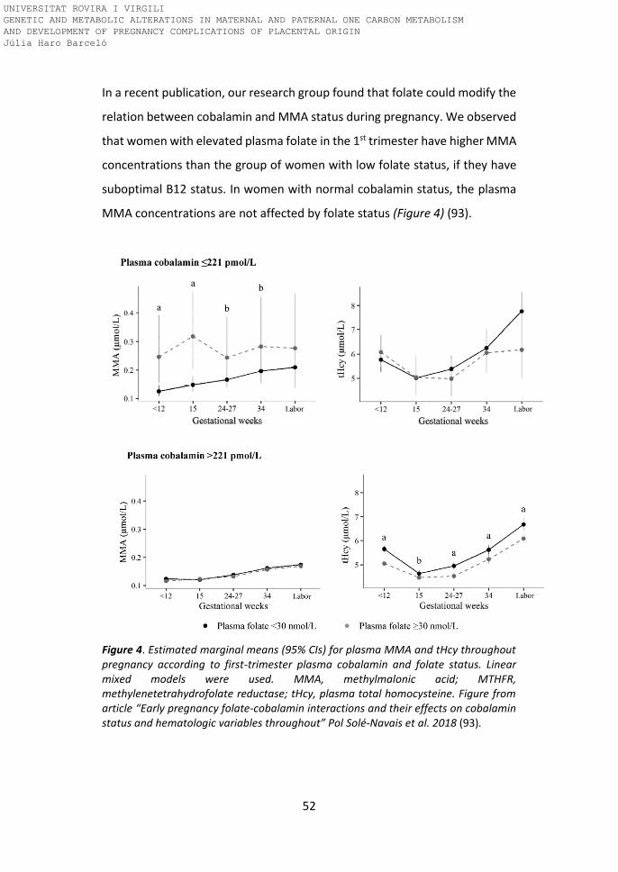
In a recent publication, our research group found that folate could modify the
relation between cobalamin and MMA status during pregnancy. We observed
that women with elevated plasma folate in the 1st trimester have higher MMA
concentrations than the group of women with low folate status, if they have
suboptimal B12 status. In women with normal cobalamin status, the plasma
MMA concentrations are not affected by folate status (Figure 4) (93).
Figure 4. Estimated marginal means (95% CIs) for plasma MMA and tHcy throughout pregnancy according to first-trimester plasma cobalamin and folate status. Linear mixed models were used. MMA, methylmalonic acid; MTHFR, methylenetetrahydrofolate reductase; tHcy, plasma total homocysteine. Figure from article “Early pregnancy folate-cobalamin interactions and their effects on cobalamin status and hematologic variables throughout” Pol Solé-Navais et al. 2018 (93).
UNIVERSITAT ROVIRA I VIRGILI GENETIC AND METABOLIC ALTERATIONS IN MATERNAL AND PATERNAL ONE CARBON METABOLISM AND DEVELOPMENT OF PREGNANCY COMPLICATIONS OF PLACENTAL ORIGIN Júlia Haro Barceló

53
There are other modifiable factors related with maternal lifestyle that can
negatively affect pregnancy outcomes. Some of them are inadequate
gestational gain, inadequate prenatal care, smoking, alcohol and drug use
(104). However, other non-modifiable factors may also influence pregnancy
complications. This is the case of history of spontaneous abortion, preterm
delivery, low birth weight or intrauterine growth restriction in previous
pregnancies that can affect the current pregnancy (105). Genetics is also a
non-modifiable factor that may influence pregnancy complications and folate
status. There is potential evidence that polymorphisms, affecting genes coding
for proteins or enzymes involved in folate uptake or metabolism, can modify
plasma or body folate levels leading to health and pregnancy problems
commented before (69). MTHFR C677T is a common polymorphism that leads
to instability of methylenetetrahydrofolate reductase (106). As explained
before, this enzyme is known to catalyse the reactions involved in transferring
the methyl groups to the principal methyl donor (methionine), that are
necessary for DNA synthesis, methylation, cell division and tissue growth
(107), and is essential for placenta and foetal development. The homozygote
variant genotype (TT) is associated with elevated tHcy plasma levels, as well
as decreased levels of DNA methylation compared with those with the CC
genotype (homozygote normal) (66). The effect of the polymorphism on tHcy
is intensified under low folate and cobalamin conditions, and it has been
related with increased risk of NTDs and other adverse pregnancy outcomes
(108). The effect of this polymorphism on pregnancy and other complications
is discussed further below.
Apart from the effect of maternal factors on pregnancy outcome, some
literature supports the implication of paternal factors as one of the causes of
pregnancy complications of placental origin. Examples are impaired
placentation, IUGR and gestational hypertension. Paternal lifestyle as well as
UNIVERSITAT ROVIRA I VIRGILI GENETIC AND METABOLIC ALTERATIONS IN MATERNAL AND PATERNAL ONE CARBON METABOLISM AND DEVELOPMENT OF PREGNANCY COMPLICATIONS OF PLACENTAL ORIGIN Júlia Haro Barceló

54
nutrition and poor folate status, are some of the modifiable factors that can
lead to potential paternal precursors of pregnancy complications (109).
Likewise, non-modifiable genetic factors affecting 1C metabolism and folate
status in the father, may lead to pregnancy complications (110). The paternal
involvement in the development of adverse pregnancy outcomes, as a main
objective of this thesis, will be commented in the next chapters.
UNIVERSITAT ROVIRA I VIRGILI GENETIC AND METABOLIC ALTERATIONS IN MATERNAL AND PATERNAL ONE CARBON METABOLISM AND DEVELOPMENT OF PREGNANCY COMPLICATIONS OF PLACENTAL ORIGIN Júlia Haro Barceló

55
1.3. MTHFR C677T polymorphism
The MTHFR C677T polymorphism and pregnancy complications
Methylenetetrahydrofolate reductase (MTHFR) polymorphisms and adverse
pregnancy outcomes such as IUGR, preterm delivery, preeclampsia, pregnancy
loss and others has been the focus of interest by various authors for many
years (111). The MTHFR gene catalyzes the conversion of 5,10-
methylenetetrahydrofolate to 5-methyltetrahydrofolate, the main form of
circulatory folate, which is the co-enzyme of methionine synthase together
with vitamin B12 and plays a key role in the remethylation of homocysteine
into methionine (11).
A cytosine to thymine substitution at position 677 (MTHFR C677T) is a quite
common polymorphism in this gene, leading to the substitution of valine for
alanine in the corresponding protein resulting in a reduction of the enzyme
activity (112). Consequently, the conversion of homocysteine to methionine is
impaired, leading to elevated tHcy (113), which is a risk factor for
cardiovascular disease (114), and reduced DNA methylation due to less
available free methyl groups (66). The polymorphism is relatively common in
the Spanish adult population (18.1 %) (8) and has been suggested to be
involved in thrombotic events or impaired methylation during pregnancy
(115).
Elevated maternal plasma tHcy levels, due to the reduced enzyme activity may
contribute to placenta-mediated pregnancy complications (114). Increased
tHcy may lead to endothelial dysfunction (116), inflammatory processes (117),
oxidative stress (11), arteriolar constriction (11) and placental thrombosis
(113). These conditions might be associated with impaired blood flow and
prothrombotic changes in the uterine artery wall, inadequate trophoblast
invasion into the uterine vasculature, and placental hypo perfusion that
UNIVERSITAT ROVIRA I VIRGILI GENETIC AND METABOLIC ALTERATIONS IN MATERNAL AND PATERNAL ONE CARBON METABOLISM AND DEVELOPMENT OF PREGNANCY COMPLICATIONS OF PLACENTAL ORIGIN Júlia Haro Barceló
56
subsequently leads to pregnancy complications (11). Furthermore, these
complications may also result from the increased requirements for DNA
methylation and protein synthesis involved in the growth of the baby and the
placenta, taking into account that methyl groups are decreased in the TT
genotype of the MTHFR polymorphism (118).
Chaudhry et al (2019) reported that a 5 μmol/L increase in tHcy (4 standard
deviations of tHcy approximately) was associated with elevated probability of
IUGR and preeclampsia but not with placental abruption or pregnancy loss, in
a Canadian cohort after adjusting for gestational age at the time of blood draw
and explanatory maternal characteristics (114). Mishra et al (2018) studied the
MTHFR gene methylation patterns of placental tissue in preeclamptic Indian
women, and observed lower MTHFR gene methylation on the foetal side of
preeclamptic placenta compared to gestation-matched controls (115).
Hypomethylation of genes involved in placentation is reported to be
associated with impaired trophoblastic invasion (119), leading to unsuccessful
placentation. In the same study, preeclampsia cases with the CT genotype had
higher levels of global DNA methylation compared to controls with the CT
genotype (115). This suggests that heterozygote women with hyper global
DNA methylation are more prone to preeclampsia (115). In addition, a meta-
analysis performed by Han Wu et al (2017) aimed at clarifying the association
between maternal MTHFR C677T genotype and susceptibility to preterm birth
and low birth weight. The maternal MTHFR C677T polymorphism increased
the risk of preterm birth and low birthweight in homozygote TT versus CT or
CC genotypes. Therefore, they concluded that identification of the maternal
MTHFR C677T mutation might be useful in identifying women needing primary
prevention of these pregnancy complications (11).
Other studies have investigated the relationship between the MTHFR C677T
polymorphism and pregnancy loss. Mehandjiev et al (2019) studied the
UNIVERSITAT ROVIRA I VIRGILI GENETIC AND METABOLIC ALTERATIONS IN MATERNAL AND PATERNAL ONE CARBON METABOLISM AND DEVELOPMENT OF PREGNANCY COMPLICATIONS OF PLACENTAL ORIGIN Júlia Haro Barceló
57
association between the MTHFR C677T polymorphism and intervillous and
decidua pathology in patients with pregnancy loss, and they reported that the
T allele and MTHFR TT genotype are associated with severe intervillous and
decidua pathologies such as fibrin deposits and thrombosis in those patients
(118). Turgal et al (2018) also studied the effect of the MTHFR C677T
polymorphism on pregnancy loss, and reported that the MTHFR C677T
polymorphism increases early pregnancy loss, with the highest rates occurring
in the MTHFR homozygote variant genotype (120). They proposed an impaired
homocysteine/methionine metabolism disorder due to the polymorphism as
a potential risk factor for this complication. Yajuan Xu et al (2018) observed in
a case-control study significantly higher frequencies of the T allele of the
MTHFR C677T polymorphism in the unexplained recurrent pregnancy loss
group compared to the control group (121). On the other hand, the presence
of CC and CT genotypes was significantly reduced in the unexplained recurrent
pregnancy loss compared to the control group. Finally, our research group
recently reported from the Reus-Tarragona Birth Cohort study that mothers
with first trimester tHcy ≥ P90 (7.1 µmol/L) have increased risk of miscarriage
(OR 95% CI, 2.5 [1.1, 5.7]). The prevalence of the MTHFR 677 TT genotype was
higher in this group compared with mothers with tHcy < P90 (89).
UNIVERSITAT ROVIRA I VIRGILI GENETIC AND METABOLIC ALTERATIONS IN MATERNAL AND PATERNAL ONE CARBON METABOLISM AND DEVELOPMENT OF PREGNANCY COMPLICATIONS OF PLACENTAL ORIGIN Júlia Haro Barceló
58
The MTHFR C677T polymorphism and cardiovascular diseases
Apart from its reported involvement in some pregnancy complications,
numerous studies have also reported an association between the MTHFR
C677T polymorphism and cardiovascular diseases such as coronary artery
disease, stroke or hypertension (122). The homozygous mutant genotype is
associated with a 40% greater risk of cardiovascular disease compared to the
homozygous normal genotype (123). Most of studies have focused on
homocysteine metabolism disorders as the putative risk factor associated with
this polymorphism (124). In homozygotes, with the MTHFR 677 TT genotype,
impaired folate metabolism and higher homocysteine concentrations has
been reported (125). Homocysteine produces reactive oxygen species during
the thiol oxidation process. This species are toxic to the endothelium and may
cause endothelial injury in various organs (126). Many studies have been
performed in the general population showing that mildly elevated tHcy is
associated with increased risks of atherosclerosis and venous thrombosis,
such that hyperhomocysteinemia is considered an independent risk factor for
cardiovascular diseases (127). Endothelial cell dysfunction has been identified
as a key mechanism by which vascular risk factors may mediate their effect on
vascular disease risk (128).
Shiran et al (2015) observed higher tHcy in the presence of endothelial
dysfunction detected by flow-mediated dilatation, in individuals with B12
deficiency (B12 <150 pmol/L) with the MTHFR 677 TT compared to the CC
genotype. They also confirmed the interaction between B12 deficiency and
the MTHFR C677T mutation and observed that among individuals with B12
deficiency, TT homozygosity was associated with higher tHcy compared to the
CC or CT genotypes. In addition, endothelial function was more affected in B12
deficiency patients with the TT genotype, compared to those with normal
levels of B12 due to the role of hyperhomocysteinemia in oxidative stress and
UNIVERSITAT ROVIRA I VIRGILI GENETIC AND METABOLIC ALTERATIONS IN MATERNAL AND PATERNAL ONE CARBON METABOLISM AND DEVELOPMENT OF PREGNANCY COMPLICATIONS OF PLACENTAL ORIGIN Júlia Haro Barceló
59
reduced endothelial cell proliferation (129). Tang et al (2014) studied a cohort
of coronary slow flow phenomenon patients and found higher frequencies of
the MTHFR 677 TT and CT genotypes and MTHFR 677 T allele in the patients
compared to controls (130). They also verified that tHcy plasma levels were
higher in the patients with the homozygote genotype compared to CT and CC
genotypes. Other studies have investigated the relationship between the
MTHFR C677T polymorphism and deep vein thrombosis (DVT), caused by an
imbalance between vascular relaxing factor and vascular contraction. It was
shown in two populations, Iranian and Chinese, that the frequencies of the TT
genotype and T alleles of the polymorphism were higher in the DVT group
compared with those in the control group (130, 131). These results suggest
that the MTHFR C677T polymorphism may be a risk factor for DVT. In another
study with 160 ischemic stroke patients divided into two groups with or
without hypertension, Ariana et al (2019) observed that the TT genotype was
more prevalent in ischaemic stroke patients with hypertension (16 patients;
69.5%) compared with the group without hypertension. All patients with
ischaemic stroke in the group with hypertension have the MTHFR 677 TT
genotype (5 patients; 100%)(133). As this paper shows, many other studies
have proposed this genetic variant as an independent risk factor for
hypertension, with an increased risk of between 24 and 87% in homozygotes
for the MTHFR C677T polymorphism compared to the MTHFR 677 CC and CT
genotypes (123). Lynch et al (2019) reported in 75 cardiovascular disease
patients that those with the TT genotype had higher systolic and diastolic
blood pressure at baseline compared to those with the CC genotype (134). In
a study conducted in Southwest Cameroon, Ghogomu et al (2016) also
reported that the frequency of the T allele of the MTHFR C677T polymorphism
was higher in the hypertensive group than in the normotensive group. On the
other hand, the C allele was higher in the normotensive than in the
UNIVERSITAT ROVIRA I VIRGILI GENETIC AND METABOLIC ALTERATIONS IN MATERNAL AND PATERNAL ONE CARBON METABOLISM AND DEVELOPMENT OF PREGNANCY COMPLICATIONS OF PLACENTAL ORIGIN Júlia Haro Barceló

60
hypertensive subjects (135). These results are comparable to those obtained
in populations from Spain (136), Australia (137) and Turkey (138).
Our research group also investigated the relationship between the MTHFR
C677T polymorphism and moderately elevated tHcy and hypertension in an
adult population study (139). There was no association between either of the
variant MTHFR C677T genotypes and diagnosed hypertension in the overall
representative sample (randomly selected from population records) of the
adult population, that was unexposed to mandatory fortification with folic
acid. However, the TT genotype was associated with greater probability of
having hypertension than the CC genotype in participants ≤50 years. On the
other hand, moderately elevated tHcy was associated with hypertension in the
overall population and specifically in people over 50 years of age. Therefore,
both moderately elevated tHcy and the MTHFR C677T polymorphism are
associated with risk of hypertension, and these associations differ in
subgroups of the population (139). However, our data do not directly support
that the mechanism linking the MTHFR genotype to hypertension is via
elevated tHcy as other studies do. Several studies show that homocysteine is
a predictor of secondary, but not primary events (140), suggesting that is a
product rather than a cause of vascular disease. The inconsistent results may
be due to the methodology used for each study as the matching of correct
controls with the cases, inadequate measurements or adjustments for the risk
factors, sample storage and determinations at baseline, etc (128). Moreover,
intervention studies performed to lower tHcy have shown little or no
corresponding blood pressure response. This could be due to the fact that
there is no mechanistic association between homocysteine and blood
pressure (123), or could be because intervention is ineffective once the
disease is established. Few or no studies have investigated the effects of
lowering tHcy as a primary preventive strategy. Thus, MTHFR polymorphism
UNIVERSITAT ROVIRA I VIRGILI GENETIC AND METABOLIC ALTERATIONS IN MATERNAL AND PATERNAL ONE CARBON METABOLISM AND DEVELOPMENT OF PREGNANCY COMPLICATIONS OF PLACENTAL ORIGIN Júlia Haro Barceló

61
may be related to blood pressure via a mechanism independent of
homocysteine.
The effect of nutritional status on the MTHFR C677T polymorphism
It is well established that TT homozygotes for the MTHFR C677T polymorphism
have elevated homocysteine concentrations and lower folate status
compared to the common genotype, indicating a gene-nutrient interaction
(128). The prejudicial effect of the homozygous variant genotype is significant
only when folate status is low, such as in countries with no mandatory food
fortification, like in Spain. In regions with folic acid fortification policies the
apparent effect of the MTHFR genotype has been proposed to be null (141).
Therefore, dietary folate can influence the phenotype and thus disease risk
(pregnancy complications, cardiovascular diseases, etc.) (123). It is proven that
folic acid supplementation is an effective method to lower plasma
homocysteine levels (123).
Recently, riboflavin (vitamin B2) is taking more attention due to its important
role in one-carbon metabolism because it acts in the form of flavin adenine
dinucleotide (FAD) as a cofactor for the MTHFR enzyme (123). Low riboflavin
status, more frequent in individuals with the homozygous mutant TT genotype
(125), is associated with elevated tHcy and low folate concentrations (123).
Molecular studies have shown that MTHFR activity is reduced in the presence
of the MTHFR 677 TT genotype due to its reduced affinity for the FAD cofactor
(142). In addition, in human studies, riboflavin supplementation of individuals
with the TT genotype can reduce homocysteine levels (143). This suggests that
optimal riboflavin status can stabilize the variant enzyme and restore MTHFR
activity (123).
UNIVERSITAT ROVIRA I VIRGILI GENETIC AND METABOLIC ALTERATIONS IN MATERNAL AND PATERNAL ONE CARBON METABOLISM AND DEVELOPMENT OF PREGNANCY COMPLICATIONS OF PLACENTAL ORIGIN Júlia Haro Barceló
62
Recent evidence suggests that riboflavin may be an important modulating
factor via a novel effect on blood pressure (123). Lynch et al (2019) confirmed
the involvement of riboflavin in ensuring optimum MTHFR activity, suggesting
that this vitamin may interact with the MTHFR gene to influence blood
pressure. They observed that riboflavin supplementation, at dietary level (1.6
mg/day), can decrease blood pressure in TT hypertensive adults compared
with controls (134). They did not find a significant reduction in the CC
genotype group. In addition, people with the TT genotype and poor riboflavin
status, can reduce plasma tHcy levels with riboflavin supplementation (143).
In a randomized trial, Wilson et al (2013) demonstrated in hypertensive
individuals with the MTHFR 677 TT genotype treated with antihypertensive
drugs, that those supplemented with riboflavin decrease blood pressure more
effectively than those in the placebo group (treatment with current
antihypertensive drugs alone)(144).
Increasing the activity of the MTHFR enzyme by riboflavin supplementation is
only observed in those individuals with the homozygous MTHFR 677 TT
genotype, with no response evident in CC or CT genotype groups (143). This
effect might lead to improved endothelial function through increased
bioavailability of nitric oxide via increases in 5-methyltetrahydrofolate (123).
This emerging evidence of riboflavin supplementation in lowering tHcy and
blood pressure, suggests that nutritional and pharmacological interventions
based on folic acid and other B vitamin supplementation can provide useful
targeted therapy to reduce pregnancy complications and cardiovascular
diseases in genetically at risk people. However, large-scale clinical intervention
studies are needed to confirm the effects and correct doses to prevent
negative effects.
UNIVERSITAT ROVIRA I VIRGILI GENETIC AND METABOLIC ALTERATIONS IN MATERNAL AND PATERNAL ONE CARBON METABOLISM AND DEVELOPMENT OF PREGNANCY COMPLICATIONS OF PLACENTAL ORIGIN Júlia Haro Barceló
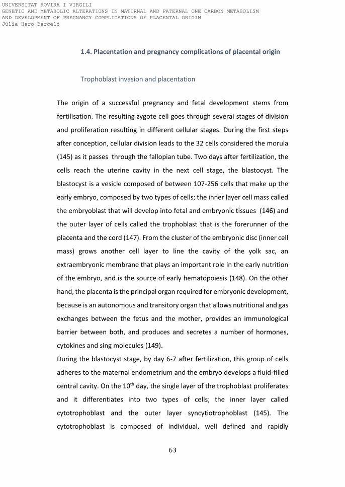
63
1.4. Placentation and pregnancy complications of placental origin
Trophoblast invasion and placentation
The origin of a successful pregnancy and fetal development stems from
fertilisation. The resulting zygote cell goes through several stages of division
and proliferation resulting in different cellular stages. During the first steps
after conception, cellular division leads to the 32 cells considered the morula
(145) as it passes through the fallopian tube. Two days after fertilization, the
cells reach the uterine cavity in the next cell stage, the blastocyst. The
blastocyst is a vesicle composed of between 107-256 cells that make up the
early embryo, composed by two types of cells; the inner layer cell mass called
the embryoblast that will develop into fetal and embryonic tissues (146) and
the outer layer of cells called the trophoblast that is the forerunner of the
placenta and the cord (147). From the cluster of the embryonic disc (inner cell
mass) grows another cell layer to line the cavity of the yolk sac, an
extraembryonic membrane that plays an important role in the early nutrition
of the embryo, and is the source of early hematopoiesis (148). On the other
hand, the placenta is the principal organ required for embryonic development,
because is an autonomous and transitory organ that allows nutritional and gas
exchanges between the fetus and the mother, provides an immunological
barrier between both, and produces and secretes a number of hormones,
cytokines and sing molecules (149).
During the blastocyst stage, by day 6-7 after fertilization, this group of cells
adheres to the maternal endometrium and the embryo develops a fluid-filled
central cavity. On the 10th day, the single layer of the trophoblast proliferates
and it differentiates into two types of cells; the inner layer called
cytotrophoblast and the outer layer syncytiotrophoblast (145). The
cytotrophoblast is composed of individual, well defined and rapidly
UNIVERSITAT ROVIRA I VIRGILI GENETIC AND METABOLIC ALTERATIONS IN MATERNAL AND PATERNAL ONE CARBON METABOLISM AND DEVELOPMENT OF PREGNANCY COMPLICATIONS OF PLACENTAL ORIGIN Júlia Haro Barceló
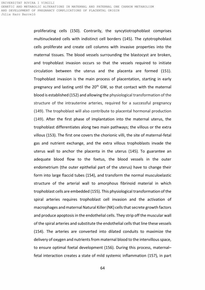
64
proliferating cells (150). Contrarily, the syncytiotrophoblast comprises
multinucleated cells with indistinct cell borders (145). The cytotrophoblast
cells proliferate and create cell columns with invasive properties into the
maternal tissues. The blood vessels surrounding the blastocyst are broken,
and trophoblast invasion occurs so that the vessels required to initiate
circulation between the uterus and the placenta are formed (151).
Trophoblast invasion is the main process of placentation, starting in early
pregnancy and lasting until the 20th GW, so that contact with the maternal
blood is established (152) and allowing the physiological transformation of the
structure of the intrauterine arteries, required for a successful pregnancy
(149). The trophoblast will also contribute to placental hormonal production
(149). After the first phase of implantation into the maternal uterus, the
trophoblast differentiates along two main pathways; the villous or the extra
villous (153). The first one covers the chorionic villi, the site of maternal-fetal
gas and nutrient exchange, and the extra villous trophoblasts invade the
uterus wall to anchor the placenta in the uterus (145). To guarantee an
adequate blood flow to the foetus, the blood vessels in the outer
endometrium (the outer epithelial part of the uterus) have to change their
form into large flaccid tubes (154), and transform the normal musculoelastic
structure of the arterial wall to amorphous fibrinoid material in which
trophoblast cells are embedded (155). This physiological transformation of the
spiral arteries requires trophoblast cell invasion and the activation of
macrophages and maternal Natural Killer (NK) cells that secrete growth factors
and produce apoptosis in the endothelial cells. They strip off the muscular wall
of the spiral arteries and substitute the endothelial cells that line these vessels
(154). The arteries are converted into dilated conduits to maximize the
delivery of oxygen and nutrients from maternal blood to the intervillous space,
to ensure optimal foetal development (156). During this process, maternal–
fetal interaction creates a state of mild systemic inflammation (157), in part
UNIVERSITAT ROVIRA I VIRGILI GENETIC AND METABOLIC ALTERATIONS IN MATERNAL AND PATERNAL ONE CARBON METABOLISM AND DEVELOPMENT OF PREGNANCY COMPLICATIONS OF PLACENTAL ORIGIN Júlia Haro Barceló

65
due to the HLA antigens such as HLA-G expressed by the trophoblast
extravillous cells, that may influence maternal rejection of the fetus (158).
Apart from the NK cells and the trophoblast cells, many molecules interact
together to regulate trophoblast growth, migration and invasion to maintain
a healthy utero-placenta equilibrium. These molecules include growth factors,
inflammatory cytokines, growth factor binding proteins and leukocytes (159).
For this reason, a complex network of soluble autocrine and paracrine factors,
signalling pathways and regulatory transcription factors are needed to
regulate it (160).
Figure 5. Uterine arteries in three different times are represented. On the left, uterine
arteries with spiral form in non-pregnant women. In the middle, unsuccesful
remodelation during pregnancy leading to preeclampsia. On the right, remodelled
uterine arteries in a normal pregnancy. Figure from article “NK Cells and Trophoblasts”
Parham P (154).
Briefly, placentation is a complex process and one of the most important
determinants of pregnancy outcome. Trophoblast invasion, as the main
process of placentation, is the key to avoiding pregnancy complications
related with inadequate transformation of the spiral arteries that are
important to ensure adequate fetal oxygen and nutrient supply. Extended
UNIVERSITAT ROVIRA I VIRGILI GENETIC AND METABOLIC ALTERATIONS IN MATERNAL AND PATERNAL ONE CARBON METABOLISM AND DEVELOPMENT OF PREGNANCY COMPLICATIONS OF PLACENTAL ORIGIN Júlia Haro Barceló

66
literature shows that successful placentation will prevent adverse pregnancy
outcomes such as severe forms of intrauterine growth restriction and
gestational hypertension (156). Both complications are explained below, as
they are the main outcomes studied in this thesis.
Pregnancy complications of placental origin: impaired placentation,
intrauterine growth retardation and gestational hypertension
Impaired placentation is a common complication in severe pregnancy
outcomes, and in part is caused by impaired physiological transformation of
the spiral arteries in the placenta, leading to high resistance to blood flow in
the uterine arteries (161). The Doppler ultrasound measurement of the
intrauterine arteries is a physiological test to measure the blood flow in these
arteries. This technique is used to assess the resistance of the blood flow
between the mother and the fetus, determined by placental volume, the
number of spiral arteries and their transformation (162). It was introduced in
the 80’s by Campbell et al (163) and still used due to its efficiency as a non-
invasive measurement of fetal pathologies as well as feto-placental and utero-
placental circulation (164). Currently, Doppler sonography of the uterine
arteries is an established screening method during the second trimester and
has been advanced to the first trimester, in many clinical settings, as an early
detection strategy of impaired uterine artery flow.
In the first trimester of pregnancy, the waveforms of the uterine arteries
present high resistance and a pronounced decrease of the blood flow velocity
at the beginning of the diastole, known as a notch (165). In normal pregnancy,
uterine artery pulsatility index decreases gradually as pregnancy progresses,
the notch tends to disappear (166) and blood flow to the placenta increases.
Pathological conditions are diagnosed as bilateral uterine artery notches
UNIVERSITAT ROVIRA I VIRGILI GENETIC AND METABOLIC ALTERATIONS IN MATERNAL AND PATERNAL ONE CARBON METABOLISM AND DEVELOPMENT OF PREGNANCY COMPLICATIONS OF PLACENTAL ORIGIN Júlia Haro Barceló
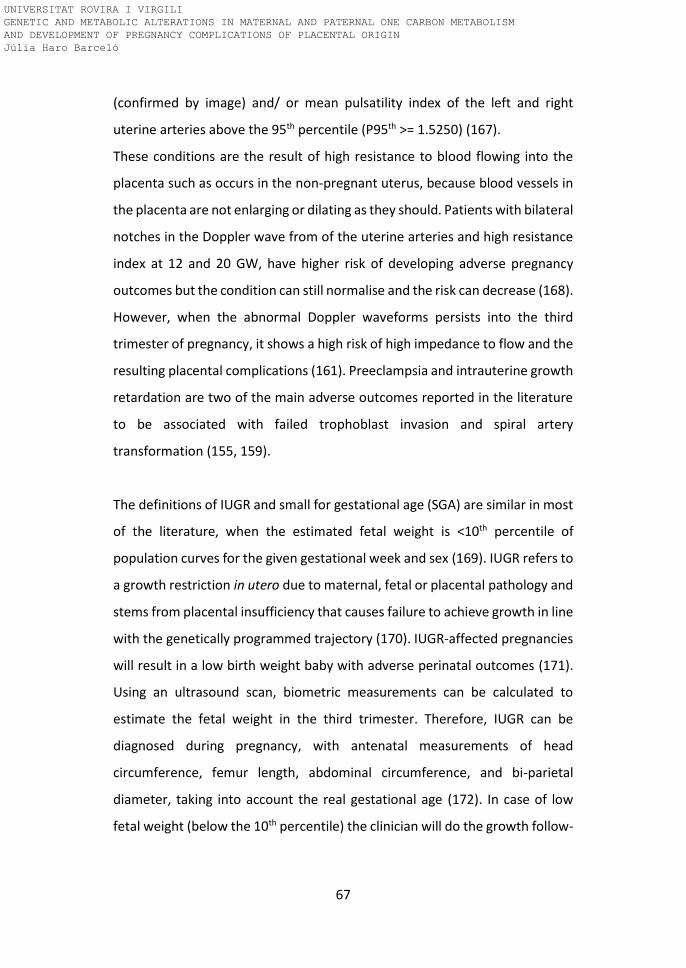
67
(confirmed by image) and/ or mean pulsatility index of the left and right
uterine arteries above the 95th percentile (P95th >= 1.5250) (167).
These conditions are the result of high resistance to blood flowing into the
placenta such as occurs in the non-pregnant uterus, because blood vessels in
the placenta are not enlarging or dilating as they should. Patients with bilateral
notches in the Doppler wave from of the uterine arteries and high resistance
index at 12 and 20 GW, have higher risk of developing adverse pregnancy
outcomes but the condition can still normalise and the risk can decrease (168).
However, when the abnormal Doppler waveforms persists into the third
trimester of pregnancy, it shows a high risk of high impedance to flow and the
resulting placental complications (161). Preeclampsia and intrauterine growth
retardation are two of the main adverse outcomes reported in the literature
to be associated with failed trophoblast invasion and spiral artery
transformation (155, 159).
The definitions of IUGR and small for gestational age (SGA) are similar in most
of the literature, when the estimated fetal weight is <10th percentile of
population curves for the given gestational week and sex (169). IUGR refers to
a growth restriction in utero due to maternal, fetal or placental pathology and
stems from placental insufficiency that causes failure to achieve growth in line
with the genetically programmed trajectory (170). IUGR-affected pregnancies
will result in a low birth weight baby with adverse perinatal outcomes (171).
Using an ultrasound scan, biometric measurements can be calculated to
estimate the fetal weight in the third trimester. Therefore, IUGR can be
diagnosed during pregnancy, with antenatal measurements of head
circumference, femur length, abdominal circumference, and bi-parietal
diameter, taking into account the real gestational age (172). In case of low
fetal weight (below the 10th percentile) the clinician will do the growth follow-
UNIVERSITAT ROVIRA I VIRGILI GENETIC AND METABOLIC ALTERATIONS IN MATERNAL AND PATERNAL ONE CARBON METABOLISM AND DEVELOPMENT OF PREGNANCY COMPLICATIONS OF PLACENTAL ORIGIN Júlia Haro Barceló
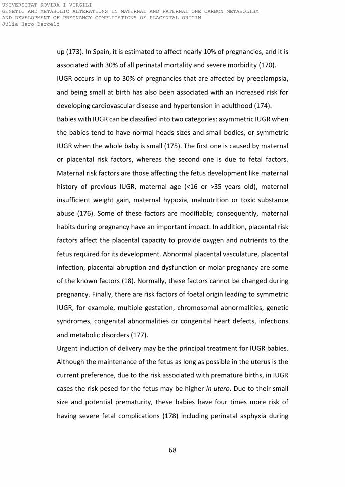
68
up (173). In Spain, it is estimated to affect nearly 10% of pregnancies, and it is
associated with 30% of all perinatal mortality and severe morbidity (170).
IUGR occurs in up to 30% of pregnancies that are affected by preeclampsia,
and being small at birth has also been associated with an increased risk for
developing cardiovascular disease and hypertension in adulthood (174).
Babies with IUGR can be classified into two categories: asymmetric IUGR when
the babies tend to have normal heads sizes and small bodies, or symmetric
IUGR when the whole baby is small (175). The first one is caused by maternal
or placental risk factors, whereas the second one is due to fetal factors.
Maternal risk factors are those affecting the fetus development like maternal
history of previous IUGR, maternal age (<16 or >35 years old), maternal
insufficient weight gain, maternal hypoxia, malnutrition or toxic substance
abuse (176). Some of these factors are modifiable; consequently, maternal
habits during pregnancy have an important impact. In addition, placental risk
factors affect the placental capacity to provide oxygen and nutrients to the
fetus required for its development. Abnormal placental vasculature, placental
infection, placental abruption and dysfunction or molar pregnancy are some
of the known factors (18). Normally, these factors cannot be changed during
pregnancy. Finally, there are risk factors of foetal origin leading to symmetric
IUGR, for example, multiple gestation, chromosomal abnormalities, genetic
syndromes, congenital abnormalities or congenital heart defects, infections
and metabolic disorders (177).
Urgent induction of delivery may be the principal treatment for IUGR babies.
Although the maintenance of the fetus as long as possible in the uterus is the
current preference, due to the risk associated with premature births, in IUGR
cases the risk posed for the fetus may be higher in utero. Due to their small
size and potential prematurity, these babies have four times more risk of
having severe fetal complications (178) including perinatal asphyxia during
UNIVERSITAT ROVIRA I VIRGILI GENETIC AND METABOLIC ALTERATIONS IN MATERNAL AND PATERNAL ONE CARBON METABOLISM AND DEVELOPMENT OF PREGNANCY COMPLICATIONS OF PLACENTAL ORIGIN Júlia Haro Barceló

69
delivery, hypothermia, metabolic and hematologic alterations, cerebral palsy
and higher risk of cardio pathologies (179).
Gestational hypertension described as the first appearance of systolic blood
pressure >= 140 mmHg and/or diastolic blood pressure >= 90 mmHg after 20
gestational weeks in women that had normal blood pressure up until 20 GW
or a difference in first trimester blood pressure and that after 17GW of
>=30mmHg for systolic and/or >= 15mmHg for diastolic (180), is the first
clinical sign of preeclampsia. Severe hypertension is considered for systolic
blood pressure >=160 mmHg and/or diastolic blood pressure >=110 mmHg
(96). Preeclampsia is a serious maternal disorder causing new onset
hypertension and proteinuria (>300mg/24 hours) only during pregnancy, after
20 GW, and in some cases lasts up to 6 weeks after delivery (182). This
syndrome of placental origin is associated with significant maternal and
neonatal morbidity (159). Risk factors that may increase the risk of developing
preeclampsia include first pregnancy, more than one foetus, poor trophoblast
invasion, unsuccessful placentation and family history of preeclampsia (183).
Other factors such as elevated maternal age (>35 years old), history of
previous hypertension, diabetes and obesity, genetics, environmental factors
or nutrition, especially low folate status and high homocysteine
concentrations, may also predispose to development of the disease (96). The
prevalence of this disease is relatively low in Spain, between 1-2%, compared
with other developed countries where it is double (182).
The exact causes are unclear, but the origin might come from abnormal
placenta development. As explained before, in a normal pregnancy the spiral
arteries dilate to 10 times their normal size. However, in preeclampsia the
uterine arteries become fibrous and thus narrower, causing less blood flow to
the placenta (184). In such conditions, pro-inflammatory proteins are released
into the maternal circulation and predispose the endothelial cells to become
UNIVERSITAT ROVIRA I VIRGILI GENETIC AND METABOLIC ALTERATIONS IN MATERNAL AND PATERNAL ONE CARBON METABOLISM AND DEVELOPMENT OF PREGNANCY COMPLICATIONS OF PLACENTAL ORIGIN Júlia Haro Barceló

70
dysfunctional, causing vasoconstriction and more salt retention by the kidney
(185), which can result in hypertension. Vasoconstriction may also affect other
local areas leading to decreased blood flow in certain parts of the body. In the
kidney, less blood flow causes oliguria and proteinuria. In the retina, it causes
blurred vision, flashing lights and the development of scotoma. In the liver,
poor blood flow may cause injury, swelling and elevation of liver enzymes
(186). Endothelial injury increases vascular permeability, allowing the water
to leak from the vessels between endothelial cells and to enter the tissues.
Because there is also less protein in the blood, due to the proteinuria, even
more fluid from the blood vessels enters the tissue and causes generalized
edema in the legs, face and hands (186). If there is a pulmonary edema, other
complications including cough and shortness of breath can appear. If the
edema is in the brain, the symptoms may be headaches, confusion and
seizures. We talk about eclampsia when the seizures appear (182). In addition,
the poorly perfused placenta can also lead to intrauterine growth retardation
and even placental abruption, haemorrhagic stroke and fetal death in severe
cases (186). However, in some cases the symptomatology of preeclampsia can
be mild. The delivery of the fetus and the placenta is the main treatment for
preeclampsia. It will depend on gestational age of the fetus as well as the
severity of disease (how it affects both, maternal and fetal health) (187). If the
preeclampsia is mild, it is recommendable to finish the gestation on time and
control regularly the mother to prevent severe complications. If it is severe,
blood pressure should be controlled using medication to prevent a worst
scenario like eclampsia and other serious signs, and control the fetal health to
decide the optimal moment for delivery (182).
UNIVERSITAT ROVIRA I VIRGILI GENETIC AND METABOLIC ALTERATIONS IN MATERNAL AND PATERNAL ONE CARBON METABOLISM AND DEVELOPMENT OF PREGNANCY COMPLICATIONS OF PLACENTAL ORIGIN Júlia Haro Barceló
71
1.5. Paternal factors affecting placentation and pregnancy
complications
Pregnancy complications described earlier, such as impaired placentation,
intrauterine growth retardation and gestational hypertension may influence
short and long term maternal and fetal health. A variety of genetic and
environmental factors interact to produce adverse pregnancy outcomes.
Established maternal risk factors associated with pregnancy complications are
age (>35 years), multiparity, gestational diabetes, obesity and others. These
factors are usually studied in the context of the materno-fetal-placenta unit,
without taking into account paternal factors (18). However, maternal risk
factors alone may not explain many preeclampsia cases and other pregnancy
complications. For example, poor trophoblast invasion and deficient
placentation are important risk factors for these complications, and the causes
in many cases are unknown or may not be explained by maternal factors (188).
If we consider that the placenta comes from the trophoblast cells, of
embryonic origin, we can assume that paternal and maternal factors interact
in the placentation process. In fact, paternal genetic factors predict good
trophoblast development, and has a potential bearing on the process of
placentation failure (189), a key process in fetal development. For this reason,
the physiopathological process that leads to pregnancy complications may
involve the paternal-maternal-fetal unit (190). In addition, paternal
transmission of diseases may result in the exposure of male germ cells to
environmental factors such as under and over nutrition. In particular,
environmental paternal exposures can affect the epigenome, including DNA
methylation, and lead to heritable alterations in fetal gene expression (191).
The epigenetics process, as a possible mechanism linking 1C metabolism to
pregnancy complications is explained in detail in the next section.
UNIVERSITAT ROVIRA I VIRGILI GENETIC AND METABOLIC ALTERATIONS IN MATERNAL AND PATERNAL ONE CARBON METABOLISM AND DEVELOPMENT OF PREGNANCY COMPLICATIONS OF PLACENTAL ORIGIN Júlia Haro Barceló

72
Regarding the risk of developing preeclampsia during pregnancy, recent
studies have proposed some paternal factors that may be involved. These
include the length of the relationship with the same father, the compatibility
of the mother to paternal antigens, primipaternity, etc. There is also a paternal
phenomenon related with the development of preeclampsia, described as
“the dangerous father”, commented before (22). In addition, paternal family
history of preeclampsia may affect pregnancy complications, as shown in a
study where fathers born from preeclampsia- affected pregnancies have an
increased probability of fathering a pregnancy affected by this complication
(16). It was also observed that the risk of developing preeclampsia in
subsequent pregnancies was reduced when mothers with a previous history
of preeclampsia, became pregnant by a different partner (192).
There is also evidence for paternal involvement in other pregnancy
complications such as IUGR. Some articles have shown a relationship between
advanced paternal age (>45 years) and IUGR (178), suggesting paternal
genetics as a possible mechanism inducing the complications because of the
increasing number of mutations with age (191). However, there are
controversial results due to the poor association between age and IUGR in
other studies (193)(191). In the case of Hurley et al (2017), they found no
association probably because of the small sample size compared to other
studies that found a relationship between paternal age and IUGR, and due to
the inaccuracy of paternal age or missing data recorded from the assisted
reproductive technology pregnancies (194). Furthermore, paternal low birth
weight, IUGR or SGA, have been reported to be potential factors enhancing
the probability of fathering an IUGR baby (20, 193). Some studies have also
investigated the effect of mixed paternal and maternal ethnicity on the risk of
IUGR and report increased likelihood in these circumstances. Possible
mechanisms proposed include maternal chronic stress associated with
UNIVERSITAT ROVIRA I VIRGILI GENETIC AND METABOLIC ALTERATIONS IN MATERNAL AND PATERNAL ONE CARBON METABOLISM AND DEVELOPMENT OF PREGNANCY COMPLICATIONS OF PLACENTAL ORIGIN Júlia Haro Barceló
73
everyday interpersonal and institutional racism (17) and differential prenatal
and maternity care (196).
There is a clear paternal implication in successful placentation that can
prevent pregnancy complications and guarantee optimal health in both the
mother and the baby. The main paternal factors studied, so far, can be
classified as modifiable or non-modifiable. Modifiable paternal factors are
related with lifestyle such as smoking, alcohol consumption, poor nutrition
status, stress, socioeconomic status, and the non-modifiable ones are age,
ethnicity and genetics, etc.
Modifiable paternal factors affecting placentation and pregnancy
complications
Many modifiable factors, stemming from paternal habits and lifestyle, may
influence the development of pregnancy complications. It’s well known that
paternal, as well as maternal toxic habits including smoking (19), alcohol
consumption or drug use (20), are related with impaired placentation leading
to low birth weight or preterm babies. Other paternal behaviour factors such
as sedentary lifestyle, stress or depression, have been reported to increase the
risk of developing preterm birth (197). Family socioeconomic status (198) or
paternal educational level (199) have been also studied as possible risk factors
of pregnancy complications, due to their close relationship with nutritional
status. Nutritional deficiencies are an important detonator of pregnancy
complications because of the increased nutrition requirements for fetal
development. Evidence from animal studies suggests that paternal folate and
1C metabolism status, before conception, are possible modifiable factors
involved in adverse pregnancy outcome and long term offspring health (191).
Maloney et al (2011) in a study with rats fed with folic acid and methionine
UNIVERSITAT ROVIRA I VIRGILI GENETIC AND METABOLIC ALTERATIONS IN MATERNAL AND PATERNAL ONE CARBON METABOLISM AND DEVELOPMENT OF PREGNANCY COMPLICATIONS OF PLACENTAL ORIGIN Júlia Haro Barceló

74
deficient diet (methyl-deficient diet) during the periods of oocyte and embryo
development, observed life-long changes in proteins involved in the processes
of energy metabolism in the adult offspring (200). Cherif et al (2019)
investigated also rats fed with a methyl donor deficiency diet during gestation
and lactation and reported an association between this diet and the
development and function of energy balance circuits, affecting the expression
of neuropeptides and related receptors in the hypothalamus, resulting in
growth and weight deficits (201).
In human studies, paternal deficiencies or excess of macronutrient during pre
and post conception periods have also been proposed as potential factors
affecting pregnancy related complications (202). In a Chinese population,
Chen et al (2017) reported that a high quality protein paternal diet, with eggs
and meat, was associated with lower incidence of preterm SGA (21). In a case
control study with men from infertile couples, Eslamian G et al (2012) showed
that those fed with a western-type diet with processed meat, sweets, refined
grains and snacks were associated with reduced sperm motility (203).
The importance of the paternal 1C metabolic network and nutrition on
successful placentation and optimum fetal development have recently come
to light. As men contribute to half of the embryo’s genetic material, paternal
1C metabolism is also important for pregnancy opportunities and outcomes
(204). This network has an important role in biological processes such as
cellular division, synthesis and DNA repair and is an important source of
methyl groups for epigenetic programming of the paternal seminal fluid and
subsequently the embryonic genome. Some nutrients such as folate,
cobalamin and methionine are involved in this network acting as substrates or
cofactors for the enzymes that participate in the cycle. The deficiency of one
or more of these nutrients can alter the general network working, therefore,
paternal folate status is crucial for optimal paternal 1C metabolism (109).
UNIVERSITAT ROVIRA I VIRGILI GENETIC AND METABOLIC ALTERATIONS IN MATERNAL AND PATERNAL ONE CARBON METABOLISM AND DEVELOPMENT OF PREGNANCY COMPLICATIONS OF PLACENTAL ORIGIN Júlia Haro Barceló

75
Some studies have investigated the relationship between impaired paternal
1C metabolism and complications related with pregnancy. Studies in humans
have largely focused on the influence of paternal folate status on semen
parameters and they have confirmed that plasma and seminal optimal folate
concentrations positively affect semen quality (205). Boonyarangkul et al
(2015) showed that after 3-months of treatment with 5 mg of folic acid per
day the percentage of motile semen increased from 11.4% to 20.4% (206).
Other studies focused on homocysteine semen concentrations have reported
higher homocysteine concentrations in infertile men’s sperm compared with
fertile men. They also reported poor embryo quality in the group of high
homocysteine sperm concentrations (207). In a study of 280 couples,
Govindaiah et al reported 7 times more risk of spontaneous abortion in those
couples with paternal hyperhomocysteinemia (values higher than P.95th,
≥19.6 μmol/L, measured after the pregnancy) compared to those with normal
tHcy (110). In a study of mice, epigenetic alterations in DNA methylation in
two types of folic acid diets were reported. One was a folic acid deficient diet
and the other one highly supplemented, and both affected sperm DNA
methylation, sperm counts and the offspring’s abnormal development and
death (191). Similarly, Lambrot et al (2013) reported that low paternal folate
status may alter the sperm epigenome and result in adverse pregnancy
outcomes in mice (208). In addition, in a human study, it has been published
recently that both low and high long term paternal folate status, assessed by
RBC folate levels from a blood sample taken between 7 and 9 GW, are
associated with reduced embryonic growth (109).
Adverse pregnancy outcomes originating from nutritional deficiencies, such as
folate deficiency, are interrelated with both, maternal and paternal deficiency.
Ratan et al (2008) reported lower folate levels in fathers of children born with
NTDs, compared to controls (209). One possible explanation is that folate
UNIVERSITAT ROVIRA I VIRGILI GENETIC AND METABOLIC ALTERATIONS IN MATERNAL AND PATERNAL ONE CARBON METABOLISM AND DEVELOPMENT OF PREGNANCY COMPLICATIONS OF PLACENTAL ORIGIN Júlia Haro Barceló

76
status during spermatogenesis influences the paternal epigenome of the
embryo, with further consequences for the embryonic development (109).
Another one may be that the final consequences originate from deficient
intracellular folate concentration in germ cells (71). Current strategies for
primary prevention proposed to prevent pregnancy complications and
malformations such as NTDs are aimed at the mother, following the prenatal
care protocol in Catalonia (180) and European countries. Maternal nutritional
interventions with folic acid supplementation is the main strategy used. No
strategies focused on the father are implemented yet, but there is more
interest in paternal involvement in pregnancy complications. This problem is
overcome in countries with mandatory folic acid food fortification, but in
countries where the fortification is absent and only some food is
supplemented, such as Spain, people have to be aware of the benefits of
taking folic acid.
Moreover, 1C network regulation also depends on nutrient-gene interactions.
MTHFR 677C>T and SLC19A1 are two important polymorphism determinants
for folate and homocysteine status, because of their roles in folate metabolism
and entrance into the cell (8). Therefore, we should consider non-modifiable
paternal factors such as genetics, due to their important role in guaranteeing
an optimal nutritional status and in preventing pregnancy complications.
UNIVERSITAT ROVIRA I VIRGILI GENETIC AND METABOLIC ALTERATIONS IN MATERNAL AND PATERNAL ONE CARBON METABOLISM AND DEVELOPMENT OF PREGNANCY COMPLICATIONS OF PLACENTAL ORIGIN Júlia Haro Barceló
77
Non-modifiable paternal factors affecting placentation and
pregnancy complications
As previously stated, paternal genetics, age and ethnicity are some of the non-
modifiable factors that have been related with adverse pregnancy outcomes
such as SGA, preterm birth, IUGR and preeclampsia. In the context of this
thesis, we are very interested in the genetic factors that may provide links
between paternal and pregnancy health. Since foetal genes are derived from
each of a maternal and a paternal copy, the non-imprinted expression in the
placenta and in the fetus can be affected by the paternal genotype (18). In the
literature, some articles have proposed genetic factors as a possible
precipitating cause of inadequate trophoblast invasion, which in turn affects
pregnancy health (160). Imprinted genes, for example, are important for
embryo growth and development, with evidence generally showing that
paternally expressed genes promote fetal growth, while maternally expressed
genes suppress it (210).
Genetic factors are closely related with nutritional status due to gene-nutrient
interactions. Some polymorphisms are affected by nutrients and may modify
the activity of the enzyme, protein or biological molecule they are affecting.
In our group, we are interested in studying 1C metabolism, the vitamins
involved in the cycle and the genes involved in the regulation and optimum
functioning of the pathway. We focus our investigation on two polymorphisms
affecting folate metabolism (MTHFR 677C>T) and transport (SLC19A1 80G>A).
Data from a case-control observational study, show that the homozygote
mutant genotype (TT) of the paternal MTHFR 677C>T polymorphism was
linked with 2.4 times increased risk of miscarriage, compared with the control
group (110). However, another study with 151 cases and 157 controls studied
two months after the last pregnancy did not obtain comparable data (211).
UNIVERSITAT ROVIRA I VIRGILI GENETIC AND METABOLIC ALTERATIONS IN MATERNAL AND PATERNAL ONE CARBON METABOLISM AND DEVELOPMENT OF PREGNANCY COMPLICATIONS OF PLACENTAL ORIGIN Júlia Haro Barceló

78
On the other hand, the reduced folate carrierencoded by the SLC19A1 gene,
is the main entrance of folate to the cell and it is strongly associated with the
final folate status. Fetal folate availability is dependent on maternal folate
levels and placental folate transport capacity. To date, several studies
demonstrated that maternal SLC19A1 80AA polymorphism, affecting the RFC
transport activity, was associated with pregnancy complications such as NTDs
and fetal malformations, due to elevated folate requirements during
pregnancy (211, 212). In addition, this risk was influenced by a gene–
environment interaction between the SLC19A1 80AA genotype and maternal
periconceptional use of folic acid reducing the occurrence of NTDs (214). In
2015, Pei L et al observed a significant interaction between maternal GG/GA
genotype and the risk of developing an NTDs in the offspring (215). The
SLC19A1 80AA polymorphism has also been related with an elevated risk of
other adverse pregnancy outcomes including recurrent pregnancy loss (216),
IUGR (217) and preeclampsia (218). Protein expression of the RFC was
decreased in placentas from pregnancies with IUGR and preeclampsia
compared with placentas of uncomplicated pregnancies (216, 217). However,
we did not find any evidence in the literature focusing on paternal RFC
expression or activity and pregnancy complications.
With the current evidences, the hypothesis that paternal factors may
contribute to pregnancy complications of placental origin seems to be clear.
However, there is limited and sometimes conflicting evidence in the literature.
We still have a limited understanding of the influence of paternal factors on
human offspring. The most frequently proposed biological mechanisms in the
literature, to explain the role of the father are based on the immunological
tolerance of the mother and the paternal genes. The invasion of trophoblasts
into the decidua and myometrium appears to be primarily controlled by
immune mechanisms, for this reason the mother has to tolerate the paternal
UNIVERSITAT ROVIRA I VIRGILI GENETIC AND METABOLIC ALTERATIONS IN MATERNAL AND PATERNAL ONE CARBON METABOLISM AND DEVELOPMENT OF PREGNANCY COMPLICATIONS OF PLACENTAL ORIGIN Júlia Haro Barceló
79
antigens to avoid unsuccessful placenta development due to poor vascular
adaptation. Paternal genes inducing angiogenesis of the placenta, trophoblast
apoptosis, oxidative stress, etc may increase the predisposition of the mother
to suffer pregnancy complications. In addition, paternal epigenetic changes
may also affect genetic expression involved in vascular dysfunction,
inflammation and other causes of pregnancy diseases (22). Further research is
needed to validate this hypothesis and establish the paternal biological
mechanisms involved in the development of pregnancy complications. This
way, it will be possible, if necessary, to establish primary prevention strategies
against pregnancy complications focusing on the father. In the present thesis
project, we want to study this topic focusing our hypothesis and investigation
on the possible role of paternal one carbon metabolism, considering paternal
genetics and folate status, in the development of some adverse pregnancy
outcomes described before.
UNIVERSITAT ROVIRA I VIRGILI GENETIC AND METABOLIC ALTERATIONS IN MATERNAL AND PATERNAL ONE CARBON METABOLISM AND DEVELOPMENT OF PREGNANCY COMPLICATIONS OF PLACENTAL ORIGIN Júlia Haro Barceló

80
1.6. Epigenetics, a potential link between 1CM and outcomes
A potential mechanism proposed to explain the association between one
carbon metabolism and the outcomes studied are epigenetic changes, which
can regulate gene expression. Some nutrients can modulate gene expression
induced by epigenetic modifications. Epigenetic factors are DNA modifications
that can affect gene expression without altering the sequence (219). Some of
them, including DNA methylation, histone acetylation, as well as micro RNA
modifications can modify chromatin structure making it accessible or not for
transcription mechanisms. Chromatin is the genetic material packed into
eukaryotic cells, composed by the association of DNA with histones and other
proteins. Depending on the chromatin structure, euchromatin as the active
form or heterochromatin as inactive, the gene will be expressed or not (220).
DNA can be only modified by methylation, but histones can suffer changes by
acetylation, methylation, phosphorylation and others.
The 1C network is an important methyl group donor system that can
contribute to epigenetic modifications of DNA and histones, especially by
methylation (220). Methylation is the covalent addition of a methyl group at a
nitrogenous base, mediated by the enzyme methyl transferase. In the CpG
islands, in the zone of the eukaryotic genome with regulatory and promoter
parts of the genes, the cytosine of the dinucleotide CpG is methylated. S-
Adenosyl methionine, synthetized in our body by methionine coming from
diet, is the methyl group donor. Methylation of DNA sequence at the CpG
islands is linked to downstream gene silencing by recruiting histone
deacetylases that contribute to chromatin condensation restricting the access
of regulatory transcription factors (219). It is a heritable mechanism to
inactivate genes in a stable way, therefore, an important process to control
gene expression. On the other hand, histone modifications are post-
translational affecting the N-terminal part of some histones in the
UNIVERSITAT ROVIRA I VIRGILI GENETIC AND METABOLIC ALTERATIONS IN MATERNAL AND PATERNAL ONE CARBON METABOLISM AND DEVELOPMENT OF PREGNANCY COMPLICATIONS OF PLACENTAL ORIGIN Júlia Haro Barceló

81
nucleosome. These modifications affect the condensed status of chromatin
(221). Apart from methylation, histones can be acetylated by the acetyl
transferase in lysine residues. When histones are acetylated, chromatin has a
non-condensed status and transcription can be activated. They can also be
deacetylated, catalyzed by histone deacetylase. The elimination of acetyl
groups blocks the union between transcriptional factors and DNA (219). Both,
histone deacetylation and DNA methylation decrease gene expression by
increasing chromatin condensation and lowering the accessibility of
transcriptional factors to DNA.
Vitamins and amino acids acting in one carbon metabolism can be responsible
for the epigenetic changes explained. As commented before, methionine is
the precursor of the methyl group donor, and is an essential amino acid
obtained exclusively from the diet because it cannot be synthetized in our
body (222). Meat, fish, eggs, dairy products and other foods rich in proteins
are good sources of methionine (223). In addition, the transfer of the methyl
group from the donor to cytosine also requires the participation of folates and
cobalamin (222). Niacin (vitamin B3) is also needed for DNA methylation. This
vitamin is involved in the synthesis of some factors that participate in the
inhibition of enzymes that regulates DNA methylation. Niacin is also needed
for NAD+ synthesis, required for DNA reparation (224). Vitamin B3 is found in
eggs, fish, legumes, lactic products, enriched bread, and cereals (225). We can
suppose that diets poor in this nutrient, or deficiencies, causing an imbalance
in one carbon metabolism can reduce DNA methylation and modify gene
expression. Such alterations can induce health problems, and if they occur
during prenatal or pregnancy periods they can lead to pregnancy
complications such as impaired implantation, intrauterine growth retardation
or gestational hypertension (221).
UNIVERSITAT ROVIRA I VIRGILI GENETIC AND METABOLIC ALTERATIONS IN MATERNAL AND PATERNAL ONE CARBON METABOLISM AND DEVELOPMENT OF PREGNANCY COMPLICATIONS OF PLACENTAL ORIGIN Júlia Haro Barceló
82
There is evidence in the literature linking epigenetic changes with adverse
pregnancy outcomes. Related with preeclampsia, epigenetic changes play a
crucial role in the development and the progression of the disease, particularly
by methylation process. In a review, Kamrani et al (2019) reported evidence
of the methylation pattern changes in pregnancies affected by preeclampsia
compared to healthy pregnancies. In preeclamptic placentas, changed global
DNA methylation models are related to maternal blood pressure. They review
DNA methylation of some genes in relation to preeclampsia. For example, the
TET2 enzyme involved in the regulation of trophoblast cell invasion through
regulating MMP-9 promoter demethylation is hypermethylated in
preeclamptic patients. Furthermore, the hypermethylation of the IGF-1
promoter by DNMT-1 reduces IGF-1 in the hypoxic trophoblast and causes
preeclampsia (221). Herzog et al (2017) performed a case-control study to
compare the epigenetic patterns of placental and fetal tissues between
preeclamptic pregnancies and three control groups (uncomplicated
pregnancies, normotensive fetal growth restricted and normotensive preterm
birth pregnancies). They have demonstrated significant differences in
genome-wide white blood cells and placental DNA methylation between the
group with preeclampsia and the control preterm birth group, but no
differences were found with the other control groups. The differences in the
hypermethylated genes between groups were those closely associated with
cardiovascular and metabolic developmental pathways (226).
Regarding IUGR, placental epigenetic dysregulation has been proposed as a
cause. In a study with monozygotic twins, with the same genotype at
conception and a shared maternal environment, Roifman et al (2016) studied
the differences in genome-wide placental DNA methylation patterns between
severely growth-discordant twins and normal twins to identify novel
candidate genes. They observed differentially methylated regions in the
promoters of eight genes related with fatty acid beta-oxidation and
UNIVERSITAT ROVIRA I VIRGILI GENETIC AND METABOLIC ALTERATIONS IN MATERNAL AND PATERNAL ONE CARBON METABOLISM AND DEVELOPMENT OF PREGNANCY COMPLICATIONS OF PLACENTAL ORIGIN Júlia Haro Barceló
83
transcriptional regulation in the IUGR compared to the control group,
suggesting that IUGR in monozygotic twins is associated with impaired lipid
metabolism and transcriptional regulation (227). Other studies also indicate
epigenetic dysregulation associated with IUGR. Einstein et al (2010) collected
samples from women who delivered IUGR or infants with appropriate growth
to detect global patterns of epigenetic changes. They found that changes in
cytosine methylation occur in response to IUGR of moderate degree and
involving a restricted number of loci. They also identify specific loci that are
targeted for dysregulation of DNA methylation, in particular the hepatocyte
nuclear factor 4a (HNF4A) gene, a well-known diabetes candidate gene and
other loci encoding HNF4A-interacting proteins (228).
In addition, paternal nutritional imbalance in 1C metabolism can also alter
genotype expression and induce abnormal phenotypes, following the
fundamental epigenetic gene-nutrients link commented before (71). The
father can transmit epigenetic modifications to the oocyte, modulating gene
expression during fetal development and consequently, affecting postnatal
and adult health. The epigenetic process can be considered as a
reprogramming of the DNA methylation patterns in the zygote, and signifies
an environmental regulation of the expression pattern of some genes that
persist throughout life (229). Paternal folate status is also involved in the
epigenetic programming of the seminal and subsequent embryonic genome.
Steegers-Theunissen et al (2013) reviewed evidence indicating that paternal
nutrition and other lifestyle factors during the periconception period can
affect reproductive performance via 1C metabolism pathways (204).
Restricted dietary provisions of 1C substrates and cofactors around the time
of conception could lead to genome-wide epigenetic modifications to DNA
methylation in offspring (204). In a human study with idiopathic infertility in
men, Arabi et al (2015) revealed that 6 months treatment with high-dose
UNIVERSITAT ROVIRA I VIRGILI GENETIC AND METABOLIC ALTERATIONS IN MATERNAL AND PATERNAL ONE CARBON METABOLISM AND DEVELOPMENT OF PREGNANCY COMPLICATIONS OF PLACENTAL ORIGIN Júlia Haro Barceló

84
(5mg) folic acid supplements significantly changed the overall seminal DNA
methylation pattern (230). Interestingly, the most marked loss of DNA
methylation was found in sperm form patients with the homozygous variant
genotype of the MTHFR C677T polymorphism. A study in mice also showed
changes in the seminal epigenome when paternal dietary folate intake was
low. Lambrot et al (2013) fed male mice either a folate deficient or folate
sufficient diet throughout life. They found differential methylation in sperm of
genes implicated in the development of chronic diseases such as cancer,
diabetes, autism and schizophrenia (208). This study indicates that paternal
dietary folate is essential for offspring health, because there are environment
sensitive regions in the sperm epigenome that respond to diet and transfer an
epigenomic map that influences development. Similarly, Lundi Ly et al (2017)
designed a mouse study to expose male germ cells to low, high and very high
folate levels throughout male germ cell development. Female mice were fed,
before and throughout pregnancy, diets with four different folic acid doses
(0.3 mg/kg, 2 mg/kg, 20 mg/kg and 40 mg/kg) and F1 males were weaned to
their respective prenatal diets to allow for diet exposure during all windows
of germline epigenetic reprogramming. DNA methylation at a global level and
at the differentially methylated regions of imprinted genes in F1 sperm and
placenta was assessed. The results indicate that both folic acid deficiency and
high dose supplementation can be detrimental to germ cell development and
reproductive fitness, in part by altering DNA methylation in sperm (191).
All this data provides further support for paternally transmitted
environmental effects, by epigenetic changes and reprogramming in germ
cells, associated with pregnancy complications and offspring abnormalities.
Emerging evidence indicates that derangements in 1C metabolism can lead to
epigenetic modifications to DNA methylation implicated in long-term
programming of offspring health. This provides a mechanistic explanation for
at least some of the associations between reproductive failure, age-related
UNIVERSITAT ROVIRA I VIRGILI GENETIC AND METABOLIC ALTERATIONS IN MATERNAL AND PATERNAL ONE CARBON METABOLISM AND DEVELOPMENT OF PREGNANCY COMPLICATIONS OF PLACENTAL ORIGIN Júlia Haro Barceló

85
diseases and modifications induced by nutritional factors and lifestyle (204).
This information opens new avenues of understanding the potential
mechanisms underlying inter-generational disease transmission and
preventing developmental defects via paternal routes.
UNIVERSITAT ROVIRA I VIRGILI GENETIC AND METABOLIC ALTERATIONS IN MATERNAL AND PATERNAL ONE CARBON METABOLISM AND DEVELOPMENT OF PREGNANCY COMPLICATIONS OF PLACENTAL ORIGIN Júlia Haro Barceló

86
UNIVERSITAT ROVIRA I VIRGILI GENETIC AND METABOLIC ALTERATIONS IN MATERNAL AND PATERNAL ONE CARBON METABOLISM AND DEVELOPMENT OF PREGNANCY COMPLICATIONS OF PLACENTAL ORIGIN Júlia Haro Barceló

87
HYPOTHESIS AND
AIMS
UNIVERSITAT ROVIRA I VIRGILI GENETIC AND METABOLIC ALTERATIONS IN MATERNAL AND PATERNAL ONE CARBON METABOLISM AND DEVELOPMENT OF PREGNANCY COMPLICATIONS OF PLACENTAL ORIGIN Júlia Haro Barceló

88
UNIVERSITAT ROVIRA I VIRGILI GENETIC AND METABOLIC ALTERATIONS IN MATERNAL AND PATERNAL ONE CARBON METABOLISM AND DEVELOPMENT OF PREGNANCY COMPLICATIONS OF PLACENTAL ORIGIN Júlia Haro Barceló

89
2. Hypothesis and Aims
2.1 Hypothesis
Alterations in maternal or paternal 1C metabolism, caused by MTHFR
677C>T and SLC19A1 80G>A polymorphisms and/ or deficiencies or
imbalances in folate or cobalamin status, may be associated with the
development of pregnancy complications stemming from inadequate
placental perfusion such as IUGR and gestational hypertension.
2.2 Aims
Main aim
To investigate the association between genetic, nutritional and metabolic
components of the maternal and paternal 1C metabolic network and the
probability of impaired placentation, intrauterine growth retardation and
gestational hypertension.
Specific aims
- To compare the likelihood of impaired placentation, intrauterine
growth retardation or gestational hypertension between the maternal
or paternal MTHFR 677C>T (TT versus CC, CT versus CC) and SLC19A1
80 G>A (AA versus GG, AG versus GG) genotypes.
- To investigate the association between elevated early pregnancy
maternal tHcy, elevated paternal tHcy and the probability of impaired
UNIVERSITAT ROVIRA I VIRGILI GENETIC AND METABOLIC ALTERATIONS IN MATERNAL AND PATERNAL ONE CARBON METABOLISM AND DEVELOPMENT OF PREGNANCY COMPLICATIONS OF PLACENTAL ORIGIN Júlia Haro Barceló

90
placentation diagnosed by pathological Doppler of uterine arteries at
20 gestational weeks, intrauterine growth retardation or gestational
hypertension.
- To explore whether the associations between the genotypes and the
three pathologies is modulated by the status in the 1C metabolism
nutrients.
UNIVERSITAT ROVIRA I VIRGILI GENETIC AND METABOLIC ALTERATIONS IN MATERNAL AND PATERNAL ONE CARBON METABOLISM AND DEVELOPMENT OF PREGNANCY COMPLICATIONS OF PLACENTAL ORIGIN Júlia Haro Barceló

91
MATERIAL AND
METHODS
UNIVERSITAT ROVIRA I VIRGILI GENETIC AND METABOLIC ALTERATIONS IN MATERNAL AND PATERNAL ONE CARBON METABOLISM AND DEVELOPMENT OF PREGNANCY COMPLICATIONS OF PLACENTAL ORIGIN Júlia Haro Barceló

92
UNIVERSITAT ROVIRA I VIRGILI GENETIC AND METABOLIC ALTERATIONS IN MATERNAL AND PATERNAL ONE CARBON METABOLISM AND DEVELOPMENT OF PREGNANCY COMPLICATIONS OF PLACENTAL ORIGIN Júlia Haro Barceló
93
3. Material and methods
3.1. Design and study population
This thesis is based on The Reus-Tarragona Birth Cohort (RTBC) study. The
RTBC is an ongoing population based longitudinal cohort study from early
pregnancy until mid-childhood carried out by the Area of Preventive Medicine
and Public Health from the “Universitat Rovira i Virgili” in collaboration with
the Obstetrics and Gynaecology Units of the University Hospitals Sant Joan
Reus and Joan XXIII Tarragona. The fathers of these pregnancies are also
invited to participate in the study and investigating their contribution to
pregnancy outcome is the main objective of this thesis. The study was
designed to identify early pregnancy nutrition and parental genetic factors
influencing fetal growth and pregnancy outcomes of placental origin, as well
as growth, development and mental performance in their offspring at 7.5
years. The project for this thesis is based on the pregnancy phase and the
influence of both maternal and paternal 1C metabolism in the establishment
and functioning of the materno-feto-placental unit.
The study was carried out according to the Declaration of Helsinki and was
approved by the Ethical Committees from each of the two participating
University Hospitals (Sant Joan, Reus and Joan XXIII, Tarragona). Signed
informed consent was obtained from all study participants.
UNIVERSITAT ROVIRA I VIRGILI GENETIC AND METABOLIC ALTERATIONS IN MATERNAL AND PATERNAL ONE CARBON METABOLISM AND DEVELOPMENT OF PREGNANCY COMPLICATIONS OF PLACENTAL ORIGIN Júlia Haro Barceló

94
3.2. Mother and baby
Pregnant women attending their first check-up, before 12 GW, with a
confirmed viable fetus and singleton pregnancy were eligible to participate
and were recruited by the Areas of Obstetrics and Gynecology from the two
University Hospitals. Exclusion criteria were multiple pregnancy, chronic
illness or surgical interventions affecting nutritional status or use of any
medication that affects folate or vitamin B12 metabolism.
Following the Spanish Obstetrics and Gynaecology Society guidelines (181)
pregnant women with low obstetrical risk were recommended to take daily
supplements containing 400µg of folic acid until the end of the first trimester.
The recommended prenatal supplement also contained 2µg of
cyanocobalamin. Women with a history of a previous pregnancy affected by
NTDs, other congenital defects or complications or with a first-degree relative
with NTDs were recommended to take 4-5 mg of folic acid per day until the
end of the first trimester. As a preventive measure against anaemia, low dose
iron supplements (40 mg/d) were recommended from 12 GW throughout the
second and third trimesters of pregnancy to all participants. Women that were
already anaemic were treated with iron supplements containing 80 or 120
mg/d according to the treatment regime determined by the obstetricians.
Participant recruitment started in January 2005 and 2006 in the Sant Joan,
Reus and Joan XXIII, Tarragona University Hospitals respectively. All
pregnancies completed before March 2020 were included in the thesis. This
represented 856 pregnancies. Of these, 672 fathers were eligible to invite.
Fasting blood samples were taken from mothers at <12, 24-27, and 34 GW,
and non-fasting blood samples on admission to hospital with confirmed labour
and from the umbilical cord vein. Blood samples were programmed to be
UNIVERSITAT ROVIRA I VIRGILI GENETIC AND METABOLIC ALTERATIONS IN MATERNAL AND PATERNAL ONE CARBON METABOLISM AND DEVELOPMENT OF PREGNANCY COMPLICATIONS OF PLACENTAL ORIGIN Júlia Haro Barceló

95
collected at the same time of those routinely collected during prenatal
monitoring of pregnancy according to the regional health authorities protocol
(181).
Medical and obstetrical history and lifestyle data collection
Maternal age and BMI were recorded at the first antenatal check-up. Detailed
information regarding supplement use and lifestyle data before and during
pregnancy were collected by questionnaire at 20 and 32 GW (coinciding with
programmed ultrasound scans) and covers the six months preconception and
each trimester of pregnancy.
The questionnaires specifically targeted folic acid, cobalamin and iron use and
were designed to obtain detailed information regarding supplement brand,
composition, timing of initiation, frequency and duration of use. Other
multivitamin/ mineral supplement use was also recorded with the same detail.
We used this information to estimate the daily intake of folic acid, cobalamin
and iron from supplements during the first trimester and between the 4th and
7th months of pregnancy.
Smoking habit during the 5 years prior to pregnancy and throughout the
pregnancy itself was assessed by interviews at 20 and 32 GW (number of
cigarettes/d), medical history and plasma cotinine concentrations (>10 ng/mL
confirmed active smokers) from blood samples at <12, 24-27 GW and from
cord blood. Similarly, alcohol consumption (duration, drinks/w, timing of
cessation before or during pregnancy) and illegal drug use in the previous 5
years (type and frequency of use in the 12 months prior to conception and at
the time of the interview) was also recorded.
UNIVERSITAT ROVIRA I VIRGILI GENETIC AND METABOLIC ALTERATIONS IN MATERNAL AND PATERNAL ONE CARBON METABOLISM AND DEVELOPMENT OF PREGNANCY COMPLICATIONS OF PLACENTAL ORIGIN Júlia Haro Barceló
96
Information on participant’s education, occupation and salary were recoded
to classify them according to socioeconomic status in three different
categories (low, mid and high) based on guidelines for the socioeconomic
measurement (231).
Information on parity, previous adverse obstetrical outcomes, gestational age
at labour and offspring’s sex and birthweight were obtained from the
obstetrical records. Gestational hypertension was also registered and was
defined as systolic blood pressure >140 mmHg and/or a diastolic blood
pressure >90 mmHg occurring for the first time after the 20th GW in at least in
two check-ups (6 hours apart) (181).
Preeclampsia was defined as high blood pressure and proteinuria (>300mg/24
hours) occurring after 20 GW (182).
Gestational diabetes was diagnosed according to a two-step approach
stipulated in the pregnancy monitoring protocol established in both Hospitals
(181). The first screening was performed with a 50 g oral glucose challenge
test (GCT) at 24-27 GW in low risk pregnancies. If positive (glucose
concentration >7.8 mmol/L), a 100 g Oral Glucose Tolerance Test was
performed (232). Blood samples for glucose determination were taken at
baseline (fasting) and at 1, 2 and 3 hours after glucose administration.
Gestational diabetes was diagnosed if at least two glucose determinations
were above 5.8, 10.6, 9.2 or 8.1 mmol/L at the respective sample collections.
In pregnancies with previous gestational diabetes, a glucose challenge test
was performed at the first obstetrical visit and, if negative, at 24-27 GW and
again at 30 GW. In the case of a positive glucose challenge test, the same
protocol described before used for the diagnosis in low risk pregnancies was
then applied in those patients.
UNIVERSITAT ROVIRA I VIRGILI GENETIC AND METABOLIC ALTERATIONS IN MATERNAL AND PATERNAL ONE CARBON METABOLISM AND DEVELOPMENT OF PREGNANCY COMPLICATIONS OF PLACENTAL ORIGIN Júlia Haro Barceló

97
Preterm birth was defined according to WHO criteria (birth at less than 37 GW)
(233). Intrauterine growth restriction was defined as birth weight below the
10th percentile according to sex and gestational age standardized birth weight
curves from the Spanish Obstetrics and Gynaecology Society (234).
Placentation was assessed using Doppler evaluation of the intrauterine artery
waveforms and pulsatility index. Pathological Doppler was defined as a mean
of pulsatility index of right and left uterine arteries ≥P95 and/or the presence
of a bilateral notch in the waveforms (167). The notch is the result of elevated
resistance to blood flowing into the placenta (as occurs in the non-pregnant
uterus).
Blood sample collection
Fasting blood samples were collected at <12, 24-27 and 34 GW and non-
fasting at labour into ethylenediaminetetraacetic acid (EDTA)-K2 treated
vacutainers (10 ml), a dry vacutainer for serum (10 ml) and small (EDTA)-K2
treated vacutainers (5 ml). In the case of cord samples, blood was collected
from the umbilical vein into 2 X 10 ml EDTA vacutainers and 2 X 10 ml dry
vacutainers before the placenta was expulsed.
All samples were processed at the “Institut d’Investigació Sanitària Pere Virgili
Biobank”, in the laboratories of “Facultat de Medicina de l’Universitat Rovira i
Virgili”, and in the Joan XXIII University Hospital research laboratory and the
central hospital laboratories of both hospitals (cord and labour samples)
according to a strict standardized protocol. This ensured that samples were
kept at 4ºC until processed within less than an hour of collection to separate
whole blood, plasma, serum, washed red cells and leukocyte fractions for
storage in the biobank at -80º C. Times of sample collection and separation of
UNIVERSITAT ROVIRA I VIRGILI GENETIC AND METABOLIC ALTERATIONS IN MATERNAL AND PATERNAL ONE CARBON METABOLISM AND DEVELOPMENT OF PREGNANCY COMPLICATIONS OF PLACENTAL ORIGIN Júlia Haro Barceló
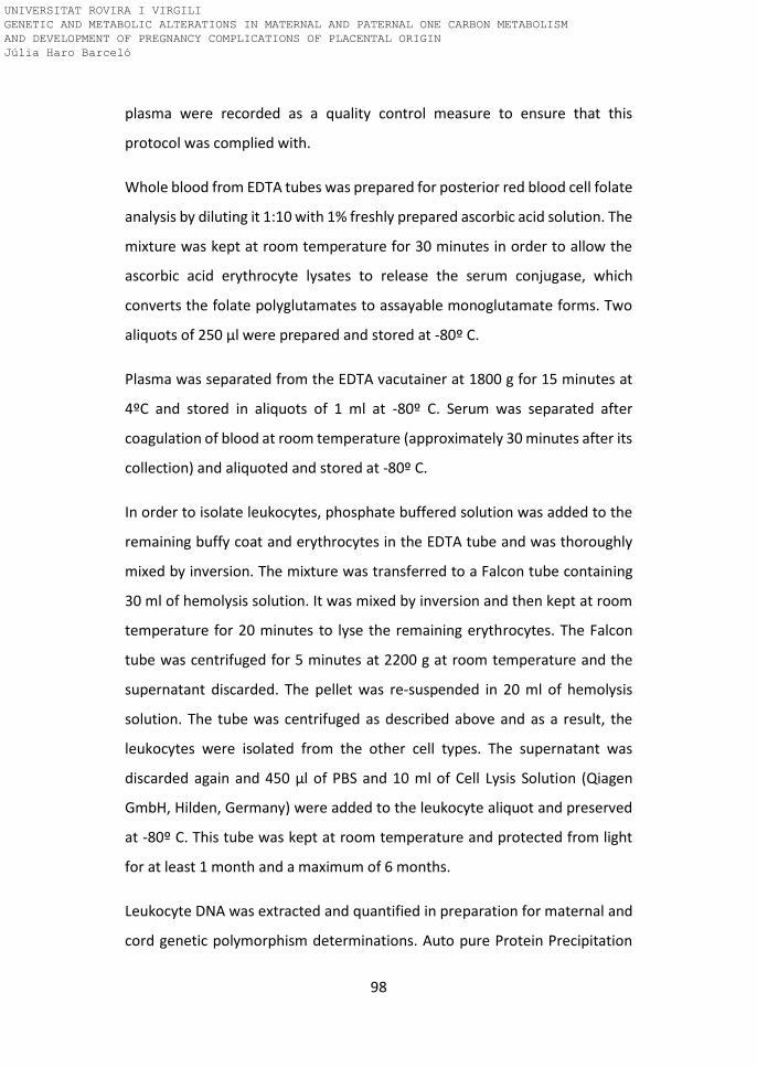
98
plasma were recorded as a quality control measure to ensure that this
protocol was complied with.
Whole blood from EDTA tubes was prepared for posterior red blood cell folate
analysis by diluting it 1:10 with 1% freshly prepared ascorbic acid solution. The
mixture was kept at room temperature for 30 minutes in order to allow the
ascorbic acid erythrocyte lysates to release the serum conjugase, which
converts the folate polyglutamates to assayable monoglutamate forms. Two
aliquots of 250 µl were prepared and stored at -80º C.
Plasma was separated from the EDTA vacutainer at 1800 g for 15 minutes at
4ºC and stored in aliquots of 1 ml at -80º C. Serum was separated after
coagulation of blood at room temperature (approximately 30 minutes after its
collection) and aliquoted and stored at -80º C.
In order to isolate leukocytes, phosphate buffered solution was added to the
remaining buffy coat and erythrocytes in the EDTA tube and was thoroughly
mixed by inversion. The mixture was transferred to a Falcon tube containing
30 ml of hemolysis solution. It was mixed by inversion and then kept at room
temperature for 20 minutes to lyse the remaining erythrocytes. The Falcon
tube was centrifuged for 5 minutes at 2200 g at room temperature and the
supernatant discarded. The pellet was re-suspended in 20 ml of hemolysis
solution. The tube was centrifuged as described above and as a result, the
leukocytes were isolated from the other cell types. The supernatant was
discarded again and 450 µl of PBS and 10 ml of Cell Lysis Solution (Qiagen
GmbH, Hilden, Germany) were added to the leukocyte aliquot and preserved
at -80º C. This tube was kept at room temperature and protected from light
for at least 1 month and a maximum of 6 months.
Leukocyte DNA was extracted and quantified in preparation for maternal and
cord genetic polymorphism determinations. Auto pure Protein Precipitation
UNIVERSITAT ROVIRA I VIRGILI GENETIC AND METABOLIC ALTERATIONS IN MATERNAL AND PATERNAL ONE CARBON METABOLISM AND DEVELOPMENT OF PREGNANCY COMPLICATIONS OF PLACENTAL ORIGIN Júlia Haro Barceló

99
Solution (3.33 mL) was added and mixed with vortex to obtain a homogenous
mix before incubation in iced water for 15 minutes. The mixture was
centrifuged at 2200 g at 4º C for 12 minutes. The supernatant containing the
suspended DNA was removed and added to a 50 ml Falcon tube with 10 ml of
cold 100% isopropanol. The tube was gently mixed by inversion until the
precipitated DNA was visible. After centrifuging at 2200 g at 4º C for 5 minutes,
the supernatant was discarded (pellet contained the DNA). The tube was dried
for 30-40 minutes on absorbent paper, and 1200 µl of DNA Hydration Solution
were added to the Falcon tube, mixed and further incubated at room
temperature for 3-4 days in a mixer incubator. DNA quantification was
performed using a spectrophotometer Nanodrop 1000 at 260 nm wavelength
using 2µl of hydrated DNA.
Biochemical and genetic determinations
Plasma aliquots of 0.5 ml from each pregnancy sample and the cord and 120
ng of lyophilized DNA from mother and cord were shipped on dry ice to BeVital
A/S (Bergen, Norway) and analyzed, ensuring that all samples from the same
pregnancy were analyzed in the same batch, within <18 months after sample
collection.
Red blood cell and plasma folate
Folate concentrations in plasma and whole blood were determined by an
automated gold standard microbiological assay using a microtiter plate and a
chloramphenicol-resistant strain of Lactobacillus casei (235). Performances
for inter- and intra-assay coefficient of variation were 4 and 5% respectively
and the lower detection limit was 2nmol/L.
UNIVERSITAT ROVIRA I VIRGILI GENETIC AND METABOLIC ALTERATIONS IN MATERNAL AND PATERNAL ONE CARBON METABOLISM AND DEVELOPMENT OF PREGNANCY COMPLICATIONS OF PLACENTAL ORIGIN Júlia Haro Barceló

100
RBC folate values were calculated using the following formula (236):
Whole blood folate – [Plasma folate*(1-hematocrit/100)] / (Haematocrit/100)
Whole blood folate was corrected for dilution prior to this.
Plasma cobalamin
Total cobalamin concentrations were determined by an automated
microbiological assay using a microtiter plate and a colistin sulphate-resistant
strain of Lactobacillus leichmannii (237). The lower detection limit was 30
pmol/L, and inter- and intra-assay coefficients of variation 4 and 5%
respectively.
Plasma total homocysteine and cotinine
Plasma total homocysteine, methylmalonic acid and cotinine were
determined by liquid chromatography–tandem mass spectrometry (238).
Cotinine, an oxidized metabolite of nicotine, is a marker of recent tobacco
exposure. Participants with plasma cotinine concentration >10 ng/mL were
considered smokers, and ≤10 ng/mL non-smokers. The lower limits of
detection were 0.1 and 0.03µmol/L and 1nmol/L, inter-assay coefficient of
variation 2, 3-8 and 6% and intra-assay coefficient 1-2, 1-4 and 2-3%
respectively for total homocysteine, methylmalonic acid and cotinine
determinations.
Plasma creatinine
Creatinine concentrations were determined by Jaffé reaction (Química Clínica
Aplicada, SA, Amposta, Spain) and were used as an estimation of renal
function (239). Inter- and intra-assay coefficients of variation were 2.1 and
1.7% respectively.
UNIVERSITAT ROVIRA I VIRGILI GENETIC AND METABOLIC ALTERATIONS IN MATERNAL AND PATERNAL ONE CARBON METABOLISM AND DEVELOPMENT OF PREGNANCY COMPLICATIONS OF PLACENTAL ORIGIN Júlia Haro Barceló

101
Genetic polymorphisms
DNA isolated from both mother and cord blood leukocytes were used to
determine genetic polymorphisms affecting folate metabolism and cobalamin
metabolism and transport. Matrix-assisted laser desorption/ionization time-
of-flight mass spectrometry was used to analyze the
methylenetretrahydrofolate reductase 677C>T, and the solute carrier family
19 member 1 80G>A genotypes, by Bevital (Norway).
UNIVERSITAT ROVIRA I VIRGILI GENETIC AND METABOLIC ALTERATIONS IN MATERNAL AND PATERNAL ONE CARBON METABOLISM AND DEVELOPMENT OF PREGNANCY COMPLICATIONS OF PLACENTAL ORIGIN Júlia Haro Barceló

102
3.3. Recruitment and follow-up of fathers
Eligible fathers were invited to participate in the study. In the first instance,
we informed the mothers of our interest in investigating the association of
paternal genetic and metabolic factors with pregnancy outcome. We asked
them if they agreed to provide us the contact details for the father. When the
mothers agreed, the fathers were contacted by phone, following a
recruitment protocol. The aims of the study, their contribution and what
participation would entail was explained to them and they were asked
whether they were interested in participating. The time of contact with the
father and the time of participation depended on when we spoke to the
mother (preferably coinciding with a prenatal visit where possible), how long
it took to secure contact with the father and when it suited the father to give
a fasting blood sample.
Those that accepted, came to the “Hospital Universitari Sant Joan de Reus” to
provide informed signed consent, a fasting blood sample and complete a
lifestyle questionnaire.
Ideally, we invited them to participate as near as possible to conception. To
evaluate the association of biochemical results at that moment with possible
paternal origin complications during placentation, it would be better for the
study to have the fasting blood sample just before the conception. To reduce
the effect of this limitation, we asked all fathers if they had changed their
nutritional habits or lifestyle from the six months before pregnancy until the
moment of participation. On the other hand, the results of the genetic analysis
will have been unaffected with lifestyle changes, if any, over a relatively short
period of time.
UNIVERSITAT ROVIRA I VIRGILI GENETIC AND METABOLIC ALTERATIONS IN MATERNAL AND PATERNAL ONE CARBON METABOLISM AND DEVELOPMENT OF PREGNANCY COMPLICATIONS OF PLACENTAL ORIGIN Júlia Haro Barceló

103
Medical history and lifestyle data collection
On the day of their blood sample, a member of our team administered a
questionnaire (exemplar in appendices) to fathers regarding basic elements of
their medical history as well as supplement use, lifestyle and toxic habits. The
questionnaire was specifically designed to cover the time around pre-
conception and conception. Any change six months before pregnancy and
during pregnancy in paternal diet, supplement use, or habits was recorded to
take into account if considered to have a relevant effect on nutritional status.
Concrete vitamin or mineral supplementation use, timing of initiation,
duration, doses, and brand was recorded. We used this information to
estimate the real intake of folic acid or other vitamins and minerals apart from
diet. As with the mothers, smoking habit data covered the 5 years before
pregnancy and during pregnancy, was assessed by questionnaire (number of
cigarettes/d), and whether the habit was maintained, reduced or given up
during pregnancy. Plasma cotinine concentrations (>10 ng/mL confirmed
active smokers) from blood was checked to corroborate their answers.
Similarly, alcohol consumption (duration, drinks /w, timing of cessation before
or during pregnancy) and illegal drug use in the previous 5 years (type and
frequency of use in the 12 months prior to conception and at the time of the
interview) was recorded.
Paternal age and BMI were calculated from the information collected during
the visit (birth data, weight and height). BMI calculated as Kg/m², was
classified as normal weight, overweight or obesity.
Medical history and blood group was obtained by the self-reported
information.
UNIVERSITAT ROVIRA I VIRGILI GENETIC AND METABOLIC ALTERATIONS IN MATERNAL AND PATERNAL ONE CARBON METABOLISM AND DEVELOPMENT OF PREGNANCY COMPLICATIONS OF PLACENTAL ORIGIN Júlia Haro Barceló

104
Before carrying out the analysis to study the objectives proposed for this
thesis, parental verification was performed to prevent introducing errors into
the analysis. A battery of 19 polymorphisms was used. If the cord genotype
did not match with parental genotypes for more than three polymorphisms,
the mismatched mother-cord or father-cord sets would have been excluded
from the analysis.
Blood sample collection
The blood sample was programmed as near as possible to conception
(determined by ability to contact the father and compatibility of his work
schedule). Protocol for collection and processing of samples for biochemical
and genetic determinations were the same as for the pregnancy phase and
the same laboratories were used.
UNIVERSITAT ROVIRA I VIRGILI GENETIC AND METABOLIC ALTERATIONS IN MATERNAL AND PATERNAL ONE CARBON METABOLISM AND DEVELOPMENT OF PREGNANCY COMPLICATIONS OF PLACENTAL ORIGIN Júlia Haro Barceló

105
3.4. Statistical analysis
All statistical analysis were performed using SPSS (SPSS Inc, Chicago, IL, USA)
for Windows, version 25.0.
The normality of continuous variables was checked with the Kolmogorov-
Smirnov test. Variables not normally distributed were natural log transformed
when necessary, to meet the necessary criteria for the application of
parametric statistical tests. Descriptive characteristics comprising geometric
means (95% CI) or median (P10-P90) and relative frequencies (95% CI), where
applicable, are reported for all the participants. Genotype and allele
frequencies (95% CI) of the studied genetic variants are also reported.
Confidence intervals of categorical variables expressed in % are calculated
using the “CIA” programme (Southampton, UK). The McNemar test was used
to compare the prevalence of plasma and red blood cell folate deficiency
between different time points of pregnancy (<12, 15, 24-27, 34 GW, labour).
Post-hoc Bonferroni correction for multiple comparisons of the P values was
applied. ANOVA for repeated measures was performed to compare means
between different pregnancy time points (<12, 15, 24-27, 34 GW, labour) and
post-hoc Bonferroni correction for multiple comparisons of the P values was
applied. Maternal, paternal and cord 1C metabolism status (plasma folate,
erythrocyte folate, total plasma homocysteine and plasma cobalamin) were
compared between the different genotypes for MTHFR 677C>T and SLC19A1
80 G>A, as well as the combination of both. Statistical significance was
accepted from p. values <0.05. Percentages (95% CI) of the outcomes included
in the study and according to genotypes are also reported.
BMI was calculated as weight (kg) divided by height squared (m²). Smoking
pattern was categorized by women who smoke throughout pregnancy versus
never or women who smoke in the first trimester versus never. Previous
UNIVERSITAT ROVIRA I VIRGILI GENETIC AND METABOLIC ALTERATIONS IN MATERNAL AND PATERNAL ONE CARBON METABOLISM AND DEVELOPMENT OF PREGNANCY COMPLICATIONS OF PLACENTAL ORIGIN Júlia Haro Barceló

106
pregnancy was categorized as confirmed previous live newborn or none.
Socioeconomic status was categorized as low versus mid-high status
considering maternal and paternal status together. Variables of the
polymorphisms were used comparing the homozygote normal versus the
heterozygote or the homozygote genotypes: CC vs CT or TT for MTHFR 677
C>T and GG vs GA or AA for SLC19A1 80G>A. The variable used as control for
the three clinical outcomes was created with participants without any
pregnancy complication such as gestational hypertension, gestational
diabetes, IUGR, etc.
The 3 main outcomes studied were: pathological doppler of uterine arteries
at 20GW, intrauterine growth restriction and pregnancy induced
hypertension. Multiple linear and logistic regressions were used to examine
the associations between maternal and paternal genetic, nutritional and
metabolic components of 1C metabolism and continuous and categorical
variables associated with the development of pregnancy outcomes and
complications respectively.
Interaction terms were calculated to assess possible interactions between
independent variables, in their relationship with the dependent variable of
interest. The product of the independent variables being tested was calculated
and included in the linear or logistic regression analysis. When the interaction
term was significant, we proceeded to perform the regression analysis on
stratified sets of data to account for the interaction.
Multiple linear regression analysis was performed to identify maternal and
paternal predictors of pulsatility index of uterine arteries (dependent variable,
natural log transformed). The models will be described in detail in the results
section. Generally, for each analysis we designed 2 models. The first model
explored the association of the 1CM variables of interest and pulsatility index.
UNIVERSITAT ROVIRA I VIRGILI GENETIC AND METABOLIC ALTERATIONS IN MATERNAL AND PATERNAL ONE CARBON METABOLISM AND DEVELOPMENT OF PREGNANCY COMPLICATIONS OF PLACENTAL ORIGIN Júlia Haro Barceló

107
For the genotype models, this included the MTHFR C677T and SLC19A1 G80A
genotypes as well as first trimester plasma cobalamin and red blood cell folate
status (low tertile versus the others). Subsequently maternal clinical and
lifestyle factors (smoking during pregnancy versus never, socioeconomic
status, age, BMI, late pregnancy anaemia (3rd trimester haemoglobin <11
g/dL), previous pregnancies longer than 20 GW versus none, gestational age
(weeks at birth and sex of the baby) were added to form the complete model.
In the tHcy models, only tHcy (representing overall 1CM status) at the
corresponding time of pregnancy was included in the first model.
Subsequently, the next model was adjusted for the same maternal clinical and
lifestyle factors described for the complete 1CM model above.
To study the involvement of paternal factors, we added the paternal variables
to the complete maternal models described above. Therefore, paternal
MTHFR 677C>T genotype, cobalamin and red blood cell folate status and
paternal tHcy ≥P90 were included in the corresponding models.
Multiple logistic regression analysis was used to identify maternal and
paternal factors associated with Pathological doppler of uterine arteries at 20
GW (dependent variable). The models to examine the association between
genotypes and impaired placentation included MTHFR 677C>T and SLC19A1
80G>A genotype, red blood cell folate and plasma cobalamin concentrations
at <12 and 15 GW. In the models focused on the association between tHcy and
impaired placentation, the categorical tHcy variable (≥P90 tHcy in early
pregnancy versus other percentiles) replaced the genotypes and red blood cell
folate and plasma cobalamin variables in each model, at <12 and 15 GW. The
models were adjusted for smoking habit, socioeconomic status, 1st trimester
BMI, age, parity and gestational age at doppler measurement. Adjusting for
study centre did not change the results in any of the models so it was not
included.
UNIVERSITAT ROVIRA I VIRGILI GENETIC AND METABOLIC ALTERATIONS IN MATERNAL AND PATERNAL ONE CARBON METABOLISM AND DEVELOPMENT OF PREGNANCY COMPLICATIONS OF PLACENTAL ORIGIN Júlia Haro Barceló
108
We checked for outliers and influential cases (Cook’s distance >4/n) and
excluded them from the models when identified.
Paternal factors were also studied in the logistic regressions. Paternal MTHFR
677C>T genotype, smoking habit, age, red blood cell folate concentrations and
elevated paternal tHcy levels (≥P90) were included in the same models used
for the mothers.
Multiple linear regression analysis was performed to identify maternal and
paternal predictors of birthweight (dependent variable) with a similar strategy
to those described for uterine artery pulsatility index above. In all multiple
linear regression analyses, participants with gestational diabetes were
excluded due to its potential effect on birth weight.
Multiple logistic regression analysis was used to identify maternal and
paternal factors associated with IUGR (dependent variable) applying a similar
strategy as described for pathological doppler above. In this case we did not
exclude participants with gestational diabetes to avoid loss of statistical
power.
Multiple logistic regressions analysis was used to study the association
between maternal and paternal genetic and 1CM related factors and
gestational hypertension. We excluded pregnancies affected by gestational
diabetes or intrauterine growth retardation from the analysis and used similar
model designs as described above for pathological Doppler and IUGR.
UNIVERSITAT ROVIRA I VIRGILI GENETIC AND METABOLIC ALTERATIONS IN MATERNAL AND PATERNAL ONE CARBON METABOLISM AND DEVELOPMENT OF PREGNANCY COMPLICATIONS OF PLACENTAL ORIGIN Júlia Haro Barceló

109
RESULTS
UNIVERSITAT ROVIRA I VIRGILI GENETIC AND METABOLIC ALTERATIONS IN MATERNAL AND PATERNAL ONE CARBON METABOLISM AND DEVELOPMENT OF PREGNANCY COMPLICATIONS OF PLACENTAL ORIGIN Júlia Haro Barceló

110
UNIVERSITAT ROVIRA I VIRGILI GENETIC AND METABOLIC ALTERATIONS IN MATERNAL AND PATERNAL ONE CARBON METABOLISM AND DEVELOPMENT OF PREGNANCY COMPLICATIONS OF PLACENTAL ORIGIN Júlia Haro Barceló

111
4. Results
4.1. Descriptive results
Eight hundred and fifty six women were screened at their first antenatal
check-up. 16 women were not eligible to participate (twin pregnancies, drug
abuse, donated oocytes and GA >20 GW). From the 840 mothers invited, 21
were not interested and 8 refused for language or cultural reasons. 811
mothers accepted and 748 were identified, as pregnancy progressed, to invite
the father (the others had miscarried, moved hospital or left the study). From
those participants, 3 were donor fathers, we could not establish contact with
60 mothers and 13 had adverse outcomes before establishing contact with
them (12 pregnancy terminations due to foetal anomalies and 1 fetal death).
Given the nature of these reasons, paternal contact details could not be
requested from the mothers due to discontinuation of prenatal follow up and
loss of contact with the study team. A total of 672 fathers were eligible to
invite. A further 46 fathers were ineligible for recruitment because 39 mothers
refused to provide their contact details and we could not establish the contact
with 7 of them. A total of 626 fathers were invited to participate. Of these, 212
fathers refused. 414 fathers agreed to participate by December 2019 allowing
us to complete 414 mother-father-neonate triads (66.1 of those eligible)
(Figure 6).
Maternal and paternal demographic and lifestyle descriptive characteristics
are shown in Table 2. A total of 25.7% of the mothers smoked at some time of
pregnancy and 36.7% of the fathers were smokers. A total of 36.0% of the
women reported taking folic acid before pregnancy and 94.2 % during the first
trimester. 51.8% did not meet the recommended red blood cell folate status
UNIVERSITAT ROVIRA I VIRGILI GENETIC AND METABOLIC ALTERATIONS IN MATERNAL AND PATERNAL ONE CARBON METABOLISM AND DEVELOPMENT OF PREGNANCY COMPLICATIONS OF PLACENTAL ORIGIN Júlia Haro Barceló

112
of 906 nmol/L for NTDs prevention. Only 6.8% of men reported taking
multivitamin or mineral supplements.
Figure 6. Participant flow chart from recruitment until triads included.
UNIVERSITAT ROVIRA I VIRGILI GENETIC AND METABOLIC ALTERATIONS IN MATERNAL AND PATERNAL ONE CARBON METABOLISM AND DEVELOPMENT OF PREGNANCY COMPLICATIONS OF PLACENTAL ORIGIN Júlia Haro Barceló
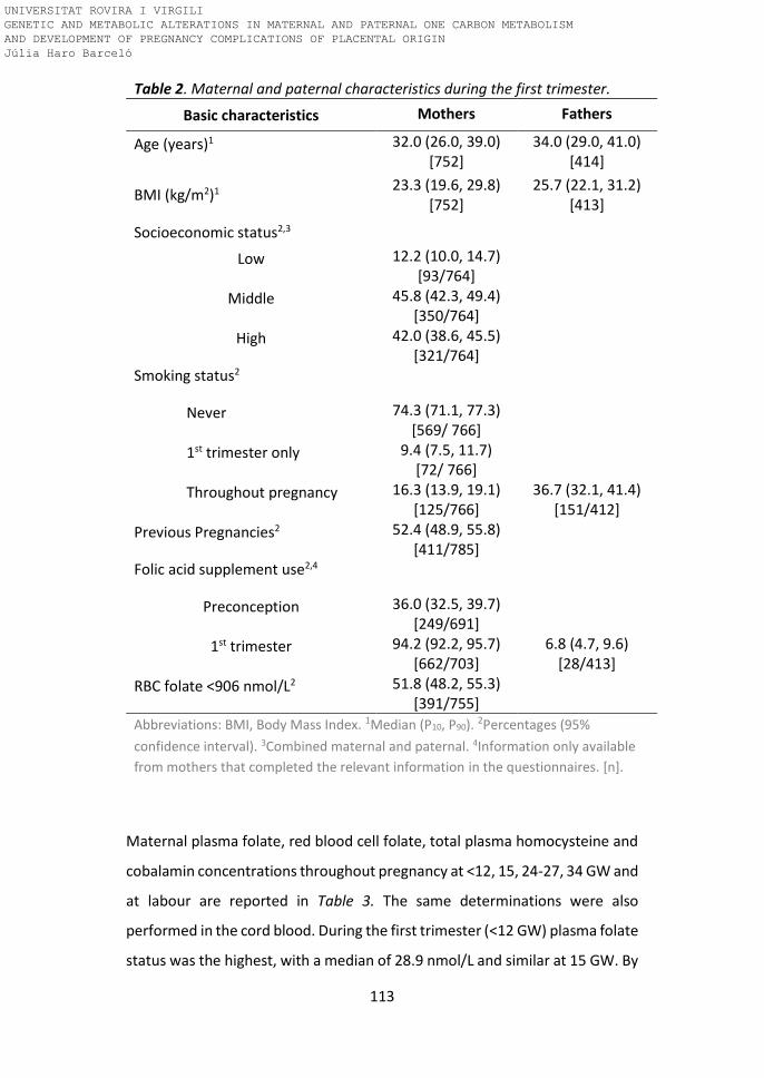
113
Maternal plasma folate, red blood cell folate, total plasma homocysteine and
cobalamin concentrations throughout pregnancy at <12, 15, 24-27, 34 GW and
at labour are reported in Table 3. The same determinations were also
performed in the cord blood. During the first trimester (<12 GW) plasma folate
status was the highest, with a median of 28.9 nmol/L and similar at 15 GW. By
Table 2. Maternal and paternal characteristics during the first trimester.
Basic characteristics Mothers Fathers
Age (years)1 32.0 (26.0, 39.0) [752]
34.0 (29.0, 41.0) [414]
BMI (kg/m2)1 23.3 (19.6, 29.8)
[752] 25.7 (22.1, 31.2)
[413]
Socioeconomic status2,3
Low 12.2 (10.0, 14.7) [93/764]
Middle 45.8 (42.3, 49.4) [350/764]
High 42.0 (38.6, 45.5) [321/764]
Smoking status2
Never 74.3 (71.1, 77.3) [569/ 766]
1st trimester only 9.4 (7.5, 11.7) [72/ 766]
Throughout pregnancy 16.3 (13.9, 19.1) [125/766]
36.7 (32.1, 41.4) [151/412]
Previous Pregnancies2 52.4 (48.9, 55.8) [411/785]
Folic acid supplement use2,4
Preconception 36.0 (32.5, 39.7) [249/691]
1st trimester 94.2 (92.2, 95.7) [662/703]
6.8 (4.7, 9.6) [28/413]
RBC folate <906 nmol/L2 51.8 (48.2, 55.3) [391/755]
Abbreviations: BMI, Body Mass Index. 1Median (P10, P90). 2Percentages (95%
confidence interval). 3Combined maternal and paternal. 4Information only available
from mothers that completed the relevant information in the questionnaires. [n].
UNIVERSITAT ROVIRA I VIRGILI GENETIC AND METABOLIC ALTERATIONS IN MATERNAL AND PATERNAL ONE CARBON METABOLISM AND DEVELOPMENT OF PREGNANCY COMPLICATIONS OF PLACENTAL ORIGIN Júlia Haro Barceló
114
24-27 GW, it had fallen to 14.4 nmol/L and continued to decrease until the
end of pregnancy. Plasma folate concentration in the cord was higher than in
the mother during labour. Erythrocyte folate concentrations increased from
<12 to 15 GW but had started to decline by 24-27 GW. Median tHcy decreased
from 5.3 µmol/L at <12 GW to 4.5 µmol/L by 15 GW. During late pregnancy, it
increased and reached 6.0 µmol/L by labour. Median cord blood tHcy was
lower than maternal tHcy at labour. Median plasma cobalamin decreased
gradually from 364 pmol/L in the first trimester to 233 pmol/L by labour and
cord cobalamin was higher than the mothers at labour. Folic acid supplement
use interacts significantly with plasma folate concentrations from 15 GW to
the end of pregnancy. This interaction was also observed between folic acid
supplementation and red blood cell folate and tHcy levels at 24-27 and at 34
GW. No interaction between folic acid supplementation and B12
concentrations was observed (data not reported in the table).
UNIVERSITAT ROVIRA I VIRGILI GENETIC AND METABOLIC ALTERATIONS IN MATERNAL AND PATERNAL ONE CARBON METABOLISM AND DEVELOPMENT OF PREGNANCY COMPLICATIONS OF PLACENTAL ORIGIN Júlia Haro Barceló

115
Table 3. Maternal 1CM folate, cobalamin and tHcy status during pregnancy and in the cord.
<12GW 15GW 24-27GW 34GW Labour Cord
Plasma folate (nmol/L)
28.9 (10.0, 61.2)¹ 26.6 (9.9, 53.8)¹ 14.4 (5.8, 39.7)¹ 11.0 (5.0, 38.6)¹ 10.0 (4.9, 36.5)¹ 26.3 (12.2, 56.5)¹
27.0 (25.7, 28.4)²
[773] 24.9 (23.4, 26.6)² ††
[448] 15.3 (14.4, 16.2)² *** †††
[665] 12.8 (12.0, 13.7)² *** ††
[633] 12.1 (11.3, 12.9)² **†
[611] 26.0 (24.8, 27.3)²
[577]
Erythrocyte folate (nmol/L)
936 (466, 1841)¹ 1266 (665, 2204)¹ 1138 (602, 2119)¹ 949 (464, 2065)¹ - -
942 (906, 980)²
[753] 1248 (1191, 1307)² ***
[446]
1150 (1105, 1196)²***
††† [651]
975 (932, 1021)² ***††† [616]
- -
Plasma total homocysteine (µmol/L)
5.3 (4.0, 7.1)¹ 4.5 (3.5, 5.9)¹ 4.6 (3.5, 6.3)¹ 5.2 (3.8, 7.3)¹ 6.0 (4.3, 8.6)¹ 4.8 (3.4, 6.7)¹
5.3 (5.2, 5.4) ²
[774] 4.5 (4.4, 4.6)² ***
[449] 4.6 (4.6, 4.7)² ** †††
[666] 5.2 (5.1, 5.3)² *** †
[634] 6.1 (6.0, 6.3)² ***
[613] 4.8 (4.7, 4.9)²
[578]
Plasma B12 (pmol/L)
364 (240, 525) ¹ 332 (213, 467)¹ 277 (178, 424)¹ 252 (160, 388)¹ 233 (143, 369)¹ 336 (115, 635)¹
360 (351, 369)²
[773] 324 (314, 334)²***
[448] 274 (267, 281)²***
[665] 250 (244, 257)²***
[633] 231 (224, 238)²***
[606] 316 (299, 333)²
[550]
Abbreviations: GW, gestational week. 1Median (P10, P90). 2Geometric mean (95% confidence interval). [n]. Geometric means between the different time points of pregnancy were compared using ANOVA for repeated measures analysis. Post-hoc Bonferroni correction for multiple comparisons was applied. Intrasubject factor: time of pregnancy; intersubject factor folic acid supplement use (>400 µg/ d in the first trimester and continued use in the second and third trimesters). Different to previous time point: *P<0.05, **p<0.01, ***p<0.001. Intersubject factor folic acid supplement use †P<0.05, ††p<0.01, †††p<0.001.
UNIVERSITAT ROVIRA I VIRGILI GENETIC AND METABOLIC ALTERATIONS IN MATERNAL AND PATERNAL ONE CARBON METABOLISM AND DEVELOPMENT OF PREGNANCY COMPLICATIONS OF PLACENTAL ORIGIN Júlia Haro Barceló

116
The following graphs illustrate the percentage of participants deficient in
plasma and red blood cell folate during pregnancy, at <12, 15, 24-27, 34
GW and labour. Deficiency was defined as plasma folate concentrations ≤
7 nmol/L (7), and red blood cell folate concentrations ≤ 340 nmol/L. The
prevalence of plasma folate deficiency increased as pregnancy progressed,
mainly after 15 GW. In the case of red blood cell folate deficiency, the
prevalence was the lowest at 15 and 24-27 GW but it had increased by 34
GW.
Figure 7. Prevalence of plasma folate deficiency (≤ 7nmol/L) in participants
during pregnancy and at labour are represented. [n]. Means (dots) and 95%
Confidence Interval (bars) are illustrated. Prevalences of deficiency with the
preceding time point were compared using Chi-square (McNemar) and
post-hoc Bonferoni correction of P values for multiple comparisons was
applied. *P<0.05, ***P<0.001.
UNIVERSITAT ROVIRA I VIRGILI GENETIC AND METABOLIC ALTERATIONS IN MATERNAL AND PATERNAL ONE CARBON METABOLISM AND DEVELOPMENT OF PREGNANCY COMPLICATIONS OF PLACENTAL ORIGIN Júlia Haro Barceló

117
Paternal 1CM nutrient and metabolite status is reported in Table 4.
Median plasma folate was relatively low at 8.8 nmol/L. Median
erythrocyte folate concentration was 456.8 nmol/L, tHcy was 9.6 µmol/L
and plasma cobalamin 367.2 pmol/L.
Figure 8. Prevalence of red blood cell folate deficiency (≤ 340 nmol/L) in
participants during pregnancy are represented. [n]. Means (dots) and 95%
Confidence Interval (bars) are illustrated. Prevalences of deficiency with the
preceding time point were compared using Chi-square (McNemar) and post-
hoc Bonferoni correction of P values for multiple comparisons was applied. *P<0.05, ***P<0.001.
UNIVERSITAT ROVIRA I VIRGILI GENETIC AND METABOLIC ALTERATIONS IN MATERNAL AND PATERNAL ONE CARBON METABOLISM AND DEVELOPMENT OF PREGNANCY COMPLICATIONS OF PLACENTAL ORIGIN Júlia Haro Barceló

118
Maternal, paternal and cord genotypes and allele frequencies for the
MTHFR 677C>T and SLC19A1 80 G>A genetic variants and the combination
of both are shown in Table 5. All the observed genotype frequencies were
in Hardy-Weinberg equilibrium (p>0.05). The homozygote variant
genotype for the MTHFR C677T polymorphism was 17.0, 14.1 and 18.6%
for the mother, father and cord respectively. For the SLC19A1 G80A
polymorphism, the homozygote variant genotype frequency was 27.2,
28.9 and 26.9% for the mother, father and cord respectively. Regarding
the combined genotypes, the CT-GA combination was the most frequent,
found in >20% of mothers, fathers and cords.
Table 4. Paternal status in 1CM nutrients and metabolites.
Fathers N
Plasma folate (nmol/L) 8.8 (4.5, 16.8)¹ 405
8.7 (8.3, 9.2)²
Erythrocyte folate (nmol/L) 456.8 (205.4, 802.9)¹ 384
425.9 (404.6, 448.4)²
Plasma total homocysteine (µmol/L)
9.6 (7.5, 14.0)¹ 404
9.9 (9.7, 10.2)²
Plasma B12 (pmol/L) 367.2 (282.5, 570.4)¹ 403
380.3 (369.6, 391.4)² 1 Median (P10, P90). 2 Geometric mean (95% confidence interval).
UNIVERSITAT ROVIRA I VIRGILI GENETIC AND METABOLIC ALTERATIONS IN MATERNAL AND PATERNAL ONE CARBON METABOLISM AND DEVELOPMENT OF PREGNANCY COMPLICATIONS OF PLACENTAL ORIGIN Júlia Haro Barceló

119
Table 5. Frequencies of maternal, paternal and cord MTHFR 677 C>T and SLC19A1 80 G>A genotypes.1
Mother Father Cord
MTHFR 677 C>T genotype
CC 34 (30.5, 37.3)
[249] 36.3 (31.6, 41.2)
[139] 35.4 (31.5, 39.6)
[188]
CT 49.3 (45.7, 52.9)
[363] 49.6 (44.6, 54.6)
[192] 46.0 (41.8, 50.2)
[244]
TT 17.0 (14.4, 19.8)
[125] 14.1 (11.0, 17.9)
[54] 18.6 (15.6, 22.2)
[99]
T allele frequency 41.6 (39.1, 44.1)
[613/1474] 38.9 (35.5, 42.4)
[298/766] 41.6 (38.7, 44.6)
[442/1062] SLC19A1 80 G>A genotype
GG 27.2 (24.2, 30.6)
[200] 27.9 (23.6, 32.6)
[106] 25.6 (22.1, 29.5)
[135]
GA 45.5 (41.9, 49.1)
[334] 43.2 (38.3, 48.2)
[164] 47.4 (43.2, 51.7)
[250]
AA 27.2 (24.2, 30.6)
[200] 28.9 (24.6, 33.7)
[112] 26.9 (23.3, 30.9)
[142]
A allele frequency 50.0 (47.4, 52.6)
[734/1468] 50.5 (47.0, 54.1)
[384/760] 50.7 (47.6, 53.7)
[534/1054] MTHFR 677 C>T + SLC19A1 80 G>A genotype
CC-GG 10.6 (8.6, 13.1) [78]
10.0 (7.4, 13.4)
[38] 10.3 (8.0, 13.2)
[54] CC-GA 15.0 (12.6, 17.8)
[110] 17.4 (13.9, 21.5)
[67] 16.6 (13.6, 20.0)
[87] CC-AA 8.3 (6.5, 10.5)
[61] 9.2 (6.7, 12.5)
[35] 8.8 (6.6, 11.5)
[46] CT-GG 11.9 (9.7, 14.4)
[87] 13.2 (10.1, 16.9)
[50] 10.9 (8.5, 13.8)
[57] CT-GA 24.3 (21.3, 27.5)
[178] 21.6 (17.7, 26.0)
[83] 21.3 (18.0, 25.0)
[112] CT-AA 12.9 (10.7, 15.6)
[96] 14.5 (11.3, 18.4)
[57] 13.5 (10.9, 16.7)
[72]
TT-GG 4.8 (3.4, 6.6) [35]
4.7 (3.0, 7.4) [18]
4.6 (3.1, 6.7) [24]
TT-GA 6.3 (4.7, 8.3) [47]
4.2 (2.6, 6.7) [16]
9.3 (7.1, 12.1) [49]
TT-AA 6.0 (4.5, 8.0)
[44] 5.3 (3.4, 8.0)
[20] 4.8 (3.2, 6.9)
[25]
1 All data shown are percentages (95% confidence interval). Abbreviations: MTHFR, methylenetetrahydrofolate reductase; SLC19A1, solute carrier family 19 member 1. [n]
UNIVERSITAT ROVIRA I VIRGILI GENETIC AND METABOLIC ALTERATIONS IN MATERNAL AND PATERNAL ONE CARBON METABOLISM AND DEVELOPMENT OF PREGNANCY COMPLICATIONS OF PLACENTAL ORIGIN Júlia Haro Barceló

120
Maternal one carbon metabolism status (plasma folate, erythrocyte folate
and total plasma homocysteine) according to MTHFR 677 C>T and
SLC19A1 80 G>A genotypes and the combination of both at 5 different
points of pregnancy (<12, 15, 24-27, 34 GW and labour) are reported in
Figure 9 and tables 6 and 7. A similar pattern to that observed for change
in plasma folate (Table 3) was observed for all the MTHFR 677 C>T
genotypes and plasma folate did not differ between them (Figure 9). In the
women with available RBC folate measurements at 15 GW, we observed
that it increased between the first trimester and 15 GW and then gradually
declined for the rest of pregnancy. In contrast to plasma folate, we did
observe lower RBC folate status in the MTHFR 677TT compared to CC
genotype from 24-27 GW and also compared to the CT genotype at 34 GW
(Figure 9). THcy decreased between the first trimester and 15 GW and
then gradually increased for the rest of pregnancy, reaching the highest
concentration at labour. THcy was higher in the MTHFR 677TT compared
to CC and CT genotypes from 15GW throughout the rest of pregnancy
(Figure 9). No significant differences were observed in plasma or tHcy
status between SLC19A1 80 G>A genotypes (Table 6). Regarding the
combination of both genotypes, only tHcy at <12, 24-27 and 34 GW and at
labour differed among these (Table 7). People with the combination of
MTHFR 677 + SLC19A1 TT-GG, TT-GA genotypes had the highest tHcy out
of all of the combinations. People with the combinations of MTHFR 677 +
SLC19A1 CC-GA and CC-AA genotypes had the lowest. People with the
combinations of MTHFR 677 + SLC19A1 CC-GG, CT-GA and CT-AA had
lower tHcy compared to the TT-GG and TT-GA genotype combinations but
higher than those with the CC-GA and CC-AA combinations. Plasma
UNIVERSITAT ROVIRA I VIRGILI GENETIC AND METABOLIC ALTERATIONS IN MATERNAL AND PATERNAL ONE CARBON METABOLISM AND DEVELOPMENT OF PREGNANCY COMPLICATIONS OF PLACENTAL ORIGIN Júlia Haro Barceló

121
cobalamin concentrations and changes during pregnancy did not differ
between genotypes (data not shown).
Figure 9. Maternal one carbon metabolism status according to MTHFR 677 C>T genotype. Geometric
means (95% confidence interval) are shown. Blue line represents CC genotype; plasma folate and tHcy
at <12GW n= 248, 15GW n=148, 24-27GW n=217, 34GW n=202 and at labour n=206. RBC folate at
<12GW n= 241, 15GW n=146, 24-27GW n=212, 34GW n=194. Orange line represents CT genotype;
plasma folate and tHcy at <12GW n=355, 15GW n=214, 24-27GW n=309, 34GW n=299 and at labour
n=289. RBC folate at <12GW n= 345, 15GW n=214, 24-27GW n=301, 34GW n=291. Grey line represents
TT genotype; plasma folate <12GW n= 122, 15GW n=83, 24-27GW n=108, 34GW n=102 and at labour
n=94. RBC folate at <12GW n= 121, 15GW n=83, 24-27GW n=107, 34GW n=101. THcy at <12GW n= 123,
15GW n=84, 24-27GW n=109, 34GW n=103 and at labour n=95. ANOVA with post-hoc Bonferroni
correction for multiple comparisons were used to compare the differences in geometric means between
the genotypes and to compare plasma concentrations of 1C metabolites between different times of
pregnancy and labour. *p<0.05 **p<0.01 *** p<0.001 TT compared to CC genotype. ††p<0.01 †††p<0.001
TT compared to CT genotype. ap <0.001 compared with <12 GW, bp <0.001 compared with 15GW, cp
<0.001 compared with 24-27GW, dp <0.001 compared with 34GW. GW, gestational week; MTHFR,
methylenetetrahydrofolate reductase.
< <
<
UNIVERSITAT ROVIRA I VIRGILI GENETIC AND METABOLIC ALTERATIONS IN MATERNAL AND PATERNAL ONE CARBON METABOLISM AND DEVELOPMENT OF PREGNANCY COMPLICATIONS OF PLACENTAL ORIGIN Júlia Haro Barceló

122
Table 6. Maternal one carbon metabolism status according to SLC19A1 80 G>A genotype.
<12GW 15GW 24-27GW 34GW Labour
Plasma folate (nmol/L) SLC19A1 GG genotype
30.4 (9.2, 61.5)¹ 28.2 (10.2, 45.6)¹ 13.5 (5.6, 36.6)¹ 9.7 (4.9, 35.6)¹ 9.0 (4.6, 37.7)¹
27.3 (24.6, 30.4)² [198]
25.9 (23.1, 29.1)² [123]
14.8 (13.3, 16.6)²bbb
[175] 11.8 (10.4, 13.3)²ccc
[169] 11.3 (9.9, 12.8)²
[163] SLC19A1 GA genotype
27.2 (10.1, 59.7)¹ 25.2 (10.1, 55.5)¹ 13.5 (5.7, 39.8)¹ 11.2 (5.0, 40.6)¹ 10.1 (4.9, 34.6)¹
26.7 (24.7, 28.8)² [330]
25.0 (22.7, 27.5)² [201]
14.8 (13.6, 16.2)²bbb [292]
12.8 (11.6, 14.2)²ccc [269]
12.0 (10.9, 13.2)²d [273]
SLC19A1 AA genotype
30.3 (10.5, 58.9)¹ 27.0 (9.6, 54.7)¹ 13.1 (5.9, 39.7)¹ 9.9 (5.1, 36.9)¹ 10.0 (4.9, 37.3)¹
26.4 (24.0, 29.1)² [194]
24.5 (21.6, 27.8)² [118]
14.7 (13.0, 16.5)²bbb [164]
12.2 (10.8, 13.7)² ccc [162]
12.2 (10.7, 14.0)² [150]
p value 0.885 0.802 0.986 0.525 0.631 Erythrocyte folate (nmol/L)
SLC19A1 GG genotype
1003.5 (454.7, 1840.3)¹
1380.3 (789.9, 2245.2)¹
1170.6 (584.9, 2006.6)¹
904.1 (462.9, 1691.1)¹
-
970.7 (897.8, 1049.5)²
[193]
1330.5 (1221.9, 1448.7)²aaa
[122]
1107.3 (1019.9, 1202.2)²bbb
[172]
869.8 (801.3, 944.2)² ccc
[166]
-
SLC19A1 GA genotype
941.6 (466.0, 1778.0)¹
1237.5 (622.3, 2196.1)¹
1137.1 (580.9, 2099.7)¹
1028.2 (461.4, 2166.3)¹
-
937.1 (884.4, 992.9)²
[323]
1228.0 (1145.5, 1316.6)²aaa
[201]
1134.2 (1068.4, 1204.0)²bbb
[284]
1006.3 (936.2, 1081.6)² ccc
[264]
-
UNIVERSITAT ROVIRA I VIRGILI GENETIC AND METABOLIC ALTERATIONS IN MATERNAL AND PATERNAL ONE CARBON METABOLISM AND DEVELOPMENT OF PREGNANCY COMPLICATIONS OF PLACENTAL ORIGIN Júlia Haro Barceló

123
Table 6. Maternal one carbon metabolism status according to SLC19A1 80 G>A genotype.
<12GW 15GW 24-27GW 34GW Labour
SLC19A1 AA genotype
886.0 (468.8, 1931.4)¹
1247.4 (681.3, 2221.4)¹
1092.5 (599.4, 2083.3)¹
815.1 (432.9, 1958.1)¹
-
914.7 (842.0, 993.6)²
[188]
1234.4 (1129.4, 1349.2)²aaa
[117]
1113.8 (1025.7, 1209.6)²bbb
[161]
925.9 (847.2, 1011.9)²ccc
[153]
-
p value 0.564 0.343 0.865 0.199 - Plasma total homocysteine (µmol/L)
SLC19A1 GG genotype
5.3 (4.2, 7.4)¹ 4.4 (3.6, 6.0)¹
4.7 (3.5, 6.5)¹
5.3 (3.9, 7.9)¹
6.2 (4.1, 9.2)¹
5.4 (5.2, 5.6)² [198]
4.5 (4.3, 4.7)²aaa [123]
4.8 (4.6, 5.0)²bb [175]
5.4 (5.2, 5.6)²ccc [169]
6.3 (6.0, 6.6)²ddd [163]
SLC19A1 GA genotype
5.3 (4.0, 7.2)¹ 4.5 (3.5, 5.7)¹ 4.6 (3.5, 6.3)¹ 5.1 (3.8, 7.0)¹ 6.0 (4.3, 8.5)¹
5.3 (5.2, 5.5)² [331]
4.5 (4.4, 4.6)²aaa [202]
4.6 (4.5, 4.7)² bb [293]
5.2 (5.1, 5.4)²ccc [270]
6.1 (5.9, 6.3)²ddd [275]
SLC19A1 AA genotype
5.3 (3.8, 6.9)¹ 4.6 (3.3, 5.9)¹ 4.7 (3.4, 6.3)¹ 5.3 (3.6, 7.1)¹ 5.9 (4.4, 8.8)¹
5.3 (5.1, 5.4)² [194]
4.6 (4.4, 4.8)²aaa [116]
4.6 (4.5, 4.8)² [132]
5.1 (4.9, 5.3)²ccc [130]
6.1 (5.9, 6.4)²ddd [120]
p value 0.587 0.605 0.347 0.190 0.598
Abbreviations: GW, gestational week; SLC19A1, solute carrier family 19 member 1.1 Median (P10, P90). 2Geometric mean (95% confidence interval).
Geometric means were compared between time points using ANOVA for repeated measures and between genotypes using ANOVA (post hoc Bonferroni p value are shown). ap <0.05, aap <0.01, aaap <0.001 compared with <12 GW, bp<0.05, bbp<0.01, bbbp <0.001 compared with 15GW, cp <0.05, ccp <0.01, cccp <0.001 compared with 24-27GW, dp <0.05, ddp <0.01, dddp <0.001 compared with 34GW.
UNIVERSITAT ROVIRA I VIRGILI GENETIC AND METABOLIC ALTERATIONS IN MATERNAL AND PATERNAL ONE CARBON METABOLISM AND DEVELOPMENT OF PREGNANCY COMPLICATIONS OF PLACENTAL ORIGIN Júlia Haro Barceló
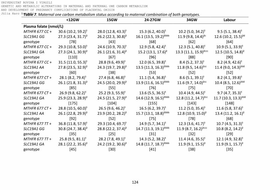
124
Table 7. Maternal one carbon metabolism status according to maternal combination of both genotypes.
<12GW 15GW 24-27GW 34GW Labour
Plasma folate (nmol/L) MTHFR 677 CC + SLC19A1 GG genotype
30.6 (10.2, 59.2)¹ 27.3 (23.4, 31.7)²
[78]
28.0 (12.8, 43.3)¹ 26.2 (22.3, 30.8)²
[43]
15.3 (6.2, 40.0)¹ 16.1 (13.5, 19.2)²bbb
[68]
10.2 (5.0, 34.2)¹ 11.9 (9.8, 14.4)²c
[62]
9.5 (5.1, 38.4)¹ 12.6 (10.2, 15.5)²d
[64] MTHFR 677 CC + SLC19A1 GA genotype
29.3 (10.8, 53.0)¹ 27.3 (24.1, 30.9)²
[110]
24.6 (10.9, 70.3)¹ 26.1 (21.6, 31.4)²
[67]
12.9 (5.8, 42.4)¹ 15.2 (13.1, 17.6)²
[96]
12.3 (5.1, 40.8)¹ 13.3 (11.1, 15.9)²ccc
[88]
10.9 (5.1, 33.9)¹ 12.5 (10.5, 14.8)²
[90] MTHFR 677 CC + SLC19A1 AA genotype
31.5 (11.0, 55.3)¹ 27.8 (23.5, 32.9)²
[60]
28.8 (9.6, 49.9)¹ 24.3 (19.7, 29.8)²
[38]
12.0 (6.5, 39.8)¹ 13.5 (11.3, 16.3)²bbb
[53]
8.4 (5.2, 37.3)¹ 11.8 (9.5, 14.6)²cc
[52]
8.2 (4.9, 42.6)¹ 11.4 (9.0, 14.3)²dd
[52] MTHFR 677 CT + SLC19A1 GG genotype
28.1 (8.2, 79.4)¹ 26.1 (21.8, 31.3)²
[85]
27.4 (6.8, 46.8)¹ 24.5 (20.0, 29.9)²
[55]
11.1 (5.4, 36.8)¹ 13.9 (11.6, 16.5)²bbb
[76]
8.6 (5.1, 39.1)¹ 11.6 (9.7, 14.0)²cc
[75]
8.2 (4.1, 39.8)¹ 10.4 (8.5, 12.6)²dd
[70] MTHFR 677 CT + SLC19A1 GA genotype
26.9 (9.8, 62.2)¹ 25.9 (23.3, 28.9)²
[175]
25.2 (9.3, 55.9)¹ 24.5 (21.5, 27.9)²
[104]
13.6 (5.5, 36.5)¹ 14.6 (12.9, 16.5)²bbb
[155]
10.4 (4.9, 44.5)¹ 12.8 (11.2, 14.7)²cc
[143]
9.7 (4.7, 35.3)¹ 11.7 (10.3, 13.3)²dd
[148] MTHFR 677 CT + SLC19A1 AA genotype
28.8 (10.5, 60.0)¹ 26.1 (22.8, 29.9)²
[92]
26.5 (9.6, 46.2)¹ 23.9 (20.2, 28.2)²
[52]
16.5 (6.2, 39.7)¹ 15.7 (13.1, 18.8)²bbb
[75]
11.2 (5.0, 35.4)¹ 12.8 (10.9, 15.0)²
[78]
11.6 (5.8, 37.6)¹ 13.4 (11.2, 16.1)²
[68] MTHFR 677 TT + SLC19A1 GG genotype
36.8 (11.8, 57.9)¹ 30.8 (24.7, 38.4)²
[35]
29.3 (10.4, 69.7)¹ 28.8 (22.2, 37.4)²
[25]
14.9 (5.7, 34.1)¹ 14.7 (11.3, 19.1)²bbb
[31]
12.3 (3.6, 41.7)¹ 11.9 (8.7, 16.2)²ccc
[32]
10.7 (4.5, 31.3)¹ 10.8 (8.2, 14.2)²
[29] MTHFR 677 TT + SLC19A1 GA genotype
25.8 (9.5, 81.1)¹ 28.1 (22.2, 35.6)²
[45]
28.2 (7.8, 49.1)¹ 24.2 (19.2, 30.6)²
[30]
14.3 (5.2, 38.2)¹ 14.8 (11.7, 18.7)²bbb
[41]
11.4 (4.6, 35.5)¹ 11.9 (9.1, 15.5)²
[38]
12.1 (4.9, 32.8)¹ 11.9 (9.1, 15.7)²
[35]
UNIVERSITAT ROVIRA I VIRGILI GENETIC AND METABOLIC ALTERATIONS IN MATERNAL AND PATERNAL ONE CARBON METABOLISM AND DEVELOPMENT OF PREGNANCY COMPLICATIONS OF PLACENTAL ORIGIN Júlia Haro Barceló

125
Table 7. Maternal one carbon metabolism status according to maternal combination of both genotypes.
<12GW 15GW 24-27GW 34GW Labour
MTHFR 677 TT + SLC19A1 AA genotype
30.2 (8.9, 60.4)¹ 25.4 (19.7, 32.6)²
[42]
25.2 (8.5, 89.0)¹ 26.2 (18.6, 36.8)²
[28]
12.3 (5.4, 47.5)¹ 14.4 (10.7, 19.3)²bbb
[36]
9.5 (4.9, 49.6)¹ 11.4 (8.5, 15.3)²
[32]
8.3 (3.3, 46.3)¹ 11.2 (7.5, 16.8)²
[30] p value 0.955 0.974 0.946 0.965 0.766 Erythrocyte folate (nmol/L)
MTHFR 677 CC + SLC19A1 GG genotype
1038.7 (452.7, 1836.0)¹
991.2 (871.7, 1126.9)²
[76]
1385.6 (799.9, 2245.2)¹
1366.4 (1189.8, 1569.2)²aaa
[42]
1218.7 (665.3, 2130.1)¹
1215.1 (1089.3, 1355.5)²bb
[66]
953.3 (466.4, 1703.2)¹
942.8 (831.2, 1069.5)²ccc
[60]
-
MTHFR 677 CC + SLC19A1 GA genotype
997.0 (520.2, 1872.2)¹
1023.9 (927.9, 1129.8)²
[107]
1171.7 (602.8, 2299.9)¹
1246.7 (1104.2, 1407.5)²aaa
[67]
1215.1 (651.2, 2402.2)¹
1217.5 (1099.5, 1348.2)²
[93]
1061.9 (472.2, 2449.8)¹ 1094.8
(960.8, 1247.5)²ccc [85]
-
MTHFR 677 CC + SLC19A1 AA genotype
975.9 (519.7, 1960.3)¹
987.1 (856.7, 1137.4)²
[58]
1186.6 (770.7, 2006.2)¹
1258.9 (1101.4, 1439.0)²aa
[37]
1122.0 (662.1, 1846.1)¹
1160.2 (1011.4, 1330.8)²
[53]
867.4 (525.7, 1936.6)¹
902.3 (777.6, 1046.9)²ccc
[49]
-
MTHFR 677 CT + SLC19A1 GG genotype
1064.6 (441.6, 1868.5)¹
982.7 (869.5, 1110.6)²
[83]
1391.7 (775.5, 2167.6)¹
1290.0 (1136.4, 1464.3)²aaa
[55]
1118.0 (527.6, 2019.8)¹
1121.8 (996.7, 1262.6)²
[76]
904.6 (426.4, 1782.2)¹
952.3 (843.8, 1074.8)²ccc
[74]
-
UNIVERSITAT ROVIRA I VIRGILI GENETIC AND METABOLIC ALTERATIONS IN MATERNAL AND PATERNAL ONE CARBON METABOLISM AND DEVELOPMENT OF PREGNANCY COMPLICATIONS OF PLACENTAL ORIGIN Júlia Haro Barceló

126
Table 7. Maternal one carbon metabolism status according to maternal combination of both genotypes.
<12GW 15GW 24-27GW 34GW Labour
MTHFR 677 CT + SLC19A1 GA genotype
939.9 (457.8, 1711.1)¹
916.8 (847.4, 991.9)²
[171]
1342.4 (756.3, 2155.8)¹
1278.8 (1161.6, 1407.9)²aaa
[104]
1136.4 (573.1, 1991.1)¹
1126.6 (1039.1, 1221.3)²bb
[150]
1026.2 (521.9, 2067.7)¹
1014.2 (923.5, 1113.8)²ccc
[141]
-
MTHFR 677 CT + SLC19A1 AA genotype
910.3 (443.9, 1895.0)¹
915.2 (814.7, 1028.2)²
[88]
1248.1 (658.6, 2195.3)¹
1215.9 (1062.8, 1391.1)²aaa
[52]
1103.9 (576.1, 2151.2)¹
1135.3 (1007.3, 1279.6)²bb
[72]
912.6 (500.0, 2018.2)¹
966.8 (856.0, 1091.8)²cc
[73]
-
MTHFR 677 TT + SLC19A1 GG genotype
843.0 (478.3, 1929.3)¹
899.2 (754.3, 1072.0)²
[34]
1339.6 (648.3, 3080.7)¹
1347.4 (1080.8, 1679.9)²aaa
[25]
1116.7 (452.4, 1987.5)¹
1072.7 (885.9, 1298.8)²b
[30]
761.4 (382.4, 1562.8)¹
829.5 (681.4, 1009.9)²ccc
[32]
-
MTHFR 677 TT + SLC19A1 GA genotype
854.7 (386.0, 1511.1)¹
824.8 (696.1, 977.4)²
[45]
992.3 (510.9, 2026.4)¹
1031.8 (853.5, 1247.4)²aaa
[30]
997.6 (446.4, 1829.7)¹
989.7 (827.3, 1183.9)²
[41]
838.5 (357.4, 1690.4)¹
809.3 (653.0, 1002.9)²ccc
[38]
-
MTHFR 677 TT + SLC19A1 AA genotype
716.2 (345.4, 2027.2)¹
822.1 (667.9, 1012.1)²
[42]
1252.2 (601.4, 2693.7)¹
1237.0 (981.5, 1559.0)²aaa
[28]
910.5 (425.9, 2950.5)¹
1009.7 (820.4, 1242.6)²b
[36]
797.5 (393.3, 2033.3)¹
871.3 (676.3, 1122.4)²ccc
[31]
-
p value 0.250 0.518 0.402 0.104 -
UNIVERSITAT ROVIRA I VIRGILI GENETIC AND METABOLIC ALTERATIONS IN MATERNAL AND PATERNAL ONE CARBON METABOLISM AND DEVELOPMENT OF PREGNANCY COMPLICATIONS OF PLACENTAL ORIGIN Júlia Haro Barceló

127
Table 7. Maternal one carbon metabolism status according to maternal combination of both genotypes.
<12GW 15GW 24-27GW 34GW Labour
Plasma total homocysteine (µmol/L) MTHFR 677 CC + SLC19A1 GG genotype
5.1 (4.2, 7.0)¹ 5.4 (5.1, 5.6)²
[78]
4.3 (3.7, 5.3)¹ 4.4 (4.2, 4.6)²aaa
[43]
4.7 (3.4, 6.2)¹ 4.6 (4.4, 4.9)²$#
[68]
5.1 (3.8, 7.1)¹ 5.1 (4.8, 5.5)²$$$# ccc
[62]
6.1 (3.8, 8.9)¹ 6.0 (5.6, 6.5)²$##ddd
[64] MTHFR 677 CC + SLC19A1 GA genotype
5.1 (4.0, 7.1)¹ 5.2 (5.0, 5.4)²
[110]
4.4 (3.5, 5.5)¹ 4.4 (4.2, 4.6)²aaa
[67]
4.4 (3.6, 6.0)¹ 4.5 (4.3, 4.7)²
[96]
5.0 (3.8, 6.8)¹ 5.2 (4.9, 5.5)²ccc
[88]
5.9 (4.1, 9.0)¹ 5.9 (5.6, 6.4)²ddd
[90] MTHFR 677 CC + SLC19A1 AA genotype
5.2 (3.8, 6.4)¹ 5.0 (4.8, 5.3)²
[60]
4.7 (3.3, 5.8)¹ 4.6 (4.3, 4.9)²aa
[38]
4.7 (3.4, 5.7)¹ 4.5 (4.3, 4.8)²
[53]
4.9 (3.6, 7.1)¹ 4.9 (4.6, 5.3)²ccc
[52]
5.8 (4.7, 7.9)¹ 6.0 (5.6, 6.3)²ddd
[52] MTHFR 677 CT + SLC19A1 GG genotype
5.0 (3.9, 7.0)¹ 5.1 (4.9, 5.4)²
[85]
4.3 (3.2, 6.0)¹ 4.4 (4.1, 4.6)²aaa
[55]
4.6 (3.4, 6.6)¹ 4.7 (4.4, 4.9)²bb
[76]
5.2 (3.7, 7.9)¹ 5.3 (5.0, 5.7)²ccc
[75]
6.1 (4.4, 8.9)¹ 6.2 (5.9, 6.7)²ddd
[70] MTHFR 677 CT + SLC19A1 GA genotype
5.3 (4.0, 7.2)¹ 5.3 (5.1, 5.5)²
[175]
4.4 (3.4, 5.8)¹ 4.4 (4.3, 4.6)²aaa
[104]
4.6 (3.4, 6.3)¹ 4.5 (4.4, 4.7)²
[155]
5.1 (3.8, 6.8)¹ 5.1 (4.9, 5.3)²ccc
[143]
6.0 (4.3, 8.0)¹ 6.0 (5.7, 6.3)²ddd
[149] MTHFR 677 CT + SLC19A1 AA genotype
5.4 (3.9, 7.0)¹ 5.3 (5.0, 5.5)²
[92]
4.5 (3.5, 5.9)¹ 4.5 (4.3, 4.8)²aaa
[52]
4.8 (3.5, 6.3)¹ 4.7 (4.4, 5.0)²
[75]
5.3 (3.6, 7.1)¹ 5.2 (4.9, 5.5)²ccc
[78]
6.0 (4.3, 9.5)¹ 6.1 (5.7, 6.6)²ddd
[68] MTHFR 677 TT + SLC19A1 GG genotype
5.8 (4.5, 10.0)¹ 6.0 (5.5, 6.6)²*††ᶴ
[35]
5.2 (3.8, 7.0)¹ 5.0 (4.6, 5.5)²aaa
[25]
5.3 (4.0, 8.2)¹ 5.4 (4.9, 6.0)²^^**†≠≠
[31]
5.8 (4.3, 9.9)¹ 6.1 (5.5, 6.8)²†≠
[32]
6.9 (4.8, 14.2)¹ 7.0 (6.0, 8.1)²d
[29] MTHFR 677 TT + SLC19A1 GA genotype
5.8 (4.4, 9.1)¹ 5.9 (5.5, 6.4)²§ɸ ɸ∞
[46]
4.7 (4.0, 6.0)¹ 4.8 (4.4, 5.2)²aaa
[31]
4.9 (3.9, 6.9)¹ 5.2 (4.8, 5.7)²£§}
[42]
5.6 (3.8, 7.9)¹ 5.8 (5.2, 6.5)²
[39]
6.4 (4.8, 10.5)¹ 6.9 (6.2, 7.7)²ddd
[36]
UNIVERSITAT ROVIRA I VIRGILI GENETIC AND METABOLIC ALTERATIONS IN MATERNAL AND PATERNAL ONE CARBON METABOLISM AND DEVELOPMENT OF PREGNANCY COMPLICATIONS OF PLACENTAL ORIGIN Júlia Haro Barceló

128
Table 7. Maternal one carbon metabolism status according to maternal combination of both genotypes.
<12GW 15GW 24-27GW 34GW Labour
MTHFR 677 TT + SLC19A1 AA genotype
5.4 (3.7, 8.1)¹ 5.5 (5.1, 6.0)²
[42]
4.9 (3.2, 6.2)¹ 4.7 (4.2, 5.1)²aa
[28]
4.8 (3.0, 6.8)¹ 4.6 (4.2, 5.2)²
[36]
5.3 (3.4, 7.4)¹ 5.3 (4.8, 6.0)²
[32]
6.4 (4.0, 10.2)¹ 6.4 (5.7, 7.3)²ddd
[30] p value <0.001 0.069 0.002 0.006 0.050 Abbreviations: GW, gestational week; MTHFR, methylenetetrahydrofolate reductase; SLC19A1, solute carrier family 19 member 1 (reduced folate
carrier). 1Median (P10, P90). 2 Geometric mean (95% confidence interval). Geometric means were compared between time points using ANOVA
for repeated measures and between genotypes using ANOVA (post hoc Bonferroni p value are shown). *P<0.05, **p<0.01 TT/GG compared to
CC/GA genotype. †p<0.05, ††p<0.01 TT/GG compared to CC/AA genotype. ʃ p<0.05 TT/GG compared to CT/GG genotype. ^^p<0.01 TT/GG compared
to CC/GG. ≠p<0.05, ≠≠p<0.05 TT/GG compaed to CT/GA genotype. §p<0.05 TT/GA compared to CC/GA genotype. ɸp<0.05, ɸɸp<0.01 TT/GA
compared to CC/AA genotype. ∞P<0.05 TT/GA compared to CT/GG. £TT/GA compared to CC/GG genotype. }P<0.05 TT/GA compared to CT/GA
genotype. $p<0.05, $$$p<0.001 CC/GG compared to CC/GA genotype. #p<0.05, ##p<0.01 CC/GG compared to CC/AA genotype. ap<0.05, aap<0.01,
aaap<0.001 compared with <12 GW, bp<0.05, bbp<0.01, bbbp <0.001 compared with 15GW, cp <0.05, ccp <0.01, cccp <0.001 compared with 24-
27GW, dp <0.05, ddp <0.01, dddp <0.001 compared with 34GW.
UNIVERSITAT ROVIRA I VIRGILI GENETIC AND METABOLIC ALTERATIONS IN MATERNAL AND PATERNAL ONE CARBON METABOLISM AND DEVELOPMENT OF PREGNANCY COMPLICATIONS OF PLACENTAL ORIGIN Júlia Haro Barceló
129
There was a significant interaction between folic acid supplementation and plasma
and red blood cell folate status according to MTHFR 677 C>T and SLC19A1 80G>A
genotypes. People with the MTHFR 677 TT genotype had lower plasma folate
concentrations than people with the CC and CT variants, even though they were
taking folic acid supplements.
Folate and tHcy status according to genotypes in fathers and cords are reported in
Tables 8 and 9. Paternal tHcy was higher in the MTHFR 677 TT genotype compared
to CC and CT genotypes. It did not differ among the SLC19A1 80 G>A genotypes.
THcy was highest for the MTHFR 677 TT + SLC19A1 80 AA and MTHFR 677 TT +
SLC19A1 80 GA genotype combinations (Table 8). Cord tHcy also differs among
maternal MTHFR 677 C>T genotypes, with the highest concentration for the TT
genotype (Table 9).
UNIVERSITAT ROVIRA I VIRGILI GENETIC AND METABOLIC ALTERATIONS IN MATERNAL AND PATERNAL ONE CARBON METABOLISM AND DEVELOPMENT OF PREGNANCY COMPLICATIONS OF PLACENTAL ORIGIN Júlia Haro Barceló

130
Table 8. Paternal one carbon metabolism status according to MTHFR 677C>T, SLC19A1 80 G>A and the combination of both genotypes.
Genotype Plasma folate
(nmol/L)
Erythrocyte folate (nmol/L)
Plasma B12 (pmol/L)
Plasma homocysteine
(µmol/L)
MTHFR 677 C>T genotype CC 9.2
(4.5, 17.0)¹ 474.2
(192.5, 806.1)¹ 371.2
(291.5, 519.4)¹ 9.3
(7.3, 12.0)¹ 8.9
(8.2, 9.7)² [136]
431.7 (393.7, 473.5)²
[128]
379.9 (362.2, 398.6)²
[136]
9.3 (9.0, 9.7)²
[136] CT 8.9
(4.8, 17.2)¹ 461.0
(233.8, 804.9)¹ 374.5
(273.2, 578.6)¹ 9.4
(7.5, 12.7)¹ 9.1
(8.4, 9.7)² [189]
439.8 (409.9, 471.9)²
[183]
385.5 (369.4, 402.2)²
[188]
9.7 (9.4, 10.0)²
[189] TT 7.1
(4.1, 18.1)¹ 396.9
(146.0, 782.2)¹ 336.9
(248.2, 586.9)¹ 10.9
(8.0, 19.4)¹ 7.8
(6.7, 9.1)² [53]
343.5
(290.0, 406.8)² *††
[50]
356.9 (328.9, 387.3)²
[53]
12.3
(10.8, 13.9)²*** ††† [53]
p value 0.156 0.010 0.235 <0.001 SLC19A1 80 G>A genotype GG 8.7
(4.7, 16.8)¹ 417.4
(174.4, 714.4)¹ 378.5
(268.2, 617.2)¹ 9.5
(7.6, 12.7)¹ 8.8
(8.0, 9.7)² [104]
400.0 (360.8, 443.4)²
[99]
394.9 (369.3, 422.4)²
[103]
9.8 (9.4, 10.2)²
[104] GA 9.0
(4.2, 16.5)¹ 481.3
(213.9, 844.0)¹ 356.1
(283.5, 526.4)¹ 9.4
(7.4, 13.7)¹ 8.7
(8.1, 9.5)² [162]
449.2 (413.2, 488.3)²
[154]
368.9 (354.4, 384.0)²
[162]
9.9 (9.4, 10.3)²
[162] AA 8.9
(4.6, 18.6)¹ 455.9
(185.5, 734.9)¹ 384.7
(261.4, 574.2)¹ 9.6
(7.5, 15.4)¹ 9.0
(8.2, 9.9)² [109]
412.3 (373.2, 455.4)²
[105]
381.8 (362.1, 402.7)²
[109]
10.0 (9.5, 10.7)²
[109] p value 0.880 0.179 0.173 0.770
UNIVERSITAT ROVIRA I VIRGILI GENETIC AND METABOLIC ALTERATIONS IN MATERNAL AND PATERNAL ONE CARBON METABOLISM AND DEVELOPMENT OF PREGNANCY COMPLICATIONS OF PLACENTAL ORIGIN Júlia Haro Barceló

131
Table 8. Paternal one carbon metabolism status according to MTHFR 677 C>T, SLC19A1 80 G>A and the combination of both genotypes.
Genotype Plasma folate
(nmol/L)
Erythrocyte folate (nmol/L)
Plasma B12 (pmol/L)
Plasma homocysteine
(µmol/L)
MTHFR 677 C>T + SLC19A1 80 G>A genotype CC-GG 8.6
(4.4, 16.6)¹ 463.9
(227.4, 843.0)¹ 349.0
(291.5, 544.5)¹ 8.5
(7.1, 12.4)¹ 8.6
(7.3, 10.2)² [37]
431.1 (362.2, 513.3)²
[36]
388.7 (346.6, 435.9)²
[37]
9.1 (8.4, 9.8)²
[37] CC-GA 9.1
(4.2, 16.5)¹ 396.2
(167.6, 745.1)¹ 378.7
(296.5, 521.2)¹ 9.3
(7.4, 11.9)¹ 8.7
(7.6, 9.9)² [65]
415.8 (358.5, 482.3)²
[60]
378.0 (355.4, 402.0)²
[65]
9.5 (8.9, 10.0)²
[65] CC-AA 9.6
(4.9, 18.2)¹ 534.2
(225.3, 748.4)¹ 379.5
(276.4, 580.9)¹ 9.6
(6.9, 12.4)¹ 9.6
(8.2, 11.2)² [35]
469.1 (400.2, 549.8)²
[33]
376.1 (342.5, 412.9)²
[35]
9.4 (8.7, 10.1)²
[35] CT-GG 8.8
(5.0, 18.6)¹ 377.7
(183.4, 672.3)¹ 391.1
(261.9, 650.8)¹ 9.9
(8.1, 12.9)¹ 9.0
(7.8, 10.3)² [50]
412.7 (361.5, 471.2)²
[49]
405.7 (367.3, 448.1)²
[49]
10.2 (9.7, 10.8)²
[50] CT-GA 9.5
(4.3, 17.4)¹ 486.0
(247.1, 848.9)¹ 342.6
(272.5, 572.9)¹ 9.4
(7.4, 13.4)¹ 9.1
(8.2, 10.2)² [83]
480.8 (430.8, 536.6.5)²
[80]
363.9 (342.6, 386.5)²
[83]
9.6 (9.1, 10.1)²
[83] CT-AA 8.7
(5.2, 17.4)¹ 464.4
(224.0, 728.9)¹ 401.6
(309.8, 564.0)¹ 9.0
(7.4, 13.0)¹ 9.1
(7.9, 10.5)² [54]
427.1 (374.4, 487.4)²
[52]
406.1 (378.5, 435.7)²
[54]
9.4 (8.8, 10.0)²
[54] TT-GG 7.8
(4.3, 19.2)¹ 307.6
(134.2, 710.8)¹ 364.0
(232.6, 634.2)¹ 9.3
(6.9, 16.4)¹ 8.5
(6.4, 11.5)² [17]
295.5 (205.8, 424.4)²
[14]
378.3 (316.0, 452.9)²
[17]
10.0 (8.6, 11.7)²
[17]
UNIVERSITAT ROVIRA I VIRGILI GENETIC AND METABOLIC ALTERATIONS IN MATERNAL AND PATERNAL ONE CARBON METABOLISM AND DEVELOPMENT OF PREGNANCY COMPLICATIONS OF PLACENTAL ORIGIN Júlia Haro Barceló

132
Table 8. Paternal one carbon metabolism status according to MTHFR 677 C>T, SLC19A1 80 G>A and the combination of both genotypes.
Genotype Plasma folate
(nmol/L)
Erythrocyte folate (nmol/L)
Plasma B12 (pmol/L)
Plasma homocysteine
(µmol/L)
TT-GA 6.5 (3.7, 14.3)¹
481.8 (248.5, 849.5)¹
389.0 (282.1, 502.9)¹
11.3 (9.2, 45.0)¹
7.1 (5.6, 9.0)²
[16]
456.7 (357.1, 584.1)²
[16]
367.3 (329.7, 409.1)²
[16]
13.5 (10.3, 17.8)² ɸɸɸ
[16] TT-AA 7.5
(2.9, 19.0)¹ 308.4
(116.4, 784.1)¹ 311.3
(248.2, 588.4)¹ 12.2
(7.8, 30.0)¹ 7.7
(5.8, 10.4)² [20]
303.8
(228.1, 404.6)² §§ [20]
332.0 (287.3, 383.6)²
[20]
13.5
(10.8, 16.8)² ᶴ ᶴ ᶴ [20]
p value 0.647 0.006 0.142 <0.001 Abbreviations: MTHFR, methylenetetrahydrofolate reductase; SLC19A1, solute carrier family 19
member 1 (reduced folate carrier).1 Median (P10, P90). 2 Geometric mean (95% confidence interval) [n].
Geometric means were compared between genotypes using ANOVA, with post hoc Bonferroni correction for multiple comparisons. *P<0.05, ***p<0.001 TT compared to CC genotype. ††p<0.01
†††p<0.001 TT compared to CT genotype. §§p<0.01 TT/AA compared to CT/GA genotype. ᶴ ᶴ ᶴp<0.001 TT/AA compared to other genotypes except TT/GA. ɸɸɸp<0.001 TT/GA compared to other genotypes except TT/AA.
UNIVERSITAT ROVIRA I VIRGILI GENETIC AND METABOLIC ALTERATIONS IN MATERNAL AND PATERNAL ONE CARBON METABOLISM AND DEVELOPMENT OF PREGNANCY COMPLICATIONS OF PLACENTAL ORIGIN Júlia Haro Barceló
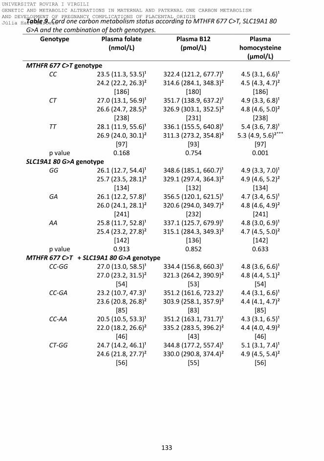
133
Table 9. Cord one carbon metabolism status according to MTHFR 677 C>T, SLC19A1 80 G>A and the combination of both genotypes.
Genotype Plasma folate (nmol/L)
Plasma B12 (pmol/L)
Plasma homocysteine
(µmol/L)
MTHFR 677 C>T genotype CC 23.5 (11.3, 53.5)¹ 322.4 (121.2, 677.7)¹ 4.5 (3.1, 6.6)¹ 24.2 (22.2, 26.3)²
[186] 314.6 (284.1, 348.3)²
[180] 4.5 (4.3, 4.7)²
[186] CT 27.0 (13.1, 56.9)¹ 351.7 (138.9, 637.2)¹ 4.9 (3.3, 6.8)¹ 26.6 (24.7, 28.5)²
[238] 326.9 (303.1, 352.5)²
[231] 4.8 (4.6, 5.0)²
[238] TT 28.1 (11.9, 55.6)¹ 336.1 (155.5, 640.8)¹ 5.4 (3.6, 7.8)¹ 26.9 (24.0, 30.1)²
[97] 311.3 (273.2, 354.8)²
[93] 5.3 (4.9, 5.6)²***
[97] p value 0.168 0.754 0.001
SLC19A1 80 G>A genotype GG 26.1 (12.7, 54.4)¹ 348.6 (185.1, 660.7)¹ 4.9 (3.3, 7.0)¹ 25.7 (23.5, 28.1)²
[134] 329.1 (297.4, 364.3)²
[132] 4.9 (4.6, 5.2)²
[134] GA 26.1 (12.2, 57.8)¹ 356.5 (120.1, 621.5)¹ 4.7 (3.4, 6.5)¹ 26.0 (24.1, 28.1)²
[241] 320.6 (294.0, 349.7)²
[232] 4.8 (4.6, 4.9)²
[241] AA 25.8 (11.7, 52.8)¹ 337.1 (125.7, 679.9)¹ 4.8 (3.0, 6.9)¹ 25.4 (23.2, 27.8)²
[142] 315.1 (284.3, 349.3)²
[136] 4.7 (4.5, 5.0)²
[142] p value 0.913 0.852 0.633
MTHFR 677 C>T + SLC19A1 80 G>A genotype CC-GG 27.0 (13.0, 58.5)¹ 334.4 (156.8, 660.3)¹ 4.8 (3.6, 6.6)¹ 27.0 (23.2, 31.5)²
[54] 321.3 (264.2, 390.9)²
[53] 4.8 (4.4, 5.1)²
[54] CC-GA 23.2 (10.7, 47.3)¹ 351.2 (161.6, 723.2)¹ 4.4 (3.1, 6.6)¹ 23.6 (20.8, 26.8)²
[85] 303.9 (258.1, 357.9)²
[83] 4.4 (4.1, 4.7)²
[85] CC-AA 20.5 (10.5, 53.3)¹ 351.2 (163.1, 731.7)¹ 4.3 (3.1, 6.5)¹ 22.0 (18.2, 26.6)²
[46] 335.2 (283.5, 396.2)²
[43] 4.4 (4.0, 4.9)²
[46] CT-GG 24.7 (14.2, 46.1)¹ 344.8 (177.2, 557.4)¹ 5.1 (3.1, 7.4)¹ 24.6 (21.8, 27.7)²
[56] 330.0 (290.8, 374.4)²
[55] 4.9 (4.5, 5.4)²
[56]
UNIVERSITAT ROVIRA I VIRGILI GENETIC AND METABOLIC ALTERATIONS IN MATERNAL AND PATERNAL ONE CARBON METABOLISM AND DEVELOPMENT OF PREGNANCY COMPLICATIONS OF PLACENTAL ORIGIN Júlia Haro Barceló

134
Table 9. Cord one carbon metabolism status according to MTHFR 677 C>T, SLC19A1 80 G>A and the combination of both genotypes.
Genotype Plasma folate (nmol/L)
Plasma B12 (pmol/L)
Plasma homocysteine
(µmol/L)
CT-GA 28.4 (12.6, 67.0)¹ 382.8 (133.8, 641.4)¹ 4.8 (3.6, 6.4)¹ 28.3 (25.0, 32.0)²
[107] 337.0 (297.1, 382.3)²
[103] 4.8 (4.6, 5.1)²
[107] CT-AA 27.7 (13.2, 50.4)¹ 329.8 (123.4, 642.5)¹ 4.8 (2.9, 6.9)¹ 26.0 (23.3, 29.1)²
[71] 317.2 (276.2, 364.2)²
[69] 4.7 (4.4, 5.2)²
[71] TT-GG 27.6 (8.9, 53.6)¹ 338.4 (161.1, 711.6)¹ 5.1 (3.7, 8.4)¹ 25.5 (19.7, 33.1)²
[24] 345.0 (272.0, 437.5)²
[24] 5.1 (4.3, 6.0)²
[24] TT-GA 25.2 (11.8, 48.7)¹ 334.5 (134.7, 613.1)¹ 5.3 (3.5, 7.8)¹ 25.4 (21.6, 30.0)²
[47] 310.6 (259.9, 371.0)² †
[44]
5.3 (4.8, 5.8)² [47]
TT-AA 30.2 (15.3, 65.7)¹ 327.0 (55.0, 722.0)¹ 5.7 (3.0, 8.1)¹ 30.7 (24.8, 38.0)²
[25] 277.1 (198.7, 386.4)²
[24] 5.4 (4.7, 6.1)²
[25] p value 0.180 0.917 0.025
Abbreviations: MTHFR, methylenetetrahydrofolate reductase; SLC19A1, solute carrier family 19
member 1.1 Median (P10, P90). 2 Geometric mean (95% confidence interval) [n]. Geometric means
were compared between genotypes using ANOVA, with post hoc Bonferroni correction for multiple comparisons. ***p<0.001 TT compared to CC genotype. †p<0.05 TT/GA compared to CC/GA genotype.
UNIVERSITAT ROVIRA I VIRGILI GENETIC AND METABOLIC ALTERATIONS IN MATERNAL AND PATERNAL ONE CARBON METABOLISM AND DEVELOPMENT OF PREGNANCY COMPLICATIONS OF PLACENTAL ORIGIN Júlia Haro Barceló
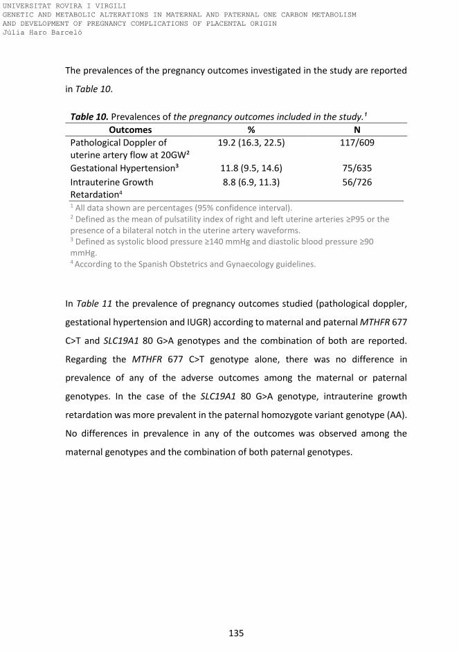
135
The prevalences of the pregnancy outcomes investigated in the study are reported
in Table 10.
Table 10. Prevalences of the pregnancy outcomes included in the study.¹
Outcomes % N
Pathological Doppler of uterine artery flow at 20GW²
19.2 (16.3, 22.5) 117/609
Gestational Hypertension³ 11.8 (9.5, 14.6) 75/635
Intrauterine Growth Retardation4
8.8 (6.9, 11.3) 56/726
1 All data shown are percentages (95% confidence interval). 2 Defined as the mean of pulsatility index of right and left uterine arteries ≥P95 or the presence of a bilateral notch in the uterine artery waveforms. 3 Defined as systolic blood pressure ≥140 mmHg and diastolic blood pressure ≥90 mmHg. 4 According to the Spanish Obstetrics and Gynaecology guidelines.
In Table 11 the prevalence of pregnancy outcomes studied (pathological doppler,
gestational hypertension and IUGR) according to maternal and paternal MTHFR 677
C>T and SLC19A1 80 G>A genotypes and the combination of both are reported.
Regarding the MTHFR 677 C>T genotype alone, there was no difference in
prevalence of any of the adverse outcomes among the maternal or paternal
genotypes. In the case of the SLC19A1 80 G>A genotype, intrauterine growth
retardation was more prevalent in the paternal homozygote variant genotype (AA).
No differences in prevalence in any of the outcomes was observed among the
maternal genotypes and the combination of both paternal genotypes.
UNIVERSITAT ROVIRA I VIRGILI GENETIC AND METABOLIC ALTERATIONS IN MATERNAL AND PATERNAL ONE CARBON METABOLISM AND DEVELOPMENT OF PREGNANCY COMPLICATIONS OF PLACENTAL ORIGIN Júlia Haro Barceló

136
Table 11. Pregnancy outcomes prevalence according to maternal and paternal MTHFR 677 C>T, SLC19A1 80 G>A and the combination of
both genotypes¹.
Pathological Doppler2 Gestational hypertension3 IUGR4
Maternal Paternal Maternal Paternal Maternal Paternal
MTHFR 677 C>T genotype
CC 21.1 (16.0, 27.3)
[42/199]
15.2 (10.0, 22.2)
[20/132]
12.8 (8.9, 18.1)
[26/203]
8.9 (5.2, 14.9)
[12/135]
8.2 (5.3, 12.4)
[19/233]
8.6 (5.0, 14.5)
[12/139]
CT 20.7 (16.3, 26.0)
[56/270]
18.8 (13.8, 25.1)
[34/181]
30.1 (22.1, 39.5)
[31/308]
12.8 (8.6, 18.6)
[22/172]
7.0 (4.8, 10.2)
[24/342]
5.2 (2.9, 9.4)
[10/191]
TT 17.5 (11.3, 25.9)
[18/103]
24.5 (14.6, 38.1)
[12/49]
12.8 (7.5, 21.0)
[12/94]
12.0 (5.6, 23.8)
[6/50]
10.4 (6.1, 17.4)
[12/115]
9.3 (4.0, 19.9)
[5/54]
SLC19A1 80 G>A genotype
GG 20.1 (14.6, 27.2)
[31/154]
13.0 (7.8, 21.0)
[13/100]
11.8 (7.7, 17.7)
[19/161]
7.8 (4.0, 14.7)
[8/102]
8.1 (4.9, 12.9)
[15/186]
6.6 (3.2, 13.0)
[7/106]
GA 19.6 (15.3, 24.8)
[52/265]
18.0 (12.7, 24.9)
[27/150]
10.1 (7.1, 14.1)
[29/287]
10.1 (6.2, 16.0)
[15/148]
7.9 (5.4, 11.4)
[25/317]
3.1 (1.3, 7.0)
[5/163]
AA 21.2 (15.4, 28.4)
[32/151]
21.8 (15.1, 30.4)
[24/110]
13.5 (9.0, 19.7)
[21/156]
16.3 (10.5, 24.6)
[17/104]
8.2 (5.0, 13.0)
[15/184]
13.4 (8.3, 20.9)**
[15/112]
UNIVERSITAT ROVIRA I VIRGILI GENETIC AND METABOLIC ALTERATIONS IN MATERNAL AND PATERNAL ONE CARBON METABOLISM AND DEVELOPMENT OF PREGNANCY COMPLICATIONS OF PLACENTAL ORIGIN Júlia Haro Barceló

137
Pathological Doppler2 Gestational hypertension3 IUGR4
Maternal Paternal Maternal Paternal Maternal Paternal
MTHFR 677 C>T + SLC19A1 80 G>A genotype
CC-GG 13.1 (6.8, 23.8)
[8/61]
11.1 (4.4, 25.3)
[4/36]
10.6 (5.2, 20.3)
[7/66]
5.6 (1.5, 18.1)
[2/36]
5.5 (2.2, 13.3)
[4/73]
10.5 (4.2, 24.1)
[4/38]
CC-GA 24.4 (16.7, 34.2)
[22/90]
17.7 (10.2, 29.0)
[11/62]
11.0 (6.1, 19.1)
[10/91]
9.2 (4.3, 18.7)
[6/65]
6.8 (3.3, 13.4)
[7/103]
4.5 (1.5, 12.4)
[3/67]
CC-AA 25.0 (14.9, 38.8)
[12/48]
14.3 (6.3, 29.4)
[5/35]
19.6 (10.7, 33.2)
[9/46]
11.4 (4.5, 26.0)
[4/35]
14.0 (7.3, 25.3)
[8/57]
14.3 (6.3, 29.4)
[5/35]
CT-GG 25.0 (16.0, 36.8)
[16/64]
16.3 (8.5, 29.0)
[8/49]
12.7 (6.8, 22.4)
[9/71]
10.4 (4.5, 22.2)
[5/48]
7.5 (3.5, 15.4)
[6/80]
4.0 (1.1, 13.5)
[2/50]
CT-GA 19.1 (13.4, 26.5)
[26/136]
15.8 (9.3, 25.6)
[12/76]
8.3 (4.9, 13.7)
[13/156]
11.3 (5.8, 20.7)
[8/71]
8.2 (4.9, 13.3)
[14/171]
1.2 (0.2, 6.6)
[1/82]
CT-AA 19.1 (11.5, 30.0)
[13/68]
21.8 (12.9, 34.4)
[12/55]
11.3 (6.0, 20.0)
[9/80]
17.6 (9.6, 30.3)
[9/51]
4.5 (1.8, 11.1)
[4/88]
12.3 (6.1, 23.2)
[7/57]
TT-GG 24.1 (12.2, 42.1)
[7/29]
6.7 (1.2, 29.8)
[1/15]
12.5 (4.3, 31.0)
[3/24]
5.6 (1.0, 25.8)
[1/18]
15.2 (6.7, 30.9)
[5/33]
5.6 (1.0, 25.8)
[1/18]
TT-GA 10.3 (4.1, 23.6)
[4/39]
28.6 (11.7, 54.6)
[4/14]
15.0 (7.1, 29.1)
[6/40]
7.1 (1.3, 31.5)
[1/14]
9.3 (3.7, 21.6)
[4/43]
6.3 (1.1, 28.3)
[1/16]
TT-AA 20.0 (10.0, 35.9)
[7/35]
35.0 (18.1, 56.7)
[7/20]
10.0 (3.5, 25.6)
[3/30]
22.2 (9.0, 45.2)
[4/18]
7.7 (2.7, 20.3)
[3/39]
15.0 (5.2, 36.0)
[3/20]
Abbreviations: MTHFR, methylenetetrahydrofolate reductase; SLC19A1, solute carrier family 19 member 1. 1All data shown are percentages (95% confidence interval). 2 Defined as the mean of pulsatility index of right and left uterine arteries ≥P95 or the presence of a bilateral notch. 3 Defined as systolic blood pressure ≥140 mmHg and diastolic blood pressure ≥90 mmHg. 4 According to the Spanish Obstetrics and Gynaecology guidelines. Prevalence of the outcomes among the genotypes for each polymorphism were compared by the chi-square test. **P<0.01 TT compared to CC genotype.
UNIVERSITAT ROVIRA I VIRGILI GENETIC AND METABOLIC ALTERATIONS IN MATERNAL AND PATERNAL ONE CARBON METABOLISM AND DEVELOPMENT OF PREGNANCY COMPLICATIONS OF PLACENTAL ORIGIN Júlia Haro Barceló
138
4.2. One carbon metabolism and uterine artery pulsatility index
Maternal MTHFR 677C>T genotype, uterine artery resistance (pulsatility
index) and impaired placentation (pathological doppler measurements of
uterine artery flow and waveforms).
We investigated the association between maternal MTHFR 677C>T genotype and
pulsatility index of the uterine arteries at 20 GW (dependent variable natural log
transformed mean of the right and left uterine artery pulsatility index) using
multiple linear regression analysis. The models included SLC19A1 80G>A genotype,
and red blood cell folate and plasma cobalamin concentrations (at <12 GW and 15
GW, in separate models). They were further adjusted for smoking habit during
pregnancy, socioeconomic status, age, 1st trimester BMI, parity and gestational age
in weeks and days at the time of the doppler measurement. None of the models
were significant (data not shown). Impaired placentation is characterised as
pathological when the mean uterine artery pulsatility index is elevated (≥P95) and/
or by the presence of bilateral notches in the uterine artery waveforms analysed by
Doppler echography. Analysis of the association between maternal MTHFR
genotype and pathological placentation using multiple logistic regression analysis in
which the dependent variable was pathological Doppler outcome, and adjusting the
models for the same confounding factors as described for uterine artery pulsatility
index, also showed that there was no association.
Maternal tHcy status and pulsatility index of uterine arteries
We studied the association between early pregnancy tHcy at <12 and 15 GW, in
separate models) and pulsatility index of uterine arteries using multiple linear
regression analysis. The dependent variable was natural log transformed mean right
UNIVERSITAT ROVIRA I VIRGILI GENETIC AND METABOLIC ALTERATIONS IN MATERNAL AND PATERNAL ONE CARBON METABOLISM AND DEVELOPMENT OF PREGNANCY COMPLICATIONS OF PLACENTAL ORIGIN Júlia Haro Barceló
139
and left uterine artery pulsatility index. THcy was included in the models and they
were adjusted for smoking during the first trimester only versus never, smoking
throughout pregnancy versus never, mid and high versus low socioeconomic status,
maternal age, 1st trimester BMI, parity (previous pregnancy <20 GW versus none)
and gestational weeks at doppler measurement. The models were not significant.
We repeated the analysis using the variable elevated tHcy status during early
pregnancy (≥P90). While the model was not significant, the association between
tHcy ≥P90 and pulsatility index was positive with a beta coefficient of 0.075 (P=
0.059). We then investigated whether elevated early pregnancy tHcy was associated
with impaired placentation (pathological measurement by Doppler echography at
20 GW) using multiple logistic regression analysis. The categorical tHcy variable
(≥P90 tHcy in early pregnancy versus <P90) replaced the genotypes, red blood cell
folate and plasma cobalamin variables described in the models in the previous
section at <12 and at 15 GW. The models were adjusted for smoking habit,
socioeconomic status, BMI, age, parity and gestational age at doppler
measurement. There was no association between elevated early pregnancy tHcy
status and impaired placentation.
Paternal genotype and uterine artery pulsatility index
The association between paternal MTHFR 677C>T genotype and uterine artery
pulsatility index was studied using multiple linear regression analysis. We performed
the same maternal models, as at <12 and 15 GW, adding paternal MTHFR 677C>T,
red blood cell folate concentration, age and smoking habit. The results are shown in
Table 12.
UNIVERSITAT ROVIRA I VIRGILI GENETIC AND METABOLIC ALTERATIONS IN MATERNAL AND PATERNAL ONE CARBON METABOLISM AND DEVELOPMENT OF PREGNANCY COMPLICATIONS OF PLACENTAL ORIGIN Júlia Haro Barceló

140
Table 12. Predictors (Beta coefficients) of pulsatility index of uterine arteries according to paternal MTHFR 677 C>T genotype.
< 12 GW 15 GW
Model 1 (maternal, non adjusted)
Model, R2, n 0.004, 310
-0.010, 186
Maternal MTHFR 677TT genotype
0.027 -0.102
Model 2 (maternal, adjusted)
Model, R2, n 0.024, 310#
0.068, 186*
Maternal MTHFR 677TT genotype
0.011 -0.083
Maternal smoking throughout pregnancy vs never
-0.088 -0.154*
Maternal BMI 0.005 0.207** Model 3 (maternal adjusted and paternal factors)
Model, R2, n 0.057, 310**
0.149, 186***
Maternal MTHFR 677TT genotype
0.007 -0.088
Maternal smoking throughout pregnancy vs never
-0.130* -0.168*
Maternal BMI -0.011 0.179* Paternal MTHFR 677CT genotype 0.161** 0.205** Paternal MTHFR 677TT genotype 0.131* 0.226** Paternal age 0.126 0.084 Paternal smoking 0.109† 0.167* Abbreviations: MTHFR, methylenetetrahydrofolate reductase; BMI, body mass index; GW,
gestational weeks. Excluded 2 outliers. Multiple linear regression analysis. Dependent variable,
natural log transformed mean of the uterine artery pulsatility index of uterine arteries (PI).
Independent variable for Model 1: maternal MTHFR 677 CT and TT versus MTHFR 677 CC genotype,
maternal SLC19A1 80 GA and AA versus 80 GG genotype, low tertile red blood cell folate (<= 724.33
nmol/L) and low tertile plasma cobalamin (<= 315.45 pmol/L) concentration at the corresponding
time points of pregnancy for each model. Model 2: same variables as model 1 as well as smoking
during the first trimester and throughout pregnancy versus never, mid and high versus low
socioeconomic status, maternal age (y), 1st trimester BMI and parity (previous pregnancy versus
none). Model 3 (paternal factors): same variables as model 2 as well as paternal MTHFR 677 CT and
TT versus MTHFR 677CC genotype, red blood cell folate levels, age and smoking. *p<0.05, **p<0.01. #P= 0.086, †P= 0.067
UNIVERSITAT ROVIRA I VIRGILI GENETIC AND METABOLIC ALTERATIONS IN MATERNAL AND PATERNAL ONE CARBON METABOLISM AND DEVELOPMENT OF PREGNANCY COMPLICATIONS OF PLACENTAL ORIGIN Júlia Haro Barceló

141
The results show a positive association between paternal MTHFR 677 CT and TT vs
CC genotypes and pulsatility index of uterine arteries in all models. Paternal smoking
habit was also positively associated with uterine artery pulsatility index at 20 GW.
As we observed before, maternal genotype was not associated. Maternal BMI was
positively associated and smoking throughout pregnancy versus never was
negatively associated with uterine artery pulsatility index.
Paternal tHcy status and uterine artery pulsatility index
The association between paternal tHcy and mean left and right uterine artery
pulsatility index was investigated using multiple linear regression analysis. The
dependent variable was the natural log transformed mean pulsatility index. We built
on the maternal models by adding paternal tHcy concentrations, age and smoking
habit. Paternal tHcy was positively associated with pulsatility index at <12 and 15
GW. There was no association between maternal tHcy and pulsatility index.
When we repeated the analysis including elevated tHcy (≥P90 versus <P90) at <12
GW and elevated paternal tHcy , the model was also significant (n= 364, Adjusted R2
=0.044, P= 0.005) and the results indicate a positive association between elevated
paternal tHcy and increased uterine artery pulsatility index (paternal tHcy ≥P90 Beta
coefficient= 0.151, P= 0.004). Paternal smoking compared to nonsmoking was also
associated with increased uterine artery pulsatility index (Beta coefficient= 0.102,
P= 0.060).
UNIVERSITAT ROVIRA I VIRGILI GENETIC AND METABOLIC ALTERATIONS IN MATERNAL AND PATERNAL ONE CARBON METABOLISM AND DEVELOPMENT OF PREGNANCY COMPLICATIONS OF PLACENTAL ORIGIN Júlia Haro Barceló

142
Paternal genotype and pathological Doppler measurement of uterine
arteries at 20 GW
We studied the association between paternal MTHFR 677C>T genotype and risk of
pathological Doppler measurement of uterine arteries at 20 GW using multiple
logistic regression analysis. We added the paternal factors MTHFR 677C>T
genotype, age, smoking habit and red blood cell folate concentration to the same
models used for the mothers, at <12 and 15 GW. The first model was not significant.
Results from the model at 15 GW are shown.
Table 13. Associations between paternal MTHFR 677C>T genotype and pathological Doppler measurement of the uterine arteries at 20 GW.
N R2 Maternal TT vs CC
Paternal CT vs CC
Paternal TT vs CC
Paternal smoking
Model 1 (maternal, non adjusted)
191 0.130* 0 - - -
Model 2 (maternal, adjusted)
191 0.178# 0 - - -
Model 3 (maternal adjusted and paternal factors)
192 0.358*** 0 4.0 (1.3, 12.6)
7.1 (1.6, 32.8)
3.2 (1.3, 7.9)
Abbreviations: MTHFR, methylenetetrahydrofolate reductase. Multiple logistic regression analysis was used. Nagelkerke R2; OR (95% CI) for pathological Doppler measurement of the uterine arteries at 20 GW in mothers with MTHFR 677 CC vs CT and MTHFR 677 CC vs TT genotype are shown. Model 1: (basic model) comparing MTHFR 677 CC vs CT and MTHFR 677 CC vs TT and SLC19A1 80GG vs GA and SLC19A1 80GG vs AA maternal genotypes. Model 2: Included the same variables as model 1 as well as lifestyle factors such as parity (previous pregnancy versus none), maternal age (y), 1st trimester BMI, smoking during pregnancy, mid and high versus low socioeconomic status, gestational age at doppler measurement, low tertile red blood cell folate (<= 724.33 nmol/L) and low tertile plasma cobalamin (<= 315.45 pmol/L) concentration at 15 GW. Model 3: Included the same variables as model 2 as well as paternal MTHFR 677 CC vs CT and MTHFR 677 CC vs TT genotype, paternal red blood cell levels, age and paternal smoking habit. *p<0.05, **p<0.01. #P=0.051.
UNIVERSITAT ROVIRA I VIRGILI GENETIC AND METABOLIC ALTERATIONS IN MATERNAL AND PATERNAL ONE CARBON METABOLISM AND DEVELOPMENT OF PREGNANCY COMPLICATIONS OF PLACENTAL ORIGIN Júlia Haro Barceló

143
There is a clear association between paternal factors and increased risk of
pathological Doppler assessment of the uterine arteries at 20 GW. Paternal MTHFR
677 CT and TT were associated with four and seven times, respectively, more risk of
pregnancy affected by impaired placentation compared to fathers with the MTHFR
677 CC genotype. Paternal smoking also conveyed a threefold greater risk. Paternal
red blood cell folate had a small but significant protective effect against impaired
placentation OR (95% CI) 0.99 (0.99, 0.99).
Paternal tHcy status and pathological Doppler measurement of the uterine
arteries at 20 GW
We investigated whether elevated paternal tHcy status is associated with risk of
pathological Doppler measurement of the uterine arteries at 20 GW. The categorical
maternal tHcy variable (≥P90 tHcy in early pregnancy versus <P90) replaced
genotypes, red blood cell folate and plasma cobalamin variables in each model,
described in the previous section for <12 and 15 GW. We included elevated paternal
tHcy status (≥P90) and smoking habit in both models. The results are shown below
in Table 14.
UNIVERSITAT ROVIRA I VIRGILI GENETIC AND METABOLIC ALTERATIONS IN MATERNAL AND PATERNAL ONE CARBON METABOLISM AND DEVELOPMENT OF PREGNANCY COMPLICATIONS OF PLACENTAL ORIGIN Júlia Haro Barceló
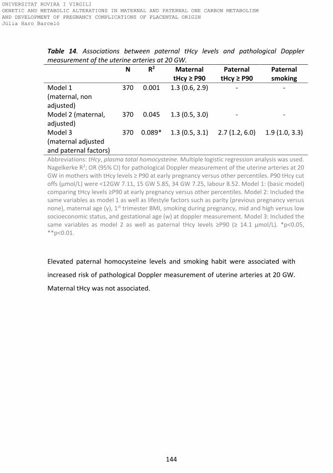
144
Table 14. Associations between paternal tHcy levels and pathological Doppler measurement of the uterine arteries at 20 GW.
N R2 Maternal tHcy ≥ P90
Paternal tHcy ≥ P90
Paternal smoking
Model 1 (maternal, non adjusted)
370 0.001 1.3 (0.6, 2.9) - -
Model 2 (maternal, adjusted)
370 0.045 1.3 (0.5, 3.0) - -
Model 3 (maternal adjusted and paternal factors)
370 0.089* 1.3 (0.5, 3.1) 2.7 (1.2, 6.0) 1.9 (1.0, 3.3)
Abbreviations: tHcy, plasma total homocysteine. Multiple logistic regression analysis was used. Nagelkerke R2; OR (95% CI) for pathological Doppler measurement of the uterine arteries at 20 GW in mothers with tHcy levels ≥ P90 at early pregnancy versus other percentiles. P90 tHcy cut offs (μmol/L) were <12GW 7.11, 15 GW 5.85, 34 GW 7.25, labour 8.52. Model 1: (basic model) comparing tHcy levels ≥P90 at early pregnancy versus other percentiles. Model 2: Included the same variables as model 1 as well as lifestyle factors such as parity (previous pregnancy versus none), maternal age (y), 1st trimester BMI, smoking during pregnancy, mid and high versus low socioeconomic status, and gestational age (w) at doppler measurement. Model 3: Included the same variables as model 2 as well as paternal tHcy levels ≥P90 (≥ 14.1 µmol/L). *p<0.05, **p<0.01.
Elevated paternal homocysteine levels and smoking habit were associated with
increased risk of pathological Doppler measurement of uterine arteries at 20 GW.
Maternal tHcy was not associated.
UNIVERSITAT ROVIRA I VIRGILI GENETIC AND METABOLIC ALTERATIONS IN MATERNAL AND PATERNAL ONE CARBON METABOLISM AND DEVELOPMENT OF PREGNANCY COMPLICATIONS OF PLACENTAL ORIGIN Júlia Haro Barceló
145
4.3. One carbon metabolism and birthweight
Association between maternal genotype and birthweight
We investigated the association between maternal MTHFR 677C>T genotype and
birthweight using multiple linear regression analysis. 3 models were designed to
adjust for maternal folate and cobalamin status at different times of pregnancy (first
trimester, mid-pregnancy, late pregnancy). We included the interaction term
MTHFR 677C>T genotype * smoking habit during pregnancy in the model. This was
significant in the 1st trimester (P=0.024). Therefore, we proceeded to carry out
stratified analyses according to smoking habit (never smoking or smoking at some
point of pregnancy). We excluded participants with gestational diabetes from these
analyses. The results are shown in Table 15.
UNIVERSITAT ROVIRA I VIRGILI GENETIC AND METABOLIC ALTERATIONS IN MATERNAL AND PATERNAL ONE CARBON METABOLISM AND DEVELOPMENT OF PREGNANCY COMPLICATIONS OF PLACENTAL ORIGIN Júlia Haro Barceló
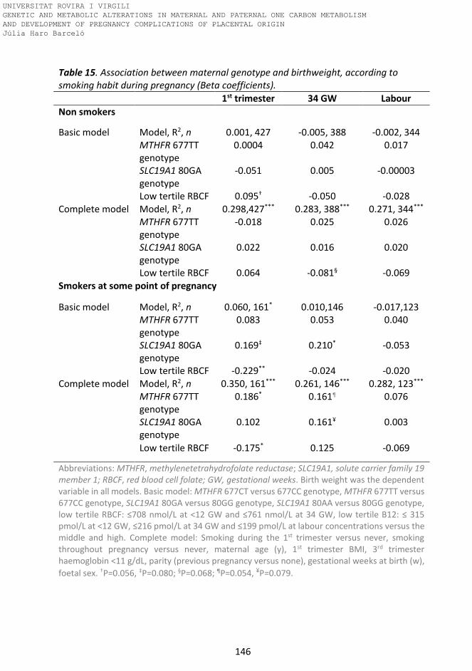
146
Table 15. Association between maternal genotype and birthweight, according to smoking habit during pregnancy (Beta coefficients).
1st trimester 34 GW Labour
Non smokers
Basic model Model, R2, n 0.001, 427 -0.005, 388 -0.002, 344 MTHFR 677TT
genotype 0.0004 0.042 0.017
SLC19A1 80GA genotype
-0.051 0.005 -0.00003
Low tertile RBCF 0.095† -0.050 -0.028 Complete model Model, R2, n 0.298,427*** 0.283, 388*** 0.271, 344*** MTHFR 677TT
genotype -0.018 0.025 0.026
SLC19A1 80GA genotype
0.022 0.016 0.020
Low tertile RBCF 0.064 -0.081§ -0.069 Smokers at some point of pregnancy
Basic model Model, R2, n 0.060, 161* 0.010,146 -0.017,123 MTHFR 677TT
genotype 0.083 0.053 0.040
SLC19A1 80GA genotype
0.169‡ 0.210* -0.053
Low tertile RBCF -0.229** -0.024 -0.020 Complete model Model, R2, n 0.350, 161*** 0.261, 146*** 0.282, 123*** MTHFR 677TT
genotype 0.186* 0.161¶ 0.076
SLC19A1 80GA genotype
0.102 0.161¥ 0.003
Low tertile RBCF -0.175* 0.125 -0.069
Abbreviations: MTHFR, methylenetetrahydrofolate reductase; SLC19A1, solute carrier family 19 member 1; RBCF, red blood cell folate; GW, gestational weeks. Birth weight was the dependent variable in all models. Basic model: MTHFR 677CT versus 677CC genotype, MTHFR 677TT versus 677CC genotype, SLC19A1 80GA versus 80GG genotype, SLC19A1 80AA versus 80GG genotype, low tertile RBCF: ≤708 nmol/L at <12 GW and ≤761 nmol/L at 34 GW, low tertile B12: ≤ 315 pmol/L at <12 GW, ≤216 pmol/L at 34 GW and ≤199 pmol/L at labour concentrations versus the middle and high. Complete model: Smoking during the 1st trimester versus never, smoking throughout pregnancy versus never, maternal age (y), 1st trimester BMI, 3rd trimester haemoglobin <11 g/dL, parity (previous pregnancy versus none), gestational weeks at birth (w),
foetal sex. †P=0.056, ‡P=0.080; §P=0.068; ¶P=0.054, ¥P=0.079.
UNIVERSITAT ROVIRA I VIRGILI GENETIC AND METABOLIC ALTERATIONS IN MATERNAL AND PATERNAL ONE CARBON METABOLISM AND DEVELOPMENT OF PREGNANCY COMPLICATIONS OF PLACENTAL ORIGIN Júlia Haro Barceló

147
The early pregnancy model showed that the MTHFR 677TT genotype was not
associated with birth weight in non smokers but was positively associated with birth
weight in smokers. A positive association between the SLC19A1 80GA genotype and
birth weight was also observed in the basic model but weakened in the adjusted
model. Low tertile red blood cell folate concentration was inversely associated with
birthweight in smokers only. Both genotype – birth weight associations were
confirmed in the mid-pregnancy model.
Maternal red blood cell folate status and cord plasma folate status according to
maternal smoking habit and genotype are shown in Table 16. Red blood cell folate
status was lower in smokers at <12, 24-27 and 34 GW than in non smokers. Red
blood cell folate status was lower in non-smoking mothers with the MTHFR 677TT
genotype compared to those with the CC and CT genotype at <12, 24-27 and 34 GW.
However, there was no difference in red blood cell folate status between genotypes
in smoking mothers. Cord folate status did not differ among genotypes in any of the
mothers or between smokers and non smokers.
UNIVERSITAT ROVIRA I VIRGILI GENETIC AND METABOLIC ALTERATIONS IN MATERNAL AND PATERNAL ONE CARBON METABOLISM AND DEVELOPMENT OF PREGNANCY COMPLICATIONS OF PLACENTAL ORIGIN Júlia Haro Barceló

148
Table 16. Maternal red blood cell folate and cord plasma folate status according to smoking habit and MTHFR 677 C>T genotype.
<12 GW 15 GW 24-27 GW 34 GW Cord
Non smokers 981 (936, 1027) [546]
1283 (1213, 1357) [313]
1184 (1131, 1241) [475]
1054 (960, 1158) [457]
26.2 (24.7, 27.7) [428]
CC 1047 (970, 1131) [167]
1311 (1311, 1311) [96]
1251 (1159, 1350) [150]
1028 (957, 1103) [135]
25.8 (25.7, 25.9) [133]
CT 979 (979, 979) [257]
1312 (1312, 1312) [155]
1169 (1168, 1169) [224]
1029 (1029, 1029) [220]
24.7 (24.6, 24.8) [208]
TT 880† (773, 1001) [85]
1245 (1070, 1449) [59]
1030† (905, 1173) [74]
853† ᶴ (732, 995) [72]
27.6 (27.5, 27.8) [69]
Smokers 853** (788, 924) [191]
1165 (1074, 1265) [133]
1057** (981, 1141) [176]
853*** (783, 929) [159]
25.4 (23.3, 27.8) [149]
CC 917 (916, 917) [67]
1231 (1231, 1231) [50]
1092 (1092, 1092) [62]
873 (873, 873) [59]
24.4 (24.2, 24.5) [58]
CT 822 (822, 822) [85]
1156 (1155, 1156) [59]
1038.1 (1037.9, 1038.2) [77]
864 (864, 864) [71]
25.2 (25.1, 25.4) [65]
TT 787 (787, 787) [34]
1061 (1061, 1061) [24]
994 (994, 994) [33]
789 (788, 789) [29]
28.0 (27.8, 28.1) [25]
Abbreviations: MTHFR, methylenetetrahydrofolate reductase; GW, Gestational weeks. Geometric mean (95% confidence interval) [n]. Geometric means were compared between groups (non smokers versus smokers) and between genotypes using ANOVA, with post hoc Bonferroni correction for multiple comparisons. *P<0.05, **p<0.01 ***p<0.001 smokers compared to non smokers. †p<0.05, ††p<0.01 TT compared to CC genotype. ᶴp<0.05, TT compared to CT genotype.
UNIVERSITAT ROVIRA I VIRGILI GENETIC AND METABOLIC ALTERATIONS IN MATERNAL AND PATERNAL ONE CARBON METABOLISM AND DEVELOPMENT OF PREGNANCY COMPLICATIONS OF PLACENTAL ORIGIN Júlia Haro Barceló

149
Maternal tHcy levels predicting birthweight
Applying the same stratification for smoking during pregnancy as in the
MTHFR 677C>T models, we also studied the association between early and
late pregnancy tHcy and birthweight, using multiple linear regression analysis.
For early pregnancy we tested the association between maternal tHcy ≥P90
(7.1 µmol/L at <12 GW or 5.9 µmol/L at 15 GW) versus <P90 with birthweight.
In late pregnancy at (34 GW) and at labour we tested tHcy as a continuous
variable. The tHcy variables replaced the genotypes and red blood cell folate
and plasma cobalamin variables in each model. All the models were significant
and the adjusted R2 ranged from 0.288 to 0.448. The results were confirmed
when limited to the pregnancies in which the father participated in the study.
These results are reported together with the fathers in the section on paternal
tHcy and birth weight below (Table 18). In non-smokers, late pregnancy tHcy
was positively associated with birthweight.
Paternal genotype predicting birthweight
We then investigated the association between paternal MTHFR 677C>T
genotype and birthweight building on the same multiple linear regression
model shown for the mothers by adding a final model including paternal
MTHFR 677C>T genotype, plasma cobalamin and red blood cell folate
concentrations. The results are shown in Table 17.
UNIVERSITAT ROVIRA I VIRGILI GENETIC AND METABOLIC ALTERATIONS IN MATERNAL AND PATERNAL ONE CARBON METABOLISM AND DEVELOPMENT OF PREGNANCY COMPLICATIONS OF PLACENTAL ORIGIN Júlia Haro Barceló
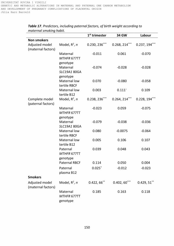
150
Table 17. Predictors, including paternal factors, of birth weight according to maternal smoking habit.
1st trimester 34 GW Labour
Non smokers Adjusted model (maternal factors)
Model, R2, n 0.230, 236*** 0.268, 214*** 0.237, 194***
Maternal MTHFR 677TT genotype
-0.011 0.061 -0.070
Maternal SLC19A1 80GA genotype
-0.074 -0.028 -0.028
Maternal low tertile RBCF
0.070 -0.080 -0.058
Maternal low tertile B12
0.003 0.111‡ 0.109
Complete model (paternal factors)
Model, R2, n 0.238, 236*** 0.264, 214*** 0.228, 194***
Maternal MTHFR 677TT genotype
-0.023 0.059 -0.075
Maternal SLC19A1 80GA
-0.079 -0.038 -0.036
Maternal low tertile RBCF
0.080 -0.0075 -0.064
Maternal low tertile B12
0.005 0.106 0.107
Paternal MTHFR 677TT genotype
0.039 0.048 0.043
Paternal RBCF 0.114 0.050 0.004
Paternal plasma B12
0.025† -0.012 -0.023
Smokers
Adjusted model (maternal factors)
Model, R2, n 0.422, 66** 0.402, 60*** 0.429, 51**
Maternal MTHFR 677TT genotype
0.185 0.163 0.118
UNIVERSITAT ROVIRA I VIRGILI GENETIC AND METABOLIC ALTERATIONS IN MATERNAL AND PATERNAL ONE CARBON METABOLISM AND DEVELOPMENT OF PREGNANCY COMPLICATIONS OF PLACENTAL ORIGIN Júlia Haro Barceló
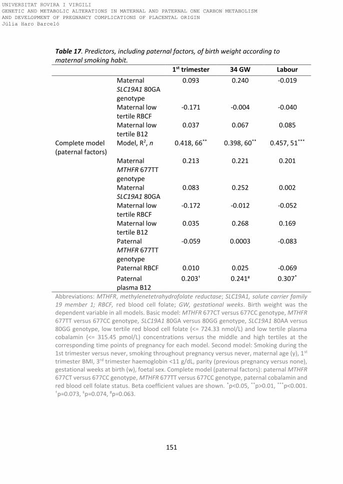
151
Table 17. Predictors, including paternal factors, of birth weight according to maternal smoking habit.
1st trimester 34 GW Labour
Maternal SLC19A1 80GA genotype
0.093 0.240 -0.019
Maternal low tertile RBCF
-0.171 -0.004 -0.040
Maternal low tertile B12
0.037 0.067 0.085
Complete model (paternal factors)
Model, R2, n 0.418, 66** 0.398, 60** 0.457, 51***
Maternal MTHFR 677TT genotype
0.213 0.221 0.201
Maternal SLC19A1 80GA
0.083 0.252 0.002
Maternal low tertile RBCF
-0.172 -0.012 -0.052
Maternal low tertile B12
0.035 0.268 0.169
Paternal MTHFR 677TT genotype
-0.059 0.0003 -0.083
Paternal RBCF 0.010 0.025 -0.069
Paternal plasma B12
0.203† 0.241# 0.307*
Abbreviations: MTHFR, methylenetetrahydrofolate reductase; SLC19A1, solute carrier family 19 member 1; RBCF, red blood cell folate; GW, gestational weeks. Birth weight was the dependent variable in all models. Basic model: MTHFR 677CT versus 677CC genotype, MTHFR 677TT versus 677CC genotype, SLC19A1 80GA versus 80GG genotype, SLC19A1 80AA versus 80GG genotype, low tertile red blood cell folate (<= 724.33 nmol/L) and low tertile plasma cobalamin (<= 315.45 pmol/L) concentrations versus the middle and high tertiles at the corresponding time points of pregnancy for each model. Second model: Smoking during the 1st trimester versus never, smoking throughout pregnancy versus never, maternal age (y), 1st trimester BMI, 3rd trimester haemoglobin <11 g/dL, parity (previous pregnancy versus none), gestational weeks at birth (w), foetal sex. Complete model (paternal factors): paternal MTHFR 677CT versus 677CC genotype, MTHFR 677TT versus 677CC genotype, paternal cobalamin and red blood cell folate status. Beta coefficient values are shown. *p<0.05, **p>0.01, ***p<0.001. †p=0.073, ‡p=0.074, #p=0.063.
UNIVERSITAT ROVIRA I VIRGILI GENETIC AND METABOLIC ALTERATIONS IN MATERNAL AND PATERNAL ONE CARBON METABOLISM AND DEVELOPMENT OF PREGNANCY COMPLICATIONS OF PLACENTAL ORIGIN Júlia Haro Barceló

152
Paternal cobalamin status was positively associated with birth weight.
Paternal MTHFR 677 C>T genotype and red blood cell folate concentration
were not associated with birth weight.
Paternal tHcy levels predicting birthweight
Continuing with the same analysis that we performed with the mothers, we
also studied the association between elevated paternal tHcy (≥P90, 14.1
µmol/L, versus <P90) and birthweight using multiple linear regression analysis.
We also applied the same stratification for smoking during pregnancy as in the
MTHFR 677C>T models. The tHcy variable replaced the genotypes and red
blood cell folate and plasma cobalamin variables in each model. Results are
shown in Table 18.
UNIVERSITAT ROVIRA I VIRGILI GENETIC AND METABOLIC ALTERATIONS IN MATERNAL AND PATERNAL ONE CARBON METABOLISM AND DEVELOPMENT OF PREGNANCY COMPLICATIONS OF PLACENTAL ORIGIN Júlia Haro Barceló

153
Table 18. The associations between maternal and paternal tHcy and birth weight, according to maternal smoking habit.
1st trimester 34 GW Labour
Non smokers Adjusted model (maternal factors)
Model, R2, n 0.255, 289*** 0.295, 263*** 0.240, 274***
Maternal tHcy -0.057 0.193*** 0.129*
Complete model (paternal factors)
Model, R2, n 0.254, 289*** 0.297, 263*** 0.271, 263***
Maternal tHcy -0.054 0.193*** 0.129*
Paternal tHcy 0.043 0.067 -0.006
Smokers Basic model (maternal factors)
Model, R2, n 0.399, 75*** 0.367, 65*** 0.425, 67***
Maternal tHcy -0.025 -0.040 -0.040
Complete model (paternal factors)
Model, R2, n 0.444, 75*** 0.443, 69*** 0.450, 67***
Maternal tHcy -0.047 -0.033 -0.039 Paternal tHcy -0.234* -0.295** 0.010 Abbreviations: tHcy, plasma total homocysteine; GW, gestational weeks. Multiple linear regression analysis was performed with birth weight as the dependent variable in all models. Basic model: maternal tHcy at each corresponding time point of pregnancy (early pregnancy, P90: <12GW ≥ 7.11 µmol/L or 15 GW ≥ 5.85 µmol/L 34 GW and labour, direct tHcy concentrations). Second model: same as basic model + adjusting covariables including mid and high versus low socioeconomic status, maternal age (y), 1st trimester BMI, 3rd trimester haemoglobin <11 g/dL, parity (previous pregnancy versus none), gestational weeks at birth (w), foetal sex. Complete model (paternal factors): same as second model + paternal tHcy ≥P90 (≥ 14.1 µmol/L). Beta coefficient values are shown. *p<0.05, **p>0.01, ***p<0.001.
Paternal tHcy concentrations were inversely associated with birthweight in
smoking mothers only, both in the early pregnancy and 34 GW models.
UNIVERSITAT ROVIRA I VIRGILI GENETIC AND METABOLIC ALTERATIONS IN MATERNAL AND PATERNAL ONE CARBON METABOLISM AND DEVELOPMENT OF PREGNANCY COMPLICATIONS OF PLACENTAL ORIGIN Júlia Haro Barceló
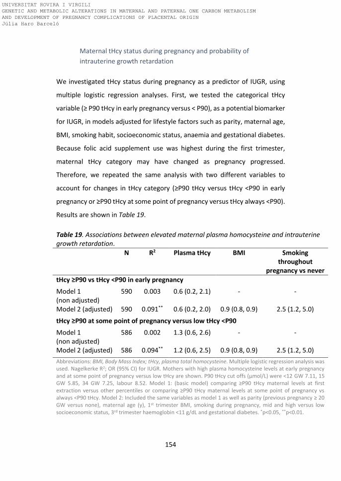
154
Maternal tHcy status during pregnancy and probability of
intrauterine growth retardation
We investigated tHcy status during pregnancy as a predictor of IUGR, using
multiple logistic regression analyses. First, we tested the categorical tHcy
variable (≥ P90 tHcy in early pregnancy versus < P90), as a potential biomarker
for IUGR, in models adjusted for lifestyle factors such as parity, maternal age,
BMI, smoking habit, socioeconomic status, anaemia and gestational diabetes.
Because folic acid supplement use was highest during the first trimester,
maternal tHcy category may have changed as pregnancy progressed.
Therefore, we repeated the same analysis with two different variables to
account for changes in tHcy category (≥P90 tHcy versus tHcy <P90 in early
pregnancy or ≥P90 tHcy at some point of pregnancy versus tHcy always <P90).
Results are shown in Table 19.
Table 19. Associations between elevated maternal plasma homocysteine and intrauterine growth retardation.
N R2 Plasma tHcy BMI Smoking throughout
pregnancy vs never
tHcy ≥P90 vs tHcy <P90 in early pregnancy
Model 1 (non adjusted)
590 0.003 0.6 (0.2, 2.1) - -
Model 2 (adjusted) 590 0.091** 0.6 (0.2, 2.0) 0.9 (0.8, 0.9) 2.5 (1.2, 5.0)
tHcy ≥P90 at some point of pregnancy versus low tHcy <P90
Model 1 (non adjusted)
586 0.002 1.3 (0.6, 2.6) - -
Model 2 (adjusted) 586 0.094** 1.2 (0.6, 2.5) 0.9 (0.8, 0.9) 2.5 (1.2, 5.0)
Abbreviations: BMI, Body Mass Index; tHcy, plasma total homocysteine. Multiple logistic regression analysis was used. Nagelkerke R2; OR (95% CI) for IUGR. Mothers with high plasma homocysteine levels at early pregnancy and at some point of pregnancy versus low tHcy are shown. P90 tHcy cut offs (μmol/L) were <12 GW 7.11, 15 GW 5.85, 34 GW 7.25, labour 8.52. Model 1: (basic model) comparing ≥P90 tHcy maternal levels at first extraction versus other percentiles or comparing ≥P90 tHcy maternal levels at some point of pregnancy vs always <P90 tHcy. Model 2: Included the same variables as model 1 as well as parity (previous pregnancy ≥ 20 GW versus none), maternal age (y), 1st trimester BMI, smoking during pregnancy, mid and high versus low socioeconomic status, 3rd trimester haemoglobin <11 g/dL and gestational diabetes. *p<0.05, **p<0.01.
UNIVERSITAT ROVIRA I VIRGILI GENETIC AND METABOLIC ALTERATIONS IN MATERNAL AND PATERNAL ONE CARBON METABOLISM AND DEVELOPMENT OF PREGNANCY COMPLICATIONS OF PLACENTAL ORIGIN Júlia Haro Barceló

155
The results showed no association between plasma homocysteine and IUGR
at early or at some other point of pregnancy. However, we observed a
tendency, but not significant, for a higher probability of developing IUGR when
tHcy is elevated at some point of pregnancy compared with always low (<P90).
There was a clear association between smoking throughout pregnancy and
increased risk of IUGR.
We then assessed plasma folate and red blood cell folate status according to
tHcy status divided in four categories at different pregnancy points; tHcy levels
always <P90 (<7.1 μmol/L at <12 GW, <5.9 μmol/L at 15 GW, <7.2 μmol/L at
34 GW, < 8.5μmol/L at labour) , tHcy levels low at 1st trimester (< 7.1 μmol/L),
tHcy levels high at 1st trimester (≥ 7.11 μmol/L) and tHcy levels always high
(≥P90 7.1 μmol/L, 7.2 μmol/L at 34 GW, 8.5 μmol/L at labour). The results are
shown in Figure 10.
UNIVERSITAT ROVIRA I VIRGILI GENETIC AND METABOLIC ALTERATIONS IN MATERNAL AND PATERNAL ONE CARBON METABOLISM AND DEVELOPMENT OF PREGNANCY COMPLICATIONS OF PLACENTAL ORIGIN Júlia Haro Barceló

156
Figure 10. Mean Plasma folate throughout pregnancy and in the cord according to
tHcy category. Blue: thcy always low (<12GW n=577, 15GW n=355, 24-27GW n=540,
34GW n=507, labour n=489 and cord n=464). Orange: thcy low in beginning only
(<12GW n=64, 15GW n=46, 24-27GW n=60, 34GW n=58, labour n=59 and cord n=55).
Green: thcy high in beginning only (<12GW n=39, 15GW n=23, 24-27GW n=32, 34GW
n=35, labour n=32 and cord n=31). Red: thcy always high (<12GW n=25, 15GW n=16,
24-27GW n=24, 34GW n=23, labour n=21 and cord n=20). Means (dots) and 95% CI
(bars) are reported. Student’s unpaired t-test comparing differences between tHcy
categories: *p<0.05, **p<0.01, ***p<0.001 compared to tHcy always low (blue line); †p<0.05, ††p<0.01, †††p<0.001 compared to tHcy always high (red line).
Plasma folate status according to pattern of tHcy change from early pregnancy
<
UNIVERSITAT ROVIRA I VIRGILI GENETIC AND METABOLIC ALTERATIONS IN MATERNAL AND PATERNAL ONE CARBON METABOLISM AND DEVELOPMENT OF PREGNANCY COMPLICATIONS OF PLACENTAL ORIGIN Júlia Haro Barceló
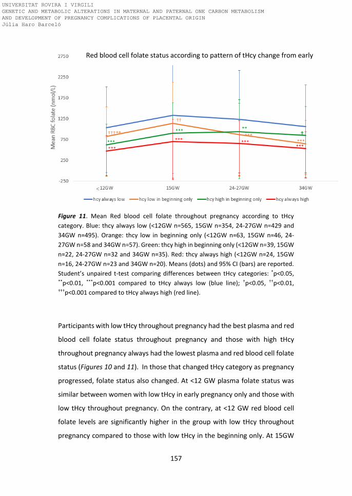
157
Figure 11. Mean Red blood cell folate throughout pregnancy according to tHcy
category. Blue: thcy always low (<12GW n=565, 15GW n=354, 24-27GW n=429 and
34GW n=495). Orange: thcy low in beginning only (<12GW n=63, 15GW n=46, 24-
27GW n=58 and 34GW n=57). Green: thcy high in beginning only (<12GW n=39, 15GW
n=22, 24-27GW n=32 and 34GW n=35). Red: thcy always high (<12GW n=24, 15GW
n=16, 24-27GW n=23 and 34GW n=20). Means (dots) and 95% CI (bars) are reported.
Student’s unpaired t-test comparing differences between tHcy categories: *p<0.05, **p<0.01, ***p<0.001 compared to tHcy always low (blue line); †p<0.05, ††p<0.01, †††p<0.001 compared to tHcy always high (red line).
Participants with low tHcy throughout pregnancy had the best plasma and red
blood cell folate status throughout pregnancy and those with high tHcy
throughout pregnancy always had the lowest plasma and red blood cell folate
status (Figures 10 and 11). In those that changed tHcy category as pregnancy
progressed, folate status also changed. At <12 GW plasma folate status was
similar between women with low tHcy in early pregnancy only and those with
low tHcy throughout pregnancy. On the contrary, at <12 GW red blood cell
folate levels are significantly higher in the group with low tHcy throughout
pregnancy compared to those with low tHcy in the beginning only. At 15GW
Red blood cell folate status according to pattern of tHcy change from early
pregnancy
<
UNIVERSITAT ROVIRA I VIRGILI GENETIC AND METABOLIC ALTERATIONS IN MATERNAL AND PATERNAL ONE CARBON METABOLISM AND DEVELOPMENT OF PREGNANCY COMPLICATIONS OF PLACENTAL ORIGIN Júlia Haro Barceló

158
plasma folate levels from participants with low tHcy in the beginning only are
lower than those with always low tHcy concentrations. However, red blood
cell folate levels do not differ between the groups (tHcy low in the beginning
only versus tHcy levels always low) at 15 GW. From 24-27 GW throughout the
rest of pregnancy, plasma folate status was consistently lower in the women
that had started pregnancy with low tHcy status but then went on to belong
to the high tHcy status group. From 24-27 GW to the end of pregnancy,
participants with high tHcy levels at the beginning of pregnancy only, had
higher red blood cell folate status than people with low tHcy levels in early
pregnancy only.
Paternal genotype and intrauterine growth retardation
We also investigated the association between paternal MTHFR 677C>T
genotype and probability of IUGR using multiple logistic regression analysis.
We added the paternal MTHFR 677C>T genotype (reference CC genotype),
plasma cobalamin and red blood cell folate concentrations to the complete
maternal model with maternal MTHFR 677C>T genotype, red blood cell folate
and plasma cobalamin at 1st and 3rd trimester, adjusted for lifestyle factors and
complications such as anaemia and gestational diabetes. None of the models
were significant and are not shown. Nevertheless, the relationship between
the paternal MTHFR 677TT genotype compared to CC was as follows: in the 1st
trimester model (n= 304) OR (95% CI) 1.1 (0.3, 4.1), in the 34 GW model (n=
272) OR (95% CI) 1.8 (0.4, 7.5) and in the Labour model (n= 242) OR (95% CI)
1.4 (0.4, 2.5).
UNIVERSITAT ROVIRA I VIRGILI GENETIC AND METABOLIC ALTERATIONS IN MATERNAL AND PATERNAL ONE CARBON METABOLISM AND DEVELOPMENT OF PREGNANCY COMPLICATIONS OF PLACENTAL ORIGIN Júlia Haro Barceló

159
Paternal tHcy status and intrauterine growth retardation
We built on the multiple logistic regression models, previously described for
the mothers, to investigate the association between elevate paternal tHcy
(≥P90 versus the other percentiles) and probability of IUGR. We included the
variable maternal tHcy ≥P90 at some point of pregnancy versus always <P90,
I the model. The results are reported in Table 20.
Table 20. Associations between elevated (≥P90 versus <P90) maternal and paternal tHcy and intrauterine growth retardation.
N R2 Maternal tHcy ≥P90 vs
<P90
BMI Smoking throughout pregnancy vs never
Paternal tHcy ≥P90 vs
<P90
Model 1 (maternal, non adjusted)
390 0.008 1.7 (0.7, 4.3)
- - -
Model 2 (maternal adjusted)
390 0.081 1.3 (0.5, 3.4)
0.9 (0.8, 1.0)
1.5 (0.5, 4.7)
-
Model 3 (maternal adjusted and paternal factors)
390 0.123# 1.4 (0.5, 3.9)
0.9 (0.7, 1.0)
1.8 (0.6, 5.7)
4.5 (1.7, 12.8)
Abbreviations: BMI, Body Mass Index; tHcy, total plasma homocysteine. Multiple logistic regression analysis was used. Nagelkerke R2; OR (95% CI) for intrauterine growth retardation in babies born to mothers with high tHcy status (≥P901, 7.1 μmol/L at <12 GW, 5.85 at 15 GW, 7.25 at 34 GW, 8.52 at labour) are shown. Model 1: (basic model) comparing ≥P90 tHcy maternal levels at some point of pregnancy vs <P90 tHcy throughout pregnancy. Model 2: Included the same variables as model 1 as well as parity (previous pregnancy ≥20 GW versus none), maternal age (y), 1st trimester BMI, smoking during pregnancy, mid and high versus low socioeconomic status, 3rd trimester haemoglobin <11 g/dL and gestational diabetes. Model 3: Included the same variables as model 2 as well as paternal tHcy (≥14.1 µmol/L, ≥P90 vs <P90). #P=0.06.
Elevated paternal tHcy was associated with a greater probability of an IUGR-
affected pregnancy.
UNIVERSITAT ROVIRA I VIRGILI GENETIC AND METABOLIC ALTERATIONS IN MATERNAL AND PATERNAL ONE CARBON METABOLISM AND DEVELOPMENT OF PREGNANCY COMPLICATIONS OF PLACENTAL ORIGIN Júlia Haro Barceló
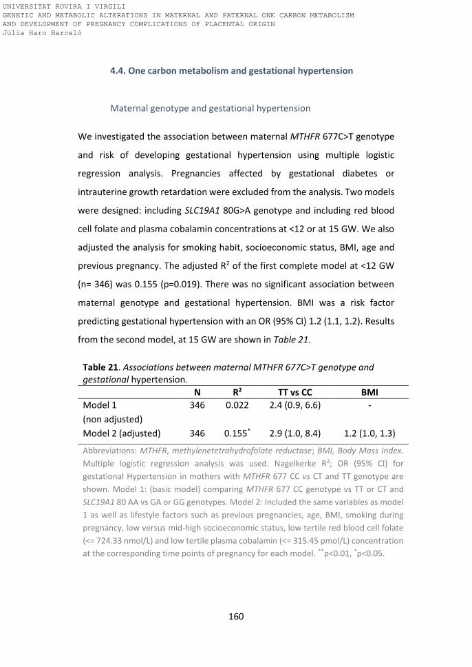
160
4.4. One carbon metabolism and gestational hypertension
Maternal genotype and gestational hypertension
We investigated the association between maternal MTHFR 677C>T genotype
and risk of developing gestational hypertension using multiple logistic
regression analysis. Pregnancies affected by gestational diabetes or
intrauterine growth retardation were excluded from the analysis. Two models
were designed: including SLC19A1 80G>A genotype and including red blood
cell folate and plasma cobalamin concentrations at <12 or at 15 GW. We also
adjusted the analysis for smoking habit, socioeconomic status, BMI, age and
previous pregnancy. The adjusted R2 of the first complete model at <12 GW
(n= 346) was 0.155 (p=0.019). There was no significant association between
maternal genotype and gestational hypertension. BMI was a risk factor
predicting gestational hypertension with an OR (95% CI) 1.2 (1.1, 1.2). Results
from the second model, at 15 GW are shown in Table 21.
Table 21. Associations between maternal MTHFR 677C>T genotype and gestational hypertension.
N R2 TT vs CC BMI
Model 1
(non adjusted)
346 0.022 2.4 (0.9, 6.6) -
Model 2 (adjusted) 346 0.155* 2.9 (1.0, 8.4) 1.2 (1.0, 1.3)
Abbreviations: MTHFR, methylenetetrahydrofolate reductase; BMI, Body Mass Index.
Multiple logistic regression analysis was used. Nagelkerke R2; OR (95% CI) for
gestational Hypertension in mothers with MTHFR 677 CC vs CT and TT genotype are
shown. Model 1: (basic model) comparing MTHFR 677 CC genotype vs TT or CT and
SLC19A1 80 AA vs GA or GG genotypes. Model 2: Included the same variables as model
1 as well as lifestyle factors such as previous pregnancies, age, BMI, smoking during
pregnancy, low versus mid-high socioeconomic status, low tertile red blood cell folate
(<= 724.33 nmol/L) and low tertile plasma cobalamin (<= 315.45 pmol/L) concentration
at the corresponding time points of pregnancy for each model. **p<0.01, *p<0.05.
UNIVERSITAT ROVIRA I VIRGILI GENETIC AND METABOLIC ALTERATIONS IN MATERNAL AND PATERNAL ONE CARBON METABOLISM AND DEVELOPMENT OF PREGNANCY COMPLICATIONS OF PLACENTAL ORIGIN Júlia Haro Barceló
161
We observed an increased risk of developing gestational hypertension in
mothers with the MTHFR 677 TT compared to CC genotype. BMI was also a
predicting factor.
Maternal tHcy status and gestational hypertension
We also studied the association between early and late pregnancy tHcy and
the risk of gestational hypertension, using multiple logistic regression analysis,
again excluding pregnancies affected by intrauterine growth retardation and
gestational diabetes. The categorical tHcy variable (≥P90 tHcy in early
pregnancy versus < P90) replaced the genotypes and red blood cell folate and
plasma cobalamin variables in each model, at <12 and 15 GW. Models were
adjusted for smoking habit, socioeconomic status, BMI, age and parity.
Elevated maternal tHcy levels in early pregnancy was not associated with
increased risk of gestational hypertension. In the adjusted model, BMI was a
risk factor for gestational hypertension with an OR (95% CI) 1.1 (1.1, 1.2) and
having been pregnant previously was protective 0.56 (0.32, 0.96).
Paternal genotype and gestational hypertension
We investigated the association between paternal MTHFR 677C>T genotype
and risk of developing gestational hypertension using multiple logistic
regression analysis. To the same models used for the mothers, at <12 and 15
GW, we added paternal MTHFR 677C>T genotype, smoking habit, age and red
blood cell folate concentration. None of the models were significant.
UNIVERSITAT ROVIRA I VIRGILI GENETIC AND METABOLIC ALTERATIONS IN MATERNAL AND PATERNAL ONE CARBON METABOLISM AND DEVELOPMENT OF PREGNANCY COMPLICATIONS OF PLACENTAL ORIGIN Júlia Haro Barceló

162
Paternal tHcy status and gestational hypertension
As we did with mothers, we also studied the association between paternal
tHcy levels and the risk of gestational hypertension. The categorical tHcy
variable (≥ P90 tHcy in early pregnancy versus < P90) replaced, genotypes, red
blood cell folate and plasma cobalamin variables in each model, at <12 and 15
GW. We included elevated paternal tHcy levels (≥P90), age and smoking habit
as paternal factors in both models. The adjusted R2 of the model was 0.129
(p=0.013). Neither elevated paternal tHcy or smoking were associated with
risk of gestational hypertension.
UNIVERSITAT ROVIRA I VIRGILI GENETIC AND METABOLIC ALTERATIONS IN MATERNAL AND PATERNAL ONE CARBON METABOLISM AND DEVELOPMENT OF PREGNANCY COMPLICATIONS OF PLACENTAL ORIGIN Júlia Haro Barceló

163
DISCUSSION
UNIVERSITAT ROVIRA I VIRGILI GENETIC AND METABOLIC ALTERATIONS IN MATERNAL AND PATERNAL ONE CARBON METABOLISM AND DEVELOPMENT OF PREGNANCY COMPLICATIONS OF PLACENTAL ORIGIN Júlia Haro Barceló

164
UNIVERSITAT ROVIRA I VIRGILI GENETIC AND METABOLIC ALTERATIONS IN MATERNAL AND PATERNAL ONE CARBON METABOLISM AND DEVELOPMENT OF PREGNANCY COMPLICATIONS OF PLACENTAL ORIGIN Júlia Haro Barceló

165
5. Discussion
5.1. General findings
Our study population consists of a pregnancy cohort in which participants
were recruited from their prenatal visits in the hospitals before 12GW. Most
women reported planning their pregnancy. However, only 36% took folic acid
supplements before conception. At their first prenatal visit, all women were
routinely recommended to take folic acid supplements until the end of the
first trimester. They adhered very well to this advice, and only 5.8% reported
not taking any folic acid supplements during the first trimester. Their age
ranged from 26 to 39 years, most had a mid-high socioeconomic status and
25.7% of women smoked during pregnancy, 9.4% during the first trimester
only and 16.3% throughout pregnancy. 19.2% of the mothers had impaired
placentation indicated by pathological Doppler measurements of uterine
artery flow at 20GW, 11.8% developed gestational hypertension and 8.8%
intrauterine growth retardation. The percentage of pregnancies affected by
these complications in other cohorts ranges between 2-17% for gestational
hypertension (240) and 10-15% for IUGR (241).
Paternal participation in the study was 66.1%. There were 3 main factors
affecting paternal recruitment. Firstly, maternal consent was required to
contact the fathers and 5.8% declined to provide the contact details. Secondly,
we could not establish contact with 1% of the fathers. Thirdly 33.9% of the
fathers allegated fear of needles, no interest or incompatible work schedule
as the reason for declining the invitation. Only 6.8% of the fathers reported
taking folic acid-containing supplements. Their age ranged between 29 to 41
years, most had a mid-high socioeconomic status and 36.7% of the men
smoked before and during pregnancy.
UNIVERSITAT ROVIRA I VIRGILI GENETIC AND METABOLIC ALTERATIONS IN MATERNAL AND PATERNAL ONE CARBON METABOLISM AND DEVELOPMENT OF PREGNANCY COMPLICATIONS OF PLACENTAL ORIGIN Júlia Haro Barceló
166
Folate status
Median plasma folate during the first trimester of pregnancy was relatively
high (28.9 nmol/L) and then declined as pregnancy progressed. On the other
hand, median red blood cell folate status increased between the first trimester
(936.4 nmol/L) and 15 GW (1266 nmol/L), before gradually declining into the
3rd trimester. The decline in folate status occurred earlier in plasma folate
compared to RBC folate because plasma folate reflects very recent intake
(actual status) and is sensitive to folic acid supplementation, whereas RBC
folate concentrations reflect both the long-term average of intake over the life
span of RBC and folate stores in the liver (242). In addition, plasma folate as
well as other nutrients circulating concentrations are affected by
haemodilution due to the increase on plasma volume (243).
50.1% of participants were close to plasma folate deficiency according to WHO
criteria (≤ 10 nmol/L (8)) and 30.2% according to the old criteria (≤ 7 nmol/L
(7)) at labour. The prevalence of folate deficiency depends on the cut off
applied. We applied that of Chanarin et al (≤ 7 nmol/L (7)), in Figure 7 and
Figure 8, because it is the one that was used until the WHO in 2008
recommended changing it to ≤ 10 nmol/L for folate deficiency (8). These
recommendations in both cases are applicable outside of pregnancy. In the
absence of an established cut off for pregnancy, we consider it more precise
to apply the previous (pre-mandatory fortification) cut off because our
participants were not exposed to mandatory folic acid fortification and taking
into the account that plasma micronutrient concentrations are lower due to
haemodilution and increased glomerular filtration rate. Nevertheless, we
highlight the need to establish a cut off for pregnant women.
Globally, red blood cell folate status was not deficient. The advantage of
measuring RBC folate during pregnancy is that it is not subject to the same
UNIVERSITAT ROVIRA I VIRGILI GENETIC AND METABOLIC ALTERATIONS IN MATERNAL AND PATERNAL ONE CARBON METABOLISM AND DEVELOPMENT OF PREGNANCY COMPLICATIONS OF PLACENTAL ORIGIN Júlia Haro Barceló

167
physiological changes as plasma folate concentration. Only 0.13% of the
mothers started pregnancy with red blood cell folate deficiency ( <340 nmol/L
(8)) and few cases were deficient by 34 GW (0.5%). Nevertheless, 51.8% did
not meet the recommended red blood cell folate status of 906 nmol/L for
NTDs prevention. We can expect participants to have good plasma folate
status during the 1st trimester while they were taking folic acid supplements.
The decline in plasma folate status started when folic acid supplement use was
largely stopped, after 15 GW. Red blood cell folate concentrations also
increased following the period of high folic acid supplement use. It did not fall
until the 3rd trimester. There are other factors affecting also the drop in folate
status during pregnancy when folate requirements are increased because of
elevated folate utilization and catabolism (244). The preparation for the
demands of rapid fetal growth that occur late in pregnancy may double
nutrient requirement to protect fetal development (245). The increased
demand on folate may lead to the mobilization of folate maternal reserves
ending sometimes in the depletion of those reserves. Our group has previously
shown that low folate status enhances the inverse association between
betaine and tHcy in late pregnancy, reflecting increased betaine remethylation
to homocysteine when folate availability becomes limited (246).
Haemodilution can be another factor involved in the decrease of folate
concentrations. Maternal plasma volume expansion from 10 GW throughout
pregnancy and serum albumin decrease, lead to a reduction in nutrient
concentration in the circulation during the same period (243). In addition,
changes in renal function during normal pregnancy due to increased
glomerular filtration, increased kidney plasmatic flow and decreased kidney
vascular resistance (247) may contribute to the excess of plasma folate
elimination leading to a drop in folate status (248).
UNIVERSITAT ROVIRA I VIRGILI GENETIC AND METABOLIC ALTERATIONS IN MATERNAL AND PATERNAL ONE CARBON METABOLISM AND DEVELOPMENT OF PREGNANCY COMPLICATIONS OF PLACENTAL ORIGIN Júlia Haro Barceló

168
We observed that smoking affected maternal red blood cell folate status
during pregnancy. It was lower in smokers than in non smokers. Comparing
red blood cell folate status between these two groups allows us to see the
long term effects of smoking on folate status, as well as a measurement of
folate status unaffected by haemodilution. We did not have red blood cell
folate determinations from the cord, but cord plasma folate concentration is
unaffected by haemodilution. Despite the difference in folate status between
the smoking and non-smoking mothers, plasma folate status in the cord did
not differ. During pregnancy, folate transport to the foetus is enhanced by
additional mechanisms for folate uptake and transport including folate
receptor α expression by the placenta (249). If folate status is impaired in
smoking mothers, there may be a compensatory effect to ensure the supply
of folate to the fetus by increasing cellular folate uptake, as described in
animal models. Hye Won Kim et al (2011) showed that folate receptor α
expression in the placenta was higher (by 130%) in rats fed with a deficient
folic acid diet compared to the folic acid supplemented group (250).
The median of paternal plasma folate was close to folate deficiency (8.8
nmol/L) and the median of red blood cell folate level was 456.8 nmol/L. 61%
of the fathers had folate deficiency according to WHO criteria (plasma folate
≤ 10 nmol/L (8)), and 35.6% according to the old criteria (7). This percentage
is similar to that observed in the mothers at the end of pregnancy. The
relatively high prevalence of folate deficiency in this population, compared to
others like the population from the United States described by Bailey et al
(251) with a low prevalence (< 1%) of folate deficiency (< 7 nmol/L), is likely
due to poor dietary intake of folic acid in the form of fortified foods. There is
no mandatory fortification of flour with food folic acid in Spain and, even
though voluntarily fortified food products are available (breakfast cereals,
cookies, milk, milkshakes, yogurts, canned juices, etc), consumption of these
UNIVERSITAT ROVIRA I VIRGILI GENETIC AND METABOLIC ALTERATIONS IN MATERNAL AND PATERNAL ONE CARBON METABOLISM AND DEVELOPMENT OF PREGNANCY COMPLICATIONS OF PLACENTAL ORIGIN Júlia Haro Barceló

169
is generally lower than in other countries. In addition, fathers no
recommendations are in place to take folic acid supplements before
conception.
On the other hand, current trends are towards a dietary pattern increasingly
further removed from the original Mediterranean diet in our general
population, resulting in less vegetable and fruit intake (252). However, it is
well known that dietary folate is relatively ineffective at increasing folate
status compared with folic acid supplements and fortified food (44).
We hypothesise that the lower folate status observed in the fathers compared
to the mothers is due to the lack of targeted recommendations on folic acid
supplementation in this group, to improve folate status. We previously
reported, in a study of our adult population between 1998-2002, that in the
absence of folic acid supplementation, 18.8% of women and men have folate
deficiency. Furthermore, folate status is relatively low with geometric mean
of plasma folate concentrations of 11.5 nmol/L and red blood cell folate of 810
nmol/L (9).
Homocysteine status
The pattern of change in tHcy is similar to that which our group first described
in 2002 (100). Folic acid supplementation influences tHcy, lowering its
concentrations (99, 253). The extensive use of supplements by the mothers in
our cohort may be one of the reasons why tHcy was low during the first
trimester and fell by 15 GW and remained low at 27-27 GW. There was an
increase in tHcy levels from 34 GW to the end of pregnancy. Independently of
folate status, hormonal changes occurring during pregnancy and the
physiologic role of homocysteine in the preparation of labour lead to a return
UNIVERSITAT ROVIRA I VIRGILI GENETIC AND METABOLIC ALTERATIONS IN MATERNAL AND PATERNAL ONE CARBON METABOLISM AND DEVELOPMENT OF PREGNANCY COMPLICATIONS OF PLACENTAL ORIGIN Júlia Haro Barceló
170
towards nonpregnancy concentrations of tHcy in late pregnancy (35, 99).
Previously, in a non supplemented cohort, our group observed higher tHcy
levels in early pregnancy (geometric mean of 6.48 µmol/L) than those
observed in the present population (100). The median tHcy in the fathers, of
9.6 mol/L, is higher than the mothers, as expected due to the difference in
tHcy between men and women (138, 254), the fact that tHcy was measured
during pregnancy in women, and likely also because folic acid supplement use
was prevalent in the women (99, 253).
MTHFR 677 C>T and SLC19A1 80G>A genotype
The homozygote variant genotypes of these two polymorphisms are quite
prevalent in our pregnancy cohort, with 17.0% of the women with the MTHFR
677 TT genotype and 27.2% with the SLC19A1 80AA genotype. The TT + AA
genotype combination s was observed in 6.0% of the women. Among the
fathers, 14.1% had the MTHFR 677 TT genotype, 28.9% the SLC19A1 80AA
genotype and 5.3% the combination of TT + AA. Our group previously reported
the MTHFR 677TT genotype to be present in 18.1% of adults in a
representative sample of our local population (9) and 11.8% of newborns in
Spain, data from hospitals of the National Health Service (15), lower than the
prevalence in our cohort. This prevalence is also higher than in other countries
such as Italy, France or The Netherlands (15).
The maternal MTHFR 677TT genotype does not affect plasma folate
concentrations during pregnancy in our population. In contrast, red blood cell
folate concentrations were lower in the MTHFR 677TT compared to CC
genotypes in early, mid and late pregnancy and also compared to CT
genotypes in late pregnancy. However, the stratified analysis according to
smoking habit during pregnancy showed no differences in red blood cell folate
UNIVERSITAT ROVIRA I VIRGILI GENETIC AND METABOLIC ALTERATIONS IN MATERNAL AND PATERNAL ONE CARBON METABOLISM AND DEVELOPMENT OF PREGNANCY COMPLICATIONS OF PLACENTAL ORIGIN Júlia Haro Barceló
171
levels between CC and CT compared to TT genotypes in smokers. There were
no differences in cord plasma folate among the genotypes in either group. This
might be explained by a compensatory effect in smoking mothers, commented
before, necessary to ensure adequate supply of folate to the foetus. Despite
the lack of difference in folate status, higher tHcy levels in the mothers with
the TT genotype were observed compared to CC or CT genotypes from 15 GW
to the end of pregnancy. This suggests that folate status is still in fact impaired
in the TT mothers during pregnancy. It is possible that any compensation in
maternal folate status in these mothers, is prioritised for the foetus before the
mother. Regarding the SLC19A1 80G>A polymorphism, plasma folate, red
blood cell folate and tHcy concentration did not differ between genotypes. In
a previous study our group reported that compared with the CC and GG
genotypes for MTHFR 677 and SLC19A1 80 genotypes respectively, people
with the TT or AA alleles had lower plasma folate and RBCF plasma levels (9).
We may not have observed the effect of these polymorphisms on folate status
due to extensive folic acid supplement use. Nevertheless, the effects of the
polymorphism on folate reserves (red blood cell folate) and the functional
folate status biomarker (tHcy) were evident. Also, plasma variables are
affected by the physiological effects of pregnancy including haemodilution
(243), increased glomerular filtration rate (247) and foetal growth
requirements (244). Determinations of folate status in clinical laboratories are
largely based on plasma folate concentrations and these may not accurately
reflect true underlying status due to these reasons.
Fathers with the MTHFR 677TT genotype had lower red blood cell folate
concentrations and higher tHcy compared to the other genotypes. Some
studies have previously reported the relation between the MTHFR 677TT
polymorphism and the increase in total plasma homocysteine (40, 255). Our
group have recently shown that the MTHFR 677TT genotype was the most
UNIVERSITAT ROVIRA I VIRGILI GENETIC AND METABOLIC ALTERATIONS IN MATERNAL AND PATERNAL ONE CARBON METABOLISM AND DEVELOPMENT OF PREGNANCY COMPLICATIONS OF PLACENTAL ORIGIN Júlia Haro Barceló

172
important determinant of tHcy in men and that the association was stronger
in smokers (139). Plasma folate concentrations also tended to be lower in
fathers with the TT genotype. In the case of the SLC19A1 80G>A
polymorphism, no significant differences in one carbon metabolism status
between genotypes were observed.
UNIVERSITAT ROVIRA I VIRGILI GENETIC AND METABOLIC ALTERATIONS IN MATERNAL AND PATERNAL ONE CARBON METABOLISM AND DEVELOPMENT OF PREGNANCY COMPLICATIONS OF PLACENTAL ORIGIN Júlia Haro Barceló
173
5.2. One Carbon Metabolism and uterine artery pulsatility index
and impaired placentation
Relation between genetics and uterine artery pulsatility index and
impaired placentation
We explored whether the MTHFR 677 C>T polymorphism influences
placentation, assessed by uterine artery pulsatility index and Doppler wave
forms at 20 GW. We did not find any association between the maternal
genotype and placentation. Abnormal uterine artery Doppler measurement is
commonly used in the screening of pregnant women to detect increased
resistance to flow in the uterine arteries due to impaired placentation. Poor
perfusion indicated by increased uterine artery resistance is associated with
adverse pregnancy outcomes (167). The homozygote variant MTHFR 677TT
genotype has been associated with elevated tHcy and decreased serum folate
concentrations (14) and is considered a genetic risk factor for some obstetric
complications such as IUGR (256) and preeclampsia (257). Out of pregnancy,
MTHFR 677TT genotype has been also related with increased risk of venous
thrombosis (258), due to the alterations in arterial structure and function
caused by elevated homocysteine concentrations. In that way, this
polymorphism may affect uterine artery resistance also, and increase the risk
of developing pregnancy outcomes.
Stonek et al (2008) obtained the same results as ours in a prospective study
where they measured the uterine artery pulsatility index values and unilateral
or bilateral diastolic notches at 12 and at 22 GW of 1995 singleton pregnant
women. No statistical differences between mean uterine artery Doppler
values and bilateral or unilateral notch in any MTHFR genotype were found,
showing that the MTHFR C677T polymorphism does not influence Doppler
flow measurements (259). In the same line, Driul et al (2005) in a case control
study with 103 preeclamptic women found no association between the
UNIVERSITAT ROVIRA I VIRGILI GENETIC AND METABOLIC ALTERATIONS IN MATERNAL AND PATERNAL ONE CARBON METABOLISM AND DEVELOPMENT OF PREGNANCY COMPLICATIONS OF PLACENTAL ORIGIN Júlia Haro Barceló
174
polymorphism and uterine artery flow. The frequency of homozygosity for the
MTHFR mutation in the group of women with abnormal Doppler
measurement and preeclampsia was 21.7% while it was 10.3% in the group
with normal pregnancies with no significant differences (260).
Contrarily, more recently Akdemir et al (2018) among 71 patients with
recurrent pregnancy loss, observed that the uterine artery impedance was
higher in the group with multiple thrombophilic mutations (MTHFR C677T
among them) compared with the group who had a single thrombophilic
mutation (261). Even though their study group are not comparable with our
population, because they include complicated pregnancies, and they
measured the combination of some polymorphisms, this result may suggest
that the implication of MTHFR C677T alone is not strong enough to lead to an
adverse pregnancy outcomes As Stoneak et al have proposed, may be it is not
reasonable to speculate that MTHFR genotype influences Doppler flow
measurements, because in their results none of the three MTHFR genotypes
showed a significant association with Doppler measurement (259). However,
it is important to consider that environmental factors such as folate status and
smoking can modify the effect of the MTHFR 677TT polymorphism (289, 290).
We have studied those factors in detail, especially RBC folate status in the
context of smoking. We have observed no differences between MTHFR 677
C>T genotypes among smoking mothers, and cord folate status did not differ
among genotypes in any of the mothers or between smokers and nonsmokers.
The high prevalence of folic acid supplement use during the first trimester
(more than 90% of our mothers) leading to good folate status, may hide the
effect of the MTHFR 677TT polymorphism. May be for those reasons in the
studies commented before, as well as in our study, we do not observe an
association between maternal genotype and uterine artery pulsatility index
and impaired placentation. Or may be further investigation is needed to have
strong evidence.
UNIVERSITAT ROVIRA I VIRGILI GENETIC AND METABOLIC ALTERATIONS IN MATERNAL AND PATERNAL ONE CARBON METABOLISM AND DEVELOPMENT OF PREGNANCY COMPLICATIONS OF PLACENTAL ORIGIN Júlia Haro Barceló

175
On the other hand, paternal MTHFR 677 CT and TT genotypes were associated
with impaired placentation compared with the CC genotype. This was
confirmed by their association with increased resistance in the uterine arteries
and increased probability of pathological Doppler assessment of the uterine
arteries at 20 GW.
Uterine artery perfusion during pregnancy involves the maternal-placental-
foetal unit. Trophoblast invasion of the uterine arteries is the basis for
successful placentation and during placentation for establishing the maternal-
placental-foetal unit (259). As commented in the introduction, trophoblast
invasion of the uterine arteries converts them from their spiral, high
resistance, non-pregnant form to their tubular, low-resistance pregnant form.
If this conversion is incomplete, placentation is impaired and placental
perfusion is reduced leading to increased risk of severe pregnancy outcomes.
Doppler sonography of the uterine arteries is an established screening method
to detect impaired uterine artery flow.
There is a strong paternal genetic component in trophoblast formation and
function. If we consider that certain combinations of maternal and fetal
genotypes may impede proper placentation and trophoblast invasion (264),
paternal genes can also affect placentation, but is rarely considered. With the
results obtained, we propose that paternal genotype is crucial for a successful
trophoblast invasion. To the best of our knowledge, this is the first study to
report this and further investigation should replicate this association as well
as exploring other genotypes.
We found that paternal smoking was a predictive factor of uterine artery
pulsatility index and tripled the risk of impaired placentation. Paternal
smoking is a common factor studied in the investigation of the possible
implication of the father in pregnancy complications. In 1994, Martinez
showed that the number of cigarettes smoked by the father was associated
UNIVERSITAT ROVIRA I VIRGILI GENETIC AND METABOLIC ALTERATIONS IN MATERNAL AND PATERNAL ONE CARBON METABOLISM AND DEVELOPMENT OF PREGNANCY COMPLICATIONS OF PLACENTAL ORIGIN Júlia Haro Barceló

176
with lower birthweight in children with non-smoking mothers (265). Three
years later, Horta et al (1997) confirmed that women whose partner smoked
were at higher risk of having a child with IUGR [OR 95%CI 1.3 (1.1, 1.7)] (266).
It has been hypothesized that mutations in the paternal germ line caused by
the tobacco, can later cause mutations in the offspring and impact directly on
the health of the newborn (267). Further support of this hypothesis is provided
by in vitro evidence that cigarette smoking increases oxidative DNA damage in
human sperm cells and causes mutations in germ cells (268). However, the
results are still unclear and further investigation is needed to clarify the
correlation between paternal smoking habit and perinatal outcomes.
Relation between tHcy and uterine artery pulsatility index and
impaired placentation
Maternal tHcy status was not associated with placentation (either pulsatility
index or pathological Doppler measurement of uterine arteries at 20 GW).
Poor placentation, measured by Doppler uterine flow, is an underlying cause
of preeclampsia (269) and other complications related with abnormal
trophoblastic invasion. Some of the hypothesized factors leading to abnormal
trophoblast invasion are disruptions of endothelial junctional proteins (270),
subendothelial changes in uterine arteries (271) and generalized vascular
complications. Elevated total homocysteine levels have been shown to have a
deleterious effect on the vascular endothelium (272) and have been linked
with various pregnancy complications (273).
If tHcy is elevated the risk of some pregnancy complications caused by
endothelial dysfunction leading to modulation of the arterial resistance to
flow, we would expect to find an association between elevated tHcy and
Doppler parameters. However, homocysteine plasma determinations are
UNIVERSITAT ROVIRA I VIRGILI GENETIC AND METABOLIC ALTERATIONS IN MATERNAL AND PATERNAL ONE CARBON METABOLISM AND DEVELOPMENT OF PREGNANCY COMPLICATIONS OF PLACENTAL ORIGIN Júlia Haro Barceló

177
affected by folic acid supplementation and that might mask any association
between tHcy and impaired placentation. In our cohort, maternal tHcy was
normal during the first trimester when they are being supplemented and their
levels rise when supplementation stops.
Few studies have investigated this. Yu C et al (2004) in a multicentre study with
683 healthy pregnant women investigated the association between maternal
plasma homocysteine concentrations (mid-trimester) and preeclampsia, in
women with abnormal uterine artery Doppler outcome (22-24 GW).
Differences in maternal tHcy concentrations were not significant between
women with abnormal uterine artery Doppler and controls. Furthermore, they
did not report any difference in maternal tHcy levels in those with an adverse
pregnancy outcome compared to those with uncomplicated pregnancies
(190). This finding is in the same line as ours, even though their samples were
collected at mid trimester and were nonfasting. More recently, Maged et al
(2017) evaluated the role of maternal tHcy concentrations (15-19 GW) and
uterine artery Doppler (18-22 GW) as a possible predictor of pregnancy
complications related to poor placentation in 453 women. They observed that
tHcy levels and uterine artery resistance index were significantly higher in
preeclampsia, IUGR and other complications groups when compared to the
uncomplicated group. Also, the percentages of participants with uterine artery
notch (unilateral or bilateral) were significantly higher in the group with
complications compared with the control one (274).
Many other studies have investigated tHcy levels and uterine artery doppler
separately, as possible predictors for pregnancy complications. In addition, the
use of both parameters has been proposed to improve the sensitivity of
prediction of pregnancy complications relative to the use of each one alone
(274). The lack of association between maternal homocysteine plasma
concentrations and pathological Doppler measurement of uterine arteries in
our study may be because our cut off point for P90 levels of homocysteine was
UNIVERSITAT ROVIRA I VIRGILI GENETIC AND METABOLIC ALTERATIONS IN MATERNAL AND PATERNAL ONE CARBON METABOLISM AND DEVELOPMENT OF PREGNANCY COMPLICATIONS OF PLACENTAL ORIGIN Júlia Haro Barceló
178
not high enough to cause endothelial disfunction. Low levels of homocysteine
in the highest percentile may be due to folic acid supplementation in our
participants. As mentioned previously, women that temporarily took folic acid
supplements in the first trimester may only have had transitory “low” tHcy and
in that sense were misclassified as belonging to the low tHcy group. We do not
know if their tHcy was high during the early stages of placentation and before
they started to take the supplements. It would be unethical to test this in
humans because the recommendation is for women to take folic acid
supplements before and during early pregnancy, but it might be interesting to
test in animal studies. Mingda HAN et al (2012) demonstrated in a mouse
model that exposure to elevated serum homocysteine levels (by injection)
during embryonic day 6.75 have a moderate effect on altered embryonic
blood flow and placental anomalies including IUGR. They observed a mis
expressed NMM-IIs, a cytoskeletal protein family associated with cell
migration, in mouse placentas (275). Xu X et al (2016) also studied the effect
of elevated homocysteine concentrations (injections of 50mg/g/day on
embryonic day 7.5) in pregnant mice. Those animals showed preeclampsia like
symptoms such as higher systolic blood pressure and proteinuria in late
pregnancy and decreased expression levels of vascular endothelial growth
factor in the placenta compared with control mice (276).
On the other hand, elevated paternal tHcy was positively associated with
uterine artery pulsatility index and showed a threefold increase in risk of
developing pathological Doppler measurement of uterine arteries at 20 GW.
This association confirmed what we might have expected to observe in
mothers in the absence of folic acid supplementation. Paternal tHcy was not
affected by folic acid supplementation because few of them took supplements
before conception. In general, paternal folate status was lower than in the
mothers and low, as previously reported by our group in a population study
UNIVERSITAT ROVIRA I VIRGILI GENETIC AND METABOLIC ALTERATIONS IN MATERNAL AND PATERNAL ONE CARBON METABOLISM AND DEVELOPMENT OF PREGNANCY COMPLICATIONS OF PLACENTAL ORIGIN Júlia Haro Barceló

179
(9) in the absence of mandatory folic acid fortification. In this context, maybe
the potential effect of tHcy increasing uterine artery flow resistance is
detectable. It is possible that tHcy is related with pathological Doppler via
endothelial dysfunction, as previous non-pregnant animal and human studies
have described (272). We also hypothesise that in cases where the mother
develops pregnancy complications, but with no apparent underlying risk
factors, paternal factors can be the precursors of these complications. Is has
been shown that fathers born from a complicated pregnancy have more risk
of fathering a pregnancy with adverse outcomes (17). As we observe in our
results, while maternal factors seem to have no apparent relation with
pathological doppler measurement, paternal homocysteine levels were
associated with it. More investigation will be necessary to answer this
question, especially focused on paternal factors which have been scarcely
considered to date.
UNIVERSITAT ROVIRA I VIRGILI GENETIC AND METABOLIC ALTERATIONS IN MATERNAL AND PATERNAL ONE CARBON METABOLISM AND DEVELOPMENT OF PREGNANCY COMPLICATIONS OF PLACENTAL ORIGIN Júlia Haro Barceló

180
5.3. One Carbon Metabolism and birthweight and intrauterine
growth retardation
Relation between genetics and birthweight and intrauterine growth
retardation
There was an interaction between the MTHFR 677 C>T genotype and smoking
in their association with birth weight. In the stratified analysis by smoking
pattern, the MTHFR 677TT genotype was only associated with birthweight in
smoking mothers. Compared to the CC reference genotype, the TT genotype
was associated with increased birth weight. We would expect to see a
negative effect of smoking, a long established risk factor for low birth weight
(277) in all genotypes. This unexpected result might be explained by the
increase of placental folate receptors in situations of impaired folate status as
commented before (250). The only evidence we have of this is the fact that
cord plasma folate status was unaffected by either maternal smoking habit or
MTHFR 677C>T genotype, despite the fact that maternal folate status was
affected by each of these factors. Thamar Ayo Yila et al (2012) observed the
effect that we were expecting. In their study with 1784 native Japanese
mother-child pairs, the MTHFR 677 T allele was associated with lower birth
weight in the offspring of active smokers. Contrary to our results, they also
found a protective effect of the CT genotype against low birth weight only in
the absence of smoke (278). A limitation of stratified analysis is the reduced
sample size in the sub-groups. However, it is unlikely that the unexpected
effect that we observed is due to low sample size in the TT genotype group
because it was confirmed in early and mid-pregnancy and despite lack of
significance in late pregnancy, the positive association was maintained. Our
study did not set out to investigate the effect of smoking on birth weight, but
rather to control for it as an established confounding factor. We do not
interpret the TT genotype in smoking mothers as a protective factor for birth
UNIVERSITAT ROVIRA I VIRGILI GENETIC AND METABOLIC ALTERATIONS IN MATERNAL AND PATERNAL ONE CARBON METABOLISM AND DEVELOPMENT OF PREGNANCY COMPLICATIONS OF PLACENTAL ORIGIN Júlia Haro Barceló

181
weight in the offspring but hypothesise that the observed association is
related with a potential compensatory mechanism in mothers with two
factors adversely affecting birth weight. Low early pregnancy red blood cell
folate status was inversely associated with birth weight in smokers only. This
early pregnancy measurement of red blood cell folate was a true reflection of
folate status, before first trimester supplement use affected red blood cell
folate status. In countries with mandatory folic acid fortification or high intake
of voluntarily fortified foods and higher prevalence of folic acid supplement
use, it is possible that the negative association between low early pregnancy
maternal folate status and birth weight might be missed.
In several studies the MTHFR 677TT genotype in the mother has been
associated with low birth weight in the offspring. In a metanalysis published
in 2017, including 25 studies, Han Wu et al concluded that the maternal
MTHFR 677TT genotype was associated with increased probability of low
birthweight, OR (95%CI) 2.3 (1.4, 3.5). They proposed that the identification of
the maternal MTHFR 677TT genotype may play a key role for primary
prevention of low birth weight (12). Lee et al (2013) in a Mendelian
randomization analysis to investigate the causal relation between maternal
tHcy levels, as represented by MTHFR C677T genotype, with birth weight of
the offspring, found that birthweight was significantly lower in mothers with
the TT genotype compared to the CC and CT genotypes. They also observed a
dose-response association with homocysteine concentrations for each
additional T allele, and birth weight decreased as maternal tHcy at 24-28 GW
increased (p=0.06) (279). Those results may explain the association between
this polymorphism and birthweight of the newborn. Reduced enzyme activity
due to the MTHFR C677T polymorphism, increases plasma homocysteine
levels (13). This in turn may lead to oxidative stress, endothelial damage and
placental thrombosis (280). All these conditions might be associated with
UNIVERSITAT ROVIRA I VIRGILI GENETIC AND METABOLIC ALTERATIONS IN MATERNAL AND PATERNAL ONE CARBON METABOLISM AND DEVELOPMENT OF PREGNANCY COMPLICATIONS OF PLACENTAL ORIGIN Júlia Haro Barceló

182
impaired flow of uterine arteries, inadequate trophoblast invasion and poor
pregnancy outcomes (281). On the other hand, other reports observed no
association between MTHFR 677C>T genotype and risk of IUGR. Infante-
Rivarde et al, reported twice the absence of association between
thrombophilic polymorphisms and intrauterine grow restriction. In 2002 in a
case-control study including 493 newborns with IUGR, they reported no
increased risk of IUGR among mothers with the MTHFR 677TT genotype (282).
With a subgroup of the same cohort, in 2005 they conducted a family study
including 250 case trios (affected newborn, mother and father). They tested
whether there was excess transmission of the MTHFR C677T and A1298C
haplotypes (CA, TA, CC, TC) to affected offspring and they observe that
transmission and haplotype frequencies were not different between case and
controls groups (283). Their results are in line with ours, however, their study
was in the presence of mandatory folic acid fortification and paternal
genotype was performed from buccal swabs. Giovanni Larciprete et al (2007)
reported similar findings in a case control study with 284 pregnant women in
Rome. Even though MTHFR C677T genotypes were more prevalent than other
thrombophilia mutations included in the study, they did not observe an
increased incidence of adverse pregnancy outcomes in subjects with the
MTHFR TT genotype (284). Interestingly, they did not have cases with MTHFR
mutations combined with hyperhomocysteinemia. Folate supplementation of
their population may mask a potential association between the polymorphism
and IUGR or other adverse pregnancy outcomes. It has been proposed that
variations in the relative contribution of disease alleles in different
populations might explain the discrepant results of previous studies on MTHFR
and pregnancy complications (284). This could be another reason for the
absence of association between the MTHFR 677 C>T polymorphism and IUGR
in our study. In addition, an increase of IUGR cases in our population would
increase our statistical power and possibility of observing the association.
UNIVERSITAT ROVIRA I VIRGILI GENETIC AND METABOLIC ALTERATIONS IN MATERNAL AND PATERNAL ONE CARBON METABOLISM AND DEVELOPMENT OF PREGNANCY COMPLICATIONS OF PLACENTAL ORIGIN Júlia Haro Barceló

183
Also, an increase in paternal participation would improve sample size for
observing an association between the paternal polymorphism and IUGR.
Regarding a paternal effect, we found few studies investigating paternal
thrombophilic mutations as a possible risk factor for IUGR. Several are focused
on other paternal factors such as age, ethnicity, weight and height and other
polymorphisms.
Low red blood cell folate status was inversely associated with birth weight in
the mothers that smoked at some point of pregnancy. Low folate status has
previously been reported to be related with adverse pregnancy outcomes,
including birth weight (285). Exposure to tobacco smoke during pregnancy is
a well-known risk factor for adverse perinatal outcomes as many studies have
previously described (286). The first correlation between smoking and fetal
adverse outcomes was proposed in 1957 by Simpson (287) who pointed out
how the neonates of smoking mothers weighed 200g less on average than
neonates of non-smoking mothers. Furthermore, smoking has also been
shown to be associated with low RBC folate (288). We saw the inverse
association between low RBCF levels and birth weight in smoking mothers but
not in non smoking mothers. This may be because smoking mothers also had
lower RBC folate status. Or maybe because smoking has a direct negative
effect on birthweight mediated by hypoxia, as smoking reduces the oxygen
binding capacity and oxygen supply is essential for placental and fetal growth
(289). Another possible explanation is that nicotine may lead to
vasoconstriction in the maternal circulation with subsequent reduced blood
flow to the placenta and the fetus (290).
We also observed that paternal cobalamin status was positively associated
with birthweight. Maternal inadequate or deficient vitamin B12 status has
been previously associated with higher risk of low birthweight (264, 270).
Despite not finding previous studies focused on paternal B12 status, there are
UNIVERSITAT ROVIRA I VIRGILI GENETIC AND METABOLIC ALTERATIONS IN MATERNAL AND PATERNAL ONE CARBON METABOLISM AND DEVELOPMENT OF PREGNANCY COMPLICATIONS OF PLACENTAL ORIGIN Júlia Haro Barceló

184
studies suggesting that maternal and paternal micronutrient intake, including
B vitamins involved in one carbon metabolism, which are essential for DNA
methylation, can influence the programming of the offspring’s epigenome
influencing neonatal outcomes (228). Following this evidence, we hypothesize
that paternal B12 status can influence the programming of genes involved in
fetal growth and subsequent offspring’s birthweight.
Relation between tHcy and birthweight and intrauterine growth
retardation
Previously our group reported that elevated tHcy at 8 GW is associated with
increased probability of birth weight percentile <P25 in the offspring (35). That
study was carried out between 1992-1996, before prenatal supplementation
with folic acid was routinely recommended in Spain. In this study what we
have observed is a positive association between late pregnancy tHcy (34 GW
and at labour) and birthweight only in non-smoking mothers. In normal
pregnancy, tHcy increases at the end of pregnancy (292). Homocysteine can
be methylated to form methionine required for cell proliferation and gene
expression essential for fetal growth (293). In addition, in vitro studies show
that homocysteine enhances spontaneous contractions of myometrium
derived from pregnant women (294). The increase in tHcy late in
uncomplicated pregnancies may suggest a physiologic role for homocysteine
in the preparation for labour.
The Reus- Tarragona Birth Cohort study set out to investigate whether
elevated first trimester tHcy is an early pregnancy biomarker for IUGR in the
foetus. There was no association between elevated tHcy (≥P90) in the first
trimester and increased risk of IUGR. The early pregnancy blood sample was
collected later in the RTBC study (up until 12 GW) compared to the previous
UNIVERSITAT ROVIRA I VIRGILI GENETIC AND METABOLIC ALTERATIONS IN MATERNAL AND PATERNAL ONE CARBON METABOLISM AND DEVELOPMENT OF PREGNANCY COMPLICATIONS OF PLACENTAL ORIGIN Júlia Haro Barceló

185
one (at 8 GW). Geometric mean tHcy (6.48 µmol/L) was higher in the previous
study than in the RTBC study (5.3 µmol/L). Later blood sample collection as
well as folic acid supplement use in the RTBC are two factors associated with
lower tHcy, compared to the previous study.
What we observe in this thesis is a possible masking of elevated plasma
homocysteine levels due to the transitory effect of folic acid supplementation.
In a non-fortified country like Spain, women are recommended to take folic
acid supplementation 3 months before pregnancy. As we mentioned before,
93.8% of the women in the RTBC reported use of folic acid supplements but
the timing and dose was variable. In the majority of cases this
supplementation was during the first trimester only. When participants
stopped the oral supplement use, their folate status fell. tHcy is sensitive to
folic acid supplement use so the first trimester measurement may be more
indicative of recent folic acid supplement use, rather than long term 1CM
status. This is contrary to the previous study in our group where tHcy at 8 GW
was likely to reflect true underlying 1CM metabolic status.
McNulty et al (2013) showed in a randomized controlled trial that continued
folic acid supplementation during the second and third trimester prevented
the elevation in homocysteine concentrations by 36 GW that otherwise occur
with the progression of pregnancy (295). More recently, Shazia H Chaudhry et
al (2019) reported that one of the factors significantly associated with lower
homocysteine concentration was folic acid supplementation in the Ottawa
and Kingston Birth Cohort (296). Maybe for this reason we cannot see the
negative effect of elevated tHcy levels as previously observed by our group
when the folic acid supplementation was not extensive as it is now. However,
it has been proven that when women stop folic acid supplement use during
pregnancy their tHcy levels increase again. In a trial performed with German
women, 500μg of folic acid were administered for 8 weeks, and when they
stopped the supplementation homocysteine and folate returned to baseline
UNIVERSITAT ROVIRA I VIRGILI GENETIC AND METABOLIC ALTERATIONS IN MATERNAL AND PATERNAL ONE CARBON METABOLISM AND DEVELOPMENT OF PREGNANCY COMPLICATIONS OF PLACENTAL ORIGIN Júlia Haro Barceló
186
levels (297). Our group have previously described that continued supplement
use beyond the first trimester was associated with lower tHcy from 24 to 27
GW compared with non use (92). In the RTBC women, supplementation in
early pregnancy may mask the real status of tHcy in women with apparently
normal levels but that return to their true tHcy status when they stop folic acid
supplementation. In those cases, they return to their original status and
became part of the group with increased risk. The true normal tHcy mothers
would be those with tHcy consistently <P90 throughout pregnancy. Therefore,
in a setting where mandatory folic acid fortification is absent and folate status
is only temporarily enhanced for the duration of folic acid supplement use,
first trimester tHcy appears not to be a useful biomarker of IUGR for all women
and consequently the possible risk of developing IUGR may not be reflected.
We did observe in the RTBC study that women with early pregnancy tHcy <P90
but with tHcy ≥P90 later in pregnancy, had a tendency for higher risk of IUGR.
Another difference between the RTBC study and the previous one by our
group is in the endpoint. Previously it was birth weight <P25 whereas in the
RTBC study it was IUGR (≤P10). Other studies have demonstrated the
association between elevated homocysteine levels and IUGR. The Generation
R cohort in The Netherlands (2012), based on early pregnancy (13 GW) with a
higher sample size (n=5805), reported that the risk of delivering an SGA infant
was higher in women with homocysteine concentrations in the highest
quintile (≥8.3 µmol/L) compared with women in the reference group (adjusted
OR 1.68; p=0.006) (298). In India, the Pune Maternal Nutrition Study form
Yajnik et al reported that mothers with higher plasma homocysteine
concentrations delivered earlier and had lighter babies (36). They have a
cohort of 526 women who took folic supplements from 18 GW. Some studies
report no association between tHcy status and IUGR. Rosario d’Anna et al
(2004) failed to demonstrate any difference in serum homocysteine levels at
early pregnancy (16 GW) among women who later developed IUGR (299). The
UNIVERSITAT ROVIRA I VIRGILI GENETIC AND METABOLIC ALTERATIONS IN MATERNAL AND PATERNAL ONE CARBON METABOLISM AND DEVELOPMENT OF PREGNANCY COMPLICATIONS OF PLACENTAL ORIGIN Júlia Haro Barceló
187
population studied had no mandatory folic acid food fortification policy, like
ours (Italy), and they only included non-smoking participants. Another study
performed in Canada during early pregnancy (<20 GW) observed that high
tHcy was not significantly associated with having an SGA infant (273). Those
results were similar to ours but in the presence of mandatory folic acid food
fortification and a larger cohort of 2016 women. Hogg et al (2000) in a study
with prenatal zinc treatment, did not report differences between the zinc and
placebo groups in the mean plasma homocysteine level in late pregnancy (26-
37GW), and also no difference in tHcy levels between pregnancies
complicated by IUGR and those that resulted in normal foetal growth (300).
Although the study was performed with a non-comparable population, they
were African American women, the results were in line with ours.
Elevated paternal tHcy was inversely associated with birth weight in the babies
born to mothers that smoked during pregnancy and increased the probability
of IUGR. We did not stratify the IUGR models by maternal smoking status
because this would have reduced our statistical power due to the low number
of IUGR cases. However, we did adjust for maternal smoking and the negative
association between elevated paternal tHcy and foetal growth was confirmed
in these models. Elevated paternal tHcy has been previously associated with
higher risk of NTDs (207) and recurrent pregnancy loss (109). But, as far as we
know, no research has been focused on paternal homocysteine
concentrations and the pregnancy complications studied in this thesis. We
hypothesise that the link between homocysteine, other micronutrients and
metabolites from the 1C metabolic network and DNA methylation could be
involved. Coppedè et al (2019) described an inverse correlation between
plasma homocysteine and DNA methyltransferase 1 methylation levels.
DNMT1 is responsible for the maintenance of DNA methylation patterns
during cell division. The folate metabolic pathway may be relevant in the
UNIVERSITAT ROVIRA I VIRGILI GENETIC AND METABOLIC ALTERATIONS IN MATERNAL AND PATERNAL ONE CARBON METABOLISM AND DEVELOPMENT OF PREGNANCY COMPLICATIONS OF PLACENTAL ORIGIN Júlia Haro Barceló

188
development of disorders characterized by altered DNA methylation (301).
Alterations in paternal germ cells or spermatozoids DNA methylation may
transfer disease predisposition to the second generation according to the
intergenerational inheritance effect (302).
UNIVERSITAT ROVIRA I VIRGILI GENETIC AND METABOLIC ALTERATIONS IN MATERNAL AND PATERNAL ONE CARBON METABOLISM AND DEVELOPMENT OF PREGNANCY COMPLICATIONS OF PLACENTAL ORIGIN Júlia Haro Barceló
189
5.4. One Carbon Metabolism and Gestational Hypertension
Relation between genetics and Gestational hypertension
We found that women with MTHFR 677 TT versus CC genotype had almost 3
times more risk of developing gestational hypertension. Our findings were
largely in line with other studies and metanalysis confirming the relationship
between the variants of that polymorphism and gestational hypertension. The
MTHFR 677 C>T polymorphism has been previously associated with
hypertension and stroke outside of pregnancy (303). Our group recently
reported that the TT genotype is associated with increased risk of
hypertension in adults under 50 years of age (139). There are also several
studies that have investigated the association between the polymorphism and
gestational hypertension.
Hernández-Díaz et al (2005) studied U.S. and Canadian white women with
nonmalformed infants and found that the T allele was present int 69% of
women with gestational hypertension versus 57% of control women.
Compared with 677CC homozygotes, the OR (95% CI) of gestational
hypertension for women who were 677 TT/CT was 1.9 (0.9, 4.0) (304).
Stronger associations were found in two metanalysis. Kosmas et al (2004)
across 23 comparisons (3169 hypertensive women and 3044 controls) found
that having the T allele (TT or CT) increased the OR (95% CI) of gestational
hypertension by 1.2 (1.0, 1.4) in patients with diastolic hypertension >
110mmHg. No association was seen in patients with less severe diastolic
hypertension. They conclude that the C677T polymorphism increased the risk
of severe hypertension but not the risk for milder hypertension (305). More
recently, Yang et al (2014) confirmed also that the C677T polymorphism was
UNIVERSITAT ROVIRA I VIRGILI GENETIC AND METABOLIC ALTERATIONS IN MATERNAL AND PATERNAL ONE CARBON METABOLISM AND DEVELOPMENT OF PREGNANCY COMPLICATIONS OF PLACENTAL ORIGIN Júlia Haro Barceló
190
significantly associated with gestational hypertension with an OR (95% CI) 1.2
(1.1, 1.3) (306).
However, some results are controversial. An underestimated effect of the
polymorphism may come from the variance of the polymorphism according to
ethnicity. When Yang et al stratified the analysis by ethnicity they observed
significant association among East Asian and Caucasian, but not among
Latinos, Black Africans and Indians (306). The use of folic acid supplementation
during pregnancy can also mask the effect. Hernández-Díaz et al suggest that
the association between MTHFR C677T polymorphism and gestational
hypertension disappeared when they restrict the analyses to women
supplemented with folic acid during the first five months of gestation (304). In
the case of our study, we did not observe an association between the
genotype and gestational hypertension when we adjusted for folate status
when it was highest during pregnancy, during the peak of folic acid
supplement use during the first trimester. We were strict in excluding
pregnancies affected by gestational diabetes and intrauterine growth
retardation from our analysis. While these pregnancy complications and
underlying conditions can occur simultaneously, with gestational
hypertension, in an initial analysis, exploring potential mechanisms, we
excluded them. On the other hand, an inconsistent definition of gestational
hypertension or prenatal control of the same can also be bias for the results.
We strictly adhered to the definition of gestational hypertension, so that we
did not misclassify participants with underlying hypertension as having
hypertension induced by pregnancy. We only included pregnancies that had
complete prenatal control of blood pressure (consisting of a measurement
before and after 20 GW), in our analysis.
On the contrary, paternal genotype was not associated with increased risk of
gestational hypertension. We did not find in our literature search, any other
UNIVERSITAT ROVIRA I VIRGILI GENETIC AND METABOLIC ALTERATIONS IN MATERNAL AND PATERNAL ONE CARBON METABOLISM AND DEVELOPMENT OF PREGNANCY COMPLICATIONS OF PLACENTAL ORIGIN Júlia Haro Barceló

191
study of the association between paternal MTHFR 677 C>T genotype and
gestational hypertension. Nonetheless, Hernández-Díaz et al (2005) suggest a
possible effect of the offspring genotype in the risk of gestational
hypertension because they found an association between fetal 677TT/CT
genotype and maternal risk of gestational hypertension [0R (95%CI) 2.4 (1.1,
5.0)] (304). As foetal genotype is determined 50% from father’s genotype, we
propose that paternal genotype can influence maternal risk of developing
gestational hypertension. Other studies have suggested that paternal
imprinted genes in the fetus contribute to increase pregnant woman’s risk to
develop preeclampsia (307). It makes sense if we consider that preeclampsia
results from dysfunctional placentation and the placenta has the same
genotype as the fetus.
We found that maternal BMI was also a risk factor for gestational
hypertension. We expected this because BMI is a well known risk factor for
hypertension and gestational hypertension. Previous studies have also
reported the same association (308, 309, 310). In addition, is well known that
have had previous pregnancies reduces the risk to develop gestational
hypertension compared to nulliparous women (311).
Relation between tHcy and Gestational hypertension
We found no association between maternal and paternal tHcy levels and the
risk of gestational hypertension. Some studies showed that elevated tHcy is a
risk factor of developing hypertension outside of pregnancy (312, 313). Our
group recently reported that elevated tHcy was only associated with increased
risk of hypertension in adults older than 50 years (139) and it is unclear that
homocysteine is causally involved. Furthermore, a potential association is
UNIVERSITAT ROVIRA I VIRGILI GENETIC AND METABOLIC ALTERATIONS IN MATERNAL AND PATERNAL ONE CARBON METABOLISM AND DEVELOPMENT OF PREGNANCY COMPLICATIONS OF PLACENTAL ORIGIN Júlia Haro Barceló

192
harder to see between tHcy and gestational hypertension because tHcy
concentration is greatly affected during pregnancy by physiological and
endocrinological processes (100). There have been few studies of such an
association. Studies examining maternal tHcy levels at early pregnancy, like
ours, have demonstrated a relationship between elevated tHcy and
preeclampsia but they did not find the same association with gestational
hypertension. In addition, we did not find studies examining the contribution
of paternal homocysteine levels to the risk of gestational hypertension.
In a prospective cohort study, Dodds et al reported that there was no
significant association between tHcy concentration before 20 GW and the risk
of developing gestational hypertension (273). Sun et al (2017) in a
retrospective cohort study with 147 confirmed cases of gestational
hypertension failed to find a significant difference between tHcy
concentrations in the gestational hypertension and control groups (240). They
only found a significant association in the group of severe preeclampsia
compared to controls with an OR (95% CI) of 1.1 (1.1, 1.2). Cotter et al (2003)
studied 71 cases of non-severe preeclampsia and 142 controls in Dublin, taking
blood samples at early pregnancy. Participants with plasma homocysteine
values >7.8 µmol/L (upper third) had significantly increased risk of developing
preeclampsia [OR 95%CI 4.1 (1.4, 12.6)] (85).
Prenatal tHcy may not be a good biomarker for gestational hypertension, but
it is relevant for detecting preeclampsia. Normally, elevated tHcy
concentrations are related with severe disease status leading to a higher
damage of the structure of blood vessels. It has been suggested that low
concentrations of tHcy may even affect the endothelium slightly and play a
subtle role in occurrence of lowest disease stages (240) like gestational
hypertension, making this relationship almost undetectable. In our study
population, mothers are mostly folic acid supplemented at early pregnancy.
This could be a reason why our tHcy ≥P90 group have tHcy levels too low to
UNIVERSITAT ROVIRA I VIRGILI GENETIC AND METABOLIC ALTERATIONS IN MATERNAL AND PATERNAL ONE CARBON METABOLISM AND DEVELOPMENT OF PREGNANCY COMPLICATIONS OF PLACENTAL ORIGIN Júlia Haro Barceló

193
cause the endothelial dysfunction and the following rise of blood pressure.
Lastly, another possibility lies in the difference between the pathological
processes involved in gestational hypertension and preeclampsia, justified by
the higher increase on tHcy concentrations in preeclampsia than in gestational
hypertension (240).
Further investigation is needed to establish whether there is an association
between paternal factors related to one carbon metabolism and gestational
hypertension or preeclampsia. This knowledge would inform preventive
preconception policies including fathers.
UNIVERSITAT ROVIRA I VIRGILI GENETIC AND METABOLIC ALTERATIONS IN MATERNAL AND PATERNAL ONE CARBON METABOLISM AND DEVELOPMENT OF PREGNANCY COMPLICATIONS OF PLACENTAL ORIGIN Júlia Haro Barceló

194
5.5. General discussion
Pregnancy complications derived from impaired placentation, including
gestational hypertension and IUGR are the main causes of perinatal morbidity
and mortality and a risk for different diseases in the newborn (314). Even
though there are some established genetic and environmental factors that
contribute to these adverse outcomes, they can appear in women who were
healthy before pregnancy, without a history of complications themselves or in
their family (315). The risk factors are usually studied in the context of the
maternal-foetal-placental unit, without considering paternal factors (19).
However, maternal risk factors alone may not explain the cause of the
complication in many cases. In this thesis, we have proposed some paternal
factors as contributors to the development of pregnancy complications. As
important adverse pregnancy outcomes can trace their origin to the placenta,
paternal factors affecting placentation may influence the risk of adverse
pregnancy outcome. The results of the Reus Tarragona Birth Cohort reported
in this thesis show that paternal MTHFR C677T genotype and elevated tHcy
levels are associated with impaired placentation and lower birthweight. In
addition, we observed that in some cases maternal factors were not
associated as a risk factor for those adverse outcomes. Nevertheless, this
applied to elevated tHcy during early pregnancy and the MTHFR 677TT
genotype and it is possible that their underlying associations with adverse
outcomes were not detected due to the protective effect of folic acid
supplementation during the first trimester of pregnancy in 94.2% of the
mothers, and from preconception in about a 36% of them. Previous studies
have also focused on the important role of paternal genetic and nutritional
status on the success of placenta and fetal development. In a study with male
rats exposed to a high-fat diet and females on a control diet, Sheau-Fang et al
(2010) have shown that the offspring had early onset of impaired insulin
UNIVERSITAT ROVIRA I VIRGILI GENETIC AND METABOLIC ALTERATIONS IN MATERNAL AND PATERNAL ONE CARBON METABOLISM AND DEVELOPMENT OF PREGNANCY COMPLICATIONS OF PLACENTAL ORIGIN Júlia Haro Barceló

195
secretion and glucose tolerance, as well as altered gene expression in
pancreatic islets (316). Bielawski et al (2002) in a study performed also in rats,
observed that paternal chronic alcohol intake decreased messenger RNA
expression of cytosine methyltransferase in sperm, which could result in
hypomethylated DNA which is then passed on to the offspring (21). In the
same line, Lundi et al (2017) have observed that mice exposed to both a folate
deficient diet and a high dose of folic acid supplementation have male
descendants (F1) with lower sperm counts which leads to increased postnatal
death in the offspring (F2) (189). Therefore, it is also important to consider
that paternal lifestyle, apart from genetics, can play a role in pregnancy
complications and fetal programming. We consider that it will be important to
include paternal factors in the focus of interest to learn more about their
implication in complications developed during pregnancy that we cannot
explain only with maternal factors. In a context without mandatory folic acid
fortification like Spain, where 61% of the men adult have folic acid deficiency,
it would be interesting to establish a protocol of folic acid supplementation in
fertile man planning to have a baby to avoid pregnancy complications if
scientific evidences support it.
Strengths and Limitations
The principal strength of our study is the time on data collection. The
prospective design from early pregnancy allows as to have clinical, obstetric
and lifestyle data from the first trimester of pregnancy and a blood sample
before 12 GW with a confirmed viable foetus. It’s very difficult to obtain first
trimester data because it depends on the timely initiation of prenatal care by
the mother. Most studies have data from the mid or final trimesters of
pregnancy. We also have the participation of the fathers, which is difficult to
UNIVERSITAT ROVIRA I VIRGILI GENETIC AND METABOLIC ALTERATIONS IN MATERNAL AND PATERNAL ONE CARBON METABOLISM AND DEVELOPMENT OF PREGNANCY COMPLICATIONS OF PLACENTAL ORIGIN Júlia Haro Barceló

196
obtain because they are not visited as part of prenatal care and they have to
agree to come voluntarily and especially for the interview and blood sample.
Many are uninterested for work reasons or don’t want to come to the hospital
to give a blood sample. In the fathers, the absence of folic acid food
fortification in Spain and the very low use of folic acid supplementation use,
allowed us to study the effect of the MTHFR C677T polymorphism in 1C
metabolism and pregnancy complications without the masking effect of folic
acid.
One of the limitations of this thesis is that observational studies do not allow
to assume causality effect in the associations studied. However, we adjusted
for potential confounding factors. Over 60% of the fathers participated, but it
is possible that these were more motivated for research and possibly
healthier. However, this did prevent us from testing our hypothesis as shown
by the novel associations we observed. Another limitation is our participant
recruitment. We could not access participants having the prenatal check-up
outside the hospital, before 12 GW. Participants visiting the hospital for the
prenatal care were mostly people with more risk of developing pregnancy
complications and some hospital / university staff.
Future perspectives
Further investigation will be useful to replicate the results regarding paternal
involvement in pregnancy adverse outcomes. Our study reports ground
breaking results about paternal MTHFR C677T genotype and 1C metabolism
influence on pregnancy complications. More studies focused on this topic
would inform future recommendations or protocols. If the father is associated
with pregnancy complications that we cannot explain by maternal risk factors
and thus not covered by primary and secondary prevention measures, the
UNIVERSITAT ROVIRA I VIRGILI GENETIC AND METABOLIC ALTERATIONS IN MATERNAL AND PATERNAL ONE CARBON METABOLISM AND DEVELOPMENT OF PREGNANCY COMPLICATIONS OF PLACENTAL ORIGIN Júlia Haro Barceló

197
associated complications likely emerge when it is too late. We could focus on
recommendations for the father at preconception. Recommendations on diet
and healthy lifestyle or folic acid supplement use in the father might
contribute to reducing the incidence of pregnancy complications. In addition,
further investigation will be useful to know the mechanisms by which paternal
factors are affecting those adverse outcomes. Epigenetic factors have been
proposed as a possible mechanism (206). Micronutrients involved in one
carbon metabolism can contribute to DNA methylation and affect foetal
programming (228).
UNIVERSITAT ROVIRA I VIRGILI GENETIC AND METABOLIC ALTERATIONS IN MATERNAL AND PATERNAL ONE CARBON METABOLISM AND DEVELOPMENT OF PREGNANCY COMPLICATIONS OF PLACENTAL ORIGIN Júlia Haro Barceló

198
UNIVERSITAT ROVIRA I VIRGILI GENETIC AND METABOLIC ALTERATIONS IN MATERNAL AND PATERNAL ONE CARBON METABOLISM AND DEVELOPMENT OF PREGNANCY COMPLICATIONS OF PLACENTAL ORIGIN Júlia Haro Barceló

199
CONCLUSIONS
UNIVERSITAT ROVIRA I VIRGILI GENETIC AND METABOLIC ALTERATIONS IN MATERNAL AND PATERNAL ONE CARBON METABOLISM AND DEVELOPMENT OF PREGNANCY COMPLICATIONS OF PLACENTAL ORIGIN Júlia Haro Barceló

200
UNIVERSITAT ROVIRA I VIRGILI GENETIC AND METABOLIC ALTERATIONS IN MATERNAL AND PATERNAL ONE CARBON METABOLISM AND DEVELOPMENT OF PREGNANCY COMPLICATIONS OF PLACENTAL ORIGIN Júlia Haro Barceló

201
6. Conclusions
Main objective
To investigate the association between genetic, nutritional and metabolic
components of the maternal and paternal 1C metabolic network and the
probability of impaired placentation, intrauterine growth retardation and
gestational hypertension.
Specific objectives
- To compare the likelihood of impaired placentation, intrauterine
growth retardation or gestational hypertension between the maternal
or paternal MTHFR 677C>T (TT versus CC, CT versus CC) and SLC19A1
80 G>A (AA versus GG, AG versus GG) genotypes.
Maternal MTHFR 677 C>T genotype was not associated with impaired
placentation.
Fathers with MTHFR 677 CT and TT genotypes have fourfold and
sevenfold, respectively, more probability of impaired placentation
compared to the CC genotype.
Maternal MTHFR 677 TT genotype was positively associated with
birthweight only in smokers.
Maternal and paternal genotypes were not associated with increased
risk of IUGR.
UNIVERSITAT ROVIRA I VIRGILI GENETIC AND METABOLIC ALTERATIONS IN MATERNAL AND PATERNAL ONE CARBON METABOLISM AND DEVELOPMENT OF PREGNANCY COMPLICATIONS OF PLACENTAL ORIGIN Júlia Haro Barceló

202
Mothers with the MTHFR TT genotype were threefold more likely to
develop gestational hypertension compared to mothers with the CC
genotype.
Paternal genotypes were not associated with increased risk of
gestational hypertension.
Maternal and paternal SLC19A1 80 G>A genotypes were not
associated with increased risk of any of the three pathologies.
- To investigate the association between elevated early pregnancy
maternal tHcy, elevated paternal tHcy and the probability of impaired
placentation diagnosed by pathological Doppler of uterine arteries at
20 gestational weeks, intrauterine growth retardation or gestational
hypertension.
Elevated first trimester maternal tHcy was not associated with
impaired placentation.
Pregnancies from fathers with elevated tHcy have threefold increased
risk of impaired placentation compared to fathers with normal tHcy.
Elevated maternal tHcy at late pregnancy was positively associated
with birthweight in non smokers.
Elevated first trimester maternal tHcy, as a standalone measurement,
was not associated with increased risk of intrauterine growth
retardation.
Elevated paternal tHcy was associated with a nearly fivefold greater
probability of an IUGR-affected pregnancy.
UNIVERSITAT ROVIRA I VIRGILI GENETIC AND METABOLIC ALTERATIONS IN MATERNAL AND PATERNAL ONE CARBON METABOLISM AND DEVELOPMENT OF PREGNANCY COMPLICATIONS OF PLACENTAL ORIGIN Júlia Haro Barceló

203
No association between maternal and paternal tHcy concentrations
and the probability of gestational hypertension were found.
- To explore whether the associations between the genotypes and the
three pathologies is modulated by the status in the 1C metabolism
nutrients.
Maternal red blood cell folate status is affected by the MTHFR 677 C>T
genotype. Mothers with the TT genotype have lower RBC folate
compared with the CC and CT genotype at 12, 24-27 and 34 GW.
UNIVERSITAT ROVIRA I VIRGILI GENETIC AND METABOLIC ALTERATIONS IN MATERNAL AND PATERNAL ONE CARBON METABOLISM AND DEVELOPMENT OF PREGNANCY COMPLICATIONS OF PLACENTAL ORIGIN Júlia Haro Barceló

204
UNIVERSITAT ROVIRA I VIRGILI GENETIC AND METABOLIC ALTERATIONS IN MATERNAL AND PATERNAL ONE CARBON METABOLISM AND DEVELOPMENT OF PREGNANCY COMPLICATIONS OF PLACENTAL ORIGIN Júlia Haro Barceló

205
BIBLIOGRAPHY
UNIVERSITAT ROVIRA I VIRGILI GENETIC AND METABOLIC ALTERATIONS IN MATERNAL AND PATERNAL ONE CARBON METABOLISM AND DEVELOPMENT OF PREGNANCY COMPLICATIONS OF PLACENTAL ORIGIN Júlia Haro Barceló

206
UNIVERSITAT ROVIRA I VIRGILI GENETIC AND METABOLIC ALTERATIONS IN MATERNAL AND PATERNAL ONE CARBON METABOLISM AND DEVELOPMENT OF PREGNANCY COMPLICATIONS OF PLACENTAL ORIGIN Júlia Haro Barceló

207
7. Bibliography
1. Strain J, Hoey L, Pentieva K, Ward M, McNulty H. Nutrition Throughout Life:
Folate. Int J Vitam Nutr Res. 2013;82(5):348–54.
2. MRC Vitamin Study Research Group. Prevention of neural tube defects:
results of the Medical Research Council Vitamin Study. MRC Vitamin Study
Research Group. Lancet. 1991 Jul;338(8760):131–7.
3. Classen C. David, Evans R. Scott, Pestotnik L. Stanley, Horn D. Susan, Menlove
L. Ronald BPJ. Prevention of the first occurrence of neural-tube defects by
periconceptional vitamin supplementation. N Engl J Med.
1992;327(26):1285–8.
4. National Birth Defects Prevention Network. Birth defects state profile -
Oklahoma. 2010.
5. De Wals P, Tairou F, Van Allen MI, Uh S-H, Lowry RB, Sibbald B, et al.
Reduction in neural-tube defects after folic acid fortification in Canada. N
Engl J Med. 2007 Jul;357(2):135–42.
6. Henry LA, Cassidy T, McLaughlin M, Pentieva K, McNulty H, Walsh CP, et al.
Folic Acid Supplementation throughout pregnancy: psychological
developmental benefits for children. Acta Paediatr Int J Paediatr.
2018;107(8):1370–8.
7. Chanarin I. Folate Deficiency. Northwick Park Hospital and Clinical Research
Centre. Harrow. Middlesex, England.
8. Bueno O, Molloy A, Fernandez-Ballart J, García-Minguillán C, Ceruelo S, Ríos
L, et al. Common polymorphisms affecting folate transport to metabolism
modify the effect of the MTHFR 677C>T polymorphism on folate status. J
Nutr. 2015;1–8.
9. McNulty H PK. Folate in Health and Disease. Bailey LB,. CRC Press Taylor and
Francis Group; 2010. 25–48 p.
10. Johansson I, Van Guelpen B, Ulvik A, Hallmans G, Schneede J, Myte R, et al.
One‐carbon metabolite ratios as functional B‐vitamin markers and in relation
to colorectal cancer risk. Int J Cancer. 2018;956(May 2018):947–56.
UNIVERSITAT ROVIRA I VIRGILI GENETIC AND METABOLIC ALTERATIONS IN MATERNAL AND PATERNAL ONE CARBON METABOLISM AND DEVELOPMENT OF PREGNANCY COMPLICATIONS OF PLACENTAL ORIGIN Júlia Haro Barceló

208
11. Wu H, Zhu P, Geng X, Liu Z, Cui L, Gao Z, et al. Genetic polymorphism of
MTHFR C677T with preterm birth and low birth weight susceptibility: a meta-
analysis. Arch Gynecol Obstet. 2017;295(5):1105–18.
12. Frosst P, Blom HJ, Milos R, Goyette P, Sheppard CA, Matthews RG, et al. A
candidate genetic risk factor for vascular disease: A common mutation in
methylenetetrahydrofolate reductase. Nat Genet. 1995;10(1):111–3.
13. van der Put N, Steegers-Theunissen R, Frosst P, Trijbels F, Eskes T, van den
Heuvel L, et al. Mutated methylenetetrahydrofolate reductase as a risk
factor for spina bifida. Lancet. 1995;346(8982):1070–1.
14. Wilcken B. Geographical and ethnic variation of the 677C>T allele of 5,10
methylenetetrahydrofolate reductase (MTHFR): findings from over 7000
newborns from 16 areas world wide. J Med Genet. 2003;40(8):619–25.
15. Knaanie A, Harlap S, Masalha S, Caplan LS, Deutsch L, Friedlander Y, et al.
Paternal Age and Preeclampsia. Epidemiology. 2002;13(6):660–7.
16. Skjærven R, Vatten LJ, Wilcox AJ, Rønning T, Irgens LM, Lie RT. Recurrence of
pre-eclampsia across generations: Exploring fetal and maternal genetic
components in a population based cohort. Br Med J. 2005;331(7521):877–9.
17. Borrell LN, Rodriguez-Alvarez E, Savitz DA, Baquero MC. Parental
race/ethnicity and adverse birth outcomes in New York City: 2000-2010. Am
J Public Health. 2016;106(8):1491–7.
18. Roberts CT. IFPA Award in Placentology Lecture: Complicated interactions
between genes and the environment in placentation, pregnancy outcome
and long term health. Placenta [Internet]. 2010;31(SUPPL.):S47–53.
19. Hillman S, Peebles DM, Williams DJ. Paternal metabolic and cardiovascular
risk factors for fetal growth restriction: A case-control study. Diabetes Care.
2013;36(6):1675–80.
20. Bielawski DM, Zaher FM, Svinarich DM, Abel EL. Paternal Alcohol Exposure
Affects Sperm Cytosine Methyltransferase Messenger RNA Levels. Alcohol
Clin Exp Res. 2002;26(3):347–51.
21. Chen S, Zhu R, Zhu H, Yang H, Gong F, Wang L, et al. The prevalence and risk
factors of preterm small-for-gestational-age infants: A population-based
retrospective cohort study in rural Chinese population. BMC Pregnancy
UNIVERSITAT ROVIRA I VIRGILI GENETIC AND METABOLIC ALTERATIONS IN MATERNAL AND PATERNAL ONE CARBON METABOLISM AND DEVELOPMENT OF PREGNANCY COMPLICATIONS OF PLACENTAL ORIGIN Júlia Haro Barceló

209
Childbirth. 2017;17(1).
22. Dekker G, Robillard PY, Roberts C. The etiology of preeclampsia: The role of
the father. J Reprod Immunol. 2011;89(2):126–32.
23. Fowler B. The folate cycle and disease in humans. Vol. 59, Kidney
International. 2003. p. 221–9.
24. Finkelstein JD. Methionine metabolism in mammals. J Nutr Biochem.
1990;1(5):228–37.
25. Boushey CJ, Beresford SAA, Omenn GS, Motulsky AG. A Quantitative
Assessment of Plasma Homocysteine as a Risk Factor for Vascular Disease:
Probable Benefits of Increasing Folic Acid Intakes. JAMA J Am Med Assoc.
1995;274(13):1049–57.
26. Ducker GS, Rabinowitz JD. One-carbon metabolism in Health and Disease.
Cell Metab. 2017;25(1):27–42.
27. Li YN, Gulati S, Baker PJ, Brody LC, Banerjee R, Kruger WD. Cloning, mapping
and RNA analysis of the human methionine synthase gene. Hum Mol Genet.
1996;5(12):1851–8.
28. Tibbetts AS, Appling DR. Compartmentalization of mammalian folate-
mediated one-carbon metabolism. Annu Rev Nutr. 2010;30:57–81.
29. Scotti M, Stella L, Shearer EJ, Stover PJ. Modeling cellular compartmentation
in one-carbon metabolism. Wiley Interdiscip Rev Syst Biol Med.
2013;5(3):343–65.
30. Feinberg AP. Phenotypic plasticity and the epigenetics of human disease.
Nature. 2007;447(7143):433–40.
31. Liu J, Ward RL. Folate and One-Carbon Metabolism and Its Impact on
Aberrant DNA Methylation in Cancer. 1st ed. Vol. 71, Advances in Genetics.
Elsevier Inc.; 2010. 79–121 p.
32. Nixon PF, Bertino JR. Impaired utilization of serum folate in pernicious
anemia. A study with radiolabeled 5-methyltetrahydrofolate. J Clin Invest.
1972;51(6):1431–9.
33. Determinants I. 5th Amino Acid Assessment Workshop The Hordaland
Homocysteine Study : A Community-Based Study of Homocysteine , Its
Determinants , and Associations with Disease 1. Child Adolesc Ment Health.
UNIVERSITAT ROVIRA I VIRGILI GENETIC AND METABOLIC ALTERATIONS IN MATERNAL AND PATERNAL ONE CARBON METABOLISM AND DEVELOPMENT OF PREGNANCY COMPLICATIONS OF PLACENTAL ORIGIN Júlia Haro Barceló

210
2006;
34. Murphy MM, Scott JM, Arija V, Molloy AM, Fernandez-Ballart JD. Maternal
homocysteine before conception and throughout pregnancy predicts fetal
homocysteine and birth weight. Clin Chem. 2004 Aug;50(8):1406–12.
35. Yajnik CS, Chandak GR, Joglekar C, Katre P, Bhat DS, Singh SN, et al. Maternal
homocysteine in pregnancy and offspring birthweight: epidemiological
associations and Mendelian randomization analysis. Int J Epidemiol. 2014
Jul;43(5):1487–97.
36. Momb J, Appling DR. Mitochondrial one-carbon metabolism and neural tube
defects. Birth Defects Res Part A - Clin Mol Teratol. 2014;100(8):576–83.
37. Chen D-Y, Wang Y-C, Shane B, Tang F-Y, Huang W-N, Wu M-T, et al. MTHFR
C677T polymorphism increases MTX sensitivity via the inhibition of S -
adenosylmethionine and de novo purine synthesis . Clin Sci.
2019;133(2):253–67.
38. Jacalyn M.Green, David P.Ballou RGM. Examination of the role of
methylenetetrahydrofolate reductase in incorporation of
methyltetrahydrofolate into cellular metabolism. FASEB J. 1988;2:42–7.
39. Jacques, P.F., Bostom, A.G., Williams, R.R., Ellison, R.C., Eckfeldt, J.H.,
Rosenberg, I.H., Selhub, J., Rozen R. Relation between folate status, a
common mutation in methylenetetrahydrofolate reductase, and plasma
homocysteine concentrations. Circulation. 1996;93(1):7–9.
40. N.M. J. VAN DER PUT TKABE and H. . B. Is the common 677C T mutation in
the methylenetetrahydrofolate reductase gene a risk factor for neural tube
defects ? A meta-analysis. Q J Med. 1997;90:111–5.
41. Ulvik A, Ebbing M, Hustad S, Midttun Ø, Nygård O, Vollset SE, et al. Long- and
short-term effects of tobacco smoking on circulating concentrations of B
vitamins. Clin Chem. 2010;56(5):755–63.
42. Aranceta, J.; Serra, L.; Pérez, C.; Llopis, J.; Mataix, J.; Ribas, L.; Tojo, R. y Tur
JM. Las vitaminas en la alimentation de los españoles. Estudio eVe. In: Libro
Blanco. Panamericana; 2000. p. 49–93.
43. G.J Cuskelly, BSc, H McNulty, PhD, J.M Scott P. Effect of increasing dietary
folate on red-cell folate: implications for prevention of neural tube defects.
UNIVERSITAT ROVIRA I VIRGILI GENETIC AND METABOLIC ALTERATIONS IN MATERNAL AND PATERNAL ONE CARBON METABOLISM AND DEVELOPMENT OF PREGNANCY COMPLICATIONS OF PLACENTAL ORIGIN Júlia Haro Barceló

211
Lancet. 1996;347(9002):657–9.
44. Food and Drug Administration. Food standars; amendment of standars of
identify for enriched grain products to require addition of folic acid, final
rule. In: Fed Regist. 1996. p. 61: 8781-97.
45. Canada Gazette. Canada Gazette Part II. Food and drugs regulation SOR/96-
527. In: Canada Gaz. 1996. p. 130.
46. Food and Drug Administration. Food labeling; health claims and label
statements; folate andneural tube defects, final rule. In: Fed Regist. 1996. p.
61: 8752-81.
47. Gregory JF 3rd. Bioavailability of folate. Eur J Clin Nutr. 1997;51(1):54–9.
48. Sauberlich HE, Kretsch MJ, Skala JH, Johnson HL TP. Folate requirement and
metabolism in nonpregnant women. Am J Clin Nutr. 1987;46(6):1016–28.
49. Tamura T SE. The availability of food folate in man. Br J Haematol.
1973;25(4):513–32.
50. McNulty H, Pentieva K. Folate bioavailability. Folate Heal Dis Second Ed.
2009;(2004):25–47.
51. CJ L, NT C, DB W, EA Y. Estimated folate intakes: data updated to reflect food
fortification, increased bioavailability, and dietary supplement use. Am J Clin
Nutr. 1999;70(2):198.
52. Jacques PF, Bowman BA, Rogers G, Johnson CL, Gunter EW, Rosenberg IH, et
al. Serum Total Homocysteine Concentrations in the Third National Health
and Nutrition Examination Survey (1991–1994): Population Reference
Ranges and Contribution of Vitamin Status to High Serum Concentrations.
Ann Intern Med. 2013;131(5):331.
53. Diet I. Ingestas Dietéticas de Referencia (IDR) para la Población Española,
2010. Act Dietética. 2010;14(4):196–7.
54. Gomes S, Lopes C, Pinto E. Folate and folic acid in the periconceptional
period: Recommendations from official health organizations in thirty-six
countries worldwide and WHO. Public Health Nutr. 2016;19(1):176–89.
55. Chandler CJ, Wang TTY, Halsteds CH. Pteroylpolyglutamate Hydrolase from
Human Jejunal Brush Borders. J Biol Chem. 1986;261(2):928–33.
56. Zhao R, Matherly LH GI. Membrane Transportes and Folate Homeostasis;
UNIVERSITAT ROVIRA I VIRGILI GENETIC AND METABOLIC ALTERATIONS IN MATERNAL AND PATERNAL ONE CARBON METABOLISM AND DEVELOPMENT OF PREGNANCY COMPLICATIONS OF PLACENTAL ORIGIN Júlia Haro Barceló
212
Intestinal Absorption, Transport into Systemic Compartments and Tissues.
2013;
57. Halsteds CH. Intestinal absorption of dietary folates. Folic acid Metab Heal
Dis. 1990;Picciano M:23–45.
58. Chancy CD, Kekuda R, Huang W, Prasad PD, Kuhnel JM, Sirotnak FM, et al.
Expression and differential polarization of the reduced-folate transporter-1
and the folate receptor α in mammalian retinal pigment epithelium. J Biol
Chem. 2000;275(27):20676–84.
59. Christensen EILS, Jacob S, Christen- EI. acellular. 2019;
60. Zhang Q, Mitchell LE, Wicker NJ, Lammer EJ, Merriweather MY, Spiegelstein
O, et al. Embryonic development of folate binding protein-1 (Folbp1)
knockout mice: Effects of the chemical form, dose, and timing of maternal
folate supplementation. Dev Dyn. 2004;231(1):221–31.
61. Herbert V DK. Folic acid and vitamin B12. Mod Nutr Heal Dis. 1994;402–25.
62. Hoppner K, Lampi B. Folate levels in human liver from autopsies in Canada.
Am J Clin Nutr. 1980;33(4):862–4.
63. David H Alpers MD. Absorption and blood/cellular transport of folate and
cobalamin: pharmacokinetic and physiological considerations. Biochim
Author Manuscr. 2016;25(3):289–313.
64. Steinberg SE, Campbell CL, Hillman RS. Kinetics of the normal folate
enterohepatic cycle. J Clin Invest. 1979;64(1):83–8.
65. Bi-Fong L, Barry S. Regulation of Folate and One-carbon Metabolism in
Mammalian Cells. J Biol Chem. 1993;268(29):21674–9.
66. Girelli D, Jacques PF, Friso S, Dolnikowski GG, Corrocher R, Choi S-W, et al. A
common mutation in the 5,10-methylenetetrahydrofolate reductase gene
affects genomic DNA methylation through an interaction with folate status.
Proc Natl Acad Sci. 2002;99(8):5606–11.
67. McLean E, de Benoist B, Allen LH. Review of the magnitude of folate and
vitamin B12 deficiencies worldwide. Food Nutr Bull. 2008;29(2 SUPPL.):38–
51.
68. Chango A, Potier De Courcy G, Boisson F, Guilland JC, Barbé F, Perrin MO,
Christidès JP, Rabhi K, Pfister M, Galan P, Hercberg S NJ. 5,10-
UNIVERSITAT ROVIRA I VIRGILI GENETIC AND METABOLIC ALTERATIONS IN MATERNAL AND PATERNAL ONE CARBON METABOLISM AND DEVELOPMENT OF PREGNANCY COMPLICATIONS OF PLACENTAL ORIGIN Júlia Haro Barceló

213
methylenetetrahydrofolate reductase common mutations, folate status and
plasma homocysteine in healthy French adults of the Supplementation en
Vitamines et Mineraux Antioxydants (SU.VI.MAX) cohort. Br J Nutr.
2000;84(6):891–6.
69. Review F, Bailey LB, Stover PJ, Mcnulty H, Fenech MF, Iii JFG, et al.
Biomarkers of Nutrition for Development— Folate Review. J Nutr.
2015;145.:1636S–80S.
70. Scholl TO, Johnson WG. Folic acid: influence on the outcome of pregnancy.
Am J Clin Nutr. 2000;71:1295S–303S.
71. Safi J, Joyeux L, Chalouhi GE. Periconceptional Folate Deficiency and
Implications in Neural Tube Defects. J Pregnancy. 2012;2012:1–9.
72. Wills L. TREATMENT OF PERNICIOUS ANAEMIA OF PREGNANCY AND
TROPICAL ANAEMIA. Br Med J. 1931;1(3676):1059–64.
73. Williams LJ. Decline in the Prevalence of Spina Bifida and Anencephaly by
Race/Ethnicity: 1995-2002. Pediatrics. 2005;116(3):580–6.
74. Tairou F, Niyonsenga T, Van Allen MI, Fernandez B, Van den Hof MC, Lee NS,
et al. Reduction in Neural-Tube Defects After Folic Acid Fortification in
Canada. Obstet Anesth Dig. 2008;28(1):13–4.
75. Wald NJ, Morris JK, Blakemore C. Public health failure in the prevention of
neural tube defects: Time to abandon the tolerable upper intake level of
folate. Public Health Rev. 2018;39(February).
76. Allen LH. Bioavailability of vitamin B12. Int J Vitam Nutr Res. 2010;80:330–5.
77. de Benoist B. Conclusions of a WHO Technical Consultation on folate and
vitamin B12 deficiencies. Food Nutr Bull. 2008 Jun;29(2 Suppl):S238-44.
78. BLOCK G, MELIA N, LICITRA L, MADANS J, SCHREIBER GB, COX C. Vitamin
Supplement Use, By Demographic Characteristics. Am J Epidemiol.
2017;127(2):297–309.
79. de Weerd S, Steegers-Theunissen RPM, de Boo TM, Thomas CMG, Steegers
EAP. Maternal periconceptional biochemical and hematological parameters,
vitamin profiles and pregnancy outcome. Eur J Clin Nutr. 2003;57(9):1128–
34.
80. Hibbard BM. Folates and the fetus. S Afr Med J. 1975;49(30):1223–6.
UNIVERSITAT ROVIRA I VIRGILI GENETIC AND METABOLIC ALTERATIONS IN MATERNAL AND PATERNAL ONE CARBON METABOLISM AND DEVELOPMENT OF PREGNANCY COMPLICATIONS OF PLACENTAL ORIGIN Júlia Haro Barceló

214
81. Scott JM. Reduced folate status is common and increases disease risk. It can
be corrected by daily ingestion of supplements or fortification. Novartis
Found Symp. 2007;(282):105–17.
82. T O Scholl, M L Hediger, J I Schall, C S Khoo RLF. Dietary and serum folate:
their influence on the outcome of pregnancy. 1996;63(4):520–5.
83. De Ocampo MPG, Araneta MRG, Macera CA, Alcaraz JE, Moore TR, Chambers
CD. Folic acid supplement use and the risk of gestational hypertension and
preeclampsia. Women and Birth. 2018;31(2):e77–83.
84. MS K. Intrauterine growth and gestational duration determinants. Pediatrics.
1987;80(4):502.
85. Cotter AM, Molloy AM, Scott JM, Daly SF. Elevated plasma homocysteine in
early pregnancy: A risk factor for the development of nonsevere
preeclampsia. Am J Obstet Gynecol. 2003;185(4):781–5.
86. Cotter AM, Molloy AM, Scott JM, Daly SF. Elevated plasma homocysteine in
early pregnancy: A risk factor for the development of severe preeclampsia.
Am J Obstet Gynecol. 2001;185(4):781–5.
87. Rajkovic A, Catalano PM, Malinow MR. Elevated Homocyst ( e ) ine Levels
With Preeclampsia. 1996;7844(97):168–71.
88. BLOM HJ, THOMAS CMG, NELEN WLDM, ESKES TKAB, STEEGERS EAP, DEN
HEIJER M. Homocysteine and Folate Levels as Risk Factors for Recurrent Early
Pregnancy Loss. Obstet Gynecol. 2016;95(4):519–24.
89. Pere Cavalle-Busquets, Montserrat Ingles-Puig JDF-B, Júlia Haro-Barcelo,
Alejandra Rojas-Gomez, Carla Ramos-Rodriguez, Monica Ballesteros, Klaus
Meyer, Per M. Ueland MMM. Moderately elevated first trimester fasting
plasma total homocysteine is associated with increased probability of
miscarriage . The Reus- Tarragona Birth Cohort Study. Biochimie.
2020;173:62–7.
90. Furness D, Fenech M, Dekker G, Khong TY, Roberts C, Hague W. Folate,
Vitamin B12, Vitamin B6 and homocysteine: Impact on pregnancy outcome.
Matern Child Nutr. 2013;9(2):155–66.
91. Miller JW, Garrod MG, Allen LH, Haan MN, Green R. Metabolic evidence of
vitamin B-12 deficiency, including high homocysteine and methylmalonic
UNIVERSITAT ROVIRA I VIRGILI GENETIC AND METABOLIC ALTERATIONS IN MATERNAL AND PATERNAL ONE CARBON METABOLISM AND DEVELOPMENT OF PREGNANCY COMPLICATIONS OF PLACENTAL ORIGIN Júlia Haro Barceló

215
acid and low holotranscobalamin, is more pronounced in older adults with
elevated plasma folate. Am J Clin Nutr. 2009;90(6):1586–92.
92. Quinlivan EP. In vitamin B12 deficiency, higher serum folate is associated
with increased homocysteine and methylmalonic acid concentrations. Proc
Natl Acad Sci. 2008;105(5):E7–E7.
93. Solé-Navais P, Salat-Batlle J, Cavallé-Busquets P, Fernandez-Ballart J, Ueland
PM, Ballesteros M, et al. Early pregnancy folate-cobalamin interactions and
their effects on cobalamin status and hematologic variables throughout
pregnancy. Am J Clin Nutr. 2018;107(2):173–82.
94. Dwarkanath P, Barzilay JR, Thomas T, Thomas A, Bhat S, Kurpad AV. High
folate and low vitamin B-12 intakes during pregnancy are associated with
small-for-gestational age infants in South Indian women: A prospective
observational cohort study1-4. Am J Clin Nutr. 2013;98(6):1450–8.
95. Rogne T, Tielemans MJ, Foong-Fong Chong M. Address: Kamalnayan Bajaj
Diabetology Research Centre,6 th floor Banoo Coyaji Building, King Edward
Memorial Hospital Research Centre. 2017;185(3):411011.
96. Acilmis YG, Dikensoy E, Kutlar AI, Balat O, Cebesoy FB, Ozturk E, et al.
Homocysteine, folic acid and vitamin B12 levels in maternal and umbilical
cord plasma and homocysteine levels in placenta in pregnant women with
pre-eclampsia. J Obstet Gynaecol Res. 2011;37(1):45–50.
97. Zheng Y, Cantley LC. Toward a better understanding of folate metabolism in
health and disease. J Exp Med. 2018;jem.20181965.
98. Manuscript A. LOW PLASMA VITAMIN B12 AND HIGH FOLATE
CONCENTRATIONS IN PREGNANCY ARE ASSOCIATED WITH GESTATIONAL
DIABETESITY AND INCIDENT DIABETES. 2013;52(11):2350–8.
99. Lubree HG, Coyaji KJ, Refsum H, Deshpande SS, Yajnik CS, Fall CHD, et al.
Vitamin B12 and folate concentrations during pregnancy and insulin
resistance in the offspring: the Pune Maternal Nutrition Study. Diabetologia.
2007;51(1):29–38.
100. Demir B, Demir S, Pasa S, Guven S, Atamer Y, Atamer A, et al. The role of
homocysteine, asymmetric dimethylarginine and nitric oxide in pre-
eclampsia. J Obstet Gynaecol (Lahore). 2012;32(6):525–8.
UNIVERSITAT ROVIRA I VIRGILI GENETIC AND METABOLIC ALTERATIONS IN MATERNAL AND PATERNAL ONE CARBON METABOLISM AND DEVELOPMENT OF PREGNANCY COMPLICATIONS OF PLACENTAL ORIGIN Júlia Haro Barceló
216
101. Murphy MM, Scott JM, McPartlin JM, Fernandez-Ballart JD. The pregnancy-
related decrease in fasting plasma homocysteine is not explained by folic
acid supplementation, hemodilution, or a decrease in albumin in a
longitudinal study 1-3. Am J Clin Nutr. 2002;76(3):614–9.
102. Murphy MM, Molloy AM, Ueland PM, Fernandez-Ballart JD, Schneede J, Arija
V, et al. Longitudinal study of the effect of pregnancy on maternal and fetal
cobalamin status in healthy women and their offspring. J Nutr.
2007;137(8):1863–7.
103. Metz J, McGrath K, Bennett M, Hyland K, Bottiglieri T. Biochemical indices of
vitamin B12 nutrition in pregnant patients with subnormal serum vitamin
B12 levels. Am J Hematol. 1995;48(4):251–5.
104. Committee on Nutritional Status During Pregnancy and Lactation I of M.
Nutrition during pregnancy. Washingtong DC Natl Acad Press. 1990;
105. Kramer MS. Determinants of low birth weight: methodological assessment
and meta-analysis. Bull World Heal Organ. 987;65:663–737.
106. Bagley PJ, Selhub J. A common mutation in the methylenetetrahydrofolate
reductase gene is associated with an accumulation of formylated
tetrahydrofolates in red blood cells. Proc Natl Acad Sci. 2002;95(22):13217–
20.
107. Botto LD YQ. 5,10-Methylenetetrahydrofolate reductase gene variants and
congenital anomalies: a HuGE review. Am J Epidemiol. 2000;151(9):862–77.
108. Rodríguez-Barranco M, Garcia-Cavazos R, Sierra-Ramirez JA, Lacasaña M,
Borja-Aburto VH, Blanco-Muñoz J, et al. Effect on risk of anencephaly of
gene–nutrient interactions between methylenetetrahydrofolate reductase
C677T polymorphism and maternal folate, vitamin B12 and homocysteine
profile. Public Health Nutr. 2012;15(08):1419–28.
109. Hoek J, Koster MPH, Schoenmakers S, Willemsen SP, Koning AHJ, Steegers
EAP, et al. Does the father matter? The association between the
periconceptional paternal folate status and embryonic growth. Fertil Steril.
2019;111(2):270–9.
110. Govindaiah V, Naushad SM, Prabhakara K, Krishna PC, Radha Rama Devi A.
Association of parental hyperhomocysteinemia and C677T Methylene
UNIVERSITAT ROVIRA I VIRGILI GENETIC AND METABOLIC ALTERATIONS IN MATERNAL AND PATERNAL ONE CARBON METABOLISM AND DEVELOPMENT OF PREGNANCY COMPLICATIONS OF PLACENTAL ORIGIN Júlia Haro Barceló

217
tetrahydrofolate reductase (MTHFR) polymorphism with recurrent
pregnancy loss. Clin Biochem. 2009;42(4–5):380–6.
111. Burke G, Robinson K, Refsum H, Stuart B, Drumm J GI. Intrauterine growth
retardation, perinatal death, and maternal homocysteine levels. N Engl J
Med. 1992;326(1):69–70.
112. Gong M, Dong W, He T, Shi Z, Huang G, Ren R, et al. MTHFR 677C>T
polymorphism increases the male infertility risk: A meta-analysis involving 26
studies. PLoS One. 2015;10(3):1–17.
113. Chen H, Yang X, Lu M. Methylenetetrahydrofolate reductase gene
polymorphisms and recurrent pregnancy loss in China: a systematic review
and meta-analysis. Arch Gynecol Obstet. 2016;293(2):283–90.
114. Chaudhry SH, Taljaard M, MacFarlane AJ, Gaudet LM, Smith GN, Rodger M,
et al. The role of maternal homocysteine concentration in placenta-mediated
complications: findings from the Ottawa and Kingston birth cohort. BMC
Pregnancy Childbirth. 2019;19(1):1–10.
115. Mishra J, Talwar S, Kaur L, Chandiok K, Yadav S, Puri M, et al. Differential
global and MTHFR gene specific methylation patterns in preeclampsia and
recurrent miscarriages: A case-control study from North India. Gene.
2019;704:68–73.
116. KS M. Vascular pathology of homocysteinemia: implications for the
pathogenesis of arteriosclerosis. Am J pat. 1969;56(1):111–28.
117. Gurbuz RH, Atilla P, Orgul G, Tanacan A, Dolgun A, Cakar AN, et al. Impaired
Placentation and Early Pregnancy Loss in Patients with MTHFR
Polymorphisms and Type-1 Diabetes Mellitus. Fetal Pediatr Pathol.
2019;0(0):1–11.
118. Mehandjiev TR, Tenno NM, Nakura Y, Georgiev TP, Minekawa R, Wakimoto
T, et al. Impact of maternal methylenetetrahydrofolate reductase C677T
polymorphism on intervillous and decidual pathology with pregnancy loss. J
Obstet Gynaecol Res. 2019;45(1):78–85.
119. Yuen RKC, Pẽaherrera MS, Von Dadelszen P, McFadden DE, Robinson WP.
DNA methylation profiling of human placentas reveals promoter
hypomethylation of multiple genes in early-onset preeclampsia. Eur J Hum
UNIVERSITAT ROVIRA I VIRGILI GENETIC AND METABOLIC ALTERATIONS IN MATERNAL AND PATERNAL ONE CARBON METABOLISM AND DEVELOPMENT OF PREGNANCY COMPLICATIONS OF PLACENTAL ORIGIN Júlia Haro Barceló
218
Genet. 2010;18(9):1006–12.
120. Turgal M, Gumruk F, Karaagaoglu E, Beksac MS. Methylenetetrahydrofolate
Reductase Polymorphisms and Pregnancy Outcome. Geburtshilfe
Frauenheilkd. 2018;78(9):871–8.
121. Xu Y, Ban Y, Ran L, Yu Y, Zhai S, Sun Z, et al. Relationship between
unexplained recurrent pregnancy loss and 5,10-methylenetetrahydrofolate
reductase) polymorphisms. Fertil Steril. 2019;111(3):597–603.
122. Frederiksen J, Juul K, Grande P, Jensen GB, Schroeder T V., Tybjærg-Hansen
A, et al. Methylenetetrahydrofolate reductase polymorphism (C677T),
hyperhomocysteinemia, and risk of ischemic cardiovascular disease and
venous thromboembolism: Prospective and case-control studies from the
Copenhagen City Heart Study. Blood. 2004;104(10):3046–51.
123. McNulty H, Strain JJ, Hughes CF, Ward M. Riboflavin, MTHFR genotype and
blood pressure: A personalized approach to prevention and treatment of
hypertension. Mol Aspects Med. 2017;53:2–9.
124. Harpel PC, York H. Homocysteine and Hemostasis: Pathogenetic Mechanisms
Predisposing to Thrombosis. J Nutr. 1996;(March):1285–9.
125. Frosst P, Blom HJ, Milos R, Goyette P, Sheppard CA, Matthews RG, Boers GJ,
den Heijer M, Kluijtmans LA van den HL. A candidate genetic risk factor for
vascular disease: a common mutation in methylenetetrahydrofolate
reductase. Nat Genet. 1995;10:196–201.
126. Kopetz V, Kennedy J, Heresztyn T, Stafford I, Willoughby SR, Beltrame JF.
Endothelial function, oxidative stress and inflammatory studies in chronic
coronary slow flow phenomenon patients. Cardiology. 2012;121(3):197–203.
127. Evrengul H, Tanriverdi H, Enli Y, Kuru O, Seleci D, Bastemir M, et al.
Interaction of plasma homocysteine and thyroid hormone concentrations in
the pathogenesis of the slow coronary flow phenomenon. Cardiology.
2007;108(3):186–92.
128. Moat SJ, Doshi SN, Mcdowell IFW. Folate, homocysteine, endothelial
function and cardiovascular disease . What is the link ? Biomed
Pharmacother. 2001;(figure 1):425–33.
129. Shiran A, Remer E, Asmer I, Karkabi B, Zittan E, Cassel A, et al. Association of
UNIVERSITAT ROVIRA I VIRGILI GENETIC AND METABOLIC ALTERATIONS IN MATERNAL AND PATERNAL ONE CARBON METABOLISM AND DEVELOPMENT OF PREGNANCY COMPLICATIONS OF PLACENTAL ORIGIN Júlia Haro Barceló
219
Vitamin B12 Deficiency with Homozygosity of the TT MTH FR C677T
Genotype, Hyperhomocysteinemia, and Endothelial Cell Dysfunction.
2015;17(may).
130. Tang O, Wu J, Qin F. Relationship between methylenetetrahydrofolate
reductase gene polymorphism and the coronary slow flow phenomenon.
Coron Artery Dis. 2014;25(8):653–7.
131. Ehsani M, Imani A, Moravveji A. Prevalence of factor V leiden, MTHFR C677T
and MTHFR A1298C polymorphisms in patients with deep vein thrombosis in
Central Iran. Mol Biol Rep. 2018;45(4):621–4.
132. Xu J, Li K, Zhou W. Relationship between genetic polymorphism of MTHFR
C677T and lower extremities deep venous thrombosis. Hematology.
2018;0(0):1–4.
133. Arina CA, Amir D, Siregar Y, Sembiring RJ. The role of polymorphism gen
methylene tetra hydrofolate reductase (MTHFR) C677T in ischaemic stroke
patients with and without hypertension. Open Access Maced J Med Sci.
2019;7(1):29–32.
134. Lynch SM, Ward M, McNulty H, Angel CZ, Horigan G, Strain JJ, et al. Serum
levels of miR-199a-5p correlates with blood pressure in premature
cardiovascular disease patients homozygous for the MTHFR 677C > T
polymorphism. Genomics. 2019;(April):0–1.
135. Ghogomu SM, Ngolle NE, Mouliom RN, Asa BF. Association between the
MTHFR C677T gene polymorphism and essential hypertension in south west
cameroon. Genet Mol Res. 2016;15(1):1–9.
136. Rodríguez-Esparragón F, Hernández-Perera O, Rodríguez-Pérez JC,
Anabitarte A, Díaz-Cremades JM, Losada A, et al. The effect of
methylenetetrahydrofolate reductase C677T common variant on
hypertensive risk is not solely explained by increased plasma homocysteine
values. Clin Exp Hypertens. 2003;25(4):209–20.
137. Heux S, Morin F, Lea RA, Ovcaric M, Tajouri L, Griffiths LR. The
methylentetrahydrofolate reductase gene variant (C677T) as a risk factor for
essential hypertension in Caucasians. Hypertens Res. 2004;27(9):663–7.
138. Ilhan N, Kucuksu M, Kaman D, Ilhan N, Ozbay Y. The 677 C/T MTHFR
UNIVERSITAT ROVIRA I VIRGILI GENETIC AND METABOLIC ALTERATIONS IN MATERNAL AND PATERNAL ONE CARBON METABOLISM AND DEVELOPMENT OF PREGNANCY COMPLICATIONS OF PLACENTAL ORIGIN Júlia Haro Barceló

220
Polymorphism is Associated with Essential Hypertension, Coronary Artery
Disease, and Higher Homocysteine Levels. Arch Med Res. 2008;39(1):125–30.
139. Ornosa-martín AG, Fernandez-ballart JD, Ceruelo S. Homocysteine, the
MTHFR 677C>T polymorphism and hypertension: effect modifiers by lifestyle
factors and population subgroups. Br J Nutr. 2020;4:1–11.
140. Anderson JL, Muhlestein JB, Horne BD, Carlquist JF, Bair TL, Madsen TE, et al.
Plasma homocysteine predicts mortality independently of traditional risk
factors and C-reactive protein in patients with angiographically defined
coronary artery disease. Circulation. 2000;102(11):1227–32.
141. Holmes M V., Newcombe P, Hubacek JA, Sofat R, Ricketts SL, Cooper J, et al.
Effect modification by population dietary folate on the association between
MTHFR genotype, homocysteine, and stroke risk: A meta-analysis of genetic
studies and randomised trials. Lancet. 2011;378(9791):584–94.
142. Yamada K, Chen Z, Rozen R, Matthews RG. Effects of common
polymorphisms on the properties of recombinant human
methylenetetrahydrofolate reductase. Proc Natl Acad Sci U S A.
2001;98(26):14853–8.
143. McNulty H, Dowey LRC, Strain JJ, Dunne A, Ward M, Molloy AM, et al.
Riboflavin lowers homocysteine in individuals homozygous for the MTHFR
677C→T polymorphism. Circulation. 2006;113(1):74–80.
144. Wilson CP, McNulty H, Ward M, Strain JJ, Trouton TG, Hoeft BA, et al. Blood
pressure in treated hypertensive individuals with the mthfr 677tt genotype is
responsive to intervention with riboflavin: Findings of a targeted randomized
trial. Hypertension. 2013;61(6):1302–8.
145. Anin SR, Vince G, Quenby S. Trophoblast invasion. Hum Fertil.
2004;7(3):169–74.
146. Aplin JD. The cell biological basis of human implantation. Bailliere’s Best
Pract Res Clin Obstet Gynaecol. 2000;14(5):757–64.
147. Schlafer DH, Fisher PJ, Davies CJ. The bovine placenta before and after birth:
Placental development and function in health and disease. Anim Reprod Sci.
2000;60–61:145–60.
148. Harm-Gerd K Blaas JMC. Investigation of early pregnancy. In: Juriy W
UNIVERSITAT ROVIRA I VIRGILI GENETIC AND METABOLIC ALTERATIONS IN MATERNAL AND PATERNAL ONE CARBON METABOLISM AND DEVELOPMENT OF PREGNANCY COMPLICATIONS OF PLACENTAL ORIGIN Júlia Haro Barceló

221
Wladimiroff and Sturla H Eik-Nes, editor. Ultrasound in Obstetrics and
Gynaecology. Elsevier Ltd; 2009. p. 57–78.
149. Tarrade A, Lai Kuen R, Malassiné A, Tricottet V, Blain P, Vidaud M, et al.
Characterization of human villous and extravillous trophoblasts isolated from
first trimester placenta. Lab Investig. 2001;81(9):1199–211.
150. Jones CJ FH. Ultrastructure of the normal human placenta. Electron Microsc
Rev. 1991;4(1):129–78.
151. Moser G, Windsperger K, Pollheimer J, de Sousa Lopes SC, Huppertz B.
Human trophoblast invasion: new and unexpected routes and functions.
Histochem Cell Biol. 2018;150(4):361–70.
152. Aplin JD. Implantation, trophoblast differentiation and haemochorial
placentation: mechanistic evidence in vivo and in vitro. J Cell Sci. 1991;99 ( Pt
4):681–92.
153. Loke YW KiA. Human trophoblast development. Hum Implant Cell Biol
Immunol. 1995;32–62.
154. Parham P. NK Cells and Trophoblasts. J Exp Med. 2004;200(8):951–5.
155. Lyall F, Bulmer JN, Duffie E, Cousins F, Theriault A, Robson SC. Human
trophoblast invasion and spiral artery transformation: The role of PECAM-1
in normal pregnancy, preeclampsia, and fetal growth restriction. Am J
Pathol. 2001;158(5):1713–21.
156. Knöfler M, Pollheimer J. IFPA Award in Placentology Lecture: Molecular
regulation of human trophoblast invasion. Placenta. 2012;33(SUPPL.):55–62.
157. Redman CWG, Sargent IL. Preeclampsia and the systemic inflammatory
response. Semin Nephrol. 2004;24(6 SPEC.ISS.):565–70.
158. Kovats S, Main EK, Librach C, Stubblebine M, Susan J, Demars R, et al. A Class
I Antigen , HLA-G , Expressed in Human Trophoblasts. Am Assoc Adv Sci
Stable. 2016;248(4952):220–3.
159. Lala PK, Nandi P. Mechanisms of trophoblast migration, endometrial
angiogenesis in preeclampsia: The role of decorin. Cell Adhes Migr.
2016;10(1–2):111–25.
160. Harris LK. IFPA Gabor Than Award lecture: Transformation of the spiral
arteries in human pregnancy: Key events in the remodelling timeline.
UNIVERSITAT ROVIRA I VIRGILI GENETIC AND METABOLIC ALTERATIONS IN MATERNAL AND PATERNAL ONE CARBON METABOLISM AND DEVELOPMENT OF PREGNANCY COMPLICATIONS OF PLACENTAL ORIGIN Júlia Haro Barceló
222
Placenta. 2011;32(SUPPL. 2):S154–8.
161. Espinoza J, Romero R, Yeon MK, Kusanovic JP, Hassan S, Erez O, et al. Normal
and abnormal transformation of the spiral arteries during pregnancy. J
Perinat Med. 2006;34(6):447–58.
162. Brosens I, Pijnenborg R, Vercruysse L, Romero R. The “great Obstetrical
Syndromes” are associated with disorders of deep placentation. Am J Obstet
Gynecol. 2011;204(3):193–201.
163. Campbell S, Diaz-Recasens J, Griffin DR, Cohen-Overbeek TE, Pearce JM,
Willson K TM. New doppler technique for assessing uteroplacental blood
flow. 1983;1:675–7.
164. Staboulidou I, Soergel P, Schippert C, Hertel H, Hillemanns P, Scharf A. The
significance of uterine notching in Doppler sonography in early pregnancy as
a predictor for pathologic outcome of the pregnancy. Arch Gynecol Obstet.
2007;276(1):21–8.
165. Baschat AA. Neurodevelopment following fetal growth restriction and its
relationship with antepartum parameters of placental dysfunction.
Ultrasound Obstet Gynecol. 2011;37(5):501–14.
166. Gómez O, Figueras F, Fernández S, Bennasar M, Martínez JM, Puerto B, et al.
Reference ranges for uterine artery mean pulsatility index at 11-41 weeks of
gestation. Ultrasound Obstet Gynecol. 2008;32(2):128–32.
167. Bower S, Schuchter K, Campbell S. Doppler ultrasound screening as part of
routine antenatal scanning: prediction of pre‐eclampsia and intrauterine
growth retardation. BJOG An Int J Obstet Gynaecol. 1993;100(11):989–94.
168. Campbell S. The uteroplacental circulation-why computer modelling makes
sense. Ultrasound Obs Gynecol. 1995;6(4):237–9.
169. Organization WH. Expert committee Report: physical status: the use and
interpretation of anthropometry. Geneva, Switzerland; 1995.
170. Edurne Mazarico, Anna Peguero, Marta Camprubí CR, Maria Dolores Gomez
Roig, Daniel Oros PI-B, Jon Schoorlemmer, Narcís Masoller, Maria Dolors
Tàssies FF. Study protocol for a randomised controlled trial: Treatment of
early intrauterine growth restriction with low molecular weight heparin
(TRACIP). BMJ Open. 2018;8(10):1–7.
UNIVERSITAT ROVIRA I VIRGILI GENETIC AND METABOLIC ALTERATIONS IN MATERNAL AND PATERNAL ONE CARBON METABOLISM AND DEVELOPMENT OF PREGNANCY COMPLICATIONS OF PLACENTAL ORIGIN Júlia Haro Barceló

223
171. Practice Bulletin no.134. Fetal growth restriction. Obs Gynecol.
2013;121:1122–33.
172. Resnik R. Intrauterine growth restriction. Obs Gynecol. 2002;99(3):490–6.
173. Hadlock FP, Harrist RB M-PJ. In-utero Analysis of Fetal Growth: A
Sonographic weight standard. Radiology. 1991;181(1):129–33.
174. Clare Tower, Sally Chappell, Meera Acharya, Richard Crane, Stephanie Szolin
LS, Helen Chevins, Noor Kalsheker, Philip Baker LM. Altered Transmission of
Maternal Angiotensin II Receptor Haplotypes in Fetal Growth Restriction.
Hum Mutat. 2006;27(2):138–44.
175. Maciejewski E, Hamon I, Fresson J, Hascoet JM. Growth and
neurodevelopment outcome in symmetric versus asymmetric small for
gestational age term infants. J Perinatol. 2016;36(8):670–5.
176. Nahar S, Mascie-Taylor CG BH. Maternal Antropometry as a Predictor of
Birth Weight. Public Health Nutr. 2007;10(9):965–70.
177. Albu AR, Anca AF, Horhoianu V V, Horhoianu IA. Predictive factors for
intrauterine growth restriction. J Med Life. 2014;7(2):165–71.
178. Lindqvist PG, Molin J. Does antenatal identification of small-for-gestational
age fetuses significantly improve their outcome? Ultrasound Obstet Gynecol.
2005;25(3):258–64.
179. Kramer MS, Olivier M, McLean FH, Willis DM UR. Impact of intrauterine
growth retardation and body proportionality on fetal and neonatal outcome.
Pediatrics. 1990;86(5):707–13.
180. Generalitat de Catalunya. Departament de Salut. Protocol de seguiment de
l’embaràs a Catalunya. In 2018.
181. Catalunya D de SG de. Protocol de seguiment de l’embaràs a Catalunya.
Barcelona: Direcció General de Salut Pública; 2005.
182. V. Cararach Ramoneda y F. Botet Mussons. Preeclampsia y eclampsia. In:
Protocolos Diagnóstico Terapeúticos de la AEP: Neonatología. 1947. p. 84–
92.
183. Papageorghiou AT, Yu CKH, Erasmus IE, Cuckle HS, Nicolaides KH.
Assessment of risk for the development of pre-eclampsia by maternal
characteristics and uterine artery Doppler. BJOG An Int J Obstet Gynaecol.
UNIVERSITAT ROVIRA I VIRGILI GENETIC AND METABOLIC ALTERATIONS IN MATERNAL AND PATERNAL ONE CARBON METABOLISM AND DEVELOPMENT OF PREGNANCY COMPLICATIONS OF PLACENTAL ORIGIN Júlia Haro Barceló

224
2005;112(6):703–9.
184. Sciscione AC, Hayes EJ. Uterine artery Doppler flow studies in obstetric
practice. Am J Obstet Gynecol. 2009;201(2):121–6.
185. Harmon AC, Cornelius DC, Amaral LM, Faulkner JL, Cunningham MW,
Wallace K, et al. The role of inflammation in the pathology of preeclampsia
Preeclampsia: Hypertension During Pregnancy. Clin Sci. 2016;130(6):409–19.
186. Armaly Z, Jadaon JE, Jabbour A, Abassi ZA. Preeclampsia: Novel mechanisms
and potential therapeutic approaches. Front Physiol. 2018;9(JUL):1–15.
187. Paulo De Siqueira Guida J, Surita FG, Parpinelli MA, Laura Costa M, Costa L,
Fleming RA. Preterm Preeclampsia and Timing of Delivery: A Systematic
Literature Review. Rev Bras Ginecol Obs. 2017;39:622–31.
188. Brosens IA, Robertson WB DH. The role of the spiral arteries in the
pathogenesis of preeclampsia. Obs Gynecol Annu. 1972;1:177–91.
189. Furness DL, Dekker GA RC. DNA damage and health in pregnancy. J Reprod
Immunol. 2011;89(2):153–62.
190. Katsi V, Felekos I, Siristatidis C, Kasioni S, Drakontaidis A, Farmakides G,
Makris T, Aggeli C, Nihoyannopoulos P, Tousoulis D KI. Preeclampsia: What
Does the Father Have to Do with It? Curr Hypertens Rep. 2015;17(8):60.
191. Ly L, Chan D, Aarabi M, Landry M, Behan NA, MacFarlane AJ, et al.
Intergenerational impact of paternal lifetime exposures to both folic acid
deficiency and supplementation on reproductive outcomes and imprinted
gene methylation. Mol Hum Reprod. 2017;23(7):461–77.
192. Yu CKH, Lakasing L, Papageorghiou AT, Spencer K, Nicolaides KH. Uterine
artery Doppler and mid-trimester maternal plasma homocysteine in
subsequent pre-eclampsia. J Matern Neonatal Med. 2004;16(2):134–9.
193. Olshan AF, Ananth C V., Savitz DA. Intrauterine growth retardation as an
endpoint in mutation epidemiology: an evaluation based on paternal age.
Mutat Res Toxicol. 1995;344(1–2):89–94.
194. Hurley EG, DeFranco EA. Influence of paternal age on perinatal outcomes in
pregnancies achieved with assisted reproductive technologies. Fertil Steril.
2016;106(3):e166.
195. Liu Y, Zhang J, Li Z. Perinatal outcomes in native chinese and chinese-
UNIVERSITAT ROVIRA I VIRGILI GENETIC AND METABOLIC ALTERATIONS IN MATERNAL AND PATERNAL ONE CARBON METABOLISM AND DEVELOPMENT OF PREGNANCY COMPLICATIONS OF PLACENTAL ORIGIN Júlia Haro Barceló

225
american women. Paediatr Perinat Epidemiol. 2011;25(3):202–9.
196. Urquia ML, Qiao Y, Ray JG, Liu C, Hjern A. Birth outcomes of foreign-born,
native-born, and mixed couples in Sweden. Paediatr Perinat Epidemiol.
2015;29(2):123–30.
197. Liu C, Cnattingius S, Bergström M, Östberg V, Hjern A. Prenatal parental
depression and preterm birth: a national cohort study. BJOG An Int J Obstet
Gynaecol. 2016;123(12):1973–82.
198. Madden D. The relationship between low birth weight and socioeconomic
status in Ireland. J Biosoc Sci. 2014;46(2):248–65.
199. Shapiro GD, Bushnik T, Sheppard AJ, Kramer MS, Kaufman JS, Yang S.
Paternal education and adverse birth outcomes in Canada. J Epidemiol
Community Health. 2016;71(1):67–72.
200. Maloney CA, Hay SM, Reid MD, Duncan G, Nicol F, Sinclair KD, et al. A
methyl-deficient diet fed to rats during the pre- and peri- conception periods
of development modifies the hepatic proteome in the adult offspring. Genes
Nutr. 2013;(8):181–90.
201. Cherif LS, Helle D. Methyl Donor Deficiency during Gestation and Lactation in
the Rat A ff ects the Expression of Neuropeptides and Related Receptors in
the Hypothalamus. 2019;1–13.
202. Sinclair KD, Watkins AJ. Parental diet, pregnancy outcomes and offspring
health: Metabolic determinants in developing oocytes and embryos. Reprod
Fertil Dev. 2014;26(1):99–114.
203. Eslamian G, Amirjannati N, Rashidkhani B, Sadeghi M, Hekmatdoost A. Intake
of food groups and idiopathic asthenozoospermia : a case – control study.
Hum Reprod. 2012;27(11):3328–36.
204. Steegers-Theunissen RPM, Twigt J, Pestinger V, Sinclair KD. The
periconceptional period, reproduction and long-term health of offspring: The
importance of one-carbon metabolism. Hum Reprod Update.
2013;19(6):640–55.
205. Boxmeer JC, Smit M, Utomo E, Romijn JC, Eijkemans MJC, Lindemans J, et al.
Low folate in seminal plasma is associated with increased sperm DNA
damage. Fertil Steril. 2009;92(2):548–56.
UNIVERSITAT ROVIRA I VIRGILI GENETIC AND METABOLIC ALTERATIONS IN MATERNAL AND PATERNAL ONE CARBON METABOLISM AND DEVELOPMENT OF PREGNANCY COMPLICATIONS OF PLACENTAL ORIGIN Júlia Haro Barceló
226
206. Boonyarangkul A, Vinayanuvattikhun N, Chiamchanya C VP. Comparative
Study of the Effects of Tamoxifen Citrate and Folate on Semen Quality of the
Infertile Male with Semen Abnormality. J Med Assoc Thai.
2015;98(11):1057–63.
207. Ebisch IMW, Peters WHM, Thomas CMG, Wetzels AMM, Peer PGM,
Steegers-Theunissen RPM. Homocysteine, glutathione and related thiols
affect fertility parameters in the (sub)fertile couple. Hum Reprod.
2006;21(7):1725–33.
208. Lambrot R, Xu C, Saint-Phar S, Chountalos G, Cohen T, Paquet M, et al. Low
paternal dietary folate alters the mouse sperm epigenome and is associated
with negative pregnancy outcomes. Nat Commun. 2013;4.
209. Ratan SK, Rattan KN, Pandey RM, Singhal S, Kharab S, Bala M, et al.
Evaluation of the levels of folate, vitamin B12, homocysteine and fluoride in
the parents and the affected neonates with neural tube defect and their
matched controls. Pediatr Surg Int. 2008;24(7):803–8.
210. Andraweera PH, Gatford K., Dekker GA, Leemaqz S, Russell D, Thompson S.,
et al. Insulin family polymorphisms in pregnancies complicated by small for
gestational age infants. Mol Hum Reprod. 2015;21(9):745–52.
211. Toth B, Vocke F, Rogenhofer N, Friese K, Thaler CJ LP. Paternal thrombophilic
gene mutations are not associated with recurrent miscarriage. Am J Reprod
Immunol. 2008;60(4):325–32.
212. Finnell RH, Greer KA, Barber RC, Piedrahita JA, Shaw GM, Lammer EJ. Neural
tube and craniofacial defects with special emphasis on folate pathway genes.
Crit Rev Oral Biol Med. 1998;9(1):38–53.
213. LJ, Pei, Li ZW, Zhang W, Ren AG, Zhu HP, Hao L, Zhu JH LZ. [Epidemiological
study on reduced folate carrier gene(RFC1 A80G) polymorphism and other
risk factors of neural tube defects]. Beijing Da Xue Xue Bao Yi Xue Ban.
2005;37(4):341–5.
214. Cabrera RM, Hill DS, Etheredge AJ, Finnell RH. Investigations into the etiology
of neural tube defects. Birth Defects Res Part C - Embryo Today Rev.
2004;72(4):330–44.
215. Pei L, Zhu H, Ye R, Wu J, Liu J, Ren A, et al. Interaction between the SLC19A1
UNIVERSITAT ROVIRA I VIRGILI GENETIC AND METABOLIC ALTERATIONS IN MATERNAL AND PATERNAL ONE CARBON METABOLISM AND DEVELOPMENT OF PREGNANCY COMPLICATIONS OF PLACENTAL ORIGIN Júlia Haro Barceló
227
gene and maternal first trimester fever on offspring neural tube defects.
Birth Defects Res Part A - Clin Mol Teratol. 2015;103(1):3–11.
216. Mohtaram S, Sheikhha MH, Honarvar N, Sazegari A, Maraghechi N, Feizollahi
Z, et al. An Association Study of the SLC19A1 Gene
Polymorphisms/Haplotypes with Idiopathic Recurrent Pregnancy Loss in an
Iranian Population . Genet Test Mol Biomarkers. 2016;20(5):235–40.
217. Chen YY, Gupta MB, Grattton R, Powell TL, Jansson T. Down-regulation of
placental folate transporters in intrauterine growth restriction. J Nutr
Biochem. 2018;59:136–41.
218. Williams PJ, Mistry HD, Morgan L. Folate transporter expression decreases in
the human placenta throughout pregnancy and in pre-eclampsia. Pregnancy
Hypertens. 2012;2(2):123–31.
219. Jalili M, Pati S, Rath B, Bjørklund G, Singh RB. Effect of diet and nutrients on
molecular mechanism of gene expression mediated by nuclear receptor and
epigenetic modulation. Open Nutraceuticals J. 2013;6(1):27–34.
220. Black JC, Whetstine JR. Chromatin landscape: Methylation beyond
transcription. Epigenetics. 2011;6(1):13–9.
221. Kamrani A, Alipourfard I, Ahmadi‐Khiavi H, Yousefi M, Rostamzadeh D, Izadi
M, et al. The role of epigenetic changes in preeclampsia. BioFactors.
2019;(June):1–13.
222. Guéant JL, Namour F, Guéant-Rodriguez RM, Daval JL. Folate and fetal
programming: A play in epigenomics? Trends Endocrinol Metab.
2013;24(6):279–89.
223. García JM. Restricción de la metionina en la dieta y aumento de la
longevidad. Encuentros En La Biol. 2010;3(3):61–3.
224. Oommen AM, Griffin JB, Sarath G, Zempleni J. Roles for nutrients in
epigenetic events. J Nutr Biochem. 2005;16(2):74–7.
225. Navarro C. Datos sobre la niacina. 2019;127:3.
226. Herzog EM, Eggink AJ, Willemsen SP, Slieker RC, Wijnands KPJ, Felix JF, et al.
Early- and late-onset preeclampsia and the tissue-specific epigenome of the
placenta and newborn. Placenta. 2017;58:122–32.
227. Roifman M, Choufani S, Turinsky AL, Drewlo S, Keating S, Brudno M, et al.
UNIVERSITAT ROVIRA I VIRGILI GENETIC AND METABOLIC ALTERATIONS IN MATERNAL AND PATERNAL ONE CARBON METABOLISM AND DEVELOPMENT OF PREGNANCY COMPLICATIONS OF PLACENTAL ORIGIN Júlia Haro Barceló

228
Genome-wide placental DNA methylation analysis of severely growth-
discordant monochorionic twins reveals novel epigenetic targets for
intrauterine growth restriction. Clin Epigenetics. 2016;8(1):1–13.
228. Einstein F, Thompson RF, Bhagat TD, Fazzari MJ, Verma A, Barzilai N, et al.
Cytosine methylation dysregulation in neonates following intrauterine
growth restriction. PLoS One. 2010;5(1).
229. Vanhees K, Vonhögen IGC, Van Schooten FJ, Godschalk RWL. You are what
you eat, and so are your children: The impact of micronutrients on the
epigenetic programming of offspring. Cell Mol Life Sci. 2014;71(2):271–85.
230. Aarabi M, San Gabrie MC, Chan D, Behan NA, Caron M, Pastinen T, et al.
High-dose folic acid supplementation alters the human sperm methylome
and is influenced by the MTHFR C677T polymorphism. Hum Mol Genet.
2015;24(22):6301–13.
231. Alvarez-Dardet G de DS de la SE de E. La medición de la clase social en
ciencias de la salud. Vol. 66, Social science & medicine (1982). 1995. p. 1429–
36.
232. Harris ML. Classification, Diagnostic Criteria, and Screening for Diabetes.
Diabetes Am. 1979;15–36.
233. World Health Organization. International Statistical Classification of Diseases
and Related Health Problems 10th Revision (ICD-10). 2016.
234. Obstetricia Ginecología Rafael Rodríguez Zarauz U, Epidemiología DY.
PROTOCOLO CRECIMIENTO INTRAUTERINO RETARDADO. 2012;1–11.
235. Molloy AM SJ. Microbiological assay for serum, plasma, and red cell folate
using cryopreserved, microtiter plate method. Methods Enzym.
1997;281:43–53.
236. Piyathilake CJ, Robinson CB, Cornwell P. A Practical Approach to Red Blood
Cell Folate Analysis. Anal Chem Insights. 2007;(2):107–10.
237. Kelleher BP, Broin SDO. Microbiological assay for vitamin B12 performed in
96-well microtitre plates. J Clin Pathol. 1991;44(7):592–5.
238. Ueland PM, Midttun Ø, Windelberg A, Svardal A, Skålevik R, Hustad S.
Quantitative profiling of folate and one-carbon metabolism in large-scale
epidemiological studies by mass spectrometry. Clin Chem Lab Med.
UNIVERSITAT ROVIRA I VIRGILI GENETIC AND METABOLIC ALTERATIONS IN MATERNAL AND PATERNAL ONE CARBON METABOLISM AND DEVELOPMENT OF PREGNANCY COMPLICATIONS OF PLACENTAL ORIGIN Júlia Haro Barceló
229
2007;45(12):1737–45.
239. Perrone RD, Madias NE, Levey AS. Serum creatinine as an index of renal
function: new insights into old concepts. Clin Chem. 1992;38(10):1933–53.
240. Feng Sun, Wei Qian, Chen Zhang J-XF, ADG H-FH. Correlation of Maternal
Serum Homocysteine in the First Trimester with the Development of
Gestational Hypertension and Preeclampsia. Medial Sci Monit.
2017;23:5396–401.
241. Florio P, Marinoni E, Iorio R Di, Bashir M, Ciotti S. Urinary S100B Protein
Concentrations Are Increased in Intrauterine Growth-Retarded Newborns.
Pediatrics. 2006;118(3):747–54.
242. Wu A, Chanarin I, Levi AJ, Hospital NP. Folate Deficiency in the Alcoholic-its
and Folate Stores Relationship to Clinical and Haema tological Abnormalities,
Liver Disease. Br J Haematol. 1975;29.
243. Marion H, Hall, B.B.K. Pirani DC. THE CAUSE OF THE FALL IN SERUM FOLATE
1N NORMAL PREGNANCY. Br J Obstet Gynaecol. 1976;83(132):132–6.
244. J McPartlin, A Halligan, J M Scott, M Darling DGW. Accelerated folate
breakdown in pregnancy. Lancet. 1993;341:148–9.
245. King JC. Physiology of pregnancy and nutrient metabolism 1 – 3.
2000;71:1218–25.
246. Silvia Fernandez-Roig, Cavalle P, Fernandez-ballart JD, Ballesteros M,
Berrocal-zaragoza MI, Salat-batlle J, Ueland PM, et al. Low folate status
enhances pregnancy changes in plasma betaine and dimethylglycine
concentrations and the association between betaine. 2013;4(6):1252–9.
247. Peláez MMPEJEVJRFNHCRPPRP. Kidney anatomo functional disorders in the
pregnant. 1999;315–26.
248. Symonds, E. Malcom, Ian M. Symonds SA. Essential Obstetrics and
Gynaecology.
249. Rongbao Zhao, Ndeye Diop-Bove, Michele Visentin and IDG. Mechanisms of
Membrane Transport of FOlates into Cells and Across Epithelia. Annu Rev
Nutr. 2014;
250. Kim HW, Choi YJ, Kim KN, Tamura T, Chang N. Effect of paternal folate
deficiency on placental folate content and folate receptor α expression in
UNIVERSITAT ROVIRA I VIRGILI GENETIC AND METABOLIC ALTERATIONS IN MATERNAL AND PATERNAL ONE CARBON METABOLISM AND DEVELOPMENT OF PREGNANCY COMPLICATIONS OF PLACENTAL ORIGIN Júlia Haro Barceló

230
rats. Nutr Res Pract. 2011;5(2):112–6.
251. Bailey RL, Fulgoni VL, Taylor CL, Pfeiffer CM, Thuppal S V., McCabe GP, et al.
Correspondence of folate dietary intake and biomarker data. Am J Clin Nutr.
2017;105(6):1336–43.
252. Moreiras GV. Primeras Jornadas UCM-ASEN Avances y controversias en
nutricion y salud. Nutr Hosp. 2014;30(2):21–8.
253. Brouwer IA, Van Dusseldorp M, West CE, Meyboom S, Thomas CMG, Duran
M, et al. Dietary folate from vegetables and citrus fruit decreases plasma
homocysteine concentrations in humans in a dietary controlled trial. J Nutr.
1999;129(6):1135–9.
254. Morris MS, Jacques PF, Selhub J, Rosenberg IH. Total homocysteine and
estrogen status indicators in the Third National Health and Nutrition
Examination Survey. Am J Epidemiol. 2000;152(2):140–8.
255. Crider KS, Zhu JH, Hao L, Yang QH, Yang TP, Gindler J, et al. MTHFR 677C→T
genotype is associated with folate and homocysteine concentrations in a
large, population-based, double-blind trial of folic acid supplementation. Am
J Clin Nutr. 2011;93(6):1365–72.
256. Stonek F, Hafner E, Philipp K, Hefler LA, Bentz E, Tempfer CB.
Methylenetetrahydrofolate Reductase C677T Polymorphism and Pregnancy
Complications. Obstet Gynecol. 2007;110(2):363–8.
257. Chedraui P, Andrade ME, Salazar-pousada D, Gustavo S, Hidalgo L, Ramirez C,
et al. Polymorphisms of the methylenetetrahydrofolate reductase gene (
C677T and A1298C ) in the placenta of pregnancies complicated with
preeclampsia. Gynecol Endocrinol. 2015;31(7):569–72.
258. M. DEN HEIJER, S . LEWINGTON RC. Homocysteine , MTHFR and risk of
venous thrombosis : a meta-analysis of published epidemiological studies. J
Thromb Haemost. 2005;3:292–9.
259. Stonek F, Hafner E, Metzenbauer M, Philipp K, Stu I. Carriage of the
methylenetetrahydrofolate reductase ( MTHFR ) C677T polymorphism does
not influence the first and second trimester uterine artery Doppler flow. Eur
J Obstet Gynecol Gynecol Reprod Biol. 2008;140:178–82.
260. Driul L, Damante G, Elia AD, Ianni A, Springolo F, Marchesoni D, et al. Genetic
UNIVERSITAT ROVIRA I VIRGILI GENETIC AND METABOLIC ALTERATIONS IN MATERNAL AND PATERNAL ONE CARBON METABOLISM AND DEVELOPMENT OF PREGNANCY COMPLICATIONS OF PLACENTAL ORIGIN Júlia Haro Barceló

231
thrombophilias and uterine artery Doppler velocimetry and preeclampsia. Int
J Gynecol Obstet. 2005;88:265–70.
261. Akdemir Y, Ayvaci H, Uludogan M. Effect of multiple thrombophilic gene
mutations on uterine artery blood flow in nonpregnant recurrent pregnancy
loss patients : are we searching enough ? J Matern Neonatal Med.
2019;0(0):1–7.
262. Mami Hiraoka YK. Genetic polymorphisms and folate status. Congenit Anom
(Kyoto). 2017;57:142–9.
263. Luo Q, Zeng J, Li W, Lin L, Zhou X, Tian X, et al. Interaction of MTHFR gene
with smoking and alcohol use and haplotype combination susceptibility to
psoriasis in Chinese population. Immunol Res. 2018;66:543–7.
264. Carter AM. Comparative studies of placentation and immunology in non-
human primates suggest a scenario for the evolution of deep trophoblast
invasion and an explanation for human pregnancy disorders. Reproduction.
2002;141:391–6.
265. Fernando D.Martinez, Anne L. Wright LMT. The Effect of Paternal Smoking
on the Birthweight of Newborns Whose Mothers Did Not Smoke. Am J Public
Health. 1994;40(8):1489–91.
266. Bernardo Lessa Hortaa, Cesar Gomes Victorab, Ana Maria Menezesc RH and
FCB. Low birthweight , preterm births and intrauterine growth retardation in
relation to maternal smoking. Pediatr Perinat Epidemiol. 1997;11:140–51.
267. S. Grufferman, E.S. Delzell, M.C. Maile GM. Parents cigarette smoking and
childhood cancer. Med Hypotheses. 1983;12:17–20.
268. Fraga CG, Motchnik PA, Wyrobek AJ, Rempel DM, Ames BN. Smoking and
low antioxidant levels increase oxidative damage to sperm DNA. Mutat Res.
1996;351:199–203.
269. Steegers EAP, Dadelszen P Von, Duvekot JJ, Pijnenborg R. Pre-eclampsia.
Seminar. 2010;376:631–44.
270. Wang Y, Gu Y, Granger DN, Roberts JM, Alexander JS. Endothelial junctional
protein redistribution and increased monolayer permeability in human
umbilical vein endothelial cells isolated during preeclampsia. Am J Obstet
Gynecol. 2002;186(2):214–20.
UNIVERSITAT ROVIRA I VIRGILI GENETIC AND METABOLIC ALTERATIONS IN MATERNAL AND PATERNAL ONE CARBON METABOLISM AND DEVELOPMENT OF PREGNANCY COMPLICATIONS OF PLACENTAL ORIGIN Júlia Haro Barceló

232
271. Suzuki Y, Yamamoto T, Mabuchi Y, Tada T, Suzumori K, Soji T, et al.
Ultrastructural changes in omental resistance artery in women with
preeclampsia. Am J Obstet Gynecol. 2003;189(1):216–21.
272. Austin RC, Lentz SR, Werstuck GH. Role of hyperhomocysteinemia in
endothelial dysfunction and atherothrombotic disease. Cell Death Differ.
2004;11:56–64.
273. Dodds L, Fell DB, Dooley KC, Armson BA, Allen AC, Nassar BA, et al. Effect of
Homocysteine Concentration in Early Pregnancy on Gestational Hypertensive
Disorders and Other Pregnancy Outcomes. Clin Chem. 2008;54(2):326–34.
274. Maged AM, Saad H, Meshaal H, Salah E, Abdelaziz S, Omran E, et al.
Maternal serum homocysteine and uterine artery Doppler as predictors of
preeclampsia and poor placentation. Arch Gynecol Obstet. 2017;296(3):475–
82.
275. Han M, Neves AL, Serrano M, Brinez P, Huhta JC, Acharya G, et al. Effects of
alcohol, lithium, and homocysteine on nonmuscle myosin-II in the mouse
placenta and human trophoblasts. Am J Obstet Gynecol. 2012;207(2):140.e7-
140.e19.
276. X.Xu, X.Y.Yang, B.W.He, W.J.Yang WWC. Placental NRP1 and VEGF expression
in pre-eclamptic women and in a homocysteine-treated mouse model of pre-
eclampsia. Eur J Obstet Gynecol Gynecol Reprod Biol. 2016;196:69–75.
277. de Haas JH. Parental smoking. Its effects on fetus and child health. Eur J
Obstet Gynecol Reprod Biol. 1975;5(5):283–96.
278. Yila TA, Sasaki S, Miyashita C, Braimoh TS, Kashino I, Kobayashi S, et al.
Effects of Maternal 5 , 10-Methylenetetrahydrofolate Reductase C677T and
A1298C Polymorphisms and Tobacco Smoking on Infant Birth Weight in a
Japanese Population. Jouran Epidemiol. 2012;22(2):91–102.
279. Lee HA, Park EA, Cho SJ, Kim HS, Kim YJ, Lee H, et al. Mendelian
Randomization Analysis of the Effect of Maternal Homocysteine During
Pregnancy , as Represented by Maternal MTHFR C677T Genotype , on Birth
Weight. J Epidemiol. 2013;23(5):371–5.
280. M ICHAEL J. K UPFERMINC , M.D., A MIRAM E LDOR , M.D., N ITZAN S
TEINMAN , M.D., A RIEL M ANY , M.D., A MIRAM B AR -A M , M.D., A RIEL J
UNIVERSITAT ROVIRA I VIRGILI GENETIC AND METABOLIC ALTERATIONS IN MATERNAL AND PATERNAL ONE CARBON METABOLISM AND DEVELOPMENT OF PREGNANCY COMPLICATIONS OF PLACENTAL ORIGIN Júlia Haro Barceló
233
AFFA , M.D., G IDEON F AIT , M.D., AND J OSEPH B. L ESSING M. INCREASED
FREQUENCY OF GENETIC THROMBOPH ILIA IN WOMEN WITH COMPLIC
ATIONS OF PREGNA NCY INCREASED FREQUENCY OF GENETIC
THROMBOPHILIA IN WOMEN WITH COMPLICATIONS OF PREGNANCY. N Engl
J Med. 1999;340(1):9–13.
281. Valdez LL, Quintero A, Garcia E, Olivares N, Celis A, Jr FR, et al. Thrombophilic
polymorphisms in preterm delivery. Blood cells, Mol Dis. 2004;33:51–6.
282. C LAIRE I NFANTE -R IVARD , M.D., P H .D., G EORGES -E TIENNE R IVARD ,
M.D., W AGNER V. Y OTOV , M.D. PH. D, E MMANUELLE G ÉNIN , P H .D., M
ARGUERITE G UIGUET , P H .D., C LARICE W EINBERG , P H .D., R OBERT G
AUTHIER , M.D., AND J UAN C ARLOS F EOLI -F ONSECA M. ABSENCE OF
ASSOCIATION OF THROMBOPHILIA POLYMORPHISMS WITH INTRAUTERINE
GROWTH RESTRICTION. N Engl J Med. 2002;347(1):19–25.
283. Infante-Rivard C, Rivard GE, Guiguet M, Gauthier R. Thrombophilic
polymorphisms and intrauterine growth restriction. Epidemiology.
2005;16(3):281–7.
284. Larciprete G, Gioia S, Angelucci PA, Brosio F, Barbati G, Angelucci GP, et al.
Single inherited thrombophilias and adverse pregnancy outcomes. J Obstet
Gynaecol (Lahore). 2007;33(4):423–30.
285. Molloy AM, Kirke PN, Brody LC, Scott JM, Mills JL. Effects of folate and
vitamin B 12 deficiencies during pregnancy on fetal , infant , and child
development. Food Nutr Bull. 2008;29(2):101–11.
286. Dessì A, Corona L, Pintus R, Fanos V. Exposure to tobacco smoke and low
birth weight: from epidemiology to metabolomics Angelica. Expert Rev
Proteomics. 2018.
287. Simposn WJ. A preliminary report on cigarette smoking and the incidence of
prematurity. Am J Obstet Gynecol. 1957;73(808).
288. Furness DLF, Yasin N, Dekker GA, Thompson SD, Roberts CT. Maternal red
blood cell folate concentration at 10 – 12 weeks gestation and pregnancy
outcome. J Matern Neonatal Med. 2012;25(8):1423–7.
289. D.Longo L. Carbon Monoxide : Effects on Oxygenation of the Fetus in utero
references. Science (80- ). 1976;194(4264):523–5.
UNIVERSITAT ROVIRA I VIRGILI GENETIC AND METABOLIC ALTERATIONS IN MATERNAL AND PATERNAL ONE CARBON METABOLISM AND DEVELOPMENT OF PREGNANCY COMPLICATIONS OF PLACENTAL ORIGIN Júlia Haro Barceló
234
290. Wickström R. Effects of Nicotine During Pregnancy : Human and
Experimental Evidence. Curr Neuropharmacol. 2007;5:213–22.
291. Rogne T. Maternal vitamin B12 in pregnancy and risk of preterm birth and
low birth weight : A systematic review and individual participant data meta-
analysis. Am Jouranl Epidemiol. 2017;185(3):212–23.
292. Holmes VA, Wallace JMW, Alexander HD, Gilmore WS, Bradbury I, Ward M,
et al. Homocysteine Is Lower in the Third Trimester of Pregnancy in Women
with Enhanced Folate Status from Continued Folic Acid Supplementation.
Clin Chem. 2005;51(3):629–34.
293. Kalhan SC, Marczewski SE. Methionine , homocysteine , one carbon
metabolism and fetal growth. J Endocrino Metab discorders.
2012;13(1):109–19.
294. AHMET AYAR, HUSNU CELIK, OGUZ OZCELIK HK. Homocysteine-induced
enhancement of spontaneous contractions of myometrium isolated from
pregnant women. Acta Obstet Gynecol Scand. 2003;82:789–93.
295. Mcnulty B, Mcnulty H, Marshall B, Ward M, Molloy AM, Scott JM, et al.
Impact of continuing folic acid after the first trimester of pregnancy : findings
of a randomized trial of Folic Acid Supplementation in the Second and Third
Trimesters. Am J Clin Nutr. 2013;98:92–8.
296. Chaudhry SH, Taljaard M, Macfarlane AJ, Gaudet LM, Smith GN, Rodger M, et
al. The determinants of maternal homocysteine in pregnancy : findings from
the Ottawa and Kingston Birth Cohort. Public Health Nutr. 2020;1–11.
297. Bakker DJ, Berg LTWDJ Den, Fokkema MR. Controlled study on folate status
following folic acid supplementation and discontinuation in women of child-
bearing age. Ann Clin Biochem. 2009;46:231–4.
298. Bergen NE, Jaddoe VW V, Timmermans S, Hofman A, Lindemans J, Russcher
H, et al. Homocysteine and folate concentrations in early pregnancy and the
risk of adverse pregnancy outcomes : the Generation R Study. Int J Obstet
Gynaecol. 2012;739–51.
299. D’ANNA, ROSARIO, GIOVANNI BAVIERA, FRANCESCO CORRADO, RICCARDO
IENTILE, DOMENICO GRANESE NCS. Plasma homocysteine in early and late
pregnancies complicated with preeclampsia and isolated intrauterine growth
UNIVERSITAT ROVIRA I VIRGILI GENETIC AND METABOLIC ALTERATIONS IN MATERNAL AND PATERNAL ONE CARBON METABOLISM AND DEVELOPMENT OF PREGNANCY COMPLICATIONS OF PLACENTAL ORIGIN Júlia Haro Barceló
235
restriction. Acta Obtetricia Gynecol Scand. 2004;83:155–8.
300. Hogg BB, Tamura T, Johnston KE, Dubard MB, Goldenberg RL. Second-
trimester plasma homocysteine levels and pregnancy- induced hypertension
, preeclampsia , and intrauterine growth restriction. Am J Obstet Gynecol.
2000;183(4):805–9.
301. Copped F, Stoccoro A, Tannorella P, Migliore L. Plasma Homocysteine and
Polymorphisms of Genes Involved in Folate Metabolism Correlate with
DNMT1 Gene Methylation Levels. Metabolites. 2019;298(9):1–14.
302. Zhang Z, Luo X, Lv Y, Yan L, Xu S, Wang Y, et al. Intrauterine Growth
Restriction Programs Intergenerational Transmission of Pulmonary Arterial
Hypertension and Endothelial Dysfunction via Sperm Epigenetic
Modifications. Hypertension. 2019;74:1160–71.
303. Jiang S, Li J, Zhang Y, Venners SA, Tang G, Wang Y, et al.
Methylenetetrahydrofolate reductase C677T polymorphism, hypertension
and risk of stroke: a prospective, nested case-control study. Int J Neurosci.
2017;127(3):253–60.
304. Hernández-Díaz S, Wu XF, Hayes C, Werler MM, Ashok TDS, Badovinac R, et
al. Methylenetetrahydrofolate reductase polymorphisms and the risk of
gestational hypertension. Epidemiology. 2005;16(5):628–34.
305. Kumar A, Kumar P, Prasad M, Sagar R, Yadav AK, Pandit AK, et al. Association
of C677T polymorphism in the methylenetetrahydrofolate reductase gene
(MTHFR gene) with ischemic stroke: A metaanalysis. Neurol Res.
2015;37(7):568–77.
306. Yang B, Fan S, Zhi X, Li Y, Liu Y, Wang D, et al. Associations of MTHFR Gene
Polymorphisms with Hypertension and Hypertension in Pregnancy: A
MetaAnalysis from 114 Studies with 15411 Cases and 21970 Controls. PLoS
One. 2014;9(2).
307. Broughton Pipkin F. What is the place of genetics in the pathogenesis of pre-
eclampsia? Biol Neonate. 1999;76(6):325–30.
308. Farah N, Maher N, Barry S, Kennelly M, Stuart B, Turner MJ. Maternal morbid
obesity and obstetric outcomes. Obes Facts. 2009;2(6):352–4.
309. Abenhaim HA, Kinch RA, Morin L, Benjamin A, Usher R. Effect of
UNIVERSITAT ROVIRA I VIRGILI GENETIC AND METABOLIC ALTERATIONS IN MATERNAL AND PATERNAL ONE CARBON METABOLISM AND DEVELOPMENT OF PREGNANCY COMPLICATIONS OF PLACENTAL ORIGIN Júlia Haro Barceló

236
prepregnancy body mass index categories on obstetrical and neonatal
outcomes. Arch Gynecol Obstet. 2007;275(1):39–43.
310. Yang W, Han F, Gao X, Chen Y, Ji L, Cai X. Relationship between Gestational
Weight Gain and Pregnancy Complications or Delivery Outcome. Sci Rep.
2017;7(1):1–9.
311. Thomas Desplanches, Camille Bouita, Jonathan Cottenet ES, Catherine
Quantin, Patricia Fauque PS. Combined effects of increasing maternal age
and nulliparity on hypertensive disorders of pregnancy and small for
gestational age. Pregnancy Hypertens. 2019;18:112–6.
312. Fu L, Li Y nan, Luo D, Deng S, Wu B, Hu YQ. Evidence on the causal link
between homocysteine and hypertension from a meta-analysis of 40 173
individuals implementing Mendelian randomization. J Clin Hypertens.
2019;21(12):1879–94.
313. Stehouwer CDA, van Guldener C. Does homocysteine cause hypertension?
Clin Chem Lab Med. 2003;41(11):1408–11.
314. Putzker S, Pozza SB, Kugler K, Schwarz HP. Insulin resistance in young adults
born small for gestational age ( SGA ). 2014;27:253–9.
315. Galaviz-Hernandez C, Sosa-Macias M, Teran E, Garcia-Ortiz JE, Lazalde-
Ramos BP. Paternal determinants in preeclampsia. Front Physiol.
2019;10(JAN):1–7.
316. Ng SF, Lin RCY, Laybutt DR, Barres R, Owens JA, Morris MJ. Chronic high-fat
diet in fathers programs β 2-cell dysfunction in female rat offspring. Nature.
2010;467(7318):963–6.
UNIVERSITAT ROVIRA I VIRGILI GENETIC AND METABOLIC ALTERATIONS IN MATERNAL AND PATERNAL ONE CARBON METABOLISM AND DEVELOPMENT OF PREGNANCY COMPLICATIONS OF PLACENTAL ORIGIN Júlia Haro Barceló

237
SCIENTIFIC AND
ACADEMIC
CONTRIBUTIONS
UNIVERSITAT ROVIRA I VIRGILI GENETIC AND METABOLIC ALTERATIONS IN MATERNAL AND PATERNAL ONE CARBON METABOLISM AND DEVELOPMENT OF PREGNANCY COMPLICATIONS OF PLACENTAL ORIGIN Júlia Haro Barceló

238
UNIVERSITAT ROVIRA I VIRGILI GENETIC AND METABOLIC ALTERATIONS IN MATERNAL AND PATERNAL ONE CARBON METABOLISM AND DEVELOPMENT OF PREGNANCY COMPLICATIONS OF PLACENTAL ORIGIN Júlia Haro Barceló

239
Scientific and academic contributions and other merits
Articles
Cavallé-Busquets P, Inglès-Puig M, Fernandez-Ballart JD, Haro-Barceló J, Rojas-
Gómez A, Ramos-Rodriguez C, Ballesteros M, Meyer K, Ueland PM, Murphy
MM. Moderately elevated first trimester fasting plasma total homocysteine is
associated with increased probability of miscarriage. The Reus-Tarragona
Birth Cohort Study. Biochimie. 2020;173:62-67.
doi:10.1016/j.biochi.2020.01.008
Haro-Barceló J, Cavallé-Busquets P, Fernandez-Ballart J, Ramos-Rodriguez C,
Rojas-Gomez A, Roigé J, Ballesteros M, Ueland PM, Murphy MM. The
contribution of paternal folate metabolism to placentation and fetal size. In
preparation to be published in the American Journal of Obstetrics and
Gynecology.
Haro-Barceló J, Rodriguez-Ramos C, Rojas A, Murphy MM, Fernandez-Ballar J.
The contribution of paternal factors to pregnancy complications of placental
origin: systematic review. In preparation. Registred in PROPSPERO ID: 148864.
International conference contributions
Conference: 11th International Conference on One Carbon Metabolism, B
Vitamins and Homocysteine. Arhus (Denmark), 2017.
Authors: Ornosa-Martin G, Solé-Navais P, Cavallé-Busquets P, Fernandez-
Ballart JD, Haro-Barceló J, Ballesteros M, Ueland PM, Meyer K, Murphy MM.
UNIVERSITAT ROVIRA I VIRGILI GENETIC AND METABOLIC ALTERATIONS IN MATERNAL AND PATERNAL ONE CARBON METABOLISM AND DEVELOPMENT OF PREGNANCY COMPLICATIONS OF PLACENTAL ORIGIN Júlia Haro Barceló

240
Title: Early pregnancy folate and cobalamin status and overweight and obesity
in children at 7.5 years.
Format: Poster.
Conference: Federation of American Societies for Experimental Biology
(FASEB). Nova Scotia (Canada), 2018.
Authors: Júlia Haro-Barceló, Alejandra Rojas-Gomez, Gemma Ornosa-Martin,
Pol Sole-Navais, Monica Ballesteros, Montserrat Ingles-Puig, Per M. Ueland,
Klaus Meyer, Joan D. Fernandez-Ballart, Pere Cavallé-Busquets, Michelle M.
Murphy.
Title: Associations between maternal and paternal 1C metabolism and mid-
childhood growth and health in the Reus-Tarragona Birth Cohort Study.
Format: Poster.
Conference: Federation of American Societies for Experimental Biology
(FASEB). Nova Scotia (Canada), 2018.
Authors: Alejandra Rojas-Gomez, Júlia Haro-Barceló, Gemma Ornosa-Martin,
Pol Sole-Navais, Monica Ballesteros, Montserrat Ingles-Puig, Per M. Ueland,
Klaus Meyer, Joan D. Fernandez-Ballart, Pere Cavallé-Busquets, Michelle M.
Murphy.
Title: Associations between prenatal 1C metabolism and mid-childhood
metabolic profile in the Reus-Tarragona Birth Cohort Study.
Format: Poster.
Conference: 12th International Conference on One Carbon Metabolism, B
Vitamins and Homocysteine. Montbrió del Camp (Spain), 2019.
Authors: Júlia Haro-Barceló, Alejandra Rojas-Gomez, Carla Ramos-Rodríguez,
Gemma Ornosa-Martin, Pol Sole-Navais, Monica Ballesteros, Montserrat
UNIVERSITAT ROVIRA I VIRGILI GENETIC AND METABOLIC ALTERATIONS IN MATERNAL AND PATERNAL ONE CARBON METABOLISM AND DEVELOPMENT OF PREGNANCY COMPLICATIONS OF PLACENTAL ORIGIN Júlia Haro Barceló

241
Ingles-Puig, Per M. Ueland, Luis Masana, Mercedes Heras, Joan D. Fernandez-
Ballart, Pere Cavallé-Busquets, Michelle M. Murphy.
Title: Genetic and metabolic alterations in paternal one carbon metabolism
and development of pregnancy complications of placental origin.
Format: Poster.
Conference: 12th International Conference on One Carbon Metabolism, B
Vitamins and Homocysteine. Montbrió del Camp (Spain), 2019.
Authors: Alejandra Rojas-Gomez, Júlia Haro-Barceló, Carla Ramos-Rodríguez,
Gemma Ornosa-Martin, Pol Sole-Navais, Monica Ballesteros, Montserrat
Ingles-Puig, Per M. Ueland, Luis Masana, Mercedes Heras, Joan D. Fernandez-
Ballart, Pere Cavallé-Busquets, Michelle M. Murphy.
Title: Associations between prenatal plasma homocysteine and metabolic
score at mid-childhood.
Format: Poster.
Conference: 12th International Conference on One Carbon Metabolism, B
Vitamins and Homocysteine. Montbrió del Camp (Spain), 2019.
Authors: Montserrat Inglès-Puig, Joan Fernandez-Ballart, Pere Cavallé-
Busquets, Júlia Haro-Barceló, Mónica Ballesteros, Alejandra Rojas-Gómez,
Carla Ramos-Rodriguez, Per M Ueland, Klaus Meyer, Michelle M Murphy.
Title: Moderately elevated 1st trimester fasting plasma total homocysteine is
associated with increased probability of miscarriage. A prospective cohort
study.
Format: Poster.
Conference: Virtual FASEB. The Folic Acid, Vitamin B12 and One-Carbon
Metabolism Conference. 2020
UNIVERSITAT ROVIRA I VIRGILI GENETIC AND METABOLIC ALTERATIONS IN MATERNAL AND PATERNAL ONE CARBON METABOLISM AND DEVELOPMENT OF PREGNANCY COMPLICATIONS OF PLACENTAL ORIGIN Júlia Haro Barceló

242
Authors: Michelle M. Murphy, Júlia Haro-Barceló, Luis Adolfo Santos,
Alejandra Rojas-Gomez, Carla Ramos-Rodríguez, Joan D. Fernandez-Ballart,
Marta Herrero, Pol Solé-Navais, Monica Ballesteros, Pere Cavallé-Busquets.
Title: Preconception and first trimester folic acid supplement use and folate
status. The Reus Tarragona Birth Cohort.
Format: Poster.
Teaching and academic activities
Medicine Grade in the Faculty of Medicine and Health Sciences of Universitat
Rovira i Virgili (approximately 60 hours/ year; 2016-2019)
- Research and Documentation Bases (70%)
- General Epidemiology (30%)
Other contributions
- Active role in the fieldwork of the pregnancy and follow-up phases of the
Reus-Tarragona Birth Cohort.
Involvement in scientific projects
Title: Genetic and metabolic alternations in maternal and paternal one carbon
metabolism related with pregnancy complications of placental origin.
Principal Investigator: Michelle Murphy.
Duration: 3 years.
REF: 16/00506
Funding source: Instituto de Salud Carlos III/ Fondo FEDER.
UNIVERSITAT ROVIRA I VIRGILI GENETIC AND METABOLIC ALTERATIONS IN MATERNAL AND PATERNAL ONE CARBON METABOLISM AND DEVELOPMENT OF PREGNANCY COMPLICATIONS OF PLACENTAL ORIGIN Júlia Haro Barceló

243
APPENDICES
UNIVERSITAT ROVIRA I VIRGILI GENETIC AND METABOLIC ALTERATIONS IN MATERNAL AND PATERNAL ONE CARBON METABOLISM AND DEVELOPMENT OF PREGNANCY COMPLICATIONS OF PLACENTAL ORIGIN Júlia Haro Barceló

Nom...................................................................... Data .... ..... ....
QÜESTIONARI DE FREQÜÈNCIA DE CONSUM ALIMENTARI
INSTRUCCIONS PER OMPLIR-LO
Procuri contestar tranquil·lament aquest qüestionari. Prengui’s el temps que consideri
necessari. En el cas de tenir algun dubte contacti amb nosaltres per correu electrònic:
[email protected] o per telèfon: 977 759379 (Equip NUTCIR). Aquest qüestionari
pregunta per la freqüència en la que consumia, de forma habitual, determinats aliments,
durant els 6 mesos previs a l’embaràs de la seva parella.
La freqüència de consum s’ha d’especificar als quadres de la dreta del llistat d'aliments
d’aquest qüestionari. Per a cada aliment del llistat ha d’apuntar el número de vegades que
el consumia. Per exemple :
Si el consumia tots els dies de la setmana, posi un 7 a la columna “A LA
SETMANA”.
Si el consumia alguna vegada a la setmana, posi les vegades: 1-2-3-4-5 o 6 a la
columna “A LA SETMANA”. Pensi sempre en sumar el consum de totes les
menjades del dia (esmorzar, dinar, berenar, sopar o altres). Per exemple, si prenia
llet per esmorzar cada dia i per sopar alguna vegada a la setmana: 7 + 4 = 11
vegades a la setmana.
Si consumia l’aliment alguna vegada al mes, posi les vegades: 1 ó 2 ó 3 etc. en la
columna “AL MES”.
Si no el consumia mai o gairebé mai, deixi la casella en blanc, sense posar res.
Exemple: Un home esmorzava habitualment un got de llet (7 vegades) amb magdalenes (7
vegades), per sopar a vegades prenia llet (4 vegades) i a vegades prenia iogurt de postres
(3 vegades). A més, menjava peix algunes vegades a la setmana per dinar (2 vegades) i
altres vegades per sopar (4 vegades). De llegums en consumia alguna vegada al mes
(aproximadament 3 vegades). I no menjava mai formatge de règim.
El consum l’apuntaria de la següent manera:
LLISTAT D’ALIMENTS QUANTES VEGADES MENJAVA...?
A LA SETMANA AL MES Llet 11 Iogurt 3 Coc ràpid, magdalenes.. 7 ... Peix 6 ... Llegum 3 Formatge de règim
UNIVERSITAT ROVIRA I VIRGILI GENETIC AND METABOLIC ALTERATIONS IN MATERNAL AND PATERNAL ONE CARBON METABOLISM AND DEVELOPMENT OF PREGNANCY COMPLICATIONS OF PLACENTAL ORIGIN Júlia Haro Barceló
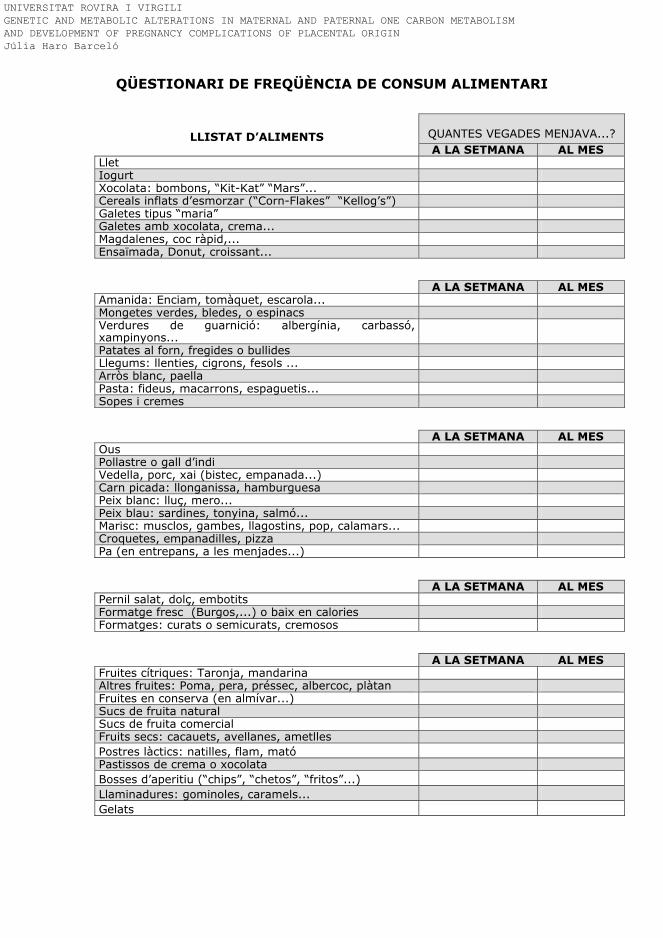
QÜESTIONARI DE FREQÜÈNCIA DE CONSUM ALIMENTARI
LLISTAT D’ALIMENTS QUANTES VEGADES MENJAVA...?
A LA SETMANA AL MES Llet Iogurt Xocolata: bombons, “Kit-Kat” “Mars”... Cereals inflats d’esmorzar (“Corn-Flakes” “Kellog’s”) Galetes tipus “maria” Galetes amb xocolata, crema... Magdalenes, coc ràpid,... Ensaïmada, Donut, croissant...
A LA SETMANA AL MES Amanida: Enciam, tomàquet, escarola... Mongetes verdes, bledes, o espinacs Verdures de guarnició: albergínia, carbassó, xampinyons...
Patates al forn, fregides o bullides Llegums: llenties, cigrons, fesols ... Arròs blanc, paella Pasta: fideus, macarrons, espaguetis... Sopes i cremes
A LA SETMANA AL MES Ous Pollastre o gall d’indi Vedella, porc, xai (bistec, empanada...) Carn picada: llonganissa, hamburguesa Peix blanc: lluç, mero... Peix blau: sardines, tonyina, salmó... Marisc: musclos, gambes, llagostins, pop, calamars... Croquetes, empanadilles, pizza Pa (en entrepans, a les menjades...)
A LA SETMANA AL MES Pernil salat, dolç, embotits Formatge fresc (Burgos,...) o baix en calories Formatges: curats o semicurats, cremosos
A LA SETMANA AL MES Fruites cítriques: Taronja, mandarina Altres fruites: Poma, pera, préssec, albercoc, plàtan Fruites en conserva (en almívar...) Sucs de fruita natural Sucs de fruita comercial Fruits secs: cacauets, avellanes, ametlles
Postres làctics: natilles, flam, mató
Pastissos de crema o xocolata
Bosses d’aperitiu (“chips”, “chetos”, “fritos”...)
Llaminadures: gominoles, caramels...
Gelats
UNIVERSITAT ROVIRA I VIRGILI GENETIC AND METABOLIC ALTERATIONS IN MATERNAL AND PATERNAL ONE CARBON METABOLISM AND DEVELOPMENT OF PREGNANCY COMPLICATIONS OF PLACENTAL ORIGIN Júlia Haro Barceló

LLISTAT D’ALIMENTS QUANTES VEGADES MENJAVA...?
A LA SETMANA AL MES Begudes ensucrades (“coca-cola”, “Fanta”...) Begudes baixes en calories (coca-cola light...) Vi, sangria Cervesa Cervesa sense alcohol Begudes destil·lades: (Whisky, ginebra, conyac,...)
Indiqui amb una X la resposta que vulgui assenyalar: 1.- Han canviat els seus hàbits alimentaris des dels 6 mesos previs a l’embaràs de la seva
parella fins ara?
Sí__ No__ En cas que la resposta sigui afirmativa, esculli el tipus de dieta que fa actualment:
Dieta baixa en calories (reducció de pes)
Dieta hipercalòrica
Dieta hiperproteica
Dieta per a hipercolesterolèmia
Dieta per a diabètics
Dieta baixa en sal
Altres ______________________________________________________
2.- A taula, s'afegia sal a les menjades?
Mai__ Alguna vegada__ Freqüentment __ Gairebé sempre __
3.- Com definiria la seva gana d’aquells 6 mesos? Molta __ Força __ Normal __ Poca __ Gens__ 4.- Quin tipus de llet prenia habitualment?: Sencera __ Semidescremada __ Descremada __ 5.- Quin tipus de iogurt consumia habitualment?
a) Natural ___ b) Natural descremat ___ c) De sabors ___ d) De sabors descremat ___ e) Amb trossets de fruita__ ___ f) Amb trossets de truites descremat ___
6.- Quin tipus de pa menjava habitualment?: Blanc __ Integral __ 7.- Es posava tomàquet i oli als entrepans?: Sempre__ Habitualment__ Alguna vegada__ Gairebé mai__
UNIVERSITAT ROVIRA I VIRGILI GENETIC AND METABOLIC ALTERATIONS IN MATERNAL AND PATERNAL ONE CARBON METABOLISM AND DEVELOPMENT OF PREGNANCY COMPLICATIONS OF PLACENTAL ORIGIN Júlia Haro Barceló

Nom.......................................................................... Data .... ..... ....
ENQUESTA SOBRE HÀBITS I ESTIL DE VIDA
Per diferents motius, els hàbits nutricionals i l’estil de vida del pare poden tenir una
clara incidència en el procés de la reproducció. Si us plau, contesti sincerament
aquestes preguntes per ajudar-nos a valorar la influència que podien haver tingut
en el desenvolupament de l’embaràs de la seva parella.
Completi aquesta informació (pensant en els 6 mesos previs a l’embaràs de la
seva parella):
Data de naixement: _____
Pes: ______ kg
Alçada: _______ cm
Antecedents mèdics: _________________________________________________
__________________________________________________________________
Grups ABO i Rh: _______
1. Ús de suplements vitamínics
Va prendre suplements vitamínics durant els 6 mesos anteriors a l’embaràs de la
seva parella? Sí No
Només en cas que sí:
- Quin? ___________________________________
- Quantes vegades a la setmana el prenia?
Cada dia
Alguns dies (1-3 vegades)
La majoria dels dies (4-6 vegades)
- Quantes pastilles/sobres/ampolles prenia cada dia? ____________
UNIVERSITAT ROVIRA I VIRGILI GENETIC AND METABOLIC ALTERATIONS IN MATERNAL AND PATERNAL ONE CARBON METABOLISM AND DEVELOPMENT OF PREGNANCY COMPLICATIONS OF PLACENTAL ORIGIN Júlia Haro Barceló

2. Esmorzar
3. Tabac :
Era fumador passiu (exposat al fum de tabac habitualment, a casa o a la feina)?
Sí No
Era fumador actiu abans que la seva parella es quedés embarassada?
Sí No
0 cigs/dia 1-5 cigs/dia 6-10 cigs/dia > 10 cigs/dia Quants cigarrets fumava els 6 mesos abans de l’embaràs de la seva parella?
Si era fumador actiu:
Va mantenir el seu hàbit durant tot l’embaràs
Va abandonar totalment el seu hàbit durant tot l’embaràs
Va reduir el seu consum de tabac durant l’embaràs
4. Alcohol:
Mai /Ocasionalment
< 3 cops /setmana
Cada dia com aperitiu i/o amb els àpats
> 7 copes /setmana
Als 6 mesos anteriors a l’embaràs de la seva parella, amb quina freqüència bevia alcohol ?
Ha pres algun tipus de substància tòxica en els 5 anys anteriors a l’embaràs de la
seva parella (marihuana, cocaïna, heroïna, etc...)?
Sí No
En el cas que sí, especifiqui quines: ____________________________________
Mai Ocasionalment Regularment Als 6 mesos anteriors a l’embaràs de la seva parella, amb quina freqüència prenia substàncies tòxiques ?
Sí No Tenia el costum d’esmorzar? Esmorzava cereals inflats habitualment (ex. Kelloggs / Nestlé etc)? Prenia cafè amb cafeïna? Prenia cafè descafeïnat?
UNIVERSITAT ROVIRA I VIRGILI GENETIC AND METABOLIC ALTERATIONS IN MATERNAL AND PATERNAL ONE CARBON METABOLISM AND DEVELOPMENT OF PREGNANCY COMPLICATIONS OF PLACENTAL ORIGIN Júlia Haro Barceló

5. Activitat física:
a ) De què treballava? ___________________________________________
b ) Com definiria el treball d’aquell moment?
- Bàsicament d’estar assegut i caminar poc (estudiant, docent, conductor de vehicles, dependent, administratiu, ...)
- Caminar força però sense fer esforç vigorós (pagès, fàbrica, carter...)
- Bàsicament de molta activitat física (esportista)
c ) Quina activitat feia en el seu temps lliure?
- Lectura, televisió i activitats que no requereixin activitat física important.
- Caminar, anar amb bicicleta, jardineria (no s’inclou el transport d’anar i tornar del treball).
- Córrer, esquiar, gimnàstica, jocs amb pilota o esports vigorosos regularment.
- Entrenament esportiu regular per competició.
Quants cops per setmana realitzava les activitats de la pregunta anterior? _____
6. Ingressos anuals nets totals a la llar, en aquell moment: - Nombre de persones que formaven la unitat familiar: _______ - Ingressos totals:
Exemple, si la dona tenia un sou de 20000 €, l’home un de 18000€ i hi havia un avi que vivia amb la família i rebia una pensió de 6000 €
< 9000 € >9000 € - 19000 € >19000 € - 25000 € >25000 € - 35000 € > 35000 €
■
≤ 9000 € >9000 € - 19000 € >19000 € - 25000 € >25000 € - 35000 € > 35000 €
Anoti qualsevol dubte o aclariment relacionat amb aquesta enquesta:
Moltes gràcies per la teva participació.
UNIVERSITAT ROVIRA I VIRGILI GENETIC AND METABOLIC ALTERATIONS IN MATERNAL AND PATERNAL ONE CARBON METABOLISM AND DEVELOPMENT OF PREGNANCY COMPLICATIONS OF PLACENTAL ORIGIN Júlia Haro Barceló
Research paper
Moderately elevated first trimester fasting plasma total homocysteineis associated with increased probability of miscarriage. The Reus-Tarragona Birth Cohort Study
Pere Cavall�e-Busquets a, b, c, Montserrat Ingl�es-Puig a, d, Joan D. Fernandez-Ballart b, c, e,Júlia Haro-Barcel�o c, e, Alejandra Rojas-G�omez c, e, Carla Ramos-Rodriguez c, e,Monica Ballesteros d, Klaus Meyer f, Per M. Ueland f, Michelle M. Murphy b, c, e, *
a Hospital Universitari Sant Joan de Reus, Spainb IISPV, Spainc CIBEROBN, Spaind Hospital Universitari Joan XXIII de Tarragona, Spaine Unitat de Medicina Preventiva i Salut Pública, Facultat de Medicina i Ci�encies de La Salut, Universitat Rovira i Virgili, Reus, Spainf BeVital AS, Bergen, Norway
a r t i c l e i n f o
Article history:
Received 30 November 2019
Accepted 15 January 2020
Available online 18 January 2020
Keywords:
Early pregnancy
Homocysteine
Red blood cell folate
Miscarriage
Reus-Tarragona birth cohort
a b s t r a c t
The association between elevated early pregnancy fasting plasma total homocysteine (tHcy) and
miscarriage risk was investigated prospectively in participants (n ¼ 544) from the Reus-Tarragona Birth
Cohort study. Pregnancy was confirmed before 12 gestational weeks (GW) by ultrasound scan and a
fasting blood sample collected. Pregnancies with complications other than miscarriages were excluded.
Miscarriages were diagnosed by ultrasound scan and gestational age at the time of miscarriage estimated
by embryo size, where possible. Cases in which blood samples were collected more than a week after the
miscarriage, or the miscarriage was of known cause, were excluded.
Fasting plasma folate, vitamin B12, tHcy, cotinine (biomarker of smoking), red blood cell (RBC) folate,
MTHFR 677C > T (rs1801133) and SLC19A1 80G>A (rs1051266) genotypes were determined.
The exposed group consisted of participants with first trimester tHcy � P90 (7.1 mmol/L) (n ¼ 57) and
unexposed of those with tHcy < P90 (n ¼ 487). Adherence to folic acid supplement recommendations,
plasma folate, plasma vitamin B12, RBC folate and prevalence of optimal RBC folate status (� 906 mmol/L)
were lower in the exposed compared to unexposed group. The prevalences of the MTHFR 677 TT ge-
notype and miscarriage were higher in the exposed group. The relative risks (95% CI) of pregnancy
ending in miscarriage were 2.5 (1.1, 5.7) and 2.1 (1.0, 4.5) for participants in the high tHcy and suboptimal
RBC folate groups (compared to the reference groups) respectively. Adherence to folic acid supplement
recommendations was positively associated, while the MTHFR 677 TT versus CC genotype and smoking
versus non-smoking were negatively associated, with RBC folate status.
© 2020 Published by Elsevier B.V.
1. Introduction
1.1. Background
Moderately elevated fasting plasma total homocysteine (tHcy)has been associated with various pregnancy complications or
adverse outcomes such as neural tube defects [1], preeclampsia [2]or low birth weight [3,4], among others [5]. Homocysteine meta-bolism is regulated by gene-nutrient interactions and depends ondietary B-group vitamins: folate, vitamin B12, pyridoxine, andriboflavin as well as choline and betaine. The MTHFR 677C > T(rs1801133) and SLC19A1 180G > A (rs1051266) polymorphismsaffect the role of folate in homocysteine metabolism and folatetransport, respectively. They have negative effects on folate statusand are associated with elevated tHcy [6].* Corresponding author. Unitat de Medicina Preventiva i Salut Pública, Facultat de
Medicina i Ci�encies de la Salut, Universitat Rovira i Virgili, 43201, Reus, Spain.
E-mail address: [email protected] (M.M. Murphy).
Contents lists available at ScienceDirect
Biochimie
journal homepage: www.elsevier .com/locate/b iochi
https://doi.org/10.1016/j.biochi.2020.01.008
0300-9084/© 2020 Published by Elsevier B.V.
Biochimie 173 (2020) 62e67
UNIVERSITAT ROVIRA I VIRGILI GENETIC AND METABOLIC ALTERATIONS IN MATERNAL AND PATERNAL ONE CARBON METABOLISM AND DEVELOPMENT OF PREGNANCY COMPLICATIONS OF PLACENTAL ORIGIN Júlia Haro Barceló

1.2. Evidence to date regarding tHcy and miscarriage/pregnancy
loss
Numerous studies have investigated the association betweentHcy and recurrent pregnancy loss or miscarriage. However, earlypregnancy tHcy determinations from the index pregnancy inwhichmiscarriage is clinically diagnosed are difficult to obtain. Moststudies compared tHcy, measured after the affected pregnancieshave ended, between women with a history of recurrent miscar-riage versus normal pregnancy. Some of these studies reportedhigher tHcy in women with a history of miscarriage compared tonormal pregnancy [7e10] and that the probability of history ofmiscarriage was increased with increasing tHcy concentration [7].However, in this latter study, vitamin B12 deficiency prevalencewas high among the miscarriage cases but low in the controls.Other studies did not observe any differences in tHcy betweenwomen with history of pregnancy loss compared to normal preg-nancies [11e14]. The disparity in results between the aforemen-tioned studies may be due to various reasons. None of themmeasured tHcy before the clinical diagnosis of miscarriage in theaffected pregnancy. tHcy levels decrease during pregnancy [15] sononpregnant measurements may not accurately reflect even earlypregnancy concentrations. Furthermore, following the pregnancyloss women were taking folic acid supplements in many studies inpreparation for the next pregnancy, thus affecting their tHcy.Studies with a reliable measurement of the exposure of interestprior to the miscarriage are lacking. In addition to the limitationsregarding the exposure measurements, endpoints based on aclinical diagnosis of miscarriage are also scarce. This is relevantbecause efforts to differentiate between miscarriages likelyresulting from other factors unrelated to tHcy, such as foetalchromosomal abnormalities ormaternal infection, arewarranted. Aprospective study from preconception throughout pregnancy inwhich conception and pregnancy loss were monitored by urinaryhCG concentrations, concluded that elevated tHcy at preconception(�12.4 mmol/L) did not increase the relative risk of early pregnancyloss [16]. Miscarriage causes are not assessed in this study ofpregnancy loss before 6 GW. Another prospective study, from thefirst prenatal visit, of 100 pregnancies measured tHcy in bloodsamples collected between 4 and 16 GW. No difference in tHcy wasobserved between the women that went on to miscarry and thosethat had a normal pregnancy outcome [17]. It is not clear whetherthe statistical power was sufficient (there were only 12 mis-carriages), the timing of sample collection covered a range of 12weeks which affects tHcy and no information was providedregarding the timing, cause or type of miscarriage. Impairedvitamin B12 status was associated with a higher probability ofmiscarriage in that same study. A large French study reportedhigher tHcy in samples collected following hospitalization formiscarriage in the index pregnancy compared to elective preg-nancy termination controls of similar gestational age [18]. Theblood samples in cases and controls were collected soon after theevents and detailed information regarding miscarriage diagnosisand exclusion of cases due to known causes is provided.
1.3. Hypothesis and aims
We hypothesised that moderately elevated early pregnancytHcy is a potential biomarker of idiopathic first trimester miscar-riage risk.
The aim of this study was to investigate, prospectively, the as-sociation between moderately elevated early pregnancy tHcy andthe risk of first trimester miscarriage in the Reus-Tarragona BirthCohort.
2. Materials and methods
2.1. Study participants
Women attending their first prenatal clinic at the Universityhospitals Sant Joan Reus and Joan XXIII Tarragona between 2005and 2016, with confirmed pregnancy of less than 12 GW, wereinvited to participate in the Reus Tarragona Birth Cohort (RTBC)study. The study was approved by the Hospitals’ Research andethical committees and signed informed consent following theguidelines of the Declaration of Helsinki was obtained from allparticipants.
2.2. Blood sample collection
Fasting blood samples were collected at < 12 GW, 15 GW,24e27 GW, 34 GW and nonfasting samples at labour. For the pur-poses of the present report, only the first blood sample will beconsidered. Participants that developed pregnancy complicationsother than miscarriage (preeclampsia, intrauterine growth retar-dation, gestational hypertension, among others) were excluded(n¼ 75) from this report. A total of 544 pregnancies were included.Samples were stored at �80 �C in the Pere Virgili Health ResearchInstitute (IISPV) biobank until analysis. Clinical, nutritional andlifestyle data were recorded and plasma folate and RBC folate(microbiological assay with Lactobacillus casei) [19], plasma vitaminB12 (microbiological assay with Lactobacillus leichmannii) [20] andhomocysteine (tHcy) and cotinine concentrations were determinedby liquid-tandem mass spectrometry [21]. The MTHFR 677C>T(rs1801133) and SLC19A1 180G>A (rs1051266) genotypes weredetermined by matrix-assisted laser desorption/ionization/time-of-flight MS [22]. (Bevital; www.bevital.no). Data regarding smok-ing habits was collected from three different sources includinginterrogation by the investigating team (questionnaire), plasmacotinine determinations and from the prenatal history (recorded bythe clinicians).
2.3. Pregnancy confirmation and miscarriage diagnosis
Between 11 and 13 þ 6 GW, pregnancy was confirmed by ul-trasound scan. The majority of the miscarriages were first trimesterspontaneous “missed” abortions diagnosed on detection of nofoetal heartbeat by ultrasound scan at 12 GW. The remaining mis-carriages were in course and diagnosed on referral from theemergency room when the clinical symptoms were manifested.Ultrasound scans revealing absence of foetal heartbeat or emptyyolk sac were diagnosed as miscarriage. Gestational age at the timeof miscarriage was estimated, where possible, from the crown-rump length or biparietal diameter of the embryo. Cases of mis-carriages occurring more than 7 days before blood sample collec-tion, were excluded from the study.
2.4. Statistical analysis
Participants were classified as exposed to moderately elevatedfirst trimester tHcy (� P90: 7.1 mmol/L), n¼ 57, or unexposed (< P90),n ¼ 487. Smokers were identified based on plasma cotinine con-centration � 10 ng/ml and/or confirmation of smoking habit byquestionnaire or on interrogation by the clinicians during theprenatal check-ups. Quantitative variables with non-normal dis-tributions were natural log transformed for the application ofparametric statistical tests. Means between groups were comparedusing ANOVA and proportions using the Chi-square test. We fitted aCox regression model to calculate the relative risk (RR) of miscar-riage associated with moderately elevated tHcy. The model was
P. Cavall�e-Busquets et al. / Biochimie 173 (2020) 62e67 63
UNIVERSITAT ROVIRA I VIRGILI GENETIC AND METABOLIC ALTERATIONS IN MATERNAL AND PATERNAL ONE CARBON METABOLISM AND DEVELOPMENT OF PREGNANCY COMPLICATIONS OF PLACENTAL ORIGIN Júlia Haro Barceló

adjusted for maternal age and smoking habit (active smokingversus non-smoking during pregnancy). Similarly, another Coxregression model was fitted to determine the RR of miscarriageassociated with suboptimal RBC folate status during the firsttrimester of pregnancy. Predictors of tHcy and RBC folate statuswere assessed using multiple linear regression analysis and mul-tiple logistics regression analysis respectively. IBM-SPSS softwarewas used for all statistical tests. Significance level was set atp < 0.05.
3. Results
3.1. Cases included
Of the miscarriage cases, nine were excluded for the followingreasons: chorioamnionitis (n ¼ 3), antiphospholipid syndrome(n ¼ 1), myoma (n ¼ 1), trisomy 18 (n ¼ 1), late miscarriage, >18 GW (n ¼ 2), missing information (n ¼ 1). The 32 miscarriagesoccurring before 18 GWand of unknown causewere included in theanalysis.
3.2. Participant characteristics according to first trimester tHcy
category
Participant characteristics are described in Table 1. SLC19A1
80G>A genotypes, smoking habits, parity, maternal age and bodymass index did not differ between the exposed and unexposedgroups. Adherence to folic acid supplement recommendations of400 mg/d was high in both groups but higher in the group withtHcy < P90. Plasma vitamin B12 status, plasma and RBC folate statuswere lower and the MTHFR 677 TT genotype prevalence higher inthe exposed versus unexposed to moderately elevated tHcy group.The WHO recommends a RBC folate status of 906 nmoL/L or moreto prevent neural tube defects [23]. A higher proportion of partic-ipants in the high tHcy group had RBC folate concentrations belowthis recommendation and the proportion of early pregnancymiscarriage was higher in the exposed (high tHcy) than the unex-posed group.
3.3. Exposure to first trimester tHcy and risk of miscarriage
The association between first trimester tHcy status and risk ofmiscarriage is reported in Table 2. Participants with tHcy at orabove the 90th percentile (7.1 mmol/L) had over twice the risk ofhaving a miscarriage. Risk of miscarriage also increased withincreasing maternal age. We assessed whether RBC folate statusbelow 906 nmoL/L affects the risk of miscarriage (Table 2). Womenwith RBC cell folate status< 906 nmol/L were twice as likely to havea miscarriage compared to women with red blood cell folate �
906 nmol/L, after adjusting for maternal age, parity and smoking.
3.4. Participant characteristics according to pregnancy outcome
The participant characteristics according to outcome (miscar-riage or normal pregnancy) are reported in Table 3. Women withpregnancies that ended in miscarriage were older, adhered less tothe recommendation to take 400 mg/d of folic acid in the form ofsupplements and more of them had suboptimal folate reserves(indicated by RBC folate concentration, showing folate reservesentering pregnancy) compared to women that went on to havenormal pregnancy outcomes.
3.5. Factors predicting first trimester tHcy
The predictors of first trimester tHcy were assessed usingmultiple lineal regression analysis (Table 4). The strongest predictorwas MTHFR 677 TT genotype, followed by plasma creatinine, parityand plasma folate. In a separate model in which first trimesterplasma folate was replaced with RBC folate, the strongest predictorwas RBC folate, followed by plasma creatinine, MTHFR 677 TT ge-notype and parity.
3.6. Factors influencing first trimester RBC folate status
The factors influencing the probability of having optimal RBC folatestatus in the first trimester of pregnancywere assessed usingmultiplelogistic regression analysis (Table 5). Regular use of folic acid sup-plements at or above the recommended dose of 400 mg/d strongly
Table 1
Participant characteristics according to exposure to first trimester fasting plasma total homocysteine category.
First trimester fasting plasma total homocysteine group
� P901
< P90 Total
MTHFR 677 C > T genotype, %
CC 23.6 [13/55]2 35.2 [170/483] 34.0 [183/538]
CT 43.6 [24/55] 49.3 [238/483] 48.7 [262/538]
TT 32.7 [18/55] 15.5 [75/483]** 17.3 [93/538]
SLC19A1 80 G > A genotype, %
GG 38.2 [21/55] 26.7 [128/480] 27.9 [149/535]
GA 38.2 [21/55] 46.5 [223/480] 45.6 [244/535]
AA 23.6 [13/55] 26.9 [129/480] 26.5 [142/535]
First trimester smoking, % 31.6 [18/57] 26.9 [131/487] 27.4 [149/544]
First trimester folic acid use, %3 89.1 [49/55] 94.5 [446/487]*** 93.9 [495/527]
Multiparity, % 47.4 [27/57] 54.2 [264/487] 54.4 [296/544]
Age (years), mean (95% CI) 32.4 (30.9, 33.9) [57] 32.1 (31.7, 32.5) [486] 32.2 (31.8, 32.5) [543]
BMI (kg/m2), mean (95% CI) 24.6 (23.2, 26.0) [55] 23.8 (23.4, 24.3) [464] 23.9 (23.5, 24.2) [516]
Plasma folate (nmol/L), geometric mean (95% CI) 14.4 (11.2, 18.4) [57] 26.7 (25.2, 28.4) [487]*** 25.1 (23.6, 26.6) [544]
Plasma vitamin B12 (pmol/L), geometric mean (95% CI) 283 (261, 343) [57] 369 (358, 381) [487]** 363 (352, 373) [544]
tHcy (mmol/L),
geometric mean (95% CI)
8.4 (8.0, 8.7) [57] 5.1 (5.0, 5.2) [487]*** 5.3 (5.2, 5.4) [544]
RBC folate (nmol/L), geometric mean (95% CI) 556 (477, 647) [57] 954 (910, 1001) [474]*** 901 (859, 945) [531]
RBC folate < 906 nmol/L, % 78.9 [45/57] 46.4 [220/474]*** 49.9 [265/531]
Miscarriage, % 14.0 [8/57] 4.9 [24/487]* 5.9 [32/544]
Gestational week at miscarriage, mean (95% CI) 9.3 (7.8, 10.8) [7] 10.4 (9.1, 11.8) [16] 10.1 (9.1, 11.1) [23]
Abbreviations: tHcy: fasting plasma total homocysteine, RBC: red blood cell. 17.1 mmol/L, 2[n], 3� 400 mg/d. Statistical comparison between 2 groups, Chi square for proportions
and ANOVA for quantitative variables: *p < 0.05, **p < 0.01, ***p < 0.001.
P. Cavall�e-Busquets et al. / Biochimie 173 (2020) 62e6764
UNIVERSITAT ROVIRA I VIRGILI GENETIC AND METABOLIC ALTERATIONS IN MATERNAL AND PATERNAL ONE CARBON METABOLISM AND DEVELOPMENT OF PREGNANCY COMPLICATIONS OF PLACENTAL ORIGIN Júlia Haro Barceló

Table 2
Assessment of Relative risks of pregnancy ending in miscarriage according to early pregnancy fasting plasma total homocysteine (tHcy) and RBC folate status using Cox
regression analysis. Abbreviations: tHcy: fasting plasma total homocysteine, RBC: red blood cell. 1Relative risk; 2measured at< 12 gestational weeks; 3P90: 7.1 mmol/L; 4adjusted
for parity (multiparous versus nulliparous) and for smoking versus nonsmoking during pregnancy.**p < 0.01.
RR1 (95% CI) Deviance likelihood ratio, chi square n, df
Unadjusted tHcy2 model 17.2 544, 1**
tHcy � P90 versus < P903 2.85 (1.28, 6.34)
Adjusted tHcy2 model 17.5 543, 4**
tHcy � P90 vs < P90 2.52 (1.12, 5.68)
Maternal age (y) 1.13 (1.04, 1.22)
Unadjusted RBC folate2 model4 2.6 531, 1
RBC folate
<906 versus � 906 nmol/L
1.83 (0.87, 3.81)
Adjusted RBC folate2 model4 15.0 530, 4**
RBC folate
<906 versus � 906 nmol/L
2.11 (1.00, 4.45)
Maternal age (y) 1.15 (1.06, 1.24)
Table 3
Participant characteristics according to pregnancy outcome.1 [n], Abbreviations: tHcy: fasting plasma total homocysteine, RBC: red blood cell. Statistical com-
parison between 2 groups, Chi square for proportions and ANOVA for quantitative variables: *p < 0.05,**p < 0.01,***p < 0.001, #p ¼ 0.067.
Miscarriage Normal pregnancy
n ¼ 32 n ¼ 512
MTHFR 677 C > T genotype, %
CC 36.7 [11/30]1 33.8 [173/508]
CT 40.0 [12/30] 49.4 [253/508]
TT 23.3 [7/30] 16.8 [86/508]
SLC19A1 80 G > A genotype, %
GG 30.0 [9/30] 27.7 [140/505]
GA 33.3 [10/30] 46.2 [234/505]
AA 36.7 [11/30] 25.9 [131/505]
First trimester smoking, % 21.9 [7/32] 28.0 [145/512]
First trimester folic acid supplement use, % 68.0 [17/25] 95.2 [478/502]***
Multiparity, % 50.0 [16/32] 53.7 [280/512]
Age (years), mean (95% CI) 34.6 (32.9, 36.3) [32] 32.0 (31.6, 32.4) [511]**
BMI (kg/m2), mean (95% CI) 23.5 (21.7, 25.3) [15] 23.9 (23.5, 24.3) [501]
Plasma folate (nmol/L), geometric mean (95% CI) 20.4 (14.7, 28.2) [32] 25.4 (23.9, 27.0) [512]*
Plasma vitamin B12 (pmol/L), geometric mean (95% CI) 359 (316, 408) [32] 363 (352, 374) [512]
tHcy (mmol/L), geometric mean (95% CI) 6.0 (5.4, 6.7) [32] 5.3 (5.2, 5.4) [512]**
RBC folate (nmol/L), geometric mean (95% CI) 837 (679, 1030) [31] 905 (861, 950) [500]
RBC folate < 906 nmol/L, % 64.5 [20/31] 49.0 [245/500]#
Table 4
Predictors of first trimester tHcy using multiple lineal regression analysis.
Beta coefficient Adjusted R square n, df
Model 1 0.090*** 504, 10
MTHFR 677 TT versus CC genotype 0.220***
Plasma creatinine (mmol/L) 0.180***
Parity (multiparous versus nulliparous) �0.126**
Plasma folate (nmol/L) �0.109*
Model 2 0.148*** 491, 10
RBC folate (nmol/L) �0.273***
Plasma creatinine (mmol/L) 0.192***
MTHFR 677 TT versus CC genotype 0.185***
Parity (multiparous versus nulliparous) �0.119*
Abbreviations: RBC: red blood cell. The dependent variable in both models is ln tHcy. Both models were adjusted for maternal age, plasma B12, smoking, MTHFR 677 CT
versus CC genotype SLC19A1 80 AA vs GG and SLC19A1 80 GA vs GG genotypes. *p < 0.05, **p < 0.01, ***p < 0.001.
Table 5
Assessment of predictors of probability of optimal red blood cell folate status (� 906 mmol/L) usingmultiple logistic regression analysis. Model adjusted for maternal age,
previous pregnancy, MTHFR 677 CT versus CC, SLC19A1 80 AA vs GG and SLC19A1 80 GA vs GG genotypes.***p < 0.001.
OR (95% CI) Nagelkerke R2 n, df
Model 0.123*** 505, 9
First trimester folic acid use (�400 mg/d) 15.1 (3.5, 64.9)
Smoking vs nonsmoking 0.53 (0.31, 0.91)
MTHFR 677 TT vs CC genotype 0.56 (0.33, 0.94)
P. Cavall�e-Busquets et al. / Biochimie 173 (2020) 62e67 65
UNIVERSITAT ROVIRA I VIRGILI GENETIC AND METABOLIC ALTERATIONS IN MATERNAL AND PATERNAL ONE CARBON METABOLISM AND DEVELOPMENT OF PREGNANCY COMPLICATIONS OF PLACENTAL ORIGIN Júlia Haro Barceló
increased the probability of having optimal RBC folate status. On theother hand, smoking versus nonsmoking and theMTHFR TT versus CCgenotype were associated with 44% and 47% reductions, respectively,in the probability of having optimal RBC folate status.
4. Discussion
4.1. Principal findings
Elevated early pregnancy tHcy was associated with more thandouble the risk of having a miscarriage. First trimester RBC folateconcentration < 906 nmol/L, indicative of suboptimal folate re-serves entering pregnancy, was also associated with increased riskof miscarriage. Smoking had a negative effect on RBC folate statuswhile folic acid supplement use at or above the recommended400 mg/d had a protective effect.
4.2. Comparison with previous studies
Most previous studies determined tHcy after the miscarriagehad occurred. A study that did collect blood samples between 4 and12 GW reported no difference in tHcy between miscarriage casesand controls [16]. There were two important differences betweenthat study and ours. Firstly, it only had 12 miscarriage cases. It isunclear whether it was sufficiently powered to detect a differencein tHcy between cases and controls, if it existed. Furthermore, RBCfolate status in general was higher in that study than in ours. Sec-ondly, no details regarding timing, types or potential causes ofmiscarriage were provided. To the best of our knowledge, the otherstudy with tHcy measurements nearest to the miscarriage was inpatients being treated for the miscarriage [17]. That study by Griset al., was large and had blood samples and ultrasound confirma-tion of the miscarriage close to the time of the event. Miscarriagesoccurring late in pregnancy or due to chromosomal abnormalitiesor maternal infection were also excluded from that study. Ourfindings confirm their findings that miscarriage risk was increasedwith increasing tHcy concentrations. They reported a twofold in-crease in risk for tHcy � 9.9 mmol/L. This effect size is similar to ourobservation regarding tHcy � 7.1 mmol/L.
4.3. Interpretation
The mechanism for the association between tHcy and miscar-riage warrants investigation. It is possible that elevated tHcy in ourstudy is marking impaired folate status. The most important pre-dictors of first trimester tHcy were RBC folate, followed by theMTHFR 677 TT genotype. We previously reported in a populationstudy from the same region that adults with the MTHFR 677 TTgenotype had lower folate status (both plasma and RBC folate) aswell as higher tHcy than their CC or CT genotype counterparts [6].We also observed in that same study that 18.8% of the participantshad folate deficiency. In contrast to widespread folic acid use in theReus Tarragona Birth Cohort, the population study did not includefolic acid users. Nevertheless, there is no mandatory fortificationwith folic acid in Spain and most participants in the Reus TarragonaBirth Cohort did not initiate folic acid supplementation until theywere pregnant [24]. Use of the recommended dose of folic acidsupplements and plasma folate status were lower in cases than incontrols and the percentage of cases with RBC folate below thethreshold recommended by theWHO to prevent neural tube defectaffected pregnancies, was higher in cases than in controls.
Impaired one carbon metabolism due to low folate status, theMTHFR 677C>T polymorphism or other polymorphisms affectingthe role of folate or other nutrients in the one carbon metabolicnetwork have been associated with adverse outcomes stemming
from anomalies in early pregnancy [5]. It is possible that the physiopathological mechanism leading to embryo developmental abnor-malities, impaired placentation and foetal growth is shared, at leastin part, in pregnancies affected by suboptimal one carbon meta-bolism. Impaired chorionic vascularisation in spontaneousmiscarriage tissue from women with history of recurrent preg-nancy loss and with tHcy >18.3 mmol/L was reported in a study byNelen et al. [25]. It is also possible that anomalies in DNA methyl-ation and other epigenetic processes arising from impaired onecarbon metabolism are involved. However, further research in thisfield is required to explore and establish the associations betweenearly pregnancy folate status and tissue-specific outcomes, theirimpact and replication between studies.
4.4. Strengths and limitations
Strengths of this study were that pregnancy was confirmed byultrasound scan and tHcy was determined before the miscarriageoccurred. Few previously reported studies have achieved thesemeasurements due to the difficulty in obtaining them. Latemiscarriage cases (caused by infections or foetal developmentalabnormalities) as well as miscarriages due to known causes such aschromosomal abnormalities were also excluded. Strictly, fastingblood samples and confirmation of pregnancy by ultrasound scanbefore 12 GW were required for eligibility to be included in thestudy. The study was designed to measure first trimester tHcy as apotential biomarker of adverse pregnancy outcome and bloodsamples were processed in strict adherence to protocol to preventhomocysteine release from blood cells [26].
Furthermore, RBC folate concentration was determined and isindicative of folate reserves during preconception and the start ofpregnancy. It is spared the effects of haemodilution and the initialeffects of folic acid supplement use (unlike plasma folate concen-tration and tHcy, which are sensitive to folic acid supplement use atthe time of the blood draw).
Estimation of time of miscarriage in “missed” spontaneousabortions based on changes in transvaginal ultrasound measure-ments of crown-rump length or parietal circumference of the em-bryo are susceptible to error depending on time elapsed betweencessation of foetal heartbeat and the performance of the scan.Gestational age based on reported date of the last menstrual periodby the participants is also subject to error. However, we were ableto minimise these errors due to the prospective nature of the studyand recording of the timing of the blood samples and ultrasoundscans. We stipulated that any miscarriage suspected to haveoccurred more than 7 days before the blood samples would beexcluded.
4.5. Implications
This study shows that in the absence of mandatory fortificationwith folic acid, women not adhering to the recommended intake of400 mg/d from folic acid supplements, are more likely not to meetthe threshold RBC folate status proposed to offer protection againstfolate sensitive neural tube defects. This study shows that RBCfolate status below this threshold also increases the risk ofmiscarriage. Smoking was negatively associated with RBC folatestatus. These results indicate that the message regarding theimportance of periconception folic acid in the prevention ofadverse pregnancy outcome needs to be reinforced, and especiallyin smokers.
5. Conclusions
Moderately elevated early pregnancy tHcy is associated with 2.5
P. Cavall�e-Busquets et al. / Biochimie 173 (2020) 62e6766
UNIVERSITAT ROVIRA I VIRGILI GENETIC AND METABOLIC ALTERATIONS IN MATERNAL AND PATERNAL ONE CARBON METABOLISM AND DEVELOPMENT OF PREGNANCY COMPLICATIONS OF PLACENTAL ORIGIN Júlia Haro Barceló

times more risk of early miscarriage, of unknown cause. The resultsprovide evidence to support the consideration of early pregnancytHcy as a potential biomarker of adverse pregnancy outcome.
Author contributions
MMM, PC-B and JDF-B designed the research; PC-B, MI-P, MBwere responsible for the clincial aspects of the study; JH-B, AR, CR-R, MMM were responsible for coordinating the field work of thestudy as well as data and sample collection, processing and bio-banking; PMU and KM were responsible for the biochemical andgenetic determinations carried out at Bevital AS; PC-B, MI-P, MMM,JF-B, JH-B, AR-G and CR-R analysed the data and wrote the manu-script; MMM and PC-B had primary responsibility for the finalcontent.
Declaration of competing interest
None to declare.
Acknowledgements
The Reus-Tarragona Birth Cohort Study is registered at www.clinicaltrials.gov as NCT01778205. This study was supported byresearch grants from the Interministerial Science and TechnologyCommittee (SAF2005-05096); Carlos III Health Institute, NationalScientific Research, Development and Technological InnovationProgramme Health Investigation Resources (co-financed by TheEuropean Regional Development Fund; FIS10/00335, FIS13/02500,FIS16/00506); Joint Programming Initiative ‘A Healthy Diet for aHealthy Life’ (ERA HDHL), JFA2 Nutrition and the Epigenome, Ho-rizon2020 grant agreement number 696300, with funding pro-vided by The Spanish State Agency for Investigation PCI2018-093098/AEI; Pere Virgili Health Research Institute (IISPV-2010/21);Biomedical Research Networking Centre for the Pathophysiology ofObesity (CIBERobn); Agency for Management of University andResearch grants, Generalitat de Catalunya (Support to ResearchGroups: AGAUR SGR 2009e1237, 2014e332) and Italfarmaco S.A.,Spain.
Alejandra Rojas’ predoctoral fellowship has been co-funded bythe European Union’s Horizon 2020 research and innovation pro-gramme under the Marie Skłodowska-Curie grant agreement No.713679 and the Universitat Rovira i Virgili (URV). Júlia Har�o-Barcel�oand Carla Ramos-Rodriguez are predoctoral fellows funded by theMartí Franques programme, URV.
References
[1] J.L. Mills, J.M. McPartlin, P.N. Kirke, Y.L. Lee, M.R. Conley, D.G. Weir, Homo-cysteine in pregnancies complicated by neural tube defects, Lancet 345 (1995)149e151.
[2] A.M. Cotter, A.M. Molloy, J.M. Scott, S.F. Daly, Elevated plasma homocysteinein early pregnancy: a risk factor for the development of severe preeclampsia,Am. J. Obstet. Gynecol. 185 (2001) 781e785.
[3] M.M. Murphy, J.M. Scott, V.A. Arija, A.M. Molloy, J.D. Fernandez-Ballart,Maternal homocysteine before conception and throughout pregnancy pre-dicts fetal homocysteine and birth weight, Clin. Chem. 50 (2004) 1406e1412.
[4] R. Onalan, G. Onalan, Z. Gunenc, E. Karabalut, Combining 2nd trimestermaternal serum homocysteine levels and uterine artery Doppler for predic-tion of preeclampsia and isolated intrauterine growth restriction, Gynecol.Obstet. Investig. 61 (2006) 142e148.
[5] M.M. Murphy, J.D. Fernandez-Ballart, Homocysteine in pregnancy, Adv. Clin.Chem. 53 (2011) 105e137.
[6] O. Bueno, A.M. Molloy, J.D. Fernandez-Ballart, C.J. García-Minguill�an,S. Ceruelo, L. Ríos, P.M. Ueland, K. Meyer, M.M. Murphy, Common poly-morphisms that affect folate transport or metabolism modify the effect of the
MTHFR 677C > T polymorphism on folate status, J. Nutr. 146 (1) (2016 Jan)1e8.
[7] M. Puri, L. Kaur, G.K. Walia, R. Mukhopadhhyay, M.P. Sachdeva, S.S. Trivedi,K.N. Saraswathy, MTHFR C677T polymorphism, folate, vitamin B12 and ho-mocysteine in recurrent pregnancy losses: a case control study among NorthIndian women, J. Perinat. Med. 41 (2013) 549e554.
[8] M. Mascarenhas, S. Habeebullah, M.G. Sridhar, Revisiting the role of firsttrimester homocysteine as an index of maternal and fetal outcome,J Pregnancy 2014 (2014), 123024.
[9] M. Nasiri, A. Arsanjani Shirazi, O. Sadeghi, M. Bagheri Bidakhavidi, The rela-tionship between homocysteine levels and spontaneous abortion in Iranianwomen with migraine, Iran. J. Public Health 46 (2017) 1149e1151.
[10] Y. Zarfeshan Fard, O. Kooshkaki, D. Kordi Tammandani, G. Anani Sarab,Investigation of the association between C677T polymorphism of the MTHFRgene and plasma homocysteine level in recurrent fetal miscarriage, J. Obstet.Gynaecol. Res. 45 (2019) 1442e1447.
[11] M. Creus, R. Deulofeu, J. Pe~narrubia, F. Carmona, J. Balasch, Plasma homo-cysteine and vitamin B12 serum levels, red blood cell folate concentrations,C677T methylenetetrahydrofolate reductase gene mutation and risk ofrecurrent miscarriage: a case-control study in Spain, Clin. Chem. Lab. Med. 51(2013) 693e699.
[12] W.V. Boas, R.O. Gonçalves, O.L.N. Costa, M.S. Goncalves, Metabolism and genepolymorphisms of the folate pathway in Brazilian women with history ofrecurrent abortion, Rev. Bras. Ginecol. Obstet. 37 (2015) 71e76. Revista DaFederacao Brasileira Das Sociedades de Ginecologia E Obstetricia.
[13] M.M. Wagner, J.W. Jukema, W. Hermes, S. le Cessie, C.J.M. de Groot,J.A. Bakker, K.W.M. Bloemenkamp, Assessment of novel cardiovascular bio-markers in women with a history of recurrent miscarriage, PregnancyHypertens 11 (2018) 129e135.
[14] Z. Lin, Q. Li, Y. Sun, J. Huang, W. Wang, J. Fu, D. Zeng, Interactions betweengenetic variants involved in the folate metabolic pathway and serum lipid,homocysteine levels on the risk of recurrent spontaneous abortion, LipidsHealth Dis. 18 (2019) 143.
[15] M.M. Murphy, J.M. Scott, J.M. McPartlin, J.D. Fernandez-Ballart, Thepregnancy-related decrease in fasting plasma homocysteine is not explainedby folic acid supplementation, hemodilution, or a decrease in albumin in alongitudinal study, Am. J. Clin. Nutr. 76 (2002) 614e619.
[16] A.G. Ronnenberg, S.A. Venners, X. Xu, C. Chen, L. Wang, W. Guang, A. Huang,X. Wang, Preconception B-vitamin and homocysteine status, conception, andearly pregnancy loss, Am. J. Epidemiol. 166 (2007) 304e312.
[17] E.M. Guerra-Shinohara, P.M. Pereira, A.M. Kubota, T.A. Silva, J.L. Reis,G.S. Miyashita, V. D’Almeida, R.H. Allen, S.P. Stabler, Increased MMA con-centration and body mass index are associated with spontaneous abortion inBrazilian women: a pilot study, Clin. Chim. Acta 411 (2010) 423e427.
[18] J.C. Gris, T.V. Perneger, I. Qu�er�e, E. Mercier, P. Fabbro-Peray, G. Lavigne-Lis-salde, M. Hoffet, H. D�echaud, J.C. Boyer, S. Ripart-Neveu, M.L. Tailland,J.P. Daur�es, P. Mar�es, M. Dauzat, Antiphospholipid/antiprotein antibodies,hemostasis-related autoantibodies, and plasma homocysteine as risk factorsfor a first early pregnancy loss: a matched case-control study, Blood 102(2003) 3504e3513.
[19] A.M. Molloy, J.M. Scott, Microbiological assay for serum, plasma, and red cellfolate using cryopreserved, microtiter plate method, Methods Enzymol. 281(1997) 43e53.
[20] B.P. Kelleher, S.D. Broin, Microbiological assay for vitamin B12 performed in96-well microtitre plates, J. Clin. Pathol. 44 (1991) 592e595.
[21] P.M. Ueland, O. Midttun, A. Windelberg, A. Svardal, R. Skålevik, S. Hustad,Quantitative profiling of folate and one-carbon metabolism in largescaleepidemiological studies by mass spectrometry, Clin. Chem. Lab. Med. 45(2007) 1737e1745.
[22] K. Meyer, A. Fredriksen, P.M. Ueland, High-level multiplex genotyping ofpolymorphisms involved in folate or homocysteine metabolism by matrix-assisted laser desorption/ionization mass spectrometry, Clin. Chem. 50(2004) 391e402.
[23] World Health Organisation, WHO Guideline: Optimal Serum and Red BloodCell Folate Concentrations in Women of Reproductive Age for Prevention ofNeural Tube Defects, WHO, Geneva (Switzerland), 2015, 978 92 4 154904 2Page 5.
[24] P. Sol�e-Navais, J. Salat-Batlle, P. Cavall�e-Busquets, J. Fernandez-Ballart,P.M. Ueland, M. Ballesteros, G. Ornosa-Martín, M. Ingl�es-Puig, J.M. Colomina,M.M. Murphy, Early pregnancy folate-vitamin B12 interactions and their ef-fects on vitamin B12 status and hematologic variables throughout pregnancy,Am. J. Clin. Nutr. 107 (2018) 173e182.
[25] W.L.D.M. Nelen, J. Bulten, E.A.P. Steegers, H.J. Blom, A.G.J.M. Hanselaart,T.K.A.B. Eskes, Maternal homocysteine and chorionic vascularization inrecurrent pregnancy loss, Hum. Reprod. 15 (2000) 954e960.
[26] H. Refsum, A.D. Smith, P.M. Ueland, E. Nexo, R. Clarke, J. McPartlin, C. Johnston,F. Engbaek, J. Schneede, C. McPartlin, et al., Facts and recommendations abouttotal homocysteine determinations: an expert opinion, Clin. Chem. 50 (2004)3e32.
P. Cavall�e-Busquets et al. / Biochimie 173 (2020) 62e67 67
UNIVERSITAT ROVIRA I VIRGILI GENETIC AND METABOLIC ALTERATIONS IN MATERNAL AND PATERNAL ONE CARBON METABOLISM AND DEVELOPMENT OF PREGNANCY COMPLICATIONS OF PLACENTAL ORIGIN Júlia Haro Barceló

UNIVERSITAT ROVIRA I VIRGILI GENETIC AND METABOLIC ALTERATIONS IN MATERNAL AND PATERNAL ONE CARBON METABOLISM AND DEVELOPMENT OF PREGNANCY COMPLICATIONS OF PLACENTAL ORIGIN Júlia Haro Barceló