Caveolae and Caveolins in the Respiratory System
13
Current Molecular Medicine 2008, 8, 741-753 741 1566-5240/08 $55.00+.00 © 2008 Bentham Science Publishers Ltd. Caveolae and Caveolins in the Respiratory System Reinoud Gosens* ,1 , Mark Mutawe 2,3 , Sarah Martin 2,3,4 , Sujata Basu 2,3 , Sophie T. Bos 1 , Thai Tran 5 and Andrew J. Halayko 2,3,4 1 Department of Molecular Pharmacology, University of Groningen, Groningen, The Netherlands 2 Departments of Physiology & Internal Medicine, University of Manitoba, Winnipeg, MB, Canada 3 Biology of Breathing Group, Manitoba Institute of Child Health, Winnipeg, MB, Canada 4 National Training Program in Allergy and Asthma, Winnipeg, MB, Canada 5 Department of Physiology, National University of Singapore, Singapore Abstract: Caveolae are flask-shaped invaginations of the plasma membrane that are present in most structu- ral cells. They owe their characteristic -shape to complexes of unique proteins, the caveolins, which indirectly tether cholesterol and sphingolipid-enriched membrane microdomains to the cytoskeleton. Caveolins possess a unique scaffolding domain that anchors receptors, ion channels, second messenger producing enzymes, and effector kinases, thereby sequestering them to caveolae, and modulating cellular signaling and vesicular trans- port. The lungs express numerous caveolae and high levels of caveolins; therefore they likely play an impor- tant role in lung physiology. Indeed, recent and ongoing studies indicate important roles for caveolae and ca- veolins in the airway epithelium, airway smooth muscle, airway fibroblasts, airway inflammatory cells and the pulmonary vasculature. We review the role of caveolae and caveolins in lung cells and discuss their involve- ment in cellular signaling associated with asthma, COPD, lung cancer, idiopathic pulmonary fibrosis and pul- monary vascular defects. Keywords: COPD, asthma, lung cancer, fibrosis, pulmonary hypertension, smooth muscle, fibroblast, airway epi- thelium. INTRODUCTION The discovery of caveolae resulted from ultrastruc- tural studies in 1953 that identified “plasmalemmal ve- sicles” [1]. In 1955, these invaginations were termed “caveolae” (little caves) [2]. Caveolae are 50- to 100- nm small bulb-shaped invaginations of the plasma membrane that represent a subclass of lipid rafts enri- ched in cholesterol and sphingomyelins. Today they are thought to play an important role in numerous cellu- lar processes, including membrane trafficking and cell signaling, endocytosis, and intracellular cholesterol transport [3, 4]. Indeed, caveolae and caveolins likely play a broad role in human physiology and pharmaco- logy [5]. Caveolae are found in many cell types, inclu- ding structural cells of the lung. Their presence is de- pendent on the expression of a unique family of pro- teins, the caveolins, which bind cholesterol and insert in the inner leaflet of the plasma membrane [6]. This article will review the role of caveolae and caveolins in lung cells, and will discuss their involvement in cellular signaling that is associated with a number of lung di- seases, including asthma, COPD, lung cancer, idiopat- hic pulmonary fibrosis and pulmonary vascular defects. *Address correspondence to this author at the Department of Mole- cular Pharmacology, University of Groningen, A. Deusinglaan 1, 9713 AV Groningen, The Netherlands; Tel: + 31 50 363 3321; Fax: +31 50 363 6908; E-mail: [email protected] THE CAVEOLIN GENE FAMILY To date, three human caveolin genes have been identified: Caveolin-1 (Cav-1), Cav-2 and Cav-3. Cav-1 was the first member of the caveolin gene family disco- vered; it is composed of 3 exons that are highly con- served in structure and sequence across species [7, 8]. Caveolin-2 was discovered by microsequencing of a 20 kDa protein that co-purified with adipocyte-derived ca- veolar membranes [9]. Further experimentation revea- led that caveolin-2 was co-expressed in many of the same cells and tissues as caveolin-1, and required ca- veolin-1 for proper membrane localization [10, 11]. Ca- veolin-3 was identified through traditional cDNA library screening and database searching [12]. The Cav-1 and Cav-2 genes are both localized to human chromosome 7q31.1, whereas the Cav-3 gene is located on chromo- some 3p25. Caveolin-2 protein is ~38% identical and ~58% simi- lar to caveolin-1, while caveolin-3 is ~65% identical and ~85% similar to caveolin-1 [13]. Caveolin proteins ran- ge in size from ~18-24 kDa, and all three types have an invariant 8 amino acid ‘FEDVIAEP’ sequence within a hydrophobic amino-terminal domain; the functional im- portance of this domain has not been fully elucidated [13]. Caveolin-1 and caveolin-3 can form homo- oligomeric complexes in the golgi apparatus, which enables their transport the plasma membrane [12, 14]. Caveolin protein complexes are typically comprised of 14-16 monomeric units [15, 16], and they can often migrate as 200-400 kDa protein bands upon electrop-
Transcript of Caveolae and Caveolins in the Respiratory System

Current Molecular Medicine 2008, 8, 741-753 741
1566-5240/08 $55.00+.00 © 2008 Bentham Science Publishers Ltd.
Caveolae and Caveolins in the Respiratory System
Reinoud Gosens*,1, Mark Mutawe2,3, Sarah Martin2,3,4, Sujata Basu2,3, Sophie T. Bos1, Thai Tran5 and Andrew J. Halayko2,3,4
1Department of Molecular Pharmacology, University of Groningen, Groningen, The Netherlands
2Departments of Physiology & Internal Medicine, University of Manitoba, Winnipeg, MB, Canada
3Biology of Breathing Group, Manitoba Institute of Child Health, Winnipeg, MB, Canada
4National Training Program in Allergy and Asthma, Winnipeg, MB, Canada
5Department of Physiology, National University of Singapore, Singapore
Abstract: Caveolae are flask-shaped invaginations of the plasma membrane that are present in most structu-
ral cells. They owe their characteristic -shape to complexes of unique proteins, the caveolins, which indirectly
tether cholesterol and sphingolipid-enriched membrane microdomains to the cytoskeleton. Caveolins possess
a unique scaffolding domain that anchors receptors, ion channels, second messenger producing enzymes, and
effector kinases, thereby sequestering them to caveolae, and modulating cellular signaling and vesicular trans-
port. The lungs express numerous caveolae and high levels of caveolins; therefore they likely play an impor-
tant role in lung physiology. Indeed, recent and ongoing studies indicate important roles for caveolae and ca-
veolins in the airway epithelium, airway smooth muscle, airway fibroblasts, airway inflammatory cells and the
pulmonary vasculature. We review the role of caveolae and caveolins in lung cells and discuss their involve-
ment in cellular signaling associated with asthma, COPD, lung cancer, idiopathic pulmonary fibrosis and pul-
monary vascular defects.
Keywords: COPD, asthma, lung cancer, fibrosis, pulmonary hypertension, smooth muscle, fibroblast, airway epi-thelium.
INTRODUCTION
The discovery of caveolae resulted from ultrastruc-tural studies in 1953 that identified “plasmalemmal ve-sicles” [1]. In 1955, these invaginations were termed “caveolae” (little caves) [2]. Caveolae are 50- to 100-nm small bulb-shaped invaginations of the plasma membrane that represent a subclass of lipid rafts enri-ched in cholesterol and sphingomyelins. Today they are thought to play an important role in numerous cellu-lar processes, including membrane trafficking and cell signaling, endocytosis, and intracellular cholesterol transport [3, 4]. Indeed, caveolae and caveolins likely play a broad role in human physiology and pharmaco-logy [5]. Caveolae are found in many cell types, inclu-ding structural cells of the lung. Their presence is de-pendent on the expression of a unique family of pro-teins, the caveolins, which bind cholesterol and insert in the inner leaflet of the plasma membrane [6]. This article will review the role of caveolae and caveolins in lung cells, and will discuss their involvement in cellular signaling that is associated with a number of lung di-seases, including asthma, COPD, lung cancer, idiopat-hic pulmonary fibrosis and pulmonary vascular defects.
*Address correspondence to this author at the Department of Mole-cular Pharmacology, University of Groningen, A. Deusinglaan 1,
9713 AV Groningen, The Netherlands; Tel: + 31 50 363 3321; Fax: +31 50 363 6908; E-mail: [email protected]
THE CAVEOLIN GENE FAMILY
To date, three human caveolin genes have been identified: Caveolin-1 (Cav-1), Cav-2 and Cav-3. Cav-1 was the first member of the caveolin gene family disco-vered; it is composed of 3 exons that are highly con-served in structure and sequence across species [7, 8]. Caveolin-2 was discovered by microsequencing of a 20 kDa protein that co-purified with adipocyte-derived ca-veolar membranes [9]. Further experimentation revea-led that caveolin-2 was co-expressed in many of the same cells and tissues as caveolin-1, and required ca-veolin-1 for proper membrane localization [10, 11]. Ca-veolin-3 was identified through traditional cDNA library screening and database searching [12]. The Cav-1 and Cav-2 genes are both localized to human chromosome 7q31.1, whereas the Cav-3 gene is located on chromo-some 3p25.
Caveolin-2 protein is ~38% identical and ~58% simi-lar to caveolin-1, while caveolin-3 is ~65% identical and ~85% similar to caveolin-1 [13]. Caveolin proteins ran-ge in size from ~18-24 kDa, and all three types have an invariant 8 amino acid ‘FEDVIAEP’ sequence within a hydrophobic amino-terminal domain; the functional im-portance of this domain has not been fully elucidated [13]. Caveolin-1 and caveolin-3 can form homo-oligomeric complexes in the golgi apparatus, which enables their transport the plasma membrane [12, 14]. Caveolin protein complexes are typically comprised of 14-16 monomeric units [15, 16], and they can often migrate as 200-400 kDa protein bands upon electrop-

742 Current Molecular Medicine, 2008, Vol. 8, No. 8 Gosens et al.
horetic separation (for an example refer to Fig. (2)). Studies using deletion mutagenesis show that caveolin-1 contains an oligomerization domain between resi-dues 61-101 [16], Fig. (1). Interestingly, caveolin-2 cannot form homo-oligomers, but can initiate the forma-tion of hetro-oligomeric complexes with caveolin-1 [11].
Two isoforms of caveolin-1, and , have been identified. The -isoform consists of 178 amino acid residues, while the isoform arises from an internal translational start site that results in truncation of resi-dues 1-31 at the amino terminus [17]. Both isoforms have two common hydrophobic segments of amino acids, the so-called C-terminal and N-terminal mem-brane attachment domains (C-MAD and N-MAD, res-pectively), Fig. (1). Caveolin-1 has emerged as a criti-cal regulator of signaling pathways that control cell ad-hesion, proliferation and differentiation [13, 18-20]. Ca-veolin-1 is implicated as a regulator in signal transduc-tion due in large part to the presence of a caveolin scaffolding domain (CSD), which includes a 20 amino acid stretch (residues 82-101) capable of binding a va-riety of signaling proteins [21] Fig. (1). Binding of these proteins to the caveolin-1 CSD is usually associated with inactivation of downstream signaling effectors.
To date, three isoforms of caveolin-2 have been identified; , and . Caveolin-2 is the full-length ge-ne product (20 kDa), while caveolin-2 (18 kDa) and 2 (15 kDa) are truncated variants formed due to alternati-ve translation initiation of caveolin-2 full length mRNA [22]. Thus far, little is known about the functional signi-ficance of the caveolin-2 and isoforms [13]. Also, the role of full length caveolin-2 in cell signaling is less
clear compared to that of caveolin-1, as caveolin-2 lacks a CSD and requires caveolin-1 for caveolae for-mation [11]. Nonetheless, caveolin-2 knockout mice have clear pulmonary defects and in fact share may of the abnormalities seen in caveolin-1 knockout mice [23]. Though this suggests a critical functional role for caveolin-2 in the lung, the mechanistic basis for this role is still only partially elucidated. The details of these pulmonary abnormalities in caveolin-1 and caveolin-2 knockout mice will be discussed in later sections.
Caveolin-3 contains a CSD similar to caveolin-1 and has also been demonstrated to compartmentalize and modulate signaling proteins and receptors in cardiac and skeletal muscles [24-26]. Caveolin-3 has also been shown to modulate membrane-cytoskeleton interac-tions and associated signaling, as it contains a WW-like domain that binds the C-terminus of both -dystroglycan and dystrophin [27]. Thus caveolin can regulate the interaction of dystrophin with -dystroglycan and linkage of the dystrophin glycoprotein complex between laminin extracellular matrix and the intracellular filamentous actin network. The functionality of the WW-domain of caveolin-1 in assembling the dys-trophin-dystroglycan complex has been demonstrated [27] and its importance in airway smooth muscle con-traction and lung development has also been postula-ted, but this still needs to be fully elucidated [28], Fig. (1).
CAVEOLIN EXPRESSION IN THE LUNG
Caveolin-1 and caveolin-2 have a relatively ubiqui-tous distribution pattern, with the highest levels being
Fig. (1). Structure and functional domains of caveolin-1. Full length caveolin-1 (caveolin 1 ) is anchored to the plasma membra-
ne through a palmitoylated (C133, C143, C156) C-terminal membrane attachment domain (C-MAD) and an N-terminal membra-
ne attachment domain (N-MAD). Overlapping with the N-MAD are the CSD and the WW binding domain, which are responsible
for oligomerization, for binding to signal transduction proteins and for binding to the cytoskeleton.

Caveolae and Caveolins in the Respiratory System Current Molecular Medicine, 2008, Vol. 8, No. 8 743
Fig. (2). Distribution of caveolin subtype mRNA and protein in human lung. A) Total protein lysates from primary cultured human
bronchial smooth muscle cells was subjected to immunoblotting for caveolins. Immunoblots show expression of caveolin-1 (21
kDa), and to a lesser extent the caveolin-1 subtypes (24 kDa). Also seen is a high molecular weight caveolin oligomer that was
resistant to disruption by the SDS sample buffer used. B) Caveolin-1 immunostaining in the guinea pig lung showing heteroge-
neity in caveolin-1 expression among different compartments of the airways and lung. Positive staining was most profound in the
lung parenchyma (P), the airway smooth muscle (ASM) and the pulmonary vasculature (V). C) Human bronchial smooth muscle
tissue, peripheral lung tissue,or tracheal smooth muscle tissue was homogenized and analyzed for expression of caveolin-1, -2
and -3 proteins. All three tissue samples expressed caveolin-1 and caveolin-2 ( and subtypes), in abundance. Caveolin-3 was
not detected. For immunoblots, the gels were all normalized for equal loading of total protein (10 μg) measured using a Bradford
Assay for lysates obtained using standard RIPA buffer. Equal loading of samples in respective lanes was also confirmed by
comparing -actin load. D) Positive control for caveolin-3 expression in mouse diaphragm, heart and skeletal muscle. Smooth
muscle obtained from the mouse small intestine was negative for caveolin-3, similar to airway smooth muscle. E) RT-PCR ana-
lysis of human bronchial smooth muscle tissue and lung tissue; mRNA for caveolin-1 and caveolin-2, but not caveolin-3 were
detected. Total RNA was extracted using the Qiagen RNeasy Mini Kit, total RNA (1 μg) was reverse transcribed using M-MLV
reverse transcriptase for 1 h at 37°C, and subsequent PCR reactions f were carried out using primer pairs for: Cav1 (forward 5‘-
CTCCGAGGGACATCTCTACAC – 3‘ & reverse 5‘- GTGCTGATGCGGATGTTGCTG -3‘), Cav-2 (forward 5’- CGATGTG-
CAGCTCTTCATGGC -3‘ & reverse 5‘CGATGCTGACCGATGAGAAGC -3‘) and for Cav-3 ( forward 5‘- GATCTGGAAGCTCG-
GATCATC -3‘ & reverse 5‘- GTGCGGATACACAGTGAGTAG-3‘).
found in endothelial cells, adipocytes, epithelial cells, smooth muscle cells and fibroblasts [13]. Caveolin-3, also known as muscle caveolin or M-caveolin, is prima-rily expressed in skeletal and cardiac muscle cells, though reports of its presence in some smooth muscle tissues also exist [12, 29]. In human lung tissue, we have observed the co-expression of abundant caveolin-1 and caveolin-2 protein and mRNA, in the trachea and bronchus, whereas caveolin-3 mRNA appears to be absent Fig. (2). In a recent study, caveolin isoform ex-pression and cell type distribution in the rat lung was studied in detail [30]. As also shown in Fig. (2), caveo-lin-1 / and caveolin-2 / isoforms are abundantly ex-pressed in whole lung homogenate. Interestingly, ca-veolin-1 immunoreactivity was observed in tracheal and bronchial epithelial cells, tracheal and bronchial smooth
muscle, vascular endothelial and smooth muscle cells, airway fibroblasts and type I alveolar cells, but was ab-sent from surfactant protein-D positive alveolar type II cells and airway epithelial cells in the rat small bronchi [30]. This fits with our own data indicating heteroge-neous caveolin-1 expression among lung cells with intense caveolin-1 immunoreactivity in the parenchy-ma, the airway smooth muscle and pulmonary blood vessels (Fig. (2)). Caveolin-2 immunoreactivity showed a similar distribution pattern though caveolin-2 was in very low abundance in tracheal epithelial cells [30]. Though absent from human lung tissue, caveolin-3 was expressed in the rat airway epithelium throughout the bronchial tree [31]. This broad and heterogeneous ex-pression profile in the lung suggests an important, but cell-type specific role for caveolae and caveolins in de-

744 Current Molecular Medicine, 2008, Vol. 8, No. 8 Gosens et al.
termining cellular responses in health and disease; this is confirmed by studies showing that caveolin-1 and caveolin-2 knockout mice have severe pulmonary ab-normalities [6, 23, 32], and by other studies showing that aberrant caveolin-1/2 expression is involved in di-seases such as idiopathic pulmonary fibrosis, lung cancer and pulmonary hypertension [33-35]. The sub-type distribution of caveolin proteins in lung cell types and their association with disease is summarized in
Table 1. The pulmonary defects seen in caveolin-1 knockout mice are summarized in Table 2.
CAVEOLAE AND CAVEOLINS IN THE REGULATION OF AIRWAY TONE
Airway Smooth Muscle Contraction
Obstructive airways diseases, such as asthma and COPD, are characterized by airway hyperresponsive-
Table 1. Subtype Distribution of Caveolin Proteins in Lung Cell Types and their Association with Disease
Cell Type Caveolin isoform
Association with disease Remarks Reference
Lung fibroblast 1 & 2 Pulmonary fibrosis Reduced caveolin expression associated with idiopathic pulmonary fibrosis (IPF).
Caveolins suppress proliferation and matrix protein / MMP production by fibroblasts.
[30, 33]
[6, 23, 32, 33, 97, 101-103]
Airway smooth muscle
1 & 2 Expression are highest in contractile phenotype.
Caveolins facilitate contractile function of airway smooth muscle, but inhibit
proliferation.
[30, 41] and Fig. (2)
[41, 54, 55, 82]
Epithelial cells – airway and alveolar
1 & 2
3
Lung cancer & Pulmonary fibrosis
Caveolin-1 & -2 absent from Type II alveolar epithelium and airway epithelial cells of small
bronchi.
Absent from human lung, but present in rat airway epithelium.
Caveolins facilitate water, electrolyte and macromolecule transport, and organize nitric
oxide production. Caveolins inhibit cell proliferation.
[30, 33, 34]
[31]
[117, 120-122,
124, 125]
Pulmonary vascula-ture
1 & 2 Pulmonary hypertension Altered expression in endothelium and smooth muscle associated with pulmonary
hypertension.
[29, 35, 140]
Macrophage 1 Not determined Reduced expression associated with changes in LPS induced cytokine release.
[126]
Neutrophil 1 Not determined Role in adhesion, migration and superoxide production
[127]
Dendritic cell 1 Not determined [126]
Lymphocyte 1 Not determined [126]
Table 2. Functional and Morphological Differences in Caveolin-1 Knockout Mice of Relevance to the Respiratory System
Functional / morphological difference Reference
Pulmonary fibrosis [6, 23, 32, 96]
Hypercellular lung tissue (endothelial cells and type I pneumocytes)
[6, 23, 32]
Pulmonary hypertension [128]
Augmented endothelium mediated vascular relaxation [6]
Increased Ca2+
sensitization of vascular smooth muscle [29]
Reduced exercise tolerance [32]
Reduced transendothelial macromolecule transport [120]
Reduced pulmonary inflammation in response to LPS exposure [130]

Caveolae and Caveolins in the Respiratory System Current Molecular Medicine, 2008, Vol. 8, No. 8 745
ness to inhaled and endogenous bronchoconstrictors [36], which is in part accompanied by contractile ab-normalities of the airway smooth muscle [37-40]. Nota-bly, caveolin-1 and caveolin-2 are abundant in airway smooth muscle (Fig. (2)). In cell culture caveolin-1 ex-pression is increased as airway smooth muscle cell undergo maturation to a contractile phenotype [41]. Moreover, the number of caveolae on smooth muscle cells is greatest in mature contractile myocytes, where as many as 100,000 such structures per cell may exist [42, 43]. Collectively, these observations suggest a prominent role for caveolae and caveolins in the regu-lation of contractile function.
Caveolae have been implicated in Ca2+
handling in airway smooth muscle [44]. Caveolae obtained from canine airway smooth muscle contain L-type Ca
2+
channels, the Ca2+
binding proteins calsequestrin and calreticulin, and the plasma membrane Ca
2+ pump [44].
Studies using other cell types indicate that IP3 recep-tors, large conductance Ca
2+ activated K
+ channels and
members of the TRPC class of transient receptor po-tential channels all localize to caveolae and likely inter-act with caveolin-1 [45-49]. The direct role for caveolae in Ca
2+ handling is further illustrated by their localiza-
tion in the close proximity of organelles that are asso-ciated with intracellular Ca
2+ signaling, such as the sar-
coplasmatic reticulum and mitochondria, possibly facili-tating filling and refilling of intracellular stores from the Ca
2+ rich extracellular environment [50, 51]. This would
also imply a direct role for caveolin-1, which is critically important for the maintenance of the caveolae invagi-nations through interaction with components of the dys-trophin-dystroglycan complex; this complex tethers ca-veolae to the actin cytoskeleton [28, 52]. Collectively, these studies illustrate a potentially important role for
caveolae in the early signaling events leading to airway smooth muscle contraction.
Plasma membrane caveolae are enriched in G-protein coupled receptors for contractile agonists, the corresponding intracellular G proteins, and associated second messenger producing enzymes [28, 53]. For example, recent studies by our group and others using human and canine airway smooth muscle indicate muscarinic M3 receptors and G q/11 localize to caveo-lae, and that muscarinic receptor-mediated intracellular Ca
2+ flux is reduced in airway smooth muscle cells after
treatment with cholesterol depleting agents, caveolin-1 gene silencing or treatment with a cell-permeable pep-tide mimicking the CSD domain of caveolin-1 [54, 55]. Caveolins do not appear to directly bind to and modula-te G protein coupled receptors; however, caveolae and caveolins likely support smooth muscle contraction by regulating Gq protein function and phospho-inositide metabolism [56, 57]. Moreover, plasma membrane re-cruitment of RhoA and PKC , which is an important component in Ca
2+-sensitization that promotes smooth
muscle contraction, is dependent on caveolin-1 [58-60]. Given the abundance of caveolin-1 and caveolin-2 in contractile airway smooth muscle, and the emerging evidence for a role of these proteins in signaling cas-cades associated with contractile responses, increa-sing future investigation is clearly warranted.
Nitric Oxide Metabolism
Nitric oxide (NO) produced by epithelial cells is a central physiological modulator of airway tone, inflam-mation and remodeling [61]. Excessive or reduced NO production is associated with the etiology of many air-ways diseases, including asthma, COPD, lung cancer
Fig. (3). Nitric oxide metabolism is organized in caveolae in airway and alveolar epithelial cells. (A) In resting epithelial cells,
eNOS associates with caveolae and is inactivated by caveolin-1. Upon induction of intracellular [Ca2+
] and concomitant forma-
tion of Ca2+
/calmodulin complexes, the inhibitory role of caveolin-1 is reversed, which activates the eNOS enzyme. Availability of
L-arginine to eNOS in caveolae is regulated by the arginine transporter CAT-1 and the arginine regenerating enzymes arginino-
succinate synthase (ASS) and argininosuccinate lyase (ASL) which also localize to caveolae. This creates an organized mole-
cular machinery for NO production in epithelial cell caveolae. See text for further details.

746 Current Molecular Medicine, 2008, Vol. 8, No. 8 Gosens et al.
and obliterative bronchiolitis [61]. Caveolae and caveo-lins play an important regulatory role in NO production. Endothelial nitric oxide synthase (eNOS) is targeted to caveolae through its palmitoylation, an essential ele-ment leading to NO generation [62-65]. This suggests that caveolae organize and fine-tune the molecular in-teractions that regulate NOS activity. In resting vascu-lar endothelial cells, NOS is maintained in an inactive state through interaction with caveolin proteins: caveo-lin-1 and -3, but not caveolin-2 inhibit the catalytic acti-vity of nNOS, iNOS and eNOS, by direct binding of the CSD to a caveolin binding motif in the NOS proteins [66]. In ciliated airway epithelial cells of the rat, a simi-lar interaction was recently described for caveolin-3 and eNOS, suggesting that regulatory caveolin-NOS interactions also exist in the airway epithelium [31]. Negative allosteric modulation of eNOS by caveolin-1 and-3 competes with positive allosteric modulation of eNOS by Ca
2+/calmodulin and hsp90 complexes. Thus,
in the presence of elevated Ca2+
/calmodulin and hsp90, the inhibitory actions of caveolin-1 on resting NOS activity can be fully reversed [67, 68]. This crea-tes a complex molecular on/off switch, regulated by intracellular Ca
2+ concentration, for NO production in
caveolae (Fig. (3)).
NO generation by NOS enzymes is dependent on the availability of its substrate L-arginine. Reduced availability of L-arginine and concomitant reductions in NO production contribute significantly to allergen-induced airway hyperreactivity [69]. This is explained by a reduction in L-arginine uptake by cationic trans-porters and increased L-arginine catabolism by the competing enzyme arginase [70, 71]. Though arginase is primarily expressed in the cytoplasm and mitochon-dria [69], the membrane arginine transporter CAT-1 colocalizes with eNOS in caveolae, which likely facilita-tes the delivery of L-arginine directly to caveolar NOS [72]. Caveolae may also play a regulatory role in the regeneration of L-arginine from L-citrulline, as the L-arginine regenerating enzymes argininosuccinate synt-hase and argininosuccinate lyase colocalize with eNOS in caveolae [73]. Collectively, these observations illus-trate the high level of subcellular organization of NO metabolism in caveolae (Fig. (3)). The variable expres-sion of caveolin isoforms in endothelial cells, airway and alveolar epithelial cells (Table 1) suggests that this regulatory role of caveolae and caveolins in NO-generation may be cell-type or airway generation-specific. Their functional role in NO production by epit-helium of different airway generations in particular is not well-described and future studies are clearly indica-ted in this area.
CAVEOLAE AND CAVEOLINS IN FIBROPRO-LIFERATIVE DISEASES OF THE LUNG
Airway Smooth Muscle / Fibroblast Proliferation
Proliferation of fibroblasts and airway smooth mus-cle cells contributes to many fibroproliferative diseases of the lung, including asthma, COPD, idiopathic pulmo-nary fibrosis, obliterative bronchiolitis and cystic fibrosis
(for additional information on the role of fibroblast and airway smooth muscle proliferation in these airways diseases, see [74-80]). Caveolin proteins play an im-portant role in mesenchymal cell proliferation by main-taining cellular quiescence and repressing the induction of excessive cell growth. This contention is firmly esta-blished by a variety of studies using both fibroblasts and smooth muscle cells that show that a reduction or ablation in caveolin-1 expression in these cells is suffi-cient to induce spontaneous cell proliferation [41, 81, 82]. Conversely, over expression of caveolin-1 induces cell cycle arrest and premature cellular senescence in fibroblasts, and suppresses the induction of smooth muscle cell proliferation induced by growth factors [83-85]. Also, quiescent airway smooth muscle cells and fibroblasts that reside in the G0/G1 phase of the cell cycle have increased endogenous expression of ca-veolin-1 [41, 82, 83]. Collectively, these data indicate a strong anti-mitogenic role for caveolin-1 in airway me-senchymal cells, suggesting that aberrant caveolin-1 expression may be involved in fibroproliferative disea-ses of the lung. Indeed, fibroblastic foci in the lungs of patients suffering from idiopathic pulmonary fibrosis have reduced caveolin-1 expression [33].
A number of signaling proteins involved in cell proli-feration can associate with caveolin-1. Indeed, the bin-ding to caveolin-1 of receptor tyrosine kinases (RTKs), including EGFR and PDGFR, inhibits tyrosine kinase activity [86, 87]. Nonetheless, an organizing role for caveolae in cellular signaling associated with prolifera-tion has also been suggested, as RTKs and non-RTKs, several G protein coupled receptors, monomeric and heterotrimeric G proteins (including Gi, Gq and Ras) all localize to caveolae of airway smooth muscle cells and fibroblasts [41, 64, 82, 88], Fig. (4). Moreover, growth factor-induced tyrosine phosphorylation and p42/p44 MAP kinase activation occur in caveolae isolated from fibroblasts, suggesting these sites are key foci for initia-ting signal transduction [88, 89]. There are, however, conflicting reports concerning the association of key downstream effector proteins such as p42/p44 MAP kinase with caveolae, which may, in part, be due to the dynamic interaction of signaling proteins with caveolin-1 and caveolae at the cell membrane. For instance, though phosphatidylinositol-3-kinase and p42/p44 MAP kinase are primarily localized to caveolae-free mem-branes in airway smooth muscle, the disruption of ca-veolae using membrane cholesterol depleting agents such as methyl- -cyclodextrin markedly augments ba-seline and growth factor-induced p42/p44 MAP kinase activation [41, 90]. This paradoxical effect can be ex-plained by the fact that p42/p44 MAP kinase activation by exposure to a mitogen such as EGF involves the rapid trafficking of EGF receptors, which are sequeste-red to caveolae in quiescent cells, to caveolae-free, p42/p44 MAP kinase enriched membranes [82]. Like-wise, immunoprecipitates of caveolin-1 contain abun-dant PDGFR , but only in quiescent cells, as the re-ceptor’s association with caveolin-1 is rapidly lost after growth factor exposure [41]. Collectively, these studies suggest that growth factor-induced airway myocyte

Caveolae and Caveolins in the Respiratory System Current Molecular Medicine, 2008, Vol. 8, No. 8 747
proliferation involves, consecutively, the release of the activated RTK from caveolin-1, followed by RTK traffic-king from caveolae to caveolae-free membrane, where p42/p44 MAP kinase is activated.
Extracellular Matrix Production and Fibrosis
Aberrant extracellular matrix deposition and fibrosis of the lungs and airways are features of several disor-ders, including asthma, COPD, idiopathic pulmonary fibrosis, cystic fibrosis and obliterative bronchiolitis (see [76, 77, 79, 91, 92] for extensive reviews). The most convincing evidence that caveolae and caveolins regu-late extracellular matrix protein deposition in the lung comes from histological studies of caveolin-1 and ca-veolin-2 knockout mice. Both show evidence of thicke-ned alveolar septa, accompanied by increased reticulin immunoreactivity in the basement membrane and ex-tracellular fibrillar deposits [6, 23, 32], (Table 2) Fig. (5). Notably, experimental lung fibrosis induced by radiation or bleomycin reduces pulmonary expression of caveo-lin-1, most notably in type I pneumocytes [93, 94]. Ca-veolin-1 expression declines progressively after irradia-tion, starting at an early stage of disease prior to histo-logical evidence of severe abnormality [93]. A strong reduction in the pulmonary expression of caveolin-1 is also observed in TGF- induced pulmonary fibrosis in mice [95] and in ovalbumin-induced airway inflamma-tion that is associated with airway remodeling [96]. Furthermore, fibrotic lesions of patients with idiopathic pulmonary fibrosis show reduced expression of caveo-lin-1 [33]; since caveolin-1 inhibits the TGF- receptor 1, its reduced abundance likely facilitates TGF- signa-
ling that underpins enhanced extracellular matrix pro-tein production in the lung [33, 97]. Collectively, these reports suggest an important inhibitory role for caveo-lae, and more directly, caveolin-1 and -2, in extracellu-lar matrix protein production in the lung, where reduced caveolin-1 expression appears to be an important initia-tor of fibrotic process after injury.
Conclusions on the specific role of caveolin-1 and caveolin-2 using knock out mouse models are not easy to discern. Caveolin-1 knockout mice show a reduction in caveolin-2 protein, despite there being no effect on caveolin-2 transcript abundance, indicating that caveo-lin-1 is necessary for translational or post-translational control of caveolin-2 stability [6, 23, 32]. It is not clear to what extent changes in caveolin-2 expression speci-fically contribute to the lung phenotype observed for caveolin-1 null mice. In addition to pulmonary fibrosis described above, caveolin-1 null mice also exhibit hy-percellular alveolae septae with markedly greater num-bers of Type I pneumocytes and endothelial cells [32]. Notably, the pulmonary abnormalities seen in caveolin-2 knockout mice, in which there is no concomitant re-duction in caveolin-1, are very similar to those seen in caveolin-1 knockout mice [6, 23, 32]. Therefore, though a reduction in the expression of caveolin-1 appears to associate with experimental or pathological lung fibro-sis, at present the potential significance of a parallel effect on caveolin-2 remains unclear.
The anti-fibrotic role of caveolins is also explained, in part, by their inhibitory effect on MEK/p42/p44 MAP kinase signaling in fibroblasts [98]. Caveolin-1 knock-down strongly increases baseline MEK and p42/p44
Fig. (4). Association of receptors and signaling effectors with caveolae microdomains. Caveolae are enriched in receptors and
signaling effectors such as receptor tyrosine kinases (RTKs) that bind via their caveolin binding motif to the caveolin scaffolding
domain of caveolin-1. The association is dynamic, allowing for trafficking of activated RTK to non-caveolae membrane com-
partments. As such, caveolae cluster and fine-tune signaling of receptors, including RTKs and G protein coupled receptors
(GPCRs). The lungs express abundant caveolin-1; therefore clustering of receptors and signaling effectors in caveolae likely
plays an important role in respiratory physiology in health and disease.

748 Current Molecular Medicine, 2008, Vol. 8, No. 8 Gosens et al.
MAP kinase activation in lung fibroblasts, and is ac-companied by increased collagen production that is sensitive to MEK inhibition. Interestingly, in a murine model of bleomycin-induced lung fibrosis, in vivo colla-gen deposition is accompanied by increased phospho-MEK and phospho-p42/p44 MAP kinase in the lung, with a concomitant reduction in caveolin-1 levels [98]. Thus, it appears that lung fibrosis may primarily be me-diated by aberrant caveolin-1 signaling.
Extracellular matrix remodeling is also governed by matrix metalloprotease (MMP) mediated breakdown, which serves an important role in releasing and activa-ting matrix-bound growth factors and cytokines that promote tissue repair [91, 92, 99]. The actions of MMPs on the extracellular matrix are destructive, and they appear to be involved in alveolar wall destruction associated with COPD [100]. Moreover, they are impor-tant players in extracellular matrix abnormalities of fi-brotic lung diseases, as they activate and attract fibro-blasts and inflammatory cells, which initiates abnormal tissue repair leading to extracellular matrix remodeling and fibrosis [92]. Several reports using cells from diffe-rent origins indicate caveolin-1 and caveolae inhibit the activity and/or secretion of MMP-1, -2, -9 and -13 [101-103]. Similar inhibitory actions of caveolin-1 on CD147/EMMPRIN, which regulates MMP production, have been described [101]. Thus, it appears that ca-veolae and caveolins play an important suppressive role in tissue remodeling associated with inflammatory and fibrotic lung diseases.
Caveolae and Caveolins in Lung Cancer
Epithelial cell proliferation is essential to repair the airway or lung parenchyma after an inflammatory insult
[104]. As already discussed in relation to airway smooth muscle and fibroblast proliferation, caveolins are important anti-proliferative regulatory molecules that control mitogenic quiescence. As would be expec-ted from this anti-mitogenic role, enhanced airway and alveolar epithelial cell proliferation have been reported in caveolin-1 and -2 knockout mice, resulting in hyper-cellular lung parenchyma and thickened airway epit-helium [23, 32], Fig. (5).
Most lung cancers result from uncontrolled airway or alveolar epithelial cell proliferation [34]. Several stu-dies have addressed the hypothesis that deregulated caveolin protein expression is involved in the genesis of lung cancer; results are varied, and occasionally contradictory. Several investigators report reduced ex-pression of caveolin-1 and/or caveolin-2 in lung cancer cell lines and in human lung cancer specimens [34, 105-108]; due to the anti-proliferative role of caveolin, these findings suggest a role in cancer onset and/or progression. Nevertheless, not all lung cancer speci-mens show reduced expression of caveolin-1, and poor clinical outcomes with increased metastatic capability and drug resistance have been reported for lung tu-mors in which caveolin-1 is expressed abundantly [109-112]. These apparently paradoxical findings could rela-te to the type of cancer cell under investigation, as one report indicates that reduced caveolin-1 expression is associated with ~95% of small cell lung cancer cases, whereas for most non-small cell lung cancer cases, nearly 76%, retain caveolin-1 expression [113].
Considering the broad biological role of caveolae and caveolins, it is not unlikely caveolin-1 plays multi-ple, and at times opposing roles in tumor cell prolifera-tion and metastasis, depending on the stage of the cancer [111]. Loss of caveolin-1 may be involved in the
Fig. (5). Lung morphology of caveolin-1 knockout mice. Formalin fixed, hematoxyllin and eosin stained lung sections obtained
from caveolin-1 knockout mice (B,D) or their genetic controls (A,C) are shown. Sections depict an airway (A,B) and peripheral
lung tissue (C,D), and indicate distinct differences as a result of the functional loss of caveolin-1, including thickened alveolar
septa, hypercellular lung parenchyma and reduced airspaces.

Caveolae and Caveolins in the Respiratory System Current Molecular Medicine, 2008, Vol. 8, No. 8 749
onset of certain lung cancers, as inhibition of the activi-ty of receptor and non-receptor tyrosine kinases, G proteins and effector enzymes by caveolin-1 is reduced or lost [13]. Downregulation of caveolin-1 is also suffi-cient to reduce E-cadherin expression, resulting in en-hanced transcriptional activity of -catenin, which re-portedly promotes epithelial-to-mesenchymal cell tran-sition and tumor progression [114]. Reduced caveolin-1 expression may result from hyperactivation of the p42/p44 MAP kinase cascade; overexpression of acti-vated (G12V) H-ras in 3T3 fibroblasts is sufficient to down-regulate caveolin-1, but this can be reversed by pharmacological inhibition of MEK [115]. Recombinant over expression of caveolin-1 in H-ras (G12V)-transformed 3T3 fibroblasts rescues their ongogenic phenotype and inhibits anchorage-independent cell growth [18].
Interestingly, lung cancers that have successfully developed despite the presence of caveolin-1, may engage caveolin-1 to stimulate focal adhesion kinase activation and filopodia formation, thus facilitating the metastatic capability of the cancer cell [112, 113]. It should be noted though that reduced abundance of caveolin-1 does not necessarily compromise metastatic capacity, as in caveolin-1 knockout mice with induced breast cancer there is enhanced formation of lung me-tastases, possibly due to the loss of the inhibitory ef-fects of caveolin-1 on MMP-2 and -9 secretion [102]. Clearly, the role of caveolae and caveolins in the onset and progression of lung cancer is complex, and alt-hough altered caveolin-1 expression and regulation may be involved, its precise role in the disease may vary from individual to individual.
CAVEOLAE AND CAVEOLINS IN LUNG INJURY AND INFLAMMATION
Microvascular Permeability and Pulmonary Edema
Due to their characteristic shape, caveolae were originally thought to function primarily as transport ve-sicles [1]. In the lungs, macromolecule, water and elec-trolyte transport serves a functional role by maintaining fluid balance and therefore, tissue integrity; excessive fluid transport from the microcirculation to the pulmona-ry interstitium leads to pulmonary edema [116]. Both microvascular permeability and transalveolar transport necessary for clearance of proteins and fluid from the alveolar air spaces may be mediated by caveolae [116, 117]. For more detail on the molecular mechanisms that contribute to transendothelial transport we refer to a recent excellent review [118].
Caveolar transport has been studied primarily in endothelial cells; these studies reveal rapid (i.e. in the order of seconds to minutes) transport of macromole-cule tracers across the capillary endothelium [117]. Transendothelial transport of macromolecules is sensi-tive to cholesterol-depleting agents such as filipin, and is reduced in caveolin-1 knockout mice, indicating an important role for caveolae in this process [119, 120]. Rather than actively shuttling macromolecules from
one side of a cell to the other, the mechanism respon-sible for caveolar transport is thought to involve repea-ted fusion and splitting of neighboring caveolae, thus creating a path for passive diffusion for macromolecu-les across the endothelial cell [117].
Although alveolar type I cells contain abundant ca-veolae, several investigations have demonstrated there is only limited transport of macromolecules across the pulmonary epithelium (reviewed in [117]). In a study of the alveolar clearance of albumin from the rabbit lung, within two hours of instillation, positive immunoreactivi-ty for albumin was detected in alveolar type I and II cells [121]; nonetheless, albumin was not effectively cleared from the lungs. More recent findings using the perfused rat lung indicate that transalveolar transport only in the presence of low, but not high, albumin con-centrations is inhibited by the cholesterol depleting agent filipin [122]. Thus, it appears that caveolae-dependent pathways contribute in a limited fashion to transalveolar albumin transport, as this pathway is sa-turated by elevated concentrations of albumin.
Electrolyte and water transport by alveolar epithelial cells is also regulated, to some extent, by caveolae. Epithelial sodium channels (ENaC) are localized to ca-veolae in human nasal epithelial cells; the Na
+/K
+ AT-
Pase, on the other hand, is absent from caveolae in these preparations [123]. Sodium transport across the epithelium aids the re-absorption water because of the osmotic gradient created. Caveolae membrane micro-domains are relatively permeable to water in compari-son to other cholesterol and sphingolipid enriched membrane domains [124]. The higher permeability is likely due to the high level of unsaturated fatty acyl chains present in caveolae [124], and by the presence of aquaporins, which are known to be localized to ca-veolae in endothelial cells [125]. Collectively, the above summarized data indicate that caveolae may play a role in the clearance of water, electrolytes and to a les-ser extent macromolecules from the alveolar airspaces.
Pulmonary Inflammation
Caveolin-1 has diverse functions, and both in vivo and in vitro studies suggest it may play an important role in the biological responses of inflammatory cells. Recent studies have confirmed that caveolin-1 is ex-pressed in macrophages, dendritic cells, neutrophils and lymphocytes [126, 127]. However, leukocytes lack caveolae, indicating that the expression of caveolin-1 by these cells is not necessarily associated with caveo-lae formation. Nonetheless, caveolin-1 does regulate cell surface signaling and intracellular trafficking in leu-kocytes [128].
Carrageenan induced acute lung injury that includes edema and vascular leakage can be inhibited with a caveolin-1 mimicking CSD peptide administered sys-temically [129]. Notably, the suppressive capability of the CSD peptide on the acute inflammatory response was associated with an inhibition of NO production and mimicked the anti-inflammatory effects obtained with

750 Current Molecular Medicine, 2008, Vol. 8, No. 8 Gosens et al.
glucocorticoids and a NOS inhibitor [129], suggesting that caveolin-1/NOS interactions are responsible for the anti-inflammatory effects of caveolin-1. In the lung, NO regulates NF- B activation and the consequent synt-hesis of proinflammatory proteins including ICAM-1 and inducible NO synthase (iNOS) [130]. Interestingly, NF-B activation is inhibited by NO at micromolar concen-
trations, whereas NO in the nanomolar range activates the transcription factor [131]. Thus the elevated levels of NO that are present in the lungs of caveolin-1 knoc-kout mice result in a suppression of NF- B activation and consequently reduced downstream expression of ICAM-1 and iNOS after LPS challenge [130]. In addi-tion, neutrophil sequestration, microvascular barrier injury and edema in response to LPS challenge were hindered. In agreement with this observation, a recent study indicates neutrophil caveolin-1 is necessary for adhesion to and migration across the pulmonary en-dothelium and that lack of caveolin-1 reduces PMA and fMLP-induced superoxide production by neutrophils [127]. These data demonstrate the potential role of ca-veolin-1 in regulating pulmonary inflammation; however its precise role is likely cell-type and stimulus depen-dent.
In vitro studies with murine alveolar macrophages have also revealed anti-inflammatory effects of caveo-lin-1 that are mediated via the MKK3/ p38 MAPK path-way [132]. Silencing of caveolin-1 expression in murine alveolar macrophages using small interfering RNA re-sults in increased production of the pro-inflammatory cytokines TNF- and IL-6 in response to LPS exposu-re. In contrast, synthesis and release of IL-10, is inhibi-ted by loss of caveolin-1 [132]. Notably, overexpression of caveolin-1 resulted in a complete reversal in the pat-tern of LPS induced cytokine production seen in caveo-lin-1 siRNA treated cells; this response was accompa-nied with an increase in p38 MAPK phosphorylation and decreased in NF- B and AP-1 activation [132]. Thus it appears that caveolin-1 is capable of selectively modulating different pathways associated with the in-flammatory response in alveolar macrophages.
CAVEOLAE AND CAVEOLINS IN PULMONARY VASCULAR DISEASE
Fundamentally, the function of caveolae and caveo-lins in vascular smooth muscle and vascular endot-helial cells parallels that we have already described for airway smooth muscle, airway fibroblasts and airway epithelial cells. Our focus is on the role of caveolae and caveolins in the pulmonary vasculature in relation to lung disease. Excellent reviews on the role of caveolins in the systemic vasculature have been published re-cently [13, 133].
A notable functional defect of the pulmonary vascu-lature in caveolin-1 knockout mice is the development of severe pulmonary hypertension [134] (Table 2). Pulmonary arterial pressure in caveolin-1 knockout mi-ce is approximately double that in wild type mice; simi-lar changes are observed in right ventricular pressure [134]. Several investigators have investigated the role
of caveolins in the pathogenesis of pulmonary hyper-tension and lung remodeling. Murine models of pulmo-nary hypertension associated with myocardial infarction [135], or induced by monocrotaline [136] are accompa-nied by a dramatic reduction in the expression of ca-veolin-1 and caveolin-2 in alveolar epithelial cells and pulmonary arterial endothelial cells. Concomitant with reduced caveolin-1 and -2 abundance, increased tyro-sine phosphorylated STAT3, and abundance of cyclin D1 and D3 were observed in whole lung homogenates of these mice [135, 136]. Similar changes are seen in lung homogenates from caveolin-1 and -2 knockout mice, suggesting that caveolin-1 and -2 are pivotal to the development of abnormalities associated with pul-monary hypertension [135]. This was confirmed by de-livery of a cell permeable caveolin-1 mimicking CSD peptide, which reversed pulmonary hypertension and the accompanying increases in pulmonary phospho-STAT3, cyclin D1, and cyclin D3 expression in rats ex-posed to monocrotaline [137].
Recent studies indicate that findings from small animal models may be extrapolated to the clinical set-ting. Microarray analysis indicates that patients under-going transplantation surgery for primary pulmonary hypertension have reduced caveolin-1 mRNA in the diseased lungs compared to healthy controls [138]. Furthermore, diseased lungs exhibit reduced or com-plete loss of caveolin-1 and caveolin-2 immunostaining within plexiform vascular lesions [35]. Heme oxygena-se-1, an enzyme responsible for carbon monoxide ge-neration was concomitantly reduced within these plexi-form lesions [35]. This latter observation is of interest on two levels, as carbon monoxide is an anti-proliferative mediator for vascular smooth muscle, and it appears to impart this effect by increasing the ex-pression of caveolin-1 [139].
However, a recent study indicated that isolated pul-monary arterial smooth muscle cells obtained from patients with idiopathic pulmonary arterial hypertension express increased caveolin-1 and caveolae, within the smooth muscle layer, even though whole lung expres-sion of caveolin-1 was clearly reduced [140]. Moreover, it was demonstrated that increased smooth muscle expression of caveolin-1 was associated with increased intracellular Ca
2+ entry, and, surprisingly, increased
DNA synthesis, suggesting that caveolin-1 contributes to elevated vascular resistance and vascular medial hypertrophy in these patients [140]. Clearly the role of caveolin-1 in pulmonary hypertension is cell type-specific, and both elevated and reduced expression have the potential to contribute to disease pathogene-sis within the same patient.
CONCLUSIONS AND FUTURE PERSPECTIVES
It is evident that caveolae and caveolins, particular-ly, caveolin-1 and -2, play critical roles in several cell types in the lung and that aberrant regulation of caveo-lin expression and its function may trigger pulmonary defects, including pulmonary fibrosis, pulmonary hyper-tension and lung cancer. Currently, the role of caveo-
Caveolae and Caveolins in the Respiratory System Current Molecular Medicine, 2008, Vol. 8, No. 8 751
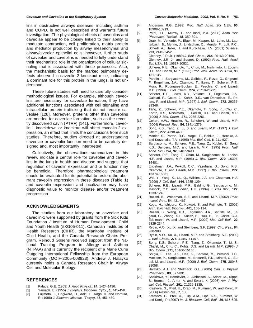
lins in obstructive airways diseases, including asthma and COPD, is not well described and warrants future investigation. The physiological effects of caveolins and caveolae appear to be closely linked to their ability to modulate contraction, cell proliferation, matrix protein and mediator production by airway mesenchymal and airway/alveolar epithelial cells; however, further study of caveolae and caveolins is needed to fully understand their mechanistic role in the organization of cellular sig-naling that is associated with these processes. Also, the mechanistic basis for the marked pulmonary de-fects observed in caveolin-2 knockout mice, indicating a dominant role for this protein in the lungs, is not un-derstood.
These future studies will need to carefully consider methodological issues. For example, although caveo-lins are necessary for caveolae formation, they have additional functions associated with cell signaling and intracellular protein trafficking that do not require ca-veolae [128]. Moreover, proteins other than caveolins are needed for caveolae formation, such as the recen-tly discovered cavin (PTRF) [141]. Furthermore, caveo-lin-1 knockdown or knockout will affect caveolin-2 ex-pression, an effect that limits the conclusions from such studies. Therefore, studies directed at understanding caveolae or caveolin function need to be carefully de-signed and, most importantly, interpreted.
Collectively, the observations summarized in this review indicate a central role for caveolae and caveo-lins in the lung in health and disease and suggest that regulation of caveolin expression and or function may be beneficial. Therefore, pharmacological treatment should be evaluated for its potential to restore the abe-rrant caveolin expression in these diseases (Table 1) and caveolin expression and localization may have diagnostic value to monitor disease and/or treatment progression.
ACKNOWLEDGEMENTS
The studies from our laboratory on caveolae and caveolin-1 were supported by grants from the Sick Kids Foundation / Institute of Human Development, Child and Youth Health (#XG05-011), Canadian Institutes of Health Research (CIHR), the Manitoba Institute of Child Health, and the Canada Research Chairs Pro-gram. Reinoud Gosens received support from the Na-tional Training Program in Allergy and Asthma (NTPAA) and is currently the recipient of a Marie Curie Outgoing International Fellowship from the European Community (MOIF-2005-008823). Andrew J. Halayko currently holds a Canada Research Chair in Airway Cell and Molecular Biology.
REFERENCES
[1] Palade, G.E. (1953) J. Appl. Physiol., 24, 1424-1436.
[2] Yamada, E. (1955) J. Biophys. Biochem. Cytol., 1, 445-458. [3] Fujimoto, T., Hagiwara, H., Aoki, T., Kogo, H. and Nomura,
R. (1998) J. Electron. Microsc. (Tokyo), 47, 451-460.
[4] Anderson, R.G. (1993) Proc. Natl. Acad. Sci. USA, 90,
10909-10913. [5] Patel, H.H., Murray, F. and Insel, P.A. (2008) Annu Rev.
Pharmacol. Toxicol., 48, 359-391.
[6] Drab, M., Verkade, P., Elger, M., Kasper, M., Lohn, M., Lau-terbach, B., Menne, J., Lindschau, C., Mende, F., Luft, F.C., Schedl, A., Haller, H. and Kurzchalia, T.V. (2001) Science,
293, 2449-2452. [7] Glenney, J.R. Jr. (1989) J. Biol. Chem., 264, 20163-20166. [8] Glenney, J.R. Jr. and Soppet, D. (1992) Proc. Natl. Acad.
Sci. USA, 89, 10517-10521. [9] Scherer, P.E., Okamoto, T., Chun, M., Nishimoto, I., Lodish,
H.F. and Lisanti, M.P. (1996) Proc. Natl. Acad. Sci. USA, 93,
131-135. [10] Parolini, I., Sargiacomo, M., Galbiati, F., Rizzo, G., Grignani,
F., Engelman, J.A., Okamoto, T., Ikezu, T., Scherer, P.E.,
Mora, R., Rodriguez-Boulan, E., Peschle, C. and Lisanti, M.P. (1999) J. Biol. Chem., 274, 25718-25725.
[11] Scherer, P.E., Lewis, R.Y., Volonte, D., Engelman, J.A.,
Galbiati, F., Couet, J., Kohtz, D.S., van Donselaar, E., Pe-ters, P. and Lisanti, M.P. (1997) J. Biol. Chem., 272, 29337-29346.
[12] Tang, Z., Scherer, P.E., Okamoto, T., Song, K., Chu, C., Kohtz, D.S., Nishimoto, I., Lodish, H.F. and Lisanti, M.P. (1996) J. Biol. Chem., 271, 2255-2261.
[13] Cohen, A.W., Hnasko, R., Schubert, W. and Lisanti, M.P. (2004) Physiol. Rev., 84, 1341-1379.
[14] Song, K.S., Tang, Z., Li, S. and Lisanti, M.P. (1997) J. Biol. Chem., 272, 4398-4403.
[15] Monier, S., Parton, R.G., Vogel, F., Behlke, J., Henske, A. and Kurzchalia, T.V. (1995) Mol. Biol. Cell, 6, 911-927.
[16] Sargiacomo, M., Scherer, P.E., Tang, Z., Kubler, E., Song,
K.S., Sanders, M.C. and Lisanti, M.P. (1995) Proc. Natl. Acad. Sci. USA, 92, 9407-9411.
[17] Scherer, P.E., Tang, Z., Chun, M., Sargiacomo, M., Lodish,
H.F. and Lisanti, M.P. (1995) J. Biol. Chem., 270, 16395-16401.
[18] Engelman, J.A., Wykoff, C.C., Yasuhara, S., Song, K.S.,
Okamoto, T. and Lisanti, M.P. (1997) J. Biol. Chem., 272, 16374-16381.
[19] Wei, Y., Yang, X., Liu, Q., Wilkins, J.A. and Chapman, H.A.
(1999) J. Cell, Biol., 144, 1285-1294. [20] Scherer, P.E., Lisanti, M.P., Baldini, G., Sargiacomo, M.,
Mastick, C.C. and Lodish, H.F. (1994) J. Cell Biol., 127,
1233-1243. [21] Razani, B., Woodman, S.E. and Lisanti, M.P. (2002) Phar-
macol. Rev., 54, 431-467.
[22] Kogo, H., Ishiguro, K., Kuwaki, S. and Fujimoto, T. (2002) Arch. Biochem. Biophys., 401, 108-114.
[23] Razani, B., Wang, X.B., Engelman, J.A., Battista, M., La-
gaud, G., Zhang, X.L., Kneitz, B., Hou, H., Jr., Christ, G.J., Edelmann, W. and Lisanti, M.P. (2002) Mol. Cell Biol., 22, 2329-2344.
[24] Rybin, V.O., Xu, X. and Steinberg, S.F. (1999) Circ. Res., 84, 980-988.
[25] Rybin, V.O., Xu, X., Lisanti, M.P. and Steinberg, S.F. (2000)
J. Biol. Chem., 275, 41447-41457. [26] Song, K.S., Scherer, P.E., Tang, Z., Okamoto, T., Li, S.,
Chafel, M., Chu, C., Kohtz, D.S. and Lisanti, M.P. (1996) J.
Biol. Chem., 271, 15160-15165. [27] Sotgia, F., Lee, J.K., Das, K., Bedford, M., Petrucci, T.C.,
Macioce, P., Sargiacomo, M., Bricarelli, F.D., Minetti, C., Su-
dol, M. and Lisanti, M.P. (2000) J. Biol. Chem., 275, 38048-38058.
[28] Halayko, A.J. and Stelmack, G.L. (2005) Can. J. Physiol.
Pharmacol., 83, 877-891. [29] Shakirova, Y., Bonnevier, J., Albinsson, S., Adner, M., Rippe,
B., Broman, J., Arner, A. and Sward, K. (2006) Am. J. Phy-
siol. Cell, Physiol., 291, C1326-1335. [30] Krasteva, G., Pfeil, U., Drab, M., Kummer, W. and Konig, P.
(2006) Respir Res., 7, 108.
[31] Krasteva, G., Pfeil, U., Filip, A.M., Lips, K.S., Kummer, W. and Konig, P. (2007) Int. J. Biochem. Cell, Biol., 39, 615-625.

752 Current Molecular Medicine, 2008, Vol. 8, No. 8 Gosens et al.
[32] Razani, B., Engelman, J.A., Wang, X.B., Schubert, W.,
Zhang, X.L., Marks, C.B., Macaluso, F., Russell, R.G., Li, M., Pestell, R.G., Di Vizio, D., Hou, H., Jr., Kneitz, B., Lagaud, G., Christ, G.J., Edelmann, W. and Lisanti, M.P. (2001) J.
Biol. Chem., 276, 38121-38138. [33] Wang, X.M., Zhang, Y., Kim, H.P., Zhou, Z., Feghali-
Bostwick, C.A., Liu, F., Ifedigbo, E., Xu, X., Oury, T.D., Ka-
minski, N. and Choi, A.M. (2006) J. Exp. Med., 203, 2895-2906.
[34] Racine, C., Belanger, M., Hirabayashi, H., Boucher, M., Cha-
kir, J. and Couet, J. (1999) Biochem. Biophys Res. Com-mun., 255, 580-586.
[35] Achcar, R.O., Demura, Y., Rai, P.R., Taraseviciene-Stewart,
L., Kasper, M., Voelkel, N.F. and Cool, C.D. (2006) Chest, 129, 696-705.
[36] Gosens, R., Zaagsma, J., Meurs, H. and Halayko, A.J.
(2006) Respir. Res., 7, 73. [37] Opazo Saez, A.M., Seow, C.Y. and Pare, P.D. (2000) Am. J.
Respir. Crit. Care. Med., 161, 910-917.
[38] Bai, T.R. (1990) Am. Rev. Respir. Dis., 141, 552-557. [39] Bramley, A.M., Thomson, R.J., Roberts, C.R. and Schellen-
berg, R.R. (1994) Eur. Respir. J., 7, 337-341.
[40] de Jongste, J.C., Mons, H., Bonta, I.L. and Kerrebijn, K.F. (1987) Eur. J. Respir. Dis., 71, 23-29.
[41] Gosens, R., Stelmack, G.L., Dueck, G., McNeill, K.D., Ya-
masaki, A., Gerthoffer, W.T., Unruh, H., Soussi-Gounni, A., Zaagsma, J. and Halayko, A.J. (2006) Am. J. Physiol. Lung, Cell, Mol. Physiol., 291, L523-534.
[42] Thyberg, J., Roy, J., Tran, P.K., Blomgren, K., Dumitrescu,
A. and Hedin, U. (1997) Lab. Invest., 77, 93-101. [43] Gabella, G. (1976) Cell Tissue Res., 170, 161-186. [44] Darby, P.J., Kwan, C.Y. and Daniel, E.E. (2000) Am. J. Phy-
siol. Lung Cell Mol. Physiol., 279, L1226-1235. [45] Ambudkar, I.S. (2006) Trends. Pharmacol. Sci., 27, 25-32. [46] Brainard, A.M., Miller, A.J., Martens, J.R. and England, S.K.
(2005) Am. J. Physiol. Cell, Physiol., 289, C49-57. [47] Bush, K.T., Stuart, R.O., Li, S.H., Moura, L.A., Sharp, A.H.,
Ross, C.A. and Nigam, S.K. (1994) J. Biol. Chem., 269,
23694-23699. [48] Fujimoto, T. (1993) J. Cell, Biol., 120, 1147-1157. [49] Isshiki, M. and Anderson, R.G. (2003) Traffic, 4, 717-723.
[50] Gherghiceanu, M. and Popescu, L.M. (2006) J. Cell Mol. Med., 10, 519-528.
[51] Kuo, K.H., Herrera, A.M. and Seow, C.Y. (2003) Respir.
Physiol. Neurobiol., 137, 197-208. [52] Kamishima, T., Burdyga, T., Gallagher, J.A. and Quayle, J.M.
(2007) Am. J. Physiol. Heart, Circ. Physiol., 293, H204-H214.
[53] Bergdahl, A. and Sward, K. (2004) Can. J. Physiol. Pharma-col., 82, 289-299.
[54] Gosens, R., Stelmack, G.L., Dueck, G., Mutawe, M.M., Hin-
ton, M.A., McNeill, K.D., Paulson, A., Dakshinamurti, S., Gerthoffer, W.T., Thliveris, J.A., Unruh, H., Zaagsma, J. and Halayko, A.J. (2007) Am. J. Physiol. Lung, Cell, Mol. Phy-
siol., 293, L1406-L1418. [55] Prakash, Y.S., Thompson, M.A., Vaa, B., Matabdin, I., Peter-
son, T.E., He, T. and Pabelick, C.M. (2007) Am. J. Physiol.
Lung, Cell, Mol. Physiol., 293, L1118-L1126. [56] Bhatnagar, A., Sheffler, D.J., Kroeze, W.K., Compton-Toth,
B. and Roth, B.L. (2004) J. Biol. Chem., 279, 34614-34623.
[57] Pike, L.J. and Casey, L. (1996) J. Biol. Chem., 271, 26453-26456.
[58] Taggart, M.J. (2001) News. Physiol. Sci., 16, 61-65.
[59] Hunter, I. and Nixon, G.F. (2006) J. Biol. Chem., 281, 34705-34715.
[60] Hunter, I., Cobban, H.J., Vandenabeele, P., MacEwan, D.J.
and Nixon, G.F. (2003) Mol. Pharmacol., 63, 714-721. [61] Ricciardolo, F.L., Sterk, P.J., Gaston, B. and Folkerts, G.
(2004) Physiol. Rev., 84, 731-765.
[62] Feron, O., Belhassen, L., Kobzik, L., Smith, T.W., Kelly, R.A. and Michel, T. (1996) J. Biol. Chem., 271, 22810-22814.
[63] Shaul, P.W., Smart, E.J., Robinson, L.J., German, Z.,
Yuhanna, I.S., Ying, Y., Anderson, R.G. and Michel, T. (1996) J. Biol. Chem., 271, 6518-6522.
[64] Liu, J., Garcia-Cardena, G. and Sessa, W.C. (1996) Bioche-
mistry, 35, 13277-13281. [65] Garcia-Cardena, G., Oh, P., Liu, J., Schnitzer, J.E. and Ses-
sa, W.C. (1996) Proc. Natl. Acad. Sci. USA, 93, 6448-6453.
[66] Garcia-Cardena, G., Martasek, P., Masters, B.S., Skidd, P.M., Couet, J., Li, S., Lisanti, M.P. and Sessa, W.C. (1997) J. Biol. Chem., 272, 25437-25440.
[67] Gratton, J.P., Fontana, J., O’Connor, D.S., Garcia-Cardena, G., McCabe, T.J. and Sessa, W.C. (2000) J. Biol. Chem., 275, 22268-22272.
[68] Ju, H., Zou, R., Venema, V.J. and Venema, R.C. (1997) J. Biol. Chem., 272, 18522-18525.
[69] Meurs, H., Maarsingh, H. and Zaagsma, J. (2003) Trends
Pharmacol. Sci., 24, 450-455. [70] Maarsingh, H., de Boer, J., Kauffman, H.F., Zaagsma, J. and
Meurs, H. (2004) Br. J. Pharmacol., 142, 1293-1299.
[71] Meurs, H., McKay, S., Maarsingh, H., Hamer, M.A., Macic, L., Molendijk, N. and Zaagsma, J. (2002) Br. J. Pharmacol., 136, 391-398.
[72] McDonald, K.K., Zharikov, S., Block, E.R. and Kilberg, M.S. (1997) J. Biol. Chem., 272, 31213-31216.
[73] Flam, B.R., Hartmann, P.J., Harrell-Booth, M., Solomonson,
L.P. and Eichler, D.C. (2001) Nitric Oxide, 5, 187-197. [74] Jeffery, P.K. (2001) Am. J. Respir. Crit. Care. Med., 164,
S28-38.
[75] Hays, S.R., Ferrando, R.E., Carter, R., Wong, H.H. and Woodruff, P.G. (2005) Thorax, 60, 226-228.
[76] Selman, M., Thannickal, V.J., Pardo, A., Zisman, D.A., Marti-nez, F.J. and Lynch, J.P., 3
rd. (2004) Drugs, 64, 405-430.
[77] Visscher, D.W. and Myers, J.L. (2006) Proc. Am. Thorac. Soc., 3, 41-47.
[78] Halayko, A.J., Tran, T., Ji, S.Y., Yamasaki, A. and Gosens,
R. (2006) Curr. Drug Targets, 7, 525-540. [79] Fernandes, D.J., Bonacci, J.V. and Stewart, A.G. (2006)
Curr. Drug Targets, 7, 567-577.
[80] Hirst, S.J., Martin, J.G., Bonacci, J.V., Chan, V., Fixman, E.D., Hamid, Q.A., Herszberg, B., Lavoie, J.P., McVicker, C.G., Moir, L.M., Nguyen, T.T., Peng, Q., Ramos-Barbon, D.,
and Stewart, A.G. (2004) J. Allergy, Clin. Immunol., 114, S2-17.
[81] Galbiati, F., Volonte, D., Engelman, J.A., Watanabe, G.,
Burk, R., Pestell, R.G. and Lisanti, M.P. (1998) EMBO J., 17, 6633-6648.
[82] Gosens, R., Dueck, G., Gerthoffer, W.T., Unruh, H., Zaags-
ma, J., Meurs, H. and Halayko, A.J. (2007) Am. J. Physiol. Lung Cell Mol. Physiol., 292, L1163-1172.
[83] Galbiati, F., Volonte, D., Liu, J., Capozza, F., Frank, P.G.,
Zhu, L., Pestell, R.G. and Lisanti, M.P. (2001) Mol. Biol. Cell 12, 2229-2244.
[84] Peterson, T.E., Guicciardi, M.E., Gulati, R., Kleppe, L.S.,
Mueske, C.S., Mookadam, M., Sowa, G., Gores, G.J., Sessa, W.C. and Simari, R.D. (2003) Arterioscler. Thromb. Vasc. Biol., 23, 1521-1527.
[85] Volonte, D., Zhang, K., Lisanti, M.P. and Galbiati, F. (2002) Mol. Biol. Cell, 13, 2502-2517.
[86] Yamamoto, M., Toya, Y., Jensen, R.A. and Ishikawa, Y.
(1999) Exp. Cell Res., 247, 380-388. [87] Couet, J., Sargiacomo, M. and Lisanti, M.P. (1997) J. Biol.
Chem., 272, 30429-30438.
[88] Liu, P., Ying, Y. and Anderson, R.G. (1997) Proc. Natl. Acad. Sci. USA, 94, 13666-13670.
[89] Liu, P., Ying, Y., Ko, Y.G. and Anderson, R.G. (1996) J. Biol.
Chem., 271, 10299-10303. [90] Furuchi, T. and Anderson, R.G. (1998) J. Biol. Chem., 273,
21099-21104.
[91] Postma, D.S. and Timens, W. (2006) Proc. Am. Thorac. Soc., 3, 434-439.
[92] Gueders, M.M., Foidart, J.M., Noel, A. and Cataldo, D.D.
(2006) Eur. J. Pharmacol., 533, 133-144. [93] Kasper, M., Reimann, T., Hempel, U., Wenzel, K.W., Bier-
haus, A., Schuh, D., Dimmer, V., Haroske, G. and Muller, M.
(1998) Histochem. Cell, Biol., 109, 41-48. [94] Barth, K., Blasche, R. and Kasper, M. (2006) Histochem. Cell
Biol., 126, 563-573.

Caveolae and Caveolins in the Respiratory System Current Molecular Medicine, 2008, Vol. 8, No. 8 753
[95] Hardie, W.D., Korfhagen, T.R., Sartor, M.A., Prestridge, A.,
Medvedovic, M., Le Cras, T.D., Ikegami, M., Wesselkamper, S.C., Davidson, C., Dietsch, M., Nichols, W., Whitsett, J.A. and Leikauf, G.D. (2007) Am. J. Respir. Cell Mol. Biol., 37,
309-321. [96] Le Saux, C.J., Teeters, K., Miyasato, S.K., Hoffmann, P.R.,
Bollt, O., Douet, V., Shohet, R.V., Broide, D.H. and Tam,
E.K. (2008) J. Biol. Chem., 283, 5760-5768. [97] Razani, B., Zhang, X.L., Bitzer, M., von Gersdorff, G., Bottin-
ger, E.P. and Lisanti, M.P. (2001) J. Biol. Chem., 276, 6727-
6738. [98] Tourkina, E., Gooz, P., Pannu, J., Bonner, M., Scholz, D.,
Hacker, S., Silver, R.M., Trojanowska, M. and Hoffman, S.
(2005) J. Biol. Chem., 280, 13879-13887. [99] Elkington, P.T. and Friedland, J.S. (2006) Thorax, 61, 259-
266.
[100] Belvisi, M.G. and Bottomley, K.M. (2003) Inflamm. Res., 52, 95-100.
[101] Tang, W. and Hemler, M.E. (2004) J. Biol. Chem., 279,
11112-11118. [102] Williams, T.M., Medina, F., Badano, I., Hazan, R.B., Hutchin-
son, J., Muller, W.J., Chopra, N.G., Scherer, P.E., Pestell,
R.G. and Lisanti, M.P. (2004) J. Biol. Chem., 279, 51630-51646.
[103] Lopez-Rivera, E., Lizarbe, T.R., Martinez-Moreno, M., Lopez-
Novoa, J.M., Rodriguez-Barbero, A., Rodrigo, J., Fernandez, A.P., Alvarez-Barrientos, A., Lamas, S. and Zaragoza, C. (2005) Proc. Natl. Acad. Sci. USA, 102, 3685-3690.
[104] Knight, D.A. and Holgate, S.T. (2003) Respirology, 8, 432-
446. [105] Wikman, H., Seppanen, J.K., Sarhadi, V.K., Kettunen, E.,
Salmenkivi, K., Kuosma, E., Vainio-Siukola, K., Nagy, B.,
Karjalainen, A., Sioris, T., Salo, J., Hollmen, J., Knuutila, S. and Anttila, S. (2004) J. Pathol., 203, 584-593.
[106] Kettunen, E., Anttila, S., Seppanen, J.K., Karjalainen, A.,
Edgren, H., Lindstrom, I., Salovaara, R., Nissen, A.M., Salo, J., Mattson, K., Hollmen, J., Knuutila, S. and Wikman, H. (2004) Cancer, Genet. Cytogenet., 149, 98-106.
[107] Wikman, H., Kettunen, E., Seppanen, J.K., Karjalainen, A., Hollmen, J., Anttila, S. and Knuutila, S. (2002) Oncogene, 21, 5804-5813.
[108] Zhang, Q., Furukawa, K., Chen, H.H., Fujinawa, R., Kozut-sumi, Y., Suzuki, A. and Urano, T. (2006) Oncol Rep., 16, 289-294.
[109] Ho, C.C., Kuo, S.H., Huang, P.H., Huang, H.Y., Yang, C.H. and Yang, P.C. (2007) Lung Cancer,
[110] Moon, K.C., Lee, G.K., Yoo, S.H., Jeon, Y.K., Chung, J.H.,
Han, J. and Chung, D.H. (2005) Anticancer Res., 25, 4631-4637.
[111] Yoo, S.H., Park, Y.S., Kim, H.R., Sung, S.W., Kim, J.H.,
Shim, Y.S., Lee, S.D., Choi, Y.L., Kim, M.K. and Chung, D.H. (2003) Lung Cancer, 42, 195-202.
[112] Ho, C.C., Huang, P.H., Huang, H.Y., Chen, Y.H., Yang, P.C.
and Hsu, S.M. (2002) Am. J. Pathol., 161, 1647-1656. [113] Sunaga, N., Miyajima, K., Suzuki, M., Sato, M., White, M.A.,
Ramirez, R.D., Shay, J.W., Gazdar, A.F. and Minna, J.D.
(2004) Cancer Res., 64, 4277-4285. [114] Lu, Z., Ghosh, S., Wang, Z. and Hunter, T. (2003) Cancer
Cell, 4, 499-515.
[115] Engelman, J.A., Chu, C., Lin, A., Jo, H., Ikezu, T., Okamoto, T., Kohtz, D.S. and Lisanti, M.P. (1998) FEBS Lett, 428, 205-211.
[116] Mehta, D., Bhattacharya, J., Matthay, M.A. and Malik, A.B. (2004) Am. J. Physiol. Lung Cell Mol. Physiol., 287, L1081-1090.
[117] Gumbleton, M. (2001) Adv. Drug, Deliv Rev., 49, 281-300.
[118] Predescu, S.A., Predescu, D.N. and Malik, A.B. (2007) Am. J. Physiol. Lung, Cell, Mol. Physiol., 293(4), L823-842.
[119] Schnitzer, J.E., Oh, P., Pinney, E. and Allard, J. (1994) J.
Cell Biol., 127, 1217-1232. [120] Schubert, W., Frank, P.G., Razani, B., Park, D.S., Chow,
C.W. and Lisanti, M.P. (2001) J. Biol. Chem., 276, 48619-
48622. [121] Hastings, R.H., Wright, J.R., Albertine, K.H., Ciriales, R. and
Matthay, M.A. (1994) Am. J. Physiol., 266, L544-552.
[122] John, T.A., Vogel, S.M., Minshall, R.D., Ridge, K., Tiruppathi, C. and Malik, A.B. (2001) J. Physiol., 533, 547-559.
[123] Jornot, L., Rochat, T., Caruso, A. and Lacroix, J.S. (2005) J.
Cell Physiol., 204, 859-870. [124] Hill, W.G., Almasri, E., Ruiz, W.G., Apodaca, G. and Zeidel,
M.L. (2005) Am. J. Physiol. Cell, Physiol., 289, C33-41.
[125] Schnitzer, J.E. and Oh, P. (1996) Am. J. Physiol., 270, H416-422.
[126] Harris, J., Werling, D., Hope, J.C., Taylor, G. and Howard,
C.J. (2002) Trends. Immunol., 23, 158-164. [127] Hu, G., Ye, R.D., Dinauer, M.C., Malik, A.B. and Minshall,
R.D. (2008) Am. J. Physiol. Lung Cell Mol. Physiol., 294,
L178-186. [128] Head, B.P. and Insel, P.A. (2007) Trends Cell Biol., 17, 51-
57.
[129] Bucci, M., Gratton, J.P., Rudic, R.D., Acevedo, L., Roviezzo, F., Cirino, G. and Sessa, W.C. (2000) Nat. Med., 6, 1362-1367.
[130] Garrean, S., Gao, X.P., Brovkovych, V., Shimizu, J., Zhao,
Y.Y., Vogel, S.M. and Malik, A.B. (2006) J. Immunol., 177, 4853-4860.
[131] Connelly, L., Palacios-Callender, M., Ameixa, C., Moncada,
S. and Hobbs, A.J. (2001) J. Immunol., 166, 3873-3881. [132] Wang, X.M., Kim, H.P., Song, R. and Choi, A.M. (2006) Am.
J. Respir. Cell, Mol. Biol., 34, 434-442.
[133] Li, X.A., Everson, W.V. and Smart, E.J. (2005) Trends Car-diovasc. Med., 15, 92-96.
[134] Zhao, Y.Y., Liu, Y., Stan, R.V., Fan, L., Gu, Y., Dalton, N.,
Chu, P.H., Peterson, K., Ross, J., Jr. and Chien, K.R. (2002) Proc. Natl. Acad. Sci. USA, 99, 11375-11380.
[135] Jasmin, J.F., Mercier, I., Hnasko, R., Cheung, M.W., Tano-
witz, H.B., Dupuis, J. and Lisanti, M.P. (2004) Cardiovasc Res., 63, 747-755.
[136] Mathew, R., Huang, J., Shah, M., Patel, K., Gewitz, M. and
Sehgal, P.B. (2004) Circulation, 110, 1499-1506. [137] Jasmin, J.F., Mercier, I., Dupuis, J., Tanowitz, H.B. and Li-
santi, M.P. (2006) Circulation, 114, 912-920.
[138] Geraci, M.W., Moore, M., Gesell, T., Yeager, M.E., Alger, L., Golpon, H., Gao, B., Loyd, J.E., Tuder, R.M. and Voelkel, N.F. (2001) Circ Res., 88, 555-562.
[139] Kim, H.P., Wang, X., Nakao, A., Kim, S.I., Murase, N., Choi, M.E., Ryter, S.W. and Choi, A.M. (2005) Proc. Natl. Acad. Sci. USA, 102, 11319-11324.
[140] Patel, H.H., Zhang, S., Murray, F., Suda, R.Y., Head, B.P., Yokoyama, U., Swaney, J.S., Niesman, I.R., Schermuly, R.T., Pullamsetti, S.S., Thistlethwaite, P.A., Miyanohara, A.,
Farquhar, M.G., Yuan, J.X. and Insel, P.A. (2007) FASEB J., 21, 2970-2979.
[141] Hill, M.M., Bastiani, M., Luetterforst, R., Kirkham, M., Kir-
kham, A., Nixon, S.J., Walser, P., Abankwa, D., Oorschot, V.M., Martin, S., Hancock, J.F. and Parton, R.G. (2008) Cell, 132, 113-124.
Received: December 14, 2007 Revised: May 07, 2008 Accepted: May 07, 2008