Respiratory Medicine - Scion Publishing
27
Respiratory Medicine Laura-Jane Smith, Jennifer Quint, Jeremy Brown Core science and medicine in one book eureka
-
Upload
khangminh22 -
Category
Documents
-
view
2 -
download
0
Transcript of Respiratory Medicine - Scion Publishing

eureka – an innovative series for students that fully integrates core science,clinical medicine and surgery. With its engaging and authoritative text, featuringinsightful clinical cases, graphic narratives, SBAs and a wealth of other learning tools,Eureka has everything students need to succeed in medicine and pass their exams.
> First principles chapter explains key structures, processes and concepts
> Clinical essentials chapter sets out diagnostic approach and treatment options
> Disease-based clinical chapters describe acute, chronic and emergency respiratory presentations
> Clinical cases teach you to think like a doctor
> Graphic narratives bring cases to life
> Starter questions stimulate curiosity and learning
> Clinical SBA chapter helps you revise and pass your exams
eureka
medicine made clear
Respiratory Medicine Sm
ith, Quint, Brow
n
eureka
Series Editors: Janine Henderson, David Oliveira, Stephen Parker
www.eurekamedicine.com
Respiratory MedicineLaura-Jane Smith, Jennifer Quint, Jeremy Brown
Core science and medicine in one book
eureka
EurekaRespiratory_COVER_16mm_Amend.indd 1 11/08/2015 14:32

Respiratory Medicineeureka


Laura-Jane Smith MBBChir MRCP FHEAHonorary Clinical Lecturer in Medical EducationUCL Medical SchoolRespiratory and Medical RegistrarNorth East LondonLondon, UK
Jennifer Quint MRCP PhD MSc Clinical Senior Lecturer in Respiratory EpidemiologyImperial College LondonHonorary ConsultantRoyal Brompton and Harefield NHS Foundation TrustLondon, UK
Jeremy S Brown MBBS MRCP DPhilProfessor of Respiratory InfectionUCL Medical SchoolHonorary ConsultantUniversity College HospitalLondon, UK
London • Philadelphia • New Delhi • Panama City
Respiratory Medicineeureka
Series Editors
Janine Henderson MRCPsych MClinEdMB BS Programme DirectorHull York Medical SchoolYork, UK
David Oliveira PhD FRCPProfessor of Renal MedicineSt George’s, University of LondonLondon, UK
Stephen Parker BSc MS DipMedEd FRCSConsultant Breast and General Paediatric SurgeonSt Mary’s HospitalNewport, UK

© 2015 JP Medical Ltd.
Published by JP Medical Ltd, 83 Victoria Street, London, SW1H 0HW, UK
First reprint 2015
Tel: +44 (0)20 3170 8910 Fax: +44 (0)20 3008 6180
Email: [email protected] Web: www.jpmedpub.com
The rights of Laura-Jane Smith, Jennifer Quint and Jeremy Brown to be identified as the authors of this work have been asserted by them in accordance with the Copyright, Designs and Patents Act 1988.
All rights reserved. No part of this publication may be reproduced, stored or transmitted in any form or by any means, electronic, mechanical, photocopying, recording or otherwise, except as permitted by the UK Copy-right, Designs and Patents Act 1988, without the prior permission in writing of the publishers. Permissions may be sought directly from JP Medical Ltd at the address printed above.
All brand names and product names used in this book are trade names, service marks, trademarks or reg-istered trademarks of their respective owners. The publisher is not associated with any product or vendor mentioned in this book.
Medical knowledge and practice change constantly. This book is designed to provide accurate, authoritative information about the subject matter in question. However readers are advised to check the most current information available on procedures included and check information from the manufacturer of each product to be administered, to verify the recommended dose, formula, method and duration of administration, adverse effects and contraindications. It is the responsibility of the practitioner to take all appropriate safety precautions. Neither the publisher nor the authors assume any liability for any injury and/or damage to per-sons or property arising from or related to use of material in this book.
This book is sold on the understanding that the publisher is not engaged in providing professional medical services. If such advice or services are required, the services of a competent medical professional should be sought.
Every effort has been made where necessary to contact holders of copyright to obtain permission to repro-duce copyright material. If any have been inadvertently overlooked, the publisher will be pleased to make the necessary arrangements at the first opportunity.
ISBN: 978-1-907816-72-7
British Library Cataloguing in Publication DataA catalogue record for this book is available from the British Library
Library of Congress Cataloging in Publication DataA catalog record for this book is available from the Library of Congress
Publisher: Richard Furn
Development Editors: Thomas Fletcher, Paul Mayhew, Alison Whitehouse
Editorial Assistant: Sophie Woolven
Copy Editor: Kim Howell
Graphic narratives: James Pollitt
Cover design: Forbes Design
Page design: Designers Collective

v
Series Editors’ ForewordToday’s medical students need to know a great deal to be effective as tomorrow’s doctors. This knowledge includes core science and clinical skills, from understanding biochemical pathways to communicating with patients. Modern medical school curricula integrate this teaching, emphasising how learning in one area can support and reinforce another. At the same time students must acquire sound clinical reasoning skills, working with complex information to understand each individual’s unique medical problems.
The Eureka series is designed to cover all aspects of today’s medical curricula and reinforce this integrated approach. Each book can be used from first year through to qualification. Core biomedical principles are introduced but given relevant clinical context: the authors have always asked themselves, ‘why does the aspiring clinician need to know this’?
Each clinical title in the series is grounded in the relevant core science, which is introduced at the start of each book. Each core science title integrates and emphasises clinical relevance throughout. Medical and surgical approaches are included to provide a complete and integrated view of the patient management options available to the clinician. Clinical insights highlight key facts and principles drawn from medical practice. Cases featuring unique graphic narratives are presented with clear explanations that show how experienced clinicians think, enabling students to develop their own clinical reasoning and decision making. Clinical SBAs help with exam revision while Starter Questions are a unique learning tool designed to stimulate interest in the subject.
Having biomedical principles and clinical applications together in one book will make their connections more explicit and easier to remember. Alongside repeated exposure to patients and practice of clinical and communication skills, we hope Eureka will equip medical students for a lifetime of successful clinical practice.
Janine Henderson, David Oliveira, Stephen Parker
eureka

About the Series EditorsJanine Henderson is the MB BS undergraduate Programme Director at Hull York Medical School (HYMS). After medical school at the University of Oxford and clinical training in psychiatry, she combined her work as a consultant psychiatrist with postgraduate teaching roles, moving to the new Hull York Medical School in 2004. She has a particular interest in modern educational methods, curriculum design and clinical reasoning.
David Oliveira is Professor of Renal Medicine at St George’s, University of London (SGUL), where he served as the MBBS Course Director between 2007 and 2013. Having trained at Cambridge University and the Westminster Hospital he obtained a PhD in cellular immunology and worked as a renal physician before being appointed as Foundation Chair of Renal Medicine at SGUL.
Stephen Parker is a Consultant Breast & General Paediatric Surgeon at St Mary’s Hospital, Isle of Wight. He trained at St George’s, University of London, and after service in the Royal Navy was appointed as Consultant Surgeon at University Hospital Coventry. He has a particular interest in e-learning and the use of multimedia platforms in medical education.
About the AuthorsLaura-Jane Smith is a Respiratory Registrar and Honorary Clinical Lecturer in Medical Education at University College London. She has been involved in clinical teaching throughout her career and pursued this interest further by taking a year out of training as a teaching fellow to complete a Postgraduate Certificate in Medical Education.
Jennifer Quint is a Clinical Senior Lecturer in Respiratory Epidemiology at Imperial College London and an Honorary Consultant at the Royal Brompton Hospital. She has extensive experience in teaching undergraduates and postgraduates and is a Fellow of the Higher Education Academy.
Jeremy Brown is a Consultant in Respiratory Medicine with 25 years’ experience of teaching medical students, for the past 12 years at the University College of London Medical School. As well as lecturing and examining finals, he holds a weekly bedside teaching session which emphasises the importance of clinical skills.
vi

PrefaceRespiratory diseases are both common and varied, and include chronic inflammatory conditions, malignancy and infectious diseases. A sound understanding of the common respiratory diseases is essential for all clinicians.
Eureka Respiratory Medicine provides the key skills to succeed in treating respiratory conditions: the ability to take a good history, interpret examination findings, integrate investigation results and apply these to the management of patients.
The book is structured to make it easy to find information in practice and during revision, and is highly illustrated to aid understanding of key concepts. Chapter 1 provides the core respiratory anatomy, physiology and immunology that are essential for an understanding of respiratory disorders. Chapter 2 outlines the clinical approach to patients, i.e. how to make sense of symptoms and signs, and the range of investigations and management options available to the clinician. The subsequent chapters describe the important respiratory diseases. Each chapter is introduced by one or more detailed cases that show how patients present, how clinicians think through the diagnosis and how they decide which investigations to perform. Chapter 11 gives concise information on respiratory emergencies and Chapter 12 explores important but often overlooked aspects of caring for patients with respiratory disease. Finally, a chapter of SBA questions with detailed answers is provided to aid revision.
We hope Eureka Respiratory Medicine provides students with all of the tools they require to become successful clinicians in the future.
Laura-Jane Smith, Jennifer Quint, Jeremy BrownApril 2015
vii

ContentsSeries Editor’s Foreword v
About the Series Editors vi
About the Authors vi
Preface vii
Glossary x
Acknowledgements xii
Chapter 1 First principles
Overview of the respiratory system 1
Development of the respiratory system 3
Anatomy of the respiratory tract 5
Ventilation and respiration 23
Control of ventilation 36
Acid–base balance 38
Pulmonary circulation 40
Lung immunity and inflammation 42
Chapter 2 Clinical essentials
Introduction 61
Epidemiology and patient demographics 61
Common symptoms and how to take a history 62
Common signs and how to examine the patient 77
Investigations 95
Management options 123
Chapter 3 Airway disease
Introduction 143
Case 1 Recurrent cough and wheeze 144
Case 2 Worsening breathlessness in a 70-year-old smoker 145
Large airways obstruction 148
Asthma 150
Chronic obstructive pulmonary disease 157
Allergic bronchopulmonary aspergillosis 165
Other causes of irreversible airways obstruction 165
Chapter 4 Interstitial lung disease
Introduction 167
Case 3 Progressive shortness of breath 168
Case 4 Cough and painful shin rash 171
Pulmonary fibrosis 172
Hypersensitivity pneumonitis 176
Sarcoidosis 178
Pneumoconiosis 181
Rare types of interstitial lung disease and lung infiltrations 182
Chapter 5 Sleep and ventilatory disorders
Introduction 185
Case 5 Falling asleep at work 186
Obstructive sleep apnoea 188
Obesity hypoventilation syndrome 192
Chest wall and neuromuscular disorders 192
Other causes of sleep-disordered breathing 193
Chapter 6 Pleural disease
Introduction 195
Case 6 Progressive breathlessness over a month 195
Case 7 Sudden onset of breathlessness 197
Pleural effusions 199
Pneumothorax 203
Benign pleural thickening 206
viii

Chapter 7 Malignancy
Introduction 209
Case 8 Breathlessness and weight loss 210
Case 9 Chest pain and breathlessness 212
Primary lung cancer 214
Mesothelioma 225
Lung metastasis 228
Carcinoid and other lung tumours 230
Chapter 8 Lung infections
Introduction 233
Case 10 Three days of cough and fever 235
Case 11 Three months of cough and fever 237
Microbiology of the respiratory tract 239
Upper respiratory tract infection, tracheitis and bronchitis 247
Bronchiolitis 248
Community-acquired pneumonia 249
Hospital- and ventilator-acquired pneumonia 256
Pneumonia in the immunocompromised patient 257
HIV infection 260
Parapneumonic effusions and empyema 261
Subacute lung infections and lung abscess 263
Tuberculosis 264
Non-tuberculous mycobacteria 270
Chapter 9 Chronic suppurative lung disease
Introduction 273
Case 12 Chronic cough with sputum production 273
Non-cystic fibrosis bronchiectasis 275
Cystic fibrosis 280
Chapter 10 Circulatory disorders
Introduction 285
Case 13 Four days of chest pain on breathing 286
Case 14 Leg swelling in a patient with COPD 288
Pulmonary embolism 290
Pulmonary hypertension 293
Pulmonary vasculitis 296
Arteriovenous malformation 298
Chapter 11 Respiratory emergencies
Introduction 301
Case 15 Massive pulmonary embolism 301
Case 16 Type 2 respiratory failure 303
Case 17 Acute severe asthma 305
Case 18 Acute respiratory distress syndrome 306
Case 19 Stridor 308
Case 20 Major haemoptysis 309
Case 21 Tension pneumothorax 311
Chapter 12 Integrated care
Introduction 313
Case 22 Poorly controlled asthma 314
Case 23 Repeated admissions for COPD 316
End-stage respiratory disease 317
Disease prevention 319
Pregnancy and respiratory disease 320
Chapter 13 Self-assessment
SBA questions 323
SBA answers 334
Index 343
ixContents

GlossaryA−a alveolar−arterialA1AT α1-antitrypsinABG arterial blood gasABPA allergic bronchopulmonary aspergillosisAC adenylyl cyclaseACE angiotensin-converting enzymeACh acetylcholineACTH adrenocorticotropic hormoneANCA anti–neutrophil cytoplasmic antibody
BALT bronchus-associated lymphoid tissueBCG bacille Calmette–Guérin
cAMP cyclic AMPCao2 oxygen-carrying capacity of arterial
bloodCAP community-acquired pneumoniaCFA cryptogenic fibrosing alveolitisCFTR cystic fibrosis transmembrane
conductance regulatorCMV cytomegalovirus COPD chronic obstructive pulmonary diseaseCPAP continuous positive airway pressureCPE complicated parapneumonic effusionCT computerised tomographyCTPA computerised tomography pulmonary
angiographyCvo2 oxygen-carrying capacity of venous
blood
Do2 oxygen delivery to tissuesDPLD diffuse parenchymal lung disease2,3-DPG 2,3-diphosphglycerateDVLA Driver and Vehicle Licensing Agency
EBUS endobronchial ultrasoundECG electrocardiogramEGFR epidermal growth factor receptorEPGA eosinophilic granulomatosis with
polyangiitis
ERV expiratory reserve volumeESR erythrocyte sedimentation rateEUS endoscopic ultrasoundFDG fluorodeoxyglucoseFEV1 forced expiratory volume (in 1 s)Fio2 fractional concentration of oxygen in
inspired airFRC functional residual capacityFVC forced vital capacity
GOLD Global Initiative for Chronic Obstructive Lung Disease
GP general practitionerGPA granulomatosis with polyangiitisGs G protein
HAP hospital-acquired pneumoniaHP hypersensitivity pneumonitisHSCT haematopoietic stem cell transplant
IC inspiratory capacityICS inhaled corticosteroidIg immunoglobulinIL interleukinILD interstitial lung diseaseINR international normalised ratioIPF idiopathic pulmonary fibrosisIRV inspiratory reserve volume
KCO transfer coefficient of the lung for carbon monoxide
LABA long-acting b2 agonistLAMA long-acting muscarinic anticholinergicLDH lactate dehydrogenaseLLL left lower lobeLMWH low-molecular-weight heparinLUL left upper lobe
MRC Medical Research CouncilMRI magnetic resonance imaging
x

mRNA messenger RNAMRSA methicillin-resistant Staphylococcus
aureusNSCLC non-small-cell lung cancerNSIP non-specific interstitial pneumoniaNTM non-tuberculous mycobacteria
Pa−ao2 alveolar−arterial oxygen partial pressure gradient
Paco2 alveolar partial pressure of carbon dioxide
Paco2 arterial partial pressure of carbon dioxide
PAMP pathogen-associated molecular patternPao2 alveolar partial pressure of oxygenPao2 arterial partial pressure of oxygenPB barometric pressurePEF peak expiratory flowPEFR peak expiratory flow ratePET positron emission tomographyPKA protein kinase APo2 partial pressure of oxygenPS performance statusPvo2 venous partial pressure of oxygen
R respiratory quotientRLL right lower lobeRML right medium lobeRUL right upper lobeRV residual volume
SABA short-acting β2 agonistSAMA short-acting muscarinic antagonistSao2 arterial oxygen saturationSCLC small-cell lung cancerSIADH syndrome of inappropriate antidiuretic
hormone hypersecretionSvo2 venous oxygen saturation
TBNA transbronchial needle aspirationTh T helperTLC total lung capacityTLCO transfer factor of the lung for carbon
monoxideTNF tumour necrosis factorTNM tumour size, nodal involvement and
metastases
UIP usual interstitial pneumoniaURTI upper respiratory tract infection
VA alveolar volumeVAP ventilator-acquired pneumoniaVATS video-assisted thoracoscopic surgeryVC vital capacityVgas gas transfer through a membraneVO2 oxygen consumptionVQ ventilation–perfusionVQ scan ventilation–perfusion scanVT tidal volume
WHO World Health Organization
xiGlossary

AcknowledgementsThanks to the following medical students for their help reviewing chapters: Jessica Dunlop, Aliza Imam, Roxanne McVittie, Daniel Roberts and Joseph Suich.
Figures 2.1, 2.3, 2.4 and 2.14 are copyright of Sam Scott-Hunter and are reproduced from: Tunstall R, Shah N. Pocket Tutor Surface Anatomy. London: JP Medical, 2012.
Figures 2.22, 2.23, 2.24d, 2.25, 2.26, 2.27, 2.28a c -d, 2.31, 3.2, 6.1, 7.1, 7.4, 7.5, 7.11, 8.6, 8.12, 8.15, 8.17 and 9.3 are reproduced from: Darby M, et al. Pocket Tutor Chest X-Ray Inter-pretation. London: JP Medical, 2012.
Figure 10.3 is reproduced from James S, Nelson K. Pocket Tutor ECG Interpretation. London: JP Medical, 2011.
Figures 2.20 and 8.4 are reproduced from: Behera D. Textbook of Pulmonary Medicine, 2e, Vol 1. Delhi: Jaypee Brothers Medical Publishers, 2010.
xii

Introduction . . . . . . . . . . . . . . . . . . 195Case 6 Progressive breathlessness
over a month . . . . . . . . . . . 195Case 7 Sudden onset of
breathlessness . . . . . . . . . . 197
Pleural effusions . . . . . . . . . . . . . . 199Pneumothorax . . . . . . . . . . . . . . . . 203Benign pleural thickening . . . . . . . 206
Chapter 6Pleural disease
IntroductionPleural disease is common, affecting an esti-mated 3000 people per million each year worldwide. The most serious conditions are fluid in the pleural space, termed pleural
effusion, and air in the pleural space, termed a pneumothorax. Pleural thickening is also common but is usually of little clinical con-sequence.
Answers to the following questions are on page 207
1 . How does fluid or air enter the pleural space?
2 . How can a pneumothorax be ‘spontaneous’? What causes spontaneous pneumothorax?
3 . How does analysis of pleural fluid help identify the cause of a pleural effusion?
Starter questions
195
PresentationMr Smith, aged 69 years, has become increasingly breathless over 3 months. He has come to the accident and emergency department after becoming breathless while walking only 20 m.
Initial interpretationIn patients over 60 years of age the likely causes of dyspnoea are chronic obstruc-tive pulmonary disease (COPD) and heart failure. Other possible explanations for a subacute presentation (occurring over days to months) of steadily progressive
Case 6 Progressive breathlessness over 3 months

196 Chapter 6 Pleural disease
dyspnoea are multiple pulmonary emboli, some forms of interstitial lung disease, progressive lobar collapse and an increas-ing pleural effusion of any cause.
HistoryMr Smith has several medical condi-tions. His hypertension and diabetes are well controlled with medication. He also has ischaemic heart disease; 3 years ago, after an episode of severe angina, he had percutaneous coronary intervention (a non-surgical procedure to treat narrowed coronary arteries) and two stents were inserted. He has COPD but has never been admitted to hospital because of it. He takes a tiotropium inhaler daily
He has no cough, wheeze or fever and has not lost any weight recently. He has no-ticed increasing swelling of his ankles but is able to sleep in a horizontal position at night and has not woken feeling breathless. He saw his general practitioner 3 weeks ago and was prescribed furosemide 40 mg twice daily. The peripheral swelling has improved a little, but he remains breathless.
Interpretation of historyPre-existing ischaemic heart disease and peripheral oedema suggest cardiac fail-ure. There are no red flag symptoms of malignancy, such as weight loss, anorexia and pain. At this point, nothing in the his-tory suggests multiple pulmonary emboli. Examination would help identify the cause of the breathlessness.
Further historyMr Smith gave up smoking 10 years ago; before this he smoked 25 cigarettes per day for 32 years (40 pack-year history). He used to work as a theatre director and does not believe that he has ever been exposed to asbestos or other toxic substances. He has not travelled recently and has remained active, with no recent immobility.
ExaminationMr Smith is breathless on minimal exer-tion. However, at rest he has a respiratory rate of 16 breaths/min and normal oxy-gen saturation. His lymph nodes are not palpable and his trachea is central. He has pitting oedema up to his mid calves and his jugular venous pressure is raised. Over the right mid and lower zones there is decreased chest expansion, a dull per-cussion note and absent vocal resonance and breath sounds.
Interpretation of findingsA unilateral pleural effusion seems likely from the clinical examination. From the history the most likely causes are heart failure and malignancy. The absence of obvious weight loss or enlarged lymph nodes make malignancy less likely, but do not rule it out. The normal position of the trachea suggests that there is no collapse of the lobe underlying the effusion, which makes malignancy less likely. The pres-ence of peripheral oedema is suggestive of heart failure, but could be due to lymph-oedema or dependant oedema.
InvestigationsThe chest X-ray shows a large right pleu-ral effusion and an enlarged cardiac out-line (Figure 6.1). The results of routine blood test are normal. A portable ultra-sound scan of the chest confirms that the opacity is pleural fluid. Therefore a therapeutic and diagnostic pleural aspi-ration is done: 1.0 L of pale yellow fluid is removed, and samples are sent to the biochemistry, microbiology and cytol-ogy laboratories.
The results of biochemical analysis of the pleural fluid show a protein content of 20 g/L and a lactate dehydrogenase (LDH) concentration of 100 IU/L; serum protein content is 75 g/L and serum LDH
Case 6 continued

197Case 7 Sudden onset of breathlessness
concentration is 250 IU/L. The results of microbiological tests of the fluid are
negative. Cytological investigations find no malignant cells. An additional blood test shows an increased concentration of NT-proBNP (N-terminal pro brain natri-uretic peptide).
DiagnosisThe low concentration of albumin and LDH show that the pleural fluid is a tran-sudate. This finding makes heart fail-ure the most likely diagnosis. Further evidence is provided by the increased concentration of NT-proBNP. An echo-cardiogram is requested and additional medications are prescribed to treat Mr Smith’s condition.
Case 6 continued
Figure 6.1 Chest X-ray showing a right-sided pleural effusion and enlarged cardiac shadow.
PresentationAnil is a 20-year-old student who presents with a sudden sharp pain on the left side of his chest and acute breathlessness.
Initial interpretationSudden onset of pain and breathlessness in a young person suggests primary spon-taneous pneumothorax, pulmonary embo-lism or musculoskeletal chest wall pain.
Further historyAnil has no medical problems. He regu-larly smokes cannabis but does not use other recreational drugs.
ExaminationAnil is able to talk in full sentences. His pulse rate is 105 beats/min and respiratory
rate is 18 breaths/min. His blood pressure is 110/70 mmHg and oxygen saturation 98% on air. His trachea is deviated to the right. Chest expansion is reduced on the left, and the percussion note is hyper-reso-nant. No breath sounds are audible on the left side.
Interpretation of findingsThe examination findings confirm a pneumothorax. The lack of breath sounds could be due to consolidation or lobar col-lapse, but the percussion note would then be dull and a sudden onset is unusual. In the absence of underlying lung disease or trauma, Anil’s pneumothorax is classified as primary spontaneous pneumothorax (PSP). Importantly, there are no signs of a tension pneumothorax, which is a medi-cal emergency (see Chapter 11).
Case 7 Sudden onset of breathlessness

198 Chapter 6 Pleural disease
InvestigationsA chest X-ray confirms a left-sided pneu-mothorax (Figure 6.2) and reveals no underlying lung disease. The results of rou-tine blood tests and electrocardiography are normal.
DiagnosisThe diagnosis is primary spontane-ous pneumothorax. Smoking cannabis increases the risk of developing this condi-tion. The pneumothorax is large enough to warrant intervention; pleural aspiration is done to reduce its size. If aspiration fails, a chest drain will be required (Figures 6.3 and 6.4).
As the chest X-ray still shows a large
pneumothorax, we need to put a
chest drain in
As the chest X-ray still shows a large
pneumothorax, we need to put a
chest drain in“Have a go”?!
How many have you done?
“Have a go”?! How many have
you done?
Can I have a go? Can I have a go?
OkOk
Yes, I’m OKYes, I’m OK
I’ll insert the drain, don’t worry, I’ve done more than 50. Joe
needs to get more experience, so he’ll assist if that’s OK?
I’ll insert the drain, don’t worry, I’ve done more than 50. Joe
needs to get more experience, so he’ll assist if that’s OK?
Great. We’re all done and it’s bubbling well. It may make you cough to start with but your breathing should be easier soon
Great. We’re all done and it’s bubbling well. It may make you cough to start with but your breathing should be easier soon
You’re doing great. The wire’s in: you’ll just feel a bit of pushing as we enlarge the hole a bit for the drain. Are you OK?
You’re doing great. The wire’s in: you’ll just feel a bit of pushing as we enlarge the hole a bit for the drain. Are you OK?
Anil has a primary spontaneous pneumothorax and needs a chest drain. It's important Anil has con�dence in whoever performs the procedure
Dr Lovell explains the procedure and its risks
With the Seldinger technique a guidewire is inserted and the tract dilated before the drain is inserted and the wire removed
The drain is connected to an underwater seal. Chest X-ray is repeated to check drain position
Pneumothorax: inserting a chest drain
Case 7 continued
Figure 6.2 Chest X-ray showing a large left primary spontaneous pneumothorax. 1 Absent lung markings. 2 Lung collapsed against hilum. 3 Pleural line.
1
2
3

199Pleural effusions
Pleural effusionsA pleural effusion is fluid in the pleural space. Pleural effusions are caused by a wide range of conditions (Table 6.1); the major clinical issues are identifying the cause and prevent-ing symptomatic fluid accumulation in the pleural space.
TypesThere are various types of pleural effusion.
■■ Transudate pleural effusions have a low protein content. They are usually caused by a systemic problem that increases fluid leak into the pleural space; the pleura itself is usually normal. Transudate pleural effusions are unilateral or bilateral.
■■ Exudate pleural effusions have a high protein content. They are usually caused by pleural disease driving fluid leak into the pleural space. Exudate pleural effusions are usually unilateral.
■■ Parapneumonic effusions are sterile exudate pleural effusions in patients with pneumonia.
■■ Complicated parapneumonic effusions are parapneumonic effusions that have become infected and are culture-positive, have a pH < 7.2, are loculated, or contain visibly turbid pleural fluid.
■■ Empyema is caused by a pleural infection. It is an accumulation of pus; the pleural fluid is visibly turbid or culture-positive. Empyema can be acquired
Case 7 continued
Figure 6.4 A Seldinger chest drain insertion kit. 1 Scalpel. 2 Chest
drain. 3 Three-way tap. 4 Guidewire. 5 Large-
bore hollow needle. 6 Connector (to drain tubing). 7 Syringe. 8 Dilator.
8
7
6
5
4
3
2
1
Figure 6.3 Chest X-ray showing a large left primary spontaneous pneumothorax after a chest drain has been inserted. 1 Visible pleural line with lack of lung markings in the apex. 2 Chest drain visible.
1
2

200 Chapter 6 Pleural disease
in the community as a complicated parapneumonic effusion or can present without evidence of associated pneumonia. Alternatively, empyema can be acquired in hospital, where it is often caused by the introduction of an infectious agent during pleural procedures or lung surgery.
■■ Loculated pleural effusions are trapped by fibrous adhesions between the visceral and parietal pleura.
■■ Hydropneumothorax is the presence of both air and fluid in the pleural space.
■■ Haemothorax is the presence of blood in the pleural space. The condition is usually caused by trauma.
Classification Common Rarer
Transudates (often bilateral)
Increased venous pressure Cardiac failure
Renal failure
Iatrogenic fluid overload
Pericardial effusion
Constrictive pericarditis
Hypoalbuminaemia (low concentration of albumin)
Liver cirrhosis
Nephrotic syndrome
Protein-losing enteropathy
Transdiaphragmatic spread of peritoneal fluid
Liver cirrhosis Peritoneal dialysis
Ovarian hyperstimulation syndrome
Meigs‘ syndrome (can be an exudate)
Exudate (usually unilateral)
Infection Parapneumonic*
Complicated parapneumonic effusions
Empyema
Tuberculosis
Subphrenic or hepatic abscess
Oesophageal rupture
Nocardia infection
Actinomyces infection
Malignancy (secondary) Primary lung cancer
Breast cancer
Gastrointestinal tract cancers
Many other less common cancers
Malignancy (primary) Mesothelioma Lymphoma
Sarcoma
Connective tissue disease Rheumatoid arthritis
Systemic lupus erythematosus
Vasculitis
Familial Mediterranean fever
Haemothorax Trauma
Iatrogenic (e.g. caused by pleural procedures and central line insertion)
Bleeding disorders
Drug use or occupational exposure Benign asbestos effusions Drug use (e.g. practolol, methysergide and bromocriptine)
Other Pulmonary embolus
Pancreatitis (high pleural fluid amylase concentration)
Hypothyroidism
Yellow nail syndrome
Chylothorax
*It is essential to distinguish a parapneumonic effusion from an empyema (see Chapter 8).
Table 6.1 Causes of pleural effusions
Causes of pleural effusions

201Pleural effusions
■■ Chylothorax is the accumulation of chyle (fat and lymphatic fluid) in the pleural space. This type of pleural effusion is usually caused by disruption of the lymphatic system as a result of lymphoma, cancer or trauma.
EpidemiologyPleural effusions are very common. Most cases are transudate pleural effusions, para-pneumonic effusions, pleural infections or malignancies.
AetiologyThe cause of the pleural effusion.
■■ In transudate pleural effusions, pleural fluid production is increased by either low oncotic pressure in the blood (as a result of low albumin concentration) or increased venous pressure.
■■ In exudate pleural effusions, there is a diseased pleura. Effusions form due to excessive production of protein and fluid by the pleura, leakage from capillaries, impaired lymphatic drainage, or any combination of these.
Clinical featuresPatients with pleural effusions become breathless as the effusion enlarges. The daily rate of fluid accumulation dictates the speed at which the severity of dyspnoea increases.
The fluid itself causes no pain. However, in-flammatory causes of effusions cause pleuritic chest pain, which also occurs if there is infiltra-tion of a tumour into the pleura and chest wall.
Examination can identify signs of the pleural effusion itself (see Chapter 2), as well as signs of the underlying cause (Table 6.1). Large effusions are obvious clinically, but small ones are difficult to detect.
Small bilateral effusions are more difficult to detect clinically. They produce less obvious asymmetry on examination .
Diagnostic approachBilateral effusions are highly likely to be transudates. If there is an obvious cause (e.g. hypoalbuminaemia) and no atypical fea-tures, no further investigation is required.
Pleural infection and malignancy are the most serious causes of unilateral effusions that must be excluded. Use a structured ap-proach, as follows:
1. A chest X-ray is done to confirm the presence of an effusion (Figure 7.2).
2. Routine blood tests are required, as well as specific tests (e.g. rheumatoid factor and tumour markers), depending on clinical presentation.
3. At least 50 mL of pleural fluid is aspirated for measurements of protein content, LDH and glucose concentration, and pH. Samples are also needed for microbiological tests (including detection of acid-fast bacilli) and cytological investigations. Pleural aspiration are best performed under guidance by pleural ultrasound to avoid damage to surrounding organs (Figure 6.5). Ultrasound also detects fluid septations (common in pleural infection) and pleural thickening or nodularity (which suggest malignancy).
4. The results of pleural fluid analysis differentiate between transudates and exudates (Table 6.2) and are used to identify many cases of infection and pleural malignancy. Combined with clinical findings, a pleural aspirate is
Figure 6.5 Ultrasound showing a pleural effusion. 1 Ultrasound probe on chest wall. 2 Pleural
fluid. 3 Diaphragm.
2
1
3

202 Chapter 6 Pleural disease
diagnostic in 75% of cases. A guide to interpreting the results of pleural fluid analysis is shown in Table 6.3. Obviously loculated fluid is often visible on chest X-ray (Figure 6.6).
5. If the pleural fluid is an exudate, computerised tomography (CT) of the thorax is needed to assess pleural thickness and nodularity. A CT scan also identifies masses that may be primary or secondary lung cancers. The scan is best done when most of the pleural fluid has been drained, leaving the underlying lung visible.
6. If the cause of an exudative effusion remains unclear, pleural biopsy is required. This is especially important if pleural nodularity or thickening is seen on CT, or if malignancy or tuberculosis is suspected. Pleura can be biopsied percutaneously under ultrasound or
Table 6.3 Typical results of pleural fluid analysis for common causes of pleural effusions
Pleural fluid analysis
Cause Appearance Protein content (g/L)
LDH concentration pleural fluid:serum ratio
Glucose concentration (mmol/L)
pH Cytological findings
Microbiological findings
Heart failure Clear and straw-coloured
< 25 Normal Normal > 7.2 None None
Parapneumonic Turbid and yellow
> 35 Normal Normal > 7.2 Neutrophils None
Empyema Purulent > 35 Increased (> 0.6)
< 3.3 < 7.2 Neutrophils Gram stain–positive
Culture-positive (30–50%)
Malignancy Clear and straw-coloured, or blood-stained
> 35 Increased (> 0.6)
< 3.3 < or > 7.2
Malignant cells (60%)
None
Pulmonary embolism
Clear and straw-coloured, or blood-stained
> 35 Normal Normal > 7.2 Neutrophils None
Rheumatoid arthritis
Clear and straw-coloured
> 35 Increased (> 0.6)
< 3.3 < or > 7.2
Lymphocytes None
Tuberculosis Clear or yellow > 35 Increased (> 0.6)
< 3.3 < or > 7.2
Lymphocytes Visible acid-fast bacilli (15%)
Culture-positive (50%)
LDH, lactate dehydrogenase.
Pleural fluid protein content (g/L)
Transudate or exudate
< 25 Transudate
> 35 Exudate
25–35 Use Light’s criteria (99% sensitivity, 98% specificity)
The effusion is an exudate if it fulfils one or more of these criteria are fulfilled
■■ protein content pleural fluid:serum ratio > 0.5
■■ lactate dehydrogenase (LDH) concentration pleural fluid:serum ratio > 0.6
■■ pleural fluid LDH over two-thirds of the upper limit of normal for serum LDH
Table 6.2 Light’s criteria for differentiating transudates and exudates
Light’s criteria
203Pneumothorax
CT guidance, or by medical or surgical thoracoscopy. A pleurodesis can be done at the same time as thoracoscopy to prevent fluid reaccumulating.
Other potentially useful investigations are positron emission tomography CT scan (to identify distal tumours), CT pulmonary angi-ography (in cases of suspected pulmonary embolism), liver ultrasound or biopsy (if cir-rhosis is suspected) and measurement of adenosine deaminase concentration in the pleural fluid (if tuberculosis is suspected).
ManagementPleural infection is discussed in Chapter 8. The aim of treatment of non-infective pleu-
ral effusions is to deal with the underlying cause. For example, diuretics and angioten-sin-converting enzyme inhibitors are used to treat transudates caused by cardiac failure.
During the delay between discovery of a pleural effusion and identification of the cause, keep the patient well informed but avoid worrying them unduly . Remember people vary in how much they want to know so be guided by your patient in how much information to give at each stage .
If treatment is not possible or is ineffective, which is often the case with malignant effu-sions, the options are as follows.
■■ Pleural aspiration or drainage is used to remove large quantities of fluid. Such procedures are often combined with diagnostic pleural aspirations and are repeated when fluid reaccumulates. Sites for aspiration or drainage are identified using pleural ultrasound to avoid damaging nearby structures.
■■ Pleurodesis or pleurectomy is used to obliterate the pleural space to prevent recurrence of the effusion. Medical or surgical pleurodesis introduces an irritant (e.g. talc) into the pleural space through the chest drain or a thoracoscopic procedure. Surgical pleurodesis is achieved by abrading the pleura or by removing it (pleurectomy).
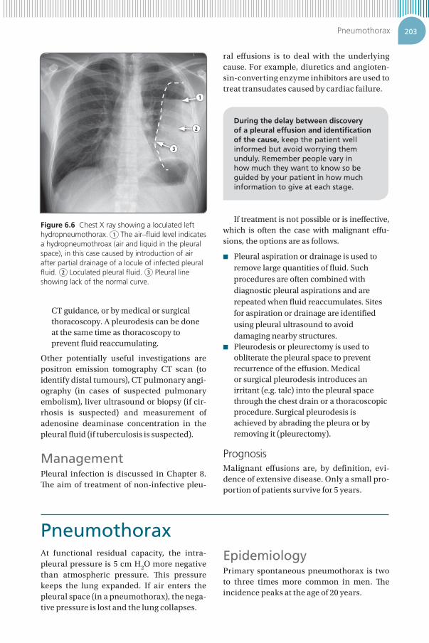
PrognosisMalignant effusions are, by definition, evi-dence of extensive disease. Only a small pro-portion of patients survive for 5 years.
PneumothoraxAt functional residual capacity, the intra-pleural pressure is 5 cm H2O more negative than atmospheric pressure. This pressure keeps the lung expanded. If air enters the pleural space (in a pneumothorax), the nega-tive pressure is lost and the lung collapses.
EpidemiologyPrimary spontaneous pneumothorax is two to three times more common in men. The incidence peaks at the age of 20 years.
Figure 6.6 Chest X ray showing a loculated left hydropneumothorax. 1 The air–fluid level indicates a hydropneumothroax (air and liquid in the pleural space), in this case caused by introduction of air after partial drainage of a locule of infected pleural fluid. 2 Loculated pleural fluid. 3 Pleural line showing lack of the normal curve.
1
2
3

204 Chapter 6 Pleural disease
AetiologyPneumothoraces are divided into the follow-ing types.
■■ Primary spontaneous pneumothorax without underlying lung disease is common in tall and thin young men, smokers and people who inhale recreational drugs. Subpleural blebs and bullae are present in most cases. This type of pneumothorax is also common in patients with Marfan’s or Ehlers–Danlos syndrome.
■■ Spontaneous secondary pneumothoraces are related to an underlying lung disease (e.g. COPD and cystic fibrosis).
■■ In tension pneumothorax, air continues to leak into the pleural space after inspiration but is unable to escape during expiration. Intrapleural pressure builds up, causing mediastinal shift and impaired cardiac function. Tension pneumothorax is a medical emergency (see Chapter 11).
■■ In traumatic pneumothoraces, the lung is punctured by a penetrating injury to the chest (which can happen during medical interventions) or by ribs fractured by blunt trauma.
Clinical featuresPneumothoraces present with sudden onset of chest pain, breathlessness or both. Even a small secondary pneumothorax can cause severe life-threatening dyspnoea if the patient’s respiratory reserve is limited by an underlying lung disease.
On examination, patients are breathless and signs of a pneumothorax are present (see Chapter 2). Hypotension and obvious tracheal deviation suggest a tension pneumothorax (see Chapter 11).
Diagnostic approachThe main differential diagnosis of the clinical presentation is pulmonary embolism. How-ever, chest X-ray readily confirms a pneumo-thorax. Rarely, a CT scan is needed to identify a small pneumothorax or to distinguish between large bullae and a loculated pneu-mothorax.
ManagementSmall primary spontaneous pneumothora-ces resolve spontaneously. They are moni-tored by repeated chest X-rays.
Guidelines suggest criteria such as estimated size of pneumothorax on imaging to decide whether to intervene . However, clinical status is much more important . The risks and benefits of intervention must be weighed up in partnership with the patient .
Patients who are symptomatic, or who have larger primary spontaneous pneumothora-ces, require intervention. First, pleural air is aspirated. If aspiration fails to improve the patient’s condition, a chest drain is needed. The British Thoracic Society has produced a treatment algorithm for primary spontane-ous pneumothorax (Figure 6.7). International guidelines are similar but differ in how they determine pneumothorax size (eg. measured at apex rather than hilum on chest X-ray).
Secondary pneumothoraces usually re-quire chest drainage. In people with under-lying lung disease, the effects of a pneumo-thorax on respiration are potentially severe (Figure 6.8).
Pleural aspiration fails if air continues to leak from the lung (in cases of bronchopleural fistula). Persisting air leaks cause continuous bubbling if a chest drain is in situ. Surgical intervention is warranted if they persist after 5–7 days. Surgery is also mandated in special circumstances, for example if the lung fails to re-expand, or in cases of bilateral pneumothoraces.
Preventive pleurectomy or pleurodesis is offered to patients with recurrent pneumo-thorax on the same side. These procedures are also offered to divers and pilots, because without surgical intervention they are unable to return to these activities and occupations.
PrognosisThe recurrence rate after one primary sponta-neous pneumothorax is up to 50%. This figure increases after two. For secondary pneumo-thorax, the recurrence rate is higher. Most recurrences occur within 6 months to 2 years. Smoking increases the risk of recurrence.

205Pneumothorax
Figure 6.8 Chest X-ray showing a right-sided chest drain inserted for secondary spontaneous pneumothorax complicated by subcutaneous emphysema, in which air tracks up between soft tissue planes. 1 Subcutaneous emphysema. 2 Chest drain. The lungs are hyperinflated due to underlying COPD.
1
2
Figure 6.7 Management of a primary spontaneous pneumothorax. Adapted from British Thorax Society Pleural Disease Guideline Group. BTS pleural disease guideline. Thorax 2010; 65 (suppl 2): ii13-ii21.
Yes
No
No
Insert chest drain andadmit patient to hospital
Yes
Success: pneumothoraxsize < 2 cm and breathing
improved
• Aspirate using 16- to 18 gauge cannula• Aspirate < 2.5 L
Consider dischargingpatient, with outpatient
review in 2–4 weeks
Pneumothorax size > 2 cmor patient is breathless
Primary spontaneouspneumothorax
Spontaneous pneumothorax management

206 Chapter 6 Pleural disease
Benign pleural thickeningThis condition is common and usually asymptomatic. Benign pleural thickening is often an incidental finding on examina-tion (dullness to percussion and quiet breath sounds) or chest X-ray (Figure 6.9). Common causes are previous pleural infection, hae-mothorax, and asbestos exposure (Table 6.4).
Extensive pleural thickening causes restric-tive lung defects and dyspnoea. Treatment is surgical pleurectomy, a procedure associated with significant morbidity and mortality. Pleu-rectomy is reserved for only the most severe cases.
The main differential diagnosis of benign pleural thickening is pleural malignancy, es-pecially mesothelioma.
Table 6.4 Causes of pleural thickening and their clinical features
Cause and common examples Features
Infection
Post-empyema Usually basolateral; can calcify; non-progressive
Post-tuberculosis Common: ‘apical capping’; non-progressive
Rare: extensive, with sheet-like calcifications; non-progressive
Active non-tuberculous mycobacteria infection or chronic aspergillosis
Slowly progressive apical disease with associated cavitation
Inflammation
Asbestos exposure Common: plaques – small patches of focal pleural thickening and calcifications, with holly leaf appearance on chest X-ray
Less common: extensive confluent pleural thickening with or without benign exudate effusions
Post-pleurodesis Diffuse, non-progressive
Post-haemothorax Usually basolateral; can calcify; non-progressive
Caused by drug use (e.g. methysergide and bromocriptine) Diffuse: may be associated with interstitial fibrosis
Malignancy
Primary malignancy (e.g. mesothelioma) – related to asbestos exposure
Progressive pleural thickening; the mediastinal pleural reflection is affected
Secondary malignancy (e.g. lung adenocarcinoma) Progressive pleural thickening associated with nodular changes and intrapulmonary masses
Causes of pleural thickening
Figure 6.9 Chest X-ray showing left-sided pleural thickening. 1 Lung edge and ribs separated by a layer of additional tissue. 2 Characteristic filled-in squared-off costrophrenic angle.
2
1

207Answers to starter questions
1. The pleura produces pleural fluid, which is absorbed by the lymphatic system. The presence of a few millilitres of pleural fluid is normal. However, a pleural effusion forms if production is increased, absorption blocked, or both.
Penetrating trauma allows air to enter the chest cavity. More commonly, air passes through an abnormal connection between the airways and the pleural space; when a subpleural bleb ruptures or a fractured rib damages the lung surface, air that was in the alveoli and bronchi enters the pleural space.
2. A spontaneous pneumothorax develops in the absence of trauma and lung disease, and therefore has no apparent cause. However, patients have an acute new defect in the lung that allows air into the pleural space. The defect is most commonly a ruptured localised bleb. The rest of the lung tissue is normal.
3. The pleural tap is one of the most informative tests for the cause of a pleural effusion. Pleural protein content and LDH concentration (see Table 6.2) differentiate a transudate from an exudate. These results therefore narrow the differential diagnosis. Cytological investigations identify about three-quarters of malignant effusions, and microbiological tests are used to diagnose pleural infection.
Several rare causes of pleural effusion can be confirmed by specific tests on pleural fluid. For example, the concentration of amylase in pleural fluid is measured if pancreatitis is suspected.
Answers to starter questions