Respiratory System
32
Van De Graaff: Human Anatomy, Sixth Edition VI. Maintenance of the Body 17. Respiratory System © The McGraw-Hill Companies, 2001 Respiratory System Clinical Case Study A 37-year-old dock worker was brought to the emergency room after having been stabbed above the left clavicle with an ice pick. The patient’s main complaint was pain in the left side of the chest. Initial evaluation revealed a minute puncture wound superior to the left clavicle, just lateral to the sternocleidomastoid muscle. The vital signs were normal except for a moder- ately high respiratory rate. The chest radiograph showed the left lung to be surrounded by blood and air, a condition known as hemopneumothorax (he''mo-noo''mo-thor'aks), and collapsed to half its normal size. Explain how the air got inside the thoracic cavity (assuming it did not enter through the puncture wound). What is the term for the space where the air and blood are located? What membranes define this space? Introduction to the Respiratory System 603 Conducting Passages 604 Pulmonary Alveoli, Lungs, and Pleurae 612 Mechanics of Breathing 618 Regulation of Breathing 623 Developmental Exposition: The Respiratory System 624 CLINICAL CONSIDERATIONS 626 Clinical Case Study Answer 630 Chapter Summary 631 Review Activities 632 17 FIGURE: The thoracic viscera and their associated serous membranes present interesting anatomical, functional, and clinical relationships. Localized trauma frequently causes extensive and predictable problems elsewhere.
-
Upload
khangminh22 -
Category
Documents
-
view
2 -
download
0
Transcript of Respiratory System

Van De Graaff: Human Anatomy, Sixth Edition
VI. Maintenance of the Body
17. Respiratory System © The McGraw−Hill Companies, 2001
Respiratory System
Clinical Case StudyA 37-year-old dock worker was brought to the emergency room after having been stabbedabove the left clavicle with an ice pick. The patient’s main complaint was pain in the left sideof the chest. Initial evaluation revealed a minute puncture wound superior to the left clavicle,just lateral to the sternocleidomastoid muscle. The vital signs were normal except for a moder-ately high respiratory rate. The chest radiograph showed the left lung to be surrounded by bloodand air, a condition known as hemopneumothorax (he''mo-noo''mo-thor'aks), and collapsed tohalf its normal size.
Explain how the air got inside the thoracic cavity (assuming it did not enter through thepuncture wound). What is the term for the space where the air and blood are located? Whatmembranes define this space?
Introduction to the RespiratorySystem 603
Conducting Passages 604Pulmonary Alveoli, Lungs,
and Pleurae 612Mechanics of Breathing 618Regulation of Breathing 623
Developmental Exposition: The Respiratory System 624
CLINICAL CONSIDERATIONS 626
Clinical Case Study Answer 630Chapter Summary 631Review Activities 632
17
FIGURE: The thoracic viscera and theirassociated serous membranes presentinteresting anatomical, functional, and clinicalrelationships. Localized trauma frequentlycauses extensive and predictable problemselsewhere.

Van De Graaff: Human Anatomy, Sixth Edition
VI. Maintenance of the Body
17. Respiratory System © The McGraw−Hill Companies, 2001
INTRODUCTION TO THERESPIRATORY SYSTEMThe respiratory system can be divided structurally into upper andlower divisions, and functionally into a conducting division and arespiratory division. The principal functions of the respiratory sys-tem are gaseous exchange, sound production, and assistance inabdominal compression.
Objective 1 Describe the functions associated with the termrespiration.
Objective 2 Identify the organs of the respiratory systemand describe their locations.
Objective 3 List the functions of the respiratory system.
The term respiration refers to three separate but related functions:(1) ventilation (breathing); (2) gas exchange, which occurs be-tween the air and blood in the lungs and between the blood andother tissues of the body; and (3) oxygen utilization by the tis-sues in the energy-liberating reactions of cell respiration. Venti-lation and the exchange of gases (oxygen and carbon dioxide)between the air and blood are collectively called external respira-tion. Gas exchange between the blood and other tissues are col-lectively known as internal respiration.
A relaxed adult breathes an average of 15 times a minute,ventilating approximately 6 liters of air during this period. Thisamounts to over 8,000 liters in a 24-hour period. Strenuous exer-cise increases the demand for oxygen and increases the respira-tory rate fifteenfold to twentyfold, so that about 100 liters of airare breathed each minute. If breathing stops, a person will loseconsciousness after 4 or 5 minutes. Brain damage may occur after7 to 8 minutes, and the person will die after 10 minutes. Knowl-edge of the structure and function of the respiratory system istherefore of the utmost importance in a clinical setting.
Physical Requirements of the Respiratory SystemThe respiratory system includes those organs and structures thatfunction together to bring gases in contact with the blood of thecirculatory system. In order to be effective, the respiratory systemmust comply with certain physical requirements.
• The surface for gas exchange must be located deep withinthe body so that incoming air will be sufficiently warmed,moistened, and cleansed of airborne particles before com-ing in contact with it.
• The membrane must be thin-walled and selectively perme-able so that diffusion can occur easily.
• The membrane must be kept moist so that oxygen and car-bon dioxide can be dissolved in water to facilitate diffusion.
• The system must have an extensive capillary network.
• The system must include an effective ventilation mecha-nism to constantly replenish the air.
• The system must function autonomically through effectivemonitoring and feedback mechanisms. However, it mustalso be able to function voluntarily for desired increased ordecreased rates.
The respiratory system adequately meets all of these requirements,thus ensuring that all of the trillions of cells of the body will beable to carry on the metabolic processes necessary to maintain life.
Functions of the Respiratory SystemThe four basic functions of the respiratory system, not all ofwhich are associated with breathing, are as follows:
• It provides oxygen to the bloodstream and removes car-bon dioxide.
• It enables sound production or vocalization as expired airpasses over the vocal folds.
• It assists in abdominal compression during micturition (uri-nation), defecation (passing of feces), and parturition(childbirth). The abdominal muscles become more effec-tive during a deep breath when the air is held in the lungsby closing the glottis and fixing the diaphragm. This sametechnique is used when lifting a heavy object, in whichcase the diaphragm indirectly assists the back muscles.
• It enables protective and reflexive nonbreathing air move-ments, as in coughing and sneezing, to keep the air pas-sageways clean.
Basic Structure of the Respiratory SystemThe major passages and structures of the respiratory system arethe nasal cavity, pharynx, larynx and trachea, and the bronchi,bronchioles, and pulmonary alveoli within the lungs (fig. 17.1).
The structures of the upper respiratory system include thenose, pharynx, and associated structures; the lower respiratorysystem includes the larynx, trachea, bronchial tree, pulmonaryalveoli, and lungs.
On the basis of general function, the respiratory system isfrequently divided into a conducting division and a respiratorydivision. The conducting division includes all of the cavities andstructures that transport gases to and from the pulmonary alveoli.The respiratory division consists of the pulmonary alveoli,which are the functional units of the respiratory system wheregas exchange between the air and the blood occur.
Early Greek and Roman scientists placed great emphasis onthe invisible material that was breathed in. They knew nothing
about oxygen or the role of the blood in transporting this vital sub-stance to cells. For that matter, they knew nothing about microscopicstructures like cells because the microscope had not yet been in-vented. They did know, however, that respiration was essential forlife. Early Greeks referred to air as an intangible, divine spirit calledpneuma. In Latin, the term for breath, spiritus, meant lifeforce.
Chapter 17 Respiratory System 603
CH
AP
TE
R 17
respiration: L. re, back; spirare, to breathe

Van De Graaff: Human Anatomy, Sixth Edition
VI. Maintenance of the Body
17. Respiratory System © The McGraw−Hill Companies, 2001
Knowledge Check1. Define the terms external respiration and internal respiration.2. What are the physical requirements of the respiratory sys-
tem? What are its basic functions?3. List in order the major passages and structures through
which inspired air would pass from the nostrils to the pul-monary alveoli of the lungs.
CONDUCTING PASSAGESAir is conducted through the oral and nasal cavities to the phar-ynx, and then through the larynx to the trachea and bronchial tree.These structures deliver warmed and humidified air to the respira-tory division within the lungs.
Objective 4 List the types of epithelial tissue that characterizeeach region of the respiratory tract and comment on thesignificance of the special attributes of each type.
Objective 5 Identify the boundaries of the nasal cavity anddiscuss the relationship of the paranasal sinuses to the restof the respiratory system.
Objective 6 Describe the three regions of the pharynx andidentify the structures located in each.
Objective 7 Discuss the role of the laryngeal region indigestion and respiration.
Objective 8 Identify the anatomical features of the larynxassociated with sound production and respiration.
The conducting passages serve to transport air to the respiratorystructures of the lungs. The passageways are lined with various
604 Unit 6 Maintenance of the Body
CH
AP
TE
R 1
7
Left principal(primary) bronchus
Lobarbronchus
Segmentalbronchus
Left lung:
Superior lobe
Inferior lobe
Superior lobe
Inferior lobe Cardiac impression
Nasal cavity
Nostril
Hard palate
Larynx
Trachea
Right lung:
Middle lobe
Choana
Soft palate
Pharynx
Epiglottis
Esophagus
FIGURE 17.1 The basic anatomy of the respiratory system.

Van De Graaff: Human Anatomy, Sixth Edition
VI. Maintenance of the Body
17. Respiratory System © The McGraw−Hill Companies, 2001
types of epithelia that cleanse, warm, and humidify the air. Themajority of the conducting passages are held permanently openby muscle or a bony or cartilaginous framework.
NoseThe nose includes an external portion that protrudes from the faceand an internal nasal cavity for the passage of air. The externalportion of the nose is covered with skin and supported by pairednasal bones, which form the bridge, and pliable cartilage, whichforms the distal portions (fig. 17.2). The septal cartilage forms theanterior portion of the nasal septum, and the paired lateral carti-lages and alar cartilages form the framework around the nostrils.
The vomer and the perpendicular plate of the ethmoid bone (seefig. 6.17), together with the septal cartilage, constitute the supportingframework of the nasal septum, which divides the nasal cavity into twolateral halves. Each half is referred to as a nasal fossa. The nasalvestibule is the anterior expanded portion of the nasal fossa (fig. 17.3).
Chapter 17 Respiratory System 605
CH
AP
TE
R 17
FIGURE 17.2 The supporting framework of the nose.
Oralvestibule
Nasal vestibule
Trachea
FIGURE 17.3 A sagittal section of the head showing the structures of the upper respiratory tract. There are several openings into the nasalcavity, including the openings of the various paranasal sinuses, those of the nasolacrimal ducts that drain from the eyes, and those of the audi-tory tubes that drain from the tympanic cavities. (The drainage pathways are indicated with arrows.)
nose: O.E. nosu, nose
nostril: O.E. nosu, nose; thyrel, hole

Van De Graaff: Human Anatomy, Sixth Edition
VI. Maintenance of the Body
17. Respiratory System © The McGraw−Hill Companies, 2001
606 Unit 6 Maintenance of the Body
CH
AP
TE
R 1
7
Each nasal fossa opens anteriorly through the nostril (naris; exteralnares), and communicates posteriorly with the nasopharynx throughthe choana (ko-a'na), (internal nares). The roof of the nasal cavity isformed anteriorly by the frontal bone and paired nasal bones, mediallyby the cribriform plate of the ethmoid bone, and posteriorly by thesphenoid bone (see figs. 6.17 and 6.20). The palatine and maxillarybones form the floor of the cavity. On the lateral walls of the nasal cav-ity are three bony projections, the superior, middle, and inferior nasalconchae (kong'ke), or turbinates (see fig. 6.26). Air passages betweenthe conchae are referred to as nasal meatuses (me-a'tuses) (fig. 17.3).The anterior openings of the nasal cavity are lined with stratified squa-mous epithelium, whereas the conchae are lined with pseudostratifiedciliated columnar epithelium (figs. 17.4 and 17.5). Mucus-secreting gob-let cells are present in great abundance throughout both regions.
The three functions of the nasal cavity and its contents areas follows:
• The nasal epithelium covering the conchae serves to warm,moisten, and cleanse the inspired air. The nasal epitheliumis highly vascular and covers an extensive surfacearea. This is important for warming the air but unfortu-nately also makes humans susceptible to nosebleeds be-cause the nasal mucosa may dry and crack. Nasal hairs
Simple squamous epithelium(lining pulmonary alveoli)
FIGURE 17.4 The various types of epithelial tissue found throughout the respiratory system.
606
FIGURE 17.5 A color-enhanced scanning electron micrograph ofa bronchial wall showing cilia. In the trachea and bronchi, there areabout 300 cilia per cell. The cilia move mucus-dust particles towardthe pharynx, where they can either be swallowed or expectorated.
called vibrissae (vi-bris'e), which often extend from the nos-trils, filter macroparticles that might otherwise be inhaled.Dust, pollen, smoke, and other fine particles are trappedalong the moist mucous membrane lining the nasal cavity.
• The olfactory epithelium in the upper medial portion ofthe nasal cavity is concerned with the sense of smell.
• The nasal cavity affects the voice by functioning as a res-onating chamber.
choana: Gk. choane, funnel
concha: L. choncha, mussel shell vibrissa: L. vibrare, to vibrate

Van De Graaff: Human Anatomy, Sixth Edition
VI. Maintenance of the Body
17. Respiratory System © The McGraw−Hill Companies, 2001
PharynxThe pharynx (far'ingks) is a funnel-shaped organ, approximately13 cm (5 in.) long, that connects the nasal and oral cavities tothe larynx of the respiratory system and the esophagus of the di-gestive system. The supporting walls of the pharynx are com-posed of skeletal muscle, and the lumen is lined with a mucousmembrane. Within the pharynx are several paired lymphoid or-gans called tonsils. Commonly referred to as the “throat” or “gul-let,” the pharynx has both respiratory and digestive functions. Italso provides a resonating chamber for certain speech sounds.The pharynx is divided on the basis of location and function intothree regions (see fig. 17.3).
• The nasopharynx serves only as a passageway for air, be-cause it is located above the point of food entry into thebody (the mouth). It is the uppermost portion of the phar-ynx, positioned directly behind the nasal cavity and abovethe soft palate. A pendulous uvula (yoo'vyu-la) hangs fromthe middle lower portion of the soft palate. The paired au-ditory (eustachian) tubes connect the nasopharynx withthe tympanic cavities. The pharyngeal tonsils, or ade-noids, are situated in the posterior wall of the nasal cavity.
During the act of swallowing, the soft palate and uvulaare elevated to block the nasal cavity and prevent food fromentering. Occasionally a person may suddenly exhale air (as
Chapter 17 Respiratory System 607
CH
AP
TE
R 17
FIGURE 17.6 The paranasal sinuses.
There are several drainage openings into the nasal cavity (seefig. 17.3). The paranasal ducts (discussed below) drain mucus
from the paranasal sinuses, and the nasolacrimal ducts drain tearsfrom the eyes (see fig. 15.11). An excessive secretion of tearscauses the nose to run as the tears drain into the nasal cavity. Theauditory tube from the tympanic cavity enters the upper respiratorytract posterior to the nasal cavity in the nasopharynx. With all theseaccessory connections, it is no wonder that infections can spread soeasily from one chamber to another throughout the facial area. Toavoid causing damage or spreading infections to other areas, onemust be careful not to blow the nose too forcefully.
Paranasal SinusesPaired air spaces in certain bones of the skull are called paranasalsinuses. These sinuses are named according to the bones inwhich they are found; thus, there are the maxillary, frontal,sphenoidal, and ethmoidal sinuses (fig. 17.6). Each sinus com-municates via drainage ducts within the nasal cavity on its ownside (see figs. 6.17, 6.20, and 17.3). Paranasal sinuses may helpwarm and moisten the inspired air. These sinuses are responsiblefor some sound resonance, but most important, they function todecrease the weight of the skull while providing structuralstrength.
You can observe your own paranasal sinuses. Face a mirrorin a darkened room and shine a small flashlight into your
face. The frontal sinuses will be illuminated by directing the light justbelow the eyebrow. The maxillary sinuses are illuminated by shiningthe light into the oral cavity and closing your mouth around theflashlight.
sinus: L. sinus, bend or curve
pharynx: L. pharynx, throat
tonsil: L. toles, goiter or swelling
uvula: L. uvula, small grape
adenoid: Gk. adenoeides, glandlike

Van De Graaff: Human Anatomy, Sixth Edition
VI. Maintenance of the Body
17. Respiratory System © The McGraw−Hill Companies, 2001
with a laugh) while in the process of swallowing fluid. If thisoccurs before the uvula effectively blocks the nasopharynx,fluid will be discharged through the nasal cavity.
• The oropharynx is the middle portion of the pharynx be-tween the soft palate and the level of the hyoid bone. Bothswallowed food and fluid and inhaled air pass through it.The base of the tongue forms the anterior wall of theoropharynx. Paired palatine tonsils are located on the pos-terior lateral wall, and the lingual tonsils are found on thebase of the tongue.
• The laryngopharynx (la-ring''go-far'ingks) is the lowermostportion of the pharynx. It extends inferiorly from the levelof the hyoid bone to the larynx and opens into the esopha-gus and larynx. It is at the lower laryngopharynx that therespiratory and digestive systems become distinct. Swal-lowed food and fluid are directed into the esophagus,whereas inhaled air is directed anteriorly into the larynx.
During a routine physical examination, the physician will com-monly depress the patient’s tongue and examine the condition
of the palatine tonsils. Tonsils are lymphoid organs and tend to be-come swollen and inflamed after persistent infections. If after periodsof infection, the tonsils become so large as to obstruct breathing,they may have to be surgically removed. The removal of the palatinetonsils is called a tonsillectomy, whereas the removal of the pharyn-geal tonsils is called an adenoidectomy (ad''e-noid-ek'to-me).
LarynxThe larynx (lar'ingks), or “voice box,” is a continuation of the con-ducting division that connects the laryngopharynx with the trachea.It is positioned in the anterior midline of the neck at the level of the
fourth through sixth cervical vertebrae. The larynx has two func-tions. Its primary function is to prevent food or fluid from enteringthe trachea and lungs during swallowing and to permit passage of airwhile breathing. A secondary role is to produce sound.
Laryngitis is the inflammation of the mucosal epithelium of thelarynx and vocal folds, which causes a hoarseness of a per-
son’s voice or an inability to speak above a whisper. Laryngitis mayresult from overuse of the voice, inhalation of an irritating chemical, ora bacterial or viral infection. Mild cases are temporary and seldom ofmajor concern.
The larynx is shaped like a triangular box (fig. 17.7). It iscomposed of a framework involving nine cartilages: three arelarge unpaired structures, and six are smaller and paired. Thelargest of the unpaired cartilages is the anterior thyroid cartilage.The laryngeal prominence of the thyroid cartilage is commonlycalled the “Adam’s apple.” It is an anterior vertical ridge alongthe larynx that can be palpated on the midline of the neck. Thethyroid cartilage is typically larger and more prominent in malesthan in females because of the effect of male sex hormones onthe development of the larynx during puberty.
The spoon-shaped epiglottis (ep''ı-glot'is) has a frameworkof hyaline cartilage, referred to as the epiglottic cartilage. Theepiglottis is located behind the root of the tongue where it aidsin closing the glottis, or laryngeal opening, during swallowing.
The entire larynx elevates during swallowing to close the glot-tis against the epiglottis. This movement can be noted by cup-
ping the fingers lightly over the larynx and then swallowing. If theglottis is not closed as it should be during swallowing, food may be-come lodged within the glottis. In this case, the abdominal thrust(Heimlich) maneuver can be used to prevent suffocation. (See “Clini-cal Considerations” for how to perform this maneuver.)
608 Unit 6 Maintenance of the Body
CH
AP
TE
R 1
7
larynx: Gk. larynx, upper windpipe
FIGURE 17.7 The structure of the larynx. (a) An anterior view, (b) a posterior view, and (c) a sagittal view.
thyroid: Gk. thyreos, shieldlike

Van De Graaff: Human Anatomy, Sixth Edition
VI. Maintenance of the Body
17. Respiratory System © The McGraw−Hill Companies, 2001
The lower end of the larynx is formed by the ring-shapedcricoid (kri'koid) cartilage. This third unpaired cartilage connectsthe thyroid cartilage above and the trachea below. The pairedarytenoid (ar'' ı-te'noid) cartilages, located above the cricoid andbehind the thyroid, are the posterior attachments of the vocalfolds. The other paired cuneiform cartilages and corniculate(kor-nik'yu-lat) cartilages are small accessory cartilages that areclosely associated with the arytenoid cartilages (fig. 17.8).
Two pairs of strong connective tissue bands are stretchedacross the upper opening of the larynx from the thyroid cartilageanteriorly to the paired arytenoid cartilages posteriorly. These arethe vocal folds (true vocal cords) and the vestibular folds (falsevocal cords) (fig. 17.8). The vestibular folds support the vocalfolds and produce mucus from its epithelial lining, which keepthe vestibular folds from drying out. The vestibular folds are notused in sound production, but rather the vocal folds vibrate toproduce sound. Stratified squamous epithelium lines the vocalfolds, whereas the rest of the larynx is lined with pseudostratifiedciliated columnar epithelium. This is an important anatomicalmodification considering the tremendous vibratory action of thevocal folds in the production of sound.
The laryngeal muscles are extremely important in closing theglottis during swallowing and in speech. There are two groups of la-ryngeal muscles: extrinsic muscles, responsible for elevating the lar-ynx during swallowing, and intrinsic muscles that, when contracted,change the length, position, and tension of the vocal folds. Variouspitches are produced as air passes over the altered vocal folds. If thevocal folds are taut, vibration is more rapid and causes a higher pitch.Less tension on the vocal folds produces lower sounds. Mature malesgenerally have thicker and longer vocal folds than females; therefore,the vocal folds of males vibrate more slowly and produce lowerpitches. The loudness of vocal sound is determined by the force ofthe air passed over the vocal folds and the amount of vibration. Thevocal folds do not vibrate when a person whispers.
Sounds originate in the larynx, but other structures are nec-essary to convert sound into recognizable speech. Vowel sounds,for example, are produced by constriction of the walls of the phar-ynx. The pharynx, paranasal sinuses, and oral and nasal cavitiesact as resonating chambers. The final enunciation of words is ac-complished through movements of the tongue and lips.
TracheaThe trachea (tra'ke-a) commonly called the “windpipe,” is a semirigid, tubular organ, approximately 12 cm (4 in.) long and 2.5 cm (1 in.) in diameter, connecting the larynx to the
Chapter 17 Respiratory System 609
CH
AP
TE
R 17
Base oftongue
Corniculatecartilage
Epiglottis
(a)
(b)
Inner liningof trachea
Glottis
Cuneiformcartilage
Vocalfolds
Vestibularfolds
Posterior
Posterior
Anterior
Anterior
FIGURE 17.8 A superior view of the vocal folds (vocal cords). In (a) the vocal folds are taut; in (b) they are relaxed and the glottis is opened.(c) A photograph through a laryngoscope showing the glottis, the vestibular folds, and the vocal folds.
(c)
cricoid: Gk. krikos, ring; eidos, form
arytenoid: Gk. arytaina, ladle- or cup-shaped
cuneiform: L. cuneus, wedge-shaped
corniculate: L. corniculum, diminutive of cornu, horn trachea: L. trachia, rough air vessel

Van De Graaff: Human Anatomy, Sixth Edition
VI. Maintenance of the Body
17. Respiratory System © The McGraw−Hill Companies, 2001
principal (primary) bronchi (fig. 17.9). It is positioned anteriorto the esophagus as it extends into the thoracic cavity. A seriesof 16 to 20 C-shaped hyaline cartilages form the supporting wallsof the trachea (fig. 17.10). These tracheal cartilages ensure thatthe airway will always remain open. The open part of each ofthese cartilages faces the esophagus and permits the esophagus toexpand slightly into the trachea during swallowing. The mucosa(surface lining the lumen) consists of pseudostratified ciliatedcolumnar epithelium containing numerous mucus-secreting gob-let cells (see figs. 17.4 and 17.5). It provides the same protectionagainst dust and other particles as the membrane lining the nasalcavity and larynx. Medial to the lungs, the trachea splits to formthe right and left principal bronchi. This junction is reinforcedby the carina (ka-ri'na), a keel-like cartilage plate (see fig. 17.9).
If the trachea becomes occluded through inflammation, exces-sive secretion, trauma, or aspiration of a foreign object, it may
be necessary to create an emergency opening into this tube so thatventilation can still occur. A tracheotomy is the procedure of surgi-cally opening the trachea, and a tracheostomy involves inserting a
tube into the trachea to permit breathing and to keep the passage-way open (fig. 17.11). A tracheotomy should be performed only by acompetent physician as there is a great risk of cutting a recurrent la-ryngeal nerve or the common carotid artery.
Bronchial TreeThe bronchial tree is so named because it is composed of a seriesof respiratory tubes that branch into progressively narrower tubesas they extend into the lung (fig. 17.12). The trachea bifurcatesinto right and left principal (primary) bronchi at the level of thesternal angle behind the manubrium. Each principal bronchushas hyaline cartilage rings within its wall surrounding the lumento keep it open as it extends into the lung. Because of the morevertical position of the right principal bronchus, foreign particlesare more likely to lodge here than in the left principal bronchus.
The principal bronchus divides deeper in the lungs to formlobar (secondary) bronchi and segmental (tertiary) bronchi (seefigs. 17.1 and 17.9). The bronchial tree continues to branch into
610 Unit 6 Maintenance of the Body
CH
AP
TE
R 1
7
Trachea
Left principal (primary)bronchus
Lobar (secondary)bronchus
Segmental (tertiary)bronchus
Cricoid cartilage
Thyroidcartilage
Larynx
Tracheal cartilage
Carina
Right principal(primary) bronchus
FIGURE 17.9 An anterior view of the larynx, trachea, and bronchi.
carina: L. carina, keel bronchus: L. bronchus, windpipe

Van De Graaff: Human Anatomy, Sixth Edition
VI. Maintenance of the Body
17. Respiratory System © The McGraw−Hill Companies, 2001
connect to respiratory bronchioles that lead into alveolar ducts,and then into alveolar sacs (see fig. 17.14). The conduction por-tion of the respiratory system ends at the terminal bronchioles,and the respiratory portion begins at the respiratory bronchioles.
Asthma is an infectious or allergenic condition that in-volves the bronchi. During an asthma attack, there is a spasm ofthe smooth muscles in the respiratory bronchioles. Because of anabsence of cartilage at this level, the air passageways constrict.
Chapter 17 Respiratory System 611
CH
AP
TE
R 17
Lumen ofesophagus
Trachealepithelium
Trachealcartilage
Trachealcartilage
Adventitia
Trachealismuscle
Lumen oftrachea
Trachealepithelium
Thyroidgland
Trachealcartilage
(a)
(b)
FIGURE 17.10 The histology of the trachea. (a) A photomicrograph showing the relationship of the trachea to the esophagus (3×) and (b) a photomicrograph of tracheal cartilage (63×).
Jugler
Sternocleidomastoidmuscle
FIGURE 17.11 The site for a tracheostomy.
even smaller tubules called bronchioles (brong'ke-olz). There islittle cartilage in the bronchioles. The thick smooth muscle thatencircles their lumina can constrict or dilate these airways. Bron-chioles provide the greatest resistance to air flow in the conduct-ing passages—a function analogous to that of arterioles in thecirculatory system. A simple cuboidal epithelium lines the bron-chioles rather than the pseudostratified columnar epithelium thatlines the bronchi (see fig. 17.4). Numerous terminal bronchioles

Van De Graaff: Human Anatomy, Sixth Edition
VI. Maintenance of the Body
17. Respiratory System © The McGraw−Hill Companies, 2001
A fluoroscopic examination of the bronchi using a radiopaquemedium for contrast is called bronchography. This technique
enables the physician to visualize the bronchial tree on a bron-chogram (fig. 17.13).
Knowledge Check4. List in order the types of epithelia through which inspired
air would pass in traveling through the nasal cavity to thealveolar sacs of the lungs. What is the function of each ofthese epithelia?
5. What are the functions of the nasal cavity?6. Identify the structures that make up the nasal septum.7. Describe the location of the nasopharynx and list the
structures within this organ.8. List the paired and unpaired cartilages of the larynx and
describe the functions of the larynx.9. Describe the structure of the conducting airways from the
trachea to the terminal bronchioles.
PULMONARY ALVEOLI, LUNGS, AND PLEURAEPulmonary alveoli are the functional units of the lungs, where gasexchange occurs. Right and left lungs are separately contained inpleural membranes.
Objective 9 Describe the structure and function of thepulmonary alveoli.
Objective 10 Describe the surface anatomy of the lungs inrelation to the thorax.
Objective 11 Discuss the structural arrangement of thethoracic serous membranes and explain the functions ofthese membranes.
Pulmonary AlveoliThe alveolar ducts open into pulmonary alveoli (al-ve'o-li) as out-pouchings along their length. Alveolar sacs are clusters of pul-monary alveoli (fig. 17.14). The alveolar ducts, pulmonary alveoli,and alveolar sacs make up the respiratory division of the lungs. Gasexchange occurs across the walls of the tiny pulmonary alveoli;hence, these minute expansions (0.25–0.50 mm in diameter) arethe functional units of the respiratory system. The vast number ofthese structures (about 350 million per lung) provides a very largesurface area (60–80 square meters, or 760 square feet) for the diffu-sion of gases. The diffusion rate is further increased by the fact thatthe wall of each pulmonary alveolus is only one cell layer thick, sothat the total air-blood barrier is only one pulmonary alveolar cellwith its basement membrane and one blood capillary cell across, orabout 2 micrometers. This is an average distance because type IIalveolar cells are thicker than type I alveolar cells (fig. 17.15).Type I alveolar cells permit diffusion, and type II alveolar cells(septal cells) secrete a substance called surfactant that reduces thetendency for pulmonary alveoli to collapse.
Pulmonary alveoli are polyhedral in shape and are usuallyclustered together, like the units of a honeycomb, in the alveolarsacs at the ends of the alveolar ducts (fig. 17.16). Although the
612 Unit 6 Maintenance of the Body
CH
AP
TE
R 1
7
FIGURE 17.12 A photograph of a plastic cast of the conductingairways from the trachea to the terminal bronchioles.
Trachea
Left principal bronchus
Right principal bronchus
Bronchioles
FIGURE 17.13 An anteroposterior bronchogram.
alveolus: L. diminutive of alveus, cavity

Van De Graaff: Human Anatomy, Sixth Edition
VI. Maintenance of the Body
17. Respiratory System © The McGraw−Hill Companies, 2001
Chapter 17 Respiratory System 613
CH
AP
TE
R 17
Bloodflow
Bronchiole
Smoothmuscle
Pulmonary venule
Pulmonary arteriole
Terminalbronchiole
Respiratorybronchiole
Pulmonaryalveoli
Alveolar sac
Alveolar duct
Capillary networkon surface ofpulmonary alveolus
Pulmonaryvenule
Pulmonaryarteriole
FIGURE 17.14 The respiratory division of the respiratory system. The respiratory tubes end in pulmonary alveoli, each of which is surroundedby an extensive pulmonary capillary network.
Pulmonaryalveolus
White blood cell
Red blood cell
Macrophage
Capillary
Type I alveolar cellFluid with surfactant
Type II alveolar cell
FIGURE 17.15 The relationship between a pulmonary alveolusand a pulmonary capillary.
Alveolar sacs Pulmonary alveoli
FIGURE 17.16 A scanning electron micrograph of lung tissueshowing alveolar sacs and pulmonary alveoli.

Van De Graaff: Human Anatomy, Sixth Edition
VI. Maintenance of the Body
17. Respiratory System © The McGraw−Hill Companies, 2001
distance between each alveolar duct and its terminal pulmonaryalveoli is only about 0.5 mm, these units together compose mostof the mass of the lungs.
LungsThe large, spongy lungs are paired organs within the thoracic cav-ity (fig. 17.17). Each lung extends from the diaphragm to a pointjust above the clavicle, and its surfaces are bordered by the ribs tothe front and back. The lungs are separated from one another by theheart and other structures of the mediastinum (me''de-a-sti'num)which is the area between the lungs. All structures of the respira-tory system beyond the principal bronchi, including the bronchialtree and the pulmonary alveoli, are contained within the lungs.
Each lung has four surfaces that match the contour of thethoracic cavity. The mediastinal (medial) surface of the lung isslightly concave and contains a vertical slit, the hilum through
which pulmonary vessels, nerves, and bronchi pass (fig. 17.18).The inferior surface of the lung, called the base of the lung, isconcave as it fits over the convex dome of the diaphragm. Thesuperior surface, called the apex (cupola) of the lung, extendsabove the level of the clavicle. Finally, the broad, rounded sur-face in contact with the membranes covering the ribs is calledthe costal surface of the lung.
Although the right and left lungs are basically similar, theyare not identical. The left lung is somewhat smaller than theright and has a cardiac impression on its medial surface to ac-commodate the heart. The left lung is subdivided into a superiorlobe and an inferior lobe by a single fissure. The right lung issubdivided by two fissures into three lobes: superior, middle, andinferior lobes (see figs. 17.1, 17.17, and 17.18). Each lobe of thelung is divided into many small lobules, which in turn containthe pulmonary alveoli. Lobular divisions of the lungs make upspecific bronchial segments. Each bronchial segment has its own
614 Unit 6 Maintenance of the Body
CH
AP
TE
R 1
7
Thyroid cartilage
Trachea
Apex of left lung
Superior lobeof left lung
Sternum
Cardiac impression
Inferior lobeof left lung
Base ofleft lung
Creek
Cricoid cartilage
Clavicle
Scapula
Superior lobeof right lung
Middle lobeof right lungInferior lobeof right lung
Costal cartilage
FIGURE 17.17 The position of the lungs within the rib cage.
mediastinum: L. mediastinus, intermediate
hilum: L. hilum, a trifle (little significance) cupola: L. cupula, diminutive of cupa, dome or tub

Van De Graaff: Human Anatomy, Sixth Edition
VI. Maintenance of the Body
17. Respiratory System © The McGraw−Hill Companies, 2001
blood supply and if diseased it can be surgically isolated. Theright lung contains 10 bronchial segments and the left lung con-tains 8 (fig. 17.19).
The lungs of a newborn are pink but may become discoloredin an adult as a result of smoking or air pollution. Smoking not
only discolors the lungs, it may also cause deterioration of the pul-monary alveoli. Emphysema (em''f ı-se'ma) and lung cancer (see figs.17.32 and 17.33) are diseases that are linked to smoking. If a personmoves to a less polluted environment or gives up smoking, the lungswill get pinker and function more efficiently, unless they have beenpermanently damaged by disease.
PleuraePleurae (ploor'e) are serous membranes surrounding the lungs andlining the thoracic cavity (figs. 17.20 and 17.21). The visceralpleura adheres to the outer surface of the lung and extends intoeach of the interlobar fissures. The parietal pleura lines the tho-racic walls and the thoracic surface of the diaphragm. A continua-tion of the parietal pleura and between the lungs forms theboundary of the mediastinum. Between the visceral and parietalpleurae is the slitlike pleural cavity. It contains a lubricating fluidthat allows the membranes to slide past each other easily duringrespiration. An inferiorly extending reflection of the pleural layersaround the roots of each lung is called the pulmonary ligament.The pulmonary ligaments help support the lungs.
The moistened serous membranes of the visceral and pari-etal pleurae are normally flush against each other like two wetpieces of glass, and therefore the lungs are stuck to the thoracic
wall. The pleural cavity (intrapleural space) between the twomoistened membranes contains only a thin layer of fluid secretedby the serous membranes. The pleural cavity in a healthy, livingperson is thus potential rather than real; it can become real onlyin abnormal situations when air enters the intrapleural space. Be-cause the lungs normally remain in contact with the thoracicwall, they get larger and smaller along with the thoracic cavityduring respiratory movements.
The thoracic cavity has four distinct compartments: apleural cavity surrounds each lung; the pericardial cavity sur-rounds the heart; and the mediastinum contains the esophagus,thoracic duct, major vessels, various nerves, and portions of therespiratory tract. This compartmentalization has protective valuein that infections are usually confined to one compartment.Also, damage to one organ usually will not involve another. Forexample, pleurisy, an inflamed pleura, is generally confined toone side; and a penetrating injury to the thoracic cavity, such asa knife wound, may cause one lung to collapse but not the other.
A summary of the major structures of the respiratory sys-tem is presented in table 17.1.
Knowledge Check10. Describe the structure of the respiratory division of the
lungs and explain how this structure aids in gas exchange.11. Compare the structure of the right and left lungs.12. Describe the location of the mediastinum and list the or-
gans it contains.13. Describe the arrangement of the visceral pleura, parietal
pleura, and pleural cavity. Comment on the functional sig-nificance of the compartmentalization of the thoracic cavity.
Chapter 17 Respiratory System 615
CH
AP
TE
R 17
Apex of lung
Superior lobe
Bronchus
Pulmonary arteryPulmonary vein
Middle lobe
Inferior lobe
Oblique fissure
Pulmonary ligament
Diaphragmatic surface
Apex of lung
Superior lobe
Oblique fissure
Pulmonary artery
Bronchus
Pulmonary veins
Inferior lobe
Cardiac impression
Aortic impression
Diaphragmatic surface
FIGURE 17.18 Lungs from a cadaver. (a) A medial view of the right lung and (b) a medial view of the left lung.
pleura: Gk. pleura, side or rib
pulmonary: Gk. pleumon, lung

Van De Graaff: Human Anatomy, Sixth Edition
VI. Maintenance of the Body
17. Respiratory System © The McGraw−Hill Companies, 2001
616 Unit 6 Maintenance of the Body
CH
AP
TE
R 1
7
Anterior view of bronchopulmonary segmentsof the lungs
Posterior
Apicoposterior
Medial
Apical
Anterior
Anterior basalMedial basal
Posterior basalLateral basal
Right principal bronchus
Trachea
CarinaLeft principal bronchus
Apico-posterior
Fused intosingle element
Servingsuperior lobe
Servinginferior lobe
Lingular
Anterior
SuperiorInferior
Anterior basalMedial basalLateral basalPosterior basal
Segmental (tertiary) bronchi
Posterior view of bronchopulmonary segmentsof the lungs
ApicalServingsuperior lobe
Servingmiddle lobe
Servinginferior lobe
Apicoposterior Apical
Posterior
Superior
Posterior basal
Anterior
Right lungLeft lung
Lateral
Creek
Lateral basal
Anterior
Superiorlingular
Posterior
Anterior
LateralMedial
SuperiorAnterior basalMedial basalLateral basalPosterior basal
Right lung Left lung
Superiorlingular
Inferiorlingular
Lateral
Superior
Lobar(secondary)
bronchi
FIGURE 17.19 Lobes, lobules, and bronchopulmonary segments of the lungs.

Van De Graaff: Human Anatomy, Sixth Edition
VI. Maintenance of the Body
17. Respiratory System © The McGraw−Hill Companies, 2001
CH
AP
TE
R 17
Apex of lung
Parietal pleuraIntercostalmuscles
Rib
Lung
Superior lobeof left lung
Cardiac notch
Oblique fissure
Inferior lobeof left lung
Base of lung
Diaphragm
Pleural cavity
Visceral pleura
Trachea
Thymus
Superior lobeof right lung
Horizontal fissure
Oblique fissure
Middle lobeof right lung
Inferior lobeof right lung
Heart (in mediastinum)
Creek
FIGURE 17.20 The position of the lungs and associated pleurae.
Left lung
Left pulmonaryartery
Trachea
Thoracic aorta
FIGURE 17.21 A cross section of the thoracic cavity showing the mediastinum and pleural membranes.

Van De Graaff: Human Anatomy, Sixth Edition
VI. Maintenance of the Body
17. Respiratory System © The McGraw−Hill Companies, 2001
MECHANICS OF BREATHINGNormal quiet inspiration is achieved by muscle contraction, andquiet expiration results from muscle relaxation and elastic recoil. Adeeper inspiration and expiration can be forced by contractions ofthe accessory respiratory muscles. The amount of air inspired andexpired can be measured to test pulmonary function.
Objective 12 Identify and describe the actions of themuscles involved in both quiet and forced inspiration.
Objective 13 Describe how quiet expiration occurs andidentify and describe the actions of the muscles involved inforced expiration.
Objective 14 List the various lung volumes and capacitiesand describe how pulmonary function tests help in thediagnosis of lung disorders.
Breathing, or pulmonary ventilation, requires that the thorax beflexible in order to function as a bellows during the ventilationcycle. Breathing consists of two phases, inspiration and expiration.Inspiration (inhaling) and expiration (exhaling) are accom-plished by alternately increasing and decreasing the volume ofthe thoracic cavity (fig. 17.22). Breathing in takes place whenthe air pressure within the lungs is lower than the atmosphericpressure; breathing out takes place when the air pressure withinthe lungs is greater than the atmospheric pressure.
Pressure gradients change as the size of the thoracic cavitychanges. Not only must the thorax be flexible, it also must besufficiently rigid to protect the vital organs it contains. In addi-tion, it must provide extensive attachment surfaces for manyshort, powerful muscles. These requirements are met through thestructure and composition of the rib cage. The rib cage is pliablebecause the ribs are separated from one another and becausemost ribs (upper 10 of the 12 pairs) are attached to the sternumby resilient costal cartilage. The vertebral attachment likewiseallows for considerable mobility. The structure of the rib cageand associated cartilage provides continuous elastic tension sothat an expanded thorax will return passively to its resting posi-tion when relaxed.
InspirationThe overall size of the thoracic cavity increases with inspiration(fig. 17.23). During relaxed inspiration, the muscles of impor-tance are the diaphragm, the external intercostal muscles, and theinterchondral portion of the internal intercostal muscles(fig. 17.24). Contraction of the dome-shaped diaphragm causes itto flatten, lowering its dome. This increases the vertical dimen-
618 Unit 6 Maintenance of the Body
CH
AP
TE
R 1
7
TABLE 17.1 Major Structures of the Respiratory System
Structure Description Function
Nose Primary passageway for air entering the respiratory system; consists Warms, moistens, and filters inhaled air as it is conducted to the of jutting external portion and internal nasal cavity pharynx
Paranasal sinuses Air spaces in the maxillary, frontal, sphenoid, and ethmoid bones Produce mucus; provide sound resonance; lighten the skull
Pharynx Tubular organ connecting oral and nasal cavities to the larynx Serves as passageway for air entering the larynx and for food entering the esophagus
Larynx Voice box; short passageway that connects the pharynx to the Serves as passageway for air; produces sound; prevents foreign trachea materials from entering the trachea
Trachea Flexible tubular connection between the larynx and bronchial Serves as passageway for air; pseudostratified ciliated columnar tree epithelium cleanses the air
Bronchial tree Bronchi and branching bronchioles in the lung; tubular Serves as passageway for air; continued cleansing of inhaled airconnection between the trachea and pulmonary alveoli
Pulmonary alveoli Microscopic membranous air sacs within the lungs Functional units of respiration; site of gaseous exchange between the respiratory and circulatory systems
Lungs Major organs of the respiratory system; located in the thoracic Contain bronchial trees, pulmonary alveoli, and associated cavity and surrounded by pleural cavities pulmonary vessels
Pleurae Serous membranes covering the lungs and lining the thoracic Compartmentalize, protect, and lubricate the lungscavity
diaphragm: Gk. dia, across; phragma, fence

Van De Graaff: Human Anatomy, Sixth Edition
VI. Maintenance of the Body
17. Respiratory System © The McGraw−Hill Companies, 2001
sion of the thoracic cavity. A simultaneous contraction of theexternal intercostal muscles and interchondral portion of the in-ternal intercostal muscles increases the diameter of the thorax.
The scalenes and sternocleidomastoid muscles are involved indeep inspiration or forced breathing. When these muscles are con-tracted, the ribs are elevated. At the same time, the upper rib cageis stabilized so that the external intercostal muscles become moreeffective.
The expanded thoracic cavity decreases the air pressurewithin the pleural cavities to below that of the atmosphere. It isthis pressure difference that causes the lungs to become inflated.
ExpirationFor the most part, expiration is a passive process that occurs as themuscles of inspiration are relaxed and the rib cage returns to itsoriginal position. The lungs recoil during expiration as elastic fiberswithin the lung tissue shorten and the pulmonary alveoli draw to-gether. Lowering of the surface tension in the pulmonary alveoli,
Chapter 17 Respiratory System 619
CH
AP
TE
R 17
At rest Inspiration
Expiration
FIGURE 17.22 The mechanics of pulmonary ventilation. At rest (a), the atmospheric pressure at sea level and within the plural cavities is 760mmHg. During inspiration (b), the diaphragm contracts, causing a decrease in intrapleural pressure and consequent inflation of the lungs. Duringexpiration (c), the diaphragm recoils, causing an increase in intrapleural pressure and consequent deflation of the lungs.
FIGURE 17.23 A change in lung volume, as shown by radi-ographs, during expiration (a) and inspiration (b). The increase inlung volume during full inspiration is shown by comparison with thelung volume in full expiration (dashed lines).
(a) (b)

Van De Graaff: Human Anatomy, Sixth Edition
VI. Maintenance of the Body
17. Respiratory System © The McGraw−Hill Companies, 2001
which brings on recoiling, is due to a lipoprotein substance calledsurfactant produced by type II alveolar cells. Surfactant is extremelyimportant in reducing the surface tension in the pulmonary alveoliby becoming interspersed between water molecules, thereby reduc-ing the attracting forces of hydrogen bonds. A deficiency in surfac-tant in premature infants can cause respiratory distress syndrome(RDS) or, as it is commonly called, hyaline membrane disease.
Even under normal conditions, the first breath of life is a diffi-cult one because the newborn must overcome large surface
tension forces in order to inflate its partially collapsed pulmonaryalveoli. The transpulmonary pressure required for the first breath is15 to 20 times that required for subsequent breaths, and an infantwith respiratory distress syndrome must duplicate this effort withevery breath. Fortunately, many babies with this condition can besaved by mechanical ventilators that keep them alive long enough fortheir lungs to mature and manufacture sufficient surfactant.
During forced expiration, such as coughing or sneezing,contraction of the interosseous portion of the internal intercostalmuscles causes the rib cage to be depressed. The abdominal mus-cles may also aid expiration because, when contracted, they forceabdominal organs up against the diaphragm and further decreasethe volume of the thorax. Thus, intrapulmonary pressure can riseto 20 or 30 mmHg above the atmospheric pressure.
The events that occur during inspiration and expirationare summarized in table 17.2.
Respiratory Volumes and CapacitiesThe respiratory system is somewhat inefficient because the airenters and exits at the same place, through either the nose or themouth. Consequently, there is an incomplete exchange of gasduring each ventilatory cycle, and approximately five-sixths ofthe air present in the lungs still remains when the next inspira-tion begins.
The amount of air breathed in a given time and the de-gree of difficulty in breathing are important indicators of a per-son’s respiratory status. The amount of air exchanged duringpulmonary ventilation varies from person to person accordingto age, gender, activity level, general health, and individual dif-ferences. Respiratory volumes are measured with a spirometer(fig. 17.25). Any ventilatory abnormalities can then be com-pared to what is accepted as normal. The normal adult respira-tory volumes and capacities are presented in table 17.3 andfigure 17.26.
620 Unit 6 Maintenance of the Body
CH
AP
TE
R 1
7
FIGURE 17.24 The muscles of respiration. The principal muscles of inspiration are shown on the right side of the trunk and the principal mus-cles of forced expiration are shown on the left side. For the most part, expiration is passive.

Van De Graaff: Human Anatomy, Sixth Edition
VI. Maintenance of the Body
17. Respiratory System © The McGraw−Hill Companies, 2001
People with pulmonary disorders frequently complain of dysp-nea (disp'ne-a), a subjective feeling of shortness of breath.
Dyspnea may occur even when ventilation is normal, however, andmay not occur even during exercise, when the total volume of airmovement is very high. Some of the terms used to describe ventila-tion are defined in table 17.4.
Nonrespiratory Air MovementsAir movements through the respiratory system that are not asso-ciated with pulmonary ventilation are termed nonrespiratorymovements. Such movements accompany emotional displays suchas laughing, sighing, crying, or yawning, or they may function toexpel foreign matter from the respiratory tract, as in coughingand sneezing. Nonrespiratory movements are generally reflexive.Some of them, however, can be voluntarily initiated. These types
of air movements and the reflexive mechanisms involved aresummarized in table 17.5.
Knowledge Check14. Describe the actions of the diaphragm and intercostal mus-
cles during relaxed inspiration.15. Describe how forced inspiration and forced expiration are
produced.16. Define the terms tidal volume and vital capacity.17. Indicate the respiratory volumes being used during a
sneeze, a deep inspiration prior to jumping into a swim-ming pool, maximum ventilation while running, and quietbreathing while sleeping.
Chapter 17 Respiratory System 621
CH
AP
TE
R 17
TABLE 17.2 Pulmonary Ventilation:Events of Inspiration and Expiration*
Nerve Stimulus Event
InspirationPhrenic nerves The diaphragm contracts, moving inferiorly,
which measures the volume of the thorax.The diaphragm is the principal muscleinvolved in quiet inspiration.
Intercostal nerves Contraction of the external intercostal muscles and the interchondral portion of the internalintercostal muscles elevates the ribs, thusincreasing the capacity of the thoracic cavity.
Accessory, cervical, Forced inspiration is accomplished through and thoracic nerves contraction of the scalenes and sternocleido-
mastoid muscles, which increases thedimension of the thoracic cavity anteropos-teriorly. Pulmonary ventilation during forcedinspiration usually occurs through the mouthrather than through the nose.
As the dimension of the thoracic cavity increases, the pressure within the pleuralcavities decreases; the lungs inflate becausethe atmospheric pressure is greater than theintraplural pressure.
ExpirationNerve stimuli to the inspiratory muscles cease
and the muscles relax.
The rib cage and lungs recoil as air is forced outof the lungs because of the increased pressure.
Intercostal and lower Forced expiration occurs when the interosseus spinal nerves portion of the internal intercostal and
abdominal muscles are contracted.
*Some of the events during inspiration and expiration may occursimultaneously.
dyspnea: Gk. dys, bad; pnoe, breathing
FIGURE 17.25 A spirometer. With the exception of the residualvolume, which is measured using special techniques, this instrumentcan determine respiratory air volumes.
TABLE 17.3 Respiratory Volumes and Capacities of Healthy Adult Males
QuanityVolume of Air Description
Tidal volume (TV) 500 cc Volume moved in or out of thelungs during quiet breathing
Inspiratory reserve 3,000 cc Volume that can be inhaled during volume (IRV) forced breathing in addition to
tidal volume
Expiratory reserve 1,000 cc Volume that can be exhaled during volume (ERV) forced breathing in addition to
tidal volume
Vital capacity (VC) 4,500 cc Maximum amount of air that can beexhaled after taking the deepestbreath possible: VC = TV + IRV+ ERV
Residual volume 1,500 cc Volume that cannot be exhaled(RV)
Total lung 6,000 cc Total volume of air that the lungs capacity (TLC) can hold: TLC = VC + RV

Van De Graaff: Human Anatomy, Sixth Edition
VI. Maintenance of the Body
17. Respiratory System © The McGraw−Hill Companies, 2001
622 Unit 6 Maintenance of the Body
CH
AP
TE
R 1
7
Insp
irato
ry
capa
city
Fun
ctio
nal r
esid
ual
capa
city
FIGURE 17.26 Respiratory volumes and capacities.
TABLE 17.4 Ventilation Terminology
Term Definition Term Definition
Air spaces Alveolar ducts, alveolar sacs, and pulmonary alveoli
Airways Structures that conduct air from the mouth and nose to therespiratory bronchioles
Alveolar Removal and replacement of gas in pulmonary alveoli; equal ventilation to the tidal volume minus the volume of dead space times
the breathing rate
Anatomical dead Volume of the conducting airways to the zone where gas space exchange occurs
Apnea Cessation of breathing
Dyspnea Unpleasant subjective feeling of difficult or labored breathing
Eupnea Normal, comfortable breathing at rest
Hyperventilation Abnormally rapid, deep breathing; results inabnormally low alveolar CO2
Hypoventilation Abnormally slow, shallow breathing; results inabnormally high alveolar CO2
Orthopnea Inability to breathe comfortably while lying down
Physiological Combination of anatomical dead space and dead space underventilated pulmonary alveoli that do not
contribute normally to blood-gas exchange
Pneumothorax Presence of gas in the pleural cavity (the space between the visceral and parietal pleuralmembranes) that may cause lung collapse
TABLE 17.5 Nonrespiratory Air Movements
Air Movement Mechanism CommentsCoughing Deep inspiration followed by a closure of the glottis. The forceful Reflexive or voluntary. Stimulus may be foreign material
expiration that results abruptly opens the glottis, sending a blast irritating the larynx or trachea.of air through the upper respiratory tract.
Sneezing Similar to a cough, except that the forceful expired air is directed primarily Reflexive response to irritating stimulus of the nasal mucosa. through the nasal cavity. The eyelids close reflexively during a sneeze. Sneezing clears the upper respiratory passages.
Sighing Deep, prolonged inspiration followed by a rapid, forceful expiration. Reflexive or voluntary, usually in response to boredom or sadness.
Yawning Deep inspiration through a widely opened mouth. The inspired air Usually reflexive in response to drowsiness, fatigue, or is usually held for a short period before sudden expiration. boredom, but exact stimulus-receptor cause is unknown.
Laughing Deep inspiration followed by a rapid convulsive expiration. Air Reflexive; may be voluntary to express emotions.movements are accompanied by expressive facial distortions.
Crying Similar to laughing, but the glottis remains open during entire Somewhat reflexive but under voluntary control.expiration and different facial muscles are involved.
Hiccuping Spasmodic contraction of the diaphragm while the glottis is closed, Reflexive; serves no known function.producing a sharp inspiratory sound.

Van De Graaff: Human Anatomy, Sixth Edition
VI. Maintenance of the Body
17. Respiratory System © The McGraw−Hill Companies, 2001
REGULATION OF BREATHINGThe rhythm of breathing is controlled by centers in the brain stem.These centers are influenced by higher brain function and regu-lated by sensory input that makes breathing responsive to thechanging respiratory needs of the body.
Objective 15 Describe the functions of the pneumotaxic,apneustic, and rhythmicity centers in the brain stem.
Objective 16 Identify the chemoreceptors and describetheir pathway of innervation.
Pulmonary ventilation is primarily an involuntary, rhythmic ac-tion so effective that it continues to function even when a per-son is unconscious. In order for the neural control center ofrespiration to function effectively, it must possess monitoring,stimulating, and inhibiting properties so that the body can re-spond appropriately to increased or decreased metabolic needs.In addition, the center must be connected to the cerebrum to re-ceive the voluntary impulses from a person who wants to changethe rate of respiration.
The three respiratory centers of the brain are the rhythmic-ity, apneustic, and pneumotaxic areas (see fig. 11.28). The rhyth-micity area, located in the medulla oblongata, contains twoaggregations of nerve cell bodies that form the inspiratory and ex-piratory portions. Nerve impulses from the inspiratory portiontravel through the phrenic and intercostal nerves and stimulate thediaphragm and intercostal muscles. Impulses from the expiratoryportion stimulate the muscles of expiration. These two portions actreciprocally; that is, when one is stimulated, the other is inhibited.Stretch receptors in the visceral pleura of the lungs provide feed-back through the vagus nerves to stimulate the expiratory portion.
The apneustic (ap-noo'stik) and pneumotaxic (noo''mo-tak'sik) areas are located in the pons. These areas influence theactivity of the rhythmicity area. The apneustic center promotesinspiration and the pneumotaxic center inhibits the activity ofinspiratory neurons.
The brain stem respiratory centers produce rhythmicbreathing even in the absence of other neural input. This intrin-sic respiratory pattern, however, is modified by input from higherbrain centers and by input from receptors sensitive to the chemi-cal composition of the blood. The influence of higher brain cen-ters is evidenced by the fact that you can voluntarilyhypoventilate (as in breath holding) or hyperventilate. The in-ability to hold your breath for more than a short period is due toreflex breathing in response to input from the chemoreceptors.
Two groups of chemoreceptors respond to changes in bloodchemistry. These are the central chemoreceptors in the medullaoblongata and the peripheral chemoreceptors. The peripheralchemoreceptors include the aortic bodies, located in the aorticarch, and the carotid bodies, located at the junctions of the in-ternal and external carotid arteries (fig. 17.27). These peripheralchemoreceptors control breathing indirectly via sensory neuronsto the medulla oblongata. The aortic bodies send sensory infor-mation to the medulla oblongata in the vagus nerves; the carotidbodies stimulate sensory fibers in the glossopharyngeal nerve.
Knowledge Check18. State where in the brain the three respiratory areas are lo-
cated. Which of these three areas is responsible for auto-nomic rhythmic breathing?
19. Describe the locations of the peripheral chemoreceptorsand identify the two paired cranial nerves that carry sen-sory impulses from these sites to the respiratory centerswithin the brain stem.
Chapter 17 Respiratory System 623
CH
AP
TE
R 17
Sensory nerve fibers(in glossopharyngeal nerve)
Sensory nerve fibers(in vagus nerve)Common carotid
artery
Carotid bodyCarotid sinus
Aortic bodies
Heart
Ascendingaorta
FIGURE 17.27 The peripheral chemoreceptors (aortic and carotidbodies) regulate the brain stem respiratory centers by means of sen-sory nerve stimulation.

Van De Graaff: Human Anatomy, Sixth Edition
VI. Maintenance of the Body
17. Respiratory System © The McGraw−Hill Companies, 2001
624
The Respiratory System
EXPLANATIONThe development of the respiratory system is initiated early inembryonic development and involves both ectoderm and en-doderm. Although all of the structures of the respiratory sys-tem develop simultaneously, we will consider the upper andlower systems separately because of the different germ layersinvolved.
Development of the Upper Respiratory SystemCephalization (sef''a-lı-za'shun) is the evolutionary tendency to-ward structural and functional differentiation of the cephalic, orhead, end of an organism from the rest of the body. In humans,cephalization is apparent early in development. One of the ini-tial events is the formation of the nasal cavity at 3 1/2 to 4weeks of embryonic life. A region of thickened ectoderm calledthe olfactory (nasal) placode (plak'od) appears on the front infe-rior part of the head (exhibit I). The placode invaginates toform the olfactory pit, which extends posteriorly to connectwith the foregut. The foregut, derived of endoderm, later devel-ops into the pharynx.
The mouth, or oral cavity, develops at the same time as thenasal cavity, and for a short time there is a thin oronasal mem-brane separating the two cavities. This membrane ruptures dur-
Developmental Exposition
cephalization: Gk. kephale, head
ing the seventh week, and a single large oronasal cavity forms.Shortly thereafter, tissue plates of mesoderm begin to grow hori-zontally across the cavity. At approximately the same time, a ver-tical plate develops inferiorly from the roof of the nasal cavity.These plates have completed their formation by 3 months of de-velopment. The vertical plate forms the nasal septum, and thehorizontal plates form the hard palate.
A cleft palate forms when the horizontal plates fail to meetin the midline. This condition can be corrected surgically(see fig. 17.28). The more immediate and serious problemfacing an infant with a cleft palate is that it may be unable
to create enough suction to nurse properly.
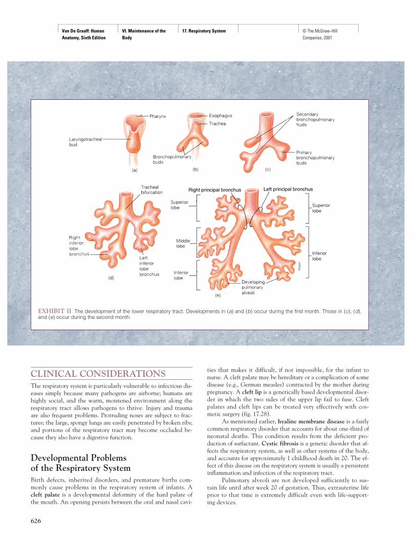
Development of the Lower Respiratory SystemThe lower respiratory system begins as a diverticulum, or out-pouching, from the ventral surface of endoderm along thelower pharyngeal region (exhibit II). This diverticulum, whichforms during the fourth week of development, is referred to asthe laryngotracheal (la-ring''go-tra'ke-al) bud. As the budgrows, the proximal portion forms the trachea and the distalportion bifurcates (splits) into a right and left principalbronchus.
The buds continue to elongate and split until the entiretubular network within the lower respiratory tract is formed(exhibit II). As the terminal portion forms air sacs, called pul-monary alveoli, at about 8 weeks of development, the sup-porting lung tissue begins to form. The complete structure ofthe lungs, however, is not fully developed until about 26weeks of fetal development. Premature infants born prior tothis time therefore require special artificial respiratory equip-ment to live.

Van De Graaff: Human Anatomy, Sixth Edition
VI. Maintenance of the Body
17. Respiratory System © The McGraw−Hill Companies, 2001
625
EXHIBIT I The development of the upper respiratory system. (a) An anterior view of the developing head of an embryo at 4 weeks show-ing the position of a transverse cut depicted in (a1), (a2), and (a3). (a2) Development at 5 weeks and (a3) at 5 1/2 weeks. (b) An anterior viewof the developing head of an embryo at 6 weeks showing the position of a sagittal cut depicted in (b1), (b2), (b3), and (b4) at 14 weeks.
Van De Graaff: Human Anatomy, Sixth Edition
VI. Maintenance of the Body
17. Respiratory System © The McGraw−Hill Companies, 2001
626
Right principal bronchus Left principal bronchus
EXHIBIT II The development of the lower respiratory tract. Developments in (a) and (b) occur during the first month. Those in (c), (d),and (e) occur during the second month.
CLINICAL CONSIDERATIONSThe respiratory system is particularly vulnerable to infectious dis-eases simply because many pathogens are airborne; humans arehighly social, and the warm, moistened environment along therespiratory tract allows pathogens to thrive. Injury and traumaare also frequent problems. Protruding noses are subject to frac-tures; the large, spongy lungs are easily penetrated by broken ribs;and portions of the respiratory tract may become occluded be-cause they also have a digestive function.
Developmental Problems of the Respiratory SystemBirth defects, inherited disorders, and premature births com-monly cause problems in the respiratory system of infants. Acleft palate is a developmental deformity of the hard palate ofthe mouth. An opening persists between the oral and nasal cavi-
ties that makes it difficult, if not impossible, for the infant tonurse. A cleft palate may be hereditary or a complication of somedisease (e.g., German measles) contracted by the mother duringpregnancy. A cleft lip is a genetically based developmental disor-der in which the two sides of the upper lip fail to fuse. Cleftpalates and cleft lips can be treated very effectively with cos-metic surgery (fig. 17.28).
As mentioned earlier, hyaline membrane disease is a fairlycommon respiratory disorder that accounts for about one-third ofneonatal deaths. This condition results from the deficient pro-duction of surfactant. Cystic fibrosis is a genetic disorder that af-fects the respiratory system, as well as other systems of the body,and accounts for approximately 1 childhood death in 20. The ef-fect of this disease on the respiratory system is usually a persistentinflammation and infection of the respiratory tract.
Pulmonary alveoli are not developed sufficiently to sus-tain life until after week 20 of gestation. Thus, extrauterine lifeprior to that time is extremely difficult even with life-support-ing devices.

Van De Graaff: Human Anatomy, Sixth Edition
VI. Maintenance of the Body
17. Respiratory System © The McGraw−Hill Companies, 2001
Trauma or InjuryHumans are especially susceptible to epistaxes (ep''ı-stak'sez)(nosebleeds) because the prominent nose can be easily bumpedand because the nasal mucosa has extensive vascularity forwarming the inspired air. Epistaxes may also be caused by highblood pressure or diseases such as leukemia.
When air enters the pleural cavity surrounding either lung,the condition is referred to as a pneumothorax (fig. 17.29). Apneumothorax can result from an external injury, such as a stab-bing, bullet wound, or penetrating fractured rib, or it can be theresult of internal conditions. A severely diseased lung, as in em-physema, can create a pneumothorax as the wall of the lung de-teriorates along with the visceral pleura and permits air to enterthe pleural cavity.
Choking on a foreign object is a common serious trauma tothe respiratory system. More than eight Americans choke todeath each day on food lodged in their trachea. A simple proce-dure termed the abdominal thrust (Heimlich) maneuver cansave the life of a person who is choking (fig. 17.30). The abdomi-nal thrust maneuver is performed as follows:
A. If the victim is standing or sitting:1. Stand behind the victim or the victim’s chair and wrap
your arms around his or her waist.2. Grasp your fist with your other hand and place the fist
against the victim’s abdomen, slightly above the naveland below the rib cage.
3. Press your fist into the victim’s abdomen with a quickupward thrust.
4. Repeat several times if necessary.B. If the victim is lying down:
1. Position the victim on his or her back.2. Face the victim, and kneel on his or her hips.3. With one of your hands on top of the other, place the
heel of your bottom hand on the abdomen, slightlyabove the navel and below the rib cage.
4. Press into the victim’s abdomen with a quick upwardthrust.
5. Repeat several times if necessary.
If you are alone and choking, use whatever is available toapply force just below your diaphragm. Press into a table or asink, or use your own fist.
Chapter 17 Respiratory System 627
CH
AP
TE
R 17
FIGURE 17.28 (a) An infant with a unilateral cleft lip and palate.(b) The same child following corrective surgery.
(a)
(b)
FIGURE 17.29 A pneumothorax of the right lung. The right side ofthe thorax appears uniformly dark because it is filled with air; thespaces between the ribs are also greater than those on the left, be-cause the ribs are released from the elastic tension of the lungs. Theleft lung appears denser (less dark) because of shunting of bloodfrom the right to the left lung.

Van De Graaff: Human Anatomy, Sixth Edition
VI. Maintenance of the Body
17. Respiratory System © The McGraw−Hill Companies, 2001
People saved from drowning and victims of shock fre-quently experience apnea (cessation of breathing) and will soondie if not revived by artificial respiration. The accepted treat-ment for reviving a person who has stopped breathing is illus-trated in figure 17.31.
Common Respiratory DisordersA cough is the most common symptom of respiratory disorders.Acute problems may be accompanied by dyspnea or wheezing.Respiratory or circulatory problems may cause cyanosis (si'a-no'-sis), a blue discoloration of the skin resulting from blood with alow oxygen content.
Although the common cold is the most widespread of allrespiratory diseases, there is still no cure for this ailment—onlymedications that offer symptomatic relief. Colds occur repeatedlybecause acquired immunity for one virus does not protect againstother viruses. Cold viruses generally incite acute inflammation inthe respiratory mucosa, causing flow of mucus, sometimes accom-panied by fever and/or headache.
Nearly all of the structures and regions of the respiratorypassageways can become infected and inflamed. Influenza is aviral disease that causes an inflammatory condition of theupper respiratory tract. Influenza can be epidemic, but fortu-
nately vaccines are available. Sinusitis is an inflammation ofthe paranasal sinuses. Sinusitis can be quite painful if thedrainage ducts from the sinuses into the nasal cavity becomeblocked. Tonsillitis may involve any or all of the tonsils andfrequently follows other lingering diseases of the oral or pharyn-geal regions. Chronic tonsillitis sometimes requires a tonsillec-tomy. Laryngitis is inflammation of the larynx, which oftenproduces a hoarse voice and limits the ability to talk. Tracheo-bronchitis and bronchitis are infections of the regions forwhich they are named. Severe inflammation in these areas cancause smaller respiratory tubules to collapse, blocking the pas-sage of air.
Diseases of the lungs are likewise common and usually seri-ous. Pneumonia is an acute infection and inflammation of lungtissue accompanied by exudation (accumulation of fluid). It isusually caused by bacteria, most commonly by the pneumococcusbacterium. Viral pneumonia is caused by a number of differentviruses. Tuberculosis is an inflammatory disease of the lungscontracted by inhaling air sneezed or coughed by someone who iscarrying active tuberculosis bacteria. Tuberculosis softens lungtissue, which eventually becomes ulcerated. Asthma is a diseasethat affects people who are allergic to certain inhaled antigens. Itcauses a swelling and blocking of lower respiratory tubes, oftenaccompanied by the formation of mucus plugs. Pleurisy (ploor' ı-se) is an inflammation of the pleura and is usually secondary tosome other respiratory disease. Inspiration may become painful,and fluid may collect within the pleural cavity. Emphysema(em''f ı-se'ma) is a disease that causes the breakdown of the pul-monary alveoli, thus increasing the size of air spaces and decreas-ing the surface area (fig. 17.32). It is a frequent cause of deathamong heavy cigarette smokers.
Cancer in the respiratory system is known to be caused bythe repeated inhalation of irritating substances, such as cigarettesmoke. Cancers of the lip, larynx, and lungs (fig. 17.33) are espe-cially common in smokers over the age of 50.
Disorders of Respiratory ControlA variety of disease processes can result in cessation of breathingduring sleep, or sleep apnea. Sudden infant death syndrome(SIDS) is an especially tragic form of sleep apnea that claims thelives of about 10,000 babies annually in the United States. Vic-tims of this condition are apparently healthy 2-to-5-month-oldbabies who die in their sleep without apparent reason—hence,the layperson’s term, “crib death.” These deaths seem to becaused by failure of the respiratory control mechanisms in thebrain stem and/or by failure of the carotid bodies to be stimulatedby reduced arterial oxygen.
628 Unit 6 Maintenance of the Body
CH
AP
TE
R 1
7
FIGURE 17.30 The abdominal thrust (Heimlich) maneuver.
cyanosis: Gk. kyanosis, dark-blue color
influenza: L. influentia, a flowing in
tuberculosis: L. tuberculum, diminutive of tuber, swelling
asthma: Gk. asthma, panting
emphysema: Gk. emphysan, blow up, inflate

Van De Graaff: Human Anatomy, Sixth Edition
VI. Maintenance of the Body
17. Respiratory System © The McGraw−Hill Companies, 2001
Chapter 17 Respiratory System 629
CH
AP
TE
R 17
FIGURE 17.31 Artificial respiration.

Van De Graaff: Human Anatomy, Sixth Edition
VI. Maintenance of the Body
17. Respiratory System © The McGraw−Hill Companies, 2001
Abnormal breathing patterns often appear prior to deathfrom brain damage or heart disease. The most common of theseabnormal patterns is Cheyne–Stokes (chan'stoks') breathing,which the depth of breathing progressively increases and thenprogressively decreases. These cycles of increasing and decreasingtidal volumes may be followed by periods of apnea of varying du-
630 Unit 6 Maintenance of the Body
CH
AP
TE
R 1
7
Tumors
FIGURE 17.32 Photomicrographs of tissue (a) from a normallung and (b) from the lung of a person with emphysema. In emphy-sema, lung tissue is destroyed, resulting in fewer and larger pul-monary alveoli.
FIGURE 17.33 A cancerous lung. For people who regularlysmoke a pack of cigarettes a day, the risk of developing lung canceris 20 times greater than for people who have never smoked.
Clinical Case Study AnswerThe ice pick traversed the parietal pleura, the visceral pleura, and thenentered the airway, at least at the alveolar–terminal bronchiole level,but possibly at larger airways. This allowed inspired air to escape fromthe airway into the pleural cavity. The air would have taken the follow-ing route: pharynx → larynx → trachea → left principal bronchus →lobar bronchus (of left upper lobe) → apical segmental (tertiary)bronchus → bronchioles through laceration into pleural space. Thiscondition is treated by inserting a tube (tube thoracostomy) into thepleural cavity to allow suction evacuation of air and blood, which resultsin reexpansion of the lung. The laceration usually seals over within aday or two. Persistent bleeding may necessitate thoracotomy (incision ofthe chest wall) for repair.
rations. Cheyne–Stokes breathing may be caused by neurologicaldamage or by insufficient oxygen delivery to the brain. The lattermay result from heart disease or from a brain tumor that diverts alarge part of the vascular supply from the respiratory centers.
Cheyne–Stokes breathing: from John Cheyne, British physician, 1777–1836; andWilliam Stokes, Irish physician, 1804–78.

Van De Graaff: Human Anatomy, Sixth Edition
VI. Maintenance of the Body
17. Respiratory System © The McGraw−Hill Companies, 2001
Chapter 17 Respiratory System 631
CH
AP
TE
R 17
CLINICAL PRACTICUM 17.1You admitted a 45-year-old male with AIDSto the hospital two days ago with shortness ofbreath and fevers. His chest radiographshowed diffuse opacities throughout bothlungs, and a diagnosis of Pneumocytsis carniipneumonia was made from sputum samples.The patient was receiving appropriate treat-ment and seemed to be doing well. The nursecalls you to report that the patient has be-come acutely short of breath and is havingleft-sided chest pain. When you examine the
patient, you note him to have greatly de-creased breath sounds on the left side of hischest. You order a new chest radiograph.
Q U E S T I O N S1. Why does this patient have worsening
shortness of breath and left-sided chestpain?
2. How does this affect the other lung andthe heart?
3. What is the proper treatment?
Chapter Summary
Introduction to the Respiratory System(pp. 603–604)
1. Respiration refers not only to ventilation(breathing), but also to the exchange ofgases between the atmosphere, the blood,and individual cells. Within cells, themetabolic reactions that release energyare called cellular respiration.
2. In order for the respiratory system tofunction, the respiratory membranes mustbe moist, thin-walled, highly vascular,and differentially permeable.
3. The functions of the respiratory systeminclude gaseous exchange, soundproduction, assistance in abdominalcompression, and reflexive coughing andsneezing, and immune response.
Conducting Passages (pp. 604–612)1. The nose is supported by nasal bones and
cartilages.2. The nasal epithelium warms, moistens,
and cleanses the inspired air.3. Olfactory epithelium is associated with
the sense of smell, and the nasal cavityacts as a resonating chamber for the voice.
4. The paranasal sinuses are found in themaxillary, frontal, sphenoid, and ethmoidbones.(a) These sinuses lighten the skull and
are lined with mucus-secreting gobletcells.
(b) Sinusitis is an inflammation of one ormore of the paranasal sinuses.
5. The pharynx is a funnel-shaped organ. Itcontains a passageway that connects theoral and nasal cavities with the esophagusand larynx.(a) The nasopharynx, connected by the
auditory tubes to the tympaniccavities, contains the pharyngealtonsils, or adenoids.
(b) The oropharynx is the middleportion, extending from the softpalate to the level of the hyoid bone;it contains the palatine and lingualtonsils.
(c) The laryngopharynx extends from thehyoid bone to the larynx andesophagus.
CLINICAL PRACTICUM 17.2You are working in the newborn nursery ofthe hospital when you are called by the ob-stetrician to evaluate a newborn in respira-tory distress. On exam, you note that thepatient has no breath sounds on the left sideof her chest and has very labored breathing.You order a chest radiograph.
Q U E S T I O N S1. What do you see in the left
hemithorax?2. What congenital anomaly resulted in
this condition?3. Once the anomaly is repaired, will the
patient’s breathing be normal?

Van De Graaff: Human Anatomy, Sixth Edition
VI. Maintenance of the Body
17. Respiratory System © The McGraw−Hill Companies, 2001
632 Unit 6 Maintenance of the Body
CH
AP
TE
R 1
7
6. The larynx contains a number ofcartilages that keep the passageway to thetrachea open during breathing and closesthe respiratory passageway duringswallowing.(a) The epiglottis is a spoon-shaped
structure that aids in closing thelaryngeal opening, or glottis, duringswallowing.
(b) The vocal folds in the larynx arecontrolled by intrinsic muscles andare used in sound production.
7. The trachea is a semirigid tubular organsupported by incomplete rings of hyalinecartilage, that leads from the larynx to thebronchial tree.
8. The bronchial tree includes a principalbronchus, which divides to produce lobarbronchi, segmental bronchi, andbronchioles; the conducting division endswith the respiratory bronchioles, whichconnect to the pulmonary alveoli.
Pulmonary Alveoli, Lungs, and Pleurae(pp. 612–618)
1. Pulmonary alveoli are the functional unitsof the lungs, where gas exchange occurs;they are small, moistened, thin-walled air sacs.
2. The right and left lungs are separated bythe mediastinum. Each lung is dividedinto lobes and lobules.(a) The right lung is subdivided by two
fissures into superior, middle, andinferior lobes.
(b) The left lung is subdivided into asuperior lobe and an inferior lobe by asingle fissure.
3. The lungs are covered by visceral pleura,and the thoracic cavity is lined by aparietal pleura.(a) The potential space between these
two pleural membranes is called thepleural cavity.
(b) The pleural membranescompartmentalize each lung andexclude the structures located in themediastinum.
Mechanics of Breathing (pp. 618–622)1. Quiet (unforced) inspiration is due to
contraction of the diaphragm and certainintercostal muscles. Forced inspiration isaided by the scalenes and the pectoralisminor and sternocleidomastoid muscles.
2. Quiet expiration is produced by relaxationof the respiratory muscles and elastic recoilof the lungs and thorax. Forced expirationis aided by certain intercostal muscles andthe abdominal muscles.
3. Among the air volumes exchanged inventilation are tidal, inspiratory reserve,and expiratory reserve volumes.
4. Nonrespiratory air movements areassociated with coughing, sneezing,sighing, yawning, laughing, crying, andhiccuping.
Regulation of Breathing (p. 623)1. Ventilation is directly controlled by the
rhythmicity center in the medullaoblongata, which in turn is influenced bythe pneumotaxic and apneustic centers inthe pons.
2. These brain stem areas are affected byhigher brain function and by sensoryinput from chemoreceptors.
3. Central chemoreceptors are located in themedulla oblongata; peripheralchemoreceptors are located in the aorticand carotid bodies.
Review Activities
Objective Questions1. Which of the following is a false
statement?(a) The term respiration can be used in
reference to ventilation (breathing)or oxygen utilization by body cells.
(b) The incoming (inhaled) air thatcontacts the pulmonary alveoli isunchanged from that whichsurrounds the body.
(c) As a resonating chamber, the nasalcavity is important in soundproduction.
(d) It is only through the walls of thepulmonary alveoli that gaseousexchange occurs.
2. Which is not a component of the nasalseptum?(a) the palatine bone(b) the vomer(c) the ethmoid bone(d) septal cartilage
3. An adenoidectomy is the removal of(a) the uvula.(b) the pharyngeal tonsils.(c) the palatine tonsils.(d) the lingual tonsils.
4. Which is not a paranasal sinus?(a) the palatine sinus(b) the ethmoidal sinus(c) the sphenoidal sinus(d) the frontal sinus(e) the maxillary sinus
5. Which of the following is notcharacteristic of the left lung?(a) a cardiac impression(b) a superior lobe(c) a single fissure(d) an inferior lobe(e) a middle lobe
6. The epithelial lining of the wall of thethoracic cavity is called(a) the parietal pleura.(b) the pleural peritoneum.(c) the mediastinal pleura.(d) the visceral pleura.(e) the costal pleura.
7. Which muscle group combination permitsinspiration?(a) diaphragm, abdominal complex(b) internal intercostals, diaphragm(c) external intercostals, internal
intercostals
(d) external intercostals, diaphragm,internal intercostals (interchondralpart)
8. The vocal folds (cords) are attached to(a) the cricoid and thyroid cartilages.(b) the cuneiform and cricoid cartilages.(c) the corniculate and thyroid cartilages.(d) the arytenoid and thyroid cartilages.
9. The maximum amount of air that can beexpired after a maximum inspiration is(a) the tidal volume.(b) the forced expiratory volume.(c) the vital capacity.(d) the maximum expiratory flow rate.(e) the residual volume.
10. The rhythmic control of breathing isproduced by the activity of inspiratoryand expiratory neurons in(a) the medulla oblongata.(b) the apneustic center of the pons.(c) the pneumotaxic center of the pons.(d) the cerebral cortex.(e) the hypothalamus.

Van De Graaff: Human Anatomy, Sixth Edition
VI. Maintenance of the Body
17. Respiratory System © The McGraw−Hill Companies, 2001
Chapter 17 Respiratory System 633
CH
AP
TE
R 17
Essay Questions1. Why is warming, moistening, and filtering
the air so important for a healthy,functioning respiratory system?
2. Explain the structural and functionaldifferences between the three regions ofthe pharynx.
3. What is a bronchial tree? Why arepulmonary alveoli rather than bronchialtrees considered the functional units ofthe respiratory system?
4. Define each of the following termsrelating to the structure of the lung: base,hilum, apex, costal surface, fissure, lobe,lobule, pulmonary ligament, and bronchialsegment.
5. Diagram the location of the lungs in thethoracic cavity with respect to the heart.Identify the various thoracic serousmembranes.
6. List the kinds of epithelial tissues foundwithin the respiratory system and describethe location of each.
7. What protective devices of the respiratorysystem guard against pollutants, thespread of infections, and dual collapse ofthe lungs?
8. What are the functions of the larynx? Listsome of the clinical conditions that couldinvolve this organ.
9. What are the advantages ofcompartmentalization of the thoracicorgans?
10. Explain the sequence of pulmonaryventilation. Discuss the mechanisms ofinspiration and expiration. How are airpressures related to ventilation?
11. Distinguish between tidal volume, vitalcapacity, and total lung capacity.
12. What is meant by a rhythmicityrespiratory area? How are the apneusticand pneumotaxic areas related to therhythmicity area?
Critical-Thinking Questions1. Identify two places in the respiratory
system where large surface areas ofcapillary networks are found. What is thefunction of each of these areas and whyare the capillary networks important inthe treatment of certain clinicalconditions?
2. On what principal is the abdominal thrustmaneuver based? When would mouth-to-mouth resuscitation rather than theabdominal thrust maneuver be used torevive a person?
3. The nature of the sounds produced bypercussing (tapping) a patient’s chest cantell a physician a great deal about thecondition of the organs within thethoracic cavity. Healthy, air-filled lungsresonate, or sound hollow. How do youthink the lungs of a person withemphysema would sound in comparisonto healthy lungs? What kind of soundwould be produced by a collapsed lung, orone that was partially filled with fluid?
4. Why do premature infants often requirerespiratory assistance (a mechanicalventilator) to keep their lungs inflated?
5. Nicotine from cigarette smoke causesmucus to build up and paralyzes the ciliathat line the respiratory tract. How dothese changes affect the lungs?
Visit our Online Learning Center at http://www.mhhe.com/vdg for chapter-by-chapter quizzing, additional study resources, and related web links.