Protective effect of autophagy in laser-induced glioma cell death in vitro
10
Lasers in Surgery and Medicine 42:338–347 (2010) Protective Effect of Autophagy in Laser-Induced Glioma Cell Death In Vitro Aleksandar J. Krmpot, PhD, 1 ** Kristina D. Janjetovic, MSc, 2,3 Maja S. Misirkic, MSc, 2,3 Ljubica M. Vucicevic, MSc, 2,3 Dejan V. Pantelic, PhD, 1 Darko M. Vasiljevic, PhD, 1 Dusan M. Popadic, MD, PhD, 2 Branislav M. Jelenkovic, PhD, 1 and Vladimir S. Trajkovic, MD, PhD 2 * 1 Institute of Physics, University of Belgrade, 11080 Belgrade, Serbia 2 Institute of Microbiology and Immunology, School of Medicine, University of Belgrade, 11000 Belgrade, Serbia 3 Institute for Biological Research, 11000 Belgrade, Serbia Background and Objective: Laser phototherapy could be potentially used for cancer treatment, but the mechanisms of laser-induced cell death are not completely understood. Autophagy is the process in which the damaged cellular proteins and organelles are engulfed by and destroyed in acidified multiple-membrane vesicles. The aim of the present study was to investigate the role of autophagy in laser-induced tumor cell death in vitro. Study Design/Materials and Methods: The monolayers of U251 human glioma tumor cells were exposed to 532 nm laser light from a single mode frequency-doubled Nd-YVO4 laser. A flattened Gaussian radial profile of laser beam (0.5– 4 W) was used to uniformly illuminate entire colony of cells for various amounts of time (15–120 seconds) in the absence of cell culture medium. The cells were grown for 24 hours and the cell viability was determined by crystal violet or MTT assay. The presence of autophagy was assessed after 16 hours by fluorescence microscopy/flow cytometric analysis of acridine orange-stained autophago- lysosomes and Western blot analysis of the autophago- some-associated LC3-II protein. The concentration of the principal pro-autophagic protein beclin-1 was determined after 6 hours by cell-based ELISA. Results: The intracytoplasmic accumulation of autophagic vesicles, increase in LC3-II and up-regulation of beclin-1 expression were clearly observed under irradiation con- ditions that caused approximately 50% cytotoxicity. Post- irradiation addition of three different autophagy inhibitors (bafilomycin A1, chloroquine, or wortmannin) further increased the laser-induced cytotoxicity, without affecting non-irradiated cells. Conclusions: These data indicate that beclin-1-dependent induction of autophagy can protect glioma cells from laser-mediated cytotoxicity. Lasers Surg. Med. 42:338– 347, 2010. ß 2010 Wiley-Liss, Inc. Key words: apoptosis; cancer; necrosis; phototherapy INTRODUCTION Laser irradiation is known for its ability to cause cell death, a feature that has been used over past decades for therapeutical killing of tumor cells [1–6]. However, the value of laser-based anticancer therapy is limited due to many side-effects and insufficient efficacy [7], the main limitation being the failure to achieve complete tumor destruction, particularly at the margins of larger tumors [8,9]. In order to increase efficiency and/or decrease the side-effects of laser anticancer therapy, a more thorough knowledge is required on the mechanisms underlying laser-induced cytotoxicity. In addition to tissue vaporization at very high tempe- ratures ( > 1008C) [10], laser irradiation can also cause necrotic cell death at high power [11–13] and apoptotic cell death at low power [14–16]. Necrosis is characterized by vacuolation of the cytoplasm, breakdown of the plasma membrane and release of cellular contents, resulting in the induction of inflammation around the dying cell [17]. Apoptosis or type I programmed cell death, on the other hand, is typified by fragmentation of DNA without plasma membrane breakdown, followed by packaging of the deceased cell into apoptotic bodies that are recognized and removed by phagocytic cells in the absence of inflammation [17]. Importantly, apoptosis was observed in a fraction of tumor cells surrounding the necrotic lesion induced by high-power laser therapy [18]. Since the secondary outgrowth of these marginal zone cells may form the basis of a reformation of the tumor mass [18], the complete elucidation of the mechanisms underlying their survival and death is of paramount importance for improving the efficiency of laser-based anticancer treatment. Macroautophagy (referred to hereafter as autophagy) is a self-cannibalization process that involves sequestration of cell structures in double-membraned organelles, called autophagosomes [19]. This is followed by fusion of Contract grant sponsor: Ministry of Science of the Republic of Serbia; Contract grant numbers: 145073, 141003. *Correspondence to: Vladimir S. Trajkovic, MD, PhD, Institute of Microbiology and Immunology, School of Medicine, University of Belgrade, Dr. Subotica 1, 11000 Belgrade, Serbia. E-mail: [email protected] **Correspondence to: Aleksandar J. Krmpot, PhD, Institute of Physics, University of Belgrade, Pregrevica 118, 11080 Belgrade, Serbia. E-mail: [email protected] Accepted 3 February 2010 Published online 15 May 2010 in Wiley InterScience (www.interscience.wiley.com). DOI 10.1002/lsm.20911 ß 2010 Wiley-Liss, Inc.
-
Upload
independent -
Category
Documents
-
view
1 -
download
0
Transcript of Protective effect of autophagy in laser-induced glioma cell death in vitro
Lasers in Surgery and Medicine 42:338–347 (2010)
Protective Effect of Autophagy in Laser-Induced GliomaCell Death In Vitro
Aleksandar J. Krmpot, PhD,1** Kristina D. Janjetovic, MSc,2,3 Maja S. Misirkic, MSc,2,3
Ljubica M. Vucicevic, MSc,2,3 Dejan V. Pantelic, PhD,1 Darko M. Vasiljevic, PhD,1 Dusan M. Popadic, MD, PhD,2
Branislav M. Jelenkovic, PhD,1 and Vladimir S. Trajkovic, MD, PhD2*
1Institute of Physics, University of Belgrade, 11080 Belgrade, Serbia2Institute of Microbiology and Immunology, School of Medicine, University of Belgrade, 11000 Belgrade, Serbia3Institute for Biological Research, 11000 Belgrade, Serbia
Background and Objective: Laser phototherapycould be potentially used for cancer treatment, but themechanisms of laser-induced cell death are not completelyunderstood. Autophagy is the process in which thedamaged cellular proteins and organelles are engulfed byand destroyed in acidified multiple-membrane vesicles.The aim of the present study was to investigate the role ofautophagy in laser-induced tumor cell death in vitro.StudyDesign/Materials andMethods: The monolayersof U251 human glioma tumor cells were exposed to 532 nmlaser light from a single mode frequency-doubled Nd-YVO4laser. A flattened Gaussian radial profile of laser beam(0.5–4 W) was used to uniformly illuminate entire colony ofcells for various amounts of time (15–120 seconds) in theabsence of cell culture medium. The cells were grown for24 hours and the cell viability was determined by crystalviolet or MTT assay. The presence of autophagy wasassessed after 16 hours by fluorescence microscopy/flowcytometric analysis of acridine orange-stained autophago-lysosomes and Western blot analysis of the autophago-some-associated LC3-II protein. The concentration of theprincipal pro-autophagic protein beclin-1 was determinedafter 6 hours by cell-based ELISA.Results:The intracytoplasmic accumulation of autophagicvesicles, increase in LC3-II and up-regulation of beclin-1expression were clearly observed under irradiation con-ditions that caused approximately 50% cytotoxicity. Post-irradiation addition of three different autophagy inhibitors(bafilomycin A1, chloroquine, or wortmannin) furtherincreased the laser-induced cytotoxicity, without affectingnon-irradiated cells.Conclusions:These data indicate that beclin-1-dependentinduction of autophagy can protect glioma cells fromlaser-mediated cytotoxicity. Lasers Surg. Med. 42:338–347, 2010. � 2010 Wiley-Liss, Inc.
Key words: apoptosis; cancer; necrosis; phototherapy
INTRODUCTION
Laser irradiation is known for its ability to cause celldeath, a feature that has been used over past decades fortherapeutical killing of tumor cells [1–6]. However, thevalue of laser-based anticancer therapy is limited due to
many side-effects and insufficient efficacy [7], the mainlimitation being the failure to achieve complete tumordestruction, particularly at the margins of larger tumors[8,9]. In order to increase efficiency and/or decrease theside-effects of laser anticancer therapy, a more thoroughknowledge is required on the mechanisms underlyinglaser-induced cytotoxicity.
In addition to tissue vaporization at very high tempe-ratures (> 1008C) [10], laser irradiation can also causenecrotic cell death at high power [11–13] and apoptotic celldeath at low power [14–16]. Necrosis is characterized byvacuolation of the cytoplasm, breakdown of the plasmamembrane and release of cellular contents, resulting in theinduction of inflammation around the dying cell [17].Apoptosis or type I programmed cell death, on the otherhand, is typified by fragmentation of DNA without plasmamembrane breakdown, followed by packaging of thedeceased cell into apoptotic bodies that are recognizedand removed by phagocytic cells in the absence ofinflammation [17]. Importantly, apoptosis was observedin a fraction of tumor cells surrounding the necroticlesion induced by high-power laser therapy [18]. Sincethe secondary outgrowth of these marginal zone cells mayform the basis of a reformation of the tumor mass [18],the complete elucidation of the mechanisms underlyingtheir survival and death is of paramount importancefor improving the efficiency of laser-based anticancertreatment.
Macroautophagy (referred to hereafter as autophagy) is aself-cannibalization process that involves sequestrationof cell structures in double-membraned organelles,called autophagosomes [19]. This is followed by fusion of
Contract grant sponsor: Ministry of Science of the Republic ofSerbia; Contract grant numbers: 145073, 141003.
*Correspondence to: Vladimir S. Trajkovic, MD, PhD, Instituteof Microbiology and Immunology, School of Medicine, Universityof Belgrade, Dr. Subotica 1, 11000 Belgrade, Serbia.E-mail: [email protected]
**Correspondence to: Aleksandar J. Krmpot, PhD, Institute ofPhysics, University of Belgrade, Pregrevica 118, 11080 Belgrade,Serbia. E-mail: [email protected]
Accepted 3 February 2010Published online 15 May 2010 in Wiley InterScience(www.interscience.wiley.com).DOI 10.1002/lsm.20911
� 2010 Wiley-Liss, Inc.
autophagosomes with lysosomes and formation of auto-phagolysosomes in which the cellular content is degradedby acidic lysosomal hydrolases [20]. The physiological roleof autophagy is to remove long-lived proteins and damagedorganelles, but when it is extensive, activated inappropri-ately or in cells which are unable to die by apoptosis,autophagy acts as an alternative cell-death pathway calledautophagic or type II programmed cell death [21]. Inapoptosis-competent cells, on the other hand, autophagymainly acts as a survival mechanism that can rescue cellsfrom apoptotic death induced by nutrient deprivation orpharmacological agents [22]. Although autophagy caneither promote or impair cell death in a context-dependentmanner, no study so far, to the best of our knowledge, hasaddressed the ability of laser light to induce autophagy inmammalian cells.
In this study we present evidence that laser irradiationcan cause autophagy in human U251 glioma cell line.Moreover, our data demonstrate that autophagy is notinvolved in cell death, but actually protects glioma cellsfrom laser-induced cytotoxicity.
MATERIALS AND METHODS
Cells and Cell Culture
The rat glioma cell line U251 was kindly donated byDr. Pedro Tranque (Universidad de Castilla-La Mancha,Albacete, Spain). The cells were maintained at 378C in ahumidified atmosphere with 5% CO2, in a HEPES (20 mM)-buffered RPMI 1640 cell culture medium (Sigma–Aldrich,St. Louis, MO) supplemented with 5% fetal calf serum
(FCS), 2 mM L-glutamine, 10 mM sodium pyruvate andpenicillin/streptomycin (all from Sigma–Aldrich). The cellswere prepared for experiments using the conventionaltrypsinization procedure with trypsin/EDTA and incu-bated as a monolayer in 96-well flat-bottom plates (1�104 cells per well). Cells were rested for 24 hours, washedwith phosphate buffered saline (PBS; Sigma–Aldrich) andexposed to laser light in the absence of cell culture medium.Following irradiation, fresh cell culture medium wasadded and the cells were incubated for an additional 6,16, or 24 hours for the measurement of beclin-1 expression,autophagy or cell number, respectively. These end-pointswere determined in preliminary experiments (data notshown) as the time points in which the particular cellularresponses were most pronounced (6 and 16 hours) orclearly observable (24 hours). In some experiments,after irradiation cells were treated with autophagy inhi-bitors bafilomycin A1 (50 nm), chloroquine (20 mM) orwortmannin (100 nm) (all from Sigma–Aldrich) and thecell viability was determined after 16 hours.
Laser Treatment and Temperature Measurement
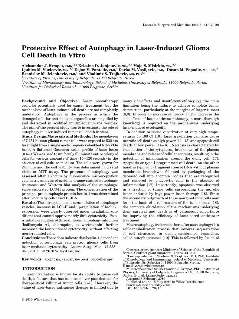
For irradiation of cells we used a continuous wavesingle mode frequency-doubled diode-pumped Nd-YVO4laser (Verdi V10, Coherent, Inc., Santa Clara, CA), whichdelivers green light (532 nm) up to 10 W power. Thebeam from the laser was directed to the cells passingthrough a lens and a diffuser to get flattened profileof �4 mm diameter (Fig. 1), as measured by the beamprofilometer (BCi4, by C-Cam Technologies, Leuven,Belgium). The cells were irradiated with different laser
Fig. 1. The setting for laser irradiation of tumor cells. The beam from the laser (532 nm) was
directed to the cell monolayer (without cell culture medium) passing through a lens and a
diffuser to get flattened profile of �4 mm diameter.
AUTOPHAGY IN LASER-INDUCED GLIOMA CELL DEATH 339
powers (0.5–4 W) for various amounts of time (15–120 seconds) at room temperature and in the absence ofcell culture medium. To prevent irradiation of adjacentwells, the convex side surface of the diffuser was coatedwith a light-reflecting silver dye. The effectiveness of thisapproach was confirmed in preliminary experiments,in which no reduction of cell number, autophagy, orsignificant increase in temperature (> 378C) was observedin the wells adjacent to those irradiated with laser (data notshown). The temperature of the cell culture well surfacewith or without cells was measured during 60 secondsirradiation with 2 W laser beam using an infrared termo-vision camera with 640�480 pixel resolution and a spectralrange of 7.5–13 mm (FLIR ThermaCAM SC640, FLIRSystems, Boston, MA) and the appropriate software.
Cytotoxicity Assays
The cytotoxic effect of laser irradiation was assessed bymeasuring the number of viable cells using crystal violet or3-(4,5-dimethylthiazol-2-yl)-2,5-diphenyltetrazolium bro-mide (MTT) assay. Crystal violet cytotoxicity test is basedon the inability of dead cells to remain adherent to cellculture plastic [23]. Cells were washed with PBS to removedead, non-adherent cells. The remaining adherent, viablecells were fixed with methanol and stained with 1% crystalviolet solution for 10 minutes. The plates were thoroughlywashed with water, and crystal violet was dissolved in 33%acetic acid. The absorbance of the dissolved dye, corre-sponding to the number of viable cells, was measured in anautomated microplate reader at 570 nm. Mitochondrialdehydrogenase activity, as another indicator of cellnumber, was assessed by the mitochondrial-dependentreduction of MTT to formazan [24]. At the end of theincubation, MTT solution (0.5 mg/ml) was added to cellcultures and the cells were incubated for an additional1 hours. Thereafter, medium was removed and the cellswere lysed in dimethylsulfoxide. The conversion of MTT toformazan by metabolically viable cells was monitored by anautomated microplate reader at 570 nm. The results ofMTT and crystal violet tests were presented as % of thecontrol value obtained in untreated cells.
Autophagy Assessment
The acidic autophagic vesicles were visualized bysupravital staining with a pH-sensitive dye acridineorange, as previously described [25]. Briefly, cells werewashed with PBS and stained with acridine orange (1mM;Sigma–Aldrich) for 15 minutes at 378C. Subsequently, cellswere washed and analyzed under the inverted fluorescencemicroscope. Depending on their acidity, autophagic lyso-somes appeared as yellow-orange to bright-red fluorescentcytoplasmic vesicles, while nuclei were stained green.Alternatively, acridine orange-stained cells were tryp-sinized, washed, and analyzed on FACSCalibur flowcytometer using Cell Quest Pro software. The intensity ofautophagy was quantified as a red/green fluorescence ratio(FL3/FL1), which increases during autophagy. The induc-tion of autophagy was confirmed by Western blot analysis ofLC3, which exists in two forms, the 18-kDa cytosolic protein
(LC3-I) and the processed 16-kDa form (LC3-II), the latterbeing associated with autophagosome membranes [19].Approximately 2�106 cells were lysed on ice in Tris-salinecontaining 1% NP-40, 1 mM phenylmethylsulfonylfluorideand protease inhibitor cocktail (all from Sigma–Aldrich),centrifuged at 18,000g (15 minutes, 48C), and the super-natants were collected as the cell lysates. Equal amounts ofprotein from each sample was separated by SDS–PAGE onpolyacrylamide gels and transferred to nitrocellulosemembranes. Following incubation with rabbit anti-human LC3 (Cell Signaling Technology, Danvers, MA) asa primary antibody (1:1,000) and peroxidase-conjugatedgoat anti-rabbit IgG (Jackson IP Laboratories, West Grove,PA) as a secondary antibody (1:3,000), specific bandscorresponding to LC3-I and LC3-II were visualized usingenhanced chemiluminescence reagents for Western blotanalysis (Amersham Pharmacia Biotech, Piscataway, NJ).
Beclin-1 Expression Analysis
The cell-based Enzyme-Linked Immunosorbent Assay(ELISA) was performed as previously described [26]. Afterirradiation, cells were fixed in 4% paraformaldehyde,endogenous peroxidase was quenched with 1% H2O2 inPBS containing 0.1% Triton X-100 (PBST), and unspecificbinding of antibodies blocked with PBST solution contain-ing 10% FCS. Primary rabbit polyclonal antibodies specificfor human beclin-1 (1:250; Abcam, Cambridge, UK) wereapplied in PBST supplemented with 2% bovine serumalbumin (PBSTB), followed by secondary peroxidase-conjugated goat anti-rabbit IgG (1:3,000 in PBSTB;Jackson IP Laboratories). Both incubations were under-taken at 378C for 1 hours. After incubation with theperoxidase substrate tetramethylbenzidine (Sigma–Aldrich), the reaction was stopped with 0.1 M HCl and theabsorbance was measured in an automated microplatereader at 450 nm. The obtained absorbances were correctedfor the cell number determined by crystal violet staining, asdescribed in the original protocol [26]. The results arepresented as relative expression in comparison with thecontrol value, which was arbitrarily set to 1.
Induction of Hyperthermia
Cell suspension (105 or 106 cells in 100 ml for the flowcytometry or Western blotting, respectively) was trans-ferred to a sterile thin wall 0.2 ml tube (Eppendorf,Hamburg, Germany) and placed in a preheated (308C)thermoblock of the thermocycler (Reaplex2, Eppendorf)programmed to gradually increase temperature to 478Cwithin 30 seconds. This was achieved by setting controlheating parameters to a ramping rate of 10% in block(simulated) tube mode with switched-off lead heating andinput of the correct volume of the cell suspension (100 ml) inthe sample volume box. Correct heating of the blockwas confirmed using an external Pt100 thermal probe(Omega Engineering, Manchester, UK) placed in thewell juxtaposed to the cell-containing well. Immediatelyafter reaching 478C cells were transferred to 24-well plates(105 cells per well) for the flow cytometry analysis or 6-wellplates (106 cells/well) for Western blot and incubated for an
340 KRMPOT ET AL.
additional 18 hours. Cells were then collected by trypsini-zation and autophagy was examined by flow cytometryfollowing acridine orange staining, while LC3 conversionwas assessed by Western blotting. We also included thepositive control for hyperthermia-induced autophagy byheating the cells continuously for 30 minutes at 438C.
Statistical Analysis
The data are presented as mean�SD values of triplicateobservations from at least three experiments, or asmean�SD values from three independent experiments.The statistical significance of the differences betweentreatments was assessed using t-test and the value ofP< 0.05 was considered significant.
RESULTS
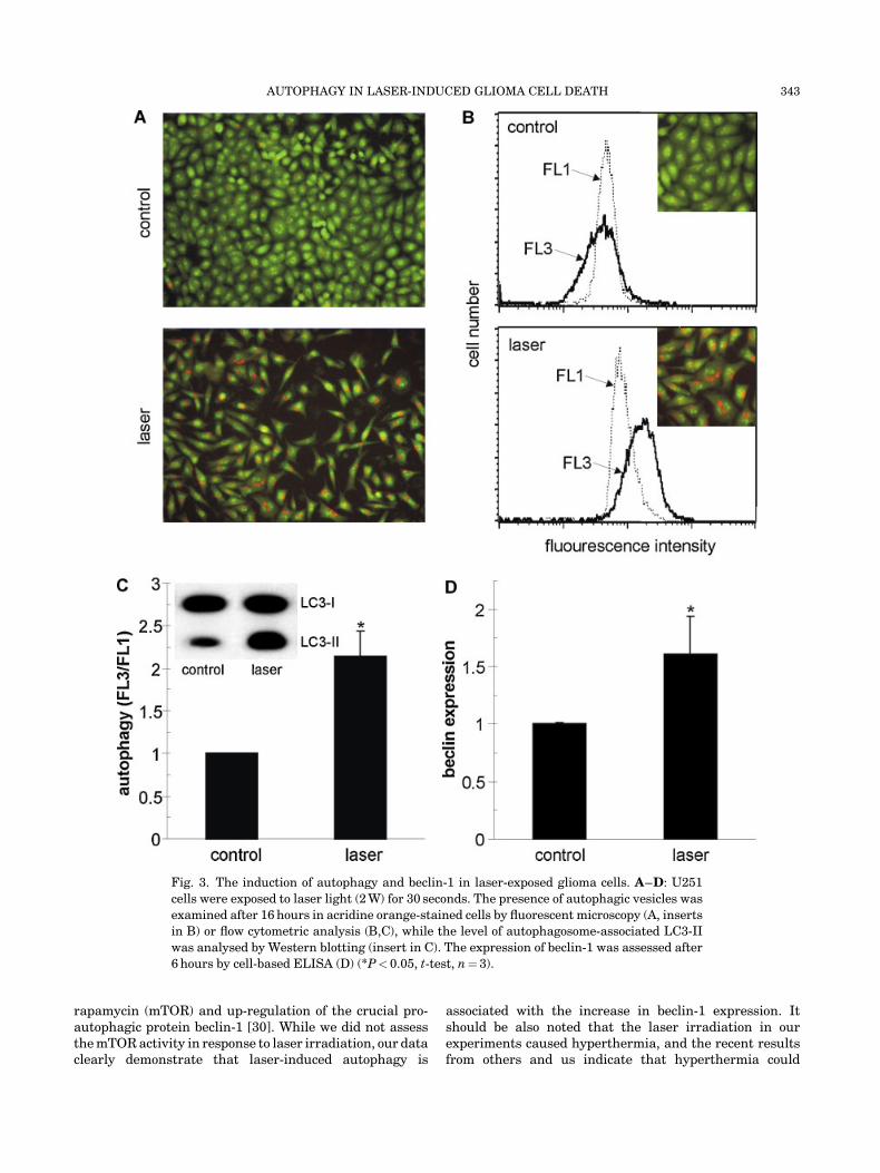
Cytotoxic Effect of Laser Irradiation
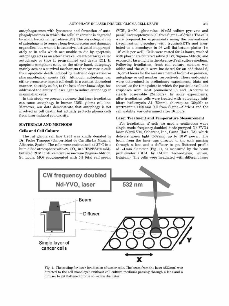
In order to establish the appropriate conditions for theassessment of autophagy, we first exposed the cells tolaser irradiation of increasing power (0.5–4 W) for variousamounts of time (15–120 seconds) and measured cellnumbers 24 hours after laser exposure. Both crystal violetand MTT assay demonstrated that the cytotoxicity of laserirradiation was energy-dependent, with 50% cytotoxicityachieved with �60 J¼ 2 W� 30 seconds (Fig. 2A,B). Thelight microscopic analysis of cell morphology has shownthat, in conditions causing complete cell death (2 W,120 seconds), the cells were totally destroyed, probably
Fig. 2. The dose- and time-dependent effects of laser light on viability of glioma cells. U251
cells were exposed to laser light of different power (A) or 2 W power (B,C) for the indicated
periods of time. The cell viability was investigated by crystal violet (A) or MTT assay (B), while
the cell morphology was examined by inverted light microscopy (C). The SD values in A were
less then 10% of the mean and were omitted for clarity (*P< 0.05, t-test, n¼ 4).
AUTOPHAGY IN LASER-INDUCED GLIOMA CELL DEATH 341
due to induction of necrosis (Fig. 2C). On the other hand, inconditions causing 50% reduction in cell numbers (2 W,30 seconds) the cell morphology was mainly preserved,while some round cells detached from the surface couldbe observed (Fig. 2C). Therefore, the conditions causing�50% cytotoxicity (2 W, 30 seconds) were chosen for furtherexperiments.
Laser-Mediated Cytotoxicity Is Accompanied bythe Induction of Autophagy and Up-Regulationof Beclin-1
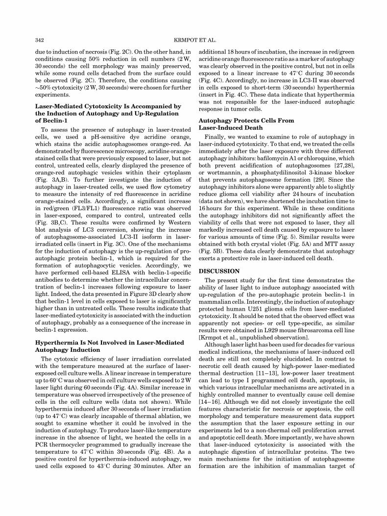
To assess the presence of autophagy in laser-treatedcells, we used a pH-sensitive dye acridine orange,which stains the acidic autophagosomes orange-red. Asdemonstrated by fluorescence microscopy, acridine orange-stained cells that were previously exposed to laser, but notcontrol, untreated cells, clearly displayed the presence oforange-red autophagic vesicles within their cytoplasm(Fig. 3A,B). To further investigate the induction ofautophagy in laser-treated cells, we used flow cytometryto measure the intensity of red fluorescence in acridineorange-stained cells. Accordingly, a significant increasein red/green (FL3/FL1) fluorescence ratio was observedin laser-exposed, compared to control, untreated cells(Fig. 3B,C). These results were confirmed by Westernblot analysis of LC3 conversion, showing the increaseof autophagosome-associated LC3-II isoform in laser-irradiated cells (insert in Fig. 3C). One of the mechanismsfor the induction of autophagy is the up-regulation of pro-autophagic protein beclin-1, which is required for theformation of autophagocytic vesicles. Accordingly, wehave performed cell-based ELISA with beclin-1-specificantibodies to determine whether the intracellular concen-tration of beclin-1 increases following exposure to laserlight. Indeed, the data presented in Figure 3D clearly showthat beclin-1 level in cells exposed to laser is significantlyhigher than in untreated cells. These results indicate thatlaser-mediated cytotoxicity is associated with the inductionof autophagy, probably as a consequence of the increase inbeclin-1 expression.
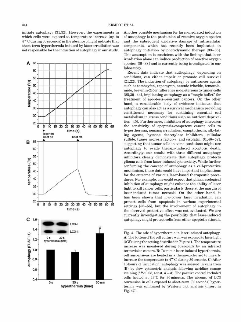
Hyperthermia Is Not Involved in Laser-MediatedAutophagy Induction
The cytotoxic efficiency of laser irradiation correlatedwith the temperature measured at the surface of laser-exposed cell culture wells. A linear increase in temperatureup to 608C was observed in cell culture wells exposed to 2 Wlaser light during 60 seconds (Fig. 4A). Similar increase intemperature was observed irrespectively of the presence ofcells in the cell culture wells (data not shown). Whilehyperthermia induced after 30 seconds of laser irradiation(up to 478C) was clearly incapable of thermal ablation, wesought to examine whether it could be involved in theinduction of autophagy. To produce laser-like temperatureincrease in the absence of light, we heated the cells in aPCR thermocycler programmed to gradually increase thetemperature to 478C within 30 seconds (Fig. 4B). As apositive control for hyperthermia-induced autophagy, weused cells exposed to 438C during 30 minutes. After an
additional 18 hours of incubation, the increase in red/greenacridine orange fluorescence ratio as a marker of autophagywas clearly observed in the positive control, but not in cellsexposed to a linear increase to 478C during 30 seconds(Fig. 4C). Accordingly, no increase in LC3-II was observedin cells exposed to short-term (30 seconds) hyperthermia(insert in Fig. 4C). These data indicate that hyperthermiawas not responsible for the laser-induced autophagicresponse in tumor cells.
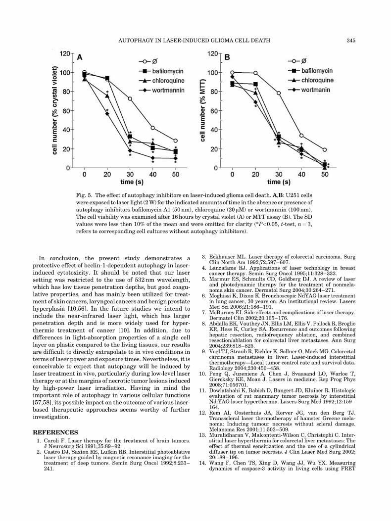
Autophagy Protects Cells FromLaser-Induced Death
Finally, we wanted to examine to role of autophagy inlaser-induced cytotoxicity. To that end, we treated the cellsimmediately after the laser exposure with three differentautophagy inhibitors: bafilomycin A1 or chloroquine, whichboth prevent acidification of autophagosomes [27,28],or wortmannin, a phosphatydilinositol 3-kinase blockerthat prevents autophagosome formation [29]. Since theautophagy inhibitors alone were apparently able to slightlyreduce glioma cell viability after 24 hours of incubation(data not shown), we have shortened the incubation time to16 hours for this experiment. While in these conditionsthe autophagy inhibitors did not significantly affect theviability of cells that were not exposed to laser, they allmarkedly increased cell death caused by exposure to laserfor various amounts of time (Fig. 5). Similar results wereobtained with both crystal violet (Fig. 5A) and MTT assay(Fig. 5B). These data clearly demonstrate that autophagyexerts a protective role in laser-induced cell death.
DISCUSSION
The present study for the first time demonstrates theability of laser light to induce autophagy associated withup-regulation of the pro-autophagic protein beclin-1 inmammalian cells. Interestingly, the induction of autophagyprotected human U251 glioma cells from laser-mediatedcytotoxicity. It should be noted that the observed effect wasapparently not species- or cell type-specific, as similarresults were obtained in L929 mouse fibrosarcoma cell line[Krmpot et al., unpublished observation].
Although laser light has been used for decades for variousmedical indications, the mechanisms of laser-induced celldeath are still not completely elucidated. In contrast tonecrotic cell death caused by high-power laser-mediatedthermal destruction [11–13], low-power laser treatmentcan lead to type I programmed cell death, apoptosis, inwhich various intracellular mechanisms are activated in ahighly controlled manner to eventually cause cell demise[14–16]. Although we did not closely investigate the cellfeatures characteristic for necrosis or apoptosis, the cellmorphology and temperature measurement data supportthe assumption that the laser exposure setting in ourexperiments led to a non-thermal cell proliferation arrestand apoptotic cell death. More importantly, we have shownthat laser-induced cytotoxicity is associated with theautophagic digestion of intracellular proteins. The twomain mechanisms for the initiation of autophagosomeformation are the inhibition of mammalian target of
342 KRMPOT ET AL.
rapamycin (mTOR) and up-regulation of the crucial pro-autophagic protein beclin-1 [30]. While we did not assessthe mTOR activity in response to laser irradiation, our dataclearly demonstrate that laser-induced autophagy is
associated with the increase in beclin-1 expression. Itshould be also noted that the laser irradiation in ourexperiments caused hyperthermia, and the recent resultsfrom others and us indicate that hyperthermia could
Fig. 3. The induction of autophagy and beclin-1 in laser-exposed glioma cells. A–D: U251
cells were exposed to laser light (2 W) for 30 seconds. The presence of autophagic vesicles was
examined after 16 hours in acridine orange-stained cells by fluorescent microscopy (A, inserts
in B) or flow cytometric analysis (B,C), while the level of autophagosome-associated LC3-II
was analysed by Western blotting (insert in C). The expression of beclin-1 was assessed after
6 hours by cell-based ELISA (D) (*P< 0.05, t-test, n¼ 3).
AUTOPHAGY IN LASER-INDUCED GLIOMA CELL DEATH 343
initiate autophagy [31,32]. However, the experiments inwhich cells were exposed to temperature increase (up to478C during 30 seconds) in the absence of light indicate thatshort-term hyperthermia induced by laser irradiation wasnot responsible for the induction of autophagy in our study.
Another possible mechanism for laser-mediated inductionof autophagy is the production of reactive oxygen speciesand the subsequent oxidative damage of intracellularcomponents, which has recently been implicated inautophagy initiation by photodynamic therapy [33–35].This assumption is consistent with the findings that laserirradiation alone can induce production of reactive oxygenspecies [36–38] and is currently being investigated in ourlaboratory.
Recent data indicate that authophagy, depending onconditions, can either impair or promote cell survival[21,22]. The induction of autophagy by anticancer agentssuch as tamoxyfen, rapamycin, arsenic trioxide, temozolo-mide, brevinin-2R or fullerenes is deleterious to tumor cells[25,39–44], implicating autophagy as a ‘‘magic bullet’’ fortreatment of apoptosis-resistant cancers. On the otherhand, a considerable body of evidence indicates thatautophagy can also act as a survival mechanism providingconstituents necessary for sustaining essential cellmetabolism in stress conditions such as nutrient depriva-tion [45]. Furthermore, inhibition of autophagy increasesthe sensitivity of apoptosis-competent cancer cells tohyperthermia, ionizing irradiation, camptothecin, alkylat-ing agents, hystone deacetylase inhibitors, sulindacsulfide, tumor necrosis factor-a, and cisplatin [31,46–52],suggesting that tumor cells in some conditions might useautophagy to evade therapy-induced apoptotic death.Accordingly, our results with three different autophagyinhibitors clearly demonstrate that autophagy protectsglioma cells from laser-induced cytotoxicity. While furtherconfirming the concept of autophagy as a cell-protectivemechanism, these data could have important implicationsfor the outcome of various laser-based therapeutic proce-dures. For example, one could expect that pharmacologicalinhibition of autophagy might enhance the ability of laserlight to kill cancer cells, particularly those at the margin oflaser-induced tumor necrosis. On the other hand, ithas been shown that low-power laser irradiation canprotect cells from apoptosis in various experimentalsettings [53–55], but the involvement of autophagy inthe observed protective effect was not evaluated. We arecurrently investigating the possibility that laser-inducedautophagy might protect cells from other apoptotic stimuli.
Fig. 4. The role of hyperthermia in laser-induced autophagy.
A: The bottom of the cell culture well was exposed to laser light
(2 W) using the setting described in Figure 1. The temperature
increase was monitored during 60 seconds by an infrared
termovision camera.B: To mimic laser-induced hyperthermia,
cell suspensions are heated in a thermocycler set to linearly
increase the temperature to 478C during 30 seconds. C: After
16 hours of incubation, autophagy was asessed in cells from
(B) by flow cytometric analysis following acridine orange
staining (*P<0.05, t-test, n¼ 3). The positive control included
cells heated at 438C for 30 minutes. The absence of LC3
conversion in cells exposed to short-term (30 seconds) hyper-
termia was confirmed by Western blot analysis (insert in
Fig. 4C).
344 KRMPOT ET AL.
In conclusion, the present study demonstrates aprotective effect of beclin-1-dependent autophagy in laser-induced cytotoxicity. It should be noted that our lasersetting was restricted to the use of 532 nm wavelength,which has low tissue penetration depths, but good coagu-lative properties, and has mainly been utilized for treat-ment of skin cancers, laryngeal cancers and benign prostatehyperplasia [10,56]. In the future studies we intend toinclude the near-infrared laser light, which has largerpenetration depth and is more widely used for hyper-thermic treatment of cancer [10]. In addition, due todifferences in light-absorption properties of a single celllayer on plastic compared to the living tissues, our resultsare difficult to directly extrapolate to in vivo conditions interms of laser power and exposure times. Nevertheless, it isconceivable to expect that autophagy will be induced bylaser treatment in vivo, particularly during low-level lasertherapy or at the margins of necrotic tumor lesions inducedby high-power laser irradiation. Having in mind theimportant role of autophagy in various cellular functions[57,58], its possible impact on the outcome of various laser-based therapeutic approaches seems worthy of furtherinvestigation.
REFERENCES
1. Caroli F. Laser therapy for the treatment of brain tumors.J Neurosurg Sci 1991;35:89–92.
2. Castro DJ, Saxton RE, Lufkin RB. Interstitial photoablativelaser therapy guided by magnetic resonance imaging for thetreatment of deep tumors. Semin Surg Oncol 1992;8:233–241.
3. Eckhauser ML. Laser therapy of colorectal carcinoma. SurgClin North Am 1992;72:597–607.
4. Lanzafame RJ. Applications of laser technology in breastcancer therapy. Semin Surg Oncol 1995;11:328–332.
5. Marmur ES, Schmults CD, Goldberg DJ. A review of laserand photodynamic therapy for the treatment of nonmela-noma skin cancer. Dermatol Surg 2004;30:264–271.
6. Moghissi K, Dixon K. Bronchoscopic NdYAG laser treatmentin lung cancer, 30 years on: An institutional review. LasersMed Sci 2006;21:186–191.
7. McBurney EI. Side effects and complications of laser therapy.Dermatol Clin 2002;20:165–176.
8. Abdalla EK, Vauthey JN, Ellis LM, Ellis V, Pollock R, BroglioKR, Hess K, Curley SA. Recurrence and outcomes followinghepatic resection, radiofrequency ablation, and combinedresection/ablation for colorectal liver metastases. Ann Surg2004;239:818–825.
9. Vogl TJ, Straub R, Eichler K, Sollner O, Mack MG. Colorectalcarcinoma metastases in liver: Laser-induced interstitialthermotherapy—Local tumor control rate and survival data.Radiology 2004;230:450–458.
10. Peng Q, Juzeniene A, Chen J, Svaasand LO, Warloe T,Giercksky KE, Moan J. Lasers in medicine. Rep Prog Phys2008;71:056701.
11. Dowlatshahi K, Babich D, Bangert JD, Kluiber R. Histologicevaluation of rat mammary tumor necrosis by interstitialNd:YAG laser hyperthermia. Lasers Surg Med 1992;12:159–164.
12. Rem AI, Oosterhuis JA, Korver JG, van den Berg TJ.Transscleral laser thermotherapy of hamster Greene mela-noma: Inducing tumour necrosis without scleral damage.Melanoma Res 2001;11:503–509.
13. Muralidharan V, Malcontenti-Wilson C, Christophi C. Inter-stitial laser hyperthermia for colorectal liver metastases: Theeffect of thermal sensitization and the use of a cylindricaldiffuser tip on tumor necrosis. J Clin Laser Med Surg 2002;20:189–196.
14. Wang F, Chen TS, Xing D, Wang JJ, Wu YX. Measuringdynamics of caspase-3 activity in living cells using FRET
Fig. 5. The effect of autophagy inhibitors on laser-induced glioma cell death. A,B: U251 cells
were exposed to laser light (2 W) for the indicated amounts of time in the absence or presence of
autophagy inhibitors bafilomycin A1 (50 nm), chloroquine (20 mM) or wortmannin (100 nm).
The cell viability was examined after 16 hours by crystal violet (A) or MTT assay (B). The SD
values were less then 10% of the mean and were omitted for clarity (*P<0.05, t-test, n¼ 3,
refers to corresponding cell cultures without autophagy inhibitors).
AUTOPHAGY IN LASER-INDUCED GLIOMA CELL DEATH 345
technique during apoptosis induced by high fluence low-power laser irradiation. Lasers Surg Med 2005;36:2–7.
15. Gao X, Chen T, Xing D, Wang F, Pei Y, Wei X. Single cellanalysis of PKC activation during proliferation and apoptosisinduced by laser irradiation. J Cell Physiol 2006;206:441–448.
16. Wu S, Xing D, Wang F, Chen T, Chen WR. Mechanistic studyof apoptosis induced by high-fluence low-power laser irradi-ation using fluorescence imaging techniques. J Biomed Opt2007;12:064015.
17. Edinger AL, Thompson CB. Death by design: Apoptosis,necrosis and autophagy. Curr Opin Cell Biol 2004;16:663–669.
18. Schulze PC, Adams V, Busert C, Bettag M, Kahn T, SchoberR. Effects of laser-induced thermotherapy (LITT) on prolif-eration and apoptosis of glioma cells in rat brain trans-plantation tumors. Lasers Surg Med 2002;30:227–232.
19. Klionsky DJ, Emr SD. Autophagy as a regulated pathway ofcellular degradation. Science 2000;290:1717–1721.
20. Eskelinen EL, Saftig P. Autophagy: A lysosomal degradationpathway with a central role in health and disease. BiochimBiophys Acta 2009;1793:664–673.
21. Tsujimoto Y, Shimizu S. Another way to die: Autophagicprogrammed cell death. Cell Death Differ 2005;12:1528–1534.
22. Maiuri MC, Zalckvar E, Kimchi A, Kroemer G. Self-eatingand self-killing: Crosstalk between autophagy and apoptosis.Nat Rev Mol Cell Biol 2007;8:741–752.
23. Flick DA, Gifford GE. Comparison of in vitro cell cytotoxicassays for tumor necrosis factor. J Immunol Methods 1984;68:167–175.
24. Mosmann T. Rapid colorimetric assay for cellular growth andsurvival: Application to proliferation and cytotoxicity assays.J Immunol Methods 1983;65:55–63.
25. Harhaji L, Isakovic A, Raicevic N, Markovic Z, Todorovic-Markovic B, Nikolic N, Vranjes-Djuric S, Markovic I,Trajkovic V. Multiple mechanisms underlying the anticanceraction of nanocrystalline fullerene. Eur J Pharmacol 2007;568:89–98.
26. Versteeg HH, Nijhuis E, van den Brink GR, Evertzen M,Pynaert GN, van Deventer SJ, Coffer PJ, Peppelenbosch MP.A new phosphospecific cell-based ELISA for p42/p44 mitogen-activated protein kinase (MAPK), p38 MAPK, protein kinaseB and cAMP-response-element-binding protein. BiochemJ 2000;350:717–722.
27. Yamamoto A, Tagawa Y, Yoshimori T, Moriyama Y, MasakiR, Tashiro Y. Bafilomycin A1 prevents maturation ofautophagic vacuoles by inhibiting fusion between autopha-gosomes and lysosomes in rat hepatoma cell line, H-4-II-Ecells. Cell Struct Funct 1998;23:33–42.
28. Boya P, Gonzalez-Polo RA, Casares N, Perfettini JL, DessenP, Larochette N, Metivier D, Meley D, Souquere S, YoshimoriT, Pierron G, Codogno P, Kroemer G. Inhibition of macro-autophagy triggers apoptosis. Mol Cell Biol 2005;25:1025–1040.
29. Blommaart EF, Krause U, Schellens JP, Vreeling-Sindelar-ova H, Meijer AJ. The phosphatidylinositol 3-kinase inhib-itors wortmannin and LY294002 inhibit autophagy inisolated rat hepatocytes. Eur J Biochem 1997;243:240–246.
30. Pattingre S, Espert L, Biard-Piechaczyk M, Codogno P.Regulation of macroautophagy by mTOR and Beclin 1 com-plexes. Biochimie 2008;90:313–323.
31. Komata T, Kanzawa T, Nashimoto T, Aoki H, Endo S,Nameta M, Takahashi H, Yamamoto T, Kondo S, Tanaka R.Mild heat shock induces autophagic growth arrest, but notapoptosis in U251-MG and U87-MG human malignantglioma cells. J Neurooncol 2004;68:101–111.
32. Janjetovic K, Misirkic M, Vucicevic L, Harhaji L, Trajkovic V.Synergistic antiglioma action of hyperthermia and nitricoxide. Eur J Pharmacol 2008;583:1–10.
33. Buytaert E, Callewaert G, Hendrickx N, Scorrano L,Hartmann D, Missiaen L, Vandenheede JR, Heirman I,Grooten J, Agostinis P. Role of endoplasmic reticulumdepletion and multidomain proapoptotic BAX and BAKproteins in shaping cell death after hypericin-mediatedphotodynamic therapy. FASEB J 2006;20:756–758.
34. Kessel D, Vicente MG, Reiners JJ, Jr. Initiation of apoptosisand autophagy by photodynamic therapy. Lasers Surg Med2006;38:482–488.
35. Xue LY, Chiu SM, Azizuddin K, Joseph S, Oleinick NL. Thedeath of human cancer cells following photodynamic therapy:Apoptosis competence is necessary for Bcl-2 protection butnot for induction of autophagy. Photochem Photobiol 2007;83:1016–1023.
36. Brancato R, Schiavone N, Siano S, Lapucci A, Papucci L,Donnini M, Formigli L, Orlandini SZ, Carella G, Carones F,Capaccioli S. Prevention of corneal keratocyte apoptosis afterargon fluoride excimer laser irradiation with the free radicalscavenger ubiquinone Q10. Eur J Ophthalmol 2000;10:32–38.
37. Tirlapur UK, Konig K, Peuckert C, Krieg R, Halbhuber KJ.Femtosecond near-infrared laser pulses elicit generation ofreactive oxygen species in mammalian cells leading toapoptosis-like death. Exp Cell Res 2001;263:88–97.
38. Jou MJ, Jou SB, Chen HM, Lin CH, Peng TI. Critical role ofmitochondrial reactive oxygen species formation in visiblelaser irradiation-induced apoptosis in rat brain astrocytes(RBA-1). J Biomed Sci 2002;9:507–916.
39. Bursch W, Ellinger A, Kienzl H, Torok L, Pandey S, SikorskaM, Walker R, Hermann RS. Active cell death induced by theanti-estrogens tamoxifen and ICI 164 384 in human mam-mary carcinoma cells (MCF-7) in culture: The role ofautophagy. Carcinogenesis 1996;17:1595–1607.
40. Bursch W, Hochegger K, Torok L, Marian B, Ellinger A,Hermann RS. Autophagic and apoptotic types of programmedcell death exhibit different fates of cytoskeletal filaments.J Cell Sci 2000;113:1189–1198.
41. Kanzawa T, Kondo Y, Ito H, Kondo S, Germano I. Inductionof autophagic cell death in malignant glioma cells by arsenictrioxide. Cancer Res 2003;63:2103–2108.
42. Kanzawa T, Germano IM, Komata T, Ito H, Kondo Y, KondoS. Role of autophagy in temozolomide-induced cytotoxicity formalignant glioma cells. Cell Death Differ 2004;11:448–457.
43. Iwamaru A, Kondo Y, Iwado E, Aoki H, Fujiwara K,Yokoyama T, Mills GB, Kondo S. Silencing mammaliantarget of rapamycin signaling by small interfering RNAenhances rapamycin-induced autophagy in malignant gliomacells. Oncogene 2007;26:1840–1851.
44. Ghavami S, Asoodeh A, Klonisch T, Halayko AJ, KadkhodaK, Kroczak TJ, Gibson SB, Booy EP, Naderi-Manesh H,Los M. Brevinin-2R(1) semi-selectively kills cancer cells bya distinct mechanism, which involves the lysosomal-mitochondrial death pathway. J Cell Mol Med 2008;12:1005–1022.
45. Codogno P, Meijer AJ. Autophagy and signaling: Their role incell survival and cell death. Cell Death Differ 2005;2:1509–1518.
46. Paglin S, Hollister T, Delohery T, Hackett N, McMahill M,Sphicas E, Domingo D, Yahalom J. A novel response of cancercells to radiation involves autophagy and formation of acidicvesicles. Cancer Res 2001;61:439–644.
47. Bauvy C, Gane P, Arico S, Codogno P, Ogier-Denis E.Autophagy delays sulindac sulfide-induced apoptosis in thehuman intestinal colon cancer cell line HT-29. Exp Cell Res2001;268:139–149.
48. Abedin MJ, Wang D, McDonnell MA, Lehmann U, Kelekar A.Autophagy delays apoptotic death in breast cancer cellsfollowing DNA damage. Cell Death Differ 2007;14:500–510.
49. Amaravadi RK, Yu D, Lum JJ, Bui T, Christophorou MA,Evan GI, Thomas-Tikhonenko A, Thompson CB. Autophagyinhibition enhances therapy-induced apoptosis in a Myc-induced model of lymphoma. J Clin Invest 2007;117:326–336.
50. Harhaji L, Mijatovic S, Maksimovic-Ivanic D, Popadic D,Isakovic A, Todorovic-Markovic B, Trajkovic V. Aloe emodininhibits the cytotoxic action of tumor necrosis factor. EurJ Pharmacol 2007;568:248–259.
51. Carew JS, Nawrocki ST, Kahue CN, Zhang H, Yang C, ChungL, Houghton JA, Huang P, Giles FJ, Cleveland JL. Targetingautophagy augments the anticancer activity of the histonedeacetylase inhibitor SAHA to overcome Bcr-Abl-mediateddrug resistance. Blood 2007;110:313–322.
346 KRMPOT ET AL.

52. Sivaprasad U, Basu A. Inhibition of ERK attenuatesautophagy and potentiates tumor necrosis factor-a-inducedcell death in MCF-7 cells. J Cell Mol Med 2008;12:1265–1271.
53. Carnevalli CM, Soares CP, Zngaro RA, Pinheiro AL, SilvaNS. Laser light prevents apoptosis in Cho K-1 cell line. J ClinLaser Med Surg 2003;21:193–196.
54. Lim W, Ko M, Lee S, Kim I, Jung M, Kim O, Cho S, Yang K,Choi N, Kim S, Choi H. Ultraviolet-C-induced apoptosisprotected by 635-nm laser irradiation in human gingivalfibroblasts. Photomed Laser Surg 2008;26:215–220.
55. Zhang L, Xing D, Zhu D, Chen Q. Low-power laser irradiationinhibiting Ab25-35-induced PC12 cell apoptosis via PKCactivation. Cell Physiol Biochem 2008;22:215–222.
56. Zeitels SM, Burns JA, Lopez-Guerra G, Anderson RR,Hillman RE. Photoangiolytic laser treatment of early glotticcancer: A new management strategy. Ann Otol RhinolLaryngol Suppl 2008;199:3–24.
57. Xiao G. Autophagy and NF-kB: Fight for fate. CytokineGrowth Factor Rev 2007;18:233–243.
58. Eskelinen EL. Maturation of autophagic vacuoles in mam-malian cells. Autophagy 2005;1:1–10.
AUTOPHAGY IN LASER-INDUCED GLIOMA CELL DEATH 347