Autophagy-Is it a preferred route for lifespan extension
7
BMB reports 65 http://bmbreports.org BMB reports *Corresponding author. Tel: 82-2-2220-4484; Fax: 82-2-2220-4474; E-mail: [email protected] Received 31 January 2009 Keywords: Autophagosome, Autophagy, Insulin/IGF-1 pathway, Lifespan, Lysosome/vacuole, TOR pathway Autophagy-Is it a preferred route for lifespan extension? Meenakshi Dwivedi 1,2 & Joohong Ahnn 1, * 1 BK21 Life Science for Gloval Warming Team, Department of Life Science, 2 Research Institute for Natural Sciences (RINS), Hanyang University, Seoul, Korea Autophagy, which is a process of self eating, has gained inter- est in the past decade due to its both beneficial and con- troversial roles in various biological phenomena. The discov- ery of autophagy genes (ATG) in yeast has led to focused re- search designed to elucidate the mechanism and regulation of this process. The role of autophagy in a variety of biological phenomena, including human disease, is still the subject of debate. However, recent findings suggest that autophagy is a highly regulated process with both beneficial and negative effects. Indeed, studies conducted using various model organ- isms have demonstrated that increased autophagy leads to an extended lifespan. Despite these findings, it is still unknown if all pathways leading to extended lifespan converge at the process of autophagy or not. Here, an overview of modern de- velopments related to the process of autophagy, its regulation and the molecular machinery involved is presented. In addi- tion, this review focuses on one of the beneficial aspects of au- tophagy, its role in lifespan regulation. [BMB reports 2009; 42(2): 65-71] INTRODUCTION Autophagy is a catabolic process that involves a process of self-cannibalization through the lysosomal degradation path- way that is evolutionarily conserved in all eukaryotic organisms. The discovery of lysosomes and simultaneous in- troduction of the term autophagy to describe the digestion of various proteins and organelles through lysosomes by Belgian cytologist Christian de Duve (1) initiated the study of autopha- gic processes and their mechanisms. The autophagy related genes (ATG) were initially discovered in yeast (Saccharomyces cerevisiae) (2-4). These genes are conserved in metazoans and plants and their evaluation has led to increased understanding of the autophagic process and its role in diverse biological processes. Autophagy is present in basal levels in almost all cell types and plays an important role in cell growth, develop- ment, differentiation and homeostasis. Autophagy is controlled by various stimuli including nutritional status, hormonal fac- tors, intracellular stress, temperature, oxygen concentrations and cell density. In addition, autophagy is dramatically up regulated in response to starvation, growth factor withdrawal and high bio-energetic demands. Autophagy has also been im- plicated in various human diseases, due to both its occasional protective roles and its contribution to cell damage under cer- tain conditions (5). There are three general types of autophagy based on the mechanism by which cargo (intracellular components to be degraded) is transported in to the lysosome: microautophagy, chaperone mediated autophagy (CMA) and macroautophagy. In microautophagy, the cargo is directly incorporated into the lysosome through the lysosomal membrane by fusion, after which it undergoes further proteolytic digestion and the essen- tial nutrients are retrieved. However, in CMA, the heat shock cognate protein of the cytosolic and lysosomal chaperone, 70 kDa (hsc70), selectively recognizes the cytosolic proteins con- taining the pentapeptide KFREQ motif (6). The interaction be- tween this chaperone and the substrate in the cytosol targets the complex to the lysosomal membrane, which results in fu- sion, further degradation and the subsequent release of nutrients. Macroautophagy differs from the other two types of autophagy because it involves the formation of a specific dou- ble walled vesicle that transports cargo to the lysosome. There are also other classifications of autophagy based on their target components (cargo). These classifications include ERphagy (7), mitophagy (8, 9), nucleophagy (10), pexophagy (11), xen- ophagy (12, 13), ribophagy (14), and aggrephagy (15, 16), which indicate specific degradation of endoplasmic reticulum (ER), mitochondria, nuclear regions, peroxisomes, intracellular pathogens, ribosomes, and protein aggregates, respectively. The last 10 years have witnessed a surprising focus on auto- phagic pathways and understanding their regulation, mecha- nisms and functions. Accordingly, the molecular mechanism, regulation and functions of autophagy have been described in detail in several recent comprehensive reviews (17-23). However, in this review, we provide insight into autophagic process and the role that they play in lifespan regulation in var- ious model organisms. Mini Review
Transcript of Autophagy-Is it a preferred route for lifespan extension
BMB reports
65http://bmbreports.org BMB reports
*Corresponding author. Tel: 82-2-2220-4484; Fax: 82-2-2220-4474;E-mail: [email protected]
Received 31 January 2009
Keywords: Autophagosome, Autophagy, Insulin/IGF-1 pathway, Lifespan,Lysosome/vacuole, TOR pathway
Autophagy-Is it a preferred route for lifespan extension?Meenakshi Dwivedi1,2 & Joohong Ahnn1,*1BK21 Life Science for Gloval Warming Team, Department of Life Science, 2Research Institute for Natural Sciences (RINS), Hanyang University, Seoul, Korea
Autophagy, which is a process of self eating, has gained inter-est in the past decade due to its both beneficial and con-troversial roles in various biological phenomena. The discov-ery of autophagy genes (ATG) in yeast has led to focused re-search designed to elucidate the mechanism and regulation of this process. The role of autophagy in a variety of biological phenomena, including human disease, is still the subject of debate. However, recent findings suggest that autophagy is a highly regulated process with both beneficial and negative effects. Indeed, studies conducted using various model organ-isms have demonstrated that increased autophagy leads to an extended lifespan. Despite these findings, it is still unknown if all pathways leading to extended lifespan converge at the process of autophagy or not. Here, an overview of modern de-velopments related to the process of autophagy, its regulation and the molecular machinery involved is presented. In addi-tion, this review focuses on one of the beneficial aspects of au-tophagy, its role in lifespan regulation. [BMB reports 2009; 42(2): 65-71]
INTRODUCTION
Autophagy is a catabolic process that involves a process of self-cannibalization through the lysosomal degradation path-way that is evolutionarily conserved in all eukaryotic organisms. The discovery of lysosomes and simultaneous in-troduction of the term autophagy to describe the digestion of various proteins and organelles through lysosomes by Belgian cytologist Christian de Duve (1) initiated the study of autopha-gic processes and their mechanisms. The autophagy related genes (ATG) were initially discovered in yeast (Saccharomyces cerevisiae) (2-4). These genes are conserved in metazoans and plants and their evaluation has led to increased understanding of the autophagic process and its role in diverse biological processes. Autophagy is present in basal levels in almost all
cell types and plays an important role in cell growth, develop-ment, differentiation and homeostasis. Autophagy is controlled by various stimuli including nutritional status, hormonal fac-tors, intracellular stress, temperature, oxygen concentrations and cell density. In addition, autophagy is dramatically up regulated in response to starvation, growth factor withdrawal and high bio-energetic demands. Autophagy has also been im-plicated in various human diseases, due to both its occasional protective roles and its contribution to cell damage under cer-tain conditions (5). There are three general types of autophagy based on the mechanism by which cargo (intracellular components to be degraded) is transported in to the lysosome: microautophagy, chaperone mediated autophagy (CMA) and macroautophagy. In microautophagy, the cargo is directly incorporated into the lysosome through the lysosomal membrane by fusion, after which it undergoes further proteolytic digestion and the essen-tial nutrients are retrieved. However, in CMA, the heat shock cognate protein of the cytosolic and lysosomal chaperone, 70 kDa (hsc70), selectively recognizes the cytosolic proteins con-taining the pentapeptide KFREQ motif (6). The interaction be-tween this chaperone and the substrate in the cytosol targets the complex to the lysosomal membrane, which results in fu-sion, further degradation and the subsequent release of nutrients. Macroautophagy differs from the other two types of autophagy because it involves the formation of a specific dou-ble walled vesicle that transports cargo to the lysosome. There are also other classifications of autophagy based on their target components (cargo). These classifications include ERphagy (7), mitophagy (8, 9), nucleophagy (10), pexophagy (11), xen-ophagy (12, 13), ribophagy (14), and aggrephagy (15, 16), which indicate specific degradation of endoplasmic reticulum (ER), mitochondria, nuclear regions, peroxisomes, intracellular pathogens, ribosomes, and protein aggregates, respectively. The last 10 years have witnessed a surprising focus on auto-phagic pathways and understanding their regulation, mecha-nisms and functions. Accordingly, the molecular mechanism, regulation and functions of autophagy have been described in detail in several recent comprehensive reviews (17-23). However, in this review, we provide insight into autophagic process and the role that they play in lifespan regulation in var-ious model organisms.
Mini Review

Autophagy and lifespan extensionMeenakshi Dwivedi and Joohong Ahnn
66 BMB reports http://bmbreports.org
Fig. 1. Schematic illustration of the auto-phagy process with the involved genes.
The molecular machinery of autophagy
The process of macroautophagy (henceforth, referred to as au-tophagy) was first described in mammals in rat hepatocytes (24, 25). Autophagy in mammals is mechanistically identical to the process that occurs in yeast. As shown in Fig. 1, the process of autophagy can be broadly divided into the follow-ing steps: a) induction; b) nucleation; c) cargo recognition; d) expansion and completion of the autophagosome; e) fusion with the lysosome vacuole; and f) digestion and recycling of the cargo.
InductionThe autophagic process can be induced by numerous stimuli; however, the most well studied stimulus is nutrient starvation. Induction is regulated by the target of rapamycin (TOR) signal-ing pathway, which includes the protein kinase, Tor, a neg-ative regulator of autophagy. Other proteins responsible for the induction step in yeast include Atg1, Atg13 and Atg17. The Atg1 and TOR kinases play a conserved role in the induction step, even in higher eukaryotes (26, 27).
NucleationThe origin of pre-autophagosomal structure or phagophore as-sembly site (PAS) is the least understood in autophagy. Besides, PAS is responsible for the formation of vesicles during autophagy nucleation. A functional complex of class III phos-phatidylinositol 3 kinase (Vps34) along with Vps15, Atg6/ Vps30 and Atg14 (28) participates in autophagic/vesicle nucle-
ation to localize other autophagy proteins to the PAS.
Cargo recognitionInadequate information exists regarding the mechanism re-sponsible for selection of the macromolecular complexes and organelles to be degraded and the regulation of these selective autophagic pathways. In S. cerevisiae, binding of Atg11 to Atg19 marks one of the first steps involved in cargo recog-nition; however, there is no evidence of such a mechanism in any other organisms. It has been reported that ubiquitin serves as a signal for cargo recognition by providing an interaction surface for specific ubiquitin-binding proteins. The p62/seques-tosome has been shown to recognize the polyubiquitinated protein aggregates and subsequently lead the aggregates to the autophagic vesicles via binding to the LC3 in mammalian cells (29). In addition, several studies have been conducted to iden-tify the mechanism involved in the specific cargo recognition. In yeast, it has been shown that mitochondria requires Uth1p (9), peroxisomes are specifically recognized by Pex3/14 pro-teins (30) and the nucleus is recognized through Nvj1p, which results in degradation of parts of the nucleolus via a process known as piecemeal autophagy of the nucleus (10). The mech-anism involved for the recognition of intracellular pathogens, leading to xenophagy, is still not well understood. Recently, Tan et al. proposed a general cellular mechanism for cargo recognition, which suggested that the K63 ubiquitin chain as-sembly is responsible for targeting intracellular inclusions for autophagy (31). Further, Cao et al. recently determined the minimum requirements for cargo packaging using knock-out
Autophagy and lifespan extensionMeenakshi Dwivedi and Joohong Ahnn
67http://bmbreports.org BMB reports
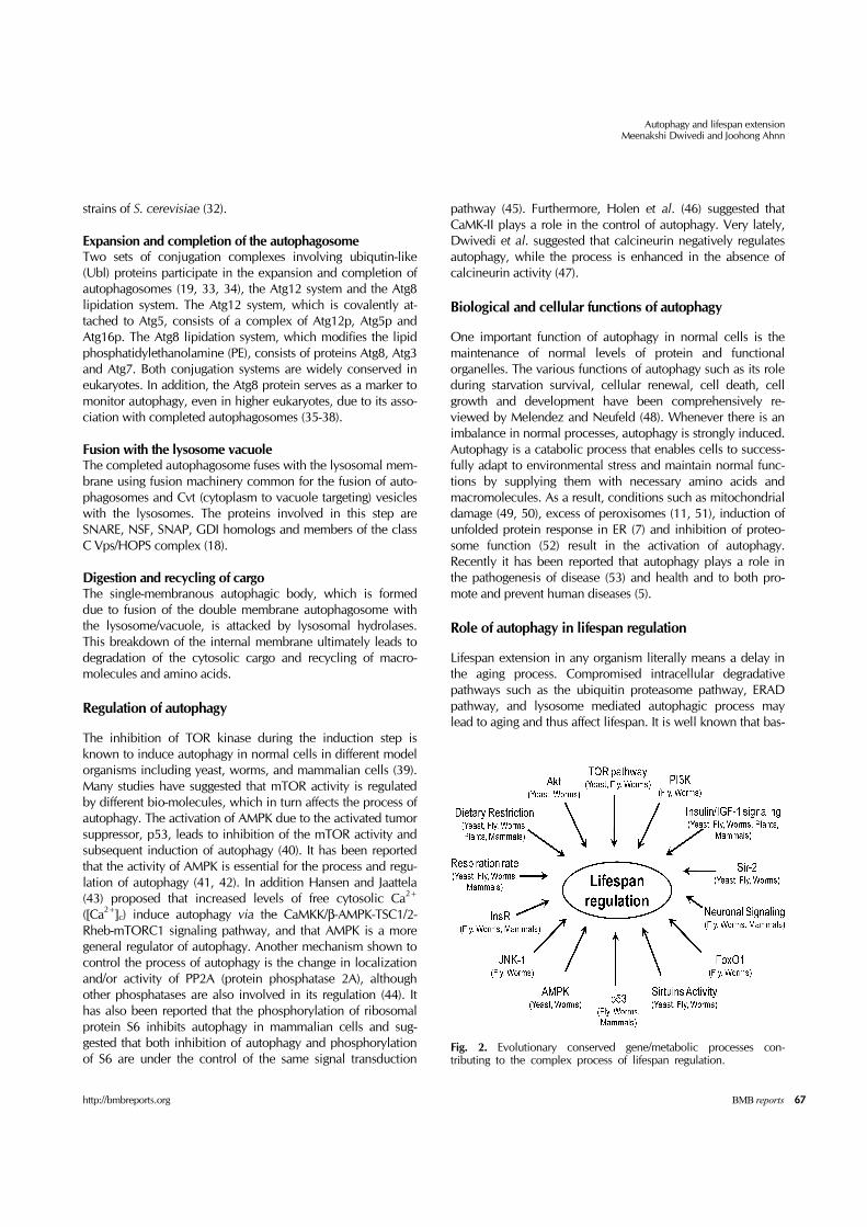
Fig. 2. Evolutionary conserved gene/metabolic processes con-tributing to the complex process of lifespan regulation.
strains of S. cerevisiae (32).
Expansion and completion of the autophagosomeTwo sets of conjugation complexes involving ubiqutin-like (Ubl) proteins participate in the expansion and completion of autophagosomes (19, 33, 34), the Atg12 system and the Atg8 lipidation system. The Atg12 system, which is covalently at-tached to Atg5, consists of a complex of Atg12p, Atg5p and Atg16p. The Atg8 lipidation system, which modifies the lipid phosphatidylethanolamine (PE), consists of proteins Atg8, Atg3 and Atg7. Both conjugation systems are widely conserved in eukaryotes. In addition, the Atg8 protein serves as a marker to monitor autophagy, even in higher eukaryotes, due to its asso-ciation with completed autophagosomes (35-38).
Fusion with the lysosome vacuoleThe completed autophagosome fuses with the lysosomal mem-brane using fusion machinery common for the fusion of auto-phagosomes and Cvt (cytoplasm to vacuole targeting) vesicles with the lysosomes. The proteins involved in this step are SNARE, NSF, SNAP, GDI homologs and members of the class C Vps/HOPS complex (18).
Digestion and recycling of cargoThe single-membranous autophagic body, which is formed due to fusion of the double membrane autophagosome with the lysosome/vacuole, is attacked by lysosomal hydrolases. This breakdown of the internal membrane ultimately leads to degradation of the cytosolic cargo and recycling of macro-molecules and amino acids.
Regulation of autophagy
The inhibition of TOR kinase during the induction step is known to induce autophagy in normal cells in different model organisms including yeast, worms, and mammalian cells (39). Many studies have suggested that mTOR activity is regulated by different bio-molecules, which in turn affects the process of autophagy. The activation of AMPK due to the activated tumor suppressor, p53, leads to inhibition of the mTOR activity and subsequent induction of autophagy (40). It has been reported that the activity of AMPK is essential for the process and regu-lation of autophagy (41, 42). In addition Hansen and Jaattela (43) proposed that increased levels of free cytosolic Ca2+ ([Ca2+]c) induce autophagy via the CaMKK/β-AMPK-TSC1/2- Rheb-mTORC1 signaling pathway, and that AMPK is a more general regulator of autophagy. Another mechanism shown to control the process of autophagy is the change in localization and/or activity of PP2A (protein phosphatase 2A), although other phosphatases are also involved in its regulation (44). It has also been reported that the phosphorylation of ribosomal protein S6 inhibits autophagy in mammalian cells and sug-gested that both inhibition of autophagy and phosphorylation of S6 are under the control of the same signal transduction
pathway (45). Furthermore, Holen et al. (46) suggested that CaMK-II plays a role in the control of autophagy. Very lately, Dwivedi et al. suggested that calcineurin negatively regulates autophagy, while the process is enhanced in the absence of calcineurin activity (47).
Biological and cellular functions of autophagy
One important function of autophagy in normal cells is the maintenance of normal levels of protein and functional organelles. The various functions of autophagy such as its role during starvation survival, cellular renewal, cell death, cell growth and development have been comprehensively re-viewed by Melendez and Neufeld (48). Whenever there is an imbalance in normal processes, autophagy is strongly induced. Autophagy is a catabolic process that enables cells to success-fully adapt to environmental stress and maintain normal func-tions by supplying them with necessary amino acids and macromolecules. As a result, conditions such as mitochondrial damage (49, 50), excess of peroxisomes (11, 51), induction of unfolded protein response in ER (7) and inhibition of proteo-some function (52) result in the activation of autophagy. Recently it has been reported that autophagy plays a role in the pathogenesis of disease (53) and health and to both pro-mote and prevent human diseases (5).
Role of autophagy in lifespan regulation
Lifespan extension in any organism literally means a delay in the aging process. Compromised intracellular degradative pathways such as the ubiquitin proteasome pathway, ERAD pathway, and lysosome mediated autophagic process may lead to aging and thus affect lifespan. It is well known that bas-

Autophagy and lifespan extensionMeenakshi Dwivedi and Joohong Ahnn
68 BMB reports http://bmbreports.org
al autophagic activity decreases with age in living cells, thus increasing the accumulation of toxic materials and inefficient cellular components. Therefore, aging occurs due to the in-sufficient removal of toxic materials and inefficient cellular components. The molecular mechanisms and genetics in-volved in aging and lifespan regulation from yeast to mamma-lian systems are well understood and conserved (Fig. 2). The genes and metabolic processes involved in the aging process are evolutionary conserved. The insulin/insulin-like growth en-docrine pathway is the best known and well understood path-way involved in the aging process (54). Another well known pathway involved in aging process is the TOR pathway, which is inhibited by caloric or dietary restriction. Such inhibition of the TOR pathway results in the lifespan extension (55). Many of the pathways that have been studied found to be involved in lifespan regulation in different organisms are dependent on the autophagy pathway or autophagic genes.
Lifespan regulation dependent on autophagyMicro-organisms: Recently, Tang et al. (56) demonstrated that vacuole-vacuole fusion was required for extension of the life-span of yeast in response to caloric restriction. Furthermore, it has been shown that the autophagy protein, Atg15p, is a lipase located in the ER that is essential for lifespan extension. A re-cent study in yeast further suggests that autophagy is essential for life span extension in TOR signaling-deficient mutants (57).Flies: In Drosophila, it has been reported that low TOR signal-ing leads to up regulation of autophagy, which in turn guides to the promotion of normal cell function and survival rather than the suppression of cell growth via an endocrine response of insulin/PI3K signaling (58). In addition, Simonsen et al. (59) demonstrated that Drosophila lacking autophagy-related genes had a reduced lifespan that increased by more than 50% when the expression level of Atg8a increased in aging neurons.Worms: In Caenorhabditis elegans, the TOR pathway, which is responsible for sensing nutrients, that have been inhibited by dietary restriction (60, 61) and the insulin/IGF-1 pathway (36, 62), which responds to different environmental cues, are well known pathways involved in lifespan extension are reportedly dependent on the autophagy pathway. In addition, it has re-cently been reported that lifespan extension in mutants with lowered mitochondrial respiration (63) and cep-1 mutants (orthologue of p53) (64) is also dependent on autophagic genes. The results of a study that we recently conducted sug-gested that calcineurin also regulates lifespan through the auto-phagic pathway (47). Plants: In Arabidopsis, Hanaoka et al. found that autophagy was required for maintenance of cellular viability under nu-trient-limited conditions and efficient nutrient use by the entire plant (65). Additionally, it has been shown that the APG8/12 protein conjugation pathways play a role in autophagic re-cycling during nitrogen and carbon mobilization (66).Rodents: It has been shown that the process of autophagy de-clines with increasing age in hepatocytes isolated from rats fed
ad libitum, but this decline is prevented in the case of caloric restriction (67). In addition, the relationship between autoph-agy and caloric restriction has been extensively reviewed by Bergamini (68).Mammals: It has been demonstrated in mammalian red blood cells (erythrocytes) that mitophagy mediated by Atg7 was es-sential in developing erythroid cells, and that its absence re-sults in decreased lifespan and developmental arrest (69).
Lifespan regulation independent of autophagyThe results of many studies suggest that autophagy plays a pos-itive role in lifespan regulation and thus auspiciously suggest that lifespan extension in organisms is mediated by activation of the autophagic pathway. It is also well known that in-hibition of the TOR pathway leads to the activation of autoph-agy and lifespan extension in various organisms (39). Moreover, it has been reported that autophagy is both in-dependent and dependent on S6K, which is a critical down-stream substrate and effector of the TOR pathway. In Drosophila, Scott et al. demonstrated that TOR-mediated sup-pression of autophagy occurs independently of S6K (56). Conversely, it was found that phosphorylation of ribosomal protein S6 inhibits autophagy in rat hepatocytes (45). Moreover, S6kinase/rsks-1 C. elegans mutants were found to exhibit extended lifespan, but this extension occurred in-dependently of the autophagic process (61, 70). Recently Marino et al. (71) reported that extensive basal activation of autophagy occurred during normal aging in Zmpste24-null mice, which is contradictory to the normal level of autophagy that occurs in aging cells.
Conclusion and future perspectives
Studies conducted to evaluate autophagy, its molecular mech-anisms and regulation have focused on its biological and cel-lular functions. Autophagy has diverse physiological functions and plays an essential role in cellular housekeeping, the main-tenance of normal protein and organelle turnover and the deg-radation of damaged organelles. Autophagy is also involved in cellular remodeling during development and differentiation. In addition, autophagy plays important roles in stress adaptation, aging, defense against intracellular pathogens and antigen presentation. Furthermore, the results of recent biochemical and genetic studies have indicated that autophagy is involved in lifespan extension or anti-aging pathways. However, it is still not certain if the lifespan regulation of an individual is de-pendent or independent of the autophagic process. Future studies should be conducted to identify additional pathways involved in the regulation of lifespan and autophagy to eluci-date the mechanisms responsible for aging.
AcknowledgementsThis work was supported by the Korea Science and Engineering Foundation (KOSEF) grant funded by the Korea government

Autophagy and lifespan extensionMeenakshi Dwivedi and Joohong Ahnn
69http://bmbreports.org BMB reports
(MOST) [No. R01-2007-000-10349-0 (2008)] to J. Ahnn.
REFERENCES
1. de Duve, C. (2005) The lysosome turns fifty. Nat. Cell Biol. 7, 847-849.
2. Tsukada, M. and Ohsumi, Y. (1993) Isolation and character-ization of autophagy-defective mutants of Saccharomyces cerevisiae. FEBS Lett. 333, 169-174.
3. Thumm, M., Egner, R., Koch, B., Schlumpberger, M., Straub, M., Veenhuis, M. and Wolf, D. H. (1994) Isolation of autophagocytosis mutants of Saccharomyces cerevisiae. FEBS Lett. 349, 275-280.
4. Klionsky, D. J., Cregg, J. M., Dunn, W. A., Jr., Emr, S. D., Sakai, Y., Sandoval, I. V., Sibirny, A., Subramani, S., Thumm, M., Veenhuis, M. and Ohsumi, Y. (2003) A uni-fied nomenclature for yeast autophagy-related genes. Dev. Cell 5, 539-545.
5. Shintani, T. and Klionsky, D. J. (2004) Autophagy in health and disease: a double-edged sword. Science 306, 990-995.
6. Chiang, H. L., Terlecky, S. R., Plant, C. P. and Dice, J. F. (1989) A role for a 70-kilodalton heat shock protein in ly-sosomal degradation of intracellular proteins. Science 246, 382-385.
7. Bernales, S., Schuck, S. and Walter, P. (2007) ER-phagy: se-lective autophagy of the endoplasmic reticulum. Autophagy 3, 285-287.
8. Lemasters, J. J. (2005) Selective mitochondrial autophagy, or mitophagy, as a targeted defense against oxidative stress, mitochondrial dysfunction, and aging. Rejuvenation Res. 8, 3-5.
9. Kissová, I., Deffieu, M., Manon, S. and Camougrand, N. J. (2004) Uth1p is involved in the autophagic degradation of mitochondria. Biol. Chem. 279, 39068-39074.
10. Roberts, P., Moshitch-Moshkovitz, S., Kvam, E., O'Toole, E., Winey, M. and Goldfarb, D. S. (2003) Piecemeal mi-croautophagy of nucleus in Saccharomyces cerevisiae. Mol. Biol. Cell 14, 129-141.
11. Sakai, Y., Oku, M., van der Klei, I. J. and Kiel, J. A. (2006) Pexophagy: autophagic degradation of peroxisomes. Biochim. Biophys. Acta. 1763, 1767-1775.
12. Alexander, D. E. and Leib, D. A. (2008) Xenophagy in her-pes simplex virus replication and pathogenesis. Autophagy 4, 101-103.
13. Levine, B. (2005) Eating oneself and uninvited guests: au-tophagy related pathways in cellular defense. Cell 120, 159-162.
14. Kraft, C., Deplazes, A., Sohrmann, M. and Peter, M. (2008) Mature ribosomes are selectively degraded upon starvation by an autophagy pathway requiring the Ubp3p/Bre5p ubiq-uitin protease. Nat. Cell Biol. 10, 602-610.
15. Beau, I., Esclatine, A. and Codogno, P. (2008) Lost to translation: when autophagy targets mature ribosomes. Trends Cell Biol. 18, 311-314.
16. van der Vaart, A., Mari, M. and Reggiori, F. (2008) A picky eater: exploring the mechanisms of selective au-tophagy in human pathologies. Traffic. 9, 281-289.
17. Levine, B. and Klionsky, D. J. (2004) Development by Self-
Digestion: molecular mechanisms and biological functions of autophagy. Dev. Cell 6, 463-477.
18. Wang, C. W. and Klionsky, D. J. (2003) The molecular mechanism of autophagy. Mol. Med. 9, 65-76.
19. Mizushima, N. (2007) Autophagy: process and function. Genes Dev. 21, 2861-2873.
20. Ohsumi, Y. (1999) Molecular mechanism of autophagy in yeast, Saccharomyces cerevisiae. Philos. Trans R. Soc. Lond B. Biol. Sci. 354, 1577-1581.
21. Yorimitsu, T. and Klionsky, D. J. (2005) Autophagy: mo-lecular machinery for self-eating. Cell Death Differ. 12, 1542-1552.
22. Reggiori, F. and Klionsky, D. J. (2002) Autophagy in the eukaryotic cell. Eukaryot Cell 1, 11-21.
23. Stromhaug, P. E. and Klionsky, D. J. (2001) Approaching the molecular mechanism of autophagy. Traffic 2, 524-531.
24. Mortimore, G. E., Miotto, G., Venerando, R. and Kadowaki, M. (1996) Autophagy. Subcell Biochem. 27, 93-135.
25. Seglen, P. O., Berg, T. O., Blankson, H., Fengsrud, M., Holen, I. and Strømhaug, P. E. (1996) Structural aspects of autophagy. Adv. Exp. Med. Biol. 389, 103-111.
26. Petiot, A., Pattingre, S., Arico, S., Meley, D. and Codogno, P. (2002) Diversity of signaling controls of macroautophagy in mammalian cells. Cell Struct. Funct. 27, 431-441.
27. Scott, R. C., Juhász, G. and Neufeld, T. P. (2007) Direct induction of autophagy by Atg1 inhibits cell growth and induces apoptotic cell death. Curr. Biol. 17, 1-11.
28. Kametaka, S., Okano, T., Ohsumi, M. and Ohsumi, Y. (1998) Apg14p and Apg6/Vps30p form a protein complex essential for autophagy in the yeast, Saccharomyces cerevisiae. J. Biol. Chem. 273, 22284-22291.
29. Bjørkøy, G., Lamark, T., Brech, A., Outzen, H., Perander, M., Overvatn, A., Stenmark, H. and Johansen, T. (2005) p62/SQSTM1 forms protein aggregates degraded by au-tophagy and has a protective effect on huntingtin-induced cell death. J. Cell Biol. 171, 603-614.
30. Meijer, W. H., van der Klei, I. J., Veenhuis, M. and Kiel, J. A. (2007) ATG genes involved in non-selective autophagy are conserved from yeast to man, but the selective Cvt and pexophagy pathways also require organism-specific genes. Autophagy 3, 106-116.
31. Tan, J. M., Wong, E. S., Kirkpatrick, D. S., Pletnikova, O., Ko, H. S., Tay, S. P., Ho, M. W., Troncoso, J., Gygi, S. P., Lee, M. K., Dawson, V. L., Dawson, T. M. and Lim, K. L. (2008) Lysine 63-linked ubiquitination promotes the for-mation and autophagic clearance of protein inclusions as-sociated with neurodegenerative diseases. Hum. Mol. Genet. 17, 431-439.
32. Cao, Y., Cheong, H., Song, H. and Klionsky, D. J. (2008) In vivo reconstitution of autophagy in Saccharomyces cerevisiae. J. Cell Biol. 182, 703-713.
33. Ohsumi, Y. (2001) Molecular dissection of autophagy: two ubiquitin-like systems. Nat. Rev. Mol. Cell Biol. 2, 211-216.
34. Suzuki, K. and Ohsumi, Y. (2007) Molecular machinery of autophagosome formation in yeast, Saccharomyces cerevisiae. FEBS Lett. 581, 2156-2161.
35. Mizushima, N., Yamamoto, A., Matsui, M., Yoshimori, T. and Ohsumi, Y. (2004) In vivo analysis of autophagy in re-sponse to nutrient starvation using transgenic mice ex-

Autophagy and lifespan extensionMeenakshi Dwivedi and Joohong Ahnn
70 BMB reports http://bmbreports.org
pressing a fluorescent autophagosome marker. Mol. Biol. Cell 15, 1101-1111.
36. Meléndez, A., Tallóczy, Z., Seaman, M., Eskelinen, E. L., Hall, D. H. and Levine, B. (2003) Autophagy genes are es-sential for dauer development and life-span extension in C. elegans. Science 301, 1387-1391.
37. Kabeya, Y., Mizushima, N., Ueno, T., Yamamoto, A., Kirisako, T., Noda, T., Kominami, E., Ohsumi, Y. and Yoshimori, T. (2000) LC3, a mammalian homologue of yeast Apg8p, is localized in autophagosome membranes after processing. EMBO J. 19, 5720-5728.
38. Kim, J., Huang, W. P., Stromhaug, P. E. and Klionsky, D. J. (2002) Convergence of multiple autophagy and cytoplasm to vacuole targeting components to a perivacuolar mem-brane compartment prior to de novo vesicle formation. J. Biol. Chem. 277, 763-773.
39. Syntichaki, P. and Tavernarakis, N. (2006) Signaling path-ways regulating protein synthesis during ageing. Exp. Gerontol. 41, 1020-1025.
40. Feng, Z., Zhang, H., Levine, A. J. and Jin, S. (2005) The coordinate regulation of the p53 and mTOR pathways in cells. Proc. Natl. Acad. Sci. U.S.A. 102, 8204-8209.
41. Meijer, A. J. and Codogno, P. (2007) AMP-activated pro-tein kinase and autophagy. Autophagy 3, 238-240.
42. Meley, D., Bauvy, C., Houben-Weerts, J. H., Dubbelhuis, P. F., Helmond, M. T., Codogno, P. and Meijer, A. J. (2006) AMP-Activated protein kinase and the regulation of autophagic proteolysis. J. Biol. Chem. 281, 34870- 34879.
43. Høyer-Hansen, M. and Jäättelä, M. (2007) AMP-activated pro-tein kinase: a universal regulator of autophagy? Autophagy 3, 381-383.
44. Klionsky, D. J. and Emr, S. D. (2000) Autophagy as a regu-lated pathway of cellular degradation. Science 290, 1717- 1721.
45. Blommaart, E. F., Luiken, J. J., Blommaart, P. J., van Woerkom, G. M. and Meijer, A. J. (1995) Phosphorylation of ribosomal protein S6 is inhibitory for autophagy in iso-lated rat hepatocytes. J. Biol. Chem. 270, 2320-2326.
46. Holen, I., Gordon, P. B. and Seglen, P. O. (1992) Protein kinase-dependent effects of okadaic acid on hepatocytic autophagy and cytoskeletal integrity. Biochem. J. 284, 633-636.
47. Dwivedi, M., Song, H. and Ahnn, J. (2009) Autophagy genes mediate the effect of calcineurin on life span in C. elegans. Autophagy (In press)
48. Meléndez, A. and Neufeld, T. P. (2008) The cell biology of autophagy in metazoans: a developing story. Development. 135, 2347-2360.
49. Kissova, I., Salin, B., Schaeffer, J., Bhatia, S., Manon, S. and Camougrand, N. (2007) Selective and non-selective autophagic degradation of mitochondria in yeast. Autophagy 3, 329-336.
50. Zhang, Y., Qi, H., Taylor, R., Xu, W., Liu, L. F. and Jin, S. (2007) The role of autophagy in mitochondria main-tenance: characterization of mitochondrial functions in autophagy-deficient S. cerevisiae strains. Autophagy 3, 337-346.
51. Iwata, J., Ezaki, J., Komatsu, M., Yokota, S., Ueno, T., Tanida, I., Chiba, T., Tanaka, K. and Kominami, E. (2006) Excess peroxisomes are degraded by autophagic machi-
nery in mammals. J. Biol. Chem. 281, 4035-4041.52. Ding, W. X., Ni, H. M., Gao, W., Yoshimori, T., Stolz, D.
B., Ron, D. and Yin, X. M. (2007) Linking of autophagy to ubiquitin-proteasome system is important for the regu-lation of endoplasmic reticulum stress and cell viability. Am. J. Pathol. 171, 513-524.
53. Levine, B. and Kroemer, G. (2008) Autophagy in the pathogenesis of disease. Cell 132, 27-42.
54. Gems, D. and Partridge, L. (2001) Insulin/IGF signalling and ageing: seeing the bigger picture. Curr. Opin. Genet. Dev. 11, 287-292.
55. Blagosklonny, M. V. (2008) Aging: ROS or TOR. Cell Cycle. 7, 3344-3354.
56. Tang, F., Watkins, J. W., Bermudez, M., Gray, R., Gaban, A., Portie, K., Grace, S., Kleve, M. and Craciun, G. (2008) A life-span extending form of autophagy employs the va-cuole-vacuole fusion machinery. Autophagy 4, 874-886.
57. Yen, W. L. and Klionsky, D. J. (2008) How to live long and prosper: autophagy, mitochondria, and aging. Physiology (Bethesda). 23, 248-262.
58. Scott, R. C., Schuldiner, O. and Neufeld, T. P. (2004) Role and regulation of starvation-induced autophagy in the Drosophila fat body. Dev. Cell 7, 167-178.
59. Simonsen, A., Cumming, R. C., Brech, A., Isakson, P., Schubert, D. R. and Finley, K. D. (2008) Promoting basal levels of autophagy in the nervous system enhances lon-gevity and oxidant resistance in adult Drosophila. Autophagy 4, 176-184.
60. Jia, K. and Levine, B. (2007) Autophagy is required for di-etary restriction- mediated life span extension in C. elegans. Autophagy 3, 597-599.
61. Hansen, M., Chandra, A., Mitic, L. L., Onken, B., Driscoll, M. and Kenyon, C. (2008) A role for autophagy in the ex-tension of lifespan by dietary restriction in C. elegans. PLoS Genet 4, e24.
62. Hars, E. S., Qi, H., Ryazanov, A. G., Jin, S., Cai, L., Hu, C. and Liu, L. F. (2007) Autophagy regulates ageing in C. elegans. Autophagy 3, 93-95.
63. Tóth, M. L., Sigmond, T., Borsos, E., Barna, J., Erdélyi, P., Takács-Vellai, K., Orosz, L., Kovács, A. L., Csikós, G., Sass, M. and Vellai, T. (2008) Longevity pathways converge on autophagy genes to regulate life span in Caenorhabditis elegans. Autophagy 4, 330-338.
64. Tavernarakis, N., Pasparaki, A., Tasdemir, E., Maiuri, M. C. and Kroemer, G. (2008) The effects of p53 on whole organ-ism longevity are mediated by autophagy. Autophagy 4, 870-873.
65. Hanaoka, H., Noda, T., Shirano, Y., Kato, T., Hayashi, H., Shibata, D., Tabata, S. and Ohsumi, Y. (2002) Leaf sen-escence and starvation-induced chlorosis are accelerated by the disruption of an Arabidopsis autophagy gene. Plant Physiol. 129, 1181-1193.
66. Doelling, J. H., Walker, J. M., Friedman, E. M., Thompson, A. R. and Vierstra, R. D. (2002) The APG8/12-activating en-zyme APG7 is required for proper nutrient recycling and senescence in Arabidopsis thaliana. J. Biol. Chem. 277, 33105-33114.
67. Donati, A., Cavallini, G., Paradiso, C., Vittorini, S., Pollera, M., Gori, Z. and Bergamini, E. (2001) Age-related changes in the autophagic proteolysis of rat isolated liver cells: ef-
Autophagy and lifespan extensionMeenakshi Dwivedi and Joohong Ahnn
71http://bmbreports.org BMB reports
fects of antiaging dietary restrictions. J. Gerontol. A. Biol. Sci. Med. Sci. 56, B375-383
68. Bergamini, E. (2006) Autophagy: a cell repair mechanism that retards ageing and age-associated diseases and can be intensified pharmacologically. Mol. Aspects. Med. 27, 403-410.
69. Sandoval, H., Thiagarajan, P., Dasgupta, S. K., Schumacher, A., Prchal, J. T., Chen, M. and Wang, J. (2008) Essential role for Nix in autophagic maturation of erythroid cells. Nature 454, 232-235.
70. Pan, K. Z., Palter, J. E., Rogers, A. N., Olsen, A., Chen, D., Lithgow, G. J. and Kapahi, P. (2007) Inhibition of mRNA translation extends lifespan in Caenorhabditis elegans. Aging Cell 6, 111-119.
71. Mariño, G., Ugalde, A. P., Salvador-Montoliu, N., Varela, I., Quirós, P. M., Cadiñanos, J., van der Pluijm, I., Freije, J. M. and López-Otín, C. (2008) Premature aging in mice activates a systemic metabolic response involving autoph-agy induction. Hum. Mol. Genet. 17, 2196-2211.