Miconazole induces autophagic death in glioblastoma cells ...
Upload
khangminh22Category
view
0download
0
Regulation of hypoxia-induced autophagy inglioblastoma involves ATG9ASiti Aminah Abdul Rahim1, Anne Dirkse1,2, Anais Oudin1, Anne Schuster1, Jill Bohler1, Vanessa Barthelemy1,Arnaud Muller3, Laurent Vallar3, Bassam Janji4, Anna Golebiewska1 and Simone P Niclou*,1,5
1NorLux Neuro-Oncology Laboratory, Department of Oncology, Luxembourg Institute of Health, L-1526 Luxembourg City,Luxembourg; 2Faculty of Science, Technology and Communication, University of Luxembourg, Esch-sur-Alzette L-4365,Luxembourg; 3Proteome and Genome Research Unit, Department of Oncology, Luxembourg Institute of Health, L-1526Luxembourg City, Luxembourg; 4Laboratory of Experimental Cancer Research, Department of Oncology, Luxembourg Institute ofHealth, L-1526 Luxembourg City, Luxembourg and 5KG Jebsen Brain Tumour Research Center, Department of Biomedicine,University of Bergen, N-5019 Bergen, Norway
Background: Hypoxia is negatively associated with glioblastoma (GBM) patient survival and contributes to tumour resistance.Anti-angiogenic therapy in GBM further increases hypoxia and activates survival pathways. The aim of this study was to determinethe role of hypoxia-induced autophagy in GBM.
Methods: Pharmacological inhibition of autophagy was applied in combination with bevacizumab in GBM patient-derivedxenografts (PDXs). Sensitivity towards inhibitors was further tested in vitro under normoxia and hypoxia, followed by transcriptomicanalysis. Genetic interference was done using ATG9A-depleted cells.
Results: We find that GBM cells activate autophagy as a survival mechanism to hypoxia, although basic autophagy appears activeunder normoxic conditions. Although single agent chloroquine treatment in vivo significantly increased survival of PDXs, thecombination with bevacizumab resulted in a synergistic effect at low non-effective chloroquine dose. ATG9A was consistentlyinduced by hypoxia, and silencing of ATG9A led to decreased proliferation in vitro and delayed tumour growth in vivo. Hypoxia-induced activation of autophagy was compromised upon ATG9A depletion.
Conclusions: This work shows that inhibition of autophagy is a promising strategy against GBM and identifies ATG9 as a noveltarget in hypoxia-induced autophagy. Combination with hypoxia-inducing agents may provide benefit by allowing to decreasethe effective dose of autophagy inhibitors.
Despite considerable advancement in the molecular characterisa-tion of glioblastoma (GBM), survival of patients under currenttreatment regimen remains disappointing. Treatment failure ispartially due to the capacity of tumour cells to activate pro-survivalpathways in an unfavourable microenvironment. The GBMvasculature is poorly functional, leading to insufficient oxygensupply and necrotic areas (Evans et al, 2004). Hypoxia andangiogenic factors are correlated with tumour grade and poorpatient prognosis in brain tumours (Yang et al, 2012) and are
linked to radiation- and chemotherapy resistance (Vaupel andMayer, 2007). Although targeting angiogenesis has long beenregarded as an attractive therapeutic approach, anti-angiogenicagents are incapable to halt tumour progression and improvepatient survival (Gilbert, 2016). We have previously shown thatadministration of bevacizumab, an antibody against vascularendothelial growth factor (VEGF), resulted in an adaptivemetabolic switch leading to an increased hypoxia and inductionof glycolysis (Keunen et al, 2011; Fack et al, 2015). However, the
*Correspondence: Professor SP Niclou; E-mail: [email protected]
Revised 7 June 2017; accepted 13 July 2017; published online 10 August 2017
r 2017 Cancer Research UK. All rights reserved 0007 – 0920/17
FULL PAPER
Keywords: glioblastoma; autophagy; hypoxia; ATG9A; chloroquine; patient-derived xenografts
British Journal of Cancer (2017) 117, 813–825 | doi: 10.1038/bjc.2017.263
www.bjcancer.com | DOI:10.1038/bjc.2017.263 813

exact mechanism of GBM cell survival and adaptation underhypoxia are still incompletely understood.
Solid tumours use autophagy as one of the survival mechanismsupon various stressors including metabolic stress and starvation(Yang et al, 2011), hypoxia (Rabinowitz and White, 2010;Rouschop et al, 2010), chemotherapy (Kanzawa et al, 2004;Ciechomska et al, 2013) and radiotherapy (Firat et al, 2012). Inphysiological situations, autophagy has an important role inorganelle turnover, degradation of proteins, cellular differentiationand aging (Glick et al, 2010). During stress, autophagy protectscells by eliminating damaged organelles and proteins viaautophagosomes. Autophagosomes fuse with lysosomes to formthe autolysosome responsible for enzymatic self-digestion ofcellular waste. Recycled cellular components may serve as anenergy source during periods of starvation, hypoxia or high-energydemand. Under physiological hypoxia (0.1–3%O2), the autophagicresponse is HIF1a-dependent (Mazure and Pouyssegur, 2010) andrelies on the induction of the pro-autophagic genes BNIP3 (BCL2/adenovirus E1B 19kDA interacting protein 3) and BNIP3L (BNIP3-like) (Pouyssegur et al, 2006; Bellot et al, 2009). Furthermore,autophagy is strongly dependent on the synchronised action ofautophagy-related (ATG) genes. Although many ATG genes aremodulated upon induction of autophagy (Gasch et al, 2000), theirspecific roles are not always fully elucidated. ATG9A is the onlytransmembrane autophagy-related protein and has been associatedwith the regulation of autophagosome formation (Jin and Klionsky,2014). ATG9A cycles between the Golgi network, endosomes andthe so called ‘ATG9A reservoir’, and ATG9A-containing vesicles incytoplasm, creating a ready source to support autophagosomeformation (Reggiori and Tooze, 2012). Although the detailedmechanism is poorly understood, it is thought to support thegrowth and maturation of autophagic membranes by recruitingmembrane structures to the LC3-positive autophagosomes (Orsiet al, 2012; Yamamoto et al, 2012; Corcelle-Termeau et al, 2016;Lamb et al, 2016).
Following up on our earlier studies (Fack et al, 2015; Sanzeyet al, 2015), we addressed the role of autophagy in enabling cellsurvival in severe hypoxia and during anti-angiogenic treatment.We show that GBM cells activate autophagy in hypoxia and thatATG9A has an essential role in the autophagic response of GBM.
MATERIALS AND METHODS
GBM patient material. Human GBMs were obtained from theNeurosurgery Department of the Centre Hospitalier in Luxembourg(CHL) (T16) or the Department of Neurosurgery, HaukelandUniversity Hospital in Bergen (P3, P8), Norway. All patients hadprovided informed consent, tumour collection was approved by theNational Research Ethics Committee for Luxembourg (CNER) or bythe Regional Ethical Board at the Haukeland University Hospital inBergen. All biopsies were primary GBM based on neuropathologicaldiagnosis and genomic analysis (Supplementary Table S1). Theoriginal organotypic GBM spheroids from patient samples wereprepared as previously described (Keunen et al, 2011; Golebiewskaet al, 2013; Bougnaud et al, 2016) and maintained in spheroidmedium (DMEM medium, 10% FBS, 2 mM L-Glutamine, 0.4 mM
NEAA and 100 U ml� 1 Pen-Strep; Lonza, Basel , Switzerland) inagar pre-coated flasks for 7–10 days.
Orthotopic patient-derived GBM xenografts. Serial transplanta-tion of PDXs in eGFP-expressing NOD/SCID mice were used toexpand the tumour material and prepare spheroids for in vitroassays, as previously described (Niclou et al, 2008; Bougnaud et al,2016). For treatment experiments, P3 and T16 GBM spheroidsexpressing RFP were orthotopically implanted into the right frontallobe of Swiss nude mice (6 per mice). Tumour growth was
monitored by in vivo fluorescence imaging (IVIS LuminaFluorescence system; PerkinElmer, Waltham, MA, USA). Threeweeks post implantation mice were randomly allocated intotreatment groups (6–7 mice per group). Bevacizumab, chloroquineand normal saline were delivered by intraperitoneal injections. Thetreatment schedule is summarised in Supplementary Table S2.NCH421k and NCH644 harbouring Scramble or ATG9A shRNAwere stereotactically implanted in NOD/SCID mice (13 7500NCH421k cells or 50 000 NCH644 cells per animal; 6–7 pergroup). Animals were monitored daily and the following criteriawere evaluated: (1) loss of 410% of body weight, (2) exhibition ofstrong neurological signs (3) increased lordosis or (4) swollen belly.The criteria were scored as: 0¼ none, 1¼ early, 2¼ established,3¼ severe signs and animals were killed when three criteria withgrade 2 or 1 criteria with grade 3 were reached. All procedures wereapproved by the national authorities responsible for animalexperiments in Luxembourg.
Immunohistochemistry. For mouse-specific CD31 staining cryostatsections (10mm) of flash-frozen brains were fixed in ice-cold acetoneand acetone : chloroform (1 : 1) for 5 min each. Sections were blockedfor 1 h in TBS/2% FCS, followed by a 1 h incubation in rat anti-mouseCD31 antibody (Merck Millipore, Nottingham, UK, 1 : 200). AlexaFluor 488-conjugated secondary antibodies were applied for 1 h.Sections were analysed by fluorescence microscopy. Quantification ofvessel staining was done using ImageJ (NHS, Bethesda, MA, USA)from 3–4 mice per group (9–34 images per mice).
Western blotting. GBM cells were cultured in normoxia or 0.1%O2 hypoxia for 48 h. When indicated, 20mM chloroquine was added16 h before cell collection. Cultured cells or spheroids were lysed inRIPA buffer (Merck Millipore) with 0.1% SDS. Overall, 30mg ofproteins were loaded and separated in a NuPAGE Novex 4–12% Bis-Tris Gels (Life Technologies, Merelbeke, Belgium) followed byelectroblot transfer to a PVDF membrane (Novex, Invitrolon PVDF,Life Technologies). Membranes were blocked with 2% non-fat milkin Tris-buffered saline containing 0.1% Triton-X before incubationwith primary antibodies (LC3B: Cell Signaling Technology, Danvers,MA, USA, 1 : 2000; p62: BD Bioscience, Erembodegem, Belgium,1 : 1000; Actin: Millipore, 1 : 10 000; Tubulin: Millipore, 1 : 5000).Secondary coupled to horseradish peroxidase were detected byenhanced chemiluminescence (ECL) (Lumigen TMA6, GE Health-care) with luminescent image analyser (Image Quant LAS4000, GEHealthcare, Diegem, Belgium). Quantification was performed withthe ImageQuant TL. Owing to the substantial normalisationproblems linked to disturbed actin and tubulin signal in hypoxiccells upon induction of autophagy (Klionsky et al, 2016), WB signalswere normalised to total protein content.
Cell viability in GBM spheroids. Cell viability after 72 h oftreatment with inhibitors was assessed by double labelling with2 mM Calcein AM and 4mM Ethidium homodimer-1 (LIVE/DEADViability/Cytotoxicity assay kit, Molecular Probes, Eugene, OR,USA) for 6 h. Measurements of viable (‘green’) and dead (‘red’)cells were performed using fluorescence confocal microscopy(Zeiss LSM STO META, Zeiss, Zaventem, Belgium) by obtaining20–25 stacks of two-dimensional images from successive focalplanes (5mm). Quantification was performed using IMARISsoftware (Bitplane, Belfast, UK). The volume of viable and deadcells within a spheroid was calculated by multiplying the surfacearea of each component per stack by the total height of the imagestacks. The percentage of dead cell volume was calculated as: %dead cell in spheroids (volume)¼Dead cell volume (‘red’))� 100/Total spheroid volume (‘green’þ ‘red’). Experiments were carriedout three times with at least five spheroids each.
Cell culture. The primary adherent P3 cells (P3A) was derivedfrom patient xenograft-derived P3 3D spheroids grown inuncoated flasks until a confluent adherent culture was obtained.
BRITISH JOURNAL OF CANCER Hypoxia-induced autophagy in GBM
814 www.bjcancer.com | DOI:10.1038/bjc.2017.263

P3A, U87, U251 and T98G cells were cultured as monolayers inDMEM containing 10% FBS, 2 mM L-glutamine and 100 U ml� 1
Pen-Strep (Lonza). The normal human astrocytes (NHA) (kindlyprovided by Dr Uros Rajcevic, Ljubljana, Slovenia) grew inDMEM, 10% FBS, 2 mM L-glutamine and 100 100 U ml� 1 Pen-Strep (Lonza). GBM stem-like cultures (NCH421k, NCH660h,NCH465, NCH601 and NCH644) were kindly provided byChristel Herold-Mende (University of Heidelberg, Germany) andwere cultured as previously described (Sanzey et al, 2015).Normoxic cultures were performed at 37 1C under 5% CO2
atmospheric oxygen. Hypoxic conditions at 0.1–0.5% O2 weremaintained in the hypoxic incubator chamber (Galaxy 48Rincubator, New Brunswick, Eppendorf, Rotsellar, Belgium).
Cytotoxicity assay. Cells were plated at semi-confluency in 96 wellplates. NCH644 were attached on ECM Cell-Tak (VWR, Leuven,Belgium) precoated plates. Increasing concentrations of testedcompounds (chloroquine diphosphate (Sigma, Overijse, Belgium;C6628) and mefloquine hydrochloride (Sigma, M2319)) wereapplied for 72 h. Induction of cell death was measured after 72 hwith the Sulforhodamine (SRB) assay (In Vitro Toxicology AssayKit, Sigma). The optical density was measured at 540 nm. Thepercentage inhibition of cell mass was determined as: % cellmass reduction¼ (Mean ODcontrol�mean ODsample)� 100/MeanODcontrol. IC50 was determined with the GraphPad Prism 5(GraphPad Software, La Jolla, CA, USA).
Gene expression analysis. The gene expression profiles wereanalysed as described previously (Sanzey et al, 2015). Lists ofdifferentially expressed genes (DEGs) were obtained with ANOVA(false discovery rate (FDR)o0.01, any FC). The Ingenuity PathwayAnalysis (IPA) (Ingenuity Systems) was used for data mining. Right-tailed Fisher’s exact test was used to calculate a P value for functionalenrichment analysis (threshold: � log(P value) 41.3). Upstreamregulator analysis was used to detect potential transcriptionalregulators (an overlap of P value o0.05 and activation z-score42). Venn diagram analysis was performed with the SUMOsoftware (http://angiogenesis.dkfz.de/oncoexpress/software/). Micro-array data are available in the ArrayExpress database (www.ebi.a-c.uk/arrayexpress) under accession number E-MTAB-3085.
Real-time quantitative PCR. Overall, 1mg of total RNA wasreverse transcribed using iScript cDNA Synthesis Kit (BioRad,Temse, Belgium). Quantitative PCR (qPCR) was carried out usingFast SYBR Green Master Mix and the Viia 7 Real Time PCRSystem (Life Technologies) with ATG9A (F: GCCAGACGCCTTTTTGCCTGC; R: TAGGGATGCGCAGAGCGTGC) andEZRIN (F: TGCCCCACGTCTGAGAATC; R: CGGCGCATATACAACTCATGG) primers. Fold-change (FC) was calculated usingthe DDCt method (QBase).
shRNA-mediated knockdown of ATG9A. A control shRNA(shScramble, Open Biosystems, RHS4346) or a shRNA targetingATG9A (Open Biosystems, RHS4430-99150604) were introducedusing lentiviral particles. Individual pGIPZ shRNAmir constructswere obtained as E. coli cultures in LB-lenox medium with 8%glycerol, 100 mg ml� 1 carbenicillin and 25 mg ml� 1 zeocin. Lenti-viral particles were produced in HEK cells by co-transfection of thepGIPZ vector with the viral core packaging construct pCMVdel-taR8.74 and the VSV-G envelope protein vector pMD.G.2.Supernatant containing viral particles was used to transduce100 000 cells and puromycine selection permitted to obtain 100%of stably transduced GFP-positive cells (0.5mg ml� 1 for NCH421kand U87, 1 mg ml� 1 for NCH644 for at least 2 weeks). Cells wereregularly verified for GFP expression via flow cytometry andpuromycine selection was repeated, if required.
Transient transfection with LC3B. U87 and U251 were seeded inibidi iTreat m-Dish transfected using lipofectamine (Thermo
Fisher, Illkirch, France) with 2mg of LC3B-GFP or LC3-Tomatoplasmid for 3 h. Transfected cells were incubated for 16 h in eithernormoxia or 0.1–0.5% O2 hypoxia in the presence of 20mM
chloroquine. Nuclei were visualised with Hoechst33342. Imageswere taken using fluorescence confocal microscopy (Zeiss LSM STOMETA) by obtaining 20–25 stacks of two-dimensional images fromsuccessive focal planes (10–15mm total). Quantification of autopha-gosomes was performed with ImageJ. Experiments were performedtwice, 35 individual cells were acquired in total for analysis.
Cell proliferation assay. shScramble and shATG9A transfectedNCH421k, NCH644 (10 000 cells) and U87 (5000 cells) were platedin 6 well plates. Cells were cultured for 4, 7 and 11 days. At eachtime point, total number of viable cells was measured with aCountess cell counter (Thermo Fisher). Experiments wereperformed three times with three replicates each.
Statistical analysis. The data was analysed with unpaired inde-pendent-samples t-test (Excel software, Microsoft, Redmond, Seattle,WA, USA). Kaplan–Meier survival curves, log-rank test for survivalanalysis and IC50 were calculated with the GraphPad Prism5. Datawere considered statistically significant with a P value o0.05.
RESULTS
Bevacizumab sensitises GBM cells to anti-autophagy treatmentin vivo in orthotopic patient-derived xenografts. We showedpreviously that administration of bevacizumab (Bev), an anti-angiogenic agent, leads to a hypoxic signature in GBM patient-derived xenografts (PDXs) (Keunen et al, 2011; Demeure et al,2015; Fack et al, 2015). As autophagy appears as an essentialsurvival mechanism under hypoxia, we hypothesised that thecombination of bevacizumab with an autophagy inhibitor wouldhave an additional anti-tumour effect. We applied the well-knownautophagy inhibitor chloroquine in vivo on two different PDXs.Organotypic P3 and T16 spheroids were orthotopically implantedinto nude mice and treatment was started 3 weeks postimplantation (Supplementary Table S2).
Chloroquine treatment (20 mg kg� 1) significantly prolongedsurvival of P3 mice (þ 18.4%; Figure 1A; Supplementary Table S2),whereas it had no effect in T16 xenografts; however, increasing thedose to 50 mg kg� 1 (3� -weekly) increased the survival (þ 9.6%;Figure 1B; Supplementary Table S2). As previously shown (Keunenet al, 2011; Golebiewska et al, 2013) treatment with bevacizumabdid not significantly prolong survival of mice with these PDXs(Figure 1A and B), despite the fact that vessel morphology wasnormalised. At an effective chloroquine concentration, bevacizu-mab did not lead to a statistically significant additive benefit inboth PDXs. However, the addition of bevacizumab led to asynergistic effect in the low chloroquine dose in T16 (11.5%;P¼ 0.0095), at a concentration where chloroquine was not effectiveas a single agent. The effect was equivalent to the high-dosechloroquine treatment (CQ 50 mg kg� 1 vs CQ 20 mg kg� 1þBev;P¼ 0.85) indicating that the addition of bevacizumab allows tolower the effective chloroquine treatment dose.
It has been shown that in melanoma chloroquine acts on thenormalisation of tumour vessels independently of autophagy (Maeset al, 2014). We did not detect any direct effect of chloroquine onvessel normalisation: bevacizumab but not chloroquine signifi-cantly decreased vessel density, total vessel density reduced uponbevacizumab was not further affected by adding chloroquine(Figure 1C and D).
In conclusion, our data show that chloroquine has a therapeuticeffect as a single agent in GBM PDXs, albeit the effective dosediffering between GBM. Addition of bevacizumab allowed to lowerthe dose of chloroquine to reach the same survival benefit.
Hypoxia-induced autophagy in GBM BRITISH JOURNAL OF CANCER
www.bjcancer.com | DOI:10.1038/bjc.2017.263 815

100A
C
E
B
G
F
D
ControlBev
BevBev
CQ 20 mg kg–1**
CQ
20 m
g kg
–1
CQ
20 m
g kg
–1
CQ 20 mg kg–1
+ Bev***
CQ
20 m
g
kg–1 +
Bev CQ
20 m
g
kg–1 +
Bev
ControlBevCQ 20 mg kg–180
60
40
P3
perc
ent s
urvi
val
20
0
100
80
60
40
20
0
100
80
60
40T16
perc
ent s
urvi
val
20
00
Ves
sel n
umbe
r/m
m2
Ves
sel n
umbe
r/m
m2
0
Contro
l
Contro
l
20
40
60
80
100
120 160
*** *****
140
120
100
80
60
40
20
0
0P3 P8 T16 NHA
P3 P8 T16 NHA
10203040
% D
ead
cells
/vol
ume
% D
ead
cells
/vol
ume
5060708090
IC50
IC50
140
120
100
80
60
40
20
0 0
5
10
15
20
25
30
35
40
NCH644
*
*** *****
U87 U251 T98G P3A NCH644 U87 U251 T98G P3A
70
60
50
40
30
20
10
0
**
****
**
P3
P3
***
540 50 60 700 5 30 35 40Days Days
0 540 50 60 70Days
T16
T16
+B
evac
izum
ab
Control
Control
Hyp
oxia
Nor
mox
iaH
ypox
iaN
orm
oxia
P8
NH
AH
ypox
ia
Hypoxia
Nor
mox
ia
NormoxiaHypoxiaNormoxia
Hyp
oxia
Hypoxia
Nor
mox
ia Normoxia
Chloroquine
Chloroquine
Mefloquine Control Chloroquine ChloroquineMefloquine
Mefloquine
Mefloquine
CQ
100 µm
45 50
CQ 20 mg kg–1
+ Bev*
ControlBev nsCQ 50 mg kg–1*CQ 50 mg kg–1
+ Bev*
CQ
50 m
g kg
–1
CQ
50 m
g kg
–1
+ Bev
Figure 1. Hypoxia sensitises GBM cells to autophagy inhibitors. Chloroquine and bevacizumab were administered as single agents orsimultaneously in P3 (A: 20 mg kg�1) and T16 (B: 20 and 50 mg kg� 1) PDXs. Kaplan–Meier graphs show the survival of mice upon treatment. SeeSupplementary Table S1 for summary. Abbreviations: Bev¼Bevacizumab; CQ¼ chloroquine; log-rank test, *Po0.05, **Po0.01, ***Po0.001.(C) Blood vessels from control and treated P3 PDXs were visualised by mouse-specific anti-CD31 (scale bars 100 mm). (D) Quantification of vesselnumber per mm2 upon treatment (mean±s.e.m., *Po0.05, **Po0.01, ***Po0.001). (E) The cytotoxic effect of inhibitors (chloroquine 20mM,mefloquine 10mM) was analysed for PDX-derived spheroids and NHA after 72 h treatment in normoxia and hypoxia. Representative images oftreated spheroids are presented (‘green’¼ viable, ‘red’¼dead). (F) Quantification of cell death upon treatment displayed as % of dead cells/volume (nX5, *Po0.05, **Po0.01, ***Po0.001). (G) Sensitivity of GBM cultures to chloroquine and mefloquine 72 h after treatment.Concentration gradients were used to determine the median inhibitory concentration (IC50). IC50 are expressed as mean±s.e.m. (nX3, *Po0.05,**Po0.01, ***Po0.001).
BRITISH JOURNAL OF CANCER Hypoxia-induced autophagy in GBM
816 www.bjcancer.com | DOI:10.1038/bjc.2017.263

GBM cells exhibit increased sensitivity to chloroquine inhypoxia. To further confirm a role of hypoxia in the outcome ofanti-autophagy treatment, we assessed the efficacy of two autophagyinhibitors, chloroquine and mefloquine, at different oxygen levels.We have first assessed the cytotoxic effects in primary PDX-derived3D spheroids standardised for drug testing (Supplementary FigureS1), known to recapitulate well the genetic makeup of patienttumours (De Witt Hamer et al, 2008; Bougnaud et al, 2016)(Supplementary Table S1) and drug responses (Hirschhaeuser et al,2010). Non-transformed human astrocytes (NHA) cultured underidentical conditions were used as a control. Spheroids treated for72 h with chloroquine (20mM) or mefloquine (10mM) in normoxia orsevere hypoxia (0.1% O2; Figure 1E and F) displayed a hetero-geneous response to autophagy inhibitors. Little cell death wasobserved within P3 and T16 spheroids treated with chloroquine innormoxia, whereas cell death was markedly increased in hypoxia(Figure 1E and F). P8 spheroids were already sensitive tochloroquine in normoxia and exhibited no further increase insensitivity under hypoxia. Mefloquine, a more potent lysosomo-tropic agent, was generally more toxic already in normoxia. In P3spheroids, sensitivity, however strongly increased in hypoxia, whichappeared relatively resistant to mefloquine in normoxia. At theindicated concentration, chloroquine and mefloquine did not inducecell death in astrocytes (Figure 1E and F), suggesting that astrocytesare less dependent on autophagy compared to GBM cells.
We further determined the half maximal inhibitory concentration(IC50) for chloroquine and mefloquine in a panel of GBM cultures.Out of six cultures tested NCH644, U87 and T98G exhibitedincreased sensitivity to chloroquine in hypoxia (Figure 1G). U251and P3A were already very sensitive under normoxia and noadditive effect was observed in hypoxia (Figure 1G). Again,mefloquine was generally more potent in normoxia, and increasedsensitivity in hypoxia was observed only for NCH644, whichdisplayed highest IC50 at normal oxygen levels (Figure 1G).
Taken together, we show that hypoxia potentiates the cytotoxiceffect of autophagy inhibitors in GBM spheroids and in GBMcultures. Similar to the in vivo situation, the GBM response isheterogeneous and the additive effect is observed in hypoxic cellsonly when the treatment reaches mild/moderate effect in normoxia.
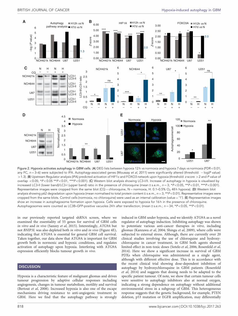
Induction of autophagy at the transcript and protein level. Wehave recently shown that GBM cells can survive under long-termsevere hypoxia, undergoing transcriptional changes and increasingdependency on glycolysis (Sanzey et al, 2015). Although autophagyis known to be regulated mainly at the post-transcriptional level,transcriptional regulation has an important role in the induction ofthe process (Moussay et al, 2011). We therefore investigatedtranscriptional regulation of autophagy-associated genes. Geneexpression patterns were obtained from a panel of GBM culturesincluding glioma stem-like cells (NCH421k, NCH644) and classicalGBM lines (U87, U251), cultured under short (12 h) and long-term(7 days) hypoxia. Differentially expressed genes (FDRo0.01; anyFC) were further subjected for functional enrichment analysis byIPA. As genes associated with the autophagy pathway genesregulating autophagy are poorly annotated in ontology databaseswe applied an in-house gene list (244 genes referred as ‘Autophagypathway’) (Moussay et al, 2011), revealing strong modulation ofthe autophagy pathway upon both short- and long-term hypoxia(Figure 2A). As expected, the upstream regulator analysis by IPApredicted the hypoxia inducible factor 1-alpha (HIF1A) transcrip-tion network to be strongly activated upon hypoxia (P valueo0.05;z-score42; Figure 2B), as was FOXO3A – one of the transcriptionfactors responsible for induction of autophagy (Figure 2B).
Activation of autophagy was further visualised via increasedconversion of LC3-I to LC3-II isoform under hypoxia (Figure 2C).To appropriately detect changes in the autophagic flux, experi-ments were performed in the absence and in the presence of the
lysosomotropic agent chloroquine, which inhibits both the fusionof autophagosome with lysosome and lysosomal protein degrada-tion. Contrary to the previous experiments where chloroquine wasused as a treatment agent (Figure 1), the inhibition of theautophagic flux was detectable upon short chloroquine treatment(3–16 h) according to well-established protocols (Shintani andKlionsky, 2004; Klionsky et al, 2016). High levels of the LC3-IIisoform were detected in all GBM cells treated with chloroquineupon hypoxia as reflected in the LC3-II/LC3-I ratios. Interestingly,NCH421k and U251 cells displayed high levels of LC3-II already innormoxia, suggesting their strong dependence on autophagy innormal conditions (Figure 2C). This is in accordance with the highsensitivity of U251 to chloroquine in both conditions (Figure1G).Induction of autophagy by hypoxia was further confirmed by adecrease in p62 (Figure 2D) and an increase in the number ofautophagosomes visualised via transient LC3-GFP transfection(Figure 2E). In conclusion, these data indicate that autophagy isinduced under severe hypoxia in GBM cells. The heterogeneoussensitivity to autophagy inhibition corroborates with the differ-ential basal level of autophagy in normoxia and further activationof autophagy in hypoxic GBM cells.
ATG9A is involved in the hypoxia-dependent autophagicresponse. To further explore the GBM-specific response tohypoxia we focused on 98 specific regulators of autophagy (71-positive and 27-negative regulators, Supplementary Table S3).Although the number of deregulated genes and the extend ofderegulation was variable, we found four commonly deregulatedgenes shared between short-term and long-term hypoxia (ATG9A,BNIP3, BNIP3L and PIK3C3; Figure 3A; Supplementary Table S3),showing increased levels upon hypoxia. BNIP3 and BNIP3L werepreviously associated with the autophagic response in hypoxicconditions (Mazure and Pouyssegur, 2010), whereas PIK3C3 is awell-known partner in the autophagy onset mechanism (Munsonand Ganley, 2015). Of note, MTOR, a negative regulator ofautophagy and of PIK3C3, was significantly downregulated in 3out of 4 GBM cultures (Supplementary Table S3). Interestingly,within the ATG family, only ATG9A was upregulated in all GBMcells (Figure 3B; Supplementary Table S3), ATG2A was high onlyin 5 out of 8 conditions (Supplementary Table S3). Theupregulation of ATG9A was confirmed by qPCR in GBM stem-like cells (NCH644, NCH421k, NCH660h, NCH601, NCH465)and adherent cultures (U87, U251) (Figure 3C).
Interestingly, analysis of the ATG9A gene promoter revealed thepresence of five hypoxia response elements (HREs) in closeproximity to the canonical transcription start site, confirmed to befunctional according to the TRANSFAC database (Matys et al,2006; Mole et al, 2009) (Supplementary Table S4). This was truealso for BNIP3, BNIP3L and PIK3C3 promoters, and is in line withthe HIF-dependent regulation reported for the BNIP3 and BNIP3L(Kothari et al, 2003; Mole et al, 2009; Slemc and Kunej, 2016). Insummary, we show for the first time that ATG9A expression isstrongly induced in hypoxic conditions, implicating ATG9A as anew player of hypoxia-dependent autophagic response in GBM.
Silencing of ATG9A affects GBM cell proliferation and tumourgrowth in vivo. To study the involvement of ATG9 in autophagy,we generated an efficient long-term ATG9A knockdown (75–98%,Figure 4A) in three GBM cultures, resulting in reduced prolifera-tion both in normoxia and hypoxia (Figure 4B). Contrary to thecontrol, ATG9A-depleted U87 cells did not increase the number ofLC3-positive vesicles upon hypoxia (Figure 4C), suggestinginefficient activation of autophagy. To examine the effect ofATG9A silencing on tumour growth in vivo, we implantedshATG9A NCH421k and NCH644 cells into the brain ofimmunodeficient mice. ATG9A knockdown led to a significantincrease in mouse survival (þ 12–18%; Figure 4D). Of note, two ofthe autopagy-associated genes, ATG9A and BNIP3L, were included
Hypoxia-induced autophagy in GBM BRITISH JOURNAL OF CANCER
www.bjcancer.com | DOI:10.1038/bjc.2017.263 817
in our previously reported targeted shRNA screen, where weexamined the essentiality of 55 genes for survival of GBM cellsin vitro and in vivo (Sanzey et al, 2015). Interestingly, ATG9A butnot BNIP3L was also depleted both in vitro and in vivo (Figure 4E),indicating that ATG9A is essential for general GBM cell survival.Taken together, our data show that ATG9A is important for GBMgrowth both in normoxic and hypoxic conditions, and regulatesactivation of autophagy upon hypoxia. Interfering with ATG9Aexpression efficiently blocks tumour growth in vivo.
DISCUSSION
Hypoxia is a characteristic feature of malignant gliomas and drivestumour progression by adaptive cellular responses includingangiogenesis, changes in tumour metabolism, motility and survival(Bertout et al, 2008). Increased hypoxia is also one of the escapemechanisms driving resistance to anti-angiogenic treatment inGBM. Here we find that the autophagy pathway is strongly
induced in GBM under hypoxia, and we identify ATG9A as a novelregulator of autophagy induction. Inhibiting autophagy was shownto potentiate various anti-cancer therapies in vitro, includinggliomas (Kanzawa et al, 2004; Shingu et al, 2009), where cells weresubjected to external stress. Although, there are currently over 20clinical studies involving the use of chloroquine and hydroxy-chloroquine in cancer treatment, in GBM both agents showedlimited effect in non-toxic doses (Sotelo et al, 2006; Rosenfeld et al,2014). Here we show a significant increase in survival of GBMPDXs when chloroquine was administered as a single agent,although with different effective dose. This is in accordance withthe recent clinical trial showing dose-dependent inhibition ofautophagy by hydroxychloroquine in GBM patients (Rosenfeldet al, 2014) and suggests that dosing needs to be adapted to thespecific patient tumour. Of note, we show that certain tumour cellswere sensitive to autophagy inhibitors also at normal oxygen,indicating a strong dependence on autophagy without additionalenvironmental stress in a subgroup of GBM. This heterogeneousresponse suggests that the genetic background, for example, PTENdeletion, p53 mutation or EGFR amplification, may differentially
Autophagypathway analysis
H12h vs NH7d vs N
H12h vs NH7d vs N
H12h vs NH7d vs N
–log
(P
val
ue)
Z-s
core
LC3-
II/LC
3-I r
atio
Nor
mal
ised
p62
Aut
opha
goso
me/
cell
Z-s
core
6
5
4
3
2
1
0
6.00
5.00
4.00
3.00
2.00
1.00
0.00
3.00
2.50
2.00
1.50
1.00
0.50
0.00
HIF1A FOXO3A
2.0
2.01.3
NCH421k NCH644 U87 U251
NCH421k
NCH644
U87
U251
NCH421k NCH644 U87 U251 LC3/Hoechst U87U251
NCH421k NCH644
NCH644
U87
U87
U251
U251
NCH421k NCH644 U87 U251
N H N H– – + +CQ
NCH421k
NCH644
U87
U251
LC3-ILC3-II
LC3-ILC3-II
LC3-ILC3-II
LC3-ILC3-II
3
2.5
2
1.5
1
0.5
0N
CQH N H N H N H N H N H N H N H
– – + + – – + + – – + + – – + +
6
5
4ns
3
2
1
0
6
5
4
3
2
1
0
6
5
4
3
2
1
0
NCH421k ns*
****
*
****
**
*
**
4.5
4
3.5
3
2.5
2
1.5
1
0.5
0
N H
Normoxia Normoxia
HypoxiaNormoxia
Hypoxia
HypoxiaU
251
U87
120
100
80
60
40
20
0
****** ******
************ *
**
* *
*
A B
C
D E
Figure 2. Hypoxia activates autophagy in GBM cells. (A) DEG lists between hypoxia 12 h vs normoxia and hypoxia 7 days vs normoxia (FDRo0.01;any FC, n¼3–6) were subjected to IPA. Autophagy-associated genes (Moussay et al, 2011) were significantly altered (threshold: � log(P value)41.3). (B) Upstream Regulator analysis (IPA) predicted activation of HIF1a and FOXO3 network upon hypoxia (threshold: z-score 42 and P value ofoverlap o0.05; *Po0.05 **Po0.01, ***Po0.001). (C) Western blot analysis showing LC3-I/II. Increase of autophagy in hypoxia is visualised byincreased LC3-II (lower band)/LC3-I (upper band) ratio in the presence of chloroquine (mean±s.e.m., n¼3; *Po0.05, **Po0.01, ***Po0.001).Representative images were cropped from the same blot (CQ¼ chloroquine, N¼ normoxia, H: 0.1–0.5% O2 48 h hypoxia). (D) Western blotanalysis showing p62 degradation upon hypoxia (mean normalised to total protein content±s.e.m., n¼3; **Po0.01). Representative images werecropped from the same blots. Control cells (normoxia, no chloroquine) were used as an internal calibration (value¼ ‘1’). (E) Representative imagesshow an increase in autophagosome formation upon hypoxia. Cells were exposed to hypoxia for 16 h in the presence of chloroquine.Autophagosomes were counted as LC3B–GFP-positive vacuoles 24 h after transfection; (mean±s.e.m.; n¼34; *Po0.05, **Po0.01).
BRITISH JOURNAL OF CANCER Hypoxia-induced autophagy in GBM
818 www.bjcancer.com | DOI:10.1038/bjc.2017.263

affect the extent of basal level of autophagy and of treatmentresponse in GBM and that appropriate biomarkers may berequired to efficiently stratify patients. EGFR is known tonegatively regulate autophagy (Chen et al, 2016) through multiplesignalling pathways, thus EGFR overexpression may partly explainthe lower sensitivity of T16 tumours to anti-autophagic treatment.
Importantly, we find that bevacizumab treatment sensitisedGBM cells to autophagy inhibition allowing to reach survivalbenefit at lower dose. This was confirmed in vitro, where hypoxiaincreased sensitivity of GBM cells to autophagy inhibitors. Thesynergistic effect of bevacizumab was visible only when the anti-autophagy effect alone was mild or moderate at normal oxygenlevels, but was masked if the autophagy inhibitor alone showed astrong effect. Interestingly, we have previously shown thatbevacizumab leads to a lower number of mitochondria in tumourcells (Keunen et al, 2011), suggesting that mitophagy might beinvolved in the survival under hypoxia. A previous study hasshown efficacy of chloroquine in combination with bevacizumab insubcutaneous U87 tumours, but failed to observe a tumoursuppressive effect with chloroquine used as a single agent (Hu et al,2012). This discrepancy may be due to the different tumourlocalisation and the heterogeneity in the GBM response tochloroquine described here. Although non-specific effects of
chloroquine cannot be excluded (Maycotte et al, 2012; Maeset al, 2014), we did not observe vessel normalisation uponchloroquine treatment. In line with a previous study (Chen et al,2008), normal astrocytes remained unaffected at the lowestchloroquine concentration affecting GBM cells, confirming moresubstantial dependence of tumour cells on autophagy. Althoughmore potent inhibitors are warranted, our data suggest theexistence of a ‘therapeutic window’ for autophagy inhibitors inGBM, and that co-treatment with anti-angiogenic agents allows tosignificantly lower effective doses.
We found that activation of autophagy in hypoxia was linked totranscriptional changes of numerous genes associated with autophagy,among which BNIP3, BNIP3L, ATG9A and PIK3C3 were upregulatedin all GBM cells. BNIP3 and BNIP3L, while activated by HIF1a,mediate autophagy by releasing Beclin1 from complexes with Bcl-2and Bcl-XL (Zhang et al, 2008; Bellot et al, 2009). Interestingly, withinthe ATG family, only ATG9A was transcriptionally activated in allGBM cells. Contrary to other ATG family members such as ATG5and ATG7, but similarly to BNIP3 and BNIP3L, we identify ATG9A aspotentially HIF1a responsive gene. These transcriptional changes wereobserved also in GBM cells that exhibit high basal autophagy atnormal oxygen levels, suggesting that specific upstream moleculessuch as FOXO3A are involved in the regulation autophagy pathway at
A
B C
Hypoxia 12 h vs normoxia
U87 (44)U87 (44)
12 28
6
6
9
11
10
3
1
1
7
2
2
2
2
0
5
5
3
1
7
7
8
Normoxia
ATG16L1ATG7ATG4AATG5
ATG2BATG3ATG16L2ATG2A
ATG9APIK3C3
BNIP3BNIP3L
BECN1
H7 d4
Normoxia
Hypoxia 12 h
Hypoxia 7 d**
***
**
**
****
***
***
**3.5
3
2.5
2
Rel
ativ
e ex
pres
sion
1.5
1
0.5
0
NCH421k
NCH644
NCH660h
NCH465
NCH601
U87U25
1
H12 h
8
6
4
Common:ATG9ABNIP3
BNIP3LPIK3C3
1
1
0
3
NCH644 (44) NCH421k (35) NCH421k(35)
NCH644 (44)
U251 (21)U251 (21)
Hypoxia 7 d vs normoxia
Figure 3. ATG9A is specifically activated upon autophagic response to hypoxia. (A) Genes directly related to autophagy (knowledge-drivenselection) were extracted from DEG lists between hypoxia 12 h vs normoxia and hypoxia 7 days vs normoxia (FDRo0.01; any FC) for each culture(n¼3–6). Venn diagrams reveal commonly deregulated genes. (B) Heatmap shows expression levels for selected genes in NCH421k in normoxia,12 h and 7 days hypoxia. See Supplementary Table S3 for more autophagy-related genes. (C) QPCR confirmed increased ATG9A expression inhypoxia. EZRIN was used as a reference (mean±s.e.m.; n¼3; *Po0.05, **Po0.01, ***Po0.001). NCH421k cells were used as an internalcalibration (value¼ ‘1’).
Hypoxia-induced autophagy in GBM BRITISH JOURNAL OF CANCER
www.bjcancer.com | DOI:10.1038/bjc.2017.263 819

different oxygen levels. Pro-autophagic genes, such as Beclin1, ATG5,ATG7, BNIP3 and BNIP3L were previously found to be essential forautophagy in cancer cells (Zhang et al, 2008; Mazure and Pouyssegur,2009). Here we show that ATG9A also represents an important pro-survival molecule, with ATG9A depletion leading to a strongreduction of tumour growth, thus confirming the relevance ofautophagy as a promising target for GBM treatment. Of note, ATG7knockdown displayed a therapeutic outcome only during anti-angiogenic treatment (Hu et al, 2012).
ATG9A was shown to be essential for autophagosomebiogenesis and membrane maturation; however, its mode of actionremains enigmatic. Recent data suggest that the Pho–Rpd3
complex regulates expression of ATG9A and other ATG genesupon induction of autophagy (Jin and Klionsky, 2014) and thatATG9A-containing vesicles are generated de novo upon starvation(Yamamoto et al, 2012). Here we show that upon ATG9Adepletion, GBM cells were not able to activate autophagy uponhypoxia. We propose that the lack of autophagic activation uponhypoxia may be due to inhibition of de novo autophagosomesynthesis. This is in accordance with a recent report, where ATG9Awas shown to have a key role in autophagosome formation duringhypoxic stress (Weerasekara et al, 2014). Thus, ATG9A maybecome essential upon autophagy induction and an increaseddemand for new autophagosome membranes (Orsi et al, 2012).
A C
D
E
B
Rel
ativ
e ex
pres
sion
0.9shScramble shScramble
shScramble
Hyp
oxia
Hypoxia
Nor
mox
ia
Normoxia
120
100
***
80
60
40
20
0
00
10 40
* P value = 0.02
* P value = 0.02
50 60 70
0 10 40 50
Survival (day)
60 70
50
Per
cent
sur
viva
l
100
150
0
0
0.5
1
1.5
2
2.5
3
3.5
4 Ratio in vivo/baseline
Ratio in vitro/baseline
50
Per
cent
sur
viva
l
100
150
Aut
opha
goso
me/
cell
GFP/LC3/Hoechst
shATG9A
shScramble
shATG9A
shATG9A
shATG9A shBNIP3L
shATG9A
shScramble
shScramble
shATG9A
0.8
0.7
0.6
0.5
0.4
0.3
0.2
0.1
0
***
**
*
0 4 7
Days
11
U87
Cel
l num
ber
NC
H64
4C
ell n
umbe
rN
CH
421k
Cel
l num
ber
0
200 000
400 000
600 000
800 000
1 000 000
1 200 000
1 400 000
**
*
0
200 000
400 000
600 000
800 000
1 000 000
Hypoxia ***
0100 000200 000300 000400 000500 000600 000700 000800 000900 000
1 000 000
Days
0 4 7 110
100 000
200 000
300 000
400 000
500 000
600 000
*
**
*0
500 000
1 000 000
1 500 000
2 000 000
2 500 000
shScramble
Normoxia
shATG9A
*1 200 000
1 000 000
600 000
400 000
200 000
0
800 000
NCH421k
NCH421k
NCH644
* * *
NCH644 U87
Figure 4. ATG9A knockdown decreases GBM cell proliferation and increases mouse survival. (A) QPCR confirmation of shATG9A knockdown(mean±s.e.m.; n¼3; *Po0.05). (B) Proliferation of shATG9A cells was decreased significantly in normoxic and hypoxic conditions (mean±s.e.m.;n¼3; *Po0.05, **Po0.01, ***Po0.001). (C) Representative images show lack of increased autophagosome formation upon hypoxia in shATG9U87 cells. Cells were exposed to hypoxia for 16 h in the presence of chloroquine. Autophagosomes were counted as LC3B–Tomato-positivevacuoles 24 h after transfection; GFP positivity confirms shRNA expression (mean±s.e.m.; n¼34; ***P value o0.001). (D) ATG9A depletion inNCH421k and NCH644 prolonged the survival of tumour-bearing mice (n¼ 6–8). (E) Targeted in vivo shRNA screen in NCH421k cells. shRNAtargeting ATG9A but not BNIP3L was depleted after in vivo (n¼ 5) and in vitro (normoxia, n¼3) growth. Relative representation of respectiveshRNAs after selection pressure is presented as ratios compared with the original shRNA pool before selection (baseline). For detailedexperimental setup, see (Sanzey et al, 2015).
BRITISH JOURNAL OF CANCER Hypoxia-induced autophagy in GBM
820 www.bjcancer.com | DOI:10.1038/bjc.2017.263

In conclusion, our data support the notion that inhibitingautophagy represents an effective therapy in primary GBM,although it may be concentration and patient dependent. Anti-autophagy treatment using genetic and pharmacological interven-tion was effective as a single treatment. However, currentlyavailable drugs, including chloroquine and hydroxychloroquine arenon-curative in non-toxic doses and novel more potent agents willbe necessary for GBM patients. Drugs directly targeting essentialproteins such as ATG9A may be of particular interest and acombination with anti-angiogenic therapy may be beneficial.Finally, the hypoxic microenvironment also contributes toimmunoresistance and hypoxia-induced autophagy impairs cyto-toxic T-lymphocyte-mediated cell lysis of tumour cells (Nomanet al, 2011, 2012) and NK-mediated target cell apoptosis (Baginskaet al, 2013; Viry et al, 2014). Therefore, targeting autophagy intumour cells may not only lead to increased tumour cell death butalso sensitise tumours to immunotherapies.
ACKNOWLEDGEMENTS
We thank Morgane Sanzey, Virginie Baus, and Amandine Bernardfor technical assistance. We thank Dr Christel Herold-Mende(Department of Neurosurgery, University of Heidelberg, Germany)and Dr Uros Rajcevic (National Institute of Biology, Ljubljana,Slovenia) for providing cell lines. This work was supported by theLuxembourg Institute of Health (LIH), the Fonds National de laRecherche (FNR) of Luxembourg (ESCAPE 784322 BM, AFRgrand to SAAR and AD) and the Fondation Cancer Luxembourg(INVGBM).
CONFLICT OF INTEREST
The authors declare no conflict of interest.
REFERENCES
Baginska J, Viry E, Berchem G, Poli A, Noman MZ, van Moer K, Medves S,Zimmer J, Oudin A, Niclou SP, Bleackley RC, Goping IS, Chouaib S, JanjiB (2013) Granzyme B degradation by autophagy decreases tumor cellsusceptibility to natural killer-mediated lysis under hypoxia. Proc NatlAcad Sci USA 110: 17450–17455.
Bellot G, Garcia-Medina R, Gounon P, Chiche J, Roux D, Pouyssegur J,Mazure NM (2009) Hypoxia-induced autophagy is mediated throughhypoxia-inducible factor induction of BNIP3 and BNIP3L via their BH3domains. Mol Cell Biol 29: 2570–2581.
Bertout JA, Patel SA, Simon MC (2008) The impact of O2 availability onhuman cancer. Nat Rev Cancer 8: 967–975.
Bougnaud S, Golebiewska A, Oudin A, Keunen O, Harter PN, Mader L,Azuaje F, Fritah S, Stieber D, Kaoma T, Vallar L, Brons NH, Daubon T,Miletic H, Sundstr½m T, Herold-Mende C, Mittelbronn M, Bjerkvig R,Niclou SP (2016) Molecular crosstalk between tumour and brainparenchyma instructs histopathological features in glioblastoma.Oncotarget.
Chen Y, Henson ES, Xiao W, Huang D, McMillan-Ward EM, Israels SJ,Gibson SB (2016) Tyrosine kinase receptor EGFR regulates the switch incancer cells between cell survival and cell death induced by autophagy inhypoxia. Autophagy 12: 1029–1046.
Chen Y, McMillan-Ward E, Kong J, Israels SJ, Gibson SB (2008) Oxidativestress induces autophagic cell death independent of apoptosis intransformed and cancer cells. Cell Death Differ 15: 171–182.
Ciechomska IA, Gabrusiewicz K, Szczepankiewicz AA, Kaminska B (2013)Endoplasmic reticulum stress triggers autophagy in malignant glioma cellsundergoing cyclosporine a-induced cell death. Oncogene 32: 1518–1529.
Corcelle-Termeau E, Vindelov SD, Hamalisto S, Mograbi B, Keldsbo A,Brasen JH, Favaro E, Adam D, Szyniarowski P, Hofman P, Krautwald S,Farkas T, Petersen NH, Rohde M, Linkermann A, Jððttelð M (2016)
Excess sphingomyelin disturbs ATG9A trafficking and autophagosomeclosure. Autophagy 12: 833–849.
De Witt Hamer PC, Van Tilborg AA, Eijk PP, Sminia P, Troost D, VanNoorden CJ, Ylstra B, Leenstra S (2008) The genomic profile of humanmalignant glioma is altered early in primary cell culture and preserved inspheroids. Oncogene 27: 2091–2096.
Demeure K, Fack F, Duriez E, Tiemann K, Bernard A, Golebiewska A,Bougnaud S, Bjerkvig R, Domon B, Niclou SP (2015) Targeted proteomicsto assess the response to anti-angiogenic treatment in humanglioblastoma. Mol Cell Proteomics 15: 481–492.
Evans SM, Judy KD, Dunphy I, Jenkins WT, Nelson PT, Collins R, WileytoEP, Jenkins K, Hahn SM, Stevens CW, Judkins AR, Phillips P, Geoerger B,Koch CJ (2004) Comparative measurements of hypoxia in human braintumors using needle electrodes and EF5 binding. Cancer Res 64: 1886–1892.
Fack F, Espedal H, Keunen O, Golebiewska A, Obad N, Harter PN,Mittelbronn M, Bahr O, Weyerbrock A, Stuhr L, Miletic H, SakariassenPA, Stieber D, Rygh CB, Lund-Johansen M, Zheng L, Gottlieb E, NiclouSP, Bjerkvig R (2015) Bevacizumab treatment induces metabolicadaptation toward anaerobic metabolism in glioblastomas. ActaNeuropathol 129: 115–131.
Firat E, Weyerbrock A, Gaedicke S, Grosu AL, Niedermann G (2012)Chloroquine or chloroquine-PI3K/Akt pathway inhibitor combinationsstrongly promote gamma-irradiation-induced cell death in primary stem-like glioma cells. PloS ONE 7: e47357.
Gasch AP, Spellman PT, Kao CM, Carmel-Harel O, Eisen MB, Storz G,Botstein D, Brown PO (2000) Genomic expression programs in theresponse of yeast cells to environmental changes. Mol Biol Cell 11:4241–4257.
Gilbert MR (2016) Antiangiogenic therapy for glioblastoma: complex biologyand complicated results. J Clin Oncol 34: 1567–1569.
Glick D, Barth S, Macleod KF (2010) Autophagy: cellular and molecularmechanisms. J Pathol 221: 3–12.
Golebiewska A, Bougnaud S, Stieber D, Brons NH, Vallar L, Hertel F, Klink B,Schrock E, Bjerkvig R, Niclou SP (2013) Side population in humanglioblastoma is non-tumorigenic and characterizes brain endothelial cells.Brain 136: 1462–1475.
Hirschhaeuser F, Menne H, Dittfeld C, West J, Mueller-Klieser W, Kunz-Schughart LA (2010) Multicellular tumor spheroids: an underestimatedtool is catching up again. J Biotechnol 148: 3–15.
Hu YL, DeLay M, Jahangiri A, Molinaro AM, Rose SD, Carbonell WS, AghiMK (2012) Hypoxia-induced autophagy promotes tumor cell survival andadaptation to antiangiogenic treatment in glioblastoma. Cancer Res 72:1773–1783.
Jin M, Klionsky DJ (2014) Transcriptional regulation of ATG9 by the Pho23-Rpd3 complex modulates the frequency of autophagosome formation.Autophagy 10: 1681–1682.
Kanzawa T, Germano IM, Komata T, Ito H, Kondo Y, Kondo S (2004) Role ofautophagy in temozolomide-induced cytotoxicity for malignant gliomacells. Cell Death Differ 11: 448–457.
Keunen O, Johansson M, Oudin A, Sanzey M, Abdul Rahim SA, Fack F,Thorsen F, Taxt T, Bartos M, Jirik R, Miletic H, Wang J, Stieber D, StuhrL, Moen I, Rygh CB, Bjerkvig R, Niclou SP (2011) Anti-VEGF treatmentreduces blood supply and increases tumor cell invasion in glioblastoma.Proc Natl Acad Sci USA 108: 3749–3754.
Klionsky DJ, Abdelmohsen K, Abe A, Abedin MJ, Abeliovich H,Acevedo Arozena A, Adachi H, Adams CM, Adams PD, Adeli K,Adhihetty PJ, Adler SG, Agam G, Agarwal R, Aghi MK, Agnello M,Agostinis P, Aguilar PV, Aguirre-Ghiso J, Airoldi EM, Ait-Si-Ali S,Akematsu T, Akporiaye ET, Al-Rubeai M, Albaiceta GM, Albanese C,Albani D, Albert ML, Aldudo J, Algul H, Alirezaei M, Alloza I, Almasan A,Almonte-Beceril M, Alnemri ES, Alonso C, Altan-Bonnet N, Altieri DC,Alvarez S, Alvarez-Erviti L, Alves S, Amadoro G, Amano A, Amantini C,Ambrosio S, Amelio I, Amer AO, Amessou M, Amon A, An Z,Anania FA, Andersen SU, Andley UP, Andreadi CK, Andrieu-Abadie N,Anel A, Ann DK, Anoopkumar-Dukie S, Antonioli M, Aoki H,Apostolova N, Aquila S, Aquilano K, Araki K, Arama E, Aranda A,Araya J, Arcaro A, Arias E, Arimoto H, Ariosa AR, Armstrong JL,Arnould T, Arsov I, Asanuma K, Askanas V, Asselin E, Atarashi R,Atherton SS, Atkin JD, Attardi LD, Auberger P, Auburger G, Aurelian L,Autelli R, Avagliano L, Avantaggiati ML, Avrahami L, Awale S, Azad N,Bachetti T, Backer JM, Bae DH, Bae JS, Bae ON, Bae SH, Baehrecke EH,Baek SH, Baghdiguian S, Bagniewska-Zadworna A, Bai H, Bai J, Bai XY,
Hypoxia-induced autophagy in GBM BRITISH JOURNAL OF CANCER
www.bjcancer.com | DOI:10.1038/bjc.2017.263 821

Bailly Y, Balaji KN, Balduini W, Ballabio A, Balzan R, Banerjee R,Banhegyi G, Bao H, Barbeau B, Barrachina MD, Barreiro E, Bartel B,Bartolome A, Bassham DC, Bassi MT, Bast Jr RC, Basu A, Batista MT,Batoko H, Battino M, Bauckman K, Baumgarner BL, Bayer KU, Beale R,Beaulieu JF, Beck Jr GR, Becker C, Beckham JD, Bedard PA, Bednarski PJ,Begley TJ, Behl C, Behrends C, Behrens GM, Behrns KE, Bejarano E,Belaid A, Belleudi F, Benard G, Berchem G, Bergamaschi D, Bergami M,Berkhout B, Berliocchi L, Bernard A, Bernard M, Bernassola F,Bertolotti A, Bess AS, Besteiro S, Bettuzzi S, Bhalla S, Bhattacharyya S,Bhutia SK, Biagosch C, Bianchi MW, Biard-Piechaczyk M, Billes V,Bincoletto C, Bingol B, Bird SW, Bitoun M, Bjedov I, Blackstone C,Blanc L, Blanco GA, Blomhoff HK, Boada-Romero E, Bockler S, Boes M,Boesze-Battaglia K, Boise LH, Bolino A, Boman A, Bonaldo P, Bordi M,Bosch J, Botana LM, Botti J, Bou G, Bouche M, Bouchecareilh M,Boucher MJ, Boulton ME, Bouret SG, Boya P, Boyer-Guittaut M,Bozhkov PV, Brady N, Braga VM, Brancolini C, Braus GH,Bravo-San Pedro JM, Brennan LA, Bresnick EH, Brest P, Bridges D,Bringer MA, Brini M, Brito GC, Brodin B, Brookes PS, Brown EJ,Brown K, Broxmeyer HE, Bruhat A, Brum PC, Brumell JH,Brunetti-Pierri N, Bryson-Richardson RJ, Buch S, Buchan AM, Budak H,Bulavin DV, Bultman SJ, Bultynck G, Bumbasirevic V, Burelle Y,Burke RE, Burmeister M, Butikofer P, Caberlotto L, Cadwell K, Cahova M,Cai D, Cai J, Cai Q, Calatayud S, Camougrand N, Campanella M,Campbell GR, Campbell M, Campello S, Candau R, Caniggia I, Cantoni L,Cao L, Caplan AB, Caraglia M, Cardinali C, Cardoso SM, Carew JS,Carleton LA, Carlin CR, Carloni S, Carlsson SR, Carmona-Gutierrez D,Carneiro LA, Carnevali O, Carra S, Carrier A, Carroll B, Casas C, Casas J,Cassinelli G, Castets P, Castro-Obregon S, Cavallini G, Ceccherini I,Cecconi F, Cederbaum AI, Cena V, Cenci S, Cerella C, Cervia D,Cetrullo S, Chaachouay H, Chae HJ, Chagin AS, Chai CY, Chakrabarti G,Chamilos G, Chan EY, Chan MT, Chandra D, Chandra P, Chang CP,Chang RC, Chang TY, Chatham JC, Chatterjee S, Chauhan S, Che Y,Cheetham ME, Cheluvappa R, Chen CJ, Chen G, Chen GC, Chen H,Chen JW, Chen JK, Chen M, Chen M, Chen P, Chen Q, Chen SD, Chen S,Chen SS, Chen W, Chen WJ, Chen WQ, Chen W, Chen X, Chen YH,Chen YG, Chen Y, Chen YJ, Chen YQ, Chen Z, Cheng A, Cheng CH,Cheng H, Cheong H, Cherry S, Chesney J, Cheung CH, Chevet E, Chi HC,Chi SG, Chiacchiera F, Chiang HL, Chiarelli R, Chiariello M, Chieppa M,Chin LS, Chiong M, Chiu GN, Cho DH, Cho SG, Cho WC, Cho YY,Cho YS, Choi AM, Choi EJ, Choi EK, Choi J, Choi ME, Choi SI, Chou TF,Chouaib S, Choubey D, Choubey V, Chow KC, Chowdhury K, Chu CT,Chuang TH, Chun T, Chung H, Chung T, Chung YL, Chwae YJ,Cianfanelli V, Ciarcia R, Ciechomska IA, Ciriolo MR, Cirone M,Claerhout S, Clague MJ, Claria J, Clarke PG, Clarke R, Clementi E,Cleyrat C, Cnop M, Coccia EM, Cocco T, Codogno P, Coers J, Cohen EE,Colecchia D, Coletto L, Coll NS, Colucci-Guyon E, Comincini S,Condello M, Cook KL, Coombs GH, Cooper CD, Cooper JM, Coppens I,Corasaniti MT, Corazzari M, Corbalan R, Corcelle-Termeau E,Cordero MD, Corral-Ramos C, Corti O, Cossarizza A, Costelli P, Costes S,Cotman SL, Coto-Montes A, Cottet S, Couve E, Covey LR, Cowart LA,Cox JS, Coxon FP, Coyne CB, Cragg MS, Craven RJ, Crepaldi T,Crespo JL, Criollo A, Crippa V, Cruz MT, Cuervo AM, Cuezva JM, Cui T,Cutillas PR, Czaja MJ, Czyzyk-Krzeska MF, Dagda RK, Dahmen U, Dai C,Dai W, Dai Y, Dalby KN, Dalla Valle L, Dalmasso G, D’Amelio M,Damme M, Darfeuille-Michaud A, Dargemont C, Darley-Usmar VM,Dasarathy S, Dasgupta B, Dash S, Dass CR, Davey HM, Davids LM,Davila D, Davis RJ, Dawson TM, Dawson VL, Daza P, de Belleroche J,de Figueiredo P, de Figueiredo RC, de la Fuente J, De Martino L,De Matteis A, De Meyer GR, De Milito A, De Santi M, de Souza W,De Tata V, De Zio D, Debnath J, Dechant R, Decuypere JP, Deegan S,Dehay B, Del Bello B, Del Re DP, Delage-Mourroux R, Delbridge LM,Deldicque L, Delorme-Axford E, Deng Y, Dengjel J, Denizot M, Dent P,Der CJ, Deretic V, Derrien B, Deutsch E, Devarenne TP, Devenish RJ, DiBartolomeo S, Di Daniele N, Di Domenico F, Di Nardo A, Di Paola S, DiPietro A, Di Renzo L, DiAntonio A, Dıaz-Araya G, Dıaz-Laviada I,Diaz-Meco MT, Diaz-Nido J, Dickey CA, Dickson RC, Diederich M,Digard P, Dikic I, Dinesh-Kumar SP, Ding C, Ding WX, Ding Z, Dini L,Distler JH, Diwan A, Djavaheri-Mergny M, Dmytruk K, Dobson RC,Doetsch V, Dokladny K, Dokudovskaya S, Donadelli M, Dong XC,Dong X, Dong Z, Donohue Jr TM, Doran KS, D’Orazi G, Dorn 2nd GW,Dosenko V, Dridi S, Drucker L, Du J, Du LL, Du L, du Toit A, Dua P,Duan L, Duann P, Dubey VK, Duchen MR, Duchosal MA, Duez H,Dugail I, Dumit VI, Duncan MC, Dunlop EA, Dunn Jr WA, Dupont N,
Dupuis L, Duran RV, Durcan TM, Duvezin-Caubet S, Duvvuri U,Eapen V, Ebrahimi-Fakhari D, Echard A, Eckhart L, Edelstein CL,Edinger AL, Eichinger L, Eisenberg T, Eisenberg-Lerner A, Eissa NT,El-Deiry WS, El-Khoury V, Elazar Z, Eldar-Finkelman H, Elliott CJ,Emanuele E, Emmenegger U, Engedal N, Engelbrecht AM, Engelender S,Enserink JM, Erdmann R, Erenpreisa J, Eri R, Eriksen JL, Erman A,Escalante R, Eskelinen EL, Espert L, Esteban-Martınez L, Evans TJ,Fabri M, Fabrias G, Fabrizi C, Facchiano A, Færgeman NJ, Faggioni A,Fairlie WD, Fan C, Fan D, Fan J, Fang S, Fanto M, Fanzani A, Farkas T,Faure M, Favier FB, Fearnhead H, Federici M, Fei E, Felizardo TC, Feng H,Feng Y, Ferguson TA, Fernandez AF, Fernandez-Barrena MG,Fernandez-Checa JC, Fernandez-Lopez A, Fernandez-Zapico ME,Feron O, Ferraro E, Ferreira-Halder CV, Fesus L, Feuer R, Fiesel FC,Filippi-Chiela EC, Filomeni G, Fimia GM, Fingert JH, Finkbeiner S,Finkel T, Fiorito F, Fisher PB, Flajolet M, Flamigni F, Florey O, Florio S,Floto RA, Folini M, Follo C, Fon EA, Fornai F, Fortunato F, Fraldi A,Franco R, Francois A, Francois A, Frankel LB, Fraser ID, Frey N,Freyssenet DG, Frezza C, Friedman SL, Frigo DE, Fu D, Fuentes JM,Fueyo J, Fujitani Y, Fujiwara Y, Fujiya M, Fukuda M, Fulda S, Fusco C,Gabryel B, Gaestel M, Gailly P, Gajewska M, Galadari S, Galili G,Galindo I, Galindo MF, Galliciotti G, Galluzzi L, Galy V, Gammoh N,Gandy S, Ganesan AK, Ganesan S, Ganley IG, Gannage M, Gao FB,Gao F, Gao JX, Garcıa Nannig L, Garcıa Vescovi E, Garcia-Macıa M,Garcia-Ruiz C, Garg AD, Garg PK, Gargini R, Gassen NC, Gatica D,Gatti E, Gavard J, Gavathiotis E, Ge L, Ge P, Ge S, Gean PW, Gelmetti V,Genazzani AA, Geng J, Genschik P, Gerner L, Gestwicki JE, Gewirtz DA,Ghavami S, Ghigo E, Ghosh D, Giammarioli AM, Giampieri F,Giampietri C, Giatromanolaki A, Gibbings DJ, Gibellini L, Gibson SB,Ginet V, Giordano A, Giorgini F, Giovannetti E, Girardin SE, Gispert S,Giuliano S, Gladson CL, Glavic A, Gleave M, Godefroy N, Gogal Jr RM,Gokulan K, Goldman GH, Goletti D, Goligorsky MS, Gomes AV,Gomes LC, Gomez H, Gomez-Manzano C, Gomez-Sanchez R,Goncalves DA, Goncu E, Gong Q, Gongora C, Gonzalez CB,Gonzalez-Alegre P, Gonzalez-Cabo P, Gonzalez-Polo RA, Goping IS,Gorbea C, Gorbunov NV, Goring DR, Gorman AM, Gorski SM,Goruppi S, Goto-Yamada S, Gotor C, Gottlieb RA, Gozes I, Gozuacik D,Graba Y, Graef M, Granato GE, Grant GD, Grant S, Gravina GL,Green DR, Greenhough A, Greenwood MT, Grimaldi B, Gros F, Grose C,Groulx JF, Gruber F, Grumati P, Grune T, Guan JL, Guan KL, Guerra B,Guillen C, Gulshan K, Gunst J, Guo C, Guo L, Guo M, Guo W, Guo XG,Gust AA, Gustafsson ÅB, Gutierrez E, Gutierrez MG, Gwak HS, Haas A,Haber JE, Hadano S, Hagedorn M, Hahn DR, Halayko AJ,Hamacher-Brady A, Hamada K, Hamai A, Hamann A, Hamasaki M,Hamer I, Hamid Q, Hammond EM, Han F, Han W, Handa JT,Hanover JA, Hansen M, Harada M, Harhaji-Trajkovic L, Harper JW,Harrath AH, Harris AL, Harris J, Hasler U, Hasselblatt P, Hasui K,Hawley RG, Hawley TS, He C, He CY, He F, He G, He RR, He XH,He YW, He YY, Heath JK, Hebert MJ, Heinzen RA, Helgason GV, HenselM, Henske EP, Her C, Herman PK, Hernandez A, Hernandez C,Hernandez-Tiedra S, Hetz C, Hiesinger PR, Higaki K, Hilfiker S, Hill BG,Hill JA, Hill WD, Hino K, Hofius D, Hofman P, Hoglinger GU, Hohfeld J,Holz MK, Hong Y, Hood DA, Hoozemans JJ, Hoppe T, Hsu C, Hsu CY,Hsu LC, Hu D, Hu G, Hu HM, Hu H, Hu MC, Hu YC, Hu ZW, Hua F,Hua Y, Huang C, Huang HL, Huang KH, Huang KY, Huang S, Huang S,Huang WP, Huang YR, Huang Y, Huber TB, Huebbe P, Huh WK,Hulmi JJ, Hur GM, Hurley JH, Husak Z, Hussain SN, Hussain S,Hwang JJ, Hwang S, Hwang TI, Ichihara A, Imai Y, Imbriano C,Inomata M, Into T, Iovane V, Iovanna JL, Iozzo RV, Ip NY, Irazoqui JE,Iribarren P, Isaka Y, Isakovic AJ, Ischiropoulos H, Isenberg JS,Ishaq M, Ishida H, Ishii I, Ishmael JE, Isidoro C, Isobe K, Isono E,Issazadeh-Navikas S, Itahana K, Itakura E, Ivanov AI, Iyer AK,Izquierdo JM, Izumi Y, Izzo V, Jaattela M, Jaber N, Jackson DJ,Jackson WT, Jacob TG, Jacques TS, Jagannath C, Jain A, Jana NR,Jang BK, Jani A, Janji B, Jannig PR, Jansson PJ, Jean S, Jendrach M,Jeon JH, Jessen N, Jeung EB, Jia K, Jia L, Jiang H, Jiang L, Jiang T, Jiang X,Jiang Y, Jimenez A, Jin C, Jin H, Jin L, Jin M, Jin S, Jinwal UK,Jo EK, Johansen T, Johnson DE, Johnson GV, Johnson JD, Jonasch E,Jones C, Joosten LA, Jordan J, Joseph AM, Joseph B, Joubert AM, Ju D,Ju J, Juan HF, Juenemann K, Juhasz G, Jung HS, Jung JU, Jung YK,Jungbluth H, Justice MJ, Jutten B, Kaakoush NO, Kaarniranta K, Kaasik A,Kabuta T, Kaeffer B, Kågedal K, Kahana A, Kajimura S, Kakhlon O,Kalia M, Kalvakolanu DV, Kamada Y, Kambas K, Kaminskyy VO,Kampinga HH, Kandouz M, Kang C, Kang R, Kang TC, Kanki T,
BRITISH JOURNAL OF CANCER Hypoxia-induced autophagy in GBM
822 www.bjcancer.com | DOI:10.1038/bjc.2017.263
Kanneganti TD, Kanno H, Kanthasamy AG, Kantorow M,Kaparakis-Liaskos M, Kapuy O, Karantza V, Karim MR, Karmakar P,Kaser A, Kaushik S, Kawula T, Kaynar AM, Ke PY, Ke ZJ, Kehrl JH,Keller KE, Kemper JK, Kenworthy AK, Kepp O, Kern A, Kesari S,Kessel D, Ketteler R, Kettelhut Ido C, Khambu B, Khan MM,Khandelwal VK, Khare S, Kiang JG, Kiger AA, Kihara A, Kim AL,Kim CH, Kim DR, Kim DH, Kim EK, Kim HY, Kim HR, Kim JS, Kim JH,Kim JC, Kim JH, Kim KW, Kim MD, Kim MM, Kim PK, Kim SW,Kim SY, Kim YS, Kim Y, Kimchi A, Kimmelman AC, Kimura T, King JS,Kirkegaard K, Kirkin V, Kirshenbaum LA, Kishi S, Kitajima Y,Kitamoto K, Kitaoka Y, Kitazato K, Kley RA, Klimecki WT,Klinkenberg M, Klucken J, Knævelsrud H, Knecht E, Knuppertz L, Ko JL,Kobayashi S, Koch JC, Koechlin-Ramonatxo C, Koenig U, Koh YH,Kohler K, Kohlwein SD, Koike M, Komatsu M, Kominami E, Kong D,Kong HJ, Konstantakou EG, Kopp BT, Korcsmaros T, Korhonen L,Korolchuk VI, Koshkina NV, Kou Y, Koukourakis MI, Koumenis C,Kovacs AL, Kovacs T, Kovacs WJ, Koya D, Kraft C, Krainc D, Kramer H,Kravic-Stevovic T, Krek W, Kretz-Remy C, Krick R, Krishnamurthy M,Kriston-Vizi J, Kroemer G, Kruer MC, Kruger R, Ktistakis NT,Kuchitsu K, Kuhn C, Kumar AP, Kumar A, Kumar D, Kumar D, KumarR, Kumar S, Kundu M, Kung HJ, Kuno A, Kuo SH, Kuret J, Kurz T, KwokT, Kwon TK, Kwon YT, Kyrmizi I, La Spada AR, Lafont F, Lahm T,Lakkaraju A, Lam T, Lamark T, Lancel S, Landowski TH, Lane DJ,Lane JD, Lanzi C, Lapaquette P, Lapierre LR, Laporte J, Laukkarinen J,Laurie GW, Lavandero S, Lavie L, LaVoie MJ, Law BY, Law HK,Law KB, Layfield R, Lazo PA, Le Cam L, Le Roch KG,Le Stunff H, Leardkamolkarn V, Lecuit M, Lee BH, Lee CH, Lee EF,Lee GM, Lee HJ, Lee H, Lee JK, Lee J, Lee JH, Lee M, Lee MS,Lee PJ, Lee SW, Lee SJ, Lee SJ, Lee SY, Lee SH, Lee SS, Lee SJ, Lee S,Lee YR, Lee YJ, Lee YH, Leeuwenburgh C, Lefort S, Legouis R, Lei J,Lei QY, Leib DA, Leibowitz G, Lekli I, Lemaire SD, Lemasters JJ,Lemberg MK, Lemoine A, Leng S, Lenz G, Lenzi P, Lerman LO,Lettieri Barbato D, Leu JI, Leung HY, Levine B, Lewis PA, Lezoualc’h F,Li C, Li F, Li FJ, Li J, Li K, Li L, Li M, Li M, Li Q, Li R, Li S, Li W,Li X, Li Y, Lian J, Liang C, Liang Q, Liao Y, Liberal J, Liberski PP, Lie P,Lieberman AP, Lim HJ, Lim KL, Lim K, Lima RT, Lin CS, Lin CF, Lin F,Lin FC, Lin K, Lin KH, Lin PH, Lin T, Lin WW, Lin YS, Linden R,Lindholm D, Lindqvist LM, Lingor P, Linkermann A, Liotta LA,Lipinski MM, Lira VA, Lisanti MP, Liton PB, Liu B, Liu C, Liu CF,Liu F, Liu HJ, Liu J, Liu JJ, Liu JL, Liu K, Liu L, Liu Q, Liu RY, Liu S, Liu S,Liu W, Liu XD, Liu X, Liu XH, Liu Y, Liu Z, Liuzzi JP, Lizard G, Ljujic M,Lodhi IJ, Logue SE, Lokeshwar BL, Long YC, Lonial S, Loos B, Lopez-OtınC, Lopez-Vicario C, Lorente M, Lorenzi PL, Lorincz P, Los M, Lotze MT,Lovat PE, Lu B, Lu J, Lu Q, Lu SM, Lu S, Lu Y, Luciano F, Luckhart S,Lucocq JM, Ludovico P, Lugea A, Lukacs NW, Lum JJ, Lund AH, Luo H,Luo J, Luo S, Luparello C, Lyons T, Ma J, Ma Y, Ma Y, Ma Z, Machado J,Machado-Santelli GM, Macian F, MacIntosh GC, MacKeigan JP, MacleodKF, MacMicking JD, MacMillan-Crow LA, Madeo F, Madesh M,Madrigal-Matute J, Maeda A, Maeda T, Maegawa G, Maellaro E, Maes H,Magarinos M, Maiese K, Maiti TK, Maiuri L, Maiuri MC, Maki CG, MalliR, Malorni W, Maloyan A, Mami-Chouaib F, Man N, Mancias JD,Mandelkow EM, Mandell MA, Manfredi AA, Manie SN, Manzoni C, MaoK, Mao Z, Mao ZW, Marambaud P, Marconi AM, Marelja Z, Marfe G,Margeta M, Margittai E, Mari M, Mariani FV, Marin C, Marinelli S,Marino G, Markovic I, Marquez R, Martelli AM, Martens S, Martin KR,Martin SJ, Martin S, Martin-Acebes MA, Martın-Sanz P, Martinand-MariC, Martinet W, Martinez J, Martinez-Lopez N, Martinez-Outschoorn U,Martınez-Velazquez M, Martinez-Vicente M, Martins WK, Mashima H,Mastrianni JA, Matarese G, Matarrese P, Mateo R, Matoba S,Matsumoto N, Matsushita T, Matsuura A, Matsuzawa T, Mattson MP,Matus S, Maugeri N, Mauvezin C, Mayer A, Maysinger D, Mazzolini GD,McBrayer MK, McCall K, McCormick C, McInerney GM, McIver SC,McKenna S, McMahon JJ, McNeish IA, Mechta-Grigoriou F, Medema JP,Medina DL, Megyeri K, Mehrpour M, Mehta JL, Mei Y, Meier UC,Meijer AJ, Melendez A, Melino G, Melino S, de Melo EJ, Mena MA,Meneghini MD, Menendez JA, Menezes R, Meng L, Meng LH, Meng S,Menghini R, Menko AS, Menna-Barreto RF, Menon MB, Meraz-Rıos MA,Merla G, Merlini L, Merlot AM, Meryk A, Meschini S, Meyer JN, Mi MT,Miao CY, Micale L, Michaeli S, Michiels C, Migliaccio AR, Mihailidou AS,Mijaljica D, Mikoshiba K, Milan E, Miller-Fleming L, Mills GB, Mills IG,Minakaki G, Minassian BA, Ming XF, Minibayeva F, Minina EA,Mintern JD, Minucci S, Miranda-Vizuete A, Mitchell CH, Miyamoto S,Miyazawa K, Mizushima N, Mnich K, Mograbi B, Mohseni S, Moita LF,
Molinari M, Molinari M, Møller AB, Mollereau B, Mollinedo F,Mongillo M, Monick MM, Montagnaro S, Montell C, Moore DJ,Moore MN, Mora-Rodriguez R, Moreira PI, Morel E, Morelli MB,Moreno S, Morgan MJ, Moris A, Moriyasu Y, Morrison JL, Morrison LA,Morselli E, Moscat J, Moseley PL, Mostowy S, Motori E, Mottet D,Mottram JC, Moussa CE, Mpakou VE, Mukhtar H, Mulcahy Levy JM,Muller S, Munoz-Moreno R, Munoz-Pinedo C, Munz C, Murphy ME,Murray JT, Murthy A, Mysorekar IU, Nabi IR, Nabissi M, Nader GA,Nagahara Y, Nagai Y, Nagata K, Nagelkerke A, Nagy P, Naidu SR, Nair S,Nakano H, Nakatogawa H, Nanjundan M, Napolitano G, Naqvi NI,Nardacci R, Narendra DP, Narita M, Nascimbeni AC, Natarajan R,Navegantes LC, Nawrocki ST, Nazarko TY, Nazarko VY, Neill T, Neri LM,Netea MG, Netea-Maier RT, Neves BM, Ney PA, Nezis IP, Nguyen HT,Nguyen HP, Nicot AS, Nilsen H, Nilsson P, Nishimura M, Nishino I,Niso-Santano M, Niu H, Nixon RA, Njar VC, Noda T, Noegel AA,Nolte EM, Norberg E, Norga KK, Noureini SK, Notomi S, Notterpek L,Nowikovsky K, Nukina N, Nurnberger T, O’Donnell VB, O’Donovan T,O’Dwyer PJ, Oehme I, Oeste CL, Ogawa M, Ogretmen B, Ogura Y, Oh YJ,Ohmuraya M, Ohshima T, Ojha R, Okamoto K, Okazaki T, Oliver FJ,Ollinger K, Olsson S, Orban DP, Ordonez P, Orhon I, Orosz L,O’Rourke EJ, Orozco H, Ortega AL, Ortona E, Osellame LD, Oshima J,Oshima S, Osiewacz HD, Otomo T, Otsu K, Ou JH, Outeiro TF,Ouyang DY, Ouyang H, Overholtzer M, Ozbun MA, Ozdinler PH,Ozpolat B, Pacelli C, Paganetti P, Page G, Pages G, Pagnini U, Pajak B,Pak SC, Pakos-Zebrucka K, Pakpour N, Palkova Z, Palladino F, Pallauf K,Pallet N, Palmieri M, Paludan SR, Palumbo C, Palumbo S, Pampliega O,Pan H, Pan W, Panaretakis T, Pandey A, Pantazopoulou A, Papackova Z,Papademetrio DL, Papassideri I, Papini A, Parajuli N, Pardo J, Parekh VV,Parenti G, Park JI, Park J, Park OK, Parker R, Parlato R, Parys JB,Parzych KR, Pasquet JM, Pasquier B, Pasumarthi KB, Patschan D,Patterson C, Pattingre S, Pattison S, Pause A, Pavenstadt H, Pavone F,Pedrozo Z, Pena FJ, Penalva MA, Pende M, Peng J, Penna F,Penninger JM, Pensalfini A, Pepe S, Pereira GJ, Pereira PC,Perez-de la Cruz V, Perez-Perez ME, Perez-Rodrıguez D, Perez-Sala D,Perier C, Perl A, Perlmutter DH, Perrotta I, Pervaiz S, Pesonen M,Pessin JE, Peters GJ, Petersen M, Petrache I, Petrof BJ, Petrovski G,Phang JM, Piacentini M, Pierdominici M, Pierre P, Pierrefite-Carlevarle V,Pietrocola F, Pimentel-Muinos FX, Pinar M, Pineda B,Pinkas-Kramarski R, Pinti M, Pinton P, Piperdi B, Piret JM, Platanias LC,Platta HW, Plowey ED, Poggeler S, Poirot M, Polcic P, Poletti A,Poon AH, Popelka H, Popova B, Poprawa I, Poulose SM, Poulton J,Powers SK, Powers T, Pozuelo-Rubio M, Prak K, Prange R, Prescott M,Priault M, Prince S, Proia RL, Proikas-Cezanne T, Prokisch H,Promponas VJ, Przyklenk K, Puertollano R, Pugazhenthi S, Puglielli L,Pujol A, Puyal J, Pyeon D, Qi X, Qian WB, Qin ZH, Qiu Y, Qu Z,Quadrilatero J, Quinn F, Raben N, Rabinowich H, Radogna F, Ragusa MJ,Rahmani M, Raina K, Ramanadham S, Ramesh R, Rami A,Randall-Demllo S, Randow F, Rao H, Rao VA, Rasmussen BB, Rasse TM,Ratovitski EA, Rautou PE, Ray SK, Razani B, Reed BH, Reggiori F,Rehm M, Reichert AS, Rein T, Reiner DJ, Reits E, Ren J, Ren X, Renna M,Reusch JE, Revuelta JL, Reyes L, Rezaie AR, Richards RI, Richardson DR,Richetta C, Riehle MA, Rihn BH, Rikihisa Y, Riley BE, Rimbach G,Rippo MR, Ritis K, Rizzi F, Rizzo E, Roach PJ, Robbins J, Roberge M,Roca G, Roccheri MC, Rocha S, Rodrıguez CI, de Cordoba SR,Rodriguez-Muela N, Roelofs J, Rogov VV, Rohn TT, Rohrer B,Romanelli D, Romani L, Romano PS, Roncero MI, Rosa JL, Rosello A,Rosen KV, Rosenstiel P, Rost-Roszkowska M, Roth K, Rodrigues CMA,Roue G, Rouis M, Rouschop KM, Ruan DT, Ruano D, Rubinsztein DC,Rucker 3rd EB, Rudich A, Rudolf E, Rudolf R, Ruegg MA, Ruiz-Roldan C,Ruparelia AA, Rusmini P, Russ DW, Russo GL, Russo G, Russo R,Rusten TE, Ryabovol V, Ryan KM, Ryter SW, Sabatini DM, Sacher M,Sachse C, Sack MN, Sadoshima J, Saftig P, Sagi-Eisenberg R, Sahni S,Saikumar P, Saito T, Saitoh T, Sakakura K, Sakoh-Nakatogawa M,Sakuraba Y, Salazar-Roa M, Salomoni P, Saluja AK, Salvaterra PM,Salvioli R, Samali A, Sanchez AM, Sanchez-Alcazar JA, Sanchez-Prieto R,Sandri M, Sanjuan MA, Santaguida S, Santambrogio L, Santoni G, DosSantos CN, Saran S, Sardiello M, Sargent G, Sarkar P, Sarkar S, Sarrias MR,Sarwal MM, Sasakawa C, Sasaki M, Sass M, Sato K, Sato M, Satriano J,Savaraj N, Saveljeva S, Schaefer L, Schaible UE, Scharl M, Schatzl HM,Schekman R, Scheper W, Schiavi A, Schipper HM, Schmeisser H,Schmidt J, Schmitz I, Schneider BE, Schneider EM, Schneider JL,Schon EA, Schonenberger MJ, Schonthal AH, Schorderet DF, Schroder B,Schuck S, Schulze RJ, Schwarten M, Schwarz TL, Sciarretta S, Scotto K,
Hypoxia-induced autophagy in GBM BRITISH JOURNAL OF CANCER
www.bjcancer.com | DOI:10.1038/bjc.2017.263 823

Scovassi AI, Screaton RA, Screen M, Seca H, Sedej S, Segatori L, Segev N,Seglen PO, Seguı-Simarro JM, Segura-Aguilar J, Seki E, Sell C, Seiliez I,Semenkovich CF, Semenza GL, Sen U, Serra AL, Serrano-Puebla A,Sesaki H, Setoguchi T, Settembre C, Shacka JJ, Shajahan-Haq AN,Shapiro IM, Sharma S, She H, Shen CK, Shen CC, Shen HM, Shen S,Shen W, Sheng R, Sheng X, Sheng ZH, Shepherd TG, Shi J, Shi Q, Shi Q,Shi Y, Shibutani S, Shibuya K, Shidoji Y, Shieh JJ, Shih CM, Shimada Y,Shimizu S, Shin DW, Shinohara ML, Shintani M, Shintani T, Shioi T,Shirabe K, Shiri-Sverdlov R, Shirihai O, Shore GC, Shu CW, Shukla D,Sibirny AA, Sica V, Sigurdson CJ, Sigurdsson EM, Sijwali PS, Sikorska B,Silveira WA, Silvente-Poirot S, Silverman GA, Simak J, Simmet T,Simon AK, Simon HU, Simone C, Simons M, Simonsen A, Singh R,Singh SV, Singh SK, Sinha D, Sinha S, Sinicrope FA, Sirko A, Sirohi K,Sishi BJ, Sittler A, Siu PM, Sivridis E, Skwarska A, Slack R, Slaninova I,Slavov N, Smaili SS, Smalley KS, Smith DR, Soenen SJ, Soleimanpour SA,Solhaug A, Somasundaram K, Son JH, Sonawane A, Song C, Song F,Song HK, Song JX, Song W, Soo KY, Sood AK, Soong TW,Soontornniyomkij V, Sorice M, Sotgia F, Soto-Pantoja DR,Sotthibundhu A, Sousa MJ, Spaink HP, Span PN, Spang A, Sparks JD,Speck PG, Spector SA, Spies CD, Springer W, Clair DS, Stacchiotti A,Staels B, Stang MT, Starczynowski DT, Starokadomskyy P, Steegborn C,Steele JW, Stefanis L, Steffan J, Stellrecht CM, Stenmark H,Stepkowski TM, Stern ST, Stevens C, Stockwell BR, Stoka V, Storchova Z,Stork B, Stratoulias V, Stravopodis DJ, Strnad P, Strohecker AM,Strom AL, Stromhaug P, Stulik J, Su YX, Su Z, Subauste CS,Subramaniam S, Sue CM, Suh SW, Sui X, Sukseree S, Sulzer D,Sun FL, Sun J, Sun SY, Sun Y, Sundaramoorthy V, Sung J,Suzuki H, Suzuki K, Suzuki N, Suzuki T, Suzuki YJ, Swanson MS,Swanton C, Sward K, Swarup G, Sweeney ST, Sylvester PW, Szatmari Z,Szegezdi E, Szlosarek PW, Taegtmeyer H, Tafani M, Taillebourg E,Tait SW, Takacs-Vellai K, Takahashi Y, Takats S, Takemura G,Takigawa N, Talbot NJ, Tamagno E, Tamburini J, Tan CP, Tan L,Tan ML, Tan M, Tan YJ, Tanaka K, Tanaka M, Tang D, Tang G,Tanida I, Tanji K, Tannous BA, Tapia JA, Tasset-Cuevas I, Tatar M,Tavassoly I, Tavernarakis N, Taylor A, Taylor GS, Taylor GA,Taylor JP, Taylor MJ, Tchetina EV, Tee AR, Teixeira-Clerc F, Telang S,Tencomnao T, Teng BB, Teng RJ, Terro F, Tettamanti G, Theiss AL,Theron AE, Thomas KJ, Thome MP, Thomes PG, Thorburn A, Thorner J,Thum T, Thumm M, Thurston TL, Tian L, Till A, Ting JP, Titorenko VI,Toker L, Toldo S, Tooze SA, Topisirovic I, Torgersen ML, Torosantucci L,Torriglia A, Torrisi MR, Tournier C, Towns R, Trajkovic V, Travassos LH,Triola G, Tripathi DN, Trisciuoglio D, Troncoso R, Trougakos IP,Truttmann AC, Tsai KJ, Tschan MP, Tseng YH, Tsukuba T, Tsung A,Tsvetkov AS, Tu S, Tuan HY, Tucci M, Tumbarello DA, Turk B, Turk V,Turner RF, Tveita AA, Tyagi SC, Ubukata M, Uchiyama Y, Udelnow A,Ueno T, Umekawa M, Umemiya-Shirafuji R, Underwood BR,Ungermann C, Ureshino RP, Ushioda R, Uversky VN, Uzcategui NL,Vaccari T, Vaccaro MI, Vachova L, Vakifahmetoglu-Norberg H,Valdor R, Valente EM, Vallette F, Valverde AM, Van den Berghe G,Van Den Bosch L, van den Brink GR, van der Goot FG, van der Klei IJ,van der Laan LJ, van Doorn WG, van Egmond M, van Golen KL,Van Kaer L, van Lookeren Campagne M, Vandenabeele P,Vandenberghe W, Vanhorebeek I, Varela-Nieto I, Vasconcelos MH,Vasko R, Vavvas DG, Vega-Naredo I, Velasco G, Velentzas AD,Velentzas PD, Vellai T, Vellenga E, Vendelbo MH, Venkatachalam K,Ventura N, Ventura S, Veras PS, Verdier M, Vertessy BG, Viale A,Vidal M, Vieira HL, Vierstra RD, Vigneswaran N, Vij N, Vila M, Villar M,Villar VH, Villarroya J, Vindis C, Viola G, Viscomi MT, Vitale G,Vogl DT, Voitsekhovskaja OV, von Haefen C, von Schwarzenberg K,Voth DE, Vouret-Craviari V, Vuori K, Vyas JM, Waeber C, Walker CL,Walker MJ, Walter J, Wan L, Wan X, Wang B, Wang C, Wang CY,Wang D, Wang F, Wang G, Wang HJ, Wang H, Wang HG, Wang HD,Wang J, Wang M, Wang MQ, Wang PY, Wang P, Wang RC, Wang S,Wang TF, Wang X, Wang XJ, Wang XW, Wang Y, Wang YJ, Wang YT,Wang ZN, Wappner P, Ward C, Ward DM, Warnes G, Watada H,Watanabe Y, Watase K, Weaver TE, Weekes CD, Wei J, Weide T,Weihl CC, Weindl G, Weis SN, Wen L, Wen X, Wen Y, Westermann B,Weyand CM, White AR, White E, Whitton JL, Whitworth AJ, Wiels J,Wild F, Wildenberg ME, Wileman T, Wilkinson DS, Wilkinson S,Willbold D, Williams C, Williams K, Williamson PR, Winklhofer KF,Witkin SS, Wohlgemuth SE, Wollert T, Wolvetang EJ, Wong E,Wong GW, Wong RW, Wong VK, Woodcock EA, Wright KL, Wu C,Wu D, Wu GS, Wu J, Wu J, Wu M, Wu M, Wu S, Wu WK, Wu Y, Wu Z,
Xavier CP, Xavier RJ, Xia GX, Xia T, Xia W, Xia Y, Xiao H, Xiao J, Xiao S,Xiao W, Xie CM, Xie Z, Xilouri M, Xiong Y, Xu C, Xu F, Xu H, Xu J, Xu L,Xu X, Xu Y, Xu ZX, Xu Z, Xue Y, Yamada T, Yamamoto A, Yamanaka K,Yamashina S, Yamashiro S, Yan B, Yan X, Yan Z, Yanagi Y, Yang DS,Yang JM, Yang L, Yang M, Yang PM, Yang P, Yang Q, Yang W, Yang WY,Yang X, Yang Y, Yang Z, Yao MC, Yao PJ, Yao X, Yao Z, Yasui LS, Ye M,Yedvobnick B, Yeganeh B, Yeh ES, Yeyati PL, Yi F, Yi L, Yin XM, Yip CK,Yoo YM, Yoo YH, Yoon SY, Yoshida K, Yoshimori T, Young KH, Yu H,Yu JJ, Yu JT, Yu J, Yu L, Yu WH, Yu XF, Yu Z, Yuan J, Yuan ZM, Yue BY,Yue J, Yue Z, Zacks DN, Zacksenhaus E, Zaffaroni N, Zaglia T, Zakeri Z,Zecchini V, Zeng J, Zeng M, Zeng Q, Zervos AS, Zhang DD, Zhang F,Zhang G, Zhang GC, Zhang H, Zhang J, Zhang JP, Zhang L, Zhang MY,Zhang X, Zhang XD, Zhang Y, Zhao M, Zhao WL, Zhao X, Zhao YG,Zhao Y, Zhao YX, Zhao Z, Zhao ZJ, Zheng D, Zheng XL, Zheng X,Zhivotovsky B, Zhong Q, Zhou GZ, Zhou G, Zhou H, Zhou SF,Zhou XJ, Zhu H, Zhu WG, Zhu W, Zhu XF, Zhu Y, Zhuang SM,Zhuang X, Ziparo E, Zois CE, Zoladek T, Zong WX, Zorzano A,Zughaier SM (2016) Guidelines for the use and interpretation of assaysfor monitoring autophagy (3rd edition). Autophagy 12: 1–222.
Kothari S, Cizeau J, McMillan-Ward E, Israels SJ, Bailes M, Ens K,Kirshenbaum LA, Gibson SB (2003) BNIP3 plays a role in hypoxic celldeath in human epithelial cells that is inhibited by growth factors EGF andIGF. Oncogene 22: 4734–4744.
Lamb CA, Nuhlen S, Judith D, Frith D, Snijders AP, Behrends C, Tooze SA(2016) TBC1D14 regulates autophagy via the TRAPP complex and ATG9traffic. EMBO J 35: 281–301.
Maes H, Kuchnio A, Peric A, Moens S, Nys K, De Bock K, Quaegebeur A,Schoors S, Georgiadou M, Wouters J, Vinckier S, Vankelecom H, GarmynM, Vion AC, Radtke F, Boulanger C, Gerhardt H, Dejana E, Dewerchin M,GhesquiOre B, Annaert W, Agostinis P, Carmeliet P (2014) Tumor vesselnormalization by chloroquine independent of autophagy. Cancer Cell 26:190–206.
Matys V, Kel-Margoulis OV, Fricke E, Liebich I, Land S, Barre-Dirrie A,Reuter I, Chekmenev D, Krull M, Hornischer K, Voss N, Stegmaier P,Lewicki-Potapov B, Saxel H, Kel AE, Wingender E (2006) TRANSFACand its module TRANSCompel: transcriptional gene regulation ineukaryotes. Nucleic Acids Res 34: D108–D110.
Maycotte P, Aryal S, Cummings CT, Thorburn J, Morgan MJ, Thorburn A(2012) Chloroquine sensitizes breast cancer cells to chemotherapyindependent of autophagy. Autophagy 8: 200–212.
Mazure NM, Pouyssegur J (2009) Atypical BH3-domains of BNIP3 andBNIP3L lead to autophagy in hypoxia. Autophagy 5: 868–869.
Mazure NM, Pouyssegur J (2010) Hypoxia-induced autophagy: cell death orcell survival? Curr Opin Cell Biol 22: 177–180.
Mole DR, Blancher C, Copley RR, Pollard PJ, Gleadle JM, Ragoussis J,Ratcliffe PJ (2009) Genome-wide association of hypoxia-induciblefactor (HIF)-1alpha and HIF-2alpha DNA binding with expressionprofiling of hypoxia-inducible transcripts. J Biol Chem 284:16767–16775.
Moussay E, Kaoma T, Baginska J, Muller A, Van Moer K, Nicot N, NazarovPV, Vallar L, Chouaib S, Berchem G, Janji B (2011) The acquisition ofresistance to TNFalpha in breast cancer cells is associated with constitutiveactivation of autophagy as revealed by a transcriptome analysis using acustom microarray. Autophagy 7: 760–770.
Munson MJ, Ganley IG (2015) MTOR, PIK3C3, and autophagy: signaling thebeginning from the end. Autophagy 11: 2375–2376.
Niclou SP, Danzeisen C, Eikesdal HP, Wiig H, Brons NH, Poli AM,Svendsen A, Torsvik A, Enger PO, Terzis JA, Bjerkvig R (2008) A noveleGFP-expressing immunodeficient mouse model to study tumor-hostinteractions. FASEB J 22: 3120–3128.
Noman MZ, Janji B, Berchem G, Mami-Chouaib F, Chouaib S (2012) Hypoxia-induced autophagy: a new player in cancer immunotherapy? Autophagy 8:704–706.
Noman MZ, Janji B, Kaminska B, Van Moer K, Pierson S, Przanowski P,Buart S, Berchem G, Romero P, Mami-Chouaib F, Chouaib S (2011)Blocking hypoxia-induced autophagy in tumors restores cytotoxic T-cellactivity and promotes regression. Cancer Res 71: 5976–5986.
Orsi A, Razi M, Dooley HC, Robinson D, Weston AE, Collinson LM,Tooze SA (2012) Dynamic and transient interactions of Atg9 withautophagosomes, but not membrane integration, are required forautophagy. Mol Biol Cell 23: 1860–1873.
Pouyssegur J, Dayan F, Mazure NM (2006) Hypoxia signalling in cancer andapproaches to enforce tumour regression. Nature 441: 437–443.
BRITISH JOURNAL OF CANCER Hypoxia-induced autophagy in GBM
824 www.bjcancer.com | DOI:10.1038/bjc.2017.263

Rabinowitz JD, White E (2010) Autophagy and metabolism. Science 330:1344–1348.
Reggiori F, Tooze SA (2012) Autophagy regulation through Atg9 traffic. J CellBiol 198: 151–153.
Rosenfeld MR, Ye X, Supko JG, Desideri S, Grossman SA, Brem S, MikkelsonT, Wang D, Chang YC, Hu J, McAfee Q, Fisher J, Troxel AB, Piao S,Heitjan DF, Tan KS, Pontiggia L, O?Dwyer PJ, Davis LE, Amaravadi RK(2014) A phase I/II trial of hydroxychloroquine in conjunction withradiation therapy and concurrent and adjuvant temozolomide inpatients with newly diagnosed glioblastoma multiforme. Autophagy 10:1359–1368.
Rouschop KM, van den Beucken T, Dubois L, Niessen H, Bussink J, SavelkoulsK, Keulers T, Mujcic H, Landuyt W, Voncken JW, Lambin P, van derKogel AJ, Koritzinsky M, Wouters BG (2010) The unfolded proteinresponse protects human tumor cells during hypoxia through regulationof the autophagy genes MAP1LC3B and ATG5. J Clin Invest 120: 127–141.
Sanzey M, Abdul Rahim SA, Oudin A, Dirkse A, Kaoma T, Vallar L, Herold-Mende C, Bjerkvig R, Golebiewska A, Niclou SP (2015) Comprehensiveanalysis of glycolytic enzymes as therapeutic targets in the treatment ofglioblastoma. PLoS One 10: e0123544.
Shingu T, Fujiwara K, Bogler O, Akiyama Y, Moritake K, Shinojima N,Tamada Y, Yokoyama T, Kondo S (2009) Inhibition of autophagy at a latestage enhances imatinib-induced cytotoxicity in human malignant gliomacells. Int J Cancer 124: 1060–1071.
Shintani T, Klionsky DJ (2004) Autophagy in health and disease: a double-edged sword. Science 306: 990–995.
Slemc L, Kunej T (2016) Transcription factor HIF1A: downstream targets,associated pathways, polymorphic hypoxia response element (HRE) sites,and initiative for standardization of reporting in scientific literature.Tumour Biol 37: 14851–14861.
Sotelo J, Briceno E, Lopez-Gonzalez MA (2006) Adding chloroquine toconventional treatment for glioblastoma multiforme: a randomized,
double-blind, placebo-controlled trial. Ann Intern Med 144:337–343.
Vaupel P, Mayer A (2007) Hypoxia in cancer: significance and impact onclinical outcome. Cancer Metastasis Rev 26: 225–239.
Viry E, Baginska J, Berchem G, Noman MZ, Medves S, Chouaib S, Janji B(2014) Autophagic degradation of GZMB/granzyme B: a new mechanismof hypoxic tumor cell escape from natural killer cell-mediated lysis.Autophagy 10: 173–175.
Weerasekara VK, Panek DJ, Broadbent DG, Mortenson JB, Mathis AD,Logan GN, Prince JT, Thomson DM, Thompson JW, Andersen JL (2014)Metabolic-stress-induced rearrangement of the 14-3-3zeta interactomepromotes autophagy via a ULK1- and AMPK-regulated 14-3-3zetainteraction with phosphorylated Atg9. Mol Cell Biol 34: 4379–4388.
Yamamoto H, Kakuta S, Watanabe TM, Kitamura A, Sekito T, Kondo-KakutaC, Ichikawa R, Kinjo M, Ohsumi Y (2012) Atg9 vesicles are an importantmembrane source during early steps of autophagosome formation. J CellBiol 198: 219–233.
Yang L, Lin C, Wang L, Guo H, Wang X (2012) Hypoxia and hypoxia-inducible factors in glioblastoma multiforme progression and therapeuticimplications. Exp Cell Res 318: 2417–2426.
Yang ZJ, Chee CE, Huang S, Sinicrope FA (2011) The role of autophagy incancer: therapeutic implications. Mol Cancer Ther 10: 1533–1541.
Zhang H, Bosch-Marce M, Shimoda LA, Tan YS, Baek JH, Wesley JB,Gonzalez FJ, Semenza GL (2008) Mitochondrial autophagy is an HIF-1-dependent adaptive metabolic response to hypoxia. J Biol Chem 283:10892–10903.
This work is published under the standard license to publish agree-ment. After 12 months the work will become freely available andthe license terms will switch to a Creative Commons Attribution-NonCommercial-Share Alike 4.0 Unported License.
Supplementary Information accompanies this paper on British Journal of Cancer website (http://www.nature.com/bjc)
Hypoxia-induced autophagy in GBM BRITISH JOURNAL OF CANCER
www.bjcancer.com | DOI:10.1038/bjc.2017.263 825
Copyright © 2022 FDOKUMEN