Time-resolved release of calcium from an epithelial cell monolayer during mucin secretion
Upload
independentCategory
view
3download
0
Structure of Cholesterol/Ceramide Monolayer Mixtures: Implications tothe Molecular Organization of Lipid Rafts
Luana Scheffer,* Inna Solomonov,y Markus Jan Weygand,z Kristian Kjaer,z Leslie Leiserowitz,y and Lia Addadi**Department of Structural Biology, Weizmann Institute of Science, Rehovot, Israel; yDepartment of Materials and Interfaces, WeizmannInstitute of Science, Rehovot, Israel; and zMaterials Research Department, Risø National Laboratory, Roskilde, Denmark
ABSTRACT The structure of monolayers of cholesterol/ceramide mixtures was investigated using grazing incidence x-raydiffraction, immunofluorescence, and atomic force microscopy techniques. Grazing incidence x-ray diffraction measurementsshowed the existence of a crystalline mixed phase of the two components within a range of compositions of cholesterol/ceramide between 100:0 and 67:33. The mixed phase coexists with the ceramide crystalline phase in the range of compositionsbetween 50:50 and 30:70; between 30:70 and 0:100 only the highly crystalline phase of ceramide was detected. The latter wasdetermined and modeled. Immunolabeling was performed with an antibody specific to the cholesterol monohydrate crystallinearrangement. The antibody recognizes crystalline cholesterol monolayers, but does not interact with crystalline ceramide.Immunofluorescence and atomic force microscopy data show that in uncompressed ceramide monolayers, the highly crystallinephase coexists with a disordered loosely packed phase. In contrast, no disordered phase coexists with the new crystallinemixed phase. We conclude that the new mixed phase represents a stable homogeneous arrangement of cholesterol withceramide. As ceramide incorporates the lipid backbone common to all sphingolipids, this arrangement may be relevant to theunderstanding of the molecular organization of lipid rafts.
INTRODUCTION
Cholesterol-sphingolipid interactions are fundamental for
lipid bilayer formation in cellular membranes, yet they are
still not well understood. A significant advance in the under-
standing of membrane organization, function, and structure
developed with the suggestion that plasma membranes of
animal cells may contain laterally segregated domains, the
so-called ‘‘lipid rafts’’ (Simons and Ikonen, 1997). This new
concept emerged as a modification of the conventional ‘‘fluid
mosaic model’’, presenting the lipid bilayer as a homoge-
neous mixture of cholesterol and lipids, with proteins inter-
spersed and freely diffusing (Singer and Nicolson, 1972).
Lipid rafts, in contrast, are thought to be formed by dy-
namical clustering of cholesterol and sphingolipids, partic-
ularly sphingomyelin, in organized structures. These domains
appear to be immersed in a medium akin to the fluid mosaic
model, where phospholipids are the main component.
Receptor-mediated signaling events originate from lipid rafts
(Simons and Toomre, 2000; Smart et al., 1999), whereas
many proteins colocalize with them in the membrane, and
are thus thought to be preferentially partitioned in the rafts
(Brown and Rose, 1992).
Evidence for the existence of cholesterol-rich domains in
cell membranes has accumulated within the last few years. A
wide range of techniques was applied, providing information
about the presence and distribution of cholesterol-rich do-
mains in cell membranes, their size, and their dynamics at
different spatial resolutions. (Friedrichson and Kurzchalia,
1998; Giocondi et al., 2000; Pralle et al., 2000; Varma and
Mayor, 1998).
The majority of sphingolipids consist of a sphingosine
backbone linked through amide bonds to long-chain fatty
acids to yield ceramide (Fig. 1). Different classes of sphingo-
lipids result upon attachment of different headgroups to the
terminal hydroxyl of ceramide. The most abundant sphingo-
lipid in the animal cell membranes is sphingomyelin, which,
due to the phosphorylcholine moiety attached to the
ceramide backbone, is considered the sphingolipid analog
of phosphatidylcholine. The bulky phosphorylcholine moi-
ety of sphingomyelin protrudes from the membrane into the
water, whereas the ceramide backbone interacts with the
other lipids forming the membrane bilayer.
The raft-associated acid sphingomyelinase cleaves off the
phosphorylcholine moiety of sphingomyelin, thus leading to
in situ release of ceramide (Schneider and Kennedy, 1967).
Ceramide is thus considered by itself a component of lipid
rafts, both strongly associating to and stabilizing the liquid-
ordered state (Xu et al., 2001).
Cholesterol, the most abundant sterol in animal tissues, is
an essential constituent of cell membranes and lipoprotein
particles. It is composed of a steroid ring system, with little
conformational flexibility, terminated with a 3b-hydroxyl
group (Fig. 1). Cholesterol partitions between lipid rafts and
the disordered lipid homogeneous phase, but shows higher
affinity for saturated sphingolipids than for unsaturated
phospholipids (Ramstedt and Slotte, 2002). It is considered
a key component of lipid rafts and its concentration is
a determining factor in raft stability.
Submitted August 25, 2004, and accepted for publication January 26, 2005.
Address reprint requests to Prof. Lia Addadi, Dept. of Structural Biology,
Weizmann Institute of Science, Rehovot 76100, Israel. E-mail: lia.addadi@
weizmann.ac.il.
� 2005 by the Biophysical Society
0006-3495/05/05/3381/11 $2.00 doi: 10.1529/biophysj.104.051870
Biophysical Journal Volume 88 May 2005 3381–3391 3381
The proposed structural model for lipid rafts involves
a liquid-ordered assembly of sphingolipids, where choles-
terol is thought to be intercalated between the long, saturated
acyl chains of the lipids (Simons and Ikonen, 1997). Both
cholesterol and ceramide are thought to stabilize the raft by
‘‘diluting’’ the repulsive interactions that would ensue be-
tween the bulky sphingolipid headgroups in the water phase
(Huang and Feigenson, 1999; Majewski et al., 2001).
The proposed model, however, assumes a unique lipid
composition and structure for the rafts, which is not nec-
essarily correct. It thus appears necessary to determine the
composition and the molecular arrangement of the various
components within the domains on a more individual scale.
What is the molecular organization of the rafts? Are they
uniform entities with the same molecular organization? Is the
component distribution within one domain homogeneous?
These are only few out of the many questions still not an-
swered that are important for the understanding of the func-
tioning of cholesterol-rich domains in general.
The tight interaction inferred between cholesterol and the
long saturated alkyl chains of the sphingolipids, explains the
resistance of the cholesterol-rich domains to detergent ex-
traction at 4�C, which is the most widely used biochemical
tool for identifying lipid rafts (Brown and London, 1997).
With the detergent approach, however, it is not possible to
determine the size or structure of the domains or to distin-
guish between different compositions, since the membrane
domains rearrange into larger detergent-resistant membranes
during the treatment (Giocondi et al., 2000).
A new approach is most likely required for characterizing
the molecular parameters of the component arrangement
within the domains.
In this study, mixed cholesterol-ceramide monolayers
were chosen as a model system to glean some information on
packing arrangements of lipids. Their two-dimensional struc-
ture was solved by grazing incidence x-ray diffraction
(GIXD). In addition, antibodies that were raised and selected
against crystals of cholesterol monohydrate were used.
GIXD has proven to be a reliable method for determining
the structure and size of ordered arrays of monolayers and
multilayers of crystalline amphiphilic molecules at the air/
water interface (Als-Nielsen et al., 1989; Kuzmenko et al.,
2001; Jensen and Kjaer, 2001). Recently, monolayers and
multilayers of cholesterol have been investigated (Rapaport
et al., 2001). The sensitivity of the method, however, pro-
gressively decreases with reduced ordering.
The use of specific antibodies as reporters of the struc-
ture of organized arrays of molecules was initiated in the
laboratory of one of us (L.A.) with a series of studies on
specific monoclonal antibodies, isolated after exposure of
organisms to crystals such as cholesterol-monohydrate and
others (Geva et al., 2003; Kessler et al., 1996; Perl-Treves
et al., 1996). These antibodies interact with the crystal and
with organized monolayers of cholesterol at the air/water
interface (Izhaky and Addadi, 2000). They do not interact
with the isolated molecule or with monolayers of other
steroids, such as cholestenone or epicholesterol (Geva et al.,
2001). Molecular modeling of various antibody binding sites
displayed chemical and geometrical complementarity be-
tween the antibodies and the specific molecular organiza-
tions they bind to (Kessler et al., 1999). Hence, it is at least
conceivable that antibodies can be used as reporters of the
specific molecular organization they were selected for, or the
absence thereof (Addadi et al., 2003; Kruth et al., 2001).
In all likelihood, lipid rafts have local order. Various
methods, GIXD, atomic force microscopy (AFM), and im-
munofluorescence, complement each other here, providing
a structural description of single layers composed of mix-
tures of cholesterol and ceramide, as an initial attempt to get
insight into structures of lipids that may result, in the future,
to be relevant for the understanding of the organization of
lipid rafts.
MATERIALS AND METHODS
N-palmitoyl-D-erythro-sphingosine (C16 ceramide) was purchased from
Avanti Polar Lipids (Birmingham, AL). Cholesterol (.99%), rhodamine_B
isothiocyanate, octadecyltrichlorosilane (OTS), as well as solvents, were
purchased from Sigma-Aldrich (Rehovot, Israel). ImmunoPure IgM puri-
fication kit 44897 was purchased from Pierce (Rockford, IL). The Centricon
centrifugal filter devices were acquired from Millipore (Bedford, MA).
Dialysis was carried out using Spectra/Por membranes from Spectrum
Medical Industries (Los Angeles, CA) with a molecular weight cutoff in the
range 12,000-14,000 Da against PBS (phosphate-buffered saline). Highly
oriented pyrolitic graphite (HOPG) grade SPI-2 was purchased from SPI
Supplies (West Chester, PA).
The uncompressed monolayers were viewed under epifluorescent light
with rhodamine filter (575 nm), in a Zeiss optical microscope, equipped with
a video camera attached to an LIS-700 integration amplifier (Applitec, Holon,
Israel) that allows amplification of the image intensity up to 30003.
Lipid/chloroform solutions (;0.1 mg/ml) were used to form the mono-
layer films at the air/water interface for surface pressure molecular area
(p-A) isotherms and GIXD measurements.
The isotherms were recorded at 5�C for each of the monolayers using
a Mini LB Trough (KSV Instruments, Helsinki, Finland) with a Wilhelmy
plate.
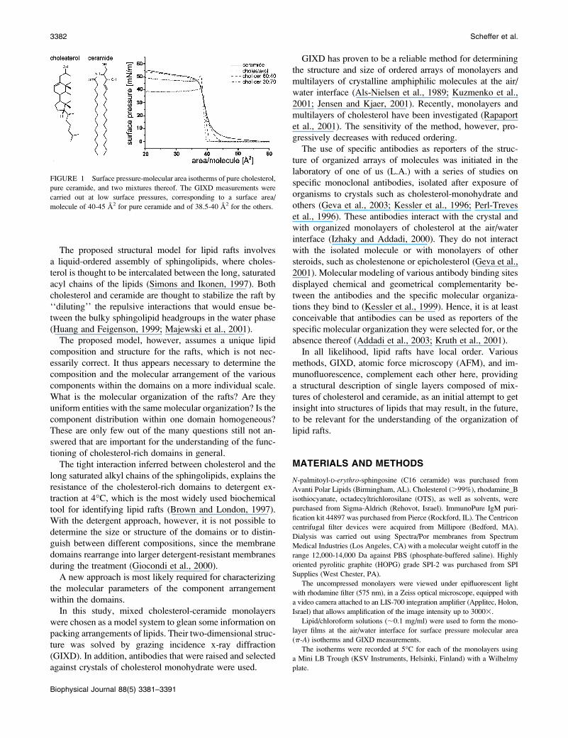
FIGURE 1 Surface pressure-molecular area isotherms of pure cholesterol,
pure ceramide, and two mixtures thereof. The GIXD measurements were
carried out at low surface pressures, corresponding to a surface area/
molecule of 40-45 A2 for pure ceramide and of 38.5-40 A2 for the others.
3382 Scheffer et al.
Biophysical Journal 88(5) 3381–3391
GIXD
The GIXD experiments were performed on the liquid surface diffractometer
at the undulator BW1 beam line at the HASYLAB synchrotron source,
DESY (Hamburg, Germany). The monolayers were formed at room temp-
erature on a film of water;0.25mm thick covering a smooth glass surface, to
reduce the amplitude of surface capillary waves. X-ray diffraction measure-
ments were performed thereon upon cooling to 5�C. For all the monolayers
studied by GIXD, the surface pressure was zero. A monochromatic x-ray
beam (l ¼ 1.304 A) was adjusted to strike the liquid surface at an incident
angle ai approximately equal to 0.85ac, where ac is the critical angle for total
external reflection; this maximizes surface sensitivity. The dimensions of the
footprint of the incoming x-ray beam on the liquid surface were ;2 3 50
mm. The scattered intensity was collected by means of a position-sensitive
detector (PSD), which intercepts photons over the range 0.0, qz, 1.1 A�1,
qz being the out-of-plane component of the scattering vector q;
qz ¼ ð2p=lÞ½sinðaiÞ1 sinðafÞ� � ð2p=lÞsinðafÞ;where ai and af are the angles of the incident and diffracted beams with the
horizontal plane. The measurements were performed by scanning the PSD
across the horizontal component qxy of the scattering vector q;
where 2uxy is the angle between the incident and diffracted beam projected
onto the horizontal plane. A more detailed explanation of the method can be
found in the literature (Als-Nielsen and Kjaer, 1989; Kuzmenko et al., 2001;
Jensen and Kjaer, 2001).
SHELX-97 was used for x-ray structure refinement of the GIXD data and
CERIUS software was used for the construction of the molecular models.
AFM
AFM observations were carried out with the aid of a Nanoscope III Multi-
Mode system (Digital Instruments, Santa Barbara, CA). Microfabricated
square-pyramidal shaped tips of silicon nitride (Digital Instruments) were
used. In situ tapping-mode imaging under MilliQ water was performed by
using a standard fluid cell and the wide-legged cantilever (nominal spring
constant, 0.58 N/m). The best results were obtained at a resonant frequency of
;8-9 KHz and a signal amplitude of ,0.2 V. Typical scan frequencies were
between 1 and 3 Hz and the images were sampled at the resolution of 256 3
256 points.
For the AFM study, the samples were prepared by transferring the mono-
layers of cholesterol and ceramide onto freshly cleaved HOPG. To achieve
transfer, the HOPG substrate was horizontally lowered in contact with the
monolayer. Immediately after the transfer, a drop of water was placed on the
substrate to preserve the humidity of the monolayer during the handling of
the sample.
Antibody purification
Immunolabeling experiments were carried out with the monoclonal IgM
antibody 36A1. This antibody was raised and selected against crystals of
cholesterol monohydrate (Perl-Treves et al., 1996). For blocking nonspecific
sites of interaction a nonspecific antibody (48E1), of the same IgM isotype
as 36A1, was used (Geva et al., 2003).
The antibody was purified from ascites fluid by affinity chromatography
using an ImmunoPure IgM purification kit, according to the manufacturer’s
instructions. Only fractions with optical density .0.2 were pooled. The
purified antibody was extensively dialyzed against PBS and stored at 4�C.
Fluorescent labeling of the antibody
The purified antibody (270 ml) was diluted in 1 N Na2CO3 buffer (30 mL,
pH 9). Rhodamine_B isothiocyanate was dissolved in dry dimethylsulf-
oxide (40 ml, 1.0 mg/ml) and slowly added to the antibody solution under
constant agitation over 1 h at room temperature. The solution was stored for
5 h at 4�C, then 1 M NH4Cl (18 mL) was added followed by 90 min addi-
tional storage at 4�C.The labeled antibody was separated from the free rhodamine by dialysis
and subsequent filtration on a Centricon centrifugal filter device with
a molecular weight cutoff of 100,000. After filtration, the labeled antibody
was stored in the dark at 4�C.The final concentration of the antibody was determined by amino acid
analysis.
Immunolabeling of monolayers
Solutions of the individual lipids and their mixtures in chloroform were
prepared at a total concentration of 10�4M.Themonolayerswere prepared by
depositing on the air/water interface an amount of the respective solution (not
fluorescently labeled) that corresponds to a surface coverage of 90%, as
estimated from the average molecular areas determined from their p-A iso-
therms. After the monolayer was allowed to equilibrate for 15 min, the water
subphase (18.2 mVMillipore water) was exchanged for a PBS solution (2.5
mL) containing both labeled antibody (36A1, 0.5mg/mL) and the nonspecific
competitor (48E1, 5 mg/mL). The system was allowed to equilibrate for 30
min, and monitored directly under the epifluorescence microscope. The
monolayers were then lifted from the air/water interface onto OTS-coated
glass coverslips. To achieve transfer, the solid substrate was horizontally
lowered in contact with the monolayer. OTS-coating of the glass cover slides
was necessary for reproducible transfer of good quality monolayers, with
correct orientation of the functional hydroxyl groups toward the solution. To
lower background fluorescence, the coverslips were then washed three times
by setting them onto a clean water surface. Care was taken that the monolayer
did not dry at any stage of transfer, washing, or observation. Transferred
monolayers were analyzed by epifluorescence microscopy.
To avoid changes in antibody concentration that could result from
antibody aggregation, sets of the monolayers of cholesterol, ceramide, and
their mixtures were observed during the same day. In the case that no
epifluorescence signal was observed (the 60:40 cholesterol/ceramide mixture)
using the same value of the exposure time, this value was increased by
several steps to verify the existence of the monolayer.
Immunofluorescence experiments on supported monolayers were carried
out following the same general procedure described above, with the dif-
ference that the lipid monolayers were first lifted onto the glass substrate and
then incubated with the antibody solution. For the incubation step, the
monolayer transferred to glass was laid for 30 min on the solution containing
the antibody and subsequently rinsed as above.
RESULTS
Lipid components of the system
Mixed monolayers of cholesterol and ceramide were chosen
as a model system for cholesterol-lipid interactions in lipid
rafts for various reasons: the most important consideration is
to get information on the packing of the monolayer backbone
qxy ¼ ð2p=lÞ½cos2ðaiÞ1 cos2ðafÞ � 2cosðaiÞcosðafÞcosð2uxyÞ�1=2
� ð2p=lÞ½11 cos2ðafÞ � 2cosðafÞcosð2uxyÞ�1=2 � ð4p=lÞsinð2uxy=2Þ1Orderða2
f Þ;
Structure of Mixed Lipid Monolayers 3383
Biophysical Journal 88(5) 3381–3391
chains, disregarding in a first stage any bulky headgroups.
These emerge from the monolayer into the solution, most
likely in a disordered partial layer. Besides, all the techniques
available, including GIXD, immunolabeling and AFM, pro-
vide the best information on packed monolayers, and they
would be impaired by bulky headgroups protruding from the
monolayers.
For a first characterization of the molecular packing of
the monolayers at the air/water interface, surface pressure-
molecular area (p-A) isotherms of cholesterol, ceramide, and
their mixtures were recorded; the isotherms of mixtures with
cholesterol/ceramide molar ratios of 30:70 and 60:40 are
reported, together with the isotherm for the pure components
(Fig. 1).
The p-A isotherm of pure C16 ceramide shows a typical
lipid-like behavior. The monolayer shows a smooth transi-
tion from an expanded state to a more condensed state upon
compression.
The average molecular areas measured for cholesterol and
ceramide are similar, but the cholesterol monolayer is much
less compressible in the low-surface pressure range. Due to its
steroid backbone, cholesterol is more rigid and even at high
molecular areas the molecule is not tilted relative to the in-
terface (Rapaport et al., 2001). The decrease in surface area
down to 40 A2 with compression causes fusion of the cho-
lesterol domains without any change in the tilt of the molecule.
The p-A isotherms of the mixed cholesterol/ceramide
monolayers are intermediate between the two pure mono-
layers of cholesterol and ceramide.
GIXD data
A detailed description of GIXD applied to films on liquid
surfaces has been given elsewhere (Als-Nielsen and Kjaer,
1989; Kuzmenko et al., 2001; Jensen and Kjaer, 2001; see
also Materials and Methods). Briefly, the monolayers are
illuminated with an incident x-ray beam at a grazing angle
from the surface lower than the critical angle for total reflec-
tion. The refracted (evanescent) beam, traveling parallel to
the interface, is diffracted by the ordered organic film re-
sulting in diffraction peaks (Bragg peaks). The diffraction
pattern from the monolayer can be interpreted as resulting
from a two-dimensional ‘‘powder’’ of crystallites all parallel
to the water surface but randomly oriented around the surface
normal. As the film has a monomolecular thickness, the
intensity distribution in the z direction, perpendicular to the
surface, appears as a continuum in the form of Bragg rods.
Measurements were performed by scanning the horizontal
component qxy and resolving the out-of-plane component qz,of the scattering vector q (see Methods).
The GIXD pattern is represented in two different ways. The
intensity profile of each Bragg peak, I(qxy), is obtained by
integrating over qz, whereas the Bragg rod intensity profiles
I(qz) are integrated across the qxy range of each diffraction
peak separately for each q value. The qxy positions of the
Bragg peaks yield the lattice repeat distances d ¼ 2p/qxy,which may be indexed by the two Miller indices h,k to yield
the unit cell. The full width at half maximum (FWHM) of the
diffraction peaks yields the lateral two-dimensional crystalline
coherence length Lxy ¼ 0.9(2p)/FWHM(qxy). The width of
the Bragg rod profile along qz similarly gives a first estimate of
the thickness of the crystalline film Lz¼ 0.9(2p)/FWHM(qz).All the GIXD data were recorded just at the point before
the surface pressure began to rise. The corresponding molec-
ular areas were in the range 38.5-40 A2, except for pure
ceramide that was between 40 and 45 A2.
Structure of monolayers of ceramide
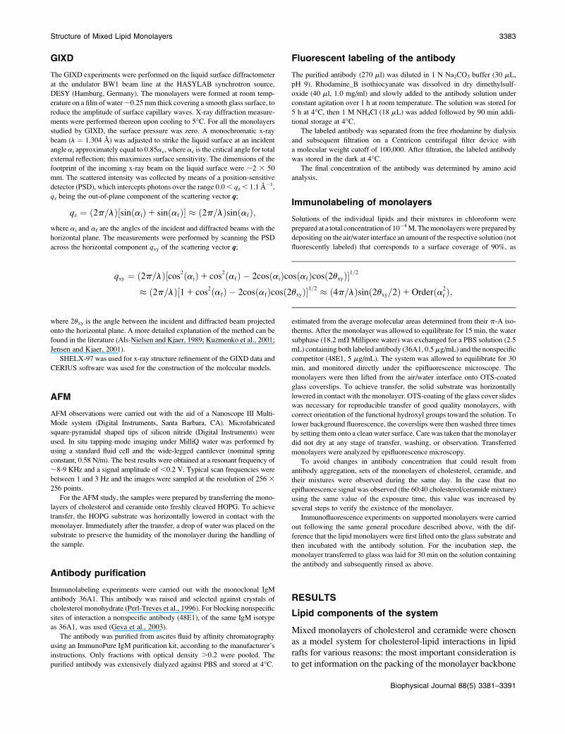
The GIXD pattern of pure ceramide in the uncompressed
state (Fig. 2, a and b), with an area/molecule of 45 A2,
displays three overlapping diffraction peaks (at qxy ¼ 1.451,
1.494, and 1.538 A�1) and an additional weak peak at qxy ¼
1.62 A�1. An identical GIXD pattern—but for the weak peak,
which was absent—was obtained for a 40:60 cholesterol/
ceramide mixture with a molecular area of 45 A2. This film
was then compressed to a molecular area of;40 A2, with no
observable surface pressure. The GIXD measurements, per-
formed ;2 h later (Fig. 2, c and d), showed a phase change,
albeit minor, coupled with a dramatic sharpening of the
diffraction peaks (cf. Fig. 2, a and c), indicative of increasedcrystal domain size. Interestingly, these well-resolved
diffraction peaks (qxy ¼ 1.434, 1.486, and 1.625 A�1, cor-
responding to a unit cell a2 ¼ 5.18 A, b2 ¼ 7.74 A, g2 ¼92.2�), are very similar to those (qxy¼1.432, 1.480, and 1.603A�1) recently reported for a monolayer film of D-erythro-
C18-ceramide at 18�C (Vaknin and Kelley, 2000). We thus
focused upon the GIXD pattern of the 40:60 cholesterol/
ceramide mixture (Fig. 2, c and d) for structure determina-
tion. We may now interpret the spectrum of pure ceramide as
being composed of a main phase 1 (with cell dimensions
a1 ¼ 5.02 A, b1 ¼ 8.17 A, g1 ¼ 91.9�) and a minor phase 2
identical to that of the 40:60 mixture. The FWHM of the
Bragg rods along qz in the two phases (Fig. 2, b and d) areindicative of a monolayer thickness of ;20 A.
The near-rectangular unit cell derived from Fig. 2, c and d,contains one independent molecule of ceramide. A rectan-
gular unit cell of dimensions 5 3 7.5 A2 is fingerprint evi-
dence of a herring-bone arrangement of hydrocarbon chains
related by glide symmetry, with their chain axes aligned
normal to the surface plane (Kuzmenko et al., 1998). In the
case described here, the positions of the maxima along qz ofthe three Bragg rods indicates that the chains are titled from
the surface normal by an angle of;14� in the direction of thea-axis (Kjaer,1994; Jensen and Kjaer, 2001). The dimen-
sions of the unit cell projected onto a plane perpendicular to
the molecular axis are a cos t 3 b ¼ 5.02 3 7.74 A2, in
complete agreement with the two chains of the molecule
forming a herring-bone arrangement related by pseudoglide
symmetry. (The ceramide molecule is chiral of unique
3384 Scheffer et al.
Biophysical Journal 88(5) 3381–3391
handedness. Its two chains cannot therefore be related by a
crystallographic glide symmetry, which incorporates a mirror
operation. Hence the description ‘‘pseudoglide’’.)
The molecular structure of cerebroside (Pascher and
Sundell, 1977) was chosen as an initial model for deter-
mining the two-dimensional crystalline structure of ceramide.
This molecular structure, constrained as a rigid body, was
refined via SHELX by x-ray structure factor computations
(see Method in Rapaport et al., 2001), yielding a reasonable fit
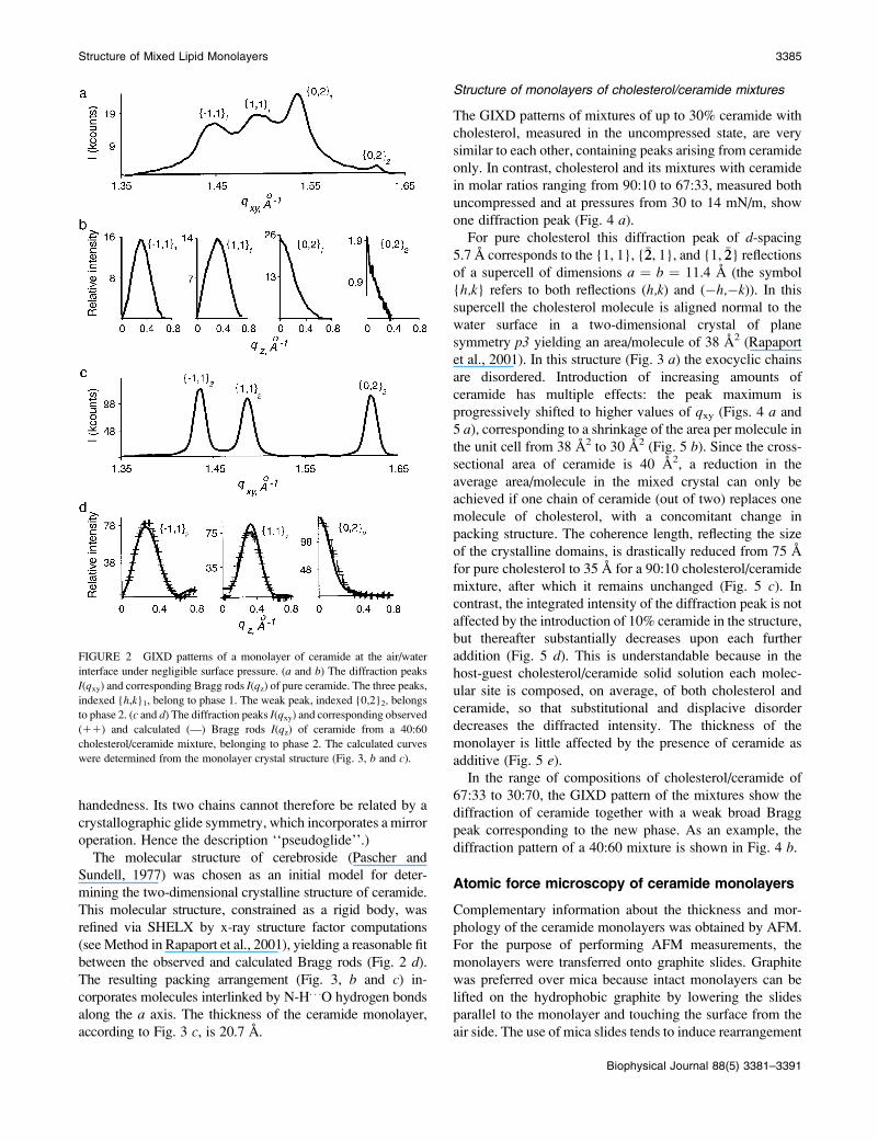
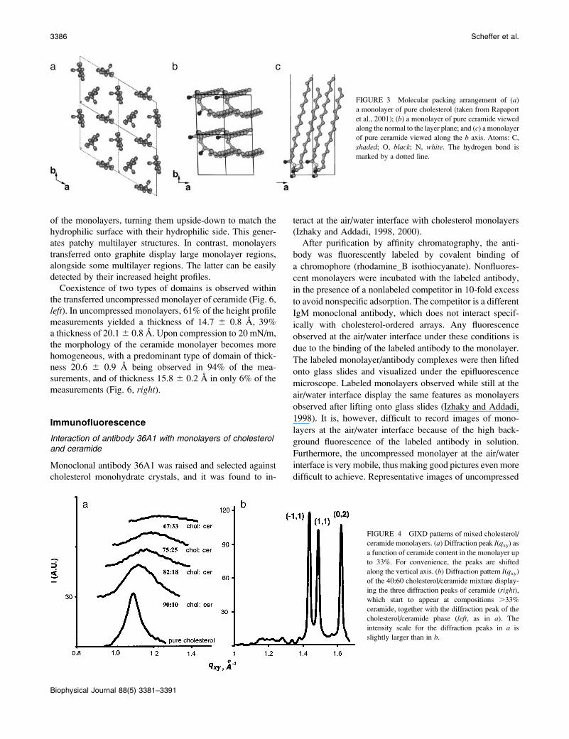
between the observed and calculated Bragg rods (Fig. 2 d).The resulting packing arrangement (Fig. 3, b and c) in-
corporates molecules interlinked by N-H. . .O hydrogen bonds
along the a axis. The thickness of the ceramide monolayer,
according to Fig. 3 c, is 20.7 A.
Structure of monolayers of cholesterol/ceramide mixtures
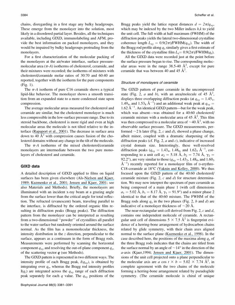
The GIXD patterns of mixtures of up to 30% ceramide with
cholesterol, measured in the uncompressed state, are very
similar to each other, containing peaks arising from ceramide
only. In contrast, cholesterol and its mixtures with ceramide
in molar ratios ranging from 90:10 to 67:33, measured both
uncompressed and at pressures from 30 to 14 mN/m, show
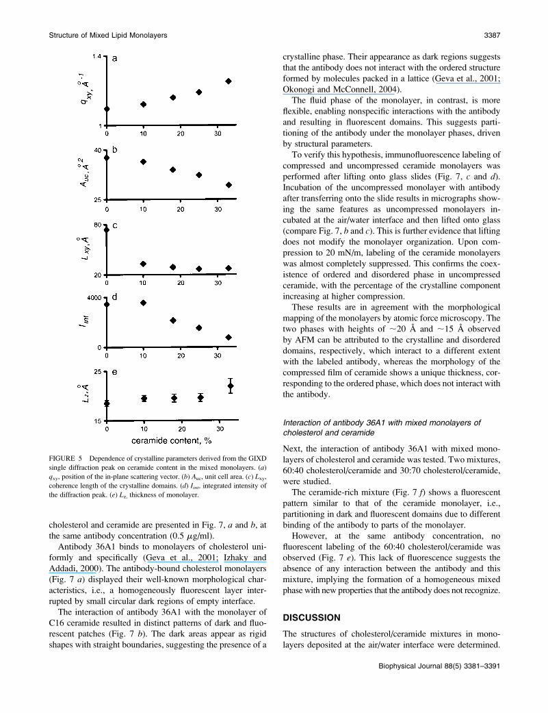
one diffraction peak (Fig. 4 a).For pure cholesterol this diffraction peak of d-spacing
5.7 A corresponds to the {1, 1}, {�22, 1}, and {1, �22} reflectionsof a supercell of dimensions a ¼ b ¼ 11.4 A (the symbol
{h,k} refers to both reflections (h,k) and (�h,�k)). In this
supercell the cholesterol molecule is aligned normal to the
water surface in a two-dimensional crystal of plane
symmetry p3 yielding an area/molecule of 38 A2 (Rapaport
et al., 2001). In this structure (Fig. 3 a) the exocyclic chainsare disordered. Introduction of increasing amounts of
ceramide has multiple effects: the peak maximum is
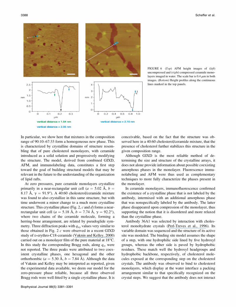
progressively shifted to higher values of qxy (Figs. 4 a and
5 a), corresponding to a shrinkage of the area per molecule in
the unit cell from 38 A2 to 30 A2 (Fig. 5 b). Since the cross-sectional area of ceramide is 40 A2, a reduction in the
average area/molecule in the mixed crystal can only be
achieved if one chain of ceramide (out of two) replaces one
molecule of cholesterol, with a concomitant change in
packing structure. The coherence length, reflecting the size
of the crystalline domains, is drastically reduced from 75 A
for pure cholesterol to 35 A for a 90:10 cholesterol/ceramide
mixture, after which it remains unchanged (Fig. 5 c). Incontrast, the integrated intensity of the diffraction peak is not
affected by the introduction of 10% ceramide in the structure,
but thereafter substantially decreases upon each further
addition (Fig. 5 d). This is understandable because in the
host-guest cholesterol/ceramide solid solution each molec-
ular site is composed, on average, of both cholesterol and
ceramide, so that substitutional and displacive disorder
decreases the diffracted intensity. The thickness of the
monolayer is little affected by the presence of ceramide as
additive (Fig. 5 e).In the range of compositions of cholesterol/ceramide of
67:33 to 30:70, the GIXD pattern of the mixtures show the
diffraction of ceramide together with a weak broad Bragg
peak corresponding to the new phase. As an example, the
diffraction pattern of a 40:60 mixture is shown in Fig. 4 b.
Atomic force microscopy of ceramide monolayers
Complementary information about the thickness and mor-
phology of the ceramide monolayers was obtained by AFM.
For the purpose of performing AFM measurements, the
monolayers were transferred onto graphite slides. Graphite
was preferred over mica because intact monolayers can be
lifted on the hydrophobic graphite by lowering the slides
parallel to the monolayer and touching the surface from the
air side. The use of mica slides tends to induce rearrangement
FIGURE 2 GIXD patterns of a monolayer of ceramide at the air/water
interface under negligible surface pressure. (a and b) The diffraction peaks
I(qxy) and corresponding Bragg rods I(qz) of pure ceramide. The three peaks,
indexed {h,k}1, belong to phase 1. The weak peak, indexed {0,2}2, belongsto phase 2. (c and d) The diffraction peaks I(qxy) and corresponding observed
(11) and calculated (—) Bragg rods I(qz) of ceramide from a 40:60
cholesterol/ceramide mixture, belonging to phase 2. The calculated curves
were determined from the monolayer crystal structure (Fig. 3, b and c).
Structure of Mixed Lipid Monolayers 3385
Biophysical Journal 88(5) 3381–3391
of the monolayers, turning them upside-down to match the
hydrophilic surface with their hydrophilic side. This gener-
ates patchy multilayer structures. In contrast, monolayers
transferred onto graphite display large monolayer regions,
alongside some multilayer regions. The latter can be easily
detected by their increased height profiles.
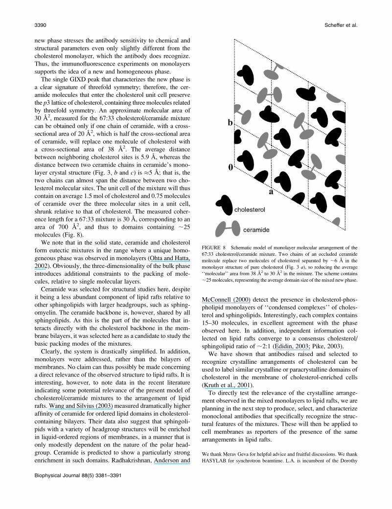
Coexistence of two types of domains is observed within
the transferred uncompressed monolayer of ceramide (Fig. 6,
left). In uncompressed monolayers, 61% of the height profile
measurements yielded a thickness of 14.7 6 0.8 A, 39%
a thickness of 20.16 0.8 A. Upon compression to 20 mN/m,
the morphology of the ceramide monolayer becomes more
homogeneous, with a predominant type of domain of thick-
ness 20.6 6 0.9 A being observed in 94% of the mea-
surements, and of thickness 15.8 6 0.2 A in only 6% of the
measurements (Fig. 6, right).
Immunofluorescence
Interaction of antibody 36A1 with monolayers of cholesteroland ceramide
Monoclonal antibody 36A1 was raised and selected against
cholesterol monohydrate crystals, and it was found to in-
teract at the air/water interface with cholesterol monolayers
(Izhaky and Addadi, 1998, 2000).
After purification by affinity chromatography, the anti-
body was fluorescently labeled by covalent binding of
a chromophore (rhodamine_B isothiocyanate). Nonfluores-
cent monolayers were incubated with the labeled antibody,
in the presence of a nonlabeled competitor in 10-fold excess
to avoid nonspecific adsorption. The competitor is a different
IgM monoclonal antibody, which does not interact specif-
ically with cholesterol-ordered arrays. Any fluorescence
observed at the air/water interface under these conditions is
due to the binding of the labeled antibody to the monolayer.
The labeled monolayer/antibody complexes were then lifted
onto glass slides and visualized under the epifluorescence
microscope. Labeled monolayers observed while still at the
air/water interface display the same features as monolayers
observed after lifting onto glass slides (Izhaky and Addadi,
1998). It is, however, difficult to record images of mono-
layers at the air/water interface because of the high back-
ground fluorescence of the labeled antibody in solution.
Furthermore, the uncompressed monolayer at the air/water
interface is very mobile, thus making good pictures even more
difficult to achieve. Representative images of uncompressed
FIGURE 4 GIXD patterns of mixed cholesterol/
ceramide monolayers. (a) Diffraction peak I(qxy) as
a function of ceramide content in the monolayer up
to 33%. For convenience, the peaks are shifted
along the vertical axis. (b) Diffraction pattern I(qxy)of the 40:60 cholesterol/ceramide mixture display-
ing the three diffraction peaks of ceramide (right),
which start to appear at compositions .33%
ceramide, together with the diffraction peak of the
cholesterol/ceramide phase (left, as in a). The
intensity scale for the diffraction peaks in a is
slightly larger than in b.
FIGURE 3 Molecular packing arrangement of (a)a monolayer of pure cholesterol (taken from Rapaport
et al., 2001); (b) a monolayer of pure ceramide viewed
along the normal to the layer plane; and (c) a monolayer
of pure ceramide viewed along the b axis. Atoms: C,
shaded; O, black; N, white. The hydrogen bond is
marked by a dotted line.
3386 Scheffer et al.
Biophysical Journal 88(5) 3381–3391
cholesterol and ceramide are presented in Fig. 7, a and b, atthe same antibody concentration (0.5 mg/ml).
Antibody 36A1 binds to monolayers of cholesterol uni-
formly and specifically (Geva et al., 2001; Izhaky and
Addadi, 2000). The antibody-bound cholesterol monolayers
(Fig. 7 a) displayed their well-known morphological char-
acteristics, i.e., a homogeneously fluorescent layer inter-
rupted by small circular dark regions of empty interface.
The interaction of antibody 36A1 with the monolayer of
C16 ceramide resulted in distinct patterns of dark and fluo-
rescent patches (Fig. 7 b). The dark areas appear as rigid
shapes with straight boundaries, suggesting the presence of a
crystalline phase. Their appearance as dark regions suggests
that the antibody does not interact with the ordered structure
formed by molecules packed in a lattice (Geva et al., 2001;
Okonogi and McConnell, 2004).
The fluid phase of the monolayer, in contrast, is more
flexible, enabling nonspecific interactions with the antibody
and resulting in fluorescent domains. This suggests parti-
tioning of the antibody under the monolayer phases, driven
by structural parameters.
To verify this hypothesis, immunofluorescence labeling of
compressed and uncompressed ceramide monolayers was
performed after lifting onto glass slides (Fig. 7, c and d).Incubation of the uncompressed monolayer with antibody
after transferring onto the slide results in micrographs show-
ing the same features as uncompressed monolayers in-
cubated at the air/water interface and then lifted onto glass
(compare Fig. 7, b and c). This is further evidence that liftingdoes not modify the monolayer organization. Upon com-
pression to 20 mN/m, labeling of the ceramide monolayers
was almost completely suppressed. This confirms the coex-
istence of ordered and disordered phase in uncompressed
ceramide, with the percentage of the crystalline component
increasing at higher compression.
These results are in agreement with the morphological
mapping of the monolayers by atomic force microscopy. The
two phases with heights of ;20 A and ;15 A observed
by AFM can be attributed to the crystalline and disordered
domains, respectively, which interact to a different extent
with the labeled antibody, whereas the morphology of the
compressed film of ceramide shows a unique thickness, cor-
responding to the ordered phase, which does not interact with
the antibody.
Interaction of antibody 36A1 with mixed monolayers ofcholesterol and ceramide
Next, the interaction of antibody 36A1 with mixed mono-
layers of cholesterol and ceramide was tested. Two mixtures,
60:40 cholesterol/ceramide and 30:70 cholesterol/ceramide,
were studied.
The ceramide-rich mixture (Fig. 7 f) shows a fluorescent
pattern similar to that of the ceramide monolayer, i.e.,
partitioning in dark and fluorescent domains due to different
binding of the antibody to parts of the monolayer.
However, at the same antibody concentration, no
fluorescent labeling of the 60:40 cholesterol/ceramide was
observed (Fig. 7 e). This lack of fluorescence suggests the
absence of any interaction between the antibody and this
mixture, implying the formation of a homogeneous mixed
phase with new properties that the antibody does not recognize.
DISCUSSION
The structures of cholesterol/ceramide mixtures in mono-
layers deposited at the air/water interface were determined.
FIGURE 5 Dependence of crystalline parameters derived from the GIXD
single diffraction peak on ceramide content in the mixed monolayers. (a)
qxy, position of the in-plane scattering vector. (b) Auc, unit cell area. (c) Lxy,coherence length of the crystalline domains. (d) Iint, integrated intensity of
the diffraction peak. (e) Lz, thickness of monolayer.
Structure of Mixed Lipid Monolayers 3387
Biophysical Journal 88(5) 3381–3391
In particular, we show here that mixtures in the composition
range of 90:10–67:33 form a homogeneous new phase. This
is characterized by crystalline domains of structure resem-
bling that of pure cholesterol monolayers, with ceramide
introduced as a solid solution and progressively modifying
the structure. The model, derived from combined GIXD,
AFM, and immunolabeling data, constitutes a first step
toward the goal of building structural models that may be
relevant in the future to the understanding of the organization
of lipid rafts.
At zero pressures, pure ceramide monolayers crystallize
primarily in a near-rectangular unit cell (a ¼ 5.02 A, b ¼8.17 A, g ¼ 91.9�). A 40:60 cholesterol/ceramide mixture
was found to also crystallize in this same structure, but with
time underwent a minor change to a much more crystalline
structure. This crystalline phase (Fig. 2, c and d) forms a near-
rectangular unit cell (a ¼ 5.18 A, b ¼ 7.74 A, g ¼ 92.2�),where two chains of the ceramide molecule, forming a
herring-bone arrangement, are related by pseudoglide sym-
metry. Three diffraction peaks with qxy values very similar to
those obtained in Fig. 2 c were observed in a recent GIXD
study of D-erythro-C18-ceramide (Vaknin and Kelley, 2000)
carried out on a monolayer film of the pure material at 18�C.In this study the corresponding Bragg rods, along qz, werenot reported. The three peaks were attributed to two coex-
istent crystalline phases, one hexagonal and the other
orthorhombic (a ¼ 5.30 A, b ¼ 7.84 A). Although the data
of Vaknin and Kelley may be interpreted as reported, given
the experimental data available, we deem our model for the
zero-pressure phase reliable, because all three observed
Bragg rods were well fitted by a single crystalline phase. It is
conceivable, based on the fact that the structure was ob-
served here in a 40:60 cholesterol/ceramide mixture, that the
presence of cholesterol further stabilizes this structure in the
given composition range.
Although GIXD is the most reliable method of de-
termining the size and structure of the crystalline arrays, it
does not alone provide information about possible coexisting
amorphous phases in the monolayer. Fluorescence immu-
nolabeling and AFM were thus used as complementary
techniques to more fully characterize the phases present in
the monolayer.
In ceramide monolayers, immunofluorescence confirmed
the existence of a crystalline phase that is not labeled by the
antibody, intermixed with an additional amorphous phase
that was nonspecifically labeled by the antibody. The latter
phase disappeared upon compression of the monolayer, thus
supporting the notion that it is disordered and more relaxed
than the crystalline phase.
Antibody 36A1 was selected by interaction with choles-
terol monohydrate crystals (Perl-Treves et al., 1996). Its
variable domain was sequenced and the structure of its active
site was modeled. The binding site model assumes the shape
of a step, with one hydrophilic side lined by five hydroxyl
groups, whereas the other side is paved by hydrophobic
residues. These match well the hydroxyl headgroups and
hydrophobic backbone, respectively, of cholesterol mole-
cules exposed at the corresponding step on the cholesterol
crystals. The antibody was observed to bind to cholesterol
monolayers, which display at the water interface a packing
arrangement similar to that specifically recognized on the
crystal steps. We suggest that the antibody does not interact
FIGURE 6 (Top) AFM height images of (left)
uncompressed and (right) compressed ceramide mono-
layers imaged in water. The scale bar is 0.4 mm in both
images. (Bottom) Height profiles along the continuous
lines marked in the top panels.
3388 Scheffer et al.
Biophysical Journal 88(5) 3381–3391

with the crystalline part of the monolayers of ceramide,
because their packing is different from that of the specifically
recognized cholesterol motif. The interaction with the amor-
phous phase is attributed to interaction with the hydroxyl and
aliphatic groups of ceramide, made possible by the ‘‘loosely
packed disordered’’ phase. The same labeling pattern of ex-
clusion from crystalline domains and labeling of disordered
domains was observed with triacontanol, a long-chain ali-
phatic alcohol also forming monolayers comprising a crys-
talline and a disordered phase (Geva et al., 2001).
We note that the loosely packed character of the amor-
phous phase is further confirmed in the GIXD analysis of our
data and that of Vaknin and Kelley. At the area/molecule
derived for the crystalline phase from the diffraction data
(�40 A2), the monolayer surface pressure would be 18 mN/m
(Fig. 1). The surface pressure being zero, a higher average
molecular area must be assumed, indicating the presence of
an additional expanded phase.
AFM studies confirmed that both the crystalline phase and
the disordered phase are single molecular layers. The un-
compressed monolayer is formed of two types of domains,
characterized by a thickness of ;20 and ;15 A. The 20-A
thickness matches the length of a C16 ceramide molecule
(sphingosine base linked through an amide to a palmitic
acid) oriented vertical to the surface, indicating that this is
the crystalline phase monitored by GIXD and not labeled
by the antibody. The 15-A high domains indicate that the
molecules are highly tilted relative to the normal of the
surface, in a poorly packed arrangement that corresponds to
the disordered phase labeled by the antibody. After com-
pressing the monolayer, the more ordered domains become
prevalent, confirming once more that the 20-A thick phase of
the ceramide monolayer is the crystalline one.
Although we are well aware of the differences that may be
induced in the monolayer organization by lifting onto a solid
support, we are convinced, to the best of our knowledge, that
these differences do not influence our conclusions, insofar as
artifacts due to lifting are easily detectable both in AFM and
by immunofluorescence. Furthermore, monolayers incu-
bated with antibody before and after lifting have the same
features, indicating that the epitope of antibody binding did
not change, either in molecular organization or in availability
to binding.
Addition of small amounts of cholesterol to ceramide did
not induce changes in the GIXD pattern of these mixtures up
to a 30:70 ratio of cholesterol/ceramide. Since the crystalline
phase of pure ceramide is highly ordered, and cholesterol is
a rigid molecule that cannot easily adapt to the packing of
ceramide, we tend to believe that cholesterol does not enter
the ceramide structure. Cholesterol should thus be rather pres-
ent in a dispersed form that does not contribute to the dif-
fraction pattern. In contrast, mixed monolayers of cholesterol/
ceramide in the composition range of 90:10–67:33 form a
new unique phase, represented in GIXD by a broad diffrac-
tion peak. By the same logic as above, ceramide, being
flexible, can accommodate within the less well-ordered phase
of cholesterol. Upon progressive addition of ceramide to
cholesterol, the maximum of the peak corresponding to pure
cholesterol shifts to higher values of qxy, the peak broadens,
and its intensity decreases, corresponding to a decrease in the
size of the crystalline domains and in their crystallinity. Such
a decrease in intensity and structure can be due either to a
destructive x-ray interference from the two components or to
formation of an amorphous phase.
We checked for the possible existence of such putative
amorphous regions by immunofluorescence experiments
carried out on mixed monolayers. The fluorescent images
show different patterns, depending on the ratio between the
two lipid components. At a cholesterol/ceramide ratio of
30:70, the labeled monolayer shows distinct dark and
fluorescent regions, illustrating a lipid demixing (Fig. 7 f).Since ceramide is in excess, we attribute the fluorescence to
ceramide-rich disordered domains that form a separate phase.
In contrast with this, and in agreement with the GIXD data,
no fluorescent labeling of the 60:40 mixture of cholesterol/
ceramide was observed (Fig. 7 e). This sort of ‘‘negative
labeling’’ indicates the lack of interaction between the anti-
body and the monolayer, together with the absence of any
amorphous phase of ceramide that would have been fluo-
rescent. The lack of interaction between the antibody and the
FIGURE 7 Epifluorescence micrographs of antibody-bound monolayers of
(a) uncompressed cholesterol; (b) uncompressed ceramide; (c) uncompressed
ceramide; (d) compressed ceramide; (e) 60:40 cholesterol/ceramide; and (f)
30:70 cholesterol/ceramide . The monolayers in a, b, e, and f were incubated
with the antibody in the trough and the monolayers in c and d were incubated
with the antibody after lifting onto a glass slide. The concentration of the anti-
body in a, b, e, and f is 0.5mg/mL and in c and d is 1mg/mL. Scale bar, 40mm.
Structure of Mixed Lipid Monolayers 3389
Biophysical Journal 88(5) 3381–3391
new phase stresses the antibody sensitivity to chemical and
structural parameters even only slightly different from the
cholesterol monolayer, which the antibody does recognize.
Thus, the immunofluorescence experiments on monolayers
supports the idea of a new and homogeneous phase.
The single GIXD peak that characterizes the new phase is
a clear signature of threefold symmetry; therefore, the cer-
amide molecules that enter the cholesterol unit cell preserve
the p3 lattice of cholesterol, containing three molecules related
by threefold symmetry. An approximate molecular area of
30 A2, measured for the 67:33 cholesterol/ceramide mixture
can be obtained only if one chain of ceramide, with a cross-
sectional area of 20 A2, which is half the cross-sectional area
of ceramide, will replace one molecule of cholesterol with
a cross-sectional area of 38 A2. The average distance
between neighboring cholesterol sites is 5.9 A, whereas the
distance between two ceramide chains in ceramide’s mono-
layer crystal structure (Fig. 3, b and c) is �5 A; that is, the
two chains can almost span the distance between two cho-
lesterol molecular sites. The unit cell of the mixture will thus
contain on average 1.5 mol of cholesterol and 0.75 molecules
of ceramide over the three molecular sites in a unit cell,
shrunk relative to that of cholesterol. The measured coher-
ence length for a 67:33 mixture is 30 A, corresponding to an
area of 700 A2, and thus to domains containing ;25
molecules (Fig. 8).
We note that in the solid state, ceramide and cholesterol
form eutectic mixtures in the range where a unique homo-
geneous phase was observed in monolayers (Ohta and Hatta,
2002). Obviously, the three-dimensionality of the bulk phase
introduces additional constraints to the packing of mole-
cules, relative to single molecular layers.
Ceramide was selected for structural studies here, despite
it being a less abundant component of lipid rafts relative to
other sphingolipids with larger headgroups, such as sphing-
omyelin. The ceramide backbone is, however, shared by all
sphingolipids. As this is the part of the molecules that in-
teracts directly with the cholesterol backbone in the mem-
brane bilayers, it was selected here as a candidate to study the
basic packing modes of the mixtures.
Clearly, the system is drastically simplified. In addition,
monolayers were addressed, rather than the bilayers of
membranes. No claim can thus possibly be made concerning
a direct relevance of the observed structure to lipid rafts. It is
interesting, however, to note data in the recent literature
indicating some potential relevance of the present model of
cholesterol/ceramide mixtures to the arrangement of lipid
rafts. Wang and Silvius (2003) measured dramatically higher
affinity of ceramide for ordered lipid domains in cholesterol-
containing bilayers. Their data also suggest that sphingoli-
pids with a variety of headgroup structures will be enriched
in liquid-ordered regions of membranes, in a manner that is
only modestly dependent on the nature of the polar head-
group. Ceramide is predicted to show a particularly strong
enrichment in such domains. Radhakrishnan, Anderson and
McConnell (2000) detect the presence in cholesterol-phos-
pholipid monolayers of ‘‘condensed complexes’’ of choles-
terol and sphingolipids. Interestingly, each complex contains
15–30 molecules, in excellent agreement with the phase
observed here. In addition, independent information col-
lected on lipid rafts converge to a consensus cholesterol/
sphingolipid ratio of ;2:1 (Edidin, 2003; Pike, 2003).
We have shown that antibodies raised and selected to
recognize crystalline arrangements of cholesterol can be
used to label similar crystalline or paracrystalline domains of
cholesterol in the membrane of cholesterol-enriched cells
(Kruth et al., 2001).
To directly test the relevance of the crystalline arrange-
ment observed in the mixed monolayers to lipid rafts, we are
planning in the next step to produce, select, and characterize
monoclonal antibodies that specifically recognize the struc-
tural features of the mixtures. These will then be applied to
cell membranes as reporters of the presence of the same
arrangements in lipid rafts.
We thank Merav Geva for helpful advice and fruitful discussions. We thank
HASYLAB for synchrotron beamtime. L.A. is incumbent of the Dorothy
FIGURE 8 Schematic model of monolayer molecular arrangement of the
67:33 cholesterol/ceramide mixture. Two chains of an occluded ceramide
molecule replace two molecules of cholesterol separated by ;6 A in the
monolayer structure of pure cholesterol (Fig. 3 a), so reducing the average
‘‘molecular’’ area from 38 A2 to 30 A2 in the mixture. The scheme contains
;25molecules, representing the average domain size of the mixed new phase.
3390 Scheffer et al.
Biophysical Journal 88(5) 3381–3391

and Patrick Gorman professorial chair, and L.S. is the recipient of the
Eshkol Fellowship, administered by the Israel Ministry of Science.
This work was supported by a grant from the Israel Science Foundation,
administered by the Israel Academy of Sciences, the Kimmelmann Center,
and the DanSync program of the Danish Natural Science Research Council
and the European Community under TMR-Contract ERBFMGECT950059.
REFERENCES
Addadi, L., M. Geva, and H. S. Kruth. 2003. Structural information aboutorganized cholesterol domains from specific antibody recognition. Bio-chim. Biophys. Acta. 1610:208–216.
Als-Nielsen, J., and K. Kjaer. 1989. X-ray reflectivity and diffractionstudies of liquid surfaces and surfactant monolayers. In Phase Transitionsin Soft Condensed Matter. T. Riste and D. Sherrington, editors. PlenumPress, New York. 113–138.
Brown, D. A., and E. London. 1997. Structure of detergent-resistantmembrane domains: does phase separation occur in biological mem-branes? Biochem. Biophys. Res. Commun. 240:1–7.
Brown, D. A., and J. K. Rose. 1992. Sorting of GPI-anchored proteins toglycolipid-enriched membrane subdomains during transport to the apicalcell surface. Cell. 68:533–544.
Edidin, M. 2003. The state of lipid rafts: from model membranes to cells.Annu. Rev. Biophys. Biomol. Struct. 32:257–283.
Friedrichson, T., and T. V. Kurzchalia. 1998. Microdomains of GPI-anchored proteins in living cells revealed by crosslinking. Nature. 394:802–805.
Geva, M., F. Frolow, M. Eisenstein, and L. Addadi. 2003. Antibodyrecognition of chiral surfaces. Enantiomorphous crystals of leucine-leucine-tyrosine. J. Am. Chem. Soc. 125:696–704.
Geva, M., D. Izhaky, D. E. Mickus, S. D. Rychnovsky, and L. Addadi. 2001.Stereoselective recognition of monolayers of cholesterol, ent-cholesterol,and epicholesterol by an antibody. ChemBioChem. 2:265–271.
Giocondi, M. C., V. Vie, E. Lesniewska, J. P. Goudonnet, and C. LeGrimellec. 2000. In situ imaging of detergent-resistant membranes byatomic force microscopy. J. Struct. Biol. 131:38–43.
Huang, J., and G. W. Feigenson. 1999. A microscopic interaction model ofmaximum solubility of cholesterol in lipid bilayers. Biophys. J. 76:2142–2157.
Izhaky, D., and L. Addadi. 1998. Pattern recognition by antibodies for two-dimensional arrays of molecules. Adv. Mater. 10:1009–1014.
Izhaky, D., and L. Addadi. 2000. Stereoselective interactions of aspecialized antibody with cholesterol and epicholesterol monolayers.Chemistry. 6:869–874.
Jensen, T. R., and K. Kjaer. 2001. Structural properties and interactions ofthin films at the air-liquid interface explored by synchrotron X-rayscattering. In Studies in Interface Science, Vol. 11. Novel Methods toStudy Interfacial Layers. D. Moebius and R. Miller, editors. Elsevier,Amsterdam. 205–254.
Kessler, N., D. Perl-Treves, and L. Addadi. 1996. Monoclonal antibodies thatspecifically recognize crystals of dinitrobenzene. FASEB J. 10:1435–1442.
Kessler, N., D. Perl-Treves, L. Addadi, and M. Eisenstein. 1999. Structuraland chemical complementarity between antibodies and the crystalsurfaces they recognize. Proteins. 34:383–394.
Kjaer, K. 1994. Some simple ideas on x-ray reflection and grazing-incidence diffraction from thin surfactant films. Physica B. 198:100–109.
Kruth, H. S., I. Ifrim, J. Chang, L. Addadi, D. Perl-Treves, and W. Y.Zhang. 2001. Monoclonal antibody detection of plasma membranecholesterol microdomains responsive to cholesterol trafficking. J. LipidRes. 42:1492–1500.
Kuzmenko, I., V. M. Kaganer, and L. Leiserowitz. 1998. Packing ofhydrocarbon chains and symmetry of condensed phases in Langmuirmonolayers. Langmuir. 14:3882–3888.
Kuzmenko, I., H. Rapaport, K. Kjaer, J. Als-Nielsen, I. Weissbuch, M.Lahav, and L. Leiserowitz. 2001. Design and characterization ofcrystalline thin film architectures at the air-liquid interface: simplicityto complexity. Chem. Rev. 101:1659–1696.
Majewski, J., T. L. Kuhl, K. Kjaer, and G. S. Smith. 2001. Packing ofganglioside-phospholipid monolayers: an x-ray diffraction and reflectiv-ity study. Biophys. J. 81:2707–2715.
Ohta, N., and I. Hatta. 2002. Interaction among molecules in mixtures ofceramide/stearic acid, ceramide/cholesterol and ceramide/stearic acid/cholesterol. Chem. Phys. Lipids. 115:93–105.
Okonogi, T. M., and H. M. McConnell. 2004. Contrast inversion in theepifluorescence of cholesterol-phospholipid monolayers. Biophys. J. 86:880–890.
Pascher, I., and S. Sundell. 1977. Molecular arrangements in sphingolipids:the crystal structure of cerebroside. Chem. Phys. Lipids. 20:175–191.
Perl-Treves, D., N. Kessler, D. Izhaky, and L. Addadi. 1996. Monoclonalantibody recognition of cholesterol monohydrate crystal faces. Chem.Biol. 3:567–577.
Pike, L. J. 2003. Lipid rafts: bringing order to chaos. J. Lipid Res. 44:655–667.
Pralle, A., P. Keller, E. L. Florin, K. Simons, and J. K. Horber. 2000.Sphingolipid-cholesterol rafts diffuse as small entities in the plasmamembrane of mammalian cells. J. Cell Biol. 148:997–1008.
Radhakrishnan, A., T. G. Anderson, and H. M. McConnell. 2000.Condensed complexes, rafts, and the chemical activity of cholesterol inmembranes. Proc. Natl. Acad. Sci. USA. 97:12422–12427.
Ramstedt, B., and J. P. Slotte. 2002. Membrane properties of sphingomye-lins. FEBS Lett. 531:33–37.
Rapaport, H., I. Kuzmenko, S. Lafont, K. Kjaer, P. B. Howes, J. Als-Nielsen, M. Lahav, and L. Leiserowitz. 2001. Cholesterol monohydratenucleation in ultrathin films on water. Biophys. J. 81:2729–2736.
Schneider, P. B., and E. P. Kennedy. 1967. Sphingomyelinase in normalhuman spleens and in spleens from subjects with Niemann-Pick disease.J. Lipid Res. 8:202–209.
Simons, K., and I. Ikonen. 1997. Functional rafts in cell membranes.Nature. 387:569–572.
Simons, K., and D. Toomre. 2000. Lipid rafts and signal transduction. Nat.Rev. Mol. Cell Biol. 1:31–39.
Singer, S. J., and G. L. Nicolson. 1972. The fluid mosaic model of thestructure of cell membranes. Science. 175:720–731.
Smart, E. J., G. A. Graf, M. A. McNiven, W. C. Sessa, J. A. Engelman,P. E. Scherer, T. Okamoto, and M. P. Lisanti. 1999. Caveolins, liquid-ordered domains, and signal transduction. Mol. Cell. Biol. 19:7289–7304.
Vaknin, D., and M. S. Kelley. 2000. The structure of D-erythro-C18ceramide at the air-water interface. Biophys. J. 79:2616–2623.
Varma, R., and S. Mayor. 1998. GPI-anchored proteins are organized insubmicron domains at the cell surface. Nature. 394:798–801.
Wang, T. Y., and J. R. Silvius. 2003. Sphingolipid partitioning into ordereddomains in cholesterol-free and cholesterol-containing lipid bilayers.Biophys. J. 84:367–378.
Xu, X., R. Bittman, G. Duportail, D. Heissler, C. Vilcheze, and E. London.2001. Effect of the structure of natural sterols and sphingolipids on theformation of ordered sphingolipid/sterol domains (rafts). Comparison ofcholesterol to plant, fungal, and disease-associated sterols and compar-ison of sphingomyelin, cerebrosides, and ceramide. J. Biol. Chem. 276:33540–33546.
Structure of Mixed Lipid Monolayers 3391
Biophysical Journal 88(5) 3381–3391
Copyright © 2022 FDOKUMEN