Disruption of Circadian Rhythm Genes in Obstructive Sleep ...

Published: September 30, 2011
r 2011 American Chemical Society 13918 dx.doi.org/10.1021/la202970g | Langmuir 2011, 27, 13918–13924
ARTICLE
pubs.acs.org/Langmuir
Melittin Adsorption and Lipid Monolayer Disruption at Liquid�LiquidInterfacesManuel A. M�endez,†,|| Zahra Nazemi,†,‡,|| Ibrahim Uyanik,§ Yu Lu,† and Hubert H. Girault*,†
†Laboratoire d'Electrochimie Physique et Analytique, Ecole Polytechnique F�ed�erale de Lausanne (EPFL), Station 6,CH-1015 Lausanne, Switzerland‡Chemistry Department, University of Isfahan, Isfahan 81746-73441, Iran§Department of Chemistry, Faculty of Science, Selcuk University, Campus, 42075 Konya, Turkey
bS Supporting Information
’ INTRODUCTION
Antimicrobial peptides are of crucial interest given their widepresence in nature. They constitute a powerful defense systemagainst external potentially harmful agents such as bacteria andviruses. The challenge of bacterial resistance to conventionalantibiotics,1 the antibacterial selectivity of many peptides, andtheir unique mode of action, have made such peptides promisingcandidates for the development of a new class of antibiotics.2
However, additional efforts are required for rationalizing theirmolecular interactions with lipidic membranes and their actionmechanisms.
Among the antimicrobial peptide, melittin is certainly one ofthe most widely studied. This peptide is the principal toxiccomponent of European honey bee venom3 and is a cationichemolytic peptide composed of 26 amino acid residues whoseamphiphilic nature makes it capable of associating with eithernatural or artificial lipid membranes.4 This pore-forming ability5
turns it into a suitable model peptide for monitoring lipid�protein interactions. Some of the analytical approaches used upto now to monitor this type of system include fluorescence,6
circular dichroism,7 surface plasmon resonance (SPR),8 nuclearmagnetic resonance (NMR),9 and electrochemical impedancespectroscopy.10
However, probing interfacial interactions at cell membranes isnot always an easy task given their complexity. Therefore, in thesearch of simpler but still realistic systems able to mimic the
properties of cellular membranes, vesicle suspensions and im-mobilized bilayers or monolayers have been proposed. None-theless, the potential difference across the layers cannot becontrolled or is neglected in many cases. Another interestingapproach is based on adsorbed monolayers at polarized liquid�liquid interfaces, where the control of the interfacial polarizationis an important experimental variable and is accurately con-trolled. Adsorbed phospholipid monolayers at the ITIES werepioneered by Koryta et al.11 followed by interfacial tensionmeasurements12 that clearly demonstrated the pH and potentialdependence of these adsorption processes.
Since then, phospholipid monolayers adsorbed at liquid�liquid interfaces formed between two immiscible electrolytesolutions (ITIES) have been used to mimic the behavior of lipidbio-membranes,12 especially to investigate the stability13 and ionpermeability14 of themonolayers as a function of the well-definedinterfacial potential difference. In parallel, less than two decadesago, the use of aqueous (organic) droplets supported on anelectrode and immersed into an organic (aqueous) electrolytesolution to study electron transfer reactions coupled to iontransfer reactions was proposed.15 In these three-phase junctions(metal�water�oil), one of the interfaces is made not polarizable
Received: July 29, 2011Revised: September 21, 2011
ABSTRACT: Melittin, a membrane-active peptide with anti-microbial activity, was investigated at the interface formedbetween two immiscible electrolyte solutions (ITIES) sup-ported on a metallic electrode. Ion-transfer voltammetryshowed well-defined semi-reversible transfer peaks along withadsorptive peaks. The reversible adsorption of melittin at theliquid�liquid interface is qualitatively discussed from voltam-metric data and experimentally confirmed by real-time imageanalysis of video snapshots. It is also demonstrated thatpolarization of the water/1,2-DCE interface results in drasticdrop shape variations caused by large variations of the interfacialtension. The experimental data also confirmed that maximumadsorption occurs near the ion transfer potential. Finally, the interaction of melittin with a monolayer of L-α-dipalmitoylphosphatidylcholine (DPPC) was also investigated showing that melittin destabilizes the lipidic monolayer facilitating itsdesorption. The non-covalent complex formation between melittin and DPPC was confirmed by mass spectrometry.
13919 dx.doi.org/10.1021/la202970g |Langmuir 2011, 27, 13918–13924
Langmuir ARTICLE
to study charge transfer reactions at the other interface. Forexample, for an aqueous droplet on a platinum electrodeimmersed in an organic electrolyte solution, the presence of anaqueous redox couple in excess in the droplet fixes the potentialdifference at the metal�water interface according to the Nernstequation for this couple, while the liquid�liquid (water�oil)interface is polarizable. Thus, a common three-electrode electro-chemical setup can be used to study charge transfer reactions atthe interface between two immiscible electrolyte solutions(ITIES).15a Additionally, the volume of one of the two phasescan be dramatically reduced using this system. The latter is a keyadvantage for the study of biologically active molecules, whichare very often available in very limited amounts.
Besides, the three-phase junction geometry has received also alot of attention not only for studying ion transfer reactions16 butalso for electro-wetting applications.17 In particular, Monroeet al. have considered the case in which ion-impermeableliquid�liquid interfaces are polarized18 to vary the contact angleof the droplet and have shown that relatively small appliedvoltages can induce very large drop shape variation. With videoimage digitizers, the shape of the droplet can be analyzed toestimate changes in the interfacial tension, and the three-phasejunctions provide an easy way to accomplish this for the polarizedITIES.12,19
Herein, we study the interaction between melittin with L-α-dipalmitoylphosphatidylcholine (DPPC) monolayers adsorbedat the ITIES under controlled interfacial polarization using athree-phase junction in which the amount of peptide required isgreatly reduced. Additionally, real-time videos were recordedduring the electrochemical measurements to monitor the contactangle along with voltammetric signals to corroborate not only theadsorption of the peptide but also the potential region where ittakes place.
Finally, to prove the existence of the complex formationbetween melittin and DPPC at the water�DCE interface,biphasic electrospray ionization (BESI)20 mass spectrometrywas employed.
’EXPERIMENTAL SECTION
Chemicals. Melittin 95% (Mel) was purchased from Serva electro-phoresis. The purity of the peptide was confirmed by UV visible,fluorescence, and mass spectrometry and was used without any furtherpurification. L-α-Dodecylphosphatydilcholine (DPPC) (M = 734.0 g/mol) and acetic acid (AcOH) were from Sigma (St. Louis, MO).FeSO4 and Fe2(SO4)3 salts were from Merck. DCE was purchasedfrom AppliChem (GmbH, Germany), and deionized water (18.2MΩ cm�1) was prepared using a Milli-Q system from Millipore(Bedford, MA). For the electrochemical measurements, the organicsalt used was bis(triphenylphosphoranylidene) ammonium tetrakis-(pentafluorophenyl) borate (BA+TB�). This salt was obtained bymetathesis of bis(triphenylphosphoranylidene) ammonium chloridewith lithium tetrakis(pentafluorophenyl)borate (all bought from Fluka).Purification was accomplished by recrystallization in acetone. All of theother employed compounds were of analytical grade and used asreceived, unless otherwise stated.Electrochemical Measurements. Voltammetric measurements
were carried out using a potentiostat (Autolab-PGSTAT 30, Metrohm)and a glass cubic cell. The cell was first cleaned by chrom-sulfuric acidand rinsed thoroughly with ultrapure water. The iR potential drop wascompensated using a positive-feedback method. A Pt disk electrode(Metrohm CH, Area = 0.06 ( 0.01 cm2) was freshly polished sequen-tially with 1.0, 0.3, and 0.05 μm alumina and sonicated for 5 min in
water/acetonitrile (50/50) mixture. A 10 μL drop of an aqueoussolution containing an equimolar mixture of Fe2+/Fe3+ ion pair wasdeposited using a micropipet onto the platinum disk electrode. Theelectrochemical cell composition can be represented as shown inScheme 1, where x stands for the concentration of peptide. All ofthe potentials for ion transfer are referred to the half-wave potential oftetraethylammonium (TEA+). Thus, at the end of each experiment, apredetermined amount of a solution in 1,2-DCE of TEA+TB� is addedto the organic phase in the cell for determining the experimental TEA+
half-wave potential. The value of the standard transfer potential of TEA+
has been taken equal to 18 mV.21 All of the measurements were carriedout at room temperature of 22( 1 �C. The pH of the aqueous solutionwas maintained at a constant value of 3, ensuring the stability of theredox pair in the aqueous solution as well as that of the peptide. Parallelexperiments demonstrated that equivalent results could be obtained ateither pH 3 or pH 7. This is in accordance with the predicted titrationcurve of melittin (Figure S1), which reveals that the charge remainsconstant within a pH interval comprehended between approximately2.5 and 8.Contact Angle Determination. Videos of the droplet during the
voltammetric measurement were obtained with a hand-held microscope(Proscope HR, Bodelyn Technologies, U.S.). Video manipulation wascarried out with Mathematica 7.0 (Wolfram Research) to calculate thecontact angle at the four-phase boundary.
For analyzing the images, oblate spheroid geometry was assumed forthe droplet. The general equation representing it corresponds to:
x2 þ y2
a2þ z2
c2¼ 1 ð1Þ
where a is the equatorial radius and c is the vertical radius. At y = 0, theequation for the spheroid can be reduced to that of a simple ellipse, andtherefore the area and the volume of the truncated spheroid can becalculated as follows:
A ¼ 2πZ z0
�cgðzÞ
ffiffiffiffiffiffiffiffiffiffiffiffiffiffiffiffiffiffiffiffiffiffi1 þ d2gðzÞ
dz2
sdz ð2Þ
V ¼ πZ z0
�cgðzÞ dz ð3Þ
Scheme 1. Electrochemical Cell Employed
Scheme 2. Illustration of an Aqueous Droplet Supported ona Platinum Electrode Immersed in DCE

13920 dx.doi.org/10.1021/la202970g |Langmuir 2011, 27, 13918–13924
Langmuir ARTICLE
where z0 is the vertical distance from the center at which the spheroid istruncated (see Scheme 2). The function g(z) reads:
gðzÞ ¼ a
ffiffiffiffiffiffiffiffiffiffiffiffiffiffiffiffiffiffiffi1� z
c
� �2s
ð4Þ
Moreover, if one considers that the platinum and Teflon surfacesremain virtually unchanged upon polarization, the force balance at theboundary region Pt�Teflon�water�DCE is related to the contactangle, θ, as follows:
γPt=w ¼ γTef=DCE þ γw=DCE cos θ ð5Þwhich can also be expressed as:
secðθÞ ¼ γw=DCEγPt=w � γTef=DCE
≈γw=DCE
kð6Þ
where the difference between γPt/w and γTef/DCE is assumed to remainnearly constant at a value k, which is in principle independent of theexternally applied potential during the time scale of voltammetricmeasurements. Therefore, the interfacial tension between water andDCE is inversely proportional to the cosine of the contact angle.However, it must be stressed that this approach is not a methodologyfor measuring interfacial tension. Even if γPt/w and γTef/DCE remainconstant in the course of a single experiment, their values mightdrastically change if the composition of the cell is modified, for example,when adding different species or when passing large currents across theinterface. Yet, this approach will be shown to be of great value tomonitoradsorption/desorption processes during linear scan voltammetry. As aresult, if a, c, and z0 are graphically determined from video snapshotstaken during a voltammetric measurement, we can measure the contactangle using:
secðθÞ ¼ � sec arctandgðzÞdz
�����z¼ z0
1A
0@
1A ¼
ffiffiffiffiffiffiffiffiffiffiffiffiffiffiffiffiffiffiffiffiffiffiffiffiffiffiffiffiffiffiffi1 þ a2z20
c2ðc2 � z20Þ
s0@
ð7ÞFor the image analysis, all of the snapshots are converted to grayscale,
and the droplet contour is determined using a program written inMathematica 7.0. Finally, the fitting quality can also be evaluated if onetakes into account the following constraints:(1) The volume of the droplet is constant; that is to say, it is
independent of the potential.(2) The transversal area of the droplet in contact with the electrode
is constant and corresponds to the area of the platinum electrodegiven that the aqueous phase volume was always large enough asto ensure the complete coverage of the metallic surface.
MS Setup andMicrospray Interface. An LTQ VELOS ion trapmass spectrometer (Thermo Fisher Scientific, San Jose, CA) was used inpositive ionization mode. The inlet capillary was kept at 100 �C. Thecommercial electrospray interface was removed and replaced by amicrospray interface consisting of a double microchannel (20 μm �50 μm � 1 cm) polyimide microchip developed by DiagnoSwiss S.A.(Monthey, Switzerland) and fixed in a holder mounted on the probeslide adapter of the mass spectrometer. Further details have beenpreviously described.20b The syringe pump (KdScientific, Holliston,MA) was used to provide constant flow rate of each solution through themicrochannels (2 μL/min for each line, i.e., a total flow rate of 4 μL/min). Two immiscible liquids are infused and put in contact only at theTaylor cone. The high voltage (U = 3.5 kV) was applied to the stainless-steel needle of the syringe containing the aqueous solution. The currentof spray was set between 30 and 80 nA by adjusting the distance betweenthe microspray outlet and the entrance of the MS and was monitored bya nanoammeter.
’RESULTS AND DISCUSSION
Voltammetric Behavior of Melittin. Figure 1a shows a cyclicion transfer voltammogram of an aqueous droplet containingmelittin 80 μM.Despite the asymmetric distribution of polar andnonpolar residues and the fact that like other membrane-bindingpeptides and proteins, melittin is predominantly hydrophobicand adopts anα-helical conformation (see Figure S1),22,23 a clearion transfer process for melittin can be identified. At the sametime, evident deformation of the aqueous droplet could also beobserved at potentials close to the ion transfer potential(Figure 1b). The half-wave potential corresponds to 0.28 V, thatis, a free Gibbs energy of transfer of 135 kJ mol�1. The peak-to-peak separation extrapolated to zero scan rate was determined tobe 17 mV, which differs from the 12 mV peak separationexpected for an ion carrying five positive charges, according tothe calculated charge versus pH profile in Figure S1.Nonetheless, it has to be stressed that accurate peak separation
determination from cyclic voltammetry data can be very difficultfor heavily charged species. In fact, the accuracy with which thepeak separation potential is limited to ca. 5 mV. In addition tothis, the charge of the peptide might differ from that theoreticallypredicted because of ion-pair formation with ions of the support-ing electrolyte or within the same molecule.24 Indeed, intramo-lecular ion-pairing could take place at the interface or in theorganic phase between the two opposite charges K-20 and G-26.Taking this into account, the ion transfer signal formelittin can
be categorized as quasi-reversible, in accordance with the size ofthis peptide and the lowering effect that this parameter plays onthe ion transfer kinetic constant.25
Further experiments in the presence of the peptide showedsmall variations of the peak potential upon variation of both scanrate and concentration (Figure 2). However, linear relationshipsare always obtained between the peak currents and the squareroot of the scan rate (inset Figure 2a) and the concentration(inset Figure 2b). Additionally, a small signal, but significantenough to be seen, located at approximately 0.4 V is observed(Figures 1a and 2). This signal is present in both forward andbackward scans and seems to be related to the adsorption processof melittin at the water�DCE interface. Nonetheless, given thelow magnitude of this peak as well as its close proximity to themelittin diffusion-controlled wave and the end of the potential
Figure 1. (a) Cyclic voltammogram obtained at the interface formedbetween DCE containing BA+TB� 10 mM and an aqueous dropletsupported on a Pt disk electrode in the absence (dashed line) and in thepresence (solid line) of melittin 80 μM. Scan rate = 10 mV s�1. In (b)and (c), video snapshots at different galvanic potentials of an aqueousdroplet containing melittin (x = 20 μM) and DCE in the absence ofDPPC are presented.

13921 dx.doi.org/10.1021/la202970g |Langmuir 2011, 27, 13918–13924
Langmuir ARTICLE
window, it is then difficult to judge about the real nature of thispeak by relying only on voltammetric data.It can be then concluded that the ion-transfer of melittin
corresponds to a quasi-reversible and diffusion-controlled pro-cess slightly distorted because of the adsorption of the aqueouspeptide. Nonetheless, to better describe the adsorption process,real-time video snapshots were acquired simultaneously with thecyclic voltammogram (see Figure S2). As mentioned above,deformation of the shape of aqueous droplet was observed duringthe voltammetric measurements. This phenomenon can berationalized in terms of an adsorption process in which theenergy per unit of area will be reduced. Accordingly, this inducesan increase in the area of the drop given that the volume of thedroplet is fixed. Equally, the angle θ at the four-phase junctionshown in Scheme 2 is considerably modified. Video analysis wasused to monitor this angle variation, and the results are plotted inFigure 3. At negative potentials, the droplet is rather spherical,and the surface tension is high. When scanning to positivepotentials, the contact angle value decreases as the aqueousdroplet becomes gradually flatter, leading to a minimum in thecontact angle profile as shown in Figure 3, which is associatedwith the adsorption of melittin. This minimum lies close tothe standard ion transfer potential (herein assumed to be
approximately equal to the half-wave potential).26 Upon furtherscanning to positive potentials, melittin desorbs and the droprecovers its quasi-spherical shape. Increasing the concentrationof the peptide was also observed to have a marked influence onthis process; however, the present video technique is too coarseto measure the adsorption isotherm. Nevertheless, it can be seenthat at higher concentrations the angle tends to be reduced to alarger extent. However, if the concentration is raised beyond acertain threshold (ca. 100 μM), the variation of the angleminimum becomes independent of the concentration (data notshown), meaning that saturation of the liquid�liquid interfacewith the peptide takes place above this concentration value.Additionally, video analysis of the forward and backward scansshowed that the adsorption process can be assumed as reversiblein the time scale of voltammetric experiments. Indeed, angleversus potential curves presented in Figure S3 do not presenthysteresis upon reversing the scan direction. Additionally, re-peated cycles did not show any sign of modification of theinterface. It must be stressed that very commonly ionic adsorp-tion at liquid�liquid interfaces is accompanied by the formationof aggregates or ion-pairs, whichmodify in an irreversible mannerthe interfacial region.27 In stark contrast, melittin, besides beingable to be transferred across the interface, also adsorbs in areversible manner at the interface.By analogy with voltammetry for redox reactions at solid
electrodes where the oxidized or reduced species can be adsorbedand where post-peaks correspond to the adsorption of thereactant of the redox reaction and pre-peaks correspond to theadsorption of the product of this reaction, post-peaks (pre-peaks) in the ion transfer voltammogram correspond to theadsorption of the transferring (transferred) ions as recentlydiscussed.26b Clear evidence of ion transfer accompanied byadsorption of the transferring ion has also been previouslydocumented.28 However, no clear experimental confirmationfor the existence of well-defined adsorptive pre- or post-peaks hasbeen reported so far for polypeptides and in particular forantimicrobial peptides.In Figure 1, the post-peak on the scan to positive potentials can
be attributed to the transfer by desorption of adsorbed Mel(ads)that requires more energy to transfer than the bulk speciesMel(w), according to the θ profile as a function of potential inFigure 3. Thus, combining the contact angle measurements with
Figure 2. (a) Ion-transfer voltammograms obtained at the interface formed between DCE in the absence of DPPC and an aqueous solution of (a)melittin (x = 80 μM) and DCE in the absence of DPPC at scan rates of 5, 10, 20, 30, 40, 50, 60, 70, 80, 90, and 100 mV s�1. In (b), the scan rate was keptconstant at 10mV s�1 and for x = 20, 30, 50, 60, 80, and 90 μM. The dependencies of the peak signals on the scan rate and concentration are presented inthe insets of (a) and (b), respectively.
Figure 3. Four-phase junction boundary angle dependence on thegalvanic potential across the interface formed between DCE in theabsence of DPPC and an aqueous solution containing melittin atconcentrations of (black) 0, (red) 20, (green) 50, and (blue) 100 μMat a scan rate of 10 mV s�1.
13922 dx.doi.org/10.1021/la202970g |Langmuir 2011, 27, 13918–13924
Langmuir ARTICLE
the voltammetry data, it is possible to describe the phenomenataking place when scanning to positive potentials while using avery small amount of the peptide of interest. At negative potentialvalues, the adsorption of the peptide does not take place in anappreciable extent, as seen in Figure 3. Indeed, upon scanning,aqueous melittin only starts to adsorb at about �0.1 V. In fact,given that melittin is a charged species, its adsorption process ispotential dependent;29 that is, the more positive is the appliedpotential, the higher is the surface coverage, the lower is theinterfacial tension, and therefore the lower is the contact angle. Atabout 0.28 V, melittin coming from the aqueous bulk transfersacross the interface, and the adsorbed layer then starts to desorbtoward the organic phase. This desorption process, according toFigure 3, is completed at ca. 0.4 V, causing the appearance of thepost-peak aforementioned.When the scan direction is reversed, a pre-peak corresponding
to a transfer facilitated by adsorption of melittin followed by theion transfer wave from the melittin returning to the aqueousphase is observed. It is remarkable that this antimicrobial peptideprovides such a clear voltammetric response, and this is to thebest of the authors’ knowledge the first report of a quasi-reversible adsorption-transfer of an antimicrobial peptide atthe ITIES.Melittin�DPPC System. Because melittin is a well-known
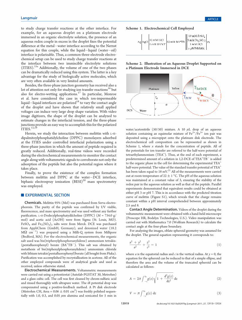
antimicrobial peptide capable of disrupting into the cell mem-brane, its effect on lipidic monolayers at the water�1,2-DCEinterface was also investigated. As shown in Figure 5, when onlyDPPC is present in the organic phase, dramatic changes areobserved in the contact angle. This is in agreement with previousreports in which strong adsorption of phosphatidylcholine hasbeen observed at the ITIES.12,13d,30 Additionally, the appearanceof a desorptive signal in the absence of peptide can be observed atapproximately 0.2 V, which stems from the complexation processof either Fe2+ or Fe3+ with the zwitterionic phospholipids at theliquid�liquid interface and further desorption from the ITIES to theorganic phase (Figure 4). This gives rise to a positive current in theforward scan because aqueous cations are being ultimately trans-ferred to the DCE phase. Similar behavior has been identified forcations such as protons,31 calcium,32 potassium,13d,33 etc.When melittin is present in the aqueous phase, the position of
this desorptive peak is shifted toward less positive potentials,while the transfer of the peptide remains unaltered as can be seenin Figure 4 (red line). This indicates that desorption of the
phospholipid from the interface occurs earlier in the presence ofthe peptide, as depicted in the inset of the same figure. Theseresults are very different from those previously reported for asmaller, non surface-active positively charged peptide like An-giotensin III:20a
(1) For Angiotensin III, a diminution in its ion transferpotential was observed in the presence of DPPC. There-fore, the phospholipid was recognized to be an ionophoriclayer that facilitates the peptide transfer reaction ratherthan a barrier.
(2) The phospholipid desorption occurs easily (i.e., at lesspositive potentials) in the presence of melittin.
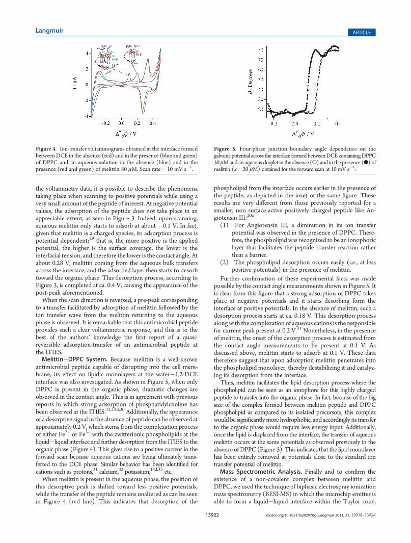
Further confirmation of these experimental facts was madepossible by the contact angle measurements shown in Figure 5. Itis clear from this figure that a strong adsorption of DPPC takesplace at negative potentials and it starts desorbing form theinterface at positive potentials. In the absence of melittin, such adesorption process starts at ca. 0.18 V. This desorption processalong with the complexation of aqueous cations is the responsiblefor current peak present at 0.2 V.31 Nonetheless, in the presenceof melittin, the onset of the desorption process is estimated fromthe contact angle measurements to be present at 0.1 V. Asdiscussed above, melittin starts to adsorb at 0.1 V. These datatherefore suggest that upon adsorption melittin penetrates intothe phospholipid monolayer, thereby destabilizing it and catalyz-ing its desorption from the interface.Thus, melittin facilitates the lipid desorption process where the
phospholipid can be seen as an ionophore for this highly chargedpeptide to transfer into the organic phase. In fact, because of the bigsize of the complex formed between melittin peptide and DPPCphospholipid as compared to its isolated precursors, this complexwould be significantlymore hydrophobic, and accordingly its transferto the organic phase would require less energy input. Additionally,once the lipid is displaced from the interface, the transfer of aqueousmelittin occurs at the same potentials as observed previously in theabsence of DPPC (Figure 3). This indicates that the lipidmonolayerhas been entirely removed at potentials close to the standard iontransfer potential of melittin.Mass Spectrometric Analysis. Finally and to confirm the
existence of a non-covalent complex between melittin andDPPC, we used the technique of biphasic electrospray ionizationmass spectrometry (BESI-MS) in which the microchip emitter isable to form a liquid�liquid interface within the Taylor cone,
Figure 4. Ion-transfer voltammograms obtained at the interface formedbetween DCE in the absence (red) and in the presence (blue and green)of DPPC and an aqueous solution in the absence (blue) and in thepresence (red and green) of melittin 80 μM. Scan rate = 10 mV s�1.
Figure 5. Four-phase junction boundary angle dependence on thegalvanic potential across the interface formed betweenDCEcontainingDPPC30μMand an aqueous droplet in the absence (O) and in the presence (b) ofmelittin (x = 20 μM) obtained for the forward scan at 10 mV s�1.
13923 dx.doi.org/10.1021/la202970g |Langmuir 2011, 27, 13918–13924
Langmuir ARTICLE
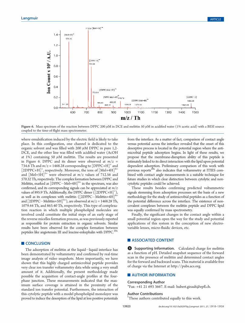
where emulsification induced by the electric field is likely to takeplace. In this configuration, one channel is dedicated to theorganic solvent and was filled with 200 μM DPPC in pure 1,2-DCE, and the other line was filled with acidified water (AcOHat 1%) containing 50 μM melittin. The results are presentedin Figure 6. DPPC and its dimer were observed at m/z =734.6 Th andm/z = 1468.28 corresponding to [DPPC+H]+ and[2DPPC+H]+, respectively. Moreover, the ions of [Mel+4H]4+
and [Mel+5H]5+ were observed at m/z values of 712.56 and570.32Th, respectively. The complex formation betweenDPPC andMelittin, marked as [DPPC�Mel+4H]4+ in the spectrum, was alsoconfirmed, and its corresponding signals can be appreciated at m/zvalues of 895.9 Th. Additionally, the DPPC dimer ([2DPPC+H]+),as well as its complexes with melittin ([2DPPC�Melittin+4H]4+
and [2DPPC�Melittin+5H]5+), are observed atm/z = 1468.28 Th,1079.44 Th, and 863.40 Th, respectively. This type of complexa-tion reaction in which multiple phospholipid molecules areinvolved could constitute the initial steps of an early stage ofthe reverse micelles formation process, as was previously reportedas responsible for protein extraction in organic solvents. Similarresults have been observed for the complex formation betweenpeptides like angiotensin III and leucine-enkephalin with DPPC.20a
’CONCLUSION
The adsorption of melittin at the liquid�liquid interface hasbeen demonstrated by voltammetry and confirmed by real-timeimage analysis of video snapshots. More importantly, we haveshown that this highly charged antimicrobial peptide providesvery clear ion transfer voltammetry data while using a very smallamount of it. Additionally, the present methodology madepossible the acquisition of contact-angle profiles at the four-phase junction. These measurements indicated that the max-imum surface coverage is attained in the proximity of thestandard ion transfer potential. Furthermore, the interaction ofthis cytolytic peptide with a model phospholipid monolayer wasproved to induce the desorption of the lipid at less positive potentials
from the interface. As a matter of fact, comparison of contact angleversus potential across the interface revealed that the onset of thisdesorptive process is located in the potential region where the anti-microbial peptide adsorption begins. In light of these results, wepropose that the membrane-disruption ability of this peptide isintimately linked to its direct interactionwith the lipid upon potentialdependent adsorption. Preliminary comparison of this work withprevious reports20a also indicates that voltammetry at ITIES com-bined with contact angle measurements is a suitable technique forfuture studies in which clear distinction between cytolytic and non-cytolytic peptides could be achieved.
These results besides confirming predicted voltammetricsignals stemming from adsorption processes set the basis of a newmethodology for the study of antimicrobial peptides as a function ofthe potential difference across the interface. The existence of non-covalent complexes between the melittin peptide and DPPC lipidwas equally confirmed by mass spectrometry.
Finally, the significant changes in the contact angle within asmall potential region open the way for the study and potentialapplications of this system in the conception of new electro-variable lenses, micro-fluidic devices, etc.
’ASSOCIATED CONTENT
bS Supporting Information. Calculated charge for melittinas a function of pH. Detailed snapshot sequence of the forwardscan in the presence of melittin and determined contact anglesfor the forward and backward scans. This material is available freeof charge via the Internet at http://pubs.acs.org.
’AUTHOR INFORMATION
Corresponding Author*Fax: +41 21 693 3667. E-mail: [email protected].
Author Contributions
)These authors contributed equally to this work.
Figure 6. Mass spectrum of the reaction between DPPC 200 μM in DCE and melittin 50 μM in acidified water (1% acetic acid) with a BESI sourcecoupled to the time-of-flight mass spectrometer.
13924 dx.doi.org/10.1021/la202970g |Langmuir 2011, 27, 13918–13924
Langmuir ARTICLE
’ACKNOWLEDGMENT
The Fonds National Suisse pour la Recherche Scientifique isthanked for financial support through the project “Electroche-mical methodology for the study of peptide lipid interaction”(Grant no. 200020-113428). Technical assistance by Val�erieDevaud is also acknowledged.
’REFERENCES
(1) Davies, J. Nature 1996, 383, 219–220.(2) Hancock, R.; Lehrer, R. Trends Biotechnol. 1998, 16, 82–88.(3) Haberman, E. Science 1972, 177, 314–322.(4) (a) Faucon, J. F.; Dufourcq, J.; Lussan, C. FEBS Lett. 1979,
102, 187–190. (b) Bernheimer, A. W.; Rudy, B. Biochim. Biophys. Acta1986, 864, 123–141. (c) Dempsey, C. E. Biochim. Biophys. Acta 1990,1031, 143–161.(5) (a) Saberwal, G.; Nagaraj, R. Biochim. Biophys. Acta 1994,
1197, 109–131. (b) Matsuzaki, K.; Yoneyama, S.; Miyajima, K. Biophys.J. 1997, 73, 831–838.(6) Raghuraman, H.; Chattopadhyay, A. Biophys. J. 2004, 87, 2419–
2432.(7) Beschiaschvili, G.; Seelig, J. Biochemistry 1990, 29, 52–58.(8) (a) Mathur, S.; Badertscher, M.; Scott, M.; Zenobi, R. Phys.
Chem. Chem. Phys. 2007, 9, 6187–6198. (b) Ha, M. H.; Endo, T.; Saito,M.; Chikae,M.; Do, K. K.; Yamamura, S.; Takamura, Y.; Tamiya, E.Anal.Chem. 2008, 80, 1859–1864.(9) Kuchinka, E.; Seelig, J. Biochemistry 1989, 28, 4216–4221.(10) Becucci, L.; Leon, R. R.; Moncelli, M. R.; Rovero, P.; Guidelli,
R. Langmuir 2006, 22, 6644–6650.(11) Koryta, J.; Hung, L. Q.; Hofmanova, A. Stud. Biophys. 1982,
90, 25–29.(12) Girault, H. H. J.; Schiffrin, D. J. J. Electroanal. Chem. 1984,
179, 277–284.(13) (a) Santos, H. A.; Carlsson, S.; Murtomaki, L.; Kontturi, K.
ChemPhysChem 2007, 8, 913–920. (b) Santos, H. A.; Ferreira, E. S.;Pereira, E. J.; Pereira, C. M.; Kontturi, K.; Silva, F.ChemPhysChem 2007,8, 1540–1547. (c) Saint Martin, E.; Konovalov, O.; Daillant, J. Thin SolidFilms 2007, 515, 5687–5690. (d) Yoshida, Y.; Maeda, K.; Shirai, O.J. Electroanal. Chem. 2005, 578, 17–24. (e) Kakiuchi, T.; Yamane, M.;Osakai, T.; Senda, M. Bull. Chem. Soc. Jpn. 1987, 60, 4223–4228.(14) (a) Kakiuchi, T.; Kondo, T.; Kotani, M.; Senda, M. Langmuir
1992, 8, 169–175. (b) Huang, J.; Chen, L.; Zhang, X.; Liu, S.; Li, G.Electrochem. Commun. 2008, 10, 451–454. (c) Monzon, L.; Yudi, L.Electrochim. Acta 2006, 51, 4573–4581.(15) (a) Ulmeanu, S.; Lee, H.; Fermin, D.; Girault, H.; Shao, Y.
Electrochem. Commun. 2001, 3, 219–223. (b) Scholz, F.; Gulaboski, R.;Caban, K. Electrochem. Commun. 2003, 5, 929–934. (c) Scholz, F.;Gulaboski, R. ChemPhysChem 2005, 6, 16–28. (d) Liu, X. H.; Yang, J.;Zuo, G. F.; Zhang, K.; Dong, C. W.; Lu, X. Q. J. Phys. Chem. C 2008,112, 148–152.(16) (a) Charreteur, K.; Quentel, F.; Elleouet, C.; L’Her, M. Anal.
Chem. 2008, 80, 5065–5070. (b) Scholz, F.; Gulaboski, R.; Caban, K.Electrochem. Commun. 2003, 5, 929–934.(17) Mugele, F.; Baret, J. C. J. Phys.: Condens. Matter 2005,
17, R705–R774.(18) (a)Monroe, C.W.; Daikhin, L. I.; Urbakh,M.; Kornyshev, A. A.
Phys. Rev. Lett. 2006, 97, 136102. (b) Monroe, C. W.; Daikhin, L. I.;Urbakh, M.; Kornyshev, A. A. J. Phys.: Condens. Matter 2006,18, 2837–2869.(19) Allen, R.; Kontturi, K.; Murtomaki, L.; Williams, D. J. Electro-
anal. Chem. 2000, 483, 57–67.(20) (a) M�endez, M. A.; Prudent, M.; Su, B.; Girault, H. H. Anal.
Chem. 2008, 80, 9499–9507. (b) Prudent, M.; M�endez, M. A.; Girault,H. H. Anal. Sci. 2008, 24, 1399–1404. (c) Prudent, M.; Mendez, M. A.;Jana, D. F.; Corminboeuf, C.; Girault, H. H. Metallomics 2010,2, 400–406.(21) Sabela, A.; Marecek, V.; Samec, Z.; Fuoco, R. Electrochim. Acta
1992, 37, 231–235.
(22) (a) Terwilliger, T. C.; EIisenberg, D. J. Biol. Chem. 1982,257, 6010–6015. (b) Terwilliger, T. C.; Eisenberg, D. J. Biol. Chem.1982, 257, 6016–6022.
(23) Humphrey, W.; Dalke, A.; Schulten, K. J. Mol. Graphics 1996,14, 33–38.
(24) Shinshi, M.; Sugihara, T.; Osakai, T.; Goto, M. Langmuir 2006,22, 5937–5944.
(25) Kakiuchi, T.; Noguchi, J.; Senda, M. J. Electroanal. Chem. 1992,327, 63–71.
(26) (a) Kakiuchi, T. J. Electroanal. Chem. 2001, 496, 137–142.(b) M�endez, M. A.; Su, B.; Girault, H. H. J. Electroanal. Chem. 2009,634, 82–89.
(27) (a) Hartvig, R.; M�endez, M.; van der Weert, M.; Jorgensen, L.;Ostergaard, J.; Girault, H. H.; Jensen, H. Anal. Chem. 2010, 82,7699–7705. (b) Herzog, G.; Kam, V.; Arrigan, D. W. M. Electrochim.Acta 2008, 53, 7204–7209.
(28) Goto, T.; Maeda, K.; Yoshida, Y. Langmuir 2005, 21, 11788–11794.
(29) Hartvig, R. A.; van de Weert, M.; Ostergaard, J.; Jorgensen, L.;Jensen, H. Langmuir 2011, 27, 2634–2643.
(30) Samec, Z.; Troj�anek, A.; Krtil, P. Faraday Discuss. 2005,129, 301–313.
(31) Janchenova, H.; Lhotsky, A.; Stulik, K.; Marecek, V. J. Electro-anal. Chem. 2007, 601, 101–106.
(32) (a) Ohnishi, S.; Ito, T. Biochemistry 1974, 13, 881–887.(b) Chesniuk, S.; Dassie, S.; Yudi, L.; Baruzzi, A. Electrochim. Acta1998, 43, 2175–2181.
(33) Monz�on, L.; Yudi, L. Electrochim. Acta 2006, 51, 1932–1940.
Copyright © 2022 FDOKUMEN