Effector-mediated membrane disruption controls cell death in ...
19
Article Effector-mediated membrane disruption controls cell death in CBASS antiphage defense Graphical abstract Highlights d CBASS antiphage transmembrane effectors induce membrane disruption and cell death d The TM effector Cap15 contains a b-barrel domain for second messenger recognition d Ligand binding triggers Cap15 oligomerization and inner- membrane disruption d Cap15 effectors disrupt membranes through raft-like clustering, not pore formation Authors Brianna Duncan-Lowey, Nora K. McNamara-Bordewick, Nitzan Tal, Rotem Sorek, Philip J. Kranzusch Correspondence [email protected] In brief Duncan-Lowey et al. determine the function of transmembrane effectors in CBASS antiphage defense. Transmembrane effectors bind nucleotide second messengers, induce inner-membrane disruption, and cause cell death to limit phage replication. The structure of one family of transmembrane effectors, Cap15, reveals a minimal b-barrel that binds activating cyclic dinucleotide signals. Duncan-Lowey et al., 2021, Molecular Cell 81, 1–13 December 16, 2021 ª 2021 Elsevier Inc. https://doi.org/10.1016/j.molcel.2021.10.020 ll
-
Upload
khangminh22 -
Category
Documents
-
view
0 -
download
0
Transcript of Effector-mediated membrane disruption controls cell death in ...

Article
Effector-mediated membr
ane disruption controlscell death in CBASS antiphage defenseGraphical abstract
Highlights
d CBASS antiphage transmembrane effectors induce
membrane disruption and cell death
d The TMeffector Cap15 contains a b-barrel domain for second
messenger recognition
d Ligand binding triggers Cap15 oligomerization and inner-
membrane disruption
d Cap15 effectors disrupt membranes through raft-like
clustering, not pore formation
Duncan-Lowey et al., 2021, Molecular Cell 81, 1–13December 16, 2021 ª 2021 Elsevier Inc.https://doi.org/10.1016/j.molcel.2021.10.020
Authors
Brianna Duncan-Lowey,
Nora K. McNamara-Bordewick,
Nitzan Tal, Rotem Sorek,
Philip J. Kranzusch
In brief
Duncan-Lowey et al. determine the
function of transmembrane effectors in
CBASS antiphage defense.
Transmembrane effectors bind
nucleotide second messengers, induce
inner-membrane disruption, and cause
cell death to limit phage replication. The
structure of one family of transmembrane
effectors, Cap15, reveals a minimal
b-barrel that binds activating cyclic
dinucleotide signals.
ll
Please cite this article in press as: Duncan-Lowey et al., Effector-mediated membrane disruption controls cell death in CBASS antiphage defense,Molecular Cell (2021), https://doi.org/10.1016/j.molcel.2021.10.020
ll
Article
Effector-mediated membrane disruptioncontrols cell death in CBASS antiphage defenseBrianna Duncan-Lowey,1,2 Nora K. McNamara-Bordewick,2 Nitzan Tal,3 Rotem Sorek,3 and Philip J. Kranzusch1,2,4,5,*1Department of Microbiology, Harvard Medical School, Boston, MA 02115, USA2Department of Cancer Immunology and Virology, Dana-Farber Cancer Institute, Boston, MA 02115, USA3Department of Molecular Genetics, Weizmann Institute of Science, Rehovot, Israel4Parker Institute for Cancer Immunotherapy at Dana-Farber Cancer Institute, Boston, MA 02115, USA5Lead contact
*Correspondence: [email protected]
https://doi.org/10.1016/j.molcel.2021.10.020
SUMMARY
Cyclic oligonucleotide-based antiphage signaling systems (CBASS) are antiviral defense operons that pro-tect bacteria from phage replication. Here, we discover a widespread class of CBASS transmembrane(TM) effector proteins that respond to antiviral nucleotide signals and limit phage propagation through directmembrane disruption. Crystal structures of the Yersinia TM effector Cap15 reveal a compact 8-strandedb-barrel scaffold that forms a cyclic dinucleotide receptor domain that oligomerizes upon activation. Wedemonstrate that activated Cap15 relocalizes throughout the cell and specifically induces rupture of the innermembrane. Screening for active effectors, we identify the function of distinct families of CBASS TM effectorsand demonstrate that cell death via disruption of inner-membrane integrity is a common mechanism of de-fense. Our results reveal the function of the most prominent class of effector protein in CBASS immunity anddefine disruption of the inner membrane as a widespread strategy of abortive infection in bacterial phagedefense.
INTRODUCTION
In bacteria, cyclic oligonucleotide-based antiphage signaling
systems (CBASS) are a major form of antiviral defense that limit
replication of diverse phages (Cohen et al., 2019). Each CBASS
operon contains a cGAS/DncV-like nucleotidyltransferase (CD-
NTase) enzyme that functions to sense phage replication and a
partner CD-NTase-associated protein (Cap) effector that directly
impairs host cell function to prevent viral spread (Lowey et al.,
2020;Whiteley et al., 2019; Ye et al., 2020). Following recognition
of phage infection, the CD-NTase synthesizes a nucleotide sec-
ond messenger, which then directly binds the Cap effector and
results in activation and induction of antiviral defense (Cohen
et al., 2019; Lau et al., 2020; Lowey et al., 2020; Whiteley
et al., 2019).
A critical step in CBASS immunity is the ability of the Cap
effector to respond to a specific CD-NTase nucleotide second
messenger signal (Govande et al., 2021; Lowey et al., 2020).
Bacterial CD-NTase enzymes produce a diverse array of nucle-
otide signals with distinct base and phosphodiester linkage
specificity, including cyclic dinucleotide (e.g., 3030 cyclic GMP-
AMP, 3030-cGAMP) and cyclic trinucleotide (e.g., 3030 cyclic
AMP-AMP-GMP, 303030-cAAG) products (Lowey et al., 2020;
Whiteley et al., 2019). Recent structures of CBASS effectors in
complex with nucleotide second messengers explain how cor-
rect signal recognition controls protein function (Lau et al.,
2020; Lowey et al., 2020; Morehouse et al., 2020). The structure
M
of A. baumannii Cap4 demonstrates that a protein domain
namedSAVED is responsible for nucleotide recognition and sub-
sequent protein oligomerization to control effector activation
(Lowey et al., 2020). Interestingly, the Cap4 SAVED domain is
a structural homolog of CRISPR-associated Rossman fold
(CARF)-family proteins, revealing structural and functional over-
lap between components of CBASS and CRISPR immune sys-
tems (Jia et al., 2019; Lowey et al., 2020; Niewoehner and Jinek,
2016). Likewise, discovery of a bacterial domain homologous to
the human cyclic dinucleotide binding protein stimulator of inter-
feron genes (STING) within CBASS effectors Cap12 and Cap13
demonstrates an evolutionary link between CBASS nucleotide-
second messenger binding and components of animal innate
immunity (Morehouse et al., 2020). These CBASS effectors con-
taining STING and SAVED domains specifically bind nucleotide
second messengers, then oligomerize when activated. The ma-
jority of CBASS operons, however, contain effectors with no
characterized nucleotide-binding domain, suggesting that major
forms of nucleotide second messenger recognition remain to be
discovered.
Following nucleotide second messenger recognition, Cap ef-
fectors induce cell death to kill the host bacteria and block phage
replication through a form of antiviral defense termed abortive
infection. Nearly all characterized abortive infection defense sys-
tems function through effectors with enzymatic domains that
degrade or modify target host or phage proteins (Lopatina
et al., 2020). Several CBASS enzymatic effectors downstream
olecular Cell 81, 1–13, December 16, 2021 ª 2021 Elsevier Inc. 1

llArticle
Please cite this article in press as: Duncan-Lowey et al., Effector-mediated membrane disruption controls cell death in CBASS antiphage defense,Molecular Cell (2021), https://doi.org/10.1016/j.molcel.2021.10.020
of CD-NTases have been mechanistically characterized,
including phospholipases (CapV), DNA endonucleases (Cap4,
Cap5, NucC), and NADases (Cap12) (Cohen et al., 2019; Lau
et al., 2020; Lowey et al., 2020; Morehouse et al., 2020; Severin
et al., 2018). The nuclease effectors Cap4, Cap5, and NucC in-
discriminately degrade double-stranded DNA (dsDNA) upon
activation (Lau et al., 2020; Lowey et al., 2020), while the phos-
pholipase effector CapV cleaves membrane phospholipids
leading to cell death (Cohen et al., 2019; Severin et al., 2018).
However, the majority of CBASS operons do not contain enzy-
matic effectors and instead encode uncharacterized proteins
with predicted transmembrane (TM) segments (Burroughs
et al., 2015; Millman et al., 2020). While it has been hypothesized
that these effectors lead to membrane disruption, it is unknown
how TM effectors function to restrict phage replication.
Here, we discover that CBASS TM effectors are potent anti-
phage defense proteins that destroy bacterial inner-membrane
integrity and induce host cell death. Through development of a
screen to identify active TM effectors, we define the family of
CBASS proteins named Cap15 that respond to cyclic dinucleo-
tide signals and block phage replication. High-resolution crystal
structures of the Cap15 nucleotide binding domain reveal a
compact b-barrel scaffold with a central pocket for ligand recog-
nition and oligomerization interfaces essential for phage de-
fense. Mechanistically, we show that Cap15 oligomerization
and activation disrupt bacterial cell integrity and lead to specific
disruption of the inner membrane. We further demonstrate that
inner-membrane disruption is a widespread mechanism shared
between diverse families of CBASS TM effectors that are en-
coded in both gram-negative and gram-positive bacteria. Our
results demonstrate the function of a major class of CBASS ef-
fectors controlling antiviral defense and define the host mem-
brane as a common target of bacterial abortive infection defense
systems.
RESULTS
Discovery of CBASS TM effectors that terminate cellgrowthBuilding upon previous bioinformatic analysis (Burroughs et al.,
2015; Cohen et al., 2019; Millman et al., 2020; Whiteley et al.,
2019), we analyzed putative effector proteins in CBASS operons
and identified a diverse set of 2,234 proteins containing TM seg-
ments. TM effectors are encoded in >40% of CBASS operons,
representing a dominant form of effector that occurs more
frequently than previously characterized phosphodiesterase,
nuclease, orNADaseproteinswith enzymatic function (Figure 1A)
(Lau et al., 2020; Lowey et al., 2020; Morehouse et al., 2020; Se-
verin et al., 2018). The most common TM effector contains two
TM segments fused to an uncharacterized b-strand-rich C-ter-
minal domain, and we named this effector CD-NTase-associ-
ated protein 15 (Cap15) (Figures 1B and 1C). To determine if
Cap15 is capable of responding to CBASS cyclic dinucleotide
signaling and inducing cell toxicity in bacteria, we leveraged
the known ability of the CD-NTase Vibrio cholerae DncV
(VcDncV; NCBI ref. WP_001901330.1) to be auto-activated as
a promiscuous 3030-cGAMP synthase during overexpression in
E. coli (Whiteley et al., 2019; Zhou et al., 2018). VcCapV is a
2 Molecular Cell 81, 1–13, December 16, 2021
phospholipase known to be activated by 3030-cGAMP in CBASS
defense and robustly induces toxicity in these conditions (Cohen
et al., 2019; Severin et al., 2018) (Figure 1D). Expression of Es-
cherichia albertii Cap15 (EaCap15, NCBI ref. WP_206748793.1)
in the presence of active 3030-cGAMP signaling prevents culture
growth (Figure 1D) and induces cell death as confirmed by flow
cytometry (Figure S1D). A VcDncV D131A, D133A substitution
that disrupts CD-NTase catalytic function rescues cell viability,
demonstrating that EaCap15 induces cell death only in the pres-
ence of active 3030-cGAMP nucleotide second messenger
signaling (Figure 1D).
We next screened a panel of 23 CBASS TMeffectors and iden-
tified a wide diversity of proteins capable of inducing cell toxicity
specifically in response to 3030-cGAMP (Figure 1E). Toxic TM
effector proteins belong to several families including Cap14 pro-
teins (TM-SAVED, Pfam PF18145) and Cap16 proteins (TM-NU-
DIX, Pfam PF18167) (Figures 1E and S1A). TM effectors are
encoded throughout the CD-NTase family tree (Whiteley et al.,
2019), suggesting that the effectors not responsive to 3030-cGAMP in our screen likely induce cell death but respond to
other CD-NTase products including pyrimidine-containing cyclic
dinucleotide and cyclic trinucleotide signals (Lowey et al., 2020;
Whiteley et al., 2019). In each case, cell toxicity is strictly depen-
dent on the presence of TM segments, revealing that membrane
association is essential for TM effector function (Figures 1F and
S1B). The only TM effector with a putative enzymatic domain is
Cap16, which contains a NUDIX hydrolase domain (Figures 1B
and 1C). Mutation of the putative Cap16 active site (E154A)
does not impact cell toxicity (Figures 1F and S1B), further con-
firming that TM effector function is dependent on TM segments
and not catalytic activity. These data define Cap15 and diverse
TM effectors in CBASS immunity that respond to antiviral nucle-
otide second messenger signaling and induce potent cell
toxicity.
CBASSCap15 TMeffectors protect bacteria from phageinfection and encode a minimal b-barrel domainTo determine the role of TM effectors in antiviral defense, we ex-
pressed complete Cap15-containing CBASS operons in E. coli
and challenged the bacteria with a panel of 10 diverse phages.
CBASS operons containing Cap15 effectors potently restrict
viral plaque formation (Figures 2A and 2B). The Escherichia
CBASS (EaCdnB [NCBI ref. WP_000995828.1] and EaCap15
[NCBI ref. WP_206748793.1]), which we identified as a 3030-cGAMP signaling system (Figure 1D), protects E. coli against
infection with T2, T5, and T6 (Figure 2A). Interestingly, a related
YersiniaCBASS operon, which encodes homologous CD-NTase
and Cap15 components (YaCdnE [NCBI ref. WP_145567548.1]
and YaCap15 [NCBI ref. WP_145567547.1]), protects E. coli
against all testedMyoviridae (T2, T4, T6), Siphoviridae (T5, SEC-
phi4, SECphi6, SECphi18, SECphi27), and the single-stranded
DNA (ssDNA) phage SECphi17, but not the Podoviridae phage
T7, demonstrating a variable range of phage defense between
CBASS operons encoding the same class of effector (Figure 2B).
We analyzed CBASS Cap15-containing operons and observed
that they are widely distributed in both gram-negative and
gram-positive bacteria (Figure 2C). CBASS Cap15-containing
operons protect against a wide diversity of phages, including

A B C
D
E F
Figure 1. Discovery of CBASS TM effectors that control cell death
(A) Schematic showing CBASS effector types and abundance in sequenced genomes. Transmembrane (TM) effectors are encoded in >40% of CBASS operons.
(B) Quantification of individual families of TM effectors in CBASS operons.
(C) Domain organization of common CBASS TM effector families. Structure prediction analysis demonstrates that SUa (Pfam PF18179) is a variant of the SAVED
nucleotide-binding domain. See also Figure S1.
(D) Growth curves of E. coli expressing a CBASS effector protein and the 3030-cGAMP synthase Vibrio cholerae DncV (VcDncV). VcDncV is known to catalyze
active 3030-cGAMP signaling upon overexpression in E. coli (Whiteley et al., 2019; Zhou et al., 2018). Cells expressing WT VcDncV are indicated in color, and
catalytically inactive VcDncV are in gray. The 3030-cGAMP-responsive effectors induce cell death upon co-expression with active VcDncV.
(E) Quantification of growth curves and cell death induced by 3030-cGAMP-responsive effector proteins. Data are displayed as the difference in OD600 of cultures
expressing WT versus catalytically inactive VcDncV 300 min after induction, relative to a control culture with no effector expression. Many families of TM-
containing effectors cause cell death in response to 3030-cGAMP production.
(F) Quantification of cell death induced by full-length or truncated (DTM) TM effectors where the TM domain was replaced by SUMO to ensure solubility. Cap16
contains a putative NUDIX hydrolase domain and was additionally tested with a NUDIX-inactivating E154A mutation. TM-containing effectors require the ap-
pended TM segments to induce cell death. Toxicity data are representative of at least two independent experiments. Error bars represent standard deviation (SD).
See also Figure S1.
llArticle
Please cite this article in press as: Duncan-Lowey et al., Effector-mediated membrane disruption controls cell death in CBASS antiphage defense,Molecular Cell (2021), https://doi.org/10.1016/j.molcel.2021.10.020
both dsDNA and ssDNA phage, suggesting the viral signal that
activates these CBASS operons is broadly conserved. In each
case, disruption of the CD-NTase active site or deletion of the
Cap15 TM region abolished all phage protection, demonstrating
that Cap15-mediated antiviral defense is strictly dependent
upon functional CD-NTase signaling and interaction with the
bacterial membrane (Figures S2B and S2C).
Each Cap15 protein comprises N-terminal TM helices fused to
an uncharacterized C-terminal domain (Figure 1C). To define the
molecular basis of Cap15 effector function, we determined a
1.9-A crystal structure of the YaCap15 C-terminal domain (Fig-
ure 2D; Table 1). The YaCap15 structure reveals eight b strands
(b2–b9) that wrap and form a compact b-barrel (Figure 2D). Strik-
ingly, theclosest structural homologs toCap15arenotnaturalpro-
teins, but instead are computationally derived b-barrel proteins
designed to bind fluorescent small molecules (Figure 2E) (Dou
et al., 2018). Similar to the rationally designed proteins, in
Cap15, a set of nine glycine residues distributed throughout the
eightb strandsarepositioned to increase strandcurvature, reduce
side chain density within the barrel center, and enable folding of
the minimized b-barrel structure (Figures S2D and S2E). Cap15
is further stabilized by a conserved ‘‘tryptophan corner’’ interac-
tion previously observed in other b-barrel structures (Dou et al.,
2018). In YaCap15, this interaction occurs where W96 on the
Molecular Cell 81, 1–13, December 16, 2021 3
A B
C D E
F
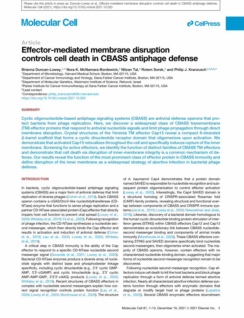
Figure 2. Cap15 protects bacteria from phage infection and encodes a minimal b-barrel nucleotide-binding domain
(A and B) Phage challenge of E. coli expressing Escherichia or YersiniaCBASS operons. CBASS operons containing Cap15 b-barrel TM effectors protect against
infection with diverse phages. Cartoon schematic shows organization of the CBASS operons encoding CD-NTases from clade B (CdnB) or E (CdnE) and the
Cap15 b-barrel-containing TM effector.
(C) Phylogenetic analysis of the distribution of Cap15 b-barrel effectors and other CBASS TM effectors across bacterial phyla demonstrates widespread
occurrence in both gram-negative and gram-positive bacteria.
(D) Crystal structure of the Yersinia aleksiciaeCap15 (YaCap15) b-barrel nucleotide-binding domain. The YaCap15 structure consists of eight b strands that form a
highly minimized b-barrel.
(E) Comparison of Cap15 to an in-silico-designed small-molecule sensor (6CZI) (Dou et al., 2018) and the biotin-binding protein streptavidin (3WZQ) (Kawato
et al., 2015).
(F) The Cap15 b-barrel domain contains a common tryptophan-corner capping motif at the bottom of the b-barrel similar to streptavidin proteins. A capping helix
common to many b-barrels is replaced by a capping b strand in Cap15.
Phage defense data are representative of at least three independent experiments. Error bars represent SD. See also Figure S2.
llArticle
Please cite this article in press as: Duncan-Lowey et al., Effector-mediated membrane disruption controls cell death in CBASS antiphage defense,Molecular Cell (2021), https://doi.org/10.1016/j.molcel.2021.10.020
N-terminal strandb2stacks against R204on theC-terminal strand
b9and formsahydrogenbondwith thepeptide carboxyl onstrand
b1 below the b-barrel (Figure 2F). Together, these data define the
TM effector Cap15 as a widespread b-barrel domain-containing
protein that enables CBASS antiphage defense.
Cap15 b-barrel nucleotide-binding domain specificallybinds uracil-containing cyclic dinucleotidesThe Cap15 b-barrel domain creates a solvent-exposed pocket at
the top of the protein that is in the same location as the small-
molecule binding site of rationally designed b-barrels (Figure 3A)
(Dou et al., 2018). Additionally, Cap15 exhibits more distantly
related structural homology to streptavidin proteins that form a
similar pocket at the top of a b-barrel domain to create a binding
site for biotin (Figure S3A). Therefore, we hypothesized that the
4 Molecular Cell 81, 1–13, December 16, 2021
function of the Cap15 b-barrel domain is to recognize the CD-
NTase product nucleotide second messenger. To test this hy-
pothesis, we purified the Yersinia aleksiciae CD-NTase (YaCdnE)
encoded adjacent to YaCap15 and determined the nucleotide
second messenger product for this CBASS operon using 32P-
labeled nucleotides and thin-layer chromatography. YaCdnE
synthesizes a mixture of di-pyrimidine and purine-pyrimidine cy-
clic dinucleotide products with a strong preference for incorpo-
ration of UTP (Figure 3B). HPLC (Figures 3C and S3B) and
mass spectrometry analysis of the purified reaction products
demonstrate that the most abundant products synthesized by
YaCdnE in the presence of ATP, GTP, CTP, and UTP are 3030-c-di-UMP (m/z = 613.058) and 3030-cUMP-AMP (m/z = 636.085).
We next analyzed the ability of the YaCap15 C-terminal
b-barrel domain to bind radiolabeled YaCdnE cyclic

Table 1. Crystallographic statistics
YaCap15 (SeMet phasing) YaCap15 (SeMet refinement) YaCap15
Data collection
Resolution (A)a 37.09–2.30 (2.38–2.30) 37.01–1.90 (1.94–1.90) 44.05–2.60
(2.72–2.60)
Wavelength (A) 0.97918 0.97918 0.97918
Space group C 1 2 1 C 1 2 1 P 61
Unit cell: a, b, c (A) 93.51, 31.20, 50.04 93.29, 31.04, 50.02 50.87,
50.87, 155.02
Unit cell: a, b, g (�) 90.0, 100.33, 90.0 90.0, 100.22, 90.0 90.0,
90.0, 120.0
Molecules per ASU 1 1 2
Total reflections 487276 77567 111881
Unique reflections 6554 11344 7040
Completeness (%)a 99.8 (98.1) 99.8 (98.9) 100.0 (100.0)
Multiplicitya 74.3 (71.4) 6.9 (6.3) 15.9 (15.2)
I/sIa 25.8 (6.1) 13.3 (1.3) 19.3 (1.3)
CC(1/2)b (%)a 99.9 (43.2) 99.9 (56.2) 100.0 (51.2)
Rpimc (%)a 2.7 (75.0) 2.9 (64.3) 1.8 (67.4)
Sites 1
Refinement
Resolution (A) 37.0–1.90 44.05–2.60
Free reflections 1134 679
R-factor/R-free 21.5 / 25.4 29.7 / 31.3
Bond distance (RMS A) 0.006 0.002
Bond angles (RMS �) 0.752 0.491
Structure/stereochemistry
No. atoms: protein 869 1687
No. atoms: solvent 24 0
Average B-factor: protein 54.89 108.24
Average B-factor: water 54.08 N/A
Ramachandran plot: favored 99.02% 95.41%
Ramachandran plot: allowed 0.98% 4.59%
Ramachandran plot: outliers 0.00% 0.00%
Rotamer outliers 1.11% 4.55%
MolProbityd score 1.12 2.20
Protein Data Bank ID 7N34 7N35aHighest-resolution shell values in parentheses.bKarplus and Diederichs (2012)cWeiss (2001)dChen et al. (2010)
llArticle
Please cite this article in press as: Duncan-Lowey et al., Effector-mediated membrane disruption controls cell death in CBASS antiphage defense,Molecular Cell (2021), https://doi.org/10.1016/j.molcel.2021.10.020
dinucleotide products and observed specific recognition of
3030-c-di-UMP and 3030-cUMP-AMP (Figures 3D, 3E, S3C,
and S3D). YaCap15 binds 3030-c-di-UMP and 3030-cUMP-AMP
with �100-nM affinity and exhibits no ability to interact with
the control cyclic dinucleotide 3030-c-di-AMP (Figures 3D, 3E,
and S3C–S3E). Additionally, we observed that Cap15 binding
to any UMP-containing cyclic dinucleotide results in a dramatic
increase in the thermostability of the complex, further support-
ing a direct role for the Cap15 C-terminal b-barrel domain in
sensing the CD-NTase cyclic dinucleotide antiviral signal (Fig-
ures 3F and S3F).
Analysis of the Cap15 solvent exposed pocket at the top of
the b-barrel reveals highly conserved residues surrounding the
putative nucleotide-binding site (Figures 3G, 3H, and S2A). To
confirm the role of this Cap15 pocket in mediating cyclic dinu-
cleotide recognition, we next mutated each conserved residue
to eliminate potential hydrogen-bonding interactions (Y153F,
Y155F, Y188F, M200I) or inserted bulkier side chains to limit
access to the binding site (T129Q, N157Q). Substitutions to
the conserved YaCap15 pocket residues each reduce 3030-c-di-UMP binding or abrogate all ligand recognition in vitro (Fig-
ures 3I and S3G). We confirmed that all mutations, with the
Molecular Cell 81, 1–13, December 16, 2021 5

A
B C
D E F G
H I J
Figure 3. Cap15 b-barrel nucleotide-binding domain specifically binds uracil-containing cyclic dinucleotides
(A) Cutaway slice through the center of Cap15 (left) and the rationally designed small-molecule sensor 6CZI (right). Protein surface is represented inmesh, and the
small molecule bound to 6CZI is shown in sticks. A solid, hydrophobic core stabilizes the bottom of each b-barrel creating a solvent-exposed pocket at the top
lined with hydrophilic amino acids for ligand recognition.
(B) Analysis of YaCdnE nucleotide second messenger synthesis. YaCdnE was incubated with a-32P-NTPs, and reactions were phosphatase treated and
separated by thin-layer chromatography.
(C) HPLC analysis of YaCdnE products after separation by ion exchange chromatography. YaCdnE products were separated as two major peaks from ion
exchange and were analyzed separately by HPLC (top) compared to cyclic dinucleotide standards (bottom). YaCdnE synthesizes 3030-c-di-UMP, 3030-cUMP-
AMP, and 3030-cUMP-CMP as major products. See also Figure S3B.
(D) Electrophoretic mobility shift assay measurement of YaCap15-3030-cUMP-AMP complex formation. A titration of YaCap15 (5 nM–5 mM) was incubated with
10-nM 32P-labeled 3030-cUMP-AMP, and bound complexes were resolved by nondenaturing polyacrylamide gel electrophoresis.
(E) Quantification of YaCap15-cyclic dinucleotide complex formation as shown in (D). See also Figures S3C and S3E.
(F) Thermal denaturation assay to quantify stabilization of the YaCap15 b-barrel upon binding to UMP-containing cyclic dinucleotides. Cyclic dinucleotide
recognition leads to an �25�C increase in YaCap15 b-barrel melting temperature, demonstrating significant stabilization.
(G) Cartoon highlighting conserved residues lining the Cap15 b-barrel solvent-exposed nucleotide-binding pocket.
(H) Conservation of select residues lining the Cap15 nucleotide-binding pocket. YaCap15 binds UMP-containing cyclic dinucleotides; EaCap15 is activated by
3030-cGAMP.
(I) Quantification of 3030-c-di-UMP binding assays using YaCap15 proteins with point mutations within the nucleotide-binding pocket verifies the importance of
individual contacts for nucleotide second messenger recognition. YaCap15 mutants were incubated at 100-nM or 1-mM concentration with 3030-c-di-UMP, and
complex formation was quantified as in (E). See also Figure S3G.
(J) Phage challenge of E. coli expressing Yersinia CBASS with mutations to the Cap15 nucleotide-binding pocket (empty vector controls shown in Figure 2B are
included again for reference). Mutations that inhibit nucleotide binding abolish protection from phage infection.
Biochemical and phage defense data are representative of at least three independent experiments. Error bars represent SD. See also Figure S3.
llArticle
Please cite this article in press as: Duncan-Lowey et al., Effector-mediated membrane disruption controls cell death in CBASS antiphage defense,Molecular Cell (2021), https://doi.org/10.1016/j.molcel.2021.10.020
exception of M200I, did not negatively impact protein stability
(Figure S3H). We next determined whether mutations that
disrupt Cap15 ligand binding prevent CBASS antiphage de-
fense. We observed that a YaCap15 N157Q mutation pre-
dicted to occlude the nucleotide-binding pocket results in
complete loss of phage protection, while mutations that only
6 Molecular Cell 81, 1–13, December 16, 2021
mildly decrease ligand binding (Y153F, Y155F) do not affect
phage defense (Figures 3J and S3I). Together, these results
demonstrate the discovery of a class of minimal b-barrel cy-
clic dinucleotide binding domains and reveal the mechanistic
basis of Cap15 cyclic dinucleotide recognition in CBASS
defense.

A B C
D E F
G
Figure 4. Cap15 oligomerizes upon ligand binding
(A and B) Analysis of packing with symmetry mates in the Cap15 crystal structure defines a b-strand oligomerization interface (interface 1) and a hydrophobic
packing interface (interface 2).
(C) Phage challenge of E. coli expressing YersiniaCBASS with mutations to the oligomerization interfaces (empty vector controls shown in Figure 2B are included
again for reference).
(D) SEC demonstrating oligomerization of WT YaCap15, which is disrupted by mutations to YaCap15 interface 1 (89–end, W120A).
(E) SEC demonstrating that recognition of activating cyclic dinucleotides results in YaCap15 higher-order complex formation (left). Mutations to YaCap15
interface 1 prevent full higher-order complex formation but do not impact cyclic dinucleotide recognition (right).
(F) Electrophoretic mobility shift assay measurement of WT or interface 1 mutant Cap15 binding to 3030-c-di-UMP. 100 nM or 1 mM of indicated protein was
incubatedwith 10 nM 32P-labeled 3030-c-di-UMP, and bound complexeswere resolved by nondenaturing polyacrylamide gel electrophoresis. Arrows indicate the
two complexes formed by Cap15 after ligand binding. The interface 1 mutant disrupts formation of the higher-order YaCap15 complex (top arrow).
(G) Structural model of Cap15 oligomerization. The experimentally determined structure of the Cap15 b-barrel is shownwith modeled TMdomains. Both interface
1 and interface 2 are compatible with clustering of Cap15 along a two-dimensional membrane surface.
Biochemical data are representative of at least two independent experiments, and phage defense data are representative of three independent experiments.
Error bars represent SD. See also Figure S4.
llArticle
Please cite this article in press as: Duncan-Lowey et al., Effector-mediated membrane disruption controls cell death in CBASS antiphage defense,Molecular Cell (2021), https://doi.org/10.1016/j.molcel.2021.10.020
Cap15 effector function requires proteinoligomerizationFollowing nucleotide second messenger recognition, soluble
CBASS effectors including Cap4, Cap12, and NucC are known
to activate by oligomerizing into higher-order protein complexes
(Lau et al., 2020; Lowey et al., 2020; Morehouse et al., 2020). To
determine if protein oligomerization also has a role in activation
of CBASS TM effectors, we next examined packing within the
YaCap15 crystal for potential oligomeric interfaces. In the Ya-
Cap15 crystal, strand b1 extends away from the folded domain
and forms a tight anti-parallel b-sheet interaction with a partner-
ing strand b1 on the neighboring YaCap15 protomer (Figure 4A).
This interface (interface 1) between neighboring protomers is
further stabilized by a stacking interaction between the W120
side chain of each protein (Figure 4A). We identified an additional
oligomerization interface (interface 2) between adjacent Ya-
Cap15 protomers mediated by a stripe of hydrophobic residues
I142, L152, F172, F189, and Y197 facing out of the barrel (Figures
4B and S2D). We determined a second 2.6-A structure of Ya-
Cap15 and verified preservation of identical interface 1 and inter-
face 2 contacts within this distinct crystal form (Figure S4A;
Table 1). To determine the importance of the observed contacts,
we designedmutations that block oligomerization and tested the
impact in vivo during phage challenge. Mutations that disrupt
interface 1 (W120A) or interface 2 (L152E, F172D) completely
abolish phage defense (Figures 4C and S4C), demonstrating
that the YaCap15 oligomerization interfaces are essential for
effector function.
Molecular Cell 81, 1–13, December 16, 2021 7

llArticle
Please cite this article in press as: Duncan-Lowey et al., Effector-mediated membrane disruption controls cell death in CBASS antiphage defense,Molecular Cell (2021), https://doi.org/10.1016/j.molcel.2021.10.020
We next measured YaCap15 oligomerization in solution using
size-exclusion chromatography with multi-angle light scattering
(SEC-MALS). Wild-type (WT) YaCap15 (15 kDa) migrates as an
oligomeric complex with a molecular weight of 91 kDa (Figures
4D and S4D). Interestingly, in the presence of 3030-c-di-UMP or
3030-cUMP-AMP, the migration profile of YaCap15 dramatically
shifts to a mixture of higher-order species, indicating that CD-
NTase signal recognition triggers further protein oligomerization
(Figure 4E). YaCap15 forms a heterogenous population of spe-
cies after binding to activating cyclic dinucleotides; therefore, it
was not possible to define a discrete molecular weight for the
higher-order complex.
To determine if YaCap15 oligomerization is required for nucle-
otide second messenger recognition, we purified a YaCap15
b-barrel domain variant where oligomerization interface 1 is dis-
rupted by aW120A mutation and deletion of strand b1 (YaCap15
89–end W120A, 14 kDa). In contrast to WT YaCap15, YaCap15
89–end W120A migrates as a monomer with a calculated mole-
cule weight of 19 kDa (Figures 4D and S4D). In the presence of
3030-c-di-UMP or 3030-cUMP-AMP, YaCap15 89–endW120A un-
dergoes a shift to form a dimer (calculated mass of 31 kDa) but
fails to form the fully oligomerized assemblies observed with
the WT protein (Figures 4E and S4D). Using radiolabeled cyclic
dinucleotides, we confirmed that YaCap15 89–end W120A re-
tains the ability to bind 3030-c-di-UMP or 3030-cUMP-AMP,
demonstrating that full oligomerization is not required for cyclic
dinucleotide recognition (Figures 4E and S4B). Similar to previ-
ous analysis of Cap12 (Morehouse et al., 2020), we further
observed that the YaCap15 oligomerization-disrupting muta-
tions prevent formation of larger complexes that migrate higher
in the gel well during electrophoretic mobility shift assay experi-
ments (Figures 4F and S4B).
Using the experimentally defined YaCap15 oligomerization in-
terfaces, we constructed a model of full-length YaCap15 to pre-
dict orientation of the N-terminal TM helices (Figure 4G). Notably,
the experimentally defined YaCap15 oligomerization interfaces
are compatible with oligomerization along the two-dimensional
surface of the bacterial cell membrane. These data support a
mechanism where Cap15 exists as a lower-order multimer and
subsequently oligomerizes within the membrane after binding
to the activating nucleotide second messenger. Together, these
results confirm the importance of YaCap15 oligomerization inter-
faces in vivo and demonstrate that effector oligomerization is
feature conserved between divergent CBASS systems with
cytosolic or membrane-bound effector proteins.
CBASS TM effectors cause membrane disruption andcell deathGiven the requirement of both theCap15 TMdomains andCap15
protein oligomerization to induce cell death (Figures 1F and 4C),
we hypothesized that TM effectors may inhibit phage replication
through direct targeting of the cell membrane after activation. To
understand how cell death is induced by Cap15 activation, we
examined E. coli expressing activated EaCap15 using phase
contrast and fluorescence microscopy. Following induction of
VcDncV 3030-cGAMP signaling, E. coli expressing EaCap15
exhibit a dramatic shrinking of the inner membrane that results
in separation from the cell wall and expansion of the periplasmic
8 Molecular Cell 81, 1–13, December 16, 2021
space (Figure 5A). Activation of EaCap15 induced no observable
defect in the outer membrane (Figure S5A). Bacteria expressing
EaCap15 with catalytically inactive VcDncV, or VcDncV alone,
exhibit normal cellular morphology, demonstrating that mem-
brane disruption requires both nucleotide second messenger
synthesis and effector activation (Figure 5C). Using a periplas-
mic-localized mCherry marker (Uehara et al., 2010), we further
confirmed that EaCap15 activation results in clear expansion of
the periplasmic space as the disrupted inner membrane pulls
away from the cell wall (Figure 5C). Moreover, we observed
that activation of the CBASS phospholipase effector Vibrio chol-
erae CapV (VcCapV), which is known to enzymatically degrade
the inner membrane (Severin et al., 2018), results in a similar
phenotype, supporting a mechanism of membrane collapse
due to direct targeting of inner-membrane integrity (Figure 5B).
To better understand the consequence of Cap15 oligomeriza-
tion and activation, we analyzed Cap15 TMdomain sequences in
comparison to the TM domains of proteins known to induce
membrane disruption. Many classes of membrane-disrupting
proteins (e.g., actinoporin, ClyA) are soluble until they are acti-
vated, when exposed TM segments then imbed within the mem-
brane and form a pore (Dal Peraro and van der Goot, 2016;
Mueller et al., 2009; Tanaka et al., 2015). In each case, the TM re-
gions of these proteins contain a clear hydrophilic face that lines
the central channel of the final pore structure (Figures S4E and
S4F). In contrast, other membrane-targeting proteins (e.g.,
phage holin, pinholin) begin poised in the membrane and then
cluster into raft-like assemblies to createmembrane lesions (Fig-
ures S4G and S4H) (Cahill and Young, 2019; Pang et al., 2013;
Young et al., 2000). The TM regions of EaCap15 and CBASS ef-
fectors lack hydrophilic faces andmore closely match the hydro-
phobic, uncharged helices that define membrane-disrupting
proteins that form raft-like assemblies (Figures S4I and S4J).
Further supporting potential mechanistic similarity, activation
of phage holin proteins results in a similar phenotype in which
collapse of the bacterial inner membrane leads to expansion of
the periplasm (Dewey et al., 2010).
To further interrogate this model, we used time-lapse imaging
to track Cap15 localization and cell fate throughout each stage of
TM effector activation. We labeled EaCap15 with GFP at the C
terminus and confirmed that the fusion protein induces cell death
similar to WT Cap15 when co-expressed with active VcDncV
(Figure S5B). Consistent with the model of Cap15 beginning
poised in the membrane prior to activation, we observed that
in the resting state, EaCap15 localizes to the membrane at the
poles of the E. coli cell (Figures 5D and S5C). Upon induction
of VcDncV expression and onset of 3030-cGAMP signaling, Ea-
Cap15 redistributes as puncta throughout the cell membrane
and co-localizes with the inner membrane as it collapses away
from the cell wall (Figures 5D, 5E, S5C, and S5D). Several mi-
nutes after shrinking of the inner membrane, bacteria return to
a phase-dark state and become permeable to the live-dead stain
TO-PRO-3, indicating cell death (Figures 5F, 5G, and S5E). At
this stage, periplasmic mCherry signal leaks throughout the bac-
terium, confirming rupture of the inner membrane (Figures 5F
and 5G). We further confirmed the EaCap15 TM domains are
required to induce the disruption of the membrane (Figure S5F).
Finally, we used transmission electron microscopy to examine
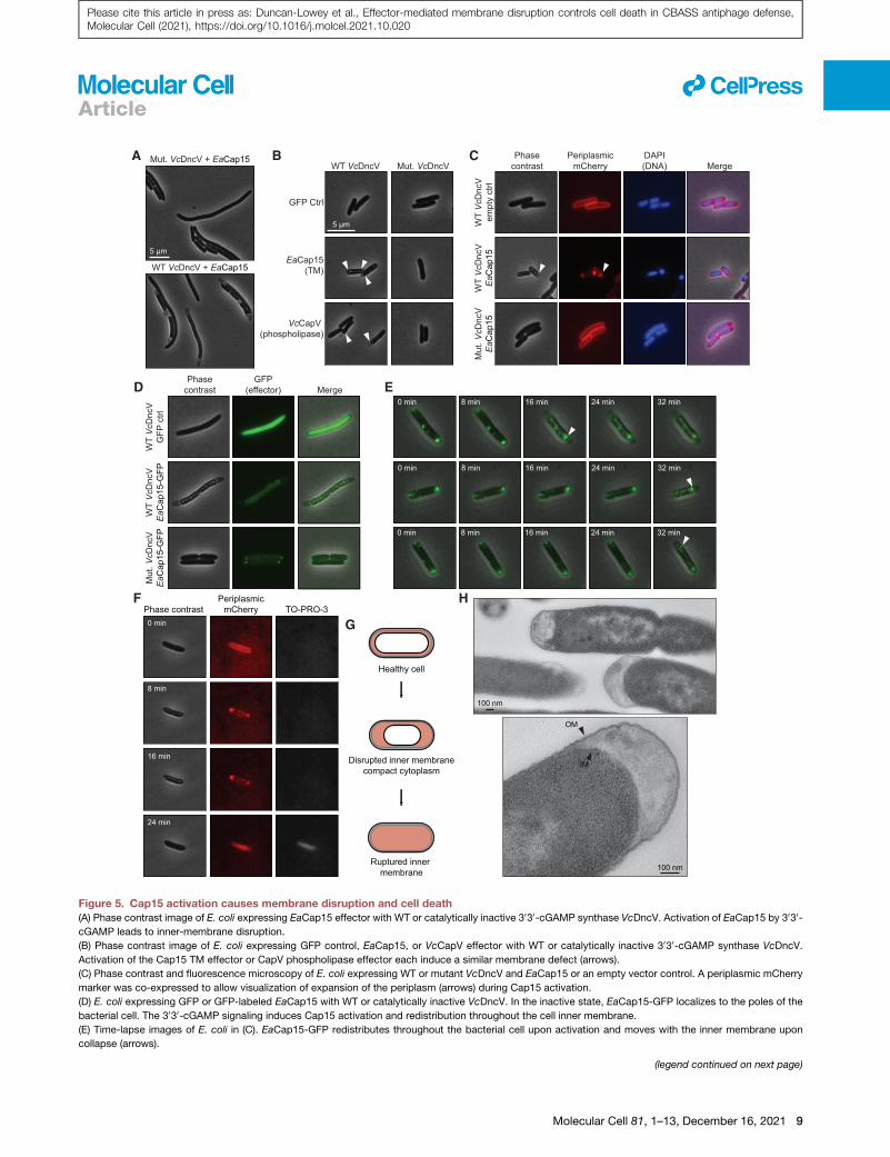
A B C
D E
F
G
H
Figure 5. Cap15 activation causes membrane disruption and cell death(A) Phase contrast image of E. coli expressing EaCap15 effector with WT or catalytically inactive 3030-cGAMP synthase VcDncV. Activation of EaCap15 by 3030-cGAMP leads to inner-membrane disruption.
(B) Phase contrast image of E. coli expressing GFP control, EaCap15, or VcCapV effector with WT or catalytically inactive 3030-cGAMP synthase VcDncV.
Activation of the Cap15 TM effector or CapV phospholipase effector each induce a similar membrane defect (arrows).
(C) Phase contrast and fluorescence microscopy of E. coli expressing WT or mutant VcDncV and EaCap15 or an empty vector control. A periplasmic mCherry
marker was co-expressed to allow visualization of expansion of the periplasm (arrows) during Cap15 activation.
(D) E. coli expressing GFP or GFP-labeled EaCap15 with WT or catalytically inactive VcDncV. In the inactive state, EaCap15-GFP localizes to the poles of the
bacterial cell. The 3030-cGAMP signaling induces Cap15 activation and redistribution throughout the cell inner membrane.
(E) Time-lapse images of E. coli in (C). EaCap15-GFP redistributes throughout the bacterial cell upon activation and moves with the inner membrane upon
collapse (arrows).
(legend continued on next page)
llArticle
Molecular Cell 81, 1–13, December 16, 2021 9
Please cite this article in press as: Duncan-Lowey et al., Effector-mediated membrane disruption controls cell death in CBASS antiphage defense,Molecular Cell (2021), https://doi.org/10.1016/j.molcel.2021.10.020

llArticle
Please cite this article in press as: Duncan-Lowey et al., Effector-mediated membrane disruption controls cell death in CBASS antiphage defense,Molecular Cell (2021), https://doi.org/10.1016/j.molcel.2021.10.020
the membranes of cells dying after Cap15 activation and
confirmed that the inner membrane specifically collapses away
from the cell wall (Figure 5H).
To expand upon these observations with Cap15, we assessed
the structurally distinct CBASS TM effector Cap14 (TM-SAVED).
Similar to EaCap15, activation of Vibrio vulnificus Cap14
(VvCap14; NCBI ref. WP_017790126.1) specifically induces in-
ner-membrane disruption (Figures S5G and S5H). Interestingly,
cells expressing each TM effector exhibit distinct morphologies,
with EaCap15 inducing collapse associated with pulling the inner
membrane away from the poles of the cell and VvCap14 sepa-
rating the cytoplasm into two segments from the center (Fig-
ure S5G). After several minutes, E. coli expressing active
VvCap14 similarly undergo inner-membrane rupture and cell
death, with the release of periplasmic mCherry into the bacte-
rium and the cells becoming permeable to TO-PRO-3 dye (Fig-
ures S5H and S5K).
Together, these data demonstrate that CBASS TM effectors
limit phage replication by disrupting the bacterial cell membrane
and initiating a cell death cascade. The data support a model in
which CBASS nucleotide second messenger signaling triggers
membrane-bound TM effectors to oligomerize into large assem-
blies that disrupt the inner membrane and induce cell death
(Figure S5L).
DISCUSSION
Our results define bacterial cell membrane disruption as a wide-
spread mechanism controlling abortive infection in CBASS anti-
phage defense. Following phage infection, CBASS immunity
begins with activation of a CD-NTase enzyme to synthesize an
antiviral nucleotide second messenger signal. We demonstrate
that TM effector proteins directly sense nucleotide second
messenger signaling and subsequently initiate a cell death
cascade that results in bacterial membrane disruption and re-
striction of phage propagation. TM effectors are the most prev-
alent form of CD-NTase-associated effector proteins (Burroughs
et al., 2015; Millman et al., 2020), revealing direct membrane tar-
geting as a critical feature of CBASS antiviral defense.
Analysis of the CBASS protein Cap15 explains a mechanism
for how TM effectors disrupt membrane integrity and terminate
phage replication. Cap15 activation specifically triggers collapse
of the bacterial inner membrane and subsequent cytoplasmic
condensation. Disruption of the inner membrane causes shrink-
ing away from the outer membrane and cell wall and eventually
results in complete membrane rupture and cell death (Figure 5).
Microscopy images of bacteria succumbing to Cap15-induced
death reveal a phenotype similar to E. coli cells dying from ge-
netic disruption of lipid homeostasis (Sutterlin et al., 2016). While
Cap15 is the most common TM effector, our results identify
(F) Time-lapse images of E. coli expressing WT VcDncV and EaCap15 in the pre
membrane collapse, the cells return to phase dark, are permeable to TO-PRO-3,
cell death.
(G) Schematic representation of membrane morphology observed upon Cap15 a
periplasm shown in red.
(H) Transmission electron microscopy of E. coli dying after EaCap15 activation, h
Light microscopy data are representative of at least three independent experime
10 Molecular Cell 81, 1–13, December 16, 2021
several additional families of CBASS TM effectors, including
Cap14 (TM-SAVED) and Cap16 (TM-NUDIX), that respond to
antiviral nucleotide second messenger signaling and similarly
induce cell death (Figure 1E). We show that Cap14 also specif-
ically triggers inner-membrane rupture (Figures S5G–S5K),
demonstrating that direct targeting of membrane integrity is a
shared feature among diverse CBASS operons. Interestingly,
Cap14- and Cap15-mediated death exhibit altered dynamics
and collapsed membrane morphologies, suggesting that while
the overall mechanism of inner-membrane disruption is
conserved, CBASS TM effectors may use distinct molecular
mechanisms to induce cell death.
Our structural characterization of Cap15 additionally explains
a mechanism for how TM effectors sense and respond to anti-
viral nucleotide second messenger signals. Crystal structures
of YaCap15 reveal a compact b-barrel domain with a hydropho-
bic lower core that enables formation of a central ligand-binding
pocket for specific nucleotide recognition (Figures 2 and 3). The
minimized architecture of YaCap15 mirrors the structural princi-
ples used for rational design of small-molecule sensors (Dou
et al., 2018), revealing a remarkable convergence between syn-
thetic protein design and natural evolution of a nucleotide-
sensing domain. The Cap15 b-barrel domain occurs in �25%
of the TM effectors in CBASS operons, representing discovery
of >600 protein sensors capable of sensitively responding to
diverse cyclic oligonucleotide signals. These b-barrel nucleo-
tide-binding domains expand the list of structurally defined
nucleotide second messenger receptors in bacterial and meta-
zoan antiviral immunity including CARF/SAVED-family proteins
and NucC receptors in bacteria (Jia et al., 2019; Lau et al.,
2020; Lowey et al., 2020; Niewoehner and Jinek, 2016),
STING-family receptors in bacteria and animal cells (Morehouse
et al., 2020; Ouyang et al., 2012; Yin et al., 2012), and RECON-
like receptors in animals (McFarland et al., 2017). We further
show that like soluble CBASS effectors, Cap15 is activated
through protein oligomerization. The organization of Cap15 as
a discrete nucleotide-binding domain fused to cell-death-
inducing TM segments further supports that CBASS effectors
use a modular architecture to couple nucleotide second
messenger recognition with diverse downstream effector func-
tions (Lowey et al., 2020). The modular organization of CBASS
effectors facilitates rapid diversification of new effector functions
to combat phage replication and likely enables CD-NTase and
STING acquisition during evolution of metazoan cGAS-STING
signaling from CBASS precursor components (Morehouse
et al., 2020; Whiteley et al., 2019).
Given membrane-targeting effectors are predominant in
CBASS defense operons, inner-membrane disruption likely pro-
vides a key evolutionary advantage in phage defense. Sequence
analysis of CBASS operons supports frequent horizontal transfer
sence of the live-dead stain TO-PRO-3 and periplasmic mCherry. After inner-
and leak mCherry throughout the cell, confirming inner-membrane rupture and
ctivation, with membranes shown in black, peptidoglycan shown in gray, and
ighlighting collapse of the inner membrane from the cell wall.
nts. See also Figures S4 and S5.

llArticle
Please cite this article in press as: Duncan-Lowey et al., Effector-mediated membrane disruption controls cell death in CBASS antiphage defense,Molecular Cell (2021), https://doi.org/10.1016/j.molcel.2021.10.020
and exchange between divergent bacterial species (Burroughs
et al., 2015; Millman et al., 2020). CBASS effector proteins medi-
ating cell death must, therefore, target ubiquitous host compo-
nents to remain functional when shared between gram-negative
and gram-positive bacteria. In line with this prediction, our re-
sults support that the vast majority of CBASS effectors target
the conserved inner membrane (TM effectors and phospholi-
pases) or dsDNA (DNA endonucleases). Targeting the host inner
membrane to control abortive infection may be particularly ad-
vantageous, as successful phage replication requires mainte-
nance of normal host membrane physiology until precisely timed
activation of lytic machinery facilitates virion release (Cahill and
Young, 2019). Interestingly, phages in the order Caudovirales,
including all dsDNA phages demonstrated here to be suscepti-
ble to CBASS TM effectors, use perturbations of the membrane
to trigger the inner-membrane proteins holin or pinholin to initiate
cell lysis and virion release (Cahill and Young, 2019). The ability of
CBASS TM effectors to prematurely disrupt membrane integrity
likely short-circuits this process and enhances antiviral defense.
Phages have evolved elaborate modification systems to protect
viral DNA from recognition and cleavage (Stern and Sorek, 2011),
and it will be interesting to uncover if mechanisms exist that allow
escape from direct host membrane disruption.
Limitations of the studyHere,we report the structure of theCBASSCap15 ligand-binding
domain and present mutational analysis of a putative ligand-
binding pocket. We also show that ligand binding induces oligo-
merization of Cap15, consistent with the activation mechanism
observed for other CBASS effectors (Lau et al., 2020; Lowey
et al., 2020; Morehouse et al., 2020). Further structural studies
of full-length Cap15 in an activated statewill be required to define
how ligand binding induces oligomerization and how CBASS TM
effector clustering results in membrane disruption. An additional
limitation of our study is that the CBASS TMeffector experiments
analyze death only in the gram-negative host E. coli. Cap15 and
CBASS TM effectors are broadly distributed among bacteria
phyla, and it will be intriguing to compare how the mechanism
of cell death and antiphage defense may differ in gram-posi-
tive hosts.
STAR+METHODS
Detailed methods are provided in the online version of this paper
and include the following:
d KEY RESOURCES TABLE
d RESOURCE AVAILABILITY
B Lead contact
B Materials availability
B Data and code availability
d EXPERIMENTAL MODEL AND SUBJECT DETAILS
B Escherichia coli strains and phages
d METHOD DETAILS
B Protein expression and purification
B Effector-induced killing assay
B Quantification of cell death by flow cytometry
B Bioinformatics analyses
B Crystallization and structure determination
B CD-NTase product thin layer chromatography
B YaCdnE product purification and analysis by HPLC
B Mass spectrometry
B Electrophoretic mobility shift assay
B Thermal shift assay
B SEC-MALS
B Plasmid and strain construction
B Plaque assays
B Microscopy
B Transmission electron microscopy
d QUANTIFICATION AND STATISTICAL ANALYSIS
SUPPLEMENTAL INFORMATION
Supplemental information can be found online at https://doi.org/10.1016/j.
molcel.2021.10.020.
ACKNOWLEDGMENTS
The authors are grateful to Tobias Herrmann, Thomas Bernhardt, Stephen
Harrison, Amy S.Y. Lee, and members of the Kranzusch lab for helpful com-
ments and discussion; Harvard University’s Center for Macromolecular Inter-
actions; Paula Montero Llopis and the Microscopy Resources on the North
Quad (MicRoN) core at Harvard Medical School; and Morten Danielsen and
Daniel Malhiero for assistance with mass spectrometry. Electron microscopy
imaging was performed in the HMS Electron Microscopy Facility. The work
was funded by the Pew Biomedical Scholars Program (P.J.K.), Burroughs
Wellcome Fund PATH award (P.J.K.), Mark Foundation For Cancer Research
(P.J.K.), Mathers Foundation (P.J.K.), Parker Institute for Cancer Immuno-
therapy (P.J.K.), the European Research Council (grant ERC-CoG 681203 to
R.S.), the Ernest and Bonnie Beutler Research Program of Excellence in
Genomic Medicine (R.S.), the Minerva Foundation (R.S.), and the Knell Family
Center for Microbiology (R.S.). B.D.-L. is supported as a Herchel Smith Grad-
uate Research Fellow.
AUTHOR CONTRIBUTIONS
Experiments were designed by B.D.-L. and P.J.K. Crystallography and
biochemical experiments were performed by B.D.-L. and N.K.M.-B. Bacterial
toxicity experiments and microscopy were performed by B.D.-L. Phage chal-
lenge assays were performed by N.T. and R.S. The manuscript was written by
B.D.-L. and P.J.K., and all authors contributed to editing the manuscript and
support the conclusions.
DECLARATION OF INTERESTS
R.S. is a scientific cofounder and advisor of BiomX, Pantheon Bioscience, and
Ecophage.
Received: May 31, 2021
Revised: August 31, 2021
Accepted: October 20, 2021
Published: November 15, 2021
REFERENCES
Adams, P.D., Afonine, P.V., Bunkoczi, G., Chen, V.B., Davis, I.W., Echols, N.,
Headd, J.J., Hung, L.W., Kapral, G.J., Grosse-Kunstleve, R.W., et al. (2010).
PHENIX: a comprehensive Python-based system for macromolecular struc-
ture solution. Acta Crystallogr. D Biol. Crystallogr. 66, 213–221.
Bernheim, A., Millman, A., Ofir, G., Meitav, G., Avraham, C., Shomar, H.,
Rosenberg, M.M., Tal, N., Melamed, S., Amitai, G., and Sorek, R. (2021).
Prokaryotic viperins produce diverse antiviral molecules. Nature 589,
120–124.
Molecular Cell 81, 1–13, December 16, 2021 11

llArticle
Please cite this article in press as: Duncan-Lowey et al., Effector-mediated membrane disruption controls cell death in CBASS antiphage defense,Molecular Cell (2021), https://doi.org/10.1016/j.molcel.2021.10.020
Burroughs, A.M., Zhang, D., Sch€affer, D.E., Iyer, L.M., and Aravind, L. (2015).
Comparative genomic analyses reveal a vast, novel network of nucleotide-
centric systems in biological conflicts, immunity and signaling. Nucleic Acids
Res. 43, 10633–10654.
Cahill, J., and Young, R. (2019). Phage Lysis: Multiple Genes for Multiple
Barriers. Adv. Virus Res. 103, 33–70.
Chen, V.B., Arendall, W.B., 3rd, Headd, J.J., Keedy, D.A., Immormino, R.M.,
Kapral, G.J., Murray, L.W., Richardson, J.S., and Richardson, D.C. (2010).
MolProbity: all-atom structure validation for macromolecular crystallography.
Acta Crystallogr. D Biol. Crystallogr. 66, 12–21.
Cohen, D., Melamed, S., Millman, A., Shulman, G., Oppenheimer-Shaanan, Y.,
Kacen, A., Doron, S., Amitai, G., and Sorek, R. (2019). Cyclic GMP-AMP sig-
nalling protects bacteria against viral infection. Nature 574, 691–695.
Dal Peraro, M., and van der Goot, F.G. (2016). Pore-forming toxins: ancient,
but never really out of fashion. Nat. Rev. Microbiol. 14, 77–92.
Dewey, J.S., Savva, C.G., White, R.L., Vitha, S., Holzenburg, A., and Young, R.
(2010). Micron-scale holes terminate the phage infection cycle. Proc. Natl.
Acad. Sci. USA 107, 2219–2223.
Dou, J., Vorobieva, A.A., Sheffler,W., Doyle, L.A., Park, H., Bick, M.J., Mao, B.,
Foight, G.W., Lee, M.Y., Gagnon, L.A., et al. (2018). De novo design of a fluo-
rescence-activating b-barrel. Nature 561, 485–491.
Emsley, P., and Cowtan, K. (2004). Coot: model-building tools for molecular
graphics. Acta Crystallogr. D Biol. Crystallogr. 60, 2126–2132.
Gabler, F., Nam, S.Z., Till, S., Mirdita, M., Steinegger, M., Soding, J., Lupas,
A.N., and Alva, V. (2020). Protein Sequence Analysis Using the MPI
Bioinformatics Toolkit. Curr. Protoc. Bioinformatics 72, e108.
Govande, A.A., Duncan-Lowey, B., Eaglesham, J.B., Whiteley, A.T., and
Kranzusch, P.J. (2021). Molecular basis of CD-NTase nucleotide selection in
CBASS anti-phage defense. Cell Rep. 35, 109206.
Hsiao, J.J., Potter, O.G., Chu, T.W., and Yin, H. (2018). Improved LC/MS
Methods for the Analysis of Metal-Sensitive Analytes Using Medronic Acid
as a Mobile Phase Additive. Anal. Chem. 90, 9457–9464.
Jia, N., Jones, R., Yang, G., Ouerfelli, O., and Patel, D.J. (2019). CRISPR-Cas
III-A Csm6 CARF Domain Is a Ring Nuclease Triggering Stepwise cA4
Cleavage with ApA>p Formation Terminating RNase Activity. Mol. Cell 75,
944–956.e6.
Jumper, J., Evans, R., Pritzel, A., Green, T., Figurnov, M., Ronneberger, O.,
Tunyasuvunakool, K., Bates, R., �Zıdek, A., Potapenko, A., et al. (2021).
Highly accurate protein structure prediction with AlphaFold. Nature 596,
583–589.
Kabsch, W. (2010). Xds. Acta Crystallogr. D Biol. Crystallogr. 66, 125–132.
Karplus, P.A., and Diederichs, K. (2012). Linking crystallographic model and
data quality. Science 336, 1030–1033.
Kawato, T., Mizohata, E., Shimizu, Y., Meshizuka, T., Yamamoto, T., Takasu,
N., Matsuoka, M., Matsumura, H., Kodama, T., Kanai, M., et al. (2015).
Structure-based design of a streptavidin mutant specific for an artificial biotin
analogue. J. Biochem. 157, 467–475.
Krogh, A., Larsson, B., von Heijne, G., and Sonnhammer, E.L. (2001).
Predicting transmembrane protein topology with a hidden Markov model:
application to complete genomes. J. Mol. Biol. 305, 567–580.
Lau, R.K., Ye, Q., Birkholz, E.A., Berg, K.R., Patel, L., Mathews, I.T., Watrous,
J.D., Ego, K., Whiteley, A.T., Lowey, B., et al. (2020). Structure andMechanism
of a Cyclic Trinucleotide-Activated Bacterial Endonuclease Mediating
Bacteriophage Immunity. Mol. Cell 77, 723–733.e6.
Liebschner, D., Afonine, P.V., Baker, M.L., Bunkoczi, G., Chen, V.B., Croll, T.I.,
Hintze, B., Hung, L.W., Jain, S., McCoy, A.J., et al. (2019). Macromolecular
structure determination using X-rays, neutrons and electrons: recent develop-
ments in Phenix. Acta Crystallogr. D Struct. Biol. 75, 861–877.
Lopatina, A., Tal, N., and Sorek, R. (2020). Abortive Infection: Bacterial Suicide
as an Antiviral Immune Strategy. Annu. Rev. Virol. 7, 371–384.
Lowey, B., Whiteley, A.T., Keszei, A.F.A., Morehouse, B.R., Mathews, I.T.,
Antine, S.P., Cabrera, V.J., Kashin, D., Niemann, P., Jain, M., et al. (2020).
12 Molecular Cell 81, 1–13, December 16, 2021
CBASS Immunity Uses CARF-Related Effectors to Sense 30-50- and 20-50-Linked Cyclic Oligonucleotide Signals and Protect Bacteria from Phage
Infection. Cell 182, 38–49.e17.
Mazzocco, A., Waddell, T.E., Lingohr, E., and Johnson, R.P. (2009).
Enumeration of bacteriophages using the small drop plaque assay system.
Methods Mol. Biol. 501, 81–85.
McFarland, A.P., Luo, S., Ahmed-Qadri, F., Zuck, M., Thayer, E.F., Goo, Y.A.,
Hybiske, K., Tong, L., and Woodward, J.J. (2017). Sensing of Bacterial Cyclic
Dinucleotides by the Oxidoreductase RECON Promotes NF-kB Activation and
Shapes a Proinflammatory Antibacterial State. Immunity 46, 433–445.
Millman, A., Melamed, S., Amitai, G., and Sorek, R. (2020). Diversity and clas-
sification of cyclic-oligonucleotide-based anti-phage signalling systems. Nat.
Microbiol. 5, 1608–1615.
Morehouse, B.R., Govande, A.A., Millman, A., Keszei, A.F.A., Lowey, B., Ofir,
G., Shao, S., Sorek, R., and Kranzusch, P.J. (2020). STING cyclic dinucleotide
sensing originated in bacteria. Nature 586, 429–433.
Mueller, M., Grauschopf, U., Maier, T., Glockshuber, R., and Ban, N. (2009).
The structure of a cytolytic a-helical toxin pore reveals its assembly mecha-
nism. Nature 459, 726–730.
Niewoehner, O., and Jinek, M. (2016). Structural basis for the endoribonu-
clease activity of the type III-A CRISPR-associated protein Csm6. RNA 22,
318–329.
Ouyang, S., Song, X., Wang, Y., Ru, H., Shaw, N., Jiang, Y., Niu, F., Zhu, Y.,
Qiu, W., Parvatiyar, K., et al. (2012). Structural analysis of the STING adaptor
protein reveals a hydrophobic dimer interface and mode of cyclic di-GMP
binding. Immunity 36, 1073–1086.
Pang, T., Fleming, T.C., Pogliano, K., and Young, R. (2013). Visualization of
pinholin lesions in vivo. Proc. Natl. Acad. Sci. USA 110, E2054–E2063.
Severin, G.B., Ramliden, M.S., Hawver, L.A., Wang, K., Pell, M.E., Kieninger,
A.K., Khataokar, A., O’Hara, B.J., Behrmann, L.V., Neiditch, M.B., et al.
(2018). Direct activation of a phospholipase by cyclic GMP-AMP in El Tor
Vibrio cholerae. Proc. Natl. Acad. Sci. USA 115, E6048–E6055.
Sonnhammer, E.L., Eddy, S.R., and Durbin, R. (1997). Pfam: a comprehensive
database of protein domain families based on seed alignments. Proteins 28,
405–420.
Steinegger, M., and Soding, J. (2017). MMseqs2 enables sensitive protein
sequence searching for the analysis of massive data sets. Nat. Biotechnol.
35, 1026–1028.
Stern, A., and Sorek, R. (2011). The phage-host arms race: shaping the evolu-
tion of microbes. BioEssays 33, 43–51.
Sutterlin, H.A., Shi, H., May, K.L., Miguel, A., Khare, S., Huang, K.C., and
Silhavy, T.J. (2016). Disruption of lipid homeostasis in the Gram-negative cell
envelope activates a novel cell death pathway. Proc. Natl. Acad. Sci. USA
113, E1565–E1574.
Tanaka, K., Caaveiro, J.M., Morante, K., Gonzalez-Manas, J.M., and Tsumoto,
K. (2015). Structural basis for self-assembly of a cytolytic pore lined by protein
and lipid. Nat. Commun. 6, 6337.
Uehara, T., Parzych, K.R., Dinh, T., and Bernhardt, T.G. (2010). Daughter cell
separation is controlled by cytokinetic ring-activated cell wall hydrolysis.
EMBO J. 29, 1412–1422.
Wang, X., and Montero Llopis, P. (2016). Visualizing Bacillus subtilis During
Vegetative Growth and Spore Formation. Methods Mol. Biol. 1431, 275–287.
Weiss, M.S. (2001). Global indicators of X-ray data quality. J. Appl. Cryst. 34,
130–135.
Whiteley, A.T., Eaglesham, J.B., de Oliveira Mann, C.C., Morehouse, B.R.,
Lowey, B., Nieminen, E.A., Danilchanka, O., King, D.S., Lee, A.S.Y.,
Mekalanos, J.J., and Kranzusch, P.J. (2019). Bacterial cGAS-like enzymes
synthesize diverse nucleotide signals. Nature 567, 194–199.
Ye, Q., Lau, R.K., Mathews, I.T., Birkholz, E.A., Watrous, J.D., Azimi, C.S.,
Pogliano, J., Jain, M., and Corbett, K.D. (2020). HORMA Domain Proteins
and a Trip13-like ATPase Regulate Bacterial cGAS-like Enzymes to Mediate
Bacteriophage Immunity. Mol. Cell 77, 709–722.e7.

llArticle
Please cite this article in press as: Duncan-Lowey et al., Effector-mediated membrane disruption controls cell death in CBASS antiphage defense,Molecular Cell (2021), https://doi.org/10.1016/j.molcel.2021.10.020
Yin, Q., Tian, Y., Kabaleeswaran, V., Jiang, X., Tu, D., Eck, M.J., Chen, Z.J.,
andWu, H. (2012). Cyclic di-GMP sensing via the innate immune signaling pro-
tein STING. Mol. Cell 46, 735–745.
Young, I., Wang, I., and Roof, W.D. (2000). Phages will out: strategies of host
cell lysis. Trends Microbiol. 8, 120–128.
Zhou, W., Whiteley, A.T., de Oliveira Mann, C.C., Morehouse, B.R., Nowak,
R.P., Fischer, E.S., Gray, N.S., Mekalanos, J.J., and Kranzusch, P.J. (2018).
Structure of the Human cGAS-DNA Complex Reveals Enhanced Control of
Immune Surveillance. Cell 174, 300–311.e11.
Zimmermann, L., Stephens, A., Nam, S.Z., Rau, D., K€ubler, J., Lozajic, M.,
Gabler, F., Soding, J., Lupas, A.N., and Alva, V. (2018). A Completely
Reimplemented MPI Bioinformatics Toolkit with a New HHpred Server at its
Core. J. Mol. Biol. 430, 2237–2243.
Molecular Cell 81, 1–13, December 16, 2021 13

llArticle
Please cite this article in press as: Duncan-Lowey et al., Effector-mediated membrane disruption controls cell death in CBASS antiphage defense,Molecular Cell (2021), https://doi.org/10.1016/j.molcel.2021.10.020
STAR+METHODS
KEY RESOURCES TABLE
REAGENT or RESOURCE SOURCE IDENTIFIER
Bacterial and virus strains
E. coli BL21-DE3 RIL Agilent 230245
E. coli BL21-DE3 New England Biolabs C2527I
Chemicals, peptides, and recombinant proteins
Ni-NTA Agarose QIAGEN 30250
HiTrap Q HP Column GE Healthcare 17115401
HiLoad 16/600 Superdex 75 PG GE Healthcare 28989333
Zorbax Bonus-RP Agilent 863668-901
SRT SEC-300 Sepax 215300-7830
[ɑ-32P] ATP Perkin Elmer BLU003H250UC
[ɑ-32P] GTP Perkin Elmer BLU006H250UC
[ɑ-32P] UTP Perkin Elmer BLU007H250UC
[ɑ-32P] CTP Perkin Elmer BLU008H250UC
PEI-Cellulose F TLC plate EMD Biosciences EM1.05579.0001
Alkaline Phosphatase, Calf Intestinal (CIP) New England Biolabs M0290S
NTPs New England Biolabs N0450S
SYPRO Orange ThermoFisher S6650
HEPES VWR 97061-824
PEG-200 Sigma-Aldrich P3015-500G
ADA Sigma-Aldrich A9883-25G
Tris[-2carboxyethyl] phosphine
hydrochloride (TCEP)
GoldBio TCEP50
3030-cUU Biolog Life Science Institute GmbH & Co. C 256
3030-cUA Biolog Life Science Institute GmbH & Co. C 357
3030-cUC Biolog Life Science Institute GmbH & Co. C 375
3030-cUG Biolog Life Science Institute GmbH & Co. C 371
3030-c-di-AMP Biolog Life Science Institute GmbH & Co. C 088
Deposited data
YaCap15 (1.9 A) This paper PDB: 7N34
YaCap15 (2.6 A) This paper PDB: 7N35
Raw data This paper https://dx.doi.org/10.17632/zdrzpgyw24.1
Software and algorithms
Phenix 1.13-2998 Adams et al., 2010 https://phenix-online.org/
Coot 0.8.9 Emsley and Cowtan, 2004 https://www2.mrc-lmb.cam.ac.uk/
personal/pemsley/coot/
Pymol v1.7.4.4 Schrodinger, LLC https://pymol.org/2/
Prism 7.0d GraphPad software https://www.graphpad.com/scientific-
software/prism/
RESOURCE AVAILABILITY
Lead contactFurther information and requests for resources and reagents should be directed to and will be fulfilled by the Lead Contact, Philip J.
Kranzusch ([email protected]).
e1 Molecular Cell 81, 1–13.e1–e5, December 16, 2021

llArticle
Please cite this article in press as: Duncan-Lowey et al., Effector-mediated membrane disruption controls cell death in CBASS antiphage defense,Molecular Cell (2021), https://doi.org/10.1016/j.molcel.2021.10.020
Materials availabilityThis study did not generate new unique reagents.
Data and code availabilityd Coordinates of the YaCap15 structures have been deposited in the RCSB Protein Data Bank under the following accession
numbers: 7N34, 7N35.
d This paper does not report original code.
d Any additional information required to reanalyze the data reported in this paper is available from the lead contact upon request.
EXPERIMENTAL MODEL AND SUBJECT DETAILS
Escherichia coli strains and phagesBL21-RIL E. coli (Agilent) and BL21(DE3) E. coli (NEB) were transformed and plated onMDG plates (0.5% glucose, 25 mMNa2HPO4,
25 mM KH2PO4, 50 mM NH4Cl, 5 mM Na2SO4, 2 mM MgSO4, 0.25% aspartic acid, 100 mg mL�1 ampicillin, 34 mg mL�1 chloram-
phenicol, and trace metals), then colonies were used to inoculate overnight MDG cultures. Overnight MDG cultures were used to
inoculate M9ZB cultures (0.5% glycerol, 1% Cas-amino Acids, 47.8 mM Na2HPO4, 22 mM KH2PO4, 18.7 mM NH4Cl, 85.6 mM
NaCl, 2 mMMgSO4, 100mgmL�1 ampicillin, 34mgmL�1 chloramphenicol, and tracemetals), which were grown in each experiment
as described below.
For phage challenge studies, E. coli strainMG1655 (ATCC 47076) was grown inMMB (LBmedia supplementedwith 0.1mMMnCl2,
5 mM MgCl2, with or without 0.5% agar) at room temperature. Whenever applicable, media were supplemented with ampicillin
(100 mg mL�1), to ensure the maintenance of plasmids. Infection was performed in MMB media at room temperature. Phages
used in this study are listed in Table S1.
METHOD DETAILS
Protein expression and purificationRecombinant TM effector proteins, Cap15 effector mutants, and CD-NTase enzymes were purified as previously described (Lowey
et al., 2020; Zhou et al., 2018). Briefly, CD-NTases and effectors were cloned into an N-terminal 6 3 His-SUMO2-tagged pET vector
and transformed intoBL21-RILE.coli (Agilent). Largescalecultures (2–4 l)weregrown for�5hat37�C, then inducedwith IPTGovernight
at 16�C. Bacterial pellets were resuspended and sonicated in lysis buffer (20mMHEPES-KOHpH7.5, 400mMNaCl, 30mM imidazole,
10%glycerol and1mMDTT) andpurifiedusingNi-NTA resin (QIAGEN). Ni-NTA resinwaswashedwith lysis buffer supplemented to 1M
NaCl and elutedwith lysis buffer supplemented to 300mM imidazole. TheNi-NTAelution fractionwasdialyzed into 20mMHEPES-KOH
pH 7.5, 250 mM KCl, 1 mM DTT overnight while removing the SUMO2 tag with recombinant human SENP2 protease (D364–L589,
M497A). Proteins were concentrated using a 10K-cutoff concentrator (Millipore) and purified by size exclusion chromatography on a
16/600 Superdex 75 column. Proteins were concentrated to > 30 mg mL�1, flash frozen with liquid nitrogen, and stored at �80�C.
Effector-induced killing assayPlasmids expressing wild-type or catalytically inactive VcDncV (D131A, D133A) (pBAD33) and an effector protein (custom pET, see
Table S1) were transformed into competent Escherichia coliBL21(DE3) (NEB) and plated onto plates withMDGmedia (0.5%glucose,
25mMNa2HPO4, 25 mMKH2PO4, 50 mMNH4Cl, 5 mMNa2SO4, 2 mMMgSO4, 0.25% aspartic acid, 100mgmL�1 ampicillin, 34mg
mL�1 chloramphenicol, and tracemetals). After overnight incubation, 5mLMDGstarter cultureswere inoculatedwith 3 colonies each
and grown overnight at 37�C with 230 RPM shaking. Cultures were diluted 1:50 into 5 mL M9ZB cultures (0.5% glycerol, 1% Cas-
amino Acids, 47.8 mM Na2HPO4, 22 mM KH2PO4, 18.7 mM NH4Cl, 85.6 mM NaCl, 2 mM MgSO4, 100 mg mL�1 ampicillin, 34 mg
mL�1 chloramphenicol, and trace metals) and grown for 3 h at 37�C with 230 RPM shaking. Cultures were then diluted 1:5 into fresh
M9ZBmedia with a final concentration of 0.2% arabinose and 5 mM IPTG to induce protein expression; 200 mL of culture were added
to 96-well plate and OD600 was recorded in technical triplicate over 300 min in a Synergy H1 plate reader (BioTek), while shaking at
37�C. Wells containing media alone were used for OD600 background subtraction. Each biological replicate was measured in tech-
nical triplicate. OD600 at 300 min (WT/mut VcDncV) was calculated as the OD600 of cultures expressing WT VcDncV and effector
divided by the OD600 of cultures expressing catalytically inactive VcDncV and effector. This value was then divided by same ratio
calculated for cultures expressing WT or mut. VcDncV and a GFP control in place of effector. This calculation is represented by
one data point in Figure 1E; all cultures used within one calculation were grown concurrently.
Quantification of cell death by flow cytometryE. coli expressing VcDncV and effector proteins were grown as induced as described above in effector-induced killing assays. After
cultures were grownwith inducer for 2 h, bacteria were diluted into 13PBSwith an approximate final dilution of 1:500 of cultures that
had an OD600 of 1. The cell-impermeable nucleic acid dye, TO-PRO-3 iodide (ThermoFisher) was added to a final concentration of
500 nM. Cells were analyzed using an LSR-II Analyzer (BDBiosciences) with excitation with a 633 nm laser and 660/20 emission filter.
Molecular Cell 81, 1–13.e1–e5, December 16, 2021 e2

llArticle
Please cite this article in press as: Duncan-Lowey et al., Effector-mediated membrane disruption controls cell death in CBASS antiphage defense,Molecular Cell (2021), https://doi.org/10.1016/j.molcel.2021.10.020
Cells expressing WT VcDncV and no effector were used to define gates on single cells using side scatter area versus side scatter
width, the to define gating for TO-PRO-3-negative live cells.
Bioinformatics analysesPreviously-identified CBASS effectors (Millman et al., 2020) were assigned to clusters with at least 20% identity over 60% of the
sequence using MMseqs2 (Gabler et al., 2020; Steinegger and Soding, 2017; Zimmermann et al., 2018). Clusters were further group-
ed into families using Pfam to identify at least two sequences within the cluster with a Pfam assignment (Sonnhammer et al., 1997).
SAVED domains within Cap14 were modeled using AlphaFold2.0 (Jumper et al., 2021). The AlphaFold prediction for the domain
previously annotated as SUa (Pfam PF18179) allowed for the identification of this family of proteins as containing SAVED domains.
Transmembrane domains were identified within experimentally determined structures where available (Mueller et al., 2009; Tanaka
et al., 2015), and were otherwise predicted using TMHMM (Krogh et al., 2001). Where modeled transmembrane domains are dis-
played, they were modeled using AlphaFold2.0 (Jumper et al., 2021).
Crystallization and structure determinationCap15 proteins were crystallized at 18�C using the hanging drop method. Concentrated protein stocks were thawed on ice and
diluted in buffer (25 mM HEPES-KOH pH 7.5, 1 mM TCEP). Drops were set by mixing protein stock and reservoir solution in 2 mL
drops over a 350 mL reservoir in 15-well Easy-Xtal trays (NeXtal). Each protein was crystallized as follows: YaCap15 W78–end C 1
2 1 crystal form, selenomethionine YaCpa15W78–endwas diluted to 10mgmL�1 andmixed at a 1:1 ratio of protein:reservoir solution
(40%PEG-200, 0.1MADApH 5.8);YaCap15W78–end P 61 crystal form, YaCap15W78–endwas diluted to 10mgmL�1 andmixed at
a 1:1 ratio of protein:reservoir solution (40% PEG-200, 0.1 M ADA pH 5.3). In each case, crystals were harvested directly from the
mother liquor without additional cryoprotectant, and flash frozen in liquid nitrogen.
X-ray data were collected at the Northeastern Collaborative Access Team beamlines 24-ID-C (P30 GM124165), and used a Pilatus
detector (S10RR029205), an Eiger detector (S10OD021527) and the Argonne National Laboratory Advanced Photon Source (DE-
AC02-06CH11357). X-ray crystallography data were processed with XDS and AIMLESS (Kabsch, 2010) using the SSRL autoxds
script (A. Gonzalez, Stanford SSRL). Experimental phase information for YaCap15 was determined using data collected from sele-
nomethionine-substituted C 1 2 1 crystal form crystals. One site was identified with HySS and an initial map was produced using
SOLVE/RESOLVE in Phenix (Liebschner et al., 2019). Model building was performed using Coot (Emsley and Cowtan, 2004), then
refined in Phenix. Statistics were analyzed as described in Table 1 (Chen et al., 2010; Karplus and Diederichs, 2012; Weiss, 2001).
CD-NTase product thin layer chromatographyPurified YaCdnEwasmixed at a final concentration of 1 mMwith 25 mMNTPs and trace a-32P-NTP in 10 mL reactions with 50mMKCl,
10 mMMgCl2, 1 mM DTT, and 50 mM CAPSO pH 9.0. These reactions were incubated for 2 h at 37�C and then terminated by treat-
ment with 1 mL of 5 units mL�1 Calf Intestinal Phosphatase (New England Biolabs) for 1 h at 37�C. 1 mL of each reaction was spotted on
a PEI-cellulose thin layer chromatography (TLC) plate (Millipore) and developed in a 1.5MKH2PO4 pH 3.8 buffer until the solvent front
was �1 cm from the top of plate. TLC plates were dried and the exposed to a phosphor screen followed by imaging with an Amer-
sham Typhoon IP (Cytiva).
YaCdnE product purification and analysis by HPLCLarge scale YaCdnE product synthesis was carried out in 20 mL reactions with 1 mM purified YaCdnE and 250 mM ATP, CTP, GTP,
and UTP in 13 reaction buffer (50mMKCl, 20mMMgCl2, 40mMCAPSO pH 9.0, 1mMTCEP), then incubated overnight at 37�Cwith
gentle shaking. The reaction was then treated with 10 mL of 5 units mL�1 Calf Intestinal Phosphatase (New England Biolabs) at 37�Covernight before filtering through a 0.2 mm filter. The reaction was diluted to 12.5 mMKCl with water, then loaded onto a 5 mL HiTrap
Q column (GEHealthcare) at 1mLmin�1 with a peristaltic pump and eluted on an AKTA FPLC (GEHealthcare) with a linear gradient of
ammonium acetate from 0–2 M over 60 min. Fractions from the two major peaks were identified by absorbance at 254 nm.
Comparison of major peak products to cyclic dinucleotide standards (3030-c-di-UMP [Biolog C 256], 3030-cUMP–CMP [Biolog C
375], 3030-cUMP–AMP [Biolog C 357]) was carried out using a C18 column (Agilent Zorbax Bonus-RP 4.6 3 150 mm, 3.5-micron).
The column was heated to 40�C and run at 1 mL min�1 with a mobile phase of 50 mM NaH2PO4 (pH 6.8 with NaOH) supplemented
with 3% acetonitrile.
Mass spectrometryYaCdnE reactions containing 25 mMATP, GTP, CTP, andUTP, 10mMMgCl2, 50mMKCl, 50mMCAPSOpH9, 1mMDTT, and 10 mM
enzyme with a final volume of 200 mL. Reactions were incubated overnight and treated with 1 mL of 5 units mL�1 Calf Intestinal Phos-
phatase (New England Biolabs) for 1 h at 37�C. Sample analysis was carried out byMS-Omics as follows. Samples were diluted 1:3 in
10%ultra-pure water and 90%acetonitrile containing 10mMammonium acetate at pH 9 then filtered through a Costar Spin-X centri-
fuge tube filter 0.22 mmNylon membrane. The analysis was carried out using a Thermo Scientific Vanquish LC coupled to Thermo Q
Exactive HF MS. An electrospray ionization interface was used as ionization source. Analysis was performed in positive ionization
mode. Peak integration was performing using Tracefinder 4.0 and the extracted data was used to create a calibration curve to quan-
tify the samples The UPLC was modified from previously-reported protocols (Hsiao et al., 2018).
e3 Molecular Cell 81, 1–13.e1–e5, December 16, 2021

llArticle
Please cite this article in press as: Duncan-Lowey et al., Effector-mediated membrane disruption controls cell death in CBASS antiphage defense,Molecular Cell (2021), https://doi.org/10.1016/j.molcel.2021.10.020
Electrophoretic mobility shift assayRadiolabeled 3030-c-di-UMP, 3030-cUMP–AMP, and 3030-c-di-AMP were synthesized by combining 25 mMNTPs and trace a32P-NTP
with purified enzymes YaCdnE (3030-c-di-UMP: UTP, a32P-UTP), EcCdnE (3030-cUMP–AMP: UTP, ATP, a32P-UTP), and V. cholerae
DncV (3030-c-di-AMP: ATP, a32P-ATP) at a final protein concentration of 1 mM in a 20 mL reaction with 5mMMgCl2, 50mMKCl, 50mM
Tris-HCl pH 9.0, 1 mM DTT. These reactions were incubated at 37�C for �24 h and then terminated by treatment with 1 mL of 5 units
mL�1 Calf Intestinal Phosphatase (New England Biolabs) for 1 h at 37�C followed by boiling at 95�C for 5 min. The radiolabeled cyclic
dinucleotide reaction products were then added at a concentration of 10 nM to reactions containing the Cap15 nucleotide-binding
domain or a no protein control in a buffer of 50mMKCl, 5 mMMgCl2, 50 mM Tris-HCl pH 7.5, and 1 mM TCEP. In wild-type YaCap15
nucleotide binding assays, bindingwasmeasured at protein concentrations ranging from 1 nM to 1 mMand inmutant YaCap15 nucle-
otide binding assays binding wasmeasured at concentrations of 100 nM and 1 mM.Reactions were incubated for 10min at 25�C then
separated on a 6% nondenaturing polyacrylamide gel (7.2 cm, 29:1 acrylamide-bisacrylamide, 0.5 3 TBE buffer) run at 100 V for
20min in 0.53 TBE buffer. The gel was immediately dried at 80�C for 1 h and then exposed to a phosphor screen followed by imaging
with an Amersham Typhoon IP (Cytiva).
Signal intensity was calculated using ImageQuant TL 8.2 software (Cytiva). Background signal intensity, taken from the no protein
control lane, was subtracted from each signal intensity reading. Percent ligand bound was calculated by dividing the signal intensity
of the bound protein-ligand complex by the total signal intensity of both the unbound ligand and the protein-ligand complex. Where
two bands are present in the bound complex, the intensity of both bands was combined to calculate the bound fraction.
Thermal shift assayPurified YaCap15W78–end b-barrel, WT or containing point mutations as indicated, was added at a final concentration of 10 mMwith
100 mM synthetic nucleotide ligands (Biolog) and 2 3 SYPRO Orange Dye in a buffer of 75 mM KCl, 20 mM HEPES-KOH pH 7.5.
Samples were slowly heated from 25 to 95�C over the course of 2 h using a qPCR CFX96 thermocycler (Biorad) and fluorescence
in the SYBR channel was measured every 0.5�C. The first derivative of each fluorescence curve was calculated, and themelting tem-
perature was identified as the peak of each derivative curve.
SEC-MALSYaCap15W78–end samples were prepared by diluting in SEC-MALS running buffer (150mMKCl, 20mMHEPES-KOH pH 7.5, 1mM
TCEP) to 1.25 mg mL�1 and incubating on ice for 5 min, then separated on an SRT SEC-150 column (Sepax). YaCap15 89–end
W120A constructs were buffer exchanged into SEC-MALS running buffer (150 mM KCl, 20 mM HEPES-KOH pH 7.5, 1 mM
TCEP) using a Superdex 200 Increase 10/300 column (Cytvia), then diluted to 1.25 mg mL�1, incubated on ice for 5 min, then sepa-
rated on an SRT SEC-150 column or SRT SEC-300 column (Sepax). Protein concentration was calculated using refractive index on a
Wyatt Optilab T-rex Refractive Index Detector assuming dn dc�1 of 0.185 and a molar mass was calculated using a Wyatt Dawn He-
leos II Multi-Angle Light Scattering detector and ASTRA software.
Plasmid and strain constructionYersinia aleksiciae and Escherichia albertii CBASS operons used in this study were synthesized by Genscript Corp. and cloned into
the pBAD plasmid (Thermofisher, cat. #43001), as previously described (Bernheim et al., 2021). Escherichia albertii and Yersinia alek-
siciae CBASS operon mutants were constructed by Genscript Corp or cloned using PCR fragments and Gibson assembly. All syn-
thesized sequences are presented in Table S1. Plasmids were transformed into E. coli strain MG1655.
Plaque assaysPhages were propagated by picking a single phage plaque into a liquid culture of E. coli MG1655 grown at 37�C to OD600 of 0.3 in
MMB medium until culture collapse. The culture was then centrifuged for 10 min at 4,000 3 RPM and the supernatant was filtered
through a 0.2 mm filter to remove remaining bacteria and bacterial debris. Lysate titer was determined using the small drop plaque
assay method as previously described (Mazzocco et al., 2009).
Plaque assays were performed as previously described (Mazzocco et al., 2009). Bacteria (E. coliMG1655 with CBASS-expressing
cells) or negative control (E. coliMG1655with a pBAD-GFP vector) were grown overnight at 37�C. Then 300 mL of the bacterial culturewasmixedwith 30mLmeltedMMBagar (LBmedia supplemented with 0.1mMMnCl2, 5mMMgCl2, 0.5%agar, and 0.2%arabinose)
and left to dry for 1 h at room temperature. CBASSoperons containing pointmutations as indicated or Cap15 transmembrane regions
were removed (YaCap15 W78–end).
10-fold serial dilutions inMMBwere performed for each of the tested phages and 10 mL dropswere spotted onto the bacterial layer.
Plates were incubated overnight at room-temperature and plaque forming units (PFUs) were determined by counting the derived pla-
ques after overnight incubation.
MicroscopyPlasmids expressing wild-type or catalytically inactive VcDncV (D131A, D133A) (pBAD33) and an effector protein (custom pET) were
transformed into competent E. coliBL21(DE3) (NEB) and plated onto plates withMDGmedia (0.5% glucose, 25mMNa2HPO4, 25mM
KH2PO4, 50mMNH4Cl, 5 mMNa2SO4, 2mMMgSO4, 0.25%aspartic acid, 100 mgmL�1 ampicillin, 34 mgmL�1 chloramphenicol, and
Molecular Cell 81, 1–13.e1–e5, December 16, 2021 e4
llArticle
Please cite this article in press as: Duncan-Lowey et al., Effector-mediated membrane disruption controls cell death in CBASS antiphage defense,Molecular Cell (2021), https://doi.org/10.1016/j.molcel.2021.10.020
trace metals), then incubated overnight at 37�C. The next day, 5 mL MDG starter cultures were inoculated with 3 colonies each, then
incubated overnight at 37�C with 230 RPM shaking. The next morning, overnight cultures were resuspended in 5 3 volume M9ZB
(0.5% glycerol, 1% Cas-amino Acids, 47.8 mM Na2HPO4, 22 mM KH2PO4, 18.7 mM NH4Cl, 85.6 mM NaCl, 2 mM MgSO4, 100 mg
mL�1 ampicillin, 34mgmL�1 chloramphenicol, and tracemetals) and grown for 4 h at 37�Cwith 230 RPM shaking. Protein expression
was then induced by resuspending cultures in 53 volume of inducing media (M9ZB, 0.2% arabinose, 5 mM IPTG), and cultures were
grown for 1.5 h at 37�Cwith 230 RPM shaking. Where indicated, the bacteria were additionally transformed with a plasmid expressing
periplasmic mCherry as previously described (Uehara et al., 2010), and subjected to additional selection with 50 mg mL�1
spectinomycin.
Where indicated, 100 mL bacteria were stainedwith dyes by adding 1 mL of 1003 stocks: DAPI (ThermoFisher D1306) 0.2mgmL�1,
FM4-64 (ThermoFisher T13320) 0.15mgmL�1, TO-PRO-3 iodide (ThermoFisher T3605) 50 mM.Bacteria were then plated on agarose
pads (1.2% agarose, M9ZB, 0.4% arabinose, 10 mM IPTG, with additional FM4-64 15 mg mL�1 or 500 nM TO-PRO-3 iodide where
indicated) and immediately covered with coverslip, as previously described (Wang and Montero Llopis, 2016). Slides were imaged
between 2 and 4 h after initial induction; during time-lapse imaging, slides were maintained at 30�C. Images were acquired using a
Nikon Ti microscope equipped with Plan Apo 1003 /1.40 Oil Ph3 DM objective and Andor Zyla 4.2 Plus sCMOS camera and Nikon
Elements 4.30 acquisition software. Adobe Photoshop was used for brightness and contrast adjustments.
Transmission electron microscopyE. coli expressing WT VcDncV and EaCap15 were grown and induced as described for phase contrast microscopy. After 2.5 h, cells
were pelleted and fixed in 2.5% paraformaldehyde, 5.0% glutaraldehyde, 0.06% picric acid in 0.2 M cacodylate buffer, followed by
osmication and uranyl acetate staining, dehydration in graded alcohols and embedded in Taab 812 Resin (Marivac Ltd., Nova Scotia,
Canada). Blocks/samples:80nm sections were cut with the Leica Ultracut Smicrotome, picked up on formvar-carbon coated slot Cu
grids, stained with 0.2% Lead Citrate, and viewed and imaged under the Philips Tecnai BioTwin Spirit or JEOL 1200x Electron
Microscope.
QUANTIFICATION AND STATISTICAL ANALYSIS
Statistical details for each experiment can be found in the figure legends and outlined in the corresponding methods details section.
Data are plotted with error bars representing the standard deviation (SD).
e5 Molecular Cell 81, 1–13.e1–e5, December 16, 2021