Radiation fields, dosimetry, biokinetics and biophysical ...
213
DE99F2868 Radiation Fields, Dosimetry, Biokinetics and Biophysical Models for Cancer Induction by Ionising Radiation 1996-1999 Mid-term Reports Association Contract between the GSF - National Research Center for Environment and Health and the European Commission Directorate General XII-F6 - Radiation Protection Research Action Editor: Dr. Jurgen Ertel, Scientific Technical Department GSF-Bericht 12/98 GSF - Forschungszentrum fur Umwelt und Gesundheit 30-10
-
Upload
khangminh22 -
Category
Documents
-
view
1 -
download
0
Transcript of Radiation fields, dosimetry, biokinetics and biophysical ...

DE99F2868
Radiation Fields, Dosimetry, Biokinetics and Biophysical Models for
Cancer Induction by Ionising Radiation1996-1999
Mid-term Reports
Association Contract between the GSF - National Research Center for Environment and Health
and theEuropean Commission
Directorate General XII-F6 - Radiation Protection ResearchAction
Editor: Dr. Jurgen Ertel, Scientific Technical Department
GSF-Bericht 12/98
GSF - Forschungszentrum fur Umwelt und Gesundheit
30-10

Herausgeber:
GSF - Forschungszentrumfur Umwelt und Gesundheit, GmbH
Ingolstadter Landstralie 1 D-85764 NeuherbergTelefon 089/3187 - 0 Telefax 089/3187 - 3372
Mitglied der Hermann von Helmholtz-Gemeinschaft Deutscher Forschungszentren (HGF)
© GSF-Forschungszentrum, 1998
ISSN 0721 -1694
Gedruckt auf umweltfreundlichem, chlorfrei gebleichtem Papier

KS002393749 R: KSDE012159955
Radiation Fields, Dosimetry, Biokinetics and Biophysical Models for Cancer Induction by Ionising
Radiation 1996 - 1999
EC-GSF Association Contract FI4P-CT95-0011 Project Leaders Mid-term Reports for the Period
1996-1997
Responsible Project Leaders and Authors:
Dr. Peter Jacob, GSF-ISS, Germany
Professor Barry D. Michael, Gray Laboratory, United Kingdom
Dr. Herwig G. Paretzke, GSF-ISS, Germany
Dr. Paul Roth, GSF-ISS, Germany
Professor Dennis O’Sullivan, DIAS, Ireland
Editor:
GSF - National Research Center for Environment and Health EC-GSF Secretary to the Steering Committee
Dr. Jurgen Ertel

Table of Contents
Page
Preface i-n
The Projects
A Study of Radiation Fields and Dosimetry at Aviation Altitudes 1-31
List of Participants and Addresses 1
1. Objectives 2
2. Progress
2.1. Work Package 1 3Calilbration of passive and real time detectors to high energy radiationfields. Instrument and dosemeter characterisation and comparison
2.2. Work Package 2 8Measurement of cosmic ray neutrons and their spectrometry at mountainand aviation altitudes
2.3. Work Package 3 11Measurement of route doses on routine flights of ionising radiation and photon radiation and measurement of flux ofZ >2 particles at supersonic and subsonic aviation altitudes on a wide range of routes.Measurement of LET spectra
2.4. Work Package 4 16Calculation and modelling of the spectra of neutrons, protons and heavy charged particles at aviation altitudes, and of the response of instruments for detecting these particles. Calculation of dosimetric quantities.Verification of cosmic models by experimental data. Evaluation of results from TEPC. Comparison and correlation with passive and real timedetectors
3. Summary of Main Achievements 21
4. Research to be Performed in the Remainder of the Project 24
5. Publications 26
6. Executive Summary 27
7. Annex 31Summary of work packages and role of individual laboratories

B Biokinetics and Dosimetry of Incorporated Radionuclides 32-83
List of Participants and Addresses 32
1. Objectives 33
2. Progress
2.1. Work Package 1 34Biokinetics of ingested radionuclides and dosimetry of thegastrointestinal tract
2.2. Work Package 2 42Biokinetics and dosimetric models for systemic radionuclides
2.3. Work Package 3 49Target cell dosimetry for short-range particles
2.4. Work Package 4 60Numerical impications of models
3. Summary of Main Achievements 65
4. Research to be Performed in the Remainder of the Project 72
5. Publications 75
6. Executive Summary 80
C Biophysical Models for the Induction of Cancer Radiation 84 -129
List of Participants and Addresses 84
1. Objectives 85
2. Progress
2.1. Work Package 1 86Mechanistic models for radiation oncogenesis
2.2. Work Package 2 89Mechanistic models for chromosome aberrations
2.3. Work Package 3 92Mechanistic models for mutagenesis
2.4. Work Package 4 93Mechanistic models for DNA damage and repair
2.5. Work Package 5 100Chemical pathways involving initial track species in cells

1062.6. Work Package 6Production of initial track species in mammalian cells
2.7. Work Package 7 110Transport of radiation to cells of interest
3. Summary of Main Achievements 115
4. Research to be Performed in the Remainder of the Project 118
5. Publications 120
6. Executive Summary 127
Dose Reconstruction 130 - 161
List of Participants and Addresses 130
1. Objectives 131
2. Progress
2.1. Work Package 1 132EPR with teeth
2.2. Work Package 2 135Chromosome painting (FISH) in lymphocytes
2.3. Work Package 3 136Luminescence techniques
2.4. Work Package 4 143Dose modelling
2.5. Work Package 5 147Evaluation of dose reconstruction outside NIS
3. Summary of Main Achievements 149
4. Research to be Performed in the Remainder of the Project 152
5. Publications 154
6. Executive Summary 156
7. Annex 161Institutes contributing to the work packages

E Experimental Data for the Induction of Cancer by Radiation of 162 - 199Different Qualities (EDICAR)
List of Participants 162
1. Objectives 163
2. Progress
2.1. Work Package 1 166Initial physical/chemical events in DNA damage induction and relatedaspects of modelling
2.2. Work Package 2 171Data for DNA damage induction
2.3. Work Package 3 176Processing of DNA damage, especially DSB
2.4. Work Package 4 180Chromosomal aberrations
2.5. Work package 5 183Dependence of mutational yield and molecular spectra on radiationquality
2.6. Work Package 6 185Microbeam and measured track irradiation of individual cells
3. Summary of Main Achievements 188
4. Objectives for the Remainder of the Project 190
5. List of Publications 191
6. Executive Summary 196

PrefaceII . Ill lllllllli 1:111 llll i!i II lllll i:|l II^DEO12159955*
In December 1995 the GSF-National Research Center for Environment and Health entered into an Association Contract (FI4P-CT95-0011) with the European Atomic Energy Community, represented by the European Commission (EC).
The Association Contract assigns to GSF the responsibility to carry out research projects with Associated contractors in the Member States of the European Union and to manage and coordinate complementary research work to be carried out in the field of radiation protection within the framework of the Nuclear Fission Safety Programme.
The contract period is four years (1 January 1996 to 31 December 1999). Initially, the contract comprised 3 research projects with 23 partners from EU-Member States and Switzerland (funded by Swiss Government). The total costs were 5.7 Mio ECU with a contribution of 2.6 Mio ECU by the European Commission. Subsequently, supplementary agreements to the Association Contracts have been concluded increasing the number of projects to 5 and the total number of partners involved to 41 from 12 different countries. The total costs now are 8.8 Mio ECU with an EC contribution of 4.75 Mio ECU.
The GSF has assumed overall responsibility for coordination of the work of the five multinational projects. To this extent the GSF signed Specific Agreements with each of the participating organisations (Associated Contractors) laying down the rights and duties with which the GSF carries out its coordinating role. Each project has its scientific project leader coordinating the work of the partners. The GSF is responsible for financial administration. The implementation of the Association Contract is supervised by a Steering Committee consisting of members of the EC and GSF (see table). GSF is providing the secretarial and administrative support to the Committee.
Members of the Steering Committee:
EC GSF
Mr. E. Andreta (Chairman)Dr. J. Sinnaeve (Deputy Chairman) Mrs. M. Wauters Dr. H. Menzel Mrs. C. Graf
Professor E.-G. Afting Professor A M. Kellerer Dr. J. Kinder Professor W. Gdssner Dr. J. Ertel (Secretary)
The Association Contract covers a range of research domains that are important to the Radiation Protection Research Action, especially in the areas "Evaluation of Radiation Risks" and "Understanding Radiation Mechanisms and Epidemiology". Three research projects concentrate on radiation dosimetry research and two projects on the modelling of radiation carcinogenesis.
The main objectives of the first dosimetry project are the measurement of neutron and charged particle flux and energy spectra at altitudes in civil aviation, the determination of response characteristics for detectors, the investigation of calibration procedures, and the evaluation of exposures of aircrews.
I

I The overall objective of the second dosimetry project is to improve estimates of dose following the I intake of radionuclides by adults and children. The work includes the development of biokinetic and j / dosimetric models, including models of the gastrointestinal tract, for the systemic behaviour of
! / radionuclides, and for the developing embryo and foetus. Further subjects are target cell dosimetry for 7 short-range particles and the development of computational tools for sensitivity and uncertainty analysis ' models.
| The third dosimetry project encompasses the study of different methods far retrospective dose; assessments for individuals or groups of individuals accidentally exposed to increased levels of
radiation. The methods investigated include electron paramagnetic resonance (EPR) of tooth enamel and chromosome painting (FISH) for lymphocytes in peripheral blood for individual retrospective dose assessments, luminescence techniques on materials in inhabited environment (ceramics, bricks) and model calculations using environmental data as input.
The two projects in the sector "Understanding Radiation Mechanisms and Epidemiology" have as a main goal the development of quantitative mechanistic models for the induction of late effects in man by low doses of radiation, at low dose rates, and of different radiation qualities to improve the present base of radiation risk quantification. In the first project mechanistic models for DNA damage and repair, chromosome aberrations, mutagenesis and radiation oncogenesis will be improved to achieve a more complete understanding of radiation action on the molecular, cellular and tissue levels. The second project complements the first by carrying out quantitative studies of radiation induced molecular and cellular effects that are critical for the model development. They range from initial physical/chemical events in DNA damage induction to chromosomal aberrations and radiation quality studies for these effects. Part of the work is carried out using a microbeam irradation facility enabling single cell / single particle irradiations.
Annual meetings of the members of the Steering Committee and the scientific project leaders ensure an effective management of the projects and strengthen the exchange of information between the project leaders. Additional administrative meetings of the Steering Committee members in Brussels ensure the regular monitoring of progress. The Association Contract fosters a close integration of dedicated molecular and cellular biological experimental and theoretical work. This is further supported by cluster meetings with the EC-NRPB Association. The research topic here is: "Molecular mechanisms and genetics of radiation carcinogenesis, including in utero exposure". In general, a close link between partners in the different ‘Associations’ established between the EC and NRPB in UK, CIEMAT in Spain, CEA in France and GSF in Germany will certainly contribute to increased and more effective collaboration which is important for the development of radiation protection.
To date the work of the five GSF-administered contracts has produced excellent advances, reflected in the mid-term review reports. Therefore, the Steering Committee decided to publish these reports as a GSF-report with financial support through the Grant ERBFI-CT96-2007.
E. Andreta E.-G. AftingDirector DG XII-F President of the GSF -RTD Actions: Energy National Research Center for
Environment and Health
II
A
Study of Radiation Fields
and
Dosimetry at Aviation Altitudes

Study of Radiation Fields and Dosimetry at Aviation Altitudes
Contract No: F14P-CT95001 laMid-Term Report for the period 1 January 1996 to 31 December 1997
Co-ordinator: D. O’Sullivan, Dublin Institute for Advanced Studies
Contractors and Sub-contractors:
1. DIAS Dublin Institute for Advanced Studies, (DIAS), 5 Merrion Square, Dublin 2, IrelandD. O’Sullivan, D. Zhou, J. Donnelly, R. Keegan, E. Flood
2. ANPA National Agency for Environmental Protection , Via V. Brancati 48, Roma 00144, ItalyL. Tommasino, M. Cavaioli, Jin Hua, and M. Notaro
National Institute of Nuclear Physics, INFN-Frascati, RomaM. Pelliccioni
3. GSF National Research Center for Environment and Health, Ingolstaedter Landstrasse 1, D-85764 Neuherberg, GermanyH. Schraube, G. Leuthold
LMU, Ludwig-Maximilian-University, Munich, GermanyV. Mares
CERN-Geneva, SwitzerlandS. Roesler
3a Subcontractor: PTB - Physikalisch-Technische-Bundesanstalt, Bundesallee 100, D-38116 BraunschweigB. Siebert
3b Sub-contractor: Prof. Heinrich, Dept, of Physics, University of Siegen, Adolf- Reichwein-Strasse, D-57078 Siegen, GermanyW. Heinrich
4. USAAR University of Saarland, Centre of Environmental Research, Dept, for Radiation and Environmental Biophysics, D-66125 Saarbrucken (Dudweiler), GermanyR. Grillmaier, T. Lim, S. Gerdung, E. Arend
4a Sub-contractor: SSI, Swedish Radiation Protection Institute, Dosimetry Laboratory, S-171, 16 Stockholm, SwedenL. Lindborg, Jan-Erik Kylldnen
5. NRPB National Radiological Protection Board, Chilton Didcot, Oxon. 0X11 ORQ, UKD. Bartlett, L. Hager, R. Tanner
5a Sub-contractor: CERN, European Laboratory for Particle Physics, CERN 1211, Geneva 23, SwitzerlandM. Hoefert, T. Otto, M. Silari
-1 -Study of Radiation Fields and Dosimetry at Aviation Altitudes F14P-CT9J001 la

1. Objectives
The main objective of the project is to measure the flux and energy spectra of neutrons and charged particles over a wide energy interval at aviation and mountain altitudes and to compare results with those calculated using various transport codes. The determination of the response characteristics and the investigation of calibration procedures for active and passive detectors will be undertaken and methods for the confirmation, by measurement, of calculated route doses will be established. An assessment will be made of risk related protection quantities and operational quantities. The results will be used to evaluate the exposure of aircraft crew over a wide range of altitudes and geographical locations.
1.1 Work Package 1
The objectives have been to characterise, intercompare, calibrate and develop where necessary, both active and passive instrumentation for the dosimetry of the radiation field at aircraft altitude. A significant part of this package is the provision and characterisation of the CERN-CEC reference field (CERE) which simulates the major characteristics, in particular the neutron spectrum of the radiation fields at aircraft altitudes. It was also planned to undertake exposures at heavy ion accelerators and high energy neutron and proton beam facilities in order to gather detailed information in specific areas of investigation as outlined in section 2 (Progress)
1.2 Work Package 2
The main objective in this work package is to measure the contribution of cosmic ray neutrons to the radiation field at aircraft altitudes, and to investigate and measure the neutron spectrum. Several instruments, both active and passive, are involved in these studies. One essential aim was to get the support of international airlines to provide facilities for the project. Another important objective is to measure ambient dose equivalent and LET spectra at aviation and mountain altitudes using TEPC techniques and other measurements.
1.3 Work Package 3
The main objectives include measurement of route doses on routine flights of low and high LET ionising radiation and photon radiation; measurement of LET spectra on a variety of routes, and determination of the charge spectra of Z>2 cosmic ray primary and secondary nuclei. Again, support of international airlines is essential and subsonic and supersonic routes are to be included where possible. A further aim is to investigate the reproducibility of the neutron route dose.
1.4 Work Package 4
The neutron component is a very significant contributor to the radiation dose at subsonic and supersonic flight altitudes. The research undertaken in this part of the project deals with both experimental and theoretical aspects of the problem. The main objectives include the following: calculation and modelling of the spectra of neutrons, protons and heavy charged particles at aviation altitudes and of the response of instruments for detecting these particles; calculation of dosimetric quantities; verification of cosmic models by experimental data. Evaluation of results from TEPC measurements and comparison and correlation of data with that obtained with passive and real time detectors, is also planned.
-2-
Study of Radiation Fields and Dosimetry at Aviation Altitudes FI4P-CT95001 la
2. Progress
2.1 Work Package 1: Calibration of passive and real time detectors to high energyradiation fields. Instrument and dosemeter characterisation and comparison.
The main objectives of Work Package 1 are to characterise, intercompare, calibrate and develop, where necessary, instrumentation, both active and passive, for the dosimetry of the radiation field at aircraft altitudes. A significant part of this package is the provision and characterisation of the CERN/European Commission Reference Field (CERE) which simulates the major characteristics, in particular the neutron spectrum, of the fields at aircraft altitudes.
CERN has provided irradiations and beam monitoring at the CERM facility for two scheduled runs in each of 1996 and 1997, with an additional unscheduled run in 1997, at which there was participation from DIAS, ANPA, USAAR, SSI and NRPB. Reports on each run are produced by CERN (Otto T and Silari M (1996a) (1996b) (1997).
The monitoring of the beam is carried out by CERN with some assistance on the field characterisation by measurement being given by the participating contractors and sub contractors. A recalculation of the field components was carried out in 1996 by collaborators from the Universita di Milano and the Institute Nazionale di Fisica Nucleare, Sezione di Milano, details of which are given in an interim report (Bartlett D T, Hager L G et al 1996). The calculated values of the neutron component of the field show good agreement with TEPC measurements. The suitability of the CERE facility for the intercomparison, response characterisation and calibration of active and passive neutron dosimetry systems is illustrated in Figure 2.1.1 which gives in histogram form the neutron fluence (la) or effective dose (lb) contents of bins (in such a form as to approximate a lethargy plot) of CERF in the concrete shielded roof area and, for comparison, calculations of the neutron field at a depth in the atmosphere of 200 g cm"2 (approximately 12 km) by O’Brien (1997), and by Roesler S, Heinrich W and Schraube H (Roesler S et al 1997).
Modifications and improvements to the CERF facility were made in 1997 and the calculations of the field component have been redone, and full measurements made for the modified shielding using a TEPC. New spectrometry measurements with a set of Bonner spheres including a high energy channel have been made by the CERN group in collaboration with the Universita di Milano, and by the GSF group.
Full reports have been drafted on the measurements made by contractors and sub-contractors at the CERF facility using passive and active dosimetry systems. Included in these reports are results for dosimetry systems for other users of the CERF facility. The results for both passive and active systems have been analysed in terms of the assessment of both the operational quantity, ambient dose equivalent, and the protection quantity, effective dose, for both a standard calibration field (Am-Be or252
Cf) normalisation of response and by comparison of the observed reading with that calculated from the field fluence spectrum and the dosemeter or instrument response characteristics. Good agreement has been found between the observed and predicted response for both passive dosemeters and instruments. Several papers have already been published on these results (Hofert et. al (1997)), Stevenson et. al (1996), Birattari et. al (1997)). The CERF facility is also being used to directly calibrate a passive dosimetry svstem to determine route doses (Bartlett et al (1997a),( 1997b). Hager et al(1997)).
Measurements concerning the determination of the response of two TEPC detectors (a spherical one and one of cylindrical shape which have been used in connection with HANDIs on in-flight measurements and in the reference field provided by CERN and on other occasions of relevance) for neutrons with energies of 46 and 65 MeV have been performed at PSI in Switzerland in co-operation with colleagues of PSI and PTB in Braunschweig. The final evaluation is not y et finished because of some problems concerning the determination of the irradiated detector area. Furthermore the correction of the monitor values as a measure of the neutron fluence has not vet been performed. A
-3 -
Study of Radiation Fields and Dosimetry at Aviation Altitudes FI4P-CT9500I la

rough estimation gives a relative H*(10) response of 50% for the cylindrical detector (relative to a :>:Cf calibration). There are some indications, that after correction the response value will be increased. Measurements of response characteristics using neutrons of much higher energy, in the range 100 to 160 MeV, are planned to be carried out at The Svedberg Laboratory in Uppsala.The influence of wall thickness has also been investigated in the PSI neutron field using tissue equivalent caps of 5 to 30 mm thickness. The results are interesting from a scientific point of view.
Fig 1a
0.5
Energy (eV)
Fig 1b
0.4
------ Roesler 260 g/crn' (FlUKA)Femrt C£RF 120*v« T3 concrete (FLUKA)
0.3
«r|0.2 -
0.1pu
r*
Energy (eV)
Figure 2.1.1 Comparison of Calculated Neutron Spectra at CERF and at Aviation Altitudes as Fluence (a) and Effective Dose (b)
Dose response measurements have been carried out at GSI in Darmstadt, Germany using high energetic heavy ions. The measured values of lineal energy are close to the calculated values of dE/dx.
Measurements with HANDIs equipped with the two TEPCs (of different radiation sensitivity) mentioned above in a low level background laboratory of PTB have been carried out in order to determine the noise of the TEPCs. As the measurements were only performed in December 1997, the results are not yet evaluated.
As well as active instruments, as described above, to measure dose equivalent rates at aircraft altitudes and to estimate route doses as required, passive dosemeter systems may also be used. In one case the determinations of route doses (and dose rates) are made using a ‘passive survey meter’ developed by NRPB under this contract. This consists of large numbers (to increase the precision) of routine-use passive personal dosemeters, in this case TLDs to estimate the non-neutron component, and PADC track etch detectors to estimate the neutron component (see Bartlett et al 1997a and 1997b). Doses of 50-100 pSv may be measured with a precision of 10-15%. The response of the neutron detectors is normalised to that determined in the CERF. The robustness of this approach is supported by a comparison of the measured response in CERF with that calculated for CERF and for the calculated
-4-Study of Radiation Fields and Dosimetry at Aviation Altitudes F14P-CT950011a

field spectrum at aviation altitudes using the detector’s response characteristics. This comparison is shown in Table 2.1.1. Data on the high energy neutron response characteristics of the detectors has been obtained from irradiations at PSI. and, during 1997, at The Svedberg Laboratory at Uppsala. Progress has also been made on an investigation of sheet-to-sheet and batch-to-batch variations in detector response characteristics.
Table 2.1.1 Neutron Spectra and PA DC Response
Calculated neutron spectrum
E(ISO) weightedconversioncoefficient
PADC Reading
(pSv cm )per unit fluence per unit E(ISO)
calculated measured calculated measured
(tracks cm 10"6) (tracks mSv"1)-2
Roesler 200 g cm FLUKA 220 20.7 94.1
O’Brien 200 g cm LUIN 111 13.5 122
Ferrari CERFt FLUKA 245 26.6 27.8<X) ± 2.827.9(b) ± 1.7
108 113 ± 11114 ± 7
t concrete shielded, position T3 (a) 1996 calibration (b) 1997 calibration
In the second case a multi detector stack (ANPA) has been developed, the most important detectors of which are bubble detectors for the measurement of low-energy neutrons, bismuth detectors for the registration of high energy nucleons (neutrons and protons) and thermoluminescent dosemeters for the assessment of the low-LET radiations. Special efforts have been devoted to the study and the calibration of the bismuth fission-track detector, which has its principal response to high energy nucleons (neutrons and protons). Information on the neutron spectra is obtained from several different passive neutron detectors also, included in the ANPA stack, and on the neutron incident angle allowing the evaluation of effective dose.
A major concern in the assessment of the aircrew exposure has been the accurate measurement of high energy nucleons, which produce nuclear disintegrations (stars) in tissue. For this reason, extensive investigations have been carried out to study the bismuth fission track detector, which has its principal response to nucleons with energy greater than 50 MeV. Calibrations of bismuth fission detectors with high energy protons, previously carried out in DUBNA, have been repeated using proton beams of 100, 150 and 250 MeV from the Paul Scherrer Institute (Switzerland). Finally these detectors have been calibrated with neutron beams of 100 and 160 MeV from The Svedberg Laboratory, Uppsala. In the past, the detector calibrations at high energy neutrons have been always hampered by the lack of monoenergetic beams, the high energy psuedoenergetic ‘peak’ being accompanied by lower energy neutrons. In the case of the bismuth detector calibration at Uppsala, this problem is less important because the neutrons with energy below 50 MeV do not induce fission reactions. Furthermore, the Uppsala neutron beam has been extensively characterised with time-of-flight spectrometry with detectors based on the real-time analogues (thin-film breakdown counters) of fission track detectors. The information provided by Uppsala for the calibration was respectively the total number of neutrons, the total number of the fission-induced in the bismuth and the total number of fissions induced by the neutrons within the high energy peak. With these data, the calibration of the bismuth detectors was straightforward, in spite of the fact that the neutron beam was not monoenergetic.
-5 -Study of Radiation Fields and Dosimetry at Aviation Altitudes F14P-CT95001 la

The calibration data with neutrons have been compared with those obtained with protons of the same energies (100 and 150 MeV). This comparison has proved experimentally what had been claimed by Cross and Tommasino (1997), i.e., that the response of Bi-detectors is directly proportional to the dose equivalent of mixed fields of neutrons and protons. This makes it possible to measure the dose without the need to differentiate neutrons from protons. On the other hand, when the Bi-detector is combined with other neutron detectors capable of measuring low and high energy neutrons (such as the extended rem counter), it becomes possible to differentiate the dose due to protons from that of neutrons.
The ANPA stack, which contains both bubble and bismuth-detectors, measures neutrons over the entire energy range. Since July 1993, this stack has been exposed several times in the concrete shielded field of CERF. Within the contract period, the ANPA stack has been irradiated respectively in the runs of May 1996, August 1996 and August 1997. The data available to date refer only to the 1996 runs. The neutron dose equivalent has been evaluated using conversion factors to MaDE given in ICRP Publication ICRP 21, and to ambient dose equivalent (using ICRP Publication 60 Q(L)) - H*(10) new, using conversion factors, given by Siebert and Schumacher (1995) for neutron energies up to 200 MeV, and above 200 MeV values from Sannikov and Savitskaya (1995). Since the bismuth fission detector has a response proportional to the dose equivalent conversion factor from 150 MeV up to a few GeV, the Bi-detector response obtained with 150 MeV neutrons and the conversion factor at the same neutron energy have been used to obtain the dose equivalent.
Table 2.1.2 reports the doses (mSv) obtained with the ANPA stack using the two different conversion factors mentioned above for the 1996 CERN exposures. Included in the table are the values of H*(10) old and new Q(L) obtained by CERN using the HANDI-type TEPC. In both measurement campaigns the MaDE dose values determined by the ANPA stack are about 30% higher than the H*(10) values. These discrepancies seem to be due to the differences in the evaluation of, and conversion coefficients for, the high energy neutron component. In the past these high energy nucleons have created much concern in the assessment of aircraft crew exposure, because they interact with tissue mainly by producing nuclear disintegrations (stars).
When using the ambient dose equivalent, H*(10), the dose values of the stack are close to those obtained with the TEPC (see Table 2.1.2). This agreement of H*(10) values obtained with the HANOI and the stack may not be necessarily encouraging, since the H*(10) conversion factors for high energy neutrons (specially for neutrons of about hundreds of MeV) H*(10) are relatively low and the major cause of the difference between the two measurement approaches is in part eliminated. It may be worth mentioning that at high neutron energies, H*(10) underestimates effective dose, E, for physical reasons. Its value as an operational quantity becomes questionable at these high energies, since as protection quantity, instead of over-estimation, provides under-estimation (Siebert et. al, 1994; Pellicioni, 1997). Furthermore, the response of the TEPC to nuclear disintegration events needs to be clarified. With most dosimetric systems, such as the extended rem-counter (Linus) and those based on LET-related quantities (TEPC, PADC), the high energy nucleon component cannot be selectively measured. Monitoring of the fluences of the star-producing radiations should be considered a minimum course of action (Reitz et. al, 1996), because of the continuous changes of dose conversion factors (with special regard to high energy neutrons) and the limited knowledge of the radiobiological effects of these radiations.
The accurate assessment of effective dose requires the knowledge of the neutron incident angle during the exposure. In the July/August run of the CERN high energy exposure, an experiment was carried out to identify the direction under which the high energy neutrons emerge from the shielding. Two stacks have been placed respectively in a parallel and perpendicular position with respect to the concrete shielding floor. The ratio between the response of PADC and Polycarbonate track detectors perpendicular to the concrete floor to that of track detectors parallel to the same floor was about 57±5 percent. With the Bi-detectors this ratio was 85%, indicating that this detector response has little dependence on the neutron incident angle.
- 6 -Study of Radiation Fields and Dosimetry at Aviation Altitudes FI J:'-CT95001 la

In the third approach, DIAS stacks are used to determine the fluence and type of charged particle present at a point in a radiation field and, in addition, its LET, thus allowing a calculation of dose and dose equivalent. Nuclear track detectors used in the investigations at flight altitudes were calibrated over the region of ionisation employed in the project. Calibration exposures were carried out at the Berkeley Bevalac prior to its closure in 1993 and at GSI, Darmstadt at various times between 1993 and 1997. Carbon ions of energy ~80 MeV/n were stopped in detector stacks and REL and LET values were measured as a function of range. The response of the detectors to stopping calcium and iron ions of initial energy ~300 MeV/n and 506 MeV/n respectively was also measured.
Table 2.1.2
ANPA stack (mSv h'l)
MaDE H*(10) new
CERNTEPC (HANOI) (mSv h') H*(10) old H*(10) new
May 1996 3.18 ±0.28 2.20 ±0.18 2.04 ±0.30 2.29 ±0.26
August 1996 1.95 ±0.15 1.65 ±0.20 1.48 ±0.21 1.74 ±0.20
The investigation provided calibration for LET values in the range 15 keV/pm LET 700 keV/pm covering most of the spectrum of ionisation observed in the detectors flown at aircraft altitudes. In addition, the response obtained for similar detectors in the range 5 keV/pm LET 20 keV/pm by exposure to protons (Keegan, 1996) was used to extend the data into this region as shown in Figure. 2.1.2. The data for the USF-3 and USF-4 detectors indicate a very similar response below 50 keV/pm with the USF-4 material exhibiting a higher response above this value (see Figure 2.1.2)
LET Calibration Curve(USF3 CR-39 & USF4 CR-39)
° USF3 Data ( Carbon, Calcium & Iron Ions ) • USF4 Data (Carbon Ions)- USF3 ( Keegan, 1996 )
(KeV/Micron)
Figure 2.1.2 Reduced etch rate versus LET for detectors used in the investigations at flight altitudes
-7-Study of Radiation Fields and Dosimetry at Aviation Altitudes FI4P-CT95001 la
Although DIAS detectors used for the investigation of Z>2 nuclei and LET spectra at aircraft altitudes are calibrated using heavy ions at GSI Darmstadt and other accelerator centres, it was decided to measure doses observed at the CEC-CERN neutron reference field. This exposure had two objectives, namely (a) to compare DIAS detector results for high LET component of the spectrum with those obtained by the HANOI TEPC instrument provided by the University of Saarland, (b) to estimate the level of agreement between the CEC-CERN approach and that of the DIAS results which are based on an independent calibration using heavy ions. The DIAS measurement assumes that, in the absence of detailed knowledge of the angular distribution characteristics of the field, the deviation from isotropy can be taken into account by a factor of -0.76 in the case of the present dose equivalent calculations (D Bartlett, private communication). The accumulated dose equivalent measured by the DIAS detectors in the July/August 1996 exposure was 1.65±0.21 mSv (ICRP Publication 60 Q(i)) compared to 1.73±0.25 (ICRP Publication 60 Q(i)) measured by the TEPC, for the high LET component.
The agreement is good and verifies that the two approaches to calibration, namely exposures to the CEC-CERN reference field and the use of heavy ions with known energies and charges provide dependable and independent methods for accurate LET and dose equivalent investigations. This result has further significance in that it is known that the method of track measurement used by the DIAS group can give rise to an underestimate of some high LET events because of the limitations of the use of optical methods of measurement. This loss has been estimated to be less than 10% (R Keegan, PhD Thesis, University College Dublin, 1996). The present DIAS results, therefore, are consistent with these findings.
2.2 Work Package 2: Measurement of Cosmic Ray Neutrons and their Spectrometry atMountain and Aviation Altitudes
Comments on the Exposure Facilities at High Altitudes:
Each contractor has tried to arrange facilities etc., for exposing detectors within airlines companies. For contractors using passive detectors, it was sometimes possible to make the necessary arrangements for the exposures on the aircraft, such as in the case of the DIAS and the NRPB groups, which have exposed detectors on the British Airways Concorde. In particular the NRPB group has arranged to fly passive detectors on NASA flights for a total of 20 hours.Limited or no support has been provided to contractors to expose real-time instrumentation mainly because of possible interferences with the onboard instrumentation. The US AAR and the NRPB groups have succeeded in arranging some facilities for the exposure of single-type real-time instruments in a few flights.The ANPA-Alitalia collaboration was an exception since it made it finally possible to carry out onboard comparison of measurements of different advanced real-time instruments. Furthermore, these inflight exposures have been carried out for such a long time that it was possible to compare the measurements of both active and different new type of passive detectors.The ANPA-Alitalia survey was requested by the Central Medical Inspectorate of the Ministry of Labour and was funded by ANPA under a special project approved by the ANPA board of directors.For these reasons the funds available were by far greater than those which could have been obtained under the present cost-sharing CEC contract, which represents a separate project.ANPA provided funding to Alitalia to get special support and arrangements for deployment of detectors for comprehensive inflight measurements. The facility has been made available to contractors (USAAR, DIAS) and other institutions using either passive detectors (DIAS) or real-time detectors approved to fly.Because of the many restrictions on the characteristics of the instruments with electric power (which can be used onboard passenger aircraft), it took more than two years (1993-1995) for the Italian Aviation Authority to approve the following instrumentation for onboard measurements:-two HANDIs (USAAR)-one Reuter Stoke (ANPA)-One extended Rem-Counter (INFN-Frascati)Eventually the University of Pisa joined the survey later with the active bubble detector.
- 8 -Study of Radiation Fields and Dosimetry at Aviation Altitudes FI4P-CT950011a

To accommodate all the instrumentation plus the measurement team (formed by two technicians), the space needed was equivalent to 6 seats of the economic class.Within the framework of the contract, the USAAR has provided two TEPCs of different sensitivities. One of the TEPC has been constructed in the US AAR laboratory.The DIAS has used CR-39 passive stacks respectively in the Alitalia, the British Airways (Concorde), and Aer Lingus Flights.Finally measurements have been performed by the USAAR subcontractor, SSI, respectively on flights from Geneva to Arlanda, Arlanda to Kastrup and from Tacoma to Kastrup.In these flights, measurements have been carried out using the Sievert Instrument (variance-covariance device equipped with a tissue equivalent detector). In the fall of 1997, comparison of measurements obtained with different detectors have been also made on mountains with height greater than 3500 m (Cervino-Italy; Chacaltaya-BoIivia). The instruments adopted in these experiments were respectively the Bonner spheres (GSF), the ANPA-stack, the NRPB passive detectors, DIAS stacks, the extended Rem-counter plus the Bubble detector spectrometer (INFN-Turin). The results available to date concerning these measurements will be reported in the Work Package 4.The USAAR carried out measurements of absorbed dose, dose equivalent, mean lineal energy spectra with TEPCs on mountains at an altitude of about 3500 m respectively on the Algide du Midi (French Alps) and Jungfraujoch (Switz. Alps).The investigations in Switzerland have been performed in co-operation with colleagues of PTB who used different systems for the determination of the radiation field. The evaluation of these measurements and the intercomparison is still in progress.
Neutron Spectrometry
Bonner spheres are not convenient for onboard neutron spectrometry, because of the limited room available in passenger aircraft. A passive multidetector system (ANPA-Stack) has been developed ad hoc to get neutron spectrum information.In addition to the two most important detectors such as the passive bubble detectors (for low energy neutrons) and the Bi-detectors (for high energy neutrons), different types of recoil track detectors have been used such as CR-39, polycarbonate and cellulose nitrate detectors (LR-115). Applications of these recoil detectors have not been straightforward, because of their limited sensitivity and strong variability of the background.A great deal of development work was required to overcome these problems, in a time when funds for the development of track detectors were no more available either in Europe or elsewhere. However, appropriate funds have been provided by ANPA with the mission-goal to use track detectors for the assessment of sufficiently low concentrations of alpha emitters and low neutron doses. Just in manpower resources, this entire research and development project required tens of man years.The background problems for CR-39, PC and LR-115 have been finally solved by a new registration method based on counting coincidence spots in matched pair of detectors. By using spark counting and electrochemical etching, which produce track spots visible with a microfiche reader, coincidences induced in two detector surfaces by a-few-microns-long tracks can be easily seen. This novel counting approach can be just considered the converse of those used in the past with the Bi-detectors (Tommasino et. al., 1997) and with chemical etched CR-39, as used at the DIAS. The latter coincidence methods can be applied only for tracks with length greater than about 15 microns.When compared with the responses of detectors based on counting tracks on a single surface, the response of the detectors based on the coincidence-method present the following advantages: -consistently low background -relatively flat response-detector with different neutron-energy thresholds.Even though the calibrations of these new track detectors (with high energy neutrons and protons) are only now being completed, they have been already included in the ANPA-Stack on the Alitalia survey. Once the data from all these detectors are analysed, it will be possible to obtain better information on the neutron spectra at high altitudes and thus a better evaluation of the doses, especially in the neutron energy range between 10 and 100 MeV.
- 9 -Study of Radiation Fields and Dosimetry at Aviation Altitudes F14P-CT950011a

At present stage of data analysis and processing, neutron spectrometry can only differentiate low energy neutrons from high energy neutrons.
Measurements on Longhaul Flights
The key strategy of the Italian survey was to obtain onboard comparison of the measurements from both passive and real-time detectors flown together with passengers.Measurements have been carried out in the following routes:-Rome-Los Angeles (10 return flights) with a MD-11 aircraft for a total measurement time of 206 hours
-Rome-Rio de Janeiro (18 return flights) with a B-747 for a total measurement time of 347 hours-Rome-Tokyo (8 return flights) with a B-747 for a total measurements time of 172 hours.
To avoid interference with the onboard instruments, the real-time instrumentation was allowed to be switched on and off only at altitudes above 9000 meters.Because of the large number of flights and the relative short measuring times (30 and 60 minutes for high and low latitude flights respectively) close to 1000 sets of data have been obtained for each realtime instrument. In the case of USAAR, about 1800 sets of data have been obtained, each set of which consists of a value of absorbed dose, dose equivalent and a complete lineal energy spectrum. Most of the sets have been already analysed. The evaluation of the remaining sets is still going on. In order to provide a short summary of all the data obtained with the HANDI-TEPCs the mean dose rates measured on the flights routes Rome-Los Angeles, Rome-Rio de Janeiro and Rome-Tokyo are reported in table 2.2.1. As it can be seen in this table there are no differences in dose rates measured on the routes Rome Los-Angeles and Rome-Tokyo.
Table 2.2.1 Dose equivalent rates (Q-values from ICRP-60)
Rome to Los Angeleslow-LET region (< 6 keV/pm) 1.80 pGy/hhigh-LET region (>6keW pm) 2.94 pSv/h
Rome to Rio de Janeirolow-LET region (< 6 keV/pm) 1.13 pGy/hhigh-LET region (>6keV/ pm) 1.11 pSv/h
Rome to Tokyolow-LET region (< 6 keV/pm) 1.77 pGy/hhigh-LET region (>6keW pm) 2.96 pSv/h
As mentioned earlier, the stack of CR-39 detectors from the DIAS group makes it possible to measure the neutron dose through the analysis of the LET of etched tracks in CR-39 detectors.The data from the AN PA stack used up to now refer only to the response of the bubble detectors and the Bi-detectors.Table 2.2.2 reports a summary of the data of the dose-equivalent rates obtained on the route Rome-Los Angeles with different passive and real-time detector systems together with the dose values calculated with Cari 3 and Luin. Comparison has been done using different conversion factors for neutron fluences and different Q(L) relations for the TEPC and the CR-39 LET detector.The first important observation to derive from Table 2.2.2 is that the estimate of the dose equivalent rates can differ by a factor of about two, since it is 3.2 pSv/h with the Cari3 calculation and (6.1±0.5) pSv/h according to the ANPA-Stack measurements.The responses of TEPC, Luin, ANPA-Stack are very consistent when using the ICRP-60 Q(L) relationship and/or H*(10) fluence -to-dose-equivalent conversion factors. Once again the dose-rates measured with such two completely different detector systems such as the TEPC and the ANPA-Stack are surprisingly identical, just as it did occur for the exposure carried out at CERN. This again occurs for the well-known shortcoming of the H*(10), which underestimates the dose. Using the conversion
Study of Radiation Fields and Dosimetry at Aviation Altitudes
- 10-F14P-CT95001la

factor. H*(10), a low weight is given to the dose contribute from high energy neutrons and the responses of different detectors become similar. The dose rate of high energy neutrons measured with ANPA-Stack, using the conversion factor H*( 10), is (1.47 ( 0.2) (Sv/h while in the case of HMADE is (3.1 ( 0.3) ( Sv/h, i.e. a factor of two different.For this reason the measurements obtained with the TEPC and the AN PA stack become different when using Q(L)-ICRP 21 and the HMADE respectively, as shown in Table 2.2.2.In this table, the neutron dose-equivalent rate obtained with the DIAS CR-39-stack is also reported. It is worth mentioning how this novel dosimetric system, made by simple foils of CR-39 plastics, has a response which falls between those of the TEPC and the ANPA-Stack.Incidentally, relatively large values of neutron dose-rates have been measured with the DIAS and the ANPA stack respectively, which have been accurately calibrated using very different facilities and procedures. The data analysed so far with the ANPA-Stack refer only to the bismuth- and bubble- detectors. A large variety of data remain to be evaluated to get more information about the neutron spectra at high altitudes and a more accurate evaluation of the neutron doses specially in the energy interval between 10-100 MeV, which is not well covered by the above detector combination.
One of the major goals of the CEC contract was the assessment of the high energy neutron component, since they produce nuclear disintegrations (stars).With most dosimetric systems, such as the extended rem-counter (Linus) and those based on LET- related quantities (TEPC, CR-39), the high energy nucleon component can not be selectively measured. Monitoring of the fluences of the star-producing radiations should be considered a minimum course of action, because of the continuous changes of dose conversion factors (with special regard to high energy neutrons) and the limited knowledge on the radiobiological effects of these radiations.
TABLE 2.2.2 Rome-Los Angeles Dose Rate (/uSv/It)
Dose Quantity Neutron dose rate Total dose rate Detector/CalculationQ(L)-ICRP 60 2.94 4.75 TEPC(USAAR)Q(L)-ICRP 60 4.5 STACK(DIAS)H*(10)-ICRP 60 2.9 ± .3 4.7 ± .3 STACK (ANPA)Q(L)-ICRP 60 5.0 LUIN(DLAS)
3.2 CARI 3 (ANPA)Q(L)-ICRP21 2.54 4.42 TEPC(USAAR)Q(L)-ICRP21 3.3 STACK(DIAS)HMADE-ICRP21 4.3 6.1 ± 0.5 STACK(ANPA)
However all these efforts are no more needed if the H*(10) quantity is used, since the contribution to the dose of the 100 MeV peak of cosmic ray neutrons, being under-estimated, becomes negligible. In the assessment of the aircrew exposure, because of dealing with international operated activities, there is a strong need to achieve harmonisation of approaches. However, at present, the currently valid quantity in Italy is the Maximum Dose Equivalent, HMADE. For this reason, the dose due to high energy neutrons will be higher for Italian aircrew.
2.3 Work Package 3: Measurement of route doses on routine flights of ionisingradiation and photon radiation and measurement of flux of Z>2 particles at supersonic and subsonic aviation altitudes on a wide range of routes. Measurement of LET Spectra.
Measurements of route doses, linear energy transfer spectra and Z>2 charge spectra have been carried out on a number of civil aviation routes. There has also been a very high altitude (22.5km) exposure in collaboration with NASA and a mountain top exposure (Chacaitaya) in collaboration with GSF. The measurements are made using both a passive survey meter developed under this contract and thick stacks of CR-39 nuclear track detectors. Since these two detector systems employ quite different approaches, we will discuss the status of the passive survey meter measurements first.
-11 -Study of Radiation Fields and Dosimetry at Aviation Altitudes F14P-CT95001 la

The completed measurements are listed in Table 2.3.1 below and, as values of effective dose rate (ISO), plotted in Figure 2.3.1: this figure also presents the summary data given in the EURADOS report 'Exposure of Air Crew to Cosmic Radiation’ (EURADOS 1996). There is good agreement of these recent data with measurements of dose rate made with active instruments.
Table 2.3.1 Measurements Completed
Max Return flightType of measurement altitude duration Date
(feet) (hours)London to Tokyo trans-Siberian10 return flights
40,000 23 June 97
London to Tokyo/ Johannesburg/ Gaborone/Los Angeles flights 37,000 June 97
Concorde transatlantic supersonic 8 return flights per measurement London to New YorkPassive Measurement 1 58,000 7.0 Oct 96Passive Measurement 2 Jan.97Passive Measurement 3 April 97Passive Measurement 4 July 97Passive Measurement 5 “ Oct. 971 return flight Active Measurement London to New York “ August 97NASA ER-2 high altitude flights from Ames, CA2 northerly flights
75,0007.0
1 easterly flight 6.5 June 972 southerly flights 6.5
Copenhagen to Seattle1 return flight 37,000 18.5 Oct. 97High altitude terrestrial measurementChacaltaya mountain laboratory, Bolivia 17,000 ~ 2weeks Nov. 97
22
20
18
16
^ 14
W3r 10
= 8
6
4
2
08 10 12 14 16 18 20 22 24
Altitude (km)
Figure 2.3.1 Typical Values of Dose Equivalent Rates from EURADOS Report with NRPB Measurements.
[WG 11]
>50 N /*/ v
ii-B- . equator
■•Tokyo Seattle
.olirmsx
T____,"la
Solar phase
» • Minimum (1974-76)oao* Maximum (1991)» (1993)* nrpb Minimum (1997)
Study of Radiation Fields and Dosimetry at Aviation Altitudes- 12-
F14P-CT95001 la
The determinations of route doses (and dose rates) are made using the ‘passive survey meter’ developed under this contract. This consists of large numbers (to increase the precision) of routine-use passive personal dosemeters, in this case TLDs to estimate the non-neutron component and PADC track etch detection to estimate the neutron component (see Bartlett et al 1997a and 1997b). Doses of 50-100 gSv may be measured with a precision of 10-15%.
The response of the neutron detectors is normalised to that determined in the CERF. The robustness of this approach is supported by a comparison of the measured response in CERF with that calculated for CERF and for the calculated field spectrum at aviation altitudes using the detector’s response characteristics. This comparison is shown in Table 2.1.1. Data on the high-energy neutron response characteristics of the detectors has been obtained from irradiations at PSI, and, during 1997, at The Svedberg Laboratory at Uppsala. Progress has also been made on an investigation of sheet-to- sheet and batch-to-batch variations in detector response characteristics.
Long Term Exposure of Detectors used for LET measurements
The CR-39 stacks exposed at aviation altitudes for long periods varied in area and thickness but were typically 150mm x 150mm in area and contained up to 30 sheets of 0.6mm thickness each. The detectors were exposed inside the passenger or crew compartments depending on availability of a convenient location. Durations of exposure range from approx. 450 hrs to 2054 hrs flying time. Details of the exposures carried out on aircraft are shown in Table 2.3.2 The stacks of CR-39 were disassembled after exposure and thin polyethylene covers which protected the detector surface from background radon were removed.
Table 2.3.2 Long Term Flight Details
Flight Route Date ofExposure
MaximumAltitude(feet/km)
MaximumLatitude(degree)
Total Time of Flight (hour)
Time Above 9km (hour)
London- New York (Supersonic)
Dec. 94 - July 95
58,000/17.7 51.5N 450 385
Dublin- New York
May 93 - Dec. 93
37,000/11.3 53.5N 2054 1729
Rome- Los Angeles
Late 95- Early 96
38,000/11.6 69N 225 205
Rome-Rio de Janeiro
Oct. 96- Dec. 96
38,000/11.6 42N23S
375 313
Figure 2.3.2 shows the integral LET spectra obtained in these investigations. Dose equivalent values, as shown, range from ~1.2 to 8.2 microSv/hr, the maximum value being recorded on the Concorde route as expected. These calculations assume isotropic particle distribution and do not yet include a small correction which must be applied to compensate for the loss of very high LET events which is due to an inherent characteristic of the method of analysis used with nuclear track detectors. Both of these phenomena will be the subject of detailed study during 1998 but are not expected to change the values above by more than 10%.
The similar shapes of the LET spectra for the different routes as seen in Fig. 2.3.2 are expected, based on the fact that it appears that the neutron spectra for different altitudes have the same spectral shapes (see Work Package 4). The short range tracks which are measured in the track detectors are the result of high LET particles generated mainly in neutron interactions. Additional contributions by proton induced interactions are small since the flux of protons is small compared to that of neutrons. This means that the integral fluence rate of high LET events should scale with the absolute neutron fluence rate for different flight routes. Calculations by W. Heinrich (University of Siegen), using the FLUKA
- 13 -Study of Radiation Fields and Dosimetry at Aviation Altitudes F14P-CT9500! la

code, show that the ratio of the number of tracks observed in the CR-39 detectors, divided by the number of neutrons penetrating the detectors is 0.00051, 0.00057 and 0.00058 for the Rome-LA, Rome-Rio and the Concorde flights respectively. This result supports the idea that measured quantities like LET spectra can be scaled to other route parameters based on calculated fluence rates.
DIAS(ICRP-60)
( London - New York)( Rome - Los Angeles )( Dublin - New York)( Rome - Rio de Janeiro )
g----o Concordea a Alitaliav - - v Aer Lingus o- - -o Alitalia
- — ■ £-
10 too 1000LET* (keV/Micron water)
Figure 2.3.2 Integral LET-Spectra (Dose Equivalent) for ICRP60
Charge Spectrum of Z>2 Nuclei at Subsonic Altitudes
The present project includes an investigation of the charge spectrum of cosmic ray primary and secondary Z>2 nuclei at subsonic and supersonic altitudes and the estimation of their contribution to the overall LET dose at these altitudes. Here we describe the measurements and results obtained on an extended investigation on a return route between Dublin and New York at average altitudes of 11.3km.
Detector sheets were placed on board an Aer Lingus Boeing 747 operating on the Dublin-New York and Dublin-Boston routes. Each stack contained 20 plates of CR-39 (Tastrack), each approximately 400 pm thick. The stacks were held rigidly in place behind the panelling of the roof of the cabin of the aircraft and remained in place during the entire exposure period, experiencing the ambient temperature and pressure conditions of the cabin. The detector stacks were returned to the DIAS laboratory after 2054 hours flying time.
It should be noted that in this investigation it was necessary to separate the primary and secondary cosmic ray events from the overwhelming background of short-range recoils produced by neutrons and protons. This was done by requiring that in order to be included in our sample a particle had to penetrate at least one detector plate and leave a recognisable track on each surface. Recoils produced by neutrons and protons have ranges R< 100pm in general, so that any particle found to penetrate at least one plate thickness (400pm) is due to a high-energy cosmic ray primary or secondary.
For all particles selected in the detector stack, identification of charge was achieved through use of the relationship between the etch rate and ionisation using calibration data as discussed in Work Package 1. For these low charge, low energy nuclei the relationship V,=a(Rel2oo)n was used. The etch rate gradient G=l/V(dV/dt) was used in conjunction with the calibration data to determine the charge of
Study of Radiation Fields and Dosimetry at Aviation Altitudes - 14- F14P-CT95001 la
point was not known in the vast majority of cases. Thus the etch rate gradient method provided the most appropriate method of analysis.
!o
I
Charge (Z)
Figure 2.3.3 Charge Spectrum of Z>2 nuclei observed on the Dublin-New York route (subsonic)
o All events-» High Charge (Z>=2)
cosmic ray primary and secondary events
10 100 1000 LET*, (KeV/MIcron water)
Figure 2.3.4 A comparison of the contribution of Z > 2 cosmic ray primary and secondary nuclei to dose equivalent with the total dose equivalent from all events.
The resulting charge distribution is shown in Fig 2.3.3. The spectrum shows a predominant peak at Z=2. The ratio is Z=2/Z>2 is 0.32. The helium flux of 2.6 x 10"4 particles cm"2 hour"1 can be compared to that calculated by Wolfgang Heinrich (University of Siegen) using the Heavy Ion Transport Code (HITCODE). The value predicted by this code which calculates element abundances at different levels
Study of Radiation Fields and Dosimetry at Aviation Altitudes-15 - F14P-CT95001 la

in the atmosphere is 1.7 x 1 O'" particles/cm2 MeV/n for a typical supersonic flight between London and New York. The corresponding subsonic value measured here is 4.4 x 10"4 particles cm"2 hour"1 giving a supersonic/subsonic ratio for similar routes, of ~4.
Figure 2.3.4 shows the LET spectrum of the Z>2 (cosmic ray primaries and secondaries) and the LET spectrum generated mainly by neutron interactions (all events). It can be seen that the contribution of the former events is < 1%.
2.4 Work package 4: Calculation and modelling of the spectra of neutrons, protons andheavy charged particles at aviation altitudes, and of the response of instruments for detecting these particles. Calculation of dosimetric quantities. Verification of cosmic models by experimental data. Evaluation of results from TEPC. Comparison and correlation with passive and real time detectors.
Introduction At subsonic and supersonic civil flight altitudes, the neutron component is an essential contributor to radiation dose. In combination with the revised quality factors for the secondary charged particles and the introduction of the radiation weighting factor for deriving the body doses by the ICRP, the quantitative determination of this contribution received special attention. Therefore, a great part of the research presented here is devoted to neutrons. As all dosimetric quantities are partly based on non-physical considerations, the direct measurements are only approaches to these quantities. The concept followed here, is to determine the spectral particle fluences at all positions of interest in the earth’s atmosphere, and to get spectral or integral verification at positions which are accessible for the experiment. On the other hand, the spectral shapes are necessary for the derivation of the required dose quantities from the reading of devices flown at air routes. Further part of the concept is to undertake measurements at mountain altitudes under different geomagnetic conditions. This may permit an interpolation between the various inflight altitude and latitude conditions.
For the calculation of field characteristics, responses of measuring devices and dosimetric quantities a number of MC-codes are available, from which the following ones were used to fulfil the goals of the research program: MCNP, MCNP-HIGH, LAHET, HADRON and FLUKA. While the first four ones were employed to derive response characteristics, the latter one was used for calculating the radiation field from the entering of the high energy radiation into the atmosphere down to the point of interest at flight levels and mountain altitudes.
Calculation of High Energy Neutron and Charged Particle Spectra. Neutron spectra were calculated using FLUKA with improved statistics making use of the input spectrum of protons corresponding to the spectrometry experiment at the summit of the Zugspitze with respect to cut-off and solar modulation. It appeared that the spectra for different altitudes of interest have the same spectral shape below 1 GeV: At high energies (above 1 GeV) these spectra reflect the power law form of the primary input spectrum. Two "peaks" can be observed of energies of around 1 MeV and 100 MeV, respectively (Figures 2.4.1 and 2.4.2). The 100 MeV peak originates from a minimum of the neutron air cross sections of energies at about 100-300 MeV. This affects the neutron spectrum only at about 100 MeV, because the proton input spectrum drops with increasing energy. The enhanced neutron flux at 1 MeV arises from the evaporation of neutrons from excited air nuclei. Recently, experimental data of the primary cosmic ray proton and helium from the IMAX experiment (Menn et al., 1997) were reported which are assumed to be more accurate than those compiled by Adams et al., 1981. These spectra were used as improved calculation input on the top of the atmosphere. In Figure 2.4.3, the results for both input spectra are shown: The more recent input spectra result in a steeper slope of the fluence dependence versus depth, the fluence values at low altitudes are thus somewhat lower, and the data for the summit of the Zugspitze are now close to the experimental findings (Schraube, Heinrich, 1997).
-16-Study of Radiation Fields and Dosimetry at Aviation Altitudes FI4P-CT9500I la

0.030
- experimental data FLUKA above air FLUKA above water
- LUIN
0.025
~ 0.020
0.010 -
0.005 -
0.00010-'210',,10'1o10'9 10* 10"7 10-® 10-5 10-4 1Q-3 -10-2 10-1 10° 10'
neutron energy E (GeV)
Figure 2.4.1: Experimental neutron spectra in comparison with FLUKA calculations (Roesler, Heinrich, Schraube, 1998) above air and water hemisphere,respectively, and with LUIN (CAR! 97) calculations for the summit of the Zugspitze.
0.030
------Zugspitze------ Chacaltaya
0.025
-r' 0.020
0.015
LLI 0.010
0.005
0.0001Q-10 IQ-9 1Q-0 10-r IQ-6 10-5 IQ-4 IQ-3 io-2 10-1 1 qo 1Q1
neutron energy E (GeV)
Figure 2.4.2: Calculated neutron spectra for the geomagnetic and altitude conditions at the time of the respective experiments at the mountains Zugspitze (47°N, 4GV, 2993m) and Chacaltaya (17°S, 14GV, 5240m) in non-log equidistant presentation.
primary spectrum—o— Zugspitze Adams eta/., 1981 —o— Zugspitze Menn et a!., 1997 -a-- Chacaltaya Adams et al., 1981 -v-- Chacaltaya Menn et a!., 1997
depth in the atmosphere (g/crrr)
Figure 2.4.3: Integrated neutron fluence rates calculated for the geomagetic conditions of the mountain experiments using two different primary particle spectra on top of the atmosphere. The altitudes of Zugspitze and Chacaltaya are indicated by the right and left perpendicular line, respectively.Response of Bonner Spheres to High Energy Nucleons. Three response matrices were created with different responses in the energy range from 10 MeV to 1 GeV coming, firstly, from the LAHET calculations using the Bertini intranuclear cascade model (INC) without the pre-equilibrium model
- 17-
(HEMA95), secondly, from the LAHET calculations using ISABEL INC with the multistage preequilibrium exciton (MPM) model (HEMA96), and thirdly, from the HADRON calculations (HADRON95). The impact of three response matrices on the unfolded neutron spectra is demonstrated in 2.4.4, where neutron spectra measured in the CERN-CEC reference field facility behind the thick concrete shielding is used as reference for the response function verification. From the comparison with the FLUKA calculations it can be concluded that in this case the most eligible agreement between the calculated and measured spectrum also in the high energy cascade peak region is only achieved, when the HEMA96 matrix is applied in the unfolding procedure (Mares, Sannikov, Schraube, 1998).
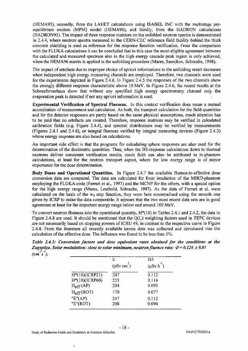
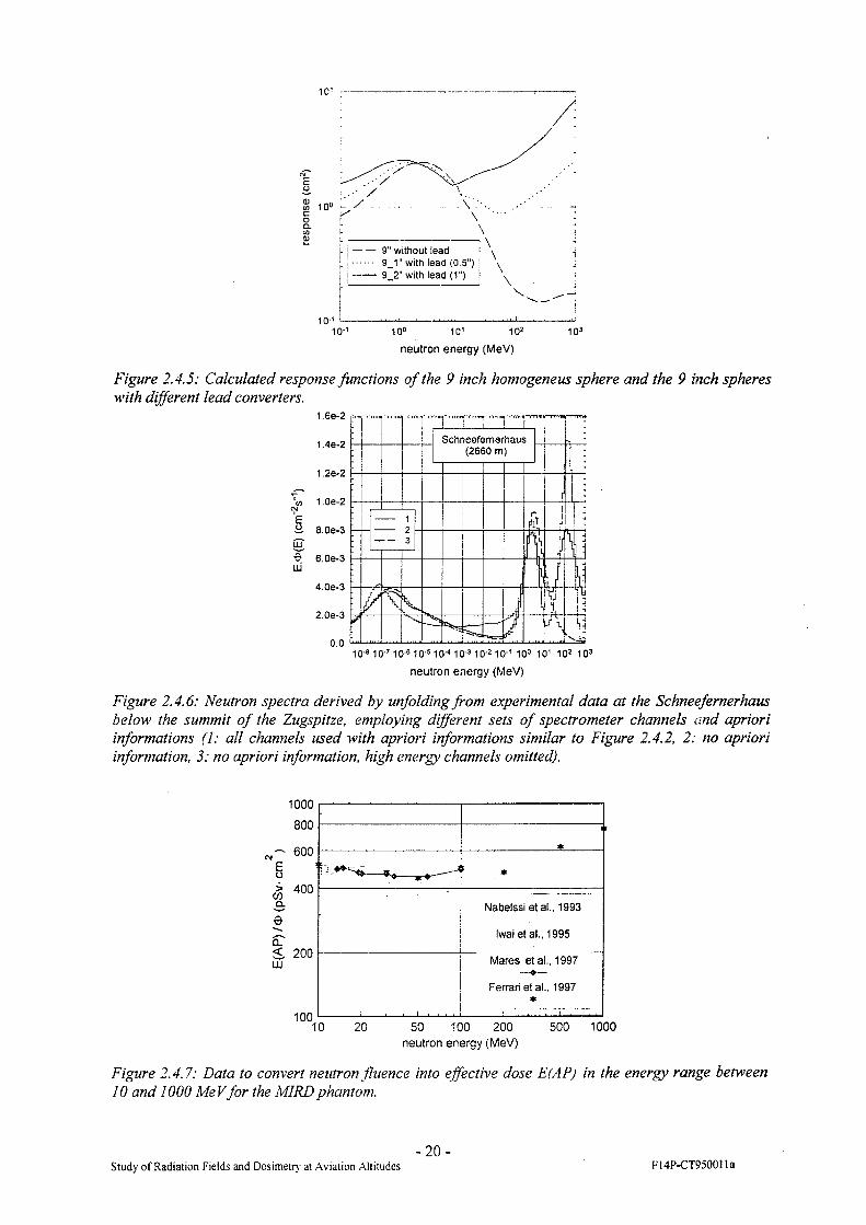
The impact of artefacts due to improper choice of apriori informations to the unfolding result decreases when independent high energy measuring channels are employed. Therefore, two channels were used for the experiments depicted in Figure 2.4.6. In Figure 2.4.5 the responses of the two channels show the strongly different response characteristic above 10 MeV. In Figure 2.4.6, the recent results at the Schneefemerhaus show that without any specified high energy spectrometry channel only the evaporation peak is detected if not any apriori information is used.
Experimental Verification of Spectral Fluences. In this context verification does mean a mutual accreditation of measurement and calculation. As both, the transport calculation for the field quantities and for the detector responses are partly based on the same physical assumptions, much attention has to be paid that no artefacts are created. Therefore, response matrices may be verified in calculated calibration fields (e.g. Figure 2.4.4), and spectral distributions may be verified by measurements (Figures 2.4.1 and 2.4.6), or integral fluences verified by integral measuring devices (Figure 2.4.3) whose energy response are also based on calculations.
An important side effect is that the programs for calculating sphere responses are also used for the determination of the dosimetric quantities. Thus, when the BS-response calculations down to thermal neutrons deliver consistent verification results, much faith can also be attributed to in-phantom calculations, at least for the neutron transport aspect, where the low energy range is of minor importance for the dose determination.
Body Doses and Operational Quantities. In Figure 2.4.7 the available fluence-to-effective dose conversion data are compared. The data are calculated for front irradiation of the MIRD-phantom employing the FLUKA-code (Ferrari et al., 1997) and the MCNP for the others, with a special option for the high energy range (Mares, Leuthold, Schraube, 1997). As the data of Ferrari et al. were calculated on the basis of the wR step function, they were here renormalised using the smooth one given by ICRP to make the data comparable. It appears that the two most recent data sets are in good agreement at least for the important energy range below and around 100 MeV.
To convert neutron fluences into the operational quantity, H*(10) in Tables 2.4.1 and 2.4.2, the data in Figure 2.4.8 are used. It should be mentioned that the Q(L) weighting factors used in TEPC devices are not necessarily based on stopping powers of ICRU 49, in contrast to the respective curve in Figure 2.4.8. From the literature all recently available kerma data was collected and introduced into the calculation of the effective dose. The influence was found to be less than 5%.
Table 2.4.1: Conversion factors and dose equivalent rates obtained for the conditions at the Zugspitze. Solar modulation: close to solar minimum, neutron fluence rate: 0=0.126 ± 0.01 (cm's ). _____________________________________________________
h(pSv cm )
H/t(pSv h ')
H*(10)(ICRP21) 247 0.112H*(10)(ICRP60) 255 0.116
Heff(AP) 204 0.093
Heff(ROT) 170 0.077
"E"(AP) 247 0.112"E"(ROT) 208 0.094
Study of Radiation Fields and Dosimetry at Aviation Altitudes-18-
F14P-CT95001 la

Table 2.4.2. Relative fluence and dose equivalent (H*(10)-ICRP60) contributions from thermal neutron energy to the upper integration boundary Enmax.
E nmax c*)Enmax,/3) 1 GeV ^enmax'HlGeV
5 MeV 0.61 0.3720 MeV 0.68 0.50180 MeV 0.94 0.92
Evaluation of results from TEPC. Extensive measurements in the reference radiation field provided by CERN have been carried out. The measurements performed under standardised conditions enables us to compare our devices with all the active and passive dosemeters which had been also used for inflight measurements. As far as the evaluation (which is still in progress) has been performed, the results obtained by the TEPC based real time detectors and the passive dosemeters are corresponding.
Furthermore the influence of wall thickness of the HANDI-TEPCs we used for measurements on board of Alitalia aircraft and of the Sievert instrument (equipped also with a TEPC) which had been used on some continental and an intercontinental flight, has been investigated in the reference field provided by CERN. The build up caps were made of tissue equivalent material and had a thickness of 5 to 100 mm. The results indicate no significant difference (neither the values of dose rate nor the lineal energy spectra) which could be of relevance for radiation protection purposes.
A direct intercomparison of results obtained on inflight measurements by several real time and passive dosemeters which participated in the Alitalia survey has partially be done and will be finished in the near future.
iuiiif■■ i iinn I IIU^ I I I lllllj I I I IllUj I 1
; Side concrete
HADRON95HEMA95HEMA96FLUKA
ill i i mini r i i mil i i
10-9 10-8 10-7 10-6 10-5 10-4 10-3 10"2 10’1 10° 101 102 103
neutron energy (MeV)
Figure 2.4.4: Verification of calculated neutron spectra (FLUKA, Roesler and Stevenson, 1993) at the SPS-CERN with the BS-spectrometer employing different physical models for the calculation of the BS-matrix.
- 19-Study of Radiation Fields and Dosimetry at Aviation Altitudes ]-14P-CT950011a
10'
... A
— 9" without lead-■ 9_1" with lead (0 5”)— 9_2" with lead (1")
neutron energy (MeV)
Figure 2.4.5: Calculated response functions of the 9 inch homogeneus sphere and the 9 inch spheres with different lead converters.
1.6e-2
1 4e-2
1,2e-2
*</) 1.06-2 iS- 8.0e-3
sO 6.0e-3 LU
4.0e-3
2.0e-3
0.010-6 10"710-8 10"5 10A0"3 10"2 10"1 10° 10' 102 103
neutron energy (MeV)
Figure 2.4.6: Neutron spectra derived by unfolding from experimental data at the Schneefernerhaus below the summit of the Zugspitze, employing different sets of spectrometer channels and apriori informations (1: all channels used with apriori informations similar to Figure 2.4.2, 2: no apriori information, 3: no apriori information, high energy channels omitted).
I&
6CL<LU
1000
800
600
400
200
100
*
1 *
Nabelssi etal.. 1993
Iwai et al., 1995
_______i____ ___ ___' ■ ■ ■ . t
Mares etal., 1997
Ferrari etal., 1997*
10 20 50 100 200 500 1000neutron energy (MeV)
Figure 2.4.7: Data to convert neutron fluence into effective dose E(AP) in the energy range between 10 and 1000 MeVfor the M1RDphantom.
-20-Study of Radiation Fields and Dosimetry at Aviation Altitudes FI4P-CT950011a

1000Leuthold et al. 1992 (ICRP60)
I
Q.
100
10
Siebert & Schuhmacher 1995 (ICRP60, ICRU49)
Sannikov & Savitzkaya 1996 (ICRP60)
MADE, 1971 (ICRP21)
Wagner et al. 1985 (ICRP21)
Sannikov & Savitzkaya 1996 (ICRP21)
1 1 ..‘ .... ....... i ■ ■ ■10 A 10 "2
neutron energy (MeV)
u.
Figure 2.4.8: Data used to convert neutron fluence into ambient dose equivalent H*(10). The central curve is identical with the recent data ofICRP Publication 74 (1996). The conversion to maximum dose equivalent, MADE, is drawn for comparison.
3. Summary of the Main Achievements
The radiation field at flight altitudes which is the subject of investigation in this contract, consists of 3 major components which result from interactions of primary galactic cosmic rays: The hadron, the electromagnetic and the muon cascade. Other components are not included here, e.g. those from solar events. While the dosimetry of low LET radiation from muons and electromagnetic cascades is relatively well understood, that of the hadron cascade, especially the neutron component and its contribution to neutron dose and risk, and the high Z particles, is not, and is the main topic of research in the current contract.
The research field covered here aims at the determination of these components at super-high (15000 - 23000 m), high (9000 - 13000 m) and low flight altitudes (mountains 2500 - 5200 m). It is clear that bulky instruments can only be flown on dedicated flights with the necessary electrical power and space provision. Therefore, the devices used during this project are either small packages of passive dosemeters, flown unattended and/or fixed to a certain place in the aeroplane, or active devices which are sufficiently small, and of low power consumption, and can be operated by accompanying persons. Some of the devices used in the project are based on well known techniques, others were especially developed by the contractors for the purpose.
Radiation protection dosimetry is based on radiological findings and risk considerations for the working population under consideration. Therefore, the quantities to be determined are not only of a physical nature. While for low LET radiation the non-physical implications are often negligible, they become very important for high LET radiation. Thus, there are limits to the accuracy with which the devices used can measure the required quantities.A straight forward concept for the interpretation of the experimental data is to determine the spectral particle fluence at any point of interest in the atmosphere and to derive from these data an average response of the device used, and an average particle fluence-to-dose conversion factor. The first one permits the interpretation of the reading of the device in terms of particle fluence. the second one the transfer into the correct dose quantity. This concept requires the following physical information: i) Spectral particle fluence with at least some information on the angular distribution, ii) Corrected reading of the device, i.e. the reading only due to the radiation component of interest, iii) Energy and angular response of the device in use. iv) Agreement on the dose quantity to be reported on, and v) the energy dependence of this dose quantity.
An important point is the choice of the quantity to be reported on. In the previous contract it had been demonstrated that the ICRU quantity ambient dose equivalent, H*(10), is a good estimator for the effective dose under any irradiation condition, at least for neutron fields. Nevertheless the comparison
Study of Radiation Fields and Dosimetry at Aviation Altitudes-21 -
F14P-CT95001 la

and harmonising of data is made difficult, as authors do not always state the origin of the quantity or use even alternative quantities, e.g. effective dose approximated, BFO-dose. maximum dose for bilateral irradiation of a slab, etc. Furthermore, if a device is calibrated in terms of ambient dose equivalent at laboratory conditions, it does not necessarily read ambient dose equivalent under cosmic ray conditions.
The calculation of the particle fields from the top of the atmosphere down to any altitude and geomagnetic position is essentially influenced by the shape of the primary galactic cosmic ray spectra. Very recent experimental cosmic ray data were used to repeat the transport calculations which resulted in a somewhat different slope of the fluence of neutrons and other secondaries to that seen earlier and provided integral fluence data which were in close agreement with the experimental data obtained from a spectrometer. However, the agreement with the TEPC data became worse. Therefore, a simultaneous experiment will be necessary to exclude the influence of weather and other temporary environmental parameters.
For the experimental determination of the neutron spectra, the Bonner sphere spectrometer was improved. The sensitivity to high neutron energies was increased by adding a further specified measuring channel for the energy range in excess of 20 MeV. The response functions for monoenergetic neutrons were calculated for the entire system up to 1 GeV with different cascade and transport models. One model was found to fit best the verification experiments at the CERN facility. For the determination of the fine structure of the 100 MeV intranuclear cascade peak, however, the energy range between 1 and 10 GeV requires further study.Recent measurements at mountain altitudes (2660 m) resulted in a very similar spectral structure as in previous measurements, but were obtained also without any apriori information during the unfolding of the experimental data set.The experimental and calculation results are in definite contradiction to findings of the American CARI-97 code. This was confirmed in a recent cluster meeting of this topic. It seems necessary that these differences are clarified before any of the models will go into a quantitative and legally fixed determination of flight doses for air crews.
Table 3.1 outlines the experimental methods and scope of the present project and we shall now summarise the main achievements to date. All calibration exposures and analysis planned by contractors and sub-contractors for the first two years of the project have been completed and in some cases extended beyond the original programme. Of particular significance is the indication that measurements made by detectors whose responses have been independently estimated by different techniques using heavy ions at Darmstadt (GSI) and neutron beams at PSI and Uppsala show very good agreement with those predicted by the CERF experiments. Although the main aspects of calibration of detectors for Z>1 particles have been satisfactorily concluded it is expected that facilities will be available at GSI for further work if required.
A major part of the investigations undertaken to date concerns the contribution of the neutron component of the radiation dose. The several experimental and theoretical approaches outlined in the four work packages testify to a very comprehensive programme which is bringing us nearer to a better understanding of the nature and magnitude of this contribution. Active and passive detectors have been employed at aircraft altitudes and on high altitude mountains to collect data used to measure and unravel spectral characteristics. Calculations of neutron and charge particle spectra in the atmosphere under different geomagnetic and solar conditions, using the FLUKA code have been completed. Response matrices of the extended Bonner Sphere spectrometer in the energy range up to 1 GeV have been evaluated with the LAHET, MCNP and HADRON codes. Following verification experiments at the CERF facility the LAHET high energy model was selected. Organ doses were calculated in the MIRD phantom for En < lOOMeV. Dosimetric operational and risk related quantities were determined for the various approaches (implication of ICRP 60). Development and application of the Bonner Sphere spectrometer with simultaneous data collection was also undertaken. Investigations at mountain altitude included experimental verification of neutron spectra at 4GV and neutron spectra measurements in the southern hemisphere at 5200m altitude and 14GV rigidity.
-22-Study of Radiation Fields and Dosimetry at Aviation Altitudes F14P-CT95001 la

Table 3.1: Basic physical characteristics of the devices used by the participating laboratories.
; Detector i Laboratory To detect Approximate energyrange
Passive Devices:
Passive Survey Meter TL.D NRPB low and high LET radiation All Energies
PADC : neutrons : All Energies
DIAS Stack (25mm)thick CR-39 DIAS : neutron interaction products j LET>5 keV/pm
DIAS Stack (25mm)thick CR-39 DIAS particles Z>2 i E>7 MeV/N
ANPA-Stacks TLD ANPA low LET radiation
bubble-detectors fast neutrons 0.5 to 20 MeV
Bi-Mylar neutrons and protons > 50 MeV
ANPA Stack ANPA fast neutrons 0.5 to 20 MeV
Active Devices
TEPC USAAR low/high LET 1 < L < 1000 keV/pm
TEPC (variance technique)
SSI low/high LET 1 < L < 1000 keV/pm
Extended REM-meter ANPA neutrons IMeV- lOGeV
Multisphere spectrometer GSF 1 neutrons 1 MeV - lOGeV
There has been significant success in obtaining data at aircraft altitudes. A significant store of data on the neutron component has been compiled by exposing passive and real-time detectors on commercial air routes including medium and long haul international flights. The co-operation of airlines has been excellent in this regard. The evaluation of detector response has been greatly enhanced by the various calibration facilities available as mentioned earlier, and there are indications that agreement between the various detector measurements are good. These measurements include low and high LET radiation.
Data obtained at altitude with bismuth detectors have the extra advantage that these detectors record dose due to both neutrons and protons. Extensive calibration at the neutron facility at Uppsala and proton beams at PSI has made this a most useful approach. The response of Bismuth detectors is directly proportional to the dose of mixed fields of neutrons and protons and this characteristic makes it possible to measure the dose without the need to differentiate the two components which is a difficult task in practice. Furthermore when the bismuth detector is combined with other neutron detectors capable of measuring low and high energy neutrons, it will be possible to differentiate the dose due to protons from that of neutrons.
Comparisons have been made between measured route doses and calculated values using different Q(L) relationships and different fluence to dose equivalent conversion factors. A passive survey meter has been developed for routine determination of route dose with a precision of 10-15% for doses of 50-
-23 -Study of Radiation Fields and Dosimetry at Aviation Altitudes F14P-CT95001 la

100 uSv. Many sets of inflight measurements have been completed, including some for supersonic aircraft as outlined in the various work packages
A wide range of measurements using the HANOI TEPC have been successfully undertaken on board Alitalia aircraft on routes from Rome to Rio de Janeiro, Rome to Tokyo and Rome to Los Angeles. Evaluation of this data has been completed. Response measurements for this instrument have also been carried out at PSI, CERN and at mountain altitude. Several measurements have also been made using the Sievert instrument on Scandinavian airline routes.
Progress made in linear energy transfer (LET) spectra and the contribution of Z>1 particles to the particle fluence at altitude has continued. Measurements at supersonic and subsonic altitudes have been extended and calibration has been successfully undertaken throughout the whole energy range involved. The LET spectra exhibit similar shapes over the whole range of measurements and the particle fluences vary in accordance with reasonable expectations, taking into account altitude in the atmosphere and geomagnetic variations with route travelled. The contribution of Z>1 particles to the particle fluence is found to be very small and its contribution to route doses not more than 1%.
4 Research to be Performed in the Remainder of the Project
The scheduled work undertaken to date has been part of a carefully planned programme designed to optimise the returns from a broad range of theoretical and experimental investigations over a 42 month period. While the schedules and time scales outlined in the original proposal have been adhered to in general, there have been some instances in which the opportunity to undertake tasks not envisaged earlier, or the availability of facilities not foreseen at the time of proposal submission, have provided the occasion to improve the programme to the benefit of the main scientific goals. Accordingly there have been some changes in the overall approach which have, in the main, enhanced the original plan. The research to be undertaken in the remainder of the project will follow the course originally agreed but will also reflect these changes.
4.1 Work Package 1
The provision of irradiation facilities and beam monitoring at the CERF facility will continue to be an important part of future investigations. CERN will provide two more beam runs at CERF with associated field measurements and monitoring. These are scheduled for 15-20 April 1998 and 15-22 July 1998. The facility has been modified to reduce scatter and further measurements and calculations will be carried out on the radiation fields at the experimental positions. Calculations will be carried out to investigate possible improvements to the neutron component of the fields such that it will better simulate the fields at aircraft altitudes. Most active and passive detectors used in the project will continue to benefit from further studies on response characteristics and indeed, there are many benefits which would result from more detailed investigation of factors such as the degree of isotropy of the beam itself. Specific experiments will include a final intercomparison of TEPC instruments with all other real time and passive devices. Calibration of the different HANOI instruments will be carried out simultaneously using the neutron calibration source provided by CERN and some investigations on the CERN stray radiation field will be finalised. Although the Sievert instrument comparison with HANOI has been completed at CERN it is hoped to repeat the experiment at some other neutron radiation facility. An investigation is to be carried out of the variability of the response characteristics of track etch detectors in high energy neutron fields. Such information is necessary to accurately interpret and to allow an estimate of the uncertainty of the estimation of dose rate and route doses using this type of detector. At least one further exposure of CR-39 stacks is planned to investigate the angular distribution characteristics in more detail. Exposures to high energy neutron beams with E > 100 MeV at Uppsala are scheduled for the spring of 1998. Several contractors and sub-contractors have already made preliminary arrangements to avail of these opportunities, thanks to the interest and assistance of the authorities there. Heavy ion exposures will continue at GSI, Darmstadt, if required, again, with the kind co-operation and assistance of GSI staff. Further tests are planned to determine various environmental and other influences on the operation of the Sievert instrument. The two reports on the
- 24 -Study of Radiation Fields and Dosimetry at Aviation Altitudes F14P-CT95001 la
response characterisation of active instruments and passive dosemeters in CERF which are currently in draft form are to be completed. More information is to be included in the response characterisation of instruments and dosemeters following high neutron energy irradiation which are planned to be carried out at The Svedberg Laboratory, Uppsala, and more detailed information on CERF and its dosimetry.
4.2 Work Package 2
This work package deals mainly with measurements of the cosmic ray neutron component, exposures at aviation and mountain altitudes and investigation of neutron and LET spectra as well as studies of ambient dose equivalent.
Further work will concentrate on a large variety of on-board measurements obtained with different passive and real time detectors for high LET radiations (low and high energy neutrons) and low LET radiation. It is also planned to compare measured route doses with calculated values using different Q(L) relationships and different fluence to dose equivalent conversion factors.
Efforts will be made to collect data in the region of the equator in order to investigate the change of dose levels and radiation field composition. Further measurements are to be carried out of dose rates and route doses on board supersonic and long haul aircraft using the NRPB ‘passive survey meter’ for routine flights and also at very high altitudes in collaboration with NASA. Where possible, direct comparisons will be made with other methods of assessing dose rate and route doses.
4.3 Work Package 3
Measurement of the spectrum of Z>2 particles will concentrate on observations made on supersonic routes. Earlier results which had shown the presence of nuclei as heavy as magnesium (Z=12) traversing the aircraft cabin will be refined in order to determine the relative abundances of Z>2 particles and their absolute fluences. The contribution of cosmic ray primary and secondary nuclei with Z>2 to the overall LET spectra at supersonic route altitudes will be determined using the same approach used for a subsonic route described in WP-3. It was shown during the first two years of this project that detector stacks used for charge spectrum studies were also ideal for measurement of LET spectra for LET values greater than ~5keV/pm. These investigations will continue using stacks exposed recently on an airbus on the Dublin-New York route and on the London-New York Concorde route. These detectors are mounted on a small platform which provide a wide range of angular orientations designed to investigate angular distribution characteristics, a knowledge of which is important for final analysis of the data.
Close contact will be maintained with the University of Siegen group as new experimental data emerges. The reproducibility of route doses will be investigated further for low energy neutrons and low LET radiation.
4.4 Work Package 4
Research to be performed in work package 4 will include several experimental and theoretical studies. Using recent experimental findings the galactic cosmic ray input spectrum will be finally adjusted and the effect of this on particle spectra and fluences will be determined for various flight routes. The Bonner Sphere Spectrometer will be optimised with respect to the high energy region. Response calculations of the BS Spectrometer and other devices will be extended beyond 1 GeV. The effect of the high energy proton component on the experimental determination of the neutron spectra will be evaluated. It is also planned to investigate and determine the influence of weather and other environmental conditions on spectral measurements and the implication it could have for models of atmospheric stratification. The calculation of dosimetric quantities will be extended beyond 100 MeV and the kerma concept for calculation of operational quantities in high energy radiation fields will be validated.
-25-Study of Radiation Fields and Dosimetry at Aviation Altitudes FI4P-CT95001 la
Inflight measurements obtained on the Rome-Los Angeles, Rome-Rio de Janeiro and Rome-Tokyo will be evaluated. The dependence of dose profiles on flight altitude and geomagnetic latitude will be completed. Final comparison of the HANOI instrument results with those of other instruments obtained at altitude will be undertaken. It is also hoped that the Sievert instrument with built up caps will be exposed at altitude for comparison with results obtained at reference fields.Comparison will be made of the results of different measurement methods for the assessment of dose rate and route doses, the use of different dose equivalent quantities, and the suitability of different approaches, including the use of calculations for the assessment of aircraft crew dose.
It is planned to incorporate the final dosimetric results into the computer code EPCARD V.2.0 (European Program Package for Calculating Airflight Route Doses) which was developed in the frame of the previous project, and is presently working with a preliminary data set.
5. Publications
Bartlett D.T., and Hager L.G: Measurements on Concorde of the Cosmic Radiation Field at Aviation Altitudes. NRPB Occupational Standards Department Technical Memorandum 8(97) (1997b).
Bartlett D.T., Hager L.G., Rancati T., Ferrari A., Silari M., and Otto T.: Neutron Radiation Fields at the CERN/EC Reference Field Facility, December 1996 Interim Report. NRPB Occupational Standards Department Technical Memorandum 25(96) (1996).
Bartlett D.T., McAulay I.R., Schrewe U.J. Schnuer, K., Menzel H.G., Bottolier-Depois J.F., Dietze G., Grnur K., Grillmaier R.E., Heinrich W., Lim T., Lindborg L., Reitz G., Schraube H., Spumy F., Tommasino L.: Dosimetry for occupational exposure to cosmic radiation, Radiat. Prot. Dosim. 70, 395-404 (1997).
Bartlett D.T., Tanner R.J., Hager L.G., and Lavelle J.: The Measurement Using Passive Dosemeters of the Neutron Component of Aircraft Crew Dose, Rad. Meas., 28 (1-6) pp518-524 (1997a).
Birattari C., Ferrari A., Hofert M., Otto T., Rancati T., and Silari M.: Recent Results at the CERN-EC High Energy Reference Field Facility, Proceedings of the Third Specialists’ Meeting on Shielding Aspects of Accelerators, Targets and Irradiation Facilities (SATIF3), Japan 12-13 May 1997, OECD Documents, pp 219-234 (1998), and CERN Divisional Report CERN/TIS-RP/97-12/CF (1997).
Cross W.G. and Tommasino L: Dosimetry of High Energy Neutrons and Protons by Bi-209 Fission, Rad. Prot. Dos. 70, pp395-404 (1997).
Donnelly J.: A Study of High Charge Cosmic Ray Particles at Civil Aviation Altitudes, MSc Thesis, National University of Ireland, University College Dublin, (1997)
Hager L.G., and Bartlett D.T.: Measurements on 747-400 Flights between London and Tokyo of the Cosmic Radiation Field, NRPB Occupational Standards Department Technical Memorandum 11(97) (1997).
Hofert M., Bartlett D.T., and McDonald J.C.: The Use of Accelerator Stray Fields for the Calibration of Passive Dosemeters, presented at the US Health Physics Society Mid-Year Symposium (1997).
Keane A.J., O’Sullivan, D., Thompson A., Drury L.O’C., and Wenzel K.-P.: A Charge Spectrum of Ultra Heavy Cosmic Ray Nuclei, including Actinides, detected on the LDEF, Proc. 25 th Int. Cosmic Ray Conf. (Durban), Vol. 3, pp 361-364 (1997).
Keane A.J., O’Sullivan D., Thompson A., Drury L.O’C., and Wenzel K.-P.: The Charge Spectrum of Ultra Heavy Nuclei, including Actinides, in the Cosmic Radiation, Adv. Space Res., Vol. 19, pp 739-742 (1997).
Keane A.J., O’Sullivan D., Thompson A., Drury L.O’C., and Wenzel K.-P.: Application and Analysis of SSNTD in the investigation of Ultra Heavy Cosmic Rays in the Dublin-ESTEC LDEF Experiment, Radiation Measurements, Vol. 28 pp 329-332 (1997).
Keegan R.: LET Spectrum Generation and Proton Induced Secondary Contribution to Total Dose Measured in Low Earth Orbit, PhD Thesis, National University of Ireland, University College Dublin, (1996).
Kylldnen, J.-E., Lindborg, L. and Samuelsen, G. A: TEPC for Variance Measurements, Proceedings of the Microdosimetry Symposium, Oxford, 1996
-26 -Study of Radiation Fields and Dosimetry at Aviation .Altitudes FI4P-CT950011a
Manfredotti C., Ongaro C., Zanini A., Cavaioli M., and Tommasino L: Spectrometry of Low and High Energy Neutrons by Unfolding Passive Detector Responses, Paper presented at the 5th Int. Conf. on Application ofNuclear Techniques, Crete, Greece, June 9-15 (1996).
Mares V., Leuthold G., Schraube H.: Organ doses and dose equivalents for neutrons above 20 MeV, Radiat. Prot. Dosim, 70, 133-138 (1997).
Mares V., Sannikov A., Schraube H.: The response functions of a He-3-Bonner spectrometer and their experimental verification in high energy neutron fields, Proc. Third Specialist Meeting on Shielding Aspects of Accelerators, Targets and Irradiation Facilities, May 12-13, 1997, Sendai, Japan, OECD Nuclear Energy Agency, pp 237-248, (1998).
Mares V., Schraube H.: High energy neutron spectrometry with Bonner spheres, In print in: Proceedings of the IRPA Regional Symposium, Prag, 8.-12.9.97
McAulay I.R. et al. Exposure of Aircrew to Cosmic Radiation, Radiation Protection 85, EURADOS Report 1996-01, Edited by I.R. McAulay et al., Luxembourg, Office for Official Publications of the European Communities, 1996
Nava E., Otto T., and Silari M.: Reference dose equivalent values for the 1997 CERN-EC runs. CERN Internal Report TIS-RPfTM/TM/97-22 (1997).
O’Brien K.: LUIN 97' Unpublished results, Personal Communication (1997).O’Sullivan D. and Thompson A.: Investigation of the Platinum Lead and Actinide Regions of the
Charge Spectrum of Galactic Cosmic Rays, Accepted for publication, New Astronomy, (North Holland), (1997).
O’Sullivan, D., Thompson, A., Keane, A.J., Drury, L.O’C., and Wenzel, K.-P.: Investigation of Z>70 Cosmic Ray Nuclei on the LDEF Mission, Radiation Measurements, Vol. 26, pp 889-892, (1996).
Otto T., and Silari M.: The July/August 1996 Run at the CERN-CEC Reference Field Facility, CERN Internal Report TIS-RP/TM/96-25 (1996b).
Otto T., and Silari M.: The May 1996 Run at the CERN-CEC Reference Field Facility, CERN Internal Report TIS-RP/IR/96-15 (1996a).
Roesler S., Heinrich W., and Schraube H.: Calculation of Radiation Fields in the Atmosphere and Comparison with Experimental Data, (In press) (1997).
Roesler S., Heinrich W., Schraube H.: Calculation of radiation fields in the atmosphere and comparison to experimental data, Radiation Research 149, 87-97, (1998).
Roesler S., Heinrich W., Schraube H.: Neutron fluxes in the atmosphere from interacting primary cosmic rays, In print in: Adv. Space Research (1997).
Sannikov A., Mares V., Schraube H.: High energy response functions of Bonner spectrometers, Radiat, Prot. Dosim. 70, 291-294 (1997).
Schraube H., Heinrich W.: Neutron exposure at civil flight levels, In: International Conference: Neutrons in Research and Industry. George Vourvopoulos, Editor, Proc. SPIE 2867, 264-273 (1997).
Schraube H., Jakes J., Sannikov A., Weitzenegger E., Roesler S., Heinrich W.: The cosmic ray induced neutron spectrum on the summit of the Zugspitze (2963 m), Radiat. Prot. Dosim. 70, 405-408 (1997).
Schraube H., Leuthold G., Roesler S., Heinrich W.: Neutron spectra at flight altitudes and their radiological estimation, In print in: Adv. Space Research (1997).
Stevenson G.R., Fasso A., Hofert M., and Tuyn J.W.N.: Dosimetry at High-Energy Accelerators, Radioprotection 31(2), ppl93—210 (1996).
Tommasino L, Caggiati F., Cavaioli M., Notaro M., Teodori R., Torri G., Zhou D., Byrne J., and O’Sullivan D Passive Multidetector Stack for the Assessment of Aircrew Exposure, Environmental International, 22, suppl. 1, pp 115-119 (1997)
Acknowledgements.* We wish to thank Alitalia for providing extensive facilities on many routes for the simultaneous exposure and operation of several instruments. We thank also Aer Lingus, British Ainvays and Scandinavian Airlines for their continued support and assistance with these experiments. The help received from staff at the CERN, GSI, PSI and Uppsala accelerator centres is greatly appreciated
Study of Radiation Fields and Dosimetry at Aviation Altitudes-27-
FI4P-CT9500Ila

6. Executive Summary
This project on the Study of Radiation Fields and Dosimetry at Aviation Altitudes has progressed steadily since its inception in January 1996. When we consider the objectives as laid out in the original technical annex we find that there are good grounds for satisfaction with the overall progress to date. The main objectives were to measure the flux and energy spectra of neutrons and charged particles over a wide energy interval at aviation and mountain altitudes and to compare results with those calculated using various transport codes. Specific goals included the use of TEPC based devices to measure dose equivalent and absorbed dose at aircraft altitudes along with other active and passive detectors which would also investigate the neutron and Z>1 component. Furthermore it was planned to construct an alternative TEPC (Sievert Instrument) to the already operating HANOI instrument All detectors were to be calibrated at the CERE field, at heavy ion accelerators, and to neutron beams of high energy to determine response characteristics and to compare results obtained. It was planned to carry out a detailed programme of calibration and development of theoretical models in parallel with data collection. Improvement of information on the neutron flux distribution and the proton and neutron and other secondary radiation fields at different depths and geographical locations in the earth’s atmosphere was envisaged. Furthermore, it was proposed that the research programme as a whole would also lead to improved propagation models for the investigation of cosmic ray nuclei and their secondaries in the earth’s atmosphere.
Without the interest and assistance of several international airline companies little of this success could have been achieved. We are greatly indebted to Alitalia and the Italian Survey authorities who provided joint exposures for the group on several international routes. We have also been greatly assisted in our endeavours by British Airways, Aer Lingus and Scandinavian Airlines
The combination of experience and resources of the five main contractors and three sub-contractors has resulted in a veiy successful collaboration to date. Instrument development and calibration procedures have continued simultaneously with data collection by active and passive detection systems at aircraft altitudes and in high altitude laboratories. Neutron and proton contributions to the radiation dose are being investigated experimentally in significant detail, and measurement of linear energy transfer spectra and the contribution of all Z>2 particles is well underway. To compliment these experimental studies a wide range of calculations have been undertaken using codes such as FLUKA and LUIN as described in detail in the four work package reports included here.
While much effort has gone into elucidating the many problems which still exist in this area of dosimetry it is obvious even at this mid way point that complexities remain. One of the main strengths of the project is the availability of joint calibration facilities at the CERE facility where contractors and sub-contractors have had, and continue to have, the opportunity to carry out investigations of the response of active and passive detectors employed in this contract, using the results of the USAAR HANOI TEPC instrument as a standard. Measurements with the Sievert Instrument have also been undertaken at CERN including several with different wall thicknesses. Availability of this facility will continue at least into 1998.
All calibration exposures and analysis planned by contractors and sub-contractors for the first two years of the project have been completed and in some cases extended beyond the original programme. For instance. Bismuth detectors have been calibrated to both high energy neutrons at Uppsala and high energy protons at PSI. At CERN full calculations of hadron spectral radiance and a full measurement programme of dose equivalent rates using a TEPC, including some angular distribution measurements have been undertaken. Two five day irradiation periods in both 1996 and 1997 have been provided, with beam monitoring and support for experiments. All contractors have taken part in a series of measurement of response characteristics of passive and active dosimetry systems and comparison of response data between systems, with associated calculations.
-28-Study of Radiation Fields and Dosimetry at Aviation Altitudes F14P-CT950011a

Of particular significance is the indication that measurements made by detectors whose responses have been independently estimated by different techniques using heavy ions at Darmstadt (GSI) and neutron beams at PSI and Uppsala show very good agreement with those predicted by the CERF experiments. Although the main aspects of calibration of detectors for Z>1 particles have been satisfactorily concluded it is expected that facilities will be available at GSI for further work if required.
A major part of the investigations undertaken to date concerns the contribution of the neutron component of the radiation dose. The several experimental and theoretical approaches outlined in the four work packages testify to a very comprehensive programme which is bringing us nearer to a better understanding of the nature and magnitude of this contribution. Active and passive detectors have been employed at aircraft altitudes and on high altitude mountains to collect data used to measure and unravel spectral characteristics. Calculations of neutron and charge particle spectra in the atmosphere under different geomagnetic and solar conditions, using the FLUKA code have been completed. Response matrices of the extended Bonner Sphere spectrometer in the energy range up to 1 GeV have been evaluated with the LAHET, MCNP and HADRON codes. Following verification experiments at the CERF facility the LAHET high energy model was selected. Organ doses were calculated in the MIRD phantom for E„ < lOOMeV. Dosimetric operational and risk related quantities were determined for the various approaches (implication of ICRP 60). Development and application of the Bonner Sphere spectrometer with simultaneous data collection was also undertaken. Investigations at mountain altitude included experimental verification of neutron spectra at 4GV and neutron spectra measurements in the southern hemisphere at 5200m altitude and 14GV rigidity.
There has been significant success in obtaining data at aircraft altitudes. A significant store of data on the neutron component has been compiled by exposing passive and real-time detectors on commercial air routes including medium and long haul international flights. The co-operation of airlines has been excellent in this regard. The evaluation of detector response has been greatly enhanced by the various calibration facilities available as mentioned earlier, and there are indications that agreement between the various detector measurements are good. These measurements include low and high LET radiation.
Data obtained at altitude with bismuth detectors have the extra advantage that these detectors record dose due to both neutrons and protons. Extensive calibration at the neutron facility at Uppsala and proton beams at PSI has made this a most useful approach. The response of Bismuth detectors is directly proportional to the dose of mixed fields of neutrons and protons and this characteristic makes it possible to measure the dose without the need to differentiate the two components which is a difficult task in practice. Furthermore when the bismuth detector is combined with other neutron detectors capable of measuring low and high energy neutrons, it will be possible to differentiate the dose due to protons from that of neutrons.
Comparisons have been made between measured route doses and calculated values using different Q(L) relationships and different fluence to dose equivalent conversion factors. A passive survey meter has been developed for routine determination of route dose with a precision of 10-15% for doses of 50- 100 gSv. Many sets of inflight measurements have been completed, including some for supersonic aircraft as outlined in the various work packagesA wide range of measurements using the HANOI TEPC have been successfully undertaken on board Alitalia aircraft on routes from Rome to Rio de Janeiro, Rome to Tokyo and Rome to Los Angeles. Evaluation of this data has been completed. Response measurements for this instrument have also been carried out at PSI, CERN and at mountain altitude.
Progress made in linear energy transfer (LET) spectra and the contribution of Z>1 particles to the particle fluence at altitude has continued. Measurements at supersonic and subsonic altitudes have been extended and calibration has been successfully undertaken throughout the whole energy range involved. The LET spectra exhibit similar shapes over the whole range of measurements and the particle fluences vary in accordance with reasonable expectations, taking into account altitude in the atmosphere and geomagnetic variations with route travelled. The assumption of isotropic angular distribution is being checked by exposure of a suitable platform on both supersonic and subsonic
-29-
Study of Radiation Fields and Dosimetry at Aviation Altitudes F14P-CT950011a

routes. The contribution of Z>1 particles to the particle fluence is found to be very small and its contribution to route doses not more than 1%. Dose equivalent values, which may vary by approximately 10% after angular corrections and corrections for small losses of very high LET events (which is inherent in the measurement technique used) agree very well with those of passive and active instruments, where available.
The remainder of the project will be devoted to a continuation of the work already outlined here and further development of experimental and theoretical investigations. New facilities for calibration and detector response studies will be sought for this future programme and collection of data at airline altitudes and at high altitude locations will continue. Accelerator based work will deal specifically with attempts to expose passive and active detector systems to monoenergetic beams of neutrons of E>100MeV for calibration and response studies purposes. At the CERE facility further work is planned on the provision of irradiation facilities and more detailed studies of angular distribution characteristics and intercomparison of all detectors employed by contractors and sub-contractors. Presentation and analysis of the results will continue in parallel with these activities.
In the area of particle spectra and spectrometry, final adjustment of the galactic cosmic ray input spectra to include recent experimental findings will be undertaken and their effect on particle spectra and fluence on flight routes will be assessed. Bonner Sphere spectrometry will be optimised with respect to the high energy region and response calculations will be extended beyond IGeV.The effect of the high energy proton component on the experimental determination of the neutron spectra will be evaluated. The influence of environmental conditions on measurements will be studied. Calculations on dosimetric quantities will be extended above 100 MeV and the kerma concept in high energy radiation fields for the calculation of operational quantities will be validated.
Further investigations into detector performance will be undertaken. For example, TEPC temperature dependence, long term gas storage characteristics and other sources of influence on measurements obtained with the Sievert instrument will be the subject of ongoing studies. At aircraft altitudes work will continue on the measurement of neutron and charged particle spectra and relative dose values, at different altitudes and latitudes for sub-sonic and supersonic routes. Reproducibility of route doses will be investigated and temporal variations measured. The influence of solar flares will also be addressed. LET spectra and Z>1 abundances will be further investigated with emphasis on supersonic flights for the abundance studies. Further exposures at high altitude locations such as at the Zugspitze are also planned.
Several formal and informal meetings of scientists included in the project will take place to discuss progress on a regular basis along with the usual daily and weekly interactions by telephone, fax and e- mail.
-30-Study of Radiation Fields and Dosimetry at Aviation Altitudes F14P-CT950011a

7. Annex
Table 7.1 Summary of Work Packages and Role of Individual Laboratories
Definition of Roles ReportingLaboratory
Work Package 1 NRPBUniversite des Saarlandes (USAAR, Germany) Sub-contractor SSI (Sweden)
Calibration of HAND! for neutrons up to 200 MeV and for ions found at aviation altitudes. Operational measurements of ambient dose equivalent using HANOI. Similar measurements using variance-co-variance techniques.
Agenzia Nationale per la Protezione dell’ Ambiente (ANPA, Italy)
Calibration and use of bismuth fission detectors for detection of high energy neutrons, protons.
National Radiological Protection Board (NRPB, UK)Sub-contractor CERN
Assess performance of different passive and active dosimetry systems in CERN reference field
Dublin Institute for Advanced Studies (DIAS, Ireland)(Co-ordinator)
Further development and calibration (at GSI, Darmstadt) of nuclear track detectors for measurement of Z>2 particles
Work Package 2 ANPAANPA Measurements of the neutron cosmic ray exposure
at aviation and mountain altitudes.Measurement of the neutron spectra.
USAARSub-contractor SSI (Sweden)
Measurements of ambient dose equivalent and LET spectra at aviation and mountain altitudes using TEPC techniques.
Work Package 3 DIASNRPBSub-contractor CERN
Measurements of route doses on routine flights of low and high LET ionising radiation and photon radiation.
DIAS Measurement of flux of Z>2 particles and LET spectra at super-sonic and sub-sonic aviation altitudes, on a wide range of routes.
ANPA Study of the reproducibility of the neutron route dose
Work Package 4 GSF and USAAR
GSF, Neuherberg (Germany)Sub-contractors: Prof. Heinrich andPTB, (Germany)
Calculation and modelling of the spectra of neutrons, protons and heavy charged particles at aviation altitudes and the response of instruments to these particles. Calculations of dosimetric quantities. Verification of cosmic models by experimental data.
USAARSub-contractor SSI (Sweden)
Evaluation of results from TEPC. Comparison and correlations with passive and real time detectors.
Final Work PackageDIAS, ANPA, GSF,USAAR, NRPB
Assessment of the aircraft crew Exposure and the Reproducibility of the Route Dose.
-31 -Study of Radiation Fields and Dosimetry at Aviation Altitudes FI4P-CT95001 la

Biokinetics
and
Dosimetry of Incorporated
Radionuklides

BIOKINETICS AND DOSIMETRY OF INCORPORATED RADIONUCLIDES
Contract No: FI4P-CT95-0011b
Mid-term Report for the period 1 January 1996 to 31 December 1997
P Roth, A Giussani, E WernerGSF - Forschimgszentrum fiir Umwelt und Gesundheit, GmbH
Ingolstadter Landstrafie 1, D-85764 Neuherberg, Germany
JD Harrison, A PhippsNRPB, National Radiological Protection Board
Didcot Oxon 0X11 ORO, Chilton, United Kingdom
D Nofike, U Buhl, K Karcher BfS, Bundesamt fur Strahlenschutz
Ingolstadter Landstrafie 1, D-85762 Oberschleifiheim, Germany
J McAugheyAEA Technology Environment
E6 Culham, Abingdon, Oxfordshire 0X14 3DB, United Kingdom
F PaquetCEA, Commisariat a VEnergie Atomique
BP 12, F-91680 Bryeres-le-Chatel, France
MC Cantone, D de Bartolo, L Garlaschelli CMRJUM1L, Consorzio Milano Ricerche
Via Cicognara 7,1-20129 Milano, Italy
R Kriehuber, M Simko
UROS, University of Rostock
Universitdtsplatz 2, D-18055 Rostock, Germany
P PihetIPSN, Institut de Protection et de Surete Nucleaire
BP 6, F-92265 Fontencty aux Roses, France
D Taylor, L WebbUIVCC, University of Wales, College of Cardiff
PO Box 912, Cardiff CF1 3TB, United Kingdom
-32-
INTERNAL DOSIMETRY
1. Objectives
The global objective of this project is to contribute to the health and safety aspects of one widely used potentially toxic agent, namely ionising radiation, especially the use of radionuclides in industry medicine and other fields. High quality radiation protection for radionuclides which may enter the human body demands sound methods for the estimation of the radiation doses delivered to organs and tissues - the so-called internal dose. For such estimates of internal dose, realistic biokinetic models are necessary in order to predict the behaviour of a particular radionuclide of concern in the human body. The aim of this project is to assist in the provision of good, preferably human, biokinetic data which can be applied to the formulation of realistic biokinetic models in a format which permits them to be used for the calculation of reliable dose coefficients. Equipped with this knowledge, Member States can ensure that their populations are well protected, without unduly limiting the beneficial uses of radioactive materials and without disrupting everyday life. This research can also help Member States to strike a balance between funding for radiation protection and other public safety measures.
The work to be carried out within this project is structured into four Work Packages:
Workpackage 1 concentrates on ingested radionuclides, considering doses to the GI tract and radionuclide absorption. A major objective is the development of a new dosimetric model of the GI tract, taking account of most recent data on gut transit and dose to sensitive cells. Experimental studies with rodents will be undertaken to provide new information on dose to sensitive cells.To improve estimates of gastrointestinal uptake, volunteer studies will be performed using stable isotopes of radiologically relevant elements and the radionuclides plutonium-244 and polonium-208.
Workpackage 2 seeks to improve and extend biokinetic and dosimetric models for systemic radionuclides. Existing models for adults and children will be extended to other elements and new models will be developed for the embryo and fetus. In both cases, the work includes experimental studies as well as model development. Stable isotope studies in humans using appropriate surrogate elements for the actinides will provide biokinetic data for elements with important radioisotopes. Studies of radionuclide transfer to the embryo/fetus will include human placental perfusion experiments and measurements using a primate species.
Workpackage 3 is to improve assessment of localised distribution of dose within tissues at the cellular level for specific examples of Auger emitters and alpha emitting isotopes, in relation to observed effects. The work will include experimental studies of dose/effect relationship and the development of localisation methods.
Workpackage 4 concerns the development of computer codes for the new dosimetric models, quality assurance of the models and the calculation of dose coefficients. Formal sensitivity analysis will be used to identify critical areas of model development and to investigate the effects of variability and incertainty in biokinetic parameters.
This work programme addresses workplan priorities under "Evaluation of Radiation Risks", specifically the improvement of the reliability and accuracy of assessments of exposure from incorporated radionuclides through better knowledge of relevant biokinetic processes and data.
-33-
INTERNAL DOSIMETRY
2 Progress Report
This section reports the progress of the project as a coherent whole without undue reference to the contributions of the individual partners. A more detailed description of the contractors contributions to the technical progress within each Work Package in which they participate is provided in a separate compilation.
2.1 Work Package 1
Biokinetics of ingested radionuclides and dosimetry of the gastrointestinal tract
Contributors: GSF (01), NRPB (02), BfS (03), CEA (05), AEA (04), UMIL (06), UWCC (10)
Objectives
Tire objectives of Work Package 1 for the reporting period were:
• Review of data on the gut transit of materials as a function of age, sex and gut physiology• Review of data on the retention of radionuclides / elements in intestinal tissue• Review of data on the GI absorption of elements and uncertainties• Begin of the development of a new alimentary tract model• Begin of rodent studies on intestinal retention / dosimetry• The creation of a computer speciation model for the human gastrointestinal tract, including the
assembly of a well validated thermodynamic database for lanthanide and actinide elements with biologically important ligands with special reference to Nd, Eu, Gd, Am and Cm
• To carry out computer simulations with Eu and Am and to analyse the data generated• Assessment of excretion patterns of Co, Sr, Zr, Nb, Ru, Ag, Cs, Th, U, Hf, and lanthanides• Optimisation and validation of mass spectrometric and activation analytical methods for measuring
isotope ratios of Te, Zr, Ru, and lanthanides• Animal studies with Te, Ru and Zr• Generation of foodstuffs, intrinsically labelled with Te, Zr, Ru, and lanthanides• Submission of proposals for Ethics Committee (Human studies with Te, Zr, Ru, lanthanides)• Volunteer studies with Te and Ru• Continuation of volunteer study using 244Pu• Begin of a volunteer study using 208Po• Complete rodent studies on absorption of 210Po, 22STh
Progress
Review of data on gut transit and retention and absorption
Reviews of data on the retention of radionuclides / elements in intestinal tissue and radionuclide absorption will provide input to the work of ICRP Task Groups charged with the revision of the dosimetric model for the alimentary tract and with the development of age-related dosimetric models. For the revision of the alimentary tract model, the possibility of radionuclide retention in different regions requires consideration in relation to the location of sensitive cells in these regions. Uncertainties in biokinetic parameters and resulting uncertainties in dose estimates also need to be considered. Currently, it is envisaged that draft reviews will be completed during 1998 but that this work will continue over the period of this contract due to the requirements for model development. For example, at the moment it is tried to get further quantitative informations on possible longer retention in the new alimentary tract compartments oral cavity and oesophagus.
-34-

INTERNAL DOSIMETRY
One of the main issues of data review was the study of uptake of elements (mainly heavy metals) by the gastrointestinal microflora and its possible dosimetric implications for example by settlement of the microflora at intestinal walls near radiation sensitive cells.
The microbes are embedded in the mucus layer of the inner surface. The mucus may act as a barrier preventing the microbes of getting in direct contact with nutrients and minerals. It would be difficult to estimate to what extent the mucus-layer influences the uptake-rates of heavy metals or radionuclides.
Many of the species are protected to toxic amounts of heavy metals by resistance mechanisms. In eubacteria (cocci and rods) normally eiflux systems are found. An increasing heavy metal concentration in the surrounding medium does not lead inevitably to an higher heavy metal content of these bacteria. Sometimes aggregates of low solubility are built on the cell surface. Respective findings were reported in experimental studies. But it is not clear if the formation of these compounds would take place in the same manner in the lumen of the gastro-intestinal tract.
Regarding the very special growth conditions used in the uptake and absorption experiments of the literature reviewed and taking into account that only little is known about the interactive mechanisms in the ecosystem GI-Tract a quantitative estimation of the heavy metal uptake by the microbes in the gut lumen is not possible at the moment. It will be continued to study iron data available for these processes.
Development of a new alimentary tract model
Within the ICRP Task Group on a Dosimetric Model for the Human Alimentary Tract (HAT) the model development has been started. There is the problem of the need of a complete model taking into account all physiological processes on the one hand and of a model as simple as possible on the other hand which can easily be applied in radiation protection and for which all necessary parameters can be determined. At the moment it is discussed to have a simple generic model including the most important pathways with some additional pathways considering longer retention in the oral cavity including teeth and oral mucosa and in the oesophagus, and special absorption and secretion processes. These additional pathways will only be used in cases when sufficient data are available.
Animal studies on intestinal retention
An aspect of intestinal retention of radionuclides that has received little attention is the behaviour of radioactive particles. Uptake of particulate materials is a recognised phenomenon for which different mechanisms have been suggested including uptake by M cells ("membranous epithelial cells") of Peyer's patches and uptake by enterocytes. A study is in progress to investigate uptake of monodisperse insoluble radioactive particles by neonatal and adult rats and guinea pigs. The material is fused montmorillonite clay (0.8 pm or 2.2 pm) tagged with either 57Co or 242Cm.
Administration of 57Co labelled particles is complete. Preliminary results for neonatal rats show whole- body retention falling to 0.05% of adminstered activity after one week with large differences between animals in the distribution of retained activity. In one animal of five, most (>80%) of retained activity was associated with mesenteric lymph nodes. In other animals, retention in tissues other than those of the gastrointestinal tract was consistent with the expected release of dissolved 57Co from the particles; retention in the intestinal tract was observed in each case. Inactive particles have also been prepared for studies of intestinal uptake by electron microscopy to be undertaken at the Queen's University, Belfast.
A computer speciation model for the human gastrointestinal tract
In accordance with the project milestones, the chemical environment in the human gastrointestinal tract, of an average healthy adult, has been modelled using the JESS 53 (Joint Expert Speciation System 53) chemical speciation computer program. This chemical speciation modelling aids the validation of the use of stable isotopes of lanthanide elements as surrogates for the highly toxic actinides, for the measurement of absorption from the gastrointestinal tract. For the purposes of initial lanthanide and
-35 -

INTERNAL DOSIMETRY
actinide simulations, the gastrointestinal tract has been split into four different compartments which represent the conditions in the mouth, stomach and small intestine. Four models have been created for the chemical environments of the compartments: saliva, gastric juice, bile and pancreatic fluid. Each model has been created using well validated thermodynamic data obtained from the following sources: the JESS 53 database (this contains over 145 000 thermodynamic constants all of which have been critically assessed); HATCHES (Harxvell/Nirex Thermodynamic Database for Chemical Equilibrium Studies); NIST Standard Reference Database 46 (Critical Stability Constants of Metal Complexes Database, 1993); and from other literature. The concentrations of the biologically important ligands for each compartment were taken from Documenta Geigy Scientific Tables, 7th Edition (1970) and from more recent references.
A special database has also been created, containing critically assessed formation constants for lanthanide and actinide elements (Nd, Eu, Gd, Am and Cm) with the biological ligands found in saliva, gastric juice, bile and pancreatic fluid. Using the constants in the database, initial aqueous models have been produced for each of the elements, Nd, Eu, Gd, Am and Cm, in the compartments of the gastrointestinal tract. These simulations assume that any solids that may form are dissolved at this stage. Comparisons of the results have then been made in order to assess the use of lanthanide elements as possible surrogates for actinides, and to identify any important variations in the biological behaviour of the element and its surrogate.
A large series of preliminary estimations of the interactions of Nd, Eu, Gd, Am and Cm (5.00 • 10"7 mol dm'") with all the metal binding species present in the mouth (saliva), stomach (gastric juice) and small intestine (bile and pancreatic fluid) over the pH ranges of pH = 5.6 to 7.6, pH = 1.5 to 5.5, and pH = 5.0 to 9.0 respectively, have been made. The chemical speciation profiles for these elements, in saliva, gastric juice, bile and pancreatic fluid, in the absence of solids, are quite different. These initial findings suggest that the use of Nd3+, Eu3+ or Gd3+ as surrogates for Am3+ or Cm3+ in these bio-fluids studies might be dubious.
The modelling is now being further developed to consider the effect of the presence of a solid phase that may form in saliva, gastric juice, bile and pancreatic fluid systems with Nd3+, Eu3+, Gd3+, Am3+ and CmJT; and also the enamel of the teeth in the saliva model and for insoluble food residues in the compartments. This latter is a complicated problem and may result in some changes to the simple aqueous speciation patterns which have been generated so far, especially at higher metal concentrations. However, the revised data for the aqueous—solid systems should enable a clear assessment of the validity of using lanthanides as surrogates for actinides in studies of gastrointestinal absorption, as well as providing a picture of the chemical behaviour of these elements in the various compartments of the upper gastrointestinal tract.
Age-related elemental excretion
In the experimental programme, baseline excretion data have been obtained for lanthanides and uranium. Urine samples (n=l 12) have been collected from children comprising a range of ages in 3 EC member states (UK, Germany and Italy) and were analysed to assess age and geographical variations in excretion of elements of radiological significance or their surrogates. Data have also been gathered from 3 UK neonates (Table 1). The study forms part of a programme of work to assess the uptake of actinide elements by children, using similar chemical elements as analogues. Spot or 24-hour urine samples were analysed using the new technique of High Resolution Inductively Coupled Plasma Mass Spectrometry (HR ICP-MS) following a simple chemical preparation of 25 ml sub-samples.
Although no significant differences were seen between each national groups for the lanthanide elements, values for the UK were consistently higher than the German and Italian values (Table 1). Uranium levels for children coming from Milano, Italy were substantially higher than for the other groups. Some individual children showed much higher than average lanthanide excretion levels in both duplicate samples, collected on different occasions. This indicates that uptake studies using different dietary combinations may be worthwhile to establish the actual uptake factors in children. Ratios for median elemental concentration for two population groups, the German (GSF) and UK (AEA) groups, to the
- 36 -

INTERNAL DOSIMETRY
whole study population median (normalised to 1.0) are shown below (Figure 1). Significant differences can be observed for Hf, U, Th and Er (Figure 1). The magnitude of intra-individual variation was similar between laboratories where samples have also been analysed by GSF.
Table 1 : Median urinary concentration (ng • l"1) of selected elements;24 hour urine samples for children aged 6 -17 in each geographical group
Element UKfng.r1)
Germany(ng.r')'
Italy (Milano) (ng.f1)
Italy (Varese) (ng.l"1)
UK Neonate (ng.l'1)
Zr 16 6 9 10 0.3Hf 15 - 3 1.5 0.009U 10 8 18 5 0.0015
Th 15 3 1 1 0.02
La 7.5 3.6 4 2.6 0.03Ce 10 5 7 4.5 0.05Nd 5 2.4 4 2.5 0.01Sm 0.7 0.5 0.8 0.4 0.004Eu 0.75 0.35 0.3 0.3 0.0015Gd 0.8 0.5 0.8 0.4 0.004Tb 2.3 0.7 0.6 1.3Dy 1.2 0.5 0.8 0.5 0.002Er 0.3 4.70 0.3 0.2 0.002Tm 0.06 - 0.05 0.09 0.002Yb 0.3 0.2 0.3 0.2 0.003
AEA:Med
5
Zr Hf U ThLaCePr NdSmEuGdTbDyHoErTmYb
GSF:Med
Zr Hf U Th La Ce Ft Nd Sm Eu Gd Tb Dy Ho Er Tm Yb
Figure 1 : Ratio of individual national group median urinary excretion to total median : UK (AEA) and German (GSF) groups
Data on natural lanthanide uptake and excretion are also being measured in a group of up to 6 neonates from St. Georges Hospital, London. To date, data are available from 1 neonate with samples from 2 further neonates awaiting analysis. Preliminary indications are that urinary excretion levels are lower in the neonate than in the older children which is indicative of their diet.
-37-

INTERNAL DOSIMETRY
Analytical measurements have been compared for U, Ca and Sr (and some Th) in urine samples exchanged between AEA and GSF. Excellent agreement has been observed for concentration and isotope ratio measurements for Ca and Sr and reasonable agreement for U, although a systematic difference is observed for U values between the laboratories: this is currently being investigated.
Elemental sources for uptake studies
In order to extend elemental uptake studies to critical population groups, natural dietary sources of lanthanide elements and naturally occurring elements of radiological significance have been sought. A survey of the published literature indicated that few data were available, but with some reports of enhanced levels in nuts, milk and chocolate. However, the intakes required to discriminate uptake fractions were likely to be unrealistic for volunteer studies. Thus, a series of bottled mineral water sources were measured at AEA and GSF, with values in good general agreement. Several of the waters contained significantly raised levels of lanthanides (e.g. for Nd Range = 7 - 180 ng • l"1). It was noteworthy that each water had a characteristic lanthanide pattern, which presumably reflects the underlying geology. For uranium values up to 7900 ng • I"1 were observed; none of the waters has a particularly noteworthy thorium level.
The range of data for lanthanide concentrations and relative concentration patterns in these mineral waters has allowed the design of an oral uptake study switching between 2 water sources, Spa™ and Aqua Pura™.
Ethical permission has been granted for a pilot study to be conducted in 6 adult male volunteers to determine whether uptake of lanthanide elements from different waters are sufficient to conduct metabolic studies in critical groups. Excreta samples have been collected from 6 volunteers over a 3 week period during which 2 switches occurred in a low-high-low pattern. These samples are currently being analysed.
Optimisation and validation of mass spectrometric and activation analytical method for measuring isotope ratios ofTe, Zr, Ru, lanthanides
For the assessment of biokinetic parameters of elements and their compounds, tracerkinetic investigations wherever possible in humans have to be carried out. Whereas radioisotopes of the respective element are convenient tracers for that purpose, their application to human healthy volunteers often is not possible due to the radiation exposure of the subjects involved. Alternatively, stable isotopes may be used as tracers in such investigations. The biokinetic parameters then are derived from the time- dependent changes of isotope ratios between experimentally increased isotopes and naturally present, but not increased isotopes. Therefore, methods for accurate assessment of isotope ratios are required. Besides procedures of activation analysis (see report of University of Milano), mass spectrometric analysis can be applied for that purpose. Thermal ionization mass spectrometry (TI-MS) is a reference method for measurement of isotope ratios. For many elements, data in the literature established by TIMS show a reproducibility of isotope ratios in the order of l%o or even better. But no data have been published so far on the measurement of isotope ratios of Te, Zr, Ru in samples originating from biological materials like blood or urine by TI-MS. Usually, the sensitivity and the reproducibility of isotope ratios in samples e.g. of decomposed blood or urine is much inferior as compared to inorganic salts and makes an optimization of the sample preparation necessary. Additionally, the results have to be validated by intercomparison with other methods e.g. activation analysis.
Measurements of Te isotopes by TI-MS and EI-MS (electron impact-MS) showed too little sensitivity to carry out tracerkinetic experiments on humans. Applying SIMS (secondary ion mass spectrometry), a detection limit of about 100 ng • ml"1 was obtained for 124Te and l26Te. The intraassay reproducibility for the ratio 124Te/126Te was 5% (n = 10). These data are somewhat inferior to the respective values which are achievable by charged particle activation analysis. Therefore, the latter technique will be primarily employed for the evaluation of biokinetic parameters from tracerkinetic investigations.
-38 -
INTERNAL DOSIMETRY
Measurements of Ru isotopes by TI-MS showed also too low sensitivity for application in tracerkinetic studies. Therefore, the mass spectrometer was modified, so that oxygen can be leaked into the source chamber through a sensitive gas pressure regulator. Then Ru isotopes can be measured by a single platinum filament technique in the negative mode (NTI-MS) as Ru03". From the data obtained so far, a detection limit of better than lOng • ml"1 and sufficient reproducibility in the assessment of isotope ratios will be reached. Due to problems with the toxicological judgement of maximum applicable amounts of tracers in studies on healthy volunteers it is up to now not clear what sensitivity will be finally required. An alternative technique to NTI-MS may also be ICP-MS (inductively coupled plasma mass spectrometry) which demonstrates a detection limit of about 1 ng • l'1. But so far the long-term reproducibility of isotope ratio measurements by ICP-MS could not be evaluated.
Preliminary tests on tellurium, ruthenium, zirconium and studies, based on the different proton induced nuclear reactions on the stable isotopes of these elements and on the decay characteristics of the radioactive products, led to the optimization of the choice of the isotopes to be used as tracers and of the reactions to be induced. The (p,n) reactions on 124Te, 126Te, on "Ru, 101Ru and on "Zr, 96Zr proved to be the best choice in consideration of: a) the sufficiently high cross section; b) the proximity between the maximum yield energies of the two reactions; c) the sufficiently long mean lives of the produced radioisotopes which allow an off-line detection of y-rays and a significant reduction of the matrix background and d) the availability of y-ray emission lines of appropriate energies and intensities for the detection.
To optimize the conditions of measurement, the proton energies corresponding to the maximum yields of the chosen reactions were experimentally determined. The procedure was followed for 124Te, 126Te, "Ru, 101Ru, 90Zr and 96Zr by using the proton beam of the Philips Cyclotron of the PSI (Villigen, Switzerland). On the basis of the obtained data the incident proton energy was chosen to have 9 MeV and 11 MeV, on the median plane of the plasma sample for Te isotopes and for Ru and Zr isotopes respectively.
For what is concerning lanthanides as surrogates for actinides, Eu, Dy and Gd have been considered as indicated in the proposal. The determination of Eu isotopes requires higher energy, Dy presents the disadvantage of a low half-life for one of the produced isotopes and this fact does not allow to wait sufficiently long to reduce the background of the matrix. From this consideration Gd seems to be the better choice; both (p,n) and (p,2n) reactions might be employed. At this stage of the work only (p,n) reactions have been tested. Yield function measurements, comparison with irradiated blank samples, half-lives verification to put in evidence eventual spurious effects and linearity tests have been performed.
Animal studies with Te, Ru and Zr
When, in tracer investigation a radioactive carrier free tracer is used, the very small amount of tracer, expressed in chemical terms, allows one to carry out studies without significantly changing the conditions in the system. The stable isotopes can be used in substitution of radioactive ones if important differences in their behaviour can be excluded. Therefore, before operating, a test was performed to establish whether and to what extent the amount of stable isotopes administered can invalidate the kinetic assumptions supporting the modelling of the processes under study and consequently it can affect the determination of the parameters of interest. The experiment for Te, as already done for Ru, was performed on the basis of the intravenous injection, considered as the most critical pathway. Proper amounts of stable Te were added to a radioactive solution ( 123mTe) in order to administer different total quantities of Te (70 kBq of l23mTe « 200 pg of Te, 50 pg of Te and 200 pg of Te respectively to three rabbits). Venous blood samples from each animal, at different times, were withdrawn and the concentration of radioactive tracer in each plasma sample was determined. The data were analysed according to a two-compartment model constituted by : 1) the transfer compartment, where the tracer is distributed and 2) the tissue pool.
As an application of the technique, an uptake study with stable isotopes was performed on animals. Two male rabbits were injected with a given quantity of one isotope (= 75 pg l01Ru) and orally given a
-39-

INTERNAL DOSIMETRY
second isotope (1 mg "Ru). From each animal, a series of blood samples were withdrawn into heparinized syringes within 300 minutes and plasma samples were prepared for irradiation.
Measurements in blood plasma enable to follow the absorption process vs. time through the determination of the concentration of both tracers after the administration. Fractional gut absorption can be obtained on the basis of a compartmental or non-compartmental approach. According to the convolution integral technique the plasma concentration data of the orally given and the intravenous injected isotopes, reported as a function of time, were fitted to obtain an analytical expression. Through the inverse process of deconvolution, the rate of initial entry into blood is obtained and the absorbed percentage is calculated. Values of percentage absorption were (5.5±0.8)% and (6.4±0.7)% respectively in the two rabbits for Ru uptake within 300 minutes. With the same procedure, in a preliminary study of Te uptake, as performed on two rabbits, values of (4.7±0.5)% and (3.4±0.3)% were obtained within 480 minutes after administration.
A first investigation on rabbits was aimed to obtain an indication of the time behaviour and fractional level of Zr appearance in plasma, depending on the administration routes. Zr plasma clearance was studied in two rabbits and Zr response to a single oral test in a third rabbit. Data were analysed according to a compartmental model and intestinal absorbed fraction was found to be decreasing when the ingested amount of Zr increases. This fact suggested the presence of a regulatory process in Zr absorption.
In literature data on Zr absorption from experiments performed by radiotracers on rats are reported from 0.03 to 0.2 %. With Zr stable isotopes on rabbits, we obtained an f, value ranging from 0.026 to 0.099 % depending on the administered Zr amounts. Therefore these values are in agreement with literature, but we would like to emphasize that biokinetic data for humans may be quite different from those obtained on animals and that the meaning we feel to give to these experiments with stable isotopes on animals is that of feasibility studies before to proceed on humans.
Generation of foodstuffs, intrinsically labelled with Te, Zr, Ru, lanthanides
In order to study the uptake of ingested radionuclides not only from aqueous solutions but also from internally contaminated foodstuffs, the generation of particular plant materials intrinsically labelled with stable isotopes of Te, Zr, Ru and Gd was investigated. Because of its easy cultivation, cress was used as a rapidly available foodstuff. When seeds of cress are grown on aqueous solutions of different concentrations of tellurite and tellurate, the relative amounts of Te which could be regained in the edible plant parts was (7.2±1.4)% of the administered dose for tellurite and (3.6±0.5)% for tellurate. But when the concentration of sodium tellurite (Na2Te03) in the tap water used for cultivation exceeded 1 mg • l"1, reduced growth, a disturbed phototropism and a bluish black discoloration of the plants was observed. Tellurium in its hexavalent stage (Na2Te04) was about ten times less toxic than tellurite.
For ruthenium the optimal concentration of the pouring water was 2 mg • l"1 Ru. Addition of 2mol sodium citrate per mol Ru resulted not only in a significant increase of Ru uptake into the cress but also in less variation of the observed concentrations. The data show that under these conditions a Ru concentration of 20 mg • kg ' cress can be obtained in the edible parts of the cress after a cultivation period of eight days, i.e. about 2% of the element supplied were transferred from the pouring water into stems and leafs. Therefore, in a tracerkinetic investigation 50 g of cress have to be administered to the volunteer in order to get an intake of 1 mg Ru. This amount of cress seems suitable as a dish.
Similar experiments were carried out to obtain cress intrinsically labelled with zirconium and gadolinium. For both elements, the concentration in the tap water used for cultivation could be increased to 10 mg • l"1 without adverse effects of growth or other defects. Nevertheless, the concentrations found in the edible parts are somewhat less than for ruthenium and show a greater variability. But tracerkinetic experiments with administration of 1 mg of Zr or Gd stable isotopes as tracers intrinsically bound in cress are feasible with acceptable amounts of cress.
A more common foodstuff which represents a natural source for trace metal supply to humans are legumes. In order to keep the consumption of tracer materials as low as possible, French beans were
-40-

INTERNAL DOSIMETRY
raised hydroponically. In a series of experiments, optimum conditions for growing of the plants and addition of tracer material were evaluated. Whereas for the essential trace element molybdenum suitable fractions of uptake into the beans were obtained, the concentrations for Ru, Zr and Gd in the harvested beans are lower by two to three orders of magnitude. The highest concentrations were found for ruthenium with values up to 0.4 mg • kg'1 of beans. Beans harvested earlier show higher Ru concentrations than those harvested later. As beans have to be cooked for human consumption, additional losses of tracer before intake by a factor of two have to be considered. Further studies are required to increase and standardize the uptake of Ru, Zr and Gd into French beans.
Alternatively the uptake of ruthenium into sprouts of mung beans has been investigated. As expected, the ruthenium concentration in the sprouts is dependent on the Ru concentration of the tap water used for cultivation. The ruthenium concentrations obtained in the sprouts are such that this material can be used in tracerkinetic investigations on humans.
Volunteer studies
The study of the intestinal absorption and long-term urinary excretion of 244Pu has continued with the intravenous administration of 244Pu in citrate solution to two volunteers. A total of five subjects have now been given 244Pu, first by ingestion, followed after a suitable period by intravenous injection. The fraction of ingested 244Pu reaching blood (ft) was in the range of KT4 - 10"3; individual values in the five subjects were 1 • KT4, 2 • 10"4, 8 • 1 O'4, 9 • 10"4 and 1 • 10'3. Urinary excretion after intravenous injection has been shown to conform to the predictions of the ICRP Publication 67 model. Daily excretion has fallen from about 1 - 2% of injected activity on the first day after intravenous injection to 0.05 - 0.1% after one week and 0.002 - 0.005% after 3 years.
Measurements of the absorption of 210Po and 228Th have been made because of the possible importance of environmental and occupational exposures to naturally-occurring alpha-emitting isotopes. Rats were fed with crabmeat, mussels or winkles over a three week period and the absorption of their natural content of 2,0Po determined. The f values obtained were about 0.9, 0.3 and 0.1, respectively, compared with previously reported values of 0.05 - 0.07 for 210Po as the nitrate or citrate. Comparisons of the tissue distribution of 210Po after oral or systemic administration in rats and guinea pigs showed some significant differences. In particular, retention in the liver accounted for a greater proportion of total retained 210Po after oral administration than after systemic administration.
The absorption of 228Th and 238Pu have been compared in adult rats, administering the two radionuclides together as the nitrate or citrate. Fractional absorption values obtained were 2 • 10° for Th and 9 • 10"5 for Pu after administration as the nitrate and T 10"4 for Th and 2 ■ 10"4 for Pu after administration as the citrate. For two day old rats, given the nuclides separately in citrate solution, the f,-values obtained were 1 - 1 O'2 in each case. A study of the absorption of 228Th oxide has also been undertaken; preliminary analyses show absorption of < 10"5.
-41 -

INTERNAL DOSIMETRY
2.2 Work Package 2
Biokinetics and dosimetric models for systemic radionuclides
Contributors: GSF (01), NRPB (02), BfS (03), CEA (05), AEA (04), UMIL (06), UWCC(10)
Objectives
The objectives of Work Package 2 for the reporting period were:
• Critical re-evaluation of available data, formulation and validation of physiologically-based biokinetic and excretion models for the lanthanide elements and H, Co, I, Cs, Th, U, Pu, Am, Cm, Sr, Zr, Nb
• Generic lanthanide model: H, Co, I, Cs : draft models to INDOS at ICRP• Th, U, Np, Pu, Am, Cm : draft models to INDOS• Baseline studies on measurements of Te, Zr, Ru, lanthanides in body fluids• Assessment of excretion pattern of Co, Sr, Zr, Nb, Ru, Ag, Cs, Th, U, Hf, lanthanides• Comparisn of biokinetics of Te after oral and i/v administration in humans• Comparisn of biokinetics of Ru after oral and i/v administration in humans• Review of biokinetic data for fetal dosimetry• Measurement of fallout 239Pu in fetal bone samples from stillbirth• Human studies on placental transfer of 23SPu and 2l0Po - preliminary study• - start of main study• Measurement of transfer of 237Np, 239Pu, 241 Am and 210Po to embryo / fetus in Macaca - start• Rodent studies - transfer of 237Np• Rodent studies - transfer of 228Th• Development of compartmental models for radionuclide kinetics in mother and fetus• Development of a generic approach for calculation of doses to the embryo / fetus• Quality assurance of dose calculations
Progress
Models for adults and children
Dosimetry reviewThe review and evaluation of the most recent information on the biokinetics of ingested and inhaled radionuclides in humans will be an on-going process throughout the whole contract period. During the period under review special attention has been given to the elements C, Co, I, Cs, Sr, Zr, Nb, Hf, Ce Th. U. Pu, and Am and also to the preliminary formulation of a generic biokinetic model for Al, Ga and In.
It is now desirable that the biokinetic models formulated for the purposes of calculating internal dose should be realistic and suitable for prospective dosimetry and also for retrospective dosimetry, that is the assessment of dose from a known exposure from the results of bioassay measurement of activity in urine and faeces or in the whole body. However, the search for physiological realism needs to be balanced against the need to avoid unnecessarily complicated computations.
An earlier draft generic model for the lanthanide, or rare earth, elements, has been up-dated to include the latest available data, especially human data on the absorption of lanthanides from the gastrointestinal tract. The model is similar in structure to that of the generic actinide model proposed in ICRP Publication 67 {Annals of the ICRP, 23(3/4), 1993). Uptake fractions and rate constants for each pathway in the model, and for each lanthanide are being derived by critical review of the available information from human studies. Where no human data are available for the element of interest, the
-42-
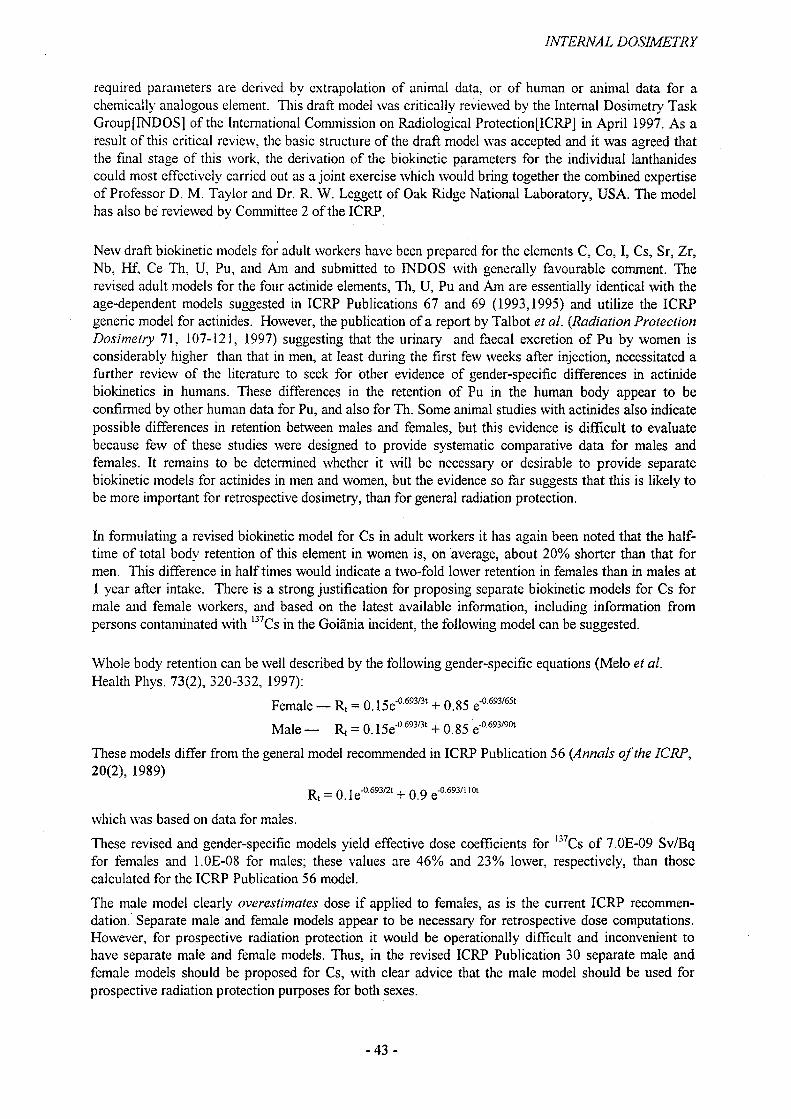
INTERNAL DOSIMETRY
required parameters are derived by extrapolation of animal data, or of human or animal data for a chemically analogous element. This draft model was critically reviewed by the Internal Dosimetry Task Group[INDOS] of the International Commission on Radiological Protection[ICRP] in April 1997. As a result of this critical review, the basic structure of the draft model was accepted and it was agreed that the final stage of this work, the derivation of the biokinetic parameters for the individual lanthanides could most effectively carried out as a joint exercise which would bring together the combined expertise of Professor D. M. Taylor and Dr. R. W. Leggett of Oak Ridge National Laboratory, USA. The model has also be reviewed by Committee 2 of the ICRP.
New draft biokinetic models for adult workers have been prepared for the elements C, Co, I, Cs, Sr, Zr, Nb, Hf, Ce Th, U, Pu, and Am and submitted to INDOS with generally favourable comment. The revised adult models for the four actinide elements, Th, U, Pu and Am are essentially identical with the age-dependent models suggested in ICRP Publications 67 and 69 (1993,1995) and utilize the ICRP generic model for actinides. However, the publication of a report by Talbot et al. (Radiation Protection Dosimetry 71, 107-121, 1997) suggesting that the urinary and faecal excretion of Pu by women is considerably higher than that in men, at least during the first few weeks after injection, necessitated a further review of the literature to seek for other evidence of gender-specific differences in actinide biokinetics in humans. These differences in the retention of Pu in the human body appear to be confirmed by other human data for Pu, and also for Th. Some animal studies with actinides also indicate possible differences in retention between males and females, but this evidence is difficult to evaluate because few of these studies were designed to provide systematic comparative data for males and females. It remains to be determined whether it will be necessary or desirable to provide separate biokinetic models for actinides in men and women, but the evidence so far suggests that this is likely to be more important for retrospective dosimetry, than for general radiation protection.
In formulating a revised biokinetic model for Cs in adult workers it has again been noted that the halftime of total body retention of this element in women is, on average, about 20% shorter than that for men. This difference in half times would indicate a two-fold lower retention in females than in males at 1 year after intake. There is a strong justification for proposing separate biokinetic models for Cs for male and female workers, and based on the latest available information, including information from persons contaminated with 137Cs in the Goiania incident, the following model can be suggested.
Whole body retention can be well described by the following gender-specific equations (Melo et al. Health Phys. 73(2), 320-332, 1997):
Female — R, = 0.15e"a693/3t + 0.85 e'a693/65t
Male — Rt = 0.15e'°693/3t + 0.85 e"a693/90t
These models differ from the general model recommended in ICRP Publication 56 (Annals of the ICRP, 20(2), 1989)
R, = 0.1e^' + 0.9e^'^which was based on data for males.
These revised and gender-specific models yield effective dose coefficients for lj7Cs of 7.0E-09 Sv/Bq for females and 1.0E-08 for males; these values are 46% and 23% lower, respectively, than those calculated for the ICRP Publication 56 model.
The male model clearly overestimates dose if applied to females, as is the current ICRP recommendation. Separate male and female models appear to be necessary for retrospective dose computations. However, for prospective radiation protection it would be operationally difficult and inconvenient to have separate male and female models. Thus, in the revised ICRP Publication 30 separate male and female models should be proposed for Cs, with clear advice that the male model should be used for prospective radiation protection purposes for both sexes.
-43-
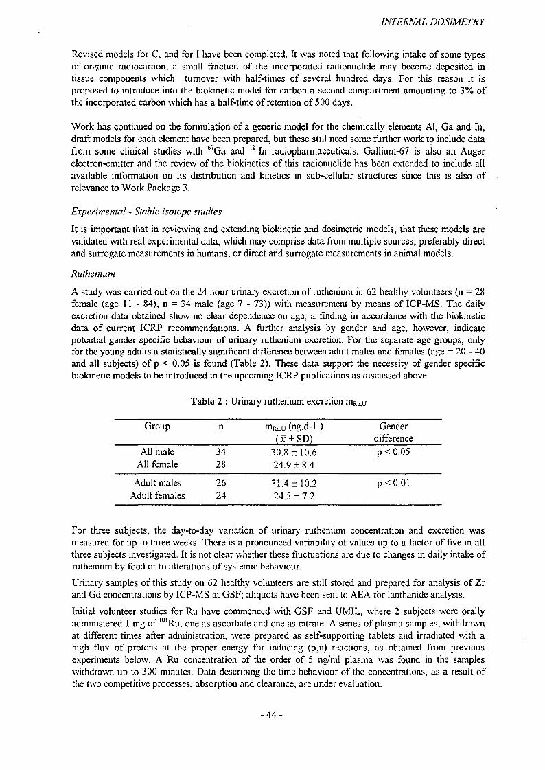
INTERNAL DOSIMETRY
Revised models for C. and for I have been completed. It was noted that following intake of some types of organic radiocarbon, a small fraction of the incorporated radionuclide may become deposited in tissue components which turnover with half-times of several hundred days. For this reason it is proposed to introduce into the biokinetic model for carbon a second compartment amounting to 3% of the incorporated carbon which has a half-time of retention of 500 days.
Work has continued on the formulation of a generic model for the chemically elements AI, Ga and In, draft models for each element have been prepared, but these still need some further work to include data from some clinical studies with 67Ga and mIn radiopharmaceuticals. Gallium-67 is also an Auger electron-emitter and the review of the biokinetics of this radionuclide has been extended to include all available information on its distribution and kinetics in sub-cellular structures since this is also of relevance to Work Package 3.
Experimental - Stable isotope studies
It is important that in reviewing and extending biokinetic and dosimetric models, that these models are validated with real experimental data, which may comprise data from multiple sources; preferably direct and surrogate measurements in humans, or direct and surrogate measurements in animal models.
Ruthenium
A study was carried out on the 24 hour urinary excretion of ruthenium in 62 healthy volunteers (n = 28 female (age 11 - 84), n - 34 male (age 7 - 73)) with measurement by means of ICP-MS. The daily excretion data obtained show no clear dependence on age, a finding in accordance with the biokinetic data of current ICRP recommendations. A further analysis by gender and age, however, indicate potential gender specific behaviour of urinary ruthenium excretion. For the separate age groups, only for the young adults a statistically significant difference between adult males and females (age = 20-40 and all subjects) of p < 0.05 is found (Table 2). These data support the necessity of gender specific biokinetic models to be introduced in the upcoming ICRP publications as discussed above.
Table 2 : Urinary ruthenium excretion mRUiU
Group n mRu,u (ng.d-1 ) (x±SD)
Genderdifference
All male 34 30.8 ±10.6 p < 0.05All female 28 24.9 ±8.4
Adult males 26 31.4 ±10.2 p<0.01Adult females 24 24.5 ±7.2
For three subjects, the day-to-day variation of urinary ruthenium concentration and excretion was measured for up to three weeks. There is a pronounced variability of values up to a factor of five in all three subjects investigated. It is not clear whether these fluctuations are due to changes in daily intake of ruthenium by food of to alterations of systemic behaviour.
Urinary samples of this study on 62 healthy volunteers are still stored and prepared for analysis of Zr and Gd concentrations by ICP-MS at GSF; aliquots have been sent to AEA for lanthanide analysis.
Initial volunteer studies for Ru have commenced with GSF and UMIL, where 2 subjects were orally administered 1 mg of l01Ru. one as ascorbate and one as citrate. A series of plasma samples, withdrawn at different times after administration, were prepared as self-supporting tablets and irradiated with a high flux of protons at the proper energy for inducing (p.n) reactions, as obtained from previous experiments below. A Ru concentration of the order of 5 ng/ml plasma was found in the samples withdrawn up to 300 minutes. Data describing the time behaviour of the concentrations, as a result of the two competitive processes, absorption and clearance, are under evaluation.
-44-

INTERNAL DOSIMETRY
Optimisation of proton activation analysis for ruthenium, zirconium and tellurium
The optimization procedures for the proton activation analysis techniques and tests on animals conducted previously had suggested the use of l24Te, 126Te and "Ru, 101Ru isotopes as tracers with suitable (p,n) reactions for determination. Detection limits in human plasma of proton activation analysis for both the isotopes of Te and of Ru to be injected and given orally, were performed upon plasma samples doped with known amounts of 124Te, l26Te as well as of "Ru, 101Ru. In the adopted experimental conditions, the minimum detectable quantity, corresponding to a signal equal to 2.7 + 4.65 (B)1'2, being B the background signal, resulted to be each 3 ng • ml"1 plasma for 124Te, 126Te, 10lRu and 12 ng • ml"1 plasma for "Ru.
Preliminary measurements were performed by proton activation analysis on blood samples from Italian donors to obtain data of Zr and Gd prior to biokinetic investigations. Plasma samples were enriched with 90Zr and 96Zr. Detection limits of 1 ng • ml"1 in human plasma for each isotope were determined for the simultaneous determination of the two isotopes, while limits of detection of 0.5 ng • ml"1 were reached by optimizing the conditions for the two isotopes separately. For Gd, only (p,n) reactions have been exploited and detection limits of 8 ng • ml"1 in human plasma have been obtained.
In the literature, data on Zr content in body fluids are scarce, a zirconium concentration of 2.6 ± 1.2 ng • ml"1 in serum, obtained by ICP-MS was reported (H. Morita et al. 1994) and a Zr concentration of 5 ng • ml'1 in blood from Italian subjects was indicated (C. Minoia et al. 1994) as a probable value by starting from the determination of Hf and by considering a Zr/Hf ratio of 40 in biological systems as found in soil and rocks. In this work, concentrations of 1.0 ± 0.3 ng • ml"1 and 5.0 ± 1.1 ng • ml"1 in plasma and in total blood respectively were obtained.
The use of (p,2n) reactions, which requires higher energy, approx, the double of the one used, but which have higher cross section, approx, four times that for (p,n) reactions, would improve sensitivity.
Calcium and Strontium Uptake in NeonatesExcreta samples have been collected and analysed following administration by ingestion of 1 mg enriched 43Ca and 50 pg enriched S4Sr to a group of six male neonates at St. Georges Hospital, London. Three sample sets have been analysed to date and confirm that Ca and Sr absorption is rapid with a significant fraction absorbed. The pattern of excretion is rapid such that urine collection for the first 24 hours post-administration will collect the majority of the Ca and Sr excreted with peak clearance approximately 6 hours post-administration. Cumulative urinary excretion data for 3 neonates are shown in Figure 2 for Sr and Figure 3 for calcium. The preliminary data indicate that Sr excretion in each neonate is consistent and dependent on dose administered. However, excretion of the Ca stable isotope is dependent on the total mass of Ca excreted by each neonate, under homeostatic control.
Urinary Sr excretion
5000 --
4000 -■
3000 --
2000 --
1000 -•
20.00 40.00 60.00 80.00
Time (h)
-e—Baby 1
•Baby 2
• Baby 3
Figure 2 : Cumulative urinary excretion of orally administered 84Sr
-45 -

INTERNAL DOSIMETRY
XS Ca (U) Excretion
0.0 20.0 40.0
Time (h)60.0 80.0 * 1
Figure 3 : Cumulative urinary excretion of orally administered 43Ca
Models for the embryo / fetus
An area of significant concern is protection of critical groups, in this case the embryo and fetus, where stem cells may at particular risk from radiation damage from internalised radionuclides. As for Activity1 and Work Package 1, a combination of model development and experimental validation is addresed to identify and reduce key uncertainties.
Fetal transfer - data review
Review of data on the transfer of radionuclides to the embryo and fetus has progressed as planned. During the period, work was undertaken on draft reviews for the following elements: H, Fe, Co, Zn, Se, Sr, Ru, Ba, Cs, Ce, Pb, Po, Ra, U, Pu and Am. For the alkaline earth elements, Sr, Ba and Ra, a biokinetic model has been developed which relates transfer to the fetus to the requirement of the fetus for Ca and the rate of accretion of Ca by the fetal skeleton. This model and information reviewed for other elements will be used in the work of the ICRP Internal Dosimetry Task Group which, together with the Task Group on Dose Calculations, is preparing a report on doses to the embryo and fetus.
The model for the transfer of alkaline earth elements to the fetus takes account of changes in maternal Ca metabolism during pregnancy as well as fetal requirements. Thus, account is taken of human data showing increased absorption of ingested Ca, increased bone turnover (both accretion and resorption) and increased urinary excretion. In modelling transfer to the fetus, account is taken of primate data showing that total transfer of Ca from maternal to fetal circulations exceeds requirements and that a large proportion is returned to the maternal circulation. Similar results have been reported for Sr. The model considers fetal bone as three compartments: bone surface, exchangeable bone volume and nonexchangeable bone volume with no separation of cortical and trabecular compartments as in postnatal models. For acute intakes of 45Ca and yoSr in late pregnancy at 36 weeks when maximum transfer occurs, the model predicts average fetal concentrations (whole-body) of about 19 and 9 times average maternal concentrations, respectively.
Draft reviews of biokinetic data on transfer of elements to the embryo/fetus are complete as input to the ICRP report on fetal dosimetry. A biokinetic model for the alkaline earth elements was presented at the
- 46 -
INTERNAL DOSIMETRY
Workshop on Intakes of Radionuclides, Avignon, September 1997. Further development is in progress; the model will be completed during the next 2-3 months and published in complete form.
Experimental studies - Human
Human fetal bone samples were supplied by Dr. D. Henshaw (Univ. Bristol). A total of 27 samples from individual cases between 17 and 32 weeks of gestation were combined (34 g) for analysis od plutonium, and 13 samples from between 36 to 41 weeks (50 g). Results obtained by mass spectrometry were significant for the 36-41 week sample but not the 17 - 32 week sample. The later sample contained (0.7 + 0.2) • 106 atoms 239Pu and (0.1 + 0.01) • 106 atoms 240Pu, corresponding to a concentration of 19 mBq • kg'1. Reported concentrations of fallout 239/240pu in adult bone (age 20y) are about 1 mBq • kg'1. Five sets of fetus and placenta from second trimester terminations (13 - 19 weeks) were also analysed. A single significant measurement was recorded for a placenta (16 weeks) weighing 83g: (1.1 + 0.2) • 106 atoms 239Pu and (0.3 + 0.01) • 106 atoms 240Pu, corresponding to a concentration of 22 pBq kg"1.
Studies of the transfer of nuclides through perfused human placenta have been initiated in collaboration with Dr. A. Preece (Univ. Bristol). The apparatus has been constructed and tested. After some initial problems, mainly in meeting requirements for work with human tissues, studies are now progressing well. The viability of the system, involving perfusion of fetal and maternal sides of an isolated cotyledon of the placenta, has been established using 99mTc. Results show that the ionic form transfers rapidly across the placenta while protein bound 99mTc-albumin does not transfer. Comparisons of the transfer of Ca and Sr are in progress. Because of difficulties over safety issues, alpha emitters will not be used in this system. The possibility of using 241 Pu (beta emitter) and comparing transfer with that of lanthanide analogues of the trivalent actinides is under consideration.
Experimental studies - Animal
Previous studies of the transfer of the actinides, Pu and Am, to the fetus in rats and guinea pigs have shown considerable differences in levels of transfer and distribution between fetal tissues, placenta and fetal membranes with generally greater values for Pu. This work has been extended to compare the behaviour of 237Np and 230Th to previous results for Pu and Am, measuring transfer in the rat at the time of yolk sac haemopoiesis (9 - 12 d gestation) and in both the rat and guinea pig in late gestation (18-21 d rat, 50 - 57 d guinea pig). Results for the transfer of 237Np are complete and 230Th results will be completed early in 1998. Results for retention of Np in the placenta and yolk sac are intermediate between those for Pu and Am. Transfer to the fetus was similar to that of Pu in the rat, but with a lower value similar to that of Am in the guinea pig. Results for transfer to the fetus for each nuclide in each rodent species were lower than corresponding values obtained previously for transfer to the primate fetus in late gestation.
Initial delays were experienced in commencement of the equivalent primate studies (CEA) due to the degree of control exerted over such studies by the member state. Four radionuclides, ^Np, ~39Pu,241 Am and 210Po, and three stages of gestation (early, mid and late) have been defined. The first set of data from baboons has demonstrated that total retention in each fetus, measured one week after injection, accounted for about 1% of administered 210Po and 237Np, 4% of 239Pu and 0.4% of241 Am. These values correspond to fetal:maternal whole body concentrations ratios, Cf'.Cm of about 0.3 for 210Po, 0.6 for 237Np, 1.3 for :39Pu and 0.1 for 241 Am. These values are lower than observed for other species, particularly for 239Pu. Further data from the macaca macaca will allow validation of these results.
DosimetryA generic model for calculation of doses to embiyo and fetus after activity intake by the mother has been developed by BfS and NRPB with other members of the ICRP Task Group on Dose Calculations. Doses to organs and tissues of the fetus (those with a specified tissue weighting factor and additionally the dose to the brain during the period from week 8 to 15 of pregnancy) are calculated assuming
-47-
INTERNAL DOSIMETRY
a constant activity concentration ratio fetus/mother and placenta/mother and an activity distribution within the fetus similar to that in the 3-month-old infant.
The parameters needed by the model have been determined for the 31 elements which will be contained in the planned ICRP Publication on doses to embryo and fetus.
The generic model has been implemented (see Working Package 4) within a computer code DOSAGE and a quality assurance programme validating results has commenced. Initial reviews have shown several problems especially in the interpretation of dosimetric models. However, these are under further review with solutions planned by the next meetings of the ICRP Task Groups on Internal Dosimetry (INDOS) and on Dose Calculations (DOCAL).
-48 -

INTERNAL DOSIMETRY
2.3 Work Package 3
Target cell dosimetry for short-range particles
Contributors: NRPB (02), CEA (05), UROS (08), IPSN (09)
Objectives
The objectives of Work Package 3 for the reporting period were:
• Prepare and analyse alpha and fission track autoradiographs of femur samples from mice given 239Pu,241 Am or 233U and to estimate distribution of alpha dose within bone
• Study the intracellular distribution of actinides in rat liver using biochemical techniques
• Assess the biological effect of alpha particles on pulmonary cells cultures
• Study cytotoxicity, micronucleus and apoptosis induction after exposure of human amniotic cells (AFFL) and human squameous cell carcinoma cells (SCL-II) either to external X-rays or to different activities of 65Zn
• Achieve autoradiography of different tissue sections sampled in animals after incorporation of actinides using a newly developed coupled scintillation-intensified CDD technique and to compare this method with other micro-autoradiography techniques
• Install an ion microscopy equipment’s and set up pilot experiments with all the partners (NRPB, CEA, UROS, and IPSN) in order to investigate the performance and the potential of SIMS for internal dosimetry research
Progress
Radionuclide and dose distribution in bone samples
A collaborative project between NRPB and Dr B. Lord of the Paterson Institute for Cancer Research (PICR) has the aim of quantifying the distributions of alpha dose in bone from the nuclides 239Pu,241 Am and 23jU. Autoradiographs of the distribution of 239Pu, 241Am and 233U in mouse bone were prepared as part of a previous project in which osteosarcoma and leukaemia induction by the three nuclides was compared. Detailed mapping of the distribution of alpha tracks in cortical and trabecular regions of femur sections is in progress, taking account of the distribution in bone mineral and marrow. The results are being used to estimate doses to cells on bone surfaces and at different depths into the marrow cavities.
Initially, autoradiographs were available for bone samples taken at Id, 28d and 224d after injection. With the collaboration of Dr. Wesch at DKFZ Heidelberg who has undertaken additional reactor irradiations of bone samples, autoradiographs are being prepared for 7d, 14d, 56d, 112d and 448d after injection.
The three nuclides differ in their patterns of initial deposition in bone and subsequent movement within bone. Thus, all three deposit initially on bone surfaces. However, 239Pu deposits preferentially on endosteal surfaces while 241 Am is more evenly distributed over all bone surfaces. The initial deposition of 2j3U shows preferential association with growing surfaces. At later times, association of each nuclide with bone surfaces is reduced by burial in bone and transfer to marrow: the latter process is most apparent for 239Pu and negligible in the case of 233U.
Preliminary estimates of dose distribution have been made, considering the region adjacent to endosteal bone surfaces (10pm layer) thought to contain target cells for osteosarcoma and different regions of the red bone marrow containing the leukaemogenic target cells. The results indicate that for administered activities giving equal average bone doses of the three nuclides, doses to the designated target regions were in each case in the order: 239Pu > 24lAm > 233U. The same relationship was seen in the ability of the
-49-
INTERNAL DOSIMETRY
nuclides to induce osteosarcoma in the life-span study undertaken previously at NRPB. However, 239Pu and ""'Am appear to be equally effective in causing acute myeloid leukaemia. This apparent discrepancy will be addressed by analysis of the spatial and temporal distribution of dose in marrow.
Following a meeting of participants of this VVorkpackage at 1PSN (June 1996), bone sections were transferred to IPSN for use in intercomparisons of imaging techniques including SIMS.
Intracellular distribution of actinides in liver
Experiments have been done in CEA with 239Np and *37Np in rats, and validated with primates. For -j9Pu and 241 Am, experiments were conducted on primates only.
Investigations of the sub-cellular distribution patterns of 239Np and 237Np in rat liver have shown that the distribution of neptunium is time and mass dependent. One hour after administration, 237Np deposits predominantly in the cytosol, whereas at later times after exposure (24 h to 40 days) the nuclei and lysosomes are the main binding sites. In contrast, 24 h after exposure, the 2j9Np was found mainly in the cytosol. Investigations in baboons with 237Np, 239Pu and241 Am have shown that the cytosol was the main binding site 24 h after exposure, and that nuclei and lysosomes were involved at later times after exposure (10 days). In cytosol, 239Pu and241 Am were bound mainly to ferritin irrespective of the time of sacrifice. For 237Np, it was shown, both in rats and baboons, that the radionuclide binds to two proteins soon after exposure (1 h to 24 h) with molecular weights of 450 and 200 kDa, respectively. The former was identified as ferritin, but the latter remains unidentified. At later times (Id to 40 d), Np was found to be bound mainly by ferritin and by high molecular weight compounds.
The main results obtained on the bio-inorganic chemistry of neptunium, plutonium and americium in the liver cells can be interpreted as follows: After their entry into the blood, the three actinides are distributed amongst the different target organs in the form of both transferrin and low molecular weight complexes. These three radionuclides are then transferred into the liver cells by an unknown mechanism. It can reasonably be assumed that the mechanism is similar as for Fe34 since these three radionuclides have the same molecular carriers as iron. After passage through the cell membrane, neptunium appears to be bound first by a cytosolic protein of MW 200 kDa. Tins protein has not yet been identified but some evidence suggests that it could be calmodulin. It cannot be stated at this stage that plutonium and americium are, or are not, bound by this protein just after their translocation into the cells; if they are, as supposed elsewhere for plutonium, this phenomenon should be very transient since 24 hours after injection this protein was free from contamination with both Pu and Am. However, the three radionuclides seem to be progressively transferred to ferritin and to high molecular weight (HMW) compounds. This is particularly true for neptunium for which, 10 d after contamination, 30% of the cytosolic radionuclide was associated with these HMW compounds. Previous studies have shown that plutonium and americium were bound to lipofuscin in the canine liver and it is not impossible that the HMW compound used here was also lipofuscin. Nevertheless further analyses are needed to confirm this speculation. Another possibility is that these HMW compounds correspond to some material resulting from lysosomal rupture. The measurement of the acid phosphatase activity in the cytosol shows that, in the experiments with baboons, less than 15% of the total marker enzyme were present in this fraction. This indicates that this phenomenon cannot be excluded but is probably very restricted.
After passage through the cytosol the radionuclides are distributed amongst the various organelles, with preferential accumulation in lysosomes and nuclei. Previous studies have shown that neptunium in rat liver cells was bound by some proteins of the nuclear matrix, which have low turnover and a very strong affinity for the radionuclide. Such data are for the moment unavailable for plutonium and americium but other studies showed clearly that a large fraction of these two radionuclides is also bound to the nuclear structures. This translocation to the nuclear structures could be mass dependant; when low masses were injected, the radionuclide seems to deposit predominantly in the lysosomes and in the cytosol, whereas high masses cause saturation of these sites and complexation elsewhere. This has already been demonstrated in vitro for 239Pu and 238Pu and in vivo for 237Np and 239Np. When accumulated by lysosomes, the radionuclides are probably bound as ferritin complexes. A search for other ligands
-50-

INTERNAL DOSIMETRY
involved in the retention of the radionuclide inside the lysosomal structures has been made but, for the moment, without success.
The biochemical behavior of americium, plutonium and neptunium proposed here is not radically different from what was known previously about the bio-inorganic chemistry of these actinides. Nevertheless, it emphasizes the particular behavior of neptunium when compared with americium and plutonium. It can not be said for the moment if this behavior is due to the relatively lower specific activity of 237Np, and hence higher mass. Nevertheless, this point has to be taken into consideration for the calculation of the doses resulting from the incorporation of the three radionuclides.
Biological effect of alpha particles on pulmonary cells
The biological effects of a-particles on pulmonary cells in culture have been often studied because of their interest in potential health effects induced after inhalation of a-emitting radionuclides. The aim of the work realized in CEA was to determine:
1) The influence of cell morphology - shape and size- on a-induced lethality,2) The variation of radiosensibility between two different strains of rat.
An epithelial cell line (RTiv3) immortalized from primary rat tracheal epithelial cell culture by a 1.25 Gy dose of y-rays 60Co has been used. The irradiation was performed using three "j9Pu electrodeposited sources with a mean flux of 1.3.1 O'4 a/(pm2.s) on 3 cm2 and with an a-energy of 5.155 MeV for 73% of the total a-disintegration. The uniformity of a-emission was controlled using CR39 solid nuclear track detector etching for 15 minutes with 12 N KOH at 85 °C.
To determine the influence of the cell morphology on the survival fraction, similar cells were irradiated; either adherent cells on mylar foil or sedimented cells obtained by trypsine treatment of the unirradiated adherent cells and irradiated 10 minutes after re-plating on mylar foil. After exposure, cells were trypsinised or washed respectively and subcultured in five culture dishes. Colony formation was the measure of the cell survival and only colonies with more than 50 cells were recorded.
The evolutions of the survival fraction for sedimented and adherent cells as a function of the mean fluence <|> (a-particule per pm2 emitted by the source in 2 n) are shown in the figure 4. With a singleexponential curve fit, the estimated <j>37% is 0.059a/pm2 and 0.037a/pm2 for sedimented and adherent cells respectively.
o sedimented cells □ adherent cells ---- S=exp(-4>/0.059) P=0.96
1.000
0.100
0.010
0.001
Figure 4 : Survival fraction of cells as a function of mean fluence ® of a-particles
-51-
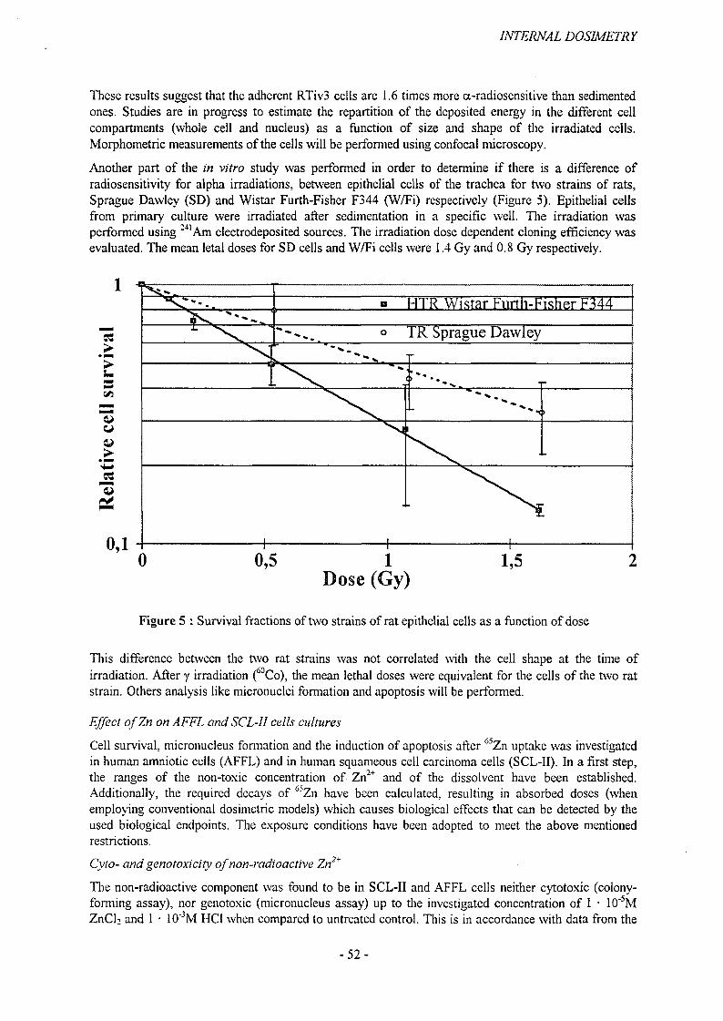
INTERNAL DOSIMETRY
These results suggest that the adherent RTiv3 cells are 1.6 times more a-radiosensitive than sedimented ones. Studies are in progress to estimate the repartition of the deposited energy in the different cell compartments (whole cell and nucleus) as a function of size and shape of the irradiated cells. Morphometric measurements of the cells will be performed using confocal microscopy.
Another part of the in vitro study was performed in order to determine if there is a difference of radiosensitivity for alpha irradiations, between epithelial cells of the trachea for two strains of rats, Sprague Dawley (SD) and Wistar Furth-Fisher F344 (W/Fi) respectively (Figure 5). Epithelial cells from primary culture were irradiated after sedimentation in a specific well. The irradiation was performed using :41Am electrodeposited sources. The irradiation dose dependent cloning efficiency was evaluated. The mean letal doses for SD cells and W/Fi cells were 1.4 Gy and 0.8 Gy respectively.
HTR Wistar Furth-Fisher F344
TR Sprague Dawley
Dose (Gy)
Figure 5 : Survival fractions of two strains of rat epithelial cells as a function of dose
This difference between the two rat strains was not correlated with the cell shape at the time of irradiation. After y irradiation (60Co), the mean lethal doses were equivalent for the cells of the two rat strain. Others analysis like micronuclei formation and apoptosis will be performed.
Effect ofZn on AFFL and SCL-II cells cultures
Cell survival, micronucleus formation and the induction of apoptosis after 65Zn uptake was investigated in human amniotic cells (AFFL) and in human squameous cell carcinoma cells (SCL-II). In a first step, the ranges of the non-toxic concentration of Zn2+ and of the dissolvent have been established. Additionally, the required decays of 65Zn have been calculated, resulting in absorbed doses (when employing conventional dosimetric models) which causes biological effects that can be detected by the used biological endpoints. The exposure conditions have been adopted to meet the above mentioned restrictions.
Cyto- and genotoxicity of non-radioactive Zn2+
The non-radioactive component was found to be in SCL-II and AFFL cells neither cytotoxic (colony- forming assay), nor genotoxic (micronucleus assay) up to the investigated concentration of 1 • 10"5M ZnCL and 1 • 10"3M HC1 when compared to untreated control. This is in accordance with data from the
-52-

INTERNAL DOSIMETRY
literature. The induction of superoxiddismutase (SOD) and metallothionein is described to start at concentration above 3 • lO^M ZnCl2 (e.g. effects on cytotoxicity start in HeLa cells at 1 • lO^M ZnCl2; Borovanskv & Riley, 1989; Chem. Biol. Interact. 69:279-291 and Hatavama et al., 1992; Mol. Cell. Biochem. 112:143-153). No increase in the induction of apoptosis was observed as well.
Development of suitable exposure conditions
Working concentration of 65Zn was chosen to be well below 1 • !0'5M and to be in the range of 2 • 10"7 to 8 • 10"7M 65Zn. HC1 is used up to a concentration of 1 • 10"3M.
Exposure conditions were chosen to obtain 1 decay per pm3 of cell volume assuming a uniform intracellular distribution of 65Zn and assuming that after 24 h the intracellular 65Zn concentration is equimolar to the medium. These considerations were found to be useful and in agreement with data from the literature for other radionuclides (Miyazaki & Shinoharay, 1993; Rad. Res. 133:182-186).
To meet the working concentrations and the intended decays/cell, cells were exposed with 2.66 MBq 65Zn under cell culture conditions. After incubation for 24 hours (h) the medium is removed, cells are trypsinized and resuspended in freezing medium. The cell suspension is transferred to a plastic vial, stored for 2 h at -20°C, followed by 24 h at -80°C and kept in liquid nitrogen at -196°C for accumulation of the disintegration of 65Zn. The period of storage at -196°C for accumulation of decay to obtain the requested 1 decay/pm3 cell volume is 98 days. This results in a total exposure time of 100 days. X-irradiated cells and unexposed cells are used as reference/control and treated in the same way, except the exposure to 65Zn. To obtain fewer decays per volume the initially applied activity was adjusted.
Estimation of dose:
Accumulated decays per volume (lpm3) are calculated, assuming that after 24 h the intracellular 65Zn concentration is equimolar to the medium and 65Zn is uniformly distributed in the cells. The "mean energy of 6-radiation and Auger-electrons" times "yield" is calculated to be 6.87 • 10"3 MeV per decay, e.g. the absorbed dose after 48 h exposure to 10 MBq 65Zn is estimated to be only 0.1 Gy. From the preliminary results on micronucleus formation under these exposure conditions it can be concluded that the dose range (accumulated decays per cell) and the resulting genotoxic effect is suitable for the planned examinations.
An activity of 2.66 MBq 65Zn (24 h cell culture followed by 100 d decay accumulation in frozen state), assuming the above mentioned conditions, will result in an absorbed dose of 0.07 Gy. However the dose estimations are based on theoretical assumptions and considerations and have to be improved as soon as the data on 65Zn up-take and on the intracellular distribution of 65Zn are available.
The cyto- and genotoxicity of65Zn
The results on cell survival (SF, Surviving fraction) show the intense cytotoxic capacity of 65Zn when compared to external controls (Figure 6). After 0.1 decays • pm"3 (approx. 20 decays per nucleus) there is a pronounced cytotoxic effect, increasing with rising numbers of decays per volume. Even at 0.055 decays • pm"3 a significant decrease in cell survival is observable. The data can be fitted best with an exponential function. The data of cell survival after external radiation follows a classical cell survival curve (shoulder region at low doses, almost exponential in the higher dose region) of a medium radiation-resistant cell-line.
Apoptosis induction
After exposure to x-rays the highest number of apoptotic cells was detected 48 h post-irradiation. This time point was concluded to be the best for evaluation of apoptosis induction. A strong induction of apoptotic cells was found after 0.1 decays • pm"3 (10 times as much as controls) and increasing with raising numbers of decays per volume (Figure 7). A high variance in apoptosis induction after 1 and 2 decays/pm3 could be detected. Apoptosis induction as a function of dose after external x-irradiation can be described as linear-quadratic in SCL-II cells and linear in AFFL respectively. However more data have to be acquired.
-53 -

INTERNAL DOSIMETRY
10
1
CO 0.1
0.01
0.001
1 = 0,055 decays / pm32 = 0,1 decays / |jm33 = 0,27 decays / |jm34 = 1 decays / pm3
| ■ SCL-II | □ SCL-ll65-Znj
0123456789Dosis (Gy)
Figure 6: Surviving fraction (SF) of SCL-II after exposure to 65Zn (□) and external x-rays (■). Dose calculation based on accumulated decays of 65Zn
1=0,1 decay / pm32 = 1 decay / pm33 = 2 decays / pm34 = 2,5 decays / pm3
■ SCL-II external
□ SCL-II 65-Zn
♦ AFC external
o AFC 65-Zn
Figure 7: Apoptosis induction in SCL-II (squares) and AFFL (diamonds) cells after exposure to 65Zn (open symbols) and external x-rays (filled symbols).Dose calculation based on accumulated decays of 65Zn.
Micronucleus induction
The micronucleus (MN) frequency (Figure 8) is increased (about 2-fold of control value) even at lowest activities resulting in 0.055 decays • pm"3 (about 10 decays per cell nucleus). It shows a very steep increase up to a maximum of about 1.55 MN per cell after 0.27 decays • pm"3 (1.1 MN after 8 Gy external x-rays) and decrease immediately after about 0.4 decays • pm'3. The data after exposure to 65Zn show at high MN frequencies a much higher variation when compared to according MN frequencies after external radiation. Additionally, much more cells with multiple MN can be detected when compared to external radiation. The rapid decrease of MN frequency at 0.4 decays • pm"3 can only be explained by an overkill effect. Those cells receiving too much damage will not divide anymore (arrested at mitosis or killed by apoptotic processes) and therefore do not express the inherent damage, for example as micronuclei. Because those cells are not represented only less damaged cells or non damaged cells contribute to the MN frequency, resulting in a weak MN induction in binucleated cells.
-54 -
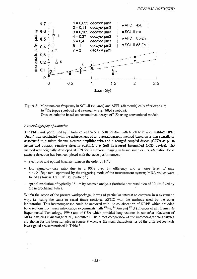
INTERNAL DOSIMETRY
decays/ pm3 decays/ pm3 decays/ pm3 decays/ pm 3 decays/ pm3 decays/ pm3 decays/ pm3
♦ AFC ext.
o AFC 65-Zn
□ SCL-II 65-Zn
dose (Gy)
Figure 8: Micronucleus frequency in SCL-II (squares) and AFFL (diamonds) cells after exposure to 65Zn (open symbols) and external x-rays (filled symbols).Dose calculation based on accumulated decays of foZn using conventional models.
Autoradiography of actinides
The PhD work performed by I. Aubineau-Laniece in collaboration with Nuclear Physics Institute (IPN, Orsay) was concluded with the achievement of an autoradiography method based on a thin scintillator associated to a micro-channel electron amplifier tube and a charged coupled device (CCD) as pulse height and position sensitive detector (aSTIC : a Self Triggered Intensified CCD device). The method was originally developed at IPN for p markers imaging in tissue samples. Its adaptation for a particle detection has been completed with the basic performances:
- electronic and optical linearity range in the order of 106;
- low signal-to-noise ratio due to a 90% over 2n efficiency and a noise level of only 4 • 10"7 Bq • mm"2 optimised by the triggering mode of the measurement system; MDA values were found as low as 1.5 • 10"5 Bq • particle"2;
- spatial resolution of typically 15 pm by centroid analysis (intrinsic best resolution of 10 pm fixed by the microchannel tube).
Within the scope of the present workpackage, it was of particular interest to compare in a systematic way, i.e. using the same or serial tissue sections, aSTIC with the methods used by the other laboratories. This intercomparison could be achieved with the collaboration of NRPB which provided bone sections from mice intoxication experiments with 239Pu,241 Am and 233U (Ellender et al., Human & Experimental Toxicology, 1994) and of CEA which provided lung sections in rats after inhalation of MOX particles (Guezingar et al., submitted). The direct comparison of the autoradiographic analyses are shown for the bone samples in Figure 9 whereas the main characteristics of the different methods investigated are summarised in Table 3.
-55 -

INTERNAL DOSIMETRY
Optical Autoradiographic Track-etched Phosphor screen Video radiation Scintillating microscopy film detector detector detector detector aSTIC
Figure 9: Comparison of serial bone sections autoradiographies
Table 3: Characteristics of the investigated autoradiography techniques.
Type Passive ActiveDetector Track-
etcheddetector
Emulsion Film Phosphorscreen
Videoradiationdetector
Gasdetector
ScintillatordetectorRJHR
Analyse surface (cm2)
30x10 slidedimension
30x40 20x2535x43
0.7x0.9 18x1826x26
0.9x1.3
spatial resolution (pm)
0.2-10 2 20 50-200 40 200-400 15
Detectedradiations
a P a,P a,p a P P
Quantificationrelative/absolute
relative absolute
The aSTIC and the phosphorus screen imager allow both quantitative autoradiography with a large linearity range and appear to be well complementary with regard to spatial resolution and field of view. CR39 nuclear track-etch detectors are expert techniques with still better spatial resolution but with limitations for quantitative analysis as passive methods. A useful approach in the frame of internal dosimetry experiments therefore seems to apply first phosphor screen imager for rapid screening of relatively large numbers of slides at medium resolution, so that aSTIC can optimise the analysis on selected samples with absolute quantification at higher resolution and efficient active detector characteristics. Moreover discrimination capabilities can be used to separate different a emitters present in the same sample. The present energy resolution is around 13% at 5 MeV alpha energy, but could be improved by a factor two (ref 5). On the other hand, discrimination of a and P emitters is straightforward with potential interest for radiotoxicology experiments.
The comparison described above could be carried out with the help of D. Henshaw (Univ. Bristol, UK) and C. Zeissler (NIST, USA). The data will be completed soon so that the report on this comparative study can be finalised.
-56-

INTERNAL DOSIMETRY
Potential application to modelling
Besides its development, the interest in microlocalisation lies to a large extent on the capability to use microdistribution data in internal dosimetry modelling, the reason for which quantification aspects have been emphasised in the present program. Two approaches were investigated including on the one hand the interpretation of microdistribution data by means of energy deposition calculations, and on the other hand their correlation with particle deposition and clearance modelling. For convenience, present work was focused on respiratory tract dosimetry, although the model is particularly complex, but could be also orientated towards bone dosimetry.
The approach adopted here consisted in investigating the relevance of the statistical distribution of energy deposition, as derived by microdosimetric methods, in comparison to average dose assessments to compare lung tissues exposed to actinides uniformly distributed or in the form of particle point- sources. The work performed was a preliminary step aimed at: (1) investigating the suitability of two well-established microdosimetric calculation methods combined to determine single-event and dose dependent multi-event distributions in epithelial cells of airway walls; and (2) assessing the relative importance of multi-event occurrence on the shape of the specific energy distributions.
The first method was based on the code RADONA developed by Caswell and Coyne for the microdosimetry of radon and radon daughters in simplified models of bronchiolar tissue (Rad. Prot. Dosim, 1994). The code considers the airway walls composed of unit-density tissue and the cavity filled with air. It assumes the radioactive material uniformly spread on the inner wall and the target cells all localised at the same depth. Fixing a given target, the code scans the airway volume according to the axial and longitudinal cylinder directions and to the mucus layer thickness. In this way, it determines the source volume of interest comprising every source element ('voxel') at a tissue-equivalent distance to the target smaller than the maximum range of a particles taking into account their paths through tissue and air. The code then determines first the spectra of those particles originating from the volume of interest and slowing down before reaching the sensitive site at its centre. Then, assuming the target cells to be spherical and considering the corresponding chord length distribution, the code generates the singleevent probability density ifi(z)) implicitly averaged over the source volume of interest.
The original code assumed the radioactive material uniformly spread through the mucus on the inner wall of the higher generation airways. In the present calculations, it could easily be applied to lower generation airways providing appropriate geometrical parameters. It was further modified to process separately within the same scan each individual source voxel in order to derive the related single-eventspectra. Those one were then used with the corresponding mean number of events, n, as input for the convolution calculation of f(z) using a second analytic code derived from Roesch (1977).
To derive the single event distributions, the first code records for each voxel its topology with regard to the considered target. Indeed, because of the non-uniform density of the airways, each voxel ischaracterised by its geometric distance to the target, rseom, and by the corresponding tissue-equivalent
distance ,r , crossed by the a particle taking into account its pathway in air. The latter one determines the slowing-down energy of the a particle when reaching the target and so is the distance parameter of f,(z;r). The former one determines the average number of a particles entering the target. Using %j,E notation to identify each source voxel, the code generates so the distributions
TP — Q£Ot7lf\ (z; rt j ^ ) and calculates the corresponding »(rjj k ) values which, referring to Roesch formalism,
correspond to the products (aw)jj,k, where 'a' is the number of a particles emitted during the selected exposure time and 'w' the ratio to 4n of the solid angle subtended by the target. In this way, it is possible to consider any configuration of activity distribution within the inner layer airway, whether it is a matter of a single source voxel, several scattered ones or a uniformly and continuously distributed activity in the source volume of interest. To derive the multi-event distributions by the recurrent convolution of
TEf\(z;Tj j j.) distributions, the second code used, set up by Li et al. (Rad. Prot. Dosim., 1996), was
based on a Fast Fourier Technique (FFT). FFT takes advantage of the symmetry in arrays of discrete
-57-

INTERNAL DOSIMETRY
Fourier transforms to reduce drastically the number of necessary mathematical operations. This reduces considerably computing time since it applies to the Fourier transform and its inverse. The results showed that in the case of multiple sources in the volume of interest, the effect of multi-events is negligible except at abnormally high activity deposition since this activity is spread among sources some of which are at the opposite position in the airway with respect to the target. In turn, the exposure to localised single sources may lead to significant multi-event distortions to the cells in the vicinity of each source (Figure 10).
The results illustrated the usefulness of the calculation to quantitatively assess the influence on energy deposition of various morphological parameters of the respiratory tracts, and to discriminate so non critical parameters. For example, the calculated spectra show to what extent disperse radioactive material is not a very different exposure situation compared with uniform distribution as assumed in conventional dose calculations. The interest of the calculation, and therefore also of high spatial resolution quantitative microlocalisation, lies in the consideration of highly localised distributions where there is a need to take into account large number of unexposed cells and cells hit with widely variable events. This in turn enhances the need for relevant biological data to be able to use such statistical information.
o
<N
Figure 10: Multi-event distributions f(z;AW‘) for single point source-target configurations derived by convolution of fi(z;r). For illustration, the curves obtained for 10, 20 and 30 pm tissue-equivalent distances (thin lines) between source and target are compared with those for two voxels (thick lines) in the simulated bronchial volume: cl with r£eom.=850 pm, rTE =22 pm, aw=0.24, a=l.3x105 a; and c2 with i^=l 10 pm, rTE=28 pm, aw=0.26, a=1.6xl05 a.The parameter aw is the product of the emission of alpha particles and its solid angle.
One possible case of non uniform distribution of the activity in the tissues is the concentration of material deposited in asymmetric regions of the respiratory tract, namely in the bifurcations of bronchi. In this configuration, the direct use of microlocalisation to perform dose calculations is not without problem. However, in this respect, ex vivo microlocalisation experiments may provide data relevant to the verification of numerical modelling such as the stochastic particle deposition and clearance modelling developed in Salzburg (contract "Inhalation of radionuclides"). A protocol, based on quantitative microlocalisation associated to outline recognition, was set-up leading to comparable results in the form of deposition versus tract diameter histograms.
-58 -
INTERNAL DOSIMETRY
Set-up and testing of SIMS equipment
The workplan of IPSN includes a pilot study using secondary ion mass spectrometry which offers among the main characteristics the microlocalisation of long-lived radionuclides and stable elements, and therefore a possible way of element localisation and "target" characterisation; a spatial resolution <1 pm; a multicellular field of view; and the analysis of elements at trace levels. Its feasibility for internal dosimetry was therefore proposed to be investigated within this contract taking opportunity of collaboration finalized in 1996 between IPSN and Institut Gustave Roussy (IGR, Villejuif, France).
The IGR equipment has been transferred to IPSN and was aligned and modernised by the manufacturer (CAMECA, Courbevoie, France). First test experiments could be performed in the CAMECA facility. These experiments were focused on the analytical performance of SIMS and the measurement procedures (reproducibility, MDA, linearity, stability, internal tracer). In a further step, the imaging part was updated before the equipment could be definitely installed at the IPSN laboratory late 1997 which now allows the partner laboratories to set up the pilot experiments for 1998.
-59-

INTERNAL DOSIMETRY
2.4 Work Package 4
Numerical implications of models
Contributors: GSF(Ol), NRPB(02), BfS(03) (with TNO as subcontractor)
Objectives
The objectives of Work Package 4 for the reporting period were:
• Produce a database of dose coefficients for a PC platform;• Extend the NRPB and BfS computer codes to enable the calculation of bioassay data for acute and
chronic intakes;• Calculate and publish a comprehensive set of bioassay data for individual monitoring of workers;• Implement a generic model for the calculation of doses to embryo/fetus after activity intake by the
mother;• Develop full biokinetic models for the embryo/fetus for special cases where sufficient data exist (eg
the alkaline earth elements);• Produce tables of dose coefficients based on this generic model;• Investigate the sensitivity of biokinetic models to changes in parameter values through the evaluation
of formal sensitivity coefficients;• Use Kalman filtering to estimate expected parameter values for biokinetic models and their
respective variances;• Use the Cramer-Rao measure to derive the lower limit of accuracy on model parameters;• Develop a Durbin filtering technique to determine the optimum number of pole parameters, and
arrive at the optimum number of compartments in a biokinetic model.• Obtain a voxel adult phantom (whose external anatomy is similar to the MIRD adult phantom)
which also has tissues relevant for inhalation i.e. extrathoracic and thoracic airways, with their associated lymph nodes;
• Calculate the Specific Absorbed Fractions (SAF) for monoenergetic photons for the most important source and target organs of the adult voxel phantom;
• Compare the SAFs obtained with the voxel phantom with those obtained with the MIRD adult phantom;
• Similarly obtain the SAFs for the two paediatric voxel phantoms BABY and CHILD. (This item was originally scheduled for 1998).
Progress
PC database of dose coefficients
The traditional compendia of dose coefficients published by the EU, ICRP and national bodies are inevitably limited in the number of results they provide by simple considerations of space. A need was therefore identified for a PC database of dose coefficients which would give many more results than could be included in a compendium.
The NRPB RAPID database, which was previously only available for mini-computers, was therefore adapted to produce a PC-based database. This greatly increases the accessibility and usefulness of the database. Dose coefficients are given for 35 tissues as well as the effective dose, at 10 times after intake. Around 800 radionuclides are addressed for intake by ingestion and by inhalation of 10 aerosol sizes (AMAD) for both workers and members of the public. The dose coefficients have been calculated and are at present undergoing essential Quality Assurance through cross-checking with results generated by BfS (3). Results will be consistent with those already published in the EU Euratom Directive, the IAEA Basic Safety Standards, and latest ICRP Publications.
-60-

INTERNAL DOSIMETRY
A substantial on-line help facility is provided which comprises the whole of the text from ICRP Publications 68 and 72 with many hypertext links to glossary items and tables. A summary of all the biokinetic models used in the dose calculations is also given. Since much of the work for the hel£ file text and the dose calculation methods was developed by ICRP Task Groups, ICRP has decided to market the package as an ICRP CD-ROM; this will increase the acceptability of the product around the world.
Bioassay quantities
In the Euratom Directive the EU has published a comprehensive set of dose coefficients for workers based on the latest ICRP biokinetic models. Therefore, there is a need to provide a companion publication of bioassay quantities based on these latest models which will enable health physicists to use a consistent set of models in dose assessments. Dr NoBke of BfS(3) and Mr Phipps of NRPB(2) were members of the ICRP Working Party which was charged with producing this document.
New modules have been added to the NRPB PLEIADES and BfS DOSAGE codes which enable the calculation of daily urinary or faecal excretion as well as retention in regions such as lung, skeleton, thyroid and whole-body. Calculations can be performed for both acute and chronic intakes. A large set of results has been generated for the ICRP document with Quality Assurance being provided by intercomparisons between BfS(3) and NRPB(2). The document has been adopted by ICRP and is expected to be available soon. A supporting NRPB memorandum containing functions for predicting results at times not given by ICRP is also to be published (Phipps, Jarvis et al), and BfS is preparing a CD package which will provide a simple means of estimating intakes from a set of measurements.
In addition BfS has implemented bioassay calculation methods for non-adults. This involves continuous change of biokinetic parameters by time. This has been implemented in two ways: firstly by using a stepwise daily intake; and secondly using a convolution procedure similar to that used for adults. The results from both methods converge. This provides the capability to handle a continuous intake (which is more realistic for inhalation) as well as several acute intakes (which may be more realistic for ingestion). Some age-dependent calculations have been performed, for example for the IAEA Safety Series Practice "Assessment of Internal Radiation Doses to the Public from Ingested Radionuclides".
Dose coefficients for the embryo/fetus
The development of models for assessing dose to the embryo/fetus falls into two categories: full element-specific models where sufficient data exist; and a generic model in other cases. In the first category, a model for calcium has been developed using data on the total calcium content of the fetus provided by co-workers in Work Package 2. Work is under way to develop models for other alkaline earth elements (strontium, barium and radium) based on the work for calcium. Dose calculation methods for this type of model have already been implemented at BfS, and initial dose results will be available soon.
In the second category, the generic model, there has been much discussion about methods, both between partners and within the ICRP Task Group on Dose Calculations. A method has now been agreed upon and implemented in the NRPB and BfS computer codes. The procedure is similar to that already implemented at NRPB and BfS for other cases of age-dependent dosimetry. The integration period is divided into several time periods in which all parameters are considered to be constant. The number of total systemic transformations in the mother during each time period is calculated and a fraction depending on the placental discrimination factor and the masses of fetus and mother is distributed among fetal source regions according to the activity distribution within the 3-month-old infant. Draft results have been produced for selected radionuclides (Stather 1998). It is intended that these will be subject to intensive QA by intercomparison of results during a visit by NRPB staff to BfS in February 1998 with a view to publication later in the year.
For the implementation of dosimetric models BfS has changed the procdure in which SEE values are calculated. SEEs are now calculated at every computer run instead of storing large input files of SEEs.
- 61 -

INTERNAL DOSIMETRY
These files had to be reproduced after every change of dosimetric parameters. This procedure has the advantage that it is straightforward to change the method of calculation of SEEs. In the future this development will facilitate the use of new specific absorbed fractions, for example those obtained from the voxel phantoms within this contract. The calculation of fetal SEE values is also performed much more easily using this procedure.
Sensitivity and uncertainty analyses
The study of uncertainties in internal dose assessment is being given increasing importance by international bodies. Work in this project is being tackled in a number of ways at different laboratories.
At NRPB, initial work on the investigation of the sensitivity of model predictions, such as compartment contents, to changes in model parameters has been completed; a paper considering the plutonium model has been accepted by Radiation Protection Dosimetry (Khursheed and Fell). This method has also been applied to the proposed new model for lanthanides being developed under Work Package 2. It is hoped that this will aid our partners by identifying the critical model parameters which can then be given particular attention during model development. One of the drawbacks of the method is that the sensitivity of a compartment content with respect to a given parameter varies with time. This means it can be difficult for the non-specialist to assess the sensitivity over the whole period of dose commitment. Therefore, the method has now been improved to calculate integrated sensitivity coefficients which give an indication of the sensitivity of the number of nuclear decays, rather than the activity, in a compartment. The number of decays is closer to committed dose than is activity, so this integrated method makes interpretation of the results in terms of committed doses more transparent. This new method has been applied to the ICRP Human Respiratory Tract Model and a paper will be submitted to a journal soon.
A method of parameter uncertainty analysis based on Monte Carlo methods with Latin Hypercube sampling has been established. Uncertainties for adult ingestion dose coefficients for a number of important nuclides have been assessed as part of an NRPB report on uncertainties in doses to critical groups. This work has also been extended to 1 and 10-year-olds in the cases of 13lI and l37Cs. The results are expressed in terms of 95% confidence intervals, the range of the interval is typically a factor of 2 or 3 for nuclides with simple biokinetic models such as 131I and l37Cs but increases to an order of magnitude for 239Pu, reflecting the higher degree of uncertainty in some of the important parameters, such as the f,-value. An NRPB Memorandum on this work has been prepared and will be published soon. A paper detailing the work for plutonium will also be submitted to a journal.
The uncertainty methods developed under this contract are also being used in another EU Contract which considers the uncertainty in predictions of the accident consequence code COSYMA based on Expert Judgement (Harrison et al, 1998). It is hoped that it will also be possible to publish uncertainties in committed effective doses based on this work.
At BfS an Expert Judgement was used as a basis for probabilistic calculations of dose coefficients. Biokinetic and dosimetric input parameters for calculations of ingestion dose coefficients for 134Cs, several iodine isotopes, 95Zr and 23SU have been assessed. The biokinetic and dosimetric models of the latest ICRP publications are used. Parameters are assumed to be log-normally distributed in most cases, and the ICRP recommended default values are usually, but not always, used as central estimates. Most SEE values are also assumed to have a log-normal distribution which includes uncertainties both in body mass and in the SEE calculation. An exception are the SEE values for iodine isotopes with the thyroid as the target organ because of the (negative) correlation between SEE values and the iodine uptake by the thyroid.
Probabilistic calculations with this input are performed by Colenco Power Consulting AG, Baden, Switzerland, using the BfS DOSAGE code together with additional probabilistic codes. The results of thgse probabilistic calculations give the probability distribution of the effective dose coefficient and the iribst sensitive parameter, giving an indication for further need of research. The results will be used in a forthcoming ICRP Publication on the reliability of dose coefficients.
-62 -
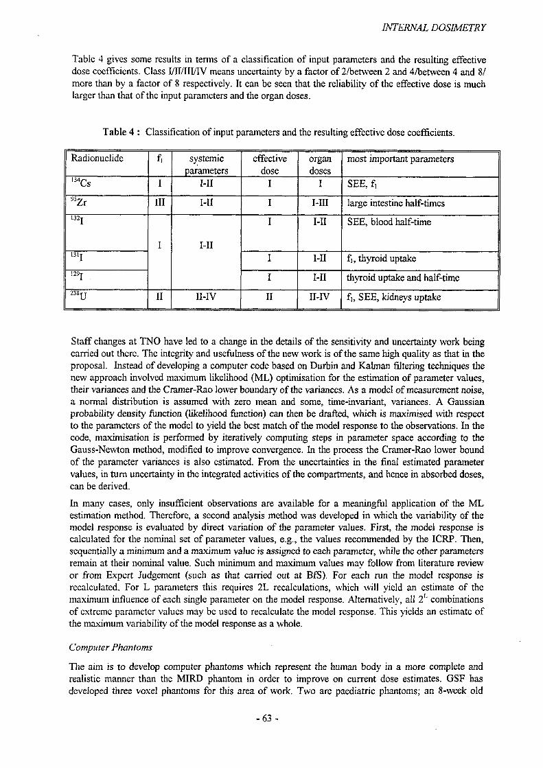
INTERNAL DOSIMETRY
Table 4 gives some results in terms of a classification of input parameters and the resulting effective dose coefficients. Class I/II/III/IV means uncertainty by a factor of 2/between 2 and 4/between 4 and 8/ more than by a factor of 8 respectively. It can be seen that the reliability of the effective dose is much larger than that of the input parameters and the organ doses.
Table 4 : Classification of input parameters and the resulting effective dose coefficients.
Radionuclide f. systemicparameters
effectivedose
organdoses
most important parameters
'*Cs I I-II I I SEE, f.
95Zr III I-II I I-III large intestine half-times132 j
I I-II
I I-II SEE, blood half-time
131! I I-II fi, thyroid uptake
J29I I I-II thyroid uptake and half-time
II II-IV II II-IV fi, SEE, kidneys uptake
Staff changes at TNO have led to a change in the details of the sensitivity and uncertainty work being carried out there. The integrity and usefulness of the new work is of the same high quality as that in the proposal. Instead of developing a computer code based on Durbin and Kalman filtering techniques the new approach involved maximum likelihood (ML) optimisation for the estimation of parameter values, their variances and the Cramer-Rao lower boundary of the variances. As a model of measurement noise, a normal distribution is assumed with zero mean and some, time-invariant, variances. A Gaussian probability density function (likelihood function) can then be drafted, which is maximised with respect to the parameters of the model to yield the best match of the model response to the observations. In the code, maximisation is performed by iteratively computing steps in parameter space according to the Gauss-Newton method, modified to improve convergence. In the process the Cramer-Rao lower bound of the parameter variances is also estimated. From the uncertainties in the final estimated parameter values, in turn uncertainty in the integrated activities of the compartments, and hence in absorbed doses, can be derived.
In many cases, only insufficient observations are available for a meaningful application of the ML estimation method. Therefore, a second analysis method was developed in which the variability of the model response is evaluated by direct variation of the parameter values. First, the model response is calculated for the nominal set of parameter values, e.g., the values recommended by the ICRP. Then, sequentially a minimum and a maximum value is assigned to each parameter, while the other parameters remain at their nominal value. Such minimum and maximum values may follow from literature review or from Expert Judgement (such as that carried out at BfS). For each run the model response is recalculated. For L parameters this requires 2L recalculations, which will yield an estimate of the maximum influence of each single parameter on the model response. Alternatively, all 2L combinations of extreme parameter values may be used to recalculate the model response. This yields an estimate of the maximum variability of the model response as a whole.
Computer Phantoms
The aim is to develop computer phantoms which represent the human body in a more complete and realistic manner than the MIRD phantom in order to improve on current dose estimates. GSF has developed three voxel phantoms for this area of work. Two are paediatric phantoms; an 8-week old
-63 -

INTERNAL DOSIMETRY
baby ("BABY") and a 7-year old child ("CHILD"). The third is an adult male ("Golem"). A further model of an adult male available from Yale University, New Haven ("Voxelman") was used for comparisons.
The segmentation of the adult phantom Golem from whole body CT data of a man is now complete. In all, 190 separate organs and tissues are defined, including all the critical organs defined by ICRP 60. The phantom allows differentiation between solid bone, red and yellow bone marrow, skin, soft tissue, muscle tissue, adipose tissue and lung tissue. The distribution of the red bone marrow in the various bones was determined by linear interpolation for each individual volume element and resulted in excellent agreement with data from ICRP Reference Man. One of the aims of this project was to model the respiratory tract regions defined in the new ICRP 66 model and use them as source and as target regions for the Monte Carlo simulations to estimate the SAFs for photons. The relevant tissues in the nasal and oral passages as well as in the trachea and main bronchi were identified and are represented by a single pixel row surrounding the respective cavities. This work is considered to be an important improvement on the traditional MIRD phantom, where surrogate organs had to be used instead.
Radiation transport in the phantoms was simulated using a Monte Carlo code following individual monoenergetic photons (in the energy range 0.02-4.00 MeV) originating various source organs. For each of the four voxel phantoms mentioned above, SAFs were obtained for more than 25 source organs and over 100 target organs.
-64-
INTERNAL DOSIMETRY
3. Summary of main achievements
The overall objective of this project is to improve estimates of dose for intakes of radionuclides by adults and children. The work programme includes the development and improvement of biokinetic and dosimetric models. Experimental studies and modelling work are performed. A major goal is the development of a new dosimetric model for the gastrointestinal tract (WP1), models for the systemic behaviour of radionuclides (WP2/Activity 1) and models for the developing embryo and fetus (WP2/Activity 2). The workplan also addresses the local delivery of dose to target cells (WP1, WP3). The results of these activities serve as input for the calculation of dose coefficients, the development of computer codes and quality assurance of the new dosimetric models (WP4).
WP1 (Biokinetics of ingested radionuclides and dosimetry of the gastrointestinal tract)
Development of a new dosimetric model for the alimentary tractWithin the ICRP Task Group on a Dosimetric Model for the Human Alimentary Tract (HAT), the model development has been started and is progressing well. The general problem is the need of a realistic model taking into account all relevant physiological processes. However, the search for physiological realism needs to be balanced against the need to avoid unnecessarily complicated computations. The current philosophy is to have a simple generic model including the most important pathways with some additional pathways considering longer retention in the oral cavity including teeth and oral mucosa and in the oesophagus, and special absorption and secretion processes. These additional pathways will only be used in cases when sufficient data are available.
A computer speciation model for the human gastrointestinal tractIn accordance with the project milestones, the chemical environment in the human gastrointestinal tract of an average healthy adult has been modelled using the JESS 53 (Joint Expert Speciation System 53) chemical speciation computer program. Four models have been created for the chemical environments of the compartments: saliva, gastric juice, bile and pancreatic fluid. A special database has also been created, containing critically assessed formation constants for lanthanide and actinide elements with the biological ligands found in these bio-fluids. The modelling is now being further developed to consider the effect of the presence of a solid phase that may form in saliva, gastric juice, bile and pancreatic fluid systems and also the enamel of the teeth in the saliva model and for insoluble food residues in the compartments. The simulations should enable a clear assessment of the validity of using lanthanides as surrogates for actinides in studies of gastrointestinal absorption, as well as providing a picture of the chemical behaviour of these elements in the various compartments of the upper gastrointestinal tract.
Gastrointestinal uptake studiesExperimental studies for important radionuclides are conducted to meet the need for new and reliable uptake data: The study of the intestinal absorption and long-term urinary excretion of 244Pu has continued with the intravenous administration of 244Pu in citrate solution to healthy volunteers. A total of five subjects have now been given 244Pu, first by ingestion, followed after a suitable period by intravenous injection. The fraction of ingested 244Pu reaching blood (f) was in the range of 10"4 - 10"3. Urinary excretion after intravenous injection has been shown to conform to the predictions of the ICRP Publication 67 model. Measurements of the absorption of 210Po and 228Th in rats have been made because of the possible importance of environmental and occupational exposures to naturally-occurring alpha-emitting isotopes. The f-values obtained are significantly higher than previously reported values. Comparisons of the tissue distribution of 210Po after oral or systemic administration in rats and guinea pigs showed some significant differences. The absorption of 228Th and 238Pu in neonatal rats is by orders of magnitude higher as compared to adult animals. Stable isotopes are used as tracers in human studies where the application of radiotracers is not possible due to ethical reasons. Gastrointestinal absorption studies with stable isotopes of Te, Ru and Zr have started. Before the administration to humans, animal experiments were conducted in rabbits to develop and validate the technique. The fi -values obtained are in broad agreement with literature, results in humans, however, for whom there exist virtually no
-65 -

INTERNAL DOSIMETRY
experimental data, may be quite different. The start of the volunteer studies was delayed by the need to identify the maximum applicable amounts of tracers in human investigations, hi order to study the uptake of ingested radionuclides not only from aqueous solutions but also from internally contaminated foodstuffs, the generation of plant material, intrinsically labelled with stable isotopes of Te, Zr, Ru and lanthanides was investigated. Because of its easy cultivation, cress was used as a rapidly available foodstuff. A more common foodstuff which represents a natural source for trace metal supply to humans are legumes. In a series of experiments, optimum conditions for growing of the plants and addition of tracer material were evaluated. The materials are intended to be supplied to all experimental groups in the project for their investigations.
WP2 (Biokinetics and dosimetric models for systemic radionuclides)
Models for adults and children
New draft biokinetic models for adult workers have been prepared for the elements C, Co, I, Cs, Sr, Zr, Nb, Hf, Ce Th, U, Pu, and Am and submitted to the Internal Dosimetry Task Group [INDOS] of ICRP with generally favourable comment. The revised adult models for the four actinide elements, Th, U, Pu and Am are essentially identical with the age-dependent models suggested in ICRP Publications 67 and 69 (1993,1995) and utilize the ICRP generic model for actinides. However, reported differences in the retention of Pu and also Th between men and women necessitates a further review of the literature to seek for other evidence of gender-specific differences in actinide biokinetics in humans. It remains to be determined whether it will be necessary or desirable to provide separate biokinetic models for actinides in men and women, but the evidence so far suggests that this is likely to be more important for retrospective dosimetry, than for general radiation protection. An earlier draft generic model for the lanthanide, or rare earth elements, has been up-dated to include the latest available data, especially human data on the absorption of lanthanides from the gastro-intestinal tract. The model is similar in structure to that of the generic actinide model proposed in ICRP Publication 67 (1993). A revised and gender-specific biokinetic model for Cs in adult workers is suggested, based on the latest available information on differences in Cs retention in males and females. Revised biokinetic models for C, and for I have been completed. Work has continued on the formulation of a generic model for the chemically elements Al, Ga and In, draft models for each element have been prepared, but these still need some further work to include data from some clinical studies with 67Ga and 1HIn radiopharmaceuticals. Gallium-67 is also an Auger electron-emitter and the review of the biokinetics of this radionuclide has been extended to include all available information on its distribution and kinetics in sub-cellular structures since this is also of relevance to Work Package 3.
Experimental - Stable isotope studiesA study was carried out in 62 healthy volunteers on the excretion pattern of ruthenium. The results are in broad agreement with the biokinetic data of current ICRP recommendations. A more detailed analysis of the data, however, indicate potential gender and age specific behaviour of urinary ruthenium excretion. These data support the necessity to consider gender specific biokinetic models for a number of elements. Measurements of the day-to-day variation of urinary ruthenium concentration and excretion showed a pronounced variability of values up to a factor of five in all subjects investigated. It is not clear whether these fluctuations are due to changes in daily intake of ruthenium by food or to alterations of systemic behaviour. Excretion patterns of calcium and strontium have been evaluated in neonates. The pattern of excretion is rapid such that urine collection for the first 24 hours post-administration will collect the majority of the Ca and Sr excreted with peak clearance approximately 6 hours postadministration. The results indicate that Sr excretion in each neonate is consistent and dependent on dose administered. However, excretion of the Ca stable isotope is dependent on the total mass of Ca excreted by each neonate, under homoeostatic control.
Models for th e embryo /fetusReview of data on the transfer of radionuclides to the embryo and fetus has progressed as planned. During the period, work was undertaken on draft reviews for the following elements: H, Fe, Co, Zn,
-66-

INTERNAL DOSIMETRY
Se, Sr, Ru, Ba, Cs, Ce, Pb, Po, Ra, U, Pu and Am. For the alkaline earth elements, Sr, Ba and Ra, a biokinetic model has been developed which relates transfer to the fetus to the requirement of the fetus for Ca and the rate of accretion of Ca by the fetal skeleton. This model and information reviewed for other elements will be used in the work of the ICRP Internal Dosimetry Task Group which, together with the Task Group on Dose Calculations, is preparing a report on doses to the embryo and fetus.
Experimental studies
Human fetal bone samples from individual cases between 17 and 41 weeks of gestation were analysed for plutonium. Results obtained by mass spectrometry were significant for the samples from the 36 - 41 week but not for the 17 - 32 week samples. Five sets of fetus and placenta from second trimester terminations (13 - 19 weeks) were also analysed. A single significant measurement was recorded for a placenta (16 weeks). Studies of the transfer of nuclides through perfused human placenta have been initiated. The apparatus has been constructed and tested. After some initial problems, mainly in meeting requirements for work with human tissues, studies are now progressing well. The viability of the system, involving perfusion of fetal and maternal sides of an isolated cotyledon of the placenta, has been established using "mTc. Results show that the ionic form transfers rapidly across the placenta while protein bound "mTc-albumin does not transfer. Comparisons of the transfer of Ca and Sr are in progress. Because of difficulties over safety issues, alpha emitters will not be used in this system. The possibility of using 241Pu (beta emitter) and comparing transfer with that of lanthanide analogues of the trivalent actinides is under consideration.
Previous studies of the transfer of the actinides, Pu and Am, to the fetus in rats and guinea pigs have shown considerable differences in levels of transfer and distribution between fetal tissues, placenta and fetal membranes with generally greater values for Pu. This work has been extended to compare the behaviour of Np and Th to previous results for Pu and Am, measuring transfer in the rat at the time of yolk sac and in both the rat and guinea pig in late gestation. Results for retention of Np in the placenta and yolk sac are intermediate between those for Pu and Am. Transfer to the fetus was similar to that of Pu in the rat, but with a lower value similar to that of Am in the guinea pig. Results for transfer to the fetus for each nuclide in each rodent species were lower than corresponding values obtained previously for transfer to the primate fetus in late gestation. Initial delays were experienced in commencement of the equivalent primate studies due to the degree of control exerted over such studies by the member state. Four radionuclides, B7Np, 239Pu, 241 Am and 210Po, and three stages of gestation (early, mid and late) have been defined. The first set of data from baboons has demonstrated that the fetakmatemal whole body concentrations are lower than observed for other species, particularly for 239Pu. Further data from the macaca macaca will allow validation of these results.
Fetal dosimetryA generic model for calculation of doses to embryo and fetus after activity intake by the mother has been developed. Doses to organs and tissues of the fetus (those with a specified tissue weighting factor and additionally the dose to the brain during the period from week 8 to 15 of pregnancy) are calculated assuming a constant activity concentration ratio fetus/mother and placenta/mother and an activity distribution within the fetus similar to that in the 3-month-old infant. The parameters needed by the model have been determined for the 31 elements which will be contained in the planned ICRP Publication on doses to embryo and fetus. The generic model has been implemented (Working Package 4) within a computer code DOSAGE and a quality assurance programme validating results has commenced. Initial reviews have shown several problems especially in the interpretation of dosimetric models. However, these are under further review with solutions planned by the next meetings of the ICRP Task Groups on Internal Dosimetry and on Dose Calculations.
WP3 (Target cell dosimetry for short-range particles)
Radionuclide and dose distribution in bone samples
Detailed mapping of the distribution of alpha tracks in cortical and trabecular regions of femur sections of mouse bone is in progress to assess the distribution of 239Pu,241 Am and 233U, taking account of the
-67-

INTERNAL DOSIMETRY
distribution in bone mineral and marrow. The results are being used to estimate doses to cells on bone surfaces and at different depths into the marrow cavities. The three nuclides differ in their patterns of initial deposition in bone and subsequent movement within bone. Preliminary estimates of dose distribution have been made, considering the region adjacent to endosteal bone surfaces (10pm layer) thought to contain target cells for osteosarcoma and different regions of the red bone marrow containing the leukaemogenic target cells. The results indicate that for administered activities giving equal average bone doses of the three nuclides, doses to the designated target regions were in each case in the order: 239Pu > 241 Am > "33U. The same relationship was seen in the ability of the nuclides to induce osteosarcoma in the life-span study undertaken previously at NRPB. However, 239Pu and 241 Am appear to be equally effective in causing acute myeloid leukaemia. This apparent discrepancy will be addressed by analysis of the spatial and temporal distribution of dose in marrow.
Intracellular distribution of actinides in liver
Experiments have been performed with 239Np and 237Np in rats, and validated with primates. For 239Pu and 241 Am, experiments were conducted on primates only. Investigations of the sub-cellular distribution patterns of 239Np and 237Np in rat liver have shown that the distribution of neptunium is time and mass dependent. For 237Np, it was shown, both in rats and baboons, that the radionuclide binds to two proteins soon after exposure with molecular weights of 450 and 200 kDa, respectively. The former was identified as ferritin, but the latter remains unidentified. At later times (Id to 40 d), Np was found to be bound mainly by ferritin and by high molecular weight compounds. The biochemical behavior of americium, plutonium and neptunium found is not radically different from what was known previously about the bio-inorganic chemistry of these actinides. Nevertheless, it emphasizes the particular behavior of neptunium when compared with americium and plutonium. It can not be said for the moment if this behavior is due to the relatively lower specific activity of ^Np, and hence higher mass. Nevertheless, this point has to be taken into consideration for the calculation of the doses resulting from the incorporation of the three radionuclides.
Biological effect of alpha particles on pulmonary cells
The biological effects of a-particles on pulmonary cells in culture was studied because of their interest in potential health effects induced after inhalation of a-emitting radionuclides. An epithelial cell line (RTiv3, rat tracheal epithelial cells) was used to determine the influence of the cell morphology on the survival fraction, either as adherent cells or as sedimented cells. The results of these studies suggest that the adherent RTiv3 cells are 1.6 times more a-radiosensitive than sedimented ones. Studies are in progress to estimate the repartition of the deposited energy in the different cell compartments (whole cell and nucleus) as a function of size and shape of the irradiated cells. Another investigation evaluated possible differences in radiosensitivity for alpha irradiations, between epithelial cells of the trachea for two strains of rats. The mean lethal doses for the two rat strains differed nearly by a factor of two.
Radiation effects of Auger emitters
Cell survival, micronucleus formation and the induction of apoptosis after 65Zn uptake was investigated in human amniotic cells (AFFL) and in human squameous cell carcinoma cells (SCL-II). The results on cell survival show the intense cytotoxic capacity of 65Zn when compared to non-treated controls. The data can be fitted best with an exponential function, in contrast to the cell survival after external radiation which follows the classical cell survival curve (shoulder region at low doses, almost exponential in the higher dose region) of medium radiation resistant cell-lines. A strong induction of apoptotic cells was found even at low activities. The micronucleus frequency is also increased even at lowest activities and shows a very steep increase with increasing number of decays per volume. Additionally, much more cells with multiple micronuclei can be detected as when compared to external radiation.
- 68 -

INTERNAL DOSIMETRY
Autoradiography of actinidesThe development of microlocalisation methods for radionuclides in tissues was concluded with the achievement of an autoradiography techniques based on a thin scintillator associated to a micro-channel electron amplifier tube and a charged coupled device (CCD) as pulse height and position sensitive detector (aSTIC : a-Self-Triggered-Intensified-CCD). The method was originally developed for p markers imaging in tissue samples. Its adaptation for a particle detection has been completed.Within the scope of the present Work Package, it was of particular interest to compare in a systematic way the present aSTIC with the methods used by the other laboratories. This intercomparison was done with bone sections from previous mice intoxication experiments with 239Pu,241 Am and 233U.
Set-up and testing of SIMS equipment
The workplan for the reporting period includes a pilot study using secondary ion mass spectrometry (SIMS) for the microlocalisation of long-lived radionuclides and stable elements, and therefore a possible way of element localisation and "target" characterisation. A spatial resolution < 1 pm, a multicellular field of view; and the analysis of elements at trace levels are additional features of this technique. The respective equipment has now been transferred to IPSN and was aligned and modernised by the manufacturer (CAMECA, Courbevoie, France). First test experiments could be performed in the CAMECA facility. These experiments were focused on the analytical performance of SIMS and the measurement procedures (reproducibility, MDA, linearity, stability, internal tracer). In a further step, the imaging part was updated and now the partner laboratories will set up the pilot experiments for 1998.
WP4 (Numerical implications of models)
PC data base of dose coefficientsThe traditional compendia of dose coefficients published by the EU, ICRP and national bodies are inevitably limited in the number of results they provide by simple considerations of space. A need was therefore identified for a PC database of dose coefficients which would give many more results than could be included in a compendium. The NRPB RAPID database was adapted to produce a PC-based database. This greatly increases the accessibility and usefulness of the database. Dose coefficients are given for 35 tissues as well as the effective dose, at 10 times after intake. Around 800 radionuclides are addressed for intake by ingestion and by inhalation of 10 aerosol sizes (AMAD) for both workers and members of the public. The dose coefficients have been calculated and are at present undergoing essential Quality Assurance. Results will be consistent with those already published in the EU Euratom Directive, the IAEA Basic Safety Standards, and latest ICRP Publications. A substantial on-line help facility is provided which comprises the whole of the text from ICRP Publications 68 and 72 with many hypertext links to glossary items and tables. A summary of all the biokinetic models used in the dose calculations is also given. Since much of the work for the help file text and the dose calculation methods was developed by ICRP Task Groups, ICRP has decided to market the package as an ICRP CD-ROM; this will increase the acceptability of the product around the world.
Bioassay models
In the Euratom Directive the EU has published a comprehensive set of dose coefficients for workers based on the latest ICRP biokinetic models. Therefore, there is a need to provide a companion publication of bioassay quantities based on these latest models which will enable health physicists to use a consistent set of models in dose assessments. New modules have been added to the NRPB PLEIADES and BfS DOSAGE codes which enable the calculation of daily urinary or faecal excretion as well as retention in regions such as lung, skeleton, thyroid and whole-body. Calculations can be performed for both acute and chronic intakes. The document has been adopted by ICRP and is expected to be available soon.
-69-

INTERNAL DOSIMETRY
Dose coefficients for the embryo/fetus
Two approaches were adopted for assessing dose to the embryo/fetus: full element-specific models where sufficient data exist; and a generic model in other cases. In the first category, a model for calcium has been developed using data on the total calcium content of the fetus provided by co-workers in Work Package 2. Work is under way to develop models for other alkaline earth elements (strontium, barium and radium) based on the work for calcium. In the second category, a method has now been agreed upon and implemented in the NRPB and BfS computer codes. The procedure is similar to that already implemented at NRPB and BfS for other cases of age-dependent dosimetry. Draft results have been produced for selected radionuclides.
Sensitivity and uncertainty analyses
The study of uncertainties in internal dose assessment is being given increasing importance by international bodies. Work in this project is being tackled in a number of ways at different laboratories. At NRPB, initial work on the investigation of the sensitivity of model predictions, such as compartment contents, to changes in model parameters has been completed. This method has also been applied to the proposed new model for lanthanides being developed under Work Package 2. It is hoped that this will aid other researchers by identifying the critical model parameters which can then be given particular attention during model development. This new method has been applied to the ICRP Human Respiratory Tract Model. A method of parameter uncertainty analysis based on Monte Carlo methods with Latin Hypercube sampling has been established. Uncertainties for adult ingestion dose coefficients for a number of important nuclides have been assessed as part of an NRPB report on uncertainties in doses to critical groups. An NRPB Memorandum on this work has been prepared and will be published soon. The uncertainty methods developed under this contract are also being used in another EU Contract which considers the uncertainty in predictions of the accident consequence code COSYMA based on Expert Judgement. It is hoped that it will also be possible to publish uncertainties in committed effective doses based on this work. At BfS, an Expert Judgement was used as a basis for probabilistic calculations of dose coefficients. Biokinetic and dosimetric input parameters for calculations of ingestion dose coefficients for a number of radionuclides have been assessed. Probabilistic calculations are performed by Colenco Power Consulting AG, Baden, Switzerland, using the BfS DOSAGE code together with additional probabilistic codes. The results of these probabilistic calculations give the probability distribution of the effective dose coefficient and the most sensitive parameter, giving an indication for further need of research. The results will be used in a forthcoming ICRP Publication on the reliability of dose coefficients. A new approach towards sensitivity and uncertainty analysis has been adopted at TNG. This approach involves maximum likelihood (ML) optimisation for the estimation of parameter values, their variances and the Cramer-Rao lower boundary of the variances. In cases where only insufficient observations are available for a meaningful application of the ML estimation method, a second analysis method was developed in which the variability of the model response is evaluated by direct variation of the parameter values.
Computer Ph antoms
The aim is to develop computer phantoms which represent the human body in a more complete and realistic manner than the MIRD phantom in order to improve on current dose estimates. GSF has developed three voxel phantoms for this area of work. Two are paediatric phantoms; an 8-week old baby ("BABY") and a 7-year old child ("CHILD"). The third is an adult male ("Golem"). A further model of an adult male available from Yale University, New Haven ("Voxelman") was used for comparisons. One of the aims of this project was to model the respiratory tract regions defined in the new ICRP 66 model and use them as source and as target regions for the Monte Carlo simulations to estimate the SAFs for photons. The relevant tissues in the nasal and oral passages as well as in the trachea and main bronchi were identified and are represented by a single pixel row surrounding the respective cavities. This work is considered to be an important improvement on the traditional MIRD phantom, where surrogate organs had to be used instead. Radiation transport in the phantoms was simulated using a Monte Carlo code following individual monoenergetic photons (in the energy range
-70-

INTERNAL DOSIMETRY
0.02-4.00 MeV) originating from various source organs. For each of the four voxel phantoms mentioned above, SAFs were obtained for more than 25 source organs and over 100 target organs.
-71 -

INTERNAL DOSIMETRY
4. Research to be performed in the remainder of the project
The work programme of the project proceeded during the reporting period in general as planned and the expected progress has been made. Apart from minor changes in the internal time schedule, the expected goals have been achieved. Whereas some work is ahead of the schedule, there is some delay in other tasks. All goals, however, that serve as input to other tasks, have been reached. It can therefore be expected that the project as a whole will proceed as planned also in the remaining period.
For the sake of brevity, the objectives for the remainder of the project are given only as an outline of the main points with respect to the four Work Packages.
4.1 Remaining objectives for Work Package 1
• Continuation of data review for the model development
• Development of a new human alimentary tract model
• Continue rodent studies of intestinal retention / dosimetry
• Computer simulations and data analysis of Gd and Cm, further computer simulations of the speciation of Nd, Eu Gd, Am and Cm to include reactions involving solids
• Creation of a speciation model of blood plasma, and to carry out speciation studies with Nd, Eu, Gd, Am and Cm
• Validation of surrogate elements for actinide studies in humans - report and critical evaluation
• Complete optimization and validation of mass spectrometric and activation analytical methods for measuring isotope ratios of Zr and Gd
• Animal studies with Te, Zr, Ru and Gd
• Complete generation of foodstuffs, intrinsically labelled with Ru, Zr, Gd
• Volunteer studies with Te, Zr, Ru and lanthanides
• Apply for Ethical Committee approval for a volunteer study using 208Po and extension of the 244Pu study to measure absorption in the absence of food
4.2 Remaining objectives for Work Package 2
• Continue reviewing the recent literature for information, especially human data, on the biokinetics of inhaled and ingested radionuclides
» Final revision of the biokinetic models for C, Co, I, Cs, Sr, Zr, Nb, Hf, Ce Th, U, Pu, and Am
• Further revision of the generic model for lanthanides
• Formulation of the generic model for Al, Ga and In
• Formulation of revised models for as many of the remaining elements of radiological importance as time and resources permit
• Validation of surrogate elements for actinide studies in humans - report and critical evaluation
• Volunteer studies with lanthanides
• Report on human studies with Te, Zr, Ru, lanthanides
• Complete the baseline studies of Te, Zr and Gd in body fluids
• Compare the biokinetics of Te, Ru, Zr and Gd after oral and intravenous administration in humans
-72-
INTERNAL DOSIMETRY
• Completion of rodent studies with :30Th
• Continuation of placental perfusion studies
• Completion of model development
• Macaca studies at various stages of gestation to determine the placental transfer
• Co-operation in the development of special compartment models
• Continuation of quality assurance procedures in dose calculations for embryo and fetus
4.3 Remaining objectives for Work Package 3
• Continue mapping alpha track distribution for 239Pu, 241Am and 233U in bone, to determine dose distribution and to begin modelling the behaviour of the three radionuclides. Some limited analysis of baboon bone will be undertaken
• Undertake further in vitro irradiation studies using a homogeneous a-beam generated by an accelerator TANDEM 7MV with different parameters of i
• Continue studies on cyto- and genotoxicity of 65Zn in the high effect range and to study its intracellular distribution using confocal microscopy, autoradiography and SIMS.
• Complete results obtained with micro-autoradiography techniques and finalise the report of the comparative study. Improve and normalise the analytic calculation codes and prepare a comprehensive report including a review of dosimetry and microdosimetric methods used for alpha particles.
• Setting-up the experimental workplan for SIMS. This provisionally includes Zn localisation in cell nuclei (UROS), actinides in bone samples (NRPB) and in lung samples (CEA).
4.4 Remaining objectives for Work Package 4
• QA of dose coefficients from the generic calculation method for the embryo/ fetus
• Complete work on special fetal model for the alkaline earth elements
• Develop codes for dose calculations using the special embryo/fetal models at NRPB (this is already achieved at BfS)
• Uncertainty analyses for important radionuclides
• Expert Judgement for uncertainties on model parameters for biokinetic models for radioactive daughters
• Implement the new model for the human alimentary tract (from WP 1) and calculate dose coefficients
• Define tissues relevant for inhalation in the voxel phantoms BABY and CHILD
• Monte Carlo calculations to estimate photons specific absorbed fractions (SAFs) for these new source organs (these results will complement the already existing lists of SAFs for the paediatric phantoms)
• Perform comparisons between these new SAFs and the current values obtained by using surrogate organs
-73 -

INTERNAL DOSIMETRY
• Calculation of dose coefficients using the new SAFs and SEEs obtained for the voxel phantoms, and comparison with published values in the Euratom Directive
• Further testing and refinement of Maximum Likelihood (ML) codes prior to release for general applications
• Extension of ML codes to handle chronic intakes and daughter nuclides
• Application of sensitivity analysis codes to the new human alimentary tract (from WP 1) model.
-74-
INTERNAL DOSIMETRY
5. Publications
Amaral A.; Cossonnet C.; Escaig F.; Franck D.; Galle P.; Pihet P. Investigation of Secondary Ion Mass Spectrometry in radiation protection dosimetry, Proc. International Congress on Radiation Protection, IRPA 9, April 1996, Vienna, Austria, Vol. 2: 492-494, 1996.
Amaral A.; Galle P.; Cossonnet C.; Franck D.; Pihet P.; Carrier M.; Stephan 0. Perspective of uranium and plutonium analysis in urine sample by secondary ion mass spectrometry. J. Radioanalytical Chemistry, 1997 (in press).
Aubineau-Laniece I. Autoradiographic quantitative des radionucleides dans les tissus biologiques par analyse nucleaire haute resolution : application en radiotoxicologie et en dosimetric. Thesis. Universite Paris-Sud, Orsay, 16 December 1997.
Aubineau-Laniece I.; Castellan G.; Caswell R.S.; Guezinguar F.; Henge-Napoli M.H.; Li W.B.; Pihet P. Application of microdosimetric methods for the determination of energy deposition distributions by inhaled actinides. Proc. Workshop on Intakes of Radionuclides, Avignon, Sept. 1997. Radiat. Prot. Dosim. 1997 (in press).
Aubineau-Laniece I.; Charon Y.; Corner D.; Franck D.; Henge-Napoli M.H.; Laniece P.; Mastrippolito R.; Pinot L.; Valentin L. Self triggered intensified CCD for high resolution a autoradiography : a quantitative and indentificative approach. Proc. Conf. "Position sensitive detectors", Manchester, Sept. 1996. Nucl. Inst, and Methods, A392: 237-243, 1997.
Bailey, M R.; Birchall, A.; Marsh, J.W.; Phipps, A.W.; Sacoyannis, V. Application of the New ICRP Respiratory Tract Model to Gases and Vapours in ICRP Publication 68. Chilton, NRPB-M688, 1996.
Bailey, M R.; Harrison, ID.; Jones, K.A.; Marsh, J.W.; Prosser, S.L. Uncertainties in aspects of internal dosimetry relevant to accident consequence assessment codes. NRPB-M763; 1996.
Bailly-Despiney I.; Poncy J.L.; Jouanny F.; Lagroye I.; Lizon C.; Massiot P.; Lefoll L.; Fritsch P. Investigation on death processes in rat epithelial cells exposed to an irradiation in vitro., Radioprotection 32: 375-376, 1997.
Bassini, R.; Cantone, M.C.; de Bartolo, D.; Gambarini, G.; Giussani, A.; Malatesta, M. A computer controlled apparatus for cyclic instrumental CPAA. J. Radioanal. Nucl. Chem, 217: 283-287; 1997.
Cantone, M.C.; de Bartolo, D.; Giussani, A. Biokinetics of ingested radionuclides and assessment of internal dose. Physica Medica, XIII, 1997.
Cantone, M.C.; de Bartolo, D.; Giussani, A.; Garlaschelli, L.; Hansen, Ch.; Roth, P.; Werner, E. Proton activation analysis for studying zirconium biokinetics using stable isotopes: tests on animals. Proc. LXXXII Congr. Societa Italiana di Fisica, Verona, Italy, Sept. 1996; 112: 1996.
Cantone, M.C.; de Bartolo, D.; Giussani, A.; Garlaschelli, L.; Hansen, Ch.; Roth, P.; Werner, E. CPAA for studying biokinetics with stable isotopes: A preliminary investigation on zirconium metabolism on animals. J. Radioanal. Nucl. Chem, 217: 279-282; 1997.
Cantone, M.C.; de Bartolo, D.; Giussani, A.; Hansen, Ch.; Roth, P.; Schramel, P.: Wendler, I.; Werner, E.; Nusslin, E.F. Stable tracers for tracer kinetic investigations of molybdenum: intrinsic and extrinsic tagging. In: Trace Elements in Man and Animals. Edited by P.W.F. Fischer et al, NRC Research Press, Ottawa, Canada, 267-269, 1997.
-75 -

INTERNAL DOSIMETRY
Cantone, M.C.; de Bartolo, D.; Giussani, A.; Ottolenghi, A.; Pirola, L.; Hansen, Ch.; Roth, P.; Werner, E. A methodology for biokinetic studies using stable isotopes: results of repeated molybdenum investigations on a healthy volunteer. Appl. Radiat. Isot. 48: 333-338; 1997.
Cantone, M.C.; de Bartolo, D ; Giussani, A.; Roth, P.; Werner, E. The stable isotope approach to elemental biokinetics. Radiat. Phvs. Chem. 1998 (in press).
Cartlidge, A.L.; Lord, B.I.; Ellender, M.; Haines, J.W.; Harrison, J.D. Microdosimetry to leukaemogenic target cells for bone-incorporated alpha-emitting radionuclides. Proc. Workshop on Intakes of Radionuclides, Avignon, Sept. 1997. Radiat. Prot. Dosim. 1997 (in press).
de Bartolo, D.; Cantone, M.C.; Giussani, A. Use of charged particle activation analysis as analytical technique in the biological field. Appl. Radiat. Isot. 1997 (in press).
Ellender, M.; Harrison, J.D.; Pottinger, H.E.; Thomas, J.E. Osteosarcoma induction in mice by the alpha-emitting nuclides, plutonium-239, americium-241 and uranium-233. Proc. 9th Int. Congress of IRPA, April 1996. Vienna, Austria, Vol.4: 67-69, 1996.
Fell, T.P.; Harrison, J.D. and Leggett, R.W. A model for the transfer of alkaline earth elements to the embryo and fetus. Proc. Workshop on Intakes of Radionuclides, Avignon, Sept. 1997. Radiat. Prot. Dosim. 1997 (in press).
Gilbi, D.; Gribi, P.; NoBke, D. Probabilistic calculations of dose coefficients. Radiat. Prot. Dosim. 1998, (in press).
Giussani, A.; Cantone, M.C.; de Bartolo, D.; Roth, P.; Werner, E.; Nusslin, F. Ermittlung der intemen Dosis nach Ingestion von radioaktivem Molybdan. Proc. Medizinische Physik 96, Graz, Austria, Sept. 1996, 373-374; 1996.
Giussani, A.; Cantone, M.C.; de Bartolo, D.; Werner, E.; Roth, P. Internal dosimetry following incorporation of molybdenum radionuclides and criteria for intervention based on a new biokinetic model. Proc. Int. Conf. Radiation Dosimetry and Safety. Taipei, Taiwan, ROC, March 1997, 48; 1997.
Giussani, A.; Roth, P.; Werner, E.; Greim, H.; Cantone, M.C.; de Bartolo, D. A biokinetic model for molybdenum radionuclides: new experimental results. Proc. Workshop on Intakes of Radionuclides, Avignon, Sept. 1997. Radiat. Prot. Dosim. 1997 (in press).
Harrison, J.D. Dosimetry and effects of parental, fetal and neonatal exposure to incorporated radionuclides and external irradiation. In: European Advances in Radiological Protection, CEC-NRPB Agreement of Association, Project Coordinator Reports for 1993-1995. EUR 16951, pp.32-45. NRPB, Chilton, 1996.
Harrison, J.D.; Haines, J.W. Lead-210 and polonium-210 in the body. Radiol. Prot. Bull. 178: 25-27; 1996.
Harrison, J.D.; Khursheed, A.; Phipps, A.W.; Goosens, L.; Kraan, B.; Harper, F. Uncertainties in biokinetic parameters and dose coefficients determined by Expert Judgement. Proc. Workshop on Intakes of Radionuclides, Avignon, Sept. 1997. Radiat. Prot. Dosim. 1997 (in press).
Harrison, J.D.; Slather, J.W. The assessment of doses and effects from intakes of radioactive particles.J. Anat. 189: 521-530; 1996.
Khursheed, A.; Fell T.P. A Sensitivity Analysis for the biokinetic model for plutonium. Radiat. Prot. Dosim. 1997 (in press).
-76-

INTERNAL DOSIMETRY
Khursheed, A.; Fell, T.P.; Kendall, G.M.: Phipps, A.W. Simplified Organ Retention Functions for Physiologically based Recycling Biokinetic Models. Health Phys. 70: 656-664, 1996.
Kriehuber R.; Simko M. Late effects in irradiated mammalian cells. Proc. 1st Russian Congress on Pathophysiology, Nov. 1996, Moscow, Russia, 241, 1996.
Kugel C.; Bailly-Despinev I.; Lagroye I.; Poncy J-L. Radiosensibilite comparative des cellules epitheliales de la trachee de rat apres irradiation alpha in vitro., Journal de Chimie Physique, 1997 (in press).
Laniece I.; Charon Y.; Laniece P.; Mastrippolito R.; Menard L.; Pinot L.; Leroy N.; Pages N.; Valentin L. A self-triggered intensified CCD pulse height analyser for discriminative a with high imaging resolution. Nucl. Inst, and Methods. 1997 (in press).
Nofike, D.; Rtihm, W.; Karcher, K. Individual dose assessment of workers. Radiat. Prot. Dosim. 1998, (in press).
Paquet F.; Ramounet B.; Metivier H.; Taylor D M. The bioinorganic chemistry of Np, Pu and Am in mammalian liver. J. Alloys and Compounds, 1997 (in press).
Paquet F.; Ramounet B.; Metivier H.; Taylor D M. The chemical form of neptunium in the rat liver cells. IRPA9, International Congress on Radiation Protection, April 1996, Vienna, Austria, Vol. 2: 454- 456, 1996.
Paquet, B.; Ramounet, H.; Metivier, H.; Taylor, D.M. The Effect of the Mass and Initial Chemical Form of Neptunium on its Molecular Associations in Blood and Liver. Radiation Research, 146: 306- 312, 1996.
Paquet, F.; Poncy, J.-L.; Ham, G.; Prosser, S.L.; Harrison, J.D. Transfer of Po, Np, Pu and Am to the primate fetus. Proc. Workshop on Intakes of Radionuclides, Avignon, Sept. 1997. Radiat. Prot. Dosim. 1997 (in press).
Fellow, P.G.; Stradling, G.N.; Hodgson, A.; Fell, T.P.; Phipps, A.W.; Ellender, M. Biokinetics of Uranium Tributyl Phosphate in the Rat: Implications for Occupational Exposure. Chilton, NRPB- M682, 1996.
Petoussi-HenB, N.; Zankl, M.; Henrichs, K. Tomographic anthropomorphic models. Part III: Specific absorbed fractions of energy for a baby and a child from internal photon source. GSF-Bericht 7/97. GSF - Forschungszentrum fur Umwelt und Gesundheit, Neuherberg, Germany, 1997.
Petoussi-HenB, N.; Zankl, M.; Voxel anthropomorphic models as a tool for internal dosimetry. Proc. Workshop on Intakes of Radionuclides, Avignon, Sept. 1997. Radiat. Prot. Dosim. 1997 (in press).
Phipps, A.W.; Fell, T.P.; Silk, T.J. Model developments and revised doses in macro-dosimetry. 6th International Conference on Radiopharmaceutical Dosimetry. Gatlinburg, TN, USA. 1996 (in press).
Phipps, A.W.; Jarvis, N.S.; Silk, T.J.; Birchall, A. Time-dependent functions to represent the bioassay quantities given in ICRP Publication 78. NRPB M-824, 1998 (in press).
Phipps, A.W.; Silk, T.J.; Fell, T.P. The impact of recent ICRP recommendations on the dose coefficients, ALIs and monitoring programmes for thorium. Proc. Workshop on Intakes of Radionuclides, Avignon, Sept. 1997. Radiat. Prot. Dosim, 1997 (in press).
Roth, P.; Hansen, Ch.; Werner, E. Calcium Kinetic Investigations and Their Clinical Use. Appl. Radiat. Isot. 1997 (in press).
-77-

INTERNAL DOSIMETRY
Roth. P.; Werner, E.; Wendler. I.; Schramel. P. Application of ICP-MS for the assesment of thorium excretion in urine. Appl. Radiat. Isotopes 47: 1055-1056, 1996.
Roth, P.; Werner, E.; Wendler. I.; Schramel, P. Variation of natural 23*Th excretion in non-exposed persons. J. Radioanal. Nucl. Chem. 1997 (in press).
Roth, P.; Giussani, A.; Werner, E. Kinetics of gastro-intestinal absorption. Proc. Workshop on Intakes of Radionuclides, Avignon, Sept. 1997. Radiat. Prot. Dosim. 1997 (in press).
Schramel, P.; Wendler, I.; Roth, P.; Werner, E. A method for the determination of thorium and uranium in urine samples by ICP-MS. Mikrochim Acta 126: 263-266, 1997.
Silk, T.J.; Fell, T.P.; Phipps, A.W. (with the ICRP Task Group on Dose Calculations) The ICRP CD- ROM of dose coefficients. Proc. Workshop on Intakes of Radionuclides, Avignon, Sept. 1997. Radiat. Prot. Dosim. 1997 (in press).
Simko, M.; NoBke, D. Basis for a revision of the gastrointestinal tract model. Radiat. Prot. Dosim. 63: 29-36; 1996.
Stather, J.W. The work of ICRP in developing dose coefficients for the embryo and fetus following intakes of radionuclides by the mother. Proc. Workshop on Intakes of Radionuclides, Avignon, Sept. 1997. Radiat. Prot. Dosim. 1997 (in press).
Stradling, G.N.; Fellow, P.G.; Hodgson, A.; Fell, T.P.; Phipps, A.W.; Pearce, M.; Ranee, E.; Ellender, M.; Taskaeva, M.; Penev, I.; Guentchev, T. Dose Coefficient and Assessment of Intake of a Radionuclide Bearing Dust formed at the Kozloduy Nuclear Power Plant. Chilton, NRPB-M679, 1996.
Taylor, D. M. The inorganic biochemistry of actinides in blood. J. Alloys and Compounds, 1998 (in press).
Taylor, D. M., Biokinetic models for internal dosimetry. Radiation Protection (China) 17, 9-12, 1997 {in Chinese).
Taylor, D.M. Gender-specific differences in the biokinetics of plutonium and other actinides. Proc. Workshop on Intakes of Radionuclides, Avignon, Sept. 1997. Radiat. Prot. Dosim. 1997 (in press).
Taylor, D.M; Leggett R.W. A generic biokinetic model for the lanthanide elements. Proc. Workshop on Intakes of Radionuclides, Avignon, Sept. 1997. Radiat. Prot. Dosim. 1997 (in press).
Taylor, D.M.; Taylor, S.K. Environmental uranium and human health. Rev. Environ. Health, 12: 147- 157, 1997.
Webb, L. M.; Taylor, D.M.; Williams, D.R. Computer modelling of the chemical speciation of lanthanide and actinide elements in the human gastrointestinal tract - Mouth and stomach. Radiat. Prot. Dosim. 1998 (in press).
Webb, L.M.;Taylor, D M.; Williams, D.R. Computer modelling of the chemical speciation of lanthanide and actinide elements in the human gastrointestinal tract. J. Alloys and Compounds 1998 (in press).
Werner, E.; Hansen, Ch.; Roth, P. Studies on Electrolyte and Trace Metal Metabolism Applying Stable Isotopes as Tracers. Appl. Radiat. Isot. (in press).
-78 -
INTERNAL DOSIMETRY
Werner, E.; Roth, P.; Hansen, Ch.; Cantone, M.C.; de Bartolo, D.; Giussani, A.; Nusslin, F. Bestimmung der intestinalen Absorption von Molybdaen beim Menschen mittels der stabilen Isotope 95Mo und 96Mo. Isot. Environ. Health, 32: 253-254; 1996.
Werner, E.; Roth, P.; Wendler, I.; Schramel, P.; Hellmann, H.; Kratzel, U. Feasibility of ICP-MS for the assessment of uranium excretion in urine. J. Radioanal. Nucl. Chem. 1997 (in press).
Werner, E.; Roth, P.; Wendler, I.; Schramel, P. Assessment of thorium excretion in urine by means of ICP-MS. Proc. International Congress on Radiation Protection, IRPA 9, April 1996, Vienna, Austria, Vol. 2: 489-491, 1996.
-79-

INTERNAL DOSIMETRY
6. Executive summary
Objectives
Estimates of risk from exposure to radionuclides in the workplace or following routine or accidental releases to the environment are dependent on reliable estimates of tissue doses. The overall objective of this project is to improve estimates of dose for intakes of radionuclides by adults and children. The work includes the development of biokinetic and dosimetric models, including models of the gastrointestinal tract, models for the systemic behaviour of radionuclides and models for the developing embryo and fetus, and will address the local delivery of dose to target cells. The work to be carried out within this project is structured into four Work Packages: Workpackage 1 concentrates on ingested radionuclides, considering doses to the GI tract and radionuclide absorption. A major objective is the development of a new dosimetric model of the GI tract. Studies in Workpackage 2 seek to improve and extend biokinetic and dosimetric models for systemic radionuclides. Existing models for adults and children will be extended to other elements and new models will be developed for the embryo and fetus. In both cases, the work includes experimental studies as well as model development. The objective of Workpackage 3 is to improve assessment of localised distribution of dose within tissues at the cellular level for specific examples of Auger and alpha emitting isotopes, in relation to observed effects. The work will include experimental studies of dose/ effect and development of localisation methods. Workpackage 4 concerns the development of computer codes for the new dosimetric models, quality assurance of the models and the calculation of dose coefficients. Formal sensitivity analysis will be used to identify critical areas of model development and to investigate the effects of variability and incertainty in biokinetic parameters.
Summary of main achievements and objectives for the remainder of the project
The work programme of the project as a whole proceeded during the reporting period in general as planned and the expected progress has been made. Whereas some work is ahead of the schedule, there is some delay in other tasks. All goals, however, that serve as input to other tasks, have been reached.
The work programme of Work Package 1 (Biokinetics of ingested radionuclides and dosimetry of the gastrointestinal tract) concentrated for the reporting period on the review of data on gastrointestinal absorption, development of a new GI tract model, and experimental investigations in animals and humans applying stable and radioactive isotopes as tracers.
Among the main achievements of this WP is a substantial progress towards the Development of a new alimentary tract model: Within the ICRP Task Group on a Dosimetric Model for the Human Alimentary Tract (HAT), the model development has been started and is progressing well. The general problem is the need of a realistic model taking into account all relevant physiological processes. However, the search for physiological realism needs to be balanced against the need to avoid unnecessarily complicated computations. The current philosophy is to have a simple generic model including the most important pathways; additional pathways will only be considered in cases when sufficient data are available. As an important step towards the understanding of the chemical behaviour of radionuclides in the GI tract and to assess the use of lanthanide elements as possible surrogates for highly toxic actinides, a computer speciation model for the human gastrointestinal tract is developed: In accordance with the project milestones, the chemical environment in the human gastrointestinal tract has been modelled using a chemical speciation computer program. Four models have been created for the chemical environments of the compartments: saliva, gastric juice, bile and pancreatic fluid. A special database has also been created, containing critically assessed formation constants for lanthanide and actinide elements with the biological ligands found in these biofluids. The modelling is now being further developed to consider the effect of the presence of a solid phase that may form in saliva, gastric juice, bile and pancreatic fluid systems and also the enamel of the teeth in the saliva model and for insoluble food residues in the compartments. Gastrointestinal uptake studies for important radionuclides are conducted to meet the need for new experimental data: The intestinal absorption and long-term urinary excretion of 244Pu is investigated in healthy volunteers. Measurements of the absorption of 2,0Po and 228Th in rats have been made; the obtained f-values are
-80-

INTERNAL DOSIMETRY
significantly higher than previously reported values. Stable isotopes are used as tracers in human studies where the application of radiotracers is not possible due to ethical reasons. Gastrointestinal absorption studies with stable isotopes of Te, Ru and Zr have started. Before the administration to humans, animal experiments were conducted to develop and validate the technique. The fi-values obtained so far are in broad agreement with literature, results in humans, however, for whom there exist virtually no experimental data, may be quite different. In order to study the uptake of ingested radionuclides not only from aqueous solutions but more realistically also from internally contaminated foodstuffs, the generation of plant material, intrinsically labelled with stable isotopes of Te, Zr, Ru and lanthanides was established; the materials are available to the other groups in the project for their experimental investigations.
In the remaining period of the project, WP1 will continue and complete the development of a new dosimetric model for the gastrointestinal tract, based on a continuing review of data on the retention of radionuclides/elements in intestinal tissue and radionuclide transit, further computer modelling of the chemical environment in the GI tract, and experimental uptake studies. The latter will include animal studies and investigations in humans with stable isotopes of Te, Zr, Ru and lanthanides and human studies with radioactive isotopes of Po and Pu.
The work programme within Work Package 2 (Biokinetics and dosimetric models for systemic radionuclides) was focused on a critical re-evaluation of biokinetic and excretion models, a review of biokinetic data for fetal dosimetry, and experimental work on the radionuclide transfer to the embryo/fetus and metabolic studies with stable isotopes.
New draft biokinetic models for adult workers have been prepared for the elements C, Co, I, Cs, Sr, Zr, Nb, Hf, Ce Th, U, Pu, and Am and submitted to the Internal Dosimetry Task Group [INDOS] of ICRP with generally favourable comment. The revised adult models for the four actinide elements, Th, U, Pu and Am utilize the ICRP generic model for actinides. It remains to be determined whether it will be necessary or desirable to provide separate biokinetic models for actinides in men and women, but the evidence so far suggests that this is likely to be more important for retrospective dosimetry, than for general radiation protection. An earlier draft generic model for the lanthanide or rare earth elements has been up-dated to include the latest available data. A revised and gender-specific biokinetic model for Cs in adult workers has been proposed, and revised biokinetic models for C, and for I have been completed. Work has continued on the formulation of a generic model for the elements Al, Ga and In, draft models for each element have been prepared. Experimental studies with stable isotopes were conducted in humans and animals. The excretion pattern of ruthenium in healthy volunteers indicate potential gender and age specific behaviour. Excretion patterns of calcium and strontium have been evaluated in neonates. The results indicate that Sr excretion in neonates is consistent and dependent on dose administered. However, excretion of the Ca stable isotope is dependent on the total mass of Ca excreted by each neonate, under homoeostatic control. Transfer of radionuclides to the embryo and fetus has been reviewed for the elements H, Fe, Co, Zn, Se, Sr, Ru, Ba, Cs, Ce, Pb, Po, Ra, U, Pu and Am. For the alkaline earth elements, Sr, Ba and Ra, a biokinetic model has been developed which relates transfer to the fetus to the requirement of the fetus for Ca and the rate of accretion of Ca by the fetal skeleton. A generic model for calculation of doses to embryo and fetus after activity intake by the mother has been developed. In the experimental work, human fetal bone samples were analysed for plutonium. Results obtained were significant for the samples from late gestation only. Sets of fetus and placenta from second trimester terminations were also analysed, with a significant result only in a single case. Studies of the transfer of nuclides through perfused human placenta have been initiated. Comparisons of the transfer of Ca and Sr are in progress. Because of difficulties over safety issues, alpha emitters will not be used in this system. The transfer of the actinides Np and Th to the fetus in rats and guinea pigs at the time of yolk sac and late gestation was investigated and compared to those for Pu and Am. For each nuclide in each rodent species the transfer to fetus were lower than corresponding values obtained previously for transfer to the primate fetus in late gestation. The first results obtained from investigations in baboons demonstrate that the fetal :matemal whole body concentrations are lower than
-81-

INTERNAL DOSIMETRY
observed for other species, particularly for Pu. Further data from the macaca macaca will allow validation of these results.
The remaining objectives for WP2 include a final revision of the biokinetic models for C, Co, I, Cs, Sr, Zr. Nb, Hf, Ce Th, U, Pu and Am, a further revision of the generic model for lanthanides, and the formulation of the generic model for Al, Ga and In. In the experimental work, the systemic behaviour of Te, Zr, Ru and lanthanides after oral and intravenous administration will be studied in healthy volunteers applying stable isotopes as tracers. The placental perfusionm studies in humans will continue and primates at various stages of gestation will be investigated to determine the placental transfer of important radionuclides. The model development for radionuclide kinetics in mother and fetus will be completed.
The work programmes of Work Package 3 (Target cell dosimetry for short-range particles) were devoted to the development of analytical techniques for the microlocalisation of radionuclides in tissue and to the radiation effects of Auger emitters.
The development of microlocalisation methods for radionuclides in tissues was continued with the achievement of an autoradiography techniques based on a thin scintillator associated to a micro-channel electron amplifier tube and a charged coupled device (CCD) as pulse height and position sensitive detector (aSTIC:a-Self-Triggered-Intensified-CCD). The method was originally developed for p markers imaging in tissue samples. Its adaptation for a particle detection has been completed. Within the scope of the present Work Package, it was of particular interest to compare in a systematic way the present aSTIC with the methods used by the other laboratories. This intercomparison was done with bone sections from previous mice intoxication experiments with 239Pu, 241 Am and 233U. Also a pilot study was initiated to use secondary ion mass spectrometry (SIMS) for the microlocalisation of long- lived radionuclides and stable elements in tissue samples. Detailed mapping of the distribution of alpha tracks in cortical and trabecular regions of femur sections of mouse bone for important actinides are being used to estimate doses to cells on bone surfaces and at different depths into the marrow cavities. Various actinides differ in their patterns of initial deposition in bone and subsequent movement within bone. The results indicate that for administered activities giving equal average bone doses of the nuclides, doses to the designated target regions were in each case in the order: 239Pu > 241 Am > 233U. The same relationship was seen in the ability of the nuclides to induce osteosarcoma. However, 239Pu and 241 Am appear to be equally effective in causing acute myeloid leukaemia. To investigate radiation effects of Auger emitters, cell survival, micronucleus formation and the induction of apoptosis after 65Zn uptake was investigated in human amniotic cells and in human squameous cell carcinoma cells. The results on cell survival show the intense cytotoxic capacity of 65Zn. A strong induction of apoptotic cells was found even at low activities. The micronucleus frequency is also increased even at lowest activities and shows a very steep increase with increasing number of decays per volume.
The intentions for the remainder of the project are to continue detailed mapping of alpha tracks in tissues, to model the behaviour of actinides in bone and to determine dose distribution on a cellular level. The studies on cyto- and genotoxicity of 65Zn in the high effect range and its intracellular distribution will be extended using confocal microscopy, autoradiography and SIMS. Analytic dose calculation codes will be improved and a comprehensive report will be prepared including a review of dosimetry and microdosimetric methods used for alpha particles. SIMS technique will be applied to the localisation of Auger emitters in cell nuclei, actinides in bone and in lung tissue samples.
The work programme of Work Package 4 (Numerical implication of models) for the reporting period concentrated on the extension of computer codes, the implementation of a generic model for the embryo/fetus, and the sensitivity and uncertainty analysis of models.
The NRPB RAPID database was adapted to produce a PC-based database of dose coefficients. This greatly increases the accessibility and usefulness of the database. Dose coefficients are given for 35 tissues as well as the effective dose, at 10 times after intake. Around 800 radionuclides are addressed for intake by ingestion and by inhalation of 10 aerosol sizes (AMAD) for both workers and members of
-82-

INTERNAL DOSIMETRY
the public. The dose coefficients have been calculated and are at present undergoing essential Quality Assurance. Results will be consistent with those already published in the EU Euratom Directive, the IAEA Basic Safety Standards, and latest ICRP Publications. It was decided to market the package as an ICRP CD-ROM; this will increase the acceptability of the product around the world. In the Euratom Directive the EU has published a comprehensive set of dose coefficients for workers based on the latest ICRP biokinetic models. Therefore, there is a need to provide a companion publication of bioassay models based on these latest models which will enable health physicists to use a consistent set of models in dose assessments. New modules have been added to the NRPB PLEIADES and BfS DOSAGE codes which enable the calculation of daily urinary or faecal excretion as well as retention in regions such as lung, skeleton, thyroid and whole-body. Calculations can be performed for both acute and chronic intakes. The document has been adopted by ICRP and is expected to be available soon. Two approaches were adopted for assessing dose to the embryo/fetus: full element-specific models where sufficient data exist; and a generic model in other cases. In the first category, a model for calcium has been developed using data on the total calcium content of the fetus provided by co-workers in Work Package 2. Work is under way to develop models for other alkaline earth elements (strontium, barium and radium) based on the work for calcium. In the second category, a method has now been agreed upon and implemented in the NRPB and BfS computer codes. Draft results have been produced for selected radionuclides.The study of uncertainties in internal dose assessment is being given increasing importance by international bodies. Work in this project is being tackled in a number of ways at different laboratories, including investigations of the sensitivity of model predictions, such as compartment contents, to changes in model parameters, a method of parameter uncertainty analysis based on Monte Carlo methods with Latin Hypercube sampling, an Expert Judgement as a basis for probabilistic calculations of dose coefficients, or maximum likelihood optimisation for the estimation of parameter values, their variances and the Cramer-Rao lower boundary of the variances.A major aim is to develop computer phantoms which represent the human body in a more complete and realistic manner than the MIRD phantom in order to improve on current dose estimates. Several voxel phantoms have been developed for this area of work. There are paediatric phantoms and adult phantoms. One of the aims of this project was to model the respiratory tract regions defined in the new ICRP 66 model and use them as source and as target regions for the Monte Carlo simulations to estimate the SAFs for photons. Radiation transport in the phantoms was simulated using a Monte Carlo code following individual monoenergetic photons (in the energy range 0.02-4.00 MeV) originating from various source organs. For each of the voxel phantoms, SAFs were obtained for more than 25 source organs and over 100 target organs.
A major objective of WP4 for the remainder of the project is to implement the new dosimetric model for the gastrointestinal tract (from WP1) and calculate dose coefficients. The work on the special fetal model for the alkaline earth elements will be completed and Quality Assurance will be performed for the dose coefficients from the generic calculation method for the embryo/fetus. Uncertainty analysis for important radionuclides and Expert Judgement for uncertainties in model parameters of biokinetic models for radioactive daughter nuclides will be performed. Tissues relevant for inhalation in the new voxel phantoms will be defined and the SAFs and SEEs for these new source organs will be calculated and compared with published values in the Euratom Directive.
The project proceeded during the reporting period in general as planned. Progress on or towards milestones and deliverables is in line with those specified in the Technical Annex of the contract, with some minor changes in the internal time schedule. It can therefore be reasonably expected that the project as a whole will proceed as planned also in the remaining period. A close cooperation has developed between the partners of the project, particularly among those that contribute to common Work Packages.
-83-

Biophysical Models
for
the Induction of Cancer
by Radiation

Mid Term Report January 1998
^Biophysical models for the induction of cancer by radiation"EU-Contract FI4P CT 950011c
GSF Forschungszentrum fur Umwelt und Gesundheit, GermanyIngolstadter LandstraBe 1, D-85764 NeuherbergH.G. Paretzke (Co-ordinator), P. Jacob, W. Heidenreich, W. Friedland,M. Dingfelder
MRC Medical Research Council, United Kingdom Harwell, Didcot. Oxfordshire 0X11 ORD D. Goodhead, P. O’Neill et al.
RIVM Rijksinstituut voor Volksgezondheit en Milieuhygiene,The NetherlandsAntonie van Leeuwenkoeklaan 9, Postbus 1, NL-3720 BA Bilthoven H P. Leenhouts et al.
ADPA Association pour le Developpement de la Physique Atomique de Toulouse, France118, Route de Narbonne, F-31062 Toulouse Cedex 4 M. Terrissol et al.
MPI Max Planck Institut fur Strahlenchemie, GermanyStiftstraBe 34-36, D-45470 Mulheim an der Ruhr C. von Sonntag et al.
UMIL Universita degli Studi di Milano, ItalyVia Celoria 16, 1-20133 MilanoA. Ottolenghi, F. Ballarini, M. Merzagora, F. Monforti
NRPB National Radiological Protection Board, United Kingdom Chilton. Didcot. Oxon OX 11 ORQ A. Edwards, M. Little et al.
PSI Paul Scherrer Institut, SwitzerlandCH-5232 Villigen PSIJ. Stepanek et al.
-84-
1. OBJECTIVES
The aim of this project is to improve the radiation protection of humans and their environment by a better understanding of the mechanisms of radiation action at low doses and dose rates for different radiation qualities. In particular the project aims to integrate a maximum of present knowledge in radiation research into comprehensive mechanistic models for the induction by radiation of somatic late effects. These models will include the formation of chromosome aberrations and of mutations in humans and the many physical, chemical, biological and medical processes that determine these consequences. These models ultimatively shall serve as a better basis for extrapolation of epidemiological data for human radiation risks to low doses and dose rates, other types of ionizing radiation and different exposed populations, and individual radiosensitivities.
This project group is fully aware of the large problems inherent in research towards these goals and of the dependence of its degree of success on fiiture progress achieved also in many other studies. However, the group is confident that it will in any case contribute substantially with this first internationally co-ordinated project on this topic to a more solid basis for such urgently needed, mechanistic risk estimation models and to stimulate other research group to concentrate their expertise also on this very important aspect of radiation protection.
The overall project is organised into seven work packages (WP1 to 7); each WP is subdivided into typically four activities as detailed below:WP1 concentrates on the development of mechanistic, quantitative models for radiation oncogenesis using selected data sets from radiation epidemiology and from experimental animal studies; this selection is strongly influenced by the present state of knowledge on multistage mechanisms of tumour development and by the systematic data sets currently in existence.WP2 concentrates on the development of mechanistic, mathematical models for the induction of chromosome aberrations since several specific cytogenetically visible chromosome modifications have been found to be strongly associated with specific human tumour types; the existence of particular data sets on radiation-induced aberrations will be exploited for improving our capabilities of mechanistic modelling for radiation protection (and also for the improvement of „biological dosimetry").For similar reasons of likely relevance in multistage oncogenesis and of existence of broad data sets WP3 develops mechanistic models for radiation mutagenesis, particularly using the HPRT-mutation as a paradigm. In WP2 and 3 selected, complementing experiments also will be undertaken; this applies also to WP4 to 6.WP4 will develop mechanistic models for damage and repair of DNA, and compare these with experimentally derived data. Thus, WP4 will provide essential input to the work in WP2 and 3. WP4 in turn needs input from WPS and WP6 because such DNA effects result from both direct action of radiation on the DNA and from nearby chemical species produced by the radiation tracks.WPS concentrates on the improvement of our knowledge on the chemical reaction pathways of initial radiation chemical species in particular those that migrate to react with the DNA and on their simulation in track structure codes.WP6 models by track structure simulation codes the production of initial physical and chemical species, within DNA, water and other components of mammalian cells, in the tracks of charged particles following the physical processes of energy transfer, migration, absorption, and decay of excited states. In WP6 the computer simulation also will be developed of realistic geometrical models of mammalian cells with their components and in their tissue environment as well as the derivation of comprehensive cross-sections important physical interactions with matter in the condensed phase.WP7 concentrates on the determination of the start spectra of those tracks considered in WP6 for different impinging radiation fields and different irradiated biological objects (e.g. single cells, experimental animals, man); such information is also needed for interpretations in WP1, since the same incoming field will be moderated to a different extent by bodies of different sizes.
The scientific progress made in these research work packages is reported below.
-85 -
2. PROGRESS
2.1 WORK PACKAGE 1: MECHANISTIC MODELS FOR RADIATION ONCOGENESIS
Task 1.1: Development of multi-step modelsThe two-stage clonal expansion model was selected as starting model. For this it was shown by GSF that one of the conventional biological parameters cannot be determined from tumor incidence data. Identifiable parameter combinations were explicitly constructed and fast algorithms for calculating the hazard function and the survival probability were given. The limits of applicability of the deterministic clonal expansion model were investigated. The primary model used by RTVM was also the two-mutation carcinogenesis model with clonal expansion. First a simplified calculation was used to explore the characteristics of the model, then RTVM also implemented the exact (stochastic) solution. For fits to the data two methods can be used: a least means squared or chi-squared analysis, and the maximum likelihood method. Using the maximum likelihood method different criteria can be used, dependent on the type of tumour (fatal or incidental). RTVM has also developed a computer model which can automatically find the best solution and the uncertainties in the parameters.The development of the carcinogenesis model concerns the choice of the basic assumptions for application of the model and the dose dependence of the parameters. This choice depends on the underlying biological processes and should be supported by the fit to the animal and human data. In this framework the model has been applied to a number of animal and human data sets. This task will continue to focus on the analysis and development of two-stage models.
Task 1.2: Analysis of experimental animal dataThe data set of F T. Cross et al. from Pacific Northwest National Laboratories with more than 4276 rats exposed to radon were investigated by GSF. The rats develop various forms of malignant lung tumors, but less than half of these tumors are classified as fatal by the histologist. It was necessary to treat incidental and fatal tumors differently in the analysis. Average exposure rates of 5 WL for fatal tumors and of 0.5 WL for incidental tumors double the spontaeous mutation rates. While the fatal tumors show a linear increase of the effective clonal expansion rate up to about 100 WL average exposure rate and a saturation at higher exposure rates, the incidental tumors follow a step like behavior of this parameter. It is proposed that only the fatal lung tumors among the rats are used for generalizations towards models for human lung cancer. The relative risk of fatal tumors is comparable to the relative risk of humans. The fitted model of fatal tumors shows an inverse dose rate effect at average exposure rates above 20 WL. However, below 10 WL the lung cancer risk per unit exposure decreases with increasing exposure duration. Between 10 and 20 WL, the difference in ERRAVLM between acute and protracted exposure is small. These analyses were submitted for publication.The data of a French rat experiment at CEA have been made available recently to our EU-project and sent to all participants. This data set will be analysed by all three groups active in this work package in order to better determine the differences in the methods used.RTVM has applied the two-stage model to the following animal data sets:1. lung tumours in mice after acute X-rays, neutrons and gamma rays of different dose rates. In this analysis it was important to use the dose-effect relationship of mutation induction, which is known from analysis of cellular data, in the two transitions of the carcinogenesis model to find the fit to the tumour data.2. bone tumours in beagles after injection with the internal alpha emitter 226Ra. This radionuclide leads to long term irradiation of the bone, which can induce osteosarcomas. The important conclusion here was that, because both steps of the two-mutation model were subject to irradiation and increased linearly with dose, the tumour incidence increased proportionally with dose squared. The apparent threshold (i.e. supra-linear dose relationship) seen for alpha emitters thus is not a real threshold, below which no radiation effect occurs, but a for alpha radiation unexpected consequence of an increase of tumours proportional with dose squared.3. skin tumours in mice after continuous irradiation to ultra-violet (UV) radiation. In this case the influence of the mutation steps were derived from cellular data, where mutation induction was found to
- 86 -

be proportional to UV-exposure squared. The best fit of the two-mutation model was found for an increase of both mutation steps proportional with UV-exposure squared.4. lung tumours in rats exposed to radon with various exposure rates. A good fit was found for a linear increase of the mutation steps with dose. However, a better fit was achieved when also the expansion rate was assumed to be dependent on dose, in combination with a dust interaction factor, which introduces a non-trivial relationship between mutation and dose. These possibilities will be deserve further studied from a mechanistic point of view.
Task 1.3: Analysis of epidemiological dataMany epidemiological studies are designed as case-control studies, which are much cheaper to do for infrequent diseases. The case-control design gives odds ratios, which are in many cases very close to relative risks. For mechanistic models also the spontaneous risk has to be estimated. Using data of the German indoor radon study by Wichmann et al., GSF has developed a new technique of using the data of case-control studies to also estimate spontaneous risks. We presently wait for data from the German statistical office concerning lung cancer mortality in the study areas.Mechanistic models are usually fitted to the raw epidemiological data. In the case of the Japanese atomic bomb survivors, small uncertainties in estimating the spontaneous hazard can cause relatively large errors in the dose-dependent parameters. This situation can be improved, if risk-specific information (like relative risk functions) are used as primary data. We investigated this possibility and have shown, that valuable information about the neutron RBE can be extracted from the analysis of city-differences of the atomic bomb survivor data in their present form. The smallest dose, at which direct epidemiological evidence shows radiation-induced tumors is often of interest. In a recent publication of KERF, evidence was claimed for 50 mSv; our analysis method could not confirm this but found statistical evidence only beyond 200 mSv.RTVM has applied the two-stage model to the epidemiological data on lung tumours in the Colorado uranium miners exposed to high levels of radon. Because smoking is a major confounding factor, the data were combined with data on a non-smoking population and on cigarette smokers. A consistent description was achieved for these three populations using the simplified calculation with one set of parameters and one dependence on smoking and radon for the three populations. The dependence of the mutation rates of the model on smoking and radon is additive, but the resulting tumour incidence is supra-additive. The model results indicate that smoking itself not only induces lung tumours, but also enhances the effect of radon.A striking feature of both solid cancer and leukaemia risks after radiation exposure in a variety of exposed groups is the decrease of excess relative risk with increasing age at exposure. For leukaemia it is well known that the excess relative risk reduces with increasing time after exposure. There is also some evidence that for solid cancers the relative risk eventually decreases with increasing time after exposure for those exposed in childhood. In order to explain these and other observations NRPB base fitted more biologically based models to the Japanese Life Span Study data set and to other exposed groups. There are indications that relatively simple Armitage-Doll type models do not provide a good description of radiation-induced leukaemia and other cancer types. As a result of these indications of lack of fit of the Armitage-Doll model, a generalisation of this model and also of the two-mutation model of Moolgavkar, Venzon and Knudson has been fitted to childhood leukaemia data obtained from the Oxford Childhood Cancer Research Group, in conjunction with OPCS adult leukaemia incidence data. The analysis indicates that acute lymphatic leukaemia and chronic lymphatic leukaemia can be adequately described by two-stage and three-stage. A paper has been published and presented at a scientific meeting describing further analysis of the UK acute lymphocytic leukaemia data using two- and three-mutation models, the implied mutation rates of the optimal models and the likely role of ionising radiation. It was concluded that while the mutation rates and stem cell numbers implied by the optimal two-mutation model are both fairly low, both two- and three-mutation models are not inconsistent with the data. Examination of the age-time patterns of excess risk seen in the Japanese atomic bomb survivor ALL incidence data suggests that radiation probably acts on the first stage in either the two- or three-mutation models. Further work is underway involving a joint analysis of the Japanese A-bomb leukaemia data and (population-based) Japanese leukaemia incidence data
-87-
mutation models. Further work is underway involving a joint analysis of the Japanese A-bomb Leukemia data and (population-based) Japanese Leukaemia incidence data (data taken from various cancer registries) to model the radiation-induced excess for other leukaemia subtypes. A paper has been
. published and presented at a scientific meeting describing the preliminary findings from this and other biophysical modelling work.Furtheron, a review paper was prepared on mechanistic modelling of radiation-induced cancer. In the near future the analyses mentioned above will be conducted of lung cancers in various radon-exposed groups of French rat data using two- or three-mutation models. Further analyses will also be conducted of human leukaemia using these models, and using additional data obtained from the UK Leukaemia Research Fund and from the Radiation Effects Research Foundation in Japan.
Task 1.4: Exploration of the characteristics of the model and of alternative models A stochastic birth-death model was used by GSF also for tumors treated in radiotherapy. This allows to connect the model parameters to the tumor control probability and thus opens another source of data for the parameter values needed in such models. The hazard function of the two-mutation clonal exansion model with time-dependent parameters was discussed, and the properties of the hazard function under pertubations of the underlying biological parameters were illustrated.The conclusions of RTVM regarding the application of the two-stage model are:1. the model has a firm biological basis. The two mutations can be associated with the recessive behaviour of the malignant factor found in cellular experiments, which implies that both alleles involved have to be transformed to achieve malignancy;2. both the simplified and the exact solution have their respective advantages: the simplified calculation makes it possible to quickly apply and solve the model; however, the results of the simplified method may deviate substantially from those of the exact calculation under given circumstances, so that scientifically the exact calculation is prefered;3. the model is flexible: it is possible to describe the age dependent incidence of most common tumours. The application of the model provides insight into the dose and time (age) dependence of tumour occurrence which leads to predictions for experiments and analysis of data sets;4. the parameters of the model are dependent on tumour type and animal;5. the numer of free parameters of the model is rather large and dependent on the basic assumptions on the dose dependence of the parameters of the model and other choices;6. in general, there is uncertainty about the absolute value of the model parameters, e.g. the number of target cells, and interplay between the fitted parameters. This implies that most parameters cannot be uniquely determined from only fitting experimental tumour data;7. fhrther investigations and comparison of solutions of different data sets are necessary to arrive at unique solutions. Input from other contract partners on the mechanistic properties of tumour formation is expected to help to fix parameters.These conchusions of RTVM will be discussed at the next RAM-Meeting in June’ 98.
This type of model deserves further investigation to better determine the parameters and model properties. At this stage of the project we see no neccessity or benefit to explore significantly different models for comparison in view of the rather firm biological basis and the general possibility of this model to fit experimental and epidemiological data.
-88-
2.2 WORK PACKAGE 2: MECHANISTIC MODELS FOR CHROMOSOMEABERRATIONS
The present overall objectives of this work package are to find a model which accurately predicts the variation of dicentric yields in human lymphocytes with dose, dose rate and radiation quality. From the model, predictions of the time course of dicentric formation and the formation of complex and simple exchanges should also be made. Later modelling of other aberrations such as centric rings, acentrics and translocations will depend on the success of dicentric modelling. Within this work package the following progress was achieved:
Task 2.1: Specification and harmonization of the primary data sets for chromosomal aberrations in human lymphocytes
The major data set presently used for modelling at NRPB comprises their own dicentric yield curves generated during the last 20 years. The yield Y may be related to dose D by Y = aD + PD2 for yields less than about 1 dicentric per cell. The coefficient a is assumed to be dependent on radiation quality, the coefficient p upon radiation quality and dose rate. Values of a and p have been published for radiations of many different qualities derived from NRPB data (Edwards, 1997). An attempt has been made to extend this data set to include yield curves for ultra-soft X-rays obtained by Harder et al. at Gottingen (IJRB 38(1980)545-557). This group used also 150 kVp X-rays, the spectrum of which is very close to the 250 kVp X-rays used at NRPB. However, the Gottingen group's coefficients are different from those measured at NRPB: a is a factor 3 higher but with similar values for p. To try to resolve this difference, blood from 2 donors was irradiated at Gottingen with X-rays and the scoring done at NRPB. Dicentric yields were 20 to 30% higher than the NRPB calibration curve using 250 kVp X-rays throughout the dose range from 0.1 to 4 Gy. This suggested a possibility that physical doses were not assessed on the same basis. We irradiated blood at MRC with X-rays of the same voltage and filtration as the Gottingen exposures and measured dicentric yields for comparison. We found that the new yields agreed very well with our standard curves and were thus lower than the yields measured from blood irradiated at Gottingen. We have now compared methods of dosimetry. At NRPB, doses are quoted to ICRU muscle tissue. At Gottingen it is dose to cellular tissue that is quoted. Taking data from a PhD thesis from Gottingen, the doses quoted by Gottingen are 6% lower than those quoted by NRPB for this radiation energy. This has still to be confirmed by Gottingen; if true this could account for about half of the reported discrepancy in dicentric yield.
Task 2.2: Review of evolving data using PCC and FISHTwo very important developments occurred during 1996. One comes from Gottingen (Rad.Res.144(1995)190-197) and involves measurements of exchanges in human lymphocytes using the PCC technique. The rate of formation of dicentrics has a fast (minutes) and a slow component (hours). The Gottingen group has identified the fast component with the a coefficient and the slow component with the P coefficient. The other development comes from the MRC group at Chilton (IJRB 69 (1996)429-436) using FISH painting techniques. Radiation causes apparently simple pairwise exchanges and more complex exchanges apparently involving 3 or more chromosome breaks. Thus both simple and complex patterns of exchange are observed. Unfortunately simple patterns can also be produced by complex exchanges and complex patterns can be produced by multiple simple exchanges. The MRC group claim that after correction, simple exchanges are linear with dose and complex exchanges are strongly curved. The work was done with fibroblasts and not lymphocytes but if true this completely alters our present assumptions concerning the conversion of double strand breaks into observable aberrations.
Task 2.3: Modelling the process of aberration productionThere are long-standing models to link basic radiation damage in chromosomal DNA to chromosomal aberrations. The classic breakage and reunion theory originally proposed by Lea and Sax envisages that radiation causes a complete cut in the chromosomal DNA so that two free ends are able to wander
-89-
about the cell nucleus. These free ends are able to rejoin (restitute) or may find other free ends and join with them to produce an exchange. Any ends left over which have not found partners remain to form acentric fragments. In principle any number of breaks can take part in an exchange so that complex rearrangements are easily formed. An alternative model is the exchange theory proposed by Revell. In this theory radiation causes small nicks or lesions in the chromosomes and these lesions exchange in a strict pairwise fashion to form complete reciprocal exchanges. A third idea, which has been proposed by various persons, eg. Goodhead and Lett, is that a Revell type exchange is formed from a single break using undamaged homologous DNA from a neighbouring chromosome as a template for repair. Faults in this process result in a reciprocal exchange of chromosome material producing an apparent pairwise exchange. Attempts to model dicentric production in human lymphocytes have so far concentrated on the Revell hypothesis of lesion exchange although similar equations could apply to the classic theory. Energy is deposited as random tracks in cell nuclei. The conversion of the deposited energy into lesions, which might be considered precursors of double-strand breaks, is random. The competition between repair and exchange of these lesions is also a random process. All of these random processes were modelled using Monte Carlo techniques. The first attempt at modelling was based on a crude representation of particle tracks. Electrons were modelled as infinitely thin straight lines with the continuous slowing down approximation. Protons and heavier charged particles were modelled as straight lines with a distribution of energy deposited about the particle track to simulate 5-rays. A constant production probability of 50 double strand breaks per Gy and a distance dependent exchange probability for conversion into dicentrics was assumed. With this model the a coefficient for all radiations measured at NRPB could be predicted within a factor 2. However, there was a trend that yields at high LET were underpredicted. Later attempts concentrated on data for y-rays, X-rays, protons and 3He ions only, for which the linear coefficient a increased with LET and the quadratic coefficient P decreased sharply for the 3He ions. In cooperation with MRC tracks of electrons and heavier charged particles generated by the Monte Carlo codes developed in this project were also used to take into account the statistical fluctuations of energy deposited within the track were also used. After adjustment of parameters, no overall improvement in the predictions of dose effect curves was seen. The time course of dicentric production could be modelled with a fast and a slow component as observed by the Gottingen group. However, the present model predicted that the p coefficient remained constant with LET in contradiction to the linear response observed with 22 MeV 3He ions (23 keV pm" '). The current attempt of NRPB at modelling abandons the idea of only one type of double strand break. Two were postulated, one resulting in fast pairwise exchanges (simple) linear with dose, the other resulting in slow complex exchanges linear-quadratic with dose. For this notion to succeed in explaining observed dicentric yield curves the probability of formation of the first type of break per unit dose must increase with LET. The probability per unit dose for the second type of break must decrease with increase of LET. The two types of breaks are modelled to be formed from different ionisation cluster sizes; large cluster sizes produce the former and small cluster sizes the latter. Attempts to optimise the parameters have resulted in some improvement to the fit to experimental data. The p term decreased by a factor of four between y-rays and 3He ions showing that it was possible to find assumptions to account for the reduced p, but the linear coefficients still did not increase sufficiently with LET. However, such a scheme does not explain the increase in the number of complex aberrations with LET. In fact, it predicts that with higher LET the exchanges should be predominantly simple. Following the results of our experiment which suggests that complex rearrangements are caused by multiple simple exchanges (see below), we have attempted to predict how many cells containing visibly complex re-arrangements should be observed. With low LET radiation lesions appear at random on chromosome arms. It is therefore a reasonable approximation that arms take part in exchanges in proportion to their length but the chosen partner is random. By Monte Carlo techniques, pairs of chromosome arms were chosen to represent an exchange. We simulated 200 cells, selected a Poisson number of exchanges by random selection and noted which arms interacted for form exchanges. We then examined the cells and deemed as complex any cell in which the same selected (equivalent to painted) chromosome took part in an exchange more than once. We also regarded as „complex“ a cell in which any chromosome interacted both with a selected chromosome and any other chromosome. We
-90 -

simulated a mean number of exchanges of 3.2 per cell which agrees with an exposure of 4 Gy of X-rays as done by Finnon et al (IJRB68(1995)423-436). The selected chromosomes were 2, 3 and 5, and of 200 cells scored 28 were recorded as „complex“. The computer program predicted that 35 cells out of the 200 were complex. Not all of the 35 complex cells would be seen as complex because it depends how each particular arm exchanges with its partner. However, by drawing all the possible combinations we deduce that about 75% of all cases would be seen as complex, i.e. about 26. This implies that the numbers of complex cells seen is reasonably predictable on the theory that only simple pairwise rearrangements occur. The calculations were analysed further to predict how often each type of complex pattern should occur. When these predictions were compared with those actually observed, the agreement was poor. Insertion type complex rearrangements were commonly predicted by this NRPB modelling study whereas apparently 3-way rearrangements are more commonly observed.UMIL has developed a different biophysical model of chromosomal aberration induction by ionizing radiation. The main assumption of this model is that only one sub-class of DNA dsb, named complex lesions, is responsible for chromosome aberrations, less severe damage being systematically repaired. Complex lesions were found to play a major role in determining LET and particle type dependence of RBEs for cell inactivation (see this report WP4), and their role as crucial lesions able to initiate also the process of chromosome aberration formation was tested. The model, implemented in a Monte Carlo simulation code, is able to produce dose-response curves for different kinds of aberrations (simple, complex and incomplete exchanges) induced by X and y-rays, and light ions. Results of the simulations were compared with experimental data obtained with both FISH and Giemsa techniques. A relevant feature of the model is that it allows to simulate specific experimental conditions, such as the scoring criteria adopted in each laboratory (a variable which strongly affects the final results), the number and type of painted chromosomes, etc.. Also the localization of chromosomes in domains within the cell nucleus is considered. For X and y radiations complex DNA lesions are assumed to be randomly distributed in chromosomes on the basis of their genomic lengths, whereas for light ions the lesions are located along the ion track, taking into account the traversal lengths in chromosomes. The rejoining between two free ends coming from the same chromosome is assumed to be more likely to occur than if two different chromosomes are involved. Incompleteness was also taken into account, i.e. a given fraction of the free ends initially induced is assumed not to join with any partner; each simulation has then been carried out for different values of the unrejoining fraction. Preliminary results of the simulations have been compared with experimental data from different laboratories, particularly with data obtained in experiments performed by the Radiation Biophysics group of the University of Naples, validating the assumption that DNA damage complexity plays a major role in the process of aberration induction. The ratios between different aberrations as a function of LET can also be simulated, in order to study a possible application in biological dosimetry. Indeed, experiments performed at University of Naples, suggest the possibility of using the ratio of complex to reciprocal exchanges as a biomarker of radiation quality. Simulations can thus provide the basis for the development of a theoretical framework with which to predict and interpret such data. The trend as a function of LET of the experimentally determinded ratios between complex and reciprocal exchanges was properly reproduced in the simulations.These different models for the induction of chromosome aberrations will be discussed at the RAM-98 meeting in Bad Honnef.
Task 2.4: Performance of crucial experimentsTwo crucial experiments concerning the production of chromosomal aberrations were proposed by NRBP. Firstly, the report by Simpson and Savage needs verifying. Secondly, the very much reduced p coefficient observed by NRPB at values of LET in the range of 20-25 keV pm"1 requires verification. NRPB has concentrated experimental effort until now on the first of these only and has attempted to verify the finding of Simpson and Savage that simple exchanges are nearly linear with dose and the curvature seen in dose effect curves is related to complex rearrangements. Blood was irradiated with doses from I to 6 Gy of X-rays; chromosome Nos. I and 2 were painted with different colours and all other chromosomes counterstained. The centromeres of all chromosomes were also painted and all
-91 -
visible aberrations in all cells which contained a 1-2 junction were recorded. All other cells were considered as normal. In the damaged cells NRBP judged which aberration came from which chromosome and counted the yield of reciprocal translocations and complete dicentrics between chromosomes 1 and 2. The 2 and 3 Gy points were scored already completely and the 1 and 4 Gy points partially. At 5 and 6 Gy we found the rearrangements were so complicated that it was difficult to judge unambiguously the associations between the aberrations. To date, the indications are that too many reciprocal exchanges are being observed compared with those expected from the linear term of the dose response curve. For example at 2 Gy, the P term accounts for 75% of dicentric yield. From the 1600 cells scored and our knowledge of the full genome dicentric yield at this dose, twelve aberrations involving an exchange between chromosomes 1 and 2 are expected, 10 were found. Eight of these were simple reciprocal exchanges, two were incomplete and there were no complex re-arrangements. This is strong evidence that simple exchanges are not linear with dose. It also suggests that complex rearrangements are predominantly the result of two or more simple re-arrangements which by chance involve the same chromosome.
In the framework of research in this task, MRC has applied monoenergetic ultrasofr X-rays of 1.5 and0.28 keV (A1k & CK) to human fibroblasts to clarify mechanisms of chromosome aberration formation. FISH-painting with chromosome specific paints has shown that these photons are remarkably efficient at producing exchange aberrations, despite the very short track-lengths of their electrons. A high proportion of complex aberrations are produced at higher doses but the proportion decreases rapidly with decreasing dose. Most notably the carbon X-rays (0.28 keV) with electron track-lengths of only about 7 nm produce simple exchange aberrations with a strong linear dose dependence, highly significant statistically, implying single-track action and that the complete exchange arises from a single radiation-damaged chromosome with an undamaged chromosome rather than from the conventionally assumed damaged pair. By contrast, the complex aberrations appear to have no linear dose-effect component, and therefore these do arise from multi-track action. The implications of these results are crucial for the modelling of radiation-induced aberrations and may be relevant also to mutation induction and oncogenic consequences. They imply that standard interpretations and modelling of linear- quadratic dose responses may be based on incorrect mechanistic premises.At the next RAM-Meeting the conclusions of all three partners will be discussed and a common future approach considered.
2.3 WORK PACKAGE 3: MECHANISTIC MODELS FOR MUTAGENESIS
Task 3.1: Evaluation of literature data on hprt mutagenesis by radiations of different qualities MRC has compiled an extensive database on radiation mutagenesis at the hprt locus, consisting of more than 374 reference records and included classification by a variety of key fields of radiation parameters, cell types, etc.. The database includes both quantitative frequency/yield studies, and also studies of molecular characterization of the mutants which should provide essential guidance to the nature of the mutations to be modelled.
Task 3.2 and 3.3: Development of models and computation of yields of hprt mutations MRC is developing and testing methods for modelling mutagenesis by alternative assumptions of deletions arising either as a result of two independent dsb in the DNA or by recombination from a single localised clustered lesion, the latter being expected to be the more feasible process. Towards developing models for high energy ions, for which full track structure simulations are not yet available, we have investigated approximation methods for identifying probabilities of dsb from radial dose profiles, including dependence on delta-ray electron energy. The approach has been tested with track structure simulations of a-particles; its limitations are most apparent towards the edge of the profile.
-92-

Calculations aimed to study the relationship between radiation quality and severity of DNA damage, and applied to cell inactivation and chromosome aberration will be extended by UMIL considering hprt mutations as the selected end-point. Preliminary results of simulations, obtained using the same kind of algorithm adopted to study cell inactivation and based on the assumption that only a subclass of dsb is able to give rise to cell mutations were compared with experimental data in the literature. The adopted hypotheses about mutation induction will be refined to distinguish more clearly between lethal lesions and mutagenic lesions, and a better molecular characterization of hprt mutation will be included in the model.
Task 3.4: Comparison of calculations with experimental data and critical tests In preparation for tests of the spatial and temporal modelling assumptions required for mutagenesis (and for chromosome aberrations) MRC has been developing experimental facilities for low dose-rate irradiation of mammalian cells with ultrasoft X-rays (0.3-1.5 keV). Irradiation times can extend to days with full temperature and gassing control of the cell-culture environment.MRC base also carried out preliminary trials using a very high-intensity pulsed laser plasma X-ray facility to test for any unexpected ultrafast feature of cellular response to a-particles to confirm the standard assumption that their biological effectiveness is due solely to their spatial track-structure properties, as is assumed by all the modelling approaches to date. This trial showed, as expected, that the low OER of high-LET radiation is not due to temporal effects (at least for time spans >7ps).
2.4 WORK PACKAGE 4: MECHANISTIC MODELS FOR DNA DAMAGE AND REPAIR
Task 4.1: Determination and evaluation of the yields of different types of DNA and chromatin damage
MRC has determined the yields of DNA double strand breaks (dsb) induced in V79 cells by ultrasoft X- rays (USX) as benchmarks for comparison with those predicted from simulations (see Task 4.5). MRC also determined the distribution of small DNA fragments produced in V79 cells after exposure to USX and a-particles to estimate the contribution of excess fragments to the yield of dsb induced. These data were also fed into the models developed in Task 4.2 by GSF. Characteristic aluminium AJk (1.5 keV) and copper Cut (-0.96 keV) USX have been used to investigate the effectiveness of the low energy, secondary electrons produced by low LET radiation. Relative to 60Co y-rays, the relative biological effectiveness (RBE) for cellular inactivation of V79-4 cells at 10% survival is 1.7+ 0.1 and 2.3+ 0.3 for A1k and CuL USX, respectively. The RBE values for DNA dsb induction in V79-4 cells are 2.5± 0.2 and 3.0+ 0.3 for A1k and Cul USX, respectively, as determined at 277 K using pulsed field gel electrophoresis. Oxygen enhancement ratios (OER) of 1.9 and 2.1 for cellular inactivation and dsb induction respectively were obtained with A1K USX. These values are less than those for 60Co y- radiation.The studies at the MPI for this task 4.1 are confined to the indirect effect of ionizing radiation on DNA and its model system. Of special interest during this period was the question to which extent OH- radicals attack the nucleobases and the sugar moiety. To assess the latter the release of free bases has been studied under various experimental conditions. In particular, the oxidation of the radicals formed by OH attack at the sugar moiety with Fe (CN)g3" allowed to ascertain the yields of an attack at the positions C (V) and C (4’) [cf. reactions (l)-(5) in Scheme 1],
It turns out that in the case of all four nucleosides approximately 8 % of the OH radicals attack these positions. For 2’-deoxyadenosine it was possible to determine the attack at the C (5’) position as well, by measuring the yield of the 2’-deoxyadenosine-5’-deoxydenosine-5’-aldehyde [2.5% of OH-radical yield, cf. reaction (1) in Scheme 2], Thus, the attack at the positions C (!’), C (4’) and C (5’) has been accounted for. The attack at the remaining positions, C (2’) and C (3’), cannot be assessed with certainty with the present method. On the other hand, one expects the position at C (3’) to undergo OH- attack in a similar manner as do C (!’), C(4’) or C (5’) [C (2’) is not activated by an electron-donating
-93 -

HO-CHi^0.
OH
Fe(CN)^'
- Fe(CN)6~
HO-CH2O. lase
©
0)OH
C(l')-Radical
(2)H20-H©
- Base
HO-CH2 Base
OH
C(4>Radical
-H
Scheme 1
substituent and so is expected to be relatively unreactive]. This allows to estimate that in this model system approximately 15 % of the OH radicals will attack the sugar moiety.
Task 4.2: Analysis of the complexity and repairability of damaged sitesThe repair kinetics for rejoining of DNA dsb following an irradiation dose of 15 Gy was found similar for by MRC both USX energies (A1K and Cuk) and 60Co y-rays. Although the characteristics of the majority of dsb are thought to be similar from their rejoining kinetics, a proportion of the dsb produced by USX are more complex than those induced by “Co y-rays as indicated by the higher residual yields of dsb and the decreased OER. From these findings it is concluded that the majority of dsb induced by low LET radiations such as hard X-rays or y-rays arise from the low energy electron track ends even though their contribution to the overall absorbed dose is only about 30%. The remainder of the low LET dose results in sparsely isolated damages which are not significantly involved in dsb induction. That the DNA damage is more complex is obtained from the ssb:dsb ratio of 12:1 for A1K USX compared with the value of 33:1 for 60Co y-rays. Further, the yields of dsb determined experimentally for AIk USX are similar to those calculated from damage simulation with 1.5 keV electron tracks. These findings provide further evidence for DNA damage complexity and have contributed to the development of models to include the contribution from diffusible water radicals (see results in Task 4.4).Low LET photons are expected to produce randomly correlated dsbs along the track to produce regionally multiply damaged sites observable as initial small fragments of DNA (if repair is prevented) and therefore they may be dependent on the packaging of the DNA in the cell. MRC has measured and compared initial distributions of small fragments from A1K (70 nm tracks) and Ck (7 nm tracks) USX, a-particles and “Co y-rays. Small fragments of DNA are induced for each radiation and the dependence
-94-

of their yield on dose is non-linear. These distributions for different radiations vary slightly with LET and have been fitted assuming random breakage models for fragment induction. For CK USX, with a track length less than that of a nucleosome but similar to that of DNA, it is concluded that the initial small fragments (without repair) are produced predominantly by multi-track events. The changes in the RBE of a-particle induced DNA dsb due to their underestimation through small fragment formation is low. The finding with USX is being used as a test of the random breakage model and structural features of chromatin (see contribution of GSF to Task 4.2).
Track structure studies have suggested that higher ionisation density will result in more complex clusters of damage, which should occur more frequently with increasing LET. MRC has assessed the induction and repair of DNA-protein crosslinks (DPC) in V79-4 mammalian cells exposed to 238Pu- a particles and A1k USX. The rates of induction of DPC under oxic conditions are linear with dose anddo not show a significant difference between low and high LET (120 keV gm"1) radiations. With y- irradiation the induction of DPC is not significantly influenced under hypoxic conditions, whereas with a-irradiation, their induction is increased by a factor of ~ 2. With A1K USX the yield of DPC is very low (RBE 0.16 c.f. y-radiation) under aerobic conditions. The percentage of DPC remaining after 3 h repair at 310 K, is similar to that of double strand breaks, with higher residual damage following the high LET radiation. In summary, the induction of DPC is not significantly influenced by radiation quality. The yield of DPC does not reflect the biological severity of the different radiations and therefore questions the contribution of these lesions to mutation induction.
Task 4.3 and 4.4: Computation of clustered and simple damagesUMIL has studied the spectra of damage complexity induced by different radiation fields in a geometrical model of DNA using the track structure codes MOCA15 and PARTRAC (developed at GSF). Distributions of energy depositions and number of ionizations in DNA segments where a dsb or a more complex lesion has occurred were calculated. By defining a complex lesion as at least two ssb in each DNA strand within 30 bp, an increase as a function of LET of the yield of complex lesions was found (by a factor of about 10 going from 10 keV/pm to 200 keV/pm); protons were found to be more effective than a-particles in inducing complex lesions in the low LET region. The trend as a function of LET of the ratio complex-lesions/dsbs (simulated) was found to agree with the ratio lethal-lesions/dsbs (measured) from the literature. These results support the hypothesis of a relevant role of clustered DNA damage in determining cell killing, i.e. of complex lesions as a class of dsb resistant to repair mechanisms. Radial distributions of lesions around charged particle tracks and the relative contribution of the primary ion and of secondary electrons were also calculated, in order to provide insight into the track structure features determining LET and particle type dependence in the induction of complex lesions. Results allowed to conclude that such dependences arise from the primary ion contribution, i.e. within a radial distance of a few nanometers around the ion track. Results were also compared with phenomenological models based on radial distributions of dose or on the mean free path of the primary particle.
MRC has carried out extensive simulations of statistically representative numbers of low-energy electron tracks (0.1-5 keV) and simulated the chemical pathways for indirect effect as well as direct interactions in order to determine the nature and frequency distributions of molecular damages to DNA. These have produced predictions for comparison with experimental results and also guidance for development of experimental methods to identify and measure realistic forms of clustered damage. The simulations estimate that about one-third of all so-called dsb from low-energy electrons (0.3-4.5 keV) are complex by virtue of containing more than two strand breaks within the very local cluster of a few base pairs. On the assumptions that most dsb from any low-LET radiation are from the ubiquitous low- energy secondary electrons and that base damage occurs more frequently than strand breakage, it seems likely that more than half of all low-LET-induced dsb are complex and therefore may be less easily repaired. High-LET radiations would be expected to damage fewer regions of DNA per unit dose, but sometimes more severely. Current simulations of damage arising from protons of 0.3-4 MeV
-95-

(59-9 keV pm'1) and a-particles of 2-10 MeV (170-55 keV pm"1) are showing that, for a given dose, increasing the ionization density decreases the total yield of DNA damage, with a marked decrease in minor damage such as ssb due to recombination and to a shift to more complex damage if the track is through or very close to the DNA. The total yield of dsb is not altered much by increasing the LET, but there is a substantial spectral shift towards greater complexity. For the higher LET a-particles, more that 70% of the dsb are estimated to have additional complexity since they contain > 3 strand breaks within one complex lesion. Base damages should add substantially to the complexity. The simulations show that protons produce a more complex spectrum of dsb than a-particles of the same LET. The results also suggest that some of the most complex forms of clustered damage may be unique to high LET radiations, but these occur at relatively low frequencies so their biological significance is difficult to judge.
At GSF, the achievements reported later in taskes 6.1 and 6.2 regarding morphological models of DNA and electron cross section data were included in the biophysical simulation code PARTRAC also for computations of the complexity of DNA damages. In the framework of this model electron tracks resulting from monoenergetic electrons with energies up to 100 keV and from photons (C% and A1k ultrasoft X-rays, 220 kpV X-rays and 60Co y-rays) were superposed on DNA target models with different chromatin fiber structures describing the whole genome in a human fibroblast cell nucleus. The yields of ssb, dsb and short single- and double-stranded DNA fragments were determined from analysis of the spatial overlap with strand atoms (see figures 6.1.2 and 6.1.3). Two parameters of the model - the energy necessary to create a ssb and the distance of two breaks to be scored as a dsb - were adapted to bring simulated and measured strand break yields after X-ray irradiation into agreement (see table 4.4.1). The integral fractions of short single- and double-stranded fragments were rather similar for condensed chromatin fiber structures. For DNA fragments below 2 kbp they agreed with experimental data (Ldbrich et. al, IJRB 70: 493-503) if the higher dsb yield due to non-random breakage was taken into account. The simulated fragment size distributions in the range from 0.1 to 1.5 kbp reflected effects of the fiber structure irrespective of strandedness or of electron energy. The distributions using a stochastic arrangement of nucleosomes in the chromatin fiber were found in better accordance with experimental data (Rydberg, Rad.Res. 145: 200-209) than those obtained with regular fiber structures. These short (<2 kbp) DNA fragments are predominantly produced by two related breaks from single tracks up to an applied dose of about 1000 Gy. At doses of 100 Gy or more (which are typically applied in corresponding experimental determinations) the production of longer DNA-fragments (>10 kbp) was dominated by strand breaks from two independent tracks. For a homogeneous irradiation (220 kVp X- rays) of 100 kGy the simulated size distributions of longer DNA fragments did not differ substantially from a random breakage model irrespectively of the organisation of the chromosomes. At 1 Gy, however, the ss- and ds-fragment yields between 30 and 1000 kbp were higher by a factor of about 2.5
Table 4.4.1 Yields of SSBs and DSBs per nucleus in model calculations and measured data !) S: sugar group atoms; PS: phosphate group and sugar group atoms 2) 1: single van-der-Waals radius; 2: double van-der-Waals radius
TargetAtoms
AtomicRadius2
Threshold Energy [eV]
Break distancefor DSBs [bp] SSBs [Gy"1] DSBs [Gy"1] Ratio
SSB/DSBS 1 0 10 368 16 22s 2 0 10 714 66 11
PS 1 0 10 542 31 17PS 2 0 10 1140 130 9PS 2 10 10 952 84 11PS 2 0 3 1280 59 22PS 2 10 3 1045 37 28
Experimental Data, Mean 1200 34 35Experimental Data, Range 900-1750 27-46 20-65
-96-

Simulation220 kVp X-Rays 100 Gy220 kVp X-Rays 1 Gy
Measurement (100 Gv) - -e- Newman 220 kVp X-Rays"
Ldbrich 150 kVp X-Rays
0.01 0.1 1 10 100 1000 10000Fragment length [kbp]
Fig. 4.4.1 Measured and simulated fragment length distributionsSimulation: _ Crossed-linker chromatin fiber structure
Territorial chromosome organisationMeasurements: Newman H et.al, Int. J. Radiat. Biol. 71, 347-363, 1997
Ldbrich M et al, Int. J. Radiat. Biol. 70,493-503, 1996.
for a territorial organisation of the chromosomes than for a random distribution of DNA loops in the nucleus. Deviations from random breakage were found in the simulations even at high doses if the cell nucleus was not uniformly irradiated. Such non-homogeneous dose distributions are obtained e.g. for CK ultrasoft X-rays due to their short range. The peak in the fragment size distribution is broader than for random breakage. In Fig. 4.4.1 measured and simulated DNA fragment size distributions are presented. UMIL has computer simulated a typical radiobiological experiment on V79 cell inactivation under the main assumption that complex lesions determine cell death. Calculations were aimed to test such assumption and to point out the influence of stochastic aspects of irradiation condition and of cell morphology on the outcome of the experiments. In particular, the effect of taking into account the following aspects was analysed: 1) Energy loss of the incoming particle inside the cell nucleus;2) Energy spread of the beam; 3) Description of a realistic cell population through the distribution of nuclear thickness and surface area (instead of average values); 4) Correlation between surface area and nuclear thickness. RBE for inactivation vs. LET curves were calculated. The main features of experimental curves could be reproduced. In particular, the RBE curves exhibit a maximum at LET » 40 keV/pm for protons and at » 130 keV/pm for a particles, the maximum RBE is higher for a- particles, the simulated RBE for inactivation increases by a factor slightly smaller than 6 going from 10 keV/pm protons to 200 keV/pm a-particles. Protons were found to be more efficient in inactivation than a-particles at equal LET in the low LET region. According to UMIL these results point out that:
-97-

1) Taking into account the energy loss along the beam results in a significant change of the biological effectiveness vs. LET and particle type for high LET irradiation as compared with the simple track segment condition approximation assuming constant particle energy;2) Taking into account the energy spread of the beam does not significantly influence the level of simulated survival of V79 cells;3) In the case of high LET light ions, the effect of considering a non-homogeneous cell population strongly affects the curvature of the dose-effect relationship; in fact, thinner cells in the population have a higher probability of surviving, and therefore act as an apparent radio-resistant sub-population;4) Different assumptions on the correlation between surface area and nuclear thickness strongly affect the level of survival. However, the general trend of RBE vs. LET curves is not influenced;This, the model pointed out the need of taking into account non-track segment conditions and non- homogeneous cell populations for calculations in experiments with high LET charged particles.The technique used in the simulation described above was also used to interpret and discuss the results of a series of experiments, performed at the University of Naples, where the number of a-particles traversals through V79 cell nuclei was explicitly counted.A critical review of the main techniques used for measuring and simulating dsb induction was carried out by UMIL in collaboration with B. Michael’s EU-consortium EDICAR, and in particular with the group of M. Belli at ISS, Rome. This common work, aimed at elucidating the critical aspects of the comparisons between models and experiments on DNA damage, has pointed out the strong dependence of the results on the experimental techniques and the model assumptions used. Crucial aspects on which to concentrate the future work, both for theoretical and for experimental studies, were singled out: the need of detailed DNA models (including DNA cross sections, higher order structures, hydration shells, etc.), models of the chemical and structural modification of the DNA molecule following radiation insult, and models of the conversion of DNA damages into biological end-points of relevancy in this context. On the experimental side, more data are needed on radiation quality dependence of DNA fragmentation, rejoining studies as indirect measurements of „complex damage11 (including small fragments), radiation quality dependence of repair/misrepair of dsb, and, more generally, on different biological endpoints at low doses, measured under the same experimental conditions.
Task 4.5: Comparison of calculated and experimental dataRecent experiments on V79 cells irradiated with two different spectra of monoenergetic photons of 250 eV and 350 eV have shown that for the same total dose, nearly twice as many cells were killed by the higher phonton energy. The authors claimed the responsibility of the carbon K-shell photoelectric peak for this difference. This gave ADPA the idea to test its model, taking into account the DNA, hydration shell, bulk water and different pathways to damage. ADPA took a model of linear DNA (Figure 5.3.2-b) surrounded by a 8 nm radius cylinder of bulk water and simulated its irradiation by photons with various energies between 100 eV and 600 eV. The number of incident photons was calculated to give the same dose at each energy to see the influence of all the different K-shell peaks present in the target. All primary interactions on K-shells were assumed to deliver one secondary plus one Auger electron. Then the tracks of these secondaries, the evolution of created species and the DNA damage were simulated as described above. In figures 4.5.1 the direct and indirect damages created by these photons and their secondary electrons are shown. The dsb curves agree with the experimental result. The curves for the direct ssb reflect the atomic composition of the target and explain the various efficiencies of the photons: DNA is rich in carbon. So, from 250 eV to 350 eV the initial photoeffect in C is more important in the DNA. The same effect is observed in addition from 350 eV to 450 eV, where the nitrogen K-shell appears. Above 540 eV, the oxygen K-shell energy, the photoelectric effect is uniformly distributed among bulk water, hydration shell and DNA. So relatively, the DNA receives less energy than its environment. This fact could be used to study the influence of the hydration shell on biological effects of photons.To compare experimental and calculated yields of DNA damage, MRC has measured the yields of DNA ssb and dsb induced in V79 cells and plasmid DNA, under conditions of limited diffusion of water
-98 -

Direct SSB1 000
800 -
600 -
400 -
200 -
200 400Photon energy eV
Indirect SSB1000 1
600 ■
400 -
Photon energy eV
Total DSB2 50 i DMA
ShellTotal
2 00 -
1 50 -1 00 -
50 -
200 400Photon energy eV
Fig. 4.5.1
-99-
radicals, by a-particles, Al< USX, and by y-radiation, and compared with the yields calculated by track structure analysis. The experimental yields of dsb induced by these radiations are similar (within a factor of 2-3) whereas the yields of ssb are larger, particularly for y-radiation. The yield of dsb induced by A1 USX is greater (by 1.6) than that for y-radiation, consistent with the notion that the dsb induced by “Co y-radiation arise predominantly through low energy (track end) electrons (see also this report on progress in Task 4.1). At low scavenging capacity where the majority of damage is produced by isolated interactions, the yield of ssb is greater by at least one order of magnitude for y-rays as compared with a- radiation. From the experimentally determined ssb:dsb ratio, under high scavenger conditions, this increased yield of DNA dsb by A1K USX and a-particles is clearly at the expense of ssb, consistent with an increased probability of producing clustered damage by A1K USX. The yields of dsb determined under experimental conditions which limit water radical diffusion to that of the cellular environment, are similar to those calculated for y-radiation and USX. The incorporation of DNA damage induced by water radicals into the models has significantly increased their predictive power as seen from the agreement with experimentally determined yields of DNA damage especially for A1K USX. The above simulations have allowed comparisons of yields with experimental data for certain classes and ratios of damage. MRC has also evaluated the more recent Iodine-125 experimental data on DNA breakage, including under scavenger conditions, because of the importance that the earlier data have played in setting the direct-breakage-probability parameter in track structure simulations of DNA damage. In addition, experimental methods are being developed for direct visualization in intact cells of regions of damage/repair, apparently along preserved images of radiation tracks, after cells have been irradiated by a low fluence of a-particles. Additional experimental and theoretical comparisons of damage in plasmid DNA are in progress.
2.5 WORK PACKAGE 5: CHEMICAL PATHWAYS INVOLVING INITIAL TRACKSPECIES IN CELLS
Task 5.1: Formation of DNA radicals and consequent damageDamage amplification reactions can contribute to an important extent to clustered lesions. It is hence of a major importance to understand these processes. A very interesting case is 2’2 deoxyadenosine. Two major products must be formed in this system due to a damage amplification reaction: 2 ’deoxyadenosine-5’-aldehyde (2 in Scheme 2) and 2’-deoxyadonesine-5’,8-cyclonucleoside (4 in Scheme 2). Although these two compounds are altered at the C (5’)-position (i.e. at the sugar moiety; cf their precursor 3 in Scheme 2) their formation is mainly due to a primary attack at the nucleobase with a subsequent attack at another molecule of 2’-deoxyadenosine. Evidence for an intermolecular rather than intramolecular damage transfer is obtained from the observation that under certain conditions (pH 9, where the yields of 2 and 4 maximize; cf Fig. 5.1.1) per OH radical two 2’-deoxyadenosine molecules are consumed. As a consequence of this, this reaction must be slow at low 2 ’-deoxyadonesine concentrations. When the radical lifetime is reduced, these reactions should no longer be significant. In fact, when high dose rates are applied such as prevail under electron beam irradiation, their yields are dramatically reduced (cf. Fig. 5.1.1). In addition, oxidation of the base radicals by Fe (CN)63" drastically reduces the formation of 2 and completely eliminates that of 4.
In the living cell the ‘repair’ of DNA free-radical damage by glutathione competes with a fixation of the damage by 0% (the so-called oxygen effect). It had been suggested that not only the DNA radicals but also the DNA peroxyl radicals can be reduced by this thiol. The rate constant given for polynucleotide- derived peroxyl radicals (ranging from 8 x 10³ to 1.5 x 10⁵ dm3 mol'1 s'1) appeared to be too high for such a reaction. For this reason, we decided to investigate a well-defined system, the oxidation of 1,4- dithiothreitol by the H02 radical. The rate constant of 120 dm3 mol"1 s'1 found for this system is considerably lower. Concomitant with our studies a reinvestigation of polynucleotide systems has been undertaken confirming (<400 dm3 mol1 s'1) our lower estimate. Thus glutathione is expected to react with DNA radicals four orders of magnitude faster than with the corresponding peroxyl radicals.
-100-
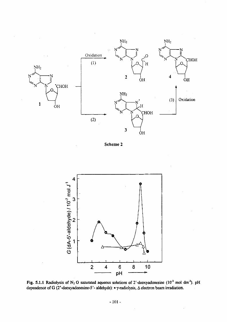
Scheme 2
PHFig. 5.1.1 Radiolysis of N20 saturated aqueous solutions of 2’-deoxyadonesine (10"3 mol dm"3). pH dependence of G (2’-deoxyadonesine-5’- aldehyde): • y-radiolysis, A electron beam irradiation.
-101 -

While thiols react with DNA radicals by H-atom transfer, the bisbenzimidazol derivatives such as Hoechst 33258 which firmly attach to the minor groove of DNA undergo electron transfer rather than H-atom transfer. These compounds are so tightly bound to DNA that they cannot diffuse to a damaged site, but an electron transfer from a damaged DNA site to the Hoechst 33258 molecule is observed on the millisecond time range {cf Fig. 5.1.2)
-0.4 -
time /10 s time /10"3 s
Fig. 5.1.2 Pulse radiolysis of N20/02- (4:l)-saturated aqueous solutions of DNA (4 x 10"4 mol dm"3) containing Hoechst 33258 (3 x 10"5 mol dm"3). Bleaching of Hoechst 33258 (left figure) and build-up of the Hoechst-derived radical (right figure) as a function of time. Practically all of the dye molecules are bound to the DNA under these conditions. The fast process shown is largely due to a reaction of OH radicals with DNA-bound Hoechst 33258.
MRC has determined types of DNA damage produced by the ‘direct effects’ of ionising radiation, using 193 nm induced photo-ionisation of DNA, in particular to find out whether radical cations of the nucleobases of DNA produced on ionisation are precursors to strand breaks, to determine the types of oxidative damage of DNA produced by ‘direct effects’, and whether the majority of the damage is localised at a specific base reflecting the migration of the radical site to guanine. Ionising radiation initially results in the formation of a radical cation in DNA and an electron. The electron is known to migrate, whereas information is lacking about the fate of the radical cations in DNA at ambient temperatures and its subsequent localisation. An approach to study the direct effects of ionising radiation at ambient temperature in an aqueous environment is to utilise 193 nm laser light which is of sufficient energy to cause monophotonic ionisation of DNA at the nucleobases. Following photoionisation of DNA with 193 nm light, the majority of the oxidative damage migrates and becomes localised at guanine. It is apparent that guanine is a major ‘hotspot’ for localisation of damage, expressed as prompt or alkali-labile ssb. One of the major consequences of hole migration in DNA is thought to be base modification preferentially at guanine. Using formamidopyrimidine-DNA glycolase (Fpg), which excise specific base lesions at purines or abasic sites to give a single strand break gap, results in a 20 fold increase in the yield of ssb compared with that for prompt or alkali-labile strand breaks. It is suggested that Fapy or 8-oxo 7,8-dihydroguanine (8-oxoG) are the major base modifications induced by 193 nm light. 8-oxoG presumably results from hydration of the radical cation of guanine in DNA, which is in an acid-base equilibrium with cytosine, its complementary base. More generally, this suggests that the chemistry of the nucleobase radical cations within DNA may differ to those previously reported for their corres-ponding monomers. To determine whether hole migration results in enhancement of base damage at guanine, a restricted fragment of dsDNA was treated with
- 102-

Fpg protein following exposure to 193 nm light and subsequent analysis by sequencing. The majority of the protein sensitive damage is located at guanine and there is little evidence for excision of damage at adenine. In summary, ionisation of DNA results in hole migration to guanine, as reflected from the findings with Fpg protein. Is the major base modification at guanine from the oxidative processes following ionisation of DNA?Since some of the OH radical adducts of the purine nucleobases dehydrate to yield the same radical to that produced upon their ionisation, the possibility of OH radical induced charge transfer in DNA arises. MRC find that at pH 7.4, there is no significant indication for radical transfer in mixtures containing different deoxynucleotides or those dinucleotides containing pyrimidine nucleobases. In contrast, OH-radical induced charge transfer occurs in the following oligonucleotides dApdG, dAGA and dAAGAA, all contianing adjacent purines. It is proposed that intramolecular radical transfer occurs from the dehydrated OH adduct of adenine to guanine with the dehydration reaction being rate determining. Therefore, OH-induced radical transfer in oligonucleotides proceeds not only by transfer of the radical site to the sugar moiety but also by transfer of the radical site from adenine to guanine.
Task 5.3: Evaluation of simulation codes for water radiolysisIn collaboration with GSF, UMIL has developed a MC code for the description of physico-chemical and chemical stages of electron track structures, based on the „step-by-step“ approach originally proposed by the Oak Ridge group. The code is now able to reproduce the time dependent radical yields reported in the literature. Specific attention was focused on the influence of the different assumptions and parameters for each step involved in the simulation, on the final result, i.e. the number and the spatial distribution of chemical species as a function of time. Indeed, models previously developed (also by ADPA) and described in the literature strongly differ in the choice of the parameters used in the calculations, since the large uncertainty in experimental data allows a large degree of freedom in such choices, which were also found to be strongly interconnected.The following aspects were analyzed separately:i) Physical stage: cross section data; relative percentage of excitation modes.ii) Physico-chemical stage: thermalization distance of subexcitation electrons, dissociation schemes
of excited water molecules, displacement of dissociation products at 10‘,2s after irradiation.
iii) Chemical stage: values of reaction rate constants and diffusion coefficients used in thesimulations, techniques used for the Monte Carlo transport of chemical species.
The relative importance of the different processes was quantified by adopting for one process the various values for the parameters proposed in the literature and keeping all other values fixed. After discussions with other partners of this collaboration, this extensive evaluation has led to the choice of the following data sets as reference sources:i) Reference data for model validation (i.e. time dependent yields): Laveme and Pimblott (Univ. of Notre
Dame).ii) Physical stage:iii) Dissociation scheme:
PARTRACderived from data of Frongillo et al., Sherbrooke University, with aslight modification of the relative percentage of different dissociationfor B1A1 states of water.derived from previous Oak Ridge models.derived from Ritchie et al. (ORNL).
iv) Displacement of products:v) Thermalization distances:vi) Reaction rate constants and diffusion coefficients: same as used by Green et al., Oxford
(derived from various sources).The agreement with independently derived data of Pimblott et al. on G-values at I0"12 s, using dissociation schemes from the Sherbrooke University group has to be considered as an indirect confirmation of the reliability of GSF cross section data, since the Sherbrooke description of the prechemical stage is the most recent and can be considered the most reliable available at present. Indeed, it has to be pointed out that experimental data against which calculations can be tested are only relative to high energy electrons, whereas the sensitivity to slight variation of the parameters used in the
- 103-
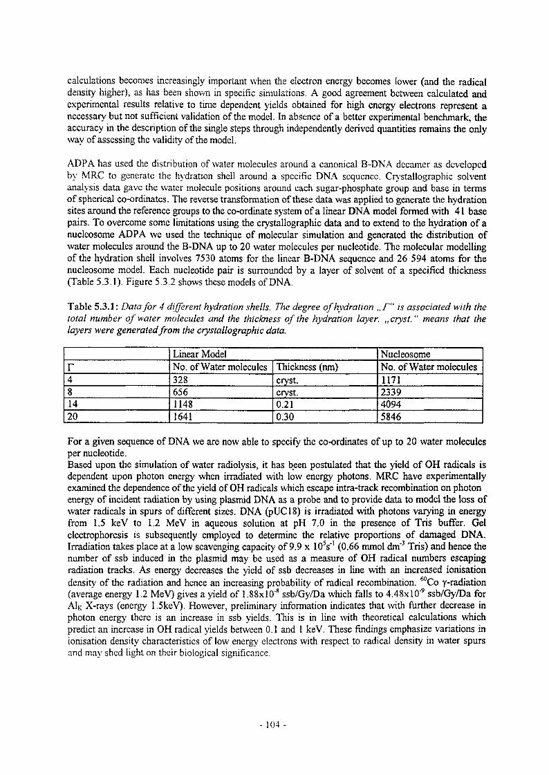
calculations becomes increasingly important when the electron energy becomes lower (and the radical density higher), as has been shown in specific simulations. A good agreement between calculated and experimental results relative to time dependent yields obtained for high energy electrons represent a necessary but not sufficient validation of the model. In absence of a better experimental benchmark, the accuracy in the description of the single steps through independently derived quantities remains the only way of assessing the validity of the model.
ADPA has used the distribution of water molecules around a canonical B-DNA decamer as developed by MRC to generate the hydration shell around a specific DNA sequence. Crystallographic solvent analysis data gave the water molecule positions around each sugar-phosphate group and base in terms of spherical co-ordinates. The reverse transformation of these data was applied to generate the hydration sites around the reference groups to the co-ordinate system of a linear DNA model formed with 41 base pairs. To overcome some limitations using the crystallographic data and to extend to the hydration of a nucleosome ADPA we used the technique of molecular simulation and generated the distribution of water molecules around the B-DNA up to 20 water molecules per nucleotide. The molecular modelling of the hydration shell involves 7530 atoms for the linear B-DNA sequence and 26 594 atoms for the nucleosome model. Each nucleotide pair is surrounded by a layer of solvent of a specified thickness (Table 5.3.1). Figure 5.3.2 shows these models of DNA.
Table 5.3.1: Data for 4 different hydration shells. The degree of hydration „ F“ is associated with the total number of water molecules and the thickness of the hydration layer. „cryst. “ means that the layers were generatedfrom the crystallographic data.
Linear Model Nucleosomer No. of Water molecules Thickness (nm) No. of Water molecules4 328 cry st. 11718 656 cryst. 233914 1148 0.21 409420 1641 0.30 5846
For a given sequence of DNA we are now able to specify the co-ordinates of up to 20 water molecules per nucleotide.Based upon the simulation of water radiolysis, it has been postulated that the yield of OH radicals is dependent upon photon energy when irradiated with low energy photons. MRC have experimentally examined the dependence of the yield of OH radicals which escape intra-track recombination on photon energy of incident radiation by using plasmid DNA as a probe and to provide data to model the loss of water radicals in spurs of different sizes. DNA (pUC18) is irradiated with photons varying in energy from 1.5 keV to 1.2 MeV in aqueous solution at pH 7.0 in the presence of Tris buffer. Gel electrophoresis is subsequently employed to determine the relative proportions of damaged DNA. Irradiation takes place at a low scavenging capacity of 9.9 x 10 V (0.66 mmol dm*3 Tris) and hence the number of ssb induced in the plasmid may be used as a measure of OH radical numbers escaping radiation tracks. As energy decreases the yield of ssb decreases in line with an increased ionisation density of the radiation and hence an increasing probability of radical recombination. *°Co y-radiation (average energy 1.2 MeV) gives a yield of 1.88xl0'8 ssb/Gy/Da which falls to 4.48xl0"9 ssb/Gy/Da for AIk X-rays (energy 1.5keV). However, preliminary information indicates that with further decrease in photon energy there is an increase in ssb yields. This is in line with theoretical calculations which predict an increase in OH radical yields between 0.1 and 1 keV. These findings emphasize variations in ionisation density characteristics of low energy electrons with respect to radical density in water spurs and may shed light on their biological significance.
-104-
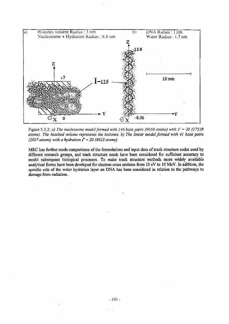
Histones volume Radius : 3 nm DNA Radius : 1 nm Water Radius : 1.7 nmNucleosome + Hydration Radius : 8.8 nm
10 nmI-125 —
Figure 5.3.2: a) The nucleosome model formed with 146 base pairs (9056 atoms) with F = 20 (17538 atoms). The hatched volume represents the histones, b) The linear model formed with 41 base pairs (2607 atoms) with a hydration F = 20 (4923 atoms).
MRC has further made comparisons of the formulations and input data of track structure codes used by different research groups, and track structure needs have been considered for sufficient accuracy to model subsequent biological processes. To make track structure methods more widely available analytical forms have been developed for electron cross sections from 10 eV to 10 MeV. In addition, the specific role of the water hydration layer on DNA has been considered in relation to the pathways to damage from radiation.
- 105 -
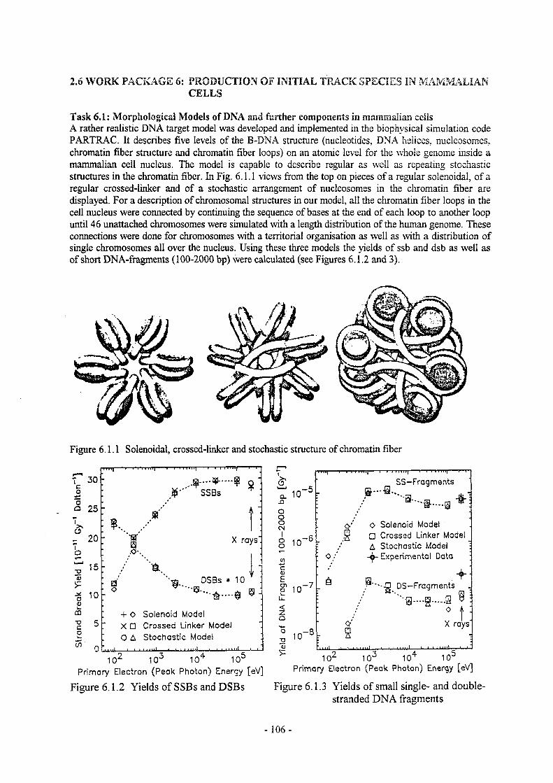
2.6 WORK PACKAGE 6: PRODUCTION OF INITIAL TRACK SPECIES IN MAMMALIANCELLS
Task 6.1: Morphological Models of DNA and further components in mammalian ceils A rather realistic DNA target model was developed and implemented in the biophysical simulation code PARTRAC. It describes five levels of the B-DNA structure (nucleotides, DNA helices, nucleosomes, chromatin fiber structure and chromatin fiber loops) on an atomic level for the whole genome inside a mammalian cell nucleus. The model is capable to describe regular as well as repeating stochastic structures in the chromatin fiber. In Fig. 6.1.1 views from the top on pieces of a regular solenoidal, of a regular crossed-linker and of a stochastic arrangement of nucleosomes in the chromatin fiber are displayed. For a description of chromosomal structures in our model, all the chromatin fiber loops in the cell nucleus were connected by continuing the sequence of bases at the end of each loop to another loop until 46 unattached chromosomes were simulated with a length distribution of the human genome. These connections were done for chromosomes with a territorial organisation as well as with a distribution of single chromosomes all over the nucleus. Using these three models the yields of ssb and dsb as well as of short DNA-fragments (100-2000 bp) were calculated (see Figures 6.1.2 and 3).
Figure 6.1.1 Solenoidal, crossed-linker and stochastic structure of chromatin fiber
7 30 ooa 25
3*_ 20
~ooS=ao
o5-oco
15
10
CO
■rq----r-r-r «i HI| .... > 1 1 iTiiy- » III 1 Hi;----r 1 .
-
• r SSBs
- / j :
■ K •- X rays"
.0-
- & -i ■
. a "■gl. DSBs * 10
- o G' A- $ 9 i
: + 0 Solenoid Model- XD Crossed Linker Model
O A Stochastic Model ■' " "1 ' ■ 1 ■ ■ ‘ » 1 1 t-ml- -- » ,'
102 103 104 105Primary Electron (Peak Photon) Energy [eV]
Figure 6.1.2 Yields of SSBs and DSBs
o'
CL.0Oo0CN
1oo
cnc0)ECD
£<zQO"O<u
'>■
10-5
. • ■
SS-Fragments
*]
10-6 r: o/
O Solenoid ModelD Crossed Linker ModelA Stochastic Model •
Experimental Data ;
10-7‘ 6 g“■-.13 DS-Fragments .
*"-g—-g...... 8 9 :
0 * :
10-8o'
F 8X rays"
102 103 104 105Primary Electron (Peak Photon) Energy [eV]
Figure 6.1.3 Yields of small single- and double- stranded DNA fragments
-106-

Task 6.2: Interaction cross sections for relevant cell constituentsTrack structure simulations require cross section data sets as input to describe important physical interactions of radiation, i. e., electrons, protons, photons and other charged particles, with molecules in matter under consideration. In the biological cell, water is the dominant component; other components are DNA and proteins. Earlier track structure calculations for radiation biophysics have been using water in the vapour state as model substance, because of the good data base. Only limited data exist for the condensed phase (ice) while for the liquid phase even less is known. Therefore, electron inelasticscattering cross section data for use as input in electron track structure calculations in liquid water were re-examined and improved by GSF. Basically the idea of the Oak Ridge Nat. Lab. approach was used The dielectric response function of liquid water, used in such cross sections, was modeled arid estimated on the basis of optical data and other experimental and theoretical data. Its imaginary part was modeled as a linear superposition of Drude-like functions. Each excitation was represented by a sharply peaked derivative Drude function while each ionisation shell was represented by an ordinary Drude function cut at the ionisation threshold using a step function and smeared out there with a Gaussian. No assumption of a collective excitation was needed in this model; there is no convincing evidence for its existence in liquid water. The real part of the dielectric rexense function was calculated analytically using the Kramers-Kronig relation. A new parameter set was obtained for this model by a numerical fit to existing experimental data in the optical limit with theoretical constraints. The model now shows reasonable partitioning of excitations and ionisations, reasonable ionisation energies and threshold behaviour, a high energy behaviour consistent with photoabsorption experiments in the gas phase, realistic shell contributions to the ionisation cross section, and fulfills all known sum rules. The mean excitation energy of the Bethe theory was calculated to I = 81.8 eV which is sufficiently close to the recent experimental value I = 79.75 + 0.5 eV of Bichsel and Hiraoka. To obtain the full energy and momentum dependent dielectric response function, a model for the momentum transfer dependence was added, which leads to a Bethe-ridge. This function is connected with the generalised oscillator strength and is the main quantity in calculating inelastic scattering cross sections in liquid water. Inelastic-scattering cross sections were evaluated from this model in the framework of the first Bom approximation (FBA), which is a non relativistic first order perturbation theoiy. Electron exchange effects and semi-empirical corrections to account for the deriation from the Bom approximation at low energies were also incorporated. The approach was extended to relativistic energies using the relativistic Bethe theory. High computational accuracy and precision was needed in these computer calculations to obtain consistency, converging results and the right asymptotic behaviour. Cross sections differential in energy transfer and momentum transfer were calculated for every excitation level or ionisation shell as well as total cross sections. The total inelastic cross section for electrons in liquid water (solid curve) is shown in the left panel of figure6.2.1 together with the contributions from excitations (short dashed curve) and ionisations (long dashed curve). A comparison to data, either experimental or theoretical, known from the gas, the condensed (ice) and the liquid phase was done to achieve consistency. As an example the calculated mass stopping power for electrons in liquid water is shown in the right panel of figure 6.2.1 and compared to a calculation made by Ashley for liquid water (squares) and gas phase data by Paretzke (diamonds).
Angular distributions for scattered primary and secondary electrons were also derived. Elastic scattering cross sections were extended to relativistic energies; a semi-empirical formula and parameters for the gas phase were used together with known experimental corrections at low energies.
- 107-

electron energy
■ Ashley
' icr 1 electron energy
Figure 6.2.1: Left panel: total inelastic cross section (solid curve) and contributions from excitation (short dashed curve) and ionisation (long dashed curve) versus incident kinetic electron energy. Right panel: Mass stopping power (solid curve) and contributions from exitation (short dashed curve) and ionisation (long dashed curve) compared to a calculation by Ashley (squares) and gas phase data (diamonds) versus incident kinetic electron energy.
Task 6.3: Production of initial species from absorption eventsThe new approaches described above by ADPA under task 5.3 have been tested and used in a complex simulation model to trace the fate of ail species created by low energy Auger electrons emitted from DNA-bound 1-125 and from K-shell photoionisation produced by low energy photons (100 to 500 eV). The physical step was simulated using the old ADPA cross-sections; they will be changed as soon as the new GSF cross sections will be available. The chemistry was simulated until up to 10**s using a diffusion controlled scheme. The pathways introduced in our chemistry model for DNA damage was the one discussed in the Bad-Honneff workshop RAM-97:
- ionisations on sugar-phosphate create a single strand break (SSB),- ionisations on bases: there is a transfer of charge to guanine,- other inelastic event on bases are scored as base event,- ionisations in hydration shell: 3 cases:
i) if the water is attached to base: transfer to guanineii) if the water is attached to sugar: creation of OH radical and H30+iii) if the water is attached to phosphate: 60% give a SSB
40% transfer to bulk water to give radicals- events in bulk water: radiolysis with creation of OH and H radicals, H2O2, and ions OH-,
H3O+, H-,... diffusion and reactions with nucleotides or scavengers up to 10"* s.Details and applications can be found in the publications listed below.
UMEL has analyzed results reported in the literature concerning the dissociation of excited water molecules and the thermalization of sub-excitation electrons using the MC code described in WP 5. The different results were analyzed in terms of their capability of connecting a selected set of cross section data with the time dependent radical yield following the physico-chemical stage of track structure. UMIL’s contribution to this topic is described under task 5.3.
-108-
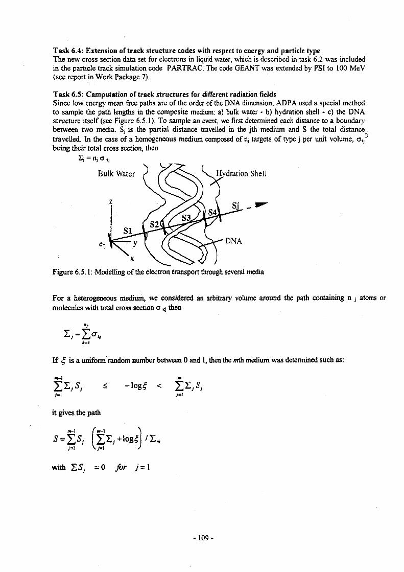
Task 6.4: Extension of track structure codes with respect to energy and particle type The new cross section data set for electrons in liquid water, which is described in task 6.2 was included in the particle track simulation code PARTRAC. The code GEANT was extended by PSI to 100 MeV (see report in Work Package 7).
Task 6.5: Computation of track structures for different radiation fieldsSince low energy mean free paths are of the order of the DNA dimension, ADPA used a special method to sample the path lengths in the composite medium: a) bulk water - b) hydration shell - c) the DNA structure itself (see Figure 6.5.1). To sample an event, we first determined each distance to a boundary between two media. Sj is the partial distance travelled in the jth medium and S the total distance. travelled. In the case of a homogeneous medium composed of n, targets of type j per unit volume, cttj' being their total cross section, then
Figure 6.5.1: Modelling of the electron transport through several media
For a heterogeneous medium, we considered an arbitrary volume around the path containing n j atoms or molecules with total cross section a ^ then
If £ is a uniform random number between 0 and 1, then the mth medium was determined such as:
2z,s, < -log# <
it gives the path
;=1 \ /= 1
with =0 for j-1
-109-

The electron interacts with the closest atom or molecule of its new sampled position. When the medium is a set on n, independent targets with cross section crry, the sampled path S is the path from the origin
to the first target with distance d; between this target and the direction of travel such that:
1 7C
Many track structure calculations have been made and published in the papers mentioned below in ^Publications".
2.7 WORK PACKAGE 7: TRANSPORT OF RADIATION TO CELLS OF INTEREST
Task 7.1: Adaptation of existing radiation transport codesTo adapt the transport code GEANT to calculations of the modifications of radiation fields in irradiated bodies, PSI has re-arranged and improved the data sets for interaction cross sections. Presently, three pointwise cross section files for photon, positron and electron atomic interactions, named EPDL, EPODL and EEDL, are available at PSI. They contain data for atoms from Z = 1 to 100. The fourth file EH20DL contains preliminary data for electron interactions with H20. All cross sections as well as secondary particle energy and angular distributions are available in the energy range from 1 eV to 100 GeV to 10 TeV using log-log rule. All four files are available in formatted form using ELDL format (Evaluated Livermore Data Library). Before the use in GEANT they have to be converted into the binary form using the computer program DIRECT. Further, using binary form the cross sections named EPDL.BIN with its directory EPDL.DIR (or EPODL.BIN and EPODL.DIR or EEDL.BIN and EH20DL.DIR) have to be converted into logarithmical energy grid using the program ELDLBIN. The particle tracking procedure was developed for use in the Monte Carlo codes PARTAC and GEANT. In preparation are the atomic interaction data for protons and a-particles and for proton-interactions with H20 vapour. In the last the experimental results of Toburen et al. will be considered.
a) Processing Code ELDLBIN: This code converts integrated data of binary versions of EPDL, EPODL, EEDL and EH20DL files into the data useful for use in GEANT. The energy grid is the same as in GEANT, but the user can define the number of points. This program also fits the angular and energy distributions. The fiourescence and Auger and Coster-Kronig emissions are also added. The resulting data are written onto the files EPDLT, EPODLT, EEDLT and EH20DL. These files have to be read in the initial phase of each calculation with GEANT. The size of data tables on the EPDL, EPODL, EEDL and EH20DL files has been minimised by presenting only those values that are required to allow log-log interpolation between tabulated values for all quantities within an accuracy of ~ 1%. Therefore, in order to determine a cross section at energy E, and E2, the following interpolation scheme is used:
log (ct (E)) = [log (EZEi) • log (ct(E2)) + log (Ez/E) • log (a(E,))] / log((E2/E,).
Only in three cases, (a) near to pair and triplet threshold, and (b) between the first two tabulated values of coherent and incoherent form factors, and (c) to obtain anomalous scattering factors, the lin-lin interpolation has to be employed.Using data available in EPDLT, EPODLT, EEDLT and EH20DLT files the integrated cross sections are interpolated for each particle energy in GEANT using linear interpolation.In 1998 the water cross sections will be improved considering the developments for liquid water which are currently performed at GSF and PSI.
-110-

b) Photon-Interaction Cross Sections and Secondary Particles: This section documents the generation of photon cross sections in the energy range from 1 eV to 100 GeV. In this energy range the cross sections are based on EPDL-97 files (Evaluated Photon Data Library) from Lawrence Livermore National Laboratory, This file was accomplished in 1989 and improved in 1997. It presently contain cross sections for Raleigh coherent and Compton incoherent scattering, photoionisation, photoexcitation and pair and triplet conversion for elements from Z = 1 to 100. The atomic form factors for coherent and incoherent scattering and anomalous scattering factors for coherent scattering are also available. The cross sections for photoionisation are given for each atomic subshell. Splitting the photoionisation cross sections to its shell contributions enables the determination of each particular shell ionisation process and is a basic prerequisitory for inclusion of the follow-on Auger and Coster-Kronig electron cascades and X-ray emission.As yet the EPDL does not include several types of data, including Delbruck and nuclear scattering, which become important at approximately 1 MeV and higher energies. The status of all these types of data is at LLNL currently under review and it is planned that these types or data will be included in EPDL in the future. The EPDL97 presently contains the following type of data:
1. Coherent scattering• of the scatterd photon Integrated cross sections• Form factors• Real and imaginary anomalous scattering factors• Average energies
2. Incoherent scattering• Integrated cross sections• Scattering function• Average energies of the scattered photon and recoil electron
3. Total photoionisation reaction• Integrated cross sections• Average energies to the residual atom, i.e. local depositions• Average energies of the secondary photons and electrons
4. Photoionisation reaction by subshell• Average energies to the residual atom, i.e. local dispositions• Average energies of the secondary electrons
5. Pair production reaction• Integrated cross sections• Average energies of the secondary electron and positron
6. Triplet production reaction• Integrated cross sections• Average energies of the secondary electron and positron
7. Excitation• Integrated cross sections• Average energies to the residual atom, i.e. local disposition
c) Particle Tracking and Generation of the Secondaries: First, described will be the single-collision particle tracking for photons and charged particles. This tracking is purely stochastical and therefore exact but leads at low energies in a case of charged particles and large, millimeter and more sized bodies to a large amount of very short steps due to Coulomb forces. The result is a large computational time. Therefore, in large bodies the single-collision particle tracking of charged particles was replaced by condensed-history particle tracking which allows larger particle steps and uses the multiscattering and the energy straggling methods. It is described in the second subsection. The user can select by the input parameter ISTR which method will be used. If ISTRA=0 is selected the single-collision method will be used, otherwise the condensed-history particle tracking will be applied.
- Ill -

i) Single-Collision Particle Tracking: Prediction of the pathlength, t, is based on the pathlength probability:
t (cm)= ^ln(^),
where R\ is a random number. Selection of the material n and nuclear reaction m at the end of each path: If
./or »=!%)#,
then material n is selected. Z„ is the total macroscopic cross section of material n and N is the number of materials in the tracking medium, and R2 is a random number, and if
for m = \toM,
then reaction m is selected cxmn and crn are the microscopic reaction and total cross sections ofmaterial n in the tracking medium, M is the number of reaction processes, and R; is a random number. Further, the subshell / of an ionisation process is selected as follows:
#7 = IW,^ m.n
where cr,_m is the microscopic ionisation cross section of z'-th subshell material n, <rm n is the totalmicroscopic ionisation cross section of material n, I is the number of the atomic subshells, and R4 is a random number.After the subshell i of the material m is selected, the emitted photons and Auger electrons are determined. Our data describing the radiative and non-radiative emission due to a subshell ionisation is based on photon/electron yields and average energies for the K, LI, L2, L3, M, N, O and P shell. The enhanced probability of emissions is used. Using 8 random numbers, the number of emitted photons and electrons is determined. If
Photon-yield,- > R4+J , J = 1 to 4,
then the emission of one photon of energy E} is considered. If
Electron-yield; - Int(Electron-yieldy) > Rj,+], j - 5 to 8,
then the emission of Int(Electron-yield,) + 1 electrons of energy E, is considered. Otherwise only Int (Electron-yieldj) electrons are banked for later tracking.
ii) Condensed-history Particle Tracking: The interactions of neutral particles are characterised by infrequent isolated collisions, so that the above described detailed particle tracking can be always applied. In contrast to that, the charged particle transport is dominated by the long-range Coulomb forces, so that the resulting number of interactions is very large. At small energies the particular tracking steps can extremely small (micrometers or even nanometers). The use of the above method would lead in large media to a very large number of interactions to slow down a charged particle down to a low energy cutoff. Therefore considerable theoretical work has been done in the past to develop a
-112-
variety of multiple-scattering theories for transport of charged particles. They attempt to use the basic cross sections in connection with the statistical nature of the transport to predict probability distributions of angular deflection and energy loss considering large tracking steps. The assumption of many individual collisions per step constitutes the ^condensed history" Monte Carlo particle tracking method. The basic reference is that of Berger from 1963. Based on this reference the electron transport in ETRAN, ITS system (TIGER) and MCNP Monte Carlo codes was developed. The energy of all emitted particles is deposited locally. To calculate the angular deflections the method here by PST chosen is, as in ETRAN, TIGER or in MCNP Monte Carlo codes, the Goudsmit-Sounderson theory.In case of the energy straggling all methods included into GEANT can be applied but they have some shortcomings. Therefore, the extended Landau theory is applied similarly as used in the ETRAN, TIGER or the MCNP codes.
d) Interface-Boundary Crossing: A proper interface-boundary crossing is especially important in very small media. In a single-step particle tracking mode, first, the pathlength t is calculated using the total cross section of the present (old) medium Z0 • Then this pathlength is reduced to the distance to the boundary t0 and the pathlength in the new medium is given by:
tn =~^r-On(-fti)-*o£oX
where is the total macroscopic cross section of the new tracking medium and t„ is the pathlength between the new collision and the boundary. The random number i?/ is the same in the old and new medium.
In the case of condensed-history particle tracking constant major steps and equal substeps are considered. If a particle is crossing a interface boundary, the substep is subdivided into two parts, one in the old medium and the rest in the new medium. The problem which occurs is that the Goudsmit- Saunderson multiple scattering distributions are pre-tabulated for fixed substep-sizes. In this case the avarage number of the single collisions Nm is calculated between the last step and the nearest geometrical boundary of after the boundary is calculated. It is given by
In (0.5)
Where Z is the total macroscopic cross section of the tracking medium at kinetic energy T, and T„ is the shortest distance to the boundary or after the boundary. If Nm < 20, than the condensed-history particle tracking is replaced by the single-collilsion tracking. As in the condensed-history tracking also in this case the energy of the emitted electrons is considered to be deposited locally.
e) Termination of HistoriesThere are three energy cutoffs below which the particle tracking is stopped and the kinetic energy is deposited locally at the position of the particle. Two cutoffs are related to the lowest allowed energy of the emitted secondary photons and electrons due to bremsstrahlung or ionisation. The main energy cutoff is given by the lowest energy of the energy grid chosen for the cross section library. If this cutoff would be lower then the smallest electron binding energy in the material isotopic mixture, 1 eV for example, the photons or electrons would travel forever because in this energy range only elastic scattering without significant energy loss is present or dominating. Therefore, such a cutoff is preset to be equal to the smallest electron binding energy of the isotopic mixture. In case of positrons there are annihilation reactions so that they can be tracked down to 1 eV. Again at this cutoff the kinetic energy of particles is deposited locally.
- 113 -

This PSI-code will be applied to calculate the moderation of radiation fields inside the body to the cells of interest.
Task 7.2: Calculation of start spectra of directly ionizing particles:No work on this task can yet be reported.
-114-

3: SUMMARY OF MAIN ACHIEVEMENTS
The wide spectrum of significant progress made in this project (and in the „sister-project“ EDICAR coordinated by B. Michael) has been reported and thoroughly discussed in several annual workshops on ^Radiation Action Mechanisms" (RAM) in the Physik-Zentrum in Bad Honnef organised by GSF; at these workshops also invited co-ordinators of other related projects reported on the results obtained in their consortia. MRC has contributed to the overall objectives of the contract also by organising the 12th Symposium on Microdosimetry at Keble College, Oxford, from 29.9. - 4.10.1996. This symposium served also as an important international forum for exchange of scientific information of central relevance to the contract and for dissemination of interim research results; the full proceedings were published in 1977. In the following the main achievements obtained in our project are summarized.
Work Package 1: Mechanistic Models for Radiation OncogenesisThe two-stage clonal expansion model of Moolgavkar, Venzon and Rnudson (MVK) was selected as the basic model. Both exact and deterministic versions of the model were used by all three groups, as well as stochastic generalizations of the two-stage MVK and Armitage-Doll models. It has been shown by GSF that one of the biological parameters cannot be determined from tumour incidence data. Identifiable parameter combinations were constructed and fast algorithms for calculating the hazard function and the survival probability were given. The F T. Cross rat data was used by both GSF and RTVM to assess risks of lung cancer in relation to radon exposure. Average exposure rates of 5 WL for fatal tumours double the spontaneous mutation rates. Fatal lung tumours in rats were proposed to be the most useful for comparisons with human data. GSF concluded that the relative risk per unit of exposure of fatal tumours is comparable to that for humans. The fitted model of fatal tumours shows an inverse dose-rate effect at exposure rates above 20 WL. However, below 10 WL the lung cancer risk per unit exposure decreases with increasing exposure duration. Between 10 and 20 WL the difference in ERRAVLM between acute and protracted exposure is small. This is due to the effects of radon exposure on the clonal expansion rate (promotion).
RTVM has applied the model to the following animal data sets: lung tumour in mice after acute irradiations with X-rays, neutrons, and with gamma rays of different dose rates; bone tumours in beagles after injection with the internal alpha emitter Ra-226; skin tumours in mice after continuous irradiation with ultraviolet radiation. Contrary to the conclusions of the analysis of radon-induced lung tumours in rats, a good fit was found when radiation was assumed to act primarily on the first mutation rate (initiation), and the radiation effect on this first mutation behaves qualitatively in accord with radiobiological expectations from cellular data.
NRPB has shown that the observed patterns of solid cancer and leukaemia risk as a function of time and age can be described by two-stage models, as well as by generalizations of the two-stage and Armitage- Doll models. Generalisations of the two-stage model have been fitted to UK leukaemia incidence data. Analysis indicates that acute lymphatic leukaemia and chronic lymphatic leukaemia can be adequately described by two-stage and three-stage models. It was concluded that while mutation rates and stem cell numbers implied by the optimal two-stage model are both fairly low, both two-stage and three-stage models are not inconsistent with the data.
RIVM has also applied the two-stage model to the lung tumour data of the radon-exposed Colorado uranium miners. Because smoking is a major confounding factor, the data were combined with data on a non-smoking population and on cigarette smokers. The dependence of the modelled mutation rates on smoking and radon was assumed to be additive, but the resulting tumour incidence is supra-additive.
- 115 -

Work Package 2: Mechanistic Models for Chromosome AberrationsThe objectives were to find a model which converts double strand breakes (DSB) into chromosomal aberrations and predicts dose response curves for dicentric productions and their variation with radiation quality, human lymphotytes produce the major data base for this work package. The important advances in modelling are that in order to explain dicentric yield curves for a limited number of radiation, i.e. X-rays, 5 keV-um'1 protons, and 22 keV-pm'1 helium-3 ions, the assumption of having only one type of DSB acting in one way is not sufficient. However, one type of break may be sufficient to predict ratios of aberrations of different types for particular radiations at selected doses.Advances in data obtained to guide modelling are twofold. First, following X-ray irradiation simple pairwise exchanges are more numerous in human lymphocytes than those necessary to accept for the initial slope of the X-ray dicentric yield curve. Secondly, in human fibroblasts, ultra-soft X-rays of particularly 0.28 keV (carbon X-rays) show a relatively high exchange aberration yield which is linear with dose. This implies that a lesion / nonlesion interaction model may be important at least for some radiations.A biophysical model implemented in a MC computer code of chromosome aberration induction by ionizing radiation was also developed by UMIL. The code allows to simulate dose response curves for different kinds of aberrations induced by light ions, under the main assumption that a subclass of DNA dsb, namely complex lesions, are responsible for chromosome aberrations, less severe damages being repaired systematically. Preliminary results (also on the ratio complex/reciprocal exchanges) were compared with positive results with experimental data from different laboratories.
Work Package 3: Mechanistic Models for MutagenesisMRC has compiled an extensive database on radiation mutagenesis at the hprt locus, as the most extensively studied mutation paradigm. For guidance of modelling this consists of about 400 reference records with key fields for physical and biological parameters, including yield frequencies and molecular characterisation.UMIL and MRC were developing and testing alternative methods for modelling mutagenesis by reasonable assumptions for their formation, particularly from single localised clustered lesions but also from two independent dsb. UMIL have applied a similar algorithm as they previously adopted for cell inactivation, to relate a subclass of dsb to mutation frequencies as a function of radiation quality. MRC is also including an approach based on complexity or severity of DNA damage from individual radiation tracks. They have explored approximation methods to describe the extensive delta-ray contribution from high energy ions. Here, no final results can be reported yet.MRC has also developed novel experimental facilities for critical tests of mechanistic assumptions in the mutagenesis process. These include the key question of a single dsb leading to a deletion mutation, to be addressed with ultrasoft X-rays including at low dose rate, and also the role of temporal versus spatial aspects of radiation tracks.
Work Package 4: Mechanistic Models for DNA Damage and RepairFrom the use of ultrasoft X-rays to determine the yield of dsb induced and their repairability, it is proposed that the majority of the dsb induced by high energy electrons or 60Co y-radiation are mainly produced by the low energy secondery electrons which account for about 20-30% of the total dose.It is essential that ~ 15% of the OH radicals interact with the sugar moiety, a benchmark value of fundamental importance for simulations of the yields of DNA dsb and ssb.The induction of DNA - protein cross links is not significantly influenced by radiation quality. Their yields do not reflect the biological severity of the different radiations and therefore question the constribution of the lesions to mutation induction.From comparison of experimental studies on the formation of small frequents of DNA by various radiations and morphological models of DNA, the production of DNA fragments > lOkbp is dominated by multi- track events especially for Ck and AJK USX radiations. For short DNA fragments (<2 kbp) theoretical and experimental agreement was obtaind using a stochastic arrangement of nucieosomes and assuming these fragments are produced by a single track.
-116-

Computations of clustered and simple DNA damage have shown a relevant role of clustered DNA damage determining cell killing. Inclusion of chemical paths ways for indirect effects in the models show that >50% dsb induced by low LET radiation are complex.For high LET radiations, the spectrum of lesions shifts to more complexity and some of the very complex lesions may be unique to high LET radiation. Protons produce a more complex spectrum of dsb than a-particles for the some LET.Calculations on the contribution of C photoelectrons to cellular inactivations by photons of a energy of 250 and 350 eV have indicated that the atomic composition of the target, e.g. DNA is important. For hydrated DNA it is proposed and experimentally tested that for energies above the oxygen K-shell, DNA will receive less energy in its environment.
Work Package 5: Chemical Pathways Involving Track Species in CellsThe four activities in WP 5 have generated data to highlight aspects of chemical pathways involved in initial track species generated in cells. A Monte Carlo track structure program was constructed to generate radical species in water and in DNA by ionizing radiation of different radiation quality. A knowledgebased model of DNA hydration was produced in a sequence-specific DNA structure. This model provides positions of water molecules in the first hydration shell. Comparisons of various Monte Carlo track structure codes show similarities in the distribution of damage at the chromatin level whereas differences are noticed at the level of naked DNA. In a model compound such as 2’-deoxyadenosine, an intermolecular radical transfer from the adenine to the sugar moiety of another 2’-deoxyadenosine molecule leads to damage amplification. Intermolecular radical transfer of oxidative damage from an OH-adduct of adenine to guanine was observed. Therefore, OH-induced radical transfer in oligonucleotides proceed not only by transfer of radical sites to the sugar moiety but also by transfer from adenine to guanine.
Work Package 6: Production of Initial Track Species in Mammalian CellsGSF completed the inclusion of an atomic model of the DNA in the PARTRAC code. The model describes levels of organization from nucleotides up to chromatin fiber loops, which can in turn be connected to model a whole chromosome. Calculations of ssb, dsb and base damage induction were performed for low LET radiation, showing good agreement with experimental data.A new set of cross sections for electrons in liquid water differential in energy transfer and momentum transfer was also derived at GSF, as well as the angular distributions for scattered primary and secondary electrons. Elastic scattering cross sections were extended to relativistic energies. The dielectric response function of liquid water was modeled on the basis of optical data and other experimental and theoretical data. A new parameter set was obtained for the model by a numerical fit to existing experimental data in the optical limit with theoretical constraints. The whole approach is internally consistent and fully documented, and it is able to match existing experimental data. The cross section data set has been included in PARTRAC.An overview of existing models treating the dissociation of excited an ionized water molecules was conducted by UMIL; the models have been discussed in terms of their capability of connecting the newly derived cross sections with the initial yield of water radiolysis products. A set of parameters describing the processes in the physico-chemical and chemical stages of track structure has been selected and implemented in a MC module included in the PARTRAC code.PSI extended the energy range of the particle transport code GEANT up to 100 MeV.A technique for transporting electrons of low mean free path in the vicinity of DNA was developed by ADPA assuming a three compartment structure (bulk water, hydration shells, DNA). Simulations of direct and indirect DNA damage induced by low energy electrons (emitted from DNA-bound 1-125 and from K-shell photoionisation produced by photons in the energy range 100-500 eV) has been performed taking into account the role of DNA hydration shells (as reported in WP5).
Work Package 7: Transport of Radiation to Cells of InterestNo work in WP 7 within this project was performed. However, data from related GSF- and PSI- work will provide the necessary start spectra by the end of 1998.
-117-

4. RESEARCH TO BE PERFORMED IN THE REMAINDER OF THE PROJECT
Until now work has progressed as anticipated. The following tasks are planned for the next 18 months until the end of the project.
Work Package 1: Mechanistic Models for Radiation OncogenesisAll three partners will jointly analyse the data on radon-exposed rats provided by Dr. M. Morin, CEA. Generalizations of the two-stage model will be used to model lung cancer risk in this data, and comparisons will also be made with model fits to lung cancer in the F.T. Cross rat data.
GSF will work on application of the model to the smoking data of the German indoor radon case control study, and to the new data of the Colorado Plateau Uranium miners.
Following on from joint analyses of leukaemia data in the Japanese atomic bomb survivors, in the UK ankylosing spondylitis patients and in the International Radiation Study of Cervical Cancer, an initiation model for leukaemia will be constructed, taking account of bone marrow cell killing and repopulation. This initiation model will be combined with previously developed models for intermediate cell progression and development to make assessments of leukaemia risk following various assumed exposure distributions.In addition to the analysis of lung cancer in the Colorado miners after radon exposure RTVM will make an attempt to improve the estimation of the effect of radon exposure to populations. An analysis will also be made of the induction of bone tumours in radium dial painters.
Work Package 2: Mechanistic Models for Chromosome AberrationsNRPB will complete scoring of the experiment using paints for two different colours to confirm or reject the observations of Simpson and Savage that simple exchanges are linear with dose.MRC will perform an experiment to measure exchange aberrations in human fibroblasts following chronic irradiation with carbon-K X-rays. This will lead to a better unterstanding of the mechanism by which aberrations are formed.UMIL will further develop the MC code to introduce the time dependence of aberration formation and will compare simulation results with PCC data from the literature. Different hypotheses arising from new experimental findings concerning the mechanisms of aberration formation will be included in the model and tested.
Work Package 3: Mechanistic Models for MutagenesisThe latest literature publications will be incorporated by MRC into the hprt database, as will new experimental results from consortium partners. In modelling, UMIL will concentrate on the complexity of initial DNA damage responsible for hprt mutation and on the correlation between this and inactivation. Analytical models as well as Monte-Carlo simulations will be developed to simulate mutation an inactivation simultaneously. MRC will apply to hprt-mutations the damage-complexity model as currently developed for Drosophila mutations. Also hprt mutations experiments will be initiated with ultrasoft X-rays to test the single radiation-lesion hypothesis following corresponding data for chromosome aberrations.
Work Package 4: Mechanistic Models for DNA Damage and RepairMRC will determine the dependence of the yield and repairability of dsb in V79-4 cells on the photon energy using various USX energies and analyse the contribution of random breakeage induction to small fragments. They will also study the rejoining kinetics of cellular dsb using enzymatic type saturation kinetics or different repair times reflecting the spectrum of complexity of the damages.The contribution of base damages to damage complexity will be studied in DNA model systems to provide benchmarks for biophysical simulation calculations. This study will also consider the role of diffusible water radicals.
-118-

Furtheron, the role of the complexity of DNA damage in the induction of mutations will be studied. Model predictions for the induction of clustered damage by Auger-emitters incorporated into DNA will be compared to experiments.Particular emphasis will be put to an intercomparison of calculated and measured yields of DNA damages to improve the predictive power of the biophysical models in the regime of low doses developed in this project.
Work Package 5: Chemical Pathways Involving Track Species in CellsSimulation of OH-induced DNA damage will be performed using the PARTRAC code. Different approaches for the description of the mechanism of lesion induction will be compared. Measured total reaction rate constants of DNA with OH radicals will be compared with values obtained considering individual base and sugar phosphate reactions.Improvement of the chemistry code with special attention to the modelling of OH reactions with DNA. Hydration shells will be taken into account to study differences in the OH attack in the major an minor groves of the double helix sensitivity of input data to track structure codes used for biophysical calculations will be assessed.The model system thymidine will be used to assess the positions of OH radical attack on the base and sugar moieties and the influence of hydration on DNA and the induction of damage by radiation of different qualities will be tested. Furtheron, we will study whether guanine is a „hotspot“ for base damage induced by the direct effects of radiation.
Work Package 6: Production of Initial Track Species in Mammalia CellsFurther development of the DNA target model in PARTRAC will be focused on the inclusion of nuclear proteins and the construction of further higher-order DNA structures by a specified arrangement of nonoverlapping chromatin fiber loops describing 46 chromosomes with a territorial organisation in a human cell nucleus. These improvements are essential for an adequate simulation of OH-induced DNA damage and of the production of large DNA fragments (0.1 to 10 Mbp) (see WP5).Proton interaction cross sections in liquid water will be set up. From relativistic energies down to a few hundred keV, Bom-Bethe theory will be applied for ionization and excitation cross sections, based on the dielectric response function already obtained for electrons. At lower energies, semiempirical approaches including electron capture and loss processes will be adopted, using experimental data and theoretical constraints.In order to improve the previous method of computation of mean free paths, a quantum mechanical method will be studied for the transport of low energy electrons in condensed media.
Work Package 7: Transport of Radiation to Cells of InterestFor all radiation fields considered in WP1 the initial start spectra of directly ionising particles and their degradation spectra will be calculated for the moderated impinging fields and tragrets.
-119-

5. Publications
5.1 Work-package Nr.l (26 publ.)
K.H. Chadwick , H.P. Leenhouts. Multi-step carcinogenesis and the implications for low-dose radiation risk philosophy. Trans IChemE, Vol 73, Part B (1995) 18-23
K.H. Chadwick, H.P. Leenhouts. DNA double strand break repair and RBE after high LET radiation: implications for complex damage. Poster. 12th Symposium on Microdosimetry, Keble College, Oxford (1996)
K.H. Chadwick , H.P. Leenhouts. Cancer modelling for protracted radiation exposures. In: Proceedings of the First International Symposium on Chronic Radiation Exposures: Risks of Late Effects, Cheliyabinsk, Russia (1996) 143
K.H. Chadwick, H.P. Leenhouts. Multi-step carcinogenesis modelling and the initiation event. In: Proceedings of the 7th L.H. Gray Workshop on Research Strategies for the Prevention of Early Events in Human Radiation Carcinogenesis, Dublin (1996)
M. Harris, H.P. Leenhouts, Haag de Uijt. A two-mutation model of carcinogenesis: Application to lung tumors using rat experimental data. RTVM report (1998)
W.F. Heidenreich. Stochastic tumor models. In: H.D. Doebner, W. Scherer, C. Schulte (eds.) GROUP 21, Physical Applications and Mathematical Aspects of Geometry, Groups and Algebras, Vol.II, World Scientific (1997) 879-883
W.F. Heidenreich, EG. Luebeck, S.H. Moolgavkar. Some properties of the hazard function of the two- dimensional clonal expansion model, Risk Analysis 17 (1997) 391-399
W.F. Heidenreich, H.G. Paretzke, P. Jacob. No evidence for increased tumor rates below 200 mSv in the atomic bomb survivors data. Radiat. Environ. Biophys. 36 (1997) 205-207
W.F. Heidenreich, H.G. Paretzke, P. Jacob. Reply to the ‘Commentary’ by D A. Pierce and D.L. Preston. Radiat. Environ. Biophys.36 (1997) 211-212
W.F. Heidenreich, H.G. Paretzke. City-effects in the atomic bomb survivors data, submitted for publ.
W.F. Heidenreich, P. Jacob, H.G. Paretzke, F T. Cross, G.E. Dagle. Two step model for fatal and incidental lung tumor risk of rats exposed to radon, submitted for publ.
H.P. Leenhouts. Lung cancer incidence after smoking and exposure to radon using a two-mutation model. ESRB Annual Meeting, Montpellier, France (1996)
H.P. Leenhouts. Lineaire extrapolatieproblematiek, risico’s bij lage doses. Wetenschappelijk vergadering RJVM (1996)
H.P. Leenhouts. Lung cancer incidence after smoking and exposure to radon using a two-mutation model. Radioprotection 32 (1997) 363-364
H.P. Leenhouts, K.H. Chadwick. Use of a two-mutation carcinogeneses model for analysis of epidemiological data, Health Effects Low Dose Radiation: Challenges of the 21st Century. British Nuclear Energy Society (1997) 145-149
- 120-
H P. Leenhouts, M. Harris, Haag de Uijt, P.A.M. Risico’s van Radon en Roken: enige beschouwingen vanuit mechanistisch perspectief. NVS-nieuws, 22. Jaargang No.5 (1997) 7-11
H P. Leenhouts, Haag de Uijt, P.A.M. Analysis of lung cancer incidence after exposure to smoking and radon. RTVM Annual Scientific Report ‘96. RIVM (1997) 17-18
H P. Leenhouts, Haag de Uijt, P.A.M., K.H. Chadwick. Analysis of lung cancer after exposure to radon using a two-mutation carcinogenesis model. In: Microdosimetry - An interdisciplinary approach, D.T. Goodhead, P. O’Neill, and H.G. Menzel, eds., Royal Society of Chemistry, Cambridge (1997) 248-251
H P. Leenhouts. Radon induced lung cancer in smokers and non-smokers: risk implications using a two- mutation carcinogenesis model (1998)
M.P. Little. Generalisations of the two-mutation and classical multi-stage models of cancerogenesis fitted to the Japanese atomic bomb survivor data. J. Radiol. Prot. 16 (1996) 7-24
M.P. Little, C.R. Muirhead, C.A. Stiller. Modelling lymphocytic leukaemia incidence in England and Wales using generalisations of the two-mutation model of carcinogenesis of Moolgavkar, Venzon and Knudson. Statist. Med. 15 (1996) 1003-1022
M.P. Little. Are two mutations sufficient to cause cancer? Modelling radiation-induced cancer in the Japanese atomic bomb survivors using generalisations of the two-mutation model of Moolgavkar, Venzon, Knudson and of the multi-stage model of Armitage and Doll. In: Health effects of low dose radiation. Challenges for the 21st Centuiy (1997) 169-174
M.P. Little, C.R. Muirhead, C.A. Stiller. Modelling acute lymphocytic leukaemia using generalizations of the MVK two-mutation model of carcinogenesis: implied mutation rates and the likely role of ionising radiation. In: Microdosimetry - An interdisciplinary approach, D.T. Goodhead, P. O’Neill, and H.G. Menzel, eds., Royal Society of Chemistry, Cambridge (1997) 244-247
M.P. Little. Mechanistic modelling of radiation-induced cancer. In: Effects of ionizing radiation: atomic bomb survivors and their children (1945-95) Victoria and William Schull Institute, Houston, Texas, in press (1998)
H.G. Paretzke, W.F. Heidenreich. Modelle fur die Strahlenkarzinogenese. In: Fachverband fur Strahlenschutz: Modeme Entwicklungen und Tendenzen in der Strahlenbiologie, Hannover (1996) 59- 64
R.K. Sachs, W.F. Heidenreich, D.J. Brenner. Dose timing in tumor radiotherapy: Considerations of cell number stochasticity, Mathematical Biosciences 138 (1996) 131-146
5.2 Work-package Nr.2 (9 publ.)
A.A. Edwards. Modelling the induction of chromosomal aberrations by ionising radiations. In: Microdosimetry - An interdisciplinary approach, D.T. Goodhead, P. O’Neill, and H.G. Menzel, eds., Royal Society of Chemistry, Cambridge (1996) 172-175
A.A. Edwards, V.V. Moiseenko, H. Nikjoo. On the mechanism of the formation of chromosomal aberrations by ionising radiation. Radiat. Environ. Biophys. 35 (1996) 25-30
A.A. Edwards. The use of chromosome aberrations in human lymphocytes for biological dosimetry. Radiat. Res. 148 (1997) 539-544
- 121 -

D.T. Goodhead, P. O’Neill, H. Menzel (Eds.): Microdosimetry - An interdisciplinary approach, D.T. Goodhead, P. O’Neill, and H.G. Menzel, eds., Koval Society of Chemistry, Cambridge (1997) 248-251
C.S. Griffin, M.A. Hill, D.L. Stevens, J.R.K. Savage. Effectiveness of ultrasoft X-rays at inducing complex exchanges in human fibroblasts. In: Microdosimetry - An interdisciplinary approach, D.T. Goodhead, P. O’Neill, and H.G. Menzel, eds., Royal Society of Chemistry. Cambridge (1996) 164-167
M.A. Hill, C.S. Griffin. Effectiveness of carbon ultrasoft X-rays at inducing complex exchanges in human fibroblasts. Radiat. Res. 148 (1997) 515-516
V.V. Moiseenko, A.A. Edwards, H. Nikjoo. Modelling the kinetics of chromosome exchange formation in human cells exposed to ionising radiation. Radiat. Environ. Biophys. 35 (1996) 31-35
V.V. Moiseenko, A.A. Edwards, H. Nikjoo, W.V. Prestwich. Modelling of chromosome exchanges in human lymphocytes exposed to radiations of different quality. In: Microdosimetry - An interdisciplinary approach, D.T. Goodhead, P. O’Neill, and H.G. Menzel, eds., Royal Society of Chemistry, Cambridge (1996)152-155
V.V. Moiseenko, A.A. Edwards, H. Nikjoo, W.V. Prestwich. The influence of track structure on the understanding of relative biological effectiveness for induction of chromosomal exchanges in human lymphocytes. Radiat. Res. 147 (1997) 208-214
5.3 Work-package Nr.3 (4 publ.)
F.A. Cucinotta, H. Nikjoo, J.W. Wilson, R. Katz, D.T. Goodhead. Radial dose model of SSB, DSB, deletions and comparisons to Monte-Carlo Track structure simulations. In: Microdosimetry - An interdisciplinary approach, D.T. Goodhead, P. O’Neill, and H.G. Menzel, eds., Royal Society of Chemistry, Cambridge (1996) 35-38
J.R. Ford, N.F. Metting, S.J. Marsden, D.L. Stevens, K.M.S. Townsend, D.T. Goodhead. Visualization of damage generated along alpha-particle tracks in irradiated rat tracheal epithelial cells. In: Microdosimetry - An interdisciplinary approach, D.T. Goodhead, P. O’Neill, and H.G. Menzel, eds., Royal Society of Chemistry, Cambridge (1996) 335-338
M.A. Hill, D.L. Stevens, D A. Bance, D.T. Goodhead. A versatile mammalian cell irradiation rig for low dose rate ultrasoft X-ray studies. In: Microdosimetry - An interdisciplinary approach, D.T. Goodhead, P. O’Neill, and H.G. Menzel, eds., Royal Society of Chemistry, Cambridge (1996) 203-206
D.L. Stevens, S.J. Marsden, M.A. Hill, Turcu, I.C.E., R. Allott, D.T. Goodhead. The separation of spatial and temporal effects of high LET radiation. In: Microdosimetrv - An interdisciplinary approach, D.T. Goodhead, P. O’Neill, and H.G. Menzel, eds., Royal Society of Chemistry, Cambridge (1996) 195-198
5.4 Work-package Nr.4 (19 publ.)
A. Adhikary, E. Bothe, V. Jain, C. von Sonntag. Inhibition of radiation-induced DNA strand breaks by Hoechst 33258: OH-radical scavenging and DNA radical quenching. Radioprotection 32 (1997) Cl-89- Cl-90
- 122 -
A. Adhikar, E. Bothe, C. von Sonntag, V. Jain. DNA radioprotection by bisbenzimidazol derivative Hoechst 33258: model studies on the nucleotide level. Radiat. Res. 148 (1997) 493-494
S. W. Botchway, D.L. Stevens, M.A. Hill, T.J. Jenner, P. O’Neill. Induction and rejoining of DNA double-strand breaks in Chinese hamster V79-4 cells irradiated with characteristic aluminium K and Copper L ultrasoft X-rays. Rad.Res. 148 (1997) 317-324
M. Demonchy. Modelisation de l’effet primaire des rayonnements sur l’ADN dans son environment. Doctoral de l’Universite Toulouse II, No.2680 (1997)
M. Demonchy, M. Terrissol. Simulation of radiation effects in DNA: Influence of the hydration shell. J.Chem.Phys. 94 (1997) 296-299
D.T. Goodhead, H. Nikjoo. Radiation track structure. Radioprotection 32 (1997) Cl-3
D. T. Goodhead, H. Nikjoo. Clustered damage in DNA: Estimates from track-structure simulations. Radiat. Res. 148 (1997) 485-486
M.A. Hill, M.D. Veccia, K.M.S. Townsend, D.T. Goodhead. Production and dosimetry of copper L ultrasoft X-rays for biological and biochemical investigations. Phys.Med.Biol. (1997)
T. J. Jenner, C. deLara, P. O’Neill. The effect of dimethyl sulfoxide on inactivation, dsb induction and repair of V79 mammalian cells exposed to 252-Cf neutrons. In: Microdosimetry - An interdisciplinary approach, D.T. Goodhead, P. O’Neill, and H.G. Menzel, eds., Royal Society of Chemistry, Cambridge (1996)93-96
M. Lai, R. Rao, X. Fang, H.-P. Schuchmann, C. von Sonntag. Radical-induced oxidation of dithiothreitol in acidic oxygenated aqueous solution: a chain reation. J.Am.Chem.Soc. 119 (1997) 5735- 5739
P. O’Neill, S. Cunniffe, D. Stevens, S. Botchway, H. Nikjoo. Strand break induction in DNA by aluminium ultrasoft X-rays: Comparison of experimental data and track structure analysis. In: Microdosimetry - An interdisciplinary approach, D.T. Goodhead, P. O’Neill, and H.G. Menzel, eds., Royal Society of Chemistry, Cambridge (1996) 81-84
H. Nikjoo, R.F. Martin, D.E. Charlton, M. Terrissol, S. Kandaiya, P. Lobachevsky. Modelling of Auger-induced DNA damage by incorporated 125-1. Acta Oncologica 35 (1996) 849-56
H. Nikjoo, P. O’Neill, D.T. Goodhead, M. Terrissol. Computational modelling of low energy electron- induced DNA damage by early physical and chemical events. Int. J. Radiat.Biol. 71 (1997) 467-483
H. Nikjoo, P. O’Neill, D.T. Goodhead, M. Terrissol. Computational Modelling of low-energy electron- induced DNA damage by early physical and chemical events. IntJ.Radiat.Biol. 71 (1997) 467-483
E. Pomplun, M. Terrissol, M. Demonchy. Modelling of initial events and chemical behaviour of species induced in DNA units by Auger electrons from 125-1, 123-1 and carbon. Acta Oncologica 35 (1996) 857-862
E. Pomplun, M. Demonchy, M. Terrissol. Auger electron action inside hydrated DNA and nucleosome models. In: Microdosimetry - An interdisciplinary approach, D.T. Goodhead, P. O’Neill, and H.G. Menzel, eds., Royal Society of Chemistry, Cambridge (1997) 19-22
- 123 -

L.D.A. Siebbeles, W.M. Bartczak, M. Terrissol, A. Hummel. Dynamics and recombination of positive ions and thermalized electrons in high energy electrons tracks calculated by computer simulation. In: Microdosimetry - An interdisciplinary approach, D.T. Goodhead, P. O’Neill, and H.G. Menzel, eds., Royal Society of Chemistry, Cambridge (1997) 11-14
L. D.A. Siebbeles, W.M. Bartczak, M. Terrissol, A. Hummel. Computer simulation of the ion escape from high energy electrons tracks in nonpolar liquids. Journal of Phys.Chem. A 101 (1997) 1619-1627
M. Terrissol, M. Demonchy, E. Pomplun. A new approach of radiation transport in the complex DNA environment. In: Microdosimetry - An interdisciplinary approach, D.T. Goodhead, P. O’Neill, and H.G. Menzel, eds., Royal Society of Chemistry, Cambridge (1997) 15-18
5.5 Work-package Nr. 5 (24 publ.)
L. Badano, ... A. Ottolenghi, .... Redaelli, ... Research project in dosimetry, treatment planning, patient alinement and radiometabolic treatment, The Rita Network and the Design of Compact Proton Accelerators, U. Amaldi, M. Grandolfo. L. Picardi (eds ), INFN, Frascati (1996) 129-145
M. Belli, M. Merzagora, A. Ottolenghi. The Rita Network and the Design of Compact Proton Accelerators. U. Amaldi, M. Grandolfo, L. Picardi (eds.) INFN, Frascati (1996) 79-113
A. Ferrari, M. Merzagora, A. Ottolenghi. Analysis of a slowing down 150 MeV proton beam in water: physical characteristics and effectiveness in inducing DNA cluster damage, GSI-Report-97-09, Darmstadt (1997) E4/1-E4/4
G. Gagliardi, I. Lax, A. Ottolenghi, L.E. Rutqvist, Long term cardiac mortality after radiotherapy of breast cancer - Application of the relative seriality model, Brit. J. Radiol. 69 (1996) 839-846
G. Gagliardi, J. Bjohle, A. Ottolenghi, I. Lax. Radiation Pneumonitis after radiotherapy for breast cancer: analysis of the complication probability using the relative seriality model. World Congress on Medical Physics and Biomedical Engineering, Nice, France, September 14-19 (1997) 932
G. Gilanella, M. Durante G.F. Grossi, M. Merzagora, F. Monforti, M. Pugliese, A. Ottolenghi. Analisi della validita dei sistemi modello in vitro per lo studio della sopra vivenza delle cellule alle radiazioni direttamente ionizzanti, LXXXII Congresso SIF, Verona 23-28 Settembre (1996) 83
G.F. Grossi, M. Durante, G. Gialanella, M. Merzagora, F. Monforti, M. Pugliese, A. Ottolenghi. A Survival of V79 cells to light ions: an analysis of the model system, In: Microdosimetry - An interdisciplinary approach, D.T. Goodhead, P. O’Neill, and H.G. Menzel, eds., Royal Society of Chemistry, Cambridge (1997) 137-140
T. Melvin, S.W. Botchway, A.W. Parker, P. O’Neill. Induction of strand breaks in single stranded polyribonucleotides and DNA by photoionisation: One electron oxidised nucleobase radicals as precursors. J. Amer. Chem. Soc. 118 (1996) 10031-10036
T. Melvin, S. Cunniffe, D. Papworth, T. Rodlan-Aijona, P. O’Neill. Irradiation of DNA glycosylase (Fpg) protein-sensitive lesions. Photochem. Photobiol. 65 (1997) 660-665
M. Merzagora W. Friedland, P. Jacob, F. Monforte, A. Ottolenghi, H.G. Paretzke. Influence of the initial spatial distribution of radical species in irradiated water on their subsequent diffusion, reactions and attack on DNA, Radiat. Res. 148 (1997) 489-490
- 124 -

F. Monforti, F. Ballarini, M. Merzagora, A. Ottolenghi, M. Durante, 0. Greco, G.F. Grossi, M. Pugliese, G. Gialanella. Ratios of the yield of different chromosomal aberrations as biomarkers of radiation quality, European Radiation Research 97, Oxford, September (1997) 157
H. Nikjoo, S. Uehara, DJ. Brenner, Track Structure calculations in radiobiology: How can we improve them and what can they do? In: Microdosimetry - An interdisciplinary approach, D.T. Goodhead, P. O’Neill, and H.G. Menzel, eds., Royal Society of Chemistry, Cambridge (1997) 3-10
H. Nikjoo, D.T. Goodhead. Role of hydration water in radiation response of DNA. Adv. Space Res. (1997) (in press)
P. O’Neill, T. Melvin, S.W. Botchway. Direct effects of radiation: Is the oxidative DNA damage mainly located at guanine? Radioprotection 32 (1997) Cl-85
A. Ottolenghi, M. Merzagora, M. Durante. A comparison between RBE for inactivation, single track lethal damage and quality of dsb induced by protons and alpha particles of different energies. In: Radiations from Theory to Multidisciplinary Applications, P. Salvador! (ed) (1996) 211-214
A. Ottolenghi, S. Alborghetti, M. Merzagora, B. Candoni, H.G. Paretzke. Electron track features determining DNA lesion induction, Proceedings of the 2nd National Joint Congress SIRR-GIR, G. Spadaro edt., Palermo, September 11-14 (1996) 417-420
A. Ottolenghi, M. Merzagora. Theoretical and experimental bases for mechanistic models of radiation- induced DNA damage, In: Microdosimetry - An interdisciplinary approach, D.T. Goodhead, P. O’Neill, and H.G. Menzel, eds., Royal Society of Chemistry, Cambridge (1997) 103-110
A. Ottolenghi, M. Merzagora, M. Monforti, B. Candoni. Mechanistic and phenomenological models of radiation induced biological damages, Physical Medica 12 (1997) 154-156
A. Ottolenghi, M. Merzagora, F. Monforti. A Monte Carlo calculation of cell inactivation by light ions, Int. J. Radiat. Biol. 72 (1997) 505-513
A. Ottolenghi, M. Merzagora, H.G. Paretzke. DNA complex lesions induced by protons and alpha particles: track structure characteristics determining LET and particle type dependence, Radiat. Environ. Biophys.36 (1997) 97-104
A. Ottolenghi, M. Merzagora, H.G. Paretzke. DNA complex lesions induced by protons and alpha particles: track structure characteristics determining LET and particle type dependence, Radiat. Environ. Biophys. 36 (1997) 97-103
M. Pugliese, M. Durante, G.F. Grossi, F. Monforte, D. Orlando, A. Ottolenghi, P. Scampoli, G. Gialanella. Inactivation of individual mammalian cells by single a-particles. Int. J. Radiat. Biol. 72 (1997) 397-407
M. Pugliese, M. Durante, G. Grossi, F. Monforti, D. Orlando, A. Ottolenghi, P. Scampoli, G. Gialanella. 3rd International Workshop: Microbeam probes of cellular radiation response, Columbia University, May 8-9 (1997)
S. Uehara, H. Nikjoo. Energy spectra of secondary electrons in water vapour. Rad. Environ. Biophys. 35 (1996) 153-157
- 125 -
5.6 Work-packages Nr. 6 (7 publ.)M. Dingfelder, D. Hantke, M. Inokuti. Interaction cross sections for electron inelastic scattering in liquid water, In: Microdosimetry - An interdisciplinary approach, D.T. Goodhead, P. O’Neill, and H.G. Menzel, eds., Royal Society of Chemistry, Cambridge (1997) 23-26
M. Dingfelder, D. Hantke, M. Inokuti, H.G. Paretzke. Electron inelastic-scattering cross sections in liquid water, Radiat.Phys.Chem. 53 (1998) 1-18
W. Friedland, P. Jacob, H.G.Paretzke, T. Stork, E. Pomplun. Simulation von Einzel- und Doppelstrangbriichen in strukturierter DNA. In: Strahlenbiologie und Strahlenschutz: Modeme Entwicklungen und Tendenzen in der Strahlenbiologie. TUV Rheinland, Koln (1996) 137-141
W. Friedland, P. Jacob, H.G. Paretzke. Simulation of strand breaks and short DNA fragments in the Biophysical Model PARTRAC, In: Microdosimetry - An interdisciplinary approach, D.T. Goodhead, P. O’Neill, and H.G. Menzel, eds., Royal Society of Chemistry, Cambridge (1997) 43-46
W. Friedland, P. Jacob, H.G. Paretzke. Biophysical simulation of DNA fragments from low-LET radiation, Radiat.Res.148 (1997) 446-447
P. Jacob, W. Friedland, T. Stork, track structure calculations, chromatin fibre structure and DNA fragment size distributions. Radioprotection 32 (1997) C 1:9-10
W. Friedland, P. Jacob, H.G. Paretzke. Simulation of strand breaks and short DNA fragments in the Biophysical Model PARTRAC, In: Microdosimetry - An interdisciplinary approach, D.T. Goodhead, P. O’Neill, and H.G. Menzel, eds., Royal Society of Chemistry, Cambridge (1997) 43-46
A. Ottolenghi, A. Ferrari. Simulazioni di traccia per terapia con protoni: aspetti dosimetrici, radiobiologici e radioterapici. (Relazione su invito). LXXXII Congresso SIF, Verona (1996) 61
-126-

6. EXECUTIVE SUMMARY
The aim of this project is to improve radiation protection by a better understanding of the mechanisms of radiation action at low doses and dose rates for different radiation qualities. In particular the project aims to integrate a maximum of present knowledge in radiation research into comprehensive mechanistic models for the induction by radiation of somatic late effects. These models will include the formation of chromosome aberrations and of mutations in humans and the many physical, chemical, biological and medical processes that determine these consequences. These models ultimatively shall serve as a better basis for extrapolation of epidemiological data for human radiation risks to low doses and dose rates, other types of ionizing radiation and different exposed populations, and individual radiosensitivities.The overall project is organised into seven work packages (WP1 to 7); each WP is subdivided into typically four activities:WP1 concentrates on the development of mechanistic, quantitative models for radiation oncogenesis using selected data sets from radiation epidemiology and from experimental animal studies;WP2 concentrates on the development of mechanistic, mathematical models for the induction of chromosome aberrations since several specific cytogenetically visible chromosome modifications have been found to be strongly associated with specific human tumour types;WP3 develops mechanistic models for radiation mutagenesis, particularly using the HPRT-mutation as a paradigm;WP4 will develop mechanistic models for damage and repair of DNA, and compare these with experimentally derived data;WP5 concentrates on the chemical reaction pathways of initial radiation chemical species in particular those that migrate to react with the DNA.;WP6 models by track structure simulation codes the production of initial physical and chemical species, within DNA, water and other components of mammalian cells, in the tracks of charged particles following the physical processes of energy transfer, migration, absorption, and decay of excited states. Realistic geometrical models of mammalian cells with their components and in their tissue environment will be developed as well as comprehensive cross-sections for important physical interactions with matter in the condensed phase;WP7 concentrates on the determination of the start spectra of those tracks for different impinging radiation fields and different irradiated biological objects (e.g. single cells, experimental animals, man).
Work Package 1. Mechanistic Models for Radiation OncogenesisThe two-stage clonal expansion model of Moolgavkar, Venzon and Knudson was selected as the basic model. By investigating the mathematical properties of the model it has been shown that while one of the biological parameters cannot be determined from fitting tumour data, the dose-rate dependence of initiation, promotion, and progression are identifiable. The model helps in extrapolation to low dose rates: when applied to data on lung tumours in radon-exposed rats, radiation was found to act on both initiation and promotion. The model predicts the possibility of a dose-rate effect at low dose rates, and an inverse dose-rate effect at high dose rates. The model can serve as a link between radiation effects at cellular level and tumour induction: it has been demonstrated that the radiation quality effect in the initiation step derived from cell mutation data could adequately describe the different effectiveness of neutrons and X-rays for tumour induction in mice.The model can supplement the empirical risk models traditionally used in radiation protection: observed variations of cancer risk with age at exposure and time since exposure could be described well, order to validate the model assumptions, and to include more biological detail, input from other biological data and scientists will be necessary.
Work Package 2: Mechanistic Models for Chromosome AberrationsThe important advances in modelling are that in order to explain dicentric yield curves for a limited number of radiation the assumption of having only one type of DSB acting in one way is not sufficient. However, one type of break may be sufficient to predict ratios of aberrations of different types for
- 127-

particular radiations at selected doses. Advances in data obtained to guide modelling are twofold. First, following X-ray irradiation simple pairwise exchanges are more numerous in human lymphocytes than those necessary to accept for the initial slope of the X-ray dicentric yield curve. Secondly, in human fibroblasts, ultra-soft X-rays show a relatively high exchange aberration yield which is linear with dose. This implies that a lesion / nonlesion interaction model may be important at least for some radiations. A biophysical model for chromosome aberration induction by ionizing radiation was developed by UMIL. The code allows to simulate dose response curves for different kinds of aberrations induced by light ions, under the main assumption that a subclass of DNA dsb, namely complex lesions, are responsible for chromosome aberrations, less severe damages being repaired systematically.
Work Package 3: Mechanistic Models for MutagenesisMutation at the hprt locus has been selected as a paradigm for modelling radiation mutagenesis because this locus has been most broadly studied experimentally. For this purpose an extensive database of about 400 reference records has been compiled of hprt radiation mutagenesis. Alternative modelling approaches have been developed, including for mutations arising from single localised complex DNA breaks produced by tracks of different radiations. Novel experimental facilities have been developed to test, with ultrasoft X-rays, this key assumption of a single localised lesion leading to a deletion mutation.
Work Package 4: Mechanistic Models for DNA Damage and RepairFrom the use of ultrasoft X-rays to determine yield of dsb induced and their repairability, it is proposed that the majority of the dsb induced by high energy electrons on 60Co y-radiation are produced by the low energy secondery electrons which account for about 20-30% of the total dose. The induction of DNA - protein cross links is not significantly influenced by radiation quality. Their yields do not reflect the biological severity of the different radiations and therefore question the constribution of the lesions to mutation induction.From comparison of experimental studies on the formation of small frequents of DNA by various radiations and morphological models of DNA, the production of DNA fragments > 1 Okbp is dominated by multi- track events especially for CK and A1K USX radiations. For short DNA fragments (<2 kbp) theoretical and experimental greement was obtaind using a stochastic arrangement of nucleosomes and assuming these fragments are produced by a single track.Computations of clustered and simple DNA damage have shown a relevant role of clustered DNA damage determining cell killing. Inclusion of chemical pathsways for indirect effects in the models show that >50% induced of low LET dsb are complex. For high LET radiations, the spectrum of complex lesions shifts to more complexity and some of the very complex lesions may be unique to high LET radiation. Protons produce a more complex spectrum of dsb than a-particles for the some LET.
Work Package 5: Chemical Pathways Involving Track Species in CellsThe four activities in have generated data to highlight aspects of chemical pathways involved in initial track species generated in cells. A Monte Carlo track structure was constructed to generate radical species in water and in DNA by ionizing radiation of different radiation quality. A knowledge-based model of DNA hydration was produced in a sequence-specific DNA structure. Comparisons of various Monte Carlo track structure codes show similarities in the distribution of damage at the chromatin level whereas differences are noticed at the level of naked DNA. In a model compound such as 2' -deoxyadenosine. an intermolecular radical transfer from the adenine to the sugar moiety of another 2’-deoxyadenosine molecule leads to damage amplification. Intermolecular radical transfer of oxidative damage from certain OH-adducts of adenine to guanine were observed. Therefore. OH-induced radical transfer in oligonucleotides proceeds not only by transfer of radical sites to the sugar mcivtv but also by transfer from adenine to guanine.
- 128-

Work Package 6: Production of Initial Track Species in Mammalin CellsAn atomic model of DNA able to simulate higher order DNA structures has been developed and coupled with the track structure modules in the PARTRAC code. A new set of cross section for electron transport in liquid water has been derived and implemented in PARTRAC. A description of the production and diffusion of water radicals in water radiolysis has been derived and implemented in PARTRAC.The energy range of the GEANT code has been extended up to 100 MeV;DNA damage induced by low energy electrons has been simulated, with specific attention to the influence of DNA hydration shells. Good agreement with experimental data was found.
Work Package 7: Transport of Radiation to Cells of InterestNo work in WP 7 within this project was performed until now. However, data from related GSF- and PSI- work will provide the necessary start spectra by the end of 1998.
- 129-

Dose Reconstruction
Dose Reconstruction
Contract No: FI4PCT95G011dMid term report for the period 1 May 1996 to 31 December 1997
P. Jacob, M. Bauchinger, H.Y. Goksu, S. Knehr, R. Meckbach, G. Prohl, A. Wieser GSF - Forschungszentmm fur Umwelt und Gesundheit
D-85764 Neuherberg, Germany
I.K. Bailiff and S. Petrov, D. Stonehamm*University of Durham, Luminescence Laboratory South Road, Durham DH1 3LE, United Kingdom
^University of Oxford, Research Laboratory for Archaeology Oxford, 0X1 3QJ, United Kingdom
L. Bdtter-Jensen, D. Banerjee, H. Jungner*Ris0 National Laboratory, Nuclear Safety Research
P.O.Box 49, DK 4000-Rosk.ilde, Denmark ^University of Helsinki
Dating Laboratory, P.O.Box 11 FIN-00014, Helsinki, Finland
A.T. Natarjan, F. Darroudi, S. Santos, C. Meijers,Leiden University, Medical Centre, Wassenaarsweg 72
2333 AL Leiden, Netherlands
K. MuckForschungszentmm Seibersdorf
A-2444 Seibersdorf, Austria
J. Savage, M. Figgitt, C. Griffin MRC Radiation and Genome Stability Unit
Harwell, Didcot, 0X11 ORD, United Kingdom
S. Onori, D. Aragno, P. Fattibene Institute Superiore di Sanita, Viale Regina Elena 299
1-00161 Rome, Italy
G. E. Pantelias, C.D. Sabani, G.I. Terzoudi Demokritos-National Centre for Scientific Research
Biodosimetry&Cytogenetics, P.O.Box 60228 GR-15310 Athens, Greece
A. Delgado and V. Correcher CIEMAT- Instittuto de Medio Ambiente
Avenida Compiutense 22 28040 Madrid- Soain
J.R Simmonds, J.S.S Penfoid. C. Toumette, C.A. Payers National Radiological Protection Board
Chilton, Didcot, Oxon, 0X110RQ, United Kingdom
-130-

1. Objectives
Methods for a retrospective determination of exposures to ionising radiation will be further developed, compared, standardised and applied to dose reconstruction cases. For the reconsonstruction of individual doses the work is focused on the electron paramagnetic resonance (EPR) method with teeth and the chromosome painting (FISH) with lymphocytes. Luminescence methods are useful the determine doses absorbed in building and household materials and can give information about characteristics of the radiation field and doses to the population. Modelling based on measurement results of gamma dose rates in air or of radionuclide activities in the environment allow to be determined retrospectively doses to population groups. Combined with information about individual consumption rates or occupancy times individual doses can be assessed.
The objective of the workpackage EPR with teeth’ is to elaborate protocols on sample preparation and spectra evaluation techniques. The protocols will be tested by intercomparison programmes among EPR laboratories involved in the three projects. Leading EPR dosimetry laboratories in the US (University of Utah, Salt Lake City) and in Ukraine (Radiation Protection Institute, Kiev) participate in internal intercomparisons. Dosimetric properties of front teeth are to be investigated to elaborate techniques for their use in dose reconstruction. External doses of selected former residents of the upper Techa river shall be determined to identify cases suited for dose reconstruction with FISH.
The main objective for the workpackage ’Chromosome painting (FISH) in lymphocytes’ is to generate a standard calibration curve for radiation-induced chromosomal changes in human lymphocytes by using multi-colour FISH/chromosome painting and centromere labelling that allows a practical application for retrospective biodosimetry. Individual dose estimates are to be derived for MAYAK workers and members of the Techa riverside population in comparison to EPR measurements with tooth enamel.
The objective of the workpackage Luminescence techniques’ is to develop and deploy luminescence techniques with ceramic materials to providing a methodology for the retrospective determination of external dose arising from fallout. This is to be achieved by: providing benchmark evaluations of integrated external gamma dose that may be used in combination with computational modelling in dose reconstruction; optimisation of sampling and developing methods for assessing the time-averaged source configuration and type; evolving a means of surveying contaminated areas for potential luminescence studies; exploiting new developments in new luminescence techniques; establishing testing protocols; testing the methodology in contaminated settlements.
The workpackage Dose modelling’ focuses on the development of a stochastic model for the reconstruction of external doses of the population living in contaminated areas. The model will be based on a critical analysis of the radionuclide activities per unit area Ak; the attenuation function r(t); location factors fjk(t); and the occupancy factors py(t). The results will be compared against TLD measurements, EPR measurements of absorbed doses in teeth and luminescence measurements of absorbed doses in building and household materials.
Another emphasis of the work is the further development of deterministic and stochastic models for the reconstruction of individual external doses to the population evacuated from Pripyat and settlements of the 30 km zone. Furthermore, deterministic and stochastic models are developed to estimate ingestion and inhalation doses received by the population of the settlements of the 30 km zone until evacuation. The uncertainty distributions of the doses for evacuees of Pripyat and from settlements of the 30 km zone will be estimated.
In a fifth workpackage Evaluation of dose reconstruction outside NIS’ dose reconstruction methods used in studies carried out in the USA will be reviewed, together with the UK experience of dose reconstruction.
Finally, results of the five workpackages will be evaluated in a comparative analysis. General recommendations will be given about dose reconstruction methods to be used under various conditions.
- 131 -

2. Progress
2.1 EPR with teeth2.1.1 Internal intercomparison programsThe investigations on sample preparation included three preparation techniques:a) A mechanical method. Dentine is removed by drill and the enamel is ground to grain size 0.5-1 mm.
The samples are not etched after grinding.b) A semi chemical method. Dentine is removed by drill. Completeness of dentine removal is
controlled by fluorescence of 360nm UV-light. Enamel is ground to grain size 0.5-2 mm and etched with ortophosphoric acid.
c) A chemical method. Dentine is removed by treatment with sodium hydroxide solution in an ultra sonic cleaner. Enamel is ground to grain size 0.1-0.6 mm and etched with acetic acid.
In the 1st internal intercomparison, each of the three labs involved (GSF, ISS, MRRC) prepared six enamel pulverised samples from unirradiated teeth. The following parameters of the background signal were compared: shape, intensity, inter-sample variation and reproducibility of the signal amplitude from the individual sample. Furthermore, the obtained samples from all preparation methods were irradiated with 100 mGy. The intensity of the dosimetric signal (quasi axial C02" signal) was determined with the spectrum simulation method. In addition the transferability of the methods between laboratories was tested for the semi-chemical and chemical methods (The mechanical method was not developed in the frame of the present contract).
All sample preparation methods produced samples with no significant differences in properties of the background signal. For all methods the reproducibility of the signal amplitude (three EPR amplitude measurements per sample) of an individual sample was characterised by a standard deviation of 7%. The inter-sample variation of the six samples per lab was 20% in all three cases. The differences in the mean amplitudes achieved by the individual methods were within 25%. The lowest signal amplitudes were obtained by semi-chemical treatment, probably due to the better removal of dentine and the use of larger grain size.
In the 18 samples an intrinsic C02" amplitude equivalent to (65±35) mGy was observed. Part of this signal is due to the natural radiation which contributes about 1 mGy per year. Other possible contributions are medical exposures and C02" concentrations due to processes intrinsic in the enamel.
The use of large grain size can increase the uncertainty in the evaluation of the dosimetric signal. In fact for the identification of the dosimetric signal EPR spectra are required which are independent of the orientation of the grains in the magnetic field. This conditions is fulfilled for grain sizes below 1 mm. For larger grain sizes the sample has to be rotated in the magentic field or measured several times with reoriented the grains, which can be achieved by shaking the sample between the measurements. The radiation induced 100 mGy signal was found to be reproducible within about 10%.
The tests of transferability gave positive results: laboratories which had not developed the method produced samples with comparable parameters of the background signal as the laboratories in which the method originated. According to the result of the 1st internal intercomparison a protocol on sample preparation should be developed in combination with a protocol on signal evaluation.
In the 2nd internal intercomparison six combined methods for sample preparation and spectra evaluation as applied by the six participating institutes were included (see Table 1). Each institute had selected three teeth and prepared tooth enamel samples according to its own protocol. EPR measurements were done in three steps with the natural samples and after irradiation with 100 mGy and 1100 mGy. In each step the measurement was repeated three times to determine the intrasample reproducibility. For comparing the results achieved with the different methods the following parameters were used:1. Range of the intrasample reproducibility in dose units obtained with three samples for doses
applied in the laboratory of 0, 100 and 1100 mGy.
-132-

-133-
Table 1. Results: Comparison EPR sample preparation/signal evaluation methods (Results arc based on preparation and evaluation of 3 samples from each institute)
Institute IMP MRRC RPI UU GSF ISS
Sample prep. Chemical/ grains 0.1-0.6mm
mechanical/ grains 0.5-1 mm
chemical/ grains 0.1-0.5mm
chemical/grains 0.25-0.85mm
chemical/ grains 0.1-0.6mm
semi-chemical/ grains 0.5-2mm
Signal eval. selective saturation method
subtraction of symmetrical! reference spectrum
subtractions of empty cavity and averaged adults spectra
subtraction empty cavity and unirradiated sample spectra
spectra simulation spectra simulation
Tooth position molars 5,7,7 molars+8 8,8,8 4.7,7 molars
Sam. Mass, mg 100 100 100 200 70-170 260
RangeIntrasample reprod., mGy
unirr. 27-54 7-30 17-24 not measured 7-16 3-23
+100 mGy 32-70 3-16 17-19 1-8 4-25 12-26
+1000 mGy 17-101 12-48 20-37 7-16 48-91 29-50
Mean intrinsic signal±a, mGy
37+14 63+17 -4+21 not measured,0 assumed
52+34 317+43
Sensitivity ±a, mGy"1 (rel. 1Gy)
100 mGy 1.77±0.38 0.83+0.14 1.36+0.15 0.94+0.18 1.19+0.18 1.07+0.07
1000 mGy 1+0.017 1+0.008 1+0.101 1+0.008 1+0.081 1+0.099

2. The dose equivalent to the mean native dosimetric signal and standard deviation.3. The mean sensitivity and standard deviation at the 100 mGy level given relative to the mean
sensitivity at 1000 mGy. For this purpose, the intrinsic C02" amplitude was subtracted from the 100 mGy amplitude, and the 100 mGy amplitude from the 1100 mGy amplitude.
4. The standard deviation of the 1000 mGy amplitude.The dose values were calculated on the base of the sensitivity obtained for the 1000 mGy amplitude.
The tooth enamel samples were prepared in the following way: Four institutes (IMP, RPI, UU, GSF) have separated chemically the dentine and prepared powders with grains smaller than 1 mm. Two institutes (MRRC, ISS) removed the dentine mechanically and prepared powders with grains smaller than 1 mm and smaller than 2 mm, respectively. ISS includes an additional chemical process to etch the powder samples.
GSF and ISS determined the intensity of the dosimetric signal by deconvolution of the spectra with simulated spectral components. The four other institutes (IMP, MRRC, RPI, UU) evaluated the dosimetric signal by a measurement of the main amplitude after subtraction of the background signal. The subtraction was done by IMP using the selective saturation method and by MRRC by subtraction of a reference spectrum which was obtain by selecting a symmetrical background spectrum from measured tooth samples of children. UU and RPI first subtracted from the spectra a spectrum as measured with empty sample holder. Further, RPI used as background spectrum an average of spectra of teeth from young adults. UU subtracted from the spectra of the irradiated samples the spectra from the same sample before irradiation.
The results of the 2nd internal intercomparison sample preparation and signal evaluation are summarised in Table 1. All of the six methods were found to be applicable for preparation of tooth enamel samples and evaluation of the dosimetric EPR signal. The variation in sensitivity of the used samples and the reproducibility of an individual sample as measured at the level of 1000 mGy was for all methods smaller than 10%. The difference in the sensitivity at 100 mGy compared to 1000 mGy was for four methods within about ±20%. For one method it was 35% and for one 77% The latter exceptional high sensitivity at 100 mGy was most probably due to the overlaying strong noise inherent to this method at low doses. This effect probably also caused the bad reproducibility for an individual sample at 100 mGy. For all other methods the reproducibility of an individual sample at 100 mGy was within 26%.
The intensity of the intrinsic dosimetric signal could not be determined by one method and was found to be small for the method of subtracting an average of EPR spectra from unirradiated teeth. The exceptional high intensity of the intrinsic signal obtained by one method may result from overlaying EPR signals from microwave cavity and sample holder. This effect can be corrected by an appropriate dose calibration curve.
In addition with three methods (GSF, RPI and UU) the variation of measured intensity of the dosimetric signal were tested between 1 day and two weeks after irradiation. With the spectra subtraction methods (RPI and UU) an increase of the intensity of about 10% was measured two weeks after irradiation. By using the simulation method (GSF) no change in signal intensity was found between 1 day and 2 weeks after irradiation. The measured intensities were within 1% in agreement.
According to these results the main parts of the protocol for sample preparation and signal evaluation have been clarified: The first part will contain statements about basic requirements for sample preparation and signal evaluation techniques. The protocol will include a description of the essential features of the methods described above. Table 1 will help a potential applicant to chose a specific method according to his requirements.
2.1.2 Variability of dosimetric properties of teeth with positionThe wide-scale application of EPR retrospective dosimetry has demonstrated that there are significant differences in the results obtained with front teeth (Pos. 1-3) and molars (Pos 4-8). The values of reconstructed doses were found to be stronger scattered for front teeth than for molars. Moreover, the results of EPR dose reconstruction by using front teeth from resments living in uncontaminated as well as contaminated areas were found to be in mean approximately 3-fold higher than those obtained from molars. As a consequence it was recommended to exclude the use of front teeth in EPR dose reconstruction until appropriate techniques are prepared to produce reliable results. In many important
- 134-

application cases only front teeth are available and therefore the investigation of their properties is urgently required. The overestimation of the absorbed dose by EPR with front teeth could possibly result from the exposure by the high energy UV-component (<300 nm) of the sun light. To verify this possibility experiments were started to measure separately the absorbed dose in the lingual and buckle part of front teeth.
From 12 residents of an uncontaminated area in Russia in the age 13-71 years samples were collected. Five of the residents were younger than 50 years. For all of them the measured dose was slightly higher in the buckle part of the tooth but not more than 25%. The mean dose measured with the buckle part was 100 mGy. This results are in contrast to the measurements of samples from residents above an age of 50 years. From 5 of 7 residents in the age 50-71 years a dose in the range 300-1000 mGy were obtained with the buckle part. The mean dose was 700 mGy which is 7-fold higher than the dose obtained with the lingual part of the same teeth. The experiments will be continued to determine for all samples the individual radiation sensitivity of the different parts of the tooth.
First experiments were started to investigate the variation of radiation sensitivity for different tooth positions. In these experiments lingual and buckle parts were not separated. As a preliminary result a tendency of increasing sensitivity from wisdom teeth to front teeth was found. The sensitivity of the mixed material of tooth positions 2 were found to be approximately 30% higher than the material from positions 7.
2.1.3 Samples from the upper Techa river populationAbsorbed doses in twelve teeth of former residents from the upper Techa river were analysed by the EPR method. The samples were collected from nine persons. The results are used to identify cases of exposure of about 1 Gy and above which are required for dose reconstruction with FISH.
For four persons (code no: 1154, 7411, 7411, 64370) an absorbed dose of about 1 Gy was reconstructed. For two persons doses were in the range of 500 mGy to 600 mGy (code no: 12460, 66612). For further two persons (code no: 44895 and 76641) the EPR dose reconstruction resulted in about 100 mGy. The following check-up of the Techa River data base revealed that the two persons were not living at the contaminated river during the time of massive releases. Some discrepancy was found in the measurements of different tooth samples from one person (code no: 63716). The reason for the discrepancy is not yet understood. In one case (code no: 74245) dose reconstruction was not possible because of too little remaining mass of tooth enamel after sample preparation.
Earlier EPR measurements had detected three further persons (code no: 27201, 29072, 69823) with doses of about 1 Gy. In total for the comparison of FISH and EPR dose reconstruction for residents at the upper Techa river seven persons are now identified, further two persons got according to EPR doses in the range of 0.5 Gy to 0.6 Gy.
2.2 Chromosome painting (FISH) in lymphocytes2.2.1 In vitro irradiation experiments and lymphocyte culturesIrradiation experiments have been performed by the Leiden group at SSI, Department of Radiobiology, Stockholm (Sweden), in collaboration with Prof. Mats Ringdahl. Blood lymphocytes from five donors (aged between 25 to 65 years) were isolated with Ficoll-Paque one day before irradiation and monocytes were removed. The cells were irradiated in vitro with 0, 0.5, 1, 2, 3 and 4 Gy y-rays delivered at a dose rate of 0.97 cGy/min.
Following irradiation, lymphocyte cultures were set up (five per dose) in complete medium with PHA, 5-BrdUrd (10 |iM for 48, 50 and 60 hours) alternatively with 2 and 12 hrs colcemid treatment. Fixed cell suspensions from peripheral lymphocytes of the five donors were distributed to the participating laboratories (contractors) for chromosomal preparation and FISH-chromosome painting.
2.2.2 FISH-chromosome paintingOriginally, we had planned to develop and use a three-colour painting protocol with whole chromosome probes (WCP) for chromosomes 1, 4 and 8 and a pancentromeric probe for centromere
- 135-
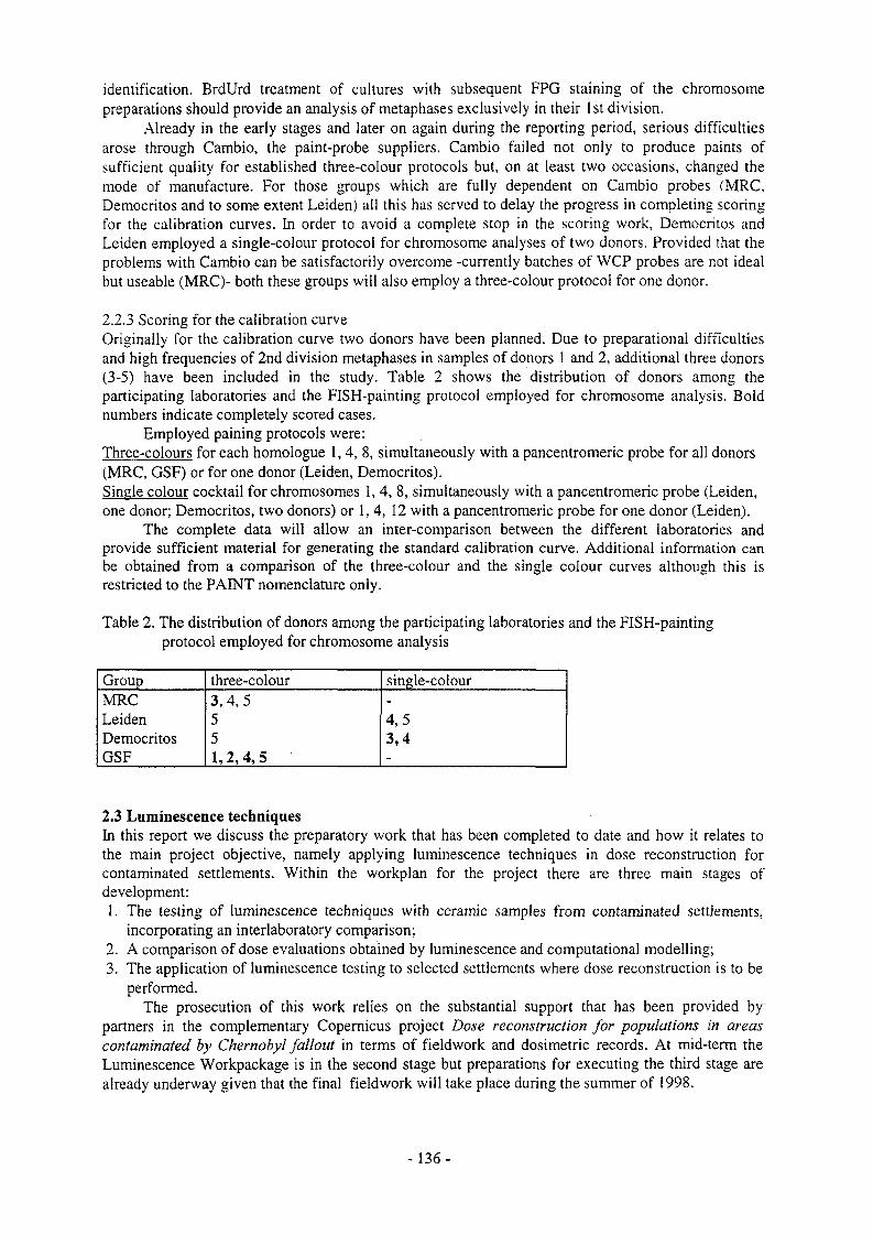
identification. BrdUrd treatment of cultures with subsequent FPG staining of the chromosome preparations should provide an analysis of metaphases exclusively in their 1st division.
Already in the early stages and later on again during the reporting period, serious difficulties arose through Cambio, the paint-probe suppliers. Cambio failed not only to produce paints of sufficient quality for established three-colour protocols but, on at least two occasions, changed the mode of manufacture. For those groups which are fully dependent on Cambio probes (MRC, Democritos and to some extent Leiden) all this has served to delay the progress in completing scoring for the calibration curves. In order to avoid a complete stop in the scoring work, Democritos and Leiden employed a single-colour protocol for chromosome analyses of two donors. Provided that the problems with Cambio can be satisfactorily overcome -currently batches of WCP probes are not ideal but useable (MRC)- both these groups will also employ a three-colour protocol for one donor.
2.2.3 Scoring for the calibration curveOriginally for the calibration curve two donors have been planned. Due to preparational difficulties and high frequencies of 2nd division metaphases in samples of donors 1 and 2, additional three donors (3-5) have been included in the study. Table 2 shows the distribution of donors among the participating laboratories and the FISH-painting protocol employed for chromosome analysis. Bold numbers indicate completely scored cases.
Employed paining protocols were:Three-colours for each homologue 1, 4, 8, simultaneously with a pancentromeric probe for all donors (MRC, GSF) or for one donor (Leiden, Democritos).Single colour cocktail for chromosomes 1, 4, 8, simultaneously with a pancentromeric probe (Leiden, one donor; Democritos, two donors) or 1, 4, 12 with a pancentromeric probe for one donor (Leiden).
The complete data will allow an inter-comparison between the different laboratories and provide sufficient material for generating the standard calibration curve. Additional information can be obtained from a comparison of the three-colour and the single colour curves although this is restricted to the PAINT nomenclature only.
Table 2. The distribution of donors among the participating laboratories and the FISH-painting protocol employed for chromosome analysis
Group three-colour single-colourMRC 3,4,5 -
Leiden 5 4,5Democritos 5 3,4GSF 1, 2, 4,5 -
2.3 Luminescence techniquesIn this report we discuss the preparatory work that has been completed to date and how it relates to the main project objective, namely applying luminescence techniques in dose reconstruction for contaminated settlements. Within the workplan for the project there are three main stages of development:
1. The testing of luminescence techniques with ceramic samples from contaminated settlements, incorporating an interlaboratory comparison;
2. A comparison of dose evaluations obtained by luminescence and computational modelling;3. The application of luminescence testing to selected settlements where dose reconstruction is to be
performed.The prosecution of this work relies on the substantial support that has been provided by
partners in the complementary Copernicus project Dose reconstruction for populations in areas contaminated by Chernobyl fallout in terms of fieldwork and dosimetric records. At mid-term the Luminescence Workpackage is in the second stage but preparations for executing the third stage are already underway given that the final fieldwork will take place during the summer of 1998.
- 136-

Before discussing the work in more detail we note here that the dose. Dl, evaluated using luminescence techniques with mineral inclusions in ceramics is the dose absorbed by mineral inclusions (quartz) and comprises the natural background dose that has accrued since the manufacture of the brick and the cumulative gamma dose arising from fallout, D%, i.e.,
Dx = Dl - A(R(x +R(3 +Ry+R c)
where. Dx = cumulative gamma dose received by the ceramic due to artificial radioisotopes (fall-out), A = sample age in years; (Ru, Rg, R, Rc) = effective alpha, beta, gamma and cosmic annual dose, respectively, due to natural sources of radioactivity.
Evaluations of DL for quartz inclusions in ceramic can be related to dose in air at an external reference location by the use of conversion factors that are derived from computational modelling. Such conversion was developed in previous work by this group and is being further developed in parallel with the experimental work in this project.
In terms of the task of delivering values of cumulative dose in air due to fallout (including sources that are in the form of either transient clouds or particulate contamination), there are four main interconnected compartments of work that we have undertaken: i) fieldwork and sampling, ii) the evaluation of DL , iii) the evaluation of the cumulative natural background dose and iv) conversion to dose in air. Deployment of the method on the scale of a settlement is a major task that we will tackle in 1998/99.
A large part of the work discussed in this report concerns dose evaluations in brick. This is largely due to the predominance of that material at the sites studied so far. However, we have also made developments in techniques appropriate for porcelain and these are available should suitable material be obtained from settlements studied in the final stage of the project.
2.3.1 IntercomparisonsThe aim of the intercomparisons discussed below was to test the performance of luminescence techniques in evaluating Dl at levels of external fallout dose broadly comparable to levels of accrued natural background i.e. ca 50 mGy for 20 year-old samples.
A) 100 mGy intercomparison with brickThree samples of brick that had been obtained in 1995 from the settlements of Novozybkov and Jarovshina in the Bryansk region of Russia were tested: NZ95-1, a Garage, Novozybkov; NZ95-2, a Pioneer Camp, Novozybkov; JA95-I, a School, Jarovshina. Slices of the three bricks were circulated to project partners (that have latterly included Copernicus contractors) for dose evaluation. It was agreed that both TL and OSL techniques should be employed. In addition to evaluating the dose absorbed in the sub-surface material for this intercomparison, depth-dose profiles were also obtained to investigate their use in establishing the nature of the external photon irradiation due to fallout. Six-point depth-dose profiles were obtained to a depth of 100 mm and the absorbed dose evaluations in the sub-surface slices of the three bricks were in the range -100 - 160 mGy, as anticipated. Figs la- c show, for each brick sample from the three locations, the evaluation of DL as a function of depth from the front surface of the brick. The thickness of the slices that were crushed and processed are indicated by horizontal bars; the uncertainty in the accrued dose evaluation is given by the vertical bars. The following aspects of the results are to be noted:I. CIEMAT, Durham and GSF employed TL procedures while Risp used OSL. For each set of results there is generally good agreement between different laboratories in the evaluation of DL. For the depth ranges 5-20 mm and 90-110 mm the standard errors associated with the weighted averages of the DL determinations for each brick are better than ±5%. However, in some cases (e.g. NZ95-1; 5-20 mm) this masks the occurrence of one or more individual evaluations which fall significantly outside these limits, as confirmed by the application of a chi squared test. Further examination of possible sources of unidentified systematic error is required; until such analysis is completed the
- 137-

iOVOZYBKCV NZ35-1 NovozyOkov NZ95-2B
■"MAT o --V! O <;:;k
RJIAM A XISO A rfj.su
.11!
DEPTH (mm)
Figure la.Depth-dose profile for NZ95-1.
Figure lb.Depth-dose profile for NZ95-2B
Jarovshina JA95-1
□ CIEMAT O I'.SF
«> UVttHAM ^ R1.W
DEPTH (mm)
<S)§>t—<cl
DEPTH (cm)
Figure !c.Depth-dose profile for JA95-1.
Figure Id.Comparison of experimental and calculated depth-dose profiles in brick from a large garage in Novozybkov; the step plot has been used to distinguish experimental data points and does not indicate slice thickness (-4mm).
- 138

Table 3.
Location Cumulative fallout dose (mGy)
5-20 mm
Cumulative fallout dose (mGy)90-110 mm
Cumulative natural background dose in
brick (mGy )
Age(years)
NZ95-1 100+10 23±6 50+5 16NZ95-2 75±7 22±7 63±6 23JA95-1 27±8 17+9 80±8 25
N.B. These are interim data and that uncertainty values reflect precision only.
root mean standard deviation is a better measure of the precision obtained in this comparison and for the determinations discussed above it falls within the range ±5% to ±12% of the average dose value. We note that there are some other cases (e.g. within the depth range 30-90 mm for sample NZ95-2B) where differences in dose evaluations obtained by different laboratories lie outside the given experimental errors and again, these may indicate as yet unidentified sources of systematic error. Nonetheless, this is the first time an intercomparison has been completed at such levels of dose and represents an encouraging level of agreement.2. The data for NZ95-1 exhibit the type of depth-dose profile that is to be expected where there is a source of external gamma radiation which is progressively reduced due to attenuation by the brick. The same can be seen in NZ95-2B, but there is a greater degree of scatter between the results. The lack of significant reduction of DL with depth (JA95-1) indicates an external gamma dose contribution that is small compared with the level of cumulative natural background dose - see para. 5 below.3. The preliminary estimates of the cumulative natural background dose for the three bricks based on the indicated ages are given in Table 3. The values are derived from experimental measurements of dose-rate and radioisotope concentration in ceramic and in-situ TLD measurements; these data will be incorporated with those for 1997 samples in a dose-rate intercomparison.Subtraction of the cumulative natural background dose from the weighted average total accrued dose (Dl) has been performed for two depth ranges corresponding to 5-20 mm and 90-110 mm to yield the cumulative dose due to fallout - see Table 3. From calculation we expect the radiation dose at the surface of the brick due to Cs-137 fallout to be reduced by a factor of ~7 if the fallout is distributed on the ground and by a factor of ~3 if Cs-137 fallout is located on the walls of the building relative to the dose at a depth of 10 cm from the external surface. A higher resolution comparison of experimental and theoretical depth-dose profiles obtained with one set of data is shown in Fig Id.4. From the cumulative fallout dose estimates we note that:Novozybkov NZ95-1 & -2. The reduction in fallout dose for the two depth ranges by a factor of 4-5 falls between the reduction factors predicted by calculation for fallout distributed on the ground and on the walls. For locations such as these in wooded locations we expect an 'intermediate' factor to apply - the data shown in Fig Id appear to be consistent with this configuration.Jarovshina JA95-1. The depth-dose profile for a brick from an uncontaminated area is not expected to be flat but to dip to lower values for depths <30 mm. Thus a 'flat' profile may reflect a small fallout dose contribution. From the experimental results, after subtraction of the cumulative natural background dose, the fallout dose is significantly above a null value at both depths, indicating a gamma dose contribution arising from fallout. However, our estimation of the natural background dose may require further adjustment since the topsoil was removed during clean-up procedures following the accident.To obtain a reduction factor of ~7 predicted by calculation (open ground is adjacent to JA95-1) for the two depth ranges, the natural background dose would need to be 15 mGy higher (95 mGy), resulting in a cumulative fallout dose of 13 mGy for a depth range of 5-20 mm. By making such adjustments it can be seen that: a) we are approaching the limits of resolution of fallout dose using current procedures and b) the high level of sensitivity of these calculations to the level of background dose.
- 139-

For wider scale work with brick in a settlement, an experimental protocol that includes the measurements with ceramic from two depth ranges is likely to be adopted.
B) Intercomparison: porcelainPrior to intercomparison measurements, each laboratory calibrated their beta radiation source using porcelain slices provided by the Oxford Laboratory and exposed to a dose of 300 mGy of gamma radiation at the Secondary Standards Laboratory, GSF. For the intercomparison, two samples were circulated: they comprised a sample of a light fitting from an external location in Pripyat (DNH-1) and a sample of a toilet cistern lid taken from an interior location in Pripyat. Both TL and OSL techniques were applied for the evaluation of accrued dose and in a preliminary assessment the mean accrued doses (of determinations by four laboratories) obtained were 102±2 mGy for the interior cistern lid and 1.96+0.11 Gy for the external light fitting, providing initial indications of good agreement.
2.3.2 Field reconnaissance and sampling fieldtripFieldwork was undertaken in Russia and Ukraine to meet two project objectives in the second and third stages to:1. undertake a comparison of dose estimates obtained by luminescence and modelling (using data
from contemporary soil analyses);2. obtain luminescence dose evaluations for ceramics in a fully or partially inhabited and
contaminated settlements where dose reconstruction was planned.Reconnaissance trips were performed to locate potentially suitable sites and buildings; in April
1997 to Ukraine and in Russia during the Spring of 1997. These visits enabled the settlements of Visnanje and Narodichy in Ukraine and Zaborie and Starry Vishkov in Russia to be identified as potentially suitable from the point of view of ceramic sampling and modelling. Visnanje and Zaborie are highly contaminated settlements that were promptly evacuated following the accident and thus considered to be most suitable for a luminescence-modelling comparison; the settlements of Narodichy and Starry Vishkov are partially inhabited and fall into objective ii) above.
During the reconnaissance trip in Ukraine it was possible to obtain a number of ceramic samples and preliminary assessments of suitability and dose evaluations were presented at the Clustered Contractors Meeting held in Kiev in June 1997. The luminescence (TL) signals were measurable with samples from both settlements and the reproducibility of the data were good; the values of DL (sub-surface material) were in the region of 500 mGy and 100 mGy for Visnanje and Narodichy respectively.
In the latter part of August 1997, a successful joint Framework FV-Copemicus overland fieldtrip was completed, commencing in Moscow and ending in Kiev. An examination of buildings within the above settlements was performed and ceramic and soil samples were collected from 12
buildings. The majority of the samples collected were brick, but some porcelain was included where the date of manufacture and location were suitable. At each site basic physical survey and dose-rate measurements were performed; also ALO3 TLD dosemeters were implanted at sampled locations; the dosemeters will be collected next summer.
2.3.3 Luminescence vs modelling comparison A) Progress with luminescence measurementsFollowing the August fieldtrip, measurements with bricks from Zaborie and Visnanje were given first priority and depth-dose data were obtained for 9 samples involving determinations for over 50 slices; examples drawn from those discussed in individual contractor reports are shown in Figs 2a and 2b which were obtained using OSL and TL techniques respectively. The preliminary experimental data shown in Fig 2b for sample VI-97-8 from Visnanje are consistent with the calculated depth-dose profile for a Cs-137 ground-distributed source. The sampled walls face open land and the cumulative natural background dose has not so far been subtracted, but constitutes less than 10% of the total at the sub-surface location. Progress in testing material from these two sites will be reported at the next Contractors Meeting to be held in April.
- 140-

SCO
ZB 97 2-1
O 300- ZB 971-1
oo>
<LUCSL
1
0.75
0.5
0^5
0
o ViSNANJE 97-8-t
7
VO V
o V
sVo
9V
o Experimental
v Calculated
0 2 4 6 8 10 12
DEPTH (cm)
Figure 2a. Figure 2b.Preliminary depth dose profiles obtained for samples Comparison of experimental and calculated depth- from Zaborie using the OSL single aliquot dose profiles for a brick from a farm outbuildingregeneration method. The error bars are within the in Visnanje. The value of Dl for the sub-surfacesymbols. ceramic (8-12 mm) was 560+30 mGy; the random
experimental errors fall within the symbols.
B) Progress with modelling calculationsA number of Monte Carlo calculations have been performed for different source energies and configurations to calculate depth-dose profiles in brick walls at a height of lm and, in the case of ii), 10m above ground level. These include; i) sources of energies 140, 352, 583, 1461 and 2615 keV distributed within a 45 cm thick brick wall; ii) sources of the energies as in i) distributed in a 20 cm thick layer of soil adjacent to a brick wall and extending to 50m from the wall; iii) sources of energy 662 keV distributed to a depth of 1 cm and 10 cm in soil adjacent to the wall and extending to 50m from the wall. These calculations will contribute to the luminescence-modelling comparison now underway.
2.3.4 Retrospective dosimetry in inhabited settlementsWork is in progress concerning the testing of samples from the potential study sites of Starry Vishkov and Narodichy; it is anticipated that larger scale sampling will take place during a fieldtrip in August 1998.
2.3.5 Developments in technique and instrumentationA number of fundamental investigations and developments in technique have been made which improve dose and dose-rate evaluations with ceramic materials. These developments are summarised below and further details are given in the reports of individual contractors:
- 141 -

Modified stimulation and detection systems for OSL and the use of shorter wavelength stimulation have improved signal detection efficiency using OSL (Figs 3a-b). Different OSL dose evaluation protocols have also been developed, based on the use of single aliquots. Dose evaluations performed with quartz grains that have not been subjected to HF etching also opens up the possibility of avoidance of the use of hazardous chemical treatments in sample preparation. Investigations of predose behaviour have resulted in new pre-dose OSL and 230°C TL pre-dose procedures which provide alternative techniques for dose evaluation in cases where standard approaches are found not to be suitable. A new effect involving the restoration of pre-dose sensitization by UV exposure has been observed and is to be investigated. The potential of single grain measurements using a CCD camera is also under investigation. The use of highly sensitive AI2O3 dosemeters with OSL and TL measurement procedures has been further developed for on-site gamma TLD and for 13-TLD measurements.
A C
0 5 10 15Exposure Time (seconds)
Figure 3 a.Schematic diagram of the new blue LED OSL unit. Key: A, detection filters; B, PM tube; C, feed-back lightguide; D, blue LEDs; E, cut-off filter; F, quartz lens; G, heating element; H, Sample; I, quartz glass vacuum seal.
Figure 3b.OSL decay curves from a 4 mg quartz sample (brick) with an ED of 117 mGy using (i) green lightstimulation (28 mW/cm2) and (ii) blue LEDstimulation (15 mW/cm2). The effect of blue light stimulation is clearly seen.
- 142-

2.4 Dose modelling2.4.1 External doses of the population living in contaminated territoriesAfter a deposition of a radionuclide k on the ground, the absorbed dose rate in air is given by the product of the activity per unit area Ak(t), a dose rate conversion factor gk and a function rk(t) which describes the attenuation of the dose rate due to the the shielding by the soil and other materials on the path from the decaying nuclide to the point of interest, in general 1 m above the air-ground interface. It was decided to use dose rate conversion factors gk for plane sources below a soil layer with a mass per unit area of 0.5 g em ".
Measured data on the nuclide-specific attenuation function rk(t) are summarized in Table 4. According to these data, the attenuation of the dose rate in air is similar for the measured radionuclides, which were l3!I, l03Ru, lj4Cs and l40Ba. Accordingly, the same attenuation fuction r(t) will be used in the model for all radionuclides. Such a function has been derived in the frame of the JSP 5 model for cesium and for three distance categories from the Chernobyl reactor plant: less than 100 km, 100 km to 1000 km, and more than 1000 km.
Time after deposition (days)
Attenuation function rk(t) "'I "»Ru '"Cs '«La
37+3 1.09+0.12 0.96±0.09 1.01+0.05 0.88±0.1869+7 0.95+0.16 0.98+0.11 -155+9 0.91±0.18 0.94+0.13 -
Table 4. Values of the nuclide-specific gamma dose rate in air over grassed areas relative to an infinite plane source in the air-ground interface below a soil slab of 0.5 g-cm"2, as measured in Southern Bavaria after the reactor accident of Chernobyl. Average values and one standard deviation of five measurements are in given in each case.
The model of the external effective dose E;(t,At) of a population group i within a time interval At will be calculated according to the following equation:
Ej(t,At) = At * X (py(t) . X (kik* fjk(t) * r(t). gk»Ak(t))),
where fjk(t) is the gamma dose rate in air at a location j relative to a grassed area, py(t) the occupancy factor of the population group i at locations of type j, and kik the conversion factor from the gamma dose in air to the effective dose. Doses to specific organs can be calculated by apropriate conversion factors which have been published in the literature.
Concerning radionuclide activities per unit area, Ak(t), extensive data sets are available for iy7Cs in each contaminated settlement. According to recently published data, values in Table 5 can be used for Russian territories for nuclide-specific acivities per unit area relative to l37Cs. A review of the data for Ukraine, revealed that the situation his here more complex due to the changes of the relative radionuclide activities per unit area within the first 100 km around the Chernobyl reactor. These data need a further close inspection.
Location factors depend on the radionuclide of interest. Measurement results from Russia, Southern Bavaria, Denmark and Sweden has been compiled and will be analysed in the next period. Further considerations are needed to define appropriate population groups and occupancy factors. Preliminary comparisons of the JSP5 model with TLD measurements of individual doses in Russia were performed, indicating a good agreement.
- 143 -

Radionuclide Bryansk (1118 settl.) Kaluga (358 settl.) Orel (895 settl.) Tula (1304 settl.)95Zr 0.07 0.06 0.06 0.0695Nb 0.080 0.068 0.068 0.068"»Ru 1.30 1.29 1.31 1.36106Ru 0.44 0.42 0.43 0.49'%Sb 0.06 0.06 0.06 0.06mI 3.13 3.25 3.27 3.34
'3"ts 0.54 0.50 0.50 0.50"°Ba 0.24 0.28 0.28 0.29140La 0.29 0.31 0.33 0.34144Ce 0.063 0.054 0.054 0.054132Te 0.73 0.76 0.76 0.78
133I 0.002 0.002 0.002 0.002
'"Cs 0.11 0.11 0.11 0.11
Table 5. Radionuclide activities per unit area relative to 137Cs on 10 May 1996 in four oblasts of Russia. The number of settlements for which spectrometric measurements have been performed is indicated. The first block of data gives average values of measurements, the second block data derived with the help of release term data.
2.4.2 External doses of evacueesDeterministic and stochastic models for individual external gamma dose reconstruction of the population evacuated from Pripjat have been completed. The models are based on data sets of dose rate measurements performed after the accident until the evacuation time, individual behaviour data obtained by a questionnaire survey and a determination of location factors for typical building types and environments. The stochastic models allow for a determination of uncertainty distributions of the individual external doses from an assessment of the uncertainty of the data, using Monte Carlo sampling methods for the calculation of the uncertainty propagation. Individual effective doses due to external gamma exposure received until the time of evacuation and their respective uncertainty distributions were calculated for 12632 evacuees.
Figure 4 shows uncertainty distributions of the external dose for two individuals: A 6 year old child which stayed most of the time indoors in the sector of Pripjat with the lowest dose rates until it was evacuated after 36 hours and a 21 year old city services worker evacuated after 44 hours, who
iO 100 15Effective dose (mSv)Effective dose (mSv)
Figure 4. Relative frequencies of uncertainty distributions of effective dose due to external gamma exposure received until evacuation from Pripjat for a child (left) and for a city services worker (right).
- 144-

stayed 75% of the time outdoors in the sector with the highest dose rates. This individual received with a value of 75 mSv for the median of the uncertainty distribution the largest external dose until evacuation of the evacuees who were considered.
Results of individual effective doses due to external gamma exposure obtained with the deterministic and stochastic models are summarized in Table 6. One can see that a close agreement was obtained between the distribution of individual doses calculated with the deterministic model and the distribution of the medians of the individual uncertainty distributions. The 95 percentiles of the individual uncertainty distributions resulted to be higher than the medians by a factor of about 2 for the lower dose range and a factor of 1.5 for the higher doses. The value of 10 mSv determined for the arithmetic mean of the distribution of the medians of the individual dose distributions is by about 13 % lower than the mean effective dose due to external radiation determined in a first assessment with a deterministic model by (Likhtarev et al. 1994, Health Physic. 66, 643-652).
A deterministic model developed for the population evacuated from settlements of the 30 km zone, which includes a reassessment of dose rate data and mutually consistent time and space interpolations of dose rates, was used for the determination of individual effective doses due to external gamma exposures of 14084 evacuees from these settlements. Figure 5 shows the cumulative frequency distribution of external doses obtained for this population. Average doses were considerably higher for evacuees who had stayed longer times outdoors and for inhabitants of villages with higher dose rates. Table 7 gives results for individual dose distributions of evacuees from several of the settlements.
EndpointDeterministic model
Effective dose (mSv)Stochastic model
medians 95 percentilesArithmetic mean 10.1 10.0 16.7Median 8.9 8.9 15.595 percentile 24 23 35
Table 6. Arithmetic mean, median and 95 percentile of the distribution of individual effective doses due to external gamma exposures of 12632 evacuees from Pripjat as obtained with a deterministic model in comparison with the corresponding results obtained with a stochastic model for the medians and 95 percentiles of the individual uncertainty distributions.
Effective dose (mSv)
Figure 5. Cumulative frequency distribution of individual effective doses due to external gamma exposure of 14084 evacuees from settlements of the 30 km zone. Arithmetic mean: 15.9 mSv; 95'percentile: 68 mSv. For 120 evacuees doses of more than 100 mSv were obtained.
- 145-

Table 7. Arithmetic mean, median and 95 percentile of the distribution of individual effective doses due to external gamma exposures for several settlements of the 30 km zone. Also indicated are the distance and co-ordinates of the settlements relative to the nuclear power plant and the number of individuals of each settlement for which doses were computed.
Settlement Distance(km)
Co-ordinates(km)
Individuals EffArithm.Mean
ective dose Median
(mSv)95
percentileChistogalovka 7.2 (-6.2, -3.6) 331 61 70 98Tolsty Les 24 (-24, -1.0) 408 52 60 81St. Schepelichy 12 (-11, 3.6) 209 21 2.2 31Mashevo 9.5 (1.4, 9.4) 162 75 79 96Kopachy 4.6 (0.2, -4.6) 432 45 53 66
Tschemobyl 15 (7.6, -13) 4558 6.0 6.0 14
2.4.3 Reconstruction of internal dosesFirst considerations were given to supplement the results on individual external doses of evacuees from settlements of the 30 km zone with ingestion and inhalation doses determined for population groups of the settlements. A model was developed to estimate the internal dose of the population evacuated from the 30 km zone (Figure 6). The model allows both, the deterministic and stochastic assessment of the doses to evacuees.
The work focused on the assessment of the input data required for the reconstruction of inhalation and ingestion doses. This work is kindly supported by the Radiation Protection Institute (Kiev, Ukraine) and the Institute for Radiation Hygiene (St. Petersburg, Russia), although no funds are allocated to these institutes for this purpose. The l37Cs-deposition is a key parameter for the assessment. Efforts were undertaken to establish a deposition map for the exclusion zone. This work is not finished and needs more refinement.
Radionuclide ratios were surveyed and assessed. One of the major efforts of the past year, therefore, was to establish these ratios from data in the literature, in unpublished reports, or in not easily available Russian literature. Considerable progress was obtained for ratios of several isotopes such as 134Cs/137Cs, 103Ru/l06Ru, l29mTe/132Te and 13II/133I. More work needs to be done on the ratios of radionuclides of different chemical species as there are considerable differences in the radionuclide ratios for the near and far field and also for the different plumes and wind directions. This relates in particular to less volatile elements such as cerium, strontium, ruthenium, and plutonium.
Very few data are available on radionuclide activity concentrations in air. For areas where the activity concentration integral is not available, an algorithm will be developed to assess it from the deposition data. The contribution of dry and wet deposition in the 30 km zone is very important for the estimation of the integral activity in air from deposition. However, in the 30 km zone, there was apparently no or very little rain during the period of the accident. Therefore, only dry deposition has to be considered to assess the activity in air.
An evaluation of the activity concentration integral in air was performed where available. Since this information is available only in very few sites of the near field zone (NPP site, Kyiv) and in several cities of the GIS and the Baltic states for the far field zone, other methods to derive the activity concentration integral in air were investigated. These included the possible derivation from the activity concentrations in various other environmental samples of the exclusion zone. For iodine, a survey showed ratios of 1:2 for particulate iodine to gaseous iodine in the sites where it was measured.
An assessment of the particle size distribution was made which is relevant for the inhalation dose factors and inhalable fractions of radionuclides. Particle size values were established for volatile and less volatile radionuclides. Preliminary work was performed on the particle size changes with increasing distance from the power plant. The dependence of inhalation dose factors as published in
-146-

ICRP 71 on particulate size was established and their influence on the methodology proposed for the derivation of inhalation doses from the assessment of deposition values investigated.
The only relevant foodstuffs to be considered at the end of April/beginning of May, are milk and leafy vegetables. Other foodstuffs, that caused considerable long-term contamination problems as potatoes, meat, fruit, berries and mushrooms were not contaminated at this time of the year. For the evacuees from the settlements, only the time until evacuation has to be considered for the assessment of the ingestion dose. There are no indications that people changed their consumption habits in the early phase as a consequence of the accident. Therefore, pre-Chernobyl consumption data are considered to be appropriate to assess the ingestion doses.
The dose will be assessed for the time period during which the population stayed in the contaminated area. It is not known that any countermeasures concerning the ingestion were recommended in this early phase in rural areas. The population the 30 km zone was not informed about the accident and its consequences. Therefore, no countermeasures have to be taken into account in the framework of this task.
Cs-137-Deposition
Deposition velocity for particles
Time integrated Gs-137-activity in air
RN/Cs-137 in air
RN-deposit on on grass and leafy vegetables
RN in vegetables |
RN in milk
RN-intake via ingestion und inhalation
Fraction of wetdeposition
Feed intake, transfer to milk
deposition
Wet RN* deposition
Consumption habits. breathing rates
Dry Cs*137*deposition
Time integrated RN-activity in air
Figure 6. Model to estimate internal doses of evacuees based on the Cs-137-deposition.
2.5 Evaluation of dose reconstruction outside NIS2.5.1 Review of dose reconstruction methodsIn the last ten years or more there has been a considerable effort devoted to dose reconstruction studies for various nuclear sites in the USA. This arose from public concern about the nature of these originally secret sites and the realisation that significant quantities of radionuclides had been released to the environment. A review has been carried out of the main US studies to determine the methods used to establish the radiation doses people received due to operations at these nuclear sites. The nuclear sites considered in the review were those at Femald, Hanford, Oak Ridge and Rocky Flats, together with the Nevada Test site used for nuclear weapons testing. In each case the review considered the following factors: the reasons the study was carried out; the nature of the releases considered, the timescales of any releases and the time period covered by the study; the numbers and characteristics of the population
-147-

considered in the study; the availability and use of environmental monitoring data; the approach used to determine doses and the consideration of uncertainties; the doses estimated and which of the radionuclides and exposure pathways considered were the largest contributors to dose; the use of other data or methods (such as chromosome analysis, EPR dosimetry and luminescence techniques) to support the dose estimates. A report has been prepared on this review and is in press.
All of the US studies considered took many years to complete and involved a number of man- years of effort. A major factor in carrying out the studies was public concern as to what happened at the nuclear sites, what radionuclides were released to the environment and what effect on health these releases might have. Some of the studies are linked to epidemiologic studies and consideration of whether public health measures are required in particular locations. A notable feature of the studies was the degree of public involvement and how successful this was. Scientific and technical meetings were open to the public and pamphlets and other literature were produced. The US agencies involved in the studies feel that it is vital to ensure that people have confidence in the studies and that this can only be achieved through openness and building trust with local communities and involved individuals.
Considerable emphasis was placed on determining the source term and characterising in detail the releases of radionuclides to the environment. For example, for the Hanford study thousands of original documents were studied to determine atmospheric releases of 13,I on an hourly basis over more than ten years. In contrast, some of the environmental modelling was less detailed. For example, in many studies simple equilibrium models were used to predict the transfer of radionuclides to food.
Doses were estimated for a number of different representative population groups and in some cases for identified individuals. Often cumulative radiation doses were calculated but there was limited discussion of what the doses meant for example through comparison with doses from other sources, such as natural background radiation or by carrying the study a stage further by estimating health risks. All of the studies were carried out with uncertainty analysis and with sensitivity analysis to determine the important parameters. This is very important as such studies, where doses are being estimated for people over a wide area and over an extended time period, will inevitably be uncertain.
The US dose reconstruction studies have concentrated on estimating radiation doses from releases of radionuclides to the environment using established environmental transfer models. Techniques such as EPR on teeth, chromosome analysis and luminescence on ceramics and building materials do not appear to have been used in any of the studies.
-148-

3. Summary of main achievements
3.1 EPR with teethAs a result of the 1st internal intercomparison on sample preparation techniques, all methods produced samples with no significant differences in properties of the background signal. The differences of the background signal (mean amplitude of six samples per lab) achieved by the individual methods were within 25%. The lowest background amplitude was obtained by semichemical treatment, probably due to the better removal of dentine and the use of larger grain size of up to 2 mm. The transferability between laboratories of the chemical and the semi-chemical sample preparation methods was successfully tested: in each case two labs were producing samples with comparable parameters of the background signal. Sample preparation and signal evaluation were found to be closely related. Therefore, protocols on the two working steps will be drafted together.
For the identification of the dosimetric signal the EPR signal has to be independent of the orientation of the grains in the magnetic field. This condition is fulfilled for enamel powders with grain size below 1 mm. For larger grain sizes the sample has to be rotated in the magnetic field or measured several times with intermediate shakings of the sample. The radiation induced 100 mGy signal was found to be reproducible within about 10%. For these low doses the uncertainty of mainly determined by the variability of the intrinsic C02' signal, which is due to natural radiation, medical exposures and intrinsic C02" concentrations in the enamel. In the 1st internal intercomparison it was found to be (65±35) mGy.
The 2nd internal intercomparison on sample preparation and spectrum evaluation has shown that all of the six involved methods are applicable. The variation in sensitivity of the used samples and the reproducibility of an individual sample as measured at the level of 1000 mGy was for all methods smaller than 10%. The difference in the sensitivity at 100 mGy compared to 1000 mGy was for four methods within about ±20%. For five of the methods the reproducibility of an individual sample at 100 mGy was within 26%.
The variation of the measured intensity of the dosimetric signal with time after irradiation was tested between 1 day and two weeks after irradiation. With the spectra subtraction methods an increase of the dosimetric signal of about 10% was measured within this period. By using the simulation method no change in signal intensity was found.
Front teeth were collected from residents in the age 13-71 years of an uncontaminated area in Russia. For residents younger than 50 years the measured dose in the front part of the teeth was about 100 mGy, the difference to the lingual part was less than 25%. For most of the samples from residents in the age 50-71 years a dose in the range 300-1000 mGy was obtained with the front part. This dose is in mean 7-fold higher than the dose obtained with the lingual part of the same teeth. The experiments will be continued to determine for all samples the individual radiation sensitivity of the different parts of the tooth.
First experiments were started to investigate the variation of radiation sensitivity for different tooth positions. Results indicate a tendency of increasing sensitivity from wisdom teeth to front teeth. The sensitivity of the mixed material of tooth positions 2 were found to be approximately 30% higher than the material from positions 7.
The reconstruction of individual doses by EPR revealed four cases (code no: 1154, 7411, 63714, 64370) from the upper Techa river with an absorbed dose of about 1 Gy. Absorbed doses in the range of 500-600 mGy was reconstructed for two persons (code no: 12460, 66612). By earlier measurements three further cases (code no: 27201, 29072, 69823) were identified by EPR to have received doses of about 1 Gy. In total for the comparison of FISH and EPR dose reconstruction for residents at the upper Techa river 7 cases were identified to have received doses of about 1 Gy.
3.2 Chromosome painting (FISH) in lymphocytesAberration scoring was performed by using both the S&S and PAINT nomenclature systems in parallel. First data proved to be suitable for generating dose-effect curves for various aberration parameters such as. translocations, dicentrics, minimal number of breaks, or 2A or 2B according to S&S. The use of multi-colour paint increases the information on the composition of exchange
- 149-
configurations, however difficulties arise in their interpretation and quantification. Actually, the criteria for aberration classification differ for each of the applied nomenclature systems. Whereas PAINT is strictly descriptive, S&S is based on mechanistic aspects of aberration formation. In principle. S&S applies only to single colour paints of complete forms because signal interrelationships must be taken into account for aberration classification. Consequently, but not unexpectedly, numerical differences arise between the nomenclature systems. To overcome these difficulties, criteria for a harmonised and pragmatic aberrations scoring had to be discussed. At three work group sessions, the following issues have been addressed:• Evaluation of multi-colour patterns by S&S• Dealing with "incomplete" or one-way exchanges• Dealing with simple and higher order Complexes• Selection of particular aberration parameters (2A, 2B, colour junctions, minimal number of
breaks, or according to PAINT the total number of t(Ab), t(Ba) or further combinations, due to three-colour painting) for an application to dose estimation.
Two discussion documents produced by J. Savage (MRC), prior to or following these meetings have been circulated amongst the FISH group members:The first document "One way exchanges'XAnnex 1) provided a critical analysis of the origin of translocations or dicentrics arising either from incomplete or terminal exchanges. Six distinctive painting patterns can now be easily discriminated. In the scoring protocols, one-way types I-IH (according to S&S) can be added to the category of symmetrical complete translocations (2B) and one-way types IV-VI to complete dicentrics (2A).The second document " Thoughts and comments on the scoring of aberrations using FISHpainting- and the synthesis of scores for the production of dose response curves" (see Annex 2) gave for a hypothetical collection of aberration paint-pattems, the results of three scoring methods, and discussed possible ways of reconciling conflicts, when this was possible, to ensure consistent scoring amongst the participating groups.
A consensus on a number of the indicated issues related to the application of the two nomenclatures was reached and will be certainly of great value for collating the data from the different groups.
3.3 Luminescence techniquesAt mid-term the project is well advanced in terms of the completion of fieldwork, the procurement of samples and preparation for applying the method to contaminated settlements. The luminescence techniques employed have been shown to have the capability to measure accrued dose in ceramic down to cumulative natural background levels for brick of -20 years in age (i.e. typically 50 mGy). In undertaking an intercomparison of accrued dose evaluations in brick and porcelain from contaminated settlements, the preliminary evaluation of cumulative fallout dose for brick samples indicate that encouraging consistency has been obtained between laboratories at levels of dose that have not previously been subjected to such an intercomparison.
A substantial amount of fieldwork in Russia and Ukraine has been undertaken with the collaboration of Copernicus project partners. This work has enabled settlements to be identified and sampled with the aim of: i) performing a comparison of dose estimates obtained by luminescence and computational modelling with soil contamination concentration analyses and ii) performing a dose evaluation exercise in populated contaminated settlements with a view to providing benchmark dose values for use in dose reconstruction.
The development of the use of depth-dose profiles in bricks from contaminated settlements and investigation of their potential for the assessment of the nature of the time-integrated external gamma field forms a distinctive contribution to the research field. The requisite Monte Carlo modelling calculations are being advanced to support the experimental studies in terms of source configuration and energy.
Also underpinning the above developments, substantial progress has been made in the development of new luminescence techniques and procedures which enhance dose and dose-rate evaluations with ceramic materials.
- 150-

3.4 Dose ModellingThe model for external exposures of the population living in contaminated areas was established. Values and uncertainty distributions for the corresponding parameters are needed for the period 1986 to 1989. The JSP 5 model will be applied for the time period 1990 to 2056. For this period uncertainty distributions need to be derived.
Main achievements concerning these tasks were the determination of relative radionuclide activities per unit area in Russian contaminated territories and an evaluation of data on radionuclide- specific gamma dose rates in air as a function of time after deposition.
Concerning location factors, as a first step frequency distributions of measured locations for rural environments in 1989, 1993 and 1994, and for urban environments in 1987, 1989, 1990, 1991, 1993, 1995 were described by lognormal distributions. The dependence of the average values on the time after deposition was approximated by analytical functions.
Individual effective doses due to external "/-exposure received until the time of evacuation and their uncertainty distributions were calculated for 12632 evacuees from Pripjat. The value of 10 mSv determined for the arithmetic mean of the individual dose distributions is by about 13 % lower than the mean effective dose due to external radiation determined in a first assessment with a deterministic model by Likhtarev et al. (1994).
Using a deterministic model, individual external doses were calculated for 14084 evacuees from settlements of the 30 km zone. The distribution of individual doses has an arithmetic mean of 15.9 mSv and a 95 percentile of 68 mSv; for 120 evacuees doses of more than 100 mSv were obtained. Dose distributions were determined for different age and profession groups, as well as for each of the settlements.
A model was developed to assess internal doses via inhalation and ingestion of the population evacuated from the 30 km zone. The starting point is the Cs-137-deposition for different locations in the 30 km zone. The literature and other available data were used to derive and to evaluate model parameters that are needed for the assessment such as radionuclide ratios, particle size distributions, the fraction of dry deposition, agricultural practices and consumption habits.
3.5 Evaluation of dose reconstruction outside NISThe main achievement is to complete a detailed review of dose reconstruction studies carried out in the USA. A report on the review is in press which outlines the various studies and discusses the methods used. This information will be used together with experience gained in carrying out a dose reconstruction study in the UK and the methods used in the CIS countries as an input into the remainder of the workpackage to evaluate dose reconstruction methods.
- 151 -

4. Research to be performed
4.1 EPR with teethIn the second half of the project the reconstruction of individual doses for remaining 136 cases needs to be done in co-operation with the INCO-COPERNICUS project. Among these. 60 cases are Mayak workers 40 cases are residents from Ozyorsk as a control group, and 36 cases from the middle and lower Tec ha river. The measurements of the samples from Mayak and Ozyorsk will be started in January 1998 by GSF and IMP and in June 1998 by ISS. The purpose is i) to test the applicability of EPR dosimetry with teeth for reconstructing external doses in the range of 100 to 300 mGy which occurred three decades ago and ii) to contribute to a better understanding and quantification of the high exposures that occurred among the MAYAK workers during the first decade of operation of the facility.
The measurements of the samples from the Techa river by GSF and IMP will start in the second half of 1998. The test persons were predominately exposed due to incorporated strontium. The purpose is to study the applicability of EPR dosimetry for such exposure conditions.
The protocol for sample preparation and determination of the dosimetric signal needs to be completed. It will be supplemented by a protocol for the calibration of EPR tooth dosimetry. The applicability of the protocols will be tested in the ‘2nd International Intercomparison on EPR Tooth Dosimetry’ starting in June 1998. This exercise might lead to an improvement of the protocols for EPR tooth dosimetry.
The remaining fundamental studies include the investigation of the contribution of x-ray diagnostic examination and energy dependence of the radiation induced EPR signals. Further investigations on the variation of radiation sensitivity in dependence on tooth position are required. They are essential for the estimation of errors if a dose calibration curve is used for EPR dose reconstruction.
4.2 Chromosome painting (FISH) in lymphocytesThe immediate priority is to complete scoring for the calibration curves. This can be achieved in the early 1998. Actual scores from the participating groups have to be collated and statistical comparisons made with a view to generating individual dose-effect curves and the standard calibration curve. Future work will be directed on scoring of Mayak and control samples.
According to Workpackage 1 of the Technical Annex of the INCO-COPERNICUS project, a total of 100 tooth samples should be analysed for the Mayak workers and for inhabitants of Ozyorsk. According to Workpackage 2, 83 of these cases should be analysed also by FISH.
At the Work Group Meeting in Syros Island, it has been decided to put more weight on achieving results for the total number of the indicated cases rather than to start again with intercomparison. This can be more efficiently achieved from the in vitro data. Thus each of the participating groups (Leiden, MRC, Democritos, GSF) will be involved in scoring of 21 cases according to the following list:
Group Cases Cells/caselow dose, 0.3-1 Sv after 1961 32 2000
high dose, 1-3 Sv 20 1000
control, Ozyorsk 32 1000
total 84
The high number of cells requires high quality metaphase preparations which will be produced by G. Snigyriova, MRIDS, Moscow. To date fixed cell suspensions from 23 Mayak cases have been sent to GSF and distributed among the participating groups for scoring.
- 152-
4.3 Luminescence techniquesIn the second half of the project, the main areas of work to be completed include: in collaboration with Copernicus partners, further fieldwork in Russia (Bryansk and Techa River regions) and Ukraine, the testing of field samples on a settlement scale, deconvolution of dose evaluation results for further modelling calculations in dose reconstruction and the production of a methodology for the application of the luminescence method.
During 1998, a full analysis of the ’100 mGy’ intercomparison data from brick and porcelain, and the luminescence-modelling intercomparison will be completed. The final phase of fieldwork in Russia (Bryansk region) and Ukraine is planned to be executed in the Spring and in the Summer, the interest being in populated settlements where dose reconstruction is required. The experimental work will be performed with brick and porcelain, as appropriate, and both TL and OSL measurement procedures will be employed. This work aims to produce luminescence evaluations with ceramics and, by means of conversion with the aid of modelling calculations, the production of dose estimates at reference locations. In addition to the production of papers and reports, the drafting of a methodology for survey, sampling and dose evaluation in settlements will commence in 1998 and finalised in the latter months of the project. Finally, in 1998 the group will be involved in the production and execution of a plan for dose evaluation intercomparisons for ceramics from the Techa River study area.
4.4 Dose modellingFor the development of a general model to assess external doses in contaminated areas, pulse height distributions of early spectral measurements of deposited radionuclides shall be analysed to verify existing results. In Ukraine, areas typical for given radionuclide compositions will be defined. The model for the y-dose rate in air based on radionuclide deposition and attenuation due to migration will be compared with measurements in the period 1986 to 1989. The contribution of radionuclide deposition inhomogenities on the variability of location factors shall be assessed. Based on these results, the variability of y-dose rates at various locations shall be modelled.
Representative distributions of occupancy factors for four population groups in Ukraine and in Russia will be derived. Model results will be compared with measurements of absorbed doses in TLDs worn by inhabitants of contaminated areas, with EPR measurements of absorbed doses in tooth enamel, and with luminescence measurements of absorbed doses in building and household materials.
The deterministic and stochastic calculations of the external exposure of the population evacuated from the settlements in the 30 km zone will be completed. Individual uncertainty distributions of the external dose will be estimated.
For the same settlements, the internal doses due to inhalation and ingestion of milk and of green vegetables until the time of evacuation will be estimated. The results of the estimation of external and internal doses will be combined in order to achieve an integrated assessment of the dose received by the population evacuated from the 30 km zone after the Chernobyl accident.
4.5 Evaluation of dose reconstruction outside NISThe information collected in the first phase of the work programme will be further evaluated to draw conclusions about methods for carrying out dose reconstruction studies. From this a general methodology for dose reconstruction will be established. The methodology will cover the purposes of such studies, the exposure pathways to be considered, guidance for using environmental measurement data together with habit and other data requirements.
4.6 Comparative analysesThree sets of the dose reconstruction results will be comparaticely analysed:i) EPR and FISH results for MAYAK workers and upper Techa river residents;ii) EPR and modelling results for inhabitants of Russian settlements contaminated by the Chernobyl accident; andiii) Luminescence and modelling results for inhabitants of Russian and Ukrainian settlements contaminated by the Chernobyl accident.
- 153 -

5. Publications
Adamiec G., Stoneham D., Goksu H. Y. (1997) Accident dose estimation using porcelain. A comparison between different TL methods. Radiation Measurements 27, 89-392.
Bailiff I.K. (1997) Retrospective assessment of external dose using luminescence techniques. In Effects of low-level radiation for residents near Semipalatinsk Nuclear Test Site. Proc. of the Second Hiroshima International Symposium, Hiroshima, July 23-25, 1996. (Eds M. Hoshi, J. Takada, R. Kim and Y. Nitta. Res. Institute for Radiation Biology and Medicine, Hiroshima University.
Bailiff I.K. (1997) Retrosr ctive dosimetry with ceramics. Radiation Measurements., 27, 923-94Botter-Jensen L.G997) Luminescence techniques: instrumentation and methods. Radiation
Measurement,.27,749-768.Bqtter-Jensen L. (1997) Retrospective radiation dosimetry using optically stimulated luminescence on
natural materials and ceramics for assessing population doses in nuclear accident areas. In refereed Proceedings of the 7th meeting of the Nordic Radiation Protection Society (NRPS) held 26-29 August 1996, Reykjavik, Iceland. Editors: Tord Walderhaug and Einar Pall Gudlaugsson, Geislavamir Rikisins, Laugarvegur 118, 150 Reykjavik, Iceland.
Bqtter-Jensen L., Agersnap Larsen N., Markey B.G. and McKeever S.W.S. (1997) ALC^C as a sensitive OSL dosemeter for rapid assessment of environmental photon dose rates. Radiation Measurements 27, 295-298.
Bulur E, Goksu H.,Y. (1997) Optically Stimulated Luminescence from a- A1203:C Using Green Emitting Diodes. Radiation Measurements 27, No 3 pp. 479-488.
Bulur E., Goksu, H.Y. IR (1997) Stimulated Luminescence from ZnS and SrS Based Storage Phosphorous: A Re examination with Linear Modulation Technique. Phys.stat.solfa) 161, R9.
Correcher V, Gomez Ros J.M. and Delgado A. (1997) On the Dosimetric Properties of Albites. Application to Accident Dose Reconstruction. Proc. Int. Conf. Radiation Dosimetry and Safety. pp334-338 (Taipei,Taiwan 1997).
Duller G.A.T., Bptter-Jensen L. and Markey B.G. (1997) A luminescence imaging system based on a CCD camera. Radiation Measurements 27, 91-99.
Goksu H.Y., Stoneham D., Bailiff I.K. (1998) A new technique in retrospective dosimetry: Pre-dose Effect in the 230°C TL Glow Peak of Porcelain, Applied Radiation and Dosimetry,49,99-104.
Goksu H.Y. (1996) Luminescence methods in retrospective dosimetry using porcelain. Effects of low level radiation for residents near Semipaladinsk Nuclear Test Site, Proceeding of the Second Hiroshima International Symposium, Hiroshima, July 23-25, 1996 Eds: M. Hoshi, J. Takada, R. Kim and Y. Nitta Daigaku Letter Press Co.Ltd Hiroshima, Japan, Hiroshima Japan (1996).
Goksu H.Y., Heide L.M., Bougrov N.G., Dalheimer A.R., Meckbach R. Jacob P. (1996) Depth dose distributions in bricks determined by thermoluminescence and by Monte Carlo calculation for dose reconstruction. Appl. Radiat. Isotopes 47, 433-440.
Hubner S., Goksu H.Y. (1997) Retrospective dosimetry using OSL-pre-dose effect in porcelain, Appl. Radiat. Isot. 48, 1231-1235.
Jacob P., Gering F„ Meckbach R. (1997) Kerma rates in air several years after a 137Cs deposition. Kerntechnik 62, 99-103.
Markey B.G., Bdtter-Jensen L. and Duller G.A.T. (1997) A new flexible system for measuring thermally and optically stimulated luminescence. Radiation Measurements 27, 83-89.
Meckbach R., Bailiff I.K., Goksu Y., Jacob P., Stoneham D. (1996) Calculation and measurement of dose depth distributions in bricks. Radiat. Prot. Dosim. 66, 183-186.
Mejdahl V. and Bptter-Jensen L. (1997) Experience with the SARA OSL method. Radiation Measurements 27, 291-294.
McKeever S.W.S.. Bptter-Jensen L., Agersnap Larsen N. and Duller G.A.T. (1997) Temperature dependence of OSL decay curves: experimental and theoretical aspects. Radiat. Meas., 27, 161- 170.
Petrov S.A. and Bailiff I.K. (1996) Thermal quenching and the Initial Rise technique of trap depth evaluation. J. Luminescence 65, 289-291.
-154-

Petrov S.A. and Bailiff, I.K. (1997) Determination of trap depths associated with TL peaks in synthetic quartz (350-550K). Radiation Measurements 27, 185-191.
Romanyukha A.A., Degteva M.O., Kozheurov V.P., Wieser A., Jacob P., Ignatiev E.A., Vorobiova M I. (1996) Pilot study of the population of the Ural region with EPR tooth dosimetry. Radiat. Environ. Biophys. 35, 305-310.
Romanyukha A.A., Ignatiev E.A., Degteva M.O., Kozheurov V.P., Wieser A., Jacob P. (1996) Radiation doses from Ural region. Nature 381, 199-200.
Simmonds J.R., Penfold J., Toumette C., Payers C., Mobbs S.F. (1998) Dose reconstruction studies in the USA. National Radiological Protection Board, Chilton, NRPB M872.
In press
Bulur E„ Goksu H.Y., Wahl W. (1998) Infrared Stimulated Luminescence from a- A1203:C. Radiation Measurements.
Correcher V. and Delgado A. (1997) On the use of a synthetic quartz as transference dosemeter in retrospective dosimetry. Proc. 3rd Inti. Symposium on Luminescent Detectors and Transformers of Ionizing Radiation (LUMDETR 97). (Ustron, Poland 1997).
In preparation
Aragno D., Fattibene P., Haskell E., Hayes R., Ivannikov A., Onori S., Romanyukha A., SholomS., Skvortzov V., Stepanenko V., Wieser A. (1998) Results of a comparison on sample preparation and spectra evaluation techniques for retrospective EPR dosimetry with teeth. Appl. Radiat. Isot. (in preparation).
Wieser A., Egersdorfer S., Koshta A.A., Romanyukha A.A. (1998) Dose reconstruction in tooth enamel by deconvolution of the EPR spectrum. Appl. Radiat. Isot. (in preparation)
Koshta A., Wieser A., Romanyukha A.A. (1998) New computer realization of routine EPR-dosimetry on tooth enamel. Appl. Radiat. Isot. (in preparation)
SubmittedBougrov N.G., Goksu H.Y., Haskell E., Degteva M.O., Meckbach R., Jacob, P. Issues in the
reconstruction of environmental doses on the basis of thermoluminescence measurements in the Techa riverside. Submitted to Health Physics.
Public media
Bailiff I.K (1996) BBC Tomorrow's World, Dosimetry in Chernobyl affected areas using bricks.
Unpublished Documents
Bailiff I.K (1996) Retrospective dosimetry with solid-state methods. Invited Seminar for the International Commission on Radiation Units (ICRU), Paris, August 1996.
Bailiff I.K (1996) Chernobyl: Tracking Radiation and People. Invited lecture, Annual Festival of Science, British Association for the Advancement of Science, University of Birmingham, 8-13 September 1996.
Jacob P., Bauchinger M., Meckbach R., Wieser A.: Dose reconstruction; Contribution to a EUR- Report; European Commission. Proc. 1st cluster meeting E sector- health effects; San Miniato, 17-22 June 1997.
-155-

6. Executive summaryThe main objectives of this project are to further develop, test and describe dose reconstruction techniques which are currently used and ii) to explore under which conditions they are best suited to be applied. Since four methods are considered (EPR with teeth, chromosome painting (FISH) with lymphocytes, luminescence techniques, and dose modelling) such an exercise needs the involvement of a large number of institutes. Section 7 lists the institutes contributing to the work which was mainly performed by contractors of this project. However there is a comparable number of other institutes that contributed, most of them are supported by the two INCO-COPERNICUS projects Dose reconstruction for populations in areas contaminated by Chernobyl fallout’ and Dose reconstruction for workers of MAYAK and for Techa riverside residents’.
6.1 EPR with teethInternal intercomparison programs were performed to test sample preparation techniques and methods to determine the dosimetric signal in the EPR spectrum. The first internal intercomparison included three preparation techniques. Each of three labs prepared six enamel pulverised samples from unirradiated teeth. The intensity of the dosimetric signal (quasi axial C02" signal) was determined with the spectrum simulation method. For each of the 18 samples EPR measurements were performed three times, with intermediate shakings of the glass tube with the sample. Subsequently, three of the samples per lab were exposed to a 60Co source.
The background signal in the EPR spectra of the samples prepared by the three methods showed no significant differences. The reproducibility of the background amplitude was characterised by a standard deviation of 7%. The inter-sample variation of the six samples per lab was 20%. The mean amplitudes of the six samples per lab agreed within 25%. The lowest background amplitudes were obtained by a semi-chemical treatment, probably due to a good removal of dentine and the use of larger grain size.
In the 18 samples an intrinsic C02" amplitude equivalent to (65±35) mGy was observed. Part of this signal is due to natural radiation which contributes about 1 mGy per year. Other possible contributions are medical exposures and C02" concentrations inherent in enamel. The C02" amplitude induced by the exposure to *°Co radiation with a dose of 100 mGy was found to be reproducible within about 10%.
Two sample preparation methods were applied in laboratories, where the methods were not developed. Samples with comparable background signals were produced. So the methods were proven to be transferable to other institutes.
In the 2nd internal intercomparison, combined methods for sample preparation and spectra evaluation as applied by the six participating institutes were tested (see Table 1). Each institute used its own method to prepare tooth enamel samples from three teeth. EPR measurements were performed and subsequently the samples were exposed to a *°Co source, first with 100 mGy and then with 1000 mGy. EPR measurements were repeated three times to determine the intrasample reproducibility. The C02" amplitude induced by the exposure to “Co radiation with a dose of 1000 mGy was determined by subtracting the EPR spectra after the two irradiations from each other.
In general, all six methods were found to be applicable for preparation of tooth enamel samples and evaluation of the dosimetric EPR signal. However, one of the spectrum evaluation methods (the selective saturation method) is not applicable to determine low doses of about 100 mGy. For all methods the variation in sensitivity of the dosimetric signal and the reproducibility at the level of 1000 mGy were within 10%. For four methods the sensitivity at 100 mGy was within 20% the same as for 1000 mGy. For one method it deviated by 35% and for one by 77%. The latter exceptional high sensitivity at 100 mGy was most probably due to the overlaying strong noise inherent to this method at low doses. This effect probably also caused a bad reproducibility of the signal due to a dose of 100 mGy absorbed in a sample. For the five other methods the reproducibility of an individual sample at 100 mGy was within 26%.
The dependence of the dosimetric signal on the time after exposure was tested for three methods. With the .spectra subtraction methods (RPI and UU) an increase of the intensity of about 10% was measured two weeks after irradiation. By using the spectra simulation method (GSF) no
- 156-

change in signal intensity was found between 1 day and 2 weeks after irradiation. This result solves a problem intensively discussed over the past two years: Transient signals can cause problems to spectrum subtraction methods, if one of the spectra is recorded within the first two weeks after an irradiation. The spectrum simulation method used in this study avoids this problem.
The main parts of the protocol for sample preparation and signal evaluation have been clarified: It will summarise basic requirements for sample preparation and signal evaluation techniques and include a description of the essential features of the methods tested in the 2nd internal comparison.
Front teeth were collected from 12 residents of an uncontaminated area in Russia in the age of 13-71 years. For residents younger than 50 years the dose in the front part of the teeth was slightly higher than in the lingual part. For most of the front teeth from residents above an age of 50 years, doses in the front part were considerably higher than in the lingual part. Further studies are needed to explore the range of applicability of EPR to dose reconstruction with front teeth.
First experiments were Parted to investigate the variation of radiation sensitivity for different tooth positions. As a preliminary result a tendency of increasing sensitivity from wisdom teeth to front teeth was found. The sensitivity of teeth from position 2 was found to be by approximately 30% higher than that for teeth from position 7.
Absorbed doses in twelve teeth from nine persons of former residents from the upper Techa river were analysed by the EPR method. The results are used to identify cases of exposure of about 1 Gy and above which are required for dose reconstruction with FISH. For four persons absorbed doses of about 1 Gy were found. Earlier EPR measurements had detected three further persons in this dose range. For two of the nine persons doses were in the range of 500 to 600 mGy.
6.2 Chromosome painting (FISH) in lymphocytesOriginally for the calibration curve two donors have been planned. Due to preparational difficulties and high frequencies of 2nd division metaphases in samples of donors 1 and 2, additional three donors have been included in the study. Irradiation experiments have been performed with blood lymphocytes. The cells were irradiated in vitro with 0, 0.5, 1, 2, 3 and 4 Gy y-rays delivered at a dose rate of 9.7 mGy/min. Following irradiation, lymphocyte cultures were set up (five per dose). Fixed cell suspensions from peripheral lymphocytes of the five donors were distributed to the participating laboratories (contractors) for chromosomal preparation and FISH-chromosome painting.
It was planned to develop and use a three-colour painting protocol with whole chromosome probes for chromosomes 1, 4 and 8 and a pancentromeric probe for centromere identification. Due to unexpected technical difficulties, two laboratories (Democritos and Leiden) employed a single-colour protocol for chromosome analyses of two donors. Provided that the problems can be satisfactorily overcome both these groups will also employ a three-colour protocol for one donor. The single colour results will be analysed together with the three colour results.
Aberration scoring was performed by using both the S&S and PAINT nomenclature systems in parallel. First data proved to be suitable for generating dose-effect curves for various aberration parameters such as translocations, dicentrics, minimal number of breaks, and 2A and 2B according to S&S. The use of multi-colour paint increases the information on the composition of exchange configurations, however difficulties arise in their interpretation and quantification. The two annexes address points related to these difficulties.
Annex 1 "One way exchanges” provides a critical analysis of the origin of translocations and dicentrics arising either from incomplete or terminal exchanges. Six distinctive painting patterns can now be easily discriminated. In the scoring protocols, one-way types I-IH can be added to the category of symmetrical complete translocations (2B) and one-way types IV-VI to complete dicentrics (2A).
Annex 2 " Thoughts and comments on the scoring of aberrations using FISH painting - and the synthesis of scores for the production of dose response curves" gives for a hypothetical collection of aberration paint-patterns, the results of three scoring methods, and discusses possible ways of reconciling conflicts, when this is possible, to ensure consistent scoring amongst the participating groups.
-157-

6.3 Luminescence techniquesThe aim of intercomparison measurements was to test the performance of luminescence techniques for accidental doses comparable to levels of accrued natural background of ca 50 mGy for 20 year-old samples. Three bricks from Novozybkov and Jarovshina in the Bryansk region of Russia were tested. Both TL and OSL techniques were applied. In addition to the determination of the dose absorbed in the sub-surface material, depth-dose profiles were measured to investigate their use in establishing the nature of the external photon radiation field.
In some cases differences in dose estimates obtained by different laboratories lie outside the given experimental errors. For depth ranges 5-20 mm and 90-110 mm the standard errors associated with the weighted averages of the dose determinations for each brick are better than ±5%. However, this masks the occurrence of individual dose estimates which fall significantly outside these limits. Further examination of possible sources of unidentified systematic error is required. Nonetheless, this is the first time an intercomparison has been completed at such levels of dose and represents an encouraging level of agreement.
Two of the samples exhibit the type of depth-dose profile that is to be expected where there is a source of external gamma radiation which is progressively reduced due to attenuation by the brick. The lack of significant reduction of dose with depth in the third sample indicates an external gamma dose contribution that is small compared with the level of cumulative natural background dose. Subtraction of the cumulative natural background dose from the weighted average total accrued dose has been performed for two depth ranges.
A number of Monte Carlo calculations have been performed for different source energies and configurations to calculate depth-dose profiles in brick walls at heights of 1 m and 10 m above ground level. These include sources within a 45 cm thick brick wall and sources distributed in soil layers of soil adjacent to a brick wall and extending to 50 m from the wall. According to these calculations the radiation dose at the surface of the brick is expected to be reduced by a factor of about 3 compared with the dose at a depth of 10 cm, if the dose is due to a l37Cs contamination of the ground and by a factor of about 7 if it is due to 137Cs contamination on the surface of the wall. The experimental result for the reduction in the two samples is a factor of 4-5. The bricks originated from buildings in wooded locations for which such an 'intermediate' factor is plausible.
Two porcelain samples from Pripyat were circulated. They comprised a sample of a light fitting from an external location and a sample of a toilet cistern lid taken from an interior location in Pripyat. Both TL and OSL techniques were applied for the evaluation of accrued dose. In a preliminary assessment the mean accrued doses (of determinations by four laboratories) obtained were 102+2
mGy for the interior cistern lid and 1.96+0.11 Gy for the external light fitting, providing initial indications of good agreement.
Reconnaissance trips were performed in Spring of 1997 to Ukraine and to Russia. Visnanje in Ukraine and Zaborie in Russia are highly contaminated settlements that were promptly evacuated following the accident and thus are considered to be most suitable for a luminescence-modelling comparison. The settlements of Narodichy in Ukraine and Starry Vishkov in Russia are partially inhabited and fall into the objective to obtain luminescence dose evaluations for ceramics in inhabited and contaminated settlements where dose reconstruction was planned. During the reconnaissance trip in Ukraine it was possible to obtain a number of ceramic samples. The luminescence (TL) signals were measurable with samples from both settlements and the reproducibility of the data was good. The absorbed doses in the sub-surface material were in the region of 500 mGy and 100 mGy for Visnanje and Narodichy respectively.
In the latter part of August 1997, an examination of buildings within the above settlements was performed and ceramic and soil samples were collected from 12 buildings. The majority of the samples collected were brick, but some porcelain was included. At each site basic physical survey and dose-rate measurements were performed; also A1203 TLD dosemeters were implanted at sampled locations and they will be collected next summer.
For bricks from Zaborie and Visnanje depth-dose data were obtained for 9 samples involving determinations for over 50 slices; examples are shown in Figure 2. The preliminary experimental data for sample VI-97-8 from Visnanje are consistent with the calculated depth-dose profile for a 137Cs ground-distributed source.
- 158 -
A number of fundamental investigations and developments in technique have been made which improve dose and dose-rate evaluations with ceramic materials. Modified stimulation and detection systems for OSL and the use of shorter wavelength stimulation have improved signal detection efficiency using OSL (Figure 3). Different OSL dose evaluation protocols have also been developed, based on the use of single aliquots. Dose evaluations performed with quartz grains that have not been subjected to HF etching also opens up the possibility of avoidance of the use of hazardous chemical treatments in sample preparation. Investigations of pre-dose behaviour have resulted in new pre-dose OSL and 230°C TL pre-dose procedures which provide alternative techniques for dose evaluation in cases where standard approaches are found not to be suitable. A new effect involving the restoration of pre-dose sensitization by UV exposure has been observed and is to be investigated. The potential of single grain measurements using a CCD camera is also under investigation. The use of highly sensitive AI2O3 dosemeters with OSL and TL measurement procedures has been further developed for on-site gamma TLD and for B-TLD measurements.
At mid-term the project is well advanced in terms of the completion of fieldwork, the procurement of samples and preparation for applying the method to contaminated settlements. In addition to the experimental tasks discussed in this report, aspects such as analysis of uncertainty and conversion of luminescence dose evaluations to dose in air at reference points and their presentation for use in dose reconstruction will form a larger part of the work in the second half of the project.
6.4 Dose modellingThe basic structure of a model for the assessment of external doses received by the population in areas contaminated by the Chernobyl accident was developed. This model will take into account the various locations and occupancy pattern for different age groups and professions and shall be applicable to the time period 1986 to 1989. The starting point for the calculations are the nuclide-specific activities deposited in the regions of interest. For the contaminated areas of Russia such data have been derived in a recent dose reconstruction document. For Ukraine, the situation is more complex, since the nuclide composition of the deposit changed considerably within the first hundred kilometer around the release point.
An evaluation of measured data on nuclide-specific gamma dose rates in air revealed, that such data a scarce. According to these data, the attenuation of the dose rate in air compared to an infinite plane source on a smooth air-ground interface is similar for the measured radionuclides, which were 131I, i03Ru, 134Cs and l40Ba. Accordingliy, the same attenuation function will be used in the model for all radionuclides.
A stochastic model to estimate individual external doses to the population evacuated from Pripyat has been applied to 12632 evacuees. If the median values of the 12632 individual dose distributions is averaged, an arithmetic mean of 10 mSv is obtained. This is by 13% lower than the first deterministic calculations of external doses for evacuees from Pripyat. 5% of the median values were larger than 23 mSv.
A deterministic model for the external exposures of evacuees from the 30 km zone was applied to 14084 individuals. The arithmetic mean of the individual doses is 16 mSv. According to the calculations 5% of the people considered received doses above 68 mSv, 0.85% doses above 100 mSv.
For the estimation of the internal exposure of evacuees via inhalation and ingestion, a model was developed that allows deterministic and stochastic estimation of the doses. Data about the deposition of radionuclides, radionuclide ratios, particle size distributions, the fraction of dry deposition, agricultural practices and consumption habits were analysed and appropriate model parameters were derived.
6.5 Evaluation of dose reconstruction outside NISDose reconstruction studies in the USA for the Feed Materials Production Centre, Femalds, the Hanford Nuclear Reservation, the Oak Ridge National Laboratory, the Rocky Flats Nuclear Weapons Facility, and the Nevada Test Site were reviewed. The studies have concentrated on estimating radiation doses from releases to radionuclides to the environment using environmental transfer
-159-

models. Techniques such as EPR on teeth, chromosome painting and luminescence on ceramics do not appear to have been used in any of the studies.
- 160-

7. Institutes contributing to the work packages
7.1 EPR with teethWork was carried out by the contractors:GSF - National Research Center for Environment and Health, Neuherberg, Germany (GSF) and Istimto Superiore di Sanita, Rome, Italy (ISS), in collaboration with:Institute of Metal Physics, Ekatarinburg, Russia (IMP),Medical Radiological Research Centre, Obninsk, Russia (MRRC),Radiation Protection Institute, Kiev, Ukraine (RPI),University of Utah, Salt Lake City, USA (UU), andUrals Research Center of Radiation Medicine, Chelyabinsk, Russia (URCRM).
7.2 Chromosome painting (FISH) with lymphocytes Work was carried out by the contractors:GSF - National Research Center for Environment and Health, Neuherberg, Germany (GSF),Leiden University, Leiden, The Netherlands (RUL),Medical Research Council, Chilton, UK (MRC), andNational Center for Scientific Research ’Demokritos’, Athens, Greece (NRCSD), and in collaboration with:Institute of Biophysics,Branch 1, Ozjorsk, Russia (FIB1),Moscow Research Institute of Diagnostic and Surgery, Moscow, Russia (MRIDS),National Radiation Protection Institute, Stockholm, Sweden (SSI), and Urals Research Center of Radiation Medicine, Chelyabinsk, Russia (URCRM).
7.3 Luminescence methodsWork was carried out by the contractors:Centro de Investigaciones Energeticas, Medioamientales y Tecnologicas, Madrid, Spain (CIEMAT), GSF - National Research Center for Environment and Health, Neuherberg, Germany (GSF),Rise National Laboratory, Roskilde, Denmark (RIS0),University of Durham, Durham, UK (Durham), in collaboration with:Insitute of Geology, Talinn, Estonia (Talinn)Medical Radiological Research Centre, Obninsk, Russia (MRRC),Radiation Protection Institute, Kiev, Ukraine (RPI),Urals Research Center of Radiation Medicine, Chelyabinsk, Russia (URCRM),University of Helsinki, Helsinki, Finland (Helsinki), and University of Oxford, Oxford, UK (Oxford).
7.4 Dose modellingWork was carried out by the contractors:Austrian Research Center Seibersdorf, Seibersdorf, Austria (ARCS) andGSF - National Research Center for Environment and Health, Neuherberg, Germany (GSF),in collaboration with:Radiation Protection Institute, Kiev, Ukraine (RPI) andScientific and Technical Centre 'Protection', St. Petersburg, Russia (Protection).
7.5 Evaluation of dose reconstruction outside NIS Work was carried out by the contractor:National Radiological Protection Board, Chilton, UK (NRPB).
- 161 -
Experimental Data
for the
Induction of Cancer
by
Radiation of Different Qualities

Experimental Data for the induction of Cancer by Radiation of Different Qualities (“EDICAR”)
European CommissionNuclear Fission Safety Programme 1996-1999
Contract FI4P-CT95-0011 e (Project 5) 1 January 1997 - 30 June 1999
Progress Report for Year 1, January - December 1997
Partners:
1. GLCRT (Gray Laboratory Cancer Research Trust, Northwood, UK)
2. DEMOKRITOS (National Centre for Scientific Research, Demokritos, Greece)
3. GAG (Georg-August-Universitat, Gottingen, Germany)
4. IMR-PSI (Institute for Medical Radiobiology, Paul Scherrer Institute, Villigen, Switzerland)
5. INFN-LNL (Istituto Nazionale di Fisica Nazionale, Laboratorl Nazionali di Legnaro, Legnaro, Italy)
6. ISS (Istituto Superiors di Sanitd, Rome, Italy)
7. RUL (Dept. Radiation Genetics and Chemical Mutagenesis, Leiden University, Netherlands)
8. SZG (Strahlenzentrum der Justus-Liebig-Universitat, Giessen, Germany)
9. UU-SU (University of Uppsala and Unversity of Stockholm, Sweden)
- 162-

Experimental Data for the Induction of Cancer bv Radiation of Different Qualities
Progress Report 1997
Contract: FI4P-CT95-0011 e (Project 5) Contract duration: 1/1/1997 - 30/6/1999
Title: Experimental Data for the Induction of Cancer by Radiation of Different Qualities (“EDICAR”): Progress Report for Year 1, January - December 1997
Authors:
1. B.D. Michael GLCRT
2. E.G. Sideris DEMOKRITOS
3. D. Frankenberg GAG
4. E. Heimgartner IMR-PSI
5. R. Cherubini INFN-LNL
6. M. Belli ISS
7. A.T. Natarajan RUL
8. J. Kiefer SZG
9. J. Carlsson uu-su
A. OBJECTIVES DURING THE REPORTING PERIOD
A.1 IntroductionEstimates of the risks of exposure to ionising radiations have to cover the extremely wide range of radiation types and exposure conditions that apply in the environment, the workplace and the clinic. Current estimates of risk have been derived mainly empirically and their refinement depends in large part on improved mechanistic modelling based on a more complete understanding of radiation action at the molecular, cellular and tissue levels. The overall aim of this project is to provide data which will input into the development of mechanistic models of radiation carcinogenesis. The project in many respects provides the experimental complement to Project 3 of Association Contract FI4P-CT95-0011 Biophysical Models for the Induction of Cancer by Radiation co-ordinated by Dr HG Paretzke (GSF, Neuherberg). The development of more realistic and accurate models depends on an improved knowledge of the chain of events that lead from the earliest physical interactions through to the development of cancer. Such models, based on a mechanistic understanding of radiation effects at the cellular level, will aid the extrapolation of human risk data obtained following A-bomb and other high-dose exposures down to the levels generally occurring in the environment and the workplace. The research is organised into six work packages which cover the interactions of radiation with DNA, double-strand break (dsb) induction and processing, the induction of chromosome aberrations and mutations and the effects of individual radiation tracks on cells. Effects of radiation quality (LET) are an underlying theme.
-163-

A.2 Objectives during the reporting period
Experimental Data for the Induction of Cancer bv Radiation of Different Qualities
WP 1 addresses the early physical and chemical stages of DNA damage induction and the processes that occur where radiation tracks intersect DNA. These processes include direct and indirect energy transfer by the ionising particle, free-radical damage and the effects of radical scavengers and chemical modifiers. Emphasis is placed on the induction of dsb, particularly dsb that are closely associated with other damages (clustered lesions). These are considered to be important precursors of the genetic alterations leading to cancer induction by radiation.Objectives during the reporting period included: • determinations of ssb and dsb induction by low-energy photons and electrons to model experimentally damage processes attributable to the core and penumbra regions of charged-particle tracks, •single- versus multiple-event induction of dsb, • extension of these studies to include the effects of hydration of the DNA up to the levels of bound water that occur intracellularly, • investigating the free-radical precursors of DNA damage (especially dsb) in terms of effects of scavengers, chemical repair and fixation reactions and radical multiplicity, • studying the influence of free-radical scavengers on the dynamic properties of DNA, the stability of the double-helix and the induction of dsb, • developing and using a new assay, based on endonuclease III, to probe the induction of clustered lesions.WP 2 concentrates on the parameters affecting the yield and complexity of dsb, especially in terms of radiation quality (track structure, particle charge and energy and LET). Critical attention is given to the methods used for measuring dsb and the determination of DNA fragmentation patterns as they relate to radiation quality. Emphasis is placed on investigating random and track-correlated break induction processes and the involvement of chromatin structure and endogenous protection. Priorities in WP 2 have changed greatly following the outcome of the first meeting of the consortium in February 1997 in Uppsala at which it was agreed that the method of choice for studying dsb induction was pulsed-field gel electrophoresis (PFGE) analysed in terms of DNA fragment size distribution. Further discussion took place during the conjoint meeting with the Paretzke consortium at the RAM ’97 Workshop at Bad Honnef in June. Efforts have concentrated on the use of this approach to measure the yields of randomly induced and track-correlated dsb to obtain a clearer and more accurate picture of the radiation quality dependence of dsb induction. Also, the unprecedented ability of the technique to assay the fragments that result from track-correlated breaks will allow the possible roles of this type of damage in relation to processing (WP 3), chromosomal aberration (WP 4) and mutation induction (WP 5) to be assessed. As agreed at the Uppsala meeting, a critical review by the consortium of the methodologies for assaying dsb and of determining the dependence of their yield on radiation quality needed to be undertaken during Year 1. Also, partners needed to co-ordinate and standardise their PFGE assays and commence a multi-centre study of dsb induction in human fibroblasts, exploiting the panel of radiation qualities available within the consortium.Objectives during the reporting period included: • standardisation of the PFGE assay and method of analysis, • quantification of dsb and DNA fragment yields in primary human skin fibroblasts (HSF) and V79 cells versus radiation quality, • iso-LET comparisons of dsb yields from ions of differing charge to distinguish the influence of track structure, • effects of radical scavengers on dsb yields with high LET, • inter-cellular heterogeneity of dsb induction in cells exposed to ions.
WP 3 investigates cellular processing of DNA damage induced by a range of radiation types as a critical step leading to gene and chromosomal changes. The aim was to analyse reparability, especially of dsb, as a function of radiation quality and chromatin organisation, addressing both general non-specific rejoining and repair fidelity (rejoining of correct ends).
- 164 -

As with WP 2, emphasis was placed on use of the PFGE assay, although it was recognised that to achieve adequate responsiveness for rejoining studies, a less exact but more sensitive mode (“FAR” analysis) would have to be used in some instances.Objectives during the reporting period included: • rejoining kinetics in mammalian human and V79 cells versus radiation quality by using the PFGE and CFGE (continuous field gel electrophoresis) techniques with the conventional FAR analysis, • evaluation of repair fidelity, • initial yields and repair kinetics of chromosome aberrations (especially PCC (premature chromosome condensation) fragments) as related to dsb, • repair after low doses in individual cells, • rejoining kinetics in cells at different stages of differentiation and chromatin organisation, • rejoining kinetics versus DNA fragment size.
Experimental Data for the Induction of Cancer by Radiation of Different Qualities
WP 4 studies the effectiveness of single charged particles with well-defined LET values for the induction of chromosomal aberrations. Particular emphasis is placed on the initial induction of aberrations and the processes by which induced lesions in DNA are repaired or misrepaired and ultimately give rise to chromosomal aberrations, using conventional metaphase, PCC and fluorescence in situ hybridisation (FISH) assays.Objectives during the reporting period included: • developing a modified PCC/FISH assay for dicentrics and translocations, • determination of dose-response curves for initial breaks and repair kinetics in human lymphocytes after exposure to a range of radiation qualities,• comparison of repair kinetics of breaks with formation of exchange-type aberrations,• comparison of the effects of a repair inhibitor (ara-A) and of repair deficiency on aberrations, both as functions of radiation quality, • standardisation of protocols for cell survival and chromosome aberrations for microbeam studies (WP 6).
WP 5 addresses the dependences of the yield of mutations and of their nature, in terms of changes in molecular structure, on radiation quality. Mutation plays an important role in the process of radiation carcinogenesis and study of the yields and molecular patterns of mutation may establish a link between primary events of energy deposition and the nature of the biologically relevant lesions. Since mutations are clearly the result of errors in repair, there is an intimate relation with the tasks of WP 3. The dependence of the yields of mutations on radiation quality can provide important clues to the understanding of basic mechanisms as they relate to radiation weighting factors. A knowledge of the molecular patterns of mutations may furthermore contribute to the identification of a “signature” of exposure indicative of radiation quality.Objectives during the reporting period included: • measuring the yields of mutation at the HPRT locus versus radiation quality, including iso-LET studies to probe effects of track structure (see also WP 2), • determining the molecular changes in mutated genes using selection and multiplex PGR, comparing high and low LET, • establishing the method of DNA fingerprinting to detect changes in the restriction pattern of minisatellites without requiring phenotypic selection.
WP 6 aims to measure the effects of single particle tracks on individual cells. These studies are designed to provide experimental models to evaluate the biological effectiveness of charged particles (protons and a-particles) at the ultimate low-dose limit, that of a single particle traversal of the cell. In terms of particle flux at the cellular level, the experiments will mimic typical environmental and occupational exposures involving short-range particles, such as with radon and with neutrons. In such exposures, only a very small fraction of the cells at risk are actually traversed by a charged particle and virtually no cell ever receives more than one traversal. The risks associated with such exposures are therefore dominated by the cellular effects of a single isolated track. This situation cannot be simulated in vitro
-165 -

Experimental Data for the Induction of Cancer bv Radiation of Different Qualities
using conventional “broad-field” irradiation because of the random distribution of tracks. Studies with the counted-particle microbeam, where cells can be targeted individually with counted single particles, or with the measured particle track technique, will provide information for mechanistic models of radiation effect, contributing to the extrapolation of high-dose data on risk down to protection levels.Objectives during the reporting period included: • optimisation of the imaging methods used with the microbeam technique so that the cells and the microbeam itself can be visualised with the same optics to ensure the highest accuracy of targeting, • commissioning of the collimated 3He++ counted charged-particle microbeam, • optimisation of the positional accuracy of the measured-track technique with 4He++, • optimisation of seeding cells and locating them in 3D with the measured-track technique, • development of a focused soft X-ray microprobe for sub-micron morphological mapping of cell responses, • development of a magnetically focused charged-particle microbeam, • studies of micronucleus induction in V79 cells following single and multiple traversals by protons and 3He++, • studies of V79 cell survival following counted proton and 4He++ traversals, • pilot microbeam experiments on chromosomal aberration induction in CHO cells by counted protons and 3He++ (see also WP 4).
B. REPORT ON PROGRESS IN INDIVIDUAL WORK PACKAGES
WORK PACKAGE 1: INITIAL PHYSICAL/CHEMICAL EVENTS IN DNA DAMAGE INDUCTION AND RELATED ASPECTS OF MODELLING
Subco-ordinators: GLCRT (B.D. Michael, Northwood) and DEMOKRITOS (E.G. Sideris, Athens)Participants: GLCRT, DEMOKRITOS
B.1.1 Objectives during the reporting period
This Work Package addresses the early physical and chemical stages of DNA damage induction, including processes which relate the patterns of energy deposition by radiations of differing quality to the types and complexity of the initial products. The data relate to the development of mechanistic models of the induction of DNA damage, particularly dsb and dsb that are closely associated with other damages (clustered lesions). These are considered to be important precursors of the genetic alterations leading to cancer induction by radiation.
Specific topics addressed in studies were:• Energetics of DNA damage induction using synchrotron photons and electrons:
development of experimental models for track core and penumbra damage to DNA• Influence of hydration on ssb and dsb induction in plasmid DNA by low-energy photons• Critical energies for induction of ssb and dsb• Induction of ssb and dsb by low-energy electrons• Single-event induction of dsb• Probing clustered damage to DNA with endonuclease III• Influence of free-radical scavengers on the dynamic properties of DNA and the induction
of dsb
- 166 -

Experimental Data for the Induction of Cancer by Radiation of Different Qualities
• Comparison of effects of different radical scavengers: stability of the double helix• Reaction pathways for dsb induction: hypoxic fixation reactions of free-radical precursors
of ssb and dsb• Single- versus paired-radical sites as precursors to ssb and dsb
B.1.2 Achievements during the reporting period
B.1.2.1 Influence of hydration on DNA strand break induction by low-energy photonsHigh-energy radiations lose their energy along tracks in many small events: the commonest energy deposition is -20-30 eV, but they extend up to several hundred eV with decreasing frequency. Modelling studies indicate that it is probably the DNA damage that is induced by energy depositions in the region of 100 eV that is of the greatest significance in terms of biological effect and that this is because this energy is sufficient to induce clustered damage sites, e.g. a dsb with nearby base damages and additional strand breaks, making repair by the cell less easy. Knowledge of the quantitative relationships between energy deposited in DNA and the resultant damages and of the mediating role of bound water is key to understanding and modelling radiation action at the cellular level. At the present time, information about the magnitudes of the energy depositions required to induce ssb, dsb and other lesions is quite limited and comes on one hand from theoretical deductions using data from high-energy experiments and on the other from efforts to make more direct measurements using low-energy sources with simple model DNA systems.Techniques have been established during this and the previous Framework with which to determine the energetics of DNA damage induction using electrons and synchrotron photons of well-defined energies from about 7 eV upwards. Work during Year 1 of the project has allowed techniques to be established for exposing monolayers of plasmid DNA to vacuum UV (VUV) under various levels of hydration. These conditions range from VUV exposure under high vacuum, which reduces hydration to a minimum of 2.5 water molecules per nucleotide (r = 2.5), to exposure either under equilibration with water vapour saturated helium gas at ~1 bar, or with saturated water vapour alone (-30 mbar) which give r « 20. This has required evaluation of window materials (LiF to -12 eV, polyimide 40-150 eV) to provide vacuum isolation and development of methods to control partial pressures of water vapour as required. After irradiation, DNA ssb and dsb are measured using gel electrophoresis. Some of the initial data are shown in Figure 1.1 comparing the responses of freeze-dried samples irradiated under high vacuum with air-dried samples irradiated in the presence of saturated water vapour at 23 - 25° C.No major changes in the shapes of the action spectra are evident, but the effect of hydration (and possibly also of the sample preparation method) influences the yields per incident photon substantially. Hydration appears to increase the yields of dsb to a greater extent than those of ssb. The data indicate that the shapes of the action spectra and the threshold energies for strand break induction in fully hydrated DNA are not substantially different from those observed in our earlier studies on samples irradiated under dry high- vacuum conditions.
B.1.2.2 Induction of ssb and dsb by low-energy electrons.The above studies with low-energy photons provide an experimental model for the effects of the direct energy transfers to DNA which occur within the core of the track of a high-energy charged particle. Other methods have been developed within this and the previous Framework to provide an experimental model for the DNA damage induced indirectly by the electrons in penumbra region lying outside the track core. The methods used are similar to those above but employ a source of mono-energetic electrons adjustable in the range 15 - 3000 eV to irradiate plasmid samples in a near molecular monolayer on a substrate which, to prevent charge build-up, has to
-167 -

Experimental Data for the Induction of Cancer bv Radiation of Different Qualities
be electrically conducting. DNA ssb and dsb yields have been determined throughout this range and the dependence on sample preparation methods has been studied. Various substrate materials have been evaluated and atomic force microscopy used to determine the uniformity of the plasmid layer.
1E-13- a/d vapour ssb
f/d vacuum ssb
a/d vapour dsb
f/d vacuum dsb
1E-16 r
Photon energy / eV
Figure 1.1 Influence of hydration on induction of ssb and dsb in plasmid DNA by synchrotron photons.
Similarities in the shapes of the energy dependences (“action spectra”) found in both the electron and the photon (vacuum and hydrated) studies indicate that, for each radiation, a large proportion of the ssb and dsb detected are formed from a common species initially induced by a single energy deposition event. This is another aspect which may have a bearing on the development of mechanistic models of damage induction. Future studies with both photons and electrons will include probing for the involvement of base damages in lesion complexity, as described below.
B.1.2.3 Probing clustered damage to DNA with endonuclease IIIAs a study of lesion complexity within irradiated DNA, pBR322 plasmid DNA has been irradiated with low-LET 60Co y-rays or with a-particles at two different scavenger (Tris) concentrations to compare the effects of differing amounts of indirect ’OH radical mediated effects and direct effects on the DNA. As predicted by models which propose that, with more densely ionising radiation, lesions are clustered locally on the DNA, increasing LET leads to a decrease in the ratio of ssb to dsb. As well as strand breaks, however, ionising radiation(s) induce large amounts of base damage and it has been postulated that many of the lesion clusters or multiply damage sites (MDS) will contain base damages, some exclusively so. In an attempt to determine whether this can be determined experimentally, we have incubated irradiated plasmid DNA with the enzyme endonuclease III which recognises base damage, principally the glycols of cytosine and thymine, and converts them to strand breaks. For low-LET y-rays we see an enhancement of damage measured as strand breakage after incubation with endonuclease III which increases with increasing scavenger concentration and is greater for dsb production than ssb production. These studies can only differentiate between simple clustered lesions where endonuclease III sensitive sites are associated on the opposite strand with an existing ssb such that with enzyme treatment a dsb is formed, or where two endonuclease III sensitive sites are close enough to each other on opposite strands of the DNA to form a dsb. For low-LET radiations, the endonuclease III sensitive sites which fall into these categories have yields of 8.1 x 10'8 /Gy/bp at 10 mM Tris and 4.9 x 10'8/Gy/bp at 200 mM Tris corresponding to
-168 -

approximately 1 - 2% of the total breaks plus endonuclease III sensitive sites produced. For high-LET a-particles, there was less of an increase in endonuclease III sensitive sites at both scavenger concentrations studied and, in contrast with y-rays, less additional effect measured for dsb production. However these additional breaks amounted to 3 - 4 % of the total breaks measured. In model substrates for endonuclease III, it is known that the ability of the enzyme to recognise base damage and cleave DNA is related to the spatial separation of the base damages. It suggests that with very complex lesions, where several endonuclease III sites may be involved, all of these sites may not be converted to breaks. In conclusion we show evidence for the association of specific types of base damage with ionising radiation induced strand breakage in line with predictions regarding clustering of lesions locally on the DNA.
B.1.2.4 The influence of free-radical scavengers on the induction of DNA dsbThe objective of these studies was to investigate the influence of free radical scavengers, which are used in experimental work related in the formulation of hypotheses on the induction of dsb in DNA following exposure to ionizing radiation, on the dynamic properties of this macromolecule. If the behaviour of the DNA molecule is affected by the presence of the scavenger molecules in its environment, then conclusions drawn on the basis of the above mentioned experimental work may need to be re-evaluated.
Experimental Results
The effects of three radical scavengers, namely those of Tris, DMSO and phenol on the dynamic properties of DNA were investigated. Three different substrates of DNA were used: a) Macromolecular calf thymus DNA with a mean MW of ~8 kb pairs, b) Fragmented calf thymus DNA molecules with a homogeneous MW of ~2 kb pairs and, c) Plasmid DNA. Macromolecular DNA was isolated and purified from calf thymus according to the classical Marmur methodology. Fragmented DNA was prepared from Macromolecular after repeated deproteinization followed up by sonication and agarose gel electrophoresis. DNA fragments of mean MW of ~2 kb pairs were isolated by cutting out an appropriate slice from the agarose electro-chromatogram and subjecting it to standard electro-elution. Plasmid DNA was isolated and purified from Bluescript plasmid E. coli cultures using the QIAGEN® procedure. The recovered supercoiled plasmid DNA molecules were cut with Pst-1 restriction endonuclease to produce the linear forms needed for thermal transition spectrophotometry.The three different types of DNA substrate were exposed to gamma radiation in the presence of Tris, DMSO and phenol radical scavengers at concentrations used in the above mentioned experimental work related to the formulation of hypotheses concerning the induction of DNA dsb following exposure to ionizing radiation. The dynamic properties of the irradiated DNA molecules were investigated through thermal transition spectrophotometry (TTS), pulsed-field gel electrophoresis (PFGE) and conventional agarose gel electrophoresis (AGE). All three radical scavengers studied were found to cause strong effects on the dynamic properties of all three DNA substrates used.Tris increases the stability of the double helix of the Macromolecular DNA. The usual decrease of the mean temperature of the double-helix to single-coil transition phase, TM, observed in DNA molecules exposed to ionizing radiation is absent when Tris is present during the exposure. The mean travel distance, Rf, during PFGE of DNA molecules from unirradiated DNA solutions containing Tris is altered strongly. Increased Rf's were observed in Macromolecular calf thymus DNA irradiated in the presence of Tris. Similar results were observed when using TTS or AGE to investigate the effects of exposure to gamma rays on Fragmented calf thymus DNA. The TM's of unirradiated plasmid DNA showed a strong increase in the presence of Tris and the expected decrease in the irradiated material was not observed. A sharp peak, indicating the expected presence of molecules of homogeneous MW was observed in the AGE electro-chromatograms of unirradiated DNA
Experimental Data for the Induction of Cancer bv Radiation of Different Qualities
- 169-

Experimental Data for the Induction of Cancer bv Radiation of Different Qualities
solutions while an elbow was present towards the low MW region. When Tris, DMSO or phenol were present, the peak was almost unaltered.These experimental results indicate that the presences of either Tris, DMSO or phenol strongly modify the dynamic properties of the three types of DNA substrate used in this work, i.e., Macromolecular or Fragmented calf thymus DNA and plasmid DNA. These alterations of the measured dynamic properties of the DNA molecules should be taken into account when interpreting the relationship between dsb and ssb in irradiated DNA in respect of the development of hypotheses relating to the formation of dsb.
B.1.2.5 Hypoxic fixation reactions of free-radical precursors of ssb and dsbEarlier studies using fast-mixing and irradiation techniques have characterised the reactions of radiation-induced DNA radicals with oxygen and with sulphydryl compounds (such as glutathione) which compete and have the opposing effects of fixing and of chemically repairing damage, respectively. The kinetics of these reactions provide the basis for modelling the damage-modifying effect of dissolved oxygen in tissue. They also provide mechanistic information on the reaction pathways by which ssb and dsb are formed. In particular, studies of these processes using plasmids as a model system have demonstrated that damage sites on DNA leading to the formation ssb and dsb are single- and paired-radical sites, respectively. Furthermore, these and other studies have shown that precursors of dsb induced by high-LET radiations contain more than two radicals and provide, in kinetic terms, a measure of the complexity of clustered lesions.Another type of reaction involved in the fixation of radical sites occurs under hypoxia and is a determinant of hypoxic radiosensitivity. Data obtained using fast-mixing methods showed that the kinetics of the hypoxic fixation reactions that lead to ssb and dsb formation also support the paired-radical model of a dsb precursor. Ongoing work will determine the reaction kinetics of these “hypoxic” sites with oxygen itself.
B.1.3 Objectives for the remainder of the project
#
#
Further studies of the action spectra for ssb and dsb induction using plasmids with synchrotron photons in the range 7 -150 eVFurther studies on the influence of hydration on ssb and dsb induction by low-energy photons: improved control of hydration state of DNAStudies of the influence of sample preparation methods on the aboveStudies of strand break induction in yeast by photons in the energy range 100 -150 eV (also WP2)Further studies of the induction of ssb and dsb by low-energy electrons following development of improved methods of forming monolayer samplesFurther studies of clustered damage to DNA using endonuclease IIIContinuation of the work on the effects of exposure of DNA to y-rays and a-particles in the presence of free radical scavengers and their influence on DNA dynamicsReaction pathways for dsb induction: hypoxic and oxic fixation reactions of free-radical precursors of ssb and dsb
-170-
WORK PACKAGE 2: DATA FOR DNA DAMAGE INDUCTION
Subco-ordinators:GAG (D. Frankenberg, Gottingen) and UU-SU (J. Carlsson, Uppsala) Participants: GLCRT, GAG, INFN-LNL, ISS, SZG, UU-SU
B.2.1 Objectives during the reporting period
Experimental Data for the Induction of Cancer bv Radiation of Different Qualities
The DNA lesions considered to be most closely associated with cancer induction by radiation are double strand breaks. Work Package 2 concentrates on the parameters affecting the yield and complexity of dsb, especially in terms of radiation quality (track structure, particle charge and energy and LET). Critical attention is given to the methods used for measuring dsb and the determination of DNA fragmentation patterns as they relate to radiation quality. Emphasis is placed on investigating random and of track- correlated break induction processes and the involvement of chromatin structure and endogenous protection.• Standardisation of PFGE within the consortium.• Induction of DNA double-strand breaks (dsb) in primary human skin fibroblasts (HSF) or
V79 cells after exposure to a variety of radiation qualities, 60Co y-rays, 18 MeV electrons or 240 kV X-rays serving as reference radiations: CK characteristic ultrasoft X-rays (Eph = 0.278 keV, LET = 23 keV pm"1), protons, deuterons, 3He2+-, 4He2+- and N-ions with different LETs. PFGE and CFGE are applied for the determination of dsb yields or RBE values, respectively.
• Determination of dsb yields after exposure to protons, 4He2+-ions and N-ions with comparable LETs.
• Dsb induction in plasmids by photons in the energy range from 6 to 150 eV (see WP 1).• Effect of radical scavengers on the yield of dsb after exposure of cells to N-ions
(125 keV pm"1).
• Heterogeneity of dsb induction after ion exposure.• DNA fragmentation measurements after exposure of cells to p, d and He-ions.The experimental results obtained in the first half of the reporting period demonstrated that analysis of DNA fragment distributions is more relevant to the determination of dsb yields than measurement by the FAR (Fraction of Activity Released) method. Consequently, there was a change of emphasis towards measurement of DNA fragmentation.
B.2.2 Achievements during the reporting period
B.2.2.1 Dsb inductionAt the first meeting of the consortium held in February in Uppsala, the dsb yields in mammalian cells after exposure to low-LET radiation as obtained by the participating groups in WP 2 and WP 3 were compared and found in good agreement. Standardisation of the PFGE analysis was achieved by establishing common protocols for the measurements of dsb yields as well as for DNA fragment measurements.Pulsed-field gel electrophoresis (PFGE) was used to determine absolute yields of radiation- induced DNA double-strand breaks (dsb). Low-LET radiations (60Co y-rays, 18 MeV electrons, 240 kV X-rays) served as reference radiations to calculate RBE values for a variety of radiations. Constant-field gel electrophoresis (CFGE) was also applied to determine RBE values. Primary human skin fibroblasts (HSF) or V79 cells in monolayers were exposed to Ck photons, protons, deuterons, 3He2+-ions, 4He2+-ions and N-ions. In
-171 -

Experimental Data for the Induction of Cancer bv Radiation of Different Qualities
Table 2.1 are listed for the different radiations their LET values, absolute yields of dsb, RBE values, the method applied for dsb determination and the cell type used.
Particleor
radiation
LETkeV pm"1
Referenceradiation
dsb yield ref. Radiation10"9 bp"1 Gy"1
dsb yield particle
10"b bp"1 Gy"1RBE Method Cells
0.278 x 23 18 MeV e" 5.0 16.0 3.2 PFGE HSFP 8 18 MeV e 6.0 8.2 1.36 PFGE HSFP 23 18 MeV e 6.0 8.7 1.45 PFGE HSFP 35 18 MeV e" 6.0 12.2 2.03 PFGE HSFP 11 60Co n. c. n. c. 0.85 CFGE V79P 31 60Co n. c. n. c. 0.92 CFGE V79d 13 n. c. n. c. 0.79 CFGE V79
3He2+ 51 ""Co n. c. n. c. 0.75 CFGE V794He2+ 25 18 MeV e 6.0 7.9 1.31 PFGE HSF4He2+ 35 18 MeV e" 6.0 8.3 1.38 PFGE HSF4He2+ 74 18 MeV e" 6.0 7.0 1.18 PFGE HSF4He2+ 110 240 kV x 8.2 7.8 0.95 PFGE V794He2+ 124 18 MeV e" 6.0 7.3 1.20 PFGE HSF4He2+ 52 =°Co n. c. n. c. 0.93 CFGE V794He2+ 104 »°Co n. c. n. c. 0.70 CFGE V79N-ions 80 *°Co 5.3 4.0 0.75 PFGE HSFN-ions 125 =°Co 5.3 4.0 0.75 PFGE HSFN-ions 175 ^Co 5.3 3.4 0.64 PFGE HSFN-ions 225 *°Co 5.3 3.0 0.57 PFGE HSF
n. c. : not calculated
Table 2.1 Double-strand break yields in human and hamster cell lines assayed by PFGE and CFGE for a range of radiation qualities.
Since the gel exclusion size is found to be about 9 Mbp for PFGE and about 6 Mbp for CFGE, the DNA of unirradiated cells remains trapped in the plugs. However, fragments smaller than the exclusion size can enter the gel. On the assumption that radiation breaks the DNA molecules randomly, the yields of dsb can be determined by integrated measurements. This can be done without separating the DNA molecules by size as they enter the gel, provided a calibration of the exclusion size is applied, based on the use of DNA size markers. This mode of PFGE or CFGE is called FAR (Fraction of Activity Released) or FR (Fraction of DNA Released) analysis. The data presented in Table 2.1 were obtained by FAR analysis.In Figure 2.1, the RBE values listed in Table 2.1 and data in the literature are plotted as a function of LET. For comparison, the RBE values for dsb induction in the yeast Saccharomyces cerevisiae are also shown.Most striking is the highly significant difference between the yields of dsb in mammalian cells after irradiation with Ck photons (Eph = 0.278 keV, LET = 23 keV pm'1) and after exposure to protons and 4He2+ with comparable LETs. CK photoelectrons are unlikely to induce two dsb simultaneously because their small range (<5 nm) results in a random distribution of dsb. Thus, PFGE calibrated for its exclusion size allows dsb yields to be determined for CK photons. In contrast, protons and 4He2+-ions deposit their energy along
- 172-

Experimental Data for the Induction of Cancer bv Radiation of Different Qualities
tracks. Considering the structure of chromatin (nucleosomes, solenoids, loops of solenoids with sites of attachment to the nuclear matrix), protons and 4He2+-ions may induce closely spaced (“neighbouring”) (i.e., track-correlated) dsb yielding DNA fragments with sizes in the range from about 80 bp to several kbp. Such small DNA fragments are lost during the lysis procedure of cells in the agarose plugs which results in underestimation of dsb yields. Comparing the dsb yields from HSF exposed to C« photons with those after protons or 4He2+ of similar LET (Table 2.1), only about half of the dsb induced by the ions are detected due to the close proximity of track-correlated dsb. This finding indicates that the assumption that dsb are induced randomly along the length of the DNA molecule is not valid, at least for particles with LETs of ~23 keV pm"1. Consequently, FAR or FR analysis, although calibrated for the exclusion size, is not generally suitable to determine dsb yields.
ions
LET / keV jam'1
Figure 2.1 RBE values for dsb induction in mammalian cells and in the yeast Saccharomyces cerevisiae as a function of LET. Large symbols and crosses (x) are for data obtained by the consortium during the reporting period. Small symbols are data from the literature. Mammalian cells, PFGE: •, ©, electron irradiations; ▼, protons; n, 4He2+; ▲, A, ions (C, N, Ne, Fe). Mammalian cells, CFGE: x, protons, deuterons, 3He2+, 4He2+. Yeast cells, sedimentation: o, electrons, 4He2+ and ions. Yeast cells, PFGE: v, 4He2+ and ions.
Figure 2.1 illustrates that there is no common relationship between the RBE values for dsb induction and LET for p, 4He2+-ions and heavier ions (C, N, Ne, Fe). This probably reflects differences in their energy deposition patterns over nm distances around the particle tracks.The RBE values for dsb induction by electrons in the yeast Saccharomyces cerevisiae are very similar to those for mammalian cells; however, they differ significantly with 4He2+ and other ions. These findings are in accordance with the different chromatin structure in these organisms, the yeast Saccharomyces cerevisiae exhibiting a less condensed solenoid structure compared with mammalian cells. Since low-LET radiations, especially ultrasoft X-rays, deposit their energy approximately randomly in the DNA, the different chromatin structures do not affect the dsb yields. In contrast, particles may induce closely spaced dsb when traversing a solenoid (mammalian cells) or the less condensed chromatin structure in the yeast cells. The dsb yields in yeast for LETs above about 100 keV pm'1 are greater by a factor of at least two compared with mammalian cells. These findings are consistent with the induction of lower frequencies of closely spaced dsb induced in less condensed chromatin structures resulting in the detection of a higher yield of dsb. These experimental
-173 -

Experimental Data for the Induction of Cancer bv Radiation of Different Qualities
findings strongly support the need for DNA fragment measurements down to around 80 bp to determine the full yields of dsb.
B.2.2.2 DNA Fragmentation patternsFor mammalian cells exposed to charged particles, measurement of DNA fragment size distributions rather than FAR or FR analysis will provide correct dsb yields. Thus, the activities in the second half of the reporting period started to concentrate on DNA fragment measurements. On the basis of DNA fragment determinations in the range from 5.7 Mbp down to about 8 kbp after exposure of V79 cells to 4He2+-ions with an LET of 110 keV pm'1 and to 240 kV X-rays as a reference radiation, the yield of dsb is significantly higher for 4He2+-ions. The RBE value rises from 0.95 (see Figure 2.1) to 1.26. This is mainly due to an excess of fragments induced by a single-hit mechanism with 4He2+-ions in the 8 - 300 kbp region reflecting the influence of higher-order chromatin repeating structures. Similar results were obtained by irradiating HSF cells with N-ions. (125 keV pm"1) and 60Co y-rays (Figure 2.2).
N-ions(125 keV pm"1)
Co y-rays
XxRandom induction of dsb (5.4 x 10'9 dsb Gy"1 bp"1)
10000
Fragment size cut-off / kbp
Figure 2.2 Cumulative plot of induced DNA fragment distribution in normal human fibroblasts (GM5758) by 100 Gy of y-rays (60Co) or 125 keV pm'1 nitrogen ions. The dashed line represents the expected fragment size distribution from randomly induced dsb with a frequency of 5.4 x 10"9 dsb Gy"1 bp"1.
Whereas for 60Co y-rays the cumulative fraction of DNA is not appreciably different from random breakage, N-ions produce non-randomly distributed dsb as indicated by the significantly higher frequency of DNA fragments up to about 1 Mbp. Preliminary results obtained by exposure of V79 cells to protons (31 keV pm'1) show that these particles are more effective at inducing DNA fragments smaller than approximately 2 Mbp while the contrary is observed above this range. Since it is not yet possible to quantitate DNA fragments below approximately 8 kbp, dsb yields obtained by DNA fragment measurements are minimum values.Using Graded Field Gel Electrophoresis (GFGE), DNA fragment distributions were determined after exposure of cells to high- and low-LET radiation at doses lower than 20 Gy. Preliminary results show differences in the DNA fragment distributions.
-174-

Experimental Data for the Induction of Cancer by Radiation of Different Qualities
B.2.2.3 The effect of chromatin structure and "OH scavenging on dsb inductionThe influence of chromatin organisation on dsb induction was investigated in actively proliferating, S-phase, Go-phase and 72 h differentiated human K562 cells after exposure to 60Co y-rays using CFGE. Go-phase and 72 h differentiated cells exhibit the same FAR values, whereas S-phase cells show significantly lower ones (see also WP 3).Other tests to determine the influence of chromatin structure on dsb induction and on DNA fragmentation have involved the manipulation of chromatin prior to irradiation. The migration of DNA into pulsed-field gels from X-ray or a-particle irradiated intact cells, isolated nuclear monolayers or nuclear monolayers where the chromatin has been condensed were measured. Compared with intact cells, the yields of dsb increased in nuclear monolayers and increased further in decondensed nuclear monolayers. These increases were found to be greater for X-rays than for a-particles, consistent with a smaller component of indirect damage by "OH radicals with the latter radiation. The changes observed can be explained on the basis of the removal of soluble scavengers from cells when isolated nuclei are produced and to increased accessibility to indirect damage when chromatin is decondensed. The fragment size distributions following a-imediation showed an increase in the 20 - 300 kbp region above that expected for a random induction of breaks in an approximately constant proportion for the three conditions used, indicating that the chromatin structure manipulations employed did not influence the distribution of breaks. Further studies are continuing in which the histone proteins are being removed from the chromatin to disrupt the higher-order loop domain structures.These and other studies show that the RBEs for dsb induction depend on the environment of the DNA. As shown in Figure 2.1, the RBE of high-LET ions for dsb induction is around 0.5. However, in the presence of high concentrations of effective 'OH scavengers, the RBE increases to values around 2.5. Similar experiments have been started to investigate the influence of 'OH scavengers and SH-compounds on the DNA fragment distributions after exposure to low- and high-LET radiations.
B.2.3 Objectives for the remainder of the project
• Measurement of DNA fragment distributions after high-LET exposure (p, 4He2+, N-ions) and AIk characteristic X-rays (1.5 keV, 15 keV pm"1) in HSF or V79 cells.
• Induction of dsb in human K562 cells before and after differentiation after exposure to charged particles at different LETs.
• Influence of the chromatin structure on dsb yield and DNA fragment distribution using controlled lysis conditions.
• Influence of 'OH scavengers and SH-compounds on the dsb yields and DNA fragment distribution after exposure of cells to low and high-LET radiations.
• Induction of strand breaks in individual cells by counted charged particles (see also WP 6).
• Continuation of studies on dsb induction in plasmids by low energy photons and charged particles (see also WP 1)
• Dsb induction in yeast after exposure to photons of synchrotron radiation (100 eV up to 300 eV).
- 175 -

Experimental Data for the Induction of Cancer by Radiation of Different Qualities
WORK PACKAGE 3: PROCESSING OF DNA DAMAGE, ESPECIALLY DSB
Subco-ordinators: ISS (M. Belli, Rome) and SZG (J. Kiefer, Giessen)Partners: GLCRT, INFN-LNL, ISS, RUL, SZG, UU-SU
B.3.1 Objectives during the reporting period
Processing of DNA damage induced by low and high-LET radiation is an important step leading from the initial induction of this damage to gene and chromosomal changes in irradiated cells. Therefore, the general objective of WP 3 was to analyse the reparability of DNA damage sites, especially dsb, as a function of radiation quality and chromatin organisation, and to relate it to cell changes such as mutations and chromosome aberrations. Specific objectives for the reporting period were:• determination of rejoining kinetics in mammalian (V79 as well as human) cells irradiated
with radiations of different qualities by using the CFGE and PFGE techniques with the conventional FAR analysis
• determination of rejoining of correct ends to evaluate repair fidelity in cells exposed to different radiation qualities
• determination of the initial frequency and repair kinetics of induced chromosome aberrations (especially PCC fragments) as related to dsb
• repair of damage after low doses in individual cellsAlso, two further objectives were considered, although they were originally planned for later years of the project:• determination of the rejoining kinetics of DNA breaks induced in cells at different stages
of differentiation, in order to detect the influence of chromatin organisation on damage reparability
• studies on the rejoining of DNA fragments of different sizes using the calibrated PFGE technique
B.3.2 Achievements during the reporting period
B.3.2.1 Dsb rejoining kinetics and residual dsbThe time course of DNA dsb rejoining during 0-2 h after irradiation was measured by CFGE/FAR analysis in V79 cells exposed to 40 Gy of y-rays, protons, deuterons, helium-3 ions, helium-4 ions of various LET . Similar studies were carried out on DNA dsb rejoining during 0-20 h after irradiation by PFGE/FAR analysis in GM5758 normal human fibroblasts exposed to 20 Gy of y-rays and nitrogen ions of various LETs.The results show different rejoining features for the different radiations: rejoining is slower for densely ionising than for sparsely ionising radiation and there is a higher proportion of residual dsb as LET increases for the same particle type (Table 3.1). This is an indication that lesions of different complexity are induced by different radiations.The large difference between 80 and 125 keV pm"1 was surprising but is also confirmed in the rejoining kinetics over 0 - 6 h.
-176-
Experimental Data for the Induction of Cancer bv Radiation of Different Qualities
Radiation LET(keV pm'1)
fraction of residual dsb
V79 Chinese hamster cells.2h reoair after 40 Gv. measured bv CFGE
y-rays 0.08±0.03protons 11 0.30±0.04
31 0.5210.05deuterons 13 0.2410.06He-3 ions 51 0.3910.05He-4 ions 52 0.43+0.03
104 0.7210.03GM5758 human fibroblasts.20 h reoair after 20 Gv. measured bv PFGE
y-rays 0.0110.01
Nitrogen ions 80 0.0110.01125 0.1210.03175 0.1310.04225 0.3210.08
Table 3.1 Fraction of residual DNA dsb after repair as measured by CFG E/FAR and PFGE/FAR analysis.
Studies of rejoining kinetics were also used to gain information on the repair of directly and ’OH-mediated dsb. By changing the quality of a break by the addition of radical scavengers (such as DMSO) it is possible to evaluate separately these two kinds of breaks. To this purpose human cells were irradiated with y-rays, He- and N-ions, in the presence of DMSO and allowed to repair for various periods of time. These experiments showed that directly- induced breaks and "OH-induced breaks were repaired differently after y-rays and He-ions of mixed LET (mean 40 keV pm"1). After 120 keV pm"1 N-ions, the two repair curves had approximately the same shape. The interpretation of these results is that after y-rays and He-ions there are two types of breaks: DMSO-sensitive (’OH-mediated) breaks that are easily repaired and DMSO resistant (directly-induced) breaks that are hard to repair. The latter may result from electron track ends or from slow He-ions. In the case of N-ions, there may also be another type of break that is changed in quality and thus in reparability by DMSO, but the break is still a dsb. It is possible that the different kinds of breaks have specific roles in different end points, such as inactivation and mutation.
B.3.2.2 Dsb rejoining fidelityMeasurements of the rejoining of correct ends were planned as a tool for investigating the fidelity of repair of radiation-induced damage.The original plans were aimed at studying the rejoining of correct ends in V79 cells in order to have comprehensive data on the same cell line for different biological endpoints. However, it has not so far been possible to find a probe suitable for hybridisation with a restriction fragment of the desired size. In the meantime, studies on rejoining (including rejoining of correct ends) have been started using human fibroblasts, using a technique based on Loebrich’s method. Optimisation of the hybridisation procedure, which is an
-177-

important step involved in the method, is in progress, along with the search for a hybridisation probe suitable for V79 cells.
B.3.2.3 PCC studies of chromatin rejoining: reparability of breaksFor low-LET radiation, it is generally believed that in normal cells DNA dsb are the main precursors of chromosomal aberrations. In order to define the biological effectiveness of dsb in relation to the induction of chromosomal aberrations by high-LET radiation, two different assays were employed: (a) the technique of premature chromosome condensation (PCC) in human lymphocytes, to study initial frequency and repair kinetics of induced chromosome aberrations, and (b) the use of wild-type and DNA dsb repair-deficient mutants of Chinese hamster ovary (CHO) cell lines to determine the frequency of chromosomal aberrations at different stages of the cell cycle (i.e. Gi and G2).Two types of high-LET radiation were used: 1 MeV neutrons and 125 keV pm'1 N-ions. For both particles at doses of 0.25 up to 1.5 Gy, a similar linear dose-response curve was obtained for the initial frequency of chromosome breaks, and an RBE of 2 was estimated. Following four recovery times of 0, 1,4 and 18 hours, it was found that lesions induced by high-LET radiation are less reparable in comparison with those after low LET (i.e. 200 kVp X-rays). Using CHO cell lines, a positive correlation was found between deficiency in repair of DNA dsb and induced chromosomal aberration in Gi and G2 cells.
B.3.2.4 Dose dependence of DNA damage repair: individual cell assayThe repair of damage was investigated in individual cells after low doses of high-LET radiation using the alkaline comet assay. Although this technique detects both dsb and ssb, the damage studied will have consisted of dsb to a large extent, since the fraction of ssb to dsb is low after high-LET compared with that after low LET. It was found that after exposure to N-ions at doses of 0.5 and 1 Gy the damage was repaired rapidly, while after 2 Gy or more the damage was repaired slower. These results may indicate that there is a dose-dependent breakpoint in the repair kinetics. Presently, work is in progress to increase the sensitivity of the technique.
Experimental Data for the Induction of Cancer bv Radiation of Different Qualities
B.3.2.5 Dsb induction and rejoining versus chromatin structureInvestigations on the rejoining of DNA breaks in cells at different stages of differentiation (originally planned for Year 2) were started to detect the influence of chromatin organisation on damage reparability. In an attempt to study the induction and repair of DNA doublestrand breaks in particular genomic regions as opposed to the genome overall, the human erythroblastoid cell line K562 was investigated after y-irradiation. This cell line can be induced chemically to undergo in vivo differentiation such that the DNA is in a much more condensed conformation than the DNA of actively proliferating cells.Dsb induction in the genome overall in the two cell conditions was measured by employing the FAR assay in combination with detection of a fluorescent DNA probe. DNA dsb induction was also studied in particular genomic regions, identified by a Southern hybridisation with suitable DNA probes. This approach made it possible to analyse dsb in the DNA region around the HPRT gene and in heterochromatic centromeric DNA. By comparing dsb induction in these two DNA regions with breaks induction in the genome overall, no significant difference was found, nor was it possible to detect a significant difference between actively proliferating cells and 48 hour differentiated cells.These results, which indicate similar dsb induction rates for heterochromatic DNA compared with "average" DNA regions, represent the starting point for addressing the question of dsb rejoining in various genomic regions.
- 178-
Experimental Data for the Induction of Cancer by Radiation of Different Qualities
Also, rejoining of DNA dsb was measured in yeast after exposure to X-ray and carbon- and oxygen-ions covering a LET range from about 10 to 10 000 keV pm'1. Comparison between yeast and mammalian cells is useful because of the differences in their chromatin structure. In effect, RBE values for dsb induction in yeast were consistently greater than 1.0 for ions with LETs between 100 and 250 keV pm"1 and the apparent difference from the values obtained with mammalian cells is attributed to differences in chromatin structure (see also WP 2). For X-rays, biphasic rejoining kinetics were found with half-times of about 5 and 14 hours, while for ions only the slow component was present.
B.3.2.6 Fragmentation of DNA and LET: track-correlated and random breaksStudies on DNA fragmentation induced by radiations of different qualities suggested that the fragmentation pattern is dependent on radiation quality and that, even with low-LET radiation, some damage may be non-randomly distributed leading to an underestimation of breaks. Therefore, it appeared important to quantify the change, with repair time, in fragmentation pattern of the DNA over as large a molecular weight range as possible.Experiments were started to test whether rejoining capacity is randomly distributed throughout the genome. These studies used PFGE under conditions able to separate fragments ranging in size from about 10 kbp to about 9 Mbp. Preliminary experiments in primary human fibroblasts exposed to X-rays and left repairing for 2 h have shown that there is a reduction in smaller fragments as rejoining occurs, with an increase in larger fragments sizes with time.Moreover, comparisons have been started between y-rays and charged particles (He and N ions of various LETs). So far, they showed that for 8 fragment sizes in the size range 48-1640 kbp, 125 keV pm"1 N ions induced fragments that were rejoined slower than those induced by y-rays and gave higher fractions of unrejoined dsb after 6 hours. The data of the complete set of experiments are still under evaluation.
B.3.3 Objectives for the remainder of the project
• Completion of the studies aimed at determining the dependence on radiation quality of rejoining ability (including rejoining of correct ends) of DNA breaks in human lung fibroblasts; similar studies in V79 cells will be performed depending on the availability of probes.
• Completion of the studies aimed at detecting possible differences in rejoining ability (including rejoining of correct ends) of DNA breaks induced in cells at different stages of differentiation, extending investigations on dsb induction and rejoining to cover particular genomic regions
• Completion of experiments aimed at determining rejoining ability for DNA fragments of different sizes induced by radiations of different qualities
• Completion of the studies on repair of damage after low doses in individual cells• Investigation on the role of the different kinds of breaks (directly induced and
"OH-induced) for different end points (survival, mutation etc.) in cells exposed to different radiations.
• Assessment of the origin of radiation-induced chromosomal aberrations (i.e. breaks and chromosome exchanges) by means of the PCC-FISH technique in human lymphocytes and using inhibitors of DNA dsb rejoining in human lymphocytes as well as in CHO wild- type and repair-deficient mutant cells.
- 179-

Experimental Data for the Induction of Cancer by Radiation of Different Qualities
WORK PACKAGE 4: CHROMOSOMAL ABERRATIONS
Subco-ordinator: RUL (AT. Natarajan, Leiden)Partners: GLCRT, GAG, IMR-PSI, RUL
B.4.1 Objectives during the reporting period
The effectiveness of single charged particles with well-defined LET values were used to investigate the induction of chromosomal aberrations. Particular emphasis was put on initial induction of aberrations and the processes in which induced lesions in DNA are repaired or misrepaired and ultimately give rise to chromosomal aberrations using conventional metaphase, premature chromosome condensation (PCC) and fluorescence in situ hybridisation (FISH) assays.The work plan for Year 1 included the following items:• Modification of PCC assay in combination with FISH, using centromere and
chromosome specific DNA libraries.• Generation of dose-response curves for the initial frequency of breaks following
X-irradiation (150 and 200 kV) of human T-lymphocytes.• Generation of dose-response curves for the initial frequency of breaks following
irradiation with a-particles (3.45 MeV), nitrogen ions (125 keV/pm) and neutrons (1 MeV).
• Estimation of the repair kinetics of breaks induced following low- and high-LET radiation and their relation to the formation of chromosome exchanges (i.e., dicentrics and translocations).
• Modulating effect of DNA double strand break (dsb) repair inhibitors (i.e. ara-A) on the initial frequency of breaks following low- and high-LET radiations.
• Correlation between deficiency in repair of DNA dsb and high-LET induced chromosome aberrations using wild-type and X-ray-sensitive (xrs) Chinese hamster ovary (CHO) cell lines.
• Standardisation of cell survival and chromosome aberrations protocols using polypropylene dishes and their application for studying the effects of microbeam irradiation (i.e. protons 40 keV/pm) in CHO cell lines (see also WP 6).
B.4.2 Achievements during the reporting period
In order to study initial frequency of chromosome aberrations, the repair kinetics of induced breaks with time and their relation to the formation of exchange-type aberrations (i.e. dicentrics and translocations) following irradiation of human lymphocytes, PCC and FISH techniques were employed.Dose-dependent yields of PCC fragments immediately following irradiation were generated for four different radiation qualities (i.e. X-rays, 1 MeV neutrons, nitrogen ions (125 keV/pm) and 3.45 MeV a-particles) (Figure 4.1). Linear dose-response curves were found with all types of radiation.
- 180 -
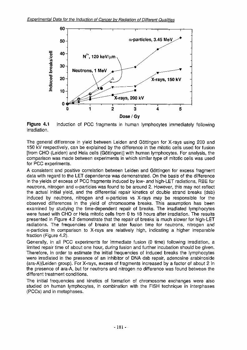
Experimental Data for the Induction of Cancer bv Radiation of Different Qualities
a-particles, 3.45 MeV
N' ,120 keV/pm
Neutrons, 1 MeV
Dose / Gy
Figure 4.1 Induction of PCC fragments in human lymphocytes immediately following irradiation.
The general difference in yield between Leiden and Gottingen for X-rays using 200 and 150 kV respectively, can be explained by the difference in the mitotic cells used for fusion [from CHO (Leiden) and Hela cells (Gottingen)] with human lymphocytes. For analysis, the comparison was made between experiments in which similar type of mitotic cells was used for PCC experiments.A consistent and positive correlation between Leiden and Gottingen for excess fragment data with regard to the LET dependence was demonstrated. On the basis of the difference in the yields of excess of PCC fragments induced by low- and high-LET radiations, RBE for neutrons, nitrogen and a-particles was found to be around 2. However, this may not reflect the actual initial yield, and the differential repair kinetics of double strand breaks (dsb) induced by neutrons, nitrogen and a-particles vs X-rays may be responsible for the observed differences in the yield of chromosome breaks. This assumption has been examined by studying the time-dependent repair of breaks. The irradiated lymphocytes were fused with CHO or Hela mitotic cells from 0 to 18 hours after irradiation. The results presented in Figure 4.2 demonstrate that the repair of breaks is much slower for high-LET radiations. The frequencies of breaks at later fusion time for neutrons, nitrogen and a-particles in comparison to X-rays are relatively high, indicating a higher irreparable fraction (Figure 4.2).Generally, in all PCC experiments for immediate fusion (0 time) following irradiation, a limited repair time of about one hour, during fusion and further incubation should be given. Therefore, in order to estimate the initial frequencies of induced breaks the lymphocytes were irradiated in the presence of an inhibitor of DMA dsb repair, adenosine arabinoside (ara-A)(Leiden group). For X-rays, excess of fragments increased by a factor of about 2 in the presence of ara-A, but for neutrons and nitrogen no difference was found between the different treatment conditions.The initial frequencies and kinetics of formation of chromosome exchanges were also studied on human lymphocytes, in combination with the FISH technique in interphases (PCCs) and in metaphases.
- 181 -

Experimental Data for the Induction of Cancer by Radiation of Different Qualities
.a-particles, 3.45 MeV
Neutrons, 1 MeVX-rays, 150 keV
N , 125 keV/pm
Recovery time / h
Figure 4.2 Repair of PCC fragments in irradiated human lymphocytes during delayed fusion.
The Gottingen group used variable delay times (from 0 up to 14 hours), tD, between irradiation and cell fusion, starting with 150 kV X-rays. PCC was combined with C-banding to identify dicentric chromosomes and with FISH painting to identify chromosome translocations. The Leiden group starting with 200 kV X-rays (from 0 up to 18 hours), first standardised the PCC-FISH technique and then used a chromosome painting probe in combination with a pancentromeric probe for the whole genome, that facilitated simultaneous detection of dicentrics and translocations. As a result of modification of the PCC technique, a higher yield of PCCs was obtained and it was therefore possible to analyse 500 -1000 cells for each experimental point.For X-rays, besides the acentric chromosome fragments seen in PCC immediately after irradiation which gradually disappear according to DNA double-strand break repair, early exchange-type aberrations were found with a yield proportional to dose (aD). They are formed in less than 60 min, and observed at 0 fusion time. On the other hand, particularly for doses above 2 Gy, the yield of the |3D2 component requires about 10 hours to be fully developed in the form of PCC-detectable complete chromosome translocations (reciprocal) and dicentrics (with bicoloured fragments).Since the X-ray experiments indicated a fast appearance of the a-component of the exchange-type aberration yield, we questioned whether experiments with neutrons and a-particles would show similar behaviour. Irradiation of human lymphocytes with 2 Gy of 3.45 MeV a-particles showed that the full magnitude of the dicentric yield occurred at to = 0, with no significant change when tD was increased further. However, for neutrons (at doses of 0.5, 1 and 1.5 Gy, following 0,1,4 and 18 hours) the frequencies of dicentrics as well as of translocations increased proportional to tD, at 4 and 18 hours.For both X-rays and neutrons, the initial frequencies of translocations were higher than those of dicentrics. Different types of complex exchanges were also found after high doses of X-rays (6 Gy) and neutrons (1.5 Gy), namely at the latest recovery times (12 and 18 hours).It was apparent that most of the early appearing dicentrics and translocations were incomplete (80%), i.e. dicentrics that were not associated with a second bicoloured fragment and non-reciprocal translocations. These incomplete forms were subsequently gradually replaced by complete forms.
-182-

Experimental Data for the Induction of Cancer bv Radiation of Different Qualities
The early incomplete forms are indicative of intermediate configurations of exchange-type aberrations and they belong to the a-component. For the (3-component, many less incomplete forms were observed. These differences indicate that the molecular processes leading to the a- and (3-components are different.In order to elucidate the relationship between different types of DNA lesions, their repair and the subsequent biological effect, wild-type and DNA dsbs repair-deficient CHO cell lines (i.e. CHO-K1, xrs 5 and xrs 6) were used (Leiden group) and irradiated with neutrons (1 MeV) (at Petten, The Netherlands), nitrogen (125 keV/pm) (at Uppsala, Sweden) and protons (40 keV/pm) (at Gray Laboratory)Positive correlations were found between deficiency in repair of DNA dsbs and induced cell killing and chromosomal aberrations. The RBE for neutrons and nitrogen ions was found to be 2-3 and for protons 1-1.2, when compared with X-ray reference data.
6.4.3 Objectives for the remainder of the project
Different types of radiation (especially 18 MeV electrons and C« photo-electrons (Gottingen group) and 4He, nitrogen ions and possibly 20Ne (Leiden group)), will be used in the future in delayed-fusion PCC-FISH experiments with human lymphocytes to further elucidate the reasons for different reaction kinetics of the a and (3 aberration yield components. On the basis of the contributions of the a- and (3-components to the formation of chromosome exchanges, a biophysical model will be developed.The modulating effect of a DNA dsb repair inhibitor (i.e. ara-A) will be further studied in human lymphocytes and CHO cell lines (Leiden group).The relationship between microbeam-induced DNA lesions, their repair and biological effects will be further elucidated (Leiden group) in collaboration with the Gray Laboratory (Northwood, UK), and, if possible, with IMR-PSI (Villigen, Switzerland) as well.The Villigen group was originally one of the participants of Work Package 4, and at the beginning of the EDICAR program several conjoint experiments were designed with the Leiden group, but unfortunately due to the unforeseen changes at IMR-PSI this collaboration could not be continued. As stated above, at the recent EDICAR group meeting in Rome the possibility of performing joint experiments has been discussed.
WORK PACKAGE 5: DEPENDENCE OF MUTATIONAL YIELD AND MOLECULAR SPECTRA ON RADIATION QUALITY.
Subco-ordinators: SZG (J. Kiefer, Giessen) and ISS (M. Belli, Rome) Partners: INFN-LNL, ISS, SZG
B.5.1 Objectives during the reporting period
Mutation plays an important role in the process of carcinogenesis. The dependence of the yields of mutations on radiation quality can hence provide important clues to the understanding of basic mechanisms. The study of the molecular patterns of mutations may furthermore establish a link between primary events of energy deposition and the nature of the biologically relevant lesions. Since mutations are clearly the result of errors in repair, there is an intimate relation with the tasks of Work Package 3.
-183 -
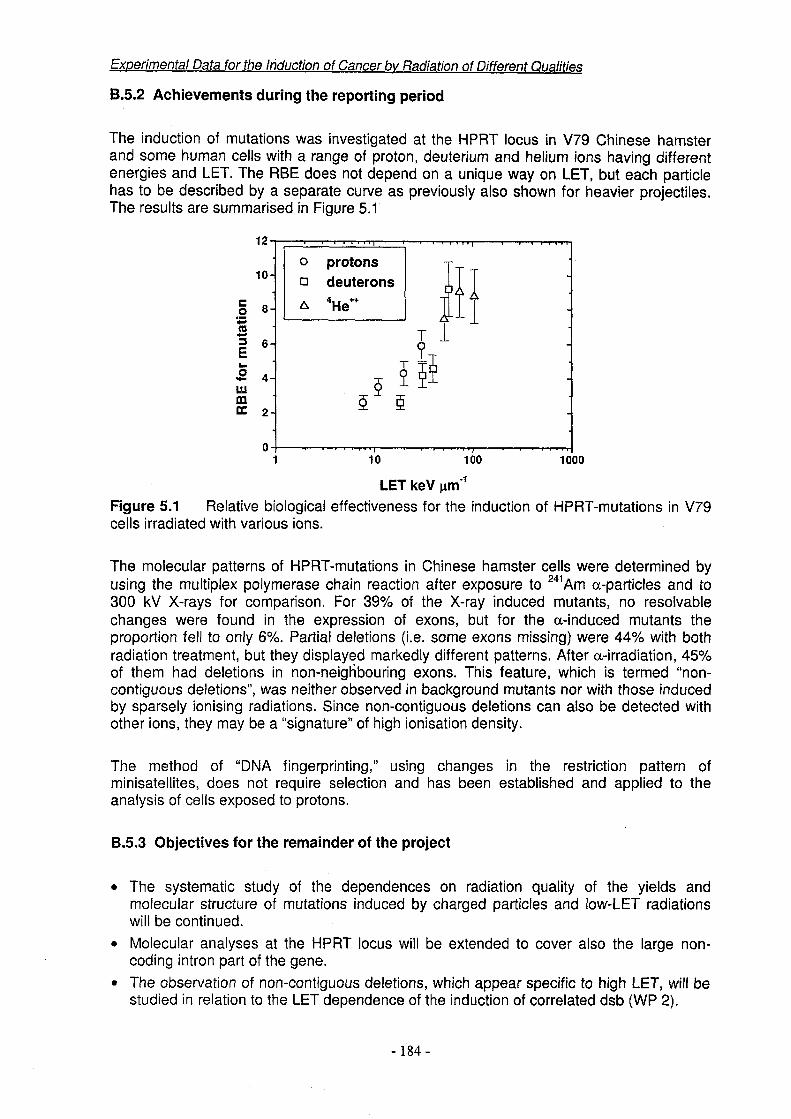
B.5.2 Achievements during the reporting period
Experimental Data for the Induction of Cancer bv Radiation of Different Qualities
The induction of mutations was investigated at the HPRT locus in V79 Chinese hamster and some human cells with a range of proton, deuterium and helium ions having different energies and LET. The RBE does not depend on a unique way on LET, but each particle has to be described by a separate curve as previously also shown for heavier projectiles. The results are summarised in Figure 5.1
o protons n deuterons
LET keV ^m'1Figure 5.1 Relative biological effectiveness for the induction of HPRT-mutations in V79 cells irradiated with various ions.
The molecular patterns of HPRT-mutations in Chinese hamster cells were determined by using the multiplex polymerase chain reaction after exposure to 241 Am a-particles and to 300 kV X-rays for comparison. For 39% of the X-ray induced mutants, no resolvable changes were found in the expression of exons, but for the a-induced mutants the proportion fell to only 6%. Partial deletions (i.e. some exons missing) were 44% with both radiation treatment, but they displayed markedly different patterns. After a-irradiation, 45% of them had deletions in non-neighbouring exons. This feature, which is termed “noncontiguous deletions”, was neither observed in background mutants nor with those induced by sparsely ionising radiations. Since non-contiguous deletions can also be detected with other ions, they may be a “signature” of high ionisation density.
The method of “DNA fingerprinting," using changes in the restriction pattern of minisatellites, does not require selection and has been established and applied to the analysis of cells exposed to protons.
B.5.3 Objectives for the remainder of the project
• The systematic study of the dependences on radiation quality of the yields and molecular structure of mutations induced by charged particles and low-LET radiations will be continued.
• Molecular analyses at the HPRT locus will be extended to cover also the large noncoding intron part of the gene.
• The observation of non-contiguous deletions, which appear specific to high LET, will be studied in relation to the LET dependence of the induction of correlated dsb (WP 2).
-184-

Experimental Data for the Induction of Cancer by Radiation of Different Qualities
• The method of “DNA fingerprinting” will be applied to a selected range of protons, deuterons and helium ions.
WORK PACKAGE 6: MICROBEAM AND MEASURED TRACK IRRADIATION OF INDIVIDUAL CELLS
Subco-ordinators: IMR-PSI (E. Heimgartner, Villigen), INFN-LNL (R. Cherubini, Legnaro) Partners: GLCRT, IMR-PSI, INFN-LNL, SZG, UU-SU
B.6.1 Objectives during the reporting period
The aim of this Work Package is to exploit the unique experimental opportunities provided by the techniques developed by partners within Framework III to probe cells individually with protons, 3He- and 4He-ions. The contributions of the different groups are complementary with respect to the techniques used and the LET'S and particle types. Two different systems to study single particle effects are now in routine use for performing radiobiological experiments. They have been further optimised in respect of the measurement accuracy and the seeding technique. Initial experiments on micronucleus induction and cell survival have been successfully conducted. These investigations, however, comprise merely the first step of the topics envisaged. They form the basis for comparisons with stochastic irradiation and are particularly relevant for the validation of mechanistic models of radiation response. The frequency of carcinogenic events at low doses is estimated by combining the survival data with those of DNA damage, chromosomal aberrations and mutations at the single particle level. These crucial endpoints are being studied in close collaboration with the groups of the other WP's.
B.6.2 Achievements during the reporting period
B.6.2.1 Microbeam developmentExperimental work with the charged-particle microbeam (GLCRT) has increased during the year and some of the results are reported below. At the same time, several aspects of the system have undergone further development. One of the main features has been to install an image intensifier between the microscope and the image capture CCD camera. This has enabled the amount of UV exposure of the cells, which are fluorescently stained, to be reduced to a level very much below that at which some UV damage was detected in earlier studies. A further benefit is that the sensitivity is sufficient to allow capture of an image of the charged-particle beam as it passes through the thin scintillator film that is used for particle counting. This provides a direct way of visualising the distribution of the beam on a micron scale and of determining its location precisely in relation to the intended target in the cell, using a common optical system. With this arrangement, beam position checks can be carried out as frequently as is necessary during the irradiation of a cell dish and any small displacements, for example due to thermal expansion, can be detected and corrected for. The improvement in aiming accuracy is substantial and our data now show that the precision with which we can target different regions of the cell is much improved. The installation of the image intensifier was not as straightforward to achieve as had been anticipated because it introduced various changes in the optics which necessitated substantial modification to the control software.Earlier work was mainly with protons, but this year has seen 3He++ brought into regular experimental use. The track structure and, therefore, the biological effectiveness of 3He++ should be identical to those for a-particles (4He++) of equivalent velocity. However, 3He++ is better suited to the beam transport system of the accelerator and consequently is the more
-185-

convenient particle to use experimentally. Beam collimation and particle counting were tested with 3He++ and found to be of similar precision to those achieved with protons. Tests were carried out using CR39 plastic etched to reveal the tracks of individual charged particles.
B.6.2.2 Improvement of measured track irradiationThe measured particle track technique (RSI) has been improved and has now the accuracy aimed for. Several polymer films have been tested with respect to distortions, beam scattering, adhesion of the nuclear emulsion and optical quality. A 4 pm thick polypropylene film was finally chosen both to support the emulsion and for the base of the cell chamber. Correlation of track positions with cellular structures was established by digital overlay of the images of the tracks on that of the cells. Lateral distortions of the detector film are measured by reference markers attached on the bottom of the cell chamber and corrected by applying a co-ordinate transform using a two-dimensional polynomial function. To minimise eventual beam divergence and scattering impairing the measuring accuracy the gap between the track detector and the base of the cell chamber has been reduced to ~15 pm. The accuracy of measurement of a track position was ascertained to be (0.9 ± 0.7) pm where scattering of the a-particles (4He++) in the vacuum window and in the nuclear emulsion is included.The seeding technique of cells on palladium islands and on naked polymer films has been optimised. The palladium island assay permits to confine the cells to restricted areas but is not applicable to all cells with regard to cloning efficiency and the time the cells take to attach. Alternatively the surface of the polypropylene film was coated with a cell adhesive (Cell-Tak, Becton Dickinson). Attachment of V79 cells was completed after 4 hours with a cloning efficiency of -75%. Selected cells were then tagged by their position with regard to the reference system of the cell chamber. Cell nuclei were stained using Hoechst 33258 at a concentration of 0.05 pg/ml and a UV exposure time of ~1 sec. A statistical model of the 3D surface contour of the cell nuclei has been developed which permits determination of the average intersection lengths of particle tracks as a function of the distance from the nuclear contour in the 2D image. The model is based on high resolution confocal microscopy images of cell nuclei (stained with SYTO, Molecular Probes), and the 3D contours are extracted using an elastic matching algorithm.
Experimental Data for the Induction of Cancer bv Radiation of Different Qualities
B.6.2.3 Progress of various techniquesFocused soft X-ray microprobe (GLCRT): By using a source of carbon-K X-rays with a spot size of about 5 microns coupled to a zone-plate diffraction "lens" it is possible to produce a ~50 nm microbeam for irradiating cells individually, targeting selected regions of the nucleus or cytoplasm. A device of this kind is now in an advanced stage of development and preliminary experiments have been carried out measuring the induction of micronuclei in V79 cells. Use of this device is in certain respects complementary to micro-irradiation with charged particles, especially in the sense that carbon-K X-rays exhibit some of the biological effects of low-LET radiation. In addition, the much finer spatial resolution achievable with the X-ray microprobe will make it the method of choice for high-resolution morphological mapping of radiation effects at the sub-cellular level.Alpha source microbeam (SZG): An experimental microbeam using a commercial 241 Am alpha source was constructed and its potential evaluated. The spatial resolution is about 5 pm. Due to personnel constraints it can presently not be followed up further but it is hoped to continue this activity in future.Focused charged-particle microbeam (INFN-LNL): The development of the magnetically focused beam set up at the AN2000 2.5 MV Van de Graaff accelerator has been continued by the LNL accelerator division and conditions as close as acceptable for "single particle
- 186 -

irradiation" in vacuum have been recently achieved. Many technical difficulties still remain to be solved, e.g. the extraction of the beam in air.
B.6.2.4 Initial investigations of micronucleus induction and cell survivalPreviously reported studies had shown that micronucleus induction in V79 cells by counted protons could be detected down to the level of single proton traversals, corresponding to a dose of -0.03 Gy. Studies carried out this year have determined V79 cell survival following exposure to counted protons (GLCRT) and measured a-particles (4He++, PSI), data are shown in Figure 6.1.The proton data (Figure 6.1(a)) show significantly reduced survival in the range corresponding to 10-20 traversals (~0.3 - ~0.6 Gy) compared with a standard linear- quadratic fit (dotted line) to the survival at higher doses. This is consistent with the phenomenon of induced radioresistance that has been found with X-rays. It is interesting that the effect occurs at an LET in the region of 10 keV/pm and ongoing work is investigating the response at higher LETs. The data indicate that >30 protons traversals of the nucleus are required to induce radioresistance.
Experimental Data for the induction of Cancer bv Radiation of Different Qualities
Specific dose in the cell nucleus [Gy]0.00 0.34 0.68 1.02 1.36 1.70
Number of a-particle traversals per cell nucleus
a
Standard L-Q model Modified L-Q model
Counted proton traverals per cell nucleus
Figure 6.1 Survival of V79 cells exposed to (a) counted numbers of 3.2 MeV protons (11.2 keV/pm) delivered to the cell nucleus and (b) to measured numbers of 2.7 MeV 01- particles (134 keV/pm) per cell nucleus.The a-particle data (Figure 6.1(b)) show a linear decrease of the survival probability with the number of nuclear traversals as it is usually found with high-LET radiation. The cell nuclei were found to be almost spherical with a mean diameter of 9 pm. One a-particle traversal corresponds to a specific dose to the cell nucleus of 0.34 Gy and reduces the survival probability to (68.2 ±15.6) %. No hypersensitivity has been observed at low doses. The slope of the survival curve is 0.62 ± 0.08 and represents the probability for one traversal to cause a lethal event. The results obtained with measured particle tracks can be compared with those of random irradiation in consideration of the Poisson distribution of the number of traversals. Thus, the inactivation cross section is reduced to (29.3 ± 2.5) pm2 and agrees with the value measured in various laboratories. The total track length of a- particles that reduces the survival to 1/e is (13.2 ± 1.2) pm and is in the range obtained by Raju et al. (Radiat. Res. 128, 204-209,1991) for several cell lines.
B.6.3 Objectives for the remainder of the projectExperimental techniques: The throughput of the measured particle track technique will be increased by shortening the time needed for the analysis per cell. UV exposure of cells will
-187-

Experimental Data for the Induction of Cancer bv Radiation of Different Qualities
be further reduced (PSI). It is anticipated that the soft X-ray microprobe will be brought into routine experimental use during Year 2 of the project (GLCRT). The possibility to design a facility similar to the GLCRT charged-particle microbeam (INFN-LNL) will be studied.Radiobiological investigations: The experiments on cell survival, micronuclei induction and apoptosis will be continued and completed. Special emphasis is put on heterogeneity of response within a cell population, spatial aspects and damage transmission between hit and non-hit cells. Measurement of the distribution of radiation response at the sub-cellular level is inherent in measured track irradiation and will be studied with the soft X-ray microprobe at high resolution. Single-cell gel electrophoresis ("comet" assay) will be applied to measure DNA damage (WP 2). Experiments on the induction of specific chromosome aberrations will be continued (WP 4). Special attention is given to the possible relations between DNA damage and chromosomal aberrations. Finally the possibility of measuring mutation induction with the single particle techniques will be studied (WP 5).
C. SUMMARY OF MAIN ACHIEVEMENTS DURING THE REPORTING PERIOD
The achievements have been described in some detail within each Work Package report and this is a general summary drawing the main achievements together. This project, Project 5, is almost entirely experimental and as such, as described under Objectives, is the counterpoint to Project 3 of Association Contract FI4P-CT95-0011 Biophysical Models for the Induction of Cancer by Radiation. The combined aim of the two projects is the mechanistic modelling of carcinogenesis by ionising radiations towards the development of improved estimates of human risk.
Progress in WP 1 has led to the quantification of several aspects of interactions between radiation tracks and DNA which contribute to concepts used in modelling the induction of ssb, dsb and complex (i.e., clustered) lesions. The action spectra studies in Year 1 have shown that experimentally, substantially lower values of deposited energy are required to induce a dsb than those generally used in Monte Carlo models. Initial studies of the influence of the hydration state of the DNA show that removal of most of the bound water has little effect on the energetics in terms of action spectra shapes and energy thresholds, but does reduce the absolute yields of ssb and dsb. Another finding likely to have a bearing on models is that the energetics indicate that a single ionising event can induce a dsb. Free-radical scavengers are often used to study the component of damage to DNA due to ‘OH radical attack and an achievement during Year 1 has been to distinguish additional effects that certain scavengers can exert by influencing the dynamics of DNA and its double-helical structure. An assay has been developed, based on endonuclease III, which will allow the assessment of clustered lesions containing base damages and is an advance in the methodology of evaluating complex lesions predicted by the models.
WP 2 is concerned with the accurate determination of the yields of dsb as a function of radiation quality. The methods used to measure dsb were completely reviewed at the first meeting of the consortium from which a critical review of methodology and of data from the literature was authored by several partners and has been submitted for publication. The main finding is that, whereas for low-LET radiation dsb are induced near-randomly along the DNA, several partners have found that with high LET there is evidence for track- correlated breaks, possibly corresponding to the intersection of tracks with loop domain structures. As these breaks are track-correlated, the fragments they produce will be
- 188 -

Experimental Data for the Induction of Cancer bv Radiation of Different Qualities
appreciable in number even at low doses and they may, therefore, be an important class of lesion leading to aberration or mutation induction (see also WP 5). Also, because of the proximity of the terminal dsb they may hinder each other’s repair, leading to a greater amount of residual damage.
Achievements in WP 3, which is concerned with cellular processing of dsb have been aided by the developments in methodology referred to in WP 2 above. Rejoining kinetics have been determined in human and rodent cell lines using a range of radiation qualities. An assay for repair fidelity (correct rejoining of ends) has been developed and initial data have been obtained with a range of radiation qualities. Other studies have used a PCC assay for repair studies. These developments will contribute towards improved understanding of the stages leading from dsb to chromosomal aberrations (WP 4). Initial work has been done to determine repair associated with DNA fragments, as described under WP 2. Progress has been made in the development of an individual cell assay (comet) for measuring repair kinetics after low doses of ions. This shows a strong dependence on dose at low levels, corresponding to a few average particle traversals (see also WP 6).
WP 4 addresses chromosomal aberrations, their induction and the associated cellular processing. Changes of this type represent one of the processes by which oncogenes may be activated and they also provide a mechanism for deletion of tumour suppressor genes. Visible chromosomal changes have been found to be associated with specific types of human tumour. Chromosomal aberrations also provide a sensitive endpoint for biological dosimetry. During Year 1, a complete set of data for the induction and repair of fragments scored during interphase using PCC/FISH techniques has been obtained for a range of radiation qualities. The data demonstrate that the higher-LET radiations give a greater overall yield due to a combination of increased initial yields with slower repair. Data have also been obtained towards the development of a kinetic model for the formation of exchange-type aberrations in terms of linear-quadratic dose-effect parameters. Progress has been made towards linking the induction and processing of aberrations in this Work Package with studies of dsb induction and rejoining in WPs 2 and 3 by using repair inhibitors and repair-deficient mutants. Progress has also been made towards evaluating the effects of single charged-particle traversals (i.e., directly relevant to protection dose levels) using the techniques in WP 6.
In WP 5 mutation at the HPRT locus has been studied as a paradigm of mutational processes associated with radiation carcinogenesis. Two significant effects have been reported in Year 1. The first was found in an evaluation of mutational yield versus radiation quality, using a conventional phenotypic expression assay. It was found that for the several types of charged particle used, the yield is not simply a function of LET but appears to depend on the particle type. Studies of this nature will give information for models which consider the influence of track structure. The other effect reported concerns the molecular analysis of the genetic changes induced and has demonstrated the occurrence of non-contiguous deletions within the mutated gene. This effect shows strong specificity for high-LET radiation and was not observed either in background mutants or in those found after exposure to low LET. It may be of interest as a potential “signature” of high LET in biological dosimetry and also mechanistically in relation to the track-correlated DNA fragmentation effects reported in WP 2 which are enhanced by high LET and which include the induction of fragments with sizes comparable to that of the HPRT gene.
In WP 6 significant achievements have been made in experimental approaches designed to measure the effects of the passage of charged-particle tracks through individual cells. With
-189-
Experimental Data for the Induction of Cancer bv Radiation of Different Qualities
both the measured-track and microbeam techniques, the responses of cells to traversal by one or more proton, a-particle or 3He++ ion can now be determined for a range of endpoints and some initial results are reported for Year 1. The techniques have been developed to allow measurement of responsiveness to medium- to high-LET radiations down to the level of single particle traversals. This equates to the situation that applies with most protection- level exposures, where the average time intervals between tracks traversing cells at risk can be estimated to range from weeks to centuries. Data from these studies will provide information about the ultimate low-dose limits of RBE for selected endpoints and mechanistic information about cellular targets and pathways and induced effects, including any transfer between hit and non-hit cells, as has been indicated in some recent reports. Achievements include detection of the effects of a single proton (micronucleus induction) and a single a-particle (cell kill) and of an induced effect (radioresistance to cell kill) at about 10 proton traversals. Also techniques have been established for scoring chromosomal aberrations, apoptosis and DNA damage in individual cells.
D. OBJECTIVES FOR THE REMAINDER OF THE PROJECT
In WP 1 the planned work includes studies continuing to characterise the relationships between the initial energy deposition events and the types and yields of damages induced in DNA. The aim is to obtain a comprehensive data set which provides basic information to input into the Monte Carlo codes being developed to model the induction of strand breaks and complex lesions in cellular DNA. Topics to be addressed include: “action spectra” for the induction of ssb, dsb and more complex lesions by low-energy photons and electrons, mimicking direct and indirect energy transfer in the core and penumbra of charged particle tracks; the influence of bound water on the energetics; comparison of the model plasmid DNA system used in these studies with genomic (yeast) DNA. The work planned also includes studies of the free-radical aspects of DNA damage and its induction, including: the effects of 'OH scavengers on the structural dynamics of DNA; the effects of agents with free-radical reactivity, such as oxygen and sulphydryl compounds; tests for multiple-radical models of dsb and complex lesion precursors. Further application is planned of methods developed in Year 1 of the project to assess DNA lesions for their complexity in terms of additional base damages using an assay based on endonuclease III treatment.
In WP 2 the planned work in Year 2 is predominantly to exploit the advances in the use of pulsed-field gel electrophoresis (PFGE) which were achieved in Year 1 to obtain critical data on the radiation quality dependence of dsb induction in human and rodent cell lines. This is based on the standardisation of the technique and methods of data analysis agreed within the consortium. The aim is to determine the true overall yields of dsb which derive from both randomly induced and track-correlated breaks. It is planned to study the DNA fragment size versus frequency distributions and compare them with random and other breakage models for a range of radiation qualities. The fragmentation patterns will be considered in relation to models of chromatin organisation and this will be aided by data from experiments in which chromatin structure is manipulated either by lysis and salt treatments or by using cell lines in which differentiation can be controlled. The influence of the chemical microenvironment in terms of 'OH scavengers and sulphydryl compounds will also be addressed.
WP 3 will also benefit from the advances in PFGE analysis referred to in WP 2. It is planned to complete the studies of rejoining ability and fidelity in human and rodent cell
-190-

Experimental Data for the Induction of Cancer by Radiation of Different Qualities
lines for a range of radiation qualities and to investigate in a human line the possible influence of differentiation status. It is also planned to examine the induction and repair of dsb in specific regions of the genome. The fragmentation pattern analysis referred to above will also be used to determine the extent to which dsb induced at short distances apart on the DNA mutually influence their repair; this will include studies with different radiation qualities. Studies will be carried out using the comet assay of repair of damage induced in individual cells by low doses. The role of the different kinds of breaks (directly and indirectly ("OH) induced) for different end points (survival, mutation etc.) in cells exposed to different radiations will be examined. To relate studies of dsb processing in this Work Package to damage at the chromosomal level, experiments will be carried out using dsb repair inhibitors and dsb repair-deficient mutants with PCC/FISH techniques (see also WP 4).
In WP 4 delayed-fusion PCC/FISH experiments are planned, using human lymphocytes and a range of radiation qualities, to determine differences in the linear-quadratic components of the formation kinetics of exchange-type aberrations and a biophysical model will be developed. Further work relating aberrations to dsb will be carried our using repair inhibitors and repair-deficient mutants (see also WP 3). Mechanistic studies of aberration induction will be carried out using counted charged-particle irradiation of individual cells with the microbeam and measured-track techniques (see also WP 6).
In WP 5 the radiation quality dependences of the yields and structures of mutations induced at the HPRT locus will be studied further and extended to cover also the large noncoding intron part of the gene. Work on the molecular analyses of the mutations will continue, using both the polymerase chain reaction and the DNA fingerprinting technique. Emphasis will be placed on further studies of the phenomenon of non-contiguous deletion reported in Year 1. This effect was only observed after high-LET irradiation and it will be examined as a potential candidate for a biological “signature” of radiation quality. It will also be considered in relation to the observation of track-correlated dsb induced by ions, as reported for Year 1 under WP 2.
In WP 6 the developments of the measured-track and microbeam techniques reported for Year 1 are planned to be continued to be applied in experimental use. However, some further developments are planned. These include increasing the throughput of cells which for the measured-track technique will be achieved by reducing the analysis time per cell and for the microbeam technique by improvements to the image acquisition and processing. It is anticipated that the soft X-ray microprobe will be brought into routine experimental use during Year 2 of the project for morphological mapping of cellular responses with sub-micron resolution. Experiments are planned using counted protons, 3He*+ and 4He++, scoring cell survival, micronucleus induction, apoptosis and chromosomal aberration induction (see also WP 4). The feasibility of including mutation induction (WP 5) will be examined in the light of improvements in cell throughput achieved. Emphasis will be placed on using the techniques to determine effectiveness at the single-track level. Initial studies reported for Year 1 of survival of V79 cells following counted proton irradiation have shown evidence of induced resistance above about 10 traversals per nucleus and this effect will be studied further with other radiation qualities. Studies will also be performed to investigate damage/signal transmission between hit and non-hit cells indicated by several recently published studies using low doses of a-particies with conventional irradiation techniques. Heterogeneity of response to single-particle traversals will be studied, in the first instance using single-cell gel electrophoresis (“comet” assay).
- 191 -

Experimental Data for the Induction of Cancer by Radiation of Different Qualities
E. LIST OF PUBLICATIONSPartners Main
WPs
1 Belli, M., Cera, F., Cherubini, R., Dalla Vecchia, M., Hague, A.M., INFN-LNL 5 ianzini, F., Moschini, G., Sapora, 0., Simone, G., Tabocchini, M.A., ,gSand Tiveron, P., RBE-LET relationships for cell inactivation and mutation induced by low-energy protons in V79 cells: further results at the LNL facility. International Journal of Radiation Biology (submitted).
2 Belli, M., Ianzini, F., Levati, L, Sapora, O., Tabocchini, M.A., Simone, INFN-LNL 2G., Cera, F., Cherubini, R., Dalla Vecchia, M., Moschini, G., Favaretto, |SS 3S., and Tiveron, P., 1997, Rejoining of DNA double-strand breaksinduced by light ions: what can it tell us? In: Radiation Damage to DNA: Techniques, Quantitation and Mechanisms. Radiation Research,148, 508.
3 Bettega, D., Calzolari, P., Marchesini, R., Noris Chiorda, G.L., INFN-LNL 2Piazzolla, A., Tallone, L., Cera, F., Cherubini, R., Dalla Vecchia, M., 3Favaretto, S., and Tiveron, P., Inactivation of C3H10T1/2 cells inducedby low-energy protons and deuterons. International Journal Of Radiation Biology (accepted).
4 Cera, F., Cherubini, R., Dalla Vecchia, M., Favaretto, S., Moschini, G., INFN-LNL 3Tiveron, P., Belli; M., Ianzini, F., Levati, L, Sapora, O., Tabocchini, ,ss 3M.A., and Simone, G, 1997, Cell inactivation, mutation and DNAdamage induced by light ions: dependence on radiation quality. In: 5Microdosimetry: An Interdisciplinary Approach, Edited by: D.T.Goodhead, P. O'Neill and H.G. Menzel (Royal Society of Chemistry,Cambridge, UK), pp 191-194.
5 Darroudi, F., Fomina, J., Meijers, M., and Natarajan, A.T., 1997, RUL 3Mechanisms of low- and high-LET radiation-induced chromosome .aberrations: Insights from PCC and FISH. Mutation Research, 379,571.
6 Fielden, E.M., Michael, B.D., and Prise, K.M. (editors), 1997, Radiation GLCRT All Damage to DNA: Techniques, Quantitation and Mechanisms.Extended abstracts of workshop April 1997, in Bowness-on-Windermere, Cumbria, Radiation Research, 148,481-522.
7 Folkard, M., Prise, K.M., Bowey, A.G., Schettino, G., Malcolmson, A., GLCRT 1Newman, H.C., Pullar, C.H.L., Michette, A.G., Pfauntsch, S.J., and 2Michael, B.D., 1997, New biophysical approaches to investigatingDNA damage by ionising radiation. In: Radiation Damage to DNA:Techniques, Quantitation and Mechanisms. Radiation Research, 148,483-485.
8 Folkard, M., Prise, K.M., Vojnovic, B., Bowey, G., Pullar, C., Schettino, GLCRT 6G. , and Michael, B.D., 1997, Targeting cells individually using a charged-particle microbeam: The biological effects of single or multiple traversals of protons and 3He2+ ions. In: Microdosimetry: An interdisciplinary Approach, Edited by: D.T. Goodhead, P. O'Neill andH. G. Menzel (Royal Society of Chemistry, Cambridge, UK), pp 323-326.
- 192-
9 Folkard, M., Vojnovic, B., Hollis, K.J., Bowey, A.G., Watts, S.J., GLCRT Schettino, G., Prise, K.M., and Michael, B.D., 1997, A charged-particle microbeam: II. A single-particle micro-coliimation and detection system. International Journal of Radiation Biology, 72, 387-395.
10 Folkard, M., Vojnovic, B., Prise, K.M., Bowey, A.G., Locke, R.J., GLCRT Schettino, G., and Michael, B.D., 1997, A charged-particle microbeam:I. Development of an experimental system for targeting cells individually with counted particles. International Journal of Radiation Biology, 72, 375-385.
11 Folkard, M., Vojnovic, B., Schettino, G., Forsberg, M., Bowey, A.G., GLCRT Prise, K.M., Michael, B.D., Michette, A.G., and Pfauntsch, S.J., 1997,Two approaches for irradiating cells individually: a charged-particle microbeam and a soft X-ray microprobe. Nuclear Instruments and Methods in Physics Research B, 130, 270-274.
12 Georgakilas, A.G., Haveles, K.S., Margaritis, L.C., Sakelliou, L., and DEMOKR. Sideris, E.G., 1997, Conformational changes induced in DNA by exposure to low doses of y- and a-irradiation. In: Radiation Damage toDNA: Techniques, Quantitation and Mechanisms. Radiation Research,148, 503-504.
13 Georgakilas, A.G., Konsta, A.A., Haveles, K.S., and Sideris, E.G., DEMOKR. Dielectric study of the double helix to single coil transition of the DNA macromolecule. Proceedings of IEEE (in press).
14 Georgakilas, A.G., Konsta, A.A., Haveles, K.S., Kanapitsas, A., and DEMOKR. Sideris, E.G., 1997, Comparative study of the DNA helix to coilthermal transition by dielectric spectroscopy and thermal transition spectrophotometry. In: Spectroscopy of Biological Molecules: Modern Trends, Kluver Academic Editions, 255-256.
15 Georgakilas, A.G., Sophianopoulou, V., and Sideris, E.G., 1997, DEMOKR. Enhanced DNA Stability as a Result of Low Doses of y- and a- Irradiation. Radioprotection, 32C, 101-102.
16 Greinert, R., and Harder, D., 1997, Biophysical analysis of the GAG dose-dependent over-dispersion and the restricted linear energy transfer dependence expressed in dicentric chromosome data from alpha-irradiated human lymphocytes. Radiation and Environmental Biophysics, 36, 89-95.
17 Haveles, K.S., Georgakilas, A.G., Sophianopoulou, V., and Sideris, DEMOKR. E.G., 1997, Effects of Tris on the DNA structure and the problem of induction of double-strand breaks by ionizing radiation. In: Radiation Damage to DNA: Techniques, Quantitation and Mechanisms.Radiation Research, 148, 500-501.
18 Heimgartner, E., Reist, H.W., 1997c, Biological effects of single alpha IMR-PSI particles, PSI Life Sciences Newsletter, (Paul Schemer Institute, CH-5232 Villigen PSI) (in press).
19 Heimgartner, E., Reist, H.W., Kelemen, A., Kohler, M., Stepanek, J., IMR-PSI Hofmann, L., 1997a, Measured particle track irradiation of individualcells. In: Microdosimetry: An International Approach, Edited by: D.T.Goodhead, P. O'Neill and H.G. Menzel (Royal Society of Chemistry,Cambridge, UK), pp. 339-342.
Experimental Data for the Induction of Cancer by Radiation of Different Qualities
6
6
6
1
1
1
1
4
1
6
6
- 193 -

Experimental Data for the Induction of Cancer by Radiation of Different Qualities
20 Heimgartner, E., Reist, H.W., Kelemen, A., Kohler, M., Stepanek, J., IMR-PSI 1997b, Study of low dose radiation response by measured particletrack irradiation. Submitted to Physica Medica, (accepted for publication).
21 Kiefer, J., Radiation-induced mutations. In: Fundamentals for the SZG Assessment of Risks from Environmental Radiation, Edited by G.Horneck (Kluwer, Dordrecht) (in press).
22 Kiefer, J., Stoll, U., and Schmidt, P., 1997, Modification by radiation SZG quality: Mutant induction by heavy ions in mammalian cells. In:Radiation, Radiomodifiers and Human Health, Edited by P. Uma Devi,K. S. Bisht and B. S. S. Rao, (National Institute of Science Communications, New Delhi) pp 178-187.
23 Lobrich, M., 1997, Undercounting of particle irradiation induced DNA SZG double strand breaks by conventional assays. In: Microdosimetry: An Interdisciplinary Approach, Edited by: D.T. Goodhead, P. O'Neill andH.G. Menzel (Royal Society of Chemistry, Cambridge, UK), pp 187-190.
24 Lobrich, M., Induction and repair of DNA double strand breaks in SZG human fibroblasts after particle irradiation. Advances in Space Research (in press).
25 Manti, L., Jamali, M., Prise, K.M., Michael, B.D., and Trott, K.-R., GLCRT 1997, Genomic instability in Chinese hamster cells after exposure toX-rays or a-particles of different mean LET. Radiation Research, 147,22-28.
26 Michael, BD., Ronto, G., Baumstark-Khan, C., Cadet, J., GLCRT Frankenberg-Schwager, M., Komova, O.V., Muller, W.-U., Quastel,M.R., and Sage, E., DNA injuries, damage induction and removal: recommendations for research needed. Proceedings of NATO Advanced Research Workshop, "Fundamentals for the Assessment of Risks from Environmental Radiation", Brno, October 1997, NATO ARW Series, (in press).
27 Newman, H.C., Prise, K.M., Folkard, M., and Michael, B.D., 1997, GLCRTDistribution of DNA double-strand breaks in X- andalpha-particle-irradiated cells. In: Radiation Damage to DNA: Techniques, Quantitation and Mechanisms. Radiation Research, 148,507-508.
28 Newman, H.C., Prise, K.M., Folkard, M., and Michael, B.D., 1997, GLCRT DNA double-strand break distributions in X-ray and alpha-particle irradiated V79 cells: Evidence for non-random breakage. International Journal of Radiation Biology, 71,347-363.
29 Ogheri, S., Bruna, V., Cera, F., Favaretto, S., Cherubini, R., and INFN-LNL Celotti, L., 1997, Mutant frequency at the hprt locus and in minisatellite sequences in Chinese hamster V79 cells irradiated with low-energy protons (31 keV/pm) and ultraviolet light (254 nm).Radiation Research, 148, 203-208.
6
5
5
2
23
4
1
23
2
2
5
- 194-

30 Prise, K.M., 1997, Ionising radiation induced clustered damage to GLCRT DNA: A review of the experimental evidence. In: Microdosimetry: An interdisciplinary Approach, Edited by: D.T. Goodhead, P. O'Neill andH.G. Menzel (Royal Society of Chemistry, Cambridge, UK), pp 111-116.
31 Prise, K.M., Ahnstrdm, G., Belli, M., Carlsson, J., Frankenberg, D., GLCRT,Kiefer, J., Lobrich, M., Michael, B.D., Simone, G., and Stenerlow, B., A GAG,review of DSB induction data for radiations of varying quality. ISS,International Journal of Radiation Biology (submitted). SZG,
UU-SU
32 Prise, K.M., Gillies, N.E., and Michael, B.D., Evidence for an oxygen- GLCRT independent fixation reaction leading to the induction of ssb and dsbin irradiated DNA, International Journal of Radiation Biology (submitted).
33 Prise, K.M., Newman, H.C., Folkard, M., and Michael, B.D., A study of GLCRT DNA fragmentation patterns in cells irradiated with charged particles:Evidence for non-random distributions. In: Proceedings of Workshopon Heavy Charged Particles in Cancer Treatment, Baveno, Italy, September 1997 (submitted).
34 Rydberg, B., Lobrich, M., and Cooper, P. K., Repair of clustered SZG damage caused by high-LET radiation in human fibroblasts. Physica Medica, (in press).
35 Schettino, G., Folkard, M., Prise, K.M., Vojnovic, B., English, T., GLCRT Michette, A.G., Pfauntsch, S.J., Forsberg, M., and Michael, B.D.,1997, The soft X-ray microprobe: A fine sub-cellular probe for investigating the spatial aspects of the interaction of ionizing radiations with tissue. In: Microdosimetry: An Interdisciplinary Approach, Edited by: D.T. Goodhead, P. O'Neill and H.G. Menzel (Royal Society of Chemistry, Cambridge, UK), pp 347-350.
36 Schmidt, P., Kiefer, J. Deletion pattern analysis of alpha-particle and SZG X-ray induced mutations at the HPRT locus of Chinese hamster cells,Mutation Research, (submitted).
37 Stenerlow, B., Hoglund, E., Blomquist, E., and Carlsson, J., 1997, UU-SU Rejoining kinetics of DNA double-strand breaks and variations in radiation quality. In: Microdosimetry: An Interdisciplinary Approach,Edited by: D.T. Goodhead, P. O'Neill and H.G. Menzel (Royal Society of Chemistry, Cambridge, UK), pp 129-132.
38 Stenerlow, B., Hoglund, E., Carlsson, J., and Blomquist, E., 1997, UU-SU Rejoining of clustered double-strand breaks induced by high-linear energy transfer radiation. In: Radiation Damage to DNA: Techniques, Quantitation and Mechanisms. Radiation Research, 148, 506-507.
Experimental Data for the induction of Cancer by Radiation of Different Qualities
1
2
2
1
2
23
6
5
23
23
- 195 -

Experimental Data for the Induction of Cancer by Radiation of Different Qualities
F. EXECUTIVE SUMMARY
The global aim of this project is to provide data which will input into the development of mechanistic models of radiation carcinogenesis. In general, the project provides the experimental complement to Project 3 of Association Contract FI4P-CT95-0011 Biophysical Models for the Induction of Cancer by Radiation co-ordinated by Dr HG Paretzke (GSF, Neuherberg). The development of more realistic and accurate models, based on a mechanistic understanding of radiation effects at the molecular and cellular levels, will aid the extrapolation of human risk data obtained from A-bomb and other high-dose exposures down to the much lower doses and dose rates generally encountered in the environment and the workplace. The research is organised into six work packages which cover the interactions of radiation with DNA, double-strand break (dsb) induction and processing, the induction of chromosome aberrations and mutations and the effects of individual radiation tracks on cells. Effects of radiation quality (LET and track structure) are an underlying theme and relate both to their use as mechanistic probes and to the radiation weighting factors used in risk and protection calculations.Initial Physical and Chemical Events in DNA Damage Induction (Work Package 1)WP 1 addresses the early physical and chemical stages of DNA damage induction and the processes that occur where radiation tracks intersect DNA. These processes include direct and indirect energy transfer by the ionising particle, free-radical damage and the modifying effects of radical scavengers. Emphasis is placed on the induction of dsb, particularly dsb that are closely associated with other damages (clustered lesions). These are considered to be important precursors of the genetic alterations leading to cancer induction by radiation.Biological effectiveness is considered to be determined by the microscopic pattern of energy deposition by radiation tracks on DNA. At this level, even the highest energy radiations transfer their energy in a large number of small events, on the order of 10's to 100’s of eV. Biophysical modelling of the patterns of energy deposition in DNA is well established. However, modelling the processes that transform the resulting ionisations and excitations into ssb, dsb and other damages is less advanced and depends on presently incomplete knowledge of the molecular mechanisms of lesion formation. Experimental models have been set up under WP 1 to determine the relationships between the yields of damage induced and the energy deposited (“action spectra”). Low-energy radiations were used in Year 1 to measure ssb and dsb induction in plasmids as a model system. Synchrotron photons and low-energy electrons were used to model energy transfer processes that take place in the core and penumbra regions of charged- particle tracks, respectively. The data obtained indicate that substantially lower values of deposited energy are capable of inducing dsb than those generally used in Monte Carlo models. Another finding which contrasts with the mechanisms generally used in biophysical models is that the energetics indicate that a single ionising event can induce a dsb. Initial studies of the influence of the hydration state of the DNA show that removal of most of the bound water has little effect on the energetics in terms of action spectra shapes and energy thresholds, but does reduce the absolute yields of ssb and dsb. During the remainder of the project, it is planned to study the action spectra and the influence of hydration in greater detail and to use chemical modifiers of free-radical processes to probe the mechanisms of the observed single-event induction of dsb. A comparison of plasmid and genomic DNA (S. cerevisiae) will be made at appropriate energies.The yields of DNA damages and their complexity (clustered lesions) are strongly influenced by free-radical reactants present within the cell, notably ’OH scavengers, sulphydryls and oxygen. An achievement during Year 1 has been to distinguish additional effects that certain scavengers can exert by influencing the dynamics of DNA and its double-helical structure. An assay has been developed, based on endonuclease III, which will allow assessment during Years 2 and 3 of the complexity of lesions containing base damages.
- 196-
Experimental Data for the Induction of Cancer by Radiation of Different Qualities
Data for DNA Damage Induction (Work Package 2)The main aims in this Work Package were to provide critical data on the yields of dsb induced by a range of radiation qualities. Although dsb have been recognised for a considerable time as the key lesions involved in the initiation of cancer by radiation and other cellular effects, the published yields of dsb, particularly as functions of radiation quality, have varied widely among different studies, depending on the techniques employed. A specialist topic reviewed during the first meeting of the consortium in February 1997 in Uppsala was dsb induction and measurement. It was agreed that the method of choice for studying dsb induction was pulsed-field gel electrophoresis (PFGE) analysed in terms of DNA fragment size distribution. Further discussion took place during the conjoint meeting with the Paretzke consortium at the RAM ’97 Workshop at Bad Honnef in June. Efforts have concentrated on the use of this approach to measure the yields of randomly induced and track-correlated dsb to obtain a clearer and more accurate picture of the radiation quality dependence of dsb induction. Also, it was recognised that the unprecedented ability of the technique to assay for fragments resulting from track- correlated breaks would allow the possible roles of this type of damage in relation to processing (WP 3), chromosomal aberration (WP 4) and mutation induction (WP 5) to be assessed. It was agreed at the Uppsala meeting that a critical review by the consortium of the methodologies for assaying dsb and of determining the dependence of their yield on radiation quality should to be undertaken during Year 1. This has been completed and submitted for publication. Also, partners agreed to co-ordinate and standardise their PFGE assays and conduct a multi-centre study of dsb induction in human fibroblasts during the remainder of the project, exploiting the panel of radiation qualities available within the consortium. Other work that is planned for Years 2 and 3 includes examining the roles that structure of chromatin and its chemical microenvironment play in the induction of random and track-correlated dsb.Processing of DNA Damage, Especially DSB (Work Package 3)The objective in WP 3 is to investigate the cellular processing of dsb induced by a range of radiation types as a critical step leading to gene and chromosomal changes. Methods have been developed to analyse reparability of dsb as functions of radiation quality and chromatin organisation, to study both general non-specific rejoining and repair fidelity (rejoining of correct ends). As with WP 2, it was determined that efforts should concentrate on the use of the PFGE assay, using fragment size distribution analysis wherever possible, although to achieve adequate sensitivity for rejoining studies, a less precise analysis (FAR) would have to be used in some instances. In Years 2 and 3, it is planned to complete studies of rejoining ability and fidelity in human and rodent cell lines for a range of radiation qualities and to investigate in a human line the possible influence of differentiation status. It is also planned to examine the induction and repair of dsb in specific regions of the genome. The fragmentation pattern analysis referred to above will also be used to determine the extent to which dsb induced at short distances apart on the DNA mutually influence their repair; this will include studies with different radiation qualities. Studies will be carried out using the comet assay of repair of damage induced in individual cells by low doses. The role of the different kinds of breaks (directly and indirectly ('OH) induced) for different end points (survival, mutation etc.) in cells exposed to different radiations will be examined. To relate studies of dsb processing in this Work Package to damage at the chromosomal level, experiments will be carried out using dsb repair inhibitors and dsb repair-deficient mutants with PCC/FISH techniques (see also WP 4).Chromosomal Aberrations (Work Package 4)WP 4 addresses chromosomal aberrations, their induction and cellular processing. Changes of this type represent one of the processes by which oncogenes may be activated and they also provide a mechanism for deletion of tumour suppressor genes. Visible chromosomal changes have been found to be associated with specific types of human tumour. Chromosomal aberrations also provide a sensitive endpoint for biological
-197-

Experimental Data for the Induction of Cancer by Radiation of Different Qualities
dosimetry. During Year 1, a complete set of data for the induction and repair of fragments scored during interphase using PCC/FISH techniques has been obtained for a range of radiation qualities. The data demonstrate that the higher-LET radiations give a greater overall yield due to a combination of increased initial yields with slower repair. Data have also been obtained towards the development of a kinetic model for the formation of exchange-type aberrations in terms of linear-quadratic dose-effect parameters. Progress has been made towards linking the induction and processing of aberrations in this Work Package with studies of dsb induction and rejoining in WPs 2 and 3 by using repair inhibitors and repair-deficient mutants. Pilot studies have been performed for evaluating the effects of single charged-particle traversals (i.e., directly relevant to protection dose levels) using the techniques in WP 6. Delayed-fusion PCC/FISH experiments are planned, using human lymphocytes and a range of radiation qualities, to determine differences in the linear-quadratic components of the formation kinetics of exchange-type aberrations and a biophysical model will be developed in Years 2 and 3. Further work relating aberrations to dsb will be carried our using repair inhibitors and repair-deficient mutants (see also WP 3). Mechanistic studies of aberration induction will be carried out using counted charged- particle irradiation of individual cells with the microbeam and measured-track techniques (see also WP 6).Dependence of Mutational Yield and Molecular Spectra on Radiation Quality (Work Package 5)Mutation plays an important role in the process of radiation carcinogenesis and study of the yields and molecular patterns of mutation as functions of radiation quality may establish a link between primary events of energy deposition and the nature of the biologically relevant lesions. Since mutations are clearly the result of errors in repair, there is an intimate relation with the tasks of WP 3. The dependence of the yields of mutations on radiation quality can provide important clues to the understanding of basic mechanisms as they relate to radiation weighting factors. Mutation at the HPRT locus has been selected as a paradigm for study. Two significant effects have been reported in Year 1. The first was found in an evaluation of mutational yield versus radiation quality, using a conventional phenotypic expression assay. It was found that for the several types of charged particle used, the yield is not simply a function of LET but appears to depend on the particle type. Studies of this nature will give information for models which treat the influence of track structure. The other effect reported concerns the molecular analysis of the genetic changes induced and has demonstrated the occurrence of non-contiguous deletions within the mutated gene. This effect appears to be specific for high-LET radiation. It may be of interest as a potential “signature” of high LET in biological dosimetry and also mechanistically in relation to the track-correlated DNA fragmentation effects reported in WP 2 which are enhanced by high LET and which include the induction of fragments with sizes comparable to that of the HPRT gene. In WP 5 the radiation quality dependences of the yields and structures of mutations induced at the HPRT locus will be studied further and extended to cover also the large non-coding intron part of the gene. Work on the molecular analyses of the mutations will continue, using both the multiplex PCR and DNA fingerprinting techniques. Emphasis will be placed on further studies of the phenomenon of non-contiguous deletion reported in Year 1.Microbeam and Measured-Track Irradiation of Individual Cells (Work Package 6)The objective in WP 6 is to measure the effects of the passage of charged-particle tracks through individual cells. With both the measured-track and microbeam techniques, the responses of cells to traversal by one or more proton, a-particle or 3He++ ion can now be determined for a range of endpoints and some initial results were obtained in Year 1. The techniques have been developed to allow measurement of responsiveness to medium- to high-LET radiations down to the level of single particle traversals. This equates to the situation that applies with most protection-level exposures, where the average time
- 198-
Experimental Data for the Induction of Cancer by Radiation of Different Qualities
intervals between the tracks traversing cells at risk can be estimated to range from weeks to centuries. Data from these studies will provide information about the ultimate low-dose limits of RBE for selected endpoints and mechanistic information about cellular targets and pathways and induced effects, including any transfer between hit and non-hit cells, as has been indicated in some recent reports. Achievements in Year 1 include detection of the effects of a single proton (micronucleus induction) and a single a-particle (cell kill) and of an induced effect (radioresistance to cell kill) at about 10 proton traversals. Also techniques have been established for scoring chromosomal aberrations, apoptosis and DNA damage in individual cells. Experiments are planned for Years 2 and 3 using counted protons, 3He++ and 4He++, scoring cell survival, micronucleus induction, apoptosis and chromosomal aberration induction (see also WP 4). The feasibility of including mutation induction (WP 5) will be examined in the light of improvements achieved in cell throughput. Emphasis will be placed on using the techniques to determine effectiveness at the single-track level. Studies will be performed to investigate damage/signal transmission between hit and non-hit cells indicated by several recently published studies which used low doses of a-particles with conventional irradiation techniques. Heterogeneity of response to single-particle traversals will be studied, in the first instance using single-cell gel electrophoresis (“comet” assay).
- 199-