BIOPHYSICAL DETERMINANTS OF NOTCH SIGNALING
182
BIOPHYSICAL DETERMINANTS OF NOTCH SIGNALING by Jeongsup Shim A dissertation submitted in partial fulfillment of the requirements for the degree of Doctor of Philosophy (Biomedical Engineering) in The University of Michigan 2009 Doctoral Committee: Associate Professor Alan J. Hunt, Co-Chair John B. Lowe, Genentech, Inc., Co-Chair Professor Jennifer J. Linderman Professor Sean Morrison
-
Upload
khangminh22 -
Category
Documents
-
view
0 -
download
0
Transcript of BIOPHYSICAL DETERMINANTS OF NOTCH SIGNALING
BIOPHYSICAL DETERMINANTS OF NOTCH SIGNALING
by
Jeongsup Shim
A dissertation submitted in partial fulfillment of the requirements for the degree of
Doctor of Philosophy (Biomedical Engineering)
in The University of Michigan 2009
Doctoral Committee:
Associate Professor Alan J. Hunt, Co-Chair John B. Lowe, Genentech, Inc., Co-Chair Professor Jennifer J. Linderman Professor Sean Morrison

Jeongsup Shim 2009

ii
To my family
iii
Acknowledgements
First, I would like to thank my mentor, Dr. John B. Lowe. I am grateful that he
gave me the opportunity to perform my graduate work in his lab and he supported my
efforts to finish my project. I followed him to relocate from Ann Arbor to Cleveland, and
now to South San Francisco. These moves were very unusual for a graduate student with
one mentor. However, I really enjoyed this trip, although sometimes this was challenging.
From this, I have learned how to do science and how to work with other people. Also, I
am grateful to my thesis committee, Dr. Alan J. Hunt, Dr. Sean J. Morrison, and Dr.
Jennifer Linderman for their interest and their helpful comment for my study.
Second, I thank the past and present members of Lowe’s lab, specially Bronia
Petryniak, Jay Myers, David Yao, and Dr. Yunfang Man. They gave me helpful
comments and suggestions on my project and had a good time in Cleveland, where my
study started to bloom. Specially, Bronia has been supporting my project from Ann Arbor
to Cleveland, and finally to South San Francisco. Without her help, I could not have
finish my work. David Yao was a good friend who enhanced my lab life in Cleveland.
Third, I would like to thank the following people who contribute to my project:
Dr. Henry T. Scheck, III and David Lorch at the University of Michigan for the use of
Optical Tweezers, Dr. Nolan Holland at Cleveland State University for helping with
AFM experiments, Dr. Satya Yadav at the Cleveland Clinic Foundation for the SPR
training and data analysis, Dr. Lino Gonzalez at Genentech, Inc. for the helpful

iv
suggestions regarding the use of the single chain ligands for the better SPR data analysis,
which was very critical to this study, Dr. Robert S. Haltiwanger and Nadia A. Rana at
Stony Brook University, Dr. Rod Keck, George (Tony) Moreno and Louisette Basa at
Genentech, Inc. for the mass spectrometry analysis, Dr. Phil Hass at Genentech, Inc. for
the helpful comments related to the protein purification.
Fourth, I thank Dr. Shuichi Takayama, Maria Steele, Kristin Romelhardt, and
Sandra Staneff in the Department of Biomedical Engineering for their support during my
graduate study.
Fifth, I would like to thank all the members of the Korean Bible Church of Ann
Arbor, Korean-American Presbyterian Church of Cleveland, and San Jose Korean
Presbyterian Church. I am indebted for their prayers and kind care for my family and me.
Lastly, I’d like to thank my family. My wife, Heewon witnessed my graduate
study and has supported me with love and prayer. During my graduate study, I got
married to her and moved together from Ann Arbor to Cleveland and later to the Bay
area. During this time, my father passed away after battling with cancer for 4 months,
while our son, Devin and our daughter, Ellie were born. From all of these experiences, I
matured and learned what the life is and how to live. My father and mother gave me
endless love, care and support. When I heard my father’s death, that loss was the saddest
thing in my life. If he lived now, he might be pleased and proud of his son more than
anyone else. I would like to thank to my in-laws. I am indebted to their prayer and the
support to finish my degree. I thank God for everything and all the spiritual people who
have taught me that the man’s chief end is to glorify God, and to enjoy him forever.
v
Table of Contents
Dedication………………………………………………......……………………………..ii
Acknowledgements…………………………………………….........…………….……..iii
List of Figures………....……………………………………….…..……………….……vii
List of Appendices..............................................................................................................ix
List of Abbreviations…………………………………………..……………………....…x
Abstracts……………………………………………………………………...…........…xii
Chapter 1. Introduction……………………..……………..................…………..…...…....……….1
Fucosylated Glycans…………………………………….............……….……..…3
Glycan-dependent Notch Signaling……………………………..............………...5
Biophysical Microenvironment in Notch
Signaling……………….…...................10
Techniques to Study the Glycan Structure and Protein-Glycan Interactions…....11
2. Construction and Characterization of Reagents……………..…...……........................24
Introduction…………………………………………………………............……24
Methods………………….,.……………………………………….….............….26
Results…………………………………………………………………............…35
Discussion……………………………………………………..……............……41
vi
Notes to Chapter 2…………………………………………………............…….46
3.The Use of Optical Tweezers to Study Notch-Notch Ligand Interactions….................56
Introduction………………………………………………………….............…..56
Methods…………………………………………………………….....................57
Results and Discussion…………………………………………….............…….59
4. Direct Force Measurement of Notch-Notch Ligand Interactions using Atomic Force Microscopy (AFM) ……………………….…………..…............…............................65
Introduction………………………………………………………............………65
Methods……………………………………………………………...........……..68
Results……………………………………………………………............………74
Discussion……………………………………………………………............…..76
5. Kinetic Analysis of Glycan-dependent Notch-Notch Ligand Interactions using Surface Plasmon Resonance (SPR) ……………..………………….....…...............…...……..84
Introduction………………………….………………...…………........................84
Methods………………………………………………………………..................88
Results………………………………………………....……………............……89
Discussion………………………..…………………………….………...............99
6. Conclusions…….………..........................…..…...……..………….……...................124
Direct Force Measurement by Force Spectroscopy.............................................124
Kinetic Analysis of Notch-Notch Ligand Binding..............................................126 Future Direction……………………..…………..……………...…............……129
Appendices......................................................................................................................136
Bibliography……………………………………….……………………………...……152
vii
List of Figures
Figure
1-1 Glycocalyx and simplified diagram of glycocalyx.………………………..………16
1-2 Fucosylated glycanx in mammals...……..................................................................17
1-3 Fucosylation pathways in mammals...…………………………………..….….…..18
1-4 Notch receptor and ligands.............………………………………………………..19
1-5 Central biochemical events in Notch signaling……………………………………20
1-6 O-fucosylationed glycan in Notch receptor………………..……………….…..….21
1-7 Force probes…………………………………………………………………….…22
1-8 The basic principles of SPR analysis…………………………...…………….……23
2-1 Mouse Notch1 mutant constructs………………………….………………………48
2-2 Site-specific mono-biotinylation system...…………….………………….……….49
2-3 Verification of site-specific mono-biotinylation system……………………..……50
2-4 Characterization of Lec13 and CL17 cells………………………………..……….51
2-5 Surface expression of 3xFlag-Notch1 in Lec13 cells...............................................52
2-6 FACS analysis for the surface expression of 3xFlag Notch1 in Lec13 cells...........53
2-7 Mass spectrometry analysis of Notch1+fucose.……………………………….…..54
2-8 Mass spectrometry analysis of Notch1-fucose.……………………………….…...55
3-1 Optical tweezers schematic diagram.……………………………………….……..62
3-2 Measurement of Notch1 and Dll-1 binding strength…………………… ………..63
viii
3-3 Optical interference in the two bead system………………………………………64
4-1 Protein immobilization method by esterfication………………………….….……77
4-2 AFM experimental setup to measure the Notch1 and Dll-4 binding strength..……78
4-3 Force-distance curve from AFM force measurement..………………….….……...79
4-4 Representative force-distance graph of Notch1 and Dll-4 binding………………..80
4-5 AFM image of esterified surface and SA immobilized surface…………………...81
4-6 Binding frequency at the retraction of 7140nm/s...........…………………………..82
4-7 Force spectrum of Notch1 and Dll-4 binding..…………………………………….83
5-1 Reagent configuration……………………………………………..……………...109
5-2 SPR experimental setup………………………………………….….………..…..110
5-3 Evaluation of regeneration condition…………………………...…....…………..111
5-4 SPR data analysis with double subtraction…………………………….………...112
5-5 Conformational change in Notch1 and Dll-4 binding……………………..……..115
5-6 Representative sensorgram of Notch1-Notch ligand binding..………..…..……..116
5-7 Kinetic analysis of Notch1-Notch ligand binding.………………………..…...…117
5-8 Kinetic analysis of Notch1 mutants and Dll-4 binding…………………...…..….118
5-9 Kinetic analysis of Dll-4 to Notch1(15) and Notch1(36) binding..……….….......119
5-10 Kinetic analysis of Notch1-Dll-4 binding with different glycosylation status.....120
5-11 Overall association and dissociation rate of Notch-Notch ligand binding...........121
5-12 Model for Notch-Notch ligand binding................................................................122
ix
List of Appendices
Appendix
1 Automated AFM data analysis program....................................................................137
2 Kinetic information of Notch-Notch ligand binding.................................................148
3 Relaxation rates of two-state binding model.............................................................150
x
List of Abbreviations ADAM A disintegrin and metalloprotease AFM Atomic Force Microscopy APC Antigen-presenting cell ASCII American Standard Code for Information Interchange ATCC American Type Culture Collection BCCP Biotin carboxyl carrier protein BM Bone Marrow CHO Chinese Hamster Ovary CL17 Clone 17 Dll Delta-like DN Double Negative DNA Deoxyribonucleic acid DOS Delta and OSM-11-like proteins DP Double Positive DSL Delta, Serrate, LAG-2 motif ECN Extracellular domain of Notch EDTA Ethylenediamine tetraactic acid EGF Epidermal Growth Factor EGFP Enhanced Green Fluorescence Protein ELISA Enzyme-Linked Immuno Sorbent Assay ER Endoplasmic Reticulum FACS Fluorescence-activated cell sorting FCS Fetal Calf Serum Fuc Fucose FUT Fucosyltransferase FX 3,5 epimerase/4-reductase GalNAc N-acetylgalactosamine GDP Guanosine Diphosphate GMD GDP-mannose 4,6-dehydratase HEK Human Embryonic Kidney HES Hairy and enhancer of split hIgG Human Immunoglobulin G HSC Hematopoietic Stem Cell ICN Intracellular domain of Notch LSK Lin-,Sca1+,c-kit+ MS Mass Spectrometry NA Numerial Aperature NHS N-hydroxysuccinimide Notch EC Notch extracellular domain
xi
Notch IC Notch intracellular domain NRR Negative regulatory region NTM Notch Transmembrane domain Ofut1 O-fucosyltransferase 1 Ofut2 O-fucosyltransferase 2 OTs Optical Tweezers PCR Polymerase Chain Reaction PEG Poly(ethylene) Glycol Pofut1 Protein O-fucosyltransferase 1 Pofut2 Protein O-fucosyltransferase 2 SA Streptavidin SPR Surface Plasmon Resonance TCR T-cell receptor Tris tris(hydroxymethyl)aminomethane WLC Worm-Like Chain Model
xii
Abstract
Notch signaling is involved in many biological contexts such as cancer, stem cell
development, and neural cell development. Because of the importance of Notch function
in health and disease, the Notch signaling pathway has emerged as a potential therapeutic
target.
Mammalian Notch receptors are single-pass transmembrane glycoprotein
receptors, which contain 29-36 EGF like repeats. The fucosyltransferase termed Pofut1
transfers fucose to the serine or threonine residue of the O-fucosylation consensus
sequence on some EGF domains of Notch receptors. The glycosyltransferases termed
Fringe can elongate O-fucose moieties by adding N-acetylglucosamine, which may be
subsequently modified by galactose and sialic acid. These O-fucosylated glycans play
key roles in modulating Notch-mediated signal transduction events.
Here, we have observed how O-fucosylated glycan modifications modulate Notch
receptor-ligand interactions using surface plasmon resonance techniques. A biphasic
binding and dissociation pattern was observed, suggesting a two-state receptor-ligand
interaction model characterized by initial formation of a transient receptor-ligand
complex followed by a conformational change that leads to a more stable receptor-ligand
complex. Primary and secondary on and off-rates for the four binding-competent
Notch1-Notch ligand pairs were observed to be distinct and characteristic for each Notch
ligand. The overall association constants observed when Dll-1 or Dll-4 interacted with
xiii
Fringe-modified Notch1 were significantly greater than when these ligands interacted
with unmodified Notch1, with enhancement likely due to Fringe modifications of fucose
moieties within EGF domains 16-36. By contrast, Fringe modification of Notch1 did not
significantly modulate interactions with Jag-1 or Jag-2. Mutational analyses confirm prior
observations that the O-fucosylation site within EGF repeat 12 dictates much, if not all of
the binding between Notch1 and its ligands. Finally, we observe that Fringe modification
of Dll-4 enhances its ability to bind to Notch1.
Our data reveal that the molecular basis of glycan-dependent Notch-Notch ligand
binding. We propose a two-state binding model with triple stranded structure for Notch-
Notch ligand complex arrangement. Here, O-fucosylation and Fringe modification of
Notch receptors play a key role in both the binding and the conformational change.
1
Chapter 1
Introduction
Carbohydrates are inevitable components for life. They are involved in the storage
and transport of energy and in the immunity, fertilization, pathogenesis, blood clotting,
and development [1].
Monosaccharides are the basic units of carbohydrates and the building blocks of
bi- and polysaccharides. Free monosacchardes can exist in open chain or ring forms. Ring
forms of the monosaccharides can be linked together via glycosidic bonds to form
polysaccharides or oligosaccharides (or glycans).
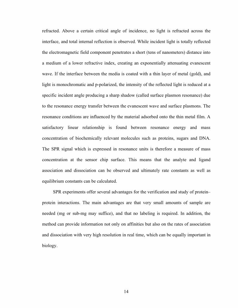
Mammalian cells synthesize various glycans which are covalently linked to
proteins and/or lipids [2]. The surfaces of most types of cells are heavily coated with
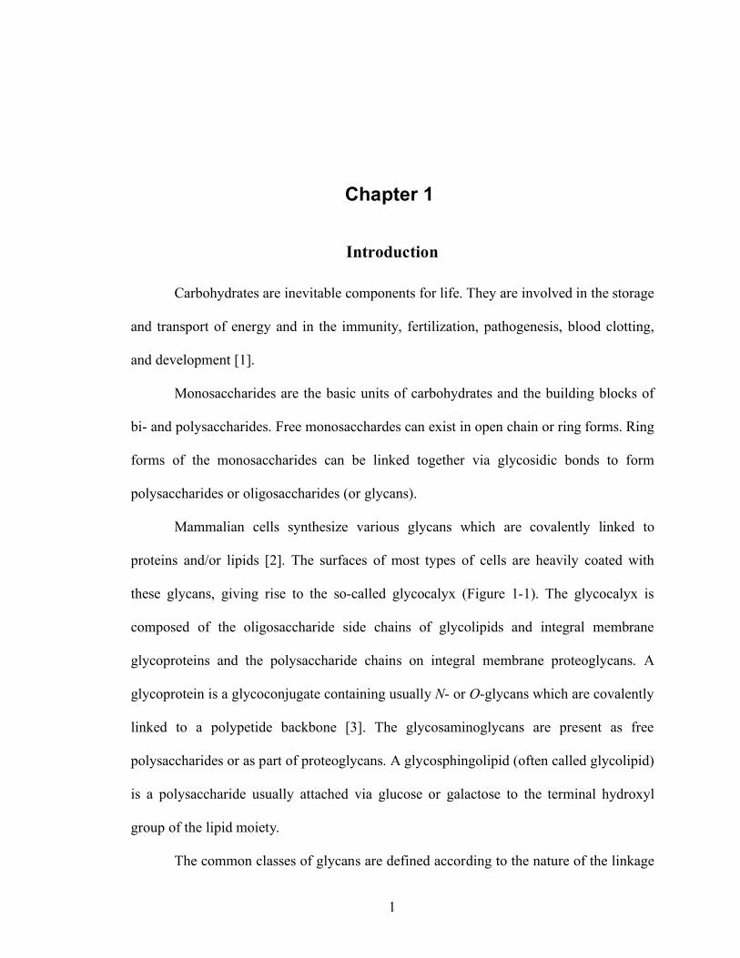
these glycans, giving rise to the so-called glycocalyx (Figure 1-1). The glycocalyx is
composed of the oligosaccharide side chains of glycolipids and integral membrane
glycoproteins and the polysaccharide chains on integral membrane proteoglycans. A
glycoprotein is a glycoconjugate containing usually N- or O-glycans which are covalently
linked to a polypetide backbone [3]. The glycosaminoglycans are present as free
polysaccharides or as part of proteoglycans. A glycosphingolipid (often called glycolipid)
is a polysaccharide usually attached via glucose or galactose to the terminal hydroxyl
group of the lipid moiety.
The common classes of glycans are defined according to the nature of the linkage
2
(core) regions to protein or lipid [1]. N-glycans (N-(Asn)-linked oligosaccharides) are
polysaccharides covalently linked to an asparagine residue of the consensus sequence
Asn-X-Ser.Thr, where X can be any amino acid besides Pro and Asp. N-glycans can be
generally subdivided into three main classes: high-mannose type, complex type, and
hybrid type. O-glycans (O-(Ser/Thr)-linked oligosaccharides) are typically connected via
N-acetylgalactosamine (GalNAc) to a serine or threonine residue. Other types of O-
linked glycans exist such as O-GlcNAc, O-mannose, O-glucose, and O-fucose.
Several human disease states are charaterized by changes in glycan biosynthesis
and degradation that can be of diagnostic and/or therapeutic significance: Leukocyte
adhesion deficiency II (LADII) [4] , carbohydrate-deficiency glycoprotein syndromes
(CDGS) [5], mucopolysaccharidosis (MPS) [6], congenital dyserythropietic anemia type
II (CDAII/HEMPAS) [7, 8], and galactosemia [9]. In the tumor environment, changes in
glycosylation are associated with the pathogenesis of cancer, and are implicated in the
tumor progression, including tumor cell proliferation, dissociation and invasion, adhesion
and metastasis, and angiogenesis [10].
Although glycans play an important role in a variety of biological events, it has
been difficult to elucidate the structure and the function of glycans in the living system
due to the complexity and diversity of glycans and the lack of tools [1]. Glycans may be
one of the most complex entities in nature. The complexity of the “glycome” exceeds the
complexity of the proteome as a result of the enormous structural diversity of glycans and
is further complicated by the combination and interaction of carbohydrates with each
other and with proteins. Recently, new technologies to explore the structure of glycan
have opened up a new frontier for glycomics and glycobiology [11, 12] and the
3
importance of glycobiology has been recognized in biotechnology and medicine [13-15].
Fucosylated Glycans
L-fucose (6-deoxy-L-galactose) is a monosaccharide that is commonly found in
many glycan structures in mammals. Lack of a hydroxyl group on the carbon at the 6-
positon (C-6) and the L-configuration distinguish fucose from other six-carbon sugars
present in mammalian cells.
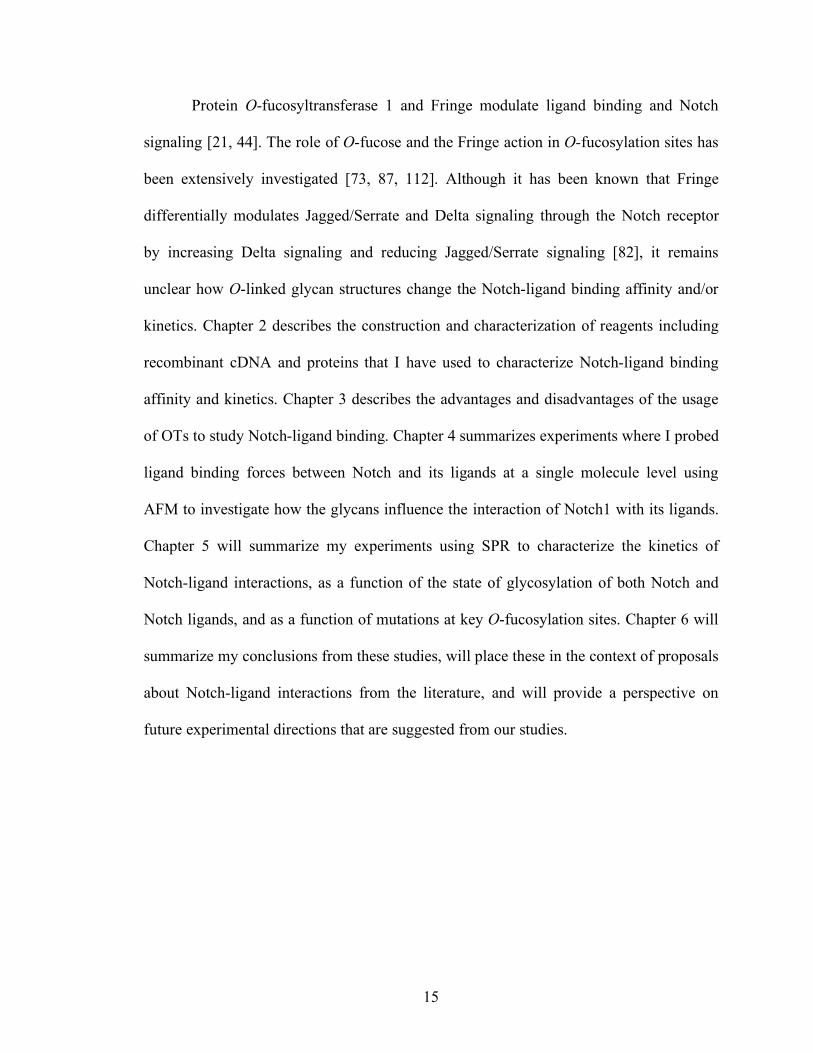
Various fucosylated glycan structures have been identified. Fucosylated glycans
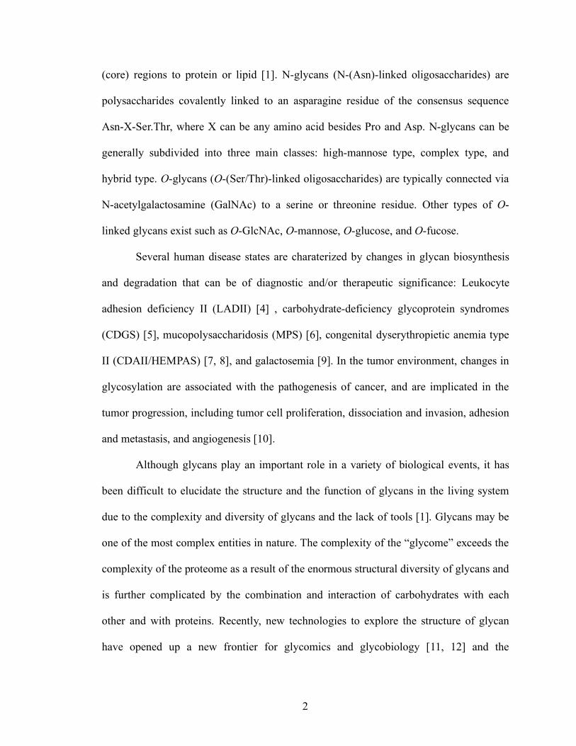
are synthesized by fucosyltransferases in the Golgi or ER (Figure 1-2). Thus far, thirteen
fucosyltransferases have been identified in mammals. The FUT1 and FUT2 loci encocde
α(1,2)-fucosyltransferases [16, 17]. The FUT3 locus encodes an α(1,4)-
fucosyltransferases and the FUT4-FUT7 and FUT9 loci encode α(1,3)-
fucosyltransferases responsible for synthesis of the Lewisx and sialyl Lewisx antigens
[18]. The FUT8 locus encodes an α(1,6)-fucosyltransferase that adds fucose to
asparagine-linked GlcNAc moieties [19]. The Pofut1 and Pofut2 loci encode O-
fucosylatransferases that add fucose directly to serine/threonine residue of only a few
proteins. Pofut1 modifies Epidermal Growth Factor-like (EGF) repeats of Notch
receptors, Notch ligands, and Cripto [20, 21] and Pofut2 modifies thrombospondin type 1
repeats [22]. The large number of fucosyltransferases and the structural diversity of their
products imply an important role for fucose in biology. It has been suggested that Pofut1
and Pofut2 reside in ER [23], although an ER-specific fucose transporter gene has not yet
been identified. FUT10 and FUT11 are the putative α(1,3)-fucosyltransferase loci, which
have been identified in the human genome by comparison with fucosylatransferase
4
sequences in the Drosophila genome [24]. However, no enzymatic activity has yet been
assigned to these hypothetical proteins.
GDP-fucose is used as a donor substrate by all fucosyltransferases in the synthesis
of fucose-containing glycoconjugates [25]. Two pathways have been known to synthesize
GDP-fucose in the cytosol of mammalian cells. These are termed the de novo pathway
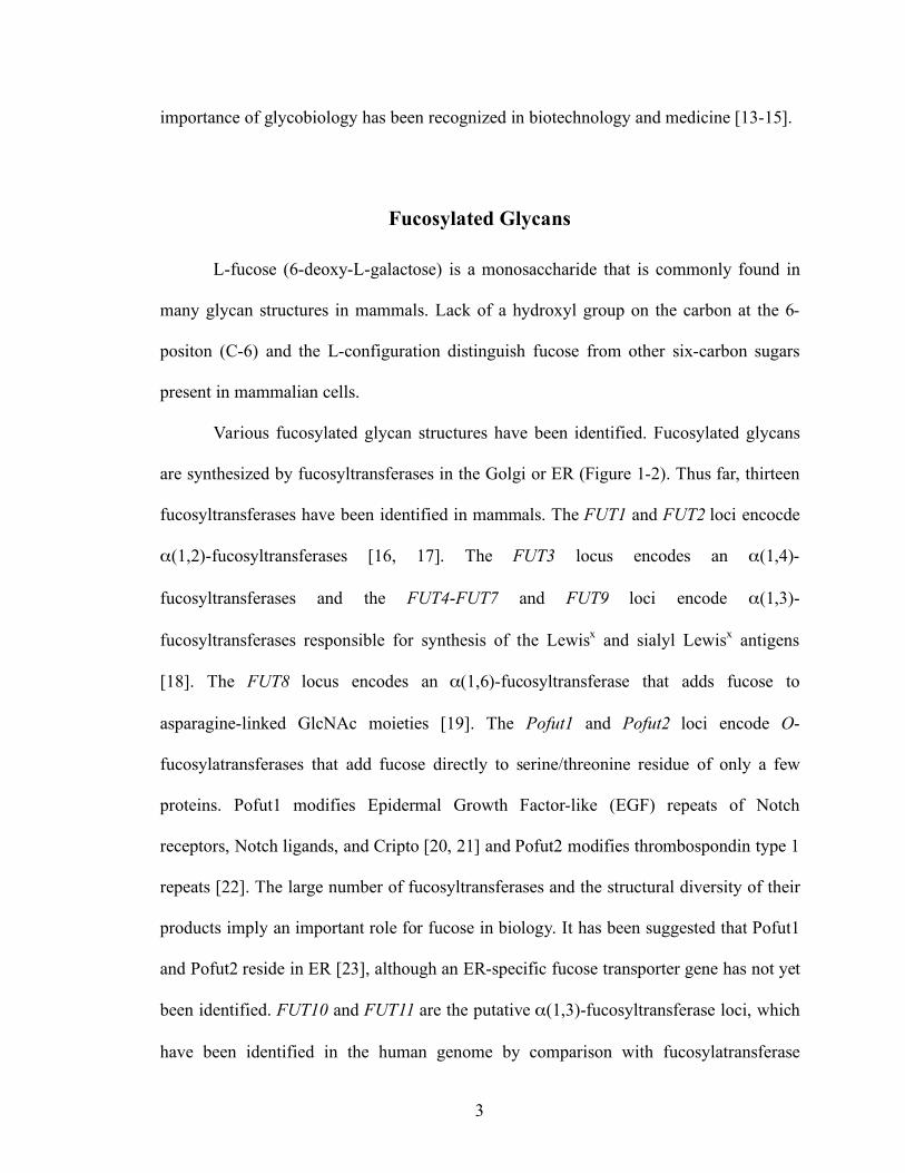
and the salvage pathway (Figure 1-3) [26]. The de novo pathway utilizes two enzymes,
GDP-mannose 4,6-dehydratease (GMD) and the FX protein (GDP-4-keto-6-deoxy-D-
mannose-3,5-epimerase-4-reductase) to convert GDP-mannose to GDP-fucose [27-29].
GDP-fucose is then transported into Golgi lumen or ER where it is utilized as a
fucosyltransferase substrate. Smith et al. [30] created a mouse lacking the FX locus (FX–/–
mice). These mice lack all forms of fucosylation and do not survive long after birth
unless fucose is added to their diet. A salvage pathway that elaborates GDP-fucose [25]
allows GDP-fucose biosynthesis to be rescued by exogenous fucose, thereby restoring
fucosylation.
Roles for fucosylated glycans have been investigated in a variety of biological
contexts [31, 32]. Well-studied fucosylated glycans include the molecules of the ABO
and Lewis blood group systems [33], host-microbe interactions [34, 35], and selectin
dependent leukocyte adhesion [36-41]. In the glycan structures relevant to these systems,
fucose mainly exists as a terminal modification of glycan structures. Recently,
glycosyltransferases capable of adding sugars to O-fucose have been found. These are
termed the Fringe family of enzymes, which act on fucose that modifies some EGF
repeats [21, 42] and one or more β(1,3)-glucosyltransferases which act on fucose
moieties that modify thrombospondin type 1 repeats [43]. O-fucosylated glycans are
5
involved in cell signaling events during a variety of development processes [21, 42].
Notch receptors, Notch ligands, the Cripto receptors, and urokinase type plasminogen
activator are glycoprotein receptors that have O-fucosylated glycans critical to
development [20, 22]. Especially, Notch receptors are of interest because inactivation of
O-fucose modulator, Pofut1 results in a failure of embryonic development, and
dysfunction of Notch signaling [44].
Glycan-dependent Notch Signaling
Notch receptors are single-pass transmembrane glycoprotein receptors essential to
cell fate determination in a wide array of developmental processes [45-47]. Inactivation
of the mouse Notch1 gene did not cause gross developmental anomalies, but did yield
delayed and disorganized somitogenesis that resulted in embryonic lethality around day
10 of gestation [48-50]. Defects in Notch function caused by mutations in genes encoding
Notch receptors or their ligands cause or are strongly associated with a variety of diseases
[51] such as several forms of cancer [52-54] including T cell acute lymphoblastic
leukemia (T-ALL) [55], tumor angiogenesis [56], Cerebral Autosomal Dominant
Arteriopathy with Sub-cortical Infarcts and Leukoencephalopathy (CADASIL) [57, 58],
multiple sclerosis [59], spondylocostal dysostosis (SCD) [60], Alagille syndrome [61],
and congenital heart defects [62]. Because of the importance of Notch function in health
and disease, Notch signaling pathway has emerged as a potential therapeutic target [63,
64].
Notch was originally discovered in Drosophila. Four homologues have now been
identified in mammals [46]. These are termed Notch1 (N1), Notch2 (N2), Notch3 (N3),
6
and Notch4 (N4) (Figure 1-4). Notch receptors are synthesized in ER, where Notch is O-
fucosylated. Further, Notch receptors are proteolytically processed during transport to the
cell surface by a furin-like protease in Golgi apparatus (at site S1), producing an
extracellular Notch (ECN) subunit and a Notch transmembrane (NTM) subunit. These
two subunits, which remain non-covalently associated, constitute the mature
heterodimeric cell-surface receptor.
Notch receptors are activated when the ECD interacts with ligands on adjacent
cells. Notch ligands were first identified in Drosophila and are called Delta and Serrate.
Multiple homologues of each are present in mammals where the Serrate homologues are
called Jagged. Five Notch ligands have now been identified in mammals. These are
termed Jagged1, Jagged2, Delta-like1, Delta-like3, and Delta-like4 (Figure 1-4). Each of
the ligands is a single-pass transmembrane protein with a conserved N-terminal Delta,
Serrate, LAG-2 (DSL) motif essential for binding to Notch. Jagged contains a cysteine-
rich region, but the exact mechanism of how the cysteine-rich region modulates Notch
signaling is not known, although it was suggested that this region is required for
activation of Notch signaling in Xenopus primary neurogenesis [65].
Activation by ligand triggers a series of proteolytic cleavages of the NTM that
release the ICN from the membrane. The NTM subunit is released by at least two
sequential proteolytic cleavages (at site S2 and S3). The ICN then translocates to the cell
nucleus, ultimately resulting in transcriptional regulation of developmental control genes
such as Hes1 (Figure 1-5).
Notch receptors are heavily glycosylated with N-glycans [66, 67] and O-glycans
such as O-fucose [21, 42, 44, 68], O-glucose [69-71], and possibly O-GlcNAc [72]. O-
7
glucosylation and O-fucosylation regulate Notch folding, trafficking, and signaling
although these post-translational modifications are very unusual [25, 44]. O-glucosylation
of Notch receptor was first discovered in a biochemical study of mouse Notch1 [71]. O-
glucose attaches to serine between the first and second cysteine residues of EGF repeats
within the consensus sequence C1XSXPC2 [70, 71]. The O-glucosylated glycan forms the
trisaccharide EGF-O-α1Glcβ1,3Xylβ1,3Xyl [71]. However, the biological role of O-
glucosylation on Notch receptors was not known until recently. Drosophila gene, rumi,
when mutated, yields a Notch loss of function phenotype, and has been found to be a
protein O-glucosyltransferase [69]. Although loss of rumi does not reduce cell surface
Notch expression or binding of Notch to Delta, Notch signaling was inhibited due to the
loss of the S2 proteolytic cleavage, which is essential for Notch activation. The biological
relevance of rumi to mammalian systems remains to be determined.
O-fucosylation of EGF repeats is catalyzed by Pofut1. Pofut1 adds GDP-fucose to
specific serine or threonine residues on certain EGF-like repeats, including some found
within Notch receptors. Pofut1 is thought to be essential for Notch function [73]. The
recent demonstration that Pofut1 is localized to the endoplasmic reticulum (ER), where it
may function in quality control or possibly even as a molecular chaperone raises the
possibility that specific O-fucosylation sites may be necessary for the processing and
maturation of the Notch receptor [23, 74].
The glycosylatransferases of the Fringe family modify O-linked fucose with
GlcNAc. The Fringe locus was originally identified during a Drosophila mutant screen
for genes involved in dorsal-ventral boundary formation during wing development.

8
Drosophila Fringe (Dfng) and its mammalian homologues, Radical (Rfng), Lunatic
(Lfing), and Manic Fringe (Mfng), play key roles in developmental processes by
modifying some O-fucosylated glycan structures [75] and may decorate the different sites
within Notch EGF repeats [76, 77]. They subsequently play different roles in the
physiological Notch signaling [78]. Fringe catalyses elongation of O-linked fucose on
EGF repeats by the addition of a β1,3-GlcNAc, which is required for Notch signaling in
mammalian cells [79]. Subsequent steps are addition of a β1,4-Gal followed by addition
of an α2,3-sialic acid moiety. This glycosylation of the extracelluar Notch (ECN) subunit
of Notch with Sia-α2,3-Gal-β1,4-GlcNAc-β1,3-α1-O-Ser/Thr, takes place during
maturation in the Golgi apparatus (Figure 1-6). A function of the secreted form of Fringe
has not yet been assigned [80].
It has been reported that controlled post-translational variation of the
glycosylation patterns of ECN subunits by Fringe glycosyltransferases can alter the
responsiveness of Notch receptors to different ligands [81]. For example, Fringe
differentially modulates Jagged1 and Delta1 signaling through the Notch receptor [82-84].
Mechanisms to account for this observation remained to be defined.
There exist some discrepancies regarding the role of the Fringe enzymes and their
cognate glycans in ligand binding. It remains unclear whether Fringe molecules and their
cognate glycans directly modulate the binding affinity of ligands to Notch, and/or other
aspects of the ligand-receptor interaction (i.e by changing the conformation of Notch, or
by causing Notch receptor clustering or repulsion in the plane of the cell membrane). To
more precisely reveal the influence of Fringe on the receptor-ligand binding, the
characteristics of the binding properties between Notch and its ligands should be
9
quantified as a function of whether Notch is modified by each of the Fringe enzymes.
Evaluation of the role of O-fucose at various sites is essential to dissect the
mechanism by which these sugars affect Notch function. Although multiple EGF
domains are thought to be involved in Notch binding to the ligands, the ligand binding
region (EGF 11-12) and the Abruptex region (EGF 24-29) are of interest for Notch
binding and signaling [85, 86]. For example, there is evidence in Drosophila that ligand
interaction with EGF 24-29 in the Abruptex region is required for Notch signaling.
Mutation of distinct O-fucosylation sites within the binding region or the Abruptex region
results in changes in Notch binding and signaling. Mutation of the O-fucosylation site in
Notch1 EGF repeat 12 abrogated Notch downstream signaling, while mutation of the O-
fucosylation site in Notch EGF 26 enhanced Notch signaling [87]. Moreover, three O-
fucosylation sites in EGF 12, 26, and 27 are highly conserved in all known Notch
homologues within 36 EGF repeats. This observation suggests that these three sites are
biologically relevant O-fucosylation sites for Notch-ligand binding and signaling.
In mammalian Notch signaling, mutation of the O-fucosylation site in EGF repeat
12 of Notch1 resulted in loss of signaling by Delta1 and Jagged1, while mutation of the
O-fucosylation site in EGF repeat 26 resulted in hyperactive signaling in response to
Delta1 and Jagged1 interaction. Mutation of the O-fucosylation site in EGF repeat 27
resulted in faulty trafficking of the Notch receptor to the cell surface and decreased S1
processing of the receptor [87]. However, it is not clear why the O-fucosylation site in
EGF 26 enhances the Notch signaling.
10
Biophysical Microenvironment in Notch Signaling
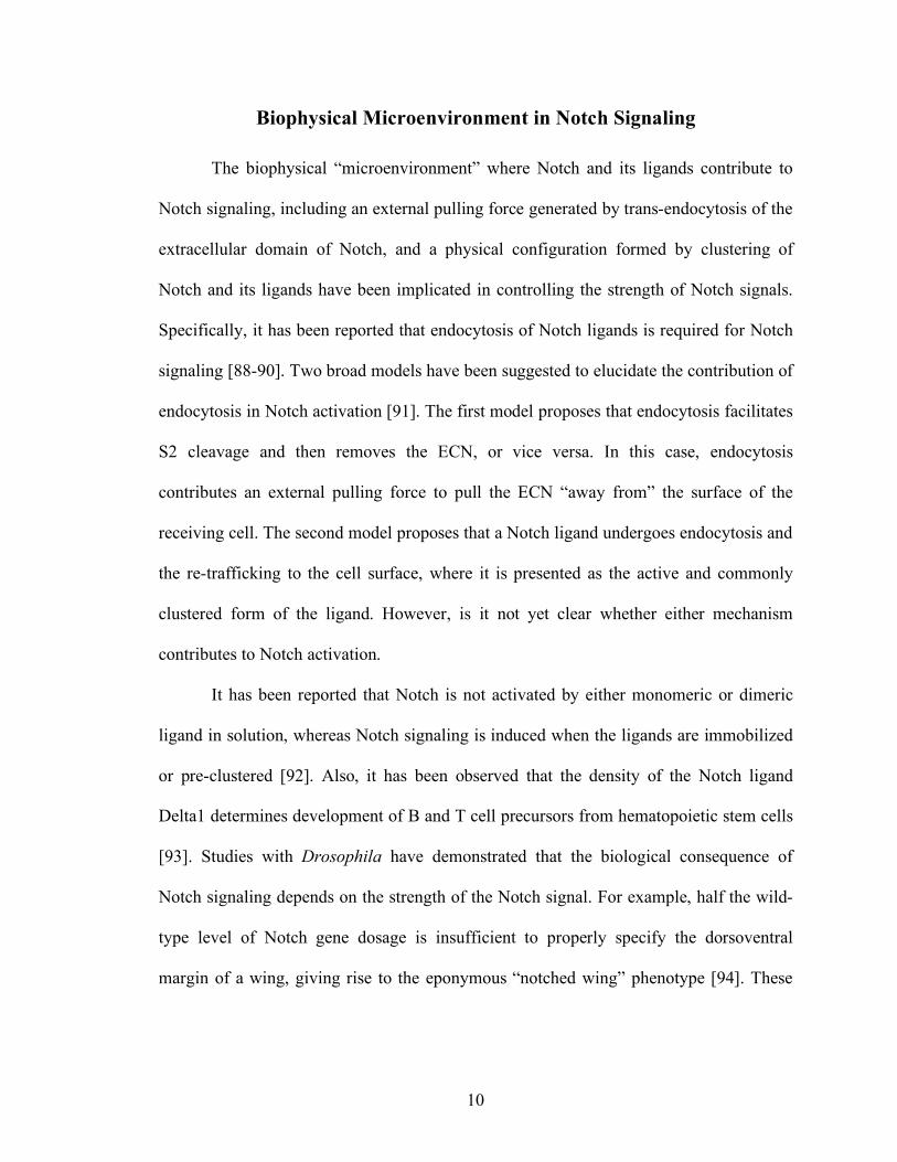
The biophysical “microenvironment” where Notch and its ligands contribute to
Notch signaling, including an external pulling force generated by trans-endocytosis of the
extracellular domain of Notch, and a physical configuration formed by clustering of
Notch and its ligands have been implicated in controlling the strength of Notch signals.
Specifically, it has been reported that endocytosis of Notch ligands is required for Notch
signaling [88-90]. Two broad models have been suggested to elucidate the contribution of
endocytosis in Notch activation [91]. The first model proposes that endocytosis facilitates
S2 cleavage and then removes the ECN, or vice versa. In this case, endocytosis
contributes an external pulling force to pull the ECN “away from” the surface of the
receiving cell. The second model proposes that a Notch ligand undergoes endocytosis and
the re-trafficking to the cell surface, where it is presented as the active and commonly
clustered form of the ligand. However, is it not yet clear whether either mechanism
contributes to Notch activation.
It has been reported that Notch is not activated by either monomeric or dimeric
ligand in solution, whereas Notch signaling is induced when the ligands are immobilized
or pre-clustered [92]. Also, it has been observed that the density of the Notch ligand
Delta1 determines development of B and T cell precursors from hematopoietic stem cells
[93]. Studies with Drosophila have demonstrated that the biological consequence of
Notch signaling depends on the strength of the Notch signal. For example, half the wild-
type level of Notch gene dosage is insufficient to properly specify the dorsoventral
margin of a wing, giving rise to the eponymous “notched wing” phenotype [94]. These
11
results imply that a critical threshold of Notch signaling is required for inducing different
cell-fate outcomes.
It is not yet known if the density of Notch ligands (perhaps controlled by
endocytosis) modulate the cell surface clustering of Notch receptors and thus modulate
Notch signal transduction strength. Similarly, it is not known if Notch molecules are
evenly distributed over the surface of the cell, or aggregated at distinct positions on the
cell surface, perhaps as a function of the state of O-glycosylation of Notch receptors,
which could in turn modulate the strength of Notch signaling in response to Notch
ligands.
A consideration of the interactions between cell surface glycans and cell surface
lectins [95, 96] suggests the possibility that Notch signaling may be facilitated by O-
fucose-linked glycan-dependent control of receptor clustering.
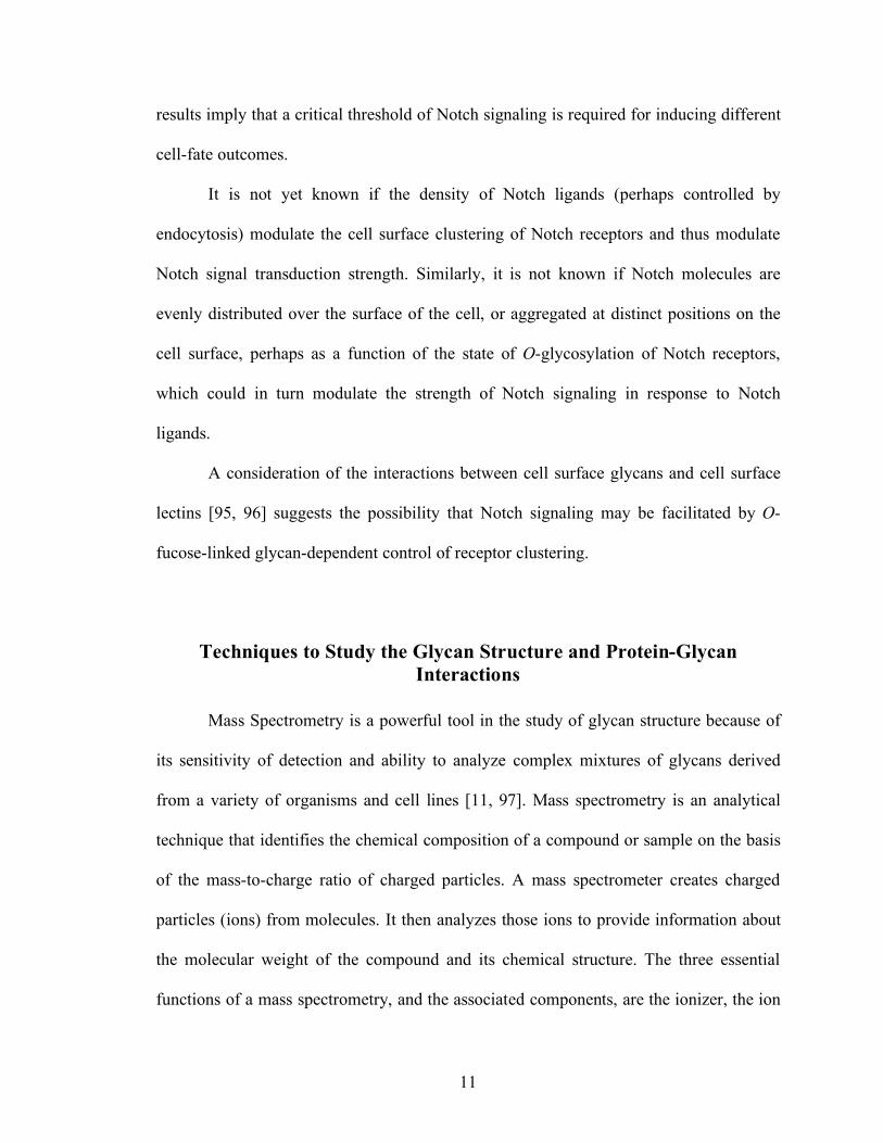
Techniques to Study the Glycan Structure and Protein-Glycan Interactions
Mass Spectrometry is a powerful tool in the study of glycan structure because of
its sensitivity of detection and ability to analyze complex mixtures of glycans derived
from a variety of organisms and cell lines [11, 97]. Mass spectrometry is an analytical
technique that identifies the chemical composition of a compound or sample on the basis
of the mass-to-charge ratio of charged particles. A mass spectrometer creates charged
particles (ions) from molecules. It then analyzes those ions to provide information about
the molecular weight of the compound and its chemical structure. The three essential
functions of a mass spectrometry, and the associated components, are the ionizer, the ion
12
analyzer, and the detector. There are many types of mass spectrometry techniques and
sample introduction techniques that allow a wide range of analyses. The Haltiwanger
group has successfully used MS analysis to identify O-linked modifications on EGF like
repeats of Notch receptor [97-99], where the glycan structure and the peptide sequence
modified with O-fucose in Notch receptors were identified from the LC-MS/MS
spectrum. Using MS, they identified only N-acetylglucosamine added onto Notch in the
presence of Fringe in Drosophila S2 cells. This result argues against models in which
subsequent modifications after Fringe is required for the Notch binding and signaling and
instead establish that the simple addition of GlcNAc is necessary and sufficient for
Drosophila Notch-ligand binding [99].
In my thesis work, I have used MS to identify the glycan structure of Notch1
receptor with various glycosylation status.
To study the protein-glycan binding, several methods have been used [100, 101],
including X-ray crystallography, NMR spectoscopy, equilibrium dialysis, affinity
chromatography, titration calorimetry, ELISA-type assays, IC50-hapten inhibition,
precipitation, electrophoresis, optical tweezers (OTs), atomic force microscopy (AFM)
and surface plasmon resonance (SPR). In my thesis work, I have used OTs, AFM, and
SPR to investigate glycan mediated Notch-Notch ligand binding events.
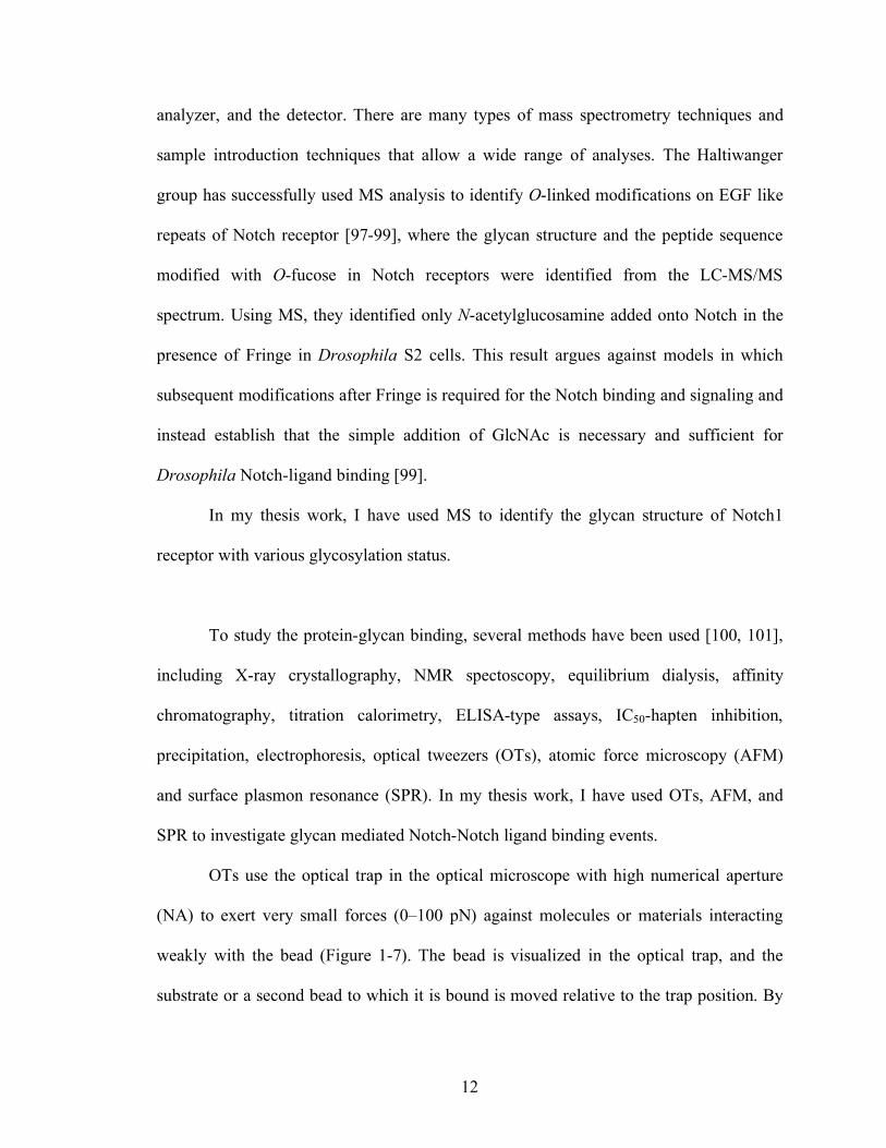
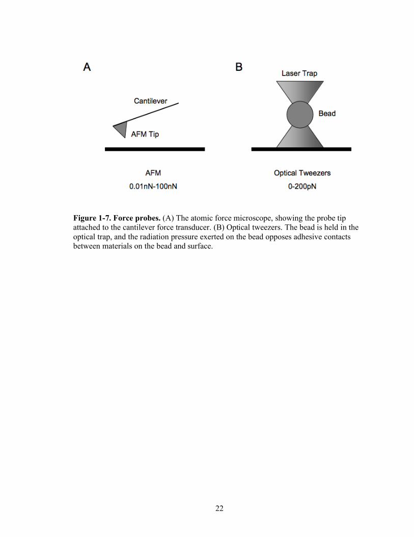
OTs use the optical trap in the optical microscope with high numerical aperture
(NA) to exert very small forces (0–100 pN) against molecules or materials interacting
weakly with the bead (Figure 1-7). The bead is visualized in the optical trap, and the
substrate or a second bead to which it is bound is moved relative to the trap position. By
13
varying the force exerted on the optically trapped bead, one can determine the force
necessary to break the bonds. Although force-distance profiles cannot be measured
accurately using OT, OTs—because of their sensitivity—have been used to measure the
force-extension profiles of soft, entropic springs such as titin [102], as well as the force-
velocity relationships of molecular motors [103-108].
AFM uses a micro-fabricated cantilever with a very low spring constant (>1
pN/nm) (Figure 1-7). This instrument has been used extensively to image soft biological
materials with a lateral resolution of ±1 nm [109]. For the force measurement of protein-
protein binding, proteins of interest are coated on the cantilever tip and the surface of
mica or glass as well. The cantilever tip is brought into the surface and makes contact.
After this contact, the cantilever is retracted to break the bond. During this event, the
position of a laser beam reflected off the cantilever surface tracks the relative movements
of the probe within ±0.1 nm and records the spring deflection. With the known spring
constant of the cantilever, the rupture force can be calculated by Hooke’s law (F=kx).
Recently, AFM has been combined with various visualizing techniques such as time-
resolved fluorescence microscopy [110] to image the lateral structure of different lipid
bilayers and their morphological changes as a function of time and combined OTs and
AFM [111] to probe fluorescence-labeled receptor clusters in the cell membrane via force
spectroscopy using antibody-functionalized tips.
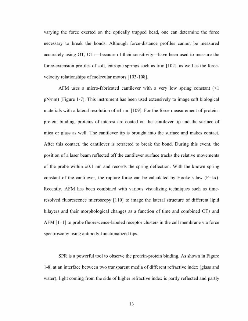
SPR is a powerful tool to observe the protein-protein binding. As shown in Figure
1-8, at an interface between two transparent media of different refractive index (glass and
water), light coming from the side of higher refractive index is partly reflected and partly
14
refracted. Above a certain critical angle of incidence, no light is refracted across the
interface, and total internal reflection is observed. While incident light is totally reflected
the electromagnetic field component penetrates a short (tens of nanometers) distance into
a medium of a lower refractive index, creating an exponentially attenuating evanescent
wave. If the interface between the media is coated with a thin layer of metal (gold), and
light is monochromatic and p-polarized, the intensity of the reflected light is reduced at a
specific incident angle producing a sharp shadow (called surface plasmon resonance) due
to the resonance energy transfer between the evanescent wave and surface plasmons. The
resonance conditions are influenced by the material adsorbed onto the thin metal film. A
satisfactory linear relationship is found between resonance energy and mass
concentration of biochemically relevant molecules such as proteins, sugars and DNA.
The SPR signal which is expressed in resonance units is therefore a measure of mass
concentration at the sensor chip surface. This means that the analyte and ligand
association and dissociation can be observed and ultimately rate constants as well as
equilibrium constants can be calculated.
SPR experiments offer several advantages for the verification and study of protein–
protein interactions. The main advantages are that very small amounts of sample are
needed (mg or sub-mg may suffice), and that no labeling is required. In addition, the
method can provide information not only on affinities but also on the rates of association
and dissociation with very high resolution in real time, which can be equally important in
biology.
15
Protein O-fucosyltransferase 1 and Fringe modulate ligand binding and Notch
signaling [21, 44]. The role of O-fucose and the Fringe action in O-fucosylation sites has
been extensively investigated [73, 87, 112]. Although it has been known that Fringe
differentially modulates Jagged/Serrate and Delta signaling through the Notch receptor
by increasing Delta signaling and reducing Jagged/Serrate signaling [82], it remains
unclear how O-linked glycan structures change the Notch-ligand binding affinity and/or
kinetics. Chapter 2 describes the construction and characterization of reagents including
recombinant cDNA and proteins that I have used to characterize Notch-ligand binding
affinity and kinetics. Chapter 3 describes the advantages and disadvantages of the usage
of OTs to study Notch-ligand binding. Chapter 4 summarizes experiments where I probed
ligand binding forces between Notch and its ligands at a single molecule level using
AFM to investigate how the glycans influence the interaction of Notch1 with its ligands.
Chapter 5 will summarize my experiments using SPR to characterize the kinetics of
Notch-ligand interactions, as a function of the state of glycosylation of both Notch and
Notch ligands, and as a function of mutations at key O-fucosylation sites. Chapter 6 will
summarize my conclusions from these studies, will place these in the context of proposals
about Notch-ligand interactions from the literature, and will provide a perspective on
future experimental directions that are suggested from our studies.
16
Figure 1-1. Glycocalyx and simplified diagram of glycocalyx. A. Scanning electron microscopic analysis of the surface of a lymphocyte stained with ruthenium red. B. The glycocalyx is made up of the oligosaccharide side chains of glycolipids and integral membrane glycoproteins and the the polysaccharide chains on integral membrane proteoglycans. (Adapted from Molecular Biology of the Cell [2])
17
Figure 1-2. Fucosylated glycans in mammals. Structures of selected fucosylated glycans are shown here, N=number of genes. (From Dr. John Lowe)
18
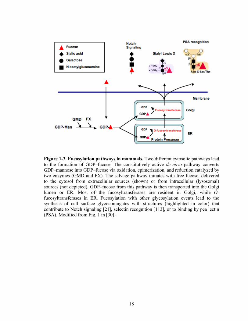
Figure 1-3. Fucosylation pathways in mammals. Two different cytosolic pathways lead to the formation of GDP–fucose. The constitutively active de novo pathway converts GDP–mannose into GDP–fucose via oxidation, epimerization, and reduction catalyzed by two enzymes (GMD and FX). The salvage pathway initiates with free fucose, delivered to the cytosol from extracellular sources (shown) or from intracellular (lysosomal) sources (not depicted). GDP–fucose from this pathway is then transported into the Golgi lumen or ER. Most of the fucosyltransferases are resident in Golgi, while O-fucosyltransferases in ER. Fucosylation with other glycosylation events lead to the synthesis of cell surface glycoconjugates with structures (highlighted in color) that contribute to Notch signaling [21], selectin recognition [113], or to binding by pea lectin (PSA). Modified from Fig. 1 in [30].
19
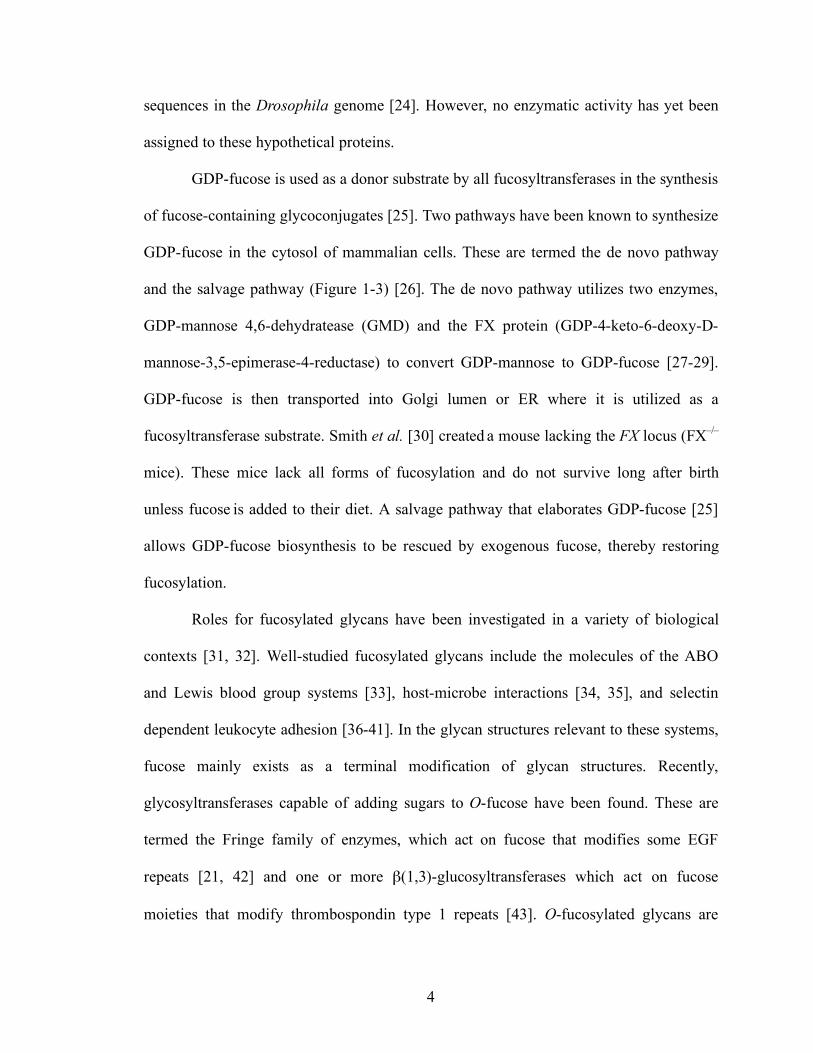
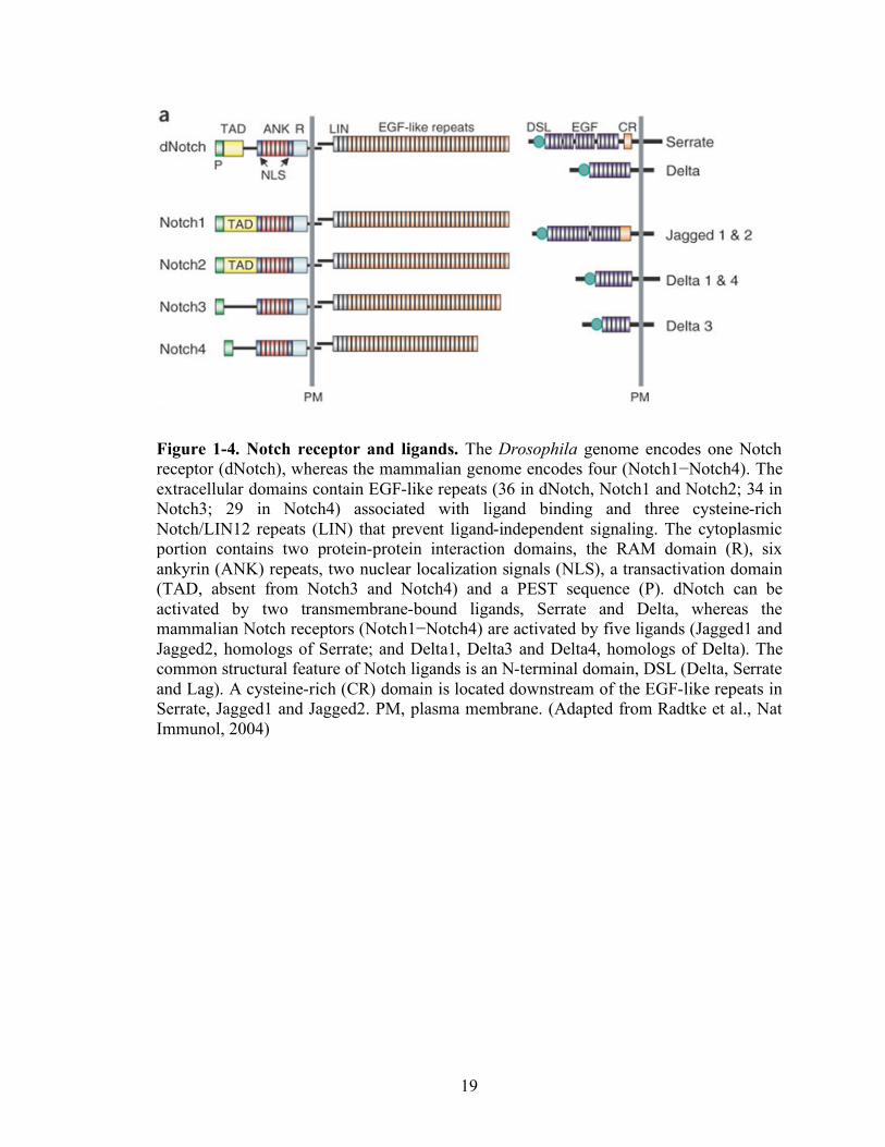
Figure 1-4. Notch receptor and ligands. The Drosophila genome encodes one Notch receptor (dNotch), whereas the mammalian genome encodes four (Notch1−Notch4). The extracellular domains contain EGF-like repeats (36 in dNotch, Notch1 and Notch2; 34 in Notch3; 29 in Notch4) associated with ligand binding and three cysteine-rich Notch/LIN12 repeats (LIN) that prevent ligand-independent signaling. The cytoplasmic portion contains two protein-protein interaction domains, the RAM domain (R), six ankyrin (ANK) repeats, two nuclear localization signals (NLS), a transactivation domain (TAD, absent from Notch3 and Notch4) and a PEST sequence (P). dNotch can be activated by two transmembrane-bound ligands, Serrate and Delta, whereas the mammalian Notch receptors (Notch1−Notch4) are activated by five ligands (Jagged1 and Jagged2, homologs of Serrate; and Delta1, Delta3 and Delta4, homologs of Delta). The common structural feature of Notch ligands is an N-terminal domain, DSL (Delta, Serrate and Lag). A cysteine-rich (CR) domain is located downstream of the EGF-like repeats in Serrate, Jagged1 and Jagged2. PM, plasma membrane. (Adapted from Radtke et al., Nat Immunol, 2004)

20
Figure 1-5. Central biochemical events in Notch signaling. Notch receptors are synthesized in ER, where Notch is O-fucosylated. Further, Notch receptors are proteolytically processed during transport to the cell surface by a furin-like protease in Golgi apparatus (at site S1), producing an extracellular Notch (ECN) subunit and a Notch transmembrane (NTM) subunit. Notch receptors are activated when the ECD interacts with DSL ligands on adjacent cells. Activation by ligand triggers a series of proteolytic cleavages of the NTM that release the ICN from the membrane. Endocytosis is required in this step. The NTM subunit is released by at least two sequential proteolytic cleavages (at site S2 and S3). The ICN then translocates to the cell nucleus, ultimately resulting in regulation of developmental control genes such as Hes1.
21
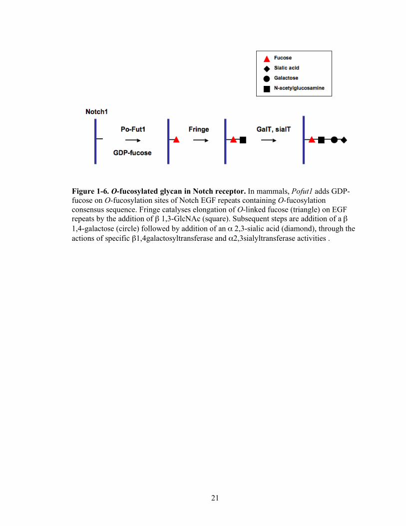
Figure 1-6. O-fucosylated glycan in Notch receptor. In mammals, Pofut1 adds GDP-fucose on O-fucosylation sites of Notch EGF repeats containing O-fucosylation consensus sequence. Fringe catalyses elongation of O-linked fucose (triangle) on EGF repeats by the addition of β 1,3-GlcNAc (square). Subsequent steps are addition of a β 1,4-galactose (circle) followed by addition of an α 2,3-sialic acid (diamond), through the actions of specific β1,4galactosyltransferase and α2,3sialyltransferase activities .
22
Figure 1-7. Force probes. (A) The atomic force microscope, showing the probe tip attached to the cantilever force transducer. (B) Optical tweezers. The bead is held in the optical trap, and the radiation pressure exerted on the bead opposes adhesive contacts between materials on the bead and surface.
23
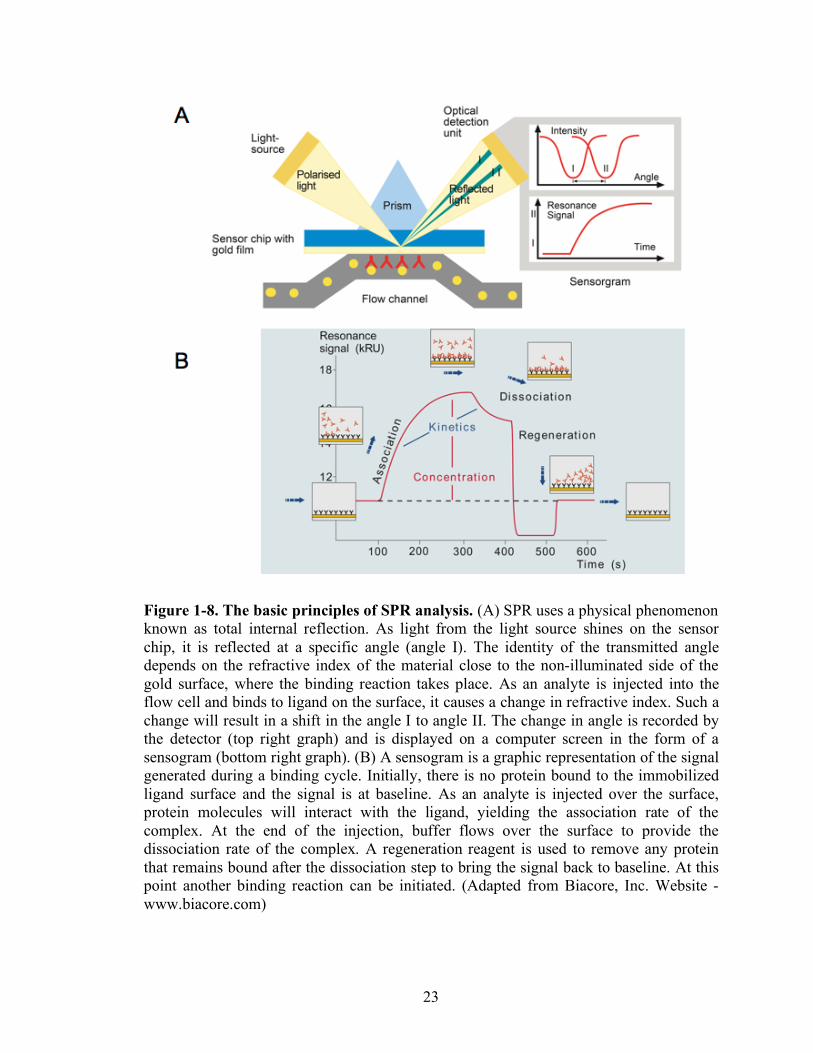
Figure 1-8. The basic principles of SPR analysis. (A) SPR uses a physical phenomenon known as total internal reflection. As light from the light source shines on the sensor chip, it is reflected at a specific angle (angle I). The identity of the transmitted angle depends on the refractive index of the material close to the non-illuminated side of the gold surface, where the binding reaction takes place. As an analyte is injected into the flow cell and binds to ligand on the surface, it causes a change in refractive index. Such a change will result in a shift in the angle I to angle II. The change in angle is recorded by the detector (top right graph) and is displayed on a computer screen in the form of a sensogram (bottom right graph). (B) A sensogram is a graphic representation of the signal generated during a binding cycle. Initially, there is no protein bound to the immobilized ligand surface and the signal is at baseline. As an analyte is injected over the surface, protein molecules will interact with the ligand, yielding the association rate of the complex. At the end of the injection, buffer flows over the surface to provide the dissociation rate of the complex. A regeneration reagent is used to remove any protein that remains bound after the dissociation step to bring the signal back to baseline. At this point another binding reaction can be initiated. (Adapted from Biacore, Inc. Website - www.biacore.com)
24
Chapter 2
Construction and Characterization of Reagents
Introduction
Notch receptors are glycoproteins which are heavily glycosylated with O-fucose
[21, 42, 68], O-glucose [69, 70], N-glycans [23, 114], and possibly O-GlcNAc [72]. O-
fucose is added to the O-fucosylation site of some EGF repeats of Notch receptors by
Pofut1 in ER and O-fucose is extended with GlcNAc by Fringe. This O-fucose-GlcNAc
is elongated more by galactose and sialic acid. O-glucose is extended by two xyloses
[115]. The structure of N-glycan that modifies Notch receptors is not well studied, and
little is known about the O-GlcNAc modification of Notch family members, beyond the
fact that this modification is present.
O-fucosylation and Fringe modification of mouse Notch1 receptors are of interest
because many in vivo and in vitro experiments suggest that O-fucosylation and
subsequent modification is critical in Notch signaling. To study the role of O-fucosylated
glycans on Notch receptors, it has proven useful to arrange to be able to precisely control
O-fucosylation of Notch. Two approaches have been used to control O-fucosylation of
Notch in mammalian cells. One is to control the synthesis of the fucosyltransferase
25
substrate GDP-fucose and another is to knock out the fucosyltransferase, including
especially Pofut1.
(i) Control of global GDP-fucose synthesis
Two pathways for the synthesis of GDP-fucose have been described in
mammalian cells. The constitutively active de novo biosynthetic pathway converts GDP-
mannose into GDP-fucose via oxidation, epimerization, and reduction catalyzed by two
enzymes (GMD and FX) (Figure 1-3, [25]). The salvage pathway initiates with free
fucose, delivered to the cytosol from extracellular sources or from intracellular
(lysosomal) sources (not depicted in Figure 1-3). GDP-fucose from this pathway is then
transported into the Golgi lumen or ER for the further process.
Several fucosylation deficient cell lines have been established: Lec13 cell lines
and CL17 cell lines. GMD enzymes are mutated in Lec13 cells [28, 116, 117] and FX
genes mutated in CL17 cells [118]. In these two cell lines, the de novo fucosylation
pathway is deficient, but the salvage pathway is intact. Therefore, fucosylation can be
controlled by adding exogenous fucose to the culture medium, which then enables GDP-
fucose synthesis through the salvage pathway.
(ii) Pofut1 knock-out
Pofut1 transfers O-fucose to EGF repeats of Notch in the ER [119]. To study the
role of O-fucose in Notch signaling, the Pofut1 locus has been genetically deleted. Mouse
embryos lacking Pofut1 die at midgestation, and exhibit severe defects in somitogenesis,
26
vasculogenesis, cardiogenesis, and neurogenesis [44]. This phenotype is similar to Notch
signaling deficient phenotype in mouse [48, 49, 120].
Drosophila Ofut1 is proposed to act as both a chaperone to promote the Notch
receptor folding and a protein O-fucosyltransferase to add fucose in EGF repeats of
Notch receptors [74]. By contrast, it has been reported that mammalian Notch receptors
are expressed at wild type levels on the cell surface in the absence of Pofut1 and O-
fucose, but exhibit reduced binding of Notch ligands and reduced ligand-induced Notch
signaling [73]. The apparent discrepancy between the consequences of Ofut1 deficiency
in Drosophila and in mammals remains unresolved, and whether mammalian Pofut1
contributes to Notch protein folding and expression is still controversial.
This Chapter will summarize the soluble Notch1 and Notch ligands that were used
in Notch-ligand binding experiments described in later chapters. Notch1 receptors were
chosen among four Notch receptors because of the importance of Notch1 receptors in
many physiological situations including T cell commitment in vivo [121, 122]. This
Chapter will also summarize experiments that were done to characterize Lec13, CL17
and CL17/Lfng cells, and their ability to support O-fucosylation of mNotch1. Finally, this
Chapter will report on the results of MS analysis done to analyze the glycan structure of
soluble mNotch1 expressed from CL17 cells.
Methods
Bir A enzyme cloning- The BirA locus (Gene Bank Number M10123) was cloned with the
primers (5’-CCC AAG CTT AAG GAT AAC ACC GTG CCA CTG AAA TTG ATT G-
27
3’ and 5’-GCA GAT ATC TTA TTT TTC TGC ACT ACG CAG GGA TAT TTC ACC-
3’) from genomic DNA of E. coli XL10 strain. 10ul of 10mM MgCl2 was additionally
added to the pfu turbo PCR reaction (Invitrogen) to optimize the PCR reaction for the
genomic DNA. The BirA locus was inserted using the HindIII and EcoRV restriction sites
into pSecTag2/HygroB expression vector (Invitrogen). This vector includes a c-Myc
epitope and a polyhistidine tag “downstream” from the site where the BirA locus was
cloned. However, the BirA insert was constructed to include a stop codon at its 3’ end, so
the c-Myc epitope and polyhistidine tag which are included in pSecTag2/HygroB will not
be expressed. BirA expressed by the pSecTag/HygroB-based vector was expressed with
an amino-terminal Ig κ-chain leader sequence, which allowed it to biotinylate membrane
and secreted proteins in mammalian hosts when those proteins include a BirA recognition
motif [123].
Site-specific mono-biotinylation system- Soluble Notch1 and Notch ligands were fused
with c-Myc tag and biotinylation tag (bioin-tag). The c-Myc tag sequence used in this
study was EQKLISEEDL and biotin tag sequence is AGGLNDIFEAQKIEWHEDTGGS.
The overall sequence is EQKLISEEDL (c-Myc)-TGG-AGG LND IFE AQK IEW HED
TGG S (biotin tag)–TAA (stop codon). This sequence was cloned between the BamHI
and XbaI sites of the vector. The primers used for the tag are 5’-/Phos/GAT CCC GAA
CAA AAA CTC ATC TCA GAA GAG GAT CTG ACT GGC GGA GCT GGC GGA
CTT AAT GAT ATT TTT GAA GCC CAG AAG ATT GAA TGG CAT GAA GAC
ACT GGT GGC TCT TAA T-3’ and 5’-/Phos/CTA GAT TAA GAG CCA CCA GTG
TCT TCA TGC CAT TCA ATC TTC TGG GCT TCA AAA ATA TCA TTA AGT CCG
28
CCA GCT CCG CCA GTC AGA TCC TCT TCT GAG ATG AGT TTT TGT TCG G-3’.
These two primers were annealed to make a double stranded DNA fragment. 5µl of 5’
primer (5µg), 5µl of 3’ primer (5µg), 8µl of water, and 2µl of 10x annealing (1M NaCl,
0.1M Tris-HCl (pH 7.8), 1mM EDTA) buffer were mixed and heated in boiling water for
5 minutes. The mixture then cooled to room temperature overnight.
To test this site-specific mono-biotinylation system, the extracelluar domain of
DLL1 (M1-H535) was appended to the c-Myc-biotin tag in pcDNA1. For the control,
lysine target for biotinylation in the biotin tag sequence was mutated to an alanine residue
to assess the monospecificity of the biotinylation system.
Generation of soluble form of mouse Notch1- Soluble mNotch1 fragments comprised of
EGF 1 through 15 (M1-E606) or EGF 1 through 36 (M1-T1633) were generated from full-
length mNotch1 (Gene Bank Number NM 008714) by PCR. These fragments were fused
in-frame to a c-Myc tag sequence and the biotin-tag sequence in expression vectors
pcDNA1 (Invitrogen) to make soluble mNotch1 with EGF 1 to15 (Notch1(15)-c-Myc-
biotin-tag) and with EGF 1 to 36 (Notch1-c-Myc-biotin-tag).
3xFlag was cloned from p3xFlag-CMV-8 (Invitrogen) with the primers, 5’-CCG
CTC GAG CCG TCA GAA TTA ATT CAC CAT GTC TGC ACT TC-3’ and 5’-GAT
CTA TCG ATG AAT TCG CGG CCG CAA G-3’, creating XhoI and NotI restriction
sites. This 3xFlag sequence has its own signal peptide from the original vector, allowing
the removal of the signal peptide from the wild type mNotch1 sequence. By mutagenesis,
a NotI restriction site was generated between the DNA sequence corresponding to amino
acid residues G52 and G59. After removing the DNA segment corresponding to the signal
29
peptide from the wild type mNotch1 between the XhoI and NotI sites, the 3xFlag tag
sequence was fused in-frame to the N-terminus of mNotch1 to create the 3xFlag-
Notch1(36)-c-Myc-biotin tag sequence.
For mass spectroscopy analysis, c-Myc and polyhistidine (6xHis) tags were fused
to soluble mNotch1 with EGF 1 to 15 (Notch1(15)-myc-His6) in pcDNA1.
Generation of soluble forms of mouse Notch ligands- Sequences encoding the
transmembrane and cytoplasmic domain were deleted from full length mouse Notch
ligands; Delta-like-1 (Gene Bank Number 007865), Delta-like-3 (Gene Bank Number
007866), Delta-like-4 (Gene Bank Number 019454), Jagged-1 (Gene Bank Number
NM013822), and Jagged-2 (Gene Bank Number 010588). A c-Myc tag sequence and a
polyhistidine sequence were fused in-frame to cDNAs encoding the extracellular domains
of each of the Notch ligands (Dll-1 (M1-H535), Dll-3 (M1-R468), Dll-4 (M1-P521), Jag-1
(M1-L1062), and Jag-2 (M1-T1068)) in expression vectors pcDNA1 (Invitrogen).
Dll-1-hIgG, Dll-4-hIgG, and Jag-1-hIgG fusion proteins were also constructed. A
cell line stably transfected with a Jag-2-hIgG fusion protein was obtained as a gift from
Dr. Bluel (Max-Planck Institute) [124]. As a negative control, a Dll4 fusion protein was
constructed in which the DLS domain was inactivated by mutation, as described by
Glittenberg et al [125]. Five residues with the DSL domain (KKRDD) were substituted
with AAAAA. In brief, two fragments were constructed independently with the primer
pairs DSL1 (5’-TTC TCG AGG CCA CCA TGA CGC CTG CGT CCC GGA GCG CCT
G-3’; 5’-TTG AAT TCA GCA GCA GCA GCA GCG CAT AGG CGA GAA CAG CTC
TCT CCA TAG TAG TTG-3’) or DSL2 (5’-TTG AAT TCG GAC ATT ATG AGT GCC

30
AGC CAG ATG GCA GCC TG-3’; 5’-CGG GAT CCG GAA ACT CGC AGC GGC
TGC CCA CAA AGC C-3’) and digested with Xho1-EcoRI and EcoRI-BamH1,
respectively. The fragments were fused together into c-Myc-His6/pcDNA1 vector.
Generation of mutations in mouse Notch1- ll mutations in the expression vector used to
generate recombinant soluble mNotch1 molecules were created using PCR-mediated site-
directed mutagenesis with Notch1(36)-c-Myc-biotin tag in pcDNA1 as template. The
mutagenesis primers were designed to generate mutations in O-fucosylation sites
(serine/threonine to alanine) based on O-fucosylation consensus sequence, The mutagenic
oligonucleotides used were 5’-CCA TGT CAG AAT GAT GCC GCT TGC CTG GAC
CAG ATT G-3’ for EGF 12, 5’-CTG CTT CAA TGG TGG TGC CTG TGT GGA TGG
TAT CAA C-3’ for EGF26, and 5’-CTG TCT GCA CGG TGG TGC CTG CCA AGA
CAG CTA TG-3’ for EGF27. All mutations were confirmed by DNA sequencing.
Generation of Fringe expression vector- cDNAs expressing Lfng (Gene Bank Number
NM 008494), Mfng (Gene Bank Number NM 00859), and Rfng gene (Gene Bank
Number NM 009053) were subcloned into plasmid pZeoSV (Invitrogen) which is a
constitutive mammalian expression vector encoding a resistant gene to the antibiotic
Zeocin.
Stable transfection of lunatic fringe- To create a CHO cell line that stably expresses Lfng,
the coding sequence of a Lfng cDNA was cloned proximal to the to sequence
corresponding to c-Myc-His6 in pcDNA1 used for soluble mNotch1 cDNA constructs.
31
Cloned Lfng fragment was constructed with primer pairs (5’-TTT AAG CTT GCC ACC
ATG CTC CAG CGG TGC GGC CGG CGC C-3’ and 5’GGA AGA TCT CCG AAG
ATG GCG GAG CGA GGA CAC CAG GGT GTG TCT G-3’).
The expression vector encoding Lfng (Lfng-myc-His6/pcDNA1) was co-
transfected into CL17 with pPUR (purimycin resistanct gene) to achieve stable
transfection. Puromycin resistant clones were selected for two weeks with 5µg/ml of
puromycin. Lfng [30]expressing CL17 was obtained by probing his-tag fused to Lfng by
western blotting.
Expression of soluble mouse Notch1 and Notch ligands- An FX null CHO cell line (CL17
cells) or a GMD null CHO cell line (Lec13 cells) was used to control the fucosylation of
mNotch1, and Notch ligands, by modulating the synthesis of GDP-fucose using
exogenous fucose in the culture media .
Expression vectors encoding the soluble mNotch1 or mNotch ligands (10µg/10cm
dish) and BirA (1µg/10cm dish) were transiently transfected into CL17 cell lines with
Lipofectamine 2000 using the manufacturer’s protocol. The next day, the cells were
transferred to 3x15cm dishes and the media was changed to the serum-free media (CD-
CHO-A, Gibco) with 1% serum and 100µM biotin. To generate fucosylated proteins,
1mM fucose was added to the media. After 5 days, the supernatant was collected for the
protein purification.
Expression of soluble Notch ligands- Expression vectors encoding the soluble Notch
ligands (10µg/10cm dish) and BirA (1µg/10cm dish) were transiently transfected into
32
HEK 293T cell lines with Lipofectamine 2000 using the manufacturer’s protocol. The
next day, the cells were transferred to 1x15cm dishes and the media was changed to
serum-free media (Hyclone). After 5 days, the supernatant was collected for protein
purification.
Protein Purification- The collected cell culture supernatants were concentrated to 5ml
using a Centricon-70 Filter unit (Millipore, MWCO 10K) and were brought up to 15ml
with binding buffer (20mM Tris, 500mM NaCl, 20mM Imidazole). 1ml of Ni-NTA
agarose beads (Invitrogen) were washed and added to the supernatant. Beads were
washed 3 times with 8ml of the binding buffer. The beads were centrifuged at 1000g for
1 minute. Protein was eluted with elution buffer (20mM Tris, 500mM NaCl, 250mM
Imidazole). Eluted protein was dialyzed 3 times with Dialysis Cassette (Pierce, MWCO
10K) in 10mM Hepes, 150mM NaCl, 1mM CaCl2. Purified proteins were analyzed by
silver staining and western blotting. Protein concentration was quantified by BCA
method or OD280 measurement.
Surface expression of mouse Notch1 receptors in 3xFlag-Notch1/Lec13 cells using EDTA
dissociation method- 3xFlag-Notch1/Lec13 and pcDNA1/Lec13 were suspended in 1x10⁶
cells. Cells were washed twice with ice cold HBSS with Ca2+ and Mg2+ followed by
centrifugation and then wash with 1ml ice cold HBSS without Ca2+ and Mg2+. Cells were
pelleted by centrifugation at 400xg for 7 min and supernatant was discarded. Cells were
re-suspended with 60 µl of 0.5mM EGTA in TBS pH 7.5 followed by incubation at 37°C
for 15 min. EDTA treated cells were centrifuged 1 time at 400xg for 7 min.
33
Supernatants were transfered to clean 1.5 ml eppendorff tubes and spun again at
800xg for 7min. Clarified supernatant was transfered to fresh tubes containing 1µl of
BSA at concentration 1mg/ml and 1ml of complete protease inhibitor with EDTA
(Roche) 60x solution. Finally, EDTA supernatants containing EDTA labial cell protein
were aliquot and snap frozen on dry ice.
Cell pellets were subjected to lysis with 60µl of RIPA buffer containing complete
protease inhibitor cocktail. Total cell protein was separated from DNA by centrifugation
at 4°C for 30 min at 14K. Aliquots of whole cell protein extracts were snap frozen on dry
ice and were stored at -80°C.
For cell surface expression of mNotch1 total cell extract was examined for protein
concentration with micro BCA protein assay kit (Pierce). Total cell extract 30µg/line or
an equivalent of EDTA released protein were separated on 4-12% NuPAGE (Invitrogen)
using MOPS electrophoresis buffer. Gels were transfered to PVDF blotting membrane.
After 8hr of blocking with 5% non-fat dry milk, membranes were washed and probed
with hamster anti human Notch1 8G10 antibody (Santa Cruz) at 1:200 dilution over night
at 4°C. Detection antibody, goat anti hamster-HRP was used for 2hr at RT. After 5
washes, each blot was developed with ECL detection system (Amersham).
Surface expression of mouse Notch1 receptors in 3xFlag-Notch1/Lec13 cells using flow
cytometry- 3xFlag-Notch1/Lec13 or pcDNA1/Lec13 were cultured in alpha-MEM with
10% dialyzed FCS without fucose at 37°C with 5% CO2 for the routine maintenance. For
fucose-dependent expression study of cell surface Notch1 and fucosylated glycoproteins,
cells were cultured in duplicate plates. Fucose was added to one plate at 1mM for 48 hr
34
before staining on alpha-MEM cultured cells. Next day cells were trypsinized followed
by centrifugation at 1200 RPM for 5 min. Cell pellets were re-suspended in CD-CHO-A
media (Gibco) and were counted and centrifuged again followed by preparing cell
suspension at concentration 0.5x10⁶ in CD-CHO-A with or without 1mM Fucose. 2 ml of
the cell suspension was plated in 35 mm tissue culture untreated dishes.
3xFlag-Notch1/Lec13 or pcDNA1/Lec13 cells cultured with or without fucose
were passed through 40µM cell strainer. Antibody staining of the cells was carried out in
V-bottom 96 well plates on ice. Cells were re-suspended in the staining media at 1x10⁶
/ml and were dispensed into V-bottom 96 well plate (0.2x10⁶ per well). Dispensed cells
were centrifuged and cell pellets were re-suspended in the staining media (HBSS with
2mM CaCl2, 1mg/ml BSA, and 0.1% NaN3) with 1: 200 dilution of Biotinylated anti-
Flag or biotinylated control antibody (mouse IgG1) and FITC conjugate-PSA lectin at
1:800 or control PSA/100mM Mannose. After 30 minutes incubation cells were
centrifuged followed by two washes and staining with Strepavidin-PE-conjugate at 1:400
for 20 minutes. PE conjugate of anti-Notch1 antibody was used at dilution 1:100. After
final step of staining protocol, cells were washed twice with the staining media and were
subjected to flow cytometry analysis on BD FACS with Cell Quest Software.
Mass Spectrometry Analysis- Mass spectral analysis of O-fucosylation sites was
performed essentially as described [98, 99]. Briefly, 1 µg of N1(15)-IgG expressed in
CL17 cells with or without fucose was reduced and alkylated, separated by SDS-PAGE,
and subjected to in-gel tryptic digestion. The resulting peptides were analyzed by LC-
MS/MS on an Agilent XCT Ion Trap mass spectrometer. Glycosylated peptides were
35
identified by searching MS/MS data for neutral losses of the GlcNAc-fucose disaccharide
(349.1 Da). Loss of the disaccharide gave a characteristic fragmentation pattern allowing
rapid identification of glycopeptides. The mass of the unglycosylated peptide was then
matched to predicted masses of tryptic peptides from Notch containing the O-fucosylation
consensus sequence:C2XXXX(S/T)C3, where C2 and C3 are the second and third conserved
cysteines of an EGF domain. Once glycopeptides were identified, additional searches of
the MS/MS data for the unmodified peptides were performed (Extracted Ion Searches).
The extracted ion searches take advantage of the fact that glycosidic linkages are more
labile than peptide bonds upon collision-induced dissociation (CID), and hence the major
product ion from fragmentation of a glycopeptide is the unglycosylated peptide. Using
this method, peptides bearing different forms of the O-fucosylated glycans (e.g. mono- or
disaccharide) were found. No glycopeptides with tri- or tetrasaccharide forms of O-fucose
were identified, nor, aside from O-glucose glycans (to be reported elsewhere) were any
other modifications of O-fucose bearing peptides identified.
Results
Specificity of mono-biotinylation
To immobilize the proteins to a surface, as a means to enable measurement of
protein-protein interactions, several methods have been suggested. Because streptavidin-
biotin bond is the strongest known protein-mediated bond in nature, systems based on the
strepavidin-biotin have been extensively used in the force-related study such as AFM
[126, 127].
36
In the streptavidin-biotin system, one protein of interest is first biotinylated and
then bound to streptavidin on a surface. A common biotinylation method uses amine
coupling with NHS. This method biotinylates all lysines in a target protein.
In our in vitro binding studies, we wished to mimic the orientation of Notch and
its ligands as presented, respectively, on the receiving cell and the sending cell. We were
thus concerned about the “orientation” of the immobilized protein as it is bound to the
solid surface, and thus sought to achieve site-specific biotinylation that would tether the
COOH-terminal end of the protein to the solid surface. We thus developed a site-specific
mono-biotinylation system as shown in Figure 2-2 and as outlined in Chapman-Smith and
Cronan [128, 129]. This system uses the BirA enzyme to add biotin to lysine in the
specific sequence. The E. coli biotin holoenzyme synthetase, BirA, catalyzes transfer of
biotin to the epsilon amino group of a specific lysine residue of the biotin carboxyl carrier
protein (BCCP) subunit of acetyl-CoA carboxylase. A 23 residue peptide has been
defined as supporting the minimal substrate requirements for BirA-catalyzed
biotinylation in vivo although the sequence of the peptide bears little resemblance to the
biotinylated sequence in BCCP [130]. This the in vivo biotinylation system allows site-
specific biotinylation without any exogenous reagents and eliminates possible
inactivation of the protein of interest by nonspecific biotinylation that is typically
achieved in vitro using chemical or enzymatic means. In order to biotinylate secreted
proteins from mammalian cells, BirA needs to be present in the secretory pathway [123].
To achieve this, BirA was inserted into a mammalian expression vector designed to target
the protein to the endoplasmic reticulum by fusion into the Igκ immunoglobulin secretory
leader.
37
To evaluate the activity and specificity of this site-specific mono-biotinylation
system, the secreted BirA (pSecBirA) was co-transfected into the HEK 293T cell lines
along with the Dll-1-cmyc-biotin tag or a mutant of Dll-1-cmyc-biotin tag designed to be
deficient in the BirA-dependent mono-biotinylation process (Figure 2-3A). In this
mutant, the lysine that is the substrate for biotinylation within the biotinylation
recognition site has been changed to alanine. The supernatants of these transfected cells
were harvested and probed by Western blotting (Figure 2-3B). The wild type
recombinant Dll-1-myc-biotin tag molecule is expressed as mono-biotinylated form,
whereas the mutant is expressed but not biotinylated. These data indicate that the in vivo
biotinylation system allows us to generate site-specific mono-biotinylated proteins.
Characterization of Lec13, CL17, and CL17/Lfng cell lines
FACS analysis with CSLEX-1, Northern blot analysis of FX transcript expression
and western blotting analysis of FX protein expression had been used to characterize the
CL17 cells in the previous study [118]. CL17 cells were generated from CHO-Tag cells,
which stably express mouse polyoma large T antigen (TAg), with a cDNA encoding
human fucosyltransferase III (FT3), an enzyme that directs synthesis of many α(1,3)-
fucosylated glycans including the sialyl LewisX epitope recognized by the monoclonal
antibody CSLEX-1. CL17 cells lack the expression of sLeX moiety when cultured in
standard medium, as assessed by flow cytometry and staining with CSLEX-1. However,
cells restored the capability of binding of CSLEX-1 in the presence of exogenous fucose,
indicating that the fucose salvage pathway is intact in CL17 and thereby fucosylation
defect is localized to the de novo pathway. Although the higher FX transcriptional level
38
was observed in CL17 cells, no detectable level of FX proteins was observed in western
blotting. By sequencing the coding region of the FX cDNA from CL17 cells, insertion of
a single G:C basepair at nucleotide position 838 was identified. This frameshift mutation
would be expected to induce protein misfolding, which could lead to degradation during
the protein synthesis process. It was confirmed that GDP-fucose was not synthesized in
CL17 cells by paper chromatography.
In this work, the fucosylation-deficient cell lines, Lec13, CL17, and CL17/Lfng
cells are further characterized by analyzing the fucosylated glycans. Two methods were
used to characterize the fucosylated glycans: fucose specific antibody binding and Notch
ligand (Dll-4) binding to Notch receptors containing fucosylated glycans.
CSLEX-1 and PSA staining to Lec13, CL17, and CL17/Lfng cells were
performed to observe whether fucosylated glycan was present on the cell surface (Figure
2-4A and B). CSLEX-1 binds to CL17+fucose and CL17/Lfng+fucose, while no
detectable level was observed from Lec13-fucose, Lec13+fucose, CL17-fucose, and
CL17/Lfng-fucose. This result indicates that Lec13 does not have the full FT activity for
the synthesis of sialyl LewisX epitope and in both cells, sialyl LewisX epitope is not fully
produced in the culture without fucose. PSA binds to all three cell lines in the presence of
fucose, while low level of binding was observed from the culture without fucose. These
data suggest that Lec13, CL17, and CL17/Lfng cells are the fucosylation deficient in
culture without fucose and can be used to control fucosylation. However, it is possible
that a very low concentration of GDP-fucose is present in these three cell lines.
Dll-4 binding to Lec13 and CL17 was carried out using flow cytometry to see
whether the exogenous fucose alters the binding to Notch receptors (Figure 2-4C).

39
Culture in the absence of exogenous fucose resulted in decreased binding to Dll-4, while
Dll-4 binding to cells was enhanced with fucose in culture. These data suggest that the
fucose may modulate Notch and Dll-4 binding, in part by regulating the surface
expression of Notch receptors or by changing the affinity of Notch receptors to Dll-4, or
both.
Surface Expression of mouse Notch1 receptors
To address the possibility that loss of fucosylation decreases the surface
expression of Notch receptors, EDTA dissociation and FACS analysis were performed
(Figure 2-5). Because of lack of anti-Notch antibody, 3xFlag tagged mouse Notch1
receptors was stably transfected in fucosylation deficient Lec13 cells. Since the Notch
receptor is a heterodimer associated with a calcum-dependent ionic bond, therefore the
ECD can be dissociated with EDTA treatment. To observe the expression of 3xFlag-
Notch1 receptors inside the cells, whole cell extracts were probed with anti-mouse Notch
antibody (8G10). The same amount of 3xFlag Notch1 was detected and no band was seen
in vector control. However, a lesser amount of 3xFlag Notch1 was detected in the culture
without fucose compared to the culture with fucose. This data demonstrate that loss of
fucosylation decreases the surface expression of Notch1 receptors.
To directly observe Notch1 receptors on the cell surface, FACS analysis was
carried out with anti-Flag antibody to detect Flag tag on Notch1 receptors Figure 2-6. To
monitor the fucosylated glycans on 3xFlag-Notch1 receptors in Lec13 cells, PSA staining
was performed. We observed that PSA binding is restored in the presence of fucose,
while PSA binding is significantly reduced in the absence of fucose. To detect the surface
40
expression of 3xFlag-Notch1, anti-Flag staining was done. Notch1 was expressed when
cultured in the presence of fucose, while the surface expression of Notch1 was
significantly reduced in the absence of fucose. Together, loss of fucosylation reduces the
surface expression of Notch receptors.
Mass Spectrometry Analysis of mouse Notch1
Our results left open the possibility that very low level of fucose is present in the
fucosylated glycans on Lec13 and CL17 cell in the fucose depleted culture. To address
this possibility, we analyzed tryptic peptides derived from mouse Notch1 by tandem mass
spectrometry (LC-MS/MS). To investigate the O-fucosylation on the EGF repeats of
mouse Notch1 receptors, two soluble mouse Notch1 peptides expressed from CL17 were
analyzed: Notch1(15)-hIgG-fucose and Notch1(15)-hIgG+fucose.
Figure 2-7 shows spectra from Notch1(15)-hIgG+fucose. In Figure 2-7A, both the
MS (top panel) and MS/MS (bottom panel) spectra show the ion from EGF3 containing
an O-fucosylation site. The top panel shows that the MS spectrum contains the O-
fucosylated peptide (m/z 866.5) that corresponds to the doubly charged form of a peptide
modified with an O-fucose monosaccharide. The MS/MS spectrum of the CID-induced
fragmentation of this ion is shown in the bottom panel. Fragmentation leads to the loss of
a dHex (fucose) (resulting in m/z 793.5 ion). Because the glycosidic linkages are
significantly more susceptible to CID than peptide bonds, the major product of
fragmentation is the deglycosylated peptide [99]. The ion at m/z 866.5 corresponds to the
predicted mass for the doubly charged form of a tryptic peptide containing an O-
fucosylation consensus sequence from EGF 3: 113NGGTCDLLTLTEYK126. These data
41
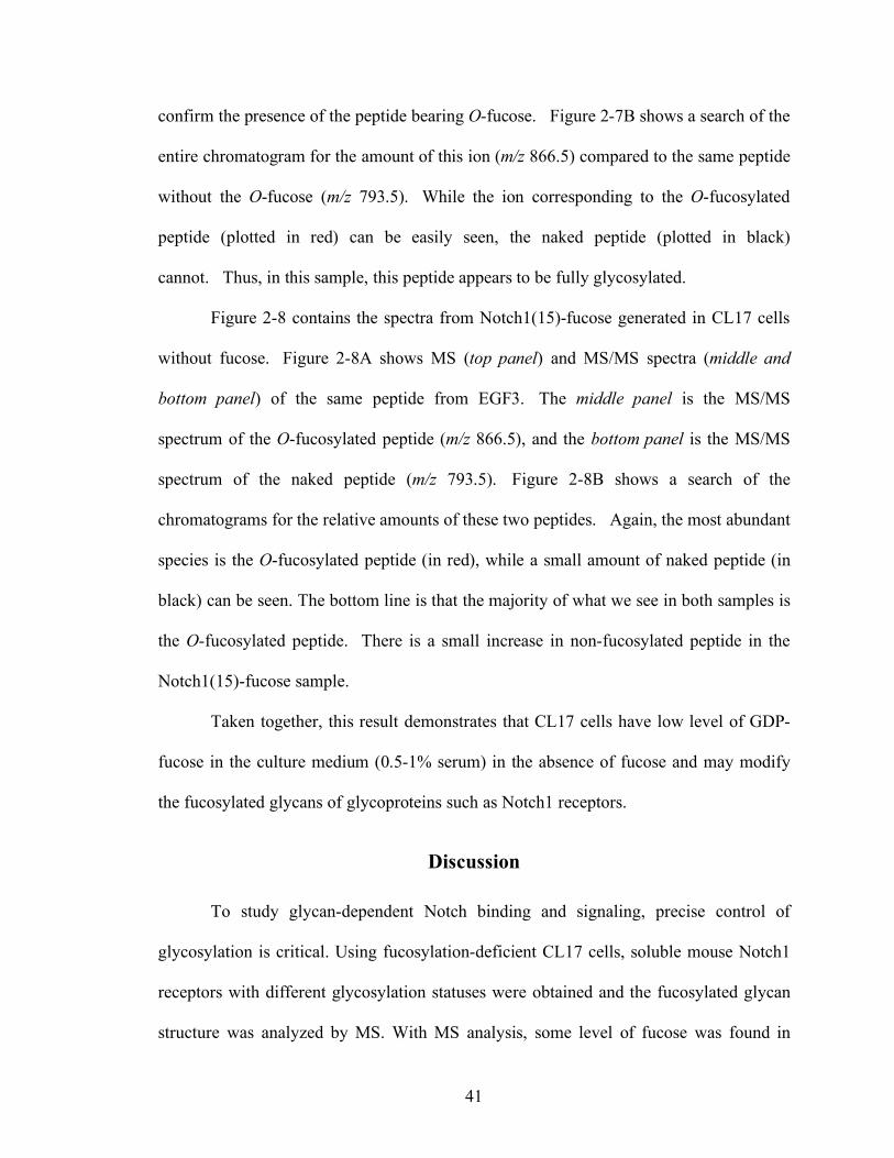
confirm the presence of the peptide bearing O-fucose. Figure 2-7B shows a search of the
entire chromatogram for the amount of this ion (m/z 866.5) compared to the same peptide
without the O-fucose (m/z 793.5). While the ion corresponding to the O-fucosylated
peptide (plotted in red) can be easily seen, the naked peptide (plotted in black)
cannot. Thus, in this sample, this peptide appears to be fully glycosylated.
Figure 2-8 contains the spectra from Notch1(15)-fucose generated in CL17 cells
without fucose. Figure 2-8A shows MS (top panel) and MS/MS spectra (middle and
bottom panel) of the same peptide from EGF3. The middle panel is the MS/MS
spectrum of the O-fucosylated peptide (m/z 866.5), and the bottom panel is the MS/MS
spectrum of the naked peptide (m/z 793.5). Figure 2-8B shows a search of the
chromatograms for the relative amounts of these two peptides. Again, the most abundant
species is the O-fucosylated peptide (in red), while a small amount of naked peptide (in
black) can be seen. The bottom line is that the majority of what we see in both samples is
the O-fucosylated peptide. There is a small increase in non-fucosylated peptide in the
Notch1(15)-fucose sample.
Taken together, this result demonstrates that CL17 cells have low level of GDP-
fucose in the culture medium (0.5-1% serum) in the absence of fucose and may modify
the fucosylated glycans of glycoproteins such as Notch1 receptors.
Discussion
To study glycan-dependent Notch binding and signaling, precise control of
glycosylation is critical. Using fucosylation-deficient CL17 cells, soluble mouse Notch1
receptors with different glycosylation statuses were obtained and the fucosylated glycan
structure was analyzed by MS. With MS analysis, some level of fucose was found in
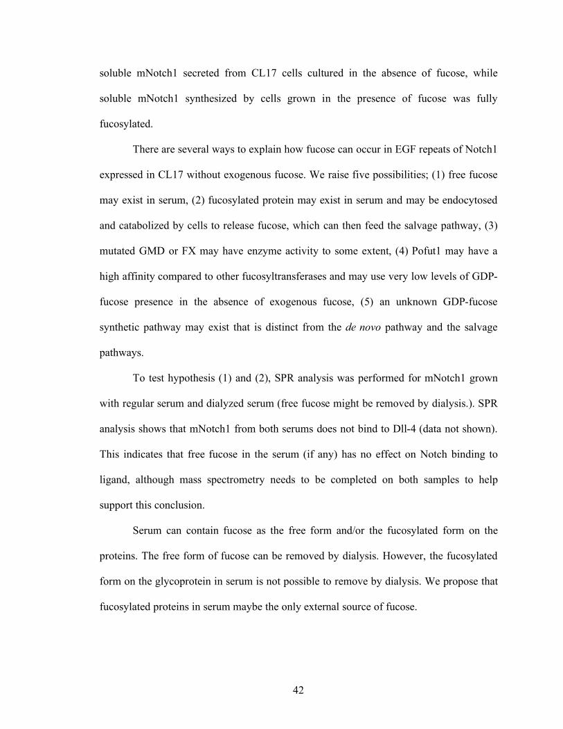
42
soluble mNotch1 secreted from CL17 cells cultured in the absence of fucose, while
soluble mNotch1 synthesized by cells grown in the presence of fucose was fully
fucosylated.
There are several ways to explain how fucose can occur in EGF repeats of Notch1
expressed in CL17 without exogenous fucose. We raise five possibilities; (1) free fucose
may exist in serum, (2) fucosylated protein may exist in serum and may be endocytosed
and catabolized by cells to release fucose, which can then feed the salvage pathway, (3)
mutated GMD or FX may have enzyme activity to some extent, (4) Pofut1 may have a
high affinity compared to other fucosyltransferases and may use very low levels of GDP-
fucose presence in the absence of exogenous fucose, (5) an unknown GDP-fucose
synthetic pathway may exist that is distinct from the de novo pathway and the salvage
pathways.
To test hypothesis (1) and (2), SPR analysis was performed for mNotch1 grown
with regular serum and dialyzed serum (free fucose might be removed by dialysis.). SPR
analysis shows that mNotch1 from both serums does not bind to Dll-4 (data not shown).
This indicates that free fucose in the serum (if any) has no effect on Notch binding to
ligand, although mass spectrometry needs to be completed on both samples to help
support this conclusion.
Serum can contain fucose as the free form and/or the fucosylated form on the
proteins. The free form of fucose can be removed by dialysis. However, the fucosylated
form on the glycoprotein in serum is not possible to remove by dialysis. We propose that
fucosylated proteins in serum maybe the only external source of fucose.
43
To quantify the fucosylation level of Lec13 and CL17 cells, soluble mNotch1 was
collected from both cell lines and we carried out a SPR binding experiment with Dll-4.
Although Lec13 cells have a very low expression level of soluble mNotch1 in the
absence of fucose, the binding kinetics and affinity of mNotch1 from Lec13 are identical
with those of mNotch1 from CL17 (data not shown). These data suggested that Lec13
and CL17 have a very low level of GDP-fucose as suggested in Kanda et al. [131]. In
Lec13 cells, 3% of GDP-fucose was found in cytoplasm compared to wild type CHO
cells even though these cells were grown in dialyzed serum [131]. Later, the Satoh group
reported that they established fucose-free cell lines [132], but fucosylation of Notch
receptor was not tested. In addition, granulocytes from FX(-/-) mice had very low level of
PSA binding, while no binding was observed to E-selectin and P-selectin binding [30]. It
demonstrated that each FT has a different affinity in the extremely low concentration of
GDP-fucose in the cytoplasm. Pofut1 may have a very high affinity for GDP-fucose,
compared to other FTs, and may be able to fucosylated Notch even in the presence of
very low concentrations of GDP-fucose. This is relevant because Drosophila Ofut1 has a
chaperone activity and mammalian Pofut1 is resident in the ER [23], the place of protein
synthesis. For proper folding, one or more specific O-fucosylation sites may serve as the
“regulatory checkpoint” for the processing of Notch receptors. However, the affinity of
Pofut1 to each EGF repeats of Notch and the O-fucosylation sites that serve as the
“regulatory checkpoint” need to be unveiled.
An unknown fucosylation pathway can be involved in the processing of the Notch
proteins. In GMD mutated CHO/DS44 cells, no detectable intracellular GDP-fucose was
44
observed using a sensitive HPLC-based detection system [131]. Therefore, the existence
of an unknown fucosylation pathway is unlikely.
Together, expression of Notch receptor is susceptible to the concentration of
GDP-fucose in the cytoplasm. This implies that Lec13 and CL17 have the capability to
transfer GDP-fucose to the EGF repeats of Notch receptors with very high affinity in an
extremely low concentration of GDP-fucose and in the extremity, Notch receptors would
not be expressed in the cultures with absolutely no GDP-fucose, at least, in mammalian
CHO cell lines.
This possibility provides a new opportunity to re-examine two strategies to
control Notch fucosylation: control of global GDP-fucose synthesis and knockout of the
Pofut1 locus. Murine ES cells lacking Pofut1 have wild type levels of cell surface Notch
receptors. However, Pofut1-/- ES cells do not bind Notch ligands or exhibit Notch
signaling [73]. This result demonstrates that Notch receptors can be processed without or,
at least with very low Pofut1 in the presence of GDP-fucose. In contrary, how does
Pofut1 process the Notch receptors in the absence of GDP-fucose? Would Notch
receptors be expressed in Pofut1-/- cells, which contain normal levels of GDP-fucose?
Functionally, these two strategies are not identical: Pofut1 without GDP-fucose and no
Pofut1 with GDP-fucose. Taken together, we propose that Pofut1 may malfunction
without GDP-fucose and induce the mis-folding of Notch receptors.
In this Chapter, we developed the soluble mNotch1 expression system using
fucosylation-deficient CL17 cells. Also, we assayed the fucosylated glycan and the
fucosylated proteins on CL17 cells by flow cytometry and characterized the glycan
structure of soluble mNotch1 by MS analysis. Because we found some level of
45
fucosylation of Notch1 receptors in CL17 grown in culture without fucose, Notch1-
fucose denotes Notch1+fucoseLow in this thesis, while Notch1+fucose and
Notch+fucose+Lfng would have wild-type glycosylation.
46
Notes to Chapter 2
1 Acknowledgements: We thank Nadia A. Rana and Dr. Robert B. Haltiwanger for the mass spectrometry analysis.
2 Attribution of data: Bronislawa Petyriak performed the experiments depicted in Figure 2-4, Figure 2-5, and Figure 2-6. Nadia A. Rana and Dr. Robert B. Haltiwanger performed the mass spectrometry analysis depicted in Figure 2-7 and Figure 2-8.
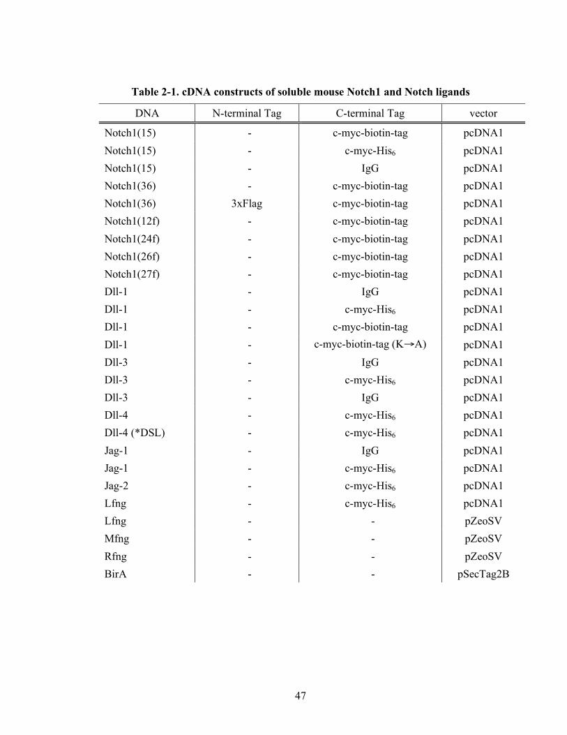
47
Table 2-1. cDNA constructs of soluble mouse Notch1 and Notch ligands
DNA N-terminal Tag C-terminal Tag vector
Notch1(15) - c-myc-biotin-tag pcDNA1 Notch1(15) - c-myc-His6 pcDNA1 Notch1(15) - IgG pcDNA1 Notch1(36) - c-myc-biotin-tag pcDNA1 Notch1(36) 3xFlag c-myc-biotin-tag pcDNA1 Notch1(12f) - c-myc-biotin-tag pcDNA1 Notch1(24f) - c-myc-biotin-tag pcDNA1 Notch1(26f) - c-myc-biotin-tag pcDNA1 Notch1(27f) - c-myc-biotin-tag pcDNA1 Dll-1 - IgG pcDNA1 Dll-1 - c-myc-His6 pcDNA1 Dll-1 - c-myc-biotin-tag pcDNA1 Dll-1 - c-myc-biotin-tag (K→A) pcDNA1 Dll-3 - IgG pcDNA1 Dll-3 - c-myc-His6 pcDNA1 Dll-3 - IgG pcDNA1 Dll-4 - c-myc-His6 pcDNA1 Dll-4 (*DSL) - c-myc-His6 pcDNA1 Jag-1 - IgG pcDNA1 Jag-1 - c-myc-His6 pcDNA1 Jag-2 - c-myc-His6 pcDNA1 Lfng - c-myc-His6 pcDNA1 Lfng - - pZeoSV Mfng - - pZeoSV Rfng - - pZeoSV BirA - - pSecTag2B
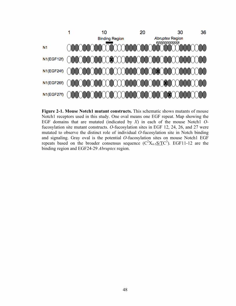
48
Figure 2-1. Mouse Notch1 mutant constructs. This schematic shows mutants of mouse Notch1 receptors used in this study. One oval means one EGF repeat. Map showing the EGF domains that are mutated (indicated by X) in each of the mouse Notch1 O-fucosylation site mutant constructs. O-fucosylation sites in EGF 12, 24, 26, and 27 were mutated to observe the distinct role of individual O-fucosylation site in Notch binding and signaling. Gray oval is the potential O-fucosylation sites on mouse Notch1 EGF repeats based on the broader consensus sequence (C2X4-5S/TC3). EGF11-12 are the binding region and EGF24-29 Abruptex region.
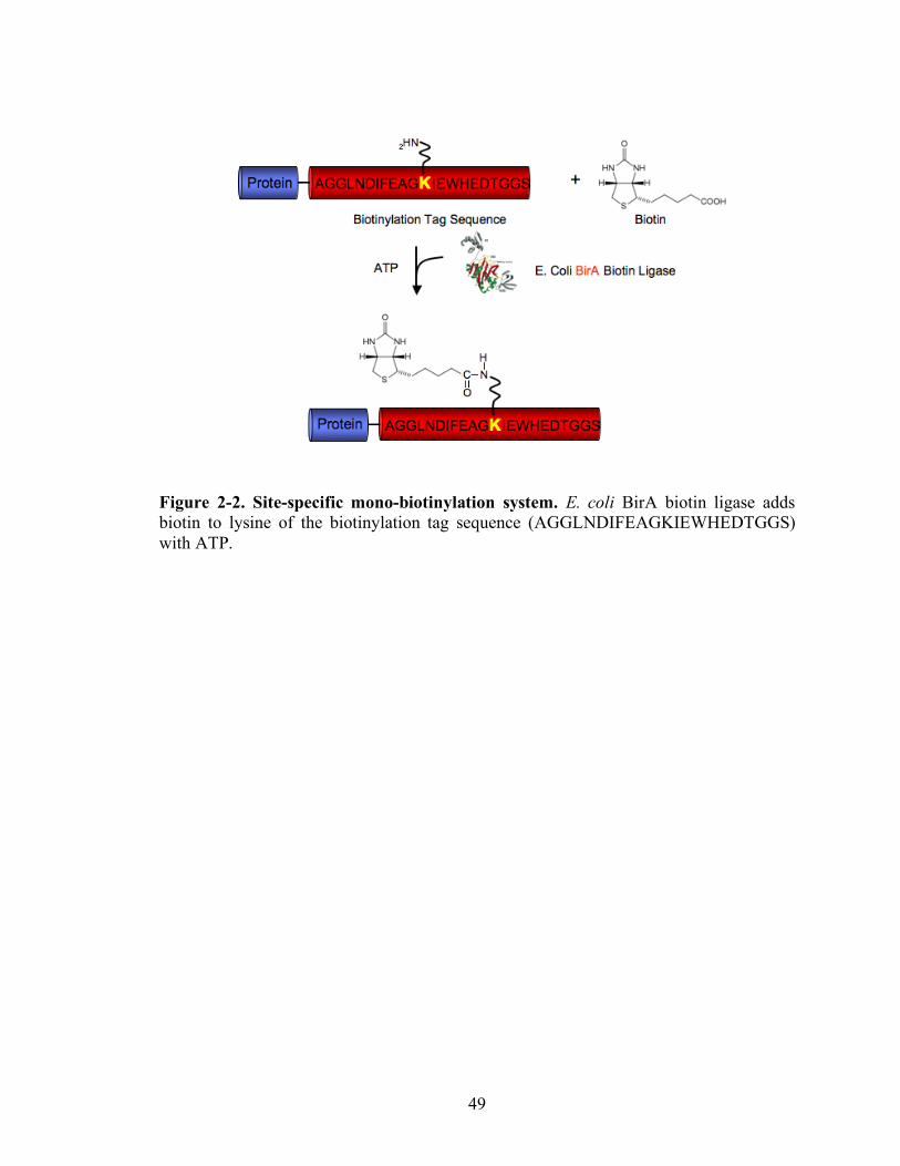
49
Figure 2-2. Site-specific mono-biotinylation system. E. coli BirA biotin ligase adds biotin to lysine of the biotinylation tag sequence (AGGLNDIFEAGKIEWHEDTGGS) with ATP.
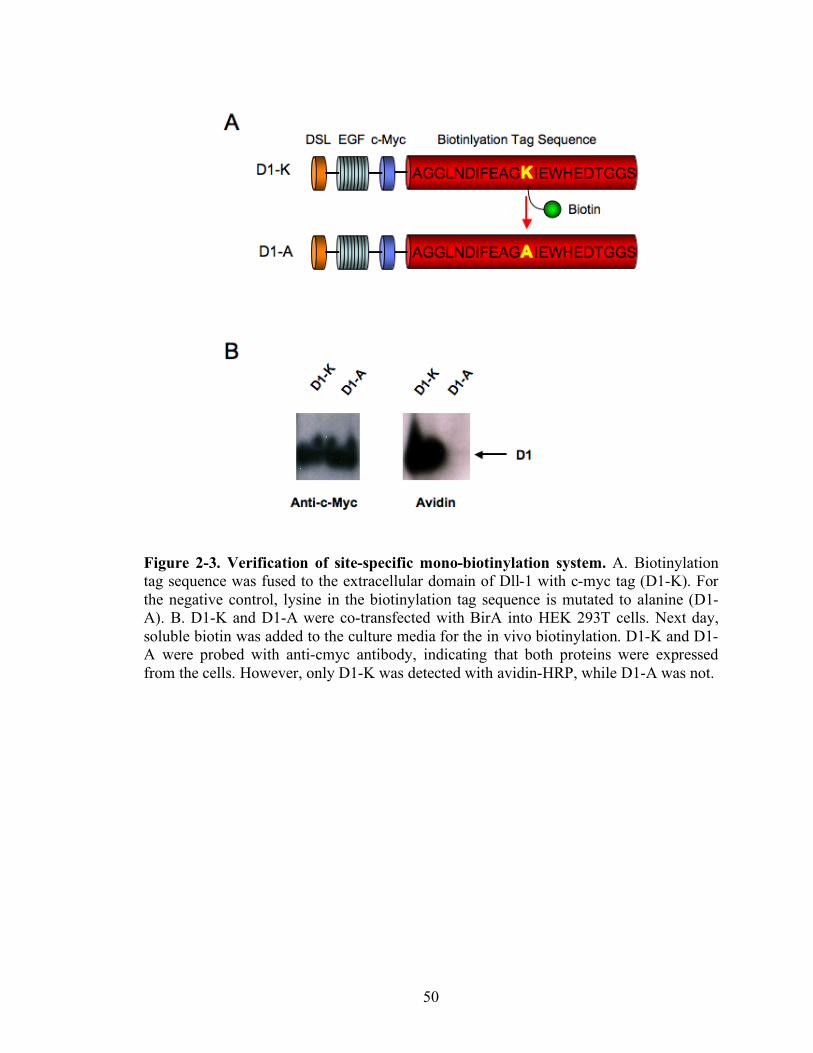
50
Figure 2-3. Verification of site-specific mono-biotinylation system. A. Biotinylation tag sequence was fused to the extracellular domain of Dll-1 with c-myc tag (D1-K). For the negative control, lysine in the biotinylation tag sequence is mutated to alanine (D1-A). B. D1-K and D1-A were co-transfected with BirA into HEK 293T cells. Next day, soluble biotin was added to the culture media for the in vivo biotinylation. D1-K and D1-A were probed with anti-cmyc antibody, indicating that both proteins were expressed from the cells. However, only D1-K was detected with avidin-HRP, while D1-A was not.
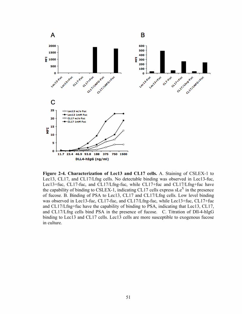
51
Figure 2-4. Characterization of Lec13 and CL17 cells. A. Staining of CSLEX-1 to Lec13, CL17, and CL17/Lfng cells. No detectable binding was observed in Lec13-fuc, Lec13+fuc, CL17-fuc, and CL17/Lfng-fuc, while CL17+fuc and CL17/Lfng+fuc have the capability of binding to CSLEX-1, indicating CL17 cells express sLeX in the presence of fucose. B. Binding of PSA to Lec13, CL17 and CL17/Lfng cells. Low level binding was observed in Lec13-fuc, CL17-fuc, and CL17/Lfng-fuc, while Lec13+fuc, CL17+fuc and CL17/Lfng+fuc have the capability of binding to PSA, indicating that Lec13, CL17, and CL17/Lfng cells bind PSA in the presence of fucose. C. Titration of Dll-4-hIgG binding to Lec13 and CL17 cells. Lec13 cells are more susceptible to exogenous fucose in culture.
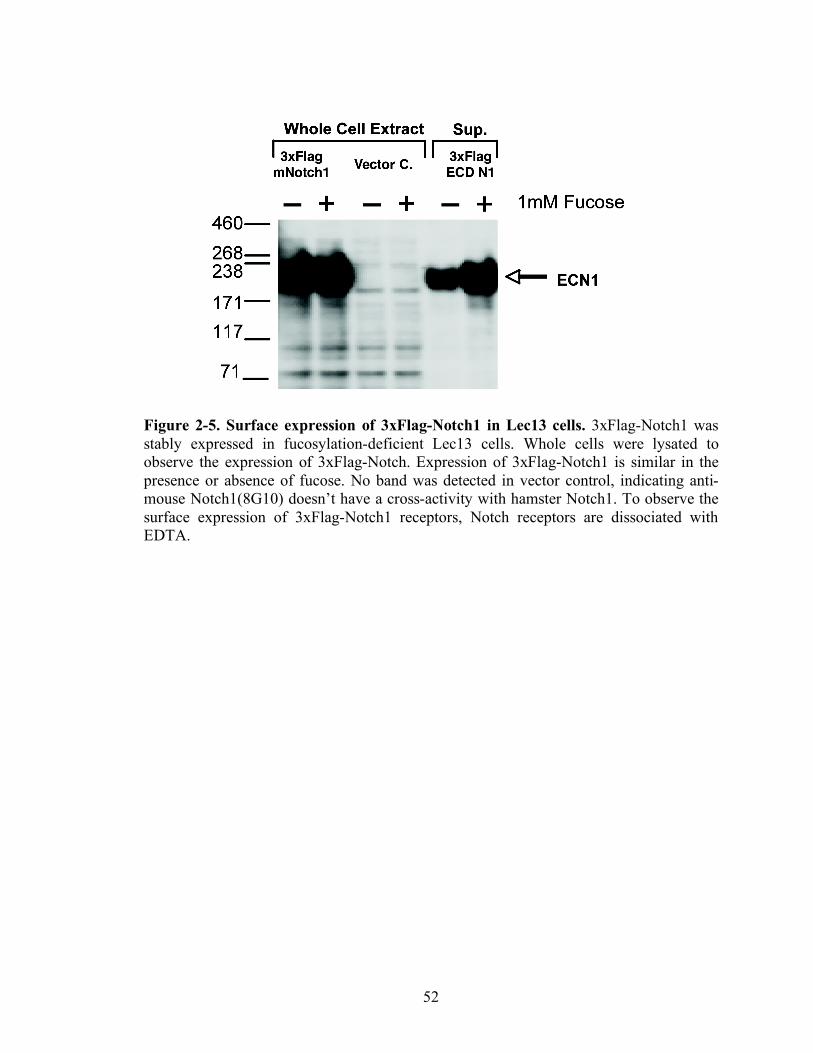
52
Figure 2-5. Surface expression of 3xFlag-Notch1 in Lec13 cells. 3xFlag-Notch1 was stably expressed in fucosylation-deficient Lec13 cells. Whole cells were lysated to observe the expression of 3xFlag-Notch. Expression of 3xFlag-Notch1 is similar in the presence or absence of fucose. No band was detected in vector control, indicating anti-mouse Notch1(8G10) doesn’t have a cross-activity with hamster Notch1. To observe the surface expression of 3xFlag-Notch1 receptors, Notch receptors are dissociated with EDTA.
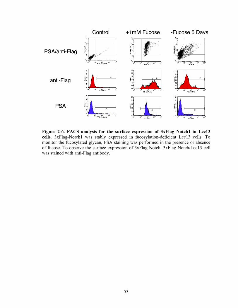
53
Figure 2-6. FACS analysis for the surface expression of 3xFlag Notch1 in Lec13 cells. 3xFlag-Notch1 was stably expressed in fucosylation-deficient Lec13 cells. To monitor the fucosylated glycan, PSA staining was performed in the presence or absence of fucose. To observe the surface expression of 3xFlag-Notch, 3xFlag-Notch/Lec13 cell was stained with anti-Flag antibody.
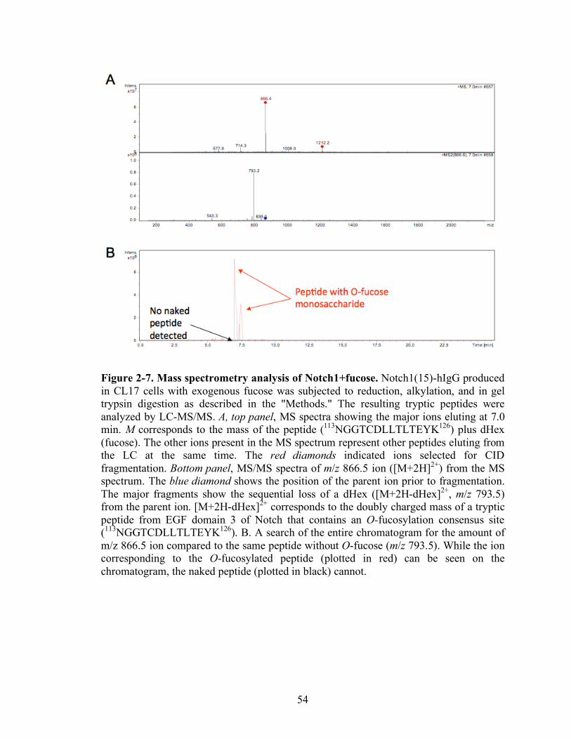
54
Figure 2-7. Mass spectrometry analysis of Notch1+fucose. Notch1(15)-hIgG produced in CL17 cells with exogenous fucose was subjected to reduction, alkylation, and in gel trypsin digestion as described in the "Methods." The resulting tryptic peptides were analyzed by LC-MS/MS. A, top panel, MS spectra showing the major ions eluting at 7.0 min. M corresponds to the mass of the peptide (113NGGTCDLLTLTEYK126) plus dHex (fucose). The other ions present in the MS spectrum represent other peptides eluting from the LC at the same time. The red diamonds indicated ions selected for CID fragmentation. Bottom panel, MS/MS spectra of m/z 866.5 ion ([M+2H]2+) from the MS spectrum. The blue diamond shows the position of the parent ion prior to fragmentation. The major fragments show the sequential loss of a dHex ([M+2H-dHex]2+, m/z 793.5) from the parent ion. [M+2H-dHex]2+ corresponds to the doubly charged mass of a tryptic peptide from EGF domain 3 of Notch that contains an O-fucosylation consensus site (113NGGTCDLLTLTEYK126). B. A search of the entire chromatogram for the amount of m/z 866.5 ion compared to the same peptide without O-fucose (m/z 793.5). While the ion corresponding to the O-fucosylated peptide (plotted in red) can be seen on the chromatogram, the naked peptide (plotted in black) cannot.
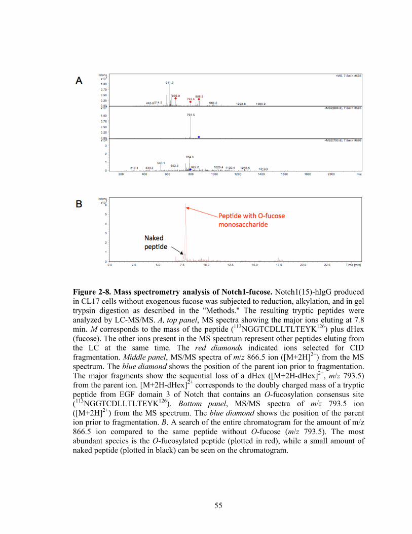
55
Figure 2-8. Mass spectrometry analysis of Notch1-fucose. Notch1(15)-hIgG produced in CL17 cells without exogenous fucose was subjected to reduction, alkylation, and in gel trypsin digestion as described in the "Methods." The resulting tryptic peptides were analyzed by LC-MS/MS. A, top panel, MS spectra showing the major ions eluting at 7.8 min. M corresponds to the mass of the peptide (113NGGTCDLLTLTEYK126) plus dHex (fucose). The other ions present in the MS spectrum represent other peptides eluting from the LC at the same time. The red diamonds indicated ions selected for CID fragmentation. Middle panel, MS/MS spectra of m/z 866.5 ion ([M+2H]2+) from the MS spectrum. The blue diamond shows the position of the parent ion prior to fragmentation. The major fragments show the sequential loss of a dHex ([M+2H-dHex]2+, m/z 793.5) from the parent ion. [M+2H-dHex]2+ corresponds to the doubly charged mass of a tryptic peptide from EGF domain 3 of Notch that contains an O-fucosylation consensus site (113NGGTCDLLTLTEYK126). Bottom panel, MS/MS spectra of m/z 793.5 ion ([M+2H]2+) from the MS spectrum. The blue diamond shows the position of the parent ion prior to fragmentation. B. A search of the entire chromatogram for the amount of m/z 866.5 ion compared to the same peptide without O-fucose (m/z 793.5). The most abundant species is the O-fucosylated peptide (plotted in red), while a small amount of naked peptide (plotted in black) can be seen on the chromatogram.
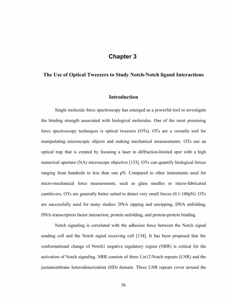
56
Chapter 3
The Use of Optical Tweezers to Study Notch-Notch ligand Interactions
Introduction
Single molecule force spectroscopy has emerged as a powerful tool to investigate
the binding strength associated with biological molecules. One of the most promising
force spectroscopy techniques is optical tweezers (OTs). OTs are a versatile tool for
manipulating microscopic objects and making mechanical measurements. OTs use an
optical trap that is created by focusing a laser to diffraction-limited spot with a high
numerical aperture (NA) microscope objective [133]. OTs can quantify biological forces
ranging from hundreds to less than one pN. Compared to other instruments used for
micro-mechanical force measurement, such as glass needles or micro-fabricated
cantilevers, OTs are generally better suited to detect very small forces (0.1-100pN). OTs
are successfully used for many studies: DNA zipping and unzipping, DNA unfolding,
DNA-transcription factor interaction, protein unfolding, and protein-protein binding.
Notch signaling is correlated with the adhesion force between the Notch signal
sending cell and the Notch signal receiving cell [134]. It has been proposed that the
conformational change of Notch1 negative regulatory region (NRR) is critical for the
activation of Notch signaling. NRR consists of three Lin12/Notch repeats (LNR) and the
juxtamembrane heterodimerization (HD) domain. Three LNR repeats cover around the
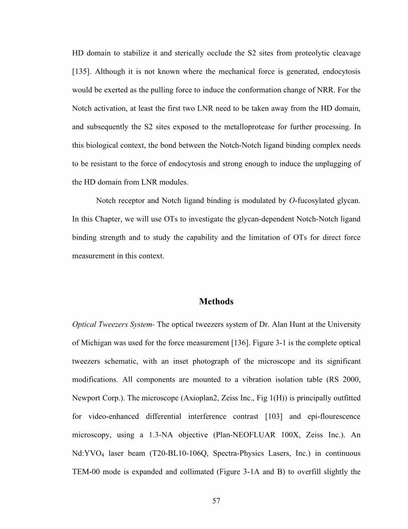
57
HD domain to stabilize it and sterically occlude the S2 sites from proteolytic cleavage
[135]. Although it is not known where the mechanical force is generated, endocytosis
would be exerted as the pulling force to induce the conformation change of NRR. For the
Notch activation, at least the first two LNR need to be taken away from the HD domain,
and subsequently the S2 sites exposed to the metalloprotease for further processing. In
this biological context, the bond between the Notch-Notch ligand binding complex needs
to be resistant to the force of endocytosis and strong enough to induce the unplugging of
the HD domain from LNR modules.
Notch receptor and Notch ligand binding is modulated by O-fucosylated glycan.
In this Chapter, we will use OTs to investigate the glycan-dependent Notch-Notch ligand
binding strength and to study the capability and the limitation of OTs for direct force
measurement in this context.
Methods
Optical Tweezers System- The optical tweezers system of Dr. Alan Hunt at the University
of Michigan was used for the force measurement [136]. Figure 3-1 is the complete optical
tweezers schematic, with an inset photograph of the microscope and its significant
modifications. All components are mounted to a vibration isolation table (RS 2000,
Newport Corp.). The microscope (Axioplan2, Zeiss Inc., Fig 1(H)) is principally outfitted
for video-enhanced differential interference contrast [103] and epi-flourescence
microscopy, using a 1.3-NA objective (Plan-NEOFLUAR 100X, Zeiss Inc.). An
Nd:YVO4 laser beam (T20-BL10-106Q, Spectra-Physics Lasers, Inc.) in continuous
TEM-00 mode is expanded and collimated (Figure 3-1A and B) to overfill slightly the
58
back aperture of the microscope’s objective. To create two noninterfering, independent
traps, a polarizing beam splitting cube, preceded by a λ/2 wave plate, divides the laser
into orthogonally polarized beams (Figure 3-1C). A liquid crystal attenuation system
(LCR-IR05m Newport Corp.) controls the laser power for each beam (Figure 3-1D). A
two axis acousto-optic deflection system (DTD-406BA6, IntraAction Corp.) steers each
beam through 1:1 telescope lenses into microscope (Figure 3-1E and F). To preserve the
polarity of the laser beams, before he entry into the microscope a λ/2 wave plate rotates
the two beams to meet the orthogonal ordinary and extraordinary axes of the Wollaston
prisms (Figure 3-1G). After existing the microscope, the beams are separated by polarity
and terminate onto separate QPDs (Spot 9DMI, UDT Instruments) located at a plane
made conjugate with the back focal plane of the microscope’s condenser (Figure 3-1I).
Differential amplifiers amplify the QPD signal and produce an output voltage
proportional to position with a bandwidth of at 7kHz. A data acquisition card digitizes
this signal at up to 100kHz (PCI-MIO-16XE10, National Insruments), and software
analyzes the signal and records it to disk (LabVIEW).
Channel Preparation- As shown in Figure 3-3, double-sided scotch tape (3M) was put on
the glass slide. The glass coverslip was put on top of the tape. After the bead solutions
were injected, microscope oil was dropped at both ends of the channel to prevent the
evaporation of the liquid solution.
Protein Immobilization- Streptavidin coated polystylene beads were purchased from
Bangs Lab. Soluble forms of mNotch1 and each of ligands were constructed and coated
59
on beads. The density of the protein on the bead surface was measured by flow cytometry
with anti-c-Myc antibody (data not shown).
Solid-phase Binding Assay- Bead solution with soluble Notch1 was injected into the
channel and incubated for 5 min upside down to immobilize the bead on the coverslip.
Bead coated with Notch ligands was allowed to flow inside the chamber and grabbed
with the laser trap for further manipulation (Figure 3-3). Trap stiffness (k) was measured
and the bead position (x) was recorded to calculate the rupture force by Hooke’s law,
F=kx.
Results and Discussion
To study the binding strength between glycan-dependent Notch-Notch ligands,
optical tweezers were used. Soluble mNotch1 was immobilized on the 2µm beads and
soluble Dll-11 was on the 5.5µm beads. Dll-1 coated bead was attached on the coverslip
and the mNotch1 coated bead was grabbed by the laser trap. The mNotch1 coated bead
approached to the Dll-1 coated beads to form a bond and subsequently retracted to break
the bond (Figure 3-2). Although the software was not programmed with the continuous
pulling, binding and rupture events were observed with optical tweezers. Before
developing this system further, we found two issues in this two-bead system: a sample
heating problem [137] and optical interference [138].
Sample Heating Problem- Output power of the trapping laser and the throughput of the
optical system determine the maximum attainable stiffness and force. Trapping forces
60
depend on multiple parameters and are difficult to calculate for most conditions of
practical interest. Generally speaking, maximum trapping forces on the order of 1 pN per
10 mW of power delivered to the specimen plane can be achieved with micron-scale
beads [139].
To generate around 100pN force with optical tweezers, 1W power is needed. That
amount of power can easily induce protein sample heating that can give damage to the
protein because in the focused trap the temperature is increased by 7.7 ± 1.2 K/W for
500-nm silica beads and 8.1 ± 2.1 K/W for 444-nm silica beads in water [137]. Even
though the heating effect in water is rather small it can have non-negligible effects on
trap calibration in typical biophysical experimental circumstances and should be taken
into consideration when laser powers of more than 100 mW are used.
The high intensity at the focus of the trapping laser that forms the optical trap,
typically 109–1012 W cm- 1, results in local heating. Trapping transparent dielectric
particles in water at a trapping wavelength of 1,064 nm results in modest heating on the
order of 1 °C per 100 mW of power in the specimen plane. Trapping absorbing particles
or trapping at laser wavelengths more strongly absorbed by water can result in
considerably more heating. Local heating can influence enzymatic activity and change
the local viscosity of the medium, whereas steep thermal gradients may produce
convection currents that can adversely affect the measurements. Local heating in the
vicinity of the optical trap can be calculated, and several techniques have been developed
to measure the temperature directly [133, 137].
Optimal operational range of optical tweezers is 0-100pN, while the single
binding force of protein-protein interaction is 50-200pN. Practically, multiple bonds may
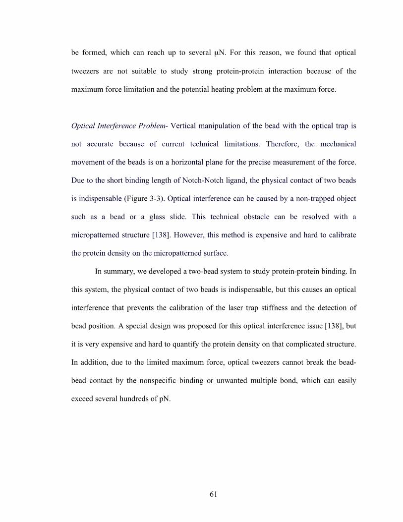
61
be formed, which can reach up to several µN. For this reason, we found that optical
tweezers are not suitable to study strong protein-protein interaction because of the
maximum force limitation and the potential heating problem at the maximum force.
Optical Interference Problem- Vertical manipulation of the bead with the optical trap is
not accurate because of current technical limitations. Therefore, the mechanical
movement of the beads is on a horizontal plane for the precise measurement of the force.
Due to the short binding length of Notch-Notch ligand, the physical contact of two beads
is indispensable (Figure 3-3). Optical interference can be caused by a non-trapped object
such as a bead or a glass slide. This technical obstacle can be resolved with a
micropatterned structure [138]. However, this method is expensive and hard to calibrate
the protein density on the micropatterned surface.
In summary, we developed a two-bead system to study protein-protein binding. In
this system, the physical contact of two beads is indispensable, but this causes an optical
interference that prevents the calibration of the laser trap stiffness and the detection of
bead position. A special design was proposed for this optical interference issue [138], but
it is very expensive and hard to quantify the protein density on that complicated structure.
In addition, due to the limited maximum force, optical tweezers cannot break the bead-
bead contact by the nonspecific binding or unwanted multiple bond, which can easily
exceed several hundreds of pN.
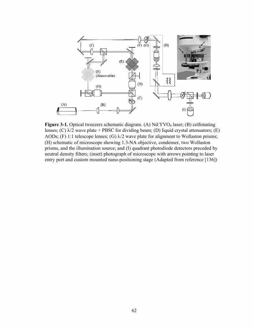
62
Figure 3-1. Optical tweezers schematic diagram. (A) Nd:YVO4 laser; (B) collimating lenses; (C) λ/2 wave plate + PBSC for dividing beam; (D) liquid crystal attenuators; (E) AODs; (F) 1:1 telescope lenses; (G) λ/2 wave plate for alignment to Wollaston prisms; (H) schematic of microscope showing 1.3-NA objective, condenser, two Wollaston prisms, and the illumination source; and (I) quadrant photodiode detectors preceded by neutral density filters; (inset) photograph of microscope with arrows pointing to laser entry port and custom mounted nano-positioning stage (Adapted from reference [136])
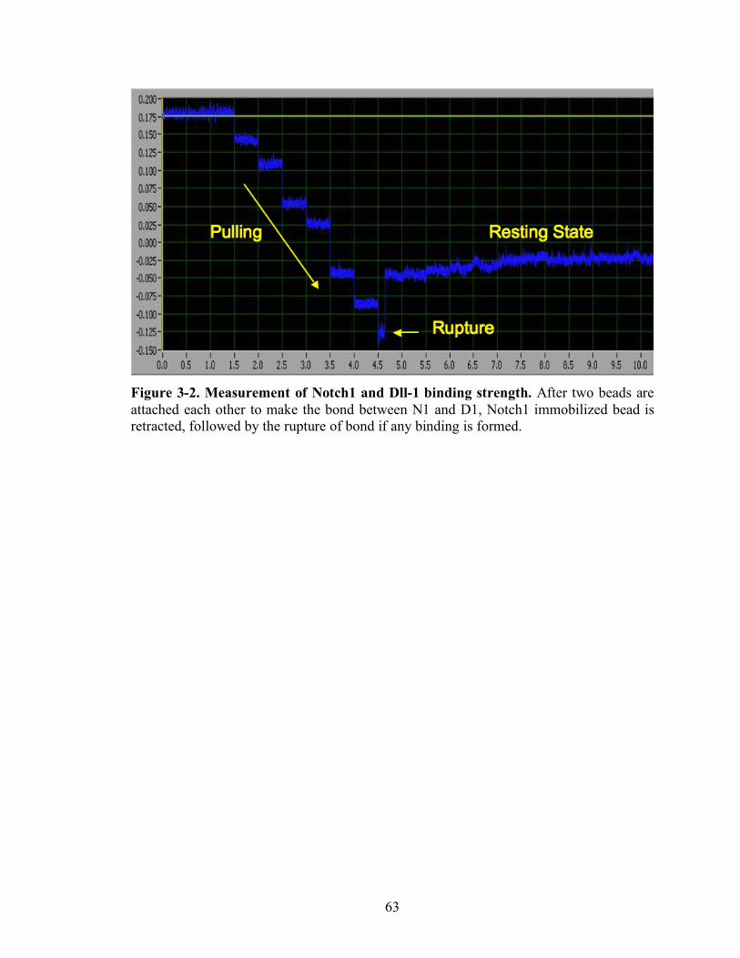
63
Figure 3-2. Measurement of Notch1 and Dll-1 binding strength. After two beads are attached each other to make the bond between N1 and D1, Notch1 immobilized bead is retracted, followed by the rupture of bond if any binding is formed.
64
Figure 3-3. Optical Interference in the two bead system. A big bead is stuck on the surface of a glass coverslip, which is attached to a glass slide with 3M double side tape. As the small bead approaches to the big bead, the path of the laser is interfered by the other bead. This optical interference alters the calibration of stiffness of the optical trap.
65
Chapter 4
Direct Force Measurement of Notch-Notch Ligand Interactions using Atomic Force Microscopy (AFM)
Introduction
The atomic force microscope is a version of the scanning probe microscope in
which the properties of the surface are investigated with a micromachined probe [140].
This technique allows mapping of the surface characteristics at sub-nanometer resolution.
The possibility of modifying the surface and manipulating individual molecules made
AFM an ideal tool for biological applications. One important advantage of the technique
is simple and rapid sample preparation. Another important feature of AFM is the ability
to conduct measurements of biological samples under near-physiological conditions
[141].
Although the atomic force microscope is primarily an imaging tool, it also allows
measurement of inter- and intramolecular interaction forces with piconewton resolution.
The vertical motion of the cantilever is controlled by piezoelectric actuators with sub-
nanometer resolution. The displacement of the cantilever is monitored directly with either
a capacitor or a linear voltage differential transformer. As a result, high-resolution force-
versus-extension curves of single molecules can be recorded. This force-extension
66
relationship provides valuable information on the structure, the folding and unfolding
processes and even the activity of the molecule. Measurement of the stretching curve is
relatively straightforward. The AFM tip is lowered toward the surface or the sample is
lifted toward the cantilever tip by piezoelectric actuators. After the initial contact with the
surface, the cantilever is pressed into the surface on which the sample is deposited with a
predetermined constant force. The tip is then retracted. Attachment of the sample (RNA,
DNA or protein) tethers the cantilever tip to the surface, causing the cantilever to bend
toward the surface as the tip is retracted. The value of this force is obtained by Hooke's
Law, F=kx, where F is the rupture force, k the spring constant of the AFM cantilever, and
x displacement. In most cases the force-versus-extension curve displays nonlinear
behavior indicative of an entropic spring, which is well described by the worm-like chain
(WLC) entropic elasticity model. AFM first used to measure the forces between
complementary strands of DNA [142]. Later, AFM has been successfully used to probe
protein-protein binding [143-147] and glycan-mediated binding [127].
The following technical requirements must be satisfied to immobilize proteins on
these surfaces: (a) avoidance of surface-induced denaturation and mechanical force (or
indentation)-induced conformational deformation, (b) minimized nonspecific adhesive
interactions with the bare probe surface or bare substrate surface, and (c) enhanced
motional freedom of proteins after their immobilization on the surface to increase the
binding chance [146]. Spacer chemistry has been proposed to meet these requirements,
which includes end-functionalized, flexible polymeric spacer such as poly(ethylene
glycol) (PEG) [143]. Moreover, the elastic behavior of polystyrene glycol (PEG) has
been well-characterized. Therefore, polymer linkages are very useful for isolation
67
reactive sites in single molecule experiments as well as for suppression of rebinding
events. However, it is possible that polymer linkages to stiff probes can introduce
unexpected deviation in bond strength under steady speed detachment [101, 148].
Autoinhibition of Notch signaling is regulated by Notch1 negative regulatory
region (NRR), which consists of three Lin12/Notch repeats (LNR) and the
juxtamembrane heterodimerization (HD) domain. Structure of the Notch1 NRR shows
that three LNR repeats wrap around the HD domain to stabilize it and sterically occlude
the S2 sites [135]. For the Notch activation, at least the first two LNR need to be taken
away from the HD domain, and subsequently the S2 sites is exposed to the
metalloprotease for further processing. This conformational change to displace the LNR
is required for the Notch activation. It has been proposed that the conformational change
is induced by mechanical force or allosteric events. However, the source of mechanical
force for Notch activation is not known. One potential candidate is the trans-endocytosis,
which is essential for Notch signaling in Drosophila and mammals. In this biological
context, Notch-Notch ligand binding force needs to be resistant to the force from the
endocytosis and strong enough to unplug the HD domain from LNR modules. Also, bond
lifetime needs to be long enough to finish all of these processes.
It is believed that bond strength correlates with the Notch signaling [134]. Direct
force measurement using AFM was performed to measure the binding force of the
glycan-dependent Notch-Notch ligands. Unexpectedly, we found that the rupture forces
between Dll-4 and Notch1(15) with different glycosylation status were very similar,
indicating the O-fucosylated glycans on Notch1(15) do not have a significant effect on
the rupture force. These data imply that O-fucosylated glycans of EGF1-15 of Notch1
68
receptor are not the major substrate of the Fringe enzyme and the promising alteration of
Notch signaling by Fringe action may be caused by the modification of EGF16-36 of
Notch1 receptors. This result will be discussed and confirmed in Chapter 5, where SPR
will be used to measure the dissociation rate, which is the determinant of the bond
strength. This result increases our understanding of glycan-dependent Notch-Notch
ligand binding.
Methods
Reagents- Notch1(15)-cmyc-BT-fucose, Notch1(15)-cmyc-BT+fucose, Notch1(15)-
cmyc-BT+fucose+Lfng, and D4-hIgG were prepared as described in Chapter 2.
Functionalization of AFM tips and substrates- Figure 4-1 showes the protein
immobilization method. In brief, for cleaning and hydroxylation, glass coverslips (12mm,
Fisher Scientific) were boiled in Piranha solution (70% of sulfuric acid and 30% of
hydrogen peroxide) for 30 minutes on hot plate. Piranha etched coverslips were rinsed in
deionized water, dried with ultrapure nitrogen gas and thoroughly dried for 30 minutes at
90 °C. The coverslips were immersed in 2% (3-Mercaptopropyl)triethoxysilane (Sigma)
for 30 minutes at room temperature to construct the thiol group on the surface. The
silanized coverslips were rinsed with acetone and subsequently dried with ultrapure
nitrogen gas. The silanized coverslips were incubated for 30 minutes at room
temperature in 30µl of 10mM NHS-PEG-Maleimide cross-linker (succinimidyl-[(N-
malemidopropionamido)-diethyleneglycol]ester, Spacer arm 25 nm, Nektar) in anhydrous
DMSO. N-hydroxysuccinimide (NHS) esters react with primary amines of proteins at pH

69
7-9 to form amide bond, while maleimides react with sulfhydryl groups at pH 6.5-7.5 to
form stable thioester bonds. PEG-coupled coverslips were washed with water, dried with
ultrapure nitrogen gas and subsequently immersed in PBS containing 1mg/ml streptavidin
(Pierce). Coverslips were then washed with PBS and incubated in Tris buffer
(Tris=tris(hydroxymethyl)aminomethane, 25mM Tris, 0.15M NaCl, pH 7.4) for 30
minutes to block NHS group on PEG. Coverslips were washed with PBS and incubated
with mono-biotinylated proteins for 30 minutes at room temperature, followed by a final
incubation in HBSS (Hank’s Balanced Salt Solution) supplemented with 2mM CaCl2.
Protein coupled coverslips were stored at 4°C for up to 2 days.
Silicon nitride (Si3N4) cantilever tip (MLCT, Veeco, Santa Barbara, CA) were
washed in cooled piranha solution (90% of sulfuric acid and 10% of hydrogen peroxide)
for 30 minutes. Piranha etched coverslips were rinsed in deionized water and gently dried
with ultrapure nitrogen gas. After this, other processes are same with the substrate
functionalization.
AFM Experimental configuration- AFM experimental configuration was shown in Figure
4-2. Soluble mNotch1 was immobilized on the surface by streptavidin and PEG and Dll-
4-hIgG on the AFM tip via anti-hIgG and PEG. An mNotch1 coated glass coverslip is
approached toward the Notch ligand coated tip and subsequently is retracted. The rupture
force will be assessed as a function of (1) loading rate, and (2) the state of glycosylation
of Notch1.
70
AFM Force Measurements- Force spectroscopy measurements were performed using a
DI Multimode connected to the NanoScope IV controller (Veeco, Santa Barbara, CA).
The interaction between PEG tethered molecules was initiated by bring the surface to the
tip and rupture forces were detected during reverse motion of the probe. Force volume
experiments were performed with 2-5nm relative trigger, a 0.2s dwell, and a forward
velocity of 1um/s. Force-distance curves collected at each probe position were stored for
subsequently analysis. The deflection sensitivity was determined by the curve fitting
around the tip-surface contact region.
At fast cantilever retraction speeds (>1 µm/s), the hydrodynamic drag on the
cantilever resulted in smaller forces recorded than were actually applied to rupture the
complex. To correct the hydrodynamic force exerted on the cantilever, the damping
coefficient of the cantilever ξ (~2 pN-s/µm) in the buffer was determined by measuring
the deflection of the cantilever at different retraction speeds.
Automation of data analysis- Force curves were exported as ASCII files and processed
with a custom program written in Matlab (MathWorks, Inc., Natick, MA). Data
processing was automated to remove user bias and speed up the data analysis (Appendix
1). The program converted the force curves from deflection versus displacement into
force versus separation.
The force-distance curve was categorized into 3 subsets: no binding, non-specific
binding, and specific binding (Figure 4-3). To detect the rupture events, student t-test was
used. At each data point, t-value from 10 points on the left side and 10 points on right
71
side were calculated and compared. If the t-values exceeded the threshold, that point was
selected as a rupture point.
After this analysis, a filter was applied to remove transitions that occur at ruptures
to close to the surface (<15nm). Double tethering used in this work decreaseed the
nonspecific binding by moving the specific interaction away from both the AFM tip
surface and the substrate surface. Also, this system can correlate the specific binding and
nonspecific binding with the rupture distance [149].
A second set of filters refined parameters prior to the curve fitting of WLC
(Worm-like Chain) model to select force events that have a force pattern typical for the
rupture events coupled to a polymer stretching. Our tests indicate that this initial
processing did not eliminate force curves that could be considered for further analysis by
a trained user. The remaining rupture events were fit with the extended WLC model to
find the loading rate for each rupture event.
The WLC model was first treated numerically by Fixman and Kovac (1973), after
which a preliminary analytical approach was performed by Kovac and Crabb (1982). Its
complete treatment was achieved by Marko and Siggia and reported by Bustamante et al.
(1994).
Marko and Siggia (Bustaminte et al., 1994) proposed the following interpolation
formula as a useful approximation to the WLC model:
F =kbT
Lp
1
4(1! z / L0)!1
4+z
L0
"
#$
%
&'.
72
where F reflects the applied force, z the actual length, kb the Boltzmann constant, T
the temperature, Lp the persistence length, and L0
the contour length. The contour length
L0
is the overall length scale in the force extension curve.
Using this interpolation in the fitting procedure leads to a small overestimate in
the value of Lp: the error is typically on the order of 5%, but actually depends upon the
range of extensions spanned by the experiment and by the statistical error for each point.
This simple WLC model has been used to investigate the elastic properties of DNA and
proteins such as titin. Using this model, the contour length of the polypetide chain
increase of an Ig domain unfolds of titin was predicted.
Bouchiat et al. [150] subtracted the Marko-Siggia interpolation formula from the
exact numerical solution a seventh-order of the WLC model and expressed the residuals
as a seventh-order polynomial, which provides all the correction terms.
F =kbT
Lp
1
4(1! z / L0)!1
4+z
L0
+ " i
z
L0
#
$%&
'(i=2
i)7
*i+
,--
.
/00,
with a2= !0.5164228 , a
3= !2.737418 , a
4= 16.07497 , a
5= !38.87607 ,
a6= 39.49944 , and a
7= !14.17718 . The order of the polynomial is arbitrary; lower or
higher order could also be used. They chose the seventh order since it leads to an
accuracy of better than 0.01% over the useful extension range.
Close fit of the WLC model to the experimental data indicates that the observed
traces indeed correspond to the rupture of a single pair of interacting molecules
connected to the surfaces of tip and sample by double PEG tethers. Force events that are
not well fit by the model are eliminated following this step.
73
Analysis of energy landscape of glycan-dependent Notch-Notch ligand interaction- The
rupture force of Notch1-Notch ligand interaction was analyzed by the Bell-Evans model
[151], which has been successfully applied to studies of other ligand-receptor systems. In
the context of this model, a pulling force f distorts the energy landscape of the ligand-
receptor complex, resulting in a lowering of the activation barriers, and consequently
increasing the dissociation rate constant k(f) as follows:
k( f ) = k0 exp f! / kBT( ),
where k0 is the dissociation rate constant in the absence of the pulling force, γ is the width
of the potential barrier projected along the direction of the applied force, T is absolute
temperature, and kB the Boltzmann constant.
Under conditions of a constant loading rate rf (rf=df/dt), the probability density
function for the unbinding of a complex at force f is given by (Evans and Ritchie, 1997):
P( f ) = k0exp
! f
kBT
"#$
%&'exp
k0kBT
! rf1( exp
! f
kBT
)
*+,
-./
01
2
34
"#5
$5
%&5
'5.
From this equation, the most probable force f* (i.e., the maximum of the distribution
!P( f ) / !f = 0 ) is
f * =kBT
!ln
!
k0kBT
"#$
%&'+kBT
!ln ! f{ }.
Hence, this equation shows that the most probable unbinding force f* is a linear function
of the logarithm of the loading rate. Experimentally, the most probable rupture force
f*was obtained for the mode of the rupture force histogram. The Bell model parameter k0
and γ was obtained from the plot of f* versus ln[rf ] [111]. Dynamic force spectroscopy

74
with the Bell-Evans model can identify the molecular determinants that give rise to the
dynamic properties of the Notch-ligand interaction.
Results
Evaluation of surface immobilization method with AFM imaging- For the direct force
measurement using AFM, a surface with very low roughness is essential to measure the
rupture force at single molecule level in order to reduce the noise and non-specific
binding. Silanization method is a common approach for the surface functionalization.
Silane has three branches to cross-link to another silane by hydrolysis, which is very
sensitive to water concentration. Due to the difficulty of controlling the water
concentration precisely, this surface provides very high surface roughness. Therefore, we
developed the surface functionalization with esterification. Here, glass surface cleaned
by Piranha solution was esterified with ethanolamine to create the amine group for the
functionalization. PEG was elongated through NHS as a linker between the surface and
the protein. Streptravidin or anti-hIgG was attached to PEG via NHS. To assess the
surface property, AFM imaging was performed. The piranha cleaned surface is shown in
Figure 4-5A. This surface has very low roughness, showing that this surface is suitable
for the single molecule study. Streptavidin immobilized surface is imaged in Figure 4-5B.
Surface roughness is about 4nm, which is the size of streptavidin and confirms that
streptavidin is immobilized. This result demonstrates that the immobilization method by
esterification and PEGylation gives very low surface roughness and may reduce noise
caused by the nonspecific binding.
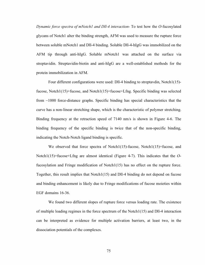
75
Dynamic force spectra of mNotch1 and Dll-4 interaction- To test how the O-fucosylated
glycans of Notch1 alter the binding strength, AFM was used to measure the rupture force
between soluble mNotch1 and Dll-4 binding. Soluble Dll-4-hIgG was immobilized on the
AFM tip through anti-hIgG. Soluble mNotch1 was attached on the surface via
streptavidin. Streptavidin-biotin and anti-hIgG are a well-established methods for the
protein immobilization in AFM.
Four different configurations were used: Dll-4 binding to streptavdin, Notch1(15)-
fucose, Notch1(15)+fucose, and Notch1(15)+fucose+Lfng. Specific binding was selected
from ~1000 force-distance graphs. Specific binding has special characteristics that the
curve has a non-linear stretching shape, which is the characteristic of polymer stretching.
Binding frequency at the retraction speed of 7140 nm/s is shown in Figure 4-6. The
binding frequency of the specific binding is twice that of the non-specific binding,
indicating the Notch-Notch ligand binding is specific.
We observed that force spectra of Notch1(15)-fucose, Notch1(15)+fucose, and
Notch1(15)+fucose+Lfng are almost identical (Figure 4-7). This indicates that the O-
fucosylation and Fringe modification of Notch1(15) has no effect on the rupture force.
Together, this result implies that Notch1(15) and Dll-4 binding do not depend on fucose
and binding enhancement is likely due to Fringe modifications of fucose moieties within
EGF domains 16-36.
We found two different slopes of rupture force versus loading rate. The existence
of multiple loading regimes in the force spectrum of the Notch1(15) and Dll-4 interaction
can be interpreted as evidence for multiple activation barriers, at least two, in the
dissociation potentials of the complexes.
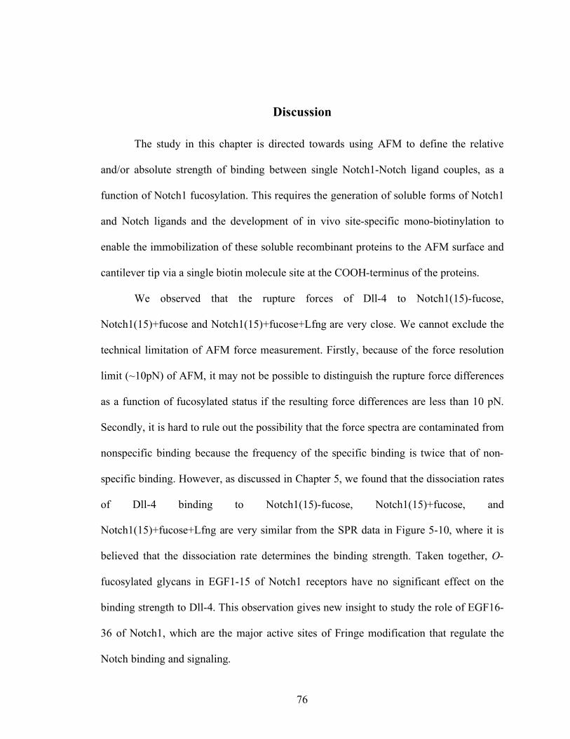
76
Discussion
The study in this chapter is directed towards using AFM to define the relative
and/or absolute strength of binding between single Notch1-Notch ligand couples, as a
function of Notch1 fucosylation. This requires the generation of soluble forms of Notch1
and Notch ligands and the development of in vivo site-specific mono-biotinylation to
enable the immobilization of these soluble recombinant proteins to the AFM surface and
cantilever tip via a single biotin molecule site at the COOH-terminus of the proteins.
We observed that the rupture forces of Dll-4 to Notch1(15)-fucose,
Notch1(15)+fucose and Notch1(15)+fucose+Lfng are very close. We cannot exclude the
technical limitation of AFM force measurement. Firstly, because of the force resolution
limit (~10pN) of AFM, it may not be possible to distinguish the rupture force differences
as a function of fucosylated status if the resulting force differences are less than 10 pN.
Secondly, it is hard to rule out the possibility that the force spectra are contaminated from
nonspecific binding because the frequency of the specific binding is twice that of non-
specific binding. However, as discussed in Chapter 5, we found that the dissociation rates
of Dll-4 binding to Notch1(15)-fucose, Notch1(15)+fucose, and
Notch1(15)+fucose+Lfng are very similar from the SPR data in Figure 5-10, where it is
believed that the dissociation rate determines the binding strength. Taken together, O-
fucosylated glycans in EGF1-15 of Notch1 receptors have no significant effect on the
binding strength to Dll-4. This observation gives new insight to study the role of EGF16-
36 of Notch1, which are the major active sites of Fringe modification that regulate the
Notch binding and signaling.
77
Figure 4-1. Protein immobilization method by esterification. Mica is esterified with ethanolamine (1) and PEGylated with PEG of MW3400 (2). Streptavdin or Anti-IgG is attached to PEG by NHS.
78
Figure 4-2. AFM experimental setup to measure the Notch1 and Dll-4 binding strength. Notch1 is attached to the glass surface through neutroavidin and PEG. D4-IgG is immobilized to the AFM tip through anti-IgG and PEG.
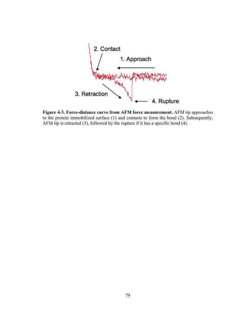
79
Figure 4-3. Force-distance curve from AFM force measurement. AFM tip approaches to the protein immobilized surface (1) and contacts to form the bond (2). Subsequently, AFM tip is retracted (3), followed by the rupture if it has a specific bond (4).
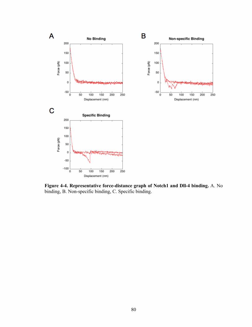
80
Figure 4-4. Representative force-distance graph of Notch1 and Dll-4 binding. A. No binding, B. Non-specific binding, C. Specific binding.
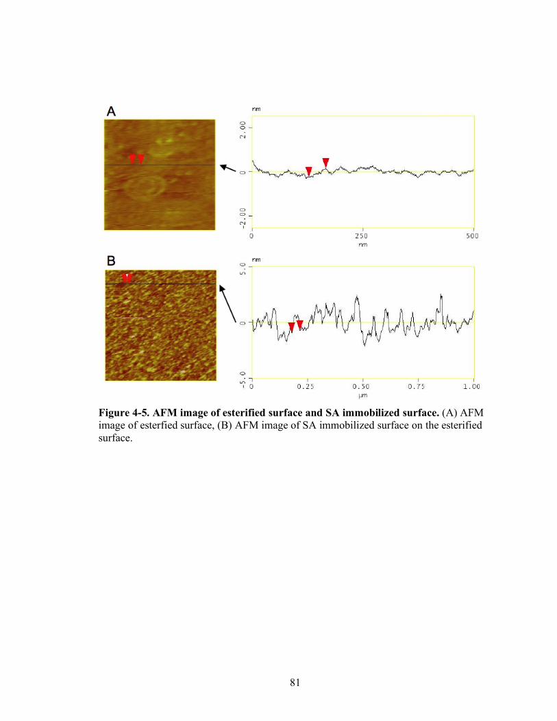
81
Figure 4-5. AFM image of esterified surface and SA immobilized surface. (A) AFM image of esterfied surface, (B) AFM image of SA immobilized surface on the esterified surface.
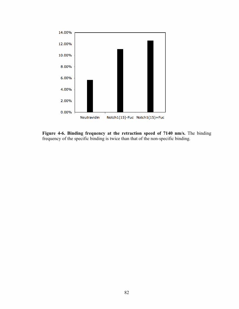
82
Figure 4-6. Binding frequency at the retraction speed of 7140 nm/s. The binding frequency of the specific binding is twice than that of the non-specific binding.
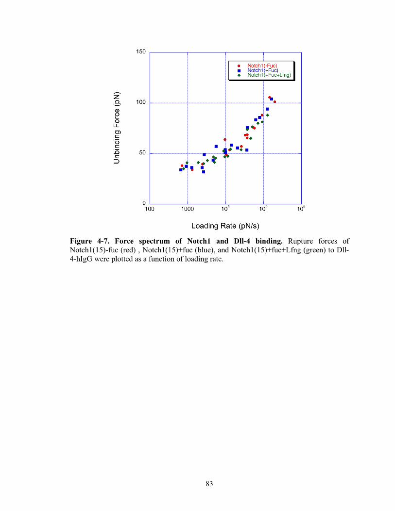
83
Figure 4-7. Force spectrum of Notch1 and Dll-4 binding. Rupture forces of Notch1(15)-fuc (red) , Notch1(15)+fuc (blue), and Notch1(15)+fuc+Lfng (green) to Dll-4-hIgG were plotted as a function of loading rate.
84
Chapter 5
Kinetic Analysis of Glycan-dependent Notch-Notch Ligand Interactions using Surface Plasmon Resonance (SPR)
Introduction
Notch receptors play a critical role in many developmental and
pathophysiological processes [45, 46, 56]. Because of the importance of Notch function
in health and disease, the Notch signaling pathway has emerged as a potential therapeutic
target [152, 153].
A Notch receptor expressed by a receiving cell is activated when the receptor’s
extracellular domain (ECD) interacts with a ligand on an adjacent (sending) cell, and is
apparently pulled away from the membrane of the receiving cell by endocytosis-
dependent processes on the sending cells [91, 154]. Activation of a Notch receptor leads
to the protease-dependent release of a specific intracellular fragment of the Notch
receptor. This intracellular fragment then translocates to the nucleus of the receiving cell
and modulates transcriptional events that in turn dictate changes in the phenotype of the
receiving cell.
Notch ligands were first identified in Drosophila and are called Delta and Serrate.
Five Notch ligands have now been identified in humans, in mice and in other mammals.
These are termed Jagged1, Jagged2, Delta-like1, Delta-like3, and Delta-like4. Each of
85
these ligands is a single-pass transmembrane protein with a conserved N-terminal Delta,
Serrate, LAG-2 (DSL) motif, which is essential for binding to Notch receptors [112].
Mammalian Notch receptors contain 29-36 EGF like repeats, which are modified
with one or more N-glycans, and are heavily modified by O-glycans initiating with
protein-linked, O-fucose [21, 42, 44, 68], O-glucose, and possibly O-GlcNAc [72]. The
fucosyltransferase termed Pofut1 transfers fucose to the serine or threonine residue of the
O-fucosylation consensus sequence C2X4-5(S/T)C3 on some EGF domains of Notch
receptors. In mammals, glycosyltransferases termed Lunatic Fringe (Lfng), Radical
Fringe (Rfng), and Manic Fringe (Mfng) can elongate O-fucose moieties by adding
GlcNAc, which may be subsequently modified by galactose and sialic acid moieties,
respectively, to form elongated structures of the form Sia-α2,3-Gal-β1,4-GlcNAc-β1,3-
α1-O-Ser/Thr [21]. Pofut1-mediated O-fucosylation of Notch receptor EGF repeats
occurs in the endoplasmic reticulum [23]. By contrast, subsequent modification of the
fucose moiety, leading to the elongated, sialic acid-terminated structures noted above,
takes place in the Golgi lumen [42]. These O-linked glycans play key roles in modulating
Notch-mediated signal transduction events, since, for example, O-fucosylated glycans,
which modify some EGF-like repeats within the ECD of Notch family members, have
been observed to regulate signaling from Notch receptors, in correspondence with their
ability to modulate the interaction between Notch receptors and their ligands [82]. O-
linked fucose is required for Notch binding to Notch ligands and the Notch activation [73,
155]. Extension of O-fucose by Fringe enhances the Dll-1 signaling, while reduces the
Jag-1 signaling [82].
While these studies begin to provide insight into how specific O-fucosylated
86
glycans control Notch receptor-ligand interactions, they do not provide a precise
understanding about how different O-fucosylated glycans dictate the kinetics with which
Notch receptors and their ligands associate, or dissociate, nor do they provide
corresponding precision about the affinities that characterize each receptor-ligand couple,
as a function of their fucosylated glycan phenotype. An understanding of these issues
will help to provide a molecular understanding of the biological variation that is
characteristic of each different Notch-Notch ligand couple, and each distinct fucosylated
glycoform.
Likewise, while it is apparent that the information on individual O-fucosylated
glycans will be required to dissect the mechanism by which these sugars affect Notch
function. Although multiple EGF domains on Notch receptors are thought to be involved
in Notch receptor binding to Notch ligands [112], published evidence indicates that the
ligand binding region (EGF 11-12; [112]) and the Abruptex region (EGF 24-29; [156,
157]) of Notch receptors are critical to Notch binding and signaling. Specifically,
mutation in distinct O-fucosylation site in EGF12 within the binding region inhibits
Notch activation [85] and the mutation of O-fucosylation sites in EGF 26 and 27 inside
the Abruptex region results in enhancement and decrease of Notch signaling, respectively
[87]. Moreover, these three O-fucosylation sites in EGF repeats 12, 26, and 27 are highly
conserved in all known Notch homologues of Notch1 receptors. This observation has
implied that these O-fucosylation sites are biologically relevant for Notch signaling.
Indeed, mutation of the O-fucosylation site in EGF repeat 12 of mNotch1 results
in reduced signaling in Drosophila and mammals [51, 85, 86]. This suggests that the
effects of the mutation at EGF repeat 12 are mediated largely by loss of the O-fucose
87
monosaccharide. Mutation of the O-fucosylation site in EGF repeat 27 of mNotch1
results in faulty trafficking of this Notch receptor to the cell surface and a decreased S1
processing of the receptor. In mammals, mutation of the O-fucosylation site in EGF
repeat 26 of Notch1 results in hyperactive signaling in response to Delta1 and Jagged1
interaction due to unknown mechanism. These observations begin to provide insight into
regions of the Notch1 molecule that play important roles in receptor-ligand interactions.
Nonetheless, they do not yet allow a detailed understanding of how individual domains,
or glycans on those domains modulate the rates of association and dissociation between
Notch1 and its ligands, nor do they provide deeper insight into how domains or specific
glycans dictate receptor-ligand affinities.
In experiments we report here, we have sought to advance the understanding of
how Fringe-mediated glycan modifications modulate the kinetics and affinities of the
interactions between mouse Notch1 and its ligands, how distinct O-fucosylation sites on
mouse Notch1 contribute to these binding interactions, and how Fringe-dependent glycan
modification of Notch ligands themselves may modulate Notch receptor-ligand
interactions, using surface plasmon resonance techniques. Efficient binding was observed
between mNotch1 and the Notch ligands Dll-1, Dll-4, Jag-1, and Jag-2, but not between
mNotch1 and Dll-3. A biphasic binding and dissociation pattern was observed with the
four binding-competent Notch1-Notch ligand pairs, suggesting a two-state receptor-
ligand interaction model characterized by initial formation of a transient receptor-ligand
complex followed by a conformational change that leads to a more stable receptor-ligand
complex. Primary and secondary on and off-rates for the four binding-competent
mNotch1-Notch ligand pairs were observed to be distinct and characteristic for each
88
Notch ligand. The magnitudes of the overall association constants (K) generated by the
mNotch1-Dll-1 couple or the mNotch1-Dll-4 couple were similar, and much greater than
the overall association constants generated in couples involving mNotch1 and either Jag-
1 or Jag-2. The overall association constants observed when Dll-1 or Dll-4 interacted
with Fringe-modified mNotch1 were significantly greater than when these ligands
interacted with unmodified mNotch1, with enhancement likely due to Fringe
modifications of fucose moieties within EGF domains 16-36. By contrast, Fringe
modification of mNotch1 did not significantly modulate interactions with Jag-1 or Jag-2.
Mutational analyses confirm prior observations that the O-fucosylation site within EGF
repeat number 12 dictates much, if not all of the binding between Notch1 and its ligands.
Finally, we observe that Fringe modification of Dll-4 enhances its ability to bind to
mNotch1.
Methods
Protein Reagents- Construction and characterization of soluble mNotch1 and Notch
ligands were discussed in Chapter 2. The protein configuration used in this Chapter is
summarized in Figure 5-1.
Surface Plasmon Resonance (SPR)- Mono-biotin tagged soluble mNotch1 proteins with
various glycosylation status were immobilized on the Biacore Sensor Chip SA at the
level of 600-1200RU using Biacore 3000 and Notch ligands were injected at the speed of
50µl/mL (Figure 5-2). Dll-1, Jag-1, and Jag-2 were injected for 1 min and Dll-4 for 3 min
because of the slow association. The dissociation phase was held for 5 minutes. The
surface was regenerated with 10mM Hepes, 0.5M NaCl, 0.01% Triton-X 100, 3mM
89
EDTA in hepes buffer (Figure 5-3). All experiments were performed at least 3 times at
25°C.
Mass Transfer Limitation (MTL) was tested followed by the Biacore manual.
Checking for MTL was performed by injecting Notch ligands at different flow rates. The
association and dissociation rate constants were the same between different flow rates.
This indicates that no mass transfer limitation is present.
The obtained sensorgrams were modified by double subtraction method shown in
Figure 5-4. Binding curves were analyzed with a two interconverting receptor binding
model or conformational change model (
!
A + B"kd1
ka1
AB"kd 2
ka2
AB* ) using BiaEvaluation
software.
Results
Patterns of the association and dissociation phases of mNotch1 and Notch ligand binding
are biphasic - To examine how the glycans alter the Notch binding to Notch ligands,
kinetic study was performed using SPR to determine the association and dissociation rate
of the interaction. Various glycosylation forms of mNotch1 were immobilized on Biacore
sensor chip SA and then Notch ligand binding to various mNotch1 were observed.
Because the multiple EGF repeats in Notch1 contribute to ligand binding,
directional immobilization is important. Site-specific mono-biotinylation was used to
express the Notch1 proteins. In this system, the extracellular domain of mNotch1 was
fused to c-Myc tag and a biotinylation tag in the C-terminal. Through biotin in the
biotinylation tag, soluble mNotch1 was immobilized on the Biacore sensor chip SA.

90
We observed the biphasic binding and dissociation patterns (Figure 5-5A and B),
which suggests that the Notch-Notch ligand binding complexes may have at least two-
state compartments. However, to rule out the possibility that simple 1:1 binding model
dictates Notch-Notch ligand binding, the binding curves were first fitted with a 1:1
Langmuir binding model. The binding curves were not fitted well by 1:1 Langmuir
binding model with high χ2 number (χ2=39.1) as shown in Figure 5-5C.
We tested several models which are proposed for the biphasic dissociation
pattern.
!
A + B"kd1
ka1
AB"kd 2
ka2
AB* (Two interconverting receptor state binding) (1)
!
A + B"kd1
ka1
AB,A + C"kd 2
ka2
AC (Two non-interconverting receptor state binding) (2)
!
A + B"kd1
ka1
AB + B"kd 2
ka2
ABB (Bivalent ligand binding) (3)
!
A + B"kd1
ka1
AB#kx
AB*"kd 2
ka2
A + B* (Two-site interconverting receptor binding) (4)
where A is Notch ligand and B and C are different forms of Notch receptors at surface.
To test which binding model dictates Notch-Notch ligand binding, the binding
curves of Notch1+fucose and Dll-4 binding were fitted with a two interconverting
receptor state model (equation 1), a two non-interconverting receptor state model
(equation 2), and a bivalent ligand binding model (equation 3). The binding data were not
fitted well by a two non-interconverting receptor state model (χ2=23.5), and a bivalent
ligand binding model (χ2=35.1) as shown in Figure 5-5E and F, respectively, while a two
interconverting receptor state model significantly enhanced the fit with low χ2 number
(χ2 =2.4) as shown in Figure 5-5D, indicating that a two interconverting receptor state
binding model may dictate Notch1-Dll-4 binding.
91
In a two interconverting receptor state model, ligand (A) binds to surface
receptors (B) with rate constant ka1 to form low-affinity receptor-ligand complexes (AB),
which convert to high-affinity complexes (AB*) with the rate constant ka2. To test
whether a two interconverting receptor state model dictates Notch-Notch ligand binding,
linked reaction test was performed according to the Biacore manual. Dll-4 (250nM) was
injected to Notch1+fuc and Notch1+fuc+Lfng immobilized surface and compared the
dissociation rate constant after injection times of 1, 3, and 20 minutes. This
interconverting receptor state model assumes that the conversion rate of low-affinity
complexes to high-affinity complexes is proportional to the incubation time. The identical
dissociation profiles may indicate that the reactions are not linked. In Figure 5-6A and B,
the dissociation rate constant was gradually decreased after longer injection time and the
biphasic patterns in association and dissociation phases are also observed from all of the
injection time, indicating that the formation of high-affinity complexes was increased
over time in both Notch1+fuc and Notch1+fuc+Lfng binding to Dll-4. This result
supports that a two interconverting receptor state model dictates Notch-Notch ligand
binding.
A two non-interconverting receptor state model can be caused by heterogeneity of
immobilized Notch1 receptors. Immobilized Notch1 may be pure because Notch1
receptors were captured by streptavidin and biotin bond which is equivalent with the
purification step. Another potential source of heterogeneity may be induced from
different glycosylation status. However, all the glycosylated forms of Notch1 have
biphasic patterns in the association and dissociation stage (Figure 5-7). Therefore, the
biphasic pattern is not from the heterogeneity of immobilized Notch1 receptors.
92
If one Notch ligand contains two binding sites, a bivalent ligand binding model
may dictate Notch-Notch ligand binding. It is known that DSL domain has one binding
surface to Notch receptor from the structural study [158]. Therefore, it is not likely that
bivalent ligand binding model dictates Notch-Notch ligand binding.
A two-site interconverting receptor state model also supports the biphasic patterns
in association and dissociation phase of Notch-Notch ligand binding. Ligand (A) binds to
surface receptors (B) with rate constant ka1 to form low-affinity receptor-ligand
complexes (AB), which convert to high-affinity complexes with rate constant kx. High-
affinity complexes are also formed by ligand binding to high-affinity state receptors (B*)
with the rate constant ka2. Ligand can dissociate from low- or high-affinity receptor-
ligand complexes with rate constants, kd1 and kd2. In this model, the conversion of low-
affinity complexes to high-affinity complexes is irreversible. If the dissociation curve can
be described with a single exponential, it indicates the absence of a low affinity site and
the complete conversion of receptors to a high affinity state. However, as shown in
Figure 5-6A and B, the biphasic pattern is still seen on long injection (20 minute),
suggesting that the dissociation is occurred with two rate constants and the conversion of
high-affinity receptor-ligand complexes directly to receptor and ligand is minimal.
To exclude the possibility that the biphasic pattern is due to the mass transfer
limitation. Mass transfer limitation was tested according to the Biacore manual. No
difference was observed in kinetics with different injection speed (data not shown) and
with different immobilized level of soluble mNotch1 receptors on the surface (data not
shown).
93
Taken together, a two interconverting receptor state model dictates Notch1 and
Notch ligand binding. A two interconverting receptor state model is also known as a
conformational change model because the conversion of the low-affinity complexes to
high-affinity complexes is induced by the conformational change of the receptor-ligand
binding complexes. In this model, Notch binding to Notch ligand is the binding step to
form a low-affinity receptor-ligand binding complex and conversion from a low-affinity
complex to a high-affinity complex is a conformation change step. This indicates that
conformational change is involved in Notch-Notch ligand binding. All the binding data in
this chapter will be analyzed with a two interconverting receptor state model or the
conformational change model.
Effect of fucosylation and Fringe modification of mNotch1 receptor in ligand binding
kinetics- To test how fucosylation and Fringe modification of Notch1 alter the binding to
Notch ligands, mNotch1 and Notch ligand binding kinetics were measured by SPR.
Soluble mNotch1 was expressed from CL17 cells and CL17/Lfng cells with exogenous
fucose. All of the Notch ligands except Dll-3 were expressed from HEK 293T cells. We
were unable to express Dll-3 consistently and found that Dll-3 is very unstable after
secretion. Recent literature proposed that Dll-3 is not a transmembrane protein, while it is
resident in Golgi [159]. However, the mechanism of Dll-3 is still controversial although
Dll-3 mutation may induce severe axial skeletal defects [160, 161].
Conditional fucosylation-deficient CL17 cells were used to express non-
fucosylated mNotch1. His-tagged mNotch1(EGF1-15) was expressed from CL17 without
exogenous fucose. With MS analysis, we found that O-fucose existed in some of EGF
94
repeats of soluble mNotch1, indicating Notch1 receptor expressed in the absence of
fucose contains fucose to some extent. The potential source of fucose may be from
serum. For simplicity, Notch1 cultured in the absence of fucose (i.e. Notch1-fucose) is
correspondent to Notch1 cultured in the presence of very low concentration of fucose.
Sensorgram and kinetic information on mNotch1 binding to Notch ligands is
summarized in Figure 5-7 and Figure 5-8, respectively. A negative control ligand wherein
the DLS domain of DLL-4 was mutationally inactivated (Dll-4 (*DSL)) did not bind to
any mNotch1 surfaces (data not shown).
Most of the kinetic information on Notch ligand binding to Notch1-fucose and
Notch1+fucose analyzed a two interconverting receptor state model are almost identical
(Figure 5-8) because Notch1-fucose and Notch1+fucose may have the similar level of O-
fucosylated glycans as suggested in mass spectrometry analysis in Chapter 2. As shown
in Figure 5-8E, in overall association constant, Dll-1 and Dll-4 have high affinity to
Notch1+fucose compared to Jag-1 and Jag-2 which have low affinity to Notch1+fucose.
To observe how lunatic Fringe modification of mNotch1 changes the binding to
each ligand, soluble mNotch1 was expressed from lunatic Fringe stably transfected CL17
cells. Lunatic Fringe was chosen because lunatic Fringe has been known as a potent
modifier in many T cell development studies [162, 163]. CHO cells have an endogenous
lunatic and radical Fringe activity, but no manic Fringe activity was detected (Data not
shown). As a consequence, a low level of Fringe modification of Notch is expected.
With Fringe modification, overall association constants of Dll-1 and Dll-4 binding
were increased, while those of Jag-1 and Jag-2 binding were the same or decreased. The
first association rate constant (ka1) of Dll-1 and Dll-4 were increased with Fringe
95
modification, while those of Jag-1 and Jag-2 were about the same. The first dissociation
rate constant (kd1) of Dll-4 was significantly reduced with Fringe modification, while
Dll-1, Jag-1, and Jag-2 were about the same or a bit increased. The second association
rate constant (ka2) of all Notch ligands were decreased or about the same, indicating that
Fringe action hinders advancing from low-affinity complexes to high-affinity complexes
of Notch-Notch ligand binding. Specially, in Dll-4 binding, lunatic Fringe modification
significantly reduces the second association rate constant (ka2), indicating low-affinity
state is favorable when Dll-4 binds to lunatic Fringe modified mNotch1. The second
dissociation rate constant (kd2) of Dll-1 was reduced. Dll-4 was increased, while Jag-1
and Jag-2 were about the same. Maximum binding capacity (Rmax) was estimated from
the curve fitting procedure with the two interconverting receptor state model of
BiaEvaluation software. Normalized Rmax was calculated by dividing the estimated
Rmax with the immobilized Notch1 on the surface and the molecular weight of each
Notch ligand. The value obtained from Dll-1 binding to Notch1-fucose was set as 1. The
normalized Rmax indicates the relative amount of bound Notch ligand per Notch1
molecule on the surface (RU). Normalized Rmax of Dll-1 was significantly increased
about 8 fold with Fringe modification of mNotch1. These data are consistent with results
showing that Dll-1 binding to Notch2 is detectable after Notch2 is processed by Lunatic
Fringe [164]. Also, Dll-4 has similar maximum binding level (normalized Rmax) in this
work, reflecting the apparent reduced dependence of the interaction on Lunatic Fringe by
flow cytometry [164]. Jag-1 and Jag-2 increased with around 2 fold with O-fucosylation
and Fringe modification, respectively.
96
Once Notch1 is modified by Fringe, Dll-1 and Dll-4 have the same affinity and
same maximum binding capacity. However, in Figure 5-8B, Dll-4 has a smaller overall
dissociation rate compared to Dll-1, implying that Notch1-Dll-4 binding complex triggers
the stronger signal. This result demonstrates that Dll-4 is the most potent ligand for Notch
signaling, which is known as the essential, non-redundant ligand in thymus for T cell
development [162, 163].
Taken together, Fringe modification of Notch1 significantly alters Notch1-Dll-4
binding kinetics. In Notch1-Dll-1 binding, binding kinetics including overall association
constant and the maximum binding level (Rmax) are dramatically changed with Fringe
modification. Jag-1 and Jag-2 binding to Notch1 are almost the same or a bit decreased
by the Fringe action. We propose that lunatic Fringe modification makes the Delta
ligands (Dll-1 and Dll-4) more competent for Notch signaling.
Role of individual O-fucosylation sites of mNotch1 in ligand binding kinetics- To evaluate
how the individual O-fucosylation sites alter the kinetics of Notch-Notch ligand binding,
mNotch1 mutants with mutations in O-fucosylation sites in EGF12, 24, 26, and 27 were
constructed (Figure 5-9A) and the binding kinetics was measured by SPR. In many
studies, mutation in the O-fucosylation site of EGF12 reduced the binding and signaling.
Also, O-fucosylation sites in EGF24, 26, and 27 are of interest because they are in the
Abruptex domain reported as the negative regulatory region. Specially, O-fucosylation
sites in EGF12, 26, and 27 are highly conserved in many species. These mutations are
unlikely to disrupt the structure of the EGF domain of Notch.
97
As shown in Figure 5-9E, mNotch1 mutant in the O-fucosylation site in EGF 12
inhibits the binding to Notch ligand, Dll-4. Both the binding step and the conformational
step were significantly altered.
Still, Dll-4 can bind to mNotch1 at a detectable level, which is rescued by Fringe
action to some extent. However, Dll-1, Jag-1, and Jag-2 binding were significantly
reduced, so the binding level from the sensorgram was too low to fit with the model, even
though the same Notch mutant immobilized surface and the same range of Notch ligand
concentration were used (data not shown). Our result suggests that O-fucose of EGF12 is
important for both the binding and the conformational step in binding kinetics and may
influence the intensity of Notch signaling.
We observed no difference in the kinetic information from the point mutation
study of O-fucosylation sites in EGF 24, 26, and 27 (Figure 5-9A, B, C, and D).
However, in terms of normalized Rmax, maximum binding capacity of Dll-4 and
Notch1(EGF26f)-fucose and Notch1(EGF26f)+fucose was increased 1.5 fold compared
to the wild type mNotch1 binding to Dll-4, while Rmax of this binding was back to the
same level to other mutant with Fringe modification. Haltiwanger’s group reported that
mutation in O-fucosylation site of EGF26 of mNotch1 has increased Notch signaling,
while mutation in O-fucosylation site of EGF 24 of mNotch1 has the same signaling
compared with wild type [87]. Our data suggest that the Notch signaling increase in the
EGF 26 mutant is induced by the increased maximum binding level of Notch ligand, not
by a change of the binding kinetics. Also, this result implies that the O-fucose in EGF26
is an inhibitory molecule for Notch binding to the ligand.
98
Role of EGF16-36 of mNotch1 in ligand binding kinetics- To further study the role of
EGF16-36 of mNotch1 in binding kinetics, a deletion mutation of mNotch1 was
constructed by removing EGF domains 16-36 of the Notch1 receptor, and binding of the
resulting Notch1(EGF1-15) to Dll-4 was examined with SPR. The kinetic binding data
we observed when Notch1(EGF1-15) and Dll-4 interacted were not altered with the Lfng
modification of Notch1(EGF1-15) (Figure 5-10). These data are consistent with the result
of Haltiwanger’s group, showing that because the O-fucosylation site in EGF12 is a poor
substrate for Fringe, other O-fucosylation sites are susceptible to Fringe modification and
can alter Notch binding and signaling. Our data indicate that EGF repeats 16-36 of
Notch1 are involved in both the binding step and conformational change steps. We
propose that a dramatic increase in Notch-Notch ligand binding by Fringe is induced by
the modification of EGF16-36, which includes the abruptex domain. This result suggests
the importance of the abruptex region, which is the one of the major substrate of lunatic
Fringe [87].
Effect of fucosylation and Fringe modification of Dll-4 in mNotch1 binding kinetics-
Notch ligands are also substrates of protein O-fucosylatransferase-1 as well as Fringe. To
test how Fringe modification on Dll-4 alters Notch binding to Notch ligands, soluble Dll-
4 was expressed from CHO/CL17 cells and CHO/CL17/Lfng with fucose. Kinetic
binding information was obtained from a Biacore experiment. Kinetic studies on
mNotch1 binding to various glycosylated forms of Dll-4 are summarized in Figure 5-11.
In these experiments, we observed that Fringe modification of Dll-4 enhanced its binding
to mNotch1. This result indicates that Lfng modification of Dll-4 enhances mNotch1

99
binding. This demonstrates that Fringe modification influences the binding characteristics
of both Notch and Notch ligand.
Discussion
In this chapter, we investigated how the O-fucosylated glycans on Notch1
receptors alter the ligand binding kinetics and affinities using SPR. We observed the
biphasic patterns in the association and dissociation phases, indicating that Notch-Notch
ligand binding has two-state complexes which is caused by the conformational change of
low-affinity receptor-ligand binding complexes to high-affinity receptor-ligand binding
complexes, where the binding is dictated by a two interconverting receptor state model.
Fringe modification on Notch1 significantly enhances the overall association constants of
Dll-1 and Dll-4, while has no significant effect on Notch1 binding to Jag-1 and Jag-2 in
the overall association constants. The O-fucosylation site in EGF repeat 12 of Notch1 is
critical in formation of low- and high-affinity complexes of Notch and Notch ligand.
EGF16-36 is the critical substrate of lunatic Fringe, which influences the formation of
both a low- and high-affinity receptor-ligand binding complex.
Models for Notch-Notch ligand binding compartments- In this study, we observed the
biphasic patterns in the association and dissociation phases of Notch-Notch ligand
binding curves in SPR experiments. We considered a binding model that has two
dissociation rate constants and proposed that a two interconverting receptor state model
dictates Notch-Notch ligand binding. However, we cannot exclude the possibility that
100
other binding models can dictate Notch-Notch ligand binding, which have three or more
association and dissociation rate constants. One such three rate constant model may be
represented by the equation
!
A + B"kd1
ka1
AB"kd 2
ka2
AB*"kd 3
ka3
A + B*. The only difference between
this model and a two-site interconverting receptor state model is that the conversion from
the low-affinity complexes to the high-affinity complexes is reversible. This model is
difficult to test because the conversion from the low-affinity complexes to the high-
affinity complexes and the dissociation of the high-affinity complexes are slow and may
be overlapped in the slow dissociation region of SPR binding curves. Consequently, in
the SPR analysis, the binding curves may not have the triple-phasic patterns in
association and dissociation phases. Further studies would be required to determine the
physical nature of the distinct Notch-Notch ligand binding compartments.
Dissociation rate correlates with Notch signaling- Many studies have suggested that
Notch binding correlates with the Notch signaling [73, 165, 166]. Free ligands in solution
do not have the ability to activate Notch signaling [92, 167]. Furthermore, Notch
signaling is not activated when Notch receptor binds to its ligand in the same cell surface
(i.e. cis-inhibition) [125, 158]. These considerations and othes make it evident that ligand
binding itself is not the sole determinant of Notch signaling. Some biophysical
microevironment, in addition to ligand binding, seems to be required for proper Notch
signaling. Endocytosis of the Notch ligand expressing cells is critical in Notch signaling
in Drosophila and mammals [88, 89]. Endocytosis serves as a pulling force that may
induce a conformational change in Notch, preceding, if not actually causing exposure of
the S2 proteolytic cleavage site of Notch required for the Notch signaling activation [135,
101
168, 169]. The strength of the Notch-Notch ligand bond apparently needs to be stronger
than the external pulling force by endocytosis. Indeed, the binding strength is determined
by the dissociation rate of the binding. This implies that the dissociation rate might be the
critical kinetic information in Notch signaling. As shown in Figure 5-8, the binding
affinity of Jag-2 is very weak compared to other Notch ligands. However, Jag-2 is
capable of triggering Notch signal better than Dll-1 and Jag-1 in some cell lines [170].
To test this hypothesis, we analyzed the kinetic variables obtained from the SPR
experiment. Because the overall association rate constant and the overall dissociation rate
constant were not directly obtained from the two interconverting receptor binding model,
the overall association rate constant was calculated from
!
A + B"kd1
ka1
AB#ka2
AB* with
assumption kd2=0 and the overall dissociation rate constant from
!
A + B"kd1
AB#kd 2
ka2
AB*
with the assumption ka1=0 as shown Appendix 3. As shown in Figure 5-12, the overall
association rate constant and overall dissociation rate constant were calculated from the
general solution of the rate equations is shown in Appendix 3 [171].
A correlation between Notch signaling and the dissociation rate constant is
evident in the context of Notch downstream signaling that has been observed when bone
marrow progenitor cells are co-cultured with OP-9 stromal cells expressing Notch ligands
[172]. In these experiments, the level of mRNA transcribed from the Notch downstream
target Deltex was observed to be increased by co-culture with OP-9 cells bearing Dll-1,
Dll-4, and Jag-2, not in co-culture with OP-9 cells expressing Jag-1 [172].
In here, the first dissociation rate constant (kd1) or the overall dissociation rate
constant (kd) may correlate with Notch signaling, however, no correlation was found
from other rate constants or the affinities. If the first or overall dissociation rate constant
102
(kd1 or kd, respectively) is larger than that of Dll-1, there is no signal from Jag-1, but if
smaller, it will signal from Dll-1, Dll-4 and Jag-2 (Figure 5-12).
Together, I propose that the first dissociation rate constant (kd1) must be below a
certain threshold before activation of Notch signaling can occur. This is relevant because
all of the Notch ligands except Dll-3 have the ability to signal in vitro. If the first or
overall dissociation rate constant (kd1 or kd, respectively) is smaller the threshold (i.e.
stronger bond), the Notch signaling would be activated.
Binding Model- We found that a two interconverting receptor state model best correlates
with our SPR-derived Notch-Notch ligand binding data, where the binding complex
forms a low-affinity complexe and then a high-affinity complexe. Although the overall
conformation of all EGF domains of Notch receptor is not known, a structural model has
been proposed by others [115, 158, 173]. In this model, one EGF domain containing 6
cysteines adopts a fold with three disulfide bonds (C1-C3, C2-C4, C5-C6). A calcium
binding EGF domain is arranged in a linear form with a neighboring EGF domain. NMR
and X-ray analysis of Notch binding domain (EGF 11 and 12) showed that Ca2+
participates in a stiff, extended arrangement on calcium binding domain of Notch
receptors. Regions containing non-calcium binding EGF domains are flexible and bent,
having a U-shaped fold. Together, calcium binding plays a role in holding the structure of
ligand binding domain of Notch receptors [174]. However, O-fucose on the EGF domain
has no significant effect on the structure of the EGF domain itself [175].
Based on the implication from these structural studies, the Irvine group proposed
a triple-stranded structure for Notch receptor [112]. In this model, EGF10-21 of the
103
Notch receptor forms a rod-like shape and both EGF6-9 and EGF22-27 are folded back
against EGF10-21, forming the binding pocket. In this model, EGF1-5 and EGF28-36
may mask some of the EGF repeats around the central region prior to the ligand binding.
We propose that ligand binding may induce the strand displacement of EGF1-5 and
EGF28-36 to facilitate the docking of a Notch ligand within the binding pocket of a
Notch receptor revealed by strand displacement.
Although speculative, we propose a two interconverting receptor states model
with the triple stranded structure as shown in Figure 5-13B. Binding is initiated by the
interaction of the DSL domain of Notch ligand and EGF11-12 of the Notch1 receptor.
Subsequently EGF domains of Notch ligand reside in the binding pocket. After this initial
binding step, low-affinity complexes convert to high-affinity complexes by
conformational change. Although the nature of this conformational change is not known
EGF6-9 and EGF22-27 of Notch1 receptor may “clip” the ligand against the EGF 10-21
backbone of Notch receptor to help form stable complexes.
Trans-activation and cis-inhibition- In the biological contexts, Notch receptors are
expressed on signal receiving cells, while Notch ligands are expressed on signal sending
cells to achieve activation of proper Notch signaling (i.e. trans-activation). However, if
Notch receptors and Notch ligands are expressed in the same cell, as has observed to be
true in many instances, there is no Notch signaling event, and in some instances, prevents
activation by Notch ligands presented in trans (i.e. cis-inhibition). An anti-parallel
complex between Notch and its ligand has been proposed for achieving trans-activation
and a parallel complex between the two has been proposed for achieving cis-inhibition
104
[158]. In the proposed parallel complex model, most of the EGF domains of Notch
ligands reside inside the binding pocket of Notch receptor. In this model, EGF10-21 of
the Notch1 receptor and the EGF domains of the Notch ligand are parallel to each other.
In the proposed anti-parallel complex model, the DSL domain of a Notch ligand binds to
the ligand binding domain (i.e. EGF11-12) of Notch receptor, but the binding direction is
anti-parallel. In this model, most of the EGF domains of Notch receptor and Notch ligand
do not interact and exist in an unbound state. This anti-parallel complex model seems
unlikely to exist, however, because there is evidence that most of the EGF repeats of
Notch receptors contribute to ligand binding [112], and this is likely to require
interactions between the Notch EGF repeats and some aspects of the Notch ligand.
We propose that a parallel complex may be consistently applied to both trans-
activation and cis-inhibition, by using a two interconverting complex state model with the
triple stranded arrangement as shown in Figure 5-13C. In this proposed model, parallel
complexes are formed to achieve cis-inhibition, as in the previous proposed model.
However, in contrast to the previous model, trans-activation is achieved when the Notch
ligand binds to a triple stranded Notch receptor to form parallel complexes. In trans-
activation, stabilization of the lower part (EGF16-36) of the binding complex is
important. If the bond strength of this region is not strong enough, the whole structure
would be unclipped when endocytosis pulls out the binding complex. The region around
Abruptex domain may play a role in stabilizing the docking of a Notch ligand to Notch
binding pocket.
105
Role of O-fucosylated glycans on Notch-Notch ligand binding- In our model, EGF11-12
domains of Notch receptor as the site for the initial recognition of Notch by a Notch
ligand. Although mutation of the O-fucosylation site in EGF12 of Notch receptors alters
ligand binding and signaling [85, 87, 112], the exact mechanism by which this occurs is
not known. It has been proposed that the O-fucosylated glycan does not directly contact
the ligand, since protein structure studies show that the O-fucose of EGF12 is on the
opposite site of the proposed binding surface of Notch receptor [158]. Since the O-fucose
on EGF12 is a poor substrate for Fringe [87], and since Lfng modification of Notch1
containing EGF 1-15 does not affect the ligand binding in our experiments, we propose
that the Fringe action on the binding domain of Notch1 receptor (i.e EGF12) has no
significant effect on ligand binding. However, the mechanism by which this O-fucose in
the binding domain of Notch receptor alters ligand binding is unknown. Though
speculative, it is possible that O-fucose on EGF12 interacts with EGF6-9, but not directly
Notch ligand [112], and in doing so, disturbs the upper part (EGF1-15) of triple stranded
structure, and the binding pocket of the Notch binding region, and thereby reduce ligand
binding.
We found that Notch1 EGF domains 16-36 modulate both the binding (step 1)
and conformational change (step 2) in our 2 stage model. EGF domains 16-36 are of
interest because they encompass an Abruptex domain, which plays a prominent role in
regulating Notch signaling events. The literature concerning the role of the Abruptex
domain and of the O-fucosylated glycans on Abruptex is inconsistent, because both
decreased and increased Notch activation have been observed in studies where the
Abruptex domain has been mutated [85, 156, 157, 176-178].
106
Elimination of the O-fucosylation site in EGF26 of mouse Notch1 receptor via
point mutation of the O-fucosylated serine increased Notch signaling elicited by Dll-1
and Jag-1 binding [87]. In our studies of the same EGF26 mutation, we observed
increased Dll-4 binding in the absence of O-fucosylation, relative to binding to wild type
Notch1. These observations indicate that O-fucose in the Abruptex domain conveys as an
inhibitory influence on Notch1 ligand binding, and thus inhibits Notch1 signaling.
In this work, we observed that Fringe modification of Notch1 alters the binding of
Delta significantly. By contrast, Fringe modification of Notch1 does not alter binding by
Jagged 1 or 2. These observations imply that Fringe modification of Notch EGF domains
16-36 can strongly modulate the biological consequences of Dll-1 and Dll-4 interactions,
but not of Jag-1 or Jag-2 interactions. Fringe-modified Notch receptor is characterized by
enhanced formation of low-affinity complexes, and slow conversion from low-affinity
complexes to high-affinity complexes. Since Fringe modified Dll-4 also has the increased
binding ability to Notch1 receptors, indicating Fringe extension is involved in binding,
not maintaining the overall structure of Dll-4. Fringe modification may mainly alter the
binding, but not maintain the overall structure of Notch receptor or Notch ligands
although we cannot exclude the possibility that the Abruptex domain is structurally
correlated with NLR.
Taken together, O-fucosylation of the Abruptex region may inhibit the ligand
binding, while Fringe modification on O-fucose may increase the ligand binding, but still
serve as an inhibitory domain to reduce the formation of high-affinity Notch-Notch
ligand complexes.
107
Our model can be interpreted to be consistent with our mutational study and
mutational studies of others. We propose that the effect of mutations in the Abruptex
domain depends on the type of mutation. Point mutation of the O-fucosylation site would
increase the binding and signaling by removing the inhibitory molecule (i,e, fucose),
while point mutation of the cysteine residue, or missense mutation would reduce binding
by inducing a distortion of triple stranded structure. Deletion mutation of some of the
Abruptex domain would hyperactivate Notch signaling by removing the inhibitory
domain.
Delta and Jagged- Delta and Jagged hierarchically modulate the Notch binding with the
combination of Fringe modification [124]. Mammalian Delta ligands (Dll-1 and Dll-4)
have 8 EGF repeats and Jagged ligands (Jag-1 and Jag-2) 16 EGF repeats. In Jagged, 9th
EGF domain and 10th EGF domain are separate by short linker peptides. Notch ligands
have also calcium binding EGF and interestingly the first and second EGF domain of all
of Notch ligands except Dll-3 are non-calcium binding domain, suggesting that this
region is flexible and the relative orientation of DSL domain is free. These first and
second EGF repeats are called DOS (Delta and OSM-11-like proteins) domain, which is
involved in receptor binding along with DSL domain [179, 180]. The mutation study
indicated that the integrity of DSL and DOS domain is required for the Notch binding,
indicating DOS domains play a role in facilitating DSL binding to Notch receptor [65].
The EGF11-12 domains of Notch receptor would bind to the DSL domain of
Notch ligand, suggesting that the DSL domain would dock in-between two EGF domains
of Notch receptor. In here, two EGF domains of Notch receptor may form one binding
108
site for the DSL domain or one EGF domain. Consistent with this implication, the
maximum number of DSL and/or EGF domain to fit within the binding pocket of Notch1
receptor might be 10 DSL and/or EGF domains because EGF11-21 domains of Notch1
receptors would form 10 binding sites (Figure 5-13A).
Previous studies and this study found that the whole domain of Notch is involved
in the binding [112], indicating that the whole EGF domain of Delta and at least the first
9 EGF domains of Jagged would contribute to the binding. Due to this structural
difference, Delta and Jagged might have a distinct binding capability in physiological
contexts. Fitting of the first 9 EGF repeats of Jagged to the Notch binding pocket may be
tight compared to the fitting of 8 EGF repeats of Delta. In consequence, the interaction
between Notch and Jagged may be weaker than that of Notch and Delta.
It has been proposed that Dll-4 expression is essential in thymus epithelial cells
(TECs) for T cell development in vivo, but not Dll-1 expression, indicating that the
function of Dll-1 and Dll-4 are different in vivo [122, 162, 163]. However, it remains to
be determined why these two ligands are not functionally equivalent in vivo.
Both Dll-1 and Dll-4 have O-fucosylation sites in EGF repeat 8 and 9, which may
contact with the EGF repeat 24 of Notch1 receptors as shown in Figure 5-13B. Dll-4 has
one more potential O-fucosylation site compared to Dll-1, which may contact with
EGF26 and/or 27 of the Abruptex domain in Notch1 receptors. This raises the possibility
that the O-fucosylation site in EGF 5 of Dll-4 is a distinct site compared to Dll-1 and may
play a role in Notch binding.
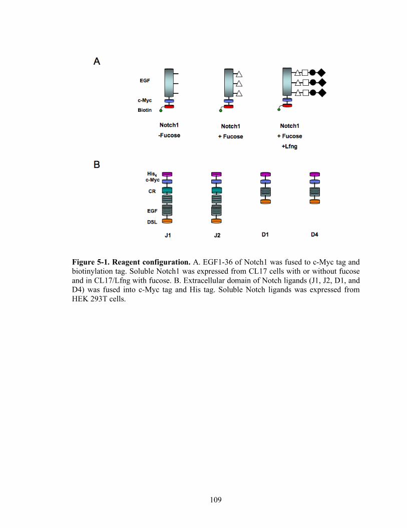
109
Figure 5-1. Reagent configuration. A. EGF1-36 of Notch1 was fused to c-Myc tag and biotinylation tag. Soluble Notch1 was expressed from CL17 cells with or without fucose and in CL17/Lfng with fucose. B. Extracellular domain of Notch ligands (J1, J2, D1, and D4) was fused into c-Myc tag and His tag. Soluble Notch ligands was expressed from HEK 293T cells.

110
Figure 5-2. SPR experimental setup. Soluble mouse Notch1 receptors were immobilized through streptavidin and biotin bond on the Biacore sensor chip SA. Monomeric Notch ligands were injected at the speed of 50µl/min over the Notch1 immobilized surface to analyze the binding kinetics of Notch1-Notch ligands.
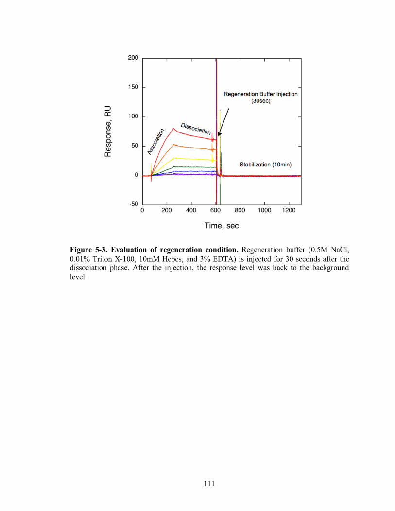
111
Figure 5-3. Evaluation of regeneration condition. Regeneration buffer (0.5M NaCl, 0.01% Triton X-100, 10mM Hepes, and 3% EDTA) is injected for 30 seconds after the dissociation phase. After the injection, the response level was back to the background level.
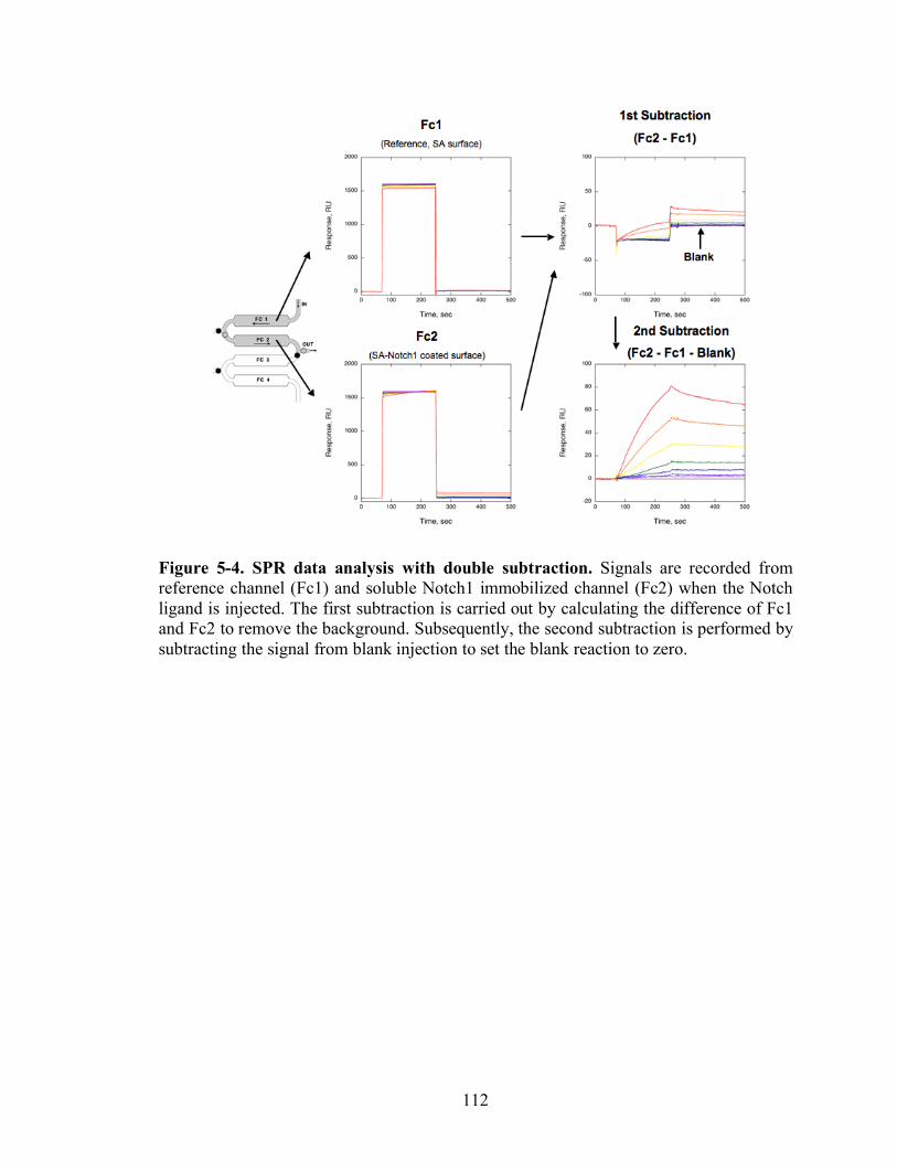
112
Figure 5-4. SPR data analysis with double subtraction. Signals are recorded from reference channel (Fc1) and soluble Notch1 immobilized channel (Fc2) when the Notch ligand is injected. The first subtraction is carried out by calculating the difference of Fc1 and Fc2 to remove the background. Subsequently, the second subtraction is performed by subtracting the signal from blank injection to set the blank reaction to zero.
113
Figure 5-5. Binding model determination. A. Biphasic pattern in association and dissociation phase of Notch1+fucose and Dll-1 binding. Dll-1 was injected over Notch1+fucose immobilized surface with different concentration. In the arrowed region, biphasic pattern was observed. In association phase, binding is initially fast and then slowed down. In dissociation phase, dissociation is fast and then slow. B. Biphasic pattern in association and dissociation phase of Notch1+fucose and Dll-4 binding. Dll-4 was injected over Notch1+fucose immobilized surface with different concentration. In the arrowed region, biphasic pattern was observed. In association phase, binding is initially fast and then slowed down. In dissociation phase, dissociation is fast and then slow. C. a 1:1 Langmuir binding model. For Notch+fucose and Dll-4 binding, 1:1 Langmuir binding model was applied. χ2 is 39.1. D. a two interconverting receptor binding model. For Notch+fucose and Dll-4 binding, two-state binding model was applied. χ2 is 2.4. E. a two non-interconverting receptor binding model. For Notch+fucose and Dll-4 binding, a two non-interconverting receptor binding model was applied. χ2 is 23.5. F. a bivalent ligand binding model. For Notch+fucose and Dll-4 binding, a bivalent ligand binding model was applied. χ2 is 35.1.
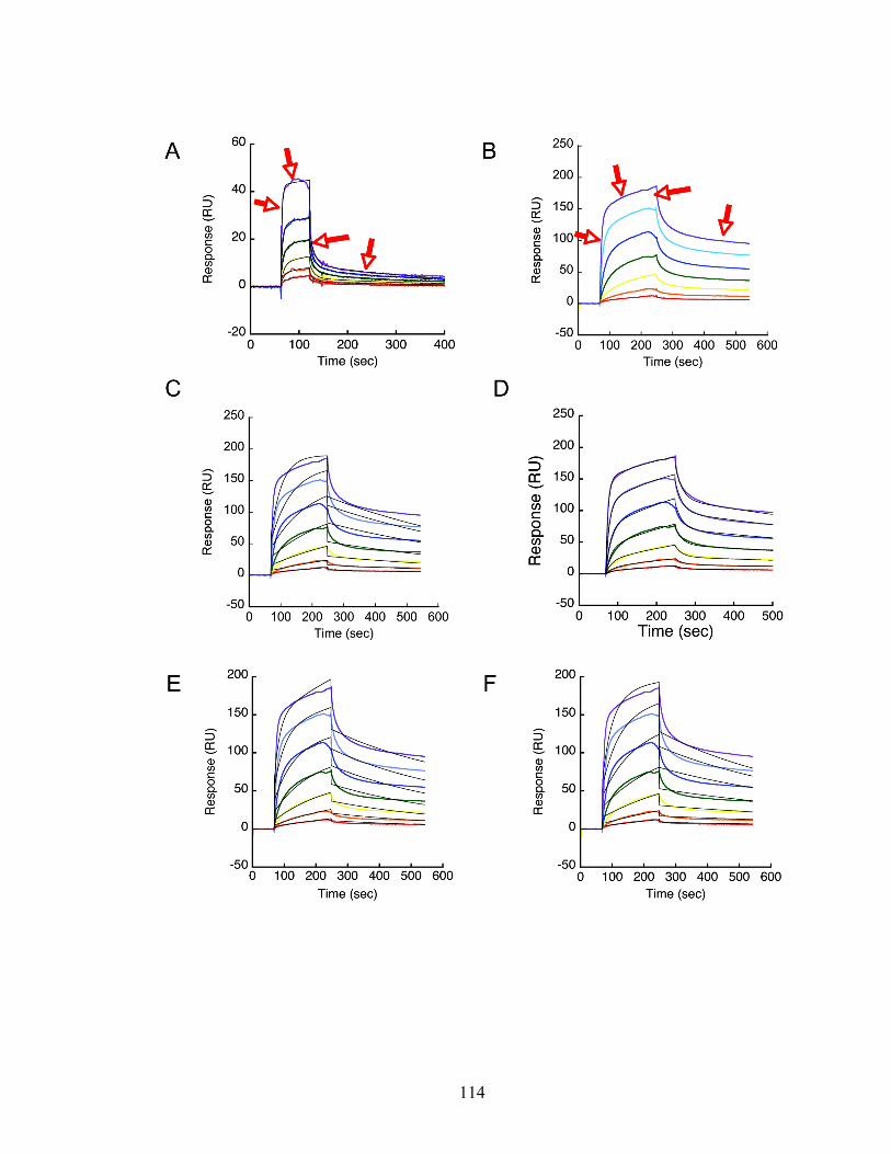
114
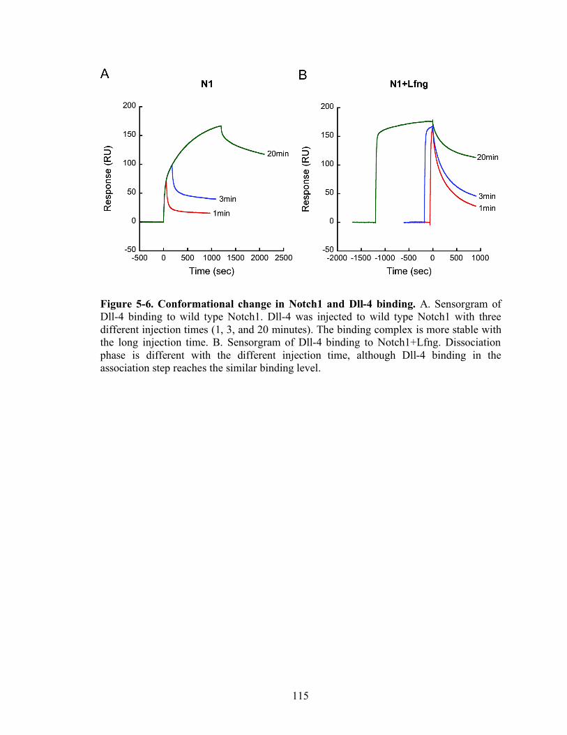
115
Figure 5-6. Conformational change in Notch1 and Dll-4 binding. A. Sensorgram of Dll-4 binding to wild type Notch1. Dll-4 was injected to wild type Notch1 with three different injection times (1, 3, and 20 minutes). The binding complex is more stable with the long injection time. B. Sensorgram of Dll-4 binding to Notch1+Lfng. Dissociation phase is different with the different injection time, although Dll-4 binding in the association step reaches the similar binding level.
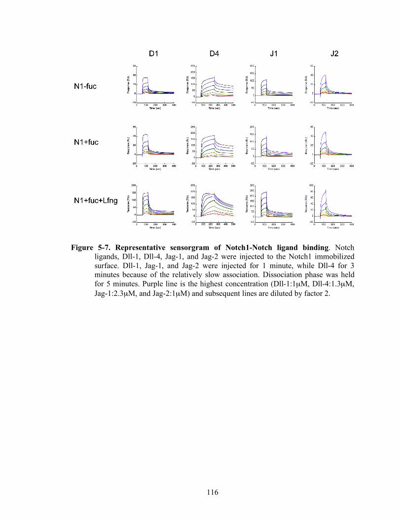
116
Figure 5-7. Representative sensorgram of Notch1-Notch ligand binding. Notch ligands, Dll-1, Dll-4, Jag-1, and Jag-2 were injected to the Notch1 immobilized surface. Dll-1, Jag-1, and Jag-2 were injected for 1 minute, while Dll-4 for 3 minutes because of the relatively slow association. Dissociation phase was held for 5 minutes. Purple line is the highest concentration (Dll-1:1µM, Dll-4:1.3µM, Jag-1:2.3µM, and Jag-2:1µM) and subsequent lines are diluted by factor 2.
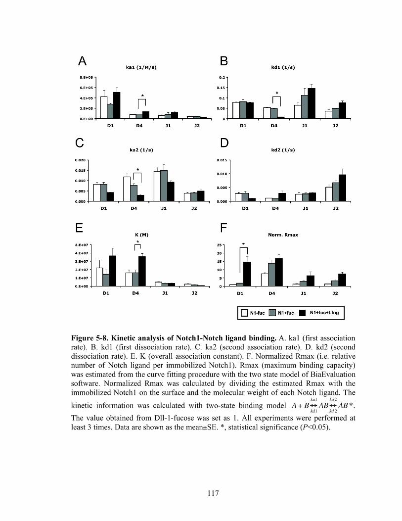
117
Figure 5-8. Kinetic analysis of Notch1-Notch ligand binding. A. ka1 (first association rate). B. kd1 (first dissociation rate). C. ka2 (second association rate). D. kd2 (second dissociation rate). E. K (overall association constant). F. Normalized Rmax (i.e. relative number of Notch ligand per immobilized Notch1). Rmax (maximum binding capacity) was estimated from the curve fitting procedure with the two state model of BiaEvaluation software. Normalized Rmax was calculated by dividing the estimated Rmax with the immobilized Notch1 on the surface and the molecular weight of each Notch ligand. The kinetic information was calculated with two-state binding model
!
A + B"kd1
ka1
AB"kd 2
ka2
AB*. The value obtained from Dll-1-fucose was set as 1. All experiments were performed at least 3 times. Data are shown as the mean±SE. *, statistical significance (P<0.05).

118
Figure 5-9. Kinetic analysis of Notch1 mutants and Dll-4 binding. A. Notch mutants. B. First association rate. C. First dissociation rate. D. Second association rate. E. Second dissociation rate. F. Association constant. G. Normalized Rmax. Rmax was estimated from the curve fitting procedure with the two state model of BiaEvaluation software. Normalized Rmax was calculated by dividing the estimated Rmax with the immobilized Notch1 on the surface and the molecular weight of D4. The value obtained from Dll-4-fucose was set as 1. All experiments were performed at least 3 times. Data are shown as the mean±SE. *, statistical significance (P<0.05).
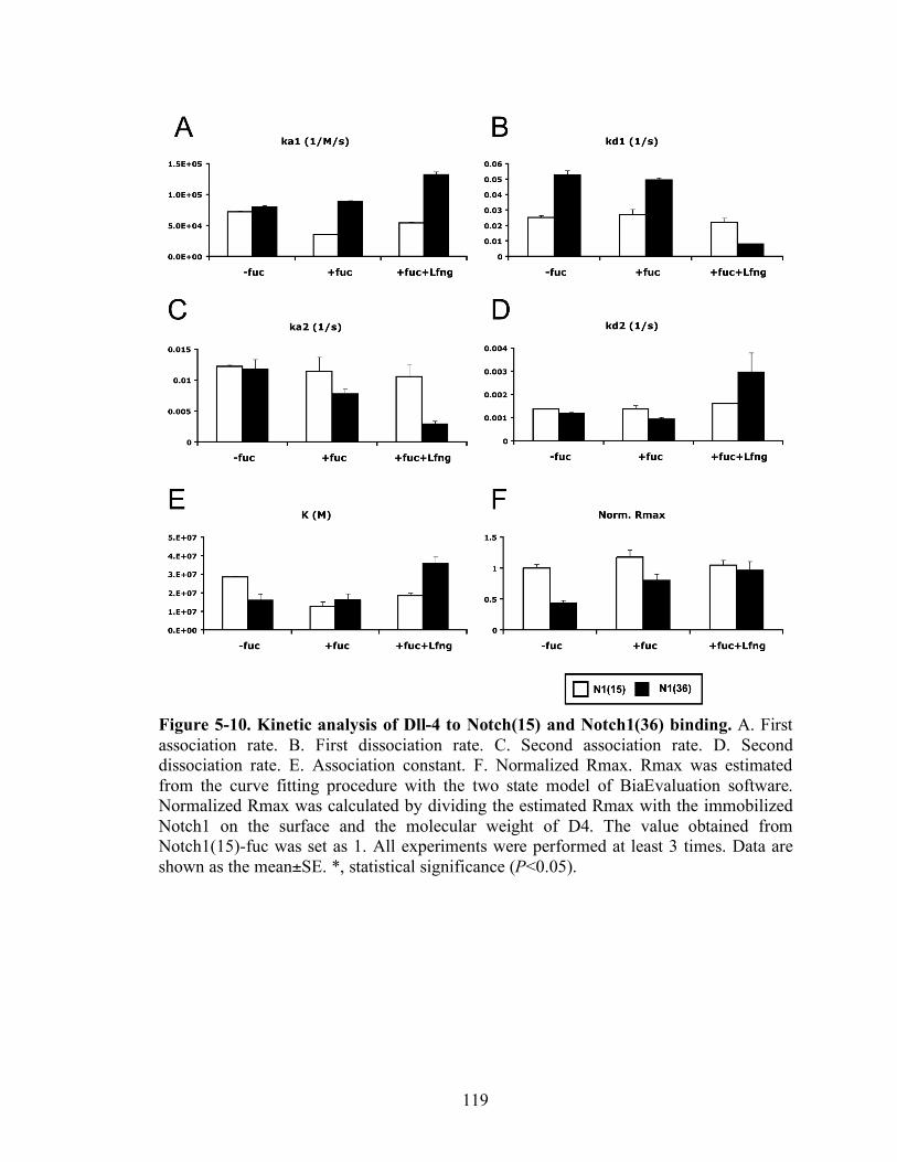
119
Figure 5-10. Kinetic analysis of Dll-4 to Notch(15) and Notch1(36) binding. A. First association rate. B. First dissociation rate. C. Second association rate. D. Second dissociation rate. E. Association constant. F. Normalized Rmax. Rmax was estimated from the curve fitting procedure with the two state model of BiaEvaluation software. Normalized Rmax was calculated by dividing the estimated Rmax with the immobilized Notch1 on the surface and the molecular weight of D4. The value obtained from Notch1(15)-fuc was set as 1. All experiments were performed at least 3 times. Data are shown as the mean±SE. *, statistical significance (P<0.05).
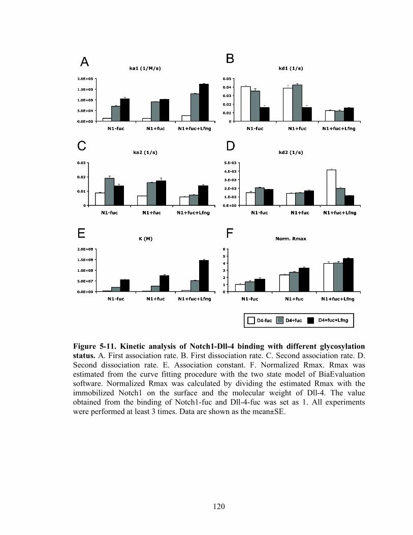
120
Figure 5-11. Kinetic analysis of Notch1-Dll-4 binding with different glycosylation status. A. First association rate. B. First dissociation rate. C. Second association rate. D. Second dissociation rate. E. Association constant. F. Normalized Rmax. Rmax was estimated from the curve fitting procedure with the two state model of BiaEvaluation software. Normalized Rmax was calculated by dividing the estimated Rmax with the immobilized Notch1 on the surface and the molecular weight of Dll-4. The value obtained from the binding of Notch1-fuc and Dll-4-fuc was set as 1. All experiments were performed at least 3 times. Data are shown as the mean±SE.

121
Figure 5-12. Overall association and dissociation rate of Notch-Notch ligand binding. A. Overall association rate of Notch ligands binding to Notch1+fucose or Notch1+fucose+Lfng. B. Overall dissociation rate of Notch ligand binding to Notch1+fucose or Notch1+fucose+Lfng.
122
Figure 5-13. Model for Notch-Notch ligand binding. A. Schematic of O-fucosylation sites and calcium binding sites of mouse Notch ligands and mouse Notch1 receptors. O-fucosylation sites are predicted with broader consensus sequence (C2X4-5S/TC3). Calcium binding EGF domains were adopted from SMART software. Red triangle denotes O-fucosylation sites. Hexagon is DSL domain of Notch Notch ligand, which is the binding site to Notch receptors. B. Two-state binding model with triple stranded structure. First, Notch ligand bind to Notch receptor via the binding region of Notch1 receptor and DSL domain of Notch ligand and subsequently EGF repeats of Notch ligand dock with lower part of the triple stranded structure of Notch receptor. Secondly, the Notch-Notch ligand binding complex adopts a conformational change to form the stable complex. C. Parallel model for both trans-activation and cis-inhibition. Solid circle in Notch1 receptor is the calcium binding EGF domain and white circle the non-calcium binding EGF domain.
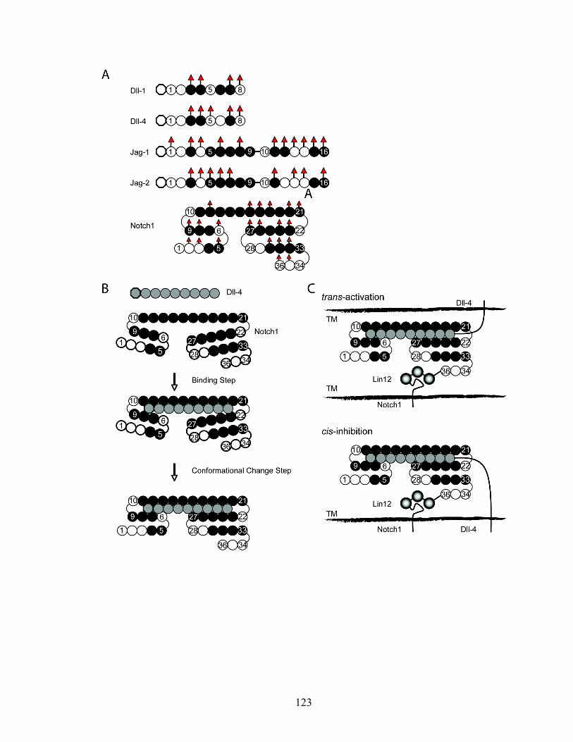
123
124
Chapter 6
Conclusions
Notch signaling is a very complicated cellular event because of the variety of
Notch receptor and Notch ligand combinations, the involvement of complex
glycosylation state such as unusual O-fucosylation, proteolytic events, endocytosis, and
cross-talk with other signaling transduction pathway [181]. Notch signaling is involved in
many biological contexts such as cancer, stem cell development [84, 182], and neural
development. However, the molecular basis of Notch signaling mechanisms remains to
be elucidated. In this work, OTs, AFM, and SPR experiment were performed to elucidate
the characteristics of Notch-Notch ligand binding.
Direct Force Measurement by Force Spectroscopy
In Chapter 3 and 4, OTs and AFM systems were used to study the glycan-related
Notch binding. These apparatuses have the capability to measure the single bond strength
of the binding complex with pN resolution and successfully used in many biological
systems such as DNA zipping and unzipping, DNA-protein interaction, protein-protein
interaction, and cell adhesion.
Notch signaling is a force-induced event, where the force is generated by trans-
endocytosis. Force causes the retraction of ECN, and subsequently induces a
125
conformational change of NLR to expose the S2 cleavage site for signal activation. The
binding strength should be strong enough to endure this retraction and conformational
process. As suggested in previous studies, O-fucosylated glycans play a role in
modulating the binding and signaling of Notch transduction events. We hypothesize that
O-fucosylated glycans regulate the strength of Notch-Notch ligand binding and finally
signaling. We tested the applicability of OTs and AFM for the direct force measurement
of Notch-Notch ligand bond. We found several issues from OT-based force
measurement. The first issue was the optical interference. To generate the optical trap, the
laser path should not be blocked. To probe the protein-protein bond, two objects
immobilized with two proteins need to be in contact each other, causing the optical
interference by objects such as beads. To prevent this interference, a special design would
be required, but it would be expensive and difficult to characterize the coating of the
proteins on the surface. Secondly, OTs are hard to generate forces exceeding 200pN
because of the power limitation and heating. Therefore, the use of OTs is restricted when
the binding strength is over 200pN, which frequently occurs when the multiple bonds of
protein-protein binding or non-specific bonds are formed. However, AFM does not have
these pitfalls and was successfully adopted to measure the single binding strength of
Notch-Notch ligand binding complex. We found that O-fucosylated glycans of
Notch1(EGF1-15) have no significant effect on binding to Dll-4, demonstrating that
EGF1-15 repeats are not strongly modulated by Fringe modification and implying that
EGF domains 16-36 are the major target for Lfng modification. This conclusion is
consistent with the results from Chapter 5.
126
Kinetic Analysis of Notch-Notch Ligand Binding
Notch signaling mainly correlates with ligand binding [73]. In experiments we
report here, we have sought to advance the understanding of how Fringe-mediated glycan
modifications modulate the kinetics and affinities of the interactions between mouse
Notch1 and its ligands, how distinct O-fucosylation sites on mouse Notch1 contribute to
these binding interactions, and how Fringe-dependent glycan modification of Notch
ligands themselves may modulate Notch receptor-ligand interactions, using surface
plasmon resonance techniques. Efficient binding was observed between mouse Notch1
and the Notch ligands Dll-1, Dll-4, Jag-1, and Jag-2, but not between Notch1 and Dll-3.
We observed a biphasic pattern of association and dissociation phases. If the
conformational change is involved in the binding, the binding complex is more stable
with time. To test this hypothesis, we carried out the linked reaction test with three
different injection times. We found that the binding complex was more stable as the
injection time was increased. A biphasic binding and dissociation pattern was observed
with the four binding-competent Notch1-Notch ligand pairs, suggesting a two-state
receptor-ligand interaction model characterized by initial formation of a transient
receptor-ligand complex followed by a conformational change that leads to a more stable
receptor-ligand complex. Here, the initial complex is rapidly formed and the intermediate
binding state is then followed by the slow production of the tight complex.
We observed that the ratio of maximum binding capacity of Dll-1 to wild type
Notch1 and Notch1+Lfng is about 1:8. Although we don’t know the stoichiometry of the
Notch-Notch ligand binding, we believe that this value doesn’t reflect the altered

127
stoichiometry from the Fringe modification. However, this is typical in two state binding
model [183].
Primary and secondary on and off-rates for the four binding-competent mNotch1-
Notch ligand pairs were observed to be distinct and characteristic for each Notch ligand.
The magnitudes of the overall association constants (K) generated by the Notch1-Dll-1
couple or the Notch1-Dll-4 couple were similar, and much greater than the overall
association constants generated in couples involving mNotch1 and either Jag-1 or Jag-2.
The overall association constants observed when Dll-1 or Dll-4 interacted with Fringe-
modified Notch1 were significantly greater than when these ligands interacted with
unmodified Notch1, with enhancement likely due to Fringe modifications of fucose
moieties within EGF repeats 16-36 by altering both the binding and conformational step.
By contrast, Fringe modification of Notch1 did not significantly modulate interactions
with Jag-1 or Jag-2. Mutational analyses confirm prior observations that the O-
fucosylation site within EGF repeat 12 dictates much, if not all of the binding between
Notch1 and its ligands. Mutation in the O-fucosylation site in EGF repeat 26 of Notch1
receptor enhances the binding level of Dll-4, indicating O-fucosylation in EGF 26 is the
inhibitory molecule to the ligand binding. Finally, we observe that Fringe modification of
Dll-4 enhances its ability to bind to Notch1, demonstrating that Lfng is mainly the
binding modulator.
We propose a two interconverting complex state model with a triple stranded
structure. In this model, EGF repeats 10-21 form a rod-like shape and both EGF domains
6-9 and 22-27 are folded back against EGF repeats 10-21, forming a binding pocket when
Notch1 binds to a Notch ligand.
128
This model can explain the trans-activation and cis-inhibition as shown in Figure
5-13C. In this proposed model, parallel complexes are formed to achieve cis-inhibition.
Trans-activation is also achieved when the Notch ligand binds to a triple stranded Notch
receptor to form parallel complexes.
O-fucosylated glycans may have two functions in Notch binding in this model.
Firstly, O-fucosylated glycans in EGF12 of Notch1 receptor may stabilize the triple
stranded structure because O-fucosylated glycans on EGF12 may interact with EGF6-9,
but not directly Notch ligand, and in doing so, disturbs the upper part (EGF1-15) of triple
stranded structure, and the binding pocket of the Notch binding region, and thereby
reduce ligand binding. Secondly, O-fucose in the Abruptex domain is inhibitory, while
Fringe extended O-fucose is a positive regulator for binding. Fringe modified O-fucose in
the Abruptex domain appears to be critical in the binding by enhancing the formation of
low-affinity complexes, and slow conversion from low-affinity complexes to high-
affinity complexes. Since Fringe modified Dll-4 also has the increased binding ability to
Notch1 receptors, indicating Fringe extension is involved in binding, not maintaining the
overall structure of Notch receptors and Notch ligands, although we cannot exclude the
possibility that the Abruptex domain is structurally correlated with other domains such as
NLR.
In conclusion, the kinetics of glycan-dependent Notch-Notch ligand binding has
been explored in Chapter 5. Our study of the kinetics of Notch-Notch ligand binding
suggests a molecular basis for Notch-Notch ligand binding, and the role of glycans in
binding. This work opens a new avenue to elucidate canonical Notch signaling.
129
Future Direction
There are still several key questions left to be investigated: force-induced Notch
signaling, Notch or Notch ligand clustering, and the structure of Notch-Notch ligand
binding complexes.
(1) Force-induced Notch signaling
Many studies have suggested that mechanical force may trigger the Notch
signaling by inducing the decomposition of the NRR domain. There is evidence that
conformational change induced by mechanical force is critical.
(i) Effect of immobilization and pre-clustering
Free ligands in solution are not capable of activating Notch signaling. This
indicates that Notch signaling is not activated merely by binding to its ligand, suggesting
that other factors are involved in the signaling process. It has been reported that
immobilization or pre-clustering of Notch ligand is required for induction of Notch
signaling [92]. In this experimental system, the other end of the immobilized or pre-
clustered ligands is stationary and can support the pulling force if any force is produced.
From the mechanical point of view, this is the only difference between free ligand and
immobilized (or pre-clustered) Notch ligand systems. In other words, free ligand cannot
retain the tension, but the supported ligand can. Although further studies are needed to
elucidate the source of the mechanical force in immobilized (or pre-clustered) Notch
130
ligand systems (this will be discussed more in the next page), these in vitro data suggest
that the mechanical force may contribute to the activation of Notch signaling.
(ii) Role of NRR domain
Calcium depletion dissociates the ECD and activates Notch signaling [184]. This
suggests that conformational change of the extracellular domain of Notch can trigger
ligand-independent activation by unfolding some calcium binding domains. Recently, the
x-ray crystal structure of Notch1 NRR was reported. A key feature is a conserved
hydrophobic plug that occludes the metalloprotease cleavage sites (S2 sites) by a steric
mechanism [135]. A novel mutation (H1545P), which alters a residue at the LNR-HD
interface, results in ligand-independent increases in signaling. Also, mutations in the
calcium binding sites in the LNR domain gave the same result. Together, these mutations
release the autoinhibitory clamp on the HD domain imposed by Lin12/Notch repeats.
In summary, S2 cleavage sites need to be exposed by the displacement of LNR
modules away from the HD domain by the mechanical force for the Notch signaling.
This conclusion leads to the next question. What is the source of the mechanical
force that activates the Notch signaling? Two external sources (trans-endocytosis and cell
movement) and one internal force (conformational change) can be postulated.
(i) Trans-endocytosis
Endocytosis is required for Notch signaling. Notch1 receptors are physically
dissociated by endocytosis before activating proteolysis can occur [89]. Endocytosis
131
produces the pulling force after the protein-protein complexes are formed. Endocytosis
can provide the mechanical force to induce the conformational change of NRR domain.
An understanding of the magnitude of forces generated by endocytosis will help
to increase our understanding of whether Notch-Notch ligand interactions are “stronger”
than the force of endocytosis and thus allow endocytosis to “pull” a Notch-Notch ligand
pair “away” from the cell surface to enable an S2 cleavage event by unclamping the NRR
domain.
(ii) Membrane tension
Exposure of cultured cells to the mechanical stimuli and matrix stiffness or
elasticity is sufficient to induce endocytosis by altering the membrane tension [185-187].
In in vitro cell culture systems, cells are in direct contact with the immobilized proteins
on the surface of the culture dish, which has a specific stiffness. This environment causes
the alteration of the membrane tension and induces endocytosis or exocytosis which may
generates the mechanical force to activate the Notch signaling.
(iii) Cell movement
To our knowledge, there is no report that cell movement can generate the force,
which can induce the conformational change in protein-protein complexes. However,
during cell movement, forward and rearward traction forces are distributed on the cell
surface, which are measured by the micromachined devices [188, 189] and traction force
microscopy [190-192]. Moreover, the mechanical environment induced by matrix
elasticity [185] and tensile force [193] plays a critical role in cell function. This suggests
132
the possibility that traction force or protrusion force generated by cell movement between
the cells or on the ligand immobilized surface can induce the conformational change of
Notch1 NRR domain to expose the S2 cleavage site for the metalloprotease enzyme.
(iv) Conformational change of Notch-Notch ligand binding complexes
It has been reported that the conformational change of protein structure can
generate mechanical force. For example, in G-protein, the force is transduced by changes
in the configurational space of the protein produced by substrate hydrolysis [194]. Also,
ligand binding induced conformational change can generate mechanical force. For
example, a two-step binding mechanism was proposed for T cell receptor recognition of
peptide-MHC [195-199]. The mechanical force caused by binding pulls the TCR-CD3
complexes towards the APC in a piston-like movement [196, 199]. The resulting change
in the positions of transmembrane and/or cytoplasmic portions of TCR-CD3 relative to
the membrane could result in conformational changes in the TCR-CD3 cytoplasmic tails
such as exposure of the Nck-binding site on CD3ε [196, 199].
In this study, we propose that the mechanism of Notch-Notch ligand binding can
be explained by the two-step binding mechanism, which consists of a binding step and a
conformational change step. Although, there is no direct evidence of force-induced Notch
signaling, it is possible that conformational change of Notch-Notch ligand components
can produce the mechanical force to unplug the HD domain from the LNR domain.
In summary, mechanical force appears to contribute to the Notch activation. The
mechanical force might be generated from trans-endocytosis, membrane tension, cell
133
movement, and/or conformational change of protein complexes. The binding force of
Notch-Notch ligand needs to be stronger than ligand binding induced mechanical force to
unclamp of the NRR domain. This bond strength is hierarchically modulated by the
combination of Notch ligand and Fringe action. Recent progress in force spectroscopy
technology opens the door to study the very small mechanical force generated by the
single biomolecule. Using the force spectroscopy such as AFM and OTs, the force-
induced Notch signaling can be probed.
(2) Notch receptor and Notch ligand clustering
In many signaling transduction systems, protein clustering is critical to magnify
the signal. One well-established clustering system is the immunological synapse
formation. T-cell activation requires clustering of a threshold number of T-cell receptors
(TCRs) at the site of antigen presentation, a number that is reduced by CD28 co-receptor
recruitment of signalling proteins to TCRs. It was demonstrated that deficiency in beta1,6
N-acetylglucosaminyltransferase V (Mgat5), an enzyme in the N-glycosylation pathway,
lowers T-cell activation thresholds by directly enhancing TCR clustering. Galectin-3 was
associated with the TCR complex at the cell surface, an interaction dependent on Mgat5.
A galectin-glycoprotein lattice strengthened by Mgat5-modified glycans restricts TCR
recruitment to the site of antigen presentation, i.e. Mgat5 enhances the TCR clustering
[95]. These data suggest that the glycan may modulate receptor clustering.
Several studies reported that Notch signaling is density- or dose-dependent [93,
200], suggesting that there is a threshold level of Notch signaling [124]. These data imply
that the Notch receptor and/or Notch ligand clustering facilitate Notch signaling, but not
134
simply the dimerization of Notch receptors on the cell surface [201]. However, It is not
reported yet whether Notch clustering is facilitated by a glycan and/or lectin.
Two mechanisms were proposed for the role of endocytosis in ligand expressing
cells. The first mechanism is that the endocytosis pulls the Notch ligand after it binds to
Notch receptor as discussed above. Secondly, ligands are endocytosed and then re-
trafficked to the cell surface as an activated and mostly clustered form. However, there is
no direct evidence for an effect of ligand clustering on Notch signaling.
In summary, Notch and Notch ligand clustering might be modulated by glycans.
Using the single biomolecule detection system, glycan-dependent Notch and Notch
ligand clustering can be investigated. This will give new insight about the role of glycans
in receptor clustering for signal activation.
(3) Structure of the Notch-Notch ligand binding complex
To understand Notch signaling, it is important to know the molecular basis of
Notch-Notch ligand binding. Because of the difficulty associated with obtaining a crystal
of the transmembrane protein, the structure of the binding region of Notch and Notch
ligand have been explored. The crystal structure of the human Notch1 ligand binding
region (EGF11-13) shows a rod-like shape rigidified by calcium, which is very similar to
well-studied calcium binding EGF domains, but the angle of tilt is significantly different
[173]. EGF repeats of Notch also include non-calcium binding EGF domains. The
combination of calcium binding domain and non-calcium binding domain give the unique
structure to the EGF bearing proteins. Several studies suggested the importance of
calcium binding domains for the protein structure containing EGF repeats [158, 174,
135
202]. In NMR studies of a pair of tandem EGF domains from Plasmodium falciparum
merozoite surface protein-1, the linker between two non-Ca2+ biding EGF domains
bends, and the EGF domains pack against each other along their length. Finally, this
gives the protein a U-shaped overall structure [202]. Following this result, “triple-
stranded” structure was proposed for the Notch structure [112]. In this model, EGF10-21
will form a long rigid chain, with EGF6-9 and EGF22-26 folded back against them and
EGF1-5 and EGF 27-33 involved in a strand displacement process after ligand binding
[112]. This creates the possibility that strand displacement of the EGF22-33 domain
would effect a conformational change as proposed in this work. The location of Abruptex
domain suggests that they could play a role in conformational change with Fringe
modification on this region. To understand the exact molecular mechanism of Notch-
Notch ligand binding, the structure of the Notch-Notch ligand binding complex needs to
be explored. Also, the stoichiometry of Notch-Notch ligand binding remains to be
investigated.

136
Appendices
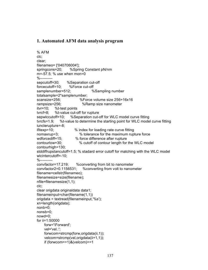
137
1. Automated AFM data analysis program
% AFM clc; clear; filenameo= ['040706004']; springcons=20; %Spring Constant pN/nm m=-57.5; % use when mon=0 %---------- sepcutoff=30; %Separation cut-off forcecutoff=10; %Force cut-off samplenumber=512; %Sampling number totalsample=2*samplenumber; scansize=256; %Force volume size 256=16x16 rampsize=256; %Ramp size nanometer itvr=10; %t-test points tvrcf=8; %t-value cut-off for rupture sepwlccutoff=10; %Separation cut-off for WLC model curve fitting tvrcfs=1.9; %t-value to determine the starting point for WLC model curve fitting iunclerupture=-8; ifitexp=10; % index for loading rate curve fitting nomaxrup=3; % tolerance for the maximum rupture force wdforcediff=15; % force difference after rupture contourlow=30; % cutoff of contour length for the WLC model contourhigh=130; stddiffrupstancutoff=1.5; % stadard error cutoff for matching with the WLC model wlcintercutoff=-10; %---------- convfactor=17.219; %converting from bit to nanometer convfactor2=0.1156531; %converting from volt to nanometer filename=cellstr(filenameo); filenamesize=size(filename); nfile=filenamesize(1,1); clc; clear origdata originaldata data1; filenameinput=char(filename(1,1)) origdata = textread(filenameinput,'%s'); xn=length(origdata); nonb=0; nonsb=0; nowd=0; for ii=1:50000 forw='\Forward'; vel='vel.:'; forwcom=strcmp(forw,origdata(ii,1)); velcom=strcmp(vel,origdata(ii+1,1)); if (forwcom==1)&(velcom)==1
138
forwardvelvolt=str2double(origdata(ii+2,1)); forwardvel=forwardvelvolt/convfactor2; break end end for ii=1:50000 rev='\Reverse'; vel='vel.:'; revcom=strcmp(rev,origdata(ii,1)); velcom=strcmp(vel,origdata(ii+1,1)); if (revcom==1)&(velcom)==1 reversevelvolt=str2double(origdata(ii+2,1)); reversevel=reversevelvolt/convfactor2; break end end for ii=1:100000 list='list'; endofdeader='end'; listcom=strcmp(list,origdata(ii,1)); endcom=strcmp(endofdeader,origdata(ii+1,1)); if (listcom==1)&(endcom==1) indexofdata=ii+1; break end end startindex=indexofdata+1; data1(:,1)=str2double(origdata(startindex:xn,1)); total_data_number=xn-indexofdata; ndata1=numel(data1); dt=ndata1/samplenumber; for ii=1:dt
originaldata(ii,:)=data1((ii-1)*samplenumber+1:ii*samplenumber); end if total_data_number~=ndata1 disp('total_data_number and ndata1 are not same!') % break end inipoint=2*scansize+1; iquit=0; for i=1:scansize; if iquit==1,break,end %Loading data dtpos=2*(i-1)+inipoint; rawdata1=originaldata(dtpos,:); rawdata2=originaldata(dtpos+1,:); rdata=[rawdata2';rawdata1']; incr=rampsize/samplenumber; displace1=(0:incr:samplenumber*incr-incr); displace=[displace1';displace1']; totaltime=rampsize/reversevel;
139
timeincr=totaltime/samplenumber; timecourse1=(0:timeincr:samplenumber*timeincr-timeincr); %Calculate the Force def=(rdata./-m); defavg=mean(def((samplenumber-20):(samplenumber-2))); relsep=displace+def; avgsep=mean(relsep(1:10)); sep=relsep-avgsep; force=springcons*(def-defavg); %Split the data to approch and retraction force1=force(1:samplenumber); force2=force(samplenumber+1:totalsample); sep1=sep(1:samplenumber); sep2=sep(samplenumber+1:totalsample); indentforce=force(1); %determine the separation point and the rupture force tvrupture=zeros(size(force1)); samplingpt=(1:size(force1)); tvcutoff=tvrcf*ones(size(force1)); sepcutoffm=sepcutoff*ones(size(force1)); sepcutoffy=(-300:1:300); sepcutoffx=sepcutoff*ones(size(sepcutoffy)); forcecutoffm=-forcecutoff*ones(size(force1)); for j=itvr:samplenumber-itvr xtl=force1(j-itvr+1:j); xtr=force1(j+1:j+itvr); meanxtl=mean(xtl); meanxtr=mean(xtr); covxtl=cov(xtl); covxtr=cov(xtr); tvrupture(j)=-(meanxtl-meanxtr)/sqrt((covxtl+covxtr)/itvr); % calculate t-value end tcount=0; unclerupture=0; isepdiff=0; for j=itvr:samplenumber-itvr tvl=tvrupture(j)-tvrupture(j-1); tvr=tvrupture(j+1)-tvrupture(j); if tvl>0&tvr<0 if tvrupture(j)>=tvrcf tcount=tcount+1; % for k=j:-1:j-10 % tvml=force1(k)-force1(k-1); % tvmr=force1(k+1)-force1(k); % if tvml<0&tvmr>0 for k=j-1:-1:j-5 forcediffp(k)=abs(force1(k)-force1(k-1)); end forcediffpavg=0.5*mean(forcediffp); forcediff=force1(j)-force1(j-1); if forcediff>forcediffpavg
140
indexrupture(tcount)=j-1; else indexrupture(tcount)=j; end septcount=sep(indexrupture(tcount)); % break % end % end for l=j-2:-1:50 if tvrupture(l)>tvrcfs&tvrupture(l+1)<tvrcfs indexzerorup(tcount)=l+5; sepdiffs=sep(indexrupture(tcount))-sep(indexzerorup(tcount)); if sepdiffs<sepwlccutoff isepdiff=1; end for ll=j-2:-1:l if tvrupture(ll)<=iunclerupture unclerupture=1; break end end minforce1=min(force1(l:indexrupture(tcount))) minforce1dif=force1(indexrupture(tcount))-minforce1; if minforce1dif>forcecutoff unclerupture=1; end for ll=j-2:-1:l if tvrupture(ll)<0&tvrupture(ll+1)>0 tvzero=ll; break end end for ll=j-2:-1:tvzero tvl=tvrupture(ll)-tvrupture(ll-1); tvr=tvrupture(ll+1)-tvrupture(ll); if tvl>0&tvr<0 if tvrupture(ll)>=0 unclerupture=1; break end end end break else indexzerorup(tcount)=50; minforce1=min(force1(indexzerorup(tcount):indexrupture(tcount))) minforce1dif=force1(indexrupture(tcount))-minforce1; if minforce1dif>forcecutoff unclerupture=1; end
141
end end indexfitexp(tcount)=j-ifitexp; end end end %Print the data to the file subplot(2,2,1); plot(displace1,force1,'r-',displace1,force2,'b-'); grid on; axis([0 250 -300 50]); xlabel('Displacement[nm]'); ylabel('Force[pN]'); title({['File name=',num2str(filenameinput)];['Dataset=',num2str(i)];['Indent Force=',num2str(indentforce)]}); subplot(2,2,3); plot(displace1,tvrupture,'r-',displace1,tvcutoff,'g--'); grid on; axis([0 250 -20 20]); xlabel('Displacement[nm]'); ylabel('t-value'); title({['Dataset=',num2str(i)];['t-test to find the rupture force']}); % Plot the force and separation subplot(2,2,4); plot(sep1,force1,'r-',sep1,force2,'b-',sepcutoffx,sepcutoffy,'g--',sep1,forcecutoffm,'g-.'); grid on; axis([-10 250 -300 50]); xlabel('Separation[nm]'); ylabel('Force[pN]'); if tcount==0 nonb=nonb+1; title({['Dataset=',num2str(i)];['No Binding']}); pause(1) elseif tcount>=1 count=tcount; for jjj=count:1 irt=indexrupture(jjj); if tcount>=2 irtp=indexrupture(tcount-1); end irtz=indexfitexp(jjj); irtc=indexzerorup(jjj); finalrup=force(irt); finalsep=sep(irt); finalrupture=abs(finalrup); clear tvx diffrup tvn noise; % data quality test if septcount<sepcutoff nonsb=nonsb+1; title({['Dataset=',num2str(i)];['Non-Specific Binding (Separation is less than cut-off)']});
142
pause(1) break end mfavgsta=indexrupture(count)+10; mfavgend=indexrupture(count)+30; if mfavgsta>samplenumber mfavgsta=samplenumber; end if mfavgend>samplenumber mfavgend=samplenumber; end forceavg=abs(mean(force1(mfavgsta:mfavgend))); if forceavg>wdforcediff nowd=nowd+1; title({['Dataset=',num2str(i)];['Wrong dataset (Irregular data points)']}); pause(1) break end if finalrupture<forcecutoff nonsb=nonsb+1; title({['Dataset=',num2str(i)];['Non-Specific Binding (Rupture force is less than cut-off)']}); pause(1) break end sepdiff=sep(irt)-sep(irtc); if sepdiff<sepwlccutoff isepdiff=1; end if isepdiff==1 nonsb=nonsb+1; title({['Dataset=',num2str(i)];['Non-Specific Binding (Short distance for WLC model curve fitting)']}); replyrup=menu('Dataset Quality','Next','Quit'); if replyrup==2 iquit=1; end break end if tcount>nomaxrup nonsb=nonsb+1; title({['Dataset=',num2str(i)];['Non-Specific Binding (Too many rupture event)']}); pause(1) break end if count>=2 sepdiffbr=sep(irt)-sep(irtp); if sepdiffbr<sepwlccutoff nonsb=nonsb+1; title({['Dataset=',num2str(i)];['Non-Specific Binding (Two peaks are too close)']}); pause(1)
143
break end end if unclerupture==1 nonsb=nonsb+1; title({['Dataset=',num2str(i)];['Non-Specific Binding (Unclear Rupture Point)']}); replyrup=menu('Dataset Quality','Next','Quit'); if replyrup==2 iquit=1; end break end %---------- lrx=[displace1(irtz:irt)]'; lry=force1(irtz:irt); [coef model]= fitexp(lrx,lry); sepint=1/coef(2)*log(1-finalrup/coef(1)); slpexp=-coef(2)*coef(1).*exp(coef(2)*sepint); intercept=finalrup-slpexp*sepint; lrxi=[displace1(irt-10:irt+10)]'; lryi=slpexp*lrxi+intercept; lrxci=[displace1(irtz:irt+10)]'; lryci=coef(1).*(1- exp(coef(2).* lrxci)); sysspringcon=-slpexp; loadrate=sysspringcon*reversevel; finalloadrate=loadrate; subplot(2,2,1); plot(displace1,force1,'r-',displace1,force2,'b-',lrxci,lryci,'b-',lrxi,lryi,'g-'); grid on; axis([0 250 -300 50]); xlabel('Displacement[nm]'); ylabel('Force[pN]'); title({['File name=',num2str(filenameinput)];['Dataset=',num2str(i)];['Indent Force=',num2str(indentforce)];['System Spring Constant=',num2str(sysspringcon)];['Loading rate=',num2str(loadrate)]}); %-------- boltzmann=1.3806503*10^(-23); temp=295; %K persistence=0.38*10^(-9); %m wlcx=displace1; a2=-0.5164228; a3=-2.737418; a4=16.07497; a5=-38.87607; a6=39.49944; a7=-14.17718; %Fixed contour in WLC model contourfix=30; sepmov=0; indexsepmov=0; for ij=1:samplenumber
144
nondimenxi=wlcx(ij)/contourfix; if nondimenxi<1 wlcfxfix(ij)=-(boltzmann*temp/persistence)*(0.25/(1-nondimenxi)^2-0.25+nondimenxi+a2*nondimenxi^2+a3*nondimenxi^3+a4*nondimenxi^4+a5*nondimenxi^5+a6*nondimenxi^6+a7*nondimenxi^7)*10^12; %pN %wlcfxfix(ij)=-(boltzmann*temp/persistence)*(0.25/(1-nondimenxi)^2-0.25+nondimenxi)*10^12; %pN if ij>2 if wlcfxfix(ij)<finalrup&wlcfxfix(ij-1)>finalrup sepmov=nondimenxi*contourfix-finalsep; indexsepmov=ij; end end else wlcfxfix(ij)=0; end end wlcxifix=wlcx-sepmov; wlcxifixmod=wlcxifix(1:indexsepmov+10); wlcfxfixmod=wlcfxfix(1:indexsepmov+10); contourfix2=250; sepmov2=0; indexsepmov2=0; for ij=1:samplenumber nondimenxi=wlcx(ij)/contourfix2; if nondimenxi<1 wlcfxfix2(ij)=-(boltzmann*temp/persistence)*(0.25/(1-nondimenxi)^2-0.25+nondimenxi+a2*nondimenxi^2+a3*nondimenxi^3+a4*nondimenxi^4+a5*nondimenxi^5+a6*nondimenxi^6+a7*nondimenxi^7)*10^12; %pN %wlcfxfix2(ij)=-(boltzmann*temp/persistence)*(0.25/(1-nondimenxi)^2-0.25+nondimenxi)*10^12; %pN if ij>2 if wlcfxfix2(ij)<finalrup&wlcfxfix2(ij-1)>finalrup sepmov2=nondimenxi*contourfix2-finalsep; indexsepmov2=ij; end end else wlcfxfix2(ij)=0; end end wlcxifix2=wlcx-sepmov2; wlcxifixmod2=wlcxifix2(1:indexsepmov2+10); wlcfxfixmod2=wlcfxfix2(1:indexsepmov2+10); % meanirtc=mean(force1(irtc-20:irtc); % if meanirtc>-5 xwlcfit=sep(irtc:irt); ywlcfit=force(irtc:irt); [wlcfit wlcfitmodel]=wlcmodelcurvefit(xwlcfit,ywlcfit); contour=wlcfit(1); wlcinterc=wlcfit(2);
145
mwlc=0.25*ones(size(xwlcfit)); ywlcfitpost=wlcinterc-(boltzmann*temp/persistence)*(mwlc./((1-(xwlcfit/contour)).^2)-0.25+(xwlcfit/contour))*10^12; irtn=irt-irtc+1; for ij=1:irtn irti=ij+irtc-1; nondimenxi=xwlcfit(ij)/contour; tvwlcfit(ij)=wlcinterc-(boltzmann*temp/persistence)*(0.25/(1-nondimenxi)^2-0.25+nondimenxi+a2*nondimenxi^2+a3*nondimenxi^3+a4*nondimenxi^4+a5*nondimenxi^5+a6*nondimenxi^6+a7*nondimenxi^7)*10^12; %pN %tvwlcfxfix(irti)=-(boltzmann*temp/persistence)*(0.25/(1-nondimenxi)^2-0.25+nondimenxi)*10^12; %pN diffrup(ij)=force1(irti)-tvwlcfit(ij); tvx(ij)=xwlcfit(ij); tvn(ij)=sep1(samplenumber-irtn+ij); noise(ij)=force1(samplenumber-irtn+ij); end subplot(2,2,2); plot(tvx,diffrup,'r-',tvn,noise,'b-'); grid on; axis([-10 250 -50 50]); xlabel('Separation[nm]'); ylabel('Force Difference[pN]'); stddiffrup=0; for ij=1:irtn stddiffrup=stddiffrup+diffrup(ij)*diffrup(ij); end stddiffrup=sqrt(stddiffrup/irtn); nowlc=irt-irtc+1; if nowlc<2 nowlc=1; end stderror=stddiffrup/sqrt(nowlc); % [h,significance,ci,stats] =ttest2(noise,diffrup,0.01); %title({['t-test to verify the PEG stretching'];['Estimated Contour=',num2str(contour)];['Interception=',num2str(wlcinterc)];['Standard Error of Contour=',num2str(stderror)];['h=',num2str(h)];['p-value=',num2str(significance)];['99% Confidence Level=',num2str(ci)]}); title({['Dataset=',num2str(i)];['t-test to verify the PEG stretching'];['Estimated Contour=',num2str(contour)];['Interception=',num2str(wlcinterc)];['Standard Error of Contour=',num2str(stderror)]}); %---------- subplot(2,2,4); plot(sep1,force1,'r-',sep1,force2,'b-',wlcxifixmod,wlcfxfixmod,'b-',wlcxifixmod2,wlcfxfixmod2,'m-',xwlcfit,ywlcfitpost,'k--',sepcutoffx,sepcutoffy,'g--',sep1,forcecutoffm,'g-.'); grid on; axis([-10 250 -300 50]); xlabel('Separation[nm]'); ylabel('Force[pN]'); if stderror>stddiffrupstancutoff
146
nonsb=nonsb+1; title({['Dataset=',num2str(i)];['Non-Specific Binding (Not match with WLC model: large Standard Error)']}); replyrup=menu('Dataset Quality','Next','Quit'); if replyrup==2 iquit=1; end break end if wlcinterc<wlcintercutoff nonsb=nonsb+1; title({['Dataset=',num2str(i)];['Non-Specific Binding (Interception is minus for WLC model fitting)']}); replyrup=menu('Dataset Quality','Next','Quit'); if replyrup==2 iquit=1; end break end text(finalsep,finalrup,'\uparrow'); if or(contour>contourhigh,contour<contourlow) title({['Dataset=',num2str(i)];['Non-Specific Binding(Too low or high contour length)'];['Separation=',num2str(finalsep)];['Rupture=',num2str(finalrupture)]}); replyrup=menu('Dataset Quality', 'Choose','No Binding','Non-Specfific','Wrong dataset','Quit'); if replyrup==1 frup = fopen('rupture.doc','a'); fprintf(frup,'%10s %5i %8.1f %8.1f %8.1f %8.1f\n',filenameinput,i,finalsep,finalrupture,sysspringcon,finalloadrate); fclose(frup); end if replyrup==2 nonb=nonb+1; end if replyrup==3 nonsb=nonsb+1; end if replyrup==4 nowd=nowd+1; end if replyrup==5 iquit=1; end break else title({['Dataset=',num2str(i)];['Specific Binding'];['Separation=',num2str(finalsep)];['Rupture=',num2str(finalrupture)]}); frup = fopen('rupture.doc','a'); fprintf(frup,'%10s %5i %8.1f %8.1f %8.1f %8.1f\n',filenameinput,i,finalsep,finalrupture,sysspringcon,finalloadrate); fclose(frup);
147
replyrup=menu('Dataset Quality','Next','Quit'); if replyrup==2 iquit=1; end break end %contour end %for jjj=count:1 end %if tcount end nospec=scansize-(nonb+nonsb+nowd); total=nospec+nonb+nonsb+nowd nonbp=100*nonb/scansize; nonsbp=100*nonsb/scansize; nowdp=100*nowd/scansize; nospecp=100*nospec/scansize; frup = fopen('number.doc','a'); fprintf(frup,'%10s %8.1f %8.1f %8.1f %8.1f %8.1f %8.1f\n',filenameinput,nonb,nonsb,nowd,nospec,total,scansize); fprintf(frup,'%10s %8.1f %8.1f %8.1f %8.1f\n',filenameinput,nonbp,nonsbp,nowdp,nospecp); fclose(frup);
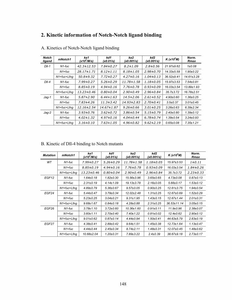
148
2. Kinetic information of Notch-Notch ligand binding
A. Kinetics of Notch-Notch ligand binding
Notch ligand mNotch1 ka1
(x104/M/s) kd1
(x0.01/s) ka2
(x0.001/s) kd2
(x0.001/s) K (x106M) Norm. Rmax
Dll-1 N1-fuc 42.3±12.53 7.84±0.27 8.2±1.09 2.8±0.56 21.97±9.62 1±0.08
N1+fuc 28.17±1.71 8.12±1.11 8.18±1.05 2.98±0.70 14.33±5.59 1.90±0.22
N1+fuc+Lfng 50.8±9.32 7.72±0.27 4.27±0.16 1.04±0.13 36.52±9.41 14.61±3.28
Dll-4 N1-fuc 7.99±0.27 5.26±0.29 11.78±1.58 1.18±0.05 15.97±3.53 7.54±0.81
N1+fuc 8.85±0.19 4.94±0.16 7.76±0.78 0.93±0.09 16.03±3.54 13.89±1.93
N1+fuc+Lfng 13.23±0.46 0.80±0.04 2.90±0.49 2.96±0.84 35.7±3.72 16.78±2.51
Jag-1 N1-fuc 5.87±2.90 6.44±1.63 14.5±2.06 2.61±0.52 4.90±0.60 1.36±0.25
N1+fuc 7.83±4.26 11.3±3.42 14.93±2.83 2.70±0.41 3.5±0.37 3.01±0.45
N1+fuc+Lfng 12.16±2.54 14.67±1.87 9.26±0.66 3.01±0.25 3.26±0.63 6.39±2.34
Jag-2 N1-fuc 3.93±0.76 3.62±0.72 3.86±0.54 5.15±0.79 2.40±0.80 1.39±0.12
N1+fuc 4.02±1.32 4.97±0.16 4.04±0.44 6.78±0.74 1.39±0.54 3.24±0.93
N1+fuc+Lfng 3.16±0.10 7.63±1.05 4.96±0.82 9.62±2.19 0.65±0.08 7.35±1.21
B. Kinetic of Dll-4 binding to Notch mutants
Mutation mNotch1 ka1 (x104/M/s)
kd1 (x0.01/s)
ka2 (x0.001/s)
kd2 (x0.001/s) K (x106M) Norm.
Rmax
WT N1-fuc 7.99±0.27 5.26±0.29 11.78±1.58 1.18±0.05 15.97±3.53 1±0.11
N1+fuc 8.85±0.19 4.94±0.16 7.76±0.78 0.93±0.09 16.03±3.54 1.84±0.26
N1+fuc+Lfng 13.23±0.46 0.80±0.04 2.90±0.49 2.96±0.84 35.7±3.72 2.23±0.33
EGF12 N1-fuc 1.64±0.18 1.82±0.30 15.56±3.96 3.65±0.65 4.73±0.08 0.87±0.13
N1+fuc 2.31±0.19 4.14±1.09 19.13±3.78 2.16±0.05 5.66±0.17 1.53±0.12
N1+fuc+Lfng 4.89±0.79 5.36±0.67 9.57±0.05 0.90±0.25 12.61±3.75 1.94±0.54
EGF24 N1-fuc 5.44±0.47 3.79±0.34 12.02±2.48 1.31±0.25 12.67±0.69 1.52±0.29
N1+fuc 5.23±0.25 3.04±0.21 9.31±1.95 1.43±0.15 12.67±1.44 2.01±0.31
N1+fuc+Lfng 9.68±1.67 0.84±0.18 4.28±0.88 2.31±0.25 36.53±11.14 3.05±0.15
EGF26 N1-fuc 3.78±1.10 3.72±0.60 10.36±1.60 0.91±0.11 11.9±0.96 2.38±0.07
N1+fuc 3.60±1.11 2.70±0.40 7.40±1.22 0.81±0.02 12.4±0.62 2.90±0.12
N1+fuc+Lfng 9.01±0.62 0.87±0.14 4.44±0.94 1.50±0.41 44.63±5.79 2.83±0.19
EGF27 N1-fuc 4.38±0.41 2.89±0.42 9.64±1.51 1.45±0.38 12.73±1.64 1.13±0.47
N1+fuc 4.44±0.44 2.45±0.34 8.74±2.11 1.48±0.31 12.07±0.45 1.48±0.62
N1+fuc+Lfng 10.68±2.04 1.20±0.31 7.89±3.22 2.4±0.39 38.67±9.18 2.73±0.17
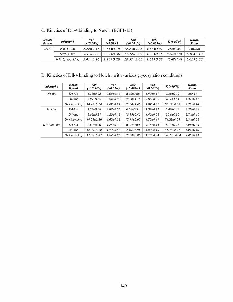
149
C. Kinetics of Dll-4 binding to Notch1(EGF1-15)
Notch ligand mNotch1 ka1
(x104/M/s) kd1
(x0.01/s) ka2
(x0.001/s) kd2
(x0.001/s) K (x106M) Norm. Rmax
Dll-4 N1(15)-fuc 7.22±0.16 2.51±0.14 12.23±0.23 1.37±0.02 28.6±0.53 1±0.06
N1(15)+fuc 3.51±0.06 2.69±0.36 11.42±2.29 1.37±0.15 12.64±2.61 1.18±0.12
N1(15)+fuc+Lfng 5.41±0.16 2.20±0.28 10.57±2.05 1.61±0.02 18.47±1.41 1.05±0.08
D. Kinetics of Dll-4 binding to Notch1 with various glycosylation conditions
mNotch1 Notch ligand
ka1 (x104/M/s)
kd1 (x0.01/s)
ka2 (x0.001/s)
kd2 (x0.001/s) K (x106M) Norm.
Rmax
N1-fuc D4-fuc 1.37±0.02 4.06±0.16 8.65±0.58 1.49±0.17 2.35±0.19 1±0.17
D4+fuc 7.02±0.53 3.54±0.30 19.00±1.75 2.05±0.08 20.4±1.61 1.37±0.17
D4+fuc+Lfng 10.48±0.76 1.62±0.27 13.60±1.45 1.87±0.05 55.17±6.85 1.76±0.24
N1+fuc D4-fuc 1.32±0.08 3.87±0.36 6.58±0.31 1.39±0.11 2.00±0.18 2.35±0.19
D4+fuc 9.08±0.21 4.26±0.19 15.95±0.40 1.46±0.08 25.6±0.80 2.71±0.15
D4+fuc+Lfng 10.25±0.20 1.62±0.26 17.18±2.07 1.72±0.11 74.23±6.06 3.31±0.25
N1+fuc+Lfng D4-fuc 2.60±0.09 1.24±0.10 5.92±0.60 4.16±0.16 5.11±0.28 3.96±0.24
D4+fuc 12.88±0.28 1.19±0.16 7.19±0.78 1.98±0.13 51.45±3.07 4.02±0.19
D4+fuc+Lfng 17.33±0.37 1.57±0.06 13.73±0.88 1.13±0.04 146.33±4.84 4.65±0.11

150
3. Relaxation rates of two-state binding model
This calculation for relaxation rates of two-state binding model was adopted from
Weikl and Deuster [171]. The association and dissociation of two-state model
(N+L⇔NL⇔NL*) have general reaction process of the following form
!
A" B#C (1)
For association process, state A corresponds to N, state B to NL, and state C to
NL*. For dissociation process, state A corresponds to NL*, state B to NL, and state C to N.
We are interested in the dominant, slowest relaxation rate into state C. therefore, the state
C is an “absorbing” state, i.e. the rate from C back to B is 0. The probability evolution
PA(t), PB(t), and PC(t) of the three states is then governed by the set of differential
equations
!
dPA(t)
dt= k
ABPB(t) " k
BAPA(t) (2)
!
dPB(t)
dt= k
BAPA(t) " (k
AB+ k
CB)P
B(t) (3)
!
dPC(t)
dt= k
CBPB(t) (4)
where kij denotes the rate from state j to state i. The three equations can be written in the
matrix form
!
dP(t)
dt= "WP(t) (5)
with P(t)=( PA(t), PB(t), and PC(t)) and
!
W =
kBA
"kAB
0
"kBA
kAB
+ kCB
0
0 "kCB
0
#
$
% % %
&
'
( ( ( (6)

151
The general solution of (5) has the form
!
P(t) = c0Y0
+ c1Y1e"#1t + c
2Y1e"#2t (7)
where Y0, Y1, and Y2 are the three eigenvectors of the matrix W, and λ1 and λ2 are the two
positive eigenvalues of the eigenvectors Y1, and Y2. The coefficients c0, c1, and c2 depend
on the initial conditions at time t=0. The eigenvalue of the eigenvector Y0 is 0, which
ensures that P(t) relaxes towards a finite equilibrium probability distribution
P(t→∞)=c0Y0 for t→∞. The two nonzero eigenvalues are
!
"1/ 2
=1
2kAB
+ KBA
+ kCB
± kAB
2 + kBA# k
CB( )2
+ 2kABkBA
+ kCB( )
$ % &
' ( ) (8)
These eigenvalues, and their eigenvectors Y1 and Y2, capture the time-dependent
relaxation process into equilibrium.

152
Bibliography
153
1. Varki, A., et al., Essentials of glycobiology. 2nd ed. 2008: Cold Spring Harbor Laboratory Press.
2. Alberts, B., et al., Molecular biology of the cell. Fitth edition ed. 2008: Garland
Sciences. 3. Lowe, J.B. and J.D. Marth, A genetic approach to Mammalian glycan function.
Annu Rev Biochem, 2003. 72: p. 643-91. 4. Becker, D.J. and J.B. Lowe, Leukocyte adhesion deficiency type II. Biochim
Biophys Acta, 1999. 1455(2-3): p. 193-204. 5. Korner, C., et al., Carbohydrate deficient glycoprotein syndrome type IV:
deficiency of dolichyl-P-Man:Man(5)GlcNAc(2)-PP-dolichyl mannosyltransferase. Embo J, 1999. 18(23): p. 6816-22.
6. Wraith, J.E., et al., Mucopolysaccharidosis type II (Hunter syndrome): a clinical
review and recommendations for treatment in the era of enzyme replacement therapy. Eur J Pediatr, 2008. 167(3): p. 267-77.
7. Denecke, J., et al., Characterization of the N-glycosylation phenotype of
erythrocyte membrane proteins in congenital dyserythropoietic anemia type II (CDA II/HEMPAS). Glycoconj J, 2008. 25(4): p. 375-82.
8. Denecke, J. and T. Marquardt, Congenital dyserythropoietic anemia type II
(CDAII/HEMPAS): Where are we now? Biochim Biophys Acta, 2008. 9. Fridovich-Keil, J.L., Galactosemia: the good, the bad, and the unknown. J Cell
Physiol, 2006. 209(3): p. 701-5. 10. Fuster, M.M. and J.D. Esko, The sweet and sour of cancer: glycans as novel
therapeutic targets. Nat Rev Cancer, 2005. 5(7): p. 526-42. 11. Zaia, J., Mass spectrometry and the emerging field of glycomics. Chem Biol,
2008. 15(9): p. 881-92. 12. Park, S., M.R. Lee, and I. Shin, Chemical tools for functional studies of glycans.
Chem Soc Rev, 2008. 37(8): p. 1579-91. 13. Liang, P.H., et al., Glycan arrays: biological and medical applications. Curr Opin
Chem Biol, 2008. 12(1): p. 86-92. 14. von Itzstein, M., The war against influenza: discovery and development of
sialidase inhibitors. Nat Rev Drug Discov, 2007. 6(12): p. 967-74.
154
15. Galonic, D.P. and D.Y. Gin, Chemical glycosylation in the synthesis of glycoconjugate antitumour vaccines. Nature, 2007. 446(7139): p. 1000-7.
16. Larsen, R.D., et al., Molecular cloning, sequence, and expression of a human
GDP-L-fucose:beta-D-galactoside 2-alpha-L-fucosyltransferase cDNA that can form the H blood group antigen. Proc Natl Acad Sci U S A, 1990. 87(17): p. 6674-8.
17. Kelly, R.J., et al., Sequence and expression of a candidate for the human Secretor
blood group alpha(1,2)fucosyltransferase gene (FUT2). Homozygosity for an enzyme-inactivating nonsense mutation commonly correlates with the non-secretor phenotype. J Biol Chem, 1995. 270(9): p. 4640-9.
18. Kaneko, M., et al., Alpha1,3-fucosyltransferase IX (Fuc-TIX) is very highly
conserved between human and mouse; molecular cloning, characterization and tissue distribution of human Fuc-TIX. FEBS Lett, 1999. 452(3): p. 237-42.
19. Miyoshi, E., et al., The alpha1-6-fucosyltransferase gene and its biological
significance. Biochim Biophys Acta, 1999. 1473(1): p. 9-20. 20. Panin, V.M., et al., Notch ligands are substrates for protein O-fucosyltransferase-
1 and Fringe. J Biol Chem, 2002. 277(33): p. 29945-52. 21. Moloney, D.J., et al., Fringe is a glycosyltransferase that modifies Notch. Nature,
2000. 406(6794): p. 369-75. 22. Shao, L. and R.S. Haltiwanger, O-fucose modifications of epidermal growth
factor-like repeats and thrombospondin type 1 repeats: unusual modifications in unusual places. Cell Mol Life Sci, 2003. 60(2): p. 241-50.
23. Luo, Y. and R.S. Haltiwanger, O-fucosylation of notch occurs in the endoplasmic
reticulum. J Biol Chem, 2005. 280(12): p. 11289-94. 24. Roos, C., et al., Composition of Drosophila melanogaster proteome involved in
fucosylated glycan metabolism. J Biol Chem, 2002. 277(5): p. 3168-75. 25. Becker, D.J. and J.B. Lowe, Fucose: biosynthesis and biological function in
mammals. Glycobiology, 2003. 13(7): p. 41R-53R. 26. Tonetti, M., et al., The metabolism of 6-deoxyhexoses in bacterial and animal
cells. Biochimie, 1998. 80(11): p. 923-31. 27. Somoza, J.R., et al., Structural and kinetic analysis of Escherichia coli GDP-
mannose 4,6 dehydratase provides insights into the enzyme's catalytic mechanism and regulation by GDP-fucose. Structure, 2000. 8(2): p. 123-35.
155
28. Ohyama, C., et al., Molecular cloning and expression of GDP-D-mannose-4,6-dehydratase, a key enzyme for fucose metabolism defective in Lec13 cells. J Biol Chem, 1998. 273(23): p. 14582-7.
29. Tonetti, M., et al., Synthesis of GDP-L-fucose by the human FX protein. J Biol
Chem, 1996. 271(44): p. 27274-9. 30. Smith, P.L., et al., Conditional control of selectin ligand expression and global
fucosylation events in mice with a targeted mutation at the FX locus. J Cell Biol, 2002. 158(4): p. 801-15.
31. Listinsky, J.J., G.P. Siegal, and C.M. Listinsky, Alpha-L-fucose: a potentially
critical molecule in pathologic processes including neoplasia. Am J Clin Pathol, 1998. 110(4): p. 425-40.
32. Staudacher, E., et al., Fucose in N-glycans: from plant to man. Biochim Biophys
Acta, 1999. 1473(1): p. 216-36. 33. Lowe, J.B., The blood group-specific human glycosyltransferases. Baillieres Clin
Haematol, 1993. 6(2): p. 465-92. 34. Hooper, L.V. and J.I. Gordon, Glycans as legislators of host-microbial
interactions: spanning the spectrum from symbiosis to pathogenicity. Glycobiology, 2001. 11(2): p. 1R-10R.
35. Moran, A.P., et al., Phenotypic variation in molecular mimicry between
Helicobacter pylori lipopolysaccharides and human gastric epithelial cell surface glycoforms. Acid-induced phase variation in Lewis(x) and Lewis(y) expression by H. Pylori lipopolysaccharides. J Biol Chem, 2002. 277(8): p. 5785-95.
36. Springer, T.A., Traffic signals for lymphocyte recirculation and leukocyte
emigration: the multistep paradigm. Cell, 1994. 76(2): p. 301-14. 37. Lowe, J.B., Glycosylation in the control of selectin counter-receptor structure and
function. Immunol Rev, 2002. 186: p. 19-36. 38. Smith, P.L., et al., Expression of the alpha(1,3)fucosyltransferase Fuc-TVII in
lymphoid aggregate high endothelial venules correlates with expression of L-selectin ligands. J Biol Chem, 1996. 271(14): p. 8250-9.
39. Maly, P., et al., The alpha(1,3)fucosyltransferase Fuc-TVII controls leukocyte
trafficking through an essential role in L-, E-, and P-selectin ligand biosynthesis. Cell, 1996. 86(4): p. 643-53.
156
40. Homeister, J.W., et al., The alpha(1,3)fucosyltransferases FucT-IV and FucT-VII exert collaborative control over selectin-dependent leukocyte recruitment and lymphocyte homing. Immunity, 2001. 15(1): p. 115-26.
41. Smithson, G., et al., Fuc-TVII is required for T helper 1 and T cytotoxic 1
lymphocyte selectin ligand expression and recruitment in inflammation, and together with Fuc-TIV regulates naive T cell trafficking to lymph nodes. J Exp Med, 2001. 194(5): p. 601-14.
42. Bruckner, K., et al., Glycosyltransferase activity of Fringe modulates Notch-Delta
interactions. Nature, 2000. 406(6794): p. 411-5. 43. Kozma, K., et al., Identification and characterization of abeta1,3-
glucosyltransferase that synthesizes the Glc-beta1,3-Fuc disaccharide on thrombospondin type 1 repeats. J Biol Chem, 2006. 281(48): p. 36742-51.
44. Shi, S. and P. Stanley, Protein O-fucosyltransferase 1 is an essential component
of Notch signaling pathways. Proc Natl Acad Sci U S A, 2003. 100(9): p. 5234-9. 45. Artavanis-Tsakonas, S., M.D. Rand, and R.J. Lake, Notch signaling: cell fate
control and signal integration in development. Science, 1999. 284(5415): p. 770-6.
46. Radtke, F., et al., Notch regulation of lymphocyte development and function. Nat
Immunol, 2004. 5(3): p. 247-53. 47. Zhou, L., et al., Notch-dependent control of myelopoiesis is regulated by
fucosylation. Blood, 2008. 112(2): p. 308-19. 48. Swiatek, P.J., et al., Notch1 is essential for postimplantation development in mice.
Genes Dev, 1994. 8(6): p. 707-19. 49. Conlon, R.A., A.G. Reaume, and J. Rossant, Notch1 is required for the
coordinate segmentation of somites. Development, 1995. 121(5): p. 1533-45. 50. Radtke, F., et al., Deficient T cell fate specification in mice with an induced
inactivation of Notch1. Immunity, 1999. 10(5): p. 547-58. 51. Rampal, R., K.B. Luther, and R.S. Haltiwanger, Notch signaling in normal and
disease States: possible therapies related to glycosylation. Curr Mol Med, 2007. 7(4): p. 427-45.
52. Ellisen, L.W., et al., TAN-1, the human homolog of the Drosophila notch gene, is
broken by chromosomal translocations in T lymphoblastic neoplasms. Cell, 1991. 66(4): p. 649-61.
157
53. Miyamoto, Y., et al., Effect of erythroid differentiation factor on erythroid differentiation and proliferation of K-562 cells. Biochem Biophys Res Commun, 1990. 168(3): p. 1149-56.
54. Robbins, J., et al., Mouse mammary tumor gene int-3: a member of the notch gene
family transforms mammary epithelial cells. J Virol, 1992. 66(4): p. 2594-9. 55. Aster, J.C., W.S. Pear, and S.C. Blacklow, Notch signaling in leukemia. Annu
Rev Pathol, 2008. 3: p. 587-613. 56. Dufraine, J., Y. Funahashi, and J. Kitajewski, Notch signaling regulates tumor
angiogenesis by diverse mechanisms. Oncogene, 2008. 27(38): p. 5132-7. 57. Joutel, A., et al., Notch3 mutations in CADASIL, a hereditary adult-onset
condition causing stroke and dementia. Nature, 1996. 383(6602): p. 707-10. 58. Arboleda-Velasquez, J.F., et al., CADASIL mutations impair Notch3 glycosylation
by Fringe. Hum Mol Genet, 2005. 14(12): p. 1631-9. 59. John, G.R., et al., Multiple sclerosis: re-expression of a developmental pathway
that restricts oligodendrocyte maturation. Nat Med, 2002. 8(10): p. 1115-21. 60. Sparrow, D.B., et al., Mutation of the LUNATIC FRINGE gene in humans causes
spondylocostal dysostosis with a severe vertebral phenotype. Am J Hum Genet, 2006. 78(1): p. 28-37.
61. Spinner, N.B., et al., Jagged1 mutations in alagille syndrome. Hum Mutat, 2001.
17(1): p. 18-33. 62. Niessen, K. and A. Karsan, Notch signaling in cardiac development. Circ Res,
2008. 102(10): p. 1169-81. 63. Nam, Y., J.C. Aster, and S.C. Blacklow, Notch signaling as a therapeutic target.
Curr Opin Chem Biol, 2002. 6(4): p. 501-9. 64. Villaronga, M.A., C.L. Bevan, and B. Belandia, Notch signaling: a potential
therapeutic target in prostate cancer. Curr Cancer Drug Targets, 2008. 8(7): p. 566-80.
65. Parks, A.L., et al., Structure-function analysis of delta trafficking, receptor
binding and signaling in Drosophila. Genetics, 2006. 174(4): p. 1947-61. 66. Sturla, L., et al., Differential terminal fucosylation of N-linked glycans versus
protein O-fucosylation in leukocyte adhesion deficiency type II (CDG IIc). J Biol Chem, 2003. 278(29): p. 26727-33.

158
67. Ishikawa, H.O., et al., Notch deficiency implicated in the pathogenesis of congenital disorder of glycosylation IIc. Proc Natl Acad Sci U S A, 2005. 102(51): p. 18532-7.
68. Shao, L., D.J. Moloney, and R. Haltiwanger, Fringe modifies O-fucose on mouse
Notch1 at epidermal growth factor-like repeats within the ligand-binding site and the Abruptex region. J Biol Chem, 2003. 278(10): p. 7775-82.
69. Acar, M., et al., Rumi is a CAP10 domain glycosyltransferase that modifies Notch
and is required for Notch signaling. Cell, 2008. 132(2): p. 247-58. 70. Stanley, P., Glucose: a novel regulator of notch signaling. ACS Chem Biol, 2008.
3(4): p. 210-3. 71. Moloney, D.J., et al., Mammalian Notch1 is modified with two unusual forms of
O-linked glycosylation found on epidermal growth factor-like modules. J Biol Chem, 2000. 275(13): p. 9604-11.
72. Matsuura, A., et al., O-linked N-acetylglucosamine is present on the extracellular
domain of notch receptors. J Biol Chem, 2008. 283(51): p. 35486-95. 73. Stahl, M., et al., Roles of Pofut1 and O-fucose in mammalian Notch signaling. J
Biol Chem, 2008. 283(20): p. 13638-51. 74. Okajima, T., et al., Chaperone activity of protein O-fucosyltransferase 1 promotes
notch receptor folding. Science, 2005. 307(5715): p. 1599-603. 75. Moran, J.L., et al., Genomic structure, mapping, and expression analysis of the
mammalian Lunatic, Manic, and Radical fringe genes. Mamm Genome, 1999. 10(6): p. 535-41.
76. Shimizu, K., et al., Manic fringe and lunatic fringe modify different sites of the
Notch2 extracellular region, resulting in different signaling modulation. J Biol Chem, 2001. 276(28): p. 25753-8.
77. Jinek, M., et al., Structural insights into the Notch-modifying glycosyltransferase
Fringe. Nat Struct Mol Biol, 2006. 13(10): p. 945-6. 78. Tan, J.B., et al., Lunatic and manic fringe cooperatively enhance marginal zone B
cell precursor competition for delta-like 1 in splenic endothelial niches. Immunity, 2009. 30(2): p. 254-63.
79. Chen, J., D.J. Moloney, and P. Stanley, Fringe modulation of Jagged1-induced
Notch signaling requires the action of beta 4galactosyltransferase-1. Proc Natl Acad Sci U S A, 2001. 98(24): p. 13716-21.
159
80. Wu, J.Y., et al., The secreted product of Xenopus gene lunatic Fringe, a vertebrate signaling molecule. Science, 1996. 273(5273): p. 355-8.
81. Okajima, T., A. Xu, and K.D. Irvine, Modulation of notch-ligand binding by
protein O-fucosyltransferase 1 and fringe. J Biol Chem, 2003. 278(43): p. 42340-5.
82. Hicks, C., et al., Fringe differentially modulates Jagged1 and Delta1 signalling
through Notch1 and Notch2. Nat Cell Biol, 2000. 2(8): p. 515-20. 83. Jaleco, A.C., et al., Differential effects of Notch ligands Delta-1 and Jagged-1 in
human lymphoid differentiation. J Exp Med, 2001. 194(7): p. 991-1002. 84. Neves, H., et al., Effects of Delta1 and Jagged1 on early human hematopoiesis:
correlation with expression of notch signaling-related genes in CD34+ cells. Stem Cells, 2006. 24(5): p. 1328-37.
85. Lei, L., et al., An O-fucose site in the ligand binding domain inhibits Notch
activation. Development, 2003. 130(26): p. 6411-21. 86. Ge, C. and P. Stanley, The O-fucose glycan in the ligand-binding domain of
Notch1 regulates embryogenesis and T cell development. Proc Natl Acad Sci U S A, 2008. 105(5): p. 1539-44.
87. Rampal, R., et al., Highly conserved O-fucose sites have distinct effects on Notch1
function. J Biol Chem, 2005. 280(37): p. 32133-40. 88. Parks, A.L., et al., Ligand endocytosis drives receptor dissociation and activation
in the Notch pathway. Development, 2000. 127(7): p. 1373-85. 89. Nichols, J.T., et al., DSL ligand endocytosis physically dissociates Notch1
heterodimers before activating proteolysis can occur. J Cell Biol, 2007. 176(4): p. 445-58.
90. Seugnet, L., P. Simpson, and M. Haenlin, Requirement for dynamin during Notch
signaling in Drosophila neurogenesis. Dev Biol, 1997. 192(2): p. 585-98. 91. Chitnis, A., Why is delta endocytosis required for effective activation of notch?
Dev Dyn, 2006. 235(4): p. 886-94. 92. Varnum-Finney, B., et al., Immobilization of Notch ligand, Delta-1, is required
for induction of notch signaling. J Cell Sci, 2000. 113 Pt 23: p. 4313-8. 93. Dallas, M.H., et al., Density of the Notch ligand Delta1 determines generation of
B and T cell precursors from hematopoietic stem cells. J Exp Med, 2005. 201(9): p. 1361-6.
160
94. Fehon, R.G., et al., Complex cellular and subcellular regulation of notch expression during embryonic and imaginal development of Drosophila: implications for notch function. J Cell Biol, 1991. 113(3): p. 657-69.
95. Demetriou, M., et al., Negative regulation of T-cell activation and autoimmunity
by Mgat5 N-glycosylation. Nature, 2001. 409(6821): p. 733-9. 96. Rudd, P.M., M.R. Wormald, and R.A. Dwek, Sugar-mediated ligand-receptor
interactions in the immune system. Trends Biotechnol, 2004. 22(10): p. 524-30. 97. Parry, S., et al., Integrated mass spectrometric strategy for characterizing the
glycans from glycosphingolipids and glycoproteins: direct identification of sialyl Le(x) in mice. Glycobiology, 2007. 17(6): p. 646-54.
98. Nita-Lazar, A. and R.S. Haltiwanger, Methods for analysis of O-linked
modifications on epidermal growth factor-like and thrombospondin type 1 repeats. Methods Enzymol, 2006. 417: p. 93-111.
99. Xu, A., et al., In vitro reconstitution of the modulation of Drosophila Notch-
ligand binding by Fringe. J Biol Chem, 2007. 282(48): p. 35153-62. 100. Leckband, D., Force as a probe of membrane protein structure and function. Curr
Opin Struct Biol, 2001. 11(4): p. 433-9. 101. Evans, E., Probing the relation between force--lifetime--and chemistry in single
molecular bonds. Annu Rev Biophys Biomol Struct, 2001. 30: p. 105-28. 102. Kellermayer, M.S., et al., Folding-unfolding transitions in single titin molecules
characterized with laser tweezers. Science, 1997. 276(5315): p. 1112-6. 103. Mehta, A.D., et al., Single-molecule biomechanics with optical methods. Science,
1999. 283(5408): p. 1689-95. 104. Svoboda, K., et al., Direct observation of kinesin stepping by optical trapping
interferometry. Nature, 1993. 365(6448): p. 721-7. 105. Veigel, C., et al., The motor protein myosin-I produces its working stroke in two
steps. Nature, 1999. 398(6727): p. 530-3. 106. Laakso, J.M., et al., Myosin I can act as a molecular force sensor. Science, 2008.
321(5885): p. 133-6. 107. Brouhard, G.J. and A.J. Hunt, Microtubule movements on the arms of mitotic
chromosomes: polar ejection forces quantified in vitro. Proc Natl Acad Sci U S A, 2005. 102(39): p. 13903-8.
161
108. Cappello, G., et al., Myosin V stepping mechanism. Proc Natl Acad Sci U S A, 2007. 104(39): p. 15328-33.
109. Holland, N.B. and R.E. Marchant, Individual plasma proteins detected on rough
biomaterials by phase imaging AFM. J Biomed Mater Res, 2000. 51(3): p. 307-15.
110. Thormann, E., et al., Ligand-receptor interactions and membrane structure
investigated by AFM and time-resolved fluorescence microscopy. J Mol Recognit, 2007. 20(6): p. 554-60.
111. Madl, J., et al., A combined optical and atomic force microscope for live cell
investigations. Ultramicroscopy, 2006. 106(8-9): p. 645-51. 112. Xu, A., L. Lei, and K.D. Irvine, Regions of Drosophila Notch that contribute to
ligand binding and the modulatory influence of Fringe. J Biol Chem, 2005. 280(34): p. 30158-65.
113. Lowe, J.B., Glycosylation, immunity, and autoimmunity. Cell, 2001. 104(6): p.
809-12. 114. Haltiwanger, R.S. and J.B. Lowe, Role of glycosylation in development. Annu
Rev Biochem, 2004. 73: p. 491-537. 115. Luther, K.B. and R.S. Haltiwanger, Role of unusual O-glycans in intercellular
signaling. Int J Biochem Cell Biol, 2009. 41(5): p. 1011-24. 116. Ripka, J., A. Adamany, and P. Stanley, Two Chinese hamster ovary glycosylation
mutants affected in the conversion of GDP-mannose to GDP-fucose. Arch Biochem Biophys, 1986. 249(2): p. 533-45.
117. Ripka, J. and P. Stanley, Lectin-resistant CHO cells: selection of four new pea
lectin-resistant phenotypes. Somat Cell Mol Genet, 1986. 12(1): p. 51-62. 118. Becker, D.J., Genetic and biochemical determinants of fucosylated glycan
expression, in Ph.D. Thesis. 2002, The University of Michigan: Ann Arbor, MI. 119. Wang, Y., et al., Modification of epidermal growth factor-like repeats with O-
fucose. Molecular cloning and expression of a novel GDP-fucose protein O-fucosyltransferase. J Biol Chem, 2001. 276(43): p. 40338-45.
120. Krebs, L.T., et al., Notch signaling is essential for vascular morphogenesis in
mice. Genes Dev, 2000. 14(11): p. 1343-52.
162
121. Kumano, K., et al., Notch1 but not Notch2 is essential for generating hematopoietic stem cells from endothelial cells. Immunity, 2003. 18(5): p. 699-711.
122. Feyerabend, T.B., et al., Deletion of Notch1 converts pro-T cells to dendritic cells
and promotes thymic B cells by cell-extrinsic and cell-intrinsic mechanisms. Immunity, 2009. 30(1): p. 67-79.
123. Parrott, M.B. and M.A. Barry, Metabolic biotinylation of secreted and cell
surface proteins from mammalian cells. Biochem Biophys Res Commun, 2001. 281(4): p. 993-1000.
124. Heinzel, K., et al., Bone marrow-derived hemopoietic precursors commit to the T
cell lineage only after arrival in the thymic microenvironment. J Immunol, 2007. 178(2): p. 858-68.
125. Glittenberg, M., et al., Role of conserved intracellular motifs in Serrate
signalling, cis-inhibition and endocytosis. Embo J, 2006. 25(20): p. 4697-706. 126. Yuan, C., et al., Energy landscape of streptavidin-biotin complexes measured by
atomic force microscopy. Biochemistry, 2000. 39(33): p. 10219-23. 127. Zhang, X., D.F. Bogorin, and V.T. Moy, Molecular basis of the dynamic strength
of the sialyl Lewis X--selectin interaction. Chemphyschem, 2004. 5(2): p. 175-82. 128. Chapman-Smith, A. and J.E. Cronan, Jr., The enzymatic biotinylation of proteins:
a post-translational modification of exceptional specificity. Trends Biochem Sci, 1999. 24(9): p. 359-63.
129. Chapman-Smith, A. and J.E. Cronan, Jr., In vivo enzymatic protein biotinylation.
Biomol Eng, 1999. 16(1-4): p. 119-25. 130. Beckett, D., E. Kovaleva, and P.J. Schatz, A minimal peptide substrate in biotin
holoenzyme synthetase-catalyzed biotinylation. Protein Sci, 1999. 8(4): p. 921-9. 131. Kanda, Y., et al., Establishment of a GDP-mannose 4,6-dehydratase (GMD)
knockout host cell line: a new strategy for generating completely non-fucosylated recombinant therapeutics. J Biotechnol, 2007. 130(3): p. 300-10.
132. Imai-Nishiya, H., et al., Double knockdown of alpha1,6-fucosyltransferase
(FUT8) and GDP-mannose 4,6-dehydratase (GMD) in antibody-producing cells: a new strategy for generating fully non-fucosylated therapeutic antibodies with enhanced ADCC. BMC Biotechnol, 2007. 7: p. 84.
163
133. Neuman, K.C. and A. Nagy, Single-molecule force spectroscopy: optical tweezers, magnetic tweezers and atomic force microscopy. Nat Methods, 2008. 5(6): p. 491-505.
134. Ahimou, F., et al., The adhesion force of Notch with Delta and the rate of Notch
signaling. J Cell Biol, 2004. 167(6): p. 1217-29. 135. Gordon, W.R., et al., Structure of the Notch1 negative regulatory region:
Implications for normal activation and pathogenic signaling in T-ALL. Blood, 2008.
136. Brouhard, G.J., H.T. Schek, 3rd, and A.J. Hunt, Advanced optical tweezers for the
study of cellular and molecular biomechanics. IEEE Trans Biomed Eng, 2003. 50(1): p. 121-5.
137. Peterman, E.J., F. Gittes, and C.F. Schmidt, Laser-induced heating in optical
traps. Biophys J, 2003. 84(2 Pt 1): p. 1308-16. 138. Schek, H.T., 3rd and A.J. Hunt, Micropatterned structures for studying the
mechanics of biological polymers. Biomed Microdevices, 2005. 7(1): p. 41-6. 139. Neuman, K.C. and S.M. Block, Optical trapping. Rev Sci Instrum, 2004. 75(9): p.
2787-809. 140. Binnig, G., C.F. Quate, and C. Gerber, Atomic Force Microscope. Physical
Review Letters, 1986. 56(9): p. 930-933. 141. Bustamante, C., C. Rivetti, and D.J. Keller, Scanning force microscopy under
aqueous solutions. Curr Opin Struct Biol, 1997. 7(5): p. 709-16. 142. Lee, G.U., L.A. Chrisey, and R.J. Colton, Direct measurement of the forces
between complementary strands of DNA. Science, 1994. 266(5186): p. 771-3. 143. Ratto, T.V., et al., Force spectroscopy of the double-tethered concanavalin-A
mannose bond. Biophys J, 2004. 86(4): p. 2430-7. 144. Wojcikiewicz, E.P., et al., Force spectroscopy of LFA-1 and its ligands, ICAM-1
and ICAM-2. Biomacromolecules, 2006. 7(11): p. 3188-95. 145. Wong, J., A. Chilkoti, and V.T. Moy, Direct force measurements of the
streptavidin-biotin interaction. Biomol Eng, 1999. 16(1-4): p. 45-55. 146. Idiris, A., et al., Force measurement for antigen-antibody interaction by atomic
force microscopy using a photograft-polymer spacer. Biomacromolecules, 2005. 6(5): p. 2776-84.
164
147. Neuert, G., et al., Dynamic force spectroscopy of the digoxigenin-antibody complex. FEBS Lett, 2006. 580(2): p. 505-9.
148. Evans, E. and K. Ritchie, Strength of a weak bond connecting flexible polymer
chains. Biophys J, 1999. 76(5): p. 2439-47. 149. Ray, C. and B.B. Akhremitchev, Conformational heterogeneity of surface-grafted
amyloidogenic fragments of alpha-synuclein dimers detected by atomic force microscopy. J Am Chem Soc, 2005. 127(42): p. 14739-44.
150. Bouchiat, C., et al., Estimating the persistence length of a worm-like chain
molecule from force-extension measurements. Biophysical Journal, 1999. 76(1): p. 409-413.
151. Evans, E. and K. Ritchie, Dynamic strength of molecular adhesion bonds.
Biophys J, 1997. 72(4): p. 1541-55. 152. Noguera-Troise, I., et al., Blockade of Dll4 inhibits tumour growth by promoting
non-productive angiogenesis. Nature, 2006. 444(7122): p. 1032-7. 153. Ridgway, J., et al., Inhibition of Dll4 signalling inhibits tumour growth by
deregulating angiogenesis. Nature, 2006. 444(7122): p. 1083-7. 154. Le Borgne, R., Regulation of Notch signalling by endocytosis and endosomal
sorting. Curr Opin Cell Biol, 2006. 18(2): p. 213-22. 155. Okajima, T. and K.D. Irvine, Regulation of notch signaling by o-linked fucose.
Cell, 2002. 111(6): p. 893-904. 156. de Celis, J.F. and S.J. Bray, The Abruptex domain of Notch regulates negative
interactions between Notch, its ligands and Fringe. Development, 2000. 127(6): p. 1291-302.
157. Brennan, K., et al., The abruptex mutations of notch disrupt the establishment of
proneural clusters in Drosophila. Dev Biol, 1999. 216(1): p. 230-42. 158. Cordle, J., et al., A conserved face of the Jagged/Serrate DSL domain is involved
in Notch trans-activation and cis-inhibition. Nat Struct Mol Biol, 2008. 15(8): p. 849-57.
159. Geffers, I., et al., Divergent functions and distinct localization of the Notch
ligands DLL1 and DLL3 in vivo. J Cell Biol, 2007. 178(3): p. 465-76. 160. Ladi, E., et al., The divergent DSL ligand Dll3 does not activate Notch signaling
but cell autonomously attenuates signaling induced by other DSL ligands. J Cell Biol, 2005. 170(6): p. 983-92.

165
161. Dunwoodie, S.L., et al., Axial skeletal defects caused by mutation in the spondylocostal dysplasia/pudgy gene Dll3 are associated with disruption of the segmentation clock within the presomitic mesoderm. Development, 2002. 129(7): p. 1795-806.
162. Hozumi, K., et al., Delta-like 4 is indispensable in thymic environment specific for
T cell development. J Exp Med, 2008. 205(11): p. 2507-13. 163. Koch, U., et al., Delta-like 4 is the essential, nonredundant ligand for Notch1
during thymic T cell lineage commitment. J Exp Med, 2008. 205(11): p. 2515-23. 164. Besseyrias, V., et al., Hierarchy of Notch-Delta interactions promoting T cell
lineage commitment and maturation. J Exp Med, 2007. 204(2): p. 331-43. 165. Yang, L.T., et al., Fringe glycosyltransferases differentially modulate Notch1
proteolysis induced by Delta1 and Jagged1. Mol Biol Cell, 2005. 16(2): p. 927-42.
166. Shimizu, K., et al., Binding of Delta1, Jagged1, and Jagged2 to Notch2 rapidly
induces cleavage, nuclear translocation, and hyperphosphorylation of Notch2. Mol Cell Biol, 2000. 20(18): p. 6913-22.
167. Hicks, C., et al., A secreted Delta1-Fc fusion protein functions both as an
activator and inhibitor of Notch1 signaling. J Neurosci Res, 2002. 68(6): p. 655-67.
168. Sulis, M.L., et al., NOTCH1 extracellular juxtamembrane expansion mutations in
T-ALL. Blood, 2008. 112(3): p. 733-40. 169. Malecki, M.J., et al., Leukemia-associated mutations within the NOTCH1
heterodimerization domain fall into at least two distinct mechanistic classes. Mol Cell Biol, 2006. 26(12): p. 4642-51.
170. Kared, H., et al., Jagged2-expressing hematopoietic progenitors promote
regulatory T cell expansion in the periphery through notch signaling. Immunity, 2006. 25(5): p. 823-34.
171. Weikl, T.R. and C. von Deuster, Selected-fit versus induced-fit protein binding:
kinetic differences and mutational analysis. Proteins, 2009. 75(1): p. 104-10. 172. Man, Y., Genetic and biochemical determinants of fucosylated glycan expression,
in Ph.D. Thesis. 2007, The University of Michigan: Ann Arbor, MI. 173. Hambleton, S., et al., Structural and functional properties of the human notch-1
ligand binding region. Structure, 2004. 12(12): p. 2173-83.
166
174. Cordle, J., et al., Localization of the delta-like-1-binding site in human Notch-1 and its modulation by calcium affinity. J Biol Chem, 2008. 283(17): p. 11785-93.
175. Kao, Y.H., et al., The effect of O-fucosylation on the first EGF-like domain from
human blood coagulation factor VII. Biochemistry, 1999. 38(22): p. 7097-110. 176. Pei, Z. and N.E. Baker, Competition between Delta and the Abruptex domain of
Notch. BMC Dev Biol, 2008. 8: p. 4. 177. Perez, L., et al., Ligand-binding and signaling properties of the Ax[M1] form of
Notch. Mech Dev, 2005. 122(4): p. 479-86. 178. de Celis, J.F. and A. Garcia-Bellido, Modifications of the notch function by
Abruptex mutations in Drosophila melanogaster. Genetics, 1994. 136(1): p. 183-94.
179. Kopan, R. and M.X. Ilagan, The canonical Notch signaling pathway: unfolding
the activation mechanism. Cell, 2009. 137(2): p. 216-33. 180. Komatsu, H., et al., OSM-11 facilitates LIN-12 Notch signaling during
Caenorhabditis elegans vulval development. PLoS Biol, 2008. 6(8): p. e196. 181. Weijzen, S., et al., Activation of Notch-1 signaling maintains the neoplastic
phenotype in human Ras-transformed cells. Nat Med, 2002. 8(9): p. 979-86. 182. Varnum-Finney, B., et al., Pluripotent, cytokine-dependent, hematopoietic stem
cells are immortalized by constitutive Notch1 signaling. Nat Med, 2000. 6(11): p. 1278-81.
183. Pratt, W.B., J.L. Kaine, and D.V. Pratt, The kinetics of glucocorticoid binding to
the soluble specific binding protein of mouse fibroblasts. J Biol Chem, 1975. 250(12): p. 4584-91.
184. Rand, M.D., et al., Calcium depletion dissociates and activates heterodimeric
notch receptors. Mol Cell Biol, 2000. 20(5): p. 1825-35. 185. Engler, A.J., et al., Matrix elasticity directs stem cell lineage specification. Cell,
2006. 126(4): p. 677-89. 186. Apodaca, G., Modulation of membrane traffic by mechanical stimuli. Am J
Physiol Renal Physiol, 2002. 282(2): p. F179-90. 187. Raucher, D. and M.P. Sheetz, Membrane expansion increases endocytosis rate
during mitosis. J Cell Biol, 1999. 144(3): p. 497-506.
167
188. Galbraith, C.G. and M.P. Sheetz, A micromachined device provides a new bend on fibroblast traction forces. Proc Natl Acad Sci U S A, 1997. 94(17): p. 9114-8.
189. Rabodzey, A., et al., Mechanical forces induced by the transendothelial migration
of human neutrophils. Biophys J, 2008. 95(3): p. 1428-38. 190. Munevar, S., Y. Wang, and M. Dembo, Traction force microscopy of migrating
normal and H-ras transformed 3T3 fibroblasts. Biophys J, 2001. 80(4): p. 1744-57.
191. Sabass, B., et al., High resolution traction force microscopy based on
experimental and computational advances. Biophys J, 2008. 94(1): p. 207-20. 192. Gardel, M.L., et al., Traction stress in focal adhesions correlates biphasically
with actin retrograde flow speed. J Cell Biol, 2008. 183(6): p. 999-1005. 193. Krieg, M., et al., Tensile forces govern germ-layer organization in zebrafish. Nat
Cell Biol, 2008. 10(4): p. 429-36. 194. Kosztin, I., et al., Mechanical force generation by G proteins. Proc Natl Acad Sci
U S A, 2002. 99(6): p. 3575-80. 195. Wu, L.C., et al., Two-step binding mechanism for T-cell receptor recognition of
peptide MHC. Nature, 2002. 418(6897): p. 552-6. 196. Choudhuri, K., et al., Immunology: how do T cells recognize antigen? Curr Biol,
2005. 15(10): p. R382-5. 197. Davis, M.M., A new trigger for T cells. Cell, 2002. 110(3): p. 285-7. 198. Minguet, S., et al., Full activation of the T cell receptor requires both clustering
and conformational changes at CD3. Immunity, 2007. 26(1): p. 43-54. 199. Gil, D., et al., Recruitment of Nck by CD3 epsilon reveals a ligand-induced
conformational change essential for T cell receptor signaling and synapse formation. Cell, 2002. 109(7): p. 901-12.
200. Guentchev, M. and R.D. McKay, Notch controls proliferation and differentiation
of stem cells in a dose-dependent manner. Eur J Neurosci, 2006. 23(9): p. 2289-96.
201. Vooijs, M., et al., Ectodomain shedding and intramembrane cleavage of
mammalian Notch proteins is not regulated through oligomerization. J Biol Chem, 2004. 279(49): p. 50864-73.

168
202. Morgan, W.D., et al., Solution structure of an EGF module pair from the Plasmodium falciparum merozoite surface protein 1. J Mol Biol, 1999. 289(1): p. 113-22.