Linear High Gain Dual Band Notch Scanning Beam Circularly ...
Upload
ifom-ieo-campusCategory
view
1download
0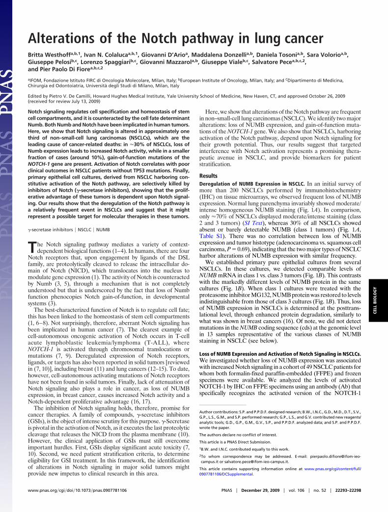
Alterations of the Notch pathway in lung cancerBritta Westhoffa,b,1, Ivan N. Colalucaa,b,1, Giovanni D’Arioa, Maddalena Donzellia,b, Daniela Tosonia,b, Sara Volorioa,b,Giuseppe Pelosib,c, Lorenzo Spaggiarib,c, Giovanni Mazzarola,b, Giuseppe Vialeb,c, Salvatore Pecea,b,c,2,and Pier Paolo Di Fiorea,b,c,2
aIFOM, Fondazione Istituto FIRC di Oncologia Molecolare, Milan, Italy; bEuropean Institute of Oncology, Milan, Italy; and CDipartimento di Medicina,Chirurgia ed Odontoiatria, Universita degli Studi di Milano, Milan, Italy
Edited by Pietro V. De Camilli, Howard Hughes Medical Institute, Yale University School of Medicine, New Haven, CT, and approved October 26, 2009(received for review July 13, 2009)
Notch signaling regulates cell specification and homeostasis of stemcell compartments, and it is counteracted by the cell fate determinantNumb. Both Numb and Notch have been implicated in human tumors.Here, we show that Notch signaling is altered in approximately onethird of non–small-cell lung carcinomas (NSCLCs), which are theleading cause of cancer-related deaths: in �30% of NSCLCs, loss ofNumb expression leads to increased Notch activity, while in a smallerfraction of cases (around 10%), gain-of-function mutations of theNOTCH-1 gene are present. Activation of Notch correlates with poorclinical outcomes in NSCLC patients without TP53 mutations. Finally,primary epithelial cell cultures, derived from NSCLC harboring con-stitutive activation of the Notch pathway, are selectively killed byinhibitors of Notch (�-secretase inhibitors), showing that the prolif-erative advantage of these tumors is dependent upon Notch signal-ing. Our results show that the deregulation of the Notch pathway isa relatively frequent event in NSCLCs and suggest that it mightrepresent a possible target for molecular therapies in these tumors.
�-secretase inhibitors � NSCLC � NUMB
The Notch signaling pathway mediates a variety of context-dependent biological functions (1–4). In humans, there are four
Notch receptors that, upon engagement by ligands of the DSLfamily, are proteolytically cleaved to release the intracellular do-main of Notch (NICD), which translocates into the nucleus tomodulate gene expression (1). The activity of Notch is counteractedby Numb (3, 5), through a mechanism that is not completelyunderstood but that is underscored by the fact that loss of Numbfunction phenocopies Notch gain-of-function, in developmentalsystems (3).
The best-characterized function of Notch is to regulate cell fate;this has been linked to the homeostasis of stem cell compartments(1, 6–8). Not surprisingly, therefore, aberrant Notch signaling hasbeen implicated in human cancer (7). The clearest example ofcell-autonomous oncogenic activation of Notch occurs in T-cellacute lymphoblastic leukemia/lymphoma (T-ALL), whereNOTCH-1 is activated through chromosomal translocations ormutations (7, 9). Deregulated expression of Notch receptors,ligands, or targets has also been reported in solid tumors [reviewedin (7, 10)], including breast (11) and lung cancers (12–15). To date,however, cell-autonomous activating mutations of Notch receptorshave not been found in solid tumors. Finally, lack of attenuation ofNotch signaling also plays a role in cancer, as loss of NUMBexpression, in breast cancer, causes increased Notch activity and aNotch-dependent proliferative advantage (16, 17).
The inhibition of Notch signaling holds, therefore, promise forcancer therapies. A family of compounds, �-secretase inhibitors(GSIs), is the object of intense scrutiny for this purpose. �-Secretaseis pivotal in the activation of Notch, as it executes the last proteolyticcleavage that releases the NICD from the plasma membrane (10).However, the clinical application of GSIs must still overcomeimportant hurdles. First, GSIs display significant acute toxicity (7,10). Second, we need patient stratification criteria, to determineeligibility for GSI treatment. In this framework, the identificationof alterations in Notch signaling in major solid tumors mightprovide new impetus to clinical research in this area.
Here, we show that alterations of the Notch pathway are frequentin non–small-cell lung carcinomas (NSCLC). We identify two majoralterations: loss of NUMB expression, and gain-of-function muta-tions of the NOTCH-1 gene. We also show that NSCLCs, harboringactivation of the Notch pathway, depend upon Notch signaling fortheir growth potential. Thus, our results suggest that targetedinterference with Notch activation represents a promising thera-peutic avenue in NSCLC, and provide biomarkers for patientstratification.
ResultsDeregulation of NUMB Expression in NSCLC. In an initial survey ofmore than 200 NSCLCs performed by immunohistochemistry(IHC) on tissue microarrays, we observed frequent loss of NUMBexpression. Normal lung parenchyma invariably showed moderate/intense homogeneous NUMB staining (Fig. 1A). In comparison,only �70% of NSCLCs displayed moderate/intense staining (class2 and 3 tumors) (SI Text), whereas 30% of all NSCLCs showedabsent or barely detectable NUMB (class 1 tumors) (Fig. 1A,Table S1). There was no correlation between loss of NUMBexpression and tumor histotype (adenocarcinoma vs. squamous cellcarcinoma, P � 0.69), indicating that the two major types of NSCLCharbor alterations of NUMB expression with similar frequency.
We established primary pure epithelial cultures from severalNSCLCs. In these cultures, we detected comparable levels ofNUMB mRNA in class 1 vs. class 3 tumors (Fig. 1B). This contrastswith the markedly different levels of NUMB protein in the samecultures (Fig. 1B). When class 1 cultures were treated with theproteasome inhibitor MG132, NUMB protein was restored to levelsindistinguishable from those of class 3 cultures (Fig. 1B). Thus, lossof NUMB expression in NSCLCs is determined at the posttrans-lational level, through enhanced protein degradation, similarly towhat was shown in breast cancers (16). Of note, we did not detectmutations in the NUMB coding sequence (cds) at the genomic levelin 13 samples representative of the various classes of NUMBstaining in NSCLC (see below).
Loss of NUMB Expression and Activation of Notch Signaling in NSCLCs.We investigated whether loss of NUMB expression was associatedwith increased Notch signaling in a cohort of 49 NSCLC patients forwhom both formalin-fixed paraffin-embedded (FFPE) and frozenspecimens were available. We analyzed the levels of activatedNOTCH-1 by IHC on FFPE specimens using an antibody (Ab) thatspecifically recognizes the activated version of the NOTCH-1
Author contributions: S.P. and P.P.D.F. designed research; B.W., I.N.C., G.D., M.D., D.T., S.V.,G.P., L.S., G.M., and S.P. performed research; G.P., L.S., and G.V. contributed new reagents/analytic tools; G.D., G.P., G.M., G.V., S.P., and P.P.D.F. analyzed data; and S.P. and P.P.D.F.wrote the paper.
The authors declare no conflict of interest.
This article is a PNAS Direct Submission.
1B.W. and I.N.C. contributed equally to this work.
2To whom correspondence may be addressed. E-mail: [email protected] or [email protected].
This article contains supporting information online at www.pnas.org/cgi/content/full/0907781106/DCSupplemental.
www.pnas.org�cgi�doi�10.1073�pnas.0907781106 PNAS � December 29, 2009 � vol. 106 � no. 52 � 22293–22298
CELL
BIO
LOG
Y

receptor (see SI Text and Fig. S1 for details). We observed a stronginverse correlation between the levels of NUMB and of activated–NOTCH-1 (Fig. 2 A and B). We then measured the expressionlevels of the Notch target gene, HES1 (4), in frozen samples. Alsoin this case, we observed a significant inverse correlation betweenHES1 mRNA levels and NUMB status (Fig. 2C, Table S2). Notethat in Fig. 2 B and C, six patients harboring mutations of theNOTCH-1 gene (see below) were not included.
We analyzed NUMB protein and HES1 mRNA levels in primaryNSCLC cultures derived from 10 patients. Under these conditions,NUMB levels and Notch pathway activity could be analyzed in apure epithelial tumor population. Again, we observed a stronginverse correlation between the levels of NUMB and of HES1mRNA (Fig. 2D). We concluded that deregulation of NUMB is afrequent event in NSCLC, and that this correlates with the activa-tion of the Notch signaling pathway.
Genetic Alterations of NOTCH-1 in NSCLC. Despite the significantinverse correlation between NUMB protein and HES1 mRNA (oractivated NOTCH-1) levels, there were a significant number ofoutliers. In particular, in some specimens (Table S2) we noted highlevels of HES1 mRNA (� 2.0) together with high/intermediatelevels of activated NOTCH-1, despite substantial levels of NUMB.We hypothesized that these patients might harbor activatingNOTCH-1 mutations. Thus, we performed a mutation analysis ofthe 49 NSCLC cohort, by sequencing the entire C-terminal regionof the NOTCH-1 cds (Fig. 3A). We found four different heterozy-gous NOTCH-1 mutations in six NSCLC samples (Fig. 3A, Fig. S2,Table S2). For patients 42 and 44, we confirmed the presence ofmutations also in primary cultures (Fig. 6A). Of note, we alsosequenced 45 breast cancers without detecting any mutations.
Alterations in the NOTCH-1 cds might represent somatic mu-tations or polymorphisms. Thus, we sequenced the NOTCH-1 cdsin matched normal samples, where these were available. Forpatients 36, 40, 42, 44, and 49 (Table S2), normal frozen lung tissuewas used; for patient 42, normal lymphocytes were also tested. Inall cases, wild-type (WT) NOTCH-1 alleles were detected in thenormal tissues (Fig. 3B, Fig. S2).
Because of the limited availability of tumor tissues, we performedour analysis at the cDNA level. It was important, therefore, toconfirm that the mutations in the NOTCH-1 cDNAs correspondedto mutations in the NOTCH-1 gene. In four cases (cases 36, 40, 42,
A
- + - +
NUMB
Vinculin
Class-1 Class-30
0.2
0.4
0.6
0.8
Class-1 Class-3
slevel A
NR
m B
MU
N evitaleR
BMG132
Nor
mal
Cla
ss-2
Cla
ss-1
Cla
ss-3
Fig. 1. Loss of NUMB expression in NSCLC. (A) NUMB expression in NSCLCsdetected by IHC. Entire TMA cores, from representative samples, are shown onthe left at an original magnification �20; the boxed areas are magnified on theright (original magnification �40) to show the typical cytosolic and plasmamembrane localization of Numb. (B) (Left) NUMB mRNA levels measured byQ-PCR in primary NSCLC cultures expressed relative to the reference cell lineBEAS-2B (� 1). Results represent the mean mRNA level detected in cultures fromfour patients for each NUMB-class shown. (Right) NUMB protein levels in primaryNSCLC cultures. Blots are representative of three independent experiments.
Cmedian
P: 0.0098
B
Primary cell lines
Cl.1 Cl.2 Cl.3
1S
EH evitale
Rslevel
AN
Rm
D
Tumors
NUMB:
Cl.1 Cl.2 Cl.3NUMB:
gniniats 1-H
CT
ON-detavitc
A)
%( seirogetac yb
0
20
40
60
80
100
Low activated-NOTCH-1
Interm. activated-NOTCH-1
High activated-NOTCH-1
P=0.0012
02
46
810
Activated-NOTCH-1
Cla
ss-3
Cla
ss-1
NUMBA
50 1 4.751 1 3.852 1 2.153 1 3.154 1 2.255 1 2.656 2 0.457 2 1.458 3 0.959 3 1.2
Patient NUMBClass
HES1levels
Fig. 2. NUMB expression and Notch signaling in NSCLCs. (A and B) Inversecorrelation between NUMB and activated-NOTCH-1 status in NSCLCs. (A) Repre-sentative IHC in serial sections (original magnification �40). (B) Quantitativeassessment of activated-NOTCH-1 in NSCLC as a function of NUMB-class. (C) HES1mRNA levels in NSCLCs displaying different levels of NUMB. Data were obtainedby Q-PCR on frozen specimens of the cohort of 49 patients and are expressedrelative to the reference cell line BEAS-2B (� 1). The six patients harboringmutations in the NOTCH-1 gene (see Figs. 3A and 5A) were excluded from theresults reported in panels (B) and (C). (D) NUMB-class (measured by IHC) and HES1levels, measured as in (C), in 10 primary cultures from NSCLC patients.
22294 � www.pnas.org�cgi�doi�10.1073�pnas.0907781106 Westhoff et al.
and 49, representing the four different mutations), we had enoughmaterial to perform this analysis, which confirmed the presence ofthe mutations at the genomic level (Fig. S3).
We also performed a mutational analysis of the NUMB cds, usinggenomic DNA extracted from FFPE samples, from four NSCLCswith low NUMB expression (patients 34, 36, 39, and 46) and nineNSCLCs with intermediate/high NUMB levels (patients 4, 9, 13, 33,38, 40, 42, 47, and 49, four of whom also displayed mutations of theNOTCH-1 gene). This analysis did not reveal any mutations in theNUMB cds (Table S2).
Finally, the availability of matched normal/tumor tissues allowedus to analyze the activation of the Notch pathway, as a function ofthe malignant conversion. The levels of HES1 mRNA were higherin tumor frozen tissues compared to the normal counterparts for allpatients tested (Fig. 3C). Comparable results were also obtained forFFPE samples, which were used to test both HES1 and anotherNotch target gene, HEY2 (Fig. S4).
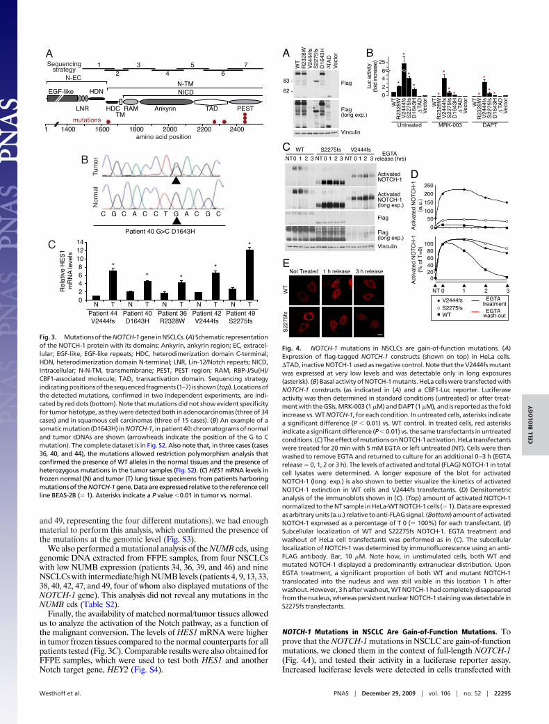
NOTCH-1 Mutations in NSCLC Are Gain-of-Function Mutations. Toprove that the NOTCH-1 mutations in NSCLC are gain-of-functionmutations, we cloned them in the context of full-length NOTCH-1(Fig. 4A), and tested their activity in a luciferase reporter assay.Increased luciferase levels were detected in cells transfected with
Patient 40 G>C D1643H
A
Tum
orN
orm
al
B
Rel
ativ
e H
ES
1m
RN
A le
vels
N T N T N T N T N T02468
101214
Patient 40D1643H
Patient 36R2328W
Patient 44V2444fs
Patient 49S2275fs
Patient 42V2444fs
C
*
* *
*
*
1400 1600 1800 2000 2200 2400
N-ECN-TM
LNR
HDN
TMRAM Ankyrin TAD PESTHDC
NICD
12
34
56
7Sequencingstrategy
EGF-like
1amino acid position
mutations
C G C C C C CG GA AT
Fig. 3. Mutationsof theNOTCH-1gene inNSCLCs. (A) Schematic representationof the NOTCH-1 protein with its domains: Ankyrin, ankyrin region; EC, extracel-lular; EGF-like, EGF-like repeats; HDC, heterodimerization domain C-terminal;HDN, heterodimerization domain N-terminal; LNR, Lin-12/Notch repeats; NICD,intracellular; N-N-TM, transmembrane; PEST, PEST region; RAM, RBP-J/Su(H)/CBF1-associated molecule; TAD, transactivation domain. Sequencing strategyindicatingpositionsof thesequencedfragments (1–7) is shown(top). Locationsofthe detected mutations, confirmed in two independent experiments, are indi-cated by red dots (bottom). Note that mutations did not show evident specificityfor tumor histotype, as they were detected both in adenocarcinomas (three of 34cases) and in squamous cell carcinomas (three of 15 cases). (B) An example of asomatic mutation (D1643H) in NOTCH-1, in patient 40: chromatograms of normaland tumor cDNAs are shown (arrowheads indicate the position of the G to Cmutation). The complete dataset is in Fig. S2. Also note that, in three cases (cases36, 40, and 44), the mutations allowed restriction polymorphism analysis thatconfirmed the presence of WT alleles in the normal tissues and the presence ofheterozygous mutations in the tumor samples (Fig. S2). (C) HES1 mRNA levels infrozen normal (N) and tumor (T) lung tissue specimens from patients harboringmutations of the NOTCH-1 gene. Data are expressed relative to the reference cellline BEAS-2B (� 1). Asterisks indicate a P value �0.01 in tumor vs. normal.
B
WT
V2444fsS2275fs
EGTA treatmentEGTA
wash-out
0
50100
150
200
250
020406080
100
NT 0 1 2 3
Act
ivat
ed N
OT
CH
-1(a
.u.)
Act
ivat
ed N
OT
CH
-1(%
of T
=0)
D
WT
Not Treated 1 h release 3 h releaseE
CNT0 1 2 3 NT NT
EGTA release (hrs)
WT S2275fs V2444fs
ActivatedNOTCH-1
Vinculin
Flag
Flag(long exp.)
0 1 2 3 0 1 2 3
ActivatedNOTCH-1(long exp.)
A
WT
R23
28W
V24
44fs
S22
75fs
D16
43H
Vec
tor
∆ TA
D
Flag
Vinculin
83
62
Flag(long exp.)
*
--------
-
ytivitca cuL) esaercni dl of(
2
46
25
0
*
*
*
*
*
***
*
***
WT
R23
28W
V24
44fs
S22
75fs
D16
43H
∆TA
DV
ecto
r
WT
R23
28W
V24
44fs
S22
75fs
D16
43H
∆TA
DV
ecto
r
WT
R23
28W
V24
44fs
S22
75fs
D16
43H
∆TA
DV
ecto
r
Untreated MRK-003 DAPT
sf5722S
Fig. 4. NOTCH-1 mutations in NSCLCs are gain-of-function mutations. (A)Expression of flag-tagged NOTCH-1 constructs (shown on top) in HeLa cells.�TAD, inactive NOTCH-1 used as negative control. Note that the V2444fs mutantwas expressed at very low levels and was detectable only in long exposures(asterisk). (B) Basal activity of NOTCH-1 mutants. HeLa cells were transfected withNOTCH-1 constructs (as indicated in (A) and a CBF1-Luc reporter. Luciferaseactivity was then determined in standard conditions (untreated) or after treat-ment with the GSIs, MRK-003 (1 �M) and DAPT (1 �M), and is reported as the foldincrease vs. WT NOTCH-1, for each condition. In untreated cells, asterisks indicatea significant difference (P � 0.01) vs. WT control. In treated cells, red asterisksindicate a significant difference (P � 0.01) vs. the same transfectants in untreatedconditions. (C)TheeffectofmutationsonNOTCH-1activation.HeLatransfectantswere treated for 20 min with 5 mM EGTA or left untreated (NT). Cells were thenwashed to remove EGTA and returned to culture for an additional 0–3 h (EGTArelease � 0, 1, 2 or 3 h). The levels of activated and total (FLAG) NOTCH-1 in totalcell lysates were determined. A longer exposure of the blot for activatedNOTCH-1 (long. exp.) is also shown to better visualize the kinetics of activatedNOTCH-1 extinction in WT cells and V2444fs transfectants. (D) Densitometricanalysis of the immunoblots shown in (C). (Top) amount of activated NOTCH-1normalized to the NT sample in HeLa-WT NOTCH-1 cells (� 1). Data are expressedas arbitrary units (a.u.) relative to anti-FLAG signal. (Bottom) amount of activatedNOTCH-1 expressed as a percentage of T 0 (� 100%) for each transfectant. (E)Subcellular localization of WT and S22275fs NOTCH-1. EGTA treatment andwashout of HeLa cell transfectants was performed as in (C). The subcellularlocalization of NOTCH-1 was determined by immunofluorescence using an anti-FLAG antibody. Bar, 10 �M. Note how, in unstimulated cells, both WT andmutated NOTCH-1 displayed a predominantly extranuclear distribution. UponEGTA treatment, a significant proportion of both WT and mutant NOTCH-1translocated into the nucleus and was still visible in this location 1 h afterwashout. However, 3 h after washout, WT NOTCH-1 had completely disappearedfromthenucleus,whereaspersistentnuclearNOTCH-1 stainingwasdetectable inS2275fs transfectants.
Westhoff et al. PNAS � December 29, 2009 � vol. 106 � no. 52 � 22295
CELL
BIO
LOG
Y
NOTCH-1 mutants compared to WT transfectants (Fig. 4B). Thisincrease was reversed by treatment with two different GSIs, MRK-003 (15, 18) and DAPT (Fig. 4B). Finally, by using the anti-activated–NOTCH-1 Ab, we directly demonstrated an increasedbasal level of activation for the NOTCH-1 mutants compared withWT NOTCH-1 (Fig. 4C, Fig. S5A, lanes NT).
To increase our understanding of the impact of the mutations onNOTCH-1 activity, we measured the degree and duration ofNOTCH-1 activation. NOTCH-1 activation was induced in HeLatransfectants by a short exposure to EGTA, which, by chelatingcalcium, activates the cleavage/activation sequence of NOTCH-1(19). The levels of activated NOTCH-1, after treatment, wereconsiderably more pronounced for the NOTCH-1 mutants com-pared with WT NOTCH-1 (Fig. 4 C and D, Fig. S5). EGTA wasthen washed away to measure the kinetics of extinction ofNOTCH-1 activation. In WT transfectants, NOTCH-1 activationdecayed with a half-life of �1/1.5 h. In contrast, in mutant trans-fectants, NOTCH-1 activation persisted for a longer time (�3 h;Fig. 4 C and D and Fig. S5). This correlated with a persistence ofNOTCH-1 in the nucleus, a hallmark of NOTCH-1 activation(Fig. 4E).
We concluded that the NOTCH-1 mutations in NSCLC aregain-of-function mutations.
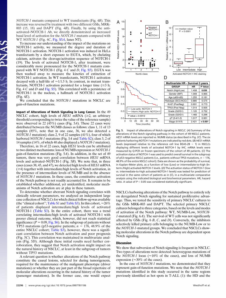
Impact of Alterations of Notch Signaling in Lung Cancer. In the 49NSCLC cohort, high levels of HES1 mRNA (�2, an arbitrarythreshold corresponding to twice the value of the reference sample)were observed in 22 (45%) cases (Fig. 5A). These 22 cases weredistributed between the NUMB classes as follows: class 1, 11 of 13samples (85%, note that in one case, 36, we also detected aNOTCH-1 mutation); class 2, 9 of 22 samples (41%), four of whichharbored NOTCH-1 mutations (Fig. 5A and Table S2); class 3, 2 of14 samples (14%, of which 40 also displayed a NOTCH-1 mutation).
Therefore, in 16 of 22 cases, high HES1 levels can be attributedto two distinct mechanisms: loss of NUMB expression or NOTCH-1gain-of-function mutations (Fig. 5A). In addition, in the set of 49tumors, there was very good correlation between HES1 mRNAlevels and activated–NOTCH-1 (Fig. 5B). We note that, in threecases (cases 38, 45, and 47), we detected high levels of HES1 mRNA(�4) together with intermediate/high levels of Notch activation, inthe presence of intermediate levels of NUMB and in the absenceof NOTCH-1 mutations. In these cases, the constitutive activationof the Notch pathway is not readily accounted for. It remains to beestablished whether additional, yet unidentified, molecular mech-anisms of Notch activation are at play in these tumors.
To determine whether aberrant Notch signaling correlates withclinically relevant parameters, we analyzed an independent largecase collection of NSCLCs for which clinical follow-up was available(the ‘‘clinical cohort’’; Table S1 and Table S3). In this cohort, �26%of patients displayed intermediate/high levels of activatedNOTCH-1 (Table S3). In the entire cohort, there was a trendcorrelating intermediate/high levels of activated NOTCH-1 withpoorer clinical outcome, which, however, did not reach statisticalsignificance (P � 0.09, Fig. S6). In the subgroup of patients withoutTP53 mutations (p53-negative patients; n � 176, 48.9% of theentire NSCLC cohort; Table S3), however, there was a signifi-cant correlation between Notch activation and poor prognosis(Fig. 5C). This correlation was maintained in multivariate anal-ysis (Fig. 5D). Although these initial results need further cor-roboration, they suggest that Notch activation might impact onthe natural history of NSCLC, at least in the subgroup of tumorswithout TP53 mutations.
A relevant question is whether alterations of the Notch pathwayconstitute the causal lesions, selected for during tumorigenesis,required for the maintenance of the malignant phenotype (drivermutations) or whether they are just part of the broad repertoire ofmolecular alterations occurring in the natural history of the tumor(passenger mutations). In the former case, one would expect
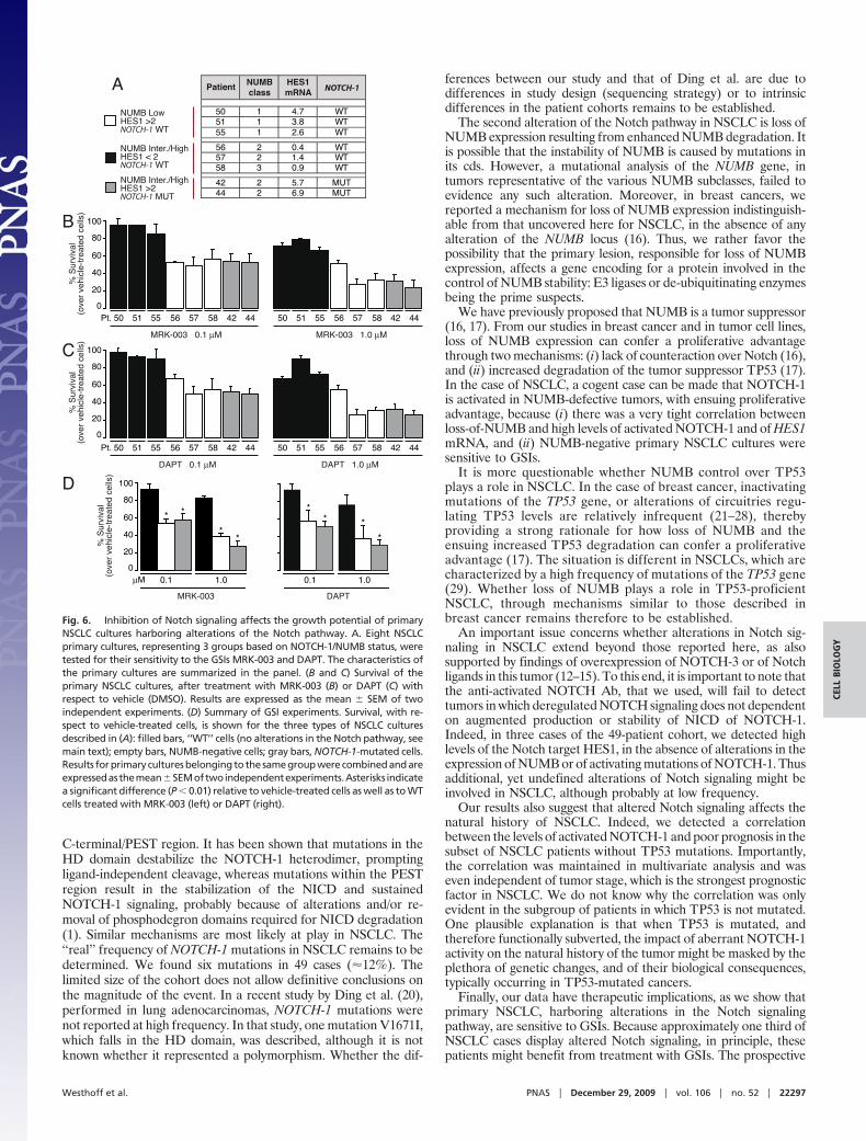
NSCLCs harboring alterations of the Notch pathway to entirely relyon deregulated Notch signaling for sustained proliferative advan-tage. Thus, we tested the sensitivity of primary NSCLC cultures tothe GSIs MRK-003 and DAPT. The selected primary NSCLCcultures belonged to three categories, based on the levels and modesof activation of the Notch pathway: WT, NUMB-Low, NOTCH-1-mutated (Fig. 6A). The survival of WT cells was not significantlyaffected by GSIs (Fig. 6 B, C, and D). Conversely, the inhibitorsselectively killed primary cells belonging to the NUMB-Low or tothe NOTCH-1-mutated groups. We concluded that NSCLCs show-ing molecular alterations in the Notch pathway are dependent uponNotch signaling.
DiscussionWe show that subversion of Notch signaling is frequent in NSCLC.Two types of alterations were detected: heterozygous mutations ofthe NOTCH-1 locus (�10% of the cases), and loss of NUMBexpression (�30% of the cases).
In the case of NOTCH-1 mutations, we demonstrated that theyrepresent gain-of-function mutations. We note that the NOTCH-1mutations identified in this study occurred in the same regionspreviously identified as hot spots in T-ALL (1): the HD and the
BA
slevel A
NR
m 1S
EH evitale
R
C
slevel A
NR
m 1-S
EH evitale
R
Low Intermediate High
Activated-NOTCH-1 IHC staining
0
2
4
6
8
10
12 median valueP: < 0.0001
P value = 0.0312lavivru
S
Time (months)
0.0
0.2
0.4
0.6
0.8
1.0
0 10 20 30 40 50 60
0
2
4
6
8
1 32
Numb class
10
12
WT NOTCH-1
Mutated NOTCH-1
GROUP HR P Value
Age (≥65/<65)
Histotype (SCC/ACC)
Ki-67 (Pos/Neg)
TP53 (Pos/ Neg)
D
Interm./High activ. NOTCH-1Low activ. NOTCH-1
Notch (High/Low)
Gender (M/F)
1.458 0.039
1.009 0.965
1.255 0.181
0.880 0.575
0.931 0.773
1.014 0.935
1.120 0.752
1.465 0.296
1.017 0.931
2.421 < 0.001
Multivariate HR - Survival 5 years
Grade (G2/G1)
Grade (G3/G1)
pT (3-4/1-2)
Stage (II-IV/I)
Fig. 5. Impact of alterations of Notch signaling in NSCLC. (A) Summary of thealterations of the Notch signaling pathway in the cohort of 49 NSCLC patients.HES1 mRNA levels are reported vs. NUMB status (as described in Fig. 2C). The sixpatients harboring NOTCH-1 mutations are indicated by red dots. (B) HES1 mRNAlevels (expressed relative to the reference cell line BEAS-2B � 1) in NSCLCsdisplaying different levels of activated NOTCH-1 by IHC. mRNA levels weremeasured by Q-PCR on frozen specimens of the cohort of 49 patients. (C) Theactivation status of NOTCH-1 was used to predict overall survival in the subgroupof p53-negative NSCLC patients (i.e., patients without TP53 mutations, n � 176,48.9% of the entire NSCLC cohort). Data are shown as the probability of survival,in Kaplan–Meier plots, as a function of low (Low) or intermediate-to-high (In-term./High) activated-NOTCH-1 levels. (D) The activation status of NOTCH-1 (lowvs. intermediate-to-high activated-NOTCH-1 levels) was tested for prediction ofsurvival in the same cohort of patients as in (C), in a multivariate comparativeanalysis using the indicated biological and biochemical parameters. HR, hazardratio. A value of P � 0.05 was considered statistically significant.
22296 � www.pnas.org�cgi�doi�10.1073�pnas.0907781106 Westhoff et al.
C-terminal/PEST region. It has been shown that mutations in theHD domain destabilize the NOTCH-1 heterodimer, promptingligand-independent cleavage, whereas mutations within the PESTregion result in the stabilization of the NICD and sustainedNOTCH-1 signaling, probably because of alterations and/or re-moval of phosphodegron domains required for NICD degradation(1). Similar mechanisms are most likely at play in NSCLC. The‘‘real’’ frequency of NOTCH-1 mutations in NSCLC remains to bedetermined. We found six mutations in 49 cases (�12%). Thelimited size of the cohort does not allow definitive conclusions onthe magnitude of the event. In a recent study by Ding et al. (20),performed in lung adenocarcinomas, NOTCH-1 mutations werenot reported at high frequency. In that study, one mutation V1671I,which falls in the HD domain, was described, although it is notknown whether it represented a polymorphism. Whether the dif-
ferences between our study and that of Ding et al. are due todifferences in study design (sequencing strategy) or to intrinsicdifferences in the patient cohorts remains to be established.
The second alteration of the Notch pathway in NSCLC is loss ofNUMB expression resulting from enhanced NUMB degradation. Itis possible that the instability of NUMB is caused by mutations inits cds. However, a mutational analysis of the NUMB gene, intumors representative of the various NUMB subclasses, failed toevidence any such alteration. Moreover, in breast cancers, wereported a mechanism for loss of NUMB expression indistinguish-able from that uncovered here for NSCLC, in the absence of anyalteration of the NUMB locus (16). Thus, we rather favor thepossibility that the primary lesion, responsible for loss of NUMBexpression, affects a gene encoding for a protein involved in thecontrol of NUMB stability: E3 ligases or de-ubiquitinating enzymesbeing the prime suspects.
We have previously proposed that NUMB is a tumor suppressor(16, 17). From our studies in breast cancer and in tumor cell lines,loss of NUMB expression can confer a proliferative advantagethrough two mechanisms: (i) lack of counteraction over Notch (16),and (ii) increased degradation of the tumor suppressor TP53 (17).In the case of NSCLC, a cogent case can be made that NOTCH-1is activated in NUMB-defective tumors, with ensuing proliferativeadvantage, because (i) there was a very tight correlation betweenloss-of-NUMB and high levels of activated NOTCH-1 and of HES1mRNA, and (ii) NUMB-negative primary NSCLC cultures weresensitive to GSIs.
It is more questionable whether NUMB control over TP53plays a role in NSCLC. In the case of breast cancer, inactivatingmutations of the TP53 gene, or alterations of circuitries regu-lating TP53 levels are relatively infrequent (21–28), therebyproviding a strong rationale for how loss of NUMB and theensuing increased TP53 degradation can confer a proliferativeadvantage (17). The situation is different in NSCLCs, which arecharacterized by a high frequency of mutations of the TP53 gene(29). Whether loss of NUMB plays a role in TP53-proficientNSCLC, through mechanisms similar to those described inbreast cancer remains therefore to be established.
An important issue concerns whether alterations in Notch sig-naling in NSCLC extend beyond those reported here, as alsosupported by findings of overexpression of NOTCH-3 or of Notchligands in this tumor (12–15). To this end, it is important to note thatthe anti-activated NOTCH Ab, that we used, will fail to detecttumors in which deregulated NOTCH signaling does not dependenton augmented production or stability of NICD of NOTCH-1.Indeed, in three cases of the 49-patient cohort, we detected highlevels of the Notch target HES1, in the absence of alterations in theexpression of NUMB or of activating mutations of NOTCH-1. Thusadditional, yet undefined alterations of Notch signaling might beinvolved in NSCLC, although probably at low frequency.
Our results also suggest that altered Notch signaling affects thenatural history of NSCLC. Indeed, we detected a correlationbetween the levels of activated NOTCH-1 and poor prognosis in thesubset of NSCLC patients without TP53 mutations. Importantly,the correlation was maintained in multivariate analysis and waseven independent of tumor stage, which is the strongest prognosticfactor in NSCLC. We do not know why the correlation was onlyevident in the subgroup of patients in which TP53 is not mutated.One plausible explanation is that when TP53 is mutated, andtherefore functionally subverted, the impact of aberrant NOTCH-1activity on the natural history of the tumor might be masked by theplethora of genetic changes, and of their biological consequences,typically occurring in TP53-mutated cancers.
Finally, our data have therapeutic implications, as we show thatprimary NSCLC, harboring alterations in the Notch signalingpathway, are sensitive to GSIs. Because approximately one third ofNSCLC cases display altered Notch signaling, in principle, thesepatients might benefit from treatment with GSIs. The prospective
0.1 1.0
% S
urvi
val
(ove
r ve
hicl
e-tr
eate
d ce
lls)
0
20
40
60
80
100
* *
**
50 1 WT51 1 WT55 1 WT
56 2 WT57 2 WT58 3 WT
42 2 MUT44 2 MUT
Patient NUMBclass NOTCH-1
NUMB LowHES1 >2NOTCH-1 WT
NUMB Inter./HighHES1 < 2NOTCH-1 WT
NUMB Inter./HighHES1 >2NOTCH-1 MUT
4.73.82.6
0.41.40.9
5.76.9
HES1mRNA
D
0.1 1.0
** *
*
µM
MRK-003 DAPT
% S
urvi
val
(ove
r ve
hicl
e-tr
eate
d ce
lls)
0
20
40
60
80
100
50 51 55 56 57 58 42 44 50 51 55 56 57 58 42 44
MRK-003 0.1 µM MRK-003 1.0 µM
Pt.
% S
urvi
val
(ove
r ve
hicl
e-tr
eate
d ce
lls)
0
20
40
60
80
100
50 51 55 56 57 58 42 44 50 51 55 56 57 58 42 44
DAPT 0.1 µM DAPT 1.0 µM
Pt.
A
B
C
Fig. 6. Inhibition of Notch signaling affects the growth potential of primaryNSCLC cultures harboring alterations of the Notch pathway. A. Eight NSCLCprimary cultures, representing 3 groups based on NOTCH-1/NUMB status, weretested for their sensitivity to the GSIs MRK-003 and DAPT. The characteristics ofthe primary cultures are summarized in the panel. (B and C) Survival of theprimary NSCLC cultures, after treatment with MRK-003 (B) or DAPT (C) withrespect to vehicle (DMSO). Results are expressed as the mean � SEM of twoindependent experiments. (D) Summary of GSI experiments. Survival, with re-spect to vehicle-treated cells, is shown for the three types of NSCLC culturesdescribed in (A): filled bars, ‘‘WT’’ cells (no alterations in the Notch pathway, seemain text); empty bars, NUMB-negative cells; gray bars, NOTCH-1-mutated cells.Results for primary cultures belonging to the same group were combined and areexpressedasthemean�SEMoftwoindependentexperiments.Asterisks indicatea significant difference (P � 0.01) relative to vehicle-treated cells as well as to WTcells treated with MRK-003 (left) or DAPT (right).
Westhoff et al. PNAS � December 29, 2009 � vol. 106 � no. 52 � 22297
CELL
BIO
LOG
Y
use of these inhibitors in the clinical setting has been plagued bytheir toxicity (7, 10). Recent developments, however, highlightedthe possibility that combinatorial treatment with glucocorticoidsmight counteract the toxicity of GSIs (30, 31). The perspectivedevelopment of combined GSI-based therapeutic protocols, withreduced toxicity, would constitute a major advancement in NSLC,in which the development of effective targeted therapies is still alargely unmet need (32). In this framework, the molecular alter-ations of the Notch pathway herein described might constitute aneffective tool for the stratification of eligible patients.
Materials and MethodsNSCLC Specimens and Analyses. All specimens were from lung cancer patientsundergoing surgery at the European Institute of Oncology (IEO) in Milan, Italy.Ethics approval for tissue collection for research purposes was obtained from theIEO Institutional Review Board, after written informed consent had been ob-tainedfromallpatients.Detailson IHCanalysisandNSCLCclassificationaccordingto their NUMB or Activated-NOTCH status are given in SI Text. In SI Text, themethodology for mutational analyses of NOTCH-1 and NUMB, and for quanti-tative RT-PCR analysis of NOTCH-1 targets is also described.
Cell Lines, Expression Vectors for NOTCH-1 Mutants and Biochemical Studies.Primary cultures were obtained as previously described (16) (SI Text). HeLa cellswere transfected with Lipofectamine PLUS (Invitrogen). For luciferase assays(Dual Luciferase Kit, Promega), cells were transfected with the NOTCH-1 con-structs together with a Notch-dependent CBF1-responsive luciferase reporter(6x-RBP-Jk-luc) (16) and a Renilla luciferase plasmid, and tested 48 h after trans-fection. GSIs were added to cells immediately after transfection. Luciferase ac-tivity was normalized to the Renilla transfection control and to NOTCH-1 expres-sion levels. Results of three independent experiments performed in triplicate are
shown. Immunofluorescence and immunoblotting were performed as described(17). Plasmids and antibodies are described in the SI Text.
Patients in Clinical Cohort and Statistical Analyses. The clinical cohort of 420consecutive NSCLC cases (Table S3) was constituted by patients who had under-gone surgical resection at IEO between June 1998 and December 2002. Overallsurvival was defined as the interval between surgery and either death from anycause, or last contact. The median duration of follow-up was 62 months (range,0–122 months). The 5-year survival rate was 52.0% (Stage I, 68.4%; Stage II–IV,38.3%). During the 5-year follow-up period, a total of 200 (48%) events (deaths)were registered. Plots of the overall survival according to activated–NOTCH-1expression were drawn using the Kaplan–Meier method. The statistical signifi-cance of differences in survival rates between groups was established by thelog-rank test. Multivariate analyses were carried out using the Cox proportionalhazards method to assess the prognostic value of activated–NOTCH-1 statusbefore and after correction for different independent risk factors, including ageat diagnosis of the tumor, pathological stage, tumor grade of differentiation,nodal status, TP53 status, and Ki-67. SAS statistical software was used for all of theanalyses (SAS Institute, Inc.). A value of P � 0.05 was considered significant.
ACKNOWLEDGMENTS. We are indebted to the following individuals: G. Vero-nesi for the acquisition of human tissue samples; G. Draetta, D. Bergstrom, and P.Strack at Merck Research Laboratories (MRL, Boston) for the MRK-003 compoundand for sharing their unpublished results; G. Taliento, M. Simone, G. Paciucci, andM. Bianchi for technical assistance; the Biological Resource Center and the TumorRegistry at IEO and the Molecular Pathology Unit, the Real Time PCR and DNASequencing Service, the Imaging Service, the Monoclonal Service, and the tissueculture Service at the IFOM-IEO Campus, for technical support; and G. Goisis forstatistical analyses. This work was supported by grants from the AssociazioneItaliana per la Ricerca sul Cancro (AIRC), Italian Ministry of Health, and MIUR (toS.P. and P.P.D.F.); the European Community (VI Framework), the CARIPLO foun-dation, the Ferrari Foundation, and the Monzino Foundation (to P.P.D.), and theG. Vollaro Foundation (to S.P.).
1. Aster JC, Pear WS, Blacklow SC (2008) Notch signaling in leukemia. Annu Rev Pathol3:587–613.
2. Fiuza UM, Arias AM (2007) Cell and molecular biology of Notch. J Endocrinol 194:459–474.3. Gonczy P (2008) Mechanisms of asymmetric cell division: Flies and worms pave the way.
Nat Rev Mol Cell Biol 9:355–366.4. Kageyama R, Ohtsuka T, Kobayashi T (2007) The Hes gene family: Repressors and
oscillators that orchestrate embryogenesis. Development 134:1243–1251.5. Roegiers F, Jan YN (2004) Asymmetric cell division. Curr Opin Cell Biol 16:195–205.6. Farnie G, Clarke RB (2007) Mammary stem cells and breast cancer–role of Notch
signalling. Stem Cell Rev 3:169–175.7. Roy M, Pear WS, Aster JC (2007) The multifaceted role of Notch in cancer. Curr Opin
Genet Dev 17:52–59.8. Watt FM, Estrach S, Ambler CA (2008) Epidermal Notch signalling: Differentiation,
cancer and adhesion. Curr Opin Cell Biol 20:171–179.9. Weng AP, et al. (2004) Activating mutations of NOTCH1 in human T cell acute
lymphoblastic leukemia. Science 306:269–271.10. Shih Ie M, Wang TL (2007) Notch signaling, gamma-secretase inhibitors, and cancer
therapy. Cancer Res 67:1879–1882.11. Reedijk M, et al. (2005) High-level coexpression of JAG1 and NOTCH1 is observed in human
breast cancer and is associated with poor overall survival. Cancer Res 65:8530–8537.12. Choi K, et al. (2009) Distinct biological roles for the notch ligands Jagged-1 and
Jagged-2. J Biol Chem 284:17766–17774.13. Lee SM, et al. (2008) Expression of Notch 1 and 3 is related to inhibition of lymph node
metastasis and progression in non-small lung carcinoma. Basic Applied Pathol 1:93–97.14. Dang TP, et al. (2000) Chromosome 19 translocation, overexpression of Notch3, and
human lung cancer. J Natl Cancer Inst 92:1355–1357.15. Konishi J, et al. (2007) Gamma-secretase inhibitor prevents Notch3 activation and
reduces proliferation in human lung cancers. Cancer Res 67:8051–8057.16. Pece S, et al. (2004) Loss of negative regulation by Numb over Notch is relevant to
human breast carcinogenesis. J Cell Biol 167:215–221.
17. Colaluca IN, et al. (2008) NUMB controls p53 tumour suppressor activity. Nature451:76–80.
18. Lewis HD, et al. (2007) Apoptosis in T cell acute lymphoblastic leukemia cells after cellcycle arrest induced by pharmacological inhibition of notch signaling. Chem Biol14:209–219.
19. Rand MD, et al. (2000) Calcium depletion dissociates and activates heterodimeric notchreceptors. Mol Cell Biol 20:1825–1835.
20. Ding L, et al. (2008) Somatic mutations affect key pathways in lung adenocarcinoma.Nature 455:1069–1075.
21. McCann AH, et al. (1995) Amplification of the MDM2 gene in human breast cancer andits association with MDM2 and p53 protein status. Br J Cancer 71:981–985.
22. Oliner JD, Kinzler KW, Meltzer PS, George DL, Vogelstein B (1992) Amplification of agene encoding a p53-associated protein in human sarcomas. Nature 358:80–83.
23. Pharoah PD, Day NE, Caldas C (1999) Somatic mutations in the p53 gene and prognosisin breast cancer: A meta-analysis. Br J Cancer 80:1968–1973.
24. Sharpless NE, DePinho RA (1999) The INK4A/ARF locus and its two gene products. CurrOpin Genet Dev 9:22–30.
25. Sherr CJ (1998) Tumor surveillance via the ARF-p53 pathway. Genes Dev 12:2984–2991.26. Silva J, et al. (2001) Analysis of genetic and epigenetic processes that influence p14ARF
expression in breast cancer. Oncogene 20:4586–4590.27. Silva J, et al. (2003) Concomitant expression of p16INK4a and p14ARF in primary breast
cancer and analysis of inactivation mechanisms. J Pathol 199:289–297.28. Vestey SB, et al. (2004) p14ARF expression in invasive breast cancers and ductal
carcinoma in situ—relationships to p53 and Hdm2. Breast Cancer Res 6:R571–R585.29. Risch A, Plass C (2008) Lung cancer epigenetics and genetics. Int J Cancer 123:1–7.30. Grosveld GC (2009) Gamma-secretase inhibitors: Notch so bad. Nat Med 15:20–21.31. Real PJ, et al. (2009) Gamma-secretase inhibitors reverse glucocorticoid resistance in T
cell acute lymphoblastic leukemia. Nat Med 15:50–58.32. Herbst RS, Heymach JV, Lippman SM (2008) Lung cancer. N Engl J Med 359:1367–
1380.
22298 � www.pnas.org�cgi�doi�10.1073�pnas.0907781106 Westhoff et al.
Copyright © 2022 FDOKUMEN