ON LUNG SOUNDS
106
THE 21st INTERNATIONAL CONFERENCE ON LUNG SOUNDS Pres ented by The International Lung Sounds Association September 4-6, 1996 Chester, England
-
Upload
khangminh22 -
Category
Documents
-
view
1 -
download
0
Transcript of ON LUNG SOUNDS

THE 21st INTERNATIONAL CONFERENCE
ON LUNG SOUNDS
Presented by
The International Lung Sounds Association
September 4-6, 1996
Chester, England

~~~~,/'.,...-- •" .~
<> { 'Y"·
·-:- . ~ ··~ {-
~. •· . ~ ·-1 ~
-" -~ ,... . . ~~· ·. ;;•
;kf'"".c.~
. ..

THE 21st INTERNATIONAL CONFERENCE
ON LUNG SOUNDS
Presented by
The International Lung Sounds Association
September 4-6, 1996 Chester, England
FINAL PROGRAM AND ABSTRACTS


2.:Zst: INTERNATIONAL CONFERENCE ON LUNG SOUNDS
Chester, England September 4 6 1 9 9 6
1976 Boston, AfA
1Y77 Cincinnall, UJ/
1978 Nc11' Orleans, L1
1979 Chicago, IL
1980 Lon(i()n, England
1981 Boston, !tfA
1982 Martinez. CA
1983 Baltimore, MD
1984 Cincinnati. OH
1985 Tol<yo,Japan
1986 Lexington, KY
1987 Paris, France
1988 Chirng", TJ,
1989 Winnipeg, Canada
1990 New Orleans, LA
1991 Verona, Italy
1992 Ilelsinki, Finland
1993 Calgary, Canada
1994 Haifa, Israel
1995 Long Beach, CA
1996 Chester, England
ORGANIZATION Steering Committee
filo1ot Ba l l, II . D. DavJd Cugell, II.D . FilibHto Dal1asso, II . D. Noat Gavridy , II.D . Sada•a Isbikava, II.D . Stevea Kra.aa, II.D . Sboji Kudob, II.D . Rob!tt Loadoa, II . D. llasasbi llori, II.D. RJyload llurpby, II . D. Haas Pasterkatp, II . D. Aassi Sorijarri, II . D. S. A.T . Scoattaa, II.D .
Balt!tore, lluylaad Cbicago, Illiaois foriao, Italy Haifa, Israel Bostoa, llassacbus!tts Leriaqcoa, Keacucty rokyo, Japaa Ciaciaaati, Obio Tokyo, Japaa Boscoa, llassacbus~cts
fiaaipeg, Caaada H!lsiaki, lialaad S11aasea, Uaited liagdo•
Conference Chairmen
Jobo Karis, II . D. Liverpool Medical Iostitutioa 114, Mouat Pleasant Liverpool L3 5SR KNGLANO
Rayaood L.B. Kurpby, K.O. Faulkner Hospital/Pulaoaary 1153 Ceotre Street Boston, K! 92139 DNITBD STATKS
Conference Assistants & Staff:
UNITED STATES:
lirstea Berqstroa Jeuifer Koraode Claudia Rot Barbara leitb
Address of The International Lung Sounds Association1
Iateraatioaal Lunq Sounds Association Rayaood L.B . llurpby, Jr . , II .D. 1153 Centre Street Boston, II& 82138 Telepboae: (617) 522·5891, 11968 ru 1: (617) 522·4156

I~

21..st: INTERNATIONAL CONFERENCE ON LUNG SOUNDS
Chester, Enq I and September 4 6 1 9 9 6
/9~(,
JY 77
/9 7 8
1979
1?[;0
1981
1982
/Q8_?
lYlU
1985
/986
19Si
19/UI
/1}81)
!990
1991
199:
/993
JY9-I
/Q9S
/1}1}{)
Rnstnn, Af,J
Cincinnati, UH
l\'cw Orleans, /.A
Chicago, lL
LOIIM/1, EllgWJlJ
Roston, MA
Martinez. CA
Baltimore, .VD
Cincinnati, UH
Tokyo, Japa11
l.exinf?lnn. KY
Paris, Fra.Jictt
t.hicngo, !1.
IVinnipe~. Canada
Ne11' Or!cmzs, L I
Verona, Italy
II ttl.sinki, Fl11lar.d
r.algnry, f"nnnrfn
Haifa, Israel
Long B~ach, Cl
Chester, t;ngland
PREFACE
Welcome to the 21st Annual International Lung Sounds Conference . We are looking forward to having you here . At the first meeting. the objectives of the conference were stated as follows:
·'Studies of lung sounds have been reported with increasing frequency in recent years. This conference is convened to provide an opportunity for exchange of ideas and experience among those who have an active interest in the subject. Clinicians, physiologists, engineers and perceptual psychologists can each contribute towards a better understanding of what lung sounds mean. They will have a better chance of doing so after talking together.
We hope that comparisons of methods of recording. analyzing and describing lung sounds wi 11 reduce ambiguity. We hope that discussions about work in progress may prevent unnecessary duplication of effort. We hope that investigators will save time and and avoid some mistakes by learning what others have done.''
While considerable progress has been made in these areas, much remains to be done to achieve our overall goal of improving noninvasive diagnosis via the improved understanding of respiratory acoustics. We hope you enjoy the meeting .
John Earis Robert Loudon Ray Murphy

~ll

LIST OF AUTHORS
Kirsten Bergstron Tan Bin-yong G. Boote A.S. Brown Peter Calverly B. Celli B.H.G. Cheetham Raymond Chow David Cugell F. Dalmasso Frank Davidson John Earis K.G. Evans Noam Gavriely D.R . Graham Jia Hai-quan V. Halla S. Haltsonen G. Hayes P. Helisto Yoko Hiramine Shinobu Horie Yuichi Ichinose Sadamu Ishikawa G. Jamieson Hyriam Jean K. Kallio P. Karp !kuma Kasuga T. Katila L. Kenney H. Kessler Hiroshi Kiyokawa Hartin Kompis Steve Kraaan Shoji Kudoh Hiroshi Kusumoto J. Lee A.H. Leung P. Lipponen S. Lukkarinen Luqi K. F. MacDonnell Muhammad Mahagnah P. Malmberg Salvatore Mangione H. Mehta Kazushige Minemura Masashi Mori J.L. Moruzzi B. Munro
Boston, HA, USA Shanghai, China Liverpool, UK Li verpool, UK Liverpool, UK Boston, MA, USA Liverpool, UK Haifa, Israel Chicago, Illinois, USA Torino, Italy Boston, MA, USA Liverpool, UK Liverpool, UK Haifa, Israel Liverpool, UK Shanghai, China Helsinki, Finland Helsinki, Finland Boston, HA, USA Helsinki, Finland Tokyo, Japan Tokyo, Japan Tokyo, Japan Boston, HA, USA Liverpool , UK Boston, HA, USA Helsinki, Finland Helsinki, Finland Tokyo, Japan Helsinki, Finland Boston, HA Liverpool, UK Tokyo, Japan W. Lafayette, Indiana, USA Lexington, Kentucky, USA Tokyo, Japan Tokyo, Japan Boston, HA, USA Headington, Oxford, UK Helsinki, Finland Helsinki, Finland Monterey, CA, USA Boston, HA, USA Haifa, Israel Helsinki, Finland Philadelphia, PA, USA Boston, HA, USA Tokyo, Japan Tokyo, Japan Liverpool, England Boston, HA


Akira Murato Margaret Murphy Raymond Murphy L.Z. Nieman E. Paajanen Lynne Parziale Hans Pasterkamp T. Penzel L. Pesu P. Piirila F. Plante R. Prota Qian Qi-yuan G. Righini P. Righini M. Rossi Daniel S. Rusin A. Saarinen Fujihiko Sakao Hiroshi Sato Michael V. Scanlon Joseph Schmelz F. Schuettler S. Sehati Atsuo Shibuya Anssi Sovijarvi X.Q. Sun Masato Takase Yasuyuki Taniguchi Keisuke Toyama E. Trayner J. Vanderschoot L. Vannuccini M. Waris P. von Wichert George Wodicka Zou Xue-Chao Naoshi Yanagisawa Ma:koto Yonemaru
1 ' I t.PM
Tokyo, Japan Boston, MA, USA Boston, MA, USA Philadelphia, PA, USA Helsinki, Finland Boston, MA, USA Winnipeg, Canada Marburg, Germany Helsinki, Finland Helsinki, Finland Liverpool, UK Torino, Italy Shanghai, China Torino, Italy Torino, Italy Arezzo, Italy Monterey, CA, USA Helsinki, Finland Higashihiroshima, Japan Higashihiroshima, Japan Adelphi, MD, USA Boston, MA, USA Marburg, Germany Headington, Oxford, UK Tokyo, Japan Helsinki, Finland Liverpool, UK Winnipeg, Canada Tokyo, Japan Tokyo, Japan Boston, MA, USA Helsinki, Finland Torino, Italy Helsinki, Finland Marburg, Germany W. Lafayette, Indiana, USA Shanghai, China Tokyo, Japan Tokyo, Japan

ill

21ST INTERNATIONAL LUNG SOUNDS CONFERENCE
Chester, England September 4-6, 1996
Wednesday, September 4
1:00 - 2:00 Lunch Available
2:00 - 3:00 Registration
3:00 - 5:00 Workshop -- The International Lung Sounds Association: Coming of Age Past History, Future Direction
6:30 Reception and Welcoming Address
Thursday, September 5
8:15
8:55
Coach from Moat House, Chester to Liverpool Medical Institute
Welcome
Session A
Dr. Steven Kraman & Dr. Noam Gavriely, Chairmen
9:00 - 9:20 Measurements and Theory of Normal Tracheal Gavriely Breath Sounds
9:20 - 9:40 Origin and Significance of Tracheal Sound Kraman Spectral Features
9:40 - 10:00 The Measurement of Acoustic Impedance of Lung Parenchyma at Low Frequencies
10:00 - 10:20 Respiratory Sound Generation and Transmission During Induced Airway Narrowing
10:20 - 10:40 Coffee Break
10:40 - 11:00 A Study On The Placing of the Inspiratory Crackles in the Flow-Volume Plane
11:00 - 11:20 "Pendelwheeze" Case Observations on
Leung
Pasterkamp
Vannuccini
Pasterkamp


Wheezing During Breath Hold
11:20 - 12:00 Talk - The Sound of Sleep
12:00 - 12:20 Photo 12:20 - 1:30 Lunch
Session B
(Instrumentation)
Calverley
Dr. Filiberto Dalmasso & Dr. Hans Pasterkamp, Chairmen
1:30 - 1:50 A New Versatile PC-Based Lung Sound Analyzer With Automatic Crackle Analysis (HeLSA)
1:50- 2:10 SIDS Wireless Acoustic Monitor
2:10- 2:30 Effects of Finger-Stiffness on the Frequency Characteristics of a HandHeld Stethoscope
2:30 - 2:50 Break
2:50- 3:20 TALK -Comparison of Air Coupled and Contact Sensors for Lung Sound Measurement
3:20 - 3:40 A Prototype System for Studying Lung Sound Microphones
3:40 - 4:00 Characterisation of Pre-Filter Response for Lung Sounds Measurements
4:00 - 4:20 Computerized Respiratory Sound Analysis (CORSA); Techniques, Standardization and Clinical Evaluation - An European Community Concerted Action Project
4:20
6:15
7:30
11:00
Discussion of CORSA Project - Workshop
Visit to Liverpool Cathedral
Drinks, Reception followed by Dinner Liverpool Haritime Huseum, Albert Docks
Coach to Chester
Sovijarvi
Rusin
Sakao
Wodicka
Lipponen
Sun
Sovijarvi


' '
Friday, September 6
Session C
(Monitoring) Disease Detection
Dr. Shoji Kudoh & Dr. Sadamu Ishikawa, Chairmen
9:00- 9:20 Detection of Mild Airway Narrowing in Children Based on Spectral Characteristics of Normal Lung Sounds
9:20 - 9:40 Sonagram-Based Automatic Wheeze Detection Method
9:40 - 10:00 Computerized Lung Sound Analysis As An Indicator of the Need for Endotracheal Suctioning in Mechanically Ventilated Patients
10:00 - 10:20 Coffee Break
10:20 - 10:40 Non-Invasive Diagnosis of Chronic Obstructive Pulmonary Disease Utilizing Multi Channel Lung Sound Analysis
10:40 - 11:00 Characteristics of Lung Sounds in Patients with Pneumonia and Congestive Heart Failure
11:00 - 11:20 The Analysis of Upper Airway Sounds By Inverse Filtering
11:20 - 11:40 Discrimination of Productive Cough and Non-Productive Cough By Sound Analysis
11:40 - 12:00 Oral Flow Transducers Can Modify Breath Sounds Spectrum
12:00 - 1:30 Lunch
1:30 - 2:00 Business Meeting
Session D
Dr. Masashi Mori & Dr. John Earis, Chairpersons
2:00 - 3:00 Poster Discussion
Digital Recording and Computer-Based Analysis of Lung Sounds
Takase
War is
Schmelz
Murphy, H.
Murphy, R.
Plante
Murata
Dalmasso
Schuettler


Classification of Fine and Coarse Crackles
Nocturnal Asthma Assessed By Intermittent Sleep Trachael Sounds Recording
Observer Variability in Chest Auscultation
3:00- 3:20 Lung Sounds Visualized by Symmetrized Dot Patterns (SOP)
3:20 - 3:40 Cough Sounds Spectra in Asthmatics: Steroid Effects
3:40 - 4:00 Measurement and Analysis of Spectral Respiratory Sound in Healthy Chinese Part One in Youths - 122 Students
4:00 - 4:20 Respiratory Auscultatory Skills Among Internal Medicine and Family Practice Trainees: A Comparison of Diagnostic Proficiency
4:20- 4:30 Closing Remarks
4:30 - 5:00 Conference Summary - Robert Loudon, M.D.
5:00 Steering Committee Meeting
Wilmot Ball, M.D. David Cugell, M.D. Filiberto Dalmasso, M.D. Noam Gavrielly, M.D. Sadamu Ishikawa, M.D . Steven Kraman, M.D. Shoji Kudoh, M.D. Robert Loudon, M.D. Masashi Mori, M.D . Raymond Murphy, M.D. Hans Pasterkamp, M.D. Anssi Sovijarvi, M.D. S.A.T. Stoneman, M.D.
Brown
Kiyokawa
Parziale
Davidson
Ishikawa
Tan BIN-Yang
Mangione

l J J]lllll~l

21ST INTERNATIONAL LUNG SOUNDS CONFERENCE
SESSION A

II JlL I

21ST INTERNATIONAL LUNG SOUNDS CONFERENCE
Chester, England September 4-6, 1996
Session A
Dr. Steven Kraman & Dr. Noam Gavriely, Chairmen
9:00 - 9:20 Measurements and Theory of Normal Tracheal Gavriely Breath Sounds
9:20 - 9:40 Origin and Significance of Tracheal Sound Kraman Spectral Features
9:40 - 10:00 The Measurement of Acoustic Impedance of Lung Parenchyma at Low Frequencies
10:00- 10:20 Respiratory Sound Generation and Transmission During Induced Airway Narrowing
10:20 - 10:40 Coffee Break
10:40 - 11:00 A Study On The Placing of the Inspiratory Crackles in the Flow-Volume Plane
11:00 - 11:20 "Pendelwheeze" Case Observations on Wheezing During Breath Hold
11:20 - 12:00 Talk - The Sound of Sleep
12:00 - 12:20 Photo 12:20 - 1:30 Lunch
Leung
Pasterkamp
Vannuccini
Pasterkamp
Calverley

r 1 11 ~ II I

MEASUREMENTS AND THEORY Of NORMAL TRACHEAL BREATH SOUNDS
i"oam Gavriely. Raymond Chow. ~luhammad ~fahagnah . and DaYid. \\ '. Cugell
Department of Physiology ;,nd B i ophy~ics , Bruce Rappaport Faculty oi ~led:eine and Institute, Technion , Haifa. l~rael 3109(• : and the . ..\nesthesia Departr!lent, iUld the Pulmc•nal)· Di\'ision. Department o f ~{~\!icinc, :-.;orthweste:-n tTni"ersity ~fedica1 Sch<X'l, Chi.:ag0 IEiuois. 6l16!1
\'1'e ~1ud i e.:i the rnec har.isn~s by which turbulent flo"' ;nduce tracheal wall Yibration. perceived and detected as trachea: breath sounds ORBS>. The effects of flow rate and ga.; density Jn TRBS were measured in 10 normal s:Jbjects. and the transfer !'unl·tion (TF) L•f :>upr,..~lollic (SG) "nd lr<icht':tl (fR) SllUncs during brc,..lhing aP..d during oral noise application were measured in 6 additiOnal subjects. We found that normalized TRB S were proportion:U to flov .. · to :he l . 75=0.17 power mespeeti\'e of gas density, \\'ere lower in :unplitude during SU-::!0<:::;. He-•) ~ breathing than during air breathing by a factor of 0.39±0.13. equiYalent to ti1e He-0: · air density ~atio . a.1d had resonances at frequencies that were higher during Ht:-0 :than dt:rbg air bre=uhing by a facto: of 1.6 -tht' "lJIIilre runt uf tht' recipruc;~l d~nsily wtil ·. E"lpiralory TF h.ld sig;nifit·anily higher coherence (0.67±0.17 J th..lll inspiuto:y ::oheren.:<: (0...+3±0.ll9). and was similar in shape to the transmitted sound TF. Both inspiratory and e:"{piratory TR spe.:tra had lower amplitudes than SG spectra, (03 7±0. 19 and O . .W±O .!5. r~spect i,· ely), :,ut e .xpiratory ~pectra had ;;ignificantly mnt·e prnmi•1c:nt res.)nance peak" \Ve .::nuclude that TRRS a~ generated by intratracheal pressure tluctuations \\ ith two eompor.~!lts - a local turbuient eddy component propor•iona1 to flow to the 7i4 ttl power and !o gas density. and a transmitted acoustic eomponen: ·with resonance frequencies determined by the upper ain\·ay length and by the speed of sound. The likely lo.::ation of TRBS generation is in the upper ainvay, mouthward to the ~upraglottic area from "'here sound wa\'es propagate rt-trogradely to the trachea during. expinton and turbulent ed·.:.ie5 get carried "ith the influ\\ing gas during insp1ration.
0
1,; -10 -=: . :: -:u
<' -30 0
........ .~··,.. . ..... ................. ................. .................. .
• Air
c Helio\ 5({) 1000
frequency ~I lz]
1500 :woo
Predicted spectra cf nomul tracheal bn-atb sounds during air and Helio:-< breathing.

L

ORIGIN AND SIGNIFICANCE OF TRACHEAL SOUND SPECTRAL FEATURES
Steve S. Kraman, M.D.l, Hans Pasterkamp, M.D.2, Martin Kompis, M.D., Ph.D.3 , Masato Takase, M.D., Ph.D.2 and George R. Wodicka, Ph.D3
VA Medical Center and Univ. of Kentucky, Lexington, KY, USA1, University of Manitoba, Winnipeg, Canada2 and Purdue University, W. Lafayette, IN, USA3
Spectral analyses of breathing sounds recorded over the trachea typically reveal two or three peaks in the vicinity of 700 Hz and 1500 Hz. In this study, we investigated the source of these peaks and the conditions that contribute to their presence. Materials and Methods : We studied five adult subjects (4 male, 1 female) with normal lung function. Sounds were measured at the suprasternal notch and on the right cheek (2 em from the lips) using contact sensors (Siemens EMT25C). While sitting in a sound-proof chamber, the subjects breathed at target airflows of 15 mVs/kg and at 30 mVslkg as measured with a pneumotachograph, first through the mouth with nose clips attached and then via the nose using a cushioned face mask. The mouth breathing maneuvers were performed sequentially with three different lengths (3.6, 21.1, 38.6 em) of 2.6 em diameter tubing between the mouth and the pneumotachograph. The nose breathing maneuver was also performed with the longest tube (between the mask and pneumotachograph). The acoustic signals were digitized at 10,240 samples/second after amplification and low pass filtering (8th order Butterworth, 2.5 kHz cutoff frequency) and data at target flows ±20% tolerance were used in the spectral estimations via 2048 point FFTs with 50% overlap of adjacent 200 ms segments. Results: All subjects had two predominant spectral peaks; a-700Hz peak loudest over the trachea and a-1500Hz peak loudest over the cheek. The frequency ofboth peaks negatively correlated with body height (and presumably, airway length). There was no systematic effect of breathing phase (inspiration v. expiration) or of the length of the tube connecting the mouth to the pneumotachograph on the two predominant spectral peaks . However, the power of tracheal sounds at frequencies above roughly 1000 Hz increased significantly when the subjects breathed through the mask and nose compared with breathing through the mouth without the mask. Conclusion: The lower tracheal sound spectral peak appears to reflect resonance within the major airways and is relatively independent of extrathoracic influences. This indicates that the acoustic behavior of tracheal sounds is not explained by a simple tube model. The effect of the breathing route illustrates a significant influence of the vocal tract configuration on tracheal sounds.
The authors acknowledge the generous support of the Department of Veterans Affairs, the Children's Hospital of Winnipeg Research Foundation, the National Science Foundation, and RESONEX INTERNATIONAL.


THE MEASUREMENT OF ACOUSTIC IMPEDANCE OF LUNG PARENCHYMA AT LOW FREQUENCIES
A. H. Leung and S. Sehati School of Engineering, Oxford Brookes University Headington, Oxford OX3 OBP, UK
Previous workers have demonstrated that sound propagation characteristic of the human lung is frequency dependent [1 ,2]. A frequency dependent model could help elucidate this behaviour. This however requires a thorough knowledge of the characteristics of the elements within the model.
The proportion of sound energy transmitted across a boundary between two acoustic media is a function of the ratio of the acoustic impedance of the media. The acoustic impedance of lung parenchyma is therefore an important parameter to investigate.
This paper investigates the feasibility of using a two-microphone transfer function method for the acoustic impedance measurement of lung parenchyma at low frequencies (1 00-500 Hz). A loudspeaker was used to issue sound at one end of a 1.6 meter long tube at the other end of which the specimen under test was placed. Two microphones were placed apart inside the tube and flush with the inner surface of the tube. The impedance of the specimen was then calculated by measuring the transfer function of the sound pressure between the two microphones with the aid of a FFT analyser.
Our findings suggest that both the microphone spacing (T) and the distance between the nearest microphone and the specimen under test (hb) are crucial to the accuracy of the results. For accurate measurement hb and T should be equal to a quarter of the wavelength of the generated sound.
(1] Leung A and Sehati S, Sound transmission through normal and diseased lungs, lEE Journal of Engineering Science and Education, Volume 5 Number 1, 1996
[2] Jackson A.C., et al. . Density dependence of respiratory system impedances between 5 and 320Hz in humans, J. Appl. Physiol. 67(6): 2323-2330, 1989
Contact person and address: Aiken Leung School of Engineering, Oxford Brookes University, Headington, Oxford OX3 OBP, UK Tel : +44 (1865) 483622 or +44 (1865) 483500 FAX: +44 (1865) 483637 Email : [email protected]
Oral presentation is preferred and an overhead projector will be required
>·

l

RESPIRATORY SOUND GENERATION AND TRANSMISSION DURING INDUCED AIRWAY NARROWING
H. Pasterkamp,1 M Tabse! M ICompii and G. R Wodic~ University of Manitoba, 1 Winnipeg. Canada, end Purdue UnivC"Sity,1 West Lafayette, USA
Characteristic changes in lung sounds occur during airway obstruction induced by bronchial provocation. Attention has therefore been drawn to the potential diagnostic use of respinltory sound measurements. However, little is known about the mechanisms of sound generation and transmission during airway narrowing. We srudied nonnal respiratory sounds and acoustic transmission during a standardized methacllolinc chalJenge (MCh) in five male volunteers with asthma, ages 10 to 19 y: Fourteen miaophones (Sony ECM155, conical cwplerwith diameter 10 mm, depth 2 mm) were attached CNer boolologrus sites of the chest in the front and back and one tracheal miaophooc wu placed at the suprasternal notch. Subjects breathed at target flows of lj ml/slkg. Recordings <X respiratory sounds and oftransmiUed broad band noise, introduced at the mouth, were made in a sound proof chamber at each step of the MCh and after subsequem broncbodilation. Foorier analysis of soonds at target
· flow± 200/o tolerance provided average spectra for inspiration and expiration. Spectra of backgrOWld noise were ootaincd during breath holding. Preliminary analysis of sounds at the trachea, the anterior and posterior right upper lobe, and the posterior basal right lower JOOe showed tbe following resulb: A) power of inspiratory tracheal souncb at mediwn (P _.: 300 to 600Hz) and high frequencies (P.,..: 600 to 1200 Hz) i.naeased by 7 ± 4 dB and 8 ± 4 dB (mean± SO). respeaively, chuing maximal airway narrowing (AmaxFEVu = -21 ± IS%); B) P -.4 and J\o of expiratory lung sounds increased at most chest wall sites and measured +7 ± 4 dB and +S ± 4 dB, respectively, at the most consistent (right posterior lateral base) site; C) all subjects showed these changes even ifFEV Ul decreased less than 2()11~ (n = 2); D) improvement of lung function after bronchodilator was not necessarily accompanied by a return of lung sound spectra to baseline; E) sound transmission to the chest wall sites relative to the trachea at L1maxFEV 1.o did not change significantly: -3 ± 4 dB at P _. and 0 ± 2 dB at P.._ over the posterior lateral base; F) comparison of sound transmitted during breath hold and during breathing showed that the magni1Ude of sound transmission at medium and high frecpacnc:ies could be estimated during expimtion. This may facilitate clinical srudies in untrained subjects who often find it difficult to keep the glottis open during brmh hold. In summary, altered regional flow patterns and sound generation rather than alterations in acoustic transmission appear to be resporuible for the dumges in lung sounds during induced airway narrowing.
Support by the fola.riua inAtu6ora ia srata£uDy ~ the Cbildral'albpita.l ol Wiaaipcs Rc:..c.rdJ FOUDdalioo(.H.P., N. T.).lbe NippcoMA:dical ScbooJ, Tok)o, lapul (M. T .).lbe Swill National Rae.ch mel Rocbo ~ Pouodalioas (M.X.) liDd 1t1C Na%ionll Scicoce Foundation (O.ll W.)

I I Il l\ ~ -

A STUDY ON THE CRACKLES IN
PLACING OF THE INSPIRATORY THE FLOW-VOLUME PLANE
L. Vannuccini*, H. Rossi**, R. Prota, G. Righini***, F. Dalmasso**** *Dipartmento di Chimica, Universita di Siena, Italy ••u.o. Pneumologia USL 8, Arezzo, Italy ***Ospedale Hauriziano, Torino, Italy ****Istituto "Galilee Ferraris", Torino, Italy
In this study we present some observations coming from the analysis of inspiratory crackles in patients with Pulmonary Fibrosis (4), Bronchiectasis (3) and COPD (1). The relationships among their position in breath cycle and the corresponding flow (at the mouth) and volume were studied. In particular, the possibility to place crackles in the flow-volume plane without loss of information was investigated. In fact, in this plane the temporal parameters are n~t represented.
The linear correlation among flow, volume and time have been investigated by Pearson's R. Principal Component Analysis (PCA) was also performed to verify the effective dimensionality of these data.
The results show a strong correlation among time of occurrence (T) and volume (V) in all cases we examined. In some cases there is also a good correlation between T and flow (F) and between F and V. It is not possible to give some interpretations of these last results since the samples are few and not.homogeneous. From these results we can only say that T and V are highly correlated. This means that they give the same information. Therefore, crackles can be observed on the flow-volume plane. PCA shows that the multidimensional datum (T,F,V) is really a bidimensional one.

jl l

I l \
I
"PENDEL \\'1I££.ZE" CASE OBSERVATIONS OS WHEEZING DURING BREATH HOLD
Hans Pasterkamp and Masato Takase Dept. of Pediatrics. Lniversity of Manitoba. Winnipeg, Canada
Whee.zing is a clinical sign of airv.ay flow obstruction and is typically found during both respiratory pnas:s. Critical air flows and transpulmor.<!J)' pressures are required to generate wheezing durb g for~ t:Xpira:.ior:. We present observations on wheeze reccrded at the chest w&!l during zero aii ilow at the mouth. Can~ 1 ij a 19 year old male st;bject with ,~ · .. ~thma who <kveio~ wheeze during a standard mathachclinc ~ i · ;:halknge ( 1 I ~-c, decrease in FEV. 0) Respiratory sounds were ·~ · •t:;:tllded at 14 sites o.,er both lungs and showed wheeze ofO 9 1]
. . . .... J•
!'et: duraricn during breath hold only at the left upper lobe: ~ ; · ;Jvsteriorly (Fig. l , arrow). Case 2 is a 12 year o!d boy with : -~- · '-asthma. !itudied with tt.e 5aJne protocol during methacholine i•j· I ch.~.lleru;e At maximum bronchoconsuiction (26% decrease i:l r "
fE V ~.~) there was ~, · :
:'
1
· • . .. .. ····l: wheeze during the ~~~~~~~~~~~ .. .. > ';· diastolic filling of the •(
heart, detected only at . the left anterior upper Fagure 1
chest (Fig.2, arrow).
J: Case J is a 1~ year ~ld l'V - ., , .. - · ../ • boy, presenting wtth ~ I · 1
· - ··
l~~;p~~~~~;l· J · acu!e a~thrna to th~ · • ,. .J . • ~ • emergency room. Lung i
F:g ·~ ~ sounds were recorded ,•i . . , lJ(,.; .:. t ' 1
. . o .. :er t.hc: .right po~te~o:- 11 j :~... ;~ ,;.' lower lung and showed wheezmg during msptrat.ton, co:~turumg · 11 t. f. ' · l.1
' I'
into expir~ion (~ig. 3a) . ~ave-fonn. analys_is (Fig. 3~) shows the •i :; · ~··, / · S wheeze ~1thout mrenuphon but wtth an arJCrease an frequency !'!~· ·~· ·ti·~~-~~~~~~~ cf1..1ring expiration. We sugge.c;t that pendelluft, or the flow of air ·~ beN~en adjacent lung unitS with greatly differe:1t time Figure 3a con\tcnt:i, could explain our observetions. In the second case, tl:e effe-:r of cardiac volume change on rhe adjacent lung is a :· lihly ~~~ha:'l.ism ofregi;>na.! flow gen~ration, kno"':1 to 3!~ • -~~ - ... atfec! Mrtnl:l lung sounJ~ in this region. To our knowledge, . ·
cas<! 3 ;s the tirllt objective documer.tation of wheeze that ~~~fj;,;;t/~~~~~~~~~ spans cor.tinuously across both respiratory phases. It can be ' 11'
assumed that srnall airways and low airflows are involved in . t!'le caSI!s of wheeze presented herein. Detailed observations ,__ -=-o--!-=---==---=-=---=-=--.., ... =----0n wheeze characteristics may therefore help to defbe the f' ) b predominant sites of ain\lay obstruct! on in astbna. lgure
~up;x>n<!d t-;· 1M l 'hildren·l H~w of Winm~ R~arcl: Fouoda:ion ~l T ~ I l P.) and :he N1ppon ~fedieal Sctlxl. To:..')·o. Japan 1-.t-:".)

l

21ST INTERNATIONAL LUNG SOUNDS CONFERENCE
SESSION B

L r

21ST INTERNATIONAL LUNG SOUNDS CONFERENCE
Chester, England September 4-6, 1996
Session B
(Instrumentation)
Dr. Filiberto Dalmasso & Dr. Hans Pasterkamp, Chairmen
1:30 - 1:50 A New Versatile PC-Based Lung Sound Analyzer With Automatic Crackle Analysis (HeLSA)
1:50- 2:10 SIDS Wireless Acoustic Monitor
2:10 - 2:30 Effects of Finger-Stiffness on the Frequency Characteristics of a HandHeld Stethoscope
2:30 - 2:50 Break
2:50- 3:20 TALK - Comparison of Air Coupled and Contact Sensors for Lung Sound Measurement
3:20 - 3:40 A Prototype System for Studying Lung Sound Microphones
3:40 - 4:00 Characterisation of Pre-Filter Response for Lung Sounds Measurements
4:00 - 4:20 Computerized Respiratory Sound Analysis (CORSA); Techniques, Standardization and Clinical Evaluation - An European Community Concerted Action Project
4:20 Discussion of CORSA Project - Workshop
Dinner
Sovijarvi
Rusin
Sakao
Wodicka
Lipponen
Sun
Sovijarvi


A NEW VERSATILE PC-BASED LUNG SOUND ANALYZER WITH AUTOMATIC CRACKLE ANALYSIS {HeLSA) Sovijiirvi A, Kallio K, Paajanen E, Malmberg P, HelistoP, Lipponen P, Piirilii P, Lukkarinen S, Pesu L, Haltsonen S, Saarinen A, Karp P, and Katila T. Laboratory of Clinical Physiology, Department of Medicine, Helsinki University Hospital and Laboratory of Biomedical Engineering, Helsinki University of Technology
A versatile PC-based lung sound analyzer {HeLSA) has been developed for short-term recording and off-line analysis of breath sounds for research and clinical purposes. For the capturing of breath sounds two types of microphones have been developed and their frequency response characteristics have been tested. The tracheal microphone is an air-coupled electret microphone with an inner diameteddepth of2513 mm, sensitivity of 10 mV/Pa, and frequency range of20 Hz-20 kHz (2 dB). The lung microphone is a larger, air-coupled condenser microphone with an inner diameteddepth of 30/2 mm, sensitivity of 50mV/Pa, and frequency range of 2.6 Hz-20 kHz (2 dB). The airflow at the mouth is measured with a pneumotachograph (Jaeger GmbH, Germany). After adjustable amplification (gain 0-90 dB) and analog high-pass prefiltration at 50 Hz, the signals in both channels are sampled at the rate of 6 kHz ( 16 bits). The system includes a signal processor with an antialiasing filter with a cut-off frequency at 2700Hz. High-pass filtering at 80Hz (24 dB/oct) is accomplished by the software. The computer is an ffiM compatible PC, Pentium 90 MHz/32 MB RAM. Data storing can be accomplished to an optical disk. For monitoring an 16 inch SVGA screen is used and a bubble jet printer or a laser printer for printing the documents. The software of HeLSA has been programmed by using Visual Basic and Labview and includes programs for graphical user interface, recording, analysis, data base and reporting. Monitoring of air flow and volume, phonopneumogram and audio control of the sound signals are accomplished on-line. The following analyses can be performed immediately after recording: phonopneumogram, expanded waveform analysis, FFT -analysis with averaging possibilities, sonagraphic analysis, automatic detection and analysis of crackles and flow volume spirometry. The following parameters can be measured, calculated and reported: peak tidal flow, sound amplitude, frequency of maximum intensity and quartile frequencies. The software includes automatic detection of crackles {Kaisla et al. Med Bioi Eng Comput 1991 ;29:517) and a program for interactive verification of the crackles by using expanded waveform display. The following crackle characteristics can be measured and reported: number of crackl~cycle, starting point of crackles, end point of crackles, duration of crackling in a respiratory cycle, initial deflection width, largest deflection width, and two cycle duration. An example of the automatic report printout is presented in the figure. With the analyzer several hundred patients and subjects have been studied. The sensitivity of the microphones meet well the needs for good signal capturing and the program is easy to use by doctors and technicians.
'-""V_....,.._ ~:-===~---~ -! ....... - -- --- ·· - . . • -- · - -
j HYI(,$~ . ..., ...... - ---- - --- --1 SKE\JHAMET tp_.- --- - - - . - .·. - - -
i -~ -! : [ I t
I 4.1 !-.:.
~ --
:fll ,_
...
__ ..., . -- - · --1 u
!4,~1~tf1t,H1'•'~~ _, ... ' I ' I ' I • -J .,. -- ....
~.-......... ......-..M
I jf-i~t~~~~~~·~ J: n..... , .... i
~ I
.!.- -1 .l-...J.--l.J -- ,. -- -U r----- - -- - - - - · I
-- .... ----
·o..r - .. : AHTTI SAARINEN .......... . 1 199~30
-·-_..., ___ ...,
---,..., ...... ,...,
.. ,... o.n
u•
- .... ' " ·"' tt.m
.. .,.
m
"' ,,.
.. T ... __ C • _..., .......
IZf U0 .. .. , .. _..., .,
---..,_ •.• 1.l7

' 'I II
l ~f l'

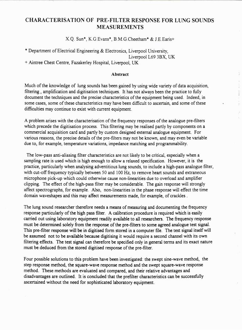
SIDS Wireless Acoustic Monitor (SWAM)
Daniel S. Rusin U.S. Naval Postgraduate School
Monterey, CA 93943
Luqi U.S. Naval Postgraduate School
Monterey, CA 93943
Michael V. Scanlon U.S. Army Research Laboratory
Adelphi, MD 20783-1145
Abstract
Sudden Infant Death Syndrome (SIDS) is the leading cause of death for infants. Current methods of monitoring respiration and pulse of sleeping infants use traditional transthoracic impedance monitors placed on the patient with adhesive. Using Acoustic Pad technology, rapid Computer Aided Prototyping, and Digital Signal Processing, this research demonstrates the use of Acoustic Pads to monitor patient vital signs in a wireless fashion .
The main problem addressed by this research is the development of a software interface and prototype for the Acoustic Monitoring Pad hardware developed by the U.S. Army Research Laboratory using the Computer Aided Prototyping System (CAPS), of the U.S. Naval Postgraduate School to create the SWAM (SIDS Wireless Acoustic Monitor) that eliminates the use of the adhesive electrodes, and monitors patients via the cardiac and respiratory sounds of the patient, in a "wireless" method.
The approach taken was to use an iterative requirements development process involving users and implementing changes to the requirements as development progressed.
The results demonstrate that by using the iterative design approach and by using CAPS, in less than eight months a prototype can be created that validates the acoustic pad concept

II

EFFECTS OF FINGER-STIFFNESS ON THE FREQUENCY CHARACTERISTICS OF A HAND-HELD STETHOSCOPE
Abstract
Fujihiko SAKAO*, Hiroshi SATO, and Masashi MORI *School ofEngineering, Kinki University
Takaya-Umenobe, Higashihiroshima, Japan 739-21
A simple model for a stethoscope (or sensor) of air-coupled-microphone type held by human fingers onto skin is devised.
Results of analysis show that elastic supporting may seriously alter the frequency characteristics of the stethoscope. In the absence of such support, the response is that of a conventional second order system, with a resonance peak. With the elastic support by fingers added, the following two alterations take place.
First, the response level at sufficiently low frequencies is higher than the response at medium range frequencies up to the resonance peak. The difference depends on the ratio of masses of the sensor-container and the skin covered by it, and can amount to as much as 10 dB's.
Second, there is a couple of resonance peak and anti-resonance valley at the transitional zone between the "low-end" and "medium" frequencies . The exact locations of the peak and valley depend on the exact value of the stiffness of the finger support, and their amplitudes depend on damping property of the support. Although those values are not definitely known at present, requiring further investigation, it is quite possible that the above mentioned effects may have serious influences on the signal obtained with a sensor of such a type.
Key Words: Hand-held stethoscope, Frequency characteristics, Stiffness of finger


A PROTOTYPE SYSTEM FOR STUDYING LUNG SOUND MICROPHONES
P. Lipponen, V. Halla, P. Helisto Laboratory of Biomedical Engineering, Helsinki University of Technology
Rakentajanaukio 2 C, 02150 Espoo, FINLAND
A prototype system is developed for measuring the frequency response of lung sound microphones under realistic conditions [lJ . The equipment consists of a PC, DSP card, the measuring bench, and microphone and audio amplifiers.
The bench is composed of two boxes on top of each other with a middle range loudspeaker in between. Sound is transmitted from the loudspeaker to the upper cavity and an artificial tissue. The microphone to be measured was attached against the artificial tissue.
Frequency responses were measured by feeding a white noise signal to the bench and by calculating the FFT power spectrum of the registered microphone signal. Because the frequency response of the bench is not flat, it's only possible to compare microphones to some reference (or to each others) . Six lung sound microphones and a laser vibrometer, which detected the surface motion of the tissue, were used to study the bench, and the results for two of them are shown in Figs. 1-2.
It was observed that there are large differences between the lung sound microphones and that the biggest differences were between air coupled and contact sensors. The results obtained here have already been useful in microphone development. As lung sound analysis becomes a more and more standardized technique, the need for objective microphone test equipment increases.
• •
Figure 1: Frequency response of the Pulmer lung microphone [2J. The lower curve is the background spectrum.
Figure 2: Frequency response of the 1" Haifa contact sensor [3J.
[lJ P. Lipponen, V. Halla, P. Helist~, "A Prototype System for Calibration of Lung Sound Microphones: Analysis of 6 Microphones", 2nd CORSA WPIII Symposium on Signal Prosessing in Lung Sounds, June 1996, Helsinki.
[2J A. Sovijlll"vi, K. Kallio, E. Paajanen, P. Malmberg, P. Helist~, S. Haltsonen, T. Katila, "A new versatile PC-based lung sound analyzer (HeLSA)", 2nd CORSA WPIII Symposium on Signal Prosessing in Lung Sounds, June 1996, Helsinki.
[3J N. Gavriely, "Phonopneumography Contact Sensor", 15th International Conference of Lung Sounds, October 1990, New Orleans.

I I I
CHARACTERISATION OF PRE-FILTER RESPONSE FOR LUNG SOUNDS MEASUREMENTS
X.Q. Sun*, K.G.Evans*, B.M.G.Cheetham* & J.E.Earis+
* Department ofElectrical Engineering & Electronics, Liverpool University, Liverpool L69 3BX, UK
+ Aintree Chest Centre, Fazakerley Hospital, Liverpool, UK
Abstract
Much of the knowledge of lung sounds has been gained by using wide variety of data acquisition, filtering , amplification and digitisation techniques. It has not always been the practice to fully document the techniques and the precise characteristics of the equipment being used. Indeed, in some cases, some of these characteristics may have been difficult to ascertain, and some of these difficulties may continue to exist with current equipment.
A problem arises with the characterisation of the frequency responses of the analogue pre-filters which precede the digitisation process. This filtering may be realised partly by components on a commercial acquisition card and partly by custom designed external analogue equipment. For various reasons, the precise details of the pre-filters may not be known, and may even be variable due to, for example, temperature variations, impedance matching and programmability.
The low-pass anti-aliasing filter characteristics are not likely to be critical, especially when a sampling rate is used which is high enough to allow a relaxed specification. However, it is the practice, particularly when analysing adventitious lung sounds, to include a high-pass analogue filter, with cut-off frequency typically between 50 and 100Hz, to remove heart sounds and extraneous microphone pick-up which could otherwise cause non-linearities due to overload and amplifier clipping. The effect of the high-pass filter may be considerable. The gain response will strongly affect spectrographs, for example. Also, non-linearities in the phase response will effect the time domain waveshapes and this may affect measurements made, for example, of crackles .
The lung sound researcher therefore needs a means of measuring and documenting the frequency response particularly of the high pass filter. A calibration procedure is required which is easily carried out using laboratory equipment readily available to all researchers. The frequency response must be determined solely from the response of the pre-filters to some agreed analogue test signal. This pre-filter response will be in digitised form stored in a computer file. The test signal itself will be assumed not to be available because digitising it would require a second channel with its own filtering effects. The test signal can therefore be specified only in general terms and its exact nature must be deduced from the stored digitised response of the pre-filter.
Four possible solutions to this problem have been investigated: the swept sine-wave method, the step response method, the square-wave response method and the swept square-wave response method. These methods are evaluated and compared, and their relative advantages and disadvantages are outlined. It is concluded that the prefilter characteristics can be successfully ascertained without the need for sophisticated laboratory equipment.


Computerized Respiratory Sound Analysis (CORSA); Techniques, Standardization and Clinical Evaluation - an European Community Concerted Action Project
A. Sovijlirvi*, J. Vanderschoot**. J Earis**, P. Helist6** and F. Dalmasso** *) Project Leader, Lab. of Clinical Physiology, Dept. of Medicine, Helsinki University Central Hospital ,
Helsinki, Finland **) Work Package Leaders
CORSA is a concerted action of project of EC (BIOMED 1). It has been started on the flfSt October 1994 to be continued for 28 months. The total economic contribution from EC is 260.000 ECU. Thirteen research centers from seven European countries: Belgium, Great Britain, Finland, France, Gennany, Italy and the Netherlands, are participating the project which contains four major objectives and work packages (WP). The purpose of the project is to produce guidelines for standardization and to enhance the development of computerized respiratory sound recording and analysis methods. The work packages of CORSA are:
WP 1) Standardization of lung sound recording and analysis. (WP leader Ass. Prof. Jan Vanderschoot, Leiden). The objective is to produce and publish guidelines or recommendations for standards of respiratory sounds recording and analysis. This is the ma.in objective of CORSA. The strategic purpose of WP1 is to obtain the necessary means for comparison of conclusions and outcomes of respiratory sound analysis techniques and research projects. The activities in WP 1 have been divided into 8 subprojects, each one dedicated to a coherent set of topics. 1. Definition of Tenns (subgroup Leader F. Dalmasso). 2. Respiratory Sounds Capturing and Analogue Preprocessing (L. Vannucini, Siena), 3. Minimal Data Sets (G. Righini, Torino). 4. Experimental Conditions (M. Rossi, Arona), 5. Digitization of Data (B . Cheetham. Liverpool), 6. Validation Methods of Analysis Techniques (A. Giordano, Veruno), 7. Report Contents and Fonn (P. Piiril!l, Helsinki), 8. Basic Standards of Analysis (G. Charbonneau, Paris). WP2) Computerized stethoscope and long-term monitoring of lung sounds .(WP leader Dr. John Earis, Liverpool). The objective is to assess how a computerized stethoscope may be developed by surveying the current European activity for measurement and long-tenn monitoring of lung sounds. WP3) Signal processing and diagnostic feature extraction . (WP leader Dr. Panu Helist6, Helsinki).The a.im is to provide a comparison of the applicability of modem digital signal processing techniques to automatic analysis of respiratory sounds. WP4) Reference values and clinical validation . (WP Leader Dr. Filiberto Dalmasso, Torino). The objectives are: 1. to provide a proposal of recommendations for making "reference values" of respiratory sounds and 2. to evaluate the collected respiratory sound data to characterize the patterns of abnonnality of lung sounds in clinical syndromes.
The deliverables of CORSA will be published guidelines, recommendations and survey reports in European Respiratory Journal (ERJ) or European Respiratory Review (ERR). The CORSA-project has been accepted as a Task Force of the European Respiratory Society. The project has progressed almost as proposed in the original time schedule. The second draft of the guidelines for standardization is at present under revision and will be sent to the International Lung Sound Association for evaluation. The draft report of WP2, a survey of computerized stethoscope and long-tenn monitoring of lung sounds in Europe will be ready in September 1996. Within the frames of WP3, signal processing and feature extraction of respiratory sounds, two scientific symposia under the title Signal Processing in Lung Sound Analysis have been organized in Helsinki, the first in June, 1995 and the second in June, 1996. The papers of the flfSt symposium have been published in the Report Series of Helsinki University of Technology (Report TKK-F-C170, ISSN 0358-0741). Revised papers of the symposia will be published in an issue of Technology and Health Care by the European Society of Engineering and Medicine (ESEM)
The task of WP4 of CORSA, to provide guidelines how to produce reference values of respiratory sounds is, as well as the tasks ofWPl (standards) and WP2 (devices) essential for a future task to create European reference values of respiratory sounds in healthy non-smoking and smoking populations.

= l

21ST INTERNATIONAL LUNG SOUNDS CONFERENCE
SESSION C


21ST INTERNATIONAL LUNG SOUNDS CONFERENCE
Chester, England September 4-6, 1996
Session C
(Monitoring) Disease Detection
Dr. Shoji Kudoh & Dr . Sadamu Ishikawa, Chairmen
9:00 - 9:20 Detection of Mild Airway Narrowing in Children Based on Spectral Characteristics of Normal Lung Sounds
9:20- 9:40 Sonagram-Based Automatic Wheeze Detection Method
9:40 - 10:00 Computerized Lung Sound Analysis As An Indicator of the Need for Endotracheal Suctioning in Mechanically Ventilated Patients
10:00 - 10:20 Coffee Break
10:20 - 10:40 Non-Invasive Diagnosis of Chronic Obstructive Pulmonary Disease Utilizing Multi Channel Lung Sound Analysis
10:40 - 11:00 Characteristics of Lung Sounds in Patients with Pneumonia and Congestive Heart Failure
11:00 - 11:20 The Analysis of Upper Airway Sounds By Inverse Filtering
11:20 - 11:40 Discrimination of Productive Cough and Non-Productive Cough By Sound Analysis
11:40 - 12:00 Oral Flow Transducers Can Modify Breath Sounds Spectrum
12:00 - 1:30 Lunch
1:30 - 2:00 Business Meeting
Takase
War is
Schmelz
Murphy, H.
Murphy, R.
Plante
Murata
Dalmasso

I; II Jll

I I I I \
\
\
\
I
I I
DETECflON OF MILD AIRWAY NARROWING IN CHILDREN£: ~ED ON SPECI'RAL CHARACTERISTICS OF NORMAL LUNG s,UND's
Masato Takase and Hans Pastabmp Dept ofPcdiatrics, University ofManitoba. Winnipeg, Canada
Cbaraderistic changes OCQJI' in the spectra of nonnallung sounds during induced airway narrowing even in the absence of wheeze. These changes are reversible by bronchodilator inhalation. To investigate the diagnostic value of lung sound mcasun::ments withoot bronchial provocation we S1Udied children who were referred for assessment of pulmonary function. Forty five subjects were initially enrolled but 1 S were excluded because spiromeuy wu not sufficiently reproducible. Of the remaining 19 boys and 11 girls (ages 7 to 17 years), 15 had diagnosed and IS had suspected astbmL Lung sounds were recorded with two accelerometers (EMT-2SC, Siemens) at the right upper anterior (RUA) and tho right lower posterior {llLP) lobe. Subjects breathed at a target flow of 1 S mMcgls and thea held their breath for a few seconds at resting e.nd-ccpiratioo. Recadings v.a-c made three times, before and after basdinc spiraneuy and Is min after salbutamol (200pg) inhalation. Flow and sound signals were digitized at 10 kHz. On subsequcot canpu1er analysis, visual and auditay vcrificatioo wu wed to avoid inclusion of wheezes and artifacts. At least 20 spectra· each cL sound within 20'A» of taiBct flow were averaged for inspiration, expiralioa and breath holding. After subtmctioo c:l background noise from lung sounds, the power in three octave bands (P.,.: 1 SO to 300 Hz, P _.: 300 to 600 Hz, PiliP: 600 to 1200 Hz) was detennined fur cac:h respira1Diy phase and site of recon:6ng. The SOtb and 99dl percentile of power distribution within the ISO to 1200 Hz range were identified (F,.: median &equency and F.: spectral edge frequency). Power ratios between P._ and P .- (P,_/P ..J and between inspiration and expiration (IIE-P._ and 1/E-P_,) were also calculated. Lung function tests before and after salbutamol inhalation detected 18 (6QilA.) of 30 subjects with mild to moderate abnormalities. Only 14 ~/e) of these 18 children were identified on basdine spirometry without mcuurement of airway resistance and oaoduaance a- bronchodilator response. Abnormalities in four acousiic paramden @spi~.._ at RLP, inspiratory F,. and JIE.P._ at RUA), predicted abocimal luos funcnon with 8~/e sensitivi and ne;. ".ficity. AbnomWity was defined as values outside the norm range (mean* 2SD) established from 12 subjects with normal lung function. Our findings indicate that most of the lung func::tioo aboonnalities in non-wheezy asthmatic children may be detectable by analysis of normal breath sounds recorded with few sensors while tbc cbild breathes at a flow rate only slightly above normal. Considering tbe easy applicatioo and the level of diagnostic ac:curacy, computerized lung sound analysis is promising as a new tool for pediatricians who often experience difficulties in obtaining reliable informatfoo from standard spirometry.
Supported by the Nippoo Mcdk:al Sc:bool. Tokyo, Jipc (M.T.) md the ChiJdn:a's H~ital of Winmpcg Racan:b Foundarioa (M. T .. f-LP.)

//11 '111111 'I '

SONAGRAM-BASED AUTOMATIC WHEEZE DETECTION METHOD
M.Waris1 , S.Haltsonen1, P.HelisW 1, A.Saarinen2 , A.Sovijarvi2
1 Laboratory of Biomedical Engineering, Helsinki University of Technology, Finland 2 Laboratory of Clinical Physiology, Helsinki University Central Hospital
Email: [email protected] .fi
A new automatic wheeze detection method is presented here ll] . The method is based on image processing techniques applied to the sonagram. In the calculation of the sonagram, FFT and AR spectrum estimation methods are compared. Edges are detected in the sonagram by using two Sobel operators [2], and peaks are found between upward and downward edges. A labelling algorithm [2J is then applied to identify the connected components. Information about the strength and duration of a wheeze is used to form a criterion for true wheezes. At the end, broken wheezes are connected with the help of a connection algorithm.
The method was validated by a pulmonary physician in four wheezing asthmatic patients and four control subjects. The lung sounds were recorded from the chest with a PC-based lung sound analyzer [3J . Nine out of ten wheezes longer than 250 ms were detected in the FFT-sonagram, and all ten wheezes were found in the AR-sonagram. However, the harmonic frequencies were detected better in the FFT-sonagram. Very short wheezes (squawks) were not detected as well. The false positive amount of wheezing in control subjects was only 1%. An example of the validated results is shown in Figure 1.
The new wheeze detection method finds wheezes varying in frequency with good accuracy. The method also extracts useful information about the frequency, duration and associated flow and volume of the wheezes. It can therefore be a useful tool for pulmonary physicians.
Figure 1: Detectected wheezes in a FFT-sonagram {FFTlength = 512 points, overlap = 35 %, sampling rate = 6 kHz) . The end points of the wheezes are connected with double lines. Vertical white lines show the boundaries of inspiration and expiration. The single horizontal lines at the bottom of the sonagram show the validation result.! of a pulmonary physictan.
llJ M.Waris, S.Haltsonen, P.HelistO, A.Saarinen, A.Sovijarvi, "A method for automatic wheeze detection", 2nd CORSA WPIII Symposium on Signal Processing in Lung Sounds, Helsinki (1996). I2J M.Sonka, V.Hlavac, R.Boyle, Image proce.!sing, analysi.! and machine vision, Chapman & Hall Computing, London (1993). I3J A.Sovijarvi, K.Kallio, E.Paajanen, P.Malmberg, P.Helisto, S.Haltsonen, T.Katila, "A new versatile PC-based lung sound analyzer (HeLSA)", 2nd CORSA WPIII Symposium on Signal Processing in Lung Sounds, Helsinki (1996).

1

COMPUTERIZED LUNG SOUND ANALYSIS AS AN INDICATOR OF THE NEED FOR ENDOTRACHEAL SUCTIONING IN
MECHANICALLY VENTILATED PATIENTS Schmelz. J.O .. Murphy, M.A.. Munro. B. H .. and Murphy, R.L.H.
Boston College School of Nursing and Faulkner Hospital Chestnut Hill , MA 02167 Jamaica Plain. MA 02130
The purpose of this study is to extend prior research on the role of adventitious lung sounds as an accurate indicator of the need for endotracheal suctioning (ETS) in adult patients requiring mechanical ventilation and endotracheal intubation. Prior research has demonstrated a link between the presence of adventitious lung sound and secretions in the tracheobronchial tree.
We used a Multi Channel Lung Sound Analyzer (MCLSA) to study the pattern of adventitious lung sounds present immediately before and following endotracheal suctioning and compared sounds to the volume of tracheobronchial secretions aspirated by ETS. Repeated measurement of 15 patients was made when the patient's primary nurse indicated the need for suctioning.
There was no consistent pattern of lung sounds prior to suctioning. Five types of adventitious lung sounds were identified; rhonchi, wheezes. crackles. type II rhonchi and coarse sounds. There was a 14% reduction in the occurrence of adventitious lung sounds after suctioning and coarse sounds decreased in duration after suctioning in most patients. There was no relationship between lung sounds and the volume of aspirate obtained or the reason the nurse gave for suctioning the patient.
This study failed to support the association between rhonchi and tracheobronchial secretions obtained by blind suctioning which may be an unreliable method of measurement. Further study utilizing specific disease categories. direct visualization and localization techniques is necessary to establish when and if ETS should be done.

I'

NON-INVASIVE DIAGNOSIS OF CHRONIC OBSTRUCTIVE PULMONARY DISEASE UTILIZING MULTICHANNEL LUNG
SOUND ANALYSIS
Murphy MA, Schmelz JO, Bergstrom K and Murphy RLH Boston College School of Nursing & Faulkner Hospital. Chestnut Hill, MA 02167 Boston, MA 02130
To determine whether objective pattern differences exist in Chronic Obstructive Lung Disease (COPD) we studied 24 patients with this diagnosis and 23 elderly volunteers using a Multichannel Lung Sound Analyzer (MCLSA). Realistic microphones embedded in the chest pieces of 3M cardiac stethoscopes were used to collect data from over the trachea and from 24 chest sites at the posterior and lateral bases bilaterally.
Patients had a clinical diagnosis of COPD and spirometric evidence of obstruction {FEV 1\ Predicted (P) • 19\ to 68%}. All volunteers denied clinical history of significant lung disease and had normal spirometry (FEV 1\ P • 76\ to 155\) .
Length of inspiration (I) and length of expiration (E) were measured and the ratio of I/E calculated (Rl). The Rl was significantly different between COPD and volunteer group (t a 4.4, p • <01). Differences between mean inspiratory time in the COPD and volunteer groups were significant (t • 3 . 2, p a <01); expiratory time was not different.
R2, the multiple of R1 and amplitude of the sounds over the chest wall during inspiration was calculated and showed significant differences between groups ( t 4, p <01). Regression of R2 on FEV 1\ P indicates a low explained variance ( R-squared • . 37). Crackles, wheezes and rhonchi were not significantly different in the two groups, but this may reflect the age of the subjects and other health conditions.
Lung sound patterns that exist in significantly different from elderly study.
COPD patients volunteers in
were this


CHARACTERISTICS OF LUNG SOUNDS IH PATIENTS WITH PHEUMOHIA AND CONGESTIVE HEART FAILURE
R. Murphy, M. Murphy, K. Bergstrom and M. Jean
Congestive Heart Failure (CHF) and Pneumonia (PN) are among the most common causes of acute illness requiring hospitalization. While the diagnosis of these conditions is often easily made, many patients present with nonspecific findings such as dyspnea, cough, chest discomfort and atypical roentgenographic shadowing, the cause of which is unclear. We studied the lung sounds of 6 patients with PN and 6 patients with CHF to determine if the patterns of their lung sounds were different. Sounds were examined at 24 chest sites and over the trachea using a rnul tichannel lung sound analyzer, as previously described. ( 1) Results are presented in the following table:
PN CHF rhuationof~Uation 871 1124
in msec (217) (350)
rhuation of Expiration 1252 1869 in msec (310) (851)
Ratio (1/E) .706 .513 (.128) (.115)
Inspiratory Crackles: Fine 3 1
Medium 22 13 Coarse 31 9
TOTAL ~iratory 56 23 ___E~iratory Crackles:
Fine 0 2 Medium 4 3 Coarse 4 15
TOTAL Expiratory 8 20 TOTAL CRACKLES 64 43 Insp. Crackles, Left 44 11
Insp. Crackles, Right 11 11 Rhonchus: ~iration 1 0
Expiration 4 1 Wheezes: ~iration 2 1
ExpUation 2 2 Numbers in parenthesis are standard deviations.
A3 seen in this table, this pilot study of a small number of patients, those with CHF tended to have a longer expiratory phase and relatively more crackles in expiration. Pneumonia patients had more crackles in inspiration and they were more asymmetric in their distribution. This suggests that lung sounds pattern differences exist that may be helpful in the differential diagnosis of these conditions. We believe that the additional study of lung sounds in such patients is warranted.
(1) Murphy M., Schmelz J .0., Murphy R.L.H., Characteristics of Lung Sounds in Chronic Obstructive Lung Disease, American Journal of Respiratory and Critical Care Medicine, April 1996: Vol 153:4 p.A322.

I'

THE ANALYSIS OF UPPER AIRWAY SOUNDS BY INVERSE FILTERING.
F. Plante*, H, Kessler+, X. Q. Sun* , B. M. G. Cheetham* & J. Earis+
*Department ofElectrical Engineering & Electronics, Liverpool University, Liverpool L69 3BX, UK
+ Aintree Chest Centre, Fazakerley Hospital, Liverpool, UK
Abstract
The human mechanism for producing upper airway sounds such as stridor, snoring and speech may be modelled as an excitation sound source coupled to a resonating acoustic tube. The nature of the excitation signal can tell us much about the physical mechanism responsible for its production e.g. vibration of vocal cords, soft palate or pharynx. It is known that abnormalities can be detected by analysing excitation signals extracted from speech for example. The nature of the resonances within the upper airway, being determined by its physical shape, is also likely to have diagnostic value. To extract the excitation signal from sound measured at the mouth, the resonances introduced by the upper airway and other spectral colouration due, for example, to lip radiation, must be cancelled out. This is possible using a time-varying digital filter whose parameters are periodically updated by a spectral estimation algorithm. The process of estimating the upper airway resonances and hence cancelling them out is referred to as inverse filtering. LPC analysis is a simple example of an algorithm for the required spectral estimation, and may be used as the basis of more sophisticated techniques.
With current digital signal processing technology, inverse filtering can be readily implemented in real time to produce time-domain or spectrographic displays. We report here preliminary results from two clinical investigations based on inverse filtering.
The first is concerned with a non-invasive method of clinical assessment of the snoring mechanism in patients being considered for treatment by surgery. This method involves the analysis of sustained vowels recorded from patients in sitting and lying postures and comparisons between these sounds. It is believed that abnormality in the upper airway may be detectable as greater than normal differences in the sounds.
The second investigation is concerned with the detection of vocal cord abnormalities which present as hoarse voice. Such abnormalities may be due to laryngeal disease, or the long term effect of inhaled steroids used for the treatment of asthma.


DISCRIMINATION OF PRODUCTIVE COUGH AND NONPRODUCTIVE COUGH BY SOUND ANALYSIS
Akira Murata 1), Shoji Kudoh 1), Yasuyuki Taniguchi 1), Atsuo Shibuya 2), Hasashi Hori3) 1) The Fourth Department of Hedical School, Tokyo, Japan
Internal Hedicine, Nippon
2) Japan Wemen's University 3) Tokyo National Chest Hospital
There are two types of coughs, productive and nonproductive. The former is caused by the excess of airway secretion. The analysis of cough may provide important clues, not only for the diagnosis, but also for the drug selection. In this study, we compared cough sounds recorded from patients with productive cough due to chronic airway diseases with those of voluntary cough sounds of healthy subjects.
Both cough sounds were recorded on a recorder (SONY TVD-07) with an electret (Panasonic RP vc3) and analyzed by Instrument Company, U. S . A. ).
digital audio tape condenser microphone Sound Scope II (GW
Non-productive cough sounds were separable into two phases, while those of productive coughs were separable into three phases. In the second and third phases of cough sounds, overtone structures were observed in the low frequency area of the sound spectrograms. We speculate that these overtones are related to the resonance, or fluttering, of the wall of the airways. In the analysis of productive cough sounds, we noticed prolongation of the duration of the cough sounds and some pulsive components in the second phase.

I I II ~ II' I

ORAL FLOW TRANSDUCERS CAN MODIFY BREATH SOUNDS SPECTRUM
Dalmasso, F, Vannuccini L. Torino
Righini, P, Prota R, Righini Divisione di Pneumologia, Osp.
G*, Rossi H, 'HAURIZIANO',
*Institute Elettrotecnico Nazionale ''Galilee Ferraris'', Torino Divisione di Pneumologia, Osp., Civ . Arezzo, ITALY
Oral flow is usually measured when the lung sounds are recorded on the chest wall. The aim of this study is to verify if and how much the oral flow transducers modify the recorded breath sounds (tracheal and bronchial breath sounds). These were detected respectively on the sternal notch, right anterior chest, 2nd intercostal space and on the posterior right in the respiratory triangle by an air coupled microphone (ECH144 Sony) and a digital tape recorder (DAT, DA-R100, Casio) with and without oral flow transducer. Two groups (A and B) of four heal thy subjects were each instructed to perform maneuvers of slow vital capacity at controlled flow rate ( 1L Is) . Group A used the Fleisch pneumotachograph n. 3 (Fen yves & Gut) and Group B used the turbo-digital transducer (Pony, Cosmed) . Sounds were analyzed in the frequency domain by Fast Fourier Transform ( FFT). The central frequency and the frequencies of given percentiles ( 2 5, 50 and 7 5%) of the spectrum energy have been used for statistical processing of the results:
FLEISH n 3
Parameter Without With p (Hz)
Central 424 ~ 35 325 ~ 67 0.04 Frequency
F 25 283 + 92 166 + 55 0.072 F 50 478 ~ 61 292 ~ 115 0.029 F 75 576 ~ 23 448 ~ 98 0.172
We found significant differences for inspiratory sounds in the spectral parameters between maneuvers, with and without, only in the tracheal site of recording. In the other sites, the differences are not significant. In Group B we have not found systematic di f fe renee s in all of the sites, but the spectrum shape differs, particularly at the bronchial site . Even at low flow resistance and at low respiratory flow, the oral flow transducer distorts the spectrum of breath sounds in different ways.

l I ~
1

21ST INTERNATIONAL LUNG SOUNDS CONFERENCE
SESSION D

l ~ ~ I
_L

21ST INTERNATIONAL LUNG SOUNDS CONFERENCE
Chester, England September 4-6, 1996
Session D
Dr. Masashi Mori & Dr. John Earis, Chairpersons
2:00 - 3:00 Poster Discussion
Digital Recording and Computer-Based Analysis of Lung Sounds
Classification of Fine and Coarse Crackles
Nocturnal Asthma Assessed By Intermittent Sleep Trachael Sounds Recording
Observer Variability in Chest Auscultation
3:00 - 3:20 Lung Sounds Visualized by Symmetrized Dot Patterns (SOP)
3:20- 3:40 Cough Sounds Spectra in Asthmatics: Steroid Effects
3:40 - 4:00 Measurement and Analysis of Spectral Respiratory Sound in Healthy Chinese Part One in Youths - 122 Students
4:00- 4:20 Respiratory Auscultatory Skills Among Internal Medicine and Family Practice Trainees: A Comparison of Diagnostic Proficiency
4:20- 4:30 Closing Remarks
4:30 Steering Committee Meeting
Wilmot Ball, M.D. David Cugell, M.D. Filiberto Dalmasso, M.D. Noam Gavrielly, M.D. Sadamu Ishikawa, H.D. Steven Kraman, M.D. Shoji Kudoh, H.D. Robert Loudon, M.D. Masashi Hori, H.D. Raymond Murphy, M.D. Hans Pasterkamp, H.D. Anssi Sovijarvi, M.D. S.A.T. Stoneman, M.D.
Schuettler
Brown
Kiyokawa
Parziale
Davidson
Ishikawa
Tan BIN-Yong
Mangione

_ _... __ _

DIGITAL RECORDING AND COMPUTER-BASED ANALYSIS OF LUNGSOUNDS
F. Schuettler , T . Penzel, P. von Wichert Dept. of Medicine of Philipps- University, Baldingerstr.l, D-35033 Marburg,
Germany [email protected];lni-marburg.de
Introduction: The clinical value of lung auscultation may be enhanced by digital recording and computer-based analysis. This technique may show variances in different lung diseases with specialized algorithms. Beyond that , it would be an advancement in the medical education to hear and visualize lungsounds on a usual personal computer.
Method: The recording system is based on an analog to digital converter (SORCUS M4/486 and M-AD12-16) and two air-coupled microphone cartridges (Monarch MCE-200) . A flow sensor (BiScope) and a pressure transducer (SenSym SCXL004DN) was used for the measurement of ventilation. The system is able to continuously record three channels (two sounds and airflow) with sampling rates up to 11024Hz and 12 bit resolution on a computer harddisk. We used the standard European Data Format (EDF) to enable easier data exchange between working groups. The program manages the transformation of the time series into the frequency domain (FFT) and also can converts any part of the data into WAVE audio files. There are many programs available to work with the recorded data in this standardised form , for example the playing-back of the sound on any multimedia suitable personal computer.
Results: In a preliminary study, we recorded the lung sounds of patient with pneumonia and volunteers with normal lung functions. The differences between normal and pathological lungsounds that were found subjectively with classic auscultation , could also be visualised in compressed spectral arrays.
Is
Figure 1: One breathing cycle of an 35 years old patient with pneumonia..
3.00(•1
2.00
1.00
Figure 2: The recording of microphone 1 in the frequency domain (analyse window=0.185s, overla.pping=30%).
'. . [, :: ,_
~ ._

1 wr / I'
1..

THE CLASSIFICATION OF FINE AND COARSE CRACKLES USING WAVElETS
A N D T I M E -F R E Q U E N C Y DISTRIBUTIONS
A.S. Brown, Dr. Graham, G. Jamieson, G. Boote, J.L. Moruzzi
Whiston Hospital & Liverpool John Moore's University, UK ..
The objective analysis of lung sounds has primarily revolved around spectral analysis. The fast Fourier transform (FFT) and paramethic modelling (PM) have been used successfully to analyze wheeze [ 1 ,2], which is a tonal sound with a duration of several seconds. FFT /PM are limited when applied to crackles which · have considerably different characteristics to wheezes. This study presents results obtained using novel Wavelet transforms and TimeFrequency distribution techniques which have advantages over the FFT /PM that allow them to be applied to crackles to generate meaningful spectrographs. Parameters extracted from the spectrographs allow crackles to be classified as fine or coarse. Thirteen patients 'With bronchiectasis/bronchitis or pulmonary fibrosis exhibiting crackles have been studied. The crackles were subjectively classified a pn'od by three experienced chest physicians and then objectively a~alyzed using the above techniques. A summary of results is shown in the follo'Ning table.
Parameter Coarse Crackles Fine Crackles ( avg breath cycle)
Median frequency 294 Hz 386Hz
Peak power -21 dB -24 dB
Band'Nidth 149Hz 246Hz
Density (crackles <13 <24 breath/cycle)
Proximity of Throughout Late crackles in cycle Inspiration Inspiration
Proportion of cycle 1:36% E:14% 1:61% E:26% 'With crackles
These nf!N techniques have been found to objectively classify coarse and fine crackles based on the parameters extracted above, with greater accuracy and robustness compared to previous work using the FFT /PM. For parameters such as peak power and median frequency, it is not possible to get meaningful results using FFT /PM, and hence, it is difficult to distinguish between coarse and fine crackles. Inter-observer variation and subjectivity are also removed using these techniques. It is envisaged that the methods employed will facilitate the establishment of an objective classification of crackles. The techniques will be of value in both clinical medicine and in education.
References: [ 1] Spence DPS; eta!. AJCCM, 1995. [2] Jamieson, G. Ph.D., University of Liverpool, UK, 1993.

JmJ~ll

NOCTURNAL ASTHMA ASSESSED BY INTERMITTENT
SLEEP TRACHEAL SOUNDS RECORDING
Hiroshi Kiyokawa, Makoto Yonemaru, lkuma Kasuga, Kazushige
Minemura, Hiroshi Kusumoto, Shinobu Horie, Yoko Hiramine, Naoshi
Yanagisawa, Yuichi lchinose and Keisuke Toyama
Tokyo Medical College, the First Department of Internal Medicine
6-7-1 Nishi-shinjuku, Shinjuku-ku, Tokyo 160, Japan
Bronchial asthma is known to cause nocturnal bronchoconstriction
(nocturnal asthma) or dyspnea early in the morning. The nocturnal
attack is important because it is one of the major complaints of
asthmatic patients and sometimes causes sudden death. Wheezes
detected by intermittent sleep tracheal sounds recording (STS R) were
used to assess nocturnal asthma. Forty STSRs were obtained from 28
asthmatics. Ninety percent of these STS Rs contained nocturnal
wheezes. The number of nocturnal wheezes had a tendency to
increase in severe attack compared to that in mild attack. The number
of nocturnal wheezes correlated with the patients perception of
severity in asthmatic attac~ during day time. The number of nocturnal
wheezes per hour significantly increased during 5 to 6 AM compared to
that during midnight to 1 AM. We conclude that STS R disclosed
several characteristics of nocturnal bronchoconstriction in bronchial
asthma.

Wll 1111-'11 "- JU~II I_
SIDS Wireless Acoustic Monitor (SWAM)
Daniel S. Rusin U.S. Naval Postgraduate School
Monterey, CA 93943 dsrusin @cs.nps.navy .mil
Luqi U.S. Naval Postgraduate School
Monterey, CA 93943 luqi @cs.nps.navy .mil
Abstract
Michael V. Scanlon U.S. Army Research Laboratory
Adelphi, MD 20783-1145 scanlon @msc.arl.mil
Sudden Infant Death Syndrome (SIDS) is the leading cause of death for infants. Current methods of monitoring respiration and pulse of sleeping infants use traditional transthoracic impedance monitors placed on the patient with adhesive. Using Acoustic Pad technology, rapid Computer Aided Prototyping, and Digital Signal Processing, this research demonstrates the use of Acoustic Pads to monitor patient vital signs in a wireless fashion. The main problem addressed by this research is the development of a software interface and prototype for the Acoustic Monitoring Pad hardware developed by the U.S. Army Research Laboratory using the Computer Aided Prototyping System (CAPS), of the U.S. Naval Postgraduate School to create the SWAM (SIDS Wireless Acoustic Monitor) that eliminates the use of the adhesive electrodes, and monitors patients via the cardiac and respiratory sounds of the patient, in a "wireless" method. The approach taken was to use an iterative requirements development process involving users and implementing changes to the requirements as development progressed. The results demonstrate that by using the iterative design approach and by using CAPS, in less than eight months a prototype can be created that validates the acoustic pad concept.

-
I. INTRODUCTION
A. BACKGROUND
1. General: This research uses the Software Engineering requirements analysis process to develop
the interface for an acoustic cardiac and respiratory apnea monitor called the "SIDS Wireless Acoustic Monitor (SWAM)". It takes the project from user's idea through the design and prototyping process by applying the Computer Aided Prototyping System (CAPS) of the Naval Postgraduate School in the development of the SIDS Wireless Acoustic Monitor (SWAM). This report is a summary of the work done in developing a computer interface for the Acoustic Pad hardware originally designed by Michael Scanlon of the U. S. Army Research Laboratory, Maryland.
This monitor uses a fluid filled pad to as the primary sensor to "listen to" the heart and lung sounds of an infant. Some of the fundamental principals are similar to current technology used in under sea acoustics to listen for and distinguish between different types of ships, submarines, etc. The use of the Acoustic Pad enables the patient to be monitored in a wireless fashion , without the attachment of electrodes to the torso.
_, ,_------------~- ,, , .
.-a .. Ji\ ~ :!1 fj SENSOR
Hydrophone
Current acoustic monitoring and noise filtering can monitor and differentiate between various sound sources.
.. - !. - -
SENSOR Hydrophone
The SWAM is an extension of current technology, separating respiratory and cardiac sounds, potentially replacing the traditional impedance monitoring approach.
Figure 1. Illustration of the Overall Acoustic Pad Concept.
2. Definitions: Sudden Infant Death Syndrome (S!DS ): The sudden death of any infant or young child,
which is unexplained by history and in which a thorough postmortem examination fails to demonstrate an adequate explanation of the cause of death.
2

. ..-~ ---*-" 1·· ---· ........ _ ... -.--~- - ·-- -- -- . -- ---- .., ...... -------- --.. --.. ·-··-- -· •
Apnea: Apnea of Infancy (AO]): Cessation of respiratory air flow. An unexplained episode of cessation of breathing for 20 seconds or longer, or a shorter pause associated with pallor, and/or hypotonia. AOI is reserved for those infants for whom no specific cause [of respiratory cessation] can be identified [1].
3. General Discussion: The current method of monitoring respiration and pulse of sleeping infants is to use
traditional transthoracic impedance monitors (or "stick-on" electrodes) with wires placed on the torso of the infant. The Sensors Branch of the Army Research Lab is experimenting with the possibility of using a wireless approach to monitor respiration and heartbeat of infants. This approach would remove the electrode wires from the patient and would provide a more convenient method of home apnea monitoring. Currently, they do not have a computer interface or a software requirements model for their acoustic sensor approach. By applying the software engineering approach and using CAPS, we developed the first iteration of a graphical interface for the Acoustic Pad sensor. Promising directions for future research include Digital Signal Processing work to apply noise cancellation techniques to the data received through the hydrophone in the acoustic pad to separate cardiac and respiratory sounds.
E. Scope of Research: This research uses prototyping and requirements analysis process to take the SWAM
system from a user's idea through the design and prototyping process. This enables users to see and comment on a working model of the system before hardware decisions are finalized. This paper is a report of the initial requirements and is intended to stimulate dialogue between parents, technicians and medical professionals to gain feedback regarding the SIDS Wireless Acoustic Monitor. The NPS Computer Aided Prototyping Design (CAPS) is being used to develop the interface to connect the sensors. We are interacting with the potential final users; the parents and medical professionals who are involved with Sudden Infant Death Syndrome, through the SIDS Network, Inc., the California SIDS Program, and through association with the SIDS electronic mailing listserver ([email protected]) to meet their needs as well. The initial product to be delivered is a model of the SIDS acoustic monitor using CAPS.
F. Approach: The actual monitor device incorporates a pad type receiver that resembles a typical crib
sized mattress pad. A hydrophone sensor placed within the pad feeds input to the monitor in real time. The monitor accepts the sensor data and activates an alarm to alert the nurse or parent in the event of respiratory or cardiac trouble. The CAPS design process allows the rapid development of a workable interface. This project demonstrates the benefit of using the Computer Aided Prototyping System in the development of a real-time, real-life system. Figure 2 shows a very abstract, top level concept of the design.
The device monitors the patient by using a fluid-filled sensor pad placed under the patient to detect pressure fluctuations caused by the acoustic signatures that accompany
3 I '

Acoustic Pad
Monitor Modem/Pager
Figure 2. Sketch of Overall Acoustic Monitoring Pad System Concept
normal breathing and heart sounds. The excellent coupling between the human body and the sensor pad gives a high signal-to-noise ratio over ambient sounds. The signals allow medical personnel to detect cessation of breathing, fluid in the lungs, an obstructed airway, or an irregular heartbeat [2].
G. Relevance of Project: "Sudden Infant Death Syndrome" is the leading cause of death for people under the
age of one each year. Each year, 7000 people under the age of one die in their sleep due to unexplained causes, i.e.: autopsies have ruled out suffocation and other possible causes (one death per 500-600 live births). There is currently no "cure" for Sudden Infant Death Syndrome (SIDS).
Home monitoring is used for high risk patients: those who have demonstrated lapses in breathing, or the subsequent siblings of SIDS victims. While home monitoring is not a cure, it is able to alert the parent/caregiver to an episode of apnea or period of cessation of breathing. Medical studies of babies who have lapsed into a period of respiratory cessation show that in some cases, the immediate stimulation with light, sound, and/or vibrations is enough to cause the infant to naturally start breathing again [3]. While this device in no way will take the place of a parent or health care provider, it is meant to assist them in the care and treatment of the infant.
Through contact with physicians involved in SIDS research, we have found that research using a pad oriented approach was done almost twenty years ago, and the technology was abandoned because the devices were not sensitive enough. Advances in computer aided prototyping, acoustic monitoring, digital signal processing and sensor technology over the past twenty years will enable this to approach to become a tremendous aid to infants, parents, and the medical community.
4

The signal processing applications of the unit will allow physicians to isolate the cardiac and respiratory sounds and focus on either of the particular sounds. It is easy to ask a patient to stop breathing for a short period so that you can listen to only the heart, however it more difficult to stop the heart from beating so that you can listen to lung sounds only. The separation of the two signals will enable physicians to more efficaciously prepare diagnosis and treatment.
Advanced Trauma Life Support (ATLS) applications are one extension of acoustic pad monitoring with future units developed from this research. A crib sized mattress pad can be placed under an adult patient's torso on a standard ambulance stretcher to monitor vital signs. This will enable the medic on the trauma scene to concentrate on other important aspects of patient care without the task of attaching adhesive torso sensors. Any method that allows medical personnel to capitalize on care during the first "Golden Hour" at a trauma scene improve the patient's chance for survival. Noise cancellation techniques can be applied to this project to allow an acoustic monitoring pad to be used during patient transport.
This area of research holds direct relevance to the Department of Defense and the civilian medical community. Remote monitoring features a distinct advantage by alerting attendants via transmitter for remote diagnostics; and by using the acoustic pad approach, this wireless system eliminates the danger of cords entangling or strangling the patient. In medical applications off the battlefield, acoustic data from a patient can be collected and remotely analyzed for indication of cessation of breathing or heartbeat.
Military medical care in the year 2001 will involve telemedicine and telepresence surgery [4,5]. Telepresence surgery will benefit from this technology in at least two major aspects. First, by minimizing time spent on patient preparation prior to battlefield surgery, surgeons will have greater time and flexibility for surgical procedures. Secondly, by removing the adhesive sensor pads and the attached wires from the torso of a patient, the mechanical manipulator arms used in telepresence surgery will not be tangled or otherwise impeded by any wires coming from the adhesive sensors. The acoustic pad would monitor patient vital signs by being placed on the stretcher or operating table under the patient in a non-intrusive manner.
H. RESEARCH QUESTIONS: This research raises the following questions in the areas of requirements analysis, rapid
prototyping, human computer interface, and software/ systems management. 1. Exactly what are the user' s requirements and how can CAPS be applied to solve
the problem? 2. How do you define and incorporate requirements that arise during the modeling
phase or later? 3. How does abstraction aid in the design process? 4. How do you go from an idea to a software design and an interface that accepts an
analog data stream from a sensor, or series of sensors? 5. What special concerns does the software engineer of a biomedical application have
and how do they affect the design process?
5 I

II. REQUIREMENTS ANALYSIS
A. STAKEHOLDERS
1. "Who is the User?": Before any clear specifications can be outlined, the customer must be identified. To be
effective, and serve the complex mix of user requirements in this SIDS Monitor project, we must balance a triad of users and act as Project Managers.
Infant Patients and Parents
Figure 3. The User Triad
The question of "Who is the user" is difficult to define and the answer incorporates all of the above elements. Each stakeholder group has individual considerations and must be integrated in the best mix to provide optimum performance for all concerned. The design presented here is the first draft of an iterative design process. As routine in a military staff process, the prototype requirement document is being routed through the three "critical edges" of the user triad. Medical input/ comments will be obtained through discussion with members of the SIDS research community, including:
a. Acoustic Pad Hardware Developers. b. Medical Providers. c. Parents who are using, or formerly used SIDS monitors representing the Infant
Patients.
2. Initial User Input: We have been in contact with several of the individuals of the groups mentioned
above. As this application of biotechnology is a unique area, as are most software engineering projects, we needed to learn a great deal about the current technology and gather a resolution of the user's perspective from the domain experts. Our findings are summarized below in section B.
6

B. REQUIREMENTS AND DESIGN CONSTRAINTS:
The requirements for a software system are expressed at different levels of abstraction and with different degrees of formality. The highest level requirements are usually informal and imprecise, but they are understood best by the customers. The lower levels are more technical, precise, and better suited for the needs of the system analysts and designers, but they are further removed from the user's experiences and less well understood by the customers. Because of the differences in the kinds of descriptions needed by the customers and developers, it is not likely that any single representation for requirements can be the "best" one for supporting the entire software development process[6]. CAPS provides the necessary means to bridge the communication gap between customers and developers.
The top level goals for the SIDS Wireless Acoustic Monitor are separated into the three corresponding areas:
1. Medical Providers/ Parents considerations: a. Consistent appearance with current apnea monitors. Several monitors are
currently in use, however they are all of the style that use impedance type sensors directly attached to the patient's torso with adhesive. Signals are transmitted via small wires to the monitor. A variety of formats are used, but several models display both cardiac and respiratory data.
b. Ease of use. Instructions should be understandable by a child in grade eight. c. Self calibrating. The unit must be able to calibrate itself to a series of potential
users, each with different cardiac and respiratory patterns. One current monitor (RespriTrace) uses the first five minutes of data collected to establish "normal" thresholds which are unique to that patient. Other self calibration techniques could be used to reduce the number of preparatory steps the provider must accomplish before monitoring.
d. Reliability/ lack of "false alarms": One of the most common complaints about current systems is that there are too many "false alarms" which ultimately reduce the urgency of the caregiver.
e. Cost: The end product should be within a reasonable market price. f. Event History/ memory: The system should provide a printout or some other
reliable history of the entire time monitored. g. Portable: Final size should be small enough for convenient transport.
2. Infant/ Patient considerations: a. Non-invasive, non-permanent monitoring: The term "wireless" in this case refers
to the approach of collecting cardio-pulmonary data from the patient without attaching any traditional wire electrodes or leads.
b. Position Insensitive: The device must remain accurate regardless of the position of the patient. One of the advantages of the acoustic pad approach is that the device should not have to be "reset" when the patient changes position during sleep.
c. Accurate: The final manufactured product will have to pass FDA approval and as such, must be accurate and reliable.
7

I I
3. Technical considerations regarding the Acoustic Pad sensor: a. Signal Processing capabilities: At a very minimum, the monitor must be able to
separate the incoming sound signals to differentiate between the cardiac sounds and the respiratory sounds. The initial sensor input is delivered via the hydrophone in the acoustic pad. To graphically display the signals for clarity and diagnosis, the signals must be separated. The separated signals are graphically displayed on the screen in stripcharts similar to a standard EKG strip. This provides consistency and familiarity with current medical devices. By separating the cardiac and respiratory sounds, both cardiac and respiratory specialists can examine the patient's history. The signal processing aspect is treated as an abstract "black box" at the onset of the project.
Data gathered by Scanlon [7 ,8] shown in figure 4 shows data collected by placing a small monitoring pad under the torso of an adult male lying on a mattress, similar to the method of collection that would be employed for home monitoring, medical transport or hospital monitoring. Heartbeats are clearly visible in the 0-50 Hz region, and the breaths from 0-500 Hz. The inhalation is higher in amplitude than the exhalation for the first two breaths, and the person takes a much deeper breath the third time in anticipation of holding his breath. From the high signal to noise ratio of the breath cycles, with respect to background noise, it is possible to determine of the patient has stopped breathing.
750 .
500.
Freq CHz)
:t$0 .
22 Seconds of Data
Inhale , Hold
Figure 4. Time-frequency spectrogram of the acoustic sensor pad output.
b. Expandable: The system design should accommodate the addition of other monitoring sensors, i.e.: pulse oxymmetery sensors, temperature sensors, etc.
8

4. Additional consideration: One non-trivial factor to be considered is the reluctance of some parents and medical
professionals to try a new type of device. Initial response seems to be quite mixed. Many people are reluctant to switch from a known system to a new, experimental prototype. Once the product is developed, the initial research and testing phase would involve simultaneous monitoring with an infant monitored traditionally, while also being monitored via the SIDS Wireless Acoustic Monitor. Because of the non-invasive nature of the device, simultaneous testing should not be a problem. I have received one very encouraging e-mail from a woman who stated:
"How are you testing the wireless monitors? Have you gotten that far? I would be interested in using one for my next child (I'm hoping to get pregnant soon). Let me know if you will need test subjects in about nine or ten months! Thanks ... Sherly"
C. SYSTEM INTERFACE: The final product that the user of the system will interact with must functionally
combine the technical aspects required. The SWAM is intended to be simple enough for home use, while maintaining sufficient technical data for the physician. F~gure 5 shows a sketch of the first version of prototype design. Home users will interact with the device via buttons that permit only limited selection of choices, i.e.: On/Off, Print patient data history, Toggle between the patient data waveforms, and a method to silence an audio-visual alarm while continuing to monitor. The monitor' s default mode is "On" and always monitoring. The moment a patient lies down on or is placed on the acoustic pad, the monitor begins sampling respiratory and cardiac sounds. A standard disk drive is necessary to capture data for later analysis by medical personnel. A modem connection allows the device to alert parents via a standard telephone pager, and will allow the ability to include automatic data transfer of the past 24 hours data to be transmitted over standard telephone lines using standard TCPIIP techniques. An external printer will provide the option for standard paper copy EKG style strips showing heartbeat, respirations, and other data side by side over time. The variety of data collection mediums permit increased flexibility in diagnosing treatment.
D. DISCUSSION OF THE INTERFACE CONCEPTS AND GOALS:
1. Use of Metaphors: The prototype should resemble similar existing interfaces. Users have preconceived notions of what the system should look like and how they should interact with it. This initial design should not be a barrier to the user.
2. Aesthetic Integrity and Consistency: Due to the medical application, it is recommended that the final product be packaged by a company who currently manufactures medical equipment and can use a standard color, texture, size combination for the external case. Any information displayed on the screen (which could be LCD) must be identical to the paper history printout. Use of one large screen LCD screen, similar to
9

Larg_e ( -4"x6") LCD Screen
I
Rate
~~~40 Heart Rate
98.6 Tern erature
other
~~ Lock/Unlock on/off print
r\ \ '
self-cn'eck (OK)
/
Self-Diagnosis Light
@ @ © © D D D
Disk Drive for Data - Transfer and Analysis
~
' \ I
Internal Speaker
~
\ I
IQput Jacks (Kesp. Audio1 Cardiac Audto, Temperature, other)
' Out\mt Jacks (Ligbt, Sound, Phone)
\ \ \
Phnter Port (for external printer)
Figure. 5. Prototype of SIDS Wireless Acoustic Monitor Design
the "Apple Newton" would support flexible interface design and the addition of future data "strip" windows.
3. Direct Manipulation: The system must have a "safety" device which requires positive (two finger) control of any act of reset or switching the system off. The alarms should be able to be silenced/ paused at the touch of a button. The only other user interaction possible is printing patient history.
4. Feedback and User Control: The first method of feedback for the system is alarm sounding, which can be extended to activate a hom, light, vibration device, and dial a
10 I

telephone modem to call a pager number. The second method of feedback is the printed history of the entire sleep period.
E. GRAPHICAL USER INTERFACE PROTOTYPE: To complete a development of the initial interface in a timely fashion, we
concentrated on the look and feel of the functional interface (shown as figure 6). The primary screen displayed is the "Waveform" screen which shows EKG style stripcharts of respiratory and cardiac sounds. A help screen can be activated and the user can be prompted if assistance is desired. Audible and flashing light alarms can be adapted to alert physically challenged providers to extend the user base.
I -~ EKG style data strips
I
\
'~
Future Addition
I
' '~
Figure. 6. SWAM Graphical User Interface Prototype
11
Current Data 1
I I
~

I I
F. SOFTWAREABSTRACTION: The initial top level functional diagram shown in figure 7 is the CAPS Augmented
Data Flow Diagram of the SIDS Wireless Acoustic Monitor. The bubbles on the left represent input to the monitor via the noninvasive Acoustic Pad and a remote temperature sensing device. Through the use data abstraction, additional sensor types or classes may be installed or modified at a later date as sensor technology improves. This type of object modeling allows the technique of code reuse to save time in future versions of the
<MdacOigltalstraam
RespPlldioStr
Figure 7. A CAPS Augmented Data Flow Diagram of the SWAM
One of the significant features of rapid computer aided prototyping and design is to take a preliminary design, convert it to a detailed design and quickly produce prototypes for evaluation and later for production. The design of computer prototypes of medical applications requires the designer to allow for a considerable amount of flexibility for future changes [9].
G. CONCLUSIONS: In order to develop any system effectively, designers must include users and domain
experts early on, and the review of initial prototypes and requirements are essential to an effective design process. Computer prototyping is a very effective method of defining and detailing the requirements of a potentially complex system. The material presented here is intended to illustrate the initial feasibility of the SWAM system through the use of CAPS. Additional work in the digital signal processing of the input signal is needed to reach a hardware solution.
12

H. ENDNOTES: Comments relating any aspect of this system are welcome and encouraged.
Correspondence regarding this paper should be sent to Captain DanielS. Rusin, Computer Science Department, Code 32, Naval Postgraduate School, Monterey, CA 93943. E-mail: dsrusin @cs.nps.navy .mil
The opinions or assertions contained herein are the private views of the authors and are not to be construed as official, or as reflecting the views of the Department of the Army, the Department of the Navy or the Department of Defense.
I. BIBLIOGRAPHY:
1. National Institute of Health Consensus Development Conference Statement, Infantile Apnea and Home Monitoring. October 1, 1986
2. Army Research Labs Technical Transfer:http://w3.arl.miVtto/ARLDTT/ FoxProdata!ttod1.html (Acoustic Monitoring Pad/ SIDS Monitor) and http://w3.arl.miVtto/ ARLDTT /FoxProdata/ttoindex.html (Opportunities)
3. Keens, Thomas, MD, Pediatric Potpourri: State of the Art 1996 University Children's Medical Group, Maui, Hawaii, February, 1996
4. Green, PhilipS ., et al.; Telepresence Surgery, IEEE Engineering in Medicine and Biology, May/June 1995
5. Satava, R., Telepresence: endoscopy in the year 2000, American Assoc. Gynecological Laparoscopists, 1992
6. Luqi, et al.; CAPS as Requirements Engineering Tool, Proc. Requirements Engineering and Analysis Workshop, Software Engineering Institute, Carnegie Mellon University, Pittsburgh, Pennsylvania, 1991.
7. Scanlon, Michael V. Acoustic Monitoring Sensor for Combat Casualty Care, Army Research Laboratory, Adelphi, Maryland 20783-1145 e-mail: [email protected]
8. Scanlon, Michael, U.S. Patent# 5,515,865, Sudden Infant Death Syndrome (SIDS) Monitor and Stimulator, U.S. Patent Office, May 14, 1996
9. Kumar, Vijay, et al. ; Rapid Design and Prototyping of Customized Rehabilitation Aids, Communications of the ACM, February, 1996, Volume 39, No.2.
13 I '

l r L

OBSERVER VARIABILITY IN CHEST AUSCULTATION
Lynne Parziale, Kirsten Bergstrom, R. Murphy
A major motivation for developing objective methods for lung sound analysis is that observer variability is considered to be a significant problem . We reviewed reported studies that provide information on observer performance of auscultation, including those that compared observers:
(1) to other observers from the same discipline (e.g., MD vs MD)
(2) to observers from other disciplines (e.g . MD vs Respiratory Therapists, nurses, etc.)
(3) to objective measurements (e.g . Time Expanded Waveform Analysis, Spectral Analysis)
(4) to x-ray interpretation (5) to pulmonary function test results (6) and to clinical diagnosis.
We also reviewed ~elated papers on lung sound nomenclature. Interobserver variability ranged from approximately 10\ to over 50\. It was greater for qualifying adjectives (56%) and less for presence vr absence of simple phenomena such as crackles in asbestos workers ( 0\ to 9%) . We conclude that observer variability remains a significant problem. Further efforts to improve the accuracy of auscultation need to be pursued .

1

LUNG SOUNDS VISUALIZED BY SYMMETRIZED DOTPATTERNS { SDP)
Frank F. Davidson, Kirsten Bergstrom, Myriam Jean & R. Murphy
SDP, a computerized method for graphic display of acoustic phenomena, has been used to demonstrate differences in speech sounds and heart sounds (1,2) . We used this method to determine whether or not lung sounds had distinctive patterns. Lung sounds of patients from an outpatient and inpatient pulmonary practice were recorded using a realistic microphone imbedded in a 3M stethoscopic chest piece and input to an IBM compatible computer after band pass filtering from 80 to 2000 Hz. Sounds were digitizing at 8000 Hz sampling rate and were analyzed using an SDP pogram installed in a high level language program (MATLAB, Mathworks, Matick, MA). Examination of the SOP's of normal tracheal. and vesicular sounds, as well as wheezes, rhonchi. and crackles. show that pattern differences exist in lung sounds that can be displayed using SDP. Examples of the results are shown in Figure 1. The benefits and limitations of this method will be discussed.
··" · .... -:. :: :: .. • • ' l - • . " .. , ...... '. 0.
, .. ~ · :\ . ::·, -tt;~~\ · .i ·: · ... :.. .... ... ... ~ .~' ..
• • • • ~ .. ;-,.'.;":\ • ·~~ ~«! ·J 0 • - ' "• \,. • •• < • • • 0 •
• • ~.'..ci~t·.,;, :.~ . • • • 1 • • • ~I • . ..:-.··~- ... ·.;_.\:.·--,.·.·· ... l .. . ~·!,. lli"'- , • • • . t ... . ·,-r·r:ot~. . ~ . ... . .. . . . . \ ,-("') " • : • • :"9 • .,. '-~ .<:t:. ...... . ,~
· ¥.. . • ~., • ._ • . , , , • I • ·- •t-'"l-. • . ~ . • . .:>. ·-·· • : .· • .. , -~:&,.· " ... ~~- • ..\ o\ .,.,...6!·w • ~ ~ .... ·:. • \c.-_;...~ ,• • · - ~··- · •: ·.· .~· ,. ~· ·.); .f ~ . ... t.~. ~· • .1.·",1~~-.. .• '1.~ ?: ~. . .,. .:
• I • .~ • .~ • • ~~.• •
• ;. ';~ • .;·. -~~~-:t~~ .... -~~~ ;! ' . - , .. , : ..
/ • · ~r-t .~ ,-. • · . .: . ~ - , ·· .. , ...... ~ .. 0 - t ·" .,.
.... ~ .:: :.: . .....
Tracheal Vesicular Wheeze
1. Pickover CA. On the use of symmetrized dot patterns for the visual characterization of speech waveforms and other sampled data. J Acoust Soc(80)3;1986:955-960.
2. Pickover, CA. Computers, Pattern, Chaos and Beauty. St . 1-;lartin's Press, New York, 1990.

L 1

COUGH SOUNDS SPECTRA IN ASTHMATICS1 EFFECTS
STEROID
S. Ishikawa*, J. Lee, M. Mehta, E. Trayner, G. Hayes, L. Kenney, K.F. MacDonnell, and B. Celli Tufts Lung Station, St. Elizabeth's Medical Center and Department of Medicine, Tufts University School of Medicine, Boston, HA U.S.A.
Cough sound spectra were studied in 10 asthmatic subjects while taking steroids and while not taking steroids. Subjects were instructed to make 6 voluntary coughs within 6 seconds. Using a fast Fourier transform spectrum analyzer, sound signals recorded at the neck with a contact microphone were digitized and real time spectrographs were displayed. Cough sound spectrographs of the subjects who had not been taking steroids showed 2 distinct spike complexes. One was within 1 kHz and had peak energy at 543 ~ 45 Hz. The second was between 1 and 2 kHz with peak energy at 1523 ~ 621 Hz. Spectrographs of the subjects, who had been taking steroids and were in a clinically stable state, showed the second high frequency components to be either markedly suppressed or to have disappeared.

L l

MEASUREMENT AND ANALYSIS OY SPECTRAL RESPIRATORY SOUND IN HEALTHY CHINESE.
PART ONE IN YOUTHS 122 STUDENTS
Tan BIN-yong• Jia Hai-quan• Zou Xue-chao• Qian Qi-yuan. The Shanghai First Pulmonology Hospital No. 507 Zheng-Hin Road, Shanghai P. C. 200433. The Heal thy Dept. of the Fu-Dan University, Shanghai P.C. 200433
122 students of the Fu-Dan University, 55 males, 67 females, ages 18-24 years, were examined. Their mean age was 21 ±. 0.958 years. The tracheal breath sounds were sampled during quiet respiration and were analyzed via computer into frequency spectra. The figures of the frequencies distribution and the peak of its waves were shown on the screen. Results: The mean of the main frequency was 146.705 ±. 42.098Hz. It was 155.907 ±. 26.936Hz for males and 139.12 ±. 50.147Hz for females. This was statistically significant {p < .05). Peak frequencies were observed with a mean value of 97. 109 ±. 3. 634. In the males, the peaks averaged 96.796 ±. 3. 3. In the females they were 97. 292 ±. 3.87. This difference was not statistically significant. As 42 students of this series had a main frequency of 158Hz, 158Hz becomes the normal distribution center of the main frequency of respiratory sound in the healthy youth.
THE KEY WORDS1 respiratory sound, acoustic frequency

!. lp!(h~_ 1

...
RESPIRATORY AUSCULTATORY SKILLS AMONG INTERNAL MEDICINE AND FAMILY PRACTICE TRAINEES:
A COMPARISON OF DIAGNOSTIC PROFICIENCY Salvatore Mangione, MD, and LZ Nieman, PhD
Allegheny University, Philadelphia, PA
Purpose: The shift towards managed care in American medicine has recently emphasized the importance of the Generalist. Because family practice has long encouraged strong bedside clinical skills, we hypothesized that trainees in family practice might show greater proficiency in physical diagnosis than their internal medicine colleagues. Methods: We surveyed the pulmonary . auscultatory skills of 404 internal medicine and 252 family practice trainees of the mid-Atlantic area. Participants were asked to listen by stethophones to a tape containing 10 respiratory events, directly recorded from patients. Trainees were allowed to listen to each event as long as needed and answered by filling a multiple choice questionnaire. Results: Trainees' identification scores for the 10 respiratory events ranged between 0-85% for internal medicine (mean = 38.6) and between 0-85% for family practice (mean = 39.6). Although family practice residents had a significantly higher identification rate for the late-inspiratory crackles (22.6% Vs. 15.6%, P=0.024), and the expiratory rhonchus (21.8% Vs. 13.9%, P=0.008), there were no significant differences between the overall soundidentification rates of the two groups. Conclusions: We found very little difference in auscultatory proficiency among internal medicine and family practice trainees. Both groups of residents showed a concerningly low identification rate for 10 commonly encountered respiratory events. Our data suggest the need for a greater emphasis on bedside clinical skills during the training of generalists.