-Amyloid (A ) Oligomers Impair Brain-derived Neurotrophic Factor Retrograde Trafficking by...
23
Ubiquitin homeostasis is important for BDNF-mediated retrograde transport 1 A oligomers impair BDNF retrograde trafficking by down-regulating ubiquitin-C-terminal hydrolase, UCH-L1 Wayne W. Poon 1,3 , Anthony J. Carlos 1,3 , Brittany L. Aguilar 1,3 , Nicole C. Berchtold 1 , Crystal Kawano 1 , Vahe Zograbyan 1 , Tim Yaopruke 1 , Michael Shelanski 2 , and Carl W. Cotman 1 . 1 Institute for Memory Impairments and Neurological Disorders University of California, Irvine, Irvine, California 92697, USA 2 Department of Pathology and Taub Institute Columbia University, New York, NY 10032, USA 3 These authors contributed equally. *Running title: Ubiquitin homeostasis is important for BDNF-mediated retrograde transport To whom correspondence should be addressed: Wayne W. Poon, Ph.D., Institute for Memory Impairments and Neurological Disorders, University of California, Irvine, 1259 Gillespie NRF, Irvine, CA 92697, Phone: (949) 824-8700; Fax (949) 824-2071; Email: [email protected] Keywords: β-amyloid; Alzheimer; BDNF; neurotrophin; axonal transport; microfluidic chamber, UCH- L1, ubiquitin-C-terminal hydrolase Background: Axonal transport deficits are part of Alzheimer disease (AD) pathobiology. Results: Beta-amyloid (A impairs BDNF- dependent retrograde signaling, which is rescued by increasing cellular UCH-L1 levels. Conclusion: In AD, A impairs neurotrophin- mediated retrograde signaling by disrupting ubiquitin homeostasis. Significance: Elucidating the mechanism by which A causes transport deficits that compromise synaptic plasticity and neuronal survival is crucial for discovering novel therapeutics to reverse cognitive deficits in AD. SUMMARY We previously found that BDNF- dependent retrograde trafficking is impaired in AD transgenic mouse neurons. Utilizing a novel microfluidic culture chamber, we demonstrate that A oligomers compromise BDNF-mediated retrograde transport by impairing endosomal vesicle velocities, resulting in impaired downstream signaling driven by BDNF/TrkB, including ERK5 activation, and CREB-dependent gene regulation. Our data suggest that a key mechanism mediating the deficit involves ubiquitin-C-terminal hydrolase L1 (UCH- L1), a deubiquitinating enzyme that functions to regulate cellular ubiquitin. Aβ-induced deficits in BDNF trafficking and signaling are mimicked by LDN (an inhibitor of UCH-L1) and can be reversed by increasing cellular UCH-L1 levels, demonstrated here using a transducible TAT-UCH-L1 strategy. Finally, our data reveal that UCH-L1 mRNA levels are decreased in the hippocampus of AD brains. Taken together, our data implicate that UCH-L1 is important for regulating neurotrophin receptor sorting to signaling endosomes and supporting retrograde transport. Further, our results support the idea that in AD, A may down-regulate UCH- L1 in the AD brain, which in turn, impairs BDNF/TrkB-mediated retrograde signaling, compromising synaptic plasticity and neuronal survival. Alzheimer disease (AD) 4 is defined pathologically by the accumulation of extracellular Aβ plaques and intracellular neurofibrillary tangles, accompanying synaptic and neuronal loss in the AD brain. While Aβ plaque accumulation is a clear risk factor associated with AD, cognitive decline precedes plaque pathology (1). Studies now suggest that http://www.jbc.org/cgi/doi/10.1074/jbc.M113.463711 The latest version is at JBC Papers in Press. Published on April 18, 2013 as Manuscript M113.463711 Copyright 2013 by The American Society for Biochemistry and Molecular Biology, Inc. by guest on February 26, 2016 http://www.jbc.org/ Downloaded from
-
Upload
georgetown -
Category
Documents
-
view
0 -
download
0
Transcript of -Amyloid (A ) Oligomers Impair Brain-derived Neurotrophic Factor Retrograde Trafficking by...
Ubiquitin homeostasis is important for BDNF-mediated retrograde transport
1
A oligomers impair BDNF retrograde trafficking by down-regulating ubiquitin-C-terminal
hydrolase, UCH-L1
Wayne W. Poon1,3
, Anthony J. Carlos1,3
, Brittany L. Aguilar1,3
, Nicole C. Berchtold1, Crystal
Kawano1, Vahe Zograbyan
1, Tim Yaopruke
1, Michael Shelanski
2, and Carl W. Cotman
1.
1Institute for Memory Impairments and Neurological Disorders
University of California, Irvine, Irvine, California 92697, USA
2Department of Pathology and Taub Institute
Columbia University, New York, NY 10032, USA
3These authors contributed equally.
*Running title: Ubiquitin homeostasis is important for BDNF-mediated retrograde transport
To whom correspondence should be addressed: Wayne W. Poon, Ph.D., Institute for Memory
Impairments and Neurological Disorders, University of California, Irvine, 1259 Gillespie NRF, Irvine,
CA 92697, Phone: (949) 824-8700; Fax (949) 824-2071; Email: [email protected]
Keywords: β-amyloid; Alzheimer; BDNF; neurotrophin; axonal transport; microfluidic chamber, UCH-
L1, ubiquitin-C-terminal hydrolase
Background: Axonal transport deficits are part
of Alzheimer disease (AD) pathobiology.
Results: Beta-amyloid (A impairs BDNF-
dependent retrograde signaling, which is rescued
by increasing cellular UCH-L1 levels.
Conclusion: In AD, A impairs neurotrophin-
mediated retrograde signaling by disrupting
ubiquitin homeostasis.
Significance: Elucidating the mechanism by
which A causes transport deficits that
compromise synaptic plasticity and neuronal
survival is crucial for discovering novel
therapeutics to reverse cognitive deficits in AD.
SUMMARY
We previously found that BDNF-
dependent retrograde trafficking is impaired
in AD transgenic mouse neurons. Utilizing a
novel microfluidic culture chamber, we
demonstrate that A oligomers compromise
BDNF-mediated retrograde transport by
impairing endosomal vesicle velocities,
resulting in impaired downstream signaling
driven by BDNF/TrkB, including ERK5
activation, and CREB-dependent gene
regulation. Our data suggest that a key
mechanism mediating the deficit involves
ubiquitin-C-terminal hydrolase L1 (UCH-
L1), a deubiquitinating enzyme that functions
to regulate cellular ubiquitin. Aβ-induced
deficits in BDNF trafficking and signaling are
mimicked by LDN (an inhibitor of UCH-L1)
and can be reversed by increasing cellular
UCH-L1 levels, demonstrated here using a
transducible TAT-UCH-L1 strategy. Finally,
our data reveal that UCH-L1 mRNA levels
are decreased in the hippocampus of AD
brains. Taken together, our data implicate
that UCH-L1 is important for regulating
neurotrophin receptor sorting to signaling
endosomes and supporting retrograde
transport. Further, our results support the
idea that in AD, A may down-regulate UCH-
L1 in the AD brain, which in turn, impairs
BDNF/TrkB-mediated retrograde signaling,
compromising synaptic plasticity and
neuronal survival.
Alzheimer disease (AD)4 is defined
pathologically by the accumulation of
extracellular Aβ plaques and intracellular
neurofibrillary tangles, accompanying synaptic
and neuronal loss in the AD brain. While Aβ
plaque accumulation is a clear risk factor
associated with AD, cognitive decline precedes
plaque pathology (1). Studies now suggest that
http://www.jbc.org/cgi/doi/10.1074/jbc.M113.463711The latest version is at JBC Papers in Press. Published on April 18, 2013 as Manuscript M113.463711
Copyright 2013 by The American Society for Biochemistry and Molecular Biology, Inc.
by guest on February 26, 2016http://w
ww
.jbc.org/D
ownloaded from
Ubiquitin homeostasis is important for BDNF-mediated retrograde transport
2
soluble and/or oligomeric Aβ that accumulates
early in the disease causes synaptic deficits and
correlates more closely with cognitive
dysfunction than Aβ plaque load (2-4).
Consistent with these data, cerebral infusion of
soluble Aβ oligomers impairs hippocampal
long-term potentiation (LTP), a form of synaptic
plasticity associated with memory formation,
and disrupts hippocampal-dependent learning
(5,6), while AD transgenic mice that accumulate
soluble oligomers exhibit impaired hippocampal
LTP and hippocampal-dependent learning along
with synaptic loss, prior to frank plaque
deposition (7-10).
BDNF/TrkB signaling plays a major role in
synaptic plasticity, learning and memory (11).
Similar to the deficits induced by oligomeric Aβ,
reduced BDNF signaling also causes AD-like
synaptic plasticity deficits (12-19). The parallels
have given rise to the hypothesis that a potential
mechanism underlying Aβ-mediated synaptic
dysfunction involves disrupted BDNF signaling
(20-22). Indeed, downregulation of BDNF
signaling may be an early and possibly primary
event in AD, based on the finding that in early
stages of AD (i.e. Mild Cognitive Impairment
(MCI)), BDNF levels are decreased and
correlate with cognitive decline (23).
Consistent with the hypothesis that Aβ-
mediated synaptic dysfunction involves
disrupted BDNF signaling, we have found that
soluble Aβ impairs retrograde axonal trafficking
of the BDNF receptor, TrkB (22). Retrograde
axonal transport of the BDNF/TrkB complex to
the soma drives downstream signaling events
important for neuronal health, survival and
plasticity, including CREB-dependent gene
transcription (24). Retrograde axonal trafficking
of the TrkB receptor involves multiple steps,
including 1) TrkB internalization from the cell
surface, 2) sorting/processing of TrkB to late
endosomes/multivesicular bodies (MVBs), and
3) transport from the axon to the soma, mediated
by dynein motors (24-29).
An important sorting signal that marks
tyrosine kinase receptors for entry into the MVB
pathway is ligand-induced ubiquitination,
particularly monoubiquination (30-33). While
the contribution of ubiquitin in TrkB retrograde
trafficking has not been elucidated in detail,
TrkB is multi-monoubiquitinated in response to
BDNF (34), suggesting that ubiquitin may be
important for TrkB signaling. Taken together,
with the recent finding that Aβ accumulation in
neurons impairs the MVB sorting pathway in
part by inhibiting the activities of deubiquinating
enzymes (35), one mechanism by which Aβ
impairs TrkB retrograde trafficking may be via
interfering with ubiquitin homeostasis.
Here we build on our previous finding that
oligomeric Aβ results in a net decrease in TrkB
retrograde transport and have identified a
potential mechanism underlying this deficit.
Oligomeric Aβ does not affect TrkB receptor
internalization, but impairs endosomal
retrograde trafficking/signaling. Also, we
demonstrate that oligomeric Aβ interferes with
BDNF/TrkB signaling by impairing ubiquitin
homeostasis. Specifically, A-mediated
trafficking/signaling deficits are mimicked by an
inhibitor of the deubiquitinating enzyme,
ubiquitin-C-terminal hydrolase L1 (UCH-L1).
Furthermore, Aβ-mediated impairments are
rescued by elevating intracellular UCH-L1
levels. UCH-L1 functions to maintain cellular
ubiquitin homeostasis and by manipulating this
pathway, we show that the ubiquitin recycling
pathway plays a role in neurotrophin mediated
retrograde signaling. These results suggest that
in AD, soluble and/or oligomeric forms of -
amyloid disrupt BDNF-mediated retrograde
signaling by altering ubiquitin homeostasis.
This leads to deficits in neurotrophin-dependent
gene expression that compromise synaptic
plasticity and neuronal survival.
EXPERIMENTAL PROCEDURES
Synthesis of Aβ oligomers. Oligomers were
prepared as previously described (36). Briefly,
Aβ that was lyophilized as a HFIP film (EMD
Millipore) was dissolved in neat, sterile DMSO
(5 mM) and diluted in phosphate buffered saline
(PBS), pH 7.4 to 100 M and aged overnight
(4°C). A oligomer preparations were
centrifuged (14,000 x g, 10 min, 4°C), the
supernatants were transferred to fresh eppendorf
tubes, and stored at 4°C until use. Confirmation
of Aβ oligomers was carried out by Western
analysis as described previously (22).
Purification of BDNF-GFP. Endotoxin-free
BDNF-GFP plasmid (generous gift from Dr.
by guest on February 26, 2016http://w
ww
.jbc.org/D
ownloaded from
Ubiquitin homeostasis is important for BDNF-mediated retrograde transport
3
Masami Kojima), was introduced by
nucleofection (Lonza) into HEK cells followed
by selection in DMEM containing 10% FBS and
G418 (1mg/ml, plasmid contains a neomycin
cassette). BDNF-GFP was isolated from stably
transfected pre-pro-BDNF-GFP HEK293 cells
as follows: After cells reached confluency,
secreted pro-BDNF-GFP from the media was
removed and concentrated with Amicon YM-30
centrifugal filters (5000g, 2h) (30,000 MW
cutoff, Millipore). Pro-BDNF was converted to
mature BDNF-GFP by treatment with plasmin
(Sigma) as previously described (37). Mature
BDNF-GFP was further purified by size
exclusion chromatography (Amicon YM-50)
where the flow-thru contained the protein of
interest. BDNF-GFP is indistinguishable from
BDNF both biochemically and biologically
(38,39), and we previously confirmed that our
purified BDNF-GFP was biologically active
(22). The BDNF-GFP concentration was
determined by BDNF ELISA (Promega).
Assembly of microfluidic culture chambers.
The chamber was fabricated in PDMS using
rapid prototyping and soft lithography similar to
previously published procedures (40). Briefly,
glass coverslips (24x40mm, No. 1, Corning
Inc.), sonicated in 95% EtOH (30 min), and
dried in a sterile hood, were immersed in sterile
aqueous solution (0.5 mg/ml poly-L-lysine
(Sigma)) in PBS (24h, 5% CO2, 37°C
incubator), rinsed, and allowed to air-dry in a
sterile hood. The chambers are non-covalently
assembled by conformal contact. The chambers
consist of two parallel microfluidic
compartments, connected by inlet and outlet
wells. The two compartments are separated by a
solid barrier region with microgrooves
embedded in the bottom of the connecting
barrier. A slight volume difference between the
two compartments (40 μl) is used to generate a
fluidic resistance within the microgrooves,
facilitating the isolation of BDNF to axons.
Primary neuronal cell cultures. All
procedures were performed under an IUCAC
approved protocol. Primary hippocampal or
cortical neuron cultures were derived from
embryonic rat (E18) as described previously
(41). Briefly, dissected tissue was dissociated
with trypsin, triturated, and either plated on
poly-L-lysine coate 6-well plates or plated in
microfluidic chambers fitted with poly-L-lysine
coated glass coverslips in serum-free Neurobasal
supplemented with B27 (Life Technologies).
Cells were plated at a density of 5 x 10⁶ cells/ml
(for microfluidic chambers) and 5 x 10⁵ cells/ml
(for 6-well plates). Neuronal purity was
assessed by immunostaining with a mouse
monoclonal β-III-tubulin (1:1000, EMD
Millipore) and rabbit polyclonal GFAP (Glial
Fibrillary Acidic Protein) (1:4000, DAKO).
Glial contamination was <5% (n=6). A
oligomer treatments (1 m) and the transduction
with TAT-HA-UCH-L1 (20nM) were carried
out at 7DIV. The expression and purification of
TAT-UCH-L1 was carried out as described
previously (42). LDN was added for 24h at a
final concentration of 5 M.
Cell surface biotinylation assays. To assess
TrkB internalization, (7DIV) primary neurons
were either treated with or without BDNF (50
ng/ml, 30 min), and then placed on ice to
prevent further TrkB internalization. The
remaining cell surface TrkB receptors were
biotinylated with Sulfo-NHS-LC-Biotin (100
mg/ml, 30 min, Thermo Scientific), and then
wash with 0.1 M Tris-HCl (pH=7.5), three
times. Cells were lysed with RIPA buffer
containing a protease inhibitor cocktail (Roche),
and biotinylated TrkB was immunoprecipitated
with Streptavidin-Agarose beads that had been
pre-equilibrated in RIPA buffer.
Immunoprecipitated proteins were incubated in
sample buffer and processed for Western blot
analysis using rabbit polyclonal TrkB (EMD
Millipore).
Measuring the velocity of BDNF-containing
endosomes. Time lapse microscopy was utilized
to measure the rates of BDNF-GFP-containing
endosomes within the microfluidic devices. Rat
primary neurons (7DIV) were imaged using an
inverted Bio-Rad Radiance 2100 confocal
microscope and a 60x oil emersion objective.
Regions of interest (ROI) from 5 axon segments
from each chamber were randomly selected for
time-lapse imaging. Images were acquired
every 5 s for a total of 60 images (5 min). To
determine velocity of BDNF-GFP particles
within axons, kymographs were generated from
the image stack of each time-lapse experiment.
The velocities of BDNF-GFP containing
endosomes were determined in each kymograph,
by guest on February 26, 2016http://w
ww
.jbc.org/D
ownloaded from
Ubiquitin homeostasis is important for BDNF-mediated retrograde transport
4
and statistical comparisons were performed
using a Student's paired t test.
Quantification of overall retrograde
trafficking (BDNF-GFP) or signaling (pERK5,
and CRE-GFP) within microfluidic chambers.
Cell culture media (40 l) was removed from
each axonal well prior to the addition of BDNF-
GFP. The resultant volume difference restricts
BDNF-GFP to only the axonal compartment.
After 2h, somal compartments were analyzed for
either net BDNF-GFP transport or p-ERK5
activation by immunocytochemical analysis as
described previously (43). In brief, the
microfluidic devices were removed and the
coverslips were rinsed with PBS,
paraformaldehyde fixed (4%), permeabilized in
0.25% Triton X-100 in PBS, (pH 7.4), and
blocked with 5% goat serum. Cells were
incubated in appropriate primary antibody
overnight at 4C. GFP was stained with rabbit
anti-GFP (Life Technologies) followed by anti-
rabbit Alexa-488 secondary antibodies. P-ERK5
was stained with anti-p-ERK5 (1:1000, Cell
Signaling) followed by anti-mouse Alexa-568.
Cells were washed and then immuno-labeled
with TOTO-3 (Life Technologies) to identify
nuclei. CREB-mediated gene expression was
assessed in neurons that had been transfected
with CRE-GFP using Amaxa nucleofection
(according to their protocol) prior to plating in
the microfluidic devices. BDNF (50ng/ml) was
added to the axonal compartment following a
similar protocol to the one for BDNF-GFP
above.
Images were captured on a BioRad Radiance
2100 confocal system using lambda strobing
mode to avoid non-specific cross-excitation or
cross-detection of fluorophores. For each
chamber device, 3 ROIs were taken using the
same settings, for each ROI, 5 random areas
were chosen and quantitated using Image J
software (NIH). The mean pixel intensity for
each area was determined and normalized to
.
P-ERK5 translocation was also assessed by
Western blot analysis. Lysates were prepared
from each treatment group by aspirating the
media from each well, and then removing the
chamber from the coverslips. Next, RIPA buffer
(100l) was added to the area of the coverslips,
which contained neurons from the somal side,
and then collected. Protein levels were
determined by BCA and equal protein amounts
were separated by SDS-PAGE (10%) and
processed for Western analysis with either p-
ERK5 or total ERK5 antibodies (1:1000, Cell
Signaling). Following secondary antibodies,
blots were developed with SuperSignal West
Femto Chemiluminescent Substrate
(ThermoFisher).
Microarray Methods. UCHL-1 gene
expression changes in AD brain were assessed
using a microarray database consisting of brain
tissue from Alzheimer’s disease (AD) cases
(n=26, range 74-95 yrs, mean age 85.7 ± 6.5 yrs)
and age-matched controls (n=33, range 69-99
yrs., mean age 84.2 ± 8.9 yrs.). The criteria for
the selection of cases was described previously
(44). RNA expression profiles were obtained
from 40 hippocampal samples (AD, n=17;
controls, n=23) and 43 superior frontal gyrus
samples (AD, n=20; control, n=23), using 83
Affymetrix HgU133plus2.0 arrays, based on as
described previously (44). Two probe sets were
identified on the HgU133plus 2.0 array
corresponding to UCHL-1 (Unigene
Hs.518731), both of which had Present flags in
all microarrays indicating high expression
reliability of the probes. Expression values were
averaged across the probe sets to obtain an
overall value for each case, followed by t-test
comparisons for each region and significance set
at p<0.05. Preparation of protein samples from brain
tissue. Transgenic mouse brain specimens were
obtained from the UCI ADRC Tissue
Repository. Wild-type and Tg2576 mouse
hippocampus or cortex (aged 15 mo) was
mechanically homogenized with a 1 ml syringe
fitted with a 28 1/2 gauge needle (BD
Biosciences) by repeated uptake in 200 μl RIPA
containing protease inhibitors (Roche). The
lysates were centrifuged (80,000 x g, 1h) and the
protein concentration of the supernatant was
determined by BCA and samples were stored at
-20oC until analyzed.
RESULTS
A oligomers directly disrupt BDNF/TrkB
axonal retrograde trafficking by impairing
vesicle velocities. Recent evidence suggests that
by guest on February 26, 2016http://w
ww
.jbc.org/D
ownloaded from
Ubiquitin homeostasis is important for BDNF-mediated retrograde transport
5
an aspect of neurodegenerative pathology is
impaired neurotrophin-dependent retrograde
transport (45,46). In the case of AD, Trk
retrograde trafficking deficits are likely
Amediated (22,47). To define the mechanism
underlying the net decrease in BDNF/TrkB
retrograde trafficking in the presence of A
oligomers (22), we investigated if soluble Aβ
interferes with 1) TrkB internalization at the
membrane surface and/or 2)
translocation/transport of the BDNF/TrkB-
containing endosome from the axon to the soma.
Cell surface biotinylation assays were
employed to determine whether A affected
TrkB internalization. We found that Adid not
impair TrkB receptor internalization in cultured
rat primary neurons (7DIV) (Fig. 1). In the
absence of A, BDNF treatment drove
internalization of 38.3±3.40% (**p<0.001) of
cell surface TrkB. Similarly, in the presence of
A oligomers, BDNF treatment led to the
internalization of 41.5±9.8% (*p<0.05) of cell
surface TrkB relative to A-only treatment. No
significant reduction in cell-surface TrkB was
observed with Aβ preincubation alone, in the
absence of BDNF. The TrkB antibody detects
both full-length TrkB and a truncated form of
TrkB (48), enabling us to determine that soluble
A does not affect internalization of either full-
length or truncated TrkB (Fig. 1A and 1B). In
addition, TrkB internalization was BDNF-
specific since neural cell adhesion molecule
(NCAM) was not internalized by BDNF
treatment. While previous studies have
demonstrated that A can alter cell surface
receptor internalization (for example, of AMPA
and NMDA receptors)(49,50), our data
demonstrate that A does not affect the
internalization of TrkB and suggests that
Amediated trafficking deficits are
downstream of TrkB internalization.
Next, we investigated if A oligomers
impair retrograde trafficking by directly
affecting the velocities of BDNF-containing
endosomes. Vesicle velocities were measured
using a novel microfluidics device developed in
our laboratory that was described previously
(51). This device allows axons to grow along a
patterned surface, and forces the separation of
axons and soma within compartments, enabling
the isolated manipulation of axons, soma, or
both. Thus, the device can be used to assess
events occurring in the soma following axonal
treatment and is ideal to study axonal retrograde
transport and downstream events.
BDNF-GFP was added to the axonal
compartment to allow for the visualization of
axonal retrograde trafficking within neurons.
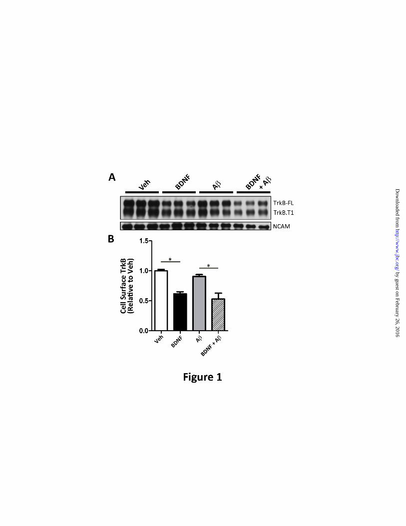
We found that A oligomers reduce BDNF-
GFP-containing vesicle velocities within axons
by 38.4±.13.4% (p<0.01) relative to vehicle-
treated neurons (Fig. 2A). The average vesicle
velocity was 2.81±0.253 m/s in vehicle-treated
axons whereas the average vesicle velocity in
A-treated axons was 1.73±0.378 m/s. This is
in agreement with our previous study which
demonstrated that the velocities of BDNF-
containing endosomes were markedly reduced in
Tg2576 neurons when compared to wild-type
neurons (22).
Examination of the vesicle velocity
distribution revealed that the presence of A
oligomers significantly decreased the percentage
of endosomes with velocities >2 um/s, with the
majority of the endosome velocities being <1
um/s (Fig. 2B). Additionally, representative
time-lapse images reveal that in the presence of
A, the BDNF-GFP signal that can be visualized
within trafficking vesicles is greatly reduced
(Fig. 2C). These results suggest that A
oligomers can disrupt retrograde trafficking by
affecting both the vesicle velocities of BDNF-
containing endosomes and the amount of TrkB
that is contained within the transported
endosomes.
-amyloid impairs BDNF-dependent
retrograde signaling. The signaling endosome
hypothesis implies that if the retrograde
trafficking of BDNF-GFP-positive endosomes is
impaired then the propagation of BDNF
retrograde signaling will also be impaired. To
test this hypothesis, BDNF-GFP was added to
the axonal compartment of the microfluidic
chambers, followed by assessment of ERK
activation. ERK activation was determined by
measuring the phosphorylation of ERK5 (p-
ERK5) in the soma of neurons. ERK5 is the
main ERK that is activated in response to
axonally-derived BDNF (28).
by guest on February 26, 2016http://w
ww
.jbc.org/D
ownloaded from
Ubiquitin homeostasis is important for BDNF-mediated retrograde transport
6
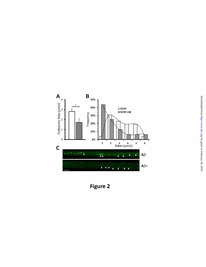
Representative images (Fig. 3) revealed that
BDNF-GFP led to robust p-ERK5 activation
within neuronal cell bodies located in the somal
compartment. This is indicated by increased p-
ERK5 labeling following BDNF-GFP, when
compared to vehicle-only neurons (Fig. 3A and
3B). However, p-ERK5 activation in response
to BDNF-GFP is not readily apparent in neurons
pre-incubated with A (Fig. 3C and 3D,
respectively). Quantification of p-ERK5
immunoreactivity revealed that BDNF treatment
increased p-ERK5 by 68.1±8.4% (*p<0.05)
when compared to vehicle-only, while p-ERK5
levels were not significantly increased by BDNF
treatment in neurons pre-incubated with A
oligomers, relative to A-only treatment (Fig.
3J). These results are consistent with a
retrograde signaling deficit due to impaired
BDNF/TrkB retrograde transport.
At higher magnification, p-ERK5 (Fig. 3F)
appeared to co-localize with the nuclear marker
TOTO-3 (Fig.3G) in a representative neuron and
suggests that p-ERK5 is translocated to the
nucleus and is consistent with the literature (28).
Additionally, BDNF-GFP immunoreactivity
decorated the outer surface of the nucleus
indicating that axonally-applied BDNF-GFP
trafficked back to the soma (Fig 3E). This
observation supports the “signaling endosome”
hypothesis for neurotrophin signaling, as it
demonstrates that ligand/receptor complexes that
originate from the axonal compartment undergo
retrograde transport to the soma.
Next, we measured the extent of p-ERK5
translocation to the somal compartment by
Western blot analysis. It revealed that somal p-
ERK5 levels (normalized to total ERK5)
increase almost three-fold (*p=0.05) in response
to axonally-applied BDNF (Figure 3I and 3K).
However, in cultures pre-incubated with A, p-
ERK5 translocation was not observed. Although
p-ERK5 levels were lower following BDNF
treatment in neurons pre-incubated with A, it
was not significant. The assessment of p-ERK5
translocation by Western blot analysis is
consistent with our immunocytochemical results
and together supports the notion that A impairs
BDNF-dependent retrograde signaling.
Next, CREB-dependent gene transcription
was assessed to further validate the hypothesis
that soluble A impairs BDNF-dependent
retrograde signaling. CREB-mediated gene
transcription was measured by quantifying GFP
within neurons transfected with a CRE-GFP
reporter plasmid (Stratagene). CRE-GFP is a
cAMP response element (CRE) fused to GFP
that is used to monitor downstream cAMP/PKA
signaling. In neurons transfected with CRE-
GFP, axonal BDNF treatment led to a robust
increase in GFP immunoreactivity within soma
(35.9±.4.73%, *p<0.01) when compared to
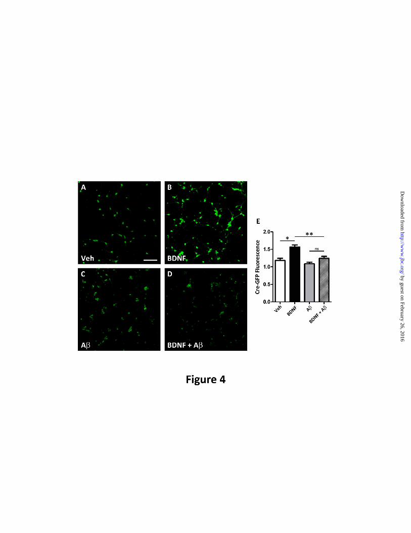
vehicle-only (Fig. 4A, 4B, 4E). GFP
immunoreactivity was normalized with the
neuronal marker, III-tubulin. In contrast, in
neurons pre-incubated with oligomers, no
significant increase in GFP immunoreactivity
was observed following BDNF (Fig. 4C, 4D,
4E). Thus, A oligomers also impair axonal
BDNF-mediated CREB-dependent gene
activation. This impairment was also observed
in APP (Tg2576) neurons (data not shown).
Taken together, these results demonstrate that
A oligomers cause deficits in BDNF/TrkB
retrograde signaling by affecting the trafficking
of BDNF-GFP-containing endosomes, which in
turn, results in the decreased retrograde transport
and activation of ERK5 and CREB-dependent
transcription that is necessary to maintain proper
synaptic function and neuronal survival.
Impaired deubiquitination mimics the effects
of amyloid on BDNF retrograde signaling.
Previous studies suggested that A impairs
proteasome function and deubiquitinating
enzyme activity, which in turn, can impair
receptor sorting to multi-vesicular bodies
(MVBs) and neurotrophin receptor trafficking
(35,52). Because MVBs represent the
endosomal compartment that mediates sustained
neurotrophin signaling from axon terminals to
the soma (25,53), it suggests that the
BDNF/TrkB trafficking deficits may be caused
by A impairing deubiquitinating activity. To
test this hypothesis, we assessed whether
inhibiting deubiquitinating activity could mimic
the effect of A oligomers on retrograde
transport. Deubiquitinating activity was
inhibited with LDN (LDN-57444, EMD), a cell-
permeable UCH-L1-specific inhibitor. As
predicted, inhibiting deubiquitination with LDN
impaired BDNF retrograde signaling as assessed
by guest on February 26, 2016http://w
ww
.jbc.org/D
ownloaded from
Ubiquitin homeostasis is important for BDNF-mediated retrograde transport
7
by measuring p-ERK5 activation (Fig. 5A and
5B). BDNF treatment led to a 45.7±10.3%
(*p<0.05) increase in nuclear p-ERK5 levels
(Fig. 5B). However, in neurons pre-treated with
LDN, the addition of BDNF did not lead to an
increase in p-ERK5 levels. Interestingly, basal
p-ERK5 levels were lower when UCH-L1 was
inhibited. We attribute this decrease to the
importance of ubiquitin turnover to synaptic
function. Nonetheless, these results support the
hypothesis that oligomers may affect TrkB
retrograde signaling by impairing UCH-L1
activity.
Similarly, LDN pre-treatment impaired the
translocation of p-ERK5 to the soma following
BDNF as assessed by Western blot analysis
(Fig. 3I and 3K). Following BDNF treatment p-
ERK5 translocation the soma was not observed.
These results were similar to A-treated neurons
and support the hypothesis that A affects
BDNF/TrkB signaling by impairing
deubiquitinating activity.
Additionally, like A, LDN did not impair
TrkB endocytosis. Using cell surface
biotinylation assays, we show that the addition
of BDNF led to a 45.7±10.2%, (*p<0.05).
decrease in cell surface TrkB levels when
compared to vehicle (Figure 5C and 5D). In
neurons pre-incubated with LDN, we observed a
similar decrease in TrkB (44.3±8.56%,*p<0.05).
Taken together, these results indicate that LDN
mimics A oligomers by not inhibiting TrkB
internalization, but impairing downstream
BDNF retrograde signaling.
UCH-L1 rescues TrkB retrograde trafficking
deficits caused by A. Next, we investigated
whether BDNF-mediated transport deficits
induced by A could be rescued by increasing
UCH-L1 levels using a TAT-UCH-L1-HA
construct as described previously (42). UCH-L1
treatment rescued BDNF-GFP retrograde
trafficking deficits caused by A (Fig. 6). In the
presence of A oligomers, BDNF-GFP levels in
the soma were 60.3±7.1% (*p=0.003) lower than
vehicle-treated neurons. However, in the
presence of UCH-L1 alone, BDNF-GFP levels
were 92.6±12.0% of vehicle-only. UCH-L1
rescued the Aβ-induced deficit in BDNF-GFP
levels in the soma to 86.3±14.1% of the vehicle-
treated neurons (*p=0.04).
Thus, we demonstrate that -mediated
deficits in retrograde transport can be rescued by
UCH-L1. Taken together with our LDN data
(Fig. 5), these results demonstrate that
modulating ubiquitin homeostasis via UCH-L1
impacts BDNF-mediated retrograde trafficking.
UCH-L1 is decreased in the hippocampus in
APP-Tg2576 mice and in AD. Next, we
determined if UCH-L1 levels are also affected in
an AD transgenic mouse model. We measured
UCH-L1 protein levels in the hippocampus or
cortex of 15 month-old wild-type or Tg2576
mice. We found that hippocampal but not
cortical UCH-L1 levels were significantly
decreased in Tg2576 mice relative to age-
matched wild-type (Fig. 7A and 7B)(*p<0.03).
Lastly, we investigated whether the decrease
in UCH-L1 translates to the in vivo condition in
humans. We compared UCH-L1 gene
expression levels in the hippocampus and
superior frontal cortex (BA9/46) in AD brain
versus age-matched cognitively intact controls.
UCHL-1 gene expression data was obtained
from a microarray database as described
previously (44), and revealed that UCH-L1 gene
expression was significantly reduced in the
hippocampal regions of AD cases versus aged-
matched controls (Fig. 7B, p<0.05). The
decrease in UCH-L1 expression levels was
hippocampus-specific, as we did not detect a
difference in UCH-L1 expression in the cortical
region assessed.
DISCUSSION
Overall, our results demonstrate that soluble
A impairs BDNF/TrkB retrograde axonal
trafficking and signaling. Building on our
previous findings that Aβ oligomers cause a net
decrease in the amount of BDNF/TrkB
trafficked back to soma (22), this is not due to
A oligomers affecting the internalization of
TrkB. While previous studies have
demonstrated that A can alter cell surface
receptor internalization (for example, of AMPA
and NMDA receptors)(49,50), our data
demonstrate that A does not affect the
internalization of TrkB and suggested that
Amediated trafficking deficits were
downstream of receptor internalization.
by guest on February 26, 2016http://w
ww
.jbc.org/D
ownloaded from
Ubiquitin homeostasis is important for BDNF-mediated retrograde transport
8
Using a novel microfluidic chamber that
facilitates the study of axonal transport, we
found that A oligomers cause a decrease in the
amount of BDNF-GFP that is found within
endosomes that undergo retrograde axonal
transport. In addition, we found that the average
velocity of BDNF-GFP-containing endosomes
was decreased in the presence of Aβ, with the
distribution of the vesicle velocities shifted to
ones with lower velocities. These data suggest
that soluble Aβ impairs the sorting of
BDNF/TrkB to MVBs, the endosomal
compartment that mediates TrkB retrograde
axonal trafficking, and also reduces the velocity
of trafficking MVBs. Together, these deficits
contribute to impaired BDNF/TrkB retrograde
transport. It is of note that early in the
progression of AD, and prior to A deposition,
when A oligomers are likely present, enlarged
endosomal structures can be detected in neurons
(54). It is tempting to speculate that the
decrease in vesicle velocity is attributed to
enlarged MVBs. Because proteins sorted to
MVBs are also degraded in a lysosome-
dependent process (33), our data are also
consistent with the finding that A impairs
lysosome-mediated degradation of TrkB (35).
We also demonstrated that BDNF/TrkB
axonal trafficking deficits induced by Awere
accompanied by impaired downstream
BDNF/TrkB signaling, notably impaired nuclear
translocation of p-ERK5 and down-regulated
CREB-mediated signaling events. These results
reveal that the presence of soluble Aβ impairs
retrograde trafficking, resulting in diminished
signaling between axons and cell bodies,
supporting the signaling endosome hypothesis
that describes how cellular signals that are
initiated at axon terminals undergo retrograde
transport and are propagated back to the soma
(55). Furthermore, the disruption by Amay be
due to a generalized impairment in trafficking as
the trafficking of other organelles/cargos are
also impaired by A (47,56-59).
Our data reveal that a mechanism underlying
the retrograde trafficking deficits in the presence
of Aβ involves altered ubiquitin homeostasis.
Ubiquitin is central for proteasome-dependent
protein turnover as well as for intracellular
trafficking of cargoes including many receptors
(i.e. glutamate, neurotrophins) that are
fundamental for synaptic remodeling and
plasticity (60). Although ubiquitin can be
synthesized de novo, the bulk of the cellular
ubiquitin pool is derived from ubiquitin that is
recovered from deubiquitinating enzymes (61).
Thus, ubiquitin levels can be regulated by
modulating deubiquitinating enzyme levels or
activity of deubiquitinating enzymes such as
UCH-L1 (42,62). Remarkably, UCH-L1
represents 1-2% of the total protein within the
brain and thus is an important regulator of brain
ubiquitin levels (62-64).
Ubiquitination regulates many important
processes including the targeting and delivery of
receptors to MVBs, including trophic factor
receptors (65-67). For example, ubiquitination
mediates the delivery of the well-studied
epidermal growth factor receptor (EGFR) to
MVBs for its degradation within lysosomes
(33). In the case of Trk receptors, ubiquitination
may regulate its endocytic trafficking to MVBs
for sustained retrograde signaling (34,53,68).
Therefore, we tested whether disrupting the
ubiquitin recycling pathway which regulates
cellular ubiquitin levels can lead to BDNF-
dependent retrograde transport deficits. Indeed,
we found that BDNF/TrkB retrograde trafficking
and signaling could be affected by manipulating
deubiquitinating activity by either inhibiting or
increasing UCH-L1. Specifically, inhibiting
UCH-L1 with LDN resulted in retrograde
trafficking deficits parallel to those found
induced by soluble Aβ, and retrograde
trafficking deficits due to Aβ could be rescued
by increasing cellular UCH-L1 levels.
Although ubiquitination mediates the
internalization of numerous receptors, we found
that neither A nor impairing ubiquitin-
recycling with LDN affected TrkB
internalization. Our data support the finding that
TrkB ubiquitination is not a prerequisite for its
internalization (34), and may only regulate its
endocytic fate (69,70). Taken together, these
results suggest that A impairs the retrograde
trafficking of TrkB by affecting ubiquitin
homeostasis via UCH-L1 at a step that is
downstream from receptor internalization.
Further, our data suggest that ubiquitin
homeostasis may be impaired in the
hippocampus in AD, in both Tg2576 mouse
by guest on February 26, 2016http://w
ww
.jbc.org/D
ownloaded from
Ubiquitin homeostasis is important for BDNF-mediated retrograde transport
9
model of AD and in the human brain. We
demonstrate that in Tg2576 mice, hippocampal
but not cortical UCH-L1 protein levels are
reduced compared to wild-type littermates,
similar to the findings in the brain of APP/PS1
mice at 4-6 months of age suggesting that the
decrease in UCH-L1 follows the development of
pathology (42). In parallel, we demonstrate that
hippocampal but not cortical UCH-L1 gene
expression is decreased in the AD brain relative
to age-matched cognitively intact cases. The
reduced availability of UCH-L1 in the AD brain
likely impairs neurotrophin signaling in vivo,
based on our in vitro data which found that
inhibiting UCH-L1 caused deficits in
TrkB/BDNF retrograde trafficking and
signaling. In support of our hypothesis that Aβ
itself directly affects UCH-L1 levels, a decrease
in monomeric ubiquitin levels due to Aβ is
reversed by increasing UCH-L1 levels in
hippocampal slices (42).
These data add to the growing evidence that
disrupted ubiquitin homeostasis is an important
aspect of AD pathobiology, with previous
studies demonstrating that impaired
deubiquitination alters synaptic protein
distribution and spine morphology, and causes
neurodegeneration (62,71), which are salient
features in AD. Altered ubiquitin homeostasis
may contribute to generalized axonal transport
deficits observed in AD. Inducing lysosome
dysfunction impairs axonal retrograde transport
of late endosomes and lysosomes and leads to
AD-like axonal pathology (72). Because the
sorting of proteins to lysosomes is ubiquitin-
dependent (73), it suggests that by altering
ubiquitin homeostasis, A can trigger lysosome
dysfunction and the observed transport deficits.
Furthermore, we and others have found that A
directly affects mitochondrial transport and may
be due to defective fission/fusion (74,75), which
is regulated by ubiquitination (76). Thus, it
suggests that ubiquitination/deubiquitination
plays a vital role in regulating axonal transport.
Lastly, balancing ubiquitination/deubiquitination
may also affect A production as APP
ubiquitination inhibits APP endocytosis and
promotes the non-amyloidogenic processing
(77).
Our data plus a growing body of evidence
suggests that in AD there may be a general
defect in intracellular trafficking. Our results
describe a novel mechanism by which A can
impair ubiquitin homeostasis that leads to
endosomal axonal retrograde transport deficits,
that impairs neurotrophin signaling, and
contributes to impaired synaptic plasticity. As
Aβ accumulates, one of the consequences may
be impaired intracellular trafficking of cellular
components that depend on ubiquitin-
conjugation for signal transduction and protein
sorting and degradation. Defective trafficking in
the etiology of AD is supported by the recent
identification of GWAS-AD-linked
polymorphisms that encode proteins linked to
endosome function, e.g., PICALM, BIN1 (78).
Therefore therapeutics aimed at modulating
ubiquitin homeostasis may rescue intracellular
trafficking deficits found in AD and improve
cognition.
by guest on February 26, 2016http://w
ww
.jbc.org/D
ownloaded from
Ubiquitin homeostasis is important for BDNF-mediated retrograde transport
10
REFERENCES
1. Jack, C. R., Jr., Knopman, D. S., Jagust, W. J., Shaw, L. M., Aisen, P. S., Weiner, M. W.,
Petersen, R. C., and Trojanowski, J. Q. (2010) Lancet Neurol 9, 119-128
2. Naslund, J., Haroutunian, V., Mohs, R., Davis, K. L., Davies, P., Greengard, P., and Buxbaum, J.
D. (2000) Jama 283, 1571-1577
3. Lue, L. F., Kuo, Y. M., Roher, A. E., Brachova, L., Shen, Y., Sue, L., Beach, T., Kurth, J. H.,
Rydel, R. E., and Rogers, J. (1999) Am J Pathol 155, 853-862
4. McLean, C. A., Cherny, R. A., Fraser, F. W., Fuller, S. J., Smith, M. J., Beyreuther, K., Bush, A.
I., and Masters, C. L. (1999) Ann Neurol 46, 860-866
5. Walsh, D. M., Klyubin, I., Fadeeva, J. V., Cullen, W. K., Anwyl, R., Wolfe, M. S., Rowan, M. J.,
and Selkoe, D. J. (2002) Nature 416, 535-539
6. Cleary, J. P., Walsh, D. M., Hofmeister, J. J., Shankar, G. M., Kuskowski, M. A., Selkoe, D. J.,
and Ashe, K. H. (2005) Nat Neurosci 8, 79-84
7. Hsiao, K., Chapman, P., Nilsen, S., Eckman, C., Harigaya, Y., Younkin, S., Yang, F., and Cole,
G. (1996) Science 274, 99-102
8. Hsia, A. Y., Masliah, E., McConlogue, L., Yu, G. Q., Tatsuno, G., Hu, K., Kholodenko, D.,
Malenka, R. C., Nicoll, R. A., and Mucke, L. (1999) Proc Natl Acad Sci U S A 96, 3228-3233
9. Mucke, L., Masliah, E., Yu, G. Q., Mallory, M., Rockenstein, E. M., Tatsuno, G., Hu, K.,
Kholodenko, D., Johnson-Wood, K., and McConlogue, L. (2000) J Neurosci 20, 4050-4058
10. Lesne, S., Koh, M. T., Kotilinek, L., Kayed, R., Glabe, C. G., Yang, A., Gallagher, M., and Ashe,
K. H. (2006) Nature 440, 352-357
11. Tyler, W. J., Alonso, M., Bramham, C. R., and Pozzo-Miller, L. D. (2002) Learn Mem 9, 224-237
12. Martinez, A., Alcantara, S., Borrell, V., Del Rio, J. A., Blasi, J., Otal, R., Campos, N., Boronat,
A., Barbacid, M., Silos-Santiago, I., and Soriano, E. (1998) J Neurosci 18, 7336-7350
13. Minichiello, L., Korte, M., Wolfer, D., Kuhn, R., Unsicker, K., Cestari, V., Rossi-Arnaud, C.,
Lipp, H. P., Bonhoeffer, T., and Klein, R. (1999) Neuron 24, 401-414
14. Xu, B., Zang, K., Ruff, N. L., Zhang, Y. A., McConnell, S. K., Stryker, M. P., and Reichardt, L.
F. (2000) Neuron 26, 233-245
15. Patterson, S. L., Abel, T., Deuel, T. A., Martin, K. C., Rose, J. C., and Kandel, E. R. (1996)
Neuron 16, 1137-1145
16. Korte, M., Carroll, P., Wolf, E., Brem, G., Thoenen, H., and Bonhoeffer, T. (1995) Proc Natl
Acad Sci U S A 92, 8856-8860
17. Pozzo-Miller, L. D., Gottschalk, W., Zhang, L., McDermott, K., Du, J., Gopalakrishnan, R., Oho,
C., Sheng, Z. H., and Lu, B. (1999) J Neurosci 19, 4972-4983
18. Genoud, C., Knott, G. W., Sakata, K., Lu, B., and Welker, E. (2004) J Neurosci 24, 2394-2400
19. Causing, C. G., Gloster, A., Aloyz, R., Bamji, S. X., Chang, E., Fawcett, J., Kuchel, G., and
Miller, F. D. (1997) Neuron 18, 257-267
20. Tong, L., Balazs, R., Thornton, P. L., and Cotman, C. W. (2004) J Neurosci 24, 6799-6809
21. Garzon, D. J., and Fahnestock, M. (2007) J Neurosci 27, 2628-2635
22. Poon, W. W., Blurton-Jones, M., Tu, C. H., Feinberg, L. M., Chabrier, M. A., Harris, J. W., Jeon,
N. L., and Cotman, C. W. (2011) Neurobiol Aging 32, 821-833
23. Peng, S., Wuu, J., Mufson, E. J., and Fahnestock, M. (2005) J Neurochem 93, 1412-1421
24. Watson, F. L., Heerssen, H. M., Moheban, D. B., Lin, M. Z., Sauvageot, C. M., Bhattacharyya,
A., Pomeroy, S. L., and Segal, R. A. (1999) J Neurosci 19, 7889-7900
25. Valdez, G., Akmentin, W., Philippidou, P., Kuruvilla, R., Ginty, D. D., and Halegoua, S. (2005) J
Neurosci 25, 5236-5247
26. Grimes, M. L., Zhou, J., Beattie, E. C., Yuen, E. C., Hall, D. E., Valletta, J. S., Topp, K. S.,
LaVail, J. H., Bunnett, N. W., and Mobley, W. C. (1996) J Neurosci 16, 7950-7964
by guest on February 26, 2016http://w
ww
.jbc.org/D
ownloaded from
Ubiquitin homeostasis is important for BDNF-mediated retrograde transport
11
27. Delcroix, J. D., Valletta, J. S., Wu, C., Hunt, S. J., Kowal, A. S., and Mobley, W. C. (2003)
Neuron 39, 69-84
28. Watson, F. L., Heerssen, H. M., Bhattacharyya, A., Klesse, L., Lin, M. Z., and Segal, R. A.
(2001) Nat Neurosci 4, 981-988
29. Heerssen, H. M., Pazyra, M. F., and Segal, R. A. (2004) Nat Neurosci 7, 596-604
30. Haglund, K., Di Fiore, P. P., and Dikic, I. (2003) Trends Biochem Sci 28, 598-603
31. Thien, C. B., and Langdon, W. Y. (2001) Nat Rev Mol Cell Biol 2, 294-307
32. Miyake, S., Lupher, M. L., Jr., Druker, B., and Band, H. (1998) Proc Natl Acad Sci U S A 95,
7927-7932
33. Levkowitz, G., Waterman, H., Zamir, E., Kam, Z., Oved, S., Langdon, W. Y., Beguinot, L.,
Geiger, B., and Yarden, Y. (1998) Genes Dev 12, 3663-3674
34. Arevalo, J. C., Waite, J., Rajagopal, R., Beyna, M., Chen, Z. Y., Lee, F. S., and Chao, M. V.
(2006) Neuron 50, 549-559
35. Almeida, C. G., Takahashi, R. H., and Gouras, G. K. (2006) J Neurosci 26, 4277-4288
36. De Felice, F. G., Wu, D., Lambert, M. P., Fernandez, S. J., Velasco, P. T., Lacor, P. N., Bigio, E.
H., Jerecic, J., Acton, P. J., Shughrue, P. J., Chen-Dodson, E., Kinney, G. G., and Klein, W. L.
(2008) Neurobiol Aging 29, 1334-1347
37. Pang, P. T., Teng, H. K., Zaitsev, E., Woo, N. T., Sakata, K., Zhen, S., Teng, K. K., Yung, W. H.,
Hempstead, B. L., and Lu, B. (2004) Science 306, 487-491
38. Kohara, K., Kitamura, A., Morishima, M., and Tsumoto, T. (2001) Science 291, 2419-2423
39. Hartmann, M., Heumann, R., and Lessmann, V. (2001) Embo J 20, 5887-5897
40. Taylor, A. M., Rhee, S. W., Tu, C. H., Cribbs, D. H., Cotman, C. W., and Jeon, N. L. (2003)
Langmuir 19, 1551-1556
41. Cribbs, D. H., Kreng, V. M., Anderson, A. J., and Cotman, C. W. (1996) Neuroscience 75, 173-
185
42. Gong, B., Cao, Z., Zheng, P., Vitolo, O. V., Liu, S., Staniszewski, A., Moolman, D., Zhang, H.,
Shelanski, M., and Arancio, O. (2006) Cell 126, 775-788
43. Blurton-Jones, M., Kuan, P. N., and Tuszynski, M. H. (2004) J Comp Neurol 468, 347-360
44. Berchtold, N. C., Cribbs, D. H., Coleman, P. D., Rogers, J., Head, E., Kim, R., Beach, T., Miller,
C., Troncoso, J., Trojanowski, J. Q., Zielke, H. R., and Cotman, C. W. (2008) Proc Natl Acad Sci
U S A 105, 15605-15610
45. Salehi, A., Delcroix, J. D., Belichenko, P. V., Zhan, K., Wu, C., Valletta, J. S., Takimoto-Kimura,
R., Kleschevnikov, A. M., Sambamurti, K., Chung, P. P., Xia, W., Villar, A., Campbell, W. A.,
Kulnane, L. S., Nixon, R. A., Lamb, B. T., Epstein, C. J., Stokin, G. B., Goldstein, L. S., and
Mobley, W. C. (2006) Neuron 51, 29-42
46. Her, L. S., and Goldstein, L. S. (2008) J Neurosci 28, 13662-13672
47. Vossel, K. A., Zhang, K., Brodbeck, J., Daub, A. C., Sharma, P., Finkbeiner, S., Cui, B., and
Mucke, L. (2010) Science 330, 198
48. Escandon, E., Soppet, D., Rosenthal, A., Mendoza-Ramirez, J. L., Szonyi, E., Burton, L. E.,
Henderson, C. E., Parada, L. F., and Nikolics, K. (1994) J Neurosci 14, 2054-2068
49. Snyder, E. M., Nong, Y., Almeida, C. G., Paul, S., Moran, T., Choi, E. Y., Nairn, A. C., Salter,
M. W., Lombroso, P. J., Gouras, G. K., and Greengard, P. (2005) Nat Neurosci 8, 1051-1058
50. Hsieh, H., Boehm, J., Sato, C., Iwatsubo, T., Tomita, T., Sisodia, S., and Malinow, R. (2006)
Neuron 52, 831-843
51. Taylor, A. M., Blurton-Jones, M., Rhee, S. W., Cribbs, D. H., Cotman, C. W., and Jeon, N. L.
(2005) Nat Methods 2, 599-605
52. Moises, T., Wuller, S., Saxena, S., Senderek, J., Weis, J., and Kruttgen, A. (2009) Biochem
Biophys Res Commun 387, 360-364
53. Philippidou, P., Valdez, G., Akmentin, W., Bowers, W. J., Federoff, H. J., and Halegoua, S.
(2011) Proc Natl Acad Sci U S A 108, 852-857
by guest on February 26, 2016http://w
ww
.jbc.org/D
ownloaded from
Ubiquitin homeostasis is important for BDNF-mediated retrograde transport
12
54. Cataldo, A. M., Petanceska, S., Terio, N. B., Peterhoff, C. M., Durham, R., Mercken, M., Mehta,
P. D., Buxbaum, J., Haroutunian, V., and Nixon, R. A. (2004) Neurobiol Aging 25, 1263-1272
55. Howe, C. L., and Mobley, W. C. (2004) J Neurobiol 58, 207-216
56. Decker, H., Lo, K. Y., Unger, S. M., Ferreira, S. T., and Silverman, M. A. (2010) J Neurosci 30,
9166-9171
57. Hiruma, H., Katakura, T., Takahashi, S., Ichikawa, T., and Kawakami, T. (2003) J Neurosci 23,
8967-8977
58. Kim, H. J., Park, J. W., Byun, J. H., Poon, W. W., Cotman, C. W., Fowlkes, C. C., and Jeon, N.
L. (2012) ACS Chemical Neuroscience 3, 433-438
59. Rui, Y., Tiwari, P., Xie, Z., and Zheng, J. Q. (2006) J Neurosci 26, 10480-10487
60. Yi, J. J., and Ehlers, M. D. (2007) Pharmacol Rev 59, 14-39
61. Wilkinson, K. D. (2000) Semin Cell Dev Biol 11, 141-148
62. Cartier, A. E., Djakovic, S. N., Salehi, A., Wilson, S. M., Masliah, E., and Patrick, G. N. (2009) J
Neurosci 29, 7857-7868
63. Wilkinson, K. D., Lee, K. M., Deshpande, S., Duerksen-Hughes, P., Boss, J. M., and Pohl, J.
(1989) Science 246, 670-673
64. Osaka, H., Wang, Y. L., Takada, K., Takizawa, S., Setsuie, R., Li, H., Sato, Y., Nishikawa, K.,
Sun, Y. J., Sakurai, M., Harada, T., Hara, Y., Kimura, I., Chiba, S., Namikawa, K., Kiyama, H.,
Noda, M., Aoki, S., and Wada, K. (2003) Hum Mol Genet 12, 1945-1958
65. Alwan, H. A., van Zoelen, E. J., and van Leeuwen, J. E. (2003) J Biol Chem 278, 35781-35790
66. Devon, R. S., Orban, P. C., Gerrow, K., Barbieri, M. A., Schwab, C., Cao, L. P., Helm, J. R.,
Bissada, N., Cruz-Aguado, R., Davidson, T. L., Witmer, J., Metzler, M., Lam, C. K., Tetzlaff, W.,
Simpson, E. M., McCaffery, J. M., El-Husseini, A. E., Leavitt, B. R., and Hayden, M. R. (2006)
Proc Natl Acad Sci U S A 103, 9595-9600
67. Longva, K. E., Blystad, F. D., Stang, E., Larsen, A. M., Johannessen, L. E., and Madshus, I. H.
(2002) J Cell Biol 156, 843-854
68. Lauwers, E., Jacob, C., and Andre, B. (2009) J Cell Biol 185, 493-502
69. Georgieva, M. V., de Pablo, Y., Sanchis, D., Comella, J. X., and Llovera, M. (2011) J Neurochem
117, 479-493
70. Geetha, T., and Wooten, M. W. (2008) Traffic 9, 1146-1156
71. Ryu, K. Y., Garza, J. C., Lu, X. Y., Barsh, G. S., and Kopito, R. R. (2008) Proc Natl Acad Sci U
S A 105, 4016-4021
72. Lee, S., Sato, Y., and Nixon, R. A. (2011) J Neurosci 31, 7817-7830
73. Piper, R. C., and Lehner, P. J. (2011) Trends Cell Biol 21, 647-655
74. Kim, H. J., Park, J. W., Byun, J. H., Poon, W. W., Cotman, C. W., Fowlkes, C. C., and Jeon, N.
L. (2012) ACS Chemical Neuroscience 3, 433-438
75. Wang, X., Perry, G., Smith, M. A., and Zhu, X. (2010) Neurodegener Dis 7, 56-59
76. Park, Y. Y., Lee, S., Karbowski, M., Neutzner, A., Youle, R. J., and Cho, H. (2010) J Cell Sci
123, 619-626
77. Watanabe, T., Hikichi, Y., Willuweit, A., Shintani, Y., and Horiguchi, T. (2012) J Neurosci 32,
3352-3365
78. Guerreiro, R. J., and Hardy, J. (2011) Biochem Soc Trans 39, 910-916
by guest on February 26, 2016http://w
ww
.jbc.org/D
ownloaded from
Ubiquitin homeostasis is important for BDNF-mediated retrograde transport
13
Acknowledgements-We thank Meredith Chabrier for critical reading of the manuscript.
FOOTNOTES
*This work was supported by NIH grants AG016573 (C.W.C.), AG000538 (C.W.C), and AG00096
(A.J.C.). C.W.C. owns interest in Xona Microfluidics LLC which began selling the microfluidics
devices. 1Institute for Memory Impairments and Neurological Disorders, University of California, Irvine, Irvine,
California 92697, USA 2Department of Pathology and Taub Institute
Columbia University, New York, NY 10032, USA 3These authors contributed equally.
4The abbreviations used are: APP, Amyloid precursor protein; AD, Alzheimer disease; Aβ, β-amyloid;
CRE-GFP, CREB responsive element fused to GFP; DIV, days in vitro; GFAP, glial fibrillary acidic
protein; HFIP, hexafluoroisopropanol; IUCAC, institutional use and care of animals committee; LDN,
LDN-57444; LTP, long-term potentiation; MVB, multivesicular bodies; PS1, presenilin 1; RIPA,
radioimmunoprecipitation assay; TAT-HA-UCH-L1, transduction domain of HIV-transactivator protein
(TAT) and hemagglutinin (HA)fused to UCH-L1, Tg2576, AD transgenic mouse line; TrkB,
tropomyosin-receptor kinase B; UCH-L1, ubiquitin-c-terminal hydrolase L1.
FIGURE LEGENDS
FIGURE 1. A oligomers do not affect the internalization of BDNF receptors. (A). Cell surface
biotinylation was employed to measure TrkB levels at the cell surface following BDNF treatment (50
ng/ml, 30 min) as described in Experimental Procedures. BDNF addition led to a decrease in cell surface
levels of full length TrkB (TrkB-FL) and truncated TrkB (TrkB-T1). Pre-incubation with A oligomers
(24h) does not impair TrkB-FL or TrkB-T1 internalization. BDNF specifically caused internalization of
BDNF receptors, but not the internalization of neural cell adhesion molecule (NCAM). (B). Cell surface
TrkB-FL was quantitated using ImageJ (NIH). The mean ± S.E. represents TrkB-FL levels normalized to
vehicle-treated neurons (n=3). Following BDNF treatment, we found that 38.3±3.40 % of TrkB-FL was
internalized (black) when compared to vehicle (white)(**p<0.001). In the presence of A, BDNF led to
41.5 ±9.80% of TrkB-FL internalized (hashes) when compared to A-only (grey)(*p<0.05).
FIGURE 2. A oligomers impair the trafficking of BDNF-GFP endosomes. (A). In the presence of
A, the average velocity of BDNF-GFP-containing endosomes was 1.73±0.378 m/s. This represented a
38.4±13.4% (*p<0.01) decrease when compared to the average velocity of endosomes in the absence of
Velocities of BDNF-GFP positive endosomes were determined as described previously (22). (B).
Distribution plot of the vesicle velocities reveal that in the presence of A oligomers, the percentage of
vesicle velocities >2 mm/s was greatly reduced (grey) and the majority of the vesicle velocity was <1
m/s. (C). Representative time lapse image of BDNF-GFP containing endosomes demonstrate that the
amount of BDNF/TrkB complex that undergoes retrograde transport of the (from right to left). Scale
bar=10m.
FIGURE 3. A oligomers impair the trafficking of the signaling endosome complex including p-
ERK5. Microfluidic devices were employed to measure the retrograde transport dependent ERK5
activation. Representative image demonstrating p-ERK5 levels (red) in the somal compartment of
microfluidic chamber of vehicle-treated neurons (A) and following BDNF treatment (B), in the presence
by guest on February 26, 2016http://w
ww
.jbc.org/D
ownloaded from
Ubiquitin homeostasis is important for BDNF-mediated retrograde transport
14
of A oligomers only (C), and after BDNF treatment (D). Scale bar=200m. (E-H) Representative
image of a neuron demonstrating the co-localization of BDNF-GFP (green) on the outer surface of the
nucleus (blue). Also, p-ERK5(red) co-localized with the nucleus suggesting that the BDNF-mediated
retrograde signal i.e. the signaling endosome undergoes retrograde transport from the axonal compartment
to the soma and specifically the nucleus. Scale bar=20m. (I) Western blot analysis of p-ERK5 and T-
ERK5 isolated from the somal compartment of microfluidics devices as described in Experimental
Procedures.(J) Somal p-ERK5 was quantitated as described in Experimental Procedures. BDNF leads to
a 68.1±8.4% (*p<0.05) increase in p-ERK5. However, in neurons pre-incubated with A oligomers, p-
ERK5 levels are not increased following axonal BDNF treatment. (K) Quantification of p-ERK5 relative
to total ERK5 levels in the somal compartment following BDNF treatment. BDNF leads to a 284%
increase in p-ERK5. However, in the presence of A oligomers, BDNF does not lead to increased p-
ERK5. Also, a UCH-L1 inhibitor (LDN-57444) was used to inhibit deubiquitinating activity and
revealed that it could mimic the effect of A oligomers in impairing BDNF-mediated retrograde
signaling.
FIGURE 4. A oligomers lead to decreased CREB-dependent gene expression. Microfluidic devices
were used to assess CREB-mediated gene expression. Rat primary neurons (E18) were transfected with a
CRE-GFP reporter construct to assess CREB-mediated gene activation. At 7DIV, the axonal
compartment was treated with BDNF (50 ng/ml, 2h) and the chambers were processed for
immunochemical analysis as described in Experimental Procedures using polyclonal anti-GFP (Life
Technologies) to measure CRE-GFP levels and were normalized to the neuronal marker, BIII-tubulin
(red). A. Representative image of CRE-GFP levels (green) within the somal compartment and co-
imaged with the neuronal marker, BIII-tubulin (red) in vehicle-treated neurons, in neurons treated with
BDNF B. C. In the presence of Aoligomers, baseline levels of CRE-GFP are not significantly reduced
when compared to vehicle. D. In the presence of A oligomers, the increase in the amount of CRE-GFP
is greatly reduced. E. CRE-GFP levels were quantified and mean±S.E. represents n=4 and demonstrate
that BDNF treatment to the axonal compartment led to a 35.9±4.73 (*p<0.01) increase in somal CRE-
GFP immunoreactivity when compared to vehicle (white). However, in the presence of A oligomers,
CRE-GFP was only increased by 14.6±5.23%, when compared to A oligomer only cells, but this was not
significant (p=0.076). Therefore, in the presence of A axonal BDNF leads to reduced CRE-GFP
immunoreactivity when compared to vehicle treated with BDNF (**p<0.01). Scale bar=200 m.
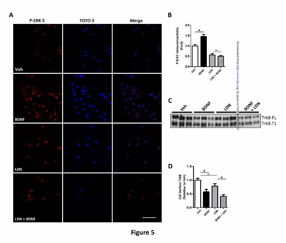
FIGURE 5. The UCH-L1 inhibitor LDN mimics the effect of Aoligomers on BDNF-dependent
retrograde signaling. To assess the effect of LDN on BDNF-dependent retrograde signaling, we
measured p-ERK5 activation in the presence of LDN. A. Representative images that demonstrate that
BDNF led to an increase in somal p-ERK5 but the UCH-L1 inhibitor, LDN, led to decreased basal somal
p-ERK5 and in the presence of BDNF, the nuclear translocation of p-ERK5 is not detected. P-ERK5
immunoreactivity was normalized to the nuclear counterstain, TOTO-3. B. Quantification of somal p-
ERK5 levels demonstrate that while BDNF treatment (black) leads to a 45.7±10.2% (*p<0.05) increase in
p-ERK5 when compared to vehicle (white), in neurons pre-incubated with LDN (grey), we do not observe
an increase in somal p-ERK5 following BDNF (hashes). C. Cell surface biotinylation assays were
employed to determine the effect of LDN on TrkB internalization and demonstrate that LDN does not
affect the internalization of full-length TrkB (TrkB-FL) or truncated TrkB (TrkB-T1). D. Quantification
of cell surface TrkB-FL demonstrate that in vehicle-treated neurons (white), the addition of BDNF (black)
led to a 45.7±10.2% (*p<0.05) decrease in cell surface TrkB levels, while in the presence of LDN (grey),
BDNF led to a 44.3±8.56 (*p<0.05) decrease in TrkB-FL. Mean±S.E. represents n=3. Scale bar=20m.
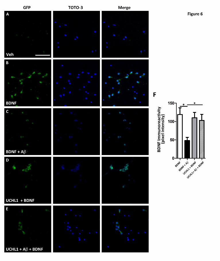
FIGURE 6. Transduction of UCH-L1 rescued A-mediated retrograde transport deficits. To
assess whether increasing UCH-L1 could rescue A-mediated transport deficits, we measured the extent
of BDNF-GFP trafficking in neurons transduced with UCH-L1. A and B. Representative images
by guest on February 26, 2016http://w
ww
.jbc.org/D
ownloaded from
Ubiquitin homeostasis is important for BDNF-mediated retrograde transport
15
demonstrate that somal levels of BDNF-GFP are increased in neurons following BDNF-GFP treatment.
GFP immunoreactivity was normalized to the nuclear marker, TOTO-3. C. Pre-treatment with A led to
decrease in BDNF-GFP when compared to vehicle-treated neurons. D. The addition of UCH-L1 alone
led to increased somal BDNF-GFP immunoreactivity. E. UCH-L1 rescues the deficit in BDNF-GFP
trafficking due to A. F. Quantification of somal BDNF levels normalized to cell number (TOTO-3
positive nuclei) reveal that A causes a 60.3±7.1% (*p=0.003) decrease in the retrograde transport of
BDNF-GFP back to soma compared to vehicle-treated neurons. In the presence of UCH-L1, BDNF-GFP
levels were 92.6±12.0% of vehicle plus BDNF. Importantly, UCH-L1 rescued the deficit in BDNF-GFP
trafficking due to ABDNF-GFP levels were 86.3±14.1% of vehicle treated and revealed that UCH-L1
restored trafficking deficits caused by A oligomers (*p=0.04). Scale bar=200m.
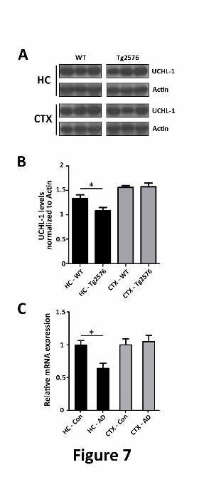
FIGURE 7. UCH-L1 is decreased in Tg2576 mice and in the Alzheimer disease brain. A. Levels of
UCH-L1 are decreased in APP-Tg2576 hippocampus. Hippocampal lysates were prepared as described
in Experimental Procedures. Protein was separated on SDS-PAGE and Western blot analysis was carried
out to determine the amount of UCH-L1 protein in wild-type and Tg2576 mouse brain. B. UCH-L1
protein levels are decreased within the hippocampus but not the cortex of 15 mo. old Tg2576 mice
(*p,0.03). UCH-L1 levels were quantitated and normalized to actin protein as described in Experimental
Procedures. C. UCHL-1 gene expression is lower in AD brain. Expression profiles were obtained from
a microarray database consisting of brain tissue from Alzheimer’s disease (AD) cases (n=26, range 74-95
yrs, mean age 85.7 ± 6.5 yrs) and age-matched controls (n=33, range 69-99 yrs., mean age 84.2 ± 8.9 yrs.)
and was generated using Affymetrix HgU133plus2.0 arrays as described previously (44). Two probe sets
corresponding to UCHL-1 (Unigene Hs.518731) were identified on the HgU133plus 2.0 array, both of
which had Present flags in all microarrays indicating high expression reliability of the probes. Expression
values were averaged across the probe sets to obtain an overall value for each case, followed by t-test
comparisons for each region and significance set at *p<0.05.
by guest on February 26, 2016http://w
ww
.jbc.org/D
ownloaded from
Kawano, Vahe Zograbyan, Tim Yaopruke, Michael Shelanski and Carl W. CotmanWayne W. Poon, Anthony J. Carlos, Brittany L. Aguilar, Nicole C. Berchtold, Crystal
ubiquitin-C-terminal hydrolase, UCH-L1 oligomers impair BDNF retrograde trafficking by down-regulatingβA
published online April 18, 2013J. Biol. Chem.
10.1074/jbc.M113.463711Access the most updated version of this article at doi:
Alerts:
When a correction for this article is posted•
When this article is cited•
to choose from all of JBC's e-mail alertsClick here
http://www.jbc.org/content/early/2013/04/18/jbc.M113.463711.full.html#ref-list-1
This article cites 0 references, 0 of which can be accessed free at
by guest on February 26, 2016http://w
ww
.jbc.org/D
ownloaded from