Untangling the folding mechanism of the 5 2 -knotted protein UCH-L3
11
Untangling the folding mechanism of the 5 2 -knotted protein UCH-L3 Fredrik I. Andersson, David G. Pina, Anna L. Mallam, Georg Blaser and Sophie E. Jackson University Chemical Laboratory, Cambridge, UK During the last two decades, an enormous amount of information has been obtained on how proteins fold into their distinctive, 3D structures. Many smaller pro- teins or protein domains, usually < 100 residues in length, have been thoroughly studied in terms of their folding pathways [1], allowing pieces of the folding puzzle to be solved and mechanisms to be proposed. By contrast, the folding of larger proteins and oligo- meric protein complexes has not been investigated to the same extent. There is an even greater dearth of knowledge on the folding mechanisms of polypeptides belonging to the group of proteins possessing deep topological crossings in their polypeptide chains known as ‘knots’ [2]. These so-called knotted proteins ( 273 have been discovered to date) have different structures and biological functions, but share an unu- sual trait in which the polypeptide chain is threaded through a loop formed by a different part of the poly- peptide chain to form deep knot-like structures [2,3]. Many of these proteins are found in prokaryotes, for example, the simple trefoil-knotted methyltransferases, YibK and YbeA. Folding studies on these knotted proteins have begun to reveal aspects of the threading and knotting processes that appear to be required for the protein to achieve its active, native structure [4–12]. Recently, however, even more complicated knotted proteins have been discovered in higher eukaryotes such as plants and humans [2,3]. This is Keywords folding kinetics; hyperfluorescent intermediate(s); knotted proteins; protein folding; ubiquitin C-terminal hydrolase Correspondence S. Jackson, University Chemical Laboratory, Lensfield Road, Cambridge CB2 1EW, UK Fax: +44 1223 336362 Tel: +44 1223 762011 E-mail: [email protected] (Received 9 December 2008, revised 27 February 2009, accepted 3 March 2009) doi:10.1111/j.1742-4658.2009.06990.x Proteins possessing deeply embedded topological knots in their structure add a stimulating new challenge to the already complex protein-folding problem. The most complicated knotted topology observed to date belongs to the human enzyme ubiquitin C-terminal hydrolase UCH-L3, which is an integral part of the ubiquitin–proteasome system. The structure of UCH- L3 contains five distinct crossings of its polypeptide chain, and it adopts a 5 2 -knotted topology, making it a fascinating target for folding studies. Here, we provide the first in depth characterization of the stability and folding of UCH-L3. We show that the protein can unfold and refold reversibly in vitro without the assistance of molecular chaperones, demon- strating that all the information necessary for the protein to find its knot- ted native structure is encoded in the amino acid sequence, just as with any other globular protein, and that the protein does not enter into any deep kinetic traps. Under equilibrium conditions, the unfolding of UCH-L3 appears to be two-state, however, multiphasic folding and unfolding kinet- ics are observed and the data are consistent with a folding pathway in which two hyperfluorescent intermediates are formed. In addition, a very slow phase in the folding kinetics is shown to be limited by proline-isomeri- zation events. Overall, the data suggest that a knotted topology, even in its most complex form, does not necessarily limit folding in vitro, however, it does seem to require a complex folding mechanism which includes the formation of several distinct intermediate species. Abbreviation UCH, ubiquitin C-terminal hydrolase. FEBS Journal 276 (2009) 2625–2635 ª 2009 The Authors Journal compilation ª 2009 FEBS 2625
-
Upload
independent -
Category
Documents
-
view
0 -
download
0
Transcript of Untangling the folding mechanism of the 5 2 -knotted protein UCH-L3

Untangling the folding mechanism of the 52-knottedprotein UCH-L3Fredrik I. Andersson, David G. Pina, Anna L. Mallam, Georg Blaser and Sophie E. Jackson
University Chemical Laboratory, Cambridge, UK
During the last two decades, an enormous amount of
information has been obtained on how proteins fold
into their distinctive, 3D structures. Many smaller pro-
teins or protein domains, usually < 100 residues in
length, have been thoroughly studied in terms of their
folding pathways [1], allowing pieces of the folding
puzzle to be solved and mechanisms to be proposed.
By contrast, the folding of larger proteins and oligo-
meric protein complexes has not been investigated to
the same extent. There is an even greater dearth of
knowledge on the folding mechanisms of polypeptides
belonging to the group of proteins possessing deep
topological crossings in their polypeptide chains
known as ‘knots’ [2]. These so-called knotted proteins
(� 273 have been discovered to date) have different
structures and biological functions, but share an unu-
sual trait in which the polypeptide chain is threaded
through a loop formed by a different part of the poly-
peptide chain to form deep knot-like structures [2,3].
Many of these proteins are found in prokaryotes, for
example, the simple trefoil-knotted methyltransferases,
YibK and YbeA. Folding studies on these knotted
proteins have begun to reveal aspects of the threading
and knotting processes that appear to be required for
the protein to achieve its active, native structure
[4–12]. Recently, however, even more complicated
knotted proteins have been discovered in higher
eukaryotes such as plants and humans [2,3]. This is
Keywords
folding kinetics; hyperfluorescent
intermediate(s); knotted proteins; protein
folding; ubiquitin C-terminal hydrolase
Correspondence
S. Jackson, University Chemical Laboratory,
Lensfield Road, Cambridge CB2 1EW, UK
Fax: +44 1223 336362
Tel: +44 1223 762011
E-mail: [email protected]
(Received 9 December 2008, revised 27
February 2009, accepted 3 March 2009)
doi:10.1111/j.1742-4658.2009.06990.x
Proteins possessing deeply embedded topological knots in their structure
add a stimulating new challenge to the already complex protein-folding
problem. The most complicated knotted topology observed to date belongs
to the human enzyme ubiquitin C-terminal hydrolase UCH-L3, which is an
integral part of the ubiquitin–proteasome system. The structure of UCH-
L3 contains five distinct crossings of its polypeptide chain, and it adopts a
52-knotted topology, making it a fascinating target for folding studies.
Here, we provide the first in depth characterization of the stability and
folding of UCH-L3. We show that the protein can unfold and refold
reversibly in vitro without the assistance of molecular chaperones, demon-
strating that all the information necessary for the protein to find its knot-
ted native structure is encoded in the amino acid sequence, just as with any
other globular protein, and that the protein does not enter into any deep
kinetic traps. Under equilibrium conditions, the unfolding of UCH-L3
appears to be two-state, however, multiphasic folding and unfolding kinet-
ics are observed and the data are consistent with a folding pathway in
which two hyperfluorescent intermediates are formed. In addition, a very
slow phase in the folding kinetics is shown to be limited by proline-isomeri-
zation events. Overall, the data suggest that a knotted topology, even in its
most complex form, does not necessarily limit folding in vitro, however, it
does seem to require a complex folding mechanism which includes the
formation of several distinct intermediate species.
Abbreviation
UCH, ubiquitin C-terminal hydrolase.
FEBS Journal 276 (2009) 2625–2635 ª 2009 The Authors Journal compilation ª 2009 FEBS 2625

highlighted by the 52-knotted human ubiquitin C-ter-
minal hydrolase UCH-L3, which contains five topolog-
ical crossings in its polypeptide chain [3]. UCH-L3 has
the most complex topology of any knotted protein dis-
covered to date, and, with the exception of carbonic
anhydrase, is the only knotted structure to be identi-
fied in humans [3]. The crystal structure of UCH-L3 is
shown in Fig. 1A and a schematic representation of
the topological crossings of the polypeptide chain
shown in Fig. 1B,C.
UCH-L3 is a 26 kDa cysteine protease (pdb code:
1XD3) that belongs to the broader group of ubiquitin
C-terminal hydrolases (UCHs). In terms of biological
activity, UCH-L3 has been shown to play an impor-
tant role in the ubiquitin–proteasome system [13,14]
and is reported to be active as a monomer [15]. More
specifically, UCH-L3 and its structural homologues,
such as the human neuronal UCH-L1 [16,17] and yeast
Yuh1 [18], proteolytically remove small polypeptide
chains linked to the C-terminus of ubiquitin [17,19].
Through this activity, UCHs are thought to control
the recycling of ubiquitin and therefore the cellular
balance of free ubiquitin [16,17]. Indeed, depletion of
these enzymes decreases the overall levels of free
ubiquitin in living cells [20]. Recently, certain UCHs
have also been assigned an ubiquitin ligase function
[15]. In more complex organisms such as humans,
many of these enzymes have tissue-specific expression
and, as a consequence, might target specific protein
substrates; however, very few substrates have been
identified to date [13]. Given their tissue-specific
expression, it comes as no surprise that impairment of
UCH activity has been linked to severe diseases. For
example, UCH-L3 has been shown to be upregulated
in breast cancer tissues [21], whereas UCH-L1 is
associated with neuronal disorders such as Parkinson’s
disease [22–24].
No stability or folding studies on any of the 52-knot-
ted UCH proteins have been reported, nor has a func-
tion for their knotted structure been elucidated.
However, it has been suggested that the knot in UCH-
L3 might make the protein more resistant to unfolding
and thereby minimize the risk of degradation by the
26S proteasome [3,25].
Given the current lack of knowledge regarding both
the stability and folding of the 52-knotted ubiquitin
hydrolases, we set out to perform an in-depth stability
and folding study on UCH-L3. We examined the
unfolding of UCH-L3 under equilibrium conditions
using chemical denaturants and both fluorescence and
far-UV CD as probes of tertiary and secondary struc-
ture, respectively. Despite its knotted topology, UCH-
L3 unfolds reversibly in vitro without the need for
molecular chaperones. A kinetic study of both unfold-
ing and refolding reactions revealed complex kinetics
in which several unfolding and refolding phases were
observed. The data are consistent with a folding mech-
anism in which two hyperfluorescent intermediates are
populated. The results are compared with our previous
study on the folding pathways of the 31-knotted meth-
yltransferases YibK and YbeA.
Results
Chemical denaturation of UCH-L3 is fully
reversible in vitro
Pure recombinant UCH-L3 was produced by over-
expression in Escherichia coli and purified to homoge-
neity via sequential chromatography. The protein was
shown to be pure using both SDS ⁄PAGE and MS
analysis. One common strategy for probing the tertiary
structure of proteins is to measure the intrinsic fluores-
cence from aromatic residues such as tyrosine and
C-terminal
A B C
N-terminal
Fig. 1. Structure of UCH-L3. Crystal structure of UCH-L3 (A). Representation of the 52-crossings are depicted in (B) and (C). (C) is adapted
from Virnau et al. [3].
The folding mechanism of UCH-L3 F. I. Andersson et al.
2626 FEBS Journal 276 (2009) 2625–2635 ª 2009 The Authors Journal compilation ª 2009 FEBS

tryptophan. The fluorescence spectrum of native UCH-
L3 shows a kmax at 340 nm (Fig. 2A). Upon incuba-
tion of the protein at high urea concentrations, there is
a slight increase in fluorescence intensity and a red-
shift of kmax to 358 nm, consistent with unfolding
(Fig. 2A). To establish that the change in fluorescence
observed was caused by a global unfolding event, and
not local unfolding in the vicinity of the two trypto-
phan residues (Trp6 and Trp29), the effects of urea on
the secondary structure of UCH-L3 were also exam-
ined using far-UV CD. The typical negative ellipticity
at 222 nm, caused by the a helices in the structure
and observed for native UCH-L3, was absent for the
sample incubated in 7 m urea (Fig. 2B).
Tests of the reversibility of unfolding of UCH-L3
were also undertaken. A denatured sample of UCH-L3
in high concentrations of urea was diluted sufficiently
to allow refolding, which was monitored using fluores-
cence and far-UV CD spectroscopy. Denaturation of
UCH-L3 proved to be fully reversible under the condi-
tions used; the spectra of a sample of native UCH-L3
that had never been unfolded and one of a sample that
had been unfolded then refolded were superimposable
(Fig. 2A,B).
Equilibrium unfolding of UCH-L3 follows a
two-state model
To gain insight into the conformational stability of
UCH-L3, unfolding curves were measured under equi-
librium conditions with urea as the chemical denatur-
ant. The degree of unfolding was measured using
tryptophan fluorescence and far-UV CD (Fig. 3A,B).
Equilibrium data from both experiments were fit sepa-
rately to a two-state model [26] to calculate: [D]50%,
the midpoint of the unfolding transition;DGH2OD�N, the
difference in free energy between the native and dena-
tured states in the absence of denaturant; and mD)N, a
measure of the change in solvent-accessible surface
area between the native and denatured states; these
values were approximately )3 m, 7 kcalÆmol)1 and
2.4 kcalÆmol)1Æm)1, respectively (Table 1). Interestingly,
a small but consistent discrepancy (� 7%) between the
values of [D]50% obtained from the fluorescence and
A
B
Fig. 2. Reversibility of UCH-L3 unfolding in urea. (A) Fluorescence
spectra for native ( ), denatured (.) and renatured ( ) UCH-L3. (B)
Far-UV CD-spectra for native ( ), denatured (.) and renatured ( )
UCH-L3.
A
B
Fig. 3. Equilibrium urea denaturation curves from (A) fluorescence
data: the average emission wavelength calculated from the spectra
between 300 and 400 nm ( ); (B) far-UV CD data obtained by inte-
gration of the CD-signal between 222 and 225 nm (d). The red
lines display the best fit of the data to a two-state model.
F. I. Andersson et al. The folding mechanism of UCH-L3
FEBS Journal 276 (2009) 2625–2635 ª 2009 The Authors Journal compilation ª 2009 FEBS 2627

far-UV CD data was observed (Table 1). Although the
values of [D]50% obtained using the different probes
are not quite within error, the behaviour of UCH-L3
is most consistent with a two-state model of folding
under equilibrium conditions.
UCH-L3 folds via a hyperfluorescent state
Having established that the unfolding of UCH-L3 in
urea under equilibrium conditions was fully reversible,
a study of the unfolding and folding kinetics was
undertaken. Tryptophan fluorescence was used as a
sensitive probe of the state of the protein.
Unfolding kinetics
First, stopped-flow techniques were used to rapidly
mix native UCH-L3 and buffer containing high
concentrations of urea, and unfolding traces were
collected. The unfolding data were a good fit to a first-
order process described by a single exponential plus
drift (Eqn 1) (Fig. 4A). The logarithm of the unfold-
ing rate constant obtained from these fits is shown as
a function of urea concentration in Fig. 5.
Refolding kinetics
Subsequently, refolding of UCH-L3 was initiated by
injecting denatured UCH-L3 (in 8 m urea) into buffer
containing lower concentrations of denaturant. The
expected loss of fluorescence upon refolding was first
observed in stopped-flow experiments over a time scale
of 10–500 s. The refolding traces were a good fit to an
equation describing a double-exponential process
(Eqn 2), resulting in two rate-constants k1 and k2(Fig. 4B). In order to obtain accurate rate constants
for the slowest folding phase (phase 2), longer traces
were collected by using manual-mixing experiments on
a fluorimeter. The dependence of the rate constants for
these two phases on the urea concentration is shown
in Fig. 5. It is clear from the chevron plot shown that
k1 is the folding phase corresponding to the unfolding
phase observed in the single-jump unfolding experi-
ment (Fig. 5).
Although the signal expected for the refolding of
UCH-L3 was observed in the experiments described
Table 1. Thermodynamic parameters for the denaturation of UCH-
L3 by urea. The parameters are from the best fit of the data to a
two-state model (Eqn 3) using KALEIDAGRAPH 4. Values for DGH2OD�N -
were calculated using Eqn (4).
Thermodynamic parameters Far-UV CD Fluorescence
mD)N (kcalÆmol)1ÆM)1) 2.5 ± 0.2 2.28 ± 0.06
DGH2OD�N (kcalÆmol)1) 7.3 ± 0.4 7.11 ± 0.2
[D]50% (M) 2.9 ± 0.02 3.12 ± 0.01
A
C
B
Fig. 4. UCH-L3 kinetic folding and unfolding traces. (A) UCH-L3
(2 lM) unfolding trace measured at 4.9 M urea at 25 �C using
stopped-flow techniques. The red line displays the best fit of the
data to a single exponential process. (B) UCH-L3 (2 lM) refolding
traces at 0.9 M urea at 25 �C using stopped-flow techniques. The
red line shows the best fit of the data to a double-exponential pro-
cess. (C) Refolding traces recorded over shorter time scales in 2 M
urea at 25 �C, highlighting the formation of the hyperfluorescent
state.
The folding mechanism of UCH-L3 F. I. Andersson et al.
2628 FEBS Journal 276 (2009) 2625–2635 ª 2009 The Authors Journal compilation ª 2009 FEBS
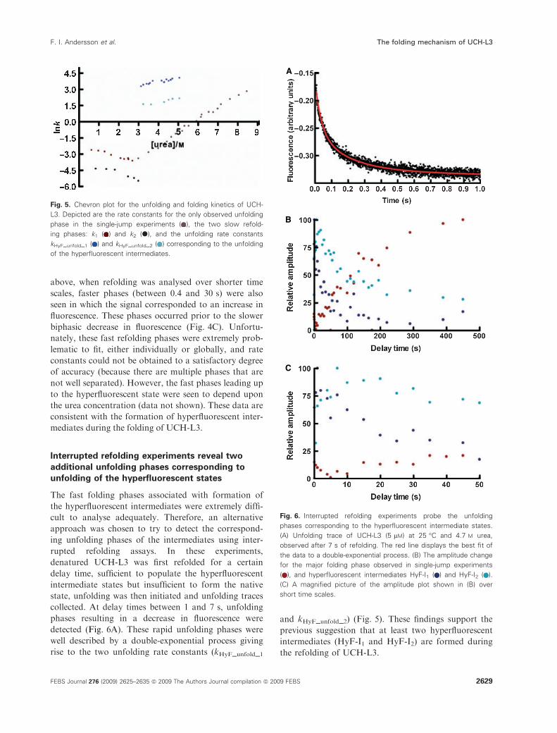
above, when refolding was analysed over shorter time
scales, faster phases (between 0.4 and 30 s) were also
seen in which the signal corresponded to an increase in
fluorescence. These phases occurred prior to the slower
biphasic decrease in fluorescence (Fig. 4C). Unfortu-
nately, these fast refolding phases were extremely prob-
lematic to fit, either individually or globally, and rate
constants could not be obtained to a satisfactory degree
of accuracy (because there are multiple phases that are
not well separated). However, the fast phases leading up
to the hyperfluorescent state were seen to depend upon
the urea concentration (data not shown). These data are
consistent with the formation of hyperfluorescent inter-
mediates during the folding of UCH-L3.
Interrupted refolding experiments reveal two
additional unfolding phases corresponding to
unfolding of the hyperfluorescent states
The fast folding phases associated with formation of
the hyperfluorescent intermediates were extremely diffi-
cult to analyse adequately. Therefore, an alternative
approach was chosen to try to detect the correspond-
ing unfolding phases of the intermediates using inter-
rupted refolding assays. In these experiments,
denatured UCH-L3 was first refolded for a certain
delay time, sufficient to populate the hyperfluorescent
intermediate states but insufficient to form the native
state, unfolding was then initiated and unfolding traces
collected. At delay times between 1 and 7 s, unfolding
phases resulting in a decrease in fluorescence were
detected (Fig. 6A). These rapid unfolding phases were
well described by a double-exponential process giving
rise to the two unfolding rate constants (kHyF_unfold_1
and kHyF_unfold_2) (Fig. 5). These findings support the
previous suggestion that at least two hyperfluorescent
intermediates (HyF-I1 and HyF-I2) are formed during
the refolding of UCH-L3.
Fig. 5. Chevron plot for the unfolding and folding kinetics of UCH-
L3. Depicted are the rate constants for the only observed unfolding
phase in the single-jump experiments ( ), the two slow refold-
ing phases: k1 ( ) and k2 (d), and the unfolding rate constants
kHyF_unfold_1 ( ) and kHyF_unfold_2 ( ) corresponding to the unfolding
of the hyperfluorescent intermediates.
A
B
C
Fig. 6. Interrupted refolding experiments probe the unfolding
phases corresponding to the hyperfluorescent intermediate states.
(A) Unfolding trace of UCH-L3 (5 lM) at 25 �C and 4.7 M urea,
observed after 7 s of refolding. The red line displays the best fit of
the data to a double-exponential process. (B) The amplitude change
for the major folding phase observed in single-jump experiments
( ), and hyperfluorescent intermediates HyF-I1 ( ) and HyF-I2 ( ).
(C) A magnified picture of the amplitude plot shown in (B) over
short time scales.
F. I. Andersson et al. The folding mechanism of UCH-L3
FEBS Journal 276 (2009) 2625–2635 ª 2009 The Authors Journal compilation ª 2009 FEBS 2629

Hyperfluorescent state: the two hyperfluorescent
intermediate states are populated rapidly during
refolding
In order to gain further insights into the nature of
HyF-I1 and HyF-I2, the time-dependent formation of
the different species along the UCH-L3 folding path-
way was studied using interrupted refolding experi-
ments with varying delay times. This method assumes
that after various delay times the amplitude of the
unfolding phase(s) are proportional to the number of
molecules in that state on the folding pathway [27].
Consequently, denatured UCH-L3 was refolded into
0.9 m urea for various delay times and then jumped
back into unfolding conditions (4.7 m urea), thus
allowing the time evolution and formation of the vari-
ous intermediate states to be monitored. The two
hyperfluorescent intermediates were populated quickly,
with formation of HyF-I2 (light blue phase) being
slightly slower than that of HyF-I1 (dark blue)
(Fig. 6B,C). As expected, at longer delay times (400 s),
the amplitudes associated with the different intermedi-
ate states diminished to zero as the intermediate con-
verted to the native state. Moreover, the rate constants
for their decay were very similar (HyF-I1: 0.024 s)1
and HyF-I2: 0.02 s)1). By contrast, the population of
the species corresponding to the major folding event
(red phase) observed in single-jump experiments, dis-
played a significant lag in its formation and was only
observed after delay times of 40–50 s, reaching a maxi-
mum after 300–400 s (Fig. 6B,C). The rate constant
for the formation of the species associated with this
phase is 0.045 s)1, which is in good agreement with the
k1 folding rate constant at 0.9 m urea (Fig. 5). These
results were consistent with our proposed model and
indicative of the population of the hyperfluorescent
intermediates decaying as the population of the native
state species increased.
The folding pathway of UCH-L3 is limited by
proline isomerization
In the native states of proteins, prolyl peptide bonds
are usually in a fixed cis or trans conformation. How-
ever, when a protein is unfolded, the structure which
constrained the prolyl bond into a specific conforma-
tion is lost and an equilibrium between cis and trans
conformations is established, in which the trans state is
favoured over cis at a ratio of � 4 : 1 [28]. Upon
refolding, isomerization of these peptidyl–prolyl bonds
back to their native conformation can be rate limiting
because this process is usually slow, with a rate con-
stant of around 0.04–0.08 s)1 [28]. Because UCH-L3
has 12 proline residues, one of which is in a cis confor-
mation in the native state (Pro48), it is highly probable
that the slow refolding phase (the black phase, giving
rise to k2) is caused by a rate-limiting proline isomeri-
zation reaction. In order to investigate this further,
interrupted unfolding experiments using stopped-flow
techniques were undertaken. In these experiments,
UCH-L3 was first unfolded for various delay times
(1–400 s) at high concentrations of guanidine
hydochloride (under these conditions the unfolding
rate constant is very fast, � 400 s)1). Subsequent
refolding was then initiated by diluting the sample in
buffer containing a low concentration of denaturant.
At the shortest unfolding delay times, no peptidyl–
prolyl bond isomerization can occur, resulting in a
native-like conformation for all proline residues [27].
The kinetic refolding traces (Fig. 7A) obtained from
these experiments with different delay times were fit
globally to a double-exponential equation with shared
values for the two rate constants, k1 and k2.
A
B
Fig. 7. Interrupted unfolding experiments. (A) Refolding traces of
UCH-L3 (2 lM) at 25 �C in 0.93 M guanidine hydrochloride that had
been unfolded in high concentrations of guanidine hydrochloride for
1 s ( ) or 30 min (.). (B) Plot of the amplitude change for k1 ( )
and k2 (d) with respect to unfolding time. The red lines display the
best fit of the data to a single exponential process.
The folding mechanism of UCH-L3 F. I. Andersson et al.
2630 FEBS Journal 276 (2009) 2625–2635 ª 2009 The Authors Journal compilation ª 2009 FEBS

A plot of the amplitudes obtained from interrupted
unfolding experiments is a direct measure of the popu-
lation of molecules that give rise to a particular kinetic
phase [27]. With increasing unfolding delay times, an
increase in molecules folding with a rate constant of k2(black phase) was observed. The rate constant with
which this phase developed is 0.04 s)1. Such behaviour
is consistent with slow proline isomerization processes
occurring in the denatured state, showing that the
slowest phase observed in the refolding of UCH-L3 is
limited by a proline isomerization event. By contrast, a
decrease in amplitude was observed for the faster of
the two slow refolding phases (the red phase, corre-
sponding to k1) with increasing delay times, with a
similar rate constant to that observed for the develop-
ment of the slowest refolding phases (black phase)
(Fig. 7B). The overall change in amplitude for the sum
of the two phases did not vary with unfolding delay
time, indicating that even at the shortest delay times,
UCH-L3 was fully unfolded (data not shown).
Discussion
In this study, we investigated the stability and folding
of the 52-knotted ubiquitin hydrolase UCH-L3.
UCH-L3 is an essential enzyme present in various
tissues in the human body, where it functions as a
deubiquitinating enzyme, hydrolysing C-terminal
ubiquityl esters [13,17]. Through this activity, UCH-L3
and other ubiquitin hydrolases control the level of free
monoubiquitin in the cell and hence play a vital role in
several processes, including the ubiquitin–proteasome
pathway [13].
The chemical denaturant urea was used to reversibly
unfold UCH-L3, enabling us to characterize the stabil-
ity and folding ⁄unfolding kinetics of the protein
in vitro. Using tryptophan fluorescence as a probe of
tertiary structure, a red-shift was observed upon chem-
ical denaturation, consistent with a process in which
buried or partially buried tryptophan residues become
exposed to the solvent and the aqueous environment
[29]. In addition, unfolding was measured using far-
UV CD as a probe of secondary structure, and the
urea-denatured state was shown to have no stable
residual secondary structure. Both fluorescence and
far-UV CD were used to establish that the unfolding
of UCH-L3 was fully reversibly under the conditions
used. These results show that, despite its complex
knotted topology, UCH-L3 is able to refold spontane-
ously in vitro without the need for molecular chaper-
ones. These findings agree well with what has been
reported for the smaller and simpler trefoil knotted
proteins YibK and YbeA [4,5,7].
Under equilibrium conditions, the unfolding of
UCH-L3 appears to follow a simple two-state folding
model, in which only native and denatured states are
significantly populated. This type of behaviour is com-
mon for smaller proteins [26]. If a protein is a true
two-state folder under equilibrium conditions, then the
thermodynamic parameters obtained by fitting the data
from different structural probes should be the same
[30]. For UCH-L3, the thermodynamic parameters
mD)N and DGH2OD�N, obtained from the fits of fluores-
cence and the far-UV CD data, are within error, sug-
gesting that it is a two-state system (Table 1).
However, the thermodynamic parameter [D]50% varies
slightly between the two data sets (Table 1), indicating
that an intermediate might be populated under equilib-
rium conditions, albeit at very low levels. Although
some proteins have folding intermediates that are sta-
ble enough with respect to the denatured state to be
sufficiently populated and observable under equilib-
rium conditions [31], many proteins which show appar-
ent two-state behaviour under equilibrium conditions
have complex kinetics with several folding and ⁄orunfolding phases resulting from the formation of
folding intermediates [32].
Thorough kinetic analysis of the folding and unfold-
ing of UCH-L3 revealed several phases which were
investigated using a series of single- and double-jump
experiments. First, UCH-L3 refolding was shown to be
limited by proline isomerization events. The rate at
which this process occurs (� 0.04 s)1) agrees well with
that observed for other proteins, for example RNase A
and ANG, limited by proline isomerization events [33].
Second, single-jump refolding experiments on UCH-L3
showed that two hyperfluorescent intermediates were
rapidly populated upon refolding from the denatured
state, as seen by the initial increase in the fluorescence
signal (Fig. 4C). Such behaviour has not been reported
for the trefoil-knotted methyl transferases [5,7]. How-
ever, we were unable to determine accurate folding rate
constants for the formation of these hyperfluorescent
intermediates despite using several different fitting strat-
egies. A likely cause of these difficulties is that the
refolding phases are overlapping. Instead, we chose to
characterize their corresponding unfolding phases by
performing interrupted refolding double-jump assays
[27]. In these assays, denatured UCH-L3 was refolded
for various times to populate the hyperfluorescent states
and then subjected to unfolding at high urea concentra-
tions. Using this strategy, two additional unfolding
phases (rate constants kHyF_unfold_1 and kHyF_unfold_2)
were obtained. These data strongly suggest that there
are two hyperfluorescent intermediates, HyF-I1 and
HyF-I2, on the folding pathway of UCH-L3.
F. I. Andersson et al. The folding mechanism of UCH-L3
FEBS Journal 276 (2009) 2625–2635 ª 2009 The Authors Journal compilation ª 2009 FEBS 2631

Kinetic intermediates which are hyperfluorescent
have also been observed for several other proteins [34–
37]. Judging from the amplitude plot for the inter-
rupted refolding experiments (Fig. 6B,C), it appears
that the hyperfluorescent intermediates are populated
rapidly (1–10 s), these then convert to the native state
over a longer time scale. That HyF-I1 failed to decay
as HyF-I2 is populated, suggests that they are formed
in parallel. Such parallel pathways can arise from a
heterogeneous denatured state. Indeed, such a mecha-
nism has recently been proposed for the knotted pro-
tein YibK [8]. Based on our results, and comparisons
with other knotted proteins, we propose a kinetic
scheme for the folding of UCH-L3 (Fig. 8). In this
case, we propose that the intermediate states HyF-I1and HyF-I2 are on-pathway, as reported for other
knotted proteins [5].
The origin of the hyperfluorescence of the folding
intermediates is not investigated in detail in this study
and extensive additional site-directed mutagenesis stud-
ies on residues in close proximity to Trp6 and Trp29
would be needed to examine this comprehensively.
However, by visual inspection of the crystal structure,
an educated guess can be made. In the denatured state,
tryptophan residues are quenched by the solvent giving
rise to a rather low fluorescence signal. In the hyperflu-
orescent state populated during refolding, quenching
may well be diminished by a reduction in solvent inter-
actions and partial burial of the side chains of the
tryptophan residues in a hydrophobic environment.
Such a hydrophobic environment may be formed by
the hydrophobic collapse of the polypeptide chain in
this region (both tryptophan residues are located near
the N-terminus of the protein). If HyF-I1 ⁄HyF-I2 are
on-pathway intermediates, it is likely that the hydro-
phobic environment is created by interactions similar
to those present in the native state. Such hydrophobic
contacts could be provided by Lys110, Met111,
Leu227 and Ala229, which are < 5 A from indole ring
of Trp29 in the native structure (Fig. S1) [14,18]. The
subsequent quenching of fluorescence in the native
state may arise from further structural rearrangements
in this region of the protein upon further folding.
These might include repositioning of residue Cys50,
which has been shown to be located orthogonally 3 A
from the indole ring of Trp29 [18] (Fig. S1), and which
could therefore participate in an excited state proton-
transfer reaction [38], thereby quenching the fluores-
cence. Indeed, polar side chains are known to quench
tryptophan fluorescence [38]. This has been reported
for the well-studied Im7 protein that folds via an
on-pathway hyperfluorescent intermediate. In this case,
the quenching of Trp75 in the native state is achieved
by close contact with a neighbouring histidine side
chain [34,39].
To summarize, we present here the first characteriza-
tion of the stability and folding of the human 52-knot-
ted protein ubiquitin hydrolase, UCH-L3. The protein
has one of the most complex knotted topologies
observed to date [3]. Despite its complex structure,
with five distinct crossings of the polypeptide chain,
UCH-L3 unfolds reversibly in vitro without the need
for molecular chaperones. Moreover, the folding kinet-
ics reveal a complex folding mechanism which includes
the formation of two hyperfluorescent intermediate
states. This initial study now paves the way for more
detailed kinetic studies of the folding pathways of
these structurally tangled proteins.
Experimental procedures
Plasmids and materials
The plasmid pRSET ⁄UCH-L3 was a kind gift from the lab-
oratory of H. Ploegh (Whitehead Institute for Biomedical
Research, Cambridge, MA, USA). Pre-cast SDS ⁄PAGE
gels and Coomassie Brilliant Blue stain were from Invitro-
gen (Carlsbad, CA, USA) and all chromatography material
was purchased from GE Healthcare (Piscataway, NJ,
USA). All other chemicals were analytical grade and
purchased from Sigma-Aldrich (St Louis, MO, USA) or
Melford Laboratories (Chelsworth, UK).
Protein expression, purification and
quantification
Human UCH-L3 was purified as described previously [17],
however an additional mono-Q ion-exchange step was used
to improve purity. Purified UCH-L3 was aliquoted and
flash-frozen in buffer A (50 mm Tris, pH 7.6, 0.5 mm
EDTA, 5 mm dithiothreitol) and stored at )80 �C. PurifiedUCH-L3 was subjected to MS analysis to confirm size and
identity and SDS ⁄PAGE to judge purity. The concentration
of UCH-L3 monomers was determined spectrophotometri-
cally using an extinction coefficient at 280 nm of
20 065 m)1Æcm)1. Care was taken that the reference buffer
also contained exactly the same amount of dithiothreitol as
Fig. 8. Proposed kinetic scheme for the folding of UCH-L3. Sche-
matic representation of the possible folding pathway obtained from
all experimental kinetic data.
The folding mechanism of UCH-L3 F. I. Andersson et al.
2632 FEBS Journal 276 (2009) 2625–2635 ª 2009 The Authors Journal compilation ª 2009 FEBS

the protein sample, because dithiothreitol can contribute to
absorbance at 280 nm.
Spectroscopic measurements
Fluorescence measurements were made using a 1 cm path-
length cuvette in a Cary Eclipse fluorimeter (Varian, Palo
Alto, CA, USA). An excitation wavelength of 280 nm was
used, with a band pass of 5 nm for excitation and 10 nm
for emission. Emission spectra were recorded between 300
and 400 nm at a scan rate of 1 nmÆs)1.
Equilibrium unfolding experiments
All equilibrium measurements were performed at 25 �C in
buffer A. For the unfolding curves a stock of urea (� 8 m
in buffer A) was prepared volumetrically and stored at
)20 �C until use. The exact concentration of urea was
determined from its refractive index using an Atago 1T
refractometer (Bellingham & Stanley Ltd., Tunbridge Wells,
UK). The urea stock was then diluted with buffer A, such
that a concentration range of 0–7 m was obtained in
800 lL aliquots. This procedure was performed using a
Hamilton Microlab apparatus (Taylor Scientific, St Louis,
MO, USA). For unfolding curves, 100 lL of UCH-L3 in
buffer A was added to the 800 lL aliquots of the various
urea concentrations to yield a final concentration of protein
of 3 lm. The protein ⁄denaturant mixtures were then left to
equilibrate for 3–20 h at room temperature after which no
change in spectroscopic signal was observed. Unfolding was
followed by Trp fluorescence by measuring the area under
the curves (from 300 to 400 nm) as described elsewhere
[40]. Far-UV CD spectra were acquired using an Applied
Photophysics chirascan spectrometer (Leatherhead, UK)
and the CD signal integrated between 222 and 225 nm.
Reversibility tests
The reversibility of the unfolding of UCH-L3 was examined
both by fluorescence (Cary eclipse Fluorimeter; Varian,
Palo Alto, CA, USA) and far-UV CD (Applied Photo-
physics chirascan spectrometer). UCH-L3 was diluted to a
final concentration of 2 lm in either buffer A or 8 m urea
containing buffer A. The samples were then incubated for
1 h at 25 �C and the denatured sample was allowed to
refold by dilution of the sample with buffer A (such that
the final concentration of urea in both cases was 0.65 m
urea). The renaturation reaction was left for 1–5 h before
spectra were acquired.
Unfolding and refolding kinetics
The unfolding and refolding kinetics of UCH-L3 were
measured using stopped-flow techniques (for shorter time
scales) or by manual mixing (for longer time scales) using
an Applied Photophysics stopped-flow spectrometer or a
Cary Eclipse fluorimeter, respectively. Native or denatured
UCH-L3 (22 lm) was prepared in buffer A or buffer A plus
urea as appropriate. Native or denatured UCH-L3 protein
was then rapidly diluted 1 : 10 in buffer A or buffer A con-
taining urea, resulting in a final protein concentration of
2 lm, with various concentrations of urea. The exact urea
concentration was determined as described above. A cut-off
filter of 320 nm was employed in the stopped-flow appara-
tus, whereas an emission wavelength of 360 nm was used
for the traces collected by manual mixing in the Cary
Eclipse fluorimeter.
Interrupted unfolding experiments
Native UCH-L3 (72 lm) was unfolded for various delay
times (1–300 s) by mixing 1 : 36 in � 5.6 m guanidine
hydochloride. After a specific delay time, refolding was ini-
tiated by rapid mixing and dilution into buffer A, such that
the final guanidine hydochloride concentration and protein
concentrations were 0.93 m and 2 lm respectively. Trp fluo-
rescence was monitored and data acquired using the
stopped-flow apparatus and settings described above.
Interrupted refolding experiments
Denatured UCH-L3 in 5.5 m urea was refolded for various
delay times (0.1–500 s) by mixing 1 : 5 in buffer A, such
that the concentration of urea was 0.93 m. Unfolding was
then initiated by rapid dilution into high concentrations of
urea, such that the final urea concentrations varied between
4 and 6 m. The final concentration of protein after the two
mixing and dilution steps was 5 lm. The double-jump
experiments were preformed on a stopped-flow instrument
as described above.
Data analysis
The kinetic parameters, including the rate constants for
the different phases were obtained from fitting the fluo-
rescence traces to various equations (see below) using
either kaleidagraph 4 or graphpad prism. Normally,
the unfolding traces were fit to a single exponential pro-
cess or a single exponential with a drift (Eqn 1), where
A1 is the amplitude, k1 is the rate constant, n is the drift
of the signal and C is the offset.
y ¼ A1ð1� expð�k1tÞÞ þ nt þ C ð1Þ
The refolding and unfolding traces from the interrupted
refolding assays fit well to a process described by a double
exponential with drift (Eqn 2), where A1 is the amplitude of
phase 1, k1 is the rate constant for phase 1, A2 is the ampli-
F. I. Andersson et al. The folding mechanism of UCH-L3
FEBS Journal 276 (2009) 2625–2635 ª 2009 The Authors Journal compilation ª 2009 FEBS 2633

tude of phase 2, k2 is the rate constant for phase 2, n is the
drift of the signal and C is the offset.
y ¼ A1ð1� expð�k1tÞÞ þ A2ð1� expð�k2tÞÞ þ nt þ C ð2Þ
To measure the changes in amplitude in the double-jump
assays, traces were fit globally with shared rate constants.
The equilibrium values for [D]50% and mD)N were calcu-
lated using Eqn (3), where aN and aD are the spectroscopic
signals of the native and denatured states in the absence
of denaturant and bN and bD are the slopes of the native
and denatured baselines and [D] is the concentration of
denaturant.
F¼ðaNþbN½D�ÞþððaDþbD½D�ÞexpððmD�Nð½D��½D�50%ÞÞÞ=RT
1þexpðmD�Nð½D��½D�50%ÞÞ=RT
ð3Þ
The value for DGH2OD�Nwas then calculated using
Eqn (4).
DGH2OD�N ¼ mD�N D½ �50% ð4Þ
Acknowledgements
This study was supported by the Leverhulme Trust.
The authors are grateful to Dr Chittaranjan Das (Pur-
due University, West Lafayette, IN, USA) for purified
UCH-L3 as a control protein for stopped-flow refold-
ing experiments to verify the presence of the hyperfluo-
rescent state.
References
1 Mallam AL & Jackson SE (2008) Use of protein
engineering techniques to elucidate protein folding
pathways. Prog Nucleic Acid Res Mol Biol 84, 57–114.
2 Taylor WR (2007) Protein knots and fold complexity:
some new twists. Comput Biol Chem 31, 151–162.
3 Virnau P, Mirny LA & Kardar M (2006) Intricate
knots in proteins: function and evolution. PLoS Comput
Biol 2, 1074–1079.
4 Mallam AL & Jackson SE (2005) Folding studies on a
knotted protein. J Mol Biol 346, 1409–1421.
5 Mallam AL & Jackson SE (2006) Probing nature’s
knots: the folding pathway of a knotted homodimeric
protein. J Mol Biol 359, 1420–1436.
6 Mallam AL & Jackson SE (2007) The dimerization of
an [alpha] ⁄ [beta]-knotted protein is essential for struc-
ture and function. Structure 15, 111–122.
7 Mallam AL & Jackson SE (2007) A comparison of the
folding of two knotted proteins: YbeA and YibK.
J Mol Biol 366, 650–665.
8 Mallam AL, Morris ER & Jackson SE (2008) Exploring
knotting mechanisms in protein folding. Proc Natl Acad
Sci USA 105, 18740–18745.
9 Mallam AL, Onuoha SC, Grossmann JG & Jackson SE
(2008) Knotted fusion proteins reveal unexpected possi-
bilities in protein folding. Mol Cell 30, 642–648.
10 Sulkowska JI, Sulkowski P, Szymczak P & Cieplak M
(2008) Tightening of knots in proteins. Phys Rev Lett
100, doi:10.1103/PhysRevLett.100.058106.
11 Wallin S, Zeldovich KB & Shakhnovich EI (2007) The
folding mechanics of a knotted protein. J Mol Biol 368,
884–893.
12 Mallam AL (2009) How does a knotted protein fold?
FEBS J 276, 365–375.
13 Love KR, Catic A, Schlieker C & Ploegh HL (2007)
Mechanisms, biology and inhibitors of deubiquitinating
enzymes. Nat Chem Biol 3, 697–705.
14 Misaghi S, Galardy PJ, Meester WJN, Ovaa H, Ploegh
HL & Gaudet R (2005) Structure of the ubiquitin
hydrolase UCH-L3 complexed with a suicide substrate.
J Biol Chem 280, 1512–1520.
15 Liu YC, Fallon L, Lashuel HA, Liu ZH & Lansbury
PT (2002) The UCH-L1 gene encodes two opposing
enzymatic activities that affect alpha-synuclein degrada-
tion and Parkinson’s disease susceptibility. Cell 111,
209–218.
16 Larsen CN, Krantz BA & Wilkinson KD (1998) Sub-
strate specificity of deubiquitinating enzymes: ubiquitin
C-terminal hydrolases. Biochemistry 37, 3358–3368.
17 Larsen CN, Price JS & Wilkinson KD (1996) Substrate
binding and catalysis by ubiquitin C-terminal hydrolas-
es: identification of two active site residues. Biochemis-
try 35, 6735–6744.
18 Johnston SC, Riddle SM, Cohen RE & Hill CP (1999)
Structural basis for the specificity of ubiquitin C-termi-
nal hydrolases. EMBO J 18, 3877–3887.
19 Pickart CM & Rose IA (1985) Ubiqutin carboxyl-termi-
nal hydrolase acts on ubiqutin carboxyl-terminal
amides. J Biol Chem 260, 7903–7910.
20 Walters BJ, Campbell SL, Chen PC, Taylor AP,
Schroeder DG, Dobrunz LE, Artavanis-Tsakonas K,
Ploegh HL, Wilson JA, Cox GA et al. (2008) Differen-
tial effects of Usp14 and Uch-L1 on the ubiquitin pro-
teasome system and synaptic activity. Mol Cell Neurosci
39, 539–548.
21 Miyoshi Y, Nakayama S, Torikoshi Y, Tanaka S, Ishi-
hara H, Taguchi T, Tamaki Y & Noguchi S (2006) High
expression of ubiquitin carboxy-terminal hydrolase-L1
and -L3 mRNA predicts early recurrence in patients with
invasive breast cancer. Cancer Sci 97, 523–529.
22 Carmine Belin A, Westerlund M, Bergman O, Niss-
brandt H, Lind C, Sydow O & Galter D (2007) S18Y
in ubiquitin carboxy-terminal hydrolase L1 (UCH-L1)
associated with decreased risk of Parkinson’s disease in
Sweden. Parkinsonism Relat Disord 13, 295–298.
The folding mechanism of UCH-L3 F. I. Andersson et al.
2634 FEBS Journal 276 (2009) 2625–2635 ª 2009 The Authors Journal compilation ª 2009 FEBS

23 Gong B, Cao ZX, Zheng P, Vitolo OV, Liu SM,
Staniszewski A, Moolman D, Zhang H, Shelanski M &
Arancio O (2006) Ubiquitin hydrolase Uch-L1 rescues
beta-amyloid-induced decreases in synaptic function
and contextual memory. Cell 126, 775–788.
24 Setsuie R & Wada K (2007) The functions of UCH-L1
and its relation to neurodegenerative diseases. Neuro-
chem Int 51, 105–111.
25 Huang L & Makarov DE (2008) Translocation of a
knotted polypeptide through a pore. J Chem Phys 129,
doi:10.1063/1.2968554.
26 Jackson SE & Fersht AR (1991) Folding of chymotryp-
sin inhibitor-2.1. Evidence for a 2-state transition.
Biochemistry 30, 10428–10435.
27 Wallace LA & Matthews CR (2002) Sequential vs.
parallel protein-folding mechanisms: experimental tests
for complex folding reactions. Biophys Chem 101, 113–
131.
28 Grathwohl C & Wuthrich K (1981) NMR-studies of the
rates of proline cis–trans isomerization in oligopeptides.
Biopolymers 20, 2623–2633.
29 Ruan K & Balny C (2002) High pressure static fluores-
cence to study macromolecular structure–function. Bio-
chim Biophys Acta Protein Struct Mol Enzymol 1595,
94–102.
30 Ervin J, Larios E, Osvath S, Schulten K & Gruebele M
(2002) What causes hyperfluorescence: folding interme-
diates or conformationally flexible native states? Biophys
J 83, 473–483.
31 Ayed A & Duckworth H (1999) A stable intermediate
in the equilibrium unfolding of Escherichia coli citrate
synthase. Protein Sci 8, 1116–1126.
32 Jemth P, Gianni S, Day R, Li B, Johnson CM, Daggett
V & Fersht AR (2004) Demonstration of a low-energy
on-pathway intermediate in a fast-folding protein by
kinetics, protein engineering, and simulation. Proc Natl
Acad Sci USA 101, 6450–6455.
33 Pradeep L, Shin H-C & Scheraga HA (2006) Correla-
tion of folding kinetics with the number and isomeriza-
tion states of prolines in three homologous proteins of
the RNase family. FEBS Lett 580, 5029–5032.
34 Capaldi AP, Shastry MCR, Kleanthous C, Roder H &
Radford SE (2001) Ultrarapid mixing experiments
reveal that Im7 folds via an on-pathway intermediate.
Nat Struct Mol Biol 8, 68–72.
35 Eifler N, Vetsch M, Gregorini M, Ringler P, Chami M,
Philippsen A, Fritz A, Muller SA, Glockshuber R,
Engel A et al. (2006) Cytotoxin ClyA from Escherichi-
a coli assembles to a 13-meric pore independent of its
redox-state. EMBO J 25, 2652–2661.
36 Friel CT, Beddard GS & Radford SE (2004) Switching
two-state to three-state kinetics in the helical protein
Im9 via the optimisation of stabilising non-native inter-
actions by design. J Mol Biol 342, 261–273.
37 Otto MR, Lillo MP & Beechem JM (1994) Resolution
of multiphasic reactions by the combination of fluores-
cence total-intensity and anistropy stopped-flow kinetic-
experiments. Biophys J 67, 2511–2521.
38 Chen Y & Barkley MD (1998) Toward understanding
tryptophan fluorescence in proteins. Biochemistry 37,
9976–9982.
39 Roder H, Maki K & Cheng H (2006) Early events in
protein folding explored by rapid mixing methods.
Chem Rev 106, 1836–1861.
40 Royer CA, Mann CJ & Matthews CR (1993) Resolu-
tion of the fluorescence equilibrium unfolding profile of
Trp apo-repressor using single tryptophan mutants.
Protein Sci 2, 1844–1852.
Supporting information
The following supplementary material is available:
Fig. S1. Hydrophobic residues in close proximity to
Trp29.
This supplementary material can be found in the
online version of this article.
Please note: Wiley-Blackwell is not responsible for
the content or functionality of any supplementary
materials supplied by the authors. Any queries (other
than missing material) should be directed to the corre-
sponding author for the article.
F. I. Andersson et al. The folding mechanism of UCH-L3
FEBS Journal 276 (2009) 2625–2635 ª 2009 The Authors Journal compilation ª 2009 FEBS 2635