COPI-mediated retrograde transport is required for efficient γ-secretase cleavage of the amyloid...
7
COPI-mediated retrograde transport is required for efficient c-secretase cleavage of the amyloid precursor protein q Alexandra Selivanova a, * , Bengt Winblad a , Mark R. Farmery a,1 , Nico P. Dantuma b , Maria Ankarcrona a a Department of Neurobiology, Caring Sciences and Society (NVS), KI Alzheimer Disease Research Center, Karolinska Institutet, Novum 5th floor, S-141 57 Stockholm, Sweden b Department of Cell and Molecular Biology, The Medical Nobel Institute, Karolinska Institutet, S-177 77 Stockholm, Sweden Received 25 August 2006 Available online 18 September 2006 Abstract Sequential cleavage of the amyloid precursor protein (APP) by b- and c-secretases results in the production of b-amyloid peptide, which is a key determinant in Alzheimer’s disease. Since several putative locations for c-secretase cleavage have been identified along the secretory pathway, trafficking of APP may be of importance for b-amyloid peptide production. Here we have studied the role of retrograde transport in APP processing. We found that APP interacts with the b subunit of the coatomer protein I (COPI) complex, which is involved in retrograde transport. In line with a role of retrograde trafficking in APP transport, inhibition of COPI-dependent transport altered APP trafficking, decreased APP cell surface expression, and coincided with a profound reduction in c-secretase cleav- age. These results suggest that COPI-dependent retrograde transport is important for APP processing and influences production of b-amyloid peptide. Ó 2006 Elsevier Inc. All rights reserved. Keywords: b-Amyloid; Alzheimer’s disease; APP; Retrograde transport; COPI; Neurodegeneration Alzheimer’s disease (AD) is characterized by the accu- mulation of b-amyloid (Ab) peptide in the brain [1]. Although the molecular mechanism for Ab peptide toxicity is not clear, a large body of evidence supports the notion that Ab peptide plays a key role in the pathophysiology of AD [2].Ab peptide is derived from the amyloid precur- sor protein (APP), which is a type I transmembrane protein that travels to the cell surface through the secretory path- way. Sequential cleavage of APP by b- and c-secretases gives rise to the Ab peptide [3]. The cleavage by the c-secretase complex is most interest- ing as a therapeutic target since it can give rise to two dif- ferent Ab fragments, Ab 1–40 and Ab 1–42 , of which the longer fragment is more aggregation-prone and notorious- ly linked to AD [4]. Hence it is important to understand c-secretase-mediated proteolytic cleavage of APP and to identify external and internal factors that influence APP processing. An important conundrum is the precise loca- tion of c-secretase cleavage [5]. The c-secretase complex is assembled in the endoplasmic reticulum (ER) and the Golgi compartments. This implies that APP is exposed to the c-secretase complex throughout the secretory pathway. Accordingly, it has been shown that Ab peptide generation 0006-291X/$ - see front matter Ó 2006 Elsevier Inc. All rights reserved. doi:10.1016/j.bbrc.2006.09.043 q Abbreviations: AICD, b-APP intracellular domain; APP, amyloid precursor protein; Ab, b-amyloid; AD, Alzheimer’s disease; Chapso, 3-[(3- cholamidopropyl)dimethylammonio]-2-hydroxy-1propanesulfonate; COPI, coatomer protein I; APP CTFs, APP C-terminal fragments; DAPT, (N-[N-(3,5-difluorophenacetyl)-l-alanyl]-S-phenylglycine-t-butyl-ester); ER, endoplasmic reticulum; GVP, Gal4/VP16 fusion; L-685, 458, [1s-benzyl-4R- [1-(1s-carbonyl-2-phenylethylcarbomoyl)-1s-3-methylbutylcarbomoyl]-2R- hydroxy-5-phenylpentyl]carbamic acid tert-butyl ester; PBS, phosphate- buffered saline; PBS–GS/BSA, PBS containing 10% goat serum and 5% BSA. * Corresponding author. Fax: +46 585 83 880. E-mail address: [email protected] (A. Selivanova). 1 Present address: Astra Zeneca R&D So ¨ derta ¨lje, S-151 85, So ¨ derta ¨lje, Sweden. www.elsevier.com/locate/ybbrc Biochemical and Biophysical Research Communications 350 (2006) 220–226 BBRC
-
Upload
independent -
Category
Documents
-
view
1 -
download
0
Transcript of COPI-mediated retrograde transport is required for efficient γ-secretase cleavage of the amyloid...
www.elsevier.com/locate/ybbrc
Biochemical and Biophysical Research Communications 350 (2006) 220–226
BBRC
COPI-mediated retrograde transport is required for efficientc-secretase cleavage of the amyloid precursor protein q
Alexandra Selivanova a,*, Bengt Winblad a, Mark R. Farmery a,1, Nico P. Dantuma b,Maria Ankarcrona a
a Department of Neurobiology, Caring Sciences and Society (NVS), KI Alzheimer Disease Research Center, Karolinska Institutet,
Novum 5th floor, S-141 57 Stockholm, Swedenb Department of Cell and Molecular Biology, The Medical Nobel Institute, Karolinska Institutet, S-177 77 Stockholm, Sweden
Received 25 August 2006Available online 18 September 2006
Abstract
Sequential cleavage of the amyloid precursor protein (APP) by b- and c-secretases results in the production of b-amyloid peptide,which is a key determinant in Alzheimer’s disease. Since several putative locations for c-secretase cleavage have been identified alongthe secretory pathway, trafficking of APP may be of importance for b-amyloid peptide production. Here we have studied the role ofretrograde transport in APP processing. We found that APP interacts with the b subunit of the coatomer protein I (COPI) complex,which is involved in retrograde transport. In line with a role of retrograde trafficking in APP transport, inhibition of COPI-dependenttransport altered APP trafficking, decreased APP cell surface expression, and coincided with a profound reduction in c-secretase cleav-age. These results suggest that COPI-dependent retrograde transport is important for APP processing and influences production ofb-amyloid peptide.� 2006 Elsevier Inc. All rights reserved.
Keywords: b-Amyloid; Alzheimer’s disease; APP; Retrograde transport; COPI; Neurodegeneration
Alzheimer’s disease (AD) is characterized by the accu-mulation of b-amyloid (Ab) peptide in the brain [1].Although the molecular mechanism for Ab peptide toxicityis not clear, a large body of evidence supports the notionthat Ab peptide plays a key role in the pathophysiology
0006-291X/$ - see front matter � 2006 Elsevier Inc. All rights reserved.
doi:10.1016/j.bbrc.2006.09.043
q Abbreviations: AICD, b-APP intracellular domain; APP, amyloidprecursor protein; Ab, b-amyloid; AD, Alzheimer’s disease; Chapso, 3-[(3-cholamidopropyl)dimethylammonio]-2-hydroxy-1propanesulfonate; COPI,coatomer protein I; APP CTFs, APP C-terminal fragments; DAPT,(N-[N-(3,5-difluorophenacetyl)-l-alanyl]-S-phenylglycine-t-butyl-ester); ER,endoplasmic reticulum; GVP, Gal4/VP16 fusion; L-685, 458, [1s-benzyl-4R-[1-(1s-carbonyl-2-phenylethylcarbomoyl)-1s-3-methylbutylcarbomoyl]-2R-hydroxy-5-phenylpentyl]carbamic acid tert-butyl ester; PBS, phosphate-buffered saline; PBS–GS/BSA, PBS containing 10% goat serum and 5% BSA.
* Corresponding author. Fax: +46 585 83 880.E-mail address: [email protected] (A. Selivanova).
1 Present address: Astra Zeneca R&D Sodertalje, S-151 85, Sodertalje,Sweden.
of AD [2]. Ab peptide is derived from the amyloid precur-sor protein (APP), which is a type I transmembrane proteinthat travels to the cell surface through the secretory path-way. Sequential cleavage of APP by b- and c-secretasesgives rise to the Ab peptide [3].
The cleavage by the c-secretase complex is most interest-ing as a therapeutic target since it can give rise to two dif-ferent Ab fragments, Ab1–40 and Ab1–42, of which thelonger fragment is more aggregation-prone and notorious-ly linked to AD [4]. Hence it is important to understandc-secretase-mediated proteolytic cleavage of APP and toidentify external and internal factors that influence APPprocessing. An important conundrum is the precise loca-tion of c-secretase cleavage [5]. The c-secretase complexis assembled in the endoplasmic reticulum (ER) and theGolgi compartments. This implies that APP is exposed tothe c-secretase complex throughout the secretory pathway.Accordingly, it has been shown that Ab peptide generation

A. Selivanova et al. / Biochemical and Biophysical Research Communications 350 (2006) 220–226 221
can take place in the ER, in the Golgi complex, and at thecell surface but the individual contributions of these loca-tions to amyloidogenesis are unclear [6–8].
The ER has a sophisticated protein quality controlmachinery that keeps immature proteins from reachingthe Golgi complex [9]. A number of studies suggest thatimmature proteins that have escaped the ER protein qual-ity control are subject to retrograde transport from theGolgi complex back to the ER [10]. Once back in theER, their maturation can be completed [11,12], or alterna-tively they can be disposed of by ER associated degrada-tion [13]. Golgi to ER transport is mediated by at leasttwo independent mechanisms, namely canonical retrogradetransport dependent on the coatomer protein I (COPI) [14],and at least one additional transport pathway that does notrequire COPI [15]. Interestingly, it has recently been dem-onstrated that presenilin 1, which is part of the c-secretasecomplex, is transported in COPI-coated vesicles [16,17].Moreover, another retrograde protein transport pathway,which is dependent on the GTPase Rab6 but does notrequire COPI, seems to influence APP processing [18]. Inaddition, Rab6 membrane association is presenilin-depen-dent [19]. Together these data suggest that APP and thec-secretase complex are both undergoing retrograde trans-port. Importantly, it is not known whether retrogradetransport in any respect influences APP processing.
In the present investigation, we studied the role ofCOPI-mediated retrograde transport in trafficking andprocessing of APP. We found that inhibition of COPIhas a profound effect on APP localization and stronglyreduces cleavage of APP by the c-secretase complex. Thus,retrograde transport of APP may be an important determi-nant for APP processing.
Materials and methods
Cell culture. The Chinese hamster ovary (CHO)-derived ldlF-2 cell line(hereafter ldlF-2 cells) and control CHO cells (hereafter control cells) werekindly provided by Dr. Monty Krieger (MIT, Cambridge, MD). LdlF-2cells express a temperature-sensitive e-COPI subunit [20]. Cells weregrown at permissive (34 �C) or restrictive (39 �C) temperature in Ham’s F-12 medium with 5 mM L-glutamine containing 5% fetal bovine serum,100 lg/ml penicillin, and 100 lg/ml streptomycin (Invitrogen, Carlsbad,CA). Human embryonic kidney 293 (HEK293) cells were grown in Dul-becco’s modified Eagle’s medium (Invitrogen) supplemented with 10%fetal calf serum (Invitrogen), 10 U/ml penicillin, and 100 lg/ml strepto-mycin. All transfections were performed using Lipofectamine Plus reagentaccording to manufacturer’s instructions (Invitrogen). Stable APP-ex-pressing CHO, ldlF-2, and HEK293 cell lines were generated by trans-fection with pcDNA3.1 APPwt695 expression vector and subsequentselection with 150 lg/ml G418 (Invitrogen) for three weeks. Survivingclones were expanded and APP expression was analyzed by Western blot.
Immunoblotting. Cells were lysed in RIPA buffer (50 mM Tris, pH 8,150 mM NaCl, 0.5% deoxycholic acid, 0.1% SDS, 1% NP-40, and proteaseinhibitor cocktail (Roche, Basel, Switzerland)) and equal amounts ofprotein were loaded on 10–20% Tricine gel (Invitrogen). The samples wereblotted onto PVDF membrane (Bio-Rad, Hercules, CA) and analyzedusing the following antibodies: mouse monoclonal 6E10 (1:2000 dilution;1–17 aa of Ab, Chemicon, Temecula, CA), mouse monoclonal C1/6.1(1:1000 dilution; C-terminal APP, gift from Dr. Paul Mathews, NathanCline Institute, Orangeburg, NY), rabbit polyclonal 369 (1:1000 dilution;
C-terminal APP, gift from Dr. Sam Gandy, Thomas Jefferson University,Philadelphia, PA), rabbit polyclonal anti-b-COPI (1:1000 dilution;Abcam, Cambridge, UK), and rabbit polyclonal anti-e-COPI (1:2000dilution; Santa Cruz, Santa Cruz, CA).
Immunoprecipitation. Control CHO, ldlF-2 cells, and HEK 293 cellsstably transfected with APP were grown in 100 mm plates. 24 h posttrans-fection, the cells were harvested and solubilized in 500 ll CHAPSO buffer(50 mM Hepes, pH 7.4, 150 mM NaCl, 2 mM EDTA, 1% 3-[(3-cholami-dopropyl)dimethylammonio]-2-hydroxy-1propanesulfonate (CHAPSO),and protease inhibitor cocktail (Roche)), followed by centrifugation at14,000 rpm for 30 min at 4 �C to remove insoluble material. The lysates werepre-cleared with 50 ll Protein G–Sepharose (GE Healthcare, Amersham,Little Chalfont, UK) for 1 h at 4 �C. Supernatants were incubated for16 h at 4 �C with monoclonal anti-APP antibody 6E10 (1:200 dilution). Next50 ll of protein G Sepharose was added and the sample was incubated for1 h at 4 �C. The beads were washed three times with 1 ml of 1% CHAPSObuffer, eluted by adding 30 ll of 2· Laemmli buffer (Sigma, St. Louis, MO),and incubated for 20 min at 50 �C. Samples were separated by sodiumdodecyl sulfate–polyacrylamide gel electrophoresis, transferred to PVDFmembrane (Millipore), and probed with a polyclonal anti-b-COPI antibody(Abcam, Cambridge, UK).
Immunostaining. Control and ldlF-2 cells stably expressing APP weregrown on sterile coverslips for 15 h at permissive or restrictive tempera-ture. Cells were fixed with 2% paraformaldehyde and 0.2% glutaraldehydefor 20 min at 4 �C. For cell surface staining, cells were blocked in phos-phate-buffered saline (PBS) containing 10% goat serum and 5% BSA(PBS–GS/BSA). The primary antibody used was anti-N-terminal APP(dilution 1:250; OMA1, Affinity Bioreagents, Rockville, MD) diluted inPBS–GS/BSA. Cells were incubated with primary antibodies for 3 h at4 �C. After washing three times with PBS, the cells were incubated for 1 hwith the appropriate secondary antibody diluted in PBS–GS/BSA. Thesecondary antibody used was Alexa Fluor 488 goat anti-mouse (dilution1:300; Molecular Probes Europe, Leiden, The Netherlands). Cells werethen rinsed three times with PBS and mounted using Vecta-shield. Theimmunostainings were analyzed using confocal laser scanning microscope(Carl Zeiss) and software (LSM 5 Image Browser).
Cell surface biotinylation. Control and ldlF-2 cells stably expressingAPP were grown in 75 cm2 flasks. Subsequently cells were treated with100 lM chloroquine (Sigma) for 15 h at permissive and restrictive tem-peratures. Cells were then washed three times with ice-cold PBS andincubated in 5 ml of 1 mg/ml EZ-link Sulfo-NHS-LC-Biotin (Pierce,Rockford, IL) in PBS for 30 min at 4 �C. Biotin solution was thenremoved and cells were washed three times with ice-cold PBS. Cells werescraped off into ice-cold PBS and spun down by centrifugation at3,000 rpm for 5 min at 4 �C. The pellet was lysed in 350 ll PBS containing1% NP-40 and protease inhibitor cocktail (Roche), followed by briefcentrifugation to remove insoluble material. Lysates were then added to80 ll streptavidin–agarose beads from Pierce (Rockford), which had beenpre-washed three times in PBS with 1% NP-40 and protease inhibitor.Samples were incubated with streptavidin beads for 16 h at 4 �C, spundown by centrifugation at 5,000 rpm for 3 min at 4 �C, and washed threetimes with ice-cold PBS with 1% NP-40 and once with ice-cold 0.9% NaCl.The beads were eluted by addition of 30 ll of 2· Laemmli buffer andboiling for 5 min. Samples were then analyzed by immunoblotting usingantibody 369 against APP.
c-Secretase cleavage assay. The cleavage assay has been previouslydescribed [21]. In brief, control and ldlF-2 cells stably expressing APP weretransfected with 100 ng MH100 (Gal4/VP16 fusion (GVP)-regulatedluciferase expression vector), and 50 ng CMV-b-galactosidase plasmid (tocontrol for transfection efficiency) combined with either 100 ng controlpcDNA3 plasmid or 100 ng C99-GVP plasmid (c-secretase reporter).MH100 and CMV-b-galactosidase plasmid have been described previously[22]. Following transfection, cells were kept at permissive or restrictivetemperature. Twenty-four hours after transfection, cells were harvestedand analyzed for reporter gene activity using a bioluminescence reader. c-Secretase inhibitors L-685,458 (10 lM), or N-[N-(3,5-difluorophenacetyl)-l-alanyl]-S-phenylglycine t-butyl ester (DAPT) (2.5 lM) were included inthe medium during the recovery phase.
222 A. Selivanova et al. / Biochemical and Biophysical Research Communications 350 (2006) 220–226
Results and discussion
APP interacts with the COPI complex
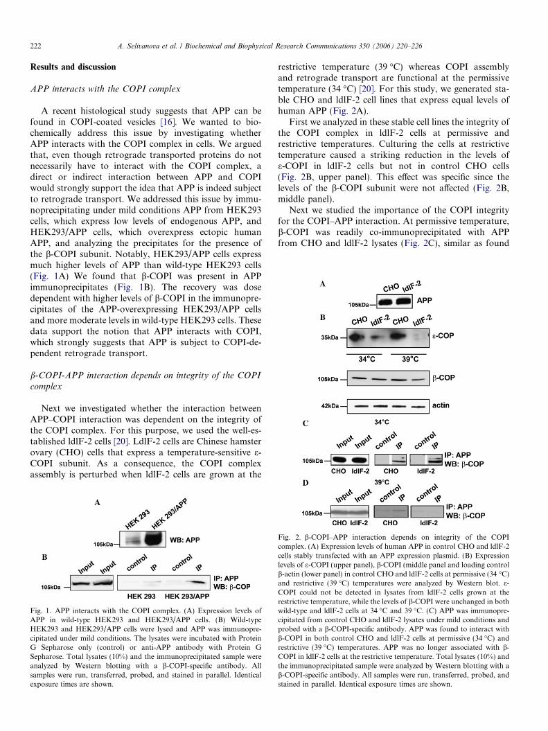
A recent histological study suggests that APP can befound in COPI-coated vesicles [16]. We wanted to bio-chemically address this issue by investigating whetherAPP interacts with the COPI complex in cells. We arguedthat, even though retrograde transported proteins do notnecessarily have to interact with the COPI complex, adirect or indirect interaction between APP and COPIwould strongly support the idea that APP is indeed subjectto retrograde transport. We addressed this issue by immu-noprecipitating under mild conditions APP from HEK293cells, which express low levels of endogenous APP, andHEK293/APP cells, which overexpress ectopic humanAPP, and analyzing the precipitates for the presence ofthe b-COPI subunit. Notably, HEK293/APP cells expressmuch higher levels of APP than wild-type HEK293 cells(Fig. 1A) We found that b-COPI was present in APPimmunoprecipitates (Fig. 1B). The recovery was dosedependent with higher levels of b-COPI in the immunopre-cipitates of the APP-overexpressing HEK293/APP cellsand more moderate levels in wild-type HEK293 cells. Thesedata support the notion that APP interacts with COPI,which strongly suggests that APP is subject to COPI-de-pendent retrograde transport.
b-COPI-APP interaction depends on integrity of the COPI
complex
Next we investigated whether the interaction betweenAPP–COPI interaction was dependent on the integrity ofthe COPI complex. For this purpose, we used the well-es-tablished ldlF-2 cells [20]. LdlF-2 cells are Chinese hamsterovary (CHO) cells that express a temperature-sensitive e-COPI subunit. As a consequence, the COPI complexassembly is perturbed when ldlF-2 cells are grown at the
Fig. 1. APP interacts with the COPI complex. (A) Expression levels ofAPP in wild-type HEK293 and HEK293/APP cells. (B) Wild-typeHEK293 and HEK293/APP cells were lysed and APP was immunopre-cipitated under mild conditions. The lysates were incubated with ProteinG Sepharose only (control) or anti-APP antibody with Protein GSepharose. Total lysates (10%) and the immunoprecipitated sample wereanalyzed by Western blotting with a b-COPI-specific antibody. Allsamples were run, transferred, probed, and stained in parallel. Identicalexposure times are shown.
restrictive temperature (39 �C) whereas COPI assemblyand retrograde transport are functional at the permissivetemperature (34 �C) [20]. For this study, we generated sta-ble CHO and ldlF-2 cell lines that express equal levels ofhuman APP (Fig. 2A).
First we analyzed in these stable cell lines the integrity ofthe COPI complex in ldlF-2 cells at permissive andrestrictive temperatures. Culturing the cells at restrictivetemperature caused a striking reduction in the levels ofe-COPI in ldlF-2 cells but not in control CHO cells(Fig. 2B, upper panel). This effect was specific since thelevels of the b-COPI subunit were not affected (Fig. 2B,middle panel).
Next we studied the importance of the COPI integrityfor the COPI–APP interaction. At permissive temperature,b-COPI was readily co-immunoprecipitated with APPfrom CHO and ldlF-2 lysates (Fig. 2C), similar as found
Fig. 2. b-COPI–APP interaction depends on integrity of the COPIcomplex. (A) Expression levels of human APP in control CHO and ldlF-2cells stably transfected with an APP expression plasmid. (B) Expressionlevels of e-COPI (upper panel), b-COPI (middle panel and loading controlb-actin (lower panel) in control CHO and ldlF-2 cells at permissive (34 �C)and restrictive (39 �C) temperatures were analyzed by Western blot. e-COPI could not be detected in lysates from ldlF-2 cells grown at therestrictive temperature, while the levels of b-COPI were unchanged in bothwild-type and ldlF-2 cells at 34 �C and 39 �C. (C) APP was immunopre-cipitated from control CHO and ldlF-2 lysates under mild conditions andprobed with a b-COPI-specific antibody. APP was found to interact withb-COPI in both control CHO and ldlF-2 cells at permissive (34 �C) andrestrictive (39 �C) temperatures. APP was no longer associated with b-COPI in ldlF-2 cells at the restrictive temperature. Total lysates (10%) andthe immunoprecipitated sample were analyzed by Western blotting with ab-COPI-specific antibody. All samples were run, transferred, probed, andstained in parallel. Identical exposure times are shown.

Fig. 3. APP cell surface expression is decreased in the absence offunctional COPI complex. Cell surface APP staining in non-permeabilizedcells in (A) control CHO cells, permissive temperature and (B) ldlF-2 cells,permissive temperature, (C) control CHO cells, restrictive temperature,and (D) ldlF-2 cells, restrictive temperature. Control CHO and ldlF-2 cellswere cultured at (E) permissive and (F) restrictive temperatures in theabsence or presence of 100 lM chloroquine for 16 h. The cells werebiotinylated and biotinylated material were precipitated with streptavidinbeads. Total lysate (input) and precipitated biotinylated material wasprobed in Western blotting with an APP antibody. Total lysates (10%) andthe immunoprecipitated sample were analyzed by Western blotting withan APP specific antibody. All samples were run, transferred, probed, andstained in parallel. Identical exposure times are shown.
Fig. 4. APP processing is reduced in the absence of functional COPIcomplex. Cell lysates from control CHO and ldlF-2 cells grown atpermissive and restrictive temperature were probed in immunoblottingwith an antibody that is specific for the C terminus of APP. The bandsrepresenting full length APP and APP CTFs are shown. As a loadingcontrol the blot was reprobed with a b-actin-specific antibody.
A. Selivanova et al. / Biochemical and Biophysical Research Communications 350 (2006) 220–226 223
for HEK293 cells. Thus, this interaction was found in twocell lines obtained from different species (human and Chi-nese hamster) and of different origins (ovary and kidney)for both endogenous and ectopically expressed humanAPP. Importantly, the interaction between b-COPI andAPP was abrogated in ldlF-2 cells, but not in controlCHO cells, at the restrictive temperature (Fig. 2D). SinceCHO and ldlF-2 cells express equal levels of b-COPI at per-missive and restrictive temperatures, this strongly suggeststhat in the absence of a functional COPI complex APPdoes not interact anymore with the COPI complex.
APP cell surface expression is decreased in the absence of
functional COPI complex
A number of studies indicate that retrograde transport isimportant for maturation of cell surface proteins [12]. Weargued that if retrograde transport would be importantfor APP maturation, inhibition of retrograde transportwas likely to cause a decrease in APP maturation and hencea downregulation in APP cell surface expression. However,if retrograde transport is irrelevant for APP maturation,one would expect rather an increase in cell surface expres-sion since the APP that escaped from retrograde transportwould now be transported to cell surface. We investigatedthe levels of APP at the cell surface in the presence orabsence of functional COPI complex by immunostainingnon-permeabilized control CHO and ldlF-2 cells with anAPP antibody. The cell surface staining of CHO cells wasnot changed at 39 �C (Fig. 3A and C) whereas cell surfaceAPP of ldlF-2 cells was strongly reduced at the restrictivetemperature (Fig. 3B and D).
To obtain more quantitative data on the effect of COPIfunctionality, we performed cell surface biotinylationexperiments. Control CHO and ldlF-2 cells were grownat permissive and restrictive temperatures, subjected to cellsurface biotinylation and the levels of biotinylated APPwere related to the total levels of APP. Since it has beendescribed that endocytosis may also be compromised inthe absence of COPI complex [23], we used this assay alsoto evaluate the status of APP endocytosis in COPI deficientcells by performing the experiments in the presence orabsence of chloroquine, which inhibits endocytosis. Atthe permissive temperature, low levels of APP were foundat the cell surface of CHO and ldlF-2 cells (Fig. 3E). Asanticipated the levels were increased in both cell lines whenendocytosis was inhibited by chloroquine confirming that

Fig. 5. Cleavage by c-secretase is reduced in the absence of functional COPI complex. (A) c-secretase reporter assay. Cells are transfected with a plasmidencoding a fusion of the APP fragment C99 fused to GVP, which is composed of the Gal4 DNA binding domain and VP16 transactivation domain, and aplasmid in which luciferase expression is driven by GVP. The C99–GVP fusion is membrane bound but upon cleavage by c-secretase at the naturalcleavage site in the APP fragment a soluble AICD–GVP fragment is released that translocates to the nucleus and activates luciferase expression. (B)Cleavage of C99–GVP was inhibited in both control CHO and ldlF-2 cells grown at 34 �C, following treatment with 2,5 lM c-secretase inhibitor DAPT.Student’s t-test was used to analyze the results of this experiment. *P < 0.05, ***P < 0.05. There was no significant difference in luciferase activity betweencontrol CHO and ldlF-2 cells not treated with DAPT. (C) Western blot analysis of lysates from CHO cells transfected with C99–GVP using an APP-specific monoclonal antibody (C1/6.1). Amounts of AICD are reduced in the presence of c-secretase inhibitor DAPT (2,5 lM). (D) Luciferase acitivity wasrecorded in control CHO and ldlF-2 cells transfected with C99–GVP and grown at the permissive temperature (34 �C). A representative experiment ofthree independent experiments is shown. Error bars represent standard error of the mean of triplicate samples in the experiment. Student’s t-test was usedto analyze the data. No significant difference was found in luciferase activity between control CHO and ldlF-2 cells. Luciferase activity recorded in (D)control CHO, and (E) ldlF-2 cells transfected with C99–GVP and kept at the restrictive temperature (39 �C) overnight. The chart is representative of threeindependent experiments and error bars show standard error of the mean of triplicate samples in this experiment. Cleavage of C99–GVP is significantlydecreased in ldlF-2 cells. Student’s t-test was used to analyze these results. **P < 0.05. (F) Western blot analysis of lysates from control CHO and ldlF-2cells transfected with C99–GVP and grown at the permissive or restrictive temperature. An APP-specific monoclonal antibody (C1/6.1) was used. TheAICD band could not be detected in ldlF-2 cells grown at the restrictive temperature. The blot was probed with a b-actin-specific antibody to check forequal loading.
224 A. Selivanova et al. / Biochemical and Biophysical Research Communications 350 (2006) 220–226
APP is internalized by endocytosis. The levels at the cellsurface were reduced in ldlF-2 but not in control CHO cellsat the restrictive temperature (Fig. 3F), confirming the
model that retrograde transport stimulates APP matura-tion and cell surface expression. It is noteworthy that eventhough the total levels of APP were substantially higher in

A. Selivanova et al. / Biochemical and Biophysical Research Communications 350 (2006) 220–226 225
ldlF-2 cells at the restrictive temperature as compared tocontrol CHO cells (see also Section APP processing isreduced in the absence of functional COPI complex), wenevertheless retrieved much less biotinylated material fromthe ldlF-2 cells underscoring the effect of COPI retrogradetransport on APP cell surface expression. Importantly, alsoat the restrictive temperature administration of chloro-quine enhanced cell surface expression showing that endo-cytosis of APP is not affected in ldlF-2 cells at therestrictive temperature under our experimental conditions.Together these data show that a functional COPI complexis important for proper cell surface expression and suggeststhat retrograde transport contributes to APP maturation.
APP processing is reduced in the absence of functional COPI
complex
Next we examined whether the absence or presence of afunctional COPI complex affected APP processing. To thisend, we detected by immunoblotting full length APP andAPP C terminal fragments (CFTs) in control CHO andldlF-2 cells at permissive and restrictive temperature. Wefound that the levels of full length APP were stronglyincreased in ldlF-2 cells when grown at the restrictive tem-peratures while a very modest increase was found in thecontrol cells (Fig. 4). The increase in full length APP coin-cided with a decrease in APP CTFs (Fig. 4). This suggeststhat absence of COPI-mediated retrograde transportinduces accumulation of full length APP and reducesAPP processing.
Cleavage by c-secretase is reduced in the absence of
functional COPI complex
Although the fact that full length APP accumulated inthe absence of functional COPI complex suggested thatcleavage by both b- and c-secretases is impaired, we decid-ed to focus in the next set of experiments on c-secretasecleavage since this particular cleavage event is most rele-vant for AD. For this purpose, we used a c-secretasereporter assay that gives highly quantitative data on therate of c-secretase cleavage in living cells [21]. This assayis based on the expression of a fusion of C99, the APPdomain containing the natural c-secretase cleavage site,to the Gal4/VP19 (GVP) chimeric protein that drivesexpression of a luciferase reporter gene. Cleavage by c-se-cretase releases from the membrane bound C99–GVPfusion AICD–GVP, which can translocate to the nucleusand induce luciferase expression (Fig. 5A).
As anticipated, high levels of luciferase were detected incontrol CHO and ldlF-2 cells that expressed the C99–VCPfusion and luciferase reporter plasmid. Luciferase activitywas completely inhibited by administration of the specificc-secretase inhibitor DAPT confirming that the assaydetects specifically c-secretase activity (Fig. 5B). Immuno-blotting also showed that C99-GVP was cleaved to theslightly smaller fragment AICD-GVP and that this cleav-
age was sensitive to DAPT (Fig. 5C). Next we assessedthe effect of COPI on c-secretase cleavage of C99-VCP.No significant differences were found in c-secretase cleav-age of the reporter in control CHO and ldlF-2 cells at thepermissive temperature (Fig. 5D). Strikingly, at the restric-tive temperature, cleavage of the reporter was completelyblocked in ldlF-2 cells while at the same temperature cleav-age was not affected in control CHO cells (Fig. 5E). Immu-noblotting also showed that at the restrictive temperaturethe generation of AICD–GVP was blocked (Fig. 5F).Together these data show that inhibition of retrogradetransport has a profound effect on c-secretase cleavage ofAPP.
In summary, we have demonstrated that inhibition ofCOPI-dependent pathways in cells expressing APP leadsto: (i) decreased cell surface expression of APP, (ii) accu-mulation of full-length APP, (iii) decreased processing ofC99 by c-secretase, and (iv) decreased levels of AICD.Our results link together COPI-dependent retrogradetransport and amyloidogenic processing of APP, gainingfurther insight into the molecular mechanisms involved intrafficking and processing of APP. Understanding of thesemechanisms is essential for the identification of additionalfactors responsible for the cell’s preference for amyloido-genic or non-amyloidogenic pathway.
Acknowledgments
We are grateful to Dr. Monty Krieger for providing ldlF-2cells. We thank Dr. Eirikur Benedikz for pcDNA3.1-APPwt695 construct, Tatiana Nilsson for APP-expressingHEK 293 cells, and Dr. Hanna Laudon for C99-GVP,MH100, and CMV-b-galactosidase constructs, Dr. EmilHansson for help with the biotinylation assay, and Dr. Flo-rian Salomons for help with confocal microscopy. This studywas supported by grants from Old Servant’s Foundation,Swedish Alzheimer Foundation, and SADF (Insamlingstif-telsen for Alzheimer- och Demensforskning). The work inthe Dantuma lab has been supported by the Swedish Re-search Council, the Nordic Center of Excellence Neurode-generation, and the Karolinska Institute.
References
[1] T.E. Golde, C.B. Eckman, S.G. Younkin, Biochemical detection ofAbeta isoforms: implications for pathogenesis, diagnosis, and treat-ment of Alzheimer’s disease, Biochim. Biophys. Acta 1502 (2000)172–187.
[2] D.J. Selkoe, Cell biology of protein misfolding: the examples ofAlzheimer’s and Parkinson’s diseases, Nat. Cell Biol. 6 (2004) 1054–1061.
[3] E.R. Vardy, A.J. Catto, N.M. Hooper, Proteolytic mechanisms inamyloid-beta metabolism: therapeutic implications for Alzheimer’sdisease, Trends Mol. Med. 11 (2005) 464–472.
[4] G. Evin, M.F. Sernee, C.L. Masters, Inhibition of gamma-secretase asa therapeutic intervention for Alzheimer’s disease: prospects, limita-tions and strategies, CNS Drugs 20 (2006) 351–372.
[5] W. Annaert, B. De Strooper, A cell biological perspective onAlzheimer’s disease, Annu. Rev. Cell Dev. Biol. 18 (2002) 25–51.

226 A. Selivanova et al. / Biochemical and Biophysical Research Communications 350 (2006) 220–226
[6] W.G. Annaert, L. Levesque, K. Craessaerts, I. Dierinck, G. Snellings,D. Westaway, P.S. George-Hyslop, B. Cordell, P. Fraser, B. DeStrooper, Presenilin 1 controls gamma-secretase processing of amy-loid precursor protein in pre-golgi compartments of hippocampalneurons, J. Cell Biol. 147 (1999) 277–294.
[7] J.H. Chyung, D.J. Selkoe, Inhibition of receptor-mediated endocy-tosis demonstrates generation of amyloid beta-protein at the cellsurface, J. Biol. Chem. 278 (2003) 51035–51043.
[8] C. Kaether, S. Schmitt, M. Willem, C. Haass, Amyloid precursorprotein and Notch intracellular domains are generated after transportof their precursors to the cell surface, Traffic 7 (2006) 408–415.
[9] L. Ellgaard, A. Helenius, Quality control in the endoplasmicreticulum, Nat. Rev. Mol. Cell Bio. 4 (2003) 181–191.
[10] K. Yamamoto, R. Fujii, Y. Toyofuku, T. Saito, H. Koseki, V.W.Hsu, T. Aoe, The KDEL receptor mediates a retrieval mechanismthat contributes to quality control at the endoplasmic reticulum,EMBO J. 20 (2001) 3082–3091.
[11] K. Paulsson, P. Wang, Chaperones and folding of MHC class Imolecules in the endoplasmic reticulum, Biochim. Biophys. Acta 1641(2003) 1–12.
[12] K.M. Paulsson, P. Wang, Quality control of MHC class I maturation,FASEB J. 18 (2004) 31–38.
[13] R.K. Plemper, D.H. Wolf, Retrograde protein translocation: ERAD-ication of secretory proteins in health and disease, Trends Biochem.Sci. 24 (1999) 266–270.
[14] M.C. Lee, E.A. Miller, J. Goldberg, L. Orci, R. Schekman, Bi-directional protein transport between the ER and Golgi, Annu. Rev.Cell Dev. Biol. 20 (2004) 87–123.
[15] B. Storrie, R. Pepperkok, T. Nilsson, Breaking the COPI monopolyon Golgi recycling, Trends Cell Biol. 10 (2000) 385–391.
[16] M. Rechards, W. Xia, V. Oorschot, S. van Dijk, W. Annaert, D.J.Selkoe, J. Klumperman, Presenilin-1-mediated retention of APPderivatives in early biosynthetic compartments, Traffic 7 (2006)354–364.
[17] M. Rechards, W. Xia, V.M. Oorschot, D.J. Selkoe, J. Klumper-man, Presenilin-1 exists in both pre- and post-Golgi compartmentsand recycles via COPI-coated membranes, Traffic 4 (2003)553–565.
[18] L. McConlogue, F. Castellano, C. deWit, D. Schenk, W.A. Maltese,Differential effects of a Rab6 mutant on secretory versus amyloido-genic processing of Alzheimer’s beta-amyloid precursor protein, J.Biol. Chem. 271 (1996) 1343–1348.
[19] W. Scheper, R. Zwart, F. Baas, Rab6 membrane association isdependent of Presenilin 1 and cellular phosphorylation events, BrainRes. Mol. Brain Res. 122 (2004) 17–23.
[20] Q. Guo, E. Vasile, M. Krieger, Disruptions in Golgi structureand membrane traffic in a conditional lethal mammalian cellmutant are corrected by epsilon-COP, J. Cell Biol. 125 (1994)1213–1224.
[21] H. Karlstrom, A. Bergman, U. Lendahl, J. Naslund, J. Lundkvist, Asensitive and quantitative assay for measuring cleavage of presenilinsubstrates, J. Biol. Chem. 277 (2002) 6763–6766.
[22] Y. Taniguchi, H. Karlstrom, J. Lundkvist, T. Mizutani, A. Otaka, M.Vestling, A. Bernstein, D. Donoviel, U. Lendahl, T. Honjo, Notchreceptor cleavage depends on but is not directly executed bypresenilins, Proc. Natl. Acad. Sci. USA 99 (2002) 4014–4019.
[23] E. Daro, D. Sheff, M. Gomez, T. Kreis, I. Mellman, Inhibition ofendosome function in CHO cells bearing a temperature-sensitivedefect in the coatomer (COPI) component epsilon-COP, J. Cell Biol.139 (1997) 1747–1759.