ARFGAP1 plays a central role in coupling COPI cargo sorting with vesicle formation
10
THE JOURNAL OF CELL BIOLOGY © The Rockefeller University Press $8.00 The Journal of Cell Biology, Vol. 168, No. 2, January 17, 2005 281–290 http://www.jcb.org/cgi/doi/10.1083/jcb.200404008 JCB: ARTICLE JCB 281 ARFGAP1 plays a central role in coupling COPI cargo sorting with vesicle formation Stella Y. Lee, 1 Jia-Shu Yang, 1 Wanjin Hong, 2 Richard T. Premont, 3 and Victor W. Hsu 1 1 Division of Rheumatology, Immunology, and Allergy, Brigham and Women’s Hospital, and Department of Medicine, Harvard Medical School, Boston, MA 02115 2 Membrane Biology Laboratory, Institute of Molecular and Cell Biology, Singapore 117609 3 Department of Medicine, Division of Gastroenterology, Duke University Medical Center, Durham, NC 27710 xamining how key components of coat protein I (COPI) transport participate in cargo sorting, we find that, instead of ADP ribosylation factor 1 (ARF1), its GTPase-activating protein (GAP) plays a di- rect role in promoting the binding of cargo proteins by coatomer (the core COPI complex). Activated ARF1 binds selectively to SNARE cargo proteins, with this binding likely to represent at least a mechanism by which acti- vated ARF1 is stabilized on Golgi membrane to propa- E gate its effector functions. We also find that the GAP cat- alytic activity plays a critical role in the formation of COPI vesicles from Golgi membrane, in contrast to the prevail- ing view that this activity antagonizes vesicle formation. Together, these findings indicate that GAP plays a central role in coupling cargo sorting and vesicle formation, with implications for simplifying models to describe how these two processes are coupled during COPI transport. Introduction Coat proteins play a central role in the intracellular sorting of proteins by coupling vesicle formation with cargo sorting (Bonifacino and Glick, 2004). A favored current model for how this coupling is achieved, known as the priming complex model (Springer et al., 1999), proposes that the activated (GTP- bound) form of an ADP ribosylation factor (ARF)–like small GTPase promotes the binding of coat proteins onto the cyto- plasmic domain of transmembrane cargo proteins to form a key intermediate that drives both cargo sorting and vesicle formation. Experimental evidence for this model has been derived mainly from studies on the COPII coat protein (Springer and Schekman, 1998), but whether coat protein I (COPI) cargo sorting uses the identical mechanism has been less clear. Cargo proteins that bind specifically to the activated form of ARF1 have not been identified. Moreover, several studies have shown that coatomer binds directly to the cytoplasmic domain of cargo proteins in the absence of activated ARF1 (Fiedler et al., 1996; Reinhard et al., 1999; Goldberg, 2000; Rein et al., 2002; Yang et al., 2002). Even more puzzling has been the role of the GTPase- activating protein (GAP) that catalyzes the deactivation of ARF1 from its GTP-bound to its GDP-bound form. A prevailing view is that the translocation of coatomer between membrane and cytosol is coupled to the GTPase cycle of ARF1 (Gold- berg, 2000; Bigay et al., 2003; Reinhard et al., 2003), leading to the prediction that the GAP activity, which catalyzes the deactivation of ARF1, should antagonize the formation of COPI vesicles by releasing coatomer from membrane. How- ever, subsequent studies have revealed that GTP hydrolysis on ARF1 is required for efficient cargo sorting (Nickel et al., 1998; Lanoix et al., 1999; Pepperkok et al., 2000), implying that GAP activity promotes cargo sorting that occurs during vesicle formation. Moreover, examining the requirements for purified soluble proteins to reconstitute COPI vesicles from Golgi membrane, we have found recently that ARFGAP1 pro- motes both vesicle formation and cargo sorting (Yang et al., 2002), implying that the GAP catalytic activity does not antag- onize vesicle coating. However, two later studies that use lipo- somal membrane to reconstitute COPI vesicle formation have concluded otherwise, showing specifically that the GAP activity promotes vesicle uncoating and antagonizes vesicle formation (Bigay et al., 2003; Reinhard et al., 2003). The importance in resolving this apparent disparity lies in the consequence for considering how cargo sorting and vesicle formation are coupled during COPI transport. As current models have been based on the view that the GAP catalytic activity possesses two opposing functions, promoting cargo sorting while inhibiting vesicle formation, these models have been necessarily complex, as they must postulate additional complex temporal and spatial mechanisms to segregate the supposed Correspondence to Victor W. Hsu: [email protected] Abbreviations used in this paper: ARF, ADP ribosylation factor; COPI, coat pro- tein I; GAP, GTPase-activating protein. The online version of this article includes supplemental material.
Transcript of ARFGAP1 plays a central role in coupling COPI cargo sorting with vesicle formation
TH
EJ
OU
RN
AL
OF
CE
LL
BIO
LO
GY
©
The Rockefeller University Press $8.00The Journal of Cell Biology, Vol. 168, No. 2, January 17, 2005 281–290http://www.jcb.org/cgi/doi/10.1083/jcb.200404008
JCB: ARTICLE
JCB 281
ARFGAP1 plays a central role in coupling COPI cargo sorting with vesicle formation
Stella Y. Lee,
1
Jia-Shu Yang,
1
Wanjin Hong,
2
Richard T. Premont,
3
and Victor W. Hsu
1
1
Division of Rheumatology, Immunology, and Allergy, Brigham and Women’s Hospital, and Department of Medicine, Harvard Medical School, Boston, MA 02115
2
Membrane Biology Laboratory, Institute of Molecular and Cell Biology, Singapore 117609
3
Department of Medicine, Division of Gastroenterology, Duke University Medical Center, Durham, NC 27710
xamining how key components of coat protein I(COPI) transport participate in cargo sorting, wefind that, instead of ADP ribosylation factor 1
(ARF1), its GTPase-activating protein (GAP) plays a di-rect role in promoting the binding of cargo proteins bycoatomer (the core COPI complex). Activated ARF1 bindsselectively to SNARE cargo proteins, with this bindinglikely to represent at least a mechanism by which acti-vated ARF1 is stabilized on Golgi membrane to propa-
E
gate its effector functions. We also find that the GAP cat-alytic activity plays a critical role in the formation of COPIvesicles from Golgi membrane, in contrast to the prevail-ing view that this activity antagonizes vesicle formation.Together, these findings indicate that GAP plays a centralrole in coupling cargo sorting and vesicle formation, withimplications for simplifying models to describe how thesetwo processes are coupled during COPI transport.
Introduction
Coat proteins play a central role in the intracellular sortingof proteins by coupling vesicle formation with cargo sorting(Bonifacino and Glick, 2004). A favored current model forhow this coupling is achieved, known as the priming complexmodel (Springer et al., 1999), proposes that the activated (GTP-bound) form of an ADP ribosylation factor (ARF)–like smallGTPase promotes the binding of coat proteins onto the cyto-plasmic domain of transmembrane cargo proteins to form a keyintermediate that drives both cargo sorting and vesicle formation.Experimental evidence for this model has been derived mainlyfrom studies on the COPII coat protein (Springer and Schekman,1998), but whether coat protein I (COPI) cargo sorting uses theidentical mechanism has been less clear. Cargo proteins thatbind specifically to the activated form of ARF1 have not beenidentified. Moreover, several studies have shown that coatomerbinds directly to the cytoplasmic domain of cargo proteins inthe absence of activated ARF1 (Fiedler et al., 1996; Reinhard etal., 1999; Goldberg, 2000; Rein et al., 2002; Yang et al., 2002).
Even more puzzling has been the role of the GTPase-activating protein (GAP) that catalyzes the deactivation ofARF1 from its GTP-bound to its GDP-bound form. A prevailingview is that the translocation of coatomer between membrane
and cytosol is coupled to the GTPase cycle of ARF1 (Gold-berg, 2000; Bigay et al., 2003; Reinhard et al., 2003), leadingto the prediction that the GAP activity, which catalyzes thedeactivation of ARF1, should antagonize the formation ofCOPI vesicles by releasing coatomer from membrane. How-ever, subsequent studies have revealed that GTP hydrolysis onARF1 is required for efficient cargo sorting (Nickel et al.,1998; Lanoix et al., 1999; Pepperkok et al., 2000), implyingthat GAP activity promotes cargo sorting that occurs duringvesicle formation. Moreover, examining the requirements forpurified soluble proteins to reconstitute COPI vesicles fromGolgi membrane, we have found recently that ARFGAP1 pro-motes both vesicle formation and cargo sorting (Yang et al.,2002), implying that the GAP catalytic activity does not antag-onize vesicle coating. However, two later studies that use lipo-somal membrane to reconstitute COPI vesicle formation haveconcluded otherwise, showing specifically that the GAP activitypromotes vesicle uncoating and antagonizes vesicle formation(Bigay et al., 2003; Reinhard et al., 2003).
The importance in resolving this apparent disparity lies inthe consequence for considering how cargo sorting and vesicleformation are coupled during COPI transport. As current modelshave been based on the view that the GAP catalytic activitypossesses two opposing functions, promoting cargo sortingwhile inhibiting vesicle formation, these models have beennecessarily complex, as they must postulate additional complextemporal and spatial mechanisms to segregate the supposed
Correspondence to Victor W. Hsu: [email protected] used in this paper: ARF, ADP ribosylation factor; COPI, coat pro-tein I; GAP, GTPase-activating protein.The online version of this article includes supplemental material.
JCB • VOLUME 168 • NUMBER 2 • 2005282
opposing functions of the GAP catalytic activity (Goldberg,2000; Lanoix et al., 2001; Bigay et al., 2003). In the currentpaper, we seek a more precise understanding for the roles ofARF1, its GAP, and coatomer during cargo sorting, and alsofor the role of the GAP catalytic activity in vesicle formationfrom Golgi membrane.
Results
When the cytoplasmic domain of the SNARE cargo proteinswas fused onto GST and then gathered onto glutathione beadsfor incubation with purified COPII components and Sar1p, theCOPII coat complex was found to bind these cargo proteinsonly in the presence of the activated form of Sar1p to form re-sulting priming complexes (Springer and Schekman, 1998). Asthis finding represented key direct evidence for the primingcomplex model of cargo sorting (Springer et al., 1999), we ini-tially used the same approach to examine how ARF1 and itsGAP may influence the binding of coatomer to the cytoplasmicdomain of cargo proteins.
Binding of activated ARF1 to a distinct set of cargo proteins
Model cargo proteins that have been well characterized to betransported by COPI were selected for detailed examination,including Wbp1 (a classic dilysine-containing cargo protein[Letourneur et al., 1994]), p23 and p25 (members of the p24family of cargo receptors [Sohn et al., 1996; Dominguez et al.,1998]), and GS15 (a SNARE protein that has been shown tobind coatomer [Xu et al., 2002]). First, we confirmed that theircytoplasmic domains bound directly to coatomer in pull-downassays. However, we also found that activated ARF1 did notaffect this binding (Fig. S1, available at http://www.jcb.org/cgi/content/full/jcb.200404008/DC1). In these incubations, andall subsequent ones unless stated otherwise, we used the full-length myristoylated form of ARF1 and also the full cytoplas-mic domain of cargo proteins. Moreover, to show that ARF1
loaded with different GTP analogues represented its activeform in a functional context, we found that ARF1 loaded withGTP, GTP
�
S, or GMP-PNP bound to an ARF1 effector do-main of GGA (Dell’Angelica et al., 2000), whereas the GDP-bound form of ARF1 did not (Fig. S2).
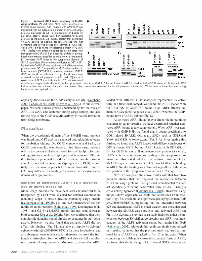
As activated ARF1 did not play a direct role in recruitingcoatomer to cargo proteins, we next determined whether acti-vated ARF1 bound to any cargo protein. When ARF1 was acti-vated with GMP-PNP, we found that it bound specifically toCOPI-related SNAREs (Xu et al., 2002), such as GS15 andYkt6, and GS28 to some extent (Fig. 1 A). Investigating thisfurther, we found that ARF1 loaded with different analogues ofGTP all bound GS15, but not ARF1 loaded with GDP (Fig. 1B). As GS15 is a type II transmembrane protein (Xu et al.,1997), with the amino terminus residing in the cytoplasmic do-main, we also tested whether the relative position of theSNARE sequence with respect to GST would affect its bindingto ARF1. Similar binding was observed regardless of the rela-tive position of the cytoplasmic domain of GS15 (Fig. 1 C).
Next, we compared the above results with that from twoprevious studies that had explored the interaction betweenARF1 and cargo proteins. First, p23 had been detected to inter-act specifically with the deactivated form of ARF1 using across-linking approach (Gommel et al., 2001). However, usingthe pull-down approach, we could not detect such an interac-tion (Fig. S3, available at http://www.jcb.org/cgi/content/full/jcb.200404008/DC1), suggesting that the interaction betweenp23 and deactivated ARF1 is much weaker than those detectedbetween the SNARE cargo proteins and activated ARF1 (inFig. 1 A). Second, a previous yeast study had shown that the in-teraction between SNARE cargo proteins and ARF1 was inde-pendent of the ARF1 activation status, but required its GAP(Rein et al., 2002). Although this result seemingly contradictedour results, we noted that the previous study had used a trun-cated form of ARF1 that lacked its first 17 amino acids. Whencomparing the full-length versus the truncated form of ARF1,we found that the full-length ARF1 bound GS15, whereas the
Figure 1. Activated ARF1 binds selectively to SNAREcargo proteins. (A) Activated ARF1 binds selectively toSNARE cargo proteins. ARF1 loaded with GMP-PNP wasincubated with the cytoplasmic domain of different cargoproteins expressed as GST fusion proteins on beads forpull-down assays. Beads were then assessed for boundproteins as indicated. GST fusion protein that containedVHSGAT served as positive control, whereas one thatcontained VHS served as negative control. (B) Only acti-vated ARF1 binds to the cytoplasmic domain of GS15.ARF1 loaded with different nucleotides as indicated wasincubated with GST-GS15 on beads for pull-down assays.Beads were then assessed for bound proteins as indicated.(C) Activated ARF1 binds to the cytoplasmic domain ofGS-15 regardless of its orientation of fusion to GST. ARF1loaded with GMP-PNP was incubated with different fusionproteins (with GS15 appended to GST either at the car-boxy terminus [GST-GS15] or amino terminus [GS15-GST]) on beads for pull-down assays. Beads were thenassessed for bound proteins as indicated. (D) An acti-vated form of ARF1 that lacks the first 17 amino-terminalresidues no longer binds efficiently to the cytoplasmic domain of GS15. Different forms of ARF1 loaded with GMP-PNP were incubated with GSTfusion proteins as indicated for pull-down assays. Beads were then assessed for bound proteins as indicated. White lines indicate that interveninglanes have been spliced out.
ARFGAP1 COUPLES CARGO SORTING WITH VESICLE FORMATION • LEE ET AL.
283
truncated ARF1 had markedly reduced capacity (Fig. 1 D). Torule out that this difference was due to differential activation ofthe two forms of ARF1, we found that both forms boundequally well to the GGA effector domain (Fig. 1 D). Thus, weconcluded that the full-length form of ARF1 was needed for itsactivation-dependent interaction with SNARE cargo proteins.
GAP regulates directly the binding of coatomer to cargo proteins
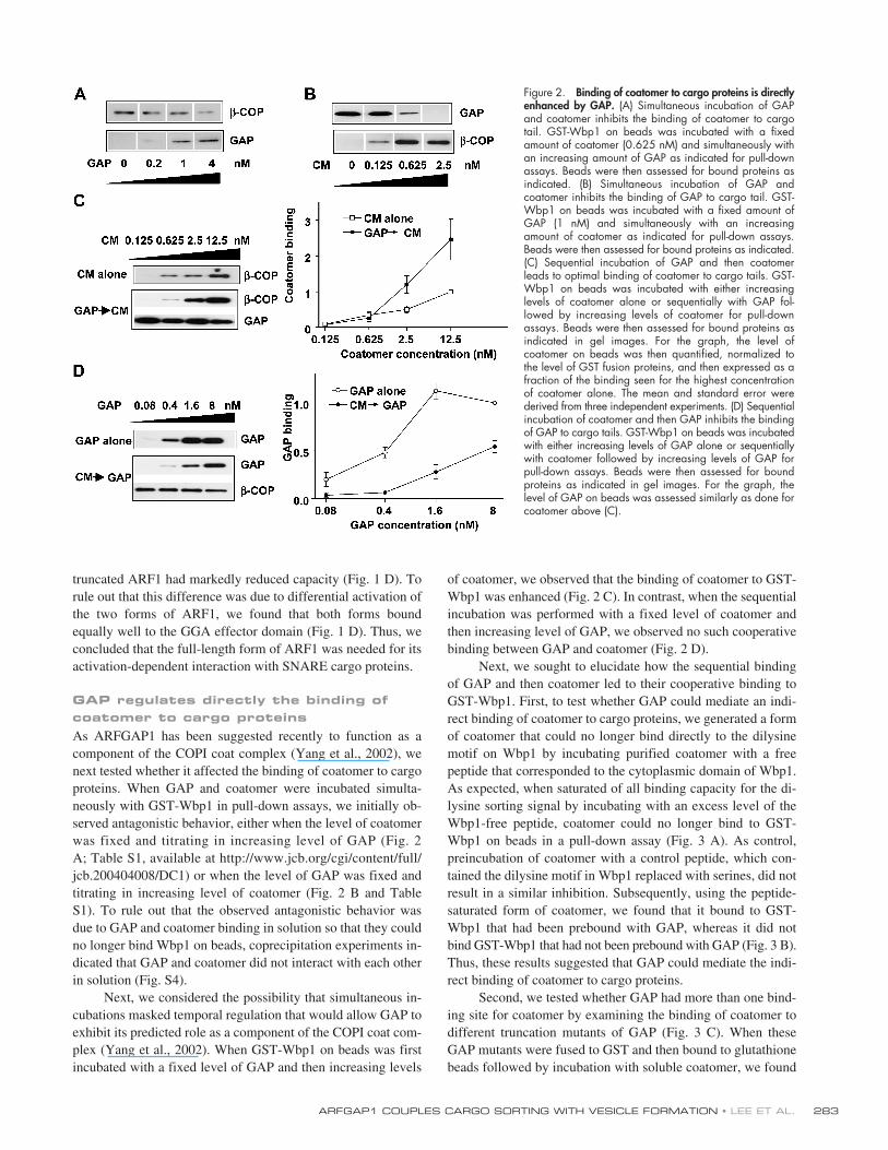
As ARFGAP1 has been suggested recently to function as acomponent of the COPI coat complex (Yang et al., 2002), wenext tested whether it affected the binding of coatomer to cargoproteins. When GAP and coatomer were incubated simulta-neously with GST-Wbp1 in pull-down assays, we initially ob-served antagonistic behavior, either when the level of coatomerwas fixed and titrating in increasing level of GAP (Fig. 2A; Table S1, available at http://www.jcb.org/cgi/content/full/jcb.200404008/DC1) or when the level of GAP was fixed andtitrating in increasing level of coatomer (Fig. 2 B and TableS1). To rule out that the observed antagonistic behavior wasdue to GAP and coatomer binding in solution so that they couldno longer bind Wbp1 on beads, coprecipitation experiments in-dicated that GAP and coatomer did not interact with each otherin solution (Fig. S4).
Next, we considered the possibility that simultaneous in-cubations masked temporal regulation that would allow GAP toexhibit its predicted role as a component of the COPI coat com-plex (Yang et al., 2002). When GST-Wbp1 on beads was firstincubated with a fixed level of GAP and then increasing levels
of coatomer, we observed that the binding of coatomer to GST-Wbp1 was enhanced (Fig. 2 C). In contrast, when the sequentialincubation was performed with a fixed level of coatomer andthen increasing level of GAP, we observed no such cooperativebinding between GAP and coatomer (Fig. 2 D).
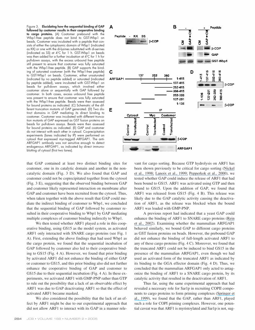
Next, we sought to elucidate how the sequential bindingof GAP and then coatomer led to their cooperative binding toGST-Wbp1. First, to test whether GAP could mediate an indi-rect binding of coatomer to cargo proteins, we generated a formof coatomer that could no longer bind directly to the dilysinemotif on Wbp1 by incubating purified coatomer with a freepeptide that corresponded to the cytoplasmic domain of Wbp1.As expected, when saturated of all binding capacity for the di-lysine sorting signal by incubating with an excess level of theWbp1-free peptide, coatomer could no longer bind to GST-Wbp1 on beads in a pull-down assay (Fig. 3 A). As control,preincubation of coatomer with a control peptide, which con-tained the dilysine motif in Wbp1 replaced with serines, did notresult in a similar inhibition. Subsequently, using the peptide-saturated form of coatomer, we found that it bound to GST-Wbp1 that had been prebound with GAP, whereas it did notbind GST-Wbp1 that had not been prebound with GAP (Fig. 3 B).Thus, these results suggested that GAP could mediate the indi-rect binding of coatomer to cargo proteins.
Second, we tested whether GAP had more than one bind-ing site for coatomer by examining the binding of coatomer todifferent truncation mutants of GAP (Fig. 3 C). When theseGAP mutants were fused to GST and then bound to glutathionebeads followed by incubation with soluble coatomer, we found
Figure 2. Binding of coatomer to cargo proteins is directlyenhanced by GAP. (A) Simultaneous incubation of GAPand coatomer inhibits the binding of coatomer to cargotail. GST-Wbp1 on beads was incubated with a fixedamount of coatomer (0.625 nM) and simultaneously withan increasing amount of GAP as indicated for pull-downassays. Beads were then assessed for bound proteins asindicated. (B) Simultaneous incubation of GAP andcoatomer inhibits the binding of GAP to cargo tail. GST-Wbp1 on beads was incubated with a fixed amount ofGAP (1 nM) and simultaneously with an increasingamount of coatomer as indicated for pull-down assays.Beads were then assessed for bound proteins as indicated.(C) Sequential incubation of GAP and then coatomerleads to optimal binding of coatomer to cargo tails. GST-Wbp1 on beads was incubated with either increasinglevels of coatomer alone or sequentially with GAP fol-lowed by increasing levels of coatomer for pull-downassays. Beads were then assessed for bound proteins asindicated in gel images. For the graph, the level ofcoatomer on beads was then quantified, normalized tothe level of GST fusion proteins, and then expressed as afraction of the binding seen for the highest concentrationof coatomer alone. The mean and standard error werederived from three independent experiments. (D) Sequentialincubation of coatomer and then GAP inhibits the bindingof GAP to cargo tails. GST-Wbp1 on beads was incubatedwith either increasing levels of GAP alone or sequentiallywith coatomer followed by increasing levels of GAP forpull-down assays. Beads were then assessed for boundproteins as indicated in gel images. For the graph, thelevel of GAP on beads was assessed similarly as done forcoatomer above (C).
JCB • VOLUME 168 • NUMBER 2 • 2005284
that GAP contained at least two distinct binding sites forcoatomer, one in its catalytic domain and another in the non-catalytic domain (Fig. 3 D). We also found that GAP andcoatomer could not be coprecipitated together from the cytosol(Fig. 3 E), suggesting that the observed binding between GAPand coatomer likely represented interaction on membrane afterGAP and coatomer have been recruited from the cytosol. Thus,when taken together with the above result that GAP could me-diate the indirect binding of coatomer to Wbp1, we concludedthat the sequential binding of GAP followed by coatomer re-sulted in their cooperative binding to Wbp1 by GAP mediatingmultiple complexes of coatomer binding indirectly to Wbp1.
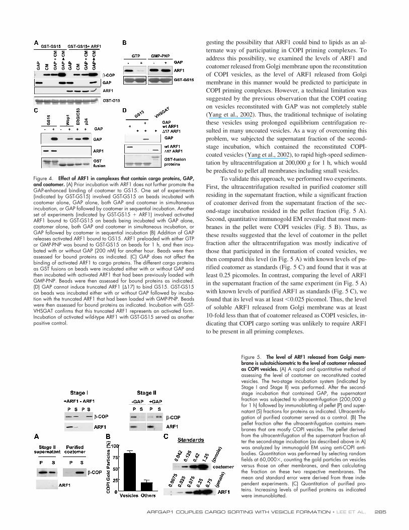
We then tested whether ARF1 played a role in this coop-erative binding, using GS15 as the model system, as activatedARF1 only interacted with SNARE cargo proteins (see Fig. 1A). First, extending the above findings that had used Wbp1 asthe cargo protein, we found that the sequential incubation ofGAP followed by coatomer also led to their cooperative bind-ing to GS15 (Fig. 4 A). However, we found that prior bindingby activated ARF1 did not enhance the binding of either GAPor coatomer to GS15, and this prior binding also did not furtherenhance the cooperative binding of GAP and coatomer toGS15 due to their sequential incubation (Fig. 4 A). In these ex-periments, we activated ARF1 with GMP-PNP rather than GTPto rule out the possibility that a lack of an observable effect byARF1 was due to GAP deactivating ARF1 so that the effect ofactivated ARF1 became masked.
We also considered the possibility that the lack of an ef-fect by ARF1 might be due to our experimental approach thatdid not allow ARF1 to interact with its GAP in a manner rele-
vant for cargo sorting. Because GTP hydrolysis on ARF1 hasbeen shown previously to be critical for cargo sorting (Nickelet al., 1998; Lanoix et al., 1999; Pepperkok et al., 2000), wetested whether GAP could induce the release of ARF1 that hadbeen bound to GS15. ARF1 was activated using GTP and thenbound to GS15. Upon the addition of GAP, we found thatARF1 was released from GS15 (Fig. 4 B). This release waslikely due to the GAP catalytic activity causing the deactiva-tion of ARF1, as the release was blocked when the boundARF1 was loaded with GMP-PNP.
A previous report had indicated that a yeast GAP couldenhance the binding of ARF1 to SNARE cargo proteins (Reinet al., 2002). Examining whether the mammalian ARFGAP1behaved similarly, we bound GAP to different cargo proteinsas GST fusion proteins on beads. However, the prebound GAPdid not enhance the binding of full-length activated ARF1 toany of these cargo proteins (Fig. 4 C). Moreover, we found thatthe truncated ARF1 could not be induced to bind GS15 in thepresence of the mammalian ARFGAP1, even though we hadused an activated form of the truncated ARF1 as indicated byits binding to the GGA effector domain (Fig. 4 D). Thus, weconcluded that the mammalian ARFGAP1 only acted to antag-onize the binding of ARF1 to a SNARE cargo protein, by itscatalytic activity that resulted in the deactivation of ARF1.
Thus far, using the same experimental approach that hadrevealed a necessary role for Sar1p in recruiting COPII compo-nents to cargo proteins to form priming complexes (Springer etal., 1999), we found that the GAP, rather than ARF1, playedsuch a role for COPI priming complexes. However, one poten-tial caveat was that ARF1 is myristoylated and Sar1p is not, sug-
Figure 3. Elucidating how the sequential binding of GAPfollowed by coatomer results in their cooperative bindingto cargo proteins. (A) Coatomer prebound with theWbp1-free peptide does not bind to GST-Wbp1 onbeads. Coatomer was incubated with a peptide that con-sists of either the cytoplasmic domain of Wbp1 (indicatedas KK) or one with the di-lysines substituted with di-serines(indicated as SS) at 4�C for 1 h. GST-Wbp1 on beadswas then added for a further incubation at 4�C for 1 h forpull-down assays, with the excess unbound free peptidestill present to ensure that coatomer was fully saturatedwith the Wbp1-free peptide. (B) GAP supports the bind-ing of saturated coatomer (with the Wbp1-free peptide)to GST-Wbp1 on beads. Coatomer, either unsaturated(indicated by no peptide added) or saturated (indicatedby peptide added), were incubated with GST-Wbp1 onbeads for pull-down assays, which involved eithercoatomer alone or sequentially with GAP followed bycoatomer. In both cases, excess unbound free peptidewas present to ensure that coatomer was fully saturatedwith the Wbp1-free peptide. Beads were then assessedfor bound proteins as indicated. (C) Schematic of the dif-ferent truncation mutants of GAP generated. (D) Two dis-tinct domains in GAP mediating its direct binding tocoatomer. Coatomer was incubated with different trunca-tion mutants of GAP expressed as GST fusion proteins onbeads for pull-down assays. Beads were then assessedfor bound proteins as indicated. (E) GAP and coatomerdo not interact with each other in cytosol. Co-precipitationexperiments (lanes indicated by IP) were performed oncytosol that expressed myc-tagged ARFGAP1. The anti-ARFGAP1 antibody was not sensitive enough to detectendogenous ARFGAP1, as indicated by direct immuno-blotting of cytosol (first two lanes).
ARFGAP1 COUPLES CARGO SORTING WITH VESICLE FORMATION • LEE ET AL.
285
gesting the possibility that ARF1 could bind to lipids as an al-ternate way of participating in COPI priming complexes. Toaddress this possibility, we examined the levels of ARF1 andcoatomer released from Golgi membrane upon the reconstitutionof COPI vesicles, as the level of ARF1 released from Golgimembrane in this manner would be predicted to participate inCOPI priming complexes. However, a technical limitation wassuggested by the previous observation that the COPI coatingon vesicles reconstituted with GAP was not completely stable(Yang et al., 2002). Thus, the traditional technique of isolatingthese vesicles using prolonged equilibrium centrifugation re-sulted in many uncoated vesicles. As a way of overcoming thisproblem, we subjected the supernatant fraction of the second-stage incubation, which contained the reconstituted COPI-coated vesicles (Yang et al., 2002), to rapid high-speed sedimen-tation by ultracentrifugation at 200,000
g
for 1 h, which wouldbe predicted to pellet all membranes including small vesicles.
To validate this approach, we performed two experiments.First, the ultracentrifugation resulted in purified coatomer stillresiding in the supernatant fraction, while a significant fractionof coatomer derived from the supernatant fraction of the sec-ond-stage incubation resided in the pellet fraction (Fig. 5 A).Second, quantitative immunogold EM revealed that most mem-branes in the pellet were COPI vesicles (Fig. 5 B). Thus, asthese results suggested that the level of coatomer in the pelletfraction after the ultracentrifugation was mostly indicative ofthose that participated in the formation of coated vesicles, wethen compared this level (in Fig. 5 A) with known levels of pu-rified coatomer as standards (Fig. 5 C) and found that it was atleast 0.25 picomoles. In contrast, comparing the level of ARF1in the supernatant fraction of the same experiment (in Fig. 5 A)with known levels of purified ARF1 as standards (Fig. 5 C), wefound that its level was at least
�
0.025 picomol. Thus, the levelof soluble ARF1 released from Golgi membrane was at least10-fold less than that of coatomer released as COPI vesicles, in-dicating that COPI cargo sorting was unlikely to require ARF1to be present in all priming complexes.
Figure 4. Effect of ARF1 in complexes that contain cargo proteins, GAP,and coatomer. (A) Prior incubation with ARF1 does not further promote theGAP-enhanced binding of coatomer to GS15. One set of experiments(indicated by GST-GS15) involved GST-GS15 on beads incubated withcoatomer alone, GAP alone, both GAP and coatomer in simultaneousincubation, or GAP followed by coatomer in sequential incubation. Anotherset of experiments (indicated by GST-GS15 � ARF1) involved activatedARF1 bound to GST-GS15 on beads being incubated with GAP alone,coatomer alone, both GAP and coatomer in simultaneous incubation, orGAP followed by coatomer in sequential incubation (B) Addition of GAPreleases activated ARF1 bound to GS15. ARF1 preloaded with either GTPor GMP-PNP was bound to GST-GS15 on beads for 1 h, and then incu-bated with or without GAP (200 nM) for another hour. Beads were thenassessed for bound proteins as indicated. (C) GAP does not affect thebinding of activated ARF1 to cargo proteins. The different cargo proteinsas GST fusions on beads were incubated either with or without GAP andthen incubated with activated ARF1 that had been previously loaded withGMP-PNP. Beads were then assessed for bound proteins as indicated.(D) GAP cannot induce truncated ARF1 (�17) to bind GS15. GST-GS15on beads was incubated either with or without GAP followed by incuba-tion with the truncated ARF1 that had been loaded with GMP-PNP. Beadswere then assessed for bound proteins as indicated. Incubation with GST-VHSGAT confirms that this truncated ARF1 represents an activated form.Incubation of activated wild-type ARF1 with GST-GS15 served as anotherpositive control.
Figure 5. The level of ARF1 released from Golgi mem-brane is substoichiometric to the level of coatomer releasedas COPI vesicles. (A) A rapid and quantitative method ofassessing the level of coatomer on reconstituted coatedvesicles. The two-stage incubation system (indicated byStage I and Stage II) was performed. After the second-stage incubation that contained GAP, the supernatantfraction was subjected to ultracentrifugation (200,000 gfor 1 h) followed by immunoblotting of pellet (P) and super-natant (S) fractions for proteins as indicated. Ultracentrifu-gation of purified coatomer served as a control. (B) Thepellet fraction after the ultracentrifugation contains mem-branes that are mostly COPI vesicles. The pellet derivedfrom the ultracentrifugation of the supernatant fraction af-ter the second-stage incubation (as described above in A)was analyzed by immunogold EM using anti-COPI anti-bodies. Quantitation was performed by selecting randomfields at 60,000�, counting the gold particles on vesiclesversus those on other membranes, and then calculatingthe fraction on these two respective membranes. Themean and standard error were derived from three inde-pendent experiments. (C) Quantitation of purified pro-teins. Increasing levels of purified proteins as indicatedwere immunoblotted.
JCB • VOLUME 168 • NUMBER 2 • 2005286
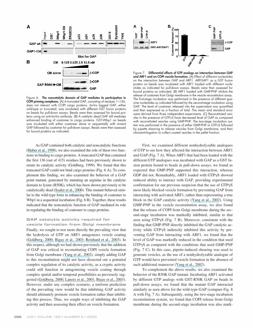
As GAP contained both catalytic and noncatalytic functions(Huber et al., 1998), we also examined the role of these two func-tions in binding to cargo proteins. A truncated GAP that containedthe first 136 (out of 415) residues had been previously shown toretain its catalytic activity (Goldberg, 1999). We found that thistruncated GAP could not bind cargo proteins (Fig. 6 A). To com-plement this finding, we also examined the behavior of a GAPpoint mutant, generated by mutating an arginine in the catalyticdomain to lysine (R50K), which has been shown previously to becatalytically dead (Szafer et al., 2000). This mutant behaved simi-lar to the wild-type form in enhancing the binding of coatomer toWbp1 in a sequential incubation (Fig. 6 B). Together, these resultsindicated that the noncatalytic function of GAP mediated its rolein regulating the binding of coatomer to cargo proteins.
GAP catalytic activity required for vesicle formation from Golgi membrane
Finally, we sought to test more directly the prevailing view thatthe hydrolysis of GTP on ARF1 antagonizes vesicle coating(Goldberg, 2000; Bigay et al., 2003; Reinhard et al., 2003). Inthis respect, although we had shown previously that the additionof GAP was critical in reconstituting COPI vesicle formationfrom Golgi membrane (Yang et al., 2002), simply adding GAPto this reconstitution might not have dissected out a potentialcomplex regulation of its catalytic activity, as a cryptic activitycould still function in antagonizing vesicle coating throughcomplex spatial and/or temporal possibilities as previously sug-gested (Goldberg, 2000; Lanoix et al., 2001; Bigay et al., 2003).However, under any complex scenario, a uniform predictionof the prevailing view would be that inhibiting GAP activityshould ultimately promote vesicle formation rather than inhibit-ing this process. Thus, we sought ways of inhibiting the GAPactivity and then assessing their effect on vesicle formation.
First, we examined different nonhydrolyzable analoguesof GTP to see how they affected the interaction between ARF1and GAP (Fig. 7 A). When ARF1 that had been loaded with thedifferent GTP analogues was incubated with GAP as a GST fu-sion protein bound to beads in pull-down assays, we found asexpected that GMP-PNP supported this interaction, whereasGDP did not. Remarkably, ARF1 loaded with GTP
�
S showedreduced ability to interact with GAP, providing experimentalconfirmation for our previous suspicion that the use of GTP
�
Smost likely blocked vesicle formation by preventing GAP frominteracting with activated ARF1, rather than representing a trueblock in the GAP catalytic activity (Yang et al., 2002). UsingGMP-PNP in the vesicle reconstitution assay, we also foundthat the release of COPI from Golgi membrane during the sec-ond-stage incubation was markedly inhibited, similar to thatseen using GTP
�
S (Fig. 7 B). Moreover, consistent with thefinding that GMP-PNP directly inhibited the GAP catalytic ac-tivity while GTP
�
S indirectly inhibited this activity by pre-venting GAP from interacting with ARF1, we found that thelevel of GAP was markedly reduced in the condition that usedGTP
�
S as compared with the conditions that used GMP-PNP(Fig. 7 C). In this case, pipette-induced shearing was used togenerate vesicles, as the use of a nonhydrolyzable analogue ofGTP would have prevented vesicle formation in the absence ofsuch additional maneuver (Yang et al., 2002).
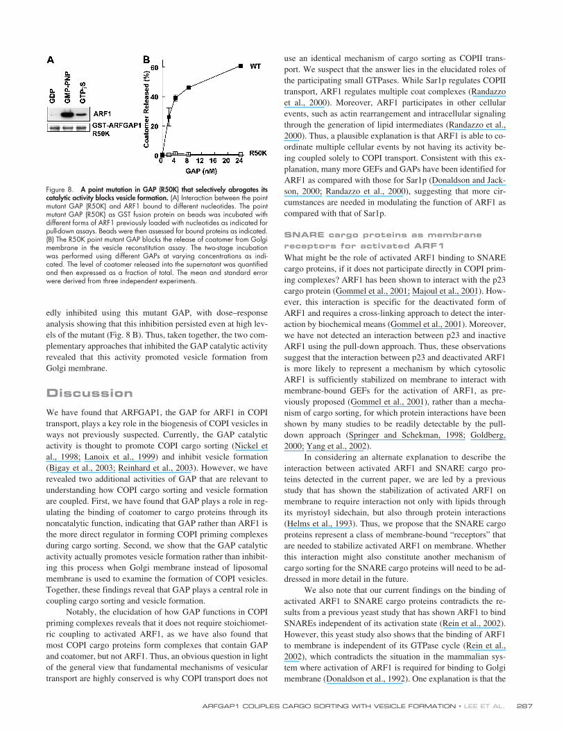
To complement the above results, we also examined thebehavior of the R50K GAP mutant. Incubating ARF1 activatedby different GTP analogs with GST-R50K GAP on beads inpull-down assays, we found that the mutant GAP interactedsimilarly as seen above for the wild-type GAP (compare Fig. 8A with Fig. 7 A). Subsequently, using the two-stage incubationreconstitution system, we found that COPI release from Golgimembrane during the second-stage incubation was also mark-
Figure 6. The noncatalytic domain of GAP mediates its participation inCOPI priming complexes. (A) A truncated GAP, consisting of residues 1–136,does not interact with COPI cargo proteins. 6x-his tagged GAP, eitherwild-type or truncated, was incubated with different GST fusion proteinson beads for pull-down assays. Beads were then assessed for bound pro-teins using an anti-6x-his antibody. (B) A catalytic dead GAP still mediatesenhanced binding of coatomer to cargo proteins. GST-Wbp1 on beadswas incubated with either coatomer alone or sequentially with mutantGAP followed by coatomer for pull-down assays. Beads were then assessedfor bound proteins as indicated.
Figure 7. Differential effects of GTP analogs on interaction between GAPand ARF1 and on COPI vesicle formation. (A) Effect of different nucleotideson the interaction between GAP and ARF1. ARFGAP1 as a GST fusionprotein on beads was incubated with ARF1 loaded with different nucle-otides as indicated for pull-down assays. Beads were then assessed forbound proteins as indicated. (B) ARF1 loaded with GMP-PNP inhibits therelease of coatomer from Golgi membrane in the vesicle reconstitution assay.The first-stage incubation was performed in the presence of different gua-nine nucleotides as indicated followed by the second-stage incubation usingGAP. The level of coatomer released into the supernatant was quantifiedand then expressed as a fraction of total. The mean and standard errorwere derived from three independent experiments. (C) Reconstituted vesi-cles in the presence of GTP�S have decreased level of GAP as comparedwith reconstituted vesicles using GMP-PNP. The two-stage incubation sys-tem was performed in the presence of either GMP-PNP or GTP�S followedby pipette shearing to release vesicles from Golgi membrane, and thenultracentrifugation to collect coated vesicles in the pellet fraction.
ARFGAP1 COUPLES CARGO SORTING WITH VESICLE FORMATION • LEE ET AL.
287
edly inhibited using this mutant GAP, with dose–responseanalysis showing that this inhibition persisted even at high lev-els of the mutant (Fig. 8 B). Thus, taken together, the two com-plementary approaches that inhibited the GAP catalytic activityrevealed that this activity promoted vesicle formation fromGolgi membrane.
Discussion
We have found that ARFGAP1, the GAP for ARF1 in COPItransport, plays a key role in the biogenesis of COPI vesicles inways not previously suspected. Currently, the GAP catalyticactivity is thought to promote COPI cargo sorting (Nickel etal., 1998; Lanoix et al., 1999) and inhibit vesicle formation(Bigay et al., 2003; Reinhard et al., 2003). However, we haverevealed two additional activities of GAP that are relevant tounderstanding how COPI cargo sorting and vesicle formationare coupled. First, we have found that GAP plays a role in reg-ulating the binding of coatomer to cargo proteins through itsnoncatalytic function, indicating that GAP rather than ARF1 isthe more direct regulator in forming COPI priming complexesduring cargo sorting. Second, we show that the GAP catalyticactivity actually promotes vesicle formation rather than inhibit-ing this process when Golgi membrane instead of liposomalmembrane is used to examine the formation of COPI vesicles.Together, these findings reveal that GAP plays a central role incoupling cargo sorting and vesicle formation.
Notably, the elucidation of how GAP functions in COPIpriming complexes reveals that it does not require stoichiomet-ric coupling to activated ARF1, as we have also found thatmost COPI cargo proteins form complexes that contain GAPand coatomer, but not ARF1. Thus, an obvious question in lightof the general view that fundamental mechanisms of vesiculartransport are highly conserved is why COPI transport does not
use an identical mechanism of cargo sorting as COPII trans-port. We suspect that the answer lies in the elucidated roles ofthe participating small GTPases. While Sar1p regulates COPIItransport, ARF1 regulates multiple coat complexes (Randazzoet al., 2000). Moreover, ARF1 participates in other cellularevents, such as actin rearrangement and intracellular signalingthrough the generation of lipid intermediates (Randazzo et al.,2000). Thus, a plausible explanation is that ARF1 is able to co-ordinate multiple cellular events by not having its activity be-ing coupled solely to COPI transport. Consistent with this ex-planation, many more GEFs and GAPs have been identified forARF1 as compared with those for Sar1p (Donaldson and Jack-son, 2000; Randazzo et al., 2000), suggesting that more cir-cumstances are needed in modulating the function of ARF1 ascompared with that of Sar1p.
SNARE cargo proteins as membrane receptors for activated ARF1
What might be the role of activated ARF1 binding to SNAREcargo proteins, if it does not participate directly in COPI prim-ing complexes? ARF1 has been shown to interact with the p23cargo protein (Gommel et al., 2001; Majoul et al., 2001). How-ever, this interaction is specific for the deactivated form ofARF1 and requires a cross-linking approach to detect the inter-action by biochemical means (Gommel et al., 2001). Moreover,we have not detected an interaction between p23 and inactiveARF1 using the pull-down approach. Thus, these observationssuggest that the interaction between p23 and deactivated ARF1is more likely to represent a mechanism by which cytosolicARF1 is sufficiently stabilized on membrane to interact withmembrane-bound GEFs for the activation of ARF1, as pre-viously proposed (Gommel et al., 2001), rather than a mecha-nism of cargo sorting, for which protein interactions have beenshown by many studies to be readily detectable by the pull-down approach (Springer and Schekman, 1998; Goldberg,2000; Yang et al., 2002).
In considering an alternate explanation to describe theinteraction between activated ARF1 and SNARE cargo pro-teins detected in the current paper, we are led by a previousstudy that has shown the stabilization of activated ARF1 onmembrane to require interaction not only with lipids throughits myristoyl sidechain, but also through protein interactions(Helms et al., 1993). Thus, we propose that the SNARE cargoproteins represent a class of membrane-bound “receptors” thatare needed to stabilize activated ARF1 on membrane. Whetherthis interaction might also constitute another mechanism ofcargo sorting for the SNARE cargo proteins will need to be ad-dressed in more detail in the future.
We also note that our current findings on the binding ofactivated ARF1 to SNARE cargo proteins contradicts the re-sults from a previous yeast study that has shown ARF1 to bindSNAREs independent of its activation state (Rein et al., 2002).However, this yeast study also shows that the binding of ARF1to membrane is independent of its GTPase cycle (Rein et al.,2002), which contradicts the situation in the mammalian sys-tem where activation of ARF1 is required for binding to Golgimembrane (Donaldson et al., 1992). One explanation is that the
Figure 8. A point mutation in GAP (R50K) that selectively abrogates itscatalytic activity blocks vesicle formation. (A) Interaction between the pointmutant GAP (R50K) and ARF1 bound to different nucleotides. The pointmutant GAP (R50K) as GST fusion protein on beads was incubated withdifferent forms of ARF1 previously loaded with nucleotides as indicated forpull-down assays. Beads were then assessed for bound proteins as indicated.(B) The R50K point mutant GAP blocks the release of coatomer from Golgimembrane in the vesicle reconstitution assay. The two-stage incubationwas performed using different GAPs at varying concentrations as indi-cated. The level of coatomer released into the supernatant was quantifiedand then expressed as a fraction of total. The mean and standard errorwere derived from three independent experiments.
JCB • VOLUME 168 • NUMBER 2 • 2005288
behavior of ARF1 in yeast is fundamentally different than thatelucidated for the mammalian system. However, another possi-bility is based on technical considerations. The yeast mem-branes used in the previous study are microsomes developedoriginally to study the formation of COPII vesicles (Rein et al.,2002), suggesting the possibility that the ER membrane mightcontribute to the noted curious behavior of ARF1. Another pos-sibility is that a truncated form of ARF1 that lacked the first 17amino acids was used in the previous study (Rein et al., 2002),whereas in the current study we have found that ARF1 bindsselectively to SNARE cargo proteins only when ARF1 is a full-length activated protein. Thus, these technical possibilities willneed to be resolved before a firm conclusion can be made thatthe noted disparities for ARF1 and its GAP in yeast and mam-malian systems represent true differences in function.
GAP activity promotes vesicle formation with implications for considering mechanisms of cargo sorting
Using a COPI vesicle reconstitution system that involves incu-bating Golgi membrane with purified components, we haveprovided evidence recently that GAP functions to promote bothcargo sorting and vesicle formation (Yang et al., 2002), imply-ing that its catalytic activity does not antagonize vesicle coat-ing. However, in two subsequent studies that use similar puri-fied protein components, but in the context of liposomes ratherthan Golgi membrane, GAP is found to antagonize vesicle for-mation and promote vesicle uncoating (Bigay et al., 2003;Reinhard et al., 2003). In the current study, we have providedmore precise ways of perturbing the GAP catalytic activity,such as the use of GMP-PNP and a GAP catalytic point mutant,as they block GAP activity without preventing GAP from inter-acting with ARF1. These perturbations profoundly affect theformation of COPI vesicles from Golgi membrane. Thus, thedisparity begs the question as to why the GAP activity func-tions so differently on liposomes as compared with Golgimembrane. As the obvious difference is that biological mem-branes contain many membrane proteins, both integral and pe-ripheral, a prediction is that one or more of these proteins playa role in aiding GAP to function in vesicle formation ratherthan in vesicle uncoating.
Our finding also indicates a need to revise the prevailingview that the translocation of coatomer between membrane andcytosol is strictly coupled to the GTPase cycle of ARF1 (Gold-berg, 2000; Bigay et al., 2003; Reinhard et al., 2003). This“strict coupling” view has been challenged by a study usinglive cells (Presley et al., 2002). However, the conclusion of thatstudy is limited by the caveat that ARF1 has multiple functions,including regulating multiple coat proteins and other cellularactivities such as signaling and actin rearrangement (Donald-son and Jackson, 2000; Randazzo et al., 2000). Thus, one can-not rule out that a strict coupling mechanism indeed describesthe pool of ARF1 that specifically participates in COPI trans-port. By addressing this caveat in the current study using a ves-icle reconstitution system that specifically examines the role ofARF1 in COPI transport, we conclude that substantial experi-mental evidence now exists against the strict coupling view.
The significance of discarding the strict coupling view isthat one no longer needs to consider the GAP catalytic activity,which is required for the deactivation of ARF1, as antagonisticto vesicle formation. This conclusion also suggests a differentcontext to explain some key recent experimental observations re-garding the regulation of GAP activity. In one report, coatomeris noted to enhance the catalytic activity of GAP, and a specificp24 family member inhibits this enhancement (Goldberg, 2000).These observations have led to a kinetic proofreading modelwhereby the GAP catalytic activity can be inhibited during vesi-cle formation, so that the two seemingly dichotomous functionsof GAP, in promoting cargo sorting and inhibiting vesicle coat-ing, can be satisfied (Goldberg, 2000). However, this model pre-dicts a critical role for the p24 family member in vesicle forma-tion, and yet, a yeast study has shown that the entire p24 familymembers can be deleted with minimal effects on transport(Springer et al., 2000). In another report, positive membrane cur-vature is noted to enhance the catalytic activity of GAP (Bigay etal., 2003), which has been interpreted in the context of vesicleuncoating. However, this interpretation predicts that the tip ofthe forming bud would be prone to releasing its coating, as it issubjected to increasingly high positive curvature that enhancesGAP activity. To accommodate this problem, the study proposesthat lateral interactions between coat components compensatesfor those ARF1 that would have been released at the bud tip, sothat uncoating does not occur before vesicle fission (Bigay et al.,2003). Such an explanation essentially contradicts the generalpremise that membrane localization of coatomer is strictly cou-pled to the GTPase cycle of ARF1, as the explanation invokes alocal exception to the rule. In contrast, for both cited examples,the noted difficulties in relating the experimental observationswith mechanistic models would be alleviated if one views theGAP catalytic activity to promote vesicle formation.
Materials and methods
Plasmids
Constructs encoding for the cytoplasmic domain of cargo proteins wereappended to the carboxy terminus of GST and were generated usingeither pGEX-KG or pGEX 4T-3 expression plasmids (Amersham Biosci-ences), or to the amino terminus of GST using pETGEX (from R. Scheckman,University of California, Berkeley, Berkeley, CA). Constructs encodingERGIC53 (from H. Hauri, University of Basel, Basel, Switzerland), andp23, p24, p25, and p26 (from J. Gruenberg, University of Geneva,Geneva, Switzerland) were used in the PCR to append their cytoplasmicdomains to GST. Other GST fusion constructs have been described previ-ously: GST-Wbp1 (Cosson and Letourneur, 1994), GST-KDELR (Yang etal., 2002), GST-VHS and GST-VHSGAT (Dell’Angelica et al., 2000),GS15 (Xu et al., 1997), GS28 (Subramaniam et al., 1996), Ykt6 (Zhangand Hong, 2001), Syntaxin3 (Wong et al., 1999), and Syntaxin5 (Wonget al., 1999) fused to GST.
Construct encoding for ARFGAP1 fused to GST was generated inpGEX 4T-3, with silent mutations introduced in the ORF (AGA to CGC en-coding for amino acid residues 5 and 7 in GAP, and AGG to CGC forresidues 357 and 358) using the PCR-based mutagenesis approach, sothat rare codon usage in bacteria is avoided (Makrides, 1996). His-tagged GAP1-136 was subcloned into pTrcHis (Invitrogen). Full-lengthARFGAP1 point mutant (R50K) was generated through PCR mutagenesisof the wild-type GAP previously subcloned in the baculovirus transfer vec-tor pVL1393 (BD Biosciences).
Proteins
Previously described purification of proteins include: coatomer (Pavel etal., 1998), ARFGAP1 (Vitale et al., 2000), and ARF1 (Randazzo, 1997).
ARFGAP1 COUPLES CARGO SORTING WITH VESICLE FORMATION • LEE ET AL.
289
His-tagged, full-length, R50K point mutant GAP was expressed in a bacu-lovirus system, as described previously for the wild-type form (Vitale et al.,2000). Truncated ARFGAP1 (1–136) and GST fusion proteins were ex-pressed using a bacterial expression system and then purified, as de-scribed previously (Yang et al., 2002). Truncated ARF1 (
�
17) was ob-tained from P. Randazzo (National Institutes of Health, Bethesda, MD).
Other materials
Nucleotides (Sigma-Aldrich) and free peptides (New England Peptide) ofthe cytoplasmic domain of Wbp1 (KKLETFKKTN) and its corresponding di-serine mutant (KKLETFSSTN) were obtained. Saturation of coatomer withthe Wbp1-free peptide was accomplished by incubating 2.5 nM coatomerwith 300
�
M free peptide. Antibodies used have been described previ-ously: mouse CM1A10 against coatomer (Palmer et al., 1993), mouseM3A5 against
�
-COP (Allan and Kreis, 1986), rabbit antibody againstARF1 (Marshansky et al., 1997), and rabbit antibody against ARFGAP1(Cukierman et al., 1995). The following antibodies were obtained fromcommercial sources: anti-six-histidine (6x-his) epitope (Santa Cruz Biotech-nology, Inc.) and anti-myc epitope (9E10; American Type Culture Collec-tion, Manassas, VA). Cytosol from transfected cells were prepared by per-meabilizing cells with 0.2% saponin in PBS at RT for 10 min and thencollecting the supernatant fraction after centrifugation at 15,000
g
for 10min. Transfections were performed using FuGENE 6 (Roche).
Loading ARF1 with guanine nucleotides
ARF1 (250 nM) was incubated at 32
�
C for 1.5 h with different nucleotidesin a buffer containing 25 mM Hepes, pH 7.2, 100 mM NaCl, 1 mM DTT,1 mM EDTA, 1 mM MgCl
2
, 0.005 g BSA/L, and 0.1% Triton X-100. Thedifferent nucleotides used were: GTP (2 mM), GDP (2 mM), GMP-PNP (20
�
M), and GTP
�
S (20
�
M).
GST pull-down assays
Incubation of cargo proteins as GST fusion proteins on beads with solubleGAP (1 nM) and/or coatomer (2.5 nM) was performed at 4
�
C for 1 h us-ing a previously described buffer (Lanoix et al., 2001) that consisted of 50mM Hepes, pH 7.2, 300 mM NaCl, 90 mM KCl, 1 mM EDTA, and 0.5%NP-40. In titration experiments, various concentrations of the titrating com-ponent are indicated in specific figures. Incubation of ARFGAP1 as a GSTfusion protein on beads with soluble coatomer (1.25 nM) was performedat 4
�
C for 1 h in a buffer containing 50 mM Hepes, pH 7.2, 90 mM KCl,2.5 mM Mg(OAc)
2
, and 1% Triton X-100. All pull-down assays involvingsoluble ARF1, interacting with GST fusions of either cargo proteins or GAPon beads, were done using the condition described above for the loadingof ARF1 with nucleotides, unless stated otherwise in figure legends. Afterthe incubations, beads were pelleted by centrifugation (500
g
for 2 min atRT) followed by three washes with the incubation buffer, and then ana-lyzed by SDS-PAGE. Western blotting was performed using chemilumines-cence (NEN Life Sciences) to detect the bound proteins. Coomassie bluestaining was performed to detect the level of GST fusion protein on beads.
Reconstitution of COPI vesicles from Golgi membrane
The two-stage incubation system was performed as described previously(Yang et al., 2002). Ultracentrifugation of the supernatant from the second-stage incubation was performed at 200,000
g
for 1 h using a rotor (modelSW55; Beckman Coulter), after having scaled-up the two-stage incubationsystem by 10-fold. The resulting supernatant was recovered by TCA precip-itation using BSA as carrier, with control experiments using a known quan-tity of purified protein revealing quantitative recovery of proteins.
Immunogold EM
Analysis of vesicles on grids was performed as described previously(Yang et al., 2002).
Image acquisition and display
Original gels were converted into digital images using a flatbed scanner(Perfection 1200U; Epson), and were then processed for figures usingAdobe Photoshop 6.0 software. White lines in figures indicate that inter-vening lanes of gels were spliced out. Quantitation of gel bands was per-formed using Scion Image software.
Online supplemental material
Fig. S1 shows that activated ARF1 does not affect the binding of coatomerto different cargo proteins. Fig. S2 shows that the ARF1 effector domain ofGGA interacts specifically with the activated forms of ARF1. Fig. S3shows that different forms of ARF1 do not interact with p23 in pull-downassays. Fig. S4 shows that purified GAP and coatomer do not interact in
solution. Table S1 quantifies the relative level of binding seen in Fig. 2.Online supplemental material available at http://www.jcb.org/cgi/content/full/jcb.200404008/DC1.
We thank Jian Li for helpful discussions and Huiya Gilbert for technical assis-tance, and also Dan Cassel and Alberto Luini for critical comments on themanuscript.
This work was supported in part by grants from the National Institutesof Health to V.W. Hsu and R.T. Premont.
Submitted: 1 April 2004Accepted: 19 November 2004
References
Allan, V.J., and T.E. Kreis. 1986. A microtubule-binding protein associatedwith membranes of the Golgi apparatus.
J. Cell Biol.
103:2229–2239.
Bigay, J., P. Gounon, S. Robineau, and B. Antonny. 2003. Lipid packing sensedby ArfGAP1 couples COPI coat disassembly to membrane bilayercurvature.
Nature.
426:563–566.
Bonifacino, J.S., and B.S. Glick. 2004. The mechanisms of vesicle budding andfusion.
Cell.
116:153–166.
Cosson, P., and F. Letourneur. 1994. Coatomer interaction with di-lysine endo-plasmic reticulum retention motifs.
Science.
263:1629–1631.
Cukierman, E., I. Huber, M. Rotman, and D. Cassel. 1995. The ARF1-GTPase-activating protein: zinc finger motif and Golgi complex localization.
Science.
270:1999–2002.
Dell’Angelica, E.C., R. Puertollano, C. Mullins, R.C. Aguilar, J.D. Vargas,L.M. Hartnell, and J.S. Bonifacino. 2000. GGAs: a family of ADP ribo-sylation factor-binding proteins related to adaptors and associated withthe Golgi complex.
J. Cell Biol.
149:81–94.
Dominguez, M., K. Dejgaard, J. Fullekrug, S. Dahan, A. Fazel, J.P. Paccaud,D.Y. Thomas, J.J. Bergeron, and T. Nilsson. 1998. gp25L/emp24/p24protein family members of the cis-Golgi network bind both COP I and IIcoatomer.
J. Cell Biol.
140:751–765.
Donaldson, J.G., and C.L. Jackson. 2000. Regulators and effectors of the ARFGTPases.
Curr. Opin. Cell Biol.
12:475–482.
Donaldson, J.G., D. Cassel, R.A. Kahn, and R.D. Klausner. 1992. ADP-ribosy-lation factor, a small GTP-binding protein, is required for binding of thecoatomer protein beta-COP to Golgi membranes.
Proc. Natl. Acad. Sci.USA.
89:6408–6412.
Fiedler, K., M. Veit, M.A. Stamnes, and J.E. Rothman. 1996. Bimodal interac-tion of coatomer with the p24 family of putative cargo receptors.
Science.
273:1396–1399.
Goldberg, J. 1999. Structural and functional analysis of the ARF1-ARFGAP com-plex reveals a role for coatomer in GTP hydrolysis.
Cell.
96:893–902.
Goldberg, J. 2000. Decoding of sorting signals by coatomer through a GTPaseswitch in the COPI coat complex.
Cell.
100:671–679.
Gommel, D.U., A.R. Memon, A. Heiss, F. Lottspeich, J. Pfannstiel, J. Lechner,C. Reinhard, J.B. Helms, W. Nickel, and F.T. Wieland. 2001. Recruit-ment to Golgi membranes of ADP-ribosylation factor 1 is mediated bythe cytoplasmic domain of p23.
EMBO J.
20:6751–6760.
Helms, J.B., D.J. Palmer, and J.E. Rothman. 1993. Two distinct populations ofARF bound to Golgi membranes.
J. Cell Biol.
121:751–760.
Huber, I., E. Cukierman, M. Rotman, T. Aoe, V.W. Hsu, and D. Cassel. 1998.Requirement for both the amino-terminal catalytic domain and a non-catalytic domain for in vivo activity of ARF1 GAP.
J. Biol. Chem.
273:24786–24791.
Lanoix, J., J. Ouwendijk, C.C. Lin, A. Stark, H.D. Love, J. Ostermann, and T.Nilsson. 1999. GTP hydrolysis by arf-1 mediates sorting and concentra-tion of Golgi resident enzymes into functional COP I vesicles.
EMBO J.
18:4935–4948.
Lanoix, J., J. Ouwendijk, A. Stark, E. Szafer, D. Cassel, K. Dejgaard, M.Weiss, and T. Nilsson. 2001. Sorting of Golgi resident proteins into dif-ferent subpopulations of COPI vesicles: a role for ArfGAP1.
J. Cell Biol.
155:1199–1212.
Letourneur, F., E.C. Gaynor, S. Hennecke, C. Demolliere, R. Duden, S.D. Emr, H.Riezman, and P. Cosson. 1994. Coatomer is essential for retrieval of di-lysine-tagged proteins to the endoplasmic reticulum.
Cell.
79:1199–1207.
Majoul, I., M. Straub, S.W. Hell, R. Duden, and H.D. Soling. 2001. KDEL-cargo regulates interactions between proteins involved in COPI vesicletraffic: measurements in living cells using FRET.
Dev. Cell.
1:139–153.
Makrides, S.C. 1996. Strategies for achieving high-level expression of genes in
Escherichia coli
.
Microbiol. Rev.
60:512–538.
Marshansky, V., S. Bourgoin, I. Londono, M. Bendayan, and P. Vinay. 1997.
JCB • VOLUME 168 • NUMBER 2 • 2005290
Identification of ADP-ribosylation factor-6 in brush-border membraneand early endosomes of human kidney proximal tubules.
Electrophoresis.
18:538–547.
Nickel, W., J. Malsam, K. Gorgas, M. Ravazzola, N. Jenne, J.B. Helms, andF.T. Wieland. 1998. Uptake by COPI-coated vesicles of both antero-grade and retrograde cargo is inhibited by GTP
�
S in vitro.
J. Cell Sci.
111:3081–3090.
Palmer, D.J., J.B. Helms, C.J. Beckers, L. Orci, and J.E. Rothman. 1993. Bind-ing of coatomer to Golgi membranes requires ADP-ribosylation factor.
J. Biol. Chem.
268:12083–12089.
Pavel, J., C. Harter, and F.T. Wieland. 1998. Reversible dissociation ofcoatomer: functional characterization of a beta/delta-coat protein sub-complex.
Proc. Natl. Acad. Sci. USA.
95:2140–2145.
Pepperkok, R., J.A. Whitney, M. Gomez, and T.E. Kreis. 2000. COPI vesiclesaccumulating in the presence of a GTP restricted arf1 mutant are de-pleted of anterograde and retrograde cargo.
J. Cell Sci.
113:135–144.
Presley, J.F., T.H. Ward, A.C. Pfeifer, E.D. Siggia, R.D. Phair, and J. Lippin-cott-Schwartz. 2002. Dissection of COPI and Arf1 dynamics in vivo androle in Golgi membrane transport.
Nature.
417:187–193.
Randazzo, P.A. 1997. Resolution of two ADP-ribosylation factor 1 GTPase-activating proteins from rat liver.
Biochem. J.
324:413–419.
Randazzo, P.A., Z. Nie, K. Miura, and V.W. Hsu. 2000. Molecular aspects ofthe cellular activities of ADP-ribosylation factors.
Sci. STKE.
59:RE1.
Rein, U., U. Andag, R. Duden, H.D. Schmitt, and A. Spang. 2002. ARF-GAP-mediated interaction between the ER-Golgi v-SNAREs and the COPIcoat.
J. Cell Biol.
157:395–404.
Reinhard, C., C. Harter, M. Bremser, B. Brugger, K. Sohn, J.B. Helms, and F.Wieland. 1999. Receptor-induced polymerization of coatomer.
Proc.Natl. Acad. Sci. USA.
96:1224–1228.
Reinhard, C., M. Schweikert, F.T. Wieland, and W. Nickel. 2003. Functional re-constitution of COPI coat assembly and disassembly using chemicallydefined components.
Proc. Natl. Acad. Sci. USA.
100:8253–8257.
Sohn, K., L. Orci, M. Ravazzola, M. Amherdt, M. Bremser, F. Lottspeich, K.Fiedler, J.B. Helms, and F.T. Wieland. 1996. A major transmembraneprotein of Golgi-derived COPI-coated vesicles involved in coatomerbinding.
J. Cell Biol.
135:1239–1248.
Springer, S., and R. Schekman. 1998. Nucleation of COPII vesicular coatcomplex by endoplasmic reticulum to Golgi vesicle SNAREs.
Science.
281:698–700.
Springer, S., A. Spang, and R. Schekman. 1999. A primer on vesicle budding.
Cell.
97:145–148.
Springer, S., E. Chen, R. Duden, M. Marzioch, A. Rowley, S. Hamamoto, S.Merchant, and R. Schekman. 2000. The p24 proteins are not essential forvesicular transport in
Saccharomyces cerevisiae
.
Proc. Natl. Acad. Sci.USA.
97:4034–4039.
Subramaniam, V.N., F. Peter, R. Philp, S.H. Wong, and W. Hong. 1996. GS28,a 28-kilodalton Golgi SNARE that participates in ER-Golgi transport.
Science.
272:1161–1163.
Szafer, E., E. Pick, M. Rotman, S. Zuck, I. Huber, and D. Cassel. 2000. Roleof coatomer and phospholipids in GTPase-activating protein-depen-dent hydrolysis of GTP by ADP-ribosylation factor-1.
J. Biol. Chem.
275:23615–23619.
Vitale, N., W.A. Patton, J. Moss, M. Vaughan, R.J. Lefkowitz, and R.T. Pre-mont. 2000. GIT proteins, A novel family of phosphatidylinositol 3,4,5-trisphosphate-stimulated GTPase-activating proteins for ARF6.
J.Biol. Chem.
275:13901–13906.
Wong, S.H., Y. Xu, T. Zhang, G. Griffiths, S.L. Lowe, V.N. Subramaniam, K.T.Seow, and W. Hong. 1999. GS32, a novel Golgi SNARE of 32 kDa, in-teracts preferentially with syntaxin 6.
Mol. Biol. Cell. 10:119–134.
Xu, Y., S.H. Wong, T. Zhang, V.N. Subramaniam, and W. Hong. 1997. GS15, a15-kilodalton Golgi soluble N-ethylmaleimide-sensitive factor attach-ment protein receptor (SNARE) homologous to rbet1. J. Biol. Chem.272:20162–20166.
Xu, Y., S. Martin, D.E. James, and W. Hong. 2002. GS15 forms a SNARE com-plex with syntaxin 5, GS28, and Ykt6 and is implicated in traffic in theearly cisternae of the Golgi apparatus. Mol. Biol. Cell. 13:3493–3507.
Yang, J.S., S.Y. Lee, M. Gao, S. Bourgoin, P.A. Randazzo, R.T. Premont, andV.W. Hsu. 2002. ARFGAP1 promotes the formation of COPI vesicles,suggesting function as a component of the coat. J. Cell Biol. 159:69–78.
Zhang, T., and W. Hong. 2001. Ykt6 forms a SNARE complex with syntaxin 5,GS28, and Bet1 and participates in a late stage in endoplasmic reticulum-Golgi transport. J. Biol. Chem. 276:27480–27487.