Roles for 2p24 and COPI in Endoplasmic Reticulum Cargo Exit Site Formation
15
The Rockefeller University Press, 0021-9525/99/07/285/15 $5.00 The Journal of Cell Biology, Volume 146, Number 2, July 26, 1999 285–299 http://www.jcb.org 285 Roles for a 2 p24 and COPI in Endoplasmic Reticulum Cargo Exit Site Formation C. Lavoie,* J. Paiement,* M. Dominguez, ‡ L. Roy,* S. Dahan, ‡ J.N. Gushue, ‡ and J.J.M. Bergeron ‡ *Département de Pathologie et Biologie Cellulaire, Faculté de Médecine, Université de Montréal, Québec, Canada H3C 3J7; and ‡ Department of Anatomy and Cell Biology, McGill University, Québec, Canada H3A 2B2 Abstract. A two-step reconstitution system for the gen- eration of ER cargo exit sites from starting ER-derived low density microsomes (LDMs; 1.17 g/cc) is described. The first step is mediated by the hydrolysis of Mg 21 ATP and Mg 21 GTP, leading to the formation of a transi- tional ER (tER) with the soluble cargo albumin, trans- ferrin, and the ER-to-Golgi recycling membrane proteins a 2 p24 and p58 (ERGIC-53, ER-Golgi interme- diate compartment protein) enriched therein. Upon further incubation (step two) with cytosol and mixed nucleotides, interconnecting smooth ER tubules within tER transforms into vesicular tubular clusters (VTCs). The cytosolic domain of a 2 p24 and cytosolic COPI coatomer affect VTC formation. This is deduced from the effect of antibodies to the COOH-terminal tail of a 2 p24, but not of antibodies to the COOH-terminal tail of calnexin on this reconstitution, as well as the demon- strated recruitment of COPI coatomer to VTCs, its aug- mentation by GTPgS, inhibition by Brefeldin A (BFA), or depletion of b-COP from cytosol. Therefore, the p24 family member, a 2 p24, and its cytosolic coat ligand, COPI coatomer, play a role in the de novo formation of VTCs and the generation of ER cargo exit sites. Key words: cell-free assembly • transitional endoplas- mic reticulum • endoplasmic reticulum cargo exit sites • a 2 p24 • COPI C URRENT views on the transport of newly synthesized secretory cargo in eukaryotic cells have been guided by two opposing theories. According to the membrane flow model (Morré and Keenan, 1997), mem- brane migration accompanies the transfer of newly syn- thesized cargo from the ER to and through the secretory apparatus, towards the plasmalemma. According to the opposing vesicular transport model, small, 60–90-nm coated vesicles ferry newly synthesized secretory cargo between discrete preexisting compartments of the secretory path- way (Rothman and Wieland, 1996). The long half-life of resident membrane and intralumenal soluble proteins (e.g., calnexin, KDEL proteins of the ER lumenal matrix, and glycosyl transferases of the ER and Golgi apparatus) compared with the rapidity of cargo transport through the compartments of the secretory apparatus has long sup- ported the second view. Recently, however, the visualization of large (1 micron diam) structures generated from the ER coincident with cargo transport has given a strong impetus towards estab- lishing the validity of the membrane flow model (Pelham, 1997; Presley et al., 1997; Scales et al., 1997). Membrane recycling would account for the discrepancy between the long half-life of resident proteins of the secretory pathway, as compared with the rapidity of cargo transport (Lippin- cott-Schwartz et al., 1998). To resolve the controversy, one approach is to identify the proteins that regulate the formation of the early secre- tory pathway and assign these functions to either the gen- eration of discrete vesicular intermediates or larger mem- branes undergoing gradual maturation. Identification of such regulatory proteins has relied on three approaches: cell-free intra-Golgi and ER-to-Golgi transport assays to identify and purify molecules regulating protein transport (Rothman, 1994; Rothman and Wieland, 1996; Schekman and Orci, 1996); the selection of Sec mutants of the yeast secretory pathway (Novick et al., 1980); and the sequenc- ing of abundant resident membrane proteins of the secre- tory pathway (Wada et al., 1991; Bajjalieh and Scheller, 1995; Dominguez et al., 1998). Recently, these three approaches have focused on cyto- solic coat proteins, as well as their potential integral mem- brane protein receptors. Thus, the elucidation of the COPI coatomer coat was deduced from Golgi cell-free transport assays (Rothman and Wieland, 1996). COPII coatomer coats were identified as a consequence of yeast genetics Address correspondence to Dr. Jacques Paiement, Département de Pa- thologie et Biologie Cellulaire, Faculté de Médecine, Université de Mon- tréal, C.P. 6128, Succursale Centre-ville, Montréal, Québec, Canada, H3C 3J7. Tel.: (514) 343-7259. Fax: (514) 343-2459. E-mail: paiemej@patho. umontreal.ca on March 18, 2013 jcb.rupress.org Downloaded from Published July 26, 1999
-
Upload
independent -
Category
Documents
-
view
2 -
download
0
Transcript of Roles for 2p24 and COPI in Endoplasmic Reticulum Cargo Exit Site Formation

The Rockefeller University Press, 0021-9525/99/07/285/15 $5.00The Journal of Cell Biology, Volume 146, Number 2, July 26, 1999 285–299http://www.jcb.org 285
Roles for
a
2
p24 and COPI in Endoplasmic Reticulum Cargo ExitSite Formation
C. Lavoie,* J. Paiement,* M. Dominguez,
‡
L. Roy,* S. Dahan,
‡
J.N. Gushue,
‡
and J.J.M. Bergeron
‡
*Département de Pathologie et Biologie Cellulaire, Faculté de Médecine, Université de Montréal, Québec, Canada H3C 3J7; and
‡
Department of Anatomy and Cell Biology, McGill University, Québec, Canada H3A 2B2
Abstract.
A two-step reconstitution system for the gen-eration of ER cargo exit sites from starting ER-derived low density microsomes (LDMs; 1.17 g/cc) is described.
The first step is mediated by the hydrolysis of Mg
2
1
ATP and Mg
2
1
GTP, leading to the formation of a transi-tional ER (tER) with the soluble cargo albumin, trans-ferrin, and the ER-to-Golgi recycling membraneproteins
a
2
p24 and p58 (ERGIC-53, ER-Golgi interme-diate compartment protein) enriched therein. Upon further incubation (step two) with cytosol and mixed nucleotides, interconnecting smooth ER tubules within tER transforms into vesicular tubular clusters (VTCs). The cytosolic domain of
a
2
p24 and cytosolic COPI coatomer affect VTC formation. This is deduced from
the effect of antibodies to the COOH-terminal tail of
a
2
p24, but not of antibodies to the COOH-terminal tail of calnexin on this reconstitution, as well as the demon-strated recruitment of COPI coatomer to VTCs, its aug-mentation by GTP
g
S, inhibition by Brefeldin A (BFA), or depletion of
b
-COP from cytosol. Therefore, the p24 family member,
a
2
p24, and its cytosolic coat ligand, COPI coatomer, play a role in the de novo formation of VTCs and the generation of ER cargo exit sites.
Key words: cell-free assembly • transitional endoplas-mic reticulum • endoplasmic reticulum cargo exit sites •
a
2
p24 • COPI
C
URRENT
views on the transport of newly synthesizedsecretory cargo in eukaryotic cells have beenguided by two opposing theories. According to the
membrane flow model (Morré and Keenan, 1997), mem-brane migration accompanies the transfer of newly syn-thesized cargo from the ER to and through the secretoryapparatus, towards the plasmalemma. According to the
opposing vesicular transport model, small, 60–90-nm coatedvesicles ferry newly synthesized secretory cargo betweendiscrete preexisting compartments of the secretory path-way (Rothman and Wieland, 1996). The long half-life ofresident membrane and intralumenal soluble proteins(e.g., calnexin, KDEL proteins of the ER lumenal matrix,and glycosyl transferases of the ER and Golgi apparatus)compared with the rapidity of cargo transport through thecompartments of the secretory apparatus has long sup-ported the second view.
Recently, however, the visualization of large (1 microndiam) structures generated from the ER coincident withcargo transport has given a strong impetus towards estab-
lishing the validity of the membrane flow model (Pelham,1997; Presley et al., 1997; Scales et al., 1997). Membranerecycling would account for the discrepancy between thelong half-life of resident proteins of the secretory pathway,as compared with the rapidity of cargo transport (Lippin-cott-Schwartz et al., 1998).
To resolve the controversy, one approach is to identifythe proteins that regulate the formation of the early secre-tory pathway and assign these functions to either the gen-eration of discrete vesicular intermediates or larger mem-branes undergoing gradual maturation. Identification ofsuch regulatory proteins has relied on three approaches:cell-free intra-Golgi and ER-to-Golgi transport assays toidentify and purify molecules regulating protein transport(Rothman, 1994; Rothman and Wieland, 1996; Schekmanand Orci, 1996); the selection of
Sec
mutants of the yeastsecretory pathway (Novick et al., 1980); and the sequenc-ing of abundant resident membrane proteins of the secre-tory pathway (Wada et al., 1991; Bajjalieh and Scheller,1995; Dominguez et al., 1998).
Recently, these three approaches have focused on cyto-solic coat proteins, as well as their potential integral mem-brane protein receptors. Thus, the elucidation of the COPIcoatomer coat was deduced from Golgi cell-free transportassays (Rothman and Wieland, 1996). COPII coatomercoats were identified as a consequence of yeast genetics
Address correspondence to Dr. Jacques Paiement, Département de Pa-thologie et Biologie Cellulaire, Faculté de Médecine, Université de Mon-tréal, C.P. 6128, Succursale Centre-ville, Montréal, Québec, Canada, H3C3J7. Tel.: (514) 343-7259. Fax: (514) 343-2459. E-mail: [email protected]
on March 18, 2013
jcb.rupress.orgD
ownloaded from
Published July 26, 1999

The Journal of Cell Biology, Volume 146, 1999 286
and ER-to-Golgi transport assays (Schekman and Orci,1996). Remarkably, the same integral membrane proteinslargely localized to the ER-Golgi intermediate compart-ment (ERGIC)
1
/cis-Golgi network (p58, the p24 family)have been proposed as receptors for both COPI andCOPII coatomers, as deduced from their abundance, loca-tion, specificity, and high affinity of binding to these coatsin vitro (Kappeler et al., 1997; Dominguez et al., 1998;Bremser et al., 1999).
In the case of the COPI coatomer coat, uncertainty ex-ists as to the direction (direct anterograde versus indirectretrograde) that COPI primarily affects the secretory ap-paratus (Elazar et al., 1994; Letourneur et al., 1994; Tayloret al., 1994; Lippincott-Schwartz et al., 1998). This likely isa consequence of the assays used to identify COPI func-tions in vitro and in vivo (Lippincott-Schwartz et al., 1998).However, COPI coatomer and the ARF1-mediated mech-anism of coat binding, as well as the regulated mechanismof coat dissociation, have been clearly demonstrated to beessential for trafficking through the early secretory path-way (reviewed in Lippincott-Schwartz et al., 1998). In thepresent study, a structural role for the Golgi apparatus-derivedintegral membrane protein
a
2
p24 and its cytosolic COPIligand is defined in a characterized cell-free assay that re-constitutes the generation of a nascent secretory apparatusfrom ER-derived low density microsomes (LDMs).
Materials and Methods
Preparation and Characterization of Microsomes
Total microsomes were obtained by differential centrifugation of rat liverhomogenates (Paiement and Bergeron, 1983). They were resuspended insucrose to give a final concentration of 1.38 M, placed under a step-gradi-ent of 1.0, 0.86, and 0.25 M sucrose, and centrifuged using a Beckman SW60 rotor at 300,000
g
av
for 60 min. A subfraction containing smooth mi-crosomes and low density rough microsomes (1.17 g/cm
3
) was obtainedfrom the upper half of the 1.38 M sucrose step above the residual pellet af-ter centrifugation. This fraction, characterized as LDMs, was washed onceby centrifugation and resuspension in 0.25 M sucrose at 100,000
g
av
(Lavoie et al., 1996). High density rough microsomes were prepared aspreviously described (Paiement and Bergeron, 1983). These fractionshave been shown by electron microscope cytochemistry to contain a uni-form distribution of the ER marker glucose-6-phosphatase. Further bio-chemical characterization for UDP-galactose:ovomucoid galactosyltrans-ferase enzyme activity revealed a 2.2-fold increase over the homogenatefor these microsomes, while that of a Golgi apparatus fraction preparedby the method of Dominguez et al. (1998) revealed a 138-fold enrichmentin galactosyl transferase activity. Hence, LDMs were at the most 1.6%contaminated by Golgi elements.
Preparation of Rat Liver Cytosol
After centrifugation of the total microsomes at 100,000
g
av
, 1 mM PMSF,1 mM DTT, and 0.9
m
g/ml leupeptin was added to the supernatant andcentrifuged at 200,000
g
av
for 2 h at 4
8
C. Ammonium sulfate was addedto the resulting supernatant (60% saturation, added from solid). After 1 hof stirring at 4
8
C, the precipitate was recovered by centrifugation at 10,000rpm for 30 min at 4
8
C. Pellets were resuspended in buffer containing 25 mMTris-HCl, pH 7.4, 50 mM KCl, and 1 mM DTT, desalted in the samebuffer by chromatography on a Sephadex G-25 column, and concentratedto 40–50 mg protein/ml using an ultrafiltration stirred cell and Diaflo ul-trafilter PM10 (Amicon; W.R. Grace and Co.). The precipitate formed at
this stage was removed by centrifugation at 200,000
g
av
for 60 min. The su-pernatant obtained was aliquoted and stored at
2
80
8
C.
Preparation of COPI-depleted Cytosol
Protein A–Sepharose beads were first incubated in 500
m
l KOAc buffer(25 mM Hepes, pH 7.4, 115 mM KOAc, 100 mM NaCl, 2.5 mM MgCl
2
)containing 15
m
g of rabbit anti–mouse IgG for 30 min at 4
8
C. Beads werewashed three times with KOAc buffer and then incubated with or without10
m
l of anti
b
-COP monoclonal IgG in 500
m
l of KOAc buffer for 45 minwith agitation at 4
8
C. Beads were finally washed three times with KOAcbuffer devoid of NaCl and incubated with 40
m
l of rat liver cytosol (37.5
m
g/
m
l), 160
m
l KOAc buffer (without NaCl), and protease inhibitor cock-tail (0.25 mM benzamidine, 2.5
m
g/ml leupeptin, 1
m
g/ml soybean trypsininhibitor) for 45 min at 4
8
C. The mixture was centrifuged and the superna-tant collected, concentrated with Centricon 10 (Amicon, W.R. Grace andCo.), and frozen in aliquots at
2
80
8
C.
Cell-free Incubation Conditions
Unless otherwise indicated, the medium consisted of 0.25 ml of buffercontaining 150
m
g microsomal protein, 100 mM Tris-HCl, pH 7.4, 5 mMMgCl
2
, 1 mM GTP, 2 mM ATP, an energy regenerating system (7.3 IU/mlcreatine kinase, 2 mM creatine phosphate), 0.1 mM DTT, 0.02 mM PMSF,1
m
g/ml leupeptin, and 50 mM sucrose. Incubations were carried out at37
8
C for 180 min. For studies on the effect of cytosol, 750
m
g of rat livercytosolic protein (in the presence or absence of 1 mM GTP
g
S) was addedto the medium described above after 180 min and the incubation was con-tinued for 5–60 min. To assess the effect of Brefeldin A (BFA; 200
m
M fi-nal concentration), this reagent was added to the medium 10 min beforeaddition of cytosol. For fusion of classical rough ER, high density roughmicrosomes were incubated at 37
8
C in the presence of Mg
2
1
GTP (Paie-ment and Bergeron, 1983). For the effects of antibodies, 20
m
g (protein) ofaffinity-purified anti-
a
2
p24 or anticalnexin was added where indicated.
b
-COP Binding Reaction
LDMs (300
m
g) were incubated for 180 min to form transitional ER(tER). 200
m
M BFA was added or methanol (BFA solvent) and incuba-tion continued for 10 min at 37
8
C. 3 mg of rat liver cytosol in the presenceor absence of 1 mM GTP
g
S were added and reactions were incubated foran additional 15 min at 37
8
C. The samples were then placed on ice and re-ceived 1 ml each of ice-cold washing buffer containing 100 mM Tris-HCl,pH 7.4, 2.5 mM MgCl
2
, and KOAc (final concentration of 250 mM). Thesamples were then centrifuged at 16,000
g
av
for 15 min at 4
8
C. The super-natant was removed and the pellet subjected to immunoblot analysis using
b
-COP specific antibodies (Sigma Chemical Co).
SDS-PAGE and Immunoblotting
Proteins were separated by SDS-PAGE using 7–15% polyacrylamide gra-dients. After electrophoresis, the separated proteins were transferred tonitrocellulose membranes. Electrophoretic blotting procedure and immu-nodetection were carried out as described in Dominguez et al. (1991).
Antibodies
Rabbit polyclonal antibodies against calnexin (Ou et al., 1993) and
a
2
p24(Dominguez et al., 1998) have been previously described. Polyclonal anti-bodies against p58 were a gift from Dr. J. Saraste (Ludwig Institute forCancer Research, Stockholm, Sweden). Mouse mAbs against
b
-COP wasobtained from Sigma Chemical Co. Antibodies to rat albumin and rattransferrin were obtained from Cappel Laboratories (Organon TeknikaInc.).
Endoglycosidase H and N-Glycosidase F Treatment and Galactosyl Transferase Assay
Deglycosylation experiments were performed according to the recom-mendation of the supplier (Boehringer Mannheim GmbH). Galactosyltransferase activity using ovomucoid as acceptor was assayed as describedby Dominguez et al. (1998).
Thin-Section EM
After incubation, membranes were fixed 12 h using 2.5% glutaraldehyde
1.
Abbreviations used in this paper:
BFA, Brefeldin A; endoH, endogly-cosidase H; ERGIC-53, ER-Golgi intermediate compartment protein;SER, interconnecting smooth ER tubules; LDMs, low density microsomes;tER, transitional endoplasmic reticulum; VTCs, vesicular tubular clusters.
on March 18, 2013
jcb.rupress.orgD
ownloaded from
Published July 26, 1999

Lavoie et al.
a
2
p24 and COPI in tER and VTC Formation
287
in cacodylate buffer (100 mM, pH 7.4), recovered onto Millipore mem-branes (0.45-
m
m pores) by the random filtration technique of Baudhuin etal. (1967), fixed with reduced osmium (Lavoie et al., 1996), dehydrated,and processed for EM.
Morphometry of ER Membranes and Microsomes
Estimates of the lengths of embedded and sectioned rough and smoothmembranes in the membrane networks were obtained by morphometryusing the membrane intersection counting procedure (Stäubli et al.,1969) exactly as described previously (Lavoie et al., 1996). Membranelengths of embedded and sectioned microsomes were obtained by mor-phometric studies as described previously (Lavoie et al., 1996).
Immunogold Labeling
Preembedding Immunogold Labeling.
Immunolocalization of
b
-COP wasmodified from that used by Dominguez et al. (1991). LDMs (750
m
g pro-tein) were incubated with GTP and ATP for 180 min to reconstitute tERformation and then post-incubated with cytosol in the presence or absenceof GTP
g
S for 10 min for the generation of vesicular tubular clusters(VTCs). After incubation, the membrane fraction was recovered by cen-trifugation (1500 rpm for 25 min). Sedimented membranes were resus-pended in 750
m
l containing 100 mM Tris-HCl, pH 7.4, 60 mM sucrose,and 60
m
l anti–
b
-COP. After an incubation of 60 min at room tempera-ture, membranes were fixed using 0.05% glutaraldehyde/cacodylate 0.1 M,pH 7.4, at 4
8
C for 30 min. After fixation, the membranes were filteredonto Millipore membranes as previously outlined (Paiement and Ber-geron, 1983). The pellicles were then washed in saline solution (threechanges, 10 min each at room temperature, incubations done with mild ag-itation) and treated again in saline solution containing 3% BSA (threechanges, 10 min each), and subsequently incubated with protein A–goldfor 1 h at room temperature. The pellicles were washed in saline solutionand cacodylate 0.1 M, pH 7.4, fixed overnight in 2.5% glutaraldehyde in0.1 M cacodylate, pH 7.4, at 4
8
C and processed for EM as above.
a
2
p24 immunolocalization was carried out as follows. LDMs (300
m
gprotein) were incubated with Mg
2
1
GTP and Mg
2
1
ATP for 180 min, andfor an additional 10 min in the presence of cytosol at 37
8
C. Anti-
a
2
p24 an-tibodies were added and membranes were incubated an additional 30 minat room temperature. Membranes were then fixed using 0.05% glutaralde-hyde in 0.1 M cacodylate buffer, pH 7.4, at 4
8
C and treated for analysis byEM as described above. Unincubated microsomes were incubated withanti-
a
2
p24 for 30 min at 10
8
C and treated for analysis by EM as describedabove. For the protein A–gold complexes, 10-nm colloidal gold particleswere prepared according to Slot and Geuze (1985) and coated with pro-tein A as described by Ghitescu and Bendayan (1990).
Immunogold Labeling of Cryosections.
After incubation, membraneswere fixed using 4% paraformaldehyde and 0.1% glutaraldehyde in 0.1 Mcacodylate buffer, pH 7.4, at 4
8
C. Cryoprotection, freezing, sectioning, im-munolabeling, and contrasting were carried out as previously described byDahan et al. (1994).
Analysis of Gold Label Distribution.
For quantification of COPI labelingafter incubation of reconstituted membranes with cytosol gold particlesassociated with rough membranes, SER and VTCs comprising the ERnetworks were counted. Rough membrane cisternae were defined as largecisternal profiles limited by ribosome-studded membranes. SER were de-fined as branching and anastomosing tubules limited by membrane devoidof associated ribosomes. In the presence of cytosol, these tubules aretransformed into clusters of closely apposed vesicles and convoluted tu-bules designated VTCs. The number of gold particles over rough ERmembranes, as well as those over the combined SER and VTCs that madeup the reconstituted networks, were expressed as average number of goldparticles per ER membrane network. Counts were compared for differentmembrane incubation conditions.
For quantification of
a
2
p24, p58, albumin, transferrin, calnexin, and ri-bophorin labeling on cryosections, gold particles associated with roughmembranes and SER comprising the ER networks were counted. Goldparticles were counted over parallel juxtaposed ER cisternae (represent-ing rough ER cisternae) and over the adjacent continuous mass of inter-connecting membranes (corresponding to interconnecting smooth ER tu-bules; see Fig. 2). Surface area measurements of each compartmentcomprising the reconstituted ER networks were measured as previouslydescribed for ER membranes in situ (Paiement et al., 1988). Gold particledensities were calculated as number of particles per compartment of ERnetwork and concentrations expressed in each category of membranes
were then expressed as average number of gold particles per surface areafor each ER network.
Cryoimmune EM of Liver Parenchyma.
Rat liver was prepared for ul-tracryotomy as described previously (Dahan et al., 1994). For immunola-beling, sections were first floated on drops of PBS (150 mM NaCl, 2.7 mMKCl, 1.5 mM KH
2
PO
4
, 6.5 mM Na
2
HPO
4
, pH 7.4) containing 0.02 M gly-cine for 10 min, followed by incubation on primary antibody for 30 min atroom temperature. The primary antibodies to
a
2
p24 were diluted 1:5 andthose to
b
-COP were diluted 1:20 in PBS containing 2% BSA/2% casein/0.5% ovalbumin (PBS-BCO). Sections were washed six times for 5 min inPBS followed by blocking in PBS-BCO (5 min) and incubation in appro-priate secondary antibodies conjugated to gold particles for 30 min. Sec-tions again were washed six times for 5 min in PBS, six times for 5 min indistilled water, stained for 5 min with uranyl acetate-oxalate solution (pH7.0), washed twice for 1.5 min in distilled water, and finally transferred todrops of methyl cellulose containing 0.4% aqueous uranyl acetate for 10min on ice. Grids were picked up with copper loops and excess methyl cel-lulose was removed with filter paper. Sections were viewed in a Phillips400 T electron microscope operating at 80 kV. E5A3 mAb to
b
-COP waskindly provided by the late Dr. Thomas Kreis, (University of Geneva,Geneva).
Results
Cytosol-dependent Formation of ER Cargo Exit Sites
Previously, we have demonstrated that incubation of LDMswith Mg
2
1
GTP and Mg
2
1
ATP leads to the formation of apartially rough, partially smooth transitional region corre-sponding to the tER (Lavoie et al., 1996). Using this as abasis for a two-step protocol, the generation of ER cargoexit sites was attempted.
In the first step, incubation of LDMs in the presenceof Mg
2
1
ATP alone had no effect on the appearance ofthese microsomes (not shown). However, incubations withMg
2
1
GTP led to membrane fusion and the formation oflarge rough ER cisternae (not shown). The majority(75%) of the vesicle profiles closely apposed to the recon-stituted cisternae were devoid of associated ribosomes andhad an average diameter of 83
6
29 nm. This vesicle size issimilar to the vesicle size of unincubated smooth mi-crosomes (Lavoie et al., 1996) and thus, these vesicles arelikely to represent unfused smooth vesicles adhering to thefused rough ER cisternae.
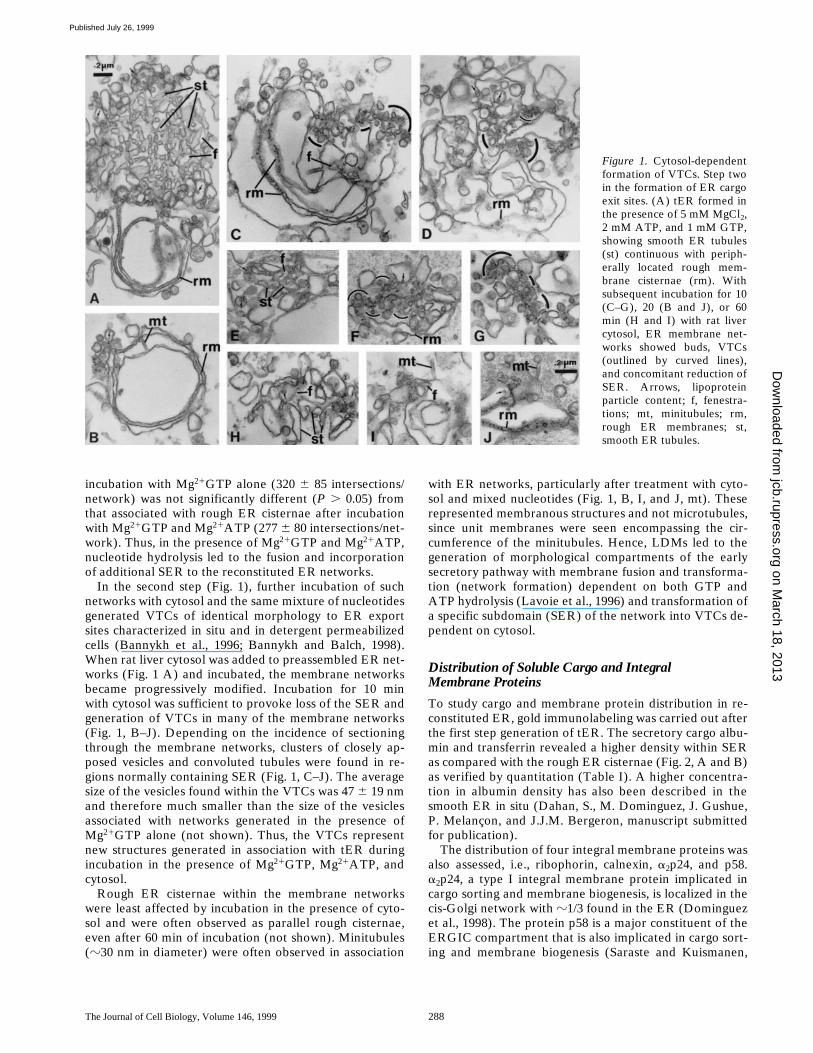
When a mixture of Mg
2
1
GTP and Mg
2
1
ATP was added,membrane differentiation consequent to membrane fusionwas observed (Fig. 1 A). As a consequence of mixed nu-cleotide hydrolysis (Lavoie et al., 1996), fused networksnow consisted of an array of rough ER cisternae, continu-ous with a fenestrated network of anastomosing smoothmembranous tubules, i.e., similar morphologically to therough ER/smooth ER boundary of liver parenchyma(Fawcett, 1955; Dallner et al., 1966). Whereas few (1–4) ri-bosomal particles were observed associated with unincu-bated vesicle profiles, numerous ribosomal particles (asmany as 20 or more) were observed aligned along the cy-toplasmic surface of the parallel rough ER cisternae as-sembled after incubation with Mg
21GTP and Mg21ATP(not shown). The average total amount of membraneassociated with networks generated in the presence ofMg21GTP and Mg21ATP (490 6 89 intersections/net-work) was significantly higher (P , 0.001) than that fornetworks generated in the presence of Mg21GTP alone(320 6 85 intersections/network). However, the amount ofmembrane associated with the rough ER cisternae after
on March 18, 2013
jcb.rupress.orgD
ownloaded from
Published July 26, 1999
The Journal of Cell Biology, Volume 146, 1999 288
incubation with Mg21GTP alone (320 6 85 intersections/network) was not significantly different (P . 0.05) fromthat associated with rough ER cisternae after incubationwith Mg21GTP and Mg21ATP (277 6 80 intersections/net-work). Thus, in the presence of Mg21GTP and Mg21ATP,nucleotide hydrolysis led to the fusion and incorporationof additional SER to the reconstituted ER networks.
In the second step (Fig. 1), further incubation of suchnetworks with cytosol and the same mixture of nucleotidesgenerated VTCs of identical morphology to ER exportsites characterized in situ and in detergent permeabilizedcells (Bannykh et al., 1996; Bannykh and Balch, 1998).When rat liver cytosol was added to preassembled ER net-works (Fig. 1 A) and incubated, the membrane networksbecame progressively modified. Incubation for 10 minwith cytosol was sufficient to provoke loss of the SER andgeneration of VTCs in many of the membrane networks(Fig. 1, B–J). Depending on the incidence of sectioningthrough the membrane networks, clusters of closely ap-posed vesicles and convoluted tubules were found in re-gions normally containing SER (Fig. 1, C–J). The averagesize of the vesicles found within the VTCs was 47 6 19 nmand therefore much smaller than the size of the vesiclesassociated with networks generated in the presence ofMg21GTP alone (not shown). Thus, the VTCs representnew structures generated in association with tER duringincubation in the presence of Mg21GTP, Mg21ATP, andcytosol.
Rough ER cisternae within the membrane networkswere least affected by incubation in the presence of cyto-sol and were often observed as parallel rough cisternae,even after 60 min of incubation (not shown). Minitubules(z30 nm in diameter) were often observed in association
with ER networks, particularly after treatment with cyto-sol and mixed nucleotides (Fig. 1, B, I, and J, mt). Theserepresented membranous structures and not microtubules,since unit membranes were seen encompassing the cir-cumference of the minitubules. Hence, LDMs led to thegeneration of morphological compartments of the earlysecretory pathway with membrane fusion and transforma-tion (network formation) dependent on both GTP andATP hydrolysis (Lavoie et al., 1996) and transformation ofa specific subdomain (SER) of the network into VTCs de-pendent on cytosol.
Distribution of Soluble Cargo and IntegralMembrane Proteins
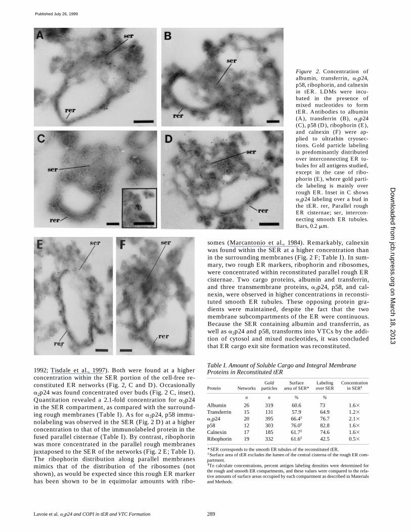
To study cargo and membrane protein distribution in re-constituted ER, gold immunolabeling was carried out afterthe first step generation of tER. The secretory cargo albu-min and transferrin revealed a higher density within SERas compared with the rough ER cisternae (Fig. 2, A and B)as verified by quantitation (Table I). A higher concentra-tion in albumin density has also been described in thesmooth ER in situ (Dahan, S., M. Dominguez, J. Gushue,P. Melançon, and J.J.M. Bergeron, manuscript submittedfor publication).
The distribution of four integral membrane proteins wasalso assessed, i.e., ribophorin, calnexin, a2p24, and p58.a2p24, a type I integral membrane protein implicated incargo sorting and membrane biogenesis, is localized in thecis-Golgi network with z1/3 found in the ER (Dominguezet al., 1998). The protein p58 is a major constituent of theERGIC compartment that is also implicated in cargo sort-ing and membrane biogenesis (Saraste and Kuismanen,
Figure 1. Cytosol-dependentformation of VTCs. Step twoin the formation of ER cargoexit sites. (A) tER formed inthe presence of 5 mM MgCl2,2 mM ATP, and 1 mM GTP,showing smooth ER tubules(st) continuous with periph-erally located rough mem-brane cisternae (rm). Withsubsequent incubation for 10(C–G), 20 (B and J), or 60min (H and I) with rat livercytosol, ER membrane net-works showed buds, VTCs(outlined by curved lines),and concomitant reduction ofSER. Arrows, lipoproteinparticle content; f, fenestra-tions; mt, minitubules; rm,rough ER membranes; st,smooth ER tubules.
on March 18, 2013
jcb.rupress.orgD
ownloaded from
Published July 26, 1999
Lavoie et al. a2p24 and COPI in tER and VTC Formation 289
Figure 2. Concentration ofalbumin, transferrin, a2p24,p58, ribophorin, and calnexinin tER. LDMs were incu-bated in the presence ofmixed nucleotides to formtER. Antibodies to albumin(A), transferrin (B), a2p24(C), p58 (D), ribophorin (E),and calnexin (F) were ap-plied to ultrathin cryosec-tions. Gold particle labelingis predominantly distributedover interconnecting ER tu-bules for all antigens studied,except in the case of ribo-phorin (E), where gold parti-cle labeling is mainly overrough ER. Inset in C showsa2p24 labeling over a bud inthe tER. rer, Parallel roughER cisternae; ser, intercon-necting smooth ER tubules.Bars, 0.2 mm.
1992; Tisdale et al., 1997). Both were found at a higherconcentration within the SER portion of the cell-free re-constituted ER networks (Fig. 2, C and D). Occasionallya2p24 was found concentrated over buds (Fig. 2 C, inset).Quantitation revealed a 2.1-fold concentration for a2p24in the SER compartment, as compared with the surround-ing rough membranes (Table I). As for a2p24, p58 immu-nolabeling was observed in the SER (Fig. 2 D) at a higherconcentration to that of the immunolabeled protein in thefused parallel cisternae (Table I). By contrast, ribophorinwas more concentrated in the parallel rough membranesjuxtaposed to the SER of the networks (Fig. 2 E; Table I).The ribophorin distribution along parallel membranesmimics that of the distribution of the ribosomes (notshown), as would be expected since this rough ER markerhas been shown to be in equimolar amounts with ribo-
somes (Marcantonio et al., 1984). Remarkably, calnexinwas found within the SER at a higher concentration thanin the surrounding membranes (Fig. 2 F; Table I). In sum-mary, two rough ER markers, ribophorin and ribosomes,were concentrated within reconstituted parallel rough ERcisternae. Two cargo proteins, albumin and transferrin,and three transmembrane proteins, a2p24, p58, and cal-nexin, were observed in higher concentrations in reconsti-tuted smooth ER tubules. These opposing protein gra-dients were maintained, despite the fact that the twomembrane subcompartments of the ER were continuous.Because the SER containing albumin and transferrin, aswell as a2p24 and p58, transforms into VTCs by the addi-tion of cytosol and mixed nucleotides, it was concludedthat ER cargo exit site formation was reconstituted.
Table I. Amount of Soluble Cargo and Integral Membrane Proteins in Reconstituted tER
Protein NetworksGold
particlesSurface
area of SER*Labelingover SER
Concentration
in SER§
n n % %
Albumin 26 319 60.6 73 1.63
Transferrin 15 131 57.9 64.9 1.23
a2p24 20 395 66.4‡ 76.7 2.13
p58 12 303 76.0‡ 82.8 1.63
Calnexin 17 185 61.7‡ 74.6 1.63
Ribophorin 19 332 61.6‡ 42.5 0.53
*SER corresponds to the smooth ER tubules of the reconstituted tER.‡Surface area of tER excludes the lumen of the central cisterna of the rough ER com-partment.§To calculate concentrations, percent antigen labeling densities were determined forthe rough and smooth ER compartments, and these values were compared to the rela-tive amounts of surface areas occupied by each compartment as described in Materialsand Methods.
on March 18, 2013
jcb.rupress.orgD
ownloaded from
Published July 26, 1999
The Journal of Cell Biology, Volume 146, 1999 290
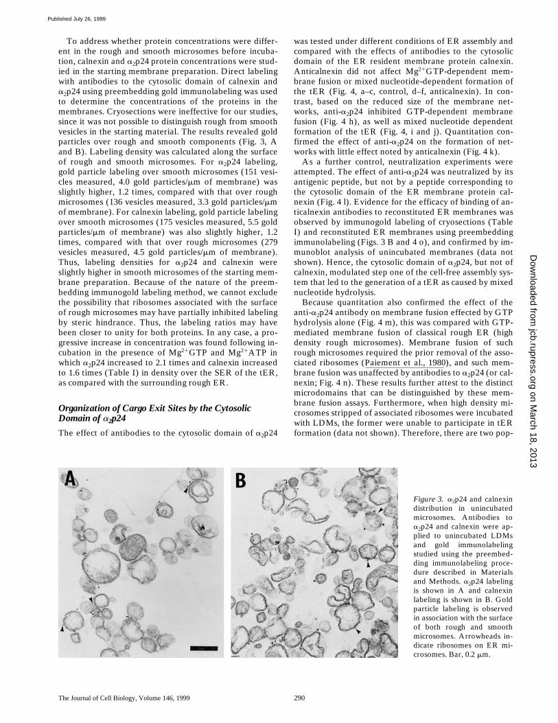
To address whether protein concentrations were differ-ent in the rough and smooth microsomes before incuba-tion, calnexin and a2p24 protein concentrations were stud-ied in the starting membrane preparation. Direct labelingwith antibodies to the cytosolic domain of calnexin anda2p24 using preembedding gold immunolabeling was usedto determine the concentrations of the proteins in themembranes. Cryosections were ineffective for our studies,since it was not possible to distinguish rough from smoothvesicles in the starting material. The results revealed goldparticles over rough and smooth components (Fig. 3, Aand B). Labeling density was calculated along the surfaceof rough and smooth microsomes. For a2p24 labeling,gold particle labeling over smooth microsomes (151 vesi-cles measured, 4.0 gold particles/mm of membrane) wasslightly higher, 1.2 times, compared with that over roughmicrosomes (136 vesicles measured, 3.3 gold particles/mmof membrane). For calnexin labeling, gold particle labelingover smooth microsomes (175 vesicles measured, 5.5 goldparticles/mm of membrane) was also slightly higher, 1.2times, compared with that over rough microsomes (279vesicles measured, 4.5 gold particles/mm of membrane).Thus, labeling densities for a2p24 and calnexin wereslightly higher in smooth microsomes of the starting mem-brane preparation. Because of the nature of the preem-bedding immunogold labeling method, we cannot excludethe possibility that ribosomes associated with the surfaceof rough microsomes may have partially inhibited labelingby steric hindrance. Thus, the labeling ratios may havebeen closer to unity for both proteins. In any case, a pro-gressive increase in concentration was found following in-cubation in the presence of Mg21GTP and Mg21ATP inwhich a2p24 increased to 2.1 times and calnexin increasedto 1.6 times (Table I) in density over the SER of the tER,as compared with the surrounding rough ER.
Organization of Cargo Exit Sites by the Cytosolic Domain of a2p24
The effect of antibodies to the cytosolic domain of a2p24
was tested under different conditions of ER assembly andcompared with the effects of antibodies to the cytosolicdomain of the ER resident membrane protein calnexin.Anticalnexin did not affect Mg21GTP-dependent mem-brane fusion or mixed nucleotide-dependent formation ofthe tER (Fig. 4, a–c, control, d–f, anticalnexin). In con-trast, based on the reduced size of the membrane net-works, anti-a2p24 inhibited GTP-dependent membranefusion (Fig. 4 h), as well as mixed nucleotide dependentformation of the tER (Fig. 4, i and j). Quantitation con-firmed the effect of anti-a2p24 on the formation of net-works with little effect noted by anticalnexin (Fig. 4 k).
As a further control, neutralization experiments wereattempted. The effect of anti-a2p24 was neutralized by itsantigenic peptide, but not by a peptide corresponding tothe cytosolic domain of the ER membrane protein cal-nexin (Fig. 4 l). Evidence for the efficacy of binding of an-ticalnexin antibodies to reconstituted ER membranes wasobserved by immunogold labeling of cryosections (TableI) and reconstituted ER membranes using preembeddingimmunolabeling (Figs. 3 B and 4 o), and confirmed by im-munoblot analysis of unincubated membranes (data notshown). Hence, the cytosolic domain of a2p24, but not ofcalnexin, modulated step one of the cell-free assembly sys-tem that led to the generation of a tER as caused by mixednucleotide hydrolysis.
Because quantitation also confirmed the effect of theanti-a2p24 antibody on membrane fusion effected by GTPhydrolysis alone (Fig. 4 m), this was compared with GTP-mediated membrane fusion of classical rough ER (highdensity rough microsomes). Membrane fusion of suchrough microsomes required the prior removal of the asso-ciated ribosomes (Paiement et al., 1980), and such mem-brane fusion was unaffected by antibodies to a2p24 (or cal-nexin; Fig. 4 n). These results further attest to the distinctmicrodomains that can be distinguished by these mem-brane fusion assays. Furthermore, when high density mi-crosomes stripped of associated ribosomes were incubatedwith LDMs, the former were unable to participate in tERformation (data not shown). Therefore, there are two pop-
Figure 3. a2p24 and calnexindistribution in unincubatedmicrosomes. Antibodies toa2p24 and calnexin were ap-plied to unincubated LDMsand gold immunolabelingstudied using the preembed-ding immunolabeling proce-dure described in Materialsand Methods. a2p24 labelingis shown in A and calnexinlabeling is shown in B. Goldparticle labeling is observedin association with the surfaceof both rough and smoothmicrosomes. Arrowheads in-dicate ribosomes on ER mi-crosomes. Bar, 0.2 mm.
on March 18, 2013
jcb.rupress.orgD
ownloaded from
Published July 26, 1999

Lavoie et al. a2p24 and COPI in tER and VTC Formation 291
ulations of rough ER that can be separated by subcellularfractionation. One is involved in the assembly of tER andis isolated as LDMs, the other is involved in the formationof large rough ER cisternae and is isolated as high densitymicrosomes, and the two exhibit different fusion pro-perties.
Finally, the effect of anti-a2p24 antibodies on cytosol-dependent loss of SER in reconstituted ER networks wastested. Incubation of reconstituted ER networks with anti-a2p24 antibodies before incubation with cytosol led to in-hibition of loss of SER within ER networks. In three sepa-rate experiments, a total of 106 reconstituted networkswere analyzed, and of these networks, 42 6 4% had recog-nizable smooth tubules. In contrast, membrane networksincubated with anticalnexin antibodies before treatmentwith cytosol lost most of their associated smooth tubules. Inthree separate experiments, a total of 71 reconstituted net-works were analyzed, and of these networks, 20 6 14%had recognizable smooth tubules. Thus, antibodies to thecytosolic tail of a2p24 led to partial inhibition of cytosol-dependent loss of SER in reconstituted ER networks.
Recruitment of COPI Coatomer
The sites of location of a2p24 antigenicity using preembed-ding gold immunolabeling was studied after induction ofVTC formation in the presence of cytosol. Gold immuno-labeling revealed a2p24 antigen in vesicular-tubular struc-tures associated with fused rough ER (Fig. 5). The re-duced gold immunolabeling is thought to be due to themasking of determinants of the cytosolic domain of a2p24caused by binding of proteins to the membranes duringpreincubation in the presence of cytosol. The cytosolic do-main of a2p24 binds COPI and COPII coatomer with highaffinity and a specificity attributed to the KKXX motif atits COOH-terminal domain for COPI and a diphenylala-nine-based motif affecting COPII binding (Dominguez et al.,1998). Because COPI coatomer has been proposed to as-sociate with pre-Golgi apparatus intermediates transport-ing anterograde cargo early in the secretory pathway(Presley et al., 1997), we elected to pursue the significanceof the effect of the a2p24 cytosolic domain on the cell-freesystem by studying the role of COPI coatomer.
The predicted binding of COPI to the cell-free system atthe ER cargo exit sites (VTCs) was tested following incu-bations at step two of the reconstitution system with cyto-sol, Mg21ATP, and the nonhydrolyzable GTP analogue,GTPgS. Biochemical studies (Fig. 6 A) revealed an aug-mented association of b-COP to membranes. Visualiza-tion of COPI coatomer during these incubations (Fig.7i) revealed an association of b-COP with the VTCsgenerated after the tER was incubated with cytosoland Mg21GTP/ATP (Fig. 7i B) or with Mg21ATP andMg21GTPgS (Fig. 7i, C and D). Little labeling with anti–b-COP was found in the absence of cytosol (Fig. 7i A).Quantitation of gold particle distribution confirmed thecytosol and GTPgS-dependent association of b-COP withthe VTCs (Fig. 7ii A).
Quantitation was also carried out to determine the ef-fects of cytosol on the formation of VTCs. The amount ofthe SER remaining in the reconstituted membrane net-works after treatment with cytosol was used as a measure
of the amount of transformation of the tER into VTCs.Percent number of networks with VTCs was also calcu-lated. Thus, a diminution of the amount of SER and a co-incident increase in amount of associated VTCs was ob-served after incubation of reconstituted ER networks inthe presence of cytosol plus Mg21ATP/GTP or Mg21ATP/GTPgS (Fig. 7ii B). A prediction of these results is thatVTC formation should be sensitive to the fungal metabo-lite BFA via its action on inhibiting an ARF1-GEF activity(Donaldson et al., 1992; Helms and Rothman, 1992). In-deed, BFA significantly inhibited the association of b-COPon tER networks (Fig. 6 A) and significantly diminishedthe formation of VTCs as evaluated quantitatively (Fig. 7iiB). No effect was observed with BFA alone, when cytosolwas omitted (data not shown). None of the incubationconditions affected the amount of rough ER cisternaeassociated with the networks, as determined by this quan-titative assay (Fig. 7ii B). That the cytosol effect was medi-ated by COPI binding to the tER was tested by partial de-pletion of COPI coatomer from cytosol with antibody tob-COP (Fig. 6 B). Such cytosol diminished in b-COP con-tent generated fewer VTCs, as compared with cytosoltreated with nonimmune IgG (see VTCs compared withloss of SER in Fig. 7ii C).
a2p24 and b-COP Are Localized to ER and Golgi Elements in Rat Liver Hepatocytes
As determined by cryoimmune EM using well character-ized antibodies specific to a2p24 (Dominguez et al., 1998),the membrane protein is clearly found in both rough andsmooth ER in situ (Fig. 8, A–C). Gold particles decoratethe cytoplasmic side of parallel ER cisternae in cryosec-tions immunolabeled with an antibody against the COOH-terminal domain of a2p24 (Fig. 8 A). Tubulo-vesicularsmooth ER networks in the Golgi region of hepatocytes,as well as the cis-Golgi intermediate compartment, werealso labeled by anti-a2p24 (Fig. 8, B and C). The COPIcoatomer subunit b-COP reveals a distribution that over-laps that of a2p24 in liver parenchyma (Fig. 8, D and E).Thus, a2p24 and b-COP are associated with similar struc-tures, including tubular-vesicular elements of the ER oftenfound next to the Golgi apparatus in situ within the rathepatocyte.
a2p24 in LDMs Is Golgi Apparatus-derived
The cargo molecules albumin and transferrin representnewly synthesized protein cargo of the ER. However, thiswas unlikely to be the case for a2p24 (and p58) observed inreconstituted ER or for a2p24 observed in hepatocyte ERin situ by immunolabeling. Whether in preparations ofLDMs, or even in highly purified stripped rough mi-crosomes (SRM), a2p24 is terminally glycosylated as evi-dent from its lack of sensitivity to endoglycosidase H (en-doH), but complete sensitivity to PNGase F (Fig. 9). Equalamounts of protein from each fraction was applied to eachlane (100 mg). a2p24 is also highly enriched in Golgi frac-tions, due to its abundance in the cis-Golgi network thatcoisolates with hepatic Golgi fractions (Dominguez et al.,1998). Therefore, a2p24 found in the liver ER fraction em-ployed in the in vitro ER reconstitution assay is terminally
on March 18, 2013
jcb.rupress.orgD
ownloaded from
Published July 26, 1999

The Journal of Cell Biology, Volume 146, 1999 292
glycosylated, and thus a molecular derivative of the Golgiapparatus.
Discussion
A Cell-free System to Study Assembly of tER and the Formation of ER Exit Sites
A two-step in vitro reconstitution system starting fromwell-characterized ER-derived LDMs purified from rat liverhomogenates (Lavoie et al., 1996) has been used to gener-ate, via nucleotide hydrolysis, membrane structures with
the morphological features of the tER. Then, in a secondstep, cytosol is employed to generate ER cargo exit sitesvia the formation of VTCs from the preassembled tER.That the in vitro system faithfully reconstituted the earlysecretory apparatus was established from the followingcriteria. Morphology: quantitation revealed the nucleotidedependent fusion of LDMs and their transformation intosmooth ER and rough ER interfaces of identical morphol-ogy to the tER, as seen in liver parenchyma in situ. Whenthe same LDMs as used here were microinjected into thecytoplasm of Xenopus oocytes, a reconstitution of ERidentical in structure to that seen in rat liver parenchyma
Figure 4.
on March 18, 2013
jcb.rupress.orgD
ownloaded from
Published July 26, 1999

Lavoie et al. a2p24 and COPI in tER and VTC Formation 293
in situ was observed (Paiement et al., 1988). Upon the ad-dition of cytosol and Mg21ATP/GTP (step two in thisstudy), only the smooth portion of the tER generatedVTCs and these correspond to the morphology of pre-Golgi apparatus intermediates based on the morphologyof the structures assembled and on the size (47 6 19 nm)of the associated vesicles (Bannykh and Balch, 1998;Morré, 1998). Cargo and p58 content: the content of al-bumin, transferrin, and p58 (ERGIC-53, the marker forthe ER-Golgi intermediate compartment; Farquhar andHauri, 1997) was consistent with this compartment as theprecursor of the VTCs. In liver parenchyma, two sites ofcargo (albumin) concentration have been elucidated. An
initial twofold concentration was found at the boundary ofthe rough ER and smooth ER (Dahan, S., M. Dominguez,J. Gushue, P. Melançon, and J.J.M. Bergeron, manuscriptsubmitted for publication), as observed here. A furtherfivefold concentration takes place between the cis-Golginetwork and stacked flattened cisternae of the Golgi com-plex (Dahan, S., M. Dominguez, J. Gushue, P. Melançon,and J.J.M. Bergeron, manuscript submitted for publica-tion).
The Role of Membrane Fusion in Assembly of tER
The nucleotide-dependent fusion of LDMs to yield tER is
Figure 4. Anti-a2p24, but notanticalnexin antibodies, mod-ulate the structure of thetER. Effect of a2p24 an-tibody on ER assembly.LDMs incubated with ATP(a, d, and g), GTP (b, e, andh), or ATP/GTP (c, f, i, andj) in the presence of antibod-ies to the cytosolic domain ofcalnexin (d–f) or a2p24 (g–j).rer, Rough ER; rm, roughmicrosomes; ser, smooth ERtubules; sm, smooth micro-somes. Quantitation of a2p24antibody effect by morphom-etry. (k) Quantitation ofrough membranes and SERprofiles in networks for con-ditions described in c, f, i,and j. (l) Conditions are asfor i and j, except that anti-bodies were preincubatedwith peptides used to pro-duce antibodies againsta2p24 or calnexin (CNX).(m) Conditions are as for b,e, and h. (n) Effect of anti-bodies on the fusion of classi-cal rough ER membranes(high density microsomesprepared and incubated asindicated in Materials andMethods). (o) Binding ofanticalnexin antibodies tothe cytosolic surface of re-constituted ER membranes.LDMs were incubated in thepresence of GTP, ATP, andanticalnexin antibodies. Goldsecondary antibody appliedusing the preembedding im-munocytochemical techniquereveals calnexin antigenicityon both the surfaces of par-allel rough membranes andinterconnecting smooth tu-bules. Bar, 0.2 mm.
on March 18, 2013
jcb.rupress.orgD
ownloaded from
Published July 26, 1999

The Journal of Cell Biology, Volume 146, 1999 294
thought to involve both fusion of like (homotypic) and un-like (heterotypic) ER membrane derivatives. Mg21GTPhydrolysis is required to stimulate fusion of partially roughER membrane derivatives and Mg21ATP hydrolysis is re-quired to stimulate fusion of smooth ER membrane deriv-atives. Hence, these nucleotides contribute to homotypicmembrane fusion. At some stage during tER formation,continuity is established between these two ER subcom-
Figure 5. a2p24 distribution in VTCs formed in the presence ofcytosol. Formation of VTCs by cytosol plus nucleotides (steptwo), followed by the addition of primary anti-a2p24 antibodyand then gold secondary antibody. a2p24 antigenicity is foundwithin the VTCs. Arrowheads indicate ribosomes on ER mem-branes. The arrow points to a fenestration of the residual SER.Bar, 0.2 mm.
Figure 6. Content of b-COP in ER membranes and cytosol. (A)Recruitment of b-COP to ER membranes. LDMs were incubatedas for Fig. 1, but with BFA or GTPgS as indicated, followed bysedimentation and Western blotting for b-COP. (B) b-COP de-pletion of cytosol as assessed by b-COP content in control cyto-sol (lane 1), cytosol incubated with protein A–Sepharose coupledto nonspecific IgG (lane 2), or anti–b-COP (lane 3) followed byWestern blotting for b-COP content.
Figure 7. Coatomer protein-dependent formation of VTCs. (i)Localization of b-COP to VTCs. Immunogold labeling of b-COPwith anti–b-COP is shown in association with tER networks post-incubated (step two) in the absence of cytosol (A), the presence ofcytosol 1 ATP/GTP (B), or in the presence of cytosol 1 ATP 1GTPgS (C and D) for 10 min. Arrows, indicate label over smoothmembranes; arrowheads, indicate label over clusters of vesicles;f, fenestrations; rm, rough membranes; st, smooth ER tubules.Immunolabeling was done using the preembedding techniquedescribed in Materials and Methods. Bar, 0.2 mm. (ii) Modula-tion of b-COP binding to tER. (A) Effect of GTPgS on theamount of b-COP labeling over VTCs. LDMs were incubated asin i, A–D. Counts are expressed as mean number of gold particlesper membrane network over rough ER cisternae (RER) andover vesicular tubular complexes (SER/VTC) formed in the ab-sence of cytosol (2CYT), the presence of cytosol and GTP(1CYT1GTP), or in the presence of cytosol and GTPgS(1CYT1GTPgS). Data is shown from three separate experi-ments using membranes from separate fractionation experi-ments. (B) Effect of BFA on the formation of VTCs. LDMs wereincubated as for Fig. 1, except for the addition of GTPgS or BFA,as indicated. Except where indicated by *, average values (6 SD)are shown from three separate experiments carried out withmembranes from different fractionations. The means for VTCswere not significantly different when compared between a–d (P .0.05, n 5 61), but were significantly different when compared be-tween b–d (P , 0.01, n 5 61). The means for rough ER mem-branes were not significantly different (P . 0.05, n 5 47) whencompared between a and b, b–d, and a–d. The means for smoothER tubules were not significantly different when compared be-tween a–d, (P . 0.05, n 5 47), but were significantly differentwhen compared between a and b (P , 0.001, n 5 47) or b and d(0.001 , P , 0.005, n 5 47). The means for rough and smoothER tubules were not significantly different when compared be-tween b and c (P . 0.05, n 5 30). RER, rough ER; SER, smoothER tubules; *, average of two experiments. (C) Effect of b-COP–depleted cytosol on formation of VTCs. Microsomes were incu-bated with mixed nucleotides (step one) for 180 min to generatetER and then further incubated (step two) for 60 min with orwithout cytosol, with b-COP–depleted cytosol, or with control-treated cytosol. Amounts of smooth ER tubules associated withthe reconstituted membrane networks are compared with thenumber of VTC structures associated with the same networks.Average of two experiments. A minimum of 32 membrane net-works were analyzed for each experimental condition.
partments and this would be expected to occur by hetero-typic membrane fusion.
However, if partially rough microsomes containing mi-crodomains of smooth ER initially fused in the presence ofMg21GTP, this would permit subsequent Mg21ATP-dependent fusion with additional smooth microsomes andobviate a necessity for heterotypic membrane fusion. Thispossibility has not been ruled out yet. The assays devel-oped here using quantitative morphology and quantitativeimmunolabeling may now be used to screen for antibodiesto proteins that can distinguish between the homotypicand heterotypic membrane fusion events.
Rough ER Membranes within tER Exhibit Unique Fusion Properties
The rough ER subcompartment comprising the tER as-sembled when LDMs are incubated in the presence of
on March 18, 2013
jcb.rupress.orgD
ownloaded from
Published July 26, 1999

Lavoie et al. a2p24 and COPI in tER and VTC Formation 295
on March 18, 2013
jcb.rupress.orgD
ownloaded from
Published July 26, 1999

The Journal of Cell Biology, Volume 146, 1999 296
Mg21GTP and Mg21ATP is very different from classicalrough ER, which is recovered from tissue homogenates ashigh density rough microsomes. Although high densityrough microsomes undergo GTP-dependent fusion, as dolow density rough microsomes, the fusion events are dif-ferent. For example, antibodies to a2p24 inhibit fusion ofthe partially rough ER comprising tER, but not that ofclassical rough ER (Fig. 4 n). Fusion of classical rough ERrequires prior removal of associated ribosomes (Paiementet al., 1980; Paiement and Bergeron 1983), which transi-tional rough ER does not (Lavoie et al., 1996). Classicalrough ER does not fuse with transitional rough ER whenthe two types of rough ER are mixed with nucleotides(Lavoie, C., and J. Paiement, unpublished observations).Hence, the fusion machinery associated with the mem-branes of tER is suggested to be different from that associ-ated with the rest of the ER, and this may be related to thecapacity of this compartment to permit formation of ER
exit sites. In this scenario, antibodies to a2p24 may affectthe generation of tER by influencing the heterooligomer-ization of p24 family members (Dominguez et al., 1998;Füllekrug et al., 1999; Marzioch et al., 1999). This may be anecessary step for subsequent recruitment of COPI whencytosol is added to generate VTCs.
a2p24 and p58 in tER
The membrane proteins a2p24 and p58 were found in mi-crodomains of the tER. These membrane proteins arefound at steady state to be enriched in the cis-Golgi net-work and ERGIC compartments (Farquhar and Hauri,1997; Dominguez et al., 1998). However, they are alsoclearly found in the ER and, as for p58 and the p24s cyclethrough the ER, intermediate compartments, and Golgicomplex (reviewed in Farquhar and Hauri, 1997; Fülle-krug et al., 1999). That a2p24 was terminally glycosylated
Figure 8. In situ localizationof a2p24 and b-COP in ratliver hepatocytes. Rat livercryosections were immunola-beled with antibodies againstthe cytosolic tail of a2p24 (Aand C), both the cytosolicand lumenal domains ofa2p24 (B), or the b-COP sub-unit of COPI (E5A3 mAb; Dand E), followed by goatanti–rabbit IgG 10-nm goldto reveal a2p24 and goatanti–mouse IgG 10-nm goldto reveal b-COP. Immunore-active a2p24 and b-COP canbe detected over the longparallel cisternae of therough ER (rER, arrows) andtubular-vesicular networks ofthe smooth ER (arrow-heads). These tubular net-works can be visualized go-ing in and out of the plane ofsection (seen clearly in B andE). The Golgi apparatus (G)also reveals gold particle la-beling for a2p24 and b-COP,predominantly at the cis faceand peripheral distensions.M, mitochondria; P, peroxi-somes. Bars, 0.4 mm.
on March 18, 2013
jcb.rupress.orgD
ownloaded from
Published July 26, 1999

Lavoie et al. a2p24 and COPI in tER and VTC Formation 297
(endoH resistant) in LDMs, and even in highly purifiedrough ER membranes, is consistent with a constantly recy-cling scenario for a2p24.
In the starting preparation of LDMs, a2p24 was found inslightly higher concentration in smooth microsomes. Afterincubation in step one conditions, a higher concentrationof the protein was observed in the SER. The evidence sug-gests that segregation of a2p24 into SER occurred coinci-dent with tER formation.
The membrane proteins p58 and a2p24 share KKXXmotifs at their COOH termini, and in vitro binding assaysshow that the cytosolic domains of these membrane pro-teins bind COPI coatomer, and unexpectedly, COPIIcoatomer as well (Kappeler et al., 1997; Dominguez et al.,1998). These membrane proteins are therefore attractivecandidates for coordinating the formation of ER cargoexit sites. As shown here, immunolabeling revealed accu-mulation of a2p24 in VTCs formed in the presence of cyto-sol and the in vitro reconstitution assay revealed inhibitionof tER formation by antibodies to the cytosolic domain ofa2p24. Antibodies to the cytosolic domain of p58 inhibitER-to-Golgi transport, as well as COPI coatomer binding(Tisdale et al., 1997). In the case of experiments done withanti-p58 (Tisdale et al., 1997) and those reported here withanti-a2p24, possible confounding effects of steric hin-drance by antibodies to an abundant protein cannot be ex-cluded. Remarkably, in our studies, antibodies to the cyto-solic domain of a2p24 affected the surrounding parallelrough ER cisternae themselves. With Mg21GTP as solenucleotide, this membrane fusion step could be studied inisolation. This fusion step was specifically inhibited by an-tibodies to the cytosolic domain of a2p24, but not calnexin.Because this step was shown to be an early event in theformation of tER (Lavoie et al., 1996) and since formationof ER exit sites occurs from tER (results presented in thispaper), we suggest that the influence of a2p24 in affectingthe formation of ER cargo exit sites extends to the roughER portion of the rough/smooth ER boundary of the tER.This boundary is a predicted consequence of incoming ret-rograde smooth membranes derived from the Golgi appa-ratus and outgoing anterograde rough membranes trans-forming into intermediate compartment elements.
Mistargeting of a mutated form of ERGIC-53 to the ERof HeLa cells was shown to impair secretion of a lysosomalenzyme while apparently not affecting gross (light micro-scope) morphological changes of the early secretory path-way (Vollenweider et al., 1998). The lack of effect on b-COPlocalization in cells carrying the mutated ERGIC-53 couldbe due to the compensatory binding of b-COP in the sameregions by p24 family members. Consistent with this sug-gestion is the fact that p24 family members are known to
bind COPI coatomer (Kappeler et al., 1997; Dominguez etal., 1998; Bremser et al., 1999) and in our study, both p58and p24 proteins were observed in the same microdomainof the tER, as shown by immunolocalization of p58 andp24 in cryosections of tER (Fig. 2; Table I).
Role of a2p24 and its COPI Coatomer Ligand in ER Cargo Exit Site Formation
An effect of COPI coatomer on VTC formation wasfound. This was concluded from the visualization ofb-COP in VTCs after the addition of cytosol, the enhance-ment of VTC formation and b-COP association withVTCs by cytosol with GTPgS, the BFA sensitivity of VTCformation, and the inhibition of VTC formation whenb-COP was depleted from cytosol. These coincidental ob-servations are consistent with, but do not prove a directlink between COPI coatomer and the cytosolic domain ofa2p24 in the formation of ER cargo exit sites. Indeed, wecannot completely rule out the possibility that binding ofantibodies to a2p24, but not calnexin, affects the ability ofother abundant membrane proteins in microsomes to ac-cess coat proteins. These observations do, however, pro-vide a structural explanation for the observations thatARF1 dominant negative mutants (Dascher and Balch,1994; Peters et al., 1995), BFA (Lippincott-Schwartz et al.,1990), or microinjected antibodies to b-COP (Pepper-kok et al., 1993) rapidly inhibit ER-to-Golgi transport ofnewly synthesized anterograde directed cargo. Further-more, BFA acts immediately to prevent cargo exit fromthe ER, suggesting that a locus of COPI binds to ER cargoexit sites (Lippincott-Schwartz et al., 1998). All of theseagents may have, as their ultimate target, p24 family mem-bers and their effect on structural transformations re-quired for the generation of ER cargo exit sites.
Roles for a2p24 and its COPI coatomer ligand in ERcargo exit site formation have been suggested by resultsobtained using the novel two-step reconstitution systemdescribed. Based on available data, the simplest explana-tion for the possible involvement of these two proteins isthat a2p24 promotes assembly of tER and COPI coatomeris required for subsequent formation of VTCs. The struc-tural modifications implicating a2p24 and COPI involve-ment are summarized in diagrammatic form (Fig. 10). Be-
Figure 9. EndoH insensitiv-ity of a2p24 in hepatocyteER. Low density mi-crosomes (SM), high densitystripped rough microsomes(SRM), and Golgi fractions
(Gol) were assessed for their a2p24 content by Western blottingand the extent of N-linked glycosylation assessed by endoH orPNGase F sensitivity. 100 mg of protein was applied to each lane.
Figure 10. Model for a2p24 and COPI involvement in formationof ER cargo exit sites. The ER-to-Golgi recycling membrane pro-tein a2p24 is suggested to play a role in the assembly of intercon-necting smooth tubules (SER) of tER. Coatomer protein COPIpromotes formation of VTCs from this ER subcompartment.RER, rough ER.
on March 18, 2013
jcb.rupress.orgD
ownloaded from
Published July 26, 1999

The Journal of Cell Biology, Volume 146, 1999 298
cause a2p24 is known to bind COPI coatomer, and sinceanti-a2p24 antibodies were observed to inhibit cytosol-dependent transformation of tER, we cannot exclude arole for a2p24 in the last step of formation of ER exit sites.
A Possible Structural Role for the p24 Familyof Proteins
A structural role for a2p24 may explain all properties thusfar documented for the p24 family of proteins. This wouldinclude their enrichment in vesicles derived from ERcargo exit sites and the physical association of p24 familymembers as heterooligomers in yeast (Belden and Bar-lowe, 1996; Marzioch et al., 1999). The effect of gene dis-ruption of members (one of eight) of the yeast p24 familyon the kinetics of a subset of secretory cargo (Schimmölleret al., 1995; Stamnes et al., 1995; Elrod-Erickson and Kai-ser, 1996), as well as the effect of gene disruption of similarfamily members on the number of ER-derived vesicles inyeast (Stamnes et al., 1995) may also be attributed tostructural consequences of ER cargo exit sites. The abilityof at least four (of the eight) p24 family members in yeastto be required for the essential phenotype of sec13 (Elrod-Erickson and Kaiser, 1996; Marzioch et al., 1999) are likelya consequence of aberrations in the structural compart-ment generated at ER cargo exit sites. Because at leastfour p24 family members are in a biochemical complex inyeast (Marzioch et al., 1999), as well as in mammalian cells(Dominguez et al., 1998; Füllekrug et al., 1999), ER cargoexit site formation may be a function of the balance of p24family members. The structural role identified here alsoexplains the effect of the addition of antibodies to the cy-toplasmic domain of a mammalian p24 family member onthe transport of secretory cargo in these cells (Rojo et al.,1997). Alternative models, whereby p24 family membersand ERGIC-53 represent specific cargo receptors (Schim-möller et al., 1995; Nichols et al., 1998; Vollenweider et al.,1998), are not completely ruled out by our study, but suchmodels introduce an increased level of complexity andspecificity.
COPI and Anterograde Transport
The role of COPI coatomer (and consequently the ARF1GTPase) as a defining feature required for the generationof membranes of the early secretory apparatus has beenargued by Lippincott-Schwartz et al. (1998), as well as byPeter et al. (1993). The hypothesis that COPI affects an-terograde transport in the early secretory pathway as sup-ported by the studies shown here also explains why theaccumulation of Golgi apparatus resident proteins at thecell surface in yeast strains carrying temperature sensitivealleles of COPI subunits have not been reported (Nickeland Wieland, 1998).
The identification of motifs (FF) involved in COPIIbinding (Dominguez et al., 1998), also shared with p58(ERGIC-53; Kappeler et al., 1997) in the cytosolic domainof all p24 family members studied thus far, provides a mo-lecular rationale for these ER-Golgi recycling proteins incoordinating cargo transport at ER cargo exit sites formedby the concerted actions of COPII and COPI coatomer.Taken together with the in vivo work of Scales et al. (1997)and Presley et al. (1997), a cooperation between COPI and
COPII coatomer at ER cargo exit sites seems clear. Theextent of cooperation remains to be determined. Cell-freestudies of yeast in which COPI- and COPII-dependentcoatomer budding from the isolated nuclear envelope hasbeen reconstituted, secretory cargo was found associatedwith COPII, but not COPI, decorated buds (Bednarek et al.,1995). In yeast, it remains to be shown whether a COPImechanism is operational in anterograde ER-to-Golgitransport via an ERGIC compartment. The relative amountof involvement of COPI and COPII in anterograde ER-to-Golgi transport could depend on the relative amount oftER within a particular cell type. Indeed, the basic mor-phological organization of export complexes has been sug-gested to vary in different cell types (Bannykh et al., 1996).Clarification, at least with regards to liver, will be obtainedwhen studies are extended using the morphologicallybased cell-free system reported here to identify further thetargets of ATP and GTP hydrolysis and the cooperationbetween COPII coatomer coating and COPI coats in theearly secretory pathway.
We thank Dr. Peter McPherson (McGill University) for criticism of themanuscript and Dr. Tommy Nilsson for support and advice throughoutthese experiments. We thank Dr. Jacopo Saraste for kindly supplying anti-bodies to p53. We thank Anne Guénette and Ali Fazel for expert assis-tance, and Jean Léveillé for photographic assistance.
This work was supported by grants from the Medical Research Councilof Canada to J. Paiement and J.J.M. Bergeron. C. Lavoie was a recipientof a studentship from the Medical Research Council of Canada.
Submitted: 2 September 1998Revised: 17 May 1999Accepted: 19 May 1999
References
Bajjalieh, S.M., and R.H. Scheller. 1995. The biochemistry of neurotransmittersecretion. J. Biol. Chem. 270:1971–1974.
Bannykh, S.I., and W.E. Balch. 1998. Selective transport of cargo between theER and Golgi compartments. Histochem. Cell Biol. 109:463–475.
Bannykh, S.I., T. Rowe, and W.E. Balch. 1996. The organization of endoplas-mic reticulum export complexes. J. Cell Biol. 135:19–35.
Baudhuin, P., P. Evrard, and J. Berthet. 1967. Electron microscopic examina-tion of subcellular fractions. I. The preparation of representative samplesfrom suspensions of particles. J. Cell Biol. 32:181–191.
Bednarek, S.Y., M. Ravazzola, M. Hosobushi, M. Amherdt, A. Perrelet, R.Schekman, and L. Orci. 1995. COPI- and COPII-coated vesicles bud directlyfrom the endoplasmic reticulum in yeast. Cell. 83:1183–1196.
Belden, W.J., and C. Barlowe. 1996. Erv25p, a component of COPII-coatedvesicles, forms a complex with emp24 that is required for efficient endoplas-mic reticulum to Golgi transport. J. Biol. Chem. 271:26939–26946.
Bremser, M., W. Nickel, M. Schweikert, M. Ravazzolla, M. Amherdt, C.A.Hughes, T.H. Söllner, J.E. Rothman, and F.T. Wieland. 1999. Coupling ofcoat assembly and vesicle budding to packaging of putative cargo receptors.Cell. 96:495–506.
Dahan, S., J.P. Ahluwalia, L. Wong, B.I. Posner, and J.J.M. Bergeron. 1994.Concentration of intracellular hepatic apolipoprotein E in Golgi apparatussaccular distensions and endosomes. J. Cell Biol. 127:1859–1869.
Dallner, G., P. Siekevitz, and G.E. Palade. 1966. Biogenesis of endoplasmicreticulum membranes. I. Structural and chemical differentiation in develop-ing rat hepatocyte. J. Cell Biol. 30:73–96.
Dascher, C., and W.E. Balch. 1994. Dominant inhibitory mutants of ARFIblock endoplasmic reticulum to Golgi transport and trigger disassembly ofthe Golgi apparatus. J. Biol. Chem. 269:1437–1448.
Dominguez, J.M., J. Lanoix, and J. Paiement. 1991. Localization of ras antige-nicity in rat hepatocyte plasma membrane and rough endoplasmic reticulumfractions. Exp. Cell Res. 192:137–147.
Dominguez, M., K. Dejgaard, J. Fullekrug, S. Dahan, A. Fazel, J-P. Paccaud,D.Y. Thomas, J.J.M. Bergeron, and T. Nilsson. 1998. gp25L/emp24/p24 pro-tein family members of the cis-Golgi network bind both COP I and IIcoatomer. J. Cell Biol. 140:751–765.
Donaldson, J.G., D. Finazzi, and R.D. Klausner. 1992. Brefeldin A inhibitsGolgi membrane-catalysed exchange of guanine nucleotide onto ARF pro-tein. Nature. 360:350–352.
Elazar, Z., L. Orci, J. Ostermann, M. Amherdt, G. Tanigawa, and J.E. Roth-
on March 18, 2013
jcb.rupress.orgD
ownloaded from
Published July 26, 1999
Lavoie et al. a2p24 and COPI in tER and VTC Formation 299
man. 1994. ADP-ribosylation factor and coatomer couple fusion to vesiclebudding. J. Cell Biol. 124:415–424.
Elrod-Erickson, M.J., and C.A. Kaiser. 1996. Genes that control fidelity of en-doplasmic reticulum to Golgi transport identified as suppressors of vesiclebudding mutations. Mol. Biol. Cell. 7:1043–1058.
Farquhar, M., and H.-P. Hauri. 1997. Protein sorting and vesicular traffic in theGolgi apparatus. In The Golgi Apparatus. E. Berger and J. Roth, editors.Birkhauser Verlag, Basel, Switzerland. pp 63–128.
Fawcett, D.W. 1955. Observations on the cytology and electron microscopy ofhepatic cells. J. Natl. Cancer Inst. 15:1475–1503.
Füllekrug, F., J.T. Suganama, T.B.L. Tang, W. Hong, B. Storrie, and T. Nilsson.1999. Localization and recycling of gp27 (gp24d3): complex formation withother p24 family members. Mol. Biol. Cell. In press.
Ghitescu, L., and M. Bendayan. 1990. Immunolabeling efficiency of proteinA–gold complexes. J. Histochem. Cytochem. 38:1523–1530.
Helms, J.B., and J.E. Rothman. 1992. Inhibition by brefeldin A of a Golgimembrane enzyme that catalyses exchange of guanine nucleotide bound toARF. Nature. 360:352–354.
Kappeler, F., D.R. Klopfenstein, M. Foguet, J.-P. Paccaud, and H.-P. Hauri.1997. The recycling of ERGIC-53 in the early secretory pathway: ERGIC-53carries a cytosolic endoplasmic reticulum-exit determinant interacting withCOPII. J. Biol. Chem. 272:31801–31808.
Lavoie, C., J. Lanoix, F.W.K. Kan, and J. Paiement. 1996. Cell-free assembly ofrough and smooth endoplasmic reticulum. J. Cell Sci. 109:1415–1425.
Letourneur, F., E.C. Gaynor, S. Hennecke, C. Démollère, R. Duden, S.D. Emr,H. Riezman, and P. Cosson. 1994. Coatomer is essential for retrieval of di-lysine-tagged proteins to the endoplasmic reticulum. Cell. 79:1199–1207.
Lippincott-Schwartz, J., J.G. Donaldson, A. Schweizer, E.G. Berger, H.-P.Hauri, L.C. Yuan, and R.D. Klausner. 1990. Microtubule-dependent retro-grade transport of proteins into the ER in the presence of brefeldin A sug-gests an ER recycling pathway. Cell. 60:821–836.
Lippincott-Schwartz, J., N.B. Cole, and J.G. Donaldson. 1998. Building a secre-tory apparatus: role of ARF1/COPI in Golgi biogenesis and maintenance.Histochem. Cell Biol. 109:449–462.
Marcantonio, E., E.A. Amar-Costesec, and G. Kreibich. 1984. Segregation ofthe polypeptide translocation apparatus to regions of the endoplasmic retic-ulum containing ribophorins and ribosomes. II. Rat liver microsomal sub-fractions contain equimolar amounts of ribophorins and ribosomes. J. CellBiol. 99:2254–2259.
Marzioch, M., D.C. Henthorn, J.M. Hermann, R. Wilson, D.Y. Thomas, J.J.M.Bergeron, R.C.E. Solari, and A. Rowley. 1999. Erp1p and Erp2p, partnersfor Emp24p and Erv25p in a yeast p24 complex. Mol. Biol. Cell. In press.
Morré, D.J. 1998. Cell-free analysis of Golgi apparatus membrane traffic in ratliver. Histochem. Cell Biol. 109:487–504.
Morré, D.J., and T.W. Keenan. 1997. Membrane flow revisited: what pathwaysare followed by membrane molecules moving through the Golgi apparatus?BioScience. 47:489–498.
Nichols, W.C., U. Seligsohn, A. Zivelin, V.H. Terry, C.E. Hertel, M.A. Wheat-ley, M.J. Moussalli, H-P. Hauri, N. Ciavarella, R.J. Kaufman, et al. 1998.Mutations in the ER-Golgi intermediate compartment protein ERGIC-53cause combined deficiency of coagulation factors V and VIII. Cell. 93:61–70.
Nickel, W., and F.T. Wieland. 1998. Biosynthetic protein transport through theearly secretory pathway. Histochem. Cell Biol. 109:477–486.
Novick, P., C. Field, and R. Schekman. 1980. Identification of 23 complementa-tion groups required for post-translational events in the yeast secretorypathway. Cell. 21:205–215.
Ou, W.J., P.H. Cameron, D.Y. Thomas, and J.J.M. Bergeron. 1993. Associationof folding intermediates of glycoproteins with calnexin during protein matu-ration. Nature. 364:771–776.
Paiement, J., and J.J.M. Bergeron. 1983. Localization of GTP-stimulated coreglycosylation to fused microsomes. J. Cell Biol. 96:1791–1796.
Paiement, J., H. Beaufay, and D. Godelaine. 1980. Coalescence of microsomalvesicles from rat liver: a phenomenon occurring in parallel with enhance-
ment of the glycosylation activity during incubation of stripped rough mi-crosomes with GTP. J. Cell Biol. 86:29–37.
Paiement, J., F.W.K. Kan, J. Lanoix, and M. Blain. 1988. Cytochemical analysisof the reconstitution of endoplasmic reticulum after microinjection of ratliver microsomes into Xenopus oocytes. J. Histochem. Cytochem. 36:1263–1273.
Pelham, H.R.B. 1997. Membrane transport: green light for Golgi traffic. Nature389:17–19.
Pepperkok, R., J. Scheel, H. Horstmann, H.-P. Hauri, G. Griffiths, and T.E.Kreis. 1993. b-COP is essential for biosynthetic membrane transport fromthe endoplasmic reticulum to the Golgi complex in vivo. Cell. 74:71–82.
Peter, F., H. Plutner, H. Zhu, T.E. Dreis, and W.E. Balch. 1993. b-COP is es-sential for transport of protein from the endoplasmic reticulum to the Golgiin vitro. J. Cell Biol. 122:1155–1167.
Peters, P.J., V.W. Hsu, C.E. Ooi, D. Finazzi, S.B. Teal, V. Oorschot, J.G.Donaldson, and R.D. Klausner. 1995. Overexpression of wild-type and mu-tant ARF1 and ARF6: distinct perturbations of nonoverlapping membranecompartments. J. Cell Biol. 128:1003–1017.
Presley, J.F., N.B. Cole, T.A. Schroer, K. Hirschberg, K.J.M. Zaal, and J. Lip-pincott-Schwartz. 1997. ER-to-Golgi transport visualized in living cells. Na-ture. 389:81–85.
Rojo, M., R. Pepperkok, G. Emery, R. Kellner, E. Stand, R.G. Parton, and J.Gruenberg. 1997. Involvement of the transmembrane protein p23 in biosyn-thetic protein transport. J. Cell Biol. 139:1119–1135.
Rothman, J.E. 1994. Mechanisms of intracellular protein transport. Nature. 372:55–63.
Rothman, J.E., and F.T. Wieland. 1996. Protein sorting by transport vesicles.Science. 272:227–234.
Saraste, J., and E. Kuismanen. 1992. Pathways of protein sorting and mem-brane traffic between the rough endoplasmic reticulum and the Golgi com-plex. Semin. Cell Biol. 3:343–355.
Scales, S.J., R. Pepperkok, and T.E. Kreis. 1997. Visualization of ER-to-Golgitransport in living cells reveals a sequential mode of action for COPII andCOPI. Cell. 90:1137–1148.
Schekman, R., and L. Orci. 1996. Coat proteins and vesicle budding. Science.271:1526–1533.
Schimmöller, F., B. Singer-Krüger, S. Schröder, U. Krüger, C. Barlowe, and H.Riezman. 1995. The absence of Emp24p, a component of ER-derivedCOPII-coated vesicles, causes a defect in transport of selected proteins tothe Golgi. EMBO (Eur. Mol. Biol. Organ.) J. 14:1329–1339.
Slot, J.W., and H.J. Geuze. 1985. A method for preparing gold probes for multi-ple labeling cytochemistry. Eur. J. Cell Biol. 38:87–93.
Stamnes, M.A., M.W. Craighead, M.H. Hoe, N. Lampen, S. Geromanos, P.Tempst, and J.E. Rothman. 1995. An integral membrane component ofcoatomer-coated transport vesicles defines a family of proteins involved inbudding. Proc. Natl. Acad. Sci. USA. 92:8011–8015.
Stäubli, W., R. Hess, and E.R. Weibel. 1969. Correlated morphometric and bio-chemical studies on the liver cell. II. Effects of phenobarbital on rat hepato-cytes. J. Cell Biol. 42:92–112.
Taylor, T.C., M. Kanstein, P. Weidman, and P. Melançon. 1994. CytosolicARFs are required for vesicle formation but not for cell-free intra-Golgitransport: evidence for coated vesicle-independent transport. Mol. Biol.Cell. 5:237–252.
Tisdale, E.J., H. Plutner, J. Matteson, and W.E. Balch. 1997. p53/58 binds COPIand is required for selective transport through the early secretory pathway.J. Cell Biol. 137:581–593.
Vollenweider, F., F. Kappeler, J. Itin, and H.-P. Hauri. 1998. Mistargeting ofthe lectin ERGIC-53 to the endoplasmic reticulum of Hela cells impairs thesecretion of a lysosomal enzyme. J. Cell Biol. 142:377–389.
Wada, I., D. Rindress, P.H. Cameron, W.-J. Ou, J.J. Doherty, D. Louvard,A.W. Bell, D. Dignard, D.Y. Thomas, and J.J.M. Bergeron. 1991. SSRa andassociated calnexin are major calcium binding proteins of the endoplasmicreticulum membrane. J. Biol. Chem. 266:19599–19610.
on March 18, 2013
jcb.rupress.orgD
ownloaded from
Published July 26, 1999