Purine receptor-mediated endocannabinoid production and retrograde synaptic signalling in the...
15
RESEARCH PAPERPurine receptor-mediated endocannabinoid production and retrograde synaptic signalling in the cerebellar cortex Flora E. Kovacs 1 , Peter Illes 2 and Bela Szabo 1 1 Institut für Experimentelle und Klinische Pharmakologie und Toxikologie, Albert-Ludwigs-Universität, Freiburg i. Br., Germany, and 2 Rudolf-Boehm Institut für Pharmakologie und Toxikologie, Universität Leipzig, Leipzig, Germany Correspondence Dr Bela Szabo, Institut für Experimentelle und Klinische Pharmakologie und Toxikologie, Albert-Ludwigs-Universität; Albertstrasse 25, D-79104 Freiburg i. Br., Germany. E-mail: [email protected] ---------------------------------------------------------------- Keywords ATP; brain slice; CB1 cannabinoid receptor; cerebellum; endocannabinoid; GABAergic synaptic transmission; patch-clamp; purine receptor; retrograde signalling; synaptic plasticity ---------------------------------------------------------------- Received 6 August 2010 Revised 6 October 2010 Accepted 11 October 2010 BACKGROUND AND PURPOSE Presynaptic CB1 cannabinoid receptors can be activated by endogenous cannabinoids (endocannabinoids) synthesized by postsynaptic neurones. The hypothesis of the present work was that activation of calcium-permeable transmitter-gated ion channels in postsynaptic neurones, specifically of P2X purine receptors, can lead to endocannabinoid production and retrograde synaptic signalling. EXPERIMENTAL APPROACH GABAergic inhibitory postsynaptic currents (IPSCs) were recorded with patch-clamp techniques in Purkinje cells in mouse cerebellar slices. Purine receptors on Purkinje cells were activated by pressure ejection of ATP from a pipette. KEY RESULTS ATP evoked an inward current in Purkinje cells, most likely due to P2X receptor activation. The ATP-evoked currents were accompanied by currents via voltage-gated calcium channels. ATP suppressed electrical stimulation-evoked IPSCs and miniature IPSCs (mIPSCs) recorded in the presence of tetrodotoxin, and these effects were prevented by the CB1 antagonist rimonabant and the calcium chelator BAPTA (applied into the Purkinje cell). ATP also suppressed mIPSCs when voltage-gated calcium channels were blocked by cadmium, and intracellular calcium stores were depleted by thapsigargin. However, ATP failed to suppress mIPSCs when the extracellular calcium concentration was zero. CONCLUSIONS AND IMPLICATIONS ATP elicits CB1 receptor-dependent retrograde synaptic suppression, which is probably mediated by an endocannabinod released by the postsynaptic neurone. An increase in intracellular calcium concentration in the postsynaptic neurone is necessary for this retrograde signalling. We propose that ATP increases the calcium concentration by two mechanisms: calcium enters into the neurone via the P2X receptor ion channel and the ATP-evoked depolarization triggers voltage-gated calcium channels. Abbreviations ACSF, artificial cerebrospinal fluid; GABA, gamma aminobutyric acid; IPSC, inhibitory postsynaptic current; PRE, initial reference period; SOL, solvent BJP British Journal of Pharmacology DOI:10.1111/j.1476-5381.2010.01106.x www.brjpharmacol.org 974 British Journal of Pharmacology (2011) 162 974–988 © 2011 The Authors British Journal of Pharmacology © 2011 The British Pharmacological Society
-
Upload
independent -
Category
Documents
-
view
1 -
download
0
Transcript of Purine receptor-mediated endocannabinoid production and retrograde synaptic signalling in the...

RESEARCH PAPERbph_1106 974..988
Purine receptor-mediatedendocannabinoidproduction and retrogradesynaptic signalling in thecerebellar cortexFlora E. Kovacs1, Peter Illes2 and Bela Szabo1
1Institut für Experimentelle und Klinische Pharmakologie und Toxikologie,
Albert-Ludwigs-Universität, Freiburg i. Br., Germany, and 2Rudolf-Boehm Institut für
Pharmakologie und Toxikologie, Universität Leipzig, Leipzig, Germany
CorrespondenceDr Bela Szabo, Institut fürExperimentelle und KlinischePharmakologie und Toxikologie,Albert-Ludwigs-Universität;Albertstrasse 25, D-79104Freiburg i. Br., Germany. E-mail:szabo@pharmakol.uni-freiburg.de----------------------------------------------------------------
KeywordsATP; brain slice; CB1 cannabinoidreceptor; cerebellum;endocannabinoid; GABAergicsynaptic transmission;patch-clamp; purine receptor;retrograde signalling; synapticplasticity----------------------------------------------------------------
Received6 August 2010Revised6 October 2010Accepted11 October 2010
BACKGROUND AND PURPOSEPresynaptic CB1 cannabinoid receptors can be activated by endogenous cannabinoids (endocannabinoids) synthesized bypostsynaptic neurones. The hypothesis of the present work was that activation of calcium-permeable transmitter-gated ionchannels in postsynaptic neurones, specifically of P2X purine receptors, can lead to endocannabinoid production andretrograde synaptic signalling.
EXPERIMENTAL APPROACHGABAergic inhibitory postsynaptic currents (IPSCs) were recorded with patch-clamp techniques in Purkinje cells in mousecerebellar slices. Purine receptors on Purkinje cells were activated by pressure ejection of ATP from a pipette.
KEY RESULTSATP evoked an inward current in Purkinje cells, most likely due to P2X receptor activation. The ATP-evoked currents wereaccompanied by currents via voltage-gated calcium channels. ATP suppressed electrical stimulation-evoked IPSCs andminiature IPSCs (mIPSCs) recorded in the presence of tetrodotoxin, and these effects were prevented by the CB1 antagonistrimonabant and the calcium chelator BAPTA (applied into the Purkinje cell). ATP also suppressed mIPSCs when voltage-gatedcalcium channels were blocked by cadmium, and intracellular calcium stores were depleted by thapsigargin. However, ATPfailed to suppress mIPSCs when the extracellular calcium concentration was zero.
CONCLUSIONS AND IMPLICATIONSATP elicits CB1 receptor-dependent retrograde synaptic suppression, which is probably mediated by an endocannabinodreleased by the postsynaptic neurone. An increase in intracellular calcium concentration in the postsynaptic neurone isnecessary for this retrograde signalling. We propose that ATP increases the calcium concentration by two mechanisms:calcium enters into the neurone via the P2X receptor ion channel and the ATP-evoked depolarization triggers voltage-gatedcalcium channels.
AbbreviationsACSF, artificial cerebrospinal fluid; GABA, gamma aminobutyric acid; IPSC, inhibitory postsynaptic current; PRE, initialreference period; SOL, solvent
BJP British Journal ofPharmacology
DOI:10.1111/j.1476-5381.2010.01106.xwww.brjpharmacol.org
974 British Journal of Pharmacology (2011) 162 974–988 © 2011 The AuthorsBritish Journal of Pharmacology © 2011 The British Pharmacological Society

Introduction
The Gai/o protein-coupled CB1 cannabinoid receptor is prob-ably the most abundant G protein-coupled receptor in thecentral nervous system. It is the primary neuronal targetof the phytocannabinoid D9-tetrahydrocannabinol and theendogenous cannabinoids (endocannabinoids; Howlett et al.,2002; Pertwee, 2005). Activation of CB1 receptors leads topresynaptic inhibition of synaptic transmission in manyregions of the central and peripheral nervous system (Freundet al., 2003; Szabo and Schlicker, 2005; Stephens, 2009).
Endocannabinoids and CB1 receptors play an importantphysiological role in both short- and long-term synapticplasticity. The basis of these actions is endocannabinoid-mediated retrograde signalling: endocannabinoids producedby postsynaptic neurones diffuse to presynaptic axon termi-nals and inhibit transmitter release by activating presynapticCB1 receptors (for review see Alger, 2002; Chevaleyre et al.,2006; Lovinger, 2008; Kano et al., 2009).
Two mechanisms are well established as triggers ofendocannabinoid production in postsynaptic neurones. Onemechanism triggering endocannabinoid production is activa-tion of Gaq/11 protein-coupled receptors. For example, activa-tion of metabotropic glutamate receptors and muscarinicacetylcholine receptors by exogenous agonists leads to acalcium-independent endocannabinoid release and retro-grade signalling (Maejima et al., 2001; Varma et al., 2001;Galante and Diana, 2004; Straiker and Mackie, 2007).
The second mechanism leading to endocannabinoidproduction is elevation of the intracellular calcium concen-tration. During depolarization-induced suppression of inhi-bition and depolarization-induced suppression of excitation,depolarization of postsynaptic neurones via the patch-clamppipette leads to opening of voltage-gated calcium channels,and the resulting calcium influx triggers endocannabinoidproduction and subsequent endocannabinoid-mediated syn-aptic suppression (for example, Wilson and Nicoll, 2001;Wallmichrath and Szabo, 2002; Diana and Marty, 2003; Kimand Alger, 2004). Calcium increase occurring physiologicallyduring action potential salvos can also trigger endocannab-inoid production in postsynaptic neurones (Fortin et al.,2004; Brenowitz et al., 2006).
A combination of depolarization-elicited calcium influxwith activation of Gaq/11 protein-coupled receptors is an espe-cially powerful trigger of endocannabinoid production, andoccurs also physiologically during activation of glutamatergicsynapses (Brown et al., 2003; Maejima et al., 2005; Marcaggiand Attwell, 2005; Rancz and Häusser, 2006).
The role of transmitter-gated ion channels (ionotropicreceptors) in triggering endocannabinoid production andretrograde signalling is not well established. Several iono-tropic receptors possess considerable calcium conductance[e.g. N-methyl-d-aspartate (NMDA)-type glutamate recep-tors, some nicotinic acetylcholine receptors and some P2Xpurine receptors], and therefore it can be assumed thatcalcium influx through these receptors can trigger endocan-nabinoid production. Indeed, it has recently been shownthat activation of NMDA receptors can lead to calcium-dependent endocannabinoid production (Ohno-Shosakuet al., 2007).
The hypothesis of the present work was that activation ofP2X purine receptors leads to calcium influx, endocannab-inoid production and endocannabinoid-mediated retrogradesynaptic signalling. We tested this hypothesis at inhibitorysynapses of Purkinje cells of the cerebellar cortex. Purkinjecells possess several types of P2X receptors (Collo et al., 1996;Mateo et al., 1998; Rubio and Soto, 2001; Xiang and Burn-stock, 2005). Our results are consistent with the followingmechanism: activation of P2X purine receptors leads tocalcium influx into the postsynaptic neurone via P2X recep-tor ion channels and voltage-gated calcium channels, and thecalcium increase triggers endocannabinoid production andCB1 receptor-mediated retrograde synaptic signalling.
Methods
The experiments conformed to the European CommunitiesCouncil Directive of 24 November 1986 (86/609/EEC). Allefforts were made to minimize the number of animals usedand their suffering. The methods were similar to thosedescribed previously (Freiman et al., 2006; Szabo et al., 2006).The nomenclature for receptors and ion channels used in thiswork conforms to the ‘Guide to receptors and channels’ bythe British Journal of Pharmacology (Alexander et al., 2009).
Brain slicesEleven to 18-day-old Naval Medical Research Institute micewere anaesthetized with isoflurane (>3%) and decapitated.The brains were rapidly removed and placed in ice-cold arti-ficial cerebrospinal fluid (ACSF) of the following composition(mM): NaCl 126, NaH2PO4 1.2, KCl 3, MgCl2 5, CaCl2 1,NaHCO3 26, glucose 20, Na-lactate 4, pH 7.3–7.4 (after thesolution was gassed with 95% O2/5% CO2) and 250 mm thicksagittal slices of the cerebellar vermis were cut. After beingcut, the slices were stored in a Gibb chamber containing ACSFof the following composition (mM): NaCl 126, NaH2PO4 1.2,KCl 3, MgCl2 1, CaCl2 2.5, NaHCO3 26, glucose 10, Na-lactate4, pH 7.3–7.4. For patch-clamping, brain slices were super-fused at a flow rate of 1.5 mL·min-1 with ACSF of the follow-ing composition (mM): NaCl 126, NaH2PO4 1.2, KCl 3, MgCl2
1, CaCl2 2.5, NaHCO3 26, glucose 10, pH 7.3–7.4. For thepreparation of calcium-free ACSF, CaCl2 was replaced by3.75 mM NaCl. If not otherwise stated, the experiments wereperformed at 20–24°C, because patch-clamp recordings inbrain slices are more stable at these temperatures.
Patch-clampingNeurones in slices were visualized with infrared video micro-scopy, and patch-clamp recordings were obtained withan EPC-9 amplifier under the control of TIDA software(HEKA Elektronik, Lambrecht, Germany). Series resistancecompensation of 50% was usually applied. Series resistancewas measured before and after recordings and experimentswith major changes in series resistance (>20%) werediscarded. GABAergic synaptic events were recorded inthe presence of the NMDA and non-NMDA glutamatereceptor antagonists DL-AP5 (2.5 ¥ 10-5 M) and DNQX(10-5 M) at a holding potential of -70 mV with pipettes(2.5–5 MW) containing (mM): CsCl 147, MgCl2 1, HEPES 10,
BJPP2X receptor-mediated endocannabinoid production
British Journal of Pharmacology (2011) 162 974–988 975

glycol-bis(2-aminoethylether)-N,N,N′,N′-tetraacetic acid,ATP-Na2 4, GTP-Na 0.4, N-ethyl-lidocaine Cl 2, pH 7.4. Elec-trically evoked inhibitory postsynaptic currents (eIPSCs) wereevoked by stimulating pipettes (filled with ACSF) insertedinto the molecular layer. Miniature inhibitory postsynapticcurrents (mIPSCs) were recorded in the presence of tetrodot-oxin (3 ¥ 10-7 M), and cadmium spontaneous IPSCs (sIPSCs)were recorded in the presence of cadmium (10-4 M). ATP waspressure ejected from a pipette positioned above the surfaceof the slice. Pressure pulses (30–45 kPa pressure; 5 s duration)were delivered by a Picopump 820 (World Precision Instru-ments, Berlin, Germany).
Fluorescence measurement of calciumconcentrations in Purkinje cellsIn addition to the intracellular solution used for recordingpostsynaptic currents, the patch pipette contained the low-affinity calcium indicator Oregon green 488 BAPTA-5N (Kd forcalcium, 2 ¥ 10-5 M; final concentration, 2 ¥ 10-4 M). Fluores-cence intensity in Purkinje cells was determined with animaging system consisting of: Polychrome IV monochro-matic light source, a cooled IMAGO VGA CCD camera andTILLvision imaging software (TILL Photonics, Gräfelfing,Germany). Fluorescence changes were evaluated in regions ofinterest. Fluorescence values were corrected for backgroundfluorescence. For further evaluation, ratios betweenstimulation-evoked fluorescence changes (DF) and baselinefluorescence measured immediately before stimulation (F0)were calculated (DF/F0 ratios).
Protocols and statisticsRecordings were started 15 (electrophysiological recordings)or 30 min (calcium imaging) after establishment of thewhole-cell configuration. mIPSCs and cadmium sIPSCs weredetected with the MiniAnalysis software (version 6.0.1; Syn-aptosoft, Decatur, GA, USA). Amplitude and frequency valuesof mIPSCs and cadmium sIPSCs were transferred from Mini-Analysis to Sigmaplot (SPSS, Chicago, IL, USA), and furthercalculations were performed by a program written by us inSigmaplot. This program calculated, for example, the cumu-lative amplitude of mIPSCs and cadmium sIPSCs by summingup the amplitudes of all mIPSCs and cadmium sIPSCs within10-s periods. Accordingly, the cumulative amplitude reflectschanges both in frequency and in amplitude. ATP-evokedchanges in synaptic transmission were quantified by express-ing eIPSC amplitude and mIPSC and cadmium sIPSC param-eters (frequency, amplitude and cumulative amplitude) aspercentages of initial reference values (PRE in the figures).Means � standard error of the mean are given throughout.Non-parametric statistical tests were used to identify signifi-cant differences. The two-tailed Mann–Whitney test was usedfor comparisons between groups; significant differences areindicated by *. The two-tailed Wilcoxon signed rank test wasused for comparisons within groups (vs. PRE); significantdifferences are indicated by black symbols or by #. P < 0.05was taken as the limit of statistical significance, and only thislevel is indicated, even if P was <0.01 or <0.001.
DrugsDrugs were obtained from the following sources. From Sanofi-Aventis (Chilly-Mazarin, France), rimonabant (previously
called SR141716A); Sigma Aldrich (Deisenhof, Germany):adenosine 5′-triphosphate disodium salt (ATP), adenosine5′-diphosphate sodium salt (ADP), ethylenedioxybis(o-phenylenenitrilo)tetraacetic acid (BAPTA), uridine 5′-triphosphate TRIS salt (UTP), guanosine 5′-[g-thio]triphos-phate tetralithium salt (GTPgS), guanosine 5′-triphosphatesodium salt (GTP), cadmium chloride (CdCl2), bicuculline andivermectin; Ascent Scientific (Weston, UK): 6,7-dinitroquin-oxaline-2,3-dione (DNQX), DL-(-)-2-amino-5-phosphon-opentanoic acid (DL-AP5) (E)-ethyl 1,1a,7,7a-tetrahydro-7-(hydroxyimino)cyclopropa[b]chromene-1a-carboxylate, 4-[[4- formyl-5-hydroxy-6-methyl-3-[(phosphonooxy)methyl]-2-pyridinyl]azo]-1,3-benzenedisulphonic acid tetrasodiumsalt (PPADS) (E)-Ethyl 1,1a,7,7a-tetrahydro-7-(hydroxyimino)cyclopropa[b]chromene-1a-carboxylate (CPCCOEt) (RS)-3,5-dihydroxyphenylglycine (DHPG); Biotrend (Köln, Germany):6-N,N-diethyl-D-b,g-dibromomethyleneATP trisodium salt(ARL 67156); Alamone Laboratories (Jerusalem, Israel):N-ethyl-lidocaine chloride (QX-314), thapsigargin; Invitrogen(Leiden, the Netherlands): Oregon green 488 BAPTA-5Nhexapotassium salt.
Rimonabant, DNQX, bicuculline and ivermectin were dis-solved in dimethylsulphoxide (DMSO), and stock solutionswere stored at -32°C. Further dilutions were made with super-fusion buffer; the final concentration of DMSO in the super-fusion fluid was �1 mL·L-1 (except in the case of ivermectin10-4 and 5 ¥ 10-5 M, 2 mL·L-1 DMSO). Control solutions (‘SOL’in the figures) always contained the appropriate concentra-tions of DMSO.
Results
ATP responses in Purkinje cellsPurkinje cells in the cerebellar cortex were patch-clamped.Because it is known that P2X purine receptors desensitizerapidly (Nörenberg and Illes, 2000; North, 2002), we decidedto apply ATP only for short periods by pressure-ejecting thesubstance from a pipette (10-2 M ATP in the pipette; ~5 mmpipette opening; <0.05 mL ejection volume). The ejectionpipette was positioned 20–30 mm above the brain slice andabove the dentritic tree of the patch-clamped Purkinje cell.We believe that due to dilution in the superfusion buffer, theconcentration of ATP at the target Purkinje cells was muchlower than the concentration in the ejection pipette (but seealso Di Angelantonio and Nistri, 2001 for the dilution ofdrugs after pressure application). ATP elicited an inwardcurrent of 1226 � 180 pA (n = 5) amplitude (Figure 1A1), andthis current will be termed ‘purinergic current’. The puriner-gic current was accompanied by several calcium spikes(Figure 1A1). The fluorometrically determined intracellularcalcium concentration increased markedly in the soma andeven more in the dendrites (Figure 1A2 and A3).
Superfusion of cadmium (10-4 M), an inhibitor of severaltypes of voltage-gated calcium channels, abolished thecalcium spikes, as expected (Figure 1B1). In the presence ofcadmium, the amplitude of the purinergic current was 1238� 162 pA (n = 5). Thus, cadmium did not affect the purinergiccurrent (compare Figure 1B1 with Figure 1A1). Cadmium sig-nificantly inhibited the ATP-evoked calcium concentrationincrease in the dendrites (compare Figure 1B3 with
BJP FE Kovacs et al.
976 British Journal of Pharmacology (2011) 162 974–988

Figure 1A3). Importantly, ATP elicited a calcium concentra-tion increase in the Purkinje cells also when voltage-gatedcalcium channels were blocked (Figure 1B3).
Sodium currents were not observed during ATP applica-tion, because the patch-clamp pipette contained N-ethyl-lidocaine, an inhibitor of voltage-gated sodium channels.Voltage-gated sodium channels are anyhow only very weaklyexpressed in the dendritic tree of Purkinje cells (Lasser-Rossand Ross, 1992; Stuart and Häusser, 1994).
Characterization of the current elicited byATP in Purkinje cellsNext, we aimed to identify the receptor activated by ATP. ATPis rapidly transformed by ecto-nucleotidases in the brain into
ADP, AMP and adenosine (Illes et al., 1996; Zimmermann,1996; Ralevic and Burnstock, 1998). ATP itself is an agonist oftransmitter-gated P2X receptors and of the G protein-coupledP2Y receptors (Abbracchio and Burnstock, 1994; Fredholmet al., 1997). ADP is an agonist at P2Y receptors, whereasadenosine activates adenosine (A) receptors (Klotz, 2000;Abbracchio et al., 2006; Burnstock, 2007). As a first step, wewanted to determine whether ATP acted itself, or acted via itsdegradation products ADP and adenosine (Figure 2A). ATPwas pressure ejected four times. The superfusion of solvent orthe ecto-nucleotidase inhibitor ARL67156 (5 ¥ 10-5 M; Brock-haus et al., 2004) started after the second ATP application.The purinergic current remained stable in the solvent-superfused slices (Figure 2A). This observation verifiesthat the short pressure-ejection of ATP did not lead to
Figure 1ATP responses in Purkinje cells. Glutamatergic and GABAergic synaptic input to Purkinje cells was blocked by DNQX, AP5 and bicuculline, and thepatch-clamp pipette contained the calcium-sensitive fluorescent dye Oregon green 488 BAPTA-5N. (A1) Pressure ejection of ATP from a pipetteelicited an inward current (‘purinergic current’) which was accompanied by several calcium spikes (a calcium spike is shown with higher temporalresolution). (A2) The fluorescent images and the (A3) statistical evaluation of calcium concentration changes indicate that ATP increased the calciumconcentration in the soma and the dendrites. (B1-B3) ATP responses of the neurones shown in (A1-A3) during cadmium (10-4 M) superfusion. Means� standard error of the mean of five experiments. Currents and calcium concentrations were recorded in the same neurones. * Indicates significantdifference versus dendrite (P < 0.05). # Indicates significant difference (P < 0.05) versus DF/F0 in the absence of cadmium (shown in A3).
BJPP2X receptor-mediated endocannabinoid production
British Journal of Pharmacology (2011) 162 974–988 977

desensitization of the receptor involved. The ecto-nucleotidase inhibitor also did not change the purinergiccurrent (Figure 2A), indicating that ATP itself elicited thecurrent, there was no need for its conversion into ADP oradenosine. Because ATP does not directly activate A1 adenos-ine receptors, this observation argues against a role for A1
receptors in the purinergic current.To assess more exactly the role of the A1 receptor in the
ATP-evoked current, we performed experiments with the A1
receptor antagonist DPCPX (Figure 2B). As in the previousseries, ATP was pressure ejected four times. After the secondATP application, solvent or DPCPX was superfused. The puri-
nergic current was not changed in either group (Figure 2B),indicating that A1 receptors were not involved in the ATP-evoked purinergic current.
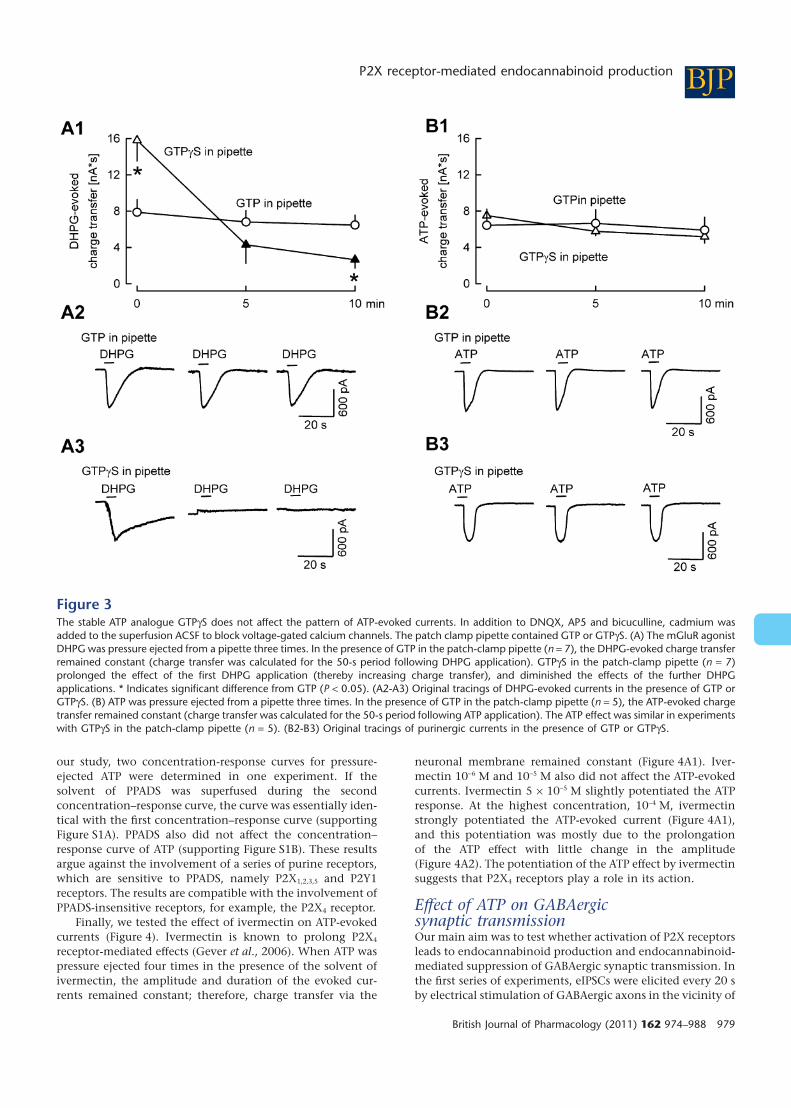
For advancing the identification of the receptor involvedin the ATP effect, we determined whether G protein-coupledreceptors were involved in the ATP effect (Figure 3). To thisaim, we tested the effect of GTPgS, applied via the patch-clamp pipette intracellularly. In the presence of this non-hydrolysable ATP analogue, G protein activation triggered bythe first agonist application becomes long-lasting, and subse-quent agonist applications trigger only moderate, if any, Gprotein activation (e.g. Jeong et al., 2001). At first, we carriedout positive-control experiments. In one group, GTP wasincluded in the patch clamp pipette, and the mGluR agonistDHPG (10-5 M) was pressure ejected three times (Figure 3A).DHPG elicited an inward current as described previously (e.g.Kim et al., 2003), and the charge transfer (area under thecurrent curve) was identical during the three DHPG applica-tions. The DHPG current was abolished by the mGluR1antagonist CPCCOEt, verifying involvement of mGluR1receptors (not shown). The pattern of effects of DHPG wasdifferent in neurones patched with pipettes containingGTPgS. The effect of the first DHPG application was pro-longed, resulting in higher charge transfer values, and thesecond and third DHPG application elicited only smallcharge transfers (Figure 3A). The results show that GTPgSenhances the first effect elicited by activation of a G protein-coupled receptor and attenuates later effects. Then, we testedthe interaction between pressure-ejected ATP and GTPgS inthe patch pipette (Figure 3B). In neurones with GTP in thepipette, ATP elicited identical currents, when applied threetimes. The picture did not change in the presence of GTPgS inthe pipette, suggesting that G protein-coupled receptors werenot involved in the purinergic currents.
For obtaining further information on the properties of thereceptor activated by ATP, we compared the effects of ATP,ADP and UTP. Ejection pipettes were filled with equimolarconcentrations (10-2 M) of ATP, ADP and UTP. ATP, ADP andUTP elicited inward currents of 1046 � 73 pA (n = 16), 75 �
39 pA (n = 10) and 214 � 49 pA (n = 13) amplitude, respec-tively. Thus, the currents elicited by ADP and UTP were muchsmaller than the current elicited by an equimolar concentra-tion of ATP (the differences were also statistically significant;P < 0.05). This observation argues against the involvement ofP2Y receptors in the ATP effect, because ATP has a lower orsimilar affinity for P2Y receptors than ADP and UTP (Ralevicand Burnstock, 1998; Burnstock, 2007). On the other hand, itis known that ATP possesses much higher affinity for severalP2X receptors than ADP and UTP. Thus, the pattern of effectsof the three nucleotides is compatible with an involvement ofP2X receptors in the ATP-evoked current.
The interaction between PPADS (10-4 M) and ATP was alsotested (Supporting Information Figure S1). PPADS is a mixedpurine receptor antagonist, which blocks P2X1,2,3,5 receptorsand some P2Y receptors (especially P2Y1) (Buell et al., 1996;Lambrecht, 2000; von Kügelgen, 2006; Jarvis and Khakh,2009). The behaviour of PPADS at the mouse P2X4 receptor isnot clear. In one study (Jones et al., 2000), PPADS was foundto be an antagonist with low potency, whereas in anotherstudy (Townsend-Nicholson et al., 1999), PPADS even poten-tiated P2X4 receptor-mediated responses evoked by ATP. In
Figure 2ATP acts itself and A1 adenosine receptors are not involved in theeffects of ATP. In addition to DNQX, AP5 and bicuculline, cadmiumwas added to the superfusion ACSF to block voltage-gated calciumchannels. ATP was pressure ejected from a pipette four times. Ampli-tudes of ATP-evoked currents were expressed as percentages of theinitial reference value PRE. Shown are means � standard error of themean. (A) After the second ATP application, solvent (SOL; n = 3) orthe ecto-nucleotidase inhibitor ARL67156 (n = 8) was superfused.The original tracings were recorded in a slice with ARL67156 super-fusion at time points 1 and 2. (B) After the second ATP application,solvent (SOL; n = 3) or the A1 receptor antagonist DPCPX (n = 4) wassuperfused. The original tracings were recorded in a slice with DPCPXsuperfusion at time points 1 and 2.
BJP FE Kovacs et al.
978 British Journal of Pharmacology (2011) 162 974–988
our study, two concentration-response curves for pressure-ejected ATP were determined in one experiment. If thesolvent of PPADS was superfused during the secondconcentration–response curve, the curve was essentially iden-tical with the first concentration–response curve (supportingFigure S1A). PPADS also did not affect the concentration–response curve of ATP (supporting Figure S1B). These resultsargue against the involvement of a series of purine receptors,which are sensitive to PPADS, namely P2X1,2,3,5 and P2Y1receptors. The results are compatible with the involvement ofPPADS-insensitive receptors, for example, the P2X4 receptor.
Finally, we tested the effect of ivermectin on ATP-evokedcurrents (Figure 4). Ivermectin is known to prolong P2X4
receptor-mediated effects (Gever et al., 2006). When ATP waspressure ejected four times in the presence of the solvent ofivermectin, the amplitude and duration of the evoked cur-rents remained constant; therefore, charge transfer via the
neuronal membrane remained constant (Figure 4A1). Iver-mectin 10-6 M and 10-5 M also did not affect the ATP-evokedcurrents. Ivermectin 5 ¥ 10-5 M slightly potentiated the ATPresponse. At the highest concentration, 10-4 M, ivermectinstrongly potentiated the ATP-evoked current (Figure 4A1),and this potentiation was mostly due to the prolongationof the ATP effect with little change in the amplitude(Figure 4A2). The potentiation of the ATP effect by ivermectinsuggests that P2X4 receptors play a role in its action.
Effect of ATP on GABAergicsynaptic transmissionOur main aim was to test whether activation of P2X receptorsleads to endocannabinoid production and endocannabinoid-mediated suppression of GABAergic synaptic transmission. Inthe first series of experiments, eIPSCs were elicited every 20 sby electrical stimulation of GABAergic axons in the vicinity of
Figure 3The stable ATP analogue GTPgS does not affect the pattern of ATP-evoked currents. In addition to DNQX, AP5 and bicuculline, cadmium wasadded to the superfusion ACSF to block voltage-gated calcium channels. The patch clamp pipette contained GTP or GTPgS. (A) The mGluR agonistDHPG was pressure ejected from a pipette three times. In the presence of GTP in the patch-clamp pipette (n = 7), the DHPG-evoked charge transferremained constant (charge transfer was calculated for the 50-s period following DHPG application). GTPgS in the patch-clamp pipette (n = 7)prolonged the effect of the first DHPG application (thereby increasing charge transfer), and diminished the effects of the further DHPGapplications. * Indicates significant difference from GTP (P < 0.05). (A2-A3) Original tracings of DHPG-evoked currents in the presence of GTP orGTPgS. (B) ATP was pressure ejected from a pipette three times. In the presence of GTP in the patch-clamp pipette (n = 5), the ATP-evoked chargetransfer remained constant (charge transfer was calculated for the 50-s period following ATP application). The ATP effect was similar in experimentswith GTPgS in the patch-clamp pipette (n = 5). (B2-B3) Original tracings of purinergic currents in the presence of GTP or GTPgS.
BJPP2X receptor-mediated endocannabinoid production
British Journal of Pharmacology (2011) 162 974–988 979

patch-clamped Purkinje cells, and ATP was pressure-ejectedfrom a pipette twice (Figure 5). ATP evoked purinergic cur-rents and calcium spikes, similar to those depicted inFigure 1A (not shown). In the presence of solvent, ATP sup-pressed the eIPSCs (Figure 5). When the experiments wereperformed in the presence of the CB1 receptor antagonistrimonabant (10-6 M), ATP failed to suppress eIPSCs (Figure 5).These results suggest that ATP evoked an endocannabinoid-and CB1 receptor-mediated suppression of GABAergic synap-tic transmission.
To analyse the mechanism of the suppression of GABAer-gic synaptic transmission by ATP, we investigated the effect ofATP on miniature IPSCs (mIPSCs). mIPSCs were isolated bythe inclusion of tetrodotoxin in the superfusion medium(Figure 6). Pressure-ejected ATP evoked strong purinergic cur-rents in these experiments (966 � 80 pA; n = 34), and thesecurrents triggered additional calcium spikes (see Figure 6D).In solvent-treated slices, ATP lowered the frequency ofmIPSCs (Figure 6A), whereas the amplitude of mIPSCs wasnot changed (Figure 6B). Corresponding to these changes inthe primary parameters, the cumulative amplitude of mIPSCswas also lowered (Figure 6C). ATP-evoked changes in mIPSCproperties were also analysed by constructing cumulativeprobability distribution plots of mIPSC amplitudes and inter-event intervals (supporting Figure S2). ATP did not cause asignificant shift in the distribution of mIPSC amplitudes.However, ATP caused a significant shift of the distributionplot of mIPSC inter-event intervals to the direction of lower
frequencies. The decrease in mIPSC frequency by ATP directlypoints to a presynaptic inhibitory mechanism. The lack ofeffect of ATP on the amplitude of mIPSCs indicates that therewas no postsynaptic influence on the synaptic current, and isan indirect proof of the presynaptic action. In the presence ofrimonabant (10-6 M), ATP no longer lowered the frequencyand cumulative amplitude of mIPSCs (Figure 6A, C), verifyingthe involvement of endocannabinoids and CB1 receptors.
Our next aim was to clarify the involvement of calcium inthe ATP-evoked suppression of GABAergic transmission. In afirst series of experiments, we included a high concentrationof the fast calcium chelator BAPTA (4 ¥ 10-2 M) in the pipetteused for patch-clamping the postsynaptic Purkinje cells, andmIPSCs were recorded in the presence of tetrodotoxin (Sup-porting Information Figure S3). Pressure-ejected ATP evokedstrong purinergic currents in these experiments (1011 �
59 pA; n = 34), and these currents triggered additionalcalcium spikes (see Supporting Information Figure S3D). Irre-spective of whether the experiments were performed in the
Figure 4Ivermectin potentiates ATP-evoked currents. In addition to DNQX,AP5 and bicuculline, cadmium was added to the superfusion ACSF toblock voltage-gated calcium channels. ATP was pressure ejected froma pipette four times. After the second ejection, solvent (SOL) orivermectin (IVM) was superfused. ATP-evoked charge transfer valueswere expressed as percentages of the initial reference value PRE.Means � standard error of the mean of seven (SOL), three (IVM10-6 M), three (IVM 10-5 M), six (IVM 5 ¥ 10-5 M) and four (IVM10-4 M) experiments. * Indicates significant difference from SOL (P <0.05). (A2) The original tracings were recorded in a slice with IVM10-4 M superfusion at time points 1 and 2 (indicated in A1).
Figure 5ATP suppresses electrically evoked inhibitory postsynaptic currents(eIPSCs). DNQX and AP5 were present in the superfusion ACSF.eIPSCs were elicited every 20 s by electrical stimulation in themolecular layer. Every three eIPSCs were averaged and expressed aspercentages of eIPSCs during the initial reference period PRE. Pres-sure ejection of ATP evoked purinergic currents which were accom-panied by calcium spikes (not shown). The points after ATPapplication are the averages of three eIPSCs elicited 5, 25 and 45 safter the 5-s ATP application. Means � standard error of the mean of9 (SOL) and 10 (RIM) experiments. Filled symbols indicate significantdifference versus the time point preceding ATP ejection (P < 0.05);* Indicates significant difference from SOL (P < 0.05). (A2) Theoriginal tracings were recorded at time points 1 and 2 (indicated inA1) in the presence of solvent or rimonabant.
BJP FE Kovacs et al.
980 British Journal of Pharmacology (2011) 162 974–988

presence of solvent or rimonabant (10-6 M), ATP did notcause any change in mIPSCs: the frequency, amplitude andcumulative amplitude of mIPSCs remained constant (Sup-porting Information Figure S3A, B and C). These results indi-cate that an increase in intracellular calcium concentration isnecessary for the CB1 receptor-mediated retrograde signallingelicited by ATP.
In the last part of our study, we wanted to determine thesource of calcium ions triggering endocannabinoid produc-tion during the application of ATP. Three sources were con-sidered: calcium influx through P2X receptor channels (ourinitial hypothesis), calcium influx through voltage-gatedcalcium channels and calcium release from intracellularstores.
At first, we wanted to verify that calcium influx from theextracellular space into Purkinje cells contributes to the ATP-evoked purinergic current and increase in intracellularcalcium concentration. Combined patch-clamp and fluoro-metric calcium imaging experiments were performed(Figure 7). Experiments were carried out in the presence ofthe broad-spectrum voltage-gated calcium channel inhibitorcadmium (10-4 M) and the sarcoplasmic reticulum calciumATPase (SERCA) inhibitor thapsigargin (10-5 M). ATP waspressure ejected at first in the presence ACSF with normalcalcium concentration (2.5 mM) and then in the presence ofcalcium-free ACSF. During superfusion of the calcium-containing ACSF, ATP evoked a purinergic current of 1275 �
137 pA (n = 4) amplitude (Figure 7A1), and increased thecalcium concentration in the dendrites and soma of the
Purkinje cells (Figure 7A2, A3). During superfusion ofcalcium-free ACSF, the amplitude of the ATP-evoked puriner-gic current significantly (P < 0.05) decreased to 476 � 82 pA(n = 4) (compare Figure 7B1 with A1). The ATP-evokedincrease in intracellular calcium concentration in the den-drites was also greatly attenuated (compare Figure 7B2, B3with A2, A3). Thus, calcium influx from the extracellularspace into Purkinje cells contributes to the ATP-evokedpurinergic current and increase in intracellular calciumconcentration.
Subsequently, we wished to clarify whether ATP is ableto suppress GABAergic transmission when calcium influxthrough voltage-gated calcium channels and calcium releasefrom intracellular stores are both inactivated (Figure 8).Experiments were carried out in the presence of the voltage-gated calcium channel inhibitor cadmium (10-4 M) and theSERCA inhibitor thapsigargin (10-5 M). Cadmium acts notonly on postsynaptic Purkinje cells, but also blocks voltage-gated calcium channels in the axon terminals of inter-neurones, thereby eliminating sIPSCs elicited by actionpotentials and subsequent activation of voltage-gated calciumchannels. It was shown that in cerebellar Purkinje cellscadmium isolates the same set of GABAergic synaptic eventswhich are also isolated by the sodium channel inhibitortetrodotoxin (see Than and Szabo, 2002). For clarity, theGABAergic synaptic events recorded in the presence ofcadmium will be termed as ‘cadmium sIPSCs’. Pressure-ejected ATP evoked strong purinergic currents in these experi-ments (1183 � 67 pA; n = 34), but no calcium spikes occurred
Figure 6ATP suppresses miniature inhibitory postsynaptic currents (mIPSCs) recorded in the presence of tetrodotoxin. In addition to DNQX and AP5,tetrodotoxin (3 ¥ 10-7 M) was present in the superfusion ACSF to block voltage-gated sodium channels. (A–C) The frequency, amplitude andcumulative amplitude of mIPSCs were evaluated in 10-s periods and expressed as percentages of values during the initial reference period PRE.Pressure ejection of ATP evoked purinergic currents which were accompanied by calcium spikes (see D). Means � standard error of the mean of16 (SOL) and 18 (RIM) experiments. Filled symbols indicate significant difference versus PRE (P < 0.05); * indicates significant difference from SOL(P < 0.05). (D) An original tracing recorded in the presence of solvent (the period between the dashed lines was not evaluated in A-C).
BJPP2X receptor-mediated endocannabinoid production
British Journal of Pharmacology (2011) 162 974–988 981

due to cadmium (see Figure 8D). ATP suppressed the fre-quency and cumulative amplitude of cadmium sIPSCs(Figure 8A, C), without influencing their amplitude(Figure 8B). In the presence of rimonabant (10-6 M), ATP nolonger suppressed the frequency and cumulative amplitude ofcadmium sIPSCs (Figure 8A, C). These experiments show thatATP can evoke endocannabinoid-mediated retrograde sup-pression of GABAergic transmission in the absence of opera-tion of voltage-gated calcium channels and calcium releasefrom intracellular stores. A likely source of calcium in thiscondition is calcium entry through P2X receptor channels.
Finally, in the same experiments, we studied therole of calcium influx from the extracellular space intoPurkinje cells in the suppression of synaptic transmission(Figure 9). Cadmium (10-4 M) and thapsigargin (10-5 M)were still present in the ACSF, but the ACSF was nominallycalcium-free. Under this condition, ATP did not affectthe frequency, amplitude and cumulative amplitude ofcadmium sIPSCs (Figure 9A–C). Thus, calcium influx fromthe extracellular space into Purkinje cells is necessary forthe suppression of the GABAergic synaptic transmissionby ATP.
Figure 7Superfusion with calcium-free artificial cerebrospinal fluid (ACSF) diminishes the ATP-evoked current and the increase in intracellular calciumconcentration. Glutamatergic and GABAergic synaptic input to Purkinje cells was blocked by DNQX, AP5 and bicuculline, and the patch-clamppipette contained the calcium-sensitive fluorescent dye Oregon green 488 BAPTA-5N. The entire experiment was performed in the presence ofcadmium (10-4 M) and thapsigargin (10-5 M). (A1) Pressure ejection of ATP from a pipette elicited a strong purinergic current. (A2) The fluorescentimages and the (A3) statistical evaluation of calcium concentration changes indicate that ATP increased the calcium concentration in the somaand the dendrites. (B1-B3) ATP responses of the neurones shown in A1–A3 during superfusion with calcium free ACSF (Ca2+
o = 0 mM).Means � standard error of the mean of four experiments. Currents and calcium concentrations were recorded in the same neurones. * Indicatessignificant difference versus dendrite (P < 0.05). # Indicates significant difference (P < 0.05) versus DF/F0 in the presence of calcium in the ACSF(shown in A3).
BJP FE Kovacs et al.
982 British Journal of Pharmacology (2011) 162 974–988

The recordings of the present study were performed atroom temperature (20–24°C). It is known that the magnitudeand duration of endocannabinoid-mediated retrogradesignalling changes with temperature (see e.g. Kreitzer andRegehr, 2001). We wanted to verify that ATP is also able tosuppress synaptic transmission at more physiological tem-peratures. The brain slices were superfused at 34°C with ACSFcontaining cadmium (10-4 M) and thapsigargin (10-5 M).ATP, pressure ejected as in all previous experiments, loweredthe frequency and the cumulative amplitude of cadmiumsIPSCs, whereas it did not change their amplitude (support-ing Figure S4). The ATP effects appeared to be smaller anddelayed compared with the ATP effects at room temperature.Rimonabant prevented the effects of ATP. When the sameexperiments were carried out in calcium-free ACSF, ATP didnot suppress the cadmium sIPSCs (not shown). These resultsindicate that ATP suppresses GABAergic transmission ina CB1 receptor-dependent manner also at physiologicaltemperatures, albeit with different kinetics than at roomtemperature.
Discussion
The present study shows for the first time that ATP, veryprobably by activating P2X purine receptors, elicitsendocannabinoid- and CB1 receptor-mediated retrograde sup-
pression of synaptic transmission. Calcium increase in thepostsynaptic neurone was necessary for triggering endocan-nabinoid production and retrograde signalling. ATP induceda calcium increase in the postsynaptic Purkinje cells by twomechanisms. Calcium entered into the Purkinje cells directlyvia P2X purine receptor ion channels. In addition, depolar-ization of Purkinje cells after activation of P2X receptorsopened voltage-gated calcium channels.
The CB1 receptor antagonist rimonabant (Rinaldi-Carmona et al., 1994; Pertwee, 2005) consistently abolishedin several series of experiments the ATP-evoked suppressionof the GABAergic synaptic input to the Purkinje cells. Thisobservation points to the involvement of endocannabinoidsand CB1 receptors in the suppression.
The analysis of ATP effects on mIPSCs recorded in thepresence of tetrodotoxin or on sIPSCs recorded in the pres-ence of cadmium suggests that presynaptic receptors on axonterminals of interneurones were involved in the ATP-evokedsuppression of GABAergic synaptic transmission. Thus, ATPdid not change the amplitude of mIPSCs or cadmium sIPSCsarguing against a postsynaptic interference with the effect ofreleased GABA. The fact that ATP suppressed the frequency ofmIPSCs and of cadmium sIPSCs indicates inhibition of GABArelease from the axon terminals. The results of neuroanatomi-cal studies are in agreement with a presynaptic action: axonterminals of GABAergic interneurones in the cerebellar cortexpossess CB1 receptors, whereas the postsynaptic Purkinje cells
Figure 8ATP suppresses sIPSCs recorded in the presence of cadmium and thapsigargin. In addition to DNQX and AP5, cadmium (10-4 M) and thapsigargin(10-5 M) were present in the superfusion ACSF. The experiments in Figures 8 and 9 were performed on the same neurones. Figure 8 shows thefirst part of the experiment, in which an ACSF with normal calcium concentration was superfused. Figure 9 shows the second part of theexperiments, during which calcium-free ACSF was superfused. (A–C) The frequency, amplitude and cumulative amplitude of cadmium sIPSCs wereevaluated in 10-s periods and expressed as percentages of values during the initial reference period PRE. Pressure ejection of ATP evoked purinergiccurrents, but calcium spikes were prevented by cadmium in the ACSF (see D). Means � standard error of the mean of 12 (SOL) and 10 (RIM)experiments. Filled symbols indicate significant difference versus PRE (P < 0.05); * indicates significant difference from SOL (P < 0.05). (D) Anoriginal tracing recorded in the presence of solvent (the period between the dashed lines was not evaluated in A–C).
BJPP2X receptor-mediated endocannabinoid production
British Journal of Pharmacology (2011) 162 974–988 983

are largely devoid of CB1 receptors (Tsou et al., 1998; Dianaet al., 2002). Exogenous cannabinoids acting on CB1 receptorson axon terminals of interneurones also inhibit synaptictransmission between interneurones and Purkinje cells (e.g.Diana et al., 2002; Szabo et al., 2004).
Several observations indicated that ATP activates P2X puri-nergic receptors in the Purkinje cells. Thus, the experimentswith the ecto-nucleotidase inhibitor ARL67156 indicated thatATP itself elicited the purinergic current, not its degradationproducts generated by ecto-nucleotidases. The purinergiccurrent was not sensitive to an intracellular activator of Gproteins, GTPgS, arguing against the involvement of Gprotein-coupled receptors in the ATP effect. The involvementof the G protein-coupled A1 receptor in the ATP-evokedcurrent was specifically excluded by using the antagonistDPCPX. Because of a lack of selective antagonists, the P2Xreceptor subtype was not definitively identified. However, theresistance of the ATP effect to PPADS and the enhancement byivermectin are compatible with a role for P2X4 receptors. Inaddition, anatomical receptor localization studies showed thepresence of mRNA and protein of several types of P2X recep-tors in Purkinje cells, including P2X4 receptors (Collo et al.,1996; for review see Nörenberg and Illes, 2000; Rubio and Soto,2001; Xiang and Burnstock, 2005; Lein et al., 2007).
Several P2X receptors are also permeable to calcium ionsin addition to sodium and potassium ions (e.g. Rogers andDani, 1995; Egan and Khakh, 2004). Hence, it would beexpected that the charge carriers of the large inward currentevoked by ATP were partly calcium ions. The fluorometrically
measured intracellular calcium concentration increased afterATP application, and a significant portion of this increasepersisted when voltage-gated calcium channels were blockedby cadmium, and intracellular calcium stores were depletedby thapsigargin. Importantly, the ATP-evoked purinergiccurrent and the intracellular calcium increase in the dendriteswere greatly attenuated in the presence of a calcium-freeextracellular buffer. Therefore, it is very likely that ATP acti-vated P2X receptors with a resulting calcium influx throughthese receptor channels. Very similarly, ATP elicited a P2Xreceptor-mediated increase in calcium concentration inPurkinje cells in the study of Mateo et al. (1998). Undoubt-edly, calcium also entered into the postsynaptic neurones viavoltage-gated calcium channels, which were activated byATP-evoked depolarization in distal dendrites escapingvoltage-clamp.
As shown by the experiments with the calcium chelatorBAPTA, an increased calcium concentration in postsynapticPurkinje cells was necessary for triggering the ATP-evokedsuppression of synaptic transmission. The ATP-evoked sup-pression of synaptic transmission also operated when calciuminflux through voltage-gated calcium channels and calciumrelease from intracellular stores were inactivated, supportingthe hypothesis that calcium influx via P2X receptors is suffi-cient for triggering endocannabinoid production. In strongsupport for the role of calcium influx via P2X receptors inthe suppression of the GABAergic synaptic transmission is theobservation that ATP did not evoke suppression when theextracellular calcium concentration was nominally zero.
Figure 9ATP does not suppress sIPSCs recorded in the presence of cadmium and thapsigargin and calcium-free ACSF. In addition to DNQX and AP5,cadmium (10-4 M) and thapsigargin (10-5 M) were present in the calcium-free superfusion ACSF. Figure 8 shows the first part of the experiment,during which ACSF with normal calcium concentration was superfused. (A–C) The frequency, amplitude and cumulative amplitude of cadmiumsIPSCs were evaluated in 10-s periods and expressed as percentages of values during the initial reference period PRE. Means � standard error ofthe mean of 12 (SOL) and 10 (RIM) experiments. (D) An original tracing recorded in the presence of solvent (the period between the dashed lineswas not evaluated in A–C).
BJP FE Kovacs et al.
984 British Journal of Pharmacology (2011) 162 974–988

Abolishment of the ATP-evoked suppression of GABAergicsynaptic transmission by BAPTA rules out the possibility thatATP (or its degradation products) lowered GABA release by adirect action on GABAergic axon terminals. It was reportedthat ATP elicits 2-arachidonoylglycerol release from micro-glial cells (Witting et al., 2004). Prevention of ATP-evokedsuppression of GABAergic transmission by BAPTA in thePurkinje cells makes it also unlikely that ATP primarily actedon glial cells and 2-arachidonoylglycerol released by glial cellsinhibited the GABA release from the axon terminals.
It is theoretically possible that ATP (or a degradationproduct of ATP) activated Gaq/11 protein-coupled P2Y recep-tors on Purkinje cells, thus triggering endocannabinoidproduction and endocannabinoid-mediated retrograde sup-pression of GABA release from interneurone axon terminals,similar to the manner in which activated mGluR1 receptorslead to retrograde signalling (e.g. Galante and Diana, 2004).One argument against the involvement of Gaq/11 protein-coupled P2Y receptors is that the ATP-evoked current wasinsensitive to GTPgS. A further argument against the involve-ment of Gaq/11 protein-coupled P2Y receptors is that the ATP-evoked retrograde signalling was prevented by BAPTA in thepostsynaptic Purkinje cells. Gaq/11 protein-mediated retro-grade signalling between Purkinje cells and their afferentaxons continues to operate when calcium in Purkinje cells iskept low by BAPTA (Maejima et al., 2001; Galante and Diana,2004). Notably, at synapses in other brain areas, Gaq/11
protein-mediated endocannabinoid production and retro-grade signalling can be blocked by buffering calcium in thepostsynaptic neurones (Kushmerick et al., 2004; Kola et al.,2008).
Altogether, the participation of CB1 receptors, theinvolvement of presynaptic inhibition and the results of theBAPTA-, cadmium- and calcium-free ACSF experimentssupport the following mode of retrograde signalling: ATP actson P2X purine receptors on postsynaptic Purkinje cells,calcium concentration increases due to calcium influx viaP2X receptors and via voltage-gated calcium channels,endocannabinoids are released, and retrogradely diffusingendocannabinoids inhibit GABA release from presynapticaxon terminals. Retrograde signalling between Purkinje cellsand interneurone axon terminals has been repeatedly shownin the past: calcium influx through voltage-gated calciumchannels and activity of Gaq/11 protein-coupled receptorswere the triggers of endocannabinoid production (Dianaet al., 2002; Brenowitz and Regehr, 2003; Diana and Marty,2003; Szabo et al., 2004, 2006).
Interneurones of the cerebellar cortex possess somatoden-dritic P2X- and P2Y-receptors. Acting on these receptors, ATPand its degradation products enhance the firing of the inter-neurones and – consequently – the frequency and amplitudeof GABAergic sIPSCs recorded in Purkinje cells (Brockhauset al., 2004; Saitow et al., 2005; Deitmer et al., 2006). Westudied the effect of ATP on mIPSCs isolated by tetrodotoxinand on sIPSCs isolated by cadmium. Tetrodotoxin andcadmium strongly impede the coupling between the somaand the axon terminals of interneurones (but a weak electro-tonic coupling may persist in the presence of these agents;Glitsch and Marty, 1999). Thus, it is unlikely that the soma-todendritic stimulant ATP effects confounded the inhibitoryATP effects on the axon terminals of interneurones.
Donato et al. (2008) observed a presynaptic inhibition ofinterneurone – Purkinje cell transmission by the antagonistPPADS and after desensitization of P2X receptors. It wasassumed that endogenous ATP tonically activates P2X recep-tors on axon terminals of GABAergic interneurones. In ourexperiments, ATP did not increase the frequency of mIPSCs orcadmium sIPSCs even in the presence of the CB1 antagonistrimonabant. The reason for this discrepancy between ourresults and the results of Donato et al. (2008) is not known. Itis notable, however, that we directly studied the effect of ATP,whereas Donato et al. (2008) studied the effects of ATPantagonists. Compatible with our observations, Brockhauset al. (2004) also did not see an ATP-evoked enhancement ofmIPSC frequency in Purkinje cells.
We have shown that activation of P2X transmitter-gatedion channels leads to calcium-dependent and CB1 receptor-dependent retrograde signalling. Although endocannab-inoids were not chemically detected in the present study, it isvery likely that the retrograde signalling was mediated byendocannabinoids produced by postsynaptic Purkinje cells. Ithas been shown previously by biochemical measurementsthat activation of P2X receptors leads to endocannabinoidproduction; ATP elicited the release of 2-arachidonoylglycerolin cultivated microglial cells (Witting et al., 2004). Our elec-trophysiological experiments showed that activation of neu-ronal P2X purine receptors can also lead to endocannabinoidproduction. The number of publications on the involvementof transmitter-gated ion channels in endocannabinoid pro-duction is limited. Thus, Ohno-Shosaku et al. (2007) showedthat activation of NMDA-type ionotropic glutamate receptorsleads to calcium-dependent 2-arachidonoylglycerol produc-tion and retrograde suppression of GABAergic synaptic trans-mission between cultivated hyppocampal neurones. Untilrecently, calcium influx through voltage-gated calcium chan-nels and activity of Gaq/11 protein-coupled receptors wereregarded as triggers of endocannabinoid production inpostsynaptic neurones. The study of Ohno-Shosaku et al.,(2007) and our study show that calcium influx viatransmitter-gated ion channels is a third possibility for trig-gering endocannabinoid production and endocannabinoid-mediated retrograde synaptic signalling. It is noteworthy thatat the resting membrane potential, P2X receptors may serve –in contrast to NMDA receptors – as major calcium entrypathways, because they are not blocked by magnesium underthis condition.
Do our observations possess a (patho)physiological rel-evance? ATP is released in the central nervous system by bothexocytotic and non-exocytotic mechanisms, and especiallyneuronal injury or neurodegenerative processes lead to amassive outflow of ATP (Burnstock, 2006). It is conceivablethat under these conditions, ATP elicits endocannabinoidrelease and suppresses the release of inhibitory, but also exci-tatory, transmitters.
Acknowledgements
This study was supported by the ‘Deutsche Forschungsge-meinschaft’ (Sz 72/5-3). Flora E. Kovacs is a recipient of a PhDscholarship from the ‘Deutscher Akademischer Austauschdi-enst’ (DAAD).
BJPP2X receptor-mediated endocannabinoid production
British Journal of Pharmacology (2011) 162 974–988 985

Conflicts of interest
The authors state no conflict of interest.
References
Abbracchio MP, Burnstock G (1994). Purinoceptors: are therefamilies of P2X and P2Y purinoceptors? Pharmacol Ther 64:445–475.
Abbracchio MP, Burnstock G, Boeynaems J-M, Barnard EA,Boyer JL, Kennedy C et al. (2006). International union ofpharmacology LVIII: update on the P2Y G protein-couplednucleotide receptors: from molecular mechanism andpathophysiology to therapy. Pharmacol Rev 58: 281–341.
Alexander SPH, Mathie A, Peters JA (2009). Guide to receptors andchannels (GRAC), 4th edn. Br J Pharmacol 158 (Suppl. 1): S1–S254.
Alger BE (2002). Retrograde signaling in the regulation of synaptictransmission: focus on endocannabinoids. Prog Neurobiol 68:247–286.
Brenowitz SD, Regehr WG (2003). Calcium dependence ofretrograde inhibition by endocannabinoids at synapses ontoPurkinje cells. J Neurosci 23: 6373–6384.
Brenowitz SD, Best AR, Regehr WG (2006). Sustained elevation ofdendritic calcium evokes widespread endocannabinoid release andsuppression of synapses onto cerebellar Purkinje cells. J Neurosci26: 6841–6850.
Brockhaus J, Dressel D, Herold S, Deitmer JW (2004). Purinergicmodulation of synaptic input to Purkinje neurons in rat cerebellarbrain slices. Eur J Neurosci 19: 2221–2230.
Brown SP, Brenowitz SD, Regehr WG (2003). Brief presynapticbursts evoke synapse-specific retrograde inhibition mediated byendogenous cannabinoids. Nat Neurosci 6: 1048–1057.
Buell G, Lewis C, Collo G, North RA, Surprenant A (1996). Anantagonist-insensitive P2X receptor experessed in epithelia andbrain. EMBO J 15: 55–62.
Burnstock G (2006). Pathophysiology and therapeutic potential ofpurinergic signaling. Pharmacol Rev 58: 58–86.
Burnstock G (2007). Purine and pyrimidine receptors. Cell Mol LifeSci 64: 1471–1483.
Chevaleyre V, Takahashi KA, Castillo PE (2006).Endocannabinoid-mediated synaptic plasticity in the CNS. AnnuRev Neurosci 29: 37–75.
Collo G, North RA, Kawashima E, Merlo-Pitch E, Niedhart S,Surprenant A et al. (1996). Cloning of P2X5 and P2X6 receptors andthe distribution and properties of an extended family of ATP-gatedion channels. J Neurosci 16: 2495–2507.
Deitmer JW, Brockhaus J, Casel D (2006). Modulation of synapticactivity in Purkinje neurons by ATP. Cerebellum 5: 49–54.
Di Angelantonio S, Nistri A (2001). Calibration of agonistconcentrations applied by pressure pulses or via rapid solutionexchanger. J Neurosci Methods 110: 155–161.
Diana MA, Marty A (2003). Characterization ofdepolarization-induced suppression of inhibition using pairedinterneuron-Purkinje cell recordings. J Neurosci 23: 5906–5918.
Diana MA, Levenes C, Mackie K, Marty A (2002). Short-termretrograde inhibition of GABAergic synaptic currents in rat Purkinjecells is mediated by endogenous cannabinoids. J Neurosci 22:200–208.
Donato R, Rodrigues RJ, Takahashi M, Chi M, Soto D, Miyagi Ket al. (2008). GABA release by basket cells onto Purkinje cells, in ratcerebellar slices, is directly controlled by presynaptic receptors,modulating Ca2+ influx. Cell Calcium 44: 521–532.
Egan TM, Khakh BS (2004). Contribution of calcium ions to P2Xchannel responses. J Neurosci 24: 3413–3420.
Fortin DA, Trettel J, Levine ES (2004). Brief trains of actionpotentials enhance pyramidal neuron excitability viaendocannabinoid-mediated suppression of inhibition. JNeurophysiol 92: 2105–2112.
Fredholm BB, Abbracchio MP, Burnstock G, Dubyak GR,Harden KT, Jacobson KA et al. (1997). Towards a revisednomenclature for P1 and P2 receptors. Trends Pharmacol Sci 18:79–82.
Freiman I, Anton A, Monyer H, Urbanski MJ, Szabo B (2006).Analysis of the effects of cannabinoids on identified synapticconnections in the caudate-putamen by paired recordings intransgenic mice. J Physiol 575: 789–806.
Freund TF, Katona I, Piomelli D (2003). Role of endogenouscannabinoids in synaptic signaling. Physiol Rev 83: 1017–1066.
Galante M, Diana MA (2004). Group I metabotropic glutamatereceptors inhibit GABA release at interneuron - Purkinje cellsynapses through endocannabinoid production. J Neurosci 24:4865–4874.
Gever JR, Cockayne DA, Dillon MP, Burnstock G, Ford APDW(2006). Pharmacology of P2X channels. Pflugers Arch 452: 513–537.
Glitsch M, Marty A (1999). Presynaptic effects of NMDA incerebellar Purkinje cells and interneurons. J Neurosci 19: 511–519.
Howlett AC, Barth F, Bonner TI, Cabral G, Casellas P, Devane WAet al. (2002). International union of pharmacology. XXVII.Classification of cannabinoid receptors. Pharmacol Rev 54:161–202.
Illes P, Nieber K, Nörenberg W (1996). Electrophysiological effectsof ATP on brain neurons. Auton Pharmacol 16: 407–411.
Jarvis MF, Khakh BF (2009). ATP-gated P2X cation channels.Neuropharmacol 56: 208–215.
Jeong H-J, Han S-H, Min B-I, Cho Y-W (2001). 5-HT1A
receptor-mediated activation of G-protein-gated inwardly rectifyingK+ current in rat periaqueductal gray neurons. Neuropharmacol 41:175–185.
Jones CA, Chessell IP, Simon J, Barnard EA, Miller KJ, Michel ADet al. (2000). Functional characterization of the P2X4 receptororthologues. Br J Pharmacol 129: 388–394.
Kano M, Ohno-Shosaku T, Hashimotodani Y, Uchigashima M,Watanabe M (2009). Endocannabinoid-mediated control of synaptictransmission. Physiol Rev 89: 309–380.
Kim J, Alger BE (2004). Inhibition of cyclooxygenase-2 potentiatesretrograde endocannabinoid effects in hippocampus. Nat Neurosci7: 697–698.
Kim SJ, Kim YS, Yuan JP, Petralia RS, Worley PF, Linden DJ (2003).Activation of the TRPC1 cation channel by metabotropic glutamatereceptor mGluR1. Nature 426: 285–291.
BJP FE Kovacs et al.
986 British Journal of Pharmacology (2011) 162 974–988

Klotz KN (2000). Adenosine receptors and their ligands. NaunynSchmiedebergs Arch Pharmacol 362: 382–391.
Kola B, Farkas I, Christ-Crain M, Wittmann G, Lolli F, Amin F et al.(2008). The orexigenic effect of ghrelin is mediated through centralactivation of the endogenous cannabinoid system. PLoS ONE 3:e1797.
Kreitzer AC, Regehr WG (2001). Retrograde inhibition ofpresynaptic calcium influx by endogenous cannabinoids atexcitatory synapses onto Purkinje cells. Neuron 29: 717–727.
Kushmerick C, Price GD, Taschenberger H, Puente N, Renden R,Wadiche JI et al. (2004). Retroinhibition of presynaptic Ca2+_currents by endocannabinoids released via postsynaptic mGluRactivation at a calyx synapse. J Neurosci 24: 2955–2965.
Lambrecht G (2000). Agonists and antagonists acting at P2Xreceptors: selectivity profiles and functional implications. NaunynSchmiedebergs Arch Pharmacol 362: 340–350.
Lasser-Ross N, Ross WN (1992). Imaging voltage and synapticallyactivated sodium transients in cerebellar Purkinje cells. Proc R SocLond 247: 35–39.
Lein ES, Hawrylycz MJ, Ao N, Ayres M, Bensinger A, Bernard A et al.(2007). Genome-wide atlas of gene expression in the adult mousebrain. Nature 445: 168–176.
Lovinger DM (2008). Presynaptic modulation by endocannabinoids.In: Südhof TC, Starke K (eds). Pharmacology of NeurotransmitterRelease. Handbook of Experimental Pharmacology, Vol. 184.Springer Press: Berlin, Heidelberg, pp. 435–477.
Maejima T, Hashimoto K, Yoshida T, Aiba A, Kano M (2001).Presynaptic inhibition caused by retrograde signal frommetabotropic glutamate to cannabinoid receptors. Neuron 31:463–475.
Maejima T, Oka S, Hashimotodani Y, Ohno-Shosaku T, Aiba A,Wu D et al. (2005). Synaptically driven endocannabinoid releaserequires Ca2+-assisted metabotropic glutamate receptor subtype 1 tophospholipase C b4 signaling cascade in the cerebellum. J Neurosci25: 6826–6835.
Marcaggi P, Attwell D (2005). Endocannabinoid signaling dependson the spatial pattern of synapse activation. Nat Neurosci 8:776–781.
Mateo J, Garcia-Lecea M, Miras-Portugal MT, Castro E (1998). Ca2+
signals mediated by P2X-type purinoceptors in cultured cerebellarPurkinje cells. J Neurosci 18: 1704–1712.
Nörenberg W, Illes P (2000). Neuronal P2X receptors: localisationand functional properties. Naunyn Schmiedebergs Arch Pharmacol362: 324–339.
North RA (2002). Molecular physiology of P2X receptors. PhysiolRev 82: 1013–1067.
Ohno-Shosaku T, Hashimotodani Y, Ano M, Takeda S,Tsubokawa H, Kano M (2007). Endocannabinoid signallingtriggered by NMDA receptor-mediated calcium entry into rathippocampal neurons. J Physiol 584: 407–418.
Pertwee RG (2005). Pharmacological actions of cannabinoids. In:Pertwee R (ed.). Cannabinoids. Handbook of ExperimentalPharmacology, Vol. 168. Springer Press: Berlin, Heidelberg, NewYork, pp. 1–51.
Ralevic V, Burnstock G (1998). Receptors for purines andpyrimidines. Pharmacol Rev 50: 413–492.
Rancz EA, Häusser M (2006). Dendritic calcium spikes are tunabletriggers of cannabinoid release and short-term synaptic plasticity incerebellar Purkinje neurons. J Neurosci 26: 5428–5437.
Rinaldi-Carmona M, Barth F, Heaulme M, Shire D, Calandra B,Congy C et al. (1994). SR141716A, a potent and selective antagonistof the brain cannabinoid receptor. FEBS Lett 350: 240–244.
Rogers M, Dani JA (1995). Comparison of quantitative calcium fluxthrough NMDA, ATP, and Ach receptor channels. Biophys J 68:501–506.
Rubio ME, Soto F (2001). Distinct localization of P2X receptors atexcitatory postsynaptic specializations. J Neurosci 21: 641–653.
Saitow F, Murakoshi T, Suzuki H, Konishu S (2005). MetabotropicP2Y purinoceptor-mediated presynaptic and postsynapticenhancement of cerebellar GABAergic transmission. J Neurosci 23:2108–2116.
Stephens GJ (2009). G-protein-coupled-receptor-mediatedpresynaptic inhibition in the cerebellum. Trends Pharmacol Sci 630:421–430.
Straiker A, Mackie K (2007). Metabotropic suppression of excitationin murine autaptic hippocampal neurons. J Physiol 578: 773–785.
Stuart G, Häusser M (1994). Initiation and spread of sodium actionpotentials in cerebellar Purkinje cells. Neuron 13: 703–712.
Szabo B, Schlicker E (2005). Effects of cannabinoids onneurotransmission. In: Pertwee R (ed.). Cannabinoids. Handbook ofExperimental Pharmacology, Vol. 168. Springer Press: Berlin,Heidelberg, New York, pp. 327–365.
Szabo B, Than M, Thorn D, Wallmichrath I (2004). Analysis of theeffects of cannabinoids on synaptic transmission between basketand Purkinje cells in the cerebellar cortex of the rat. J PharmacolExp Ther 310: 915–925.
Szabo B, Urbanski MJ, Bisogno T, Di Marzo V, Mendiguren A,Bär W et al. (2006). Depolarization-induced retrograde synapticinhibition in the cerebellar cortex is mediated by2-arachidonoylglycerol. J Physiol (Lond) 577: 263–280.
Than M, Szabo B (2002). Analysis of the function of GABAB
receptors on inhibitory afferent neurons of Purkinje cells in thecerebellar cortex of the rat. Eur J Neurosci 15: 1575–1584.
Townsend-Nicholson A, King BF, Wildman SS, Burnstock G (1999).Molecular cloning, functional characterization and possiblecooperativity between the murine P2X4 and P2X4a receptors. MolBrain Res 64: 246–254.
Tsou K, Brown S, Sanudo-Pena MC, Mackie K, Walker JM (1998).Immunohistochemical distribution of cannabinoid CB1 receptors inthe rat central nervous system. Neuroscience 83: 393–411.
Varma N, Carlson GC, Ledent C, Alger BE (2001). Metabotropicglutamate receptors drive the endocannabinoid system inhippocampus. J Neurosci 21: RC188.
Von Kügelgen I (2006). Pharmacological profiles of clonedmammalian P2Y-receptor subtypes. Pharmacol Ther 110: 415–432.
Wallmichrath I, Szabo B (2002). Cannabinoid inhibit striatonigralGABAergic neurotransmission in the mouse. Neuroscience 113:671–682.
Wilson RI, Nicoll RA (2001). Endogenous cannabinoids mediateretrograde signalling at hippocampal synapses. Nature 410:588–592.
Witting A, Walter L, Wacker J, Möller T, Stella N (2004). P2X7receptors control 2-arachidonoylglycerol production by microglialcells. Proc Natl Acad Sci USA 101: 3214–3219.
Xiang Z, Burnstock G (2005). Changes in expression of P2Xpurinoceptors in rat cerebellum during postnatal development.Brain Res Dev Brain Res 156: 147–157.
BJPP2X receptor-mediated endocannabinoid production
British Journal of Pharmacology (2011) 162 974–988 987

Zimmermann H (1996). Biochemistry, localisation and functionalroles of ecto-nucleotidase in the nervous system. Prog Neurobiol49: 589–618.
Supporting information
Additional Supporting Information may be found in theonline version of this article:
Figure S1 PPADS does not affect the ATP-evoked currents. Inaddition to DNQX, AP5 and bicuculline, cadmium was addedto the superfusion ACSF to block voltage-gated calcium chan-nels. ATP was pressure ejected from a pipette. For obtainingconcentration-response relationships, the duration of thepressure pulse was varied (0.1–5 s). In each experiment, twoconcentration-response curves were obtained. Current ampli-tudes were expressed as percentages of the current obtained atthe 5 s pressure pulse within the first concentration-responsecurve (this first curve is called PRE). (A) After the firstconcentration-response curve (PRE) the second curve wasobtained in the presence of solvent (SOL). Means � SEM of 3experiments are shown. (B) After the first concentration-response curve (PRE) the second curve was obtained in thepresence of the mixed P2X/P2Y antagonist PPADS (10-4 M).Means � SEM of 7 experiments are shown. (B2-B3) Originaltracings of ATP-evoked currents during PRE and duringPPADS superfusion.Figure S2 Effects of ATP on cumulative probability distribu-tion plots of mIPSC amplitudes and inter-event intervals. Inaddition to DNQX and AP5, tetrodotoxin (3 ¥ 10-7 M) waspresent in the superfusion ACSF to block voltage-gatedsodium channels. ATP evoked purinergic currents which wereaccompanied by calcium spikes. The plots were constructedusing 20-s periods preceding (PRE) and following ATP appli-cation (after ATP) in one of the experiments shown inFigure 6. Bins for amplitudes and inter-event intervals were
multiples of 10 pA and 50 ms respectively. *Indicates signifi-cant difference (P < 0.05) between the distribution plots iden-tified by the Kolmogorov-Smirnov test.Figure S3 BAPTA prevents the suppression of mIPSCs byATP. In addition to DNQX and AP5, tetrodotoxin (3 ¥ 10-7 M)was present in the superfusion ACSF to block voltage-gatedsodium channels. The patch-clamp pipette contained thecalcium chelator BAPTA (4 ¥ 10-2 M). (A–C) The frequency,amplitude and cumulative amplitude of mIPSCs were evalu-ated in 10-s periods and expressed as percentages of valuesduring the initial reference period PRE. Pressure ejection ofATP evoked purinergic currents which were accompanied bycalcium spikes (see D). Means � standard error of the mean of17 (SOL) and 17 (RIM) experiments are shown. (D) An origi-nal tracing recorded in the presence of solvent (the periodbetween the dashed lines was not evaluated in A–C).Figure S4 ATP suppresses sIPSCs recorded in the presence ofcadmium and thapsigargin also at 34°C. In addition to DNQXand AP5, cadmium (10-4 M) and thapsigargin (10-5 M) werepresent in the superfusion ACSF. (A–C) The frequency, ampli-tude and cumulative amplitude of cadmium sIPSCs wereevaluated in 10-s periods and expressed as percentages ofvalues during the initial reference period PRE. Pressure ejec-tion of ATP evoked purinergic currents, but calcium spikeswere prevented by cadmium in the ACSF (see D). Means �
standard error of the mean of 15 (SOL) and 15 (RIM) experi-ments are shown. Filled symbols indicate significant differ-ence versus PRE (P < 0.05); * indicates significant differencefrom SOL (P < 0.05). (D) An original tracing recorded in thepresence of solvent (the period between the dashed lines wasnot evaluated in A–C).
Please note: Wiley-Blackwell are not responsible for thecontent or functionality of any supporting materialssupplied by the authors. Any queries (other than missingmaterial) should be directed to the corresponding author forthe article.
BJP FE Kovacs et al.
988 British Journal of Pharmacology (2011) 162 974–988