Neuroinflammation and the Glial Endocannabinoid System
587
Transcript of Neuroinflammation and the Glial Endocannabinoid System


Cannabinoids and the Brain

Attila KöfalviEditor
Cannabinoids and the Brain
Editorial and Chapters 1, 9, 14, 22 were proofed by Zsófi a Gombár
With 44 illustrations and 16 tables

Attila KöfalviCenter for Neurosciences of CoimbraFaculty of MedicineUniversity of CoimbraCoimbra, 3000-045 Portugal
ISBN-13: 978-0-387-74348-6 e-ISBN-13: 978-0-387-74349-3
Library of Congress Control Number: 2007933065
© 2008 Springer Science + Business Media, LLCAll rights reserved. This work may not be translated or copied in whole or in part without the written permission of the publisher (Springer Science+Business Media, LLC, 233 Spring Street, New York, NY 10013, USA), except for brief excerpts in connection with reviews or scholarly analysis. Use in connectionwith any form of information storage and retrieval, electronic adaptation, computer software, or by similar or dissimilar methodology now known or hereafter developed is forbidden.The use in this publication of trade names, trademarks, service marks, and similar terms, even if they are not identified as such, is not to be taken as an expression of opinion as to whether or not they are subject to proprietary rights.
Printed on acid-free paper
9 8 7 6 5 4 3 2 1
springer.com

Editorial
Did you know that if you take aspirin or some other type of painkillers, you simply upregulate your endocannabinoid system against your endovanilloid system? If it happens to be a completely new piece of information to you, then this book is for you! Seriously speaking, the first part of the book you are holding in your hands is an exhaustive source of scientific reviews on the molecular biology, pharmacology, anatomy, and physiology of the endocannabinoid and related lipid mediator sys-tems. The second part of the book, however, covers the involvement of these signal-ing systems in metabolic, neurological, and psychiatric disorders, and gives an overview on clinical trials and on recent advances in cannabinoid-based medicine. Therefore, the target audience for this book are (a) physicians, especially endo-crinologists, neurologists, psychiatrists, and neuroscientists who want to update their knowledge about metabolism, basic brain physiology, molecular biology, and pathology and about novel therapeutic opportunities; (b) graduate and undergradu-ate students who also wish to broaden their knowledge about endocrinology, neuro-science, neurology, and psychiatry, or may need orientation to determine their future scientific goals; (c) politicians and health care employers who hesitate whether marijuana or cannabinoid-based medications should be legalized; and last but not least, (d) journalists who can help the scientists to convey their message to a larger audience. All the authors of the present volume are world’s leading neuroscientists and physicians, who are also regarded to be pioneers in the cannabinoid research area. Here I would like to gratefully thank them for all their altruistic contributions, and for sparing their precious time on this work.
The very first idea of writing this book occurred to me in 2005 when I had an interesting conversation with a neurologist professor from the USA, after his excit-ing lecture about the impact of adenosine receptors on epilepsy. I asked him whether he would be interested in the role of cannabinoid receptors also besides adenosine receptors. I noticed a faint note of indignation in his answer when he said: “No, I do not treat drug addicts, but epilepsy patients.” He was apparently unaware of those facts which are extensively reviewed in this book, especially the CB
1 receptor that is believed to have the highest density among metabotropic recep-
tors in the nervous tissue, and, together with its endogenous agonists, they represent a unique signaling system, which seems to be a goldmine of therapeutic targets against many neuropsychiatric disorders. The reaction of the professor may be
v

excusable, since the body’s own cannabinoid system as well as the body’s opioid system or the nicotinic receptors were discovered in the quest to find the specific targets for drugs of abuse, such as marijuana, morphine, heroin, and tobacco’s nico-tine. Importantly, the last 16 years of constant research has discovered a much broader role for endocannabinoids than for the opioid or nicotinic acetylcholine signaling. Nevertheless, this role does not seem to receive sufficient recognition by those who otherwise should find it important in their professional activity. At present, I have the growing belief that the endocannabinoid system and related sys-tems of lipid mediators, such as eicosanoids and endovanilloids, constitute a major modulator/messenger supersystem, which is at least as important as the monoam-inergic, purinergic, and cholinergic systems. Furthermore, these modulator systems work hand in hand, and thus they cannot be viewed as solitary therapeutic targets. The borders between classical pharmacological areas are likely to be forgotten. Therefore we, the authors, consider ourselves extremely fortunate to make this book happen and to disseminate challenging up-to-date reviews on the role of can-nabinoids in the brain.
Now I would like to take the opportunity of addressing a few challenging ideas to the cannabinoid research area. There are some minor and major problems can-nabinoid researchers normally encounter, which could be easily alleviated. For instance, it seems to be ironic and even ridiculous to some extent that permission is required for using certain cannabinoid research tools, such as ∆9-THC and its potent derivative HU-210. More importantly, their experimental usage is further hindered by other rules in certain places. I will never forget the incident when the police appeared in my lab, inquiring how I had used ∆9-THC and for what purpose. Absurdly enough, at that point of time, I still had not received the shipment of the compound from the pharmaceutical company due to permission issues. It is no more than pure hypocrisy, knowing that there are several other even more selective, potent, and efficacious cannabinoid ligands available, causing even more expressed effects than ∆9-THC in animals. It is understandable that ∆9-THC requires permis-sion, it being the major constituent of marijuana. Nonetheless, the price of ∆9-THCand HU-210 appears to be so high, especially considering the remarkably little buy-able amounts, that selling these products for research purposes without permission would not represent a gross criminal risk.
Normalization of chemical names would also be desirable. For instance, researchers may face a considerable challenge to find all the articles of the popular nonselective potent cannabinoid agonist WIN55212-2 in searchable databases, since the ligand is variously termed WIN-55,212-2, WIN 55212-2, WIN 55,212-2, WIN-2 or R-(+)-WIN55212, R-WIN55212, R-WIN 55212, R-WIN 55,212, etc. with all possible permutations. The same is true for other compounds, such as the popular CB
1 receptor antagonist AM251. It is frequently used as AM 251, and a
search for the terms AM and 251 in a database may result in a lot of additional unrelated articles. Thus, combining two or more ligands in one search is definitely a vain idea. The problem could be solved with only a slight common effort to stand-ardize chemical names. It is also unfortunate that several old-fashioned journals still force the authors to use the long cumbersome chemical names of cannabinoid
vi Editorial

compounds even in the abstract of the article, for example, R(+)-[2,3-Dihydro-5-methyl-3-[(morpholinyl)methyl]pyrrolo[1,2,3-de]-1,4-benzoxazinyl]-(1-naphthalenyl) methanone mesylate or [N-(piperidin-1-yl)-5-(4-chlorophenyl)-1-(2,4-dichlorophenyl)-4-methyl-1H-pyrazole-carboxamidehydrochloride]. Deciphering this long chemical name or similar ones would represent an enormous challenge to almost every researcher in the field. Even a chemist would spend several hours to realize that these terms mean WIN55212-2 and SR141716A (Rimonabant, AcompliaTM).Apparently, the reason for these unnecessary complications is again the limited knowledge about the cannabinoid field (including the lack of information about the most common chemical tools used in cannabinoid pharmacology) in the general scientific community.
My other growing concern arises from the rapidly increasing number of publica-tions (in 2006 and 2007, it was ~100 articles per month; see Fig. 2 in Chap. 1). Thus it seems difficult to keep up to date with the physiology, pharmacology, molecular biology, and pathology of cannabinoids. Recently, it has become easier to publish “unorthodox” research findings, as most of them proved to be valid, since they resulted from complex interactions between the endocannabinoid system and other signaling systems, and between new ligands, new receptors, and other targets. Although many laboratories are making an enormous effort to rule out the underly-ing mechanisms of these unorthodox findings, concomitantly, the same unusual pharmacological or physiological actions are recurrently rediscovered and reported occasionally by new research groups. To be more explicit, I would mention here the pharmacology of cholinergic, purinergic, GABAergic, or glutamatergic signaling, in which commonly accepted ligands, such as methyllicaconitine, nicotine, ATP, PPADS, CGS21680, CNQX, AP5, bicuculline, etc. with well-established maximal selective nanomolar or micromolar concentrations can be found. These concentra-tions are never to be exceeded because it is common knowledge that it would ques-tion the reliability of conclusions about the observations. In contrast, ligands of low nanomolar or picomolar affinity are often used in the micromolar range in the can-nabinoid research field. There are research reports in which SR141716A and WIN55212-2 were used even at 10–100 µM in vitro and the authors claimed that the observed effects were CB
1 receptor mediated. Chapter 9 in this book thus tries
to establish a bottom line for the pharmacology of cannabinoid research, listing common “side effects” and unorthodox mechanisms that can be easily misinter-preted as actions at novel receptors.
Another chapter also tackles the question of inverse agonism. Several antago-nists of the cannabinoid receptors are known as inverse agonists (such as SR141716A and AM251; see Chap. 7). Nonetheless, recent data shed new light on this question by indicating an apparent lack of inverse agonism in the absence of endocannabinoids (which are otherwise generally present in most experimental preparations); in other words, these antagonists would not cause an effect opposite to the agonists. This is topped by reports on novel CB
1 receptor-selective neutral/
silent antagonists. Thus, it might be worth solving this problem; otherwise one may eventually conclude that a neutral antagonist inhibits the binding of only the syntheticagonists at the CB
1 receptor, but not that of the endogenous agonists.
Editorial vii

As a concluding remark, I would like to express again my gratitude to the contribut-ing authors and to Joseph Burns from Springer-Verlag for recognizing the compel-ling need for the present volume and for giving me the opportunity to make this work happen. We (the authors) apologize for not discussing many significant publi-cations in the present volume; it is entirely unintentional and completely due to space limitations. Nevertheless, the book the reader may hold right now in his hands has made a serious attempt to give a comprehensive overview of all the essential lit-erature concerning the endocannabinoid and related systems in the nervous tissue.
Coimbra, June 2007 Attila Köfalvi
viii Editorial

Contents
Editorial . . . . . . . . . . . . . . . . . . . . . . . . . . . . . . . . . . . . . . . . . . . . . . . . . . . . . . v
Contributors . . . . . . . . . . . . . . . . . . . . . . . . . . . . . . . . . . . . . . . . . . . . . . . . . . . xiii
Part I Molecular Biology, Pharmacology, Anatomy, and Physiology of the Endocannabinoid and Related Lipidergic Signaling Systems in the Brain
1 An Historical Introduction to the Endocannabinoid and Endovanilloid Systems . . . . . . . . . . . . . . . . . . . . . . . . . . . . . . . . . . . 3Istvan Nagy, John P.M. White, Cleoper C. Paule, and Attila Köfalvi
2 Biosynthesis of Anandamide and 2-Arachidonoylglycerol . . . . . . . . . 15Takayuki Sugiura
3 Removal of Endocannabinoids by the Body: Mechanisms and Therapeutic Possibilities . . . . . . . . . . . . . . . . . . . . . . . . . . . . . . . . . 31Christopher J. Fowler and Lina Thors
4 Other Cannabimimetic Lipid Signaling Molecules . . . . . . . . . . . . . . . 47Heather B. Bradshaw
5 CB1 Cannabinoid Receptors: Molecular Biology, Second Messenger Coupling and Polarized Trafficking in Neurons. . . . . . . . . . . . . . . . . . . . . . . . . . . . . . . . . . . . . . . . . . . . . . . . . 59Andrew J. Irving, Neil A. McDonald, and Tibor Harkany
6 CB2 Cannabinoid Receptors: Molecular, Signaling, and Trafficking Properties . . . . . . . . . . . . . . . . . . . . . . . . . . . . . . . . . . . 75Paul L. Prather
7 CB1 and CB2 Receptor Pharmacology . . . . . . . . . . . . . . . . . . . . . . . . . . 91Roger G. Pertwee
ix

x Contents
8 Functional Molecular Biology of the TRPV1 Ion Channel . . . . . . . . . 101Istvan Nagy, John P.M. White, Cleoper C. Paule, Mervyn Maze, and Laszlo Urban
9 Alternative Interacting Sites and Novel Receptors for Cannabinoid Ligands . . . . . . . . . . . . . . . . . . . . . . . . . . . . . . . . . . . . 131Attila Köfalvi
10 Anatomical Distribution of Receptors, Ligands and Enzymes in the Brain and in the Spinal Cord: Circuitries and Neurochemistry . . . . . . . . . . . . . . . . . . . . . . . . . . . . . . . 161Giovanni Marsicano and Rohini Kuner
11 Endocannabinoids at the Synapse: Retrograde Signaling and Presynaptic Plasticity in the Brain . . . . . . . . . . . . . . . . . . . . . . . . . 203Gregory L. Gerdeman
12 Endocannabinoid Functions in Neurogenesis, Neuronal Migration, and Specification . . . . . . . . . . . . . . . . . . . . . . . . . 237Tibor Harkany, Manuel Guzmán, and Yasmin L. Hurd
Part II The Endocannabinoid System in Clinical Neuroscience and Experimental Neuropsychiatry
13 Cannabinoids in the Management of Nausea and Vomiting . . . . . . . . 259Linda A. Parker and Cheryl L. Limebeer
14 Endocannabinoids in Energy Homeostasis and Metabolic Disorders . . . . . . . . . . . . . . . . . . . . . . . . . . . . . . . . . . . . . 277Isabel Matias, Vincenzo Di Marzo, and Attila Köfalvi
15 Cannabinoids and Neuroprotection . . . . . . . . . . . . . . . . . . . . . . . . . . . . 317Veronica A. Campbell and Eric J. Downer
16 Neuroinflammation and the Glial Endocannabinoid System . . . . . . . . . . . . . . . . . . . . . . . . . . . . . . . . . . . . . . . . . . . . . . . . . . . . 331Cristina Benito, Rosa María Tolón, Estefanía Núñez, María Ruth Pazos, and Julián Romero
17 Targeting Cannabinoid Receptors in Brain Tumors . . . . . . . . . . . . . . 361Guillermo Velasco, Arkaitz Carracedo, Cristina Blázquez, Mar Lorente, Tania Aguado, Cristina Sánchez, Ismael Galve-Roperh, and Manuel Guzmán

Contents xi
18 Cannabinoids for the Control of Multiple Sclerosis. . . . . . . . . . . . . . . 375Gareth Pryce, Sam J. Jackson, and David Baker
19 Endocannabinoids in Alzheimer’s Disease . . . . . . . . . . . . . . . . . . . . . . 395María L. de Ceballos
20 The Endocannabinoid System as a Therapeutic Target in Epilepsy . . . . . . . . . . . . . . . . . . . . . . . . . . . . . . . . . . . . . . . . . . 407Krisztina Monory and Beat Lutz
21 The Endocannabinoid System in the Physiology and Pathology of the Basal Ganglia . . . . . . . . . . . . . . . . . . . . . . . . . . . . 423Gregory L. Gerdeman and Javier Fernández-Ruiz
22 The Endocannabinoid System is a Major Player in Schizophrenia. . . . . . . . . . . . . . . . . . . . . . . . . . . . . . . . . . . . . . . . . . . . 485Attila Köfalvi and Markus Fritzsche
23 The Cannabinoid Controversy: Cannabinoid Agonists and Antagonists as Potential Novel Therapies for Mood Disorders . . . . . . . . . . . . . . . . . . . . . . . . . . . . . . . . . . . . . . . . . 529Eleni T. Tzavara and Jeffrey M. Witkin
24 Role of Cannabinoid Receptors in Anxiety Disorders . . . . . . . . . . . . . 559Aldemar Degroot
Index . . . . . . . . . . . . . . . . . . . . . . . . . . . . . . . . . . . . . . . . . . . . . . . . . . . . . . . . . 573
Contributors
David BakerNeuroimmunology Unit, Neuroscience Centre, Institute of Cell and Molecular Science, Barts and the London School of Medicine and Dentistry, Queen Mary University of London, 4 Newark Street, Whitechapel,London E1 2AT, UK, [email protected]
Heather BradshawPsychological and Brain Sciences, The Kinsey Institute for Research in Sex, Gender and Reproduction, Indiana University Bloomington, IN 47405 [email protected]
Veronica A. CampbellDepartment of Physiology and, Trinity College Institute of Neuroscience, Trinity College, Dublin, Ireland
María L.de CeballosInstituto Cajal, CSIC, Doctor Arce, 37, 28002 Madrid, [email protected]
Aldemar DegrootAstellas Pharma Europe B.V., Elisabethhof 1, 2350 AC Leiderdorp, The Netherlands, [email protected]
Javier Fernández-RuizDepartamento de Bioquímica y Biología Molecular III, Facultad de Medicina, Universidad Complutense, 28040-Madrid, Spain
Christopher J. FowlerDepartment of Pharmacology and Clinical Neuroscience, Umeå University, SE901 87 Umeå, Sweden
xiii

Markus FritzschePraxis für Innere Medizin, Soodstrasse 13, 8134 Adliswil, Switzerland
Gregory Gerdemanc/o John Schindler, 6001 E. Pima Street, Apt. 162, Tucson, Arizona 85712
Manuel GuzmanDepartment of Biochemistry and Molecular Biology I, School of BiologyComplutense University, 28040 Madrid, Spain
Tibor HarkanyInstitute of Medical Sciences, University of Aberdeen, ForesterhillAB25 2ZD, Aberdeen, Scotland, UK
Andrew J. IrvingNeurosciences Institute, Division of Pathology & Neuroscience, Ninewells Hospital and Medical School, University of Dundee, Dundee, Scotland, DD1 9SY, UK
Rohini KunerPharmacology Institute, University of Heidelberg, Im Neuenheimer Feld 366, 69120 Heidelberg, Germany
Beat LutzInstitute of Physiological Chemistry and PathobiochemistryJohannes Gutenberg-University Mainz, Duesbergweg 6, 55099 Mainz, [email protected]
Giovanni Marsicano U 862 Centre de Recherche INSERM François Magendie, Université Bordeaux 2, 146, rue Léo Saignat, 33077 Bordeaux, [email protected]
Vincenzo Di MarzoIstituto di Chimica Biomolecolare, Consiglio Nazionale delle RicercheVia Campi Flegrei 34, Comprensorio Olivetti, 80078 Pozzuoli (NA), [email protected]
Isabel MatiasIstituto di Chimica Biomolecolare, Consiglio Nazionale delle RicercheVia Campi Flegrei 34, Comprensorio Olivetti, 80078 Pozzuoli (NA), [email protected]
xiv Contributors

Istvan NagyDepartment of Anaesthetics, Pain Medicine and Intensive Care,Imperial College London, Chelsea and Westminster Hospital, 369 Fulham Road, London SW10 9NH, UK
Linda A. ParkerDepartment of Psychology, University of Guelph, Guelph, ON N1G 2W1, Canada, [email protected]
Roger G. Pertwee Professor of Neuropharmacology, Institute of Medical SciencesUniversity of Aberdeen, Aberdeen AB25 2ZD, Scotland, [email protected]
Paul L. PratherDept of Pharmacology & Toxicology, Mail Slot 611, College of MedicineUniversity of Arkansas for Medical Sciences, 4301 W. Markham St.Little Rock, AR 72205, [email protected]
Julián RomeroLaboratorio de Apoyo a la Investigación, Fundación Hospital Alcorcón, C/ Budapest 1. 28922, Alcorcón, Madrid, Spain
Takayuki SugiuraFaculty of Pharmaceutical Sciences, Teikyo University, Sagamiko, Sagamihara, Kanagawa 229-0195, Japan
Eleni Tzavara CR1 INSERM, INSERM U-513 Neurobiologie et PsychiatrieFaculté de Médecine de Créteil, 8 rue du Général SARRAIL, F-94010, Créteil, France
Contributors xv

Part IMolecular Biology, Pharmacology,
Anatomy, and Physiology of the Endocannabinoid and Related Lipidergic
Signaling Systems in the Brain

Chapter 1An Historical Introduction to the Endocannabinoid and Endovanilloid Systems
Istvan Nagy, John P.M. White, Cleoper C. Paule, and Attila Köfalvi
Abstract Cannabis and chili pepper have been used for medical, gastronomical and recreational purposes for at least 8,000 years. Nevertheless, it was discovered only eight years ago that the cloned neuronal targets of their active principles, delta9-tetrahydrocannabinol (∆9-THC) and capsaicin are related to each other, as they all can be activated by some arachidonic acid-derivative endogenous ligands. Here, we will summarize the history of man’s relationship with cannabis and cap-saicin, and we will detail the most important scientific keystones in the evolution of cannabinoid and vanilloid research, featuring the list of cannabinoid and capsaicin effects, the discovery of endogenous ligands and the cloning of receptors, namely, the CB
1 and the CB
2 cannabinoid receptor as well as the TRPV
1 vanilloid recep-
tor, where the endogenous and the plant-derived substances act upon. This chapter serves, therefore, as an introduction to Cannabinoids and the Brain, the book which will extensively describe the neuronal and, to some extent, the peripheral cannabinoid and vanilloid systems in molecular, pharmacological, physiological, pathological and neuropsychiatric viewpoints.
Introduction
The History of Cannabis
The Asiatic plant cannabis or hemp (Cannabis sativa/indica = useful/Indian Cannabis) has been used for more than 8,000 years due to its medical and psycho-tropic effects. It is most likely that the original Sumerian word “kunibu” developed into the forms “kan(n)ab(is)” and “hanaba”, then “hennep” and finally, hemp. The plant cannabis belongs to the family Cannabaceae and the order Urticales. Its leaves and flowering tops are used to produce marijuana and hashish (also known as charas, bhang, ganja, dagga, grass, pot). Seeds of cannabis were found in 8,000 years old Chinese food remains. Interestingly, the first written note about the medical use of cannabis was also discovered in China, which dates back to 2727 B.C. The Atharvaveda, the sacred text of Hinduism, also mentions the use of cannabis for medical purposes in India between 1200 and 800 B.C. The psychotropic properties
A. Köfalvi (ed.), Cannabinoids and the Brain. 3© Springer 2008

4 I. Nagy et al.
of cannabis were first described in a Chinese medical book around 100 B.C. It is believed that Cannabis sativa was first introduced in Europe by the Scythians, as recorded by Herodotus in 430 B.C. In 100 A.D., Dioskurides inferred that cannabis was a Roman medical plant, whereas Galen highlighted its psychotropic action in 170 A.D. The medieval Europe was first informed about the popularity of cannabis in Asia by Marco Polo. Later cannabis was used mainly as a medicine in England. Even Queen Victoria was prescribed cannabis by her doctor in 1890. Consequently, can-nabis was declared harmless and legalized in 1901. However, in 1925, the Geneva Convention included cannabis and hashish in the list of dangerous and illicit drugs. In the USA, cannabis was also used for medical purposes from 1840, but the Mexican Revolution in 1910 changed the general opinion about cannabis. It became a symbol of terrible sins. Until 1931, 29 states prohibited the use of marijuana, and from 1937, the Federal Law proclaimed marijuana as an illicit drug. Still, it regained its popular-ity when both president Kennedy and president Johnson suggested that cannabis should be legalized. Presumably, due to these propositions, 200–250 million cannabis users were reported by the UN worldwide till 1970. In the last 40 years, the debate on the safety and legal status of cannabis-based medical treatments is becoming increasingly intense at both political and scientific levels (see for example Wall et al., 2001; Hayry, 2004; Comeau, 2006). Although clinical trials with cannabinoid ligands are allowed in many countries, the general use of marijuana to treat the pain and eat-ing problems of cancer patients as well as to reduce intraocular pressure in glaucoma is still not legalized. The main issue is that the concentration of beneficial constituents in the smoke of cannabis cannot be controlled, and furthermore, conservative politics can hardly agree with the medical use of an illicit drug. Nonetheless, we should mention that morphine, codeine, lidocaine and procaine, which are all illicit drug-derivatives, are commonly used in medicine. Moreover, nicotine is regarded as one of the most addictive drugs, yet its use is perfectly legal. All the same, we have to acknowledge certain concerns, as chronic marijuana consumption (ca. ≥50 times) can induce schizophrenia in susceptible persons (see Chap. 22).
The History of Capsaicin
Chili peppers (Capsicum frutescens var. longum) are members of the nightshade family (Solanaceae), and have been domesticated since about 7500 B.C. in the Americas (Perry et al., 2007). Christopher Columbus was one of the first Europeans who found chili peppers and subsequently transferred a certain amount to the Old World. He accidentally named them “peppers” because of their similar-ity in taste with the black peppers of the Piper genus. This pungent taste is due to capsaicin, a neurotoxin derived from the chili pepper plant. Capsaicin has been extensively used for centuries both as a herbal remedy and a food product prized in the cuisine of many societies. Capsaicin is also responsible for the reduced sen-sitivity of the mouth to high temperatures and painful mechanical stimuli which results from regular chili pepper consumption. The fact that capsaicin also relieves

1 An Historical Introduction 5
the spontaneous pain associated with inflammation prompted healers in many dif-ferent cultures to employ hot peppers to treat painful conditions of varying aetiolo-gies over the centuries. Thus, Native Americans rubbed their gums with hot peppers as a cure for tooth ache, while Europeans used an alcoholic extract pre-pared from chilies for a similar purpose (Szallasi and Blumberg, 1999). Capsaicin is still commonly used for treating painful conditions as an “over-the-counter” remedy in the form of capsaicin-containing ointments.
The Discovery of the Endocannabinoid and Endovanilloid Systems
The Endocannabinoid System
Although Eastern cultures have been using marijuana as medicine for centuries, Western cultures started to recognize the therapeutic potential of marijuana only recently. For instance, cannabis extract was a licensed medicine and sold under the name of “Tincture of Cannabis” in the UK (Gill et al., 1970). The first observed medicinal benefits encompassed anesthetic, airway opening, antihypertensive, eye pressure reducing (in glaucoma) as well as antiemetic actions, but for decades, the underlying physiological and molecular mechanisms were unknown. The first iso-lated plant-derived (phyto-) cannabinoid was cannabinol, found in the red oil extract of hemp more than a century ago, and in the 1930s, its chemical structure was eluci-dated (Pertwee, 2006). Although tetrahydrocannabinols (THCs) and cannabidiols were discovered and isolated from hemp extracts in the following years, the structure and stereochemistry of the naturally occurring (–)-cannabidiol (Mechoulam and Shvo, 1963) and (–)-trans-∆9-tetrahydrocannabinol (∆9-THC), the main psychoactive constituent of marijuana and hashish (Gaoni and Mechoulam, 1964) were unraveled in the decade when hippies also became interested in cannabis preparations. The major phytocannabinoid structure was identified as a tricyclic ring constituted from a phenol ring, having a 5-carbon alkyl chain meta to the hydroxyl, a central pyran ring, and a mono-unsaturated cyclohexyl ring (Fig. 1; Howlett et al., 2004). Raphael Mechoulam and his laboratory pioneered the discovery and synthesis of numerous novel phytocannabinoids, which enumerate at least 66 distinctive ones hitherto (Mechoulam and Hanus, 2000; Pertwee, 2006). In parallel with their discovery, hemp constituents were tested for psychotropic and motor effects in man and in animal models, mostly in mice, rats, rabbits, and dogs. THCs proved to be the most effective among all phytocannabinoids, whereas among THCs, ∆9-THC seems to be responsi-ble for the vast majority of effects such as motor disturbances and catalepsy, corneal areflexia (in rabbits), scratching, euphoria and dysphoria, anxiety, drowsiness, altered time and audiovisual perceptions, panic attacks and impaired memory (Haagen-Smit et al., 1940; Loewe, 1946; Paton and Pertwee, 1973; Howlett et al., 2004). The following years then proved that the more psychotropic a cannabinoid substance is

6 I. Nagy et al.
the greater motor disturbances it causes. Furthermore, among phytocannabinoids, ∆9-THC is the most potent and effective psychomotor compound. The underlying mechanisms for these effects were mostly believed to result from “non-specific” interactions between the lipophilic ∆9-THC and the cell membranes, changing the fluidity and structure of the latter, therefore affecting most cell types (Lawrence and Gill, 1975; Hillard et al., 1985). Nonetheless, a nearly identical molecule, ∆8-THC, was much less potent and efficacious than ∆9-THC, and most other phytocannabi-noids were devoid of effect, which all weakened the hypothesis of changing membrane fluidity. The next important cornerstone was the discovery that ∆9-THC inhibits cAMP accumulation (Howlett and Fleming, 1984), and the recognition of specific cannabinoid binding sites in the brain (Devane et al., 1988). These two find-ings from Allyn Howlett’s laboratory predicted that the discovery of atleast one cannabinoid receptor was imminent. And indeed, in 1990, both the rat and the human CB
1 receptors were characterized (Gérard et al., 1990, 1991; Matsuda et al., 1990),
and the first study on its distribution found the receptor at an unexpectedly high density in the brain (Herkenham et al., 1991). Right at the moment when we write
Fig. 1 The most important constituents of Cannabis sativa L., namely ∆9-tetrahydrocannabinol ( (–)-(6aR,10aR)-6,6,9-trimethyl-3-pentyl-6a,7,8,10a-tetrahydro-6H-benzo[c]chromen-1-ol; ∆9-THC),cannabinol (6,6,9-trimethyl-3-pentyl-6H-benzo[c]chromen-1-ol) and cannabidiol (2-( (1S,6S)-3-methyl-6-(prop-1-en-2-yl)cyclohex-2-enyl)-5-pentylbenzene-1,3-diol)

1 An Historical Introduction 7
these lines, there are 14,000 articles published in relation to cannabinoids. The first one – listed by PubMed – is from 1909. As Fig. 2 demonstrates, the first “boom” of cannabinoid research occurred after 1964, the year when Gaoni and Mechoulam reported the structure of ∆9-THC. The second boom – which is related to the discov-ery of the CB
1 receptor – resulted in a continuously increasing number of publications
in the last 15 years, and in 2006 and in the first five months of 2007, it reached a peak of 100 publications per month. Thus, recognizing the significance of cannabinoid research very early, Di Mahadeen and Rik Musty founded the International Cannabinoid Research Society in 1991 (http://www.cannabinoidsociety.org for fur-ther information). Soon, another cannabinoid receptor, the CB
2 receptor was discov-
ered, but its expression was found to be restricted mainly to immune tissues (Munro et al., 1993). Importantly, both cannabinoid receptors are G protein-coupled seven-transmembrane-domain receptors of the rhodopsin type (see Chaps. 5 and 6). In the meantime, the first endogenous cannabinoid ligand, arachidonoylethanolamine or anandamide, was found in porcine brain (Devane et al., 1992), which was followed by 2-arachidonoylglycerol (2-AG) described by two independent laboratories in the same year (Mechoulam et al., 1995; Sugiura et al., 1995; see Chap. 2). As one can notice from their name, both ligands are arachidonic acid derivatives, and interest-ingly, one of them, namely anandamide, is capable of activating the TRPV
1 receptor
as well (Zygmunt et al., 1999; and see below). Later in this book, we will mention some new cannabinoid receptors and endocannabinoid candidates (Chaps. 4 and 9). However, hitherto these four molecules received the biggest attention. The last 20 years provided a major boost to the renaissance of the synthesis of novel cannabinoid ligands as well, and further readings can be found in recent reviews (Mechoulam and Hanus, 2000; Howlett et al., 2004; Pertwee, 2006; see Chap. 7). However, we should highlight 1994 when the first selective CB
1 receptor antagonist, SR141716A or
Rimonabant, was reported (Rinaldi-Carmona et al., 1994) and, has been marketed in 2006 in Europe under the name Acomplia™ as a promising alternative medicine
Fig. 2 The yearly number of research and review articles on the cannabinoid field from 1950. The two arrows indicate the onset of the two booms: (a) Gaoni and Mechoulam (1964) report the structure of ∆9-THC; (b) Gérard and colleagues (1990) and Matsuda and colleagues (1990) report the cloning of the first cannabinoid receptor (the CB
1 receptor) from human and rat (See text for
further explanations.)

8 I. Nagy et al.
against cardiovascular and metabolic risk factors (see Chap. 14). Significantly, the wide-spectrum roles of CB
1 and CB
2 receptors were not determined only by the
action of antagonists. CB1 receptor knockout mice strains were engineered by
Zimmer and co-workers (1999) and Ledent and co-workers (1999). Most importantly, the vast majority of behavioural and physiological responses to cannabinoid ligands were no longer observed in these mice, compared to findings in the CB
2 receptor
knockout mouse (Buckley et al., 2000). These findings underlined that the major can-nabinoid receptor of the nervous tissue is the CB
1 receptor. As the conditional knock-
out technology became widespread, novel, neuron-specific conditional CB1 receptor
knockout animals were also generated (Marsicano et al., 2003).
The Endovanilloid System
Capsaicin was isolated as the active ingredient of chili peppers in the mid-nineteenth century (Thresh, 1846). However, its exact structure was elucidated only some seventy years later (Nelson, 1919). The revealed structure shows that capsai-cin possesses a vanilloid moiety, which results in it being assigned to the vanilloid family (see Fig. 2 in Chap. 8). Surprisingly, however, few vanilloids – other than capsaicin itself and the even more potent resiniferatoxin (RTX) – possess the char-acteristic pungency of capsaicin. Capsaicin in the nanomolar range specifically, and selectively, acts on a large sub-population of nociceptive primary sensory neurons. This remarkable property of capsaicin was first described by two Hungarian scien-tists, Janos Porszasz and Nicholas Jancso (1959), who reported that, following cap-saicin application, sensory fibres fail to produce action potentials on subsequent exposure to capsaicin. Nicholas Jancso’s son, Gabor Jancso, subsequently showed that those primary sensory neurons that are sensitive to capsaicin belong to the small diameter sub-population of dorsal root ganglion neurons, which are generally considered to be nociceptive in function (Jancso et al., 1977). His group also showed that capsaicin application in neonates ultimately results in degeneration of the capsaicin-sensitive sensory neurons. The fact that capsaicin appeared to be highly specific in its effects on nociceptive primary sensory neurons, coupled with its “desensitizing” effect, kindled major interest in the mechanisms involved in these phenomena among academics and, also, in the pharmaceutical industry. The search for the molecule, or molecules, which mediate the effects of capsaicin in primary sensory neurons, had an uncertain beginning. Voltage-gated Na+ channels and K+ channels were proposed as candidate molecules (Dubois, 1982; Yamanaka et al., 1984; Erdelyi et al., 1987). However, while Porszasz and Jancso (1959) had observed an excitatory effect of capsaicin on mammalian sensory fibres, an inhibi-tory effect on these voltage-gated ion channels was observed in recordings from frog, snail and crayfish neurons when exposed to capsaicin in micromolar concen-trations (see Chap. 9). Notwithstanding this, an attempt was made to argue that this inhibitory effect could be responsible for the loss of responsiveness of sensory neurons found following prolonged, or repeated, exposure to capsaicin. The first
1 An Historical Introduction 9
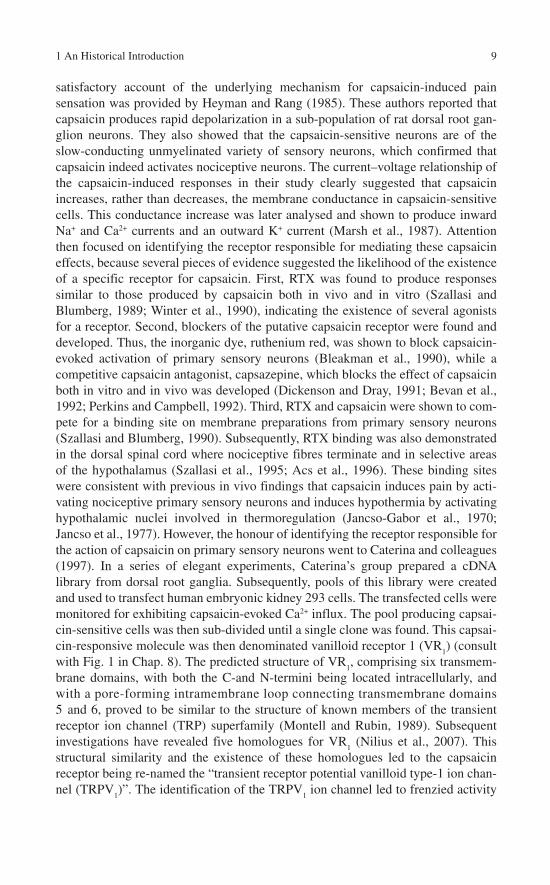
satisfactory account of the underlying mechanism for capsaicin-induced pain sensation was provided by Heyman and Rang (1985). These authors reported that capsaicin produces rapid depolarization in a sub-population of rat dorsal root gan-glion neurons. They also showed that the capsaicin-sensitive neurons are of the slow-conducting unmyelinated variety of sensory neurons, which confirmed that capsaicin indeed activates nociceptive neurons. The current–voltage relationship of the capsaicin-induced responses in their study clearly suggested that capsaicin increases, rather than decreases, the membrane conductance in capsaicin-sensitive cells. This conductance increase was later analysed and shown to produce inward Na+ and Ca2+ currents and an outward K+ current (Marsh et al., 1987). Attention then focused on identifying the receptor responsible for mediating these capsaicin effects, because several pieces of evidence suggested the likelihood of the existence of a specific receptor for capsaicin. First, RTX was found to produce responses similar to those produced by capsaicin both in vivo and in vitro (Szallasi and Blumberg, 1989; Winter et al., 1990), indicating the existence of several agonists for a receptor. Second, blockers of the putative capsaicin receptor were found and developed. Thus, the inorganic dye, ruthenium red, was shown to block capsaicin-evoked activation of primary sensory neurons (Bleakman et al., 1990), while a competitive capsaicin antagonist, capsazepine, which blocks the effect of capsaicin both in vitro and in vivo was developed (Dickenson and Dray, 1991; Bevan et al., 1992; Perkins and Campbell, 1992). Third, RTX and capsaicin were shown to com-pete for a binding site on membrane preparations from primary sensory neurons (Szallasi and Blumberg, 1990). Subsequently, RTX binding was also demonstrated in the dorsal spinal cord where nociceptive fibres terminate and in selective areas of the hypothalamus (Szallasi et al., 1995; Acs et al., 1996). These binding sites were consistent with previous in vivo findings that capsaicin induces pain by acti-vating nociceptive primary sensory neurons and induces hypothermia by activating hypothalamic nuclei involved in thermoregulation (Jancso-Gabor et al., 1970; Jancso et al., 1977). However, the honour of identifying the receptor responsible for the action of capsaicin on primary sensory neurons went to Caterina and colleagues (1997). In a series of elegant experiments, Caterina’s group prepared a cDNA library from dorsal root ganglia. Subsequently, pools of this library were created and used to transfect human embryonic kidney 293 cells. The transfected cells were monitored for exhibiting capsaicin-evoked Ca2+ influx. The pool producing capsai-cin-sensitive cells was then sub-divided until a single clone was found. This capsai-cin-responsive molecule was then denominated vanilloid receptor 1 (VR
1) (consult
with Fig. 1 in Chap. 8). The predicted structure of VR1, comprising six transmem-
brane domains, with both the C-and N-termini being located intracellularly, and with a pore-forming intramembrane loop connecting transmembrane domains 5 and 6, proved to be similar to the structure of known members of the transient receptor ion channel (TRP) superfamily (Montell and Rubin, 1989). Subsequent investigations have revealed five homologues for VR
1 (Nilius et al., 2007). This
structural similarity and the existence of these homologues led to the capsaicin receptor being re-named the “transient receptor potential vanilloid type-1 ion chan-nel (TRPV
1)”. The identification of the TRPV
1 ion channel led to frenzied activity

10 I. Nagy et al.
as scientists endeavoured to elucidate its expression pattern, function and the mechanisms involved in the regulation of its expression and activation. It has emerged that TRPV
1 is a polymodal receptor which responds to various ligands, as
well as to heat above ~42 °C, protons and post-translational modifications. Moreover, TRPV
1 is capable of integrating the effect of these activators (Caterina
et al., 1997; Tominaga et al., 1998; Chuang et al., 2001). In addition to its expres-sion in primary sensory neurons, TRPV
1 is also expressed by various neurons in the
brain and by non-neuronal cells at the periphery (Nagy et al., 2004). Generation of mice lacking TRPV
1 has shown that the capsaicin receptor is indispensable for the
development of inflammatory heat hyperalgesia (Caterina et al., 2000; Davis et al., 2000) and bladder hyper-reflexia (Charrua et al., 2007). Finally, recent publications have drawn attention to the role of endovanilloids in the activation of TRPV
1 in
pathological conditions (Dinis et al., 2004; Singh Tahim et al., 2005). Interestingly, certain of the endogenous TRPV
1 ligands, such as anandamide, are also endocan-
nabinoids (Zygmunt et al., 1999). The existence of these shared endogenous lig-ands has led to the theory that cannabinoid receptors and TRPV
1 may be the reverse
sides of the same coin, constituting the G protein-coupled receptors and the ligand-gated ion channels of the same sensory system. In Chap. 8, we describe the struc-ture and function of this remarkable ion channel and focus, in particular, on the mechanisms involved in its activation.
Acknowledgement Attila Köfalvi is grateful for the III/BIO/56/2005 grant and for the Fundação para a Ciência e Tecnologia of the Portuguese Government (POCI 2010/SFRH/BPD/18506/2004).
References
Acs G, Palkovits M, Blumberg PM (1996) Specific binding of [3H]resiniferatoxin by human and rat preoptic area, locus ceruleus, medial hypothalamus, reticular formation and ventral thala-mus membrane preparations. Life Sci 59:1899–1908.
Bevan S, Hothi S, Hughes G, James IF, Rang HP, Shah K, Walpole CS, Yeats JC (1992) Capsazepine: a competitive antagonist of the sensory neuron excitant capsaicin. Br J Pharmacol 107:544–552.
Bleakman D, Brorson, JR, Miller RJ (1990) The effect of capsaicin on voltage-gated calcium cur-rents and calcium signals in cultured dorsal root ganglion cells. Br J Pharmacol 101:423–431.
Buckley NE, McCoy KL, Mezey E, Bonner T, Zimmer A, Felder CC, Glass M, Zimmer A (2000) Immunomodulation by cannabinoids is absent in mice deficient for the cannabinoid CB
2
receptor. Eur J Pharmacol 396:141–149.Caterina MJ, Schumacher MA, Tominaga M, Rosen TA, Levine JD, Julius D (1997) The capsai-
cin receptor: a heat-activated ion channel in the pain pathway. Nature 389:816–824.Caterina MJ, Leffler A, Malmberg AB, Martin WJ, Trafton J, Petersen-Zeitz KR, Koltzenburg M, Basbaum AI, Julius D (2000) Impaired nociception and pain sensation in mice lacking the capsaicin receptor. Science 288:306–313.
Charrua A, Cruz CD, Cruz F, Avelino A (2007) Transient receptor potential vanilloid subfamily 1 is essential for the generation of noxious bladder input and bladder overactivity in cystitis. J Urol 177:1537–1541.

1 An Historical Introduction 11
Chuang HH, Prescott ED, Kong H, Shields S, Jordt SE, Basbaum AI, Chao MV, Julius D (2001) Bradykinin and nerve growth factor release the capsaicin receptor from PtdIns(4,5)P2-mediated inhibition. Nature 411:957–962.
Comeau P (2006) Cut to marijuana research sends strong message. CMAJ 175:1507–1508.Davis JB, Gray J, Gunthorpe MJ, Hatcher JP, Davey PT, Overend P, Harries MH, Latcham J,
Clapham C, Atkinson K, Hughes SA, Rance K, Grau E, Harper AJ, Pugh PL, Rogers DC, Bingham S, Randall A, Sheardown SA (2000) Vanilloid receptor-1 is essential for inflamma-tory thermal hyperalgesia. Nature 405:183–187.
Devane WA, Dysarz FA, Johnson MR, Melvin LS, Howlett AC (1988) Determination and characterization of a cannabinoid receptor in rat brain. Mol Pharmacol 34:605–613.
Devane WA, Hanus L, Breuer A, Pertwee RG, Stevenson LA, Griffin G, Gibson D, Mandelbaum A, Etinger A, Mechoulam R (1992) Isolation and structure of a brain constituent that binds to the cannabinoid receptor. Science 258:1946–1949.
Dickenson AH, Dray A (1991) Selective antagonism of capsaicin by capsazepine: evidence for a spinal receptor site in capsaicin-induced antinociception. Br J Pharmacol 104:1045–1049.
Dinis P, Charrua A, Avelino A, Yaqoob M, Bevan S, Nagy I, Cruz F (2004) Anandamide-evoked activation of vanilloid receptor 1 contributes to the development of bladder hyperreflexia and nociceptive transmission to spinal dorsal horn neurons in cystitis. J Neurosci 24:11253–11263.
Dubois JM (1982) Capsaicin blocks one class of K+ channels in the frog node of Ranvier. Brain Res 245:372–375.
Erdelyi L, Such G, Jancso G (1987) Intracellular and voltage clamp studies of capsaicin-induced effects on a sensory neuron model. Acta Physiol Hung 69:481–492.
Gaoni Y, Mechoulam R (1964) Isolation, structure and partial synthesis of an active constituent of hashish. J Am Chem Soc 86:1646–1647.
Gerard C, Mollereau C, Vassart G, Parmentier M (1990) Nucleotide sequence of a human cannabinoid receptor cDNA. Nucleic Acids Res 18:7142.
Gerard CM, Mollereau C, Vassart G, Parmentier M (1991) Molecular cloning of a human cannabinoid receptor which is also expressed in testis. Biochem J 279:129–134.
Gill EW, Paton WDM, Pertwee RG (1970) Preliminary experiments on the chemistry and pharmacology of cannabis. Nature 228:134–136.
Haagen-Smit AJ, Wawra CZ, Koepfli JB, Alles GA, Feigen GA, Prater AN (1940) A physiologi-cally active principle from Cannabis sativa (marihuana). Science 91:602–603.
Hayry M (2004) Prescribing cannabis: freedom, autonomy, and values. J Med Ethics 30:333–336.
Herkenham M, Lynn AB, Johnson MR, Melvin LS, De Costa BR, Rice KC (1991) Characterization and localization of cannabinoid receptors in rat brain: a quantitative in vitro autoradiographic study. J Neurosci 11:563–583.
Heyman I, Rang HP (1985) Depolarizing responses to capsaicin in a subpopulation of rat dorsal root ganglion cells. Neurosci Lett 56:69–75.
Hillard CJ, Harris RA, Bloom AS (1985) Effects of the cannabinoids on physical properties of brain membranes and phospholipid vesicles: fluorescence studies. J Pharmacol Exp Ther 232:579–588.
Howlett AC, Fleming RM (1984) Cannabinoid inhibition of adenylate-cyclase: pharmacology of the response in neuroblastoma cell membranes. Mol Pharmacol 26:532–538.Howlett AC, Breivogel CS, Childers SR, Deadwyler SA, Hampson RE, Porrino LJ (2004) Cannabinoid physiology and pharmacology: 30 years of progress. Neuropharmacology 47 Suppl 1:345–358.
Jancso G, Kiraly E, Jancso-Gabo A (1977) Pharmacologically induced selective degeneration of chemosensitive primary sensory neurons. Nature 270:741–743.
Jancso-Gabor A, Szolcsanyi J, Jancso N (1970) Stimulation and desensitization of the hypotha-lamic heat-sensitive structures by capsaicin in rats. J Physiol 208:449–459.
Lawrence DK, Gill EW (1975) The effects of ∆9-tetrahydrocannabinol and other cannabinoids on spin-labeled liposomes and their relationship to mechanisms of general anesthesia. Mol Pharmacol 11:595–602.

12 I. Nagy et al.
Ledent C, Valverde O, Cossu G, Petitet F, Aubert JF, Beslot F, Bohme GA, Imperato A, Pedrazzini T, Roques BP, Vassart G, Fratta W, Parmentier M (1999) Unresponsiveness to cannabinoids and reduced addictive effects of opiates in CB
1 receptor knockout mice. Science 283:401–404.
Loewe (1946) Studies on the pharmacology and acute toxicity of compounds with marihuana activity. J Pharmacol Exp Ther 88:154–161.
Marsh SJ, Stansfeld CE, Brown DA, Davey R, McCarthy D (1987) The mechanism of action of capsaicin on sensory C-type neurons and their axons in vitro. Neuroscience 23:275–289.
Marsicano G, Goodenough S, Monory K, Hermann H, Eder M, Cannich A, Azad SC, Cascio MG, Gutierrez SO, van der Stelt M, Lopez-Rodriguez ML, Casanova E, Schutz G, Zieglgansberger W, Di Marzo V, Behl C, Lutz B (2003) CB
1 cannabinoid receptors and on-demand defense
against excitotoxicity. Science 302:84–88.Matsuda LA, Lolait SJ, Brownstein MJ, Young AC, Bonner TI (1990) Structure of a cannabinoid
receptor and functional expression of the cloned cDNA. Nature 346:561–564.Mechoulam R, Hanus L (2000) A historical overview of chemical research on cannabinoids.
Chem Phys Lipids 108:1–13.Mechoulam R, Shvo Y (1963) The structure of cannabidiol. Tetrahedron 19:2073–2078.Mechoulam R, Ben-Shabat S, Hanus L, Ligumsky M, Kaminski NE, Schatz AR, Gopher A,
Almog S, Martin BR, Compton DR, Pertwee RG, Griffin G, Bayewitch M, Barg J, Vogel Z (1995) Identification of an endogenous 2-monoglyceride, present in canine gut, that binds to cannabinoid receptors. Biochem Pharmacol 50:83–90.
Montell C, Rubin GM (1989) Molecular characterization of the Drosophila trp locus: a putative integral membrane protein required for phototransduction. Neuron 2:1313–1323.
Munro S, Thomas KL, Abu-Shaar M (1993) Molecular characterization of a peripheral receptor for cannabinoids. Nature 365:61–65.
Nagy I, Santha P, Jancso G, Urban L (2004) The role of the vanilloid (capsaicin) receptor (TRPV1)
in physiology and pathology. Eur J Pharmacol 500:351–369.Nelson EK (1919) The constitution of capsaicin—the pungent principle of capsicum. J Am Chem
Soc 41:1115–1117.Nilius B, Mahieu F, Karashima Y, Voets T (2007) Regulation of TRP channels: a voltage-lipid
connection. Biochem Soc Trans 35:105–108.Paton WDM, Pertwee RG (1973). The pharmacology of cannabis in animals. In: Marijuana,
Chemistry, Pharmacology, Metabolism and Clinical Effects. Mechoulam R, ed. New York: Academic Press, pp. 191–285.
Perkins MN, Campbell EA (1992) Capsazepine reversal of the antinociceptive action of capsaicin in vivo. Br J Pharmacol 107:329–333.
Perry L, Dickau R, Zarrillo S, Holst I, Pearsall DM, Piperno DR, Berman MJ, Cooke RG, Rademaker K, Ranere AJ, Raymond JS, Sandweiss DH, Scaramelli F, Tarble K, Zeidler JA (2007) Starch fossils and the domestication and dispersal of chili peppers (Capsicum spp. L.) in the Americas. Science 315:986–988.
Pertwee RG (2006) Cannabinoid pharmacology: the first 66 years. Br J Pharmacol 147:S163–S171.
Porszasz J, Jancso N (1959) Studies on the action potentials of sensory nerves in animals desensi-tized with capsaicine. Acta Physiol Acad Sci Hung 16:299–306.
Rinaldi-Carmona M, Barth F, Heaulme M, Shire D, Calandra B, Congy C, Martinez S, Maruani J, Neliat G, Caput D, Ferrara P, Soubrié P, Brelière J-C, Fur GL (1994) SR141716A, a potent and selective antagonist of the brain cannabinoid receptor. FEBS Lett 350:240–244.
Singh Tahim A, Santha P, Nagy I (2005) Inflammatory mediators convert anandamide into a potent activator of the vanilloid type 1 transient receptor potential receptor in nociceptive pri-mary sensory neurons. Neuroscience 136:539–548.
Sugiura T, Kondo S, Sukagawa A, Nakane S, Shinoda A, Itoh K, Yamashita A, Waku K (1995) 2-Arachidonoylglycerol: a possible endogenous cannabinoid receptor ligand in brain. Biochem Biophys Res Commun 215:89–97.
1 An Historical Introduction 13
Szallasi A, Blumberg, PM (1989) Resiniferatoxin, a phorbol-related diterpene, acts as an ultrapo-tent analog of capsaicin, the irritant constituent in red pepper. Neuroscience 30:515–520.
Szallasi A, Blumberg PM (1990) Specific binding of resiniferatoxin, an ultrapotent capsaicin analog, by dorsal root ganglion membranes. Brain Res 524:106–111.
Szallasi A, Blumberg PM (1999) Vanilloid (capsaicin) receptors and mechanisms. Pharmacol Rev 51:159–212.
Szallasi A, Nilsson S, Farkas-Szallasi T, Blumberg PM, Hokfelt T. Lundberg JM (1995) Vanilloid (capsaicin) receptors in the rat: distribution in the brain, regional differences in the spinal cord, axonal transport to the periphery, and depletion by systemic vanilloid treatment. Brain Res 703:175–183.
Thresh LT (1846) Isolation of capsaicin. Pharm J 6:941.Tominaga M, Caterina MJ, Malmberg AB, Rosen TA, Gilbert H, Skinner K, Raumann BE,
Basbaum AI, Julius D (1998) The cloned capsaicin receptor integrates multiple pain-producing stimuli. Neuron 21:531–543.
Wall J, Davis S, Ridgway S (2001) Cannabis: its therapeutic use. Nurs Stand 16:39–44.Winter J, Dray A, Wood JN, Yeats JC, Bevan S (1990) Cellular mechanism of action of resinifera-
toxin: a potent sensory neuron excitotoxin. Brain Res 520:131–140.Yamanaka K, Kigoshi S, Muramatsu I (1984) Conduction-block induced by capsaicin in crayfish
giant axon. Brain Res 300:113–119.Zimmer A, Zimmer AM, Hohmann AG, Herkenham M, Bonner TI (1999) Increased mortality,
hypoactivity, and hypoalgesia in cannabinoid CB1 receptor knockout mice. Proc Natl Acad Sci
USA 96:5780–5785.Zygmunt PM, Petersson J, Andersson DA, Chuang H, Sorgard M, Di Marzo V, Julius D, Hogestatt
ED (1999) Vanilloid receptors on sensory nerves mediate the vasodilator action of anandam-ide. Nature 400:452–457.

Chapter 2Biosynthesis of Anandamide and 2-Arachidonoylglycerol
Takayuki Sugiura
Abstract Anandamide (N-arachidonoylethanolamine) can be synthesized from free arachidonic acid and ethanolamine by the action of a fatty acid amide hydrolase acting in reverse or from preexisting N-arachidonoyl phosphatidyleth-anolamine by the action of a phosphodiesterase (phospholipase D). Evidence is accumulating that anandamide is synthesized mainly by the latter pathway rather than the former in various mammalian tissues and cells. 2-Arachidonoylglycerol can be synthe sized from arachidonic acid-containing diacylglycerol derived from increased inositol phospholipid metabolism by the action of a diacylglycerol lipase. 2-Arachidonoylglycerol can also be formed via other pathways such as the hydrolysis of the diacylglycerol derived from phosphatidylcholine and phospha-tidic acid by the action of a diacylglycerol lipase and the hydrolysis of arachidonic acid- containing lysophosphatidic acid by the action of a phosphatase. The relative importance of these pathways may depend on the types of cells and stimuli. In this review, I have summarized the pathways and enzymes involved in the synthesis of anandamide and 2-arachidonoylglycerol.
Introduction
Two types of arachidonic acid-containing lipid molecules, anandamide (N-arachidonoylethanolamine, AEA) and 2-AG (sn2-arachidonoylglycerol), have been identified as the endogenous ligands (endocannabinoids) for the cannabinoid receptors. The first endocannabinoid to be found was anandamide that was isolated from pig brain by Devane and coworkers (1992). Anandamide binds to both the central and peripheral cannabinoid receptors with high affinity and exhibits a vari-ety of cannabimimetic activities such as the inhibition of mouse twitch response, reduction of spontaneous motor activities, immobility, hypothermia, analgesia, impairment of memory, and inhibition of long-term potentiation (Mechoulam et al., 1998b; Piomelli et al., 1998; Di Marzo et al., 2002). The second endocannabinoid to be found was 2-AG, an arachidonic acid-containing species of 2-monoacylglyc-erol. Sugiura and colleagues (1995) isolated 2-AG from rat brain, and Mechoulam and colleagues (1995) isolated it from canine gut. 2-AG binds to the cannabinoid
A. Köfalvi (ed.), Cannabinoids and the Brain. 15© Springer 2008

16 T. Sugiura
receptors (CB1 and CB
2) with high affinity, although its affinity was somewhat
lower than that of anandamide. 2-AG exhibits various pharmacological activities in vitro and in vivo similar to anandamide (see reviews Mechoulam et al., 1998b; Piomelli et al., 1998; Sugiura and Waku, 2000; Di Marzo et al., 2002; Sugiura et al., 2006a). A number of studies have thus far been carried out on anandamide and 2-AG, and it has widely been accepted that these lipid molecules act as impor-tant intercellular mediators in various mammalian tissues and cells. In this review, we focused on anandamide and 2-AG and summarized the pathways and enzymes involved in their synthesis.
Biosynthesis of Anandamide
Anandamide Generation
In the late 1970s to early 1980s, Schmid and coworkers (1990) reported that large amounts of N-acylethanolamines such as N-palmitoylethanolamine and N-stearoylethanolamine were produced in degenerating tissues such as infarcted hearts and ischemic brains. However, they did not mention the generation of ara-chidonic acid-containing species, i.e., anandamide, because these studies were carried out before the discovery of anandamide. The generation of anandamide in stimulated cells was first described by Di Marzo and colleagues (1994). They demonstrated that rat brain neurons generated anandamide when stimulated with ionomycin or with several membrane-depolarizing agents such as kainate, high K+,and 4-aminopyridine. Di Marzo and coworkers also demonstrated the generation of anandamide in ionomycin-treated J774 macrophages (Di Marzo et al., 1996a), ionomycin-treated RBL-2H3 cells (Bisogno et al., 1997a), and phospholipase D-treated N18TG2 neuroblastoma cells (Di Marzo et al., 1996a). Hansen and coworkers (1995) demonstrated the generation of N-acylethanolamine including anandamide in stimulated mouse cortical neurons in culture. The generation of anandamide has also been detected in ∆9-THC-stimulated N18TG2 cells (Burstein and Hunter, 1995), in ionomycin-stimulated rat macrophages (Wagner et al., 1997), in LPS-, platelet-activating factor-, and ∆9-THC-stimulated RAW264.7 mouse mac-rophages (Pestonjamasp and Burstein, 1998), in N-arachidonoylglycine-stimulatedRAW264.7 cells (Burstein et al., 2002), in mouse peritoneal macrophages in culture supplemented with ethanolamine (Kuwae et al., 1999), in rat testis following the injection of cadmium chloride (Kondo et al., 1998b), in ratridine-, 4-aminopyridine-, and A23187-stimulated SK-N-SH neuroblastoma cells (Basavarajappa and Hungund, 1999), in the periaqueductal gray region of the rat brain following elec-trical stimulation and the subcutaneous injection of formalin (Walker et al., 1999), in rat brain injected intracerebrally with N-methyl-D-aspartate (NMDA) (Hansen et al., 2001a,b), in rat cortical neurons following simultaneous activation of the NMDA receptor and the acetylcholine receptor (Stella and Piomelli, 2001), in

2 Biosynthesis of Anandamide and 2-Arachidonoylglycerol 17
capsaicin-stimulated rat sensory neurons (Ahluwalia et al., 2003), in the medial prefrontal cortex of mice reexposed to a tone 24 h after conditioning (Marsicano et al., 2002), in Clostridium difficile toxin A-treated rat ileum (McVey et al., 2003), and in endothelin-1-stimulated mouse astrocytes (Walter et al., 2002). On the other hand, Berdyshev and colleagues (2001) reported that treatment of human platelets or P388D1 macrophages with platelet-activating factor did not affect the cellular levels of anandamide. Beaulieu and colleagues (2000) demonstrated that there was no significant difference between the levels of anandamide in the control rat paw skin and inflamed paw skin. Oka and colleagues (2006) also reported that the level of anandamide in mouse ear did not change markedly following acute inflammation induced by TPA or oxazolone.
Anandamide Synthesis
Two enzyme pathways have been reported with respect to the synthesis of ananda-mide (Sugiura et al., 2006b).
a) The first pathway is the direct N-acylation of ethanolamine (the condensation pathway). The second pathway is the synthesis through the combined actions of a transacylase and a phosphodiesterase (Schmid pathway) (Fig. 1). The enzymatic formation of N-acylethanoamines from free fatty acids and ethanolamine was first described by Udenfriend and coworkers (Colodzin et al., 1963), although they did not mention the case of arachidonic acid. The enzymatic formation of anandamide (N-arachidonoylethanolamine) from free arachidonic acid and ethanolamine was first reported by Deutsch and Chin (1993). Several investiga-tors also demonstrated that anandamide can be enzymatically formed from free arachidonic acid and ethanolamine (Devane and Axelrod, 1994; Kruszka and Gross, 1994; Ueda et al., 1995; Sugiura et al., 1996b). Nevertheless, the physio-logical significance of this pathway is questioned for the following reasons: (1) The fatty acid profile of the N-acyl moiety of N-acylethanolamine is quite differ-ent from that of the free fatty acids in the same tissues. (2) Large amounts of sub-strates, especially ethanolamine, are required to form anandamide through this pathway (Devane and Axelrod, 1994; Kruszka and Gross, 1994; Ueda et al., 1995; Sugiura et al., 1996b). In fact, it has been shown that the formation of anandamide through the condensation pathway is catalyzed by a fatty acid amide hydrolase operating in reverse (Ueda et al., 1995). Thus, the formation of anan-damide through this pathway may not be physiologically relevant, although the possibility remains that a significant amount of anandamide can be formed via this pathway if high concentrations of arachidonic acid and ethanolamine are colocalized at some sites within the cell.
b) The second pathway for the biosynthesis of anandamide is the formation from N-arachidonoyl phosphatidylethanolamine (PE) through the action of a phosphodiesterase (Fig. 1). This enzyme reaction has been assumed to be the

18 T. Sugiura
major synthetic route for various N-acylethanolamines such as N-palmitoyl- and N-stearoyl-ethanolamine in mammalian tissues (Schmid et al., 1990). Schmid and coworkers (Epps et al., 1979, 1980) demonstrated the accumulation of vari-ous species of N-acyl PE (NAPE) in addition to N-acylethanolamines in several degenerating tissues. They found that a phosphodiesterase (phospholipase D-type) catalyzes the formation of N-acylethanolamine from the corresponding NAPE (Schmid et al., 1983). The addition of Triton X-100 stimulated the enzyme activity, whereas sodium dodecyl sulfate and alkyltrimethylammonium bromide were inhibitory. Ca2+ was inhibitory above 1 mM, while up to 0.5 mM it slightly stimulated the enzyme activity. In the absence of detergents, N-acyl lysoPE and glycerophospho(N-acyl)ethanolamine acted as better substrates than NAPE.
Fig. 1 Metabolic pathways for anandamide

2 Biosynthesis of Anandamide and 2-Arachidonoylglycerol 19
However, they did not examine whether the arachidonic acid-containing species, i.e., anandamide, can be formed via this pathway. Soon after the discovery of anandamide, Di Marzo and coworkers (1994) provided evidence that rat brain neurons contain N-arachidonoyl PE and that it can be hydrolyzed by a phosphodi-esterase to yield anandamide. This was further confirmed using N18TG2 cells and J774 cells (Di Marzo et al., 1994). Sugiura and colleagues (1996a,b) also provided evidence that rat brain and testis contain substantial amounts of N-arachidonoyl PE and a phosphodiesterase activity to form anandamide from N-arachidonoyl PE. Importantly, the fatty acid composition of the N-acyl moiety of NAPE resembles that of N-acylethanolamine present in the same tissue, sug-gesting that a large portion of N-acylethanolamine present in the tissues is derived from the corresponding NAPE through the action of a phosphodiesterase (NAPE-specific phospholipase D). Enzymatic hydrolysis of NAPE was also studied by several investigators. Petersen and Hansen (1999) demonstrated that the NAPE-specific phospholipase D lacks the ability to transphosphatidylate. Moesgaard and colleagues (2000) demonstrated that the NAPE-specific phospholipase D activity substantially increased during the early development of the rat brain. A breakthrough with regard to this enzyme was recently achieved by Ueda and coworkers. Okamoto and colleagues (2004) purified NAPE-specific phospholi-pase D from rat heart and cloned the gene encoding the protein. This enzyme is composed of 393–396 amino acids and has no homology with the known phos-pholipase D enzymes but is classified as a member of the zinc metallohydrolase family with the β-lactamase fold. The recombinant enzyme generated anandam-ide and other N-acylethanolamines from their corresponding NAPE at compara-ble rates. Interestingly, this enzyme did not hydrolyze phosphatidylcholine (PC) and PE. The enzyme activity was stimulated by Ca2+ and Mg2+ and was inhibited by p-chloromercuribenzoic acid and cetyltrimethylammonium chloride. Functional analysis of single mutants of NAPE-phospholipase D revealed that the mutation of Asp-147, His-185, His-187, Asp-189, His-190, His-253, Asp-284, and Cys-224 abolished or caused a remarkable reduction in the catalytic activity (Wang et al., 2006). NAPE-specific phospholipase D is widely distributed in murine organs with higher levels in the brain, kidney, and testis (Okamoto et al., 2004; see Chap. 10 for further information). Interestingly, the strongest activity was detected in the thalamus in the brain (Morishita et al., 2005). The expression of NAPE-specific phospholipase D is well correlated with the enzyme activity and the levels of anandamide in tissues (Guo et al., 2005), and the overexpression of NAPE-specific phospholipase D caused a decrease in the total amount of NAPEs by 50–90% with a 1.5-fold increase in the total amount of N-acyleth-anolamines (Okamoto et al., 2005). These results clearly indicated that NAPE-phospholipase D actually utilizes endogenous NAPE as a substrate to release N-acylethanolamines in living cells. In any case, the discovery of NAPE-specific phospholipase D strongly suggests that this enzyme is a physiologically and/or pathophysiologically important one and that saturated or monoenoic species of N-acylethanolamines, in addition to anandamide, may play some yet unknown essential roles in mammalian tissues and cells.

20 T. Sugiura
c) Very recently, Liu and colleagues (2006) reported another pathway for the syn-thesis of anandamide. In this pathway, N-arachidonoyl PE was first hydrolyzed by phospholipase C to generate N-arachidonoylethanolamine phosphate which was then hydrolyzed by a phosphatase to yield anandamide. They demonstrated that this pathway is involved in bacterial endotoxin-induced synthesis of anan-damide in macrophages. The relative importance of this pathway and the NAPE-specific phospholipase D pathway in various mammalian tissues and cells remains to be determined. The enzyme activity involved in the synthesis of NAPE was first investigated by Schmid and coworkers in the early 1980s. Natarajan and colleagues (1982, 1983) demonstrated using the dog heart, dog brain, and rat brain that the fatty acids esterified at the sn-1 position of the glyc-erophospholipids are transferred to the amino group of PE through the action of a transacylase to form NAPE. Ca2+ is required for this transacylase activity, sug-gesting that the entry of Ca2+ into the cells may trigger the formation of NAPE. Interestingly, the transacylase activity in the rat brain was very high at birth but declined shortly thereafter (Natarajan et al., 1986; Moesgaard et al., 2000), and there are marked species differences in the enzyme activity in heart tissues (Moesgaard et al., 2002). In any case, the transacylation reaction has been assumed to be responsible for the formation of various species of NAPE which accumulate in several degenerating tissues. However, until the mid-1990s, it remained to be determined whether this enzyme reaction was involved in the formation of the arachidonic acid-containing species of NAPE, i.e., N-arachido-noyl PE. A decade ago, Sugiura and colleagues (1996a,b) provided evidence that microsomal fractions obtained from the rat brain and testis contain a Ca2+-dependent transacylation activity which catalyzes the formation of N-arachido-noyl PE from PE and arachidonic acid esterified at the sn-1 position of phospholipids (Fig. 1). Various types of fatty acid esterified at the sn-1 position were transferred to PE to form N-acyl PE via this pathway. On the contrary, fatty acids esterified at the sn-2 position were not transferred. Di Marzo and cowork-ers (1996b) confirmed that the N18TG2 cell homogenate contains an enzyme activity catalyzing the formation of N-arachidonoyl PE from PE and arachidonic acid esterified at the sn-1 position of phospholipids. Cadas and colleagues (1997) also detected this enzyme activity in the rat brain particulate fraction. They demonstrated the enhanced formation of N-arachidonoyl PE in ionomycin-stimulated neurons and potentiation of the Ca2+-dependent N-acyl PE synthesis by agents which augment the level of cyclic AMP (Cadas et al., 1996). The Ca2+-dependent, membrane-associated transacylase responsible for the above reaction has not yet been cloned. Recently, Ueda and coworkers (Jin et al., 2007) demon-strated that lecithin-retinol acyltransferase-like protein (RLP)-1, catalyzed the transfer of a radioactive acyl group from PC to PE, resulting in the formation of radioactive NAPE. In contrast to the Ca2+-dependent transacylase, the RLP-1 activity was detected mainly in the cytosolic rather than the membrane fraction and was little stimulated by Ca2+. Moreover, RLP-1 did not show selectivity with respect to the sn-1 and sn-2 positions of PC as an acyl donor. These results sug-gest that RLP-1 may function in the N-acylation of PE, catalytically distinguishable

2 Biosynthesis of Anandamide and 2-Arachidonoylglycerol 21
from the known Ca2+-dependent transacylase. Further studies are needed to clarify whether this enzyme is involved in the synthesis of NAPE in living tissues. Concerning the Ca2+-dependent transacylase mentioned before, arachi-donic acid esterified at the sn-1 position of the glycerophospholipids is utilized for the formation of NAPE. However, it is well known that arachidonic acid is usually esterified at the sn-2 position of glycerophospholipids in mammalian tissues. For example, 0.3–0.5% of the fatty acyl moiety of the sn-1 position of glycerophospholipids in the rat brain is accounted for by arachidonic acid (Sugiura et al., 1996b; Cadas et al., 1997). Thus, it is not possible to generate a large amount of anandamide via this pathway. This is consistent with the obser-vation that the tissue levels of anandamide are generally low except in a few cases (Sugiura et al., 2006b).
Biosynthesis of 2-Arachidonoylglycerol
2-Arachidonoylglycerol Generation
In the early 1980s, Prescott and Majerus (1983) demonstrated the generation of arachidonoylglycerol in thrombin-stimulated platelets. Several investigators also demonstrated the generation of arachidonoylglycerol in stimulated cells such as platelet-derived growth factor-stimulated Swiss 3T3 cells (Hasegawa-Sasaki, 1985) and bradykinin-stimulated rat dorsal ganglion neurons (Gammon et al., 1989), yet these authors did not mention the possible role of this molecule as a cannabinoid receptor ligand. Stimulus-induced generation of 2-AG as an endog-enous ligand for the cannabinoid receptors was first described by Bisogno and colleagues (1997a,b) for ionomycin-stimulated N18TG2 cells and by Stella and colleagues (1997) for electrically stimulated rat hippocampal slices and ionomycin-stimulated neurons. Sugiura and coworkers also reported that 2-AG is rapidly produced in rat brain homogenate during incubation in the presence of Ca2+
(Kondo et al., 1998a), in thrombin- or A23187-stimulated human umbilical vein endothelial cells (Sugiura et al., 1998), in the picrotoxinin-stimulated rat brain (Sugiura et al., 2000), and in the rat brain after decapitation (Sugiura et al., 2001). The generation of 2-AG was also observed in the carbachol-treated rat aorta (Mechoulam et al., 1998a), in methacholine-stimulated bovine coronary endothe-lial cells (Gauthier et al., 2005), in ethanol-treated cerebellar granule neurons in culture (Basavarajappa et al., 2000), in the mouse brain following traumatic brain injury (Panikashvili et al., 2001), in NMDA-stimulated rat cortical neurons (Stella and Piomelli, 2001), in the mouse cerebral cortex after sham surgery (Franklin et al., 2003), in the medial prefontal cortex of mice reexposed to a tone which elicits aversive memories (Marsicano et al., 2002), in the rat midbrain periaqueductal gray matter after electric foot shock (Hohmann et al., 2005), in the rat hypothalamic slices after high frequency stimulation (Di et al., 2005a), in the Clostridium difficile

22 T. Sugiura
toxin A-treated rat ileum (McVey et al., 2003), in the cholera toxin-treated mouse small intestine (Izzo et al., 2003), in ATP- or ionomycin-stimulated mouse micro-glia cells (Walter et al., 2003; Witting et al., 2004), in a macrophage colony stimu-lating factor-stimulated rat microglia cell line (Carrier et al., 2004), in endothelin 1-stimulated mouse astrocytes (Walter and Stella, 2003), in ATP-stimulated mouse astrocytes (Walter et al., 2004), in pilocarpine-induced temporal lobe epileptiform seizures in the brain (Wallace et al., 2003), in U46619 (a thromboxane A
2-mimetic)-
stimulated rat middle artery (Rademacher et al., 2005), in glucocorticoid-treated rat hypothalamic slices (Di et al., 2005b), in 12-O-tetradecanoylphorbol 13-acetate (TPA)-induced acute inflammation in mouse ear (Oka et al., 2005), and in oxa-zolone-induced contact dermatitis in mouse ear (Oka et al., 2006). In contrast, Beaulieu and coworkers (2000) reported that the levels of 2-AG in rat paw skin did not differ markedly between control and formalin-induced inflammation groups. Several types of blood cells or inflammatory cells also generate 2-AG when stimulated; 2-AG has been shown to be produced in lipopolysaccharide (LPS)-stimulated rat platelets (Varga et al., 1998), in LPS-stimulated rat macrophages and LPS- or ionomycin-stimulated J774 macrophage-like cells (Di Marzo et al., 1999), in platelet-activating factor (PAF)-stimulated human platelets (Berdyshev et al., 2001), in PAF-stimulated P388D1 macrophages (Berdyshev et al., 2001), and in PAF-stimulated RAW264.7 cells (Liu et al., 2003).
2-Arachidonoylglycerol Synthesis
As for the pathways involved in the synthesis of 2-AG, Sugiura and colleagues (1995) pointed out that 2-AG can be formed from arachidonic acid-containing mem-brane phospholipids such as inositol phospholipids through the combined actions of phospholipase C and diacylglycerol lipase or through the combined actions of phos-pholipase A
1 and phospholipase C (Fig. 2). 2-AG can also be formed via other
pathways such as the hydrolysis of arachidonic acid-containing lysophosphatidic acid by the action of a phosphatase (Nakane et al., 2002).
(a) The first pathway, involving the rapid hydrolysis of inositol phospholipids by phospholipase C and subsequent hydrolysis of the resultant diacylglycerol by a diacylglycerol lipase (DG lipase), was described by Prescott and Majerus (1983) as a degradation pathway for arachidonic acid-containing diacylglycerols in plate-lets. Stella and colleagues (1997) demonstrated that these enzyme activities (phos-pholipase C and DG lipase) participate in the ionomycin-induced generation of 2-AG in cultured neurons using metabolic inhibitors. Kondo and colleagues (1998a) confirmed that this pathway is important for the Ca2+-induced generation of 2-AG in rat brain homogenate. Phosphatidylinositol (PI) is the most preferred substrate in the generation of 2-AG in brain homogenate (Sugiura et al., 2006a). Interestingly, the addition of GTP S markedly enhanced the generation of 2-AG in brain homogenate in the presence of a low concentration of Ca2+ (Sugiura et al., 2006a), suggesting that phospholipase C, regulated by G proteins, is involved in
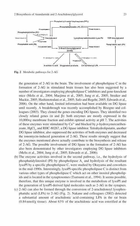
2 Biosynthesis of Anandamide and 2-Arachidonoylglycerol 23
the generation of 2-AG in the brain. The involvement of phospholipase C in the formation of 2-AG in stimulated brain tissues has also been suggested by a number of investigators employing phospholipase C inhibitors and gene-knockout mice (Melis et al., 2004; Maejima et al., 2005; Jung et al., 2005; Straiker and Mackie, 2005; Hashimotodani et al., 2005; Safo and Regehr, 2005; Edwards et al., 2006). On the other hand, limited information had been available on DG lipase until recently. A breakthrough was recently accomplished by Bisogno and col-leagues (2003). They cloned the genes encoding DG lipases. They identified two closely related genes (α and β): both enzymes are mostly expressed in the 10,000×g membrane fraction and exhibit optimal activity at pH 7. The activities of these enzymes were stimulated by Ca2+ and blocked by p-hydroxymercuriben-zoate, HgCl
2, and RHC-80267, a DG lipase inhibitor. Tetrahydrolipstatin, another
DG lipase inhibitor, also suppressed the activities of both enzymes and decreased the ionomycin-induced generation of 2-AG. These results strongly suggest that the enzymes mentioned above actually contribute to the biosynthesis and release of 2-AG. The possible involvement of DG lipase in the formation of 2-AG has also been demonstrated by other investigators employing DG lipase inhibitors (Melis et al., 2004; Jung et al., 2005; Edwards et al., 2006).
(b) The enzyme activities involved in the second pathway, i.e., the hydrolysis of phosphatidylinositol (PI) by phospholipase A
1 and hydrolysis of the resultant
lysoPI by a specific phospholipase C, were studied by Okuyama and coworkers in the mid-1990s. Interestingly, lysoPI-specific phospholipase C is distinct from various other types of phospholipase C which act on other inositol phospholip-ids and is located in the synaptosomes (Tsutsumi et al., 1994). It seems possible, therefore, that this unique enzyme is involved in the metabolism of lysoPI and the generation of lysoPI-derived lipid molecules such as 2-AG in the synapses.
(c) 2-AG can also be formed through the conversion of 2-arachidonoyl lysophos-phatidic acid (LPA) to 2-AG (Fig. 2). Nakane and colleagues (2002) detected a substantial amount of arachidonic acid-containing LPA in the rat brain (0.84 nmol/g tissue). About 63% of the arachidonic acid was esterified at the
Fig. 2 Metabolic pathways for 2-AG

24 T. Sugiura
sn-2 position. They also detected a phosphatase activity which hydrolyzes 2-arachidonoyl LPA to yield 2-AG in a rat brain homogenate (Nakane et al., 2002). Thus, it is plausible that 2-arachidonoyl LPA acts as a substrate for the synthesis of 2-AG under certain conditions in the brain.
(d) Several types of diacyl glycerophospholipids other than inositol phospholipids have also been shown to serve as precursor molecules in the synthesis of 2-AG. Bisogno and colleagues (1999) demonstrated that 2-AG is formed from phos-phatidic acid (PA) in ionomycin-stimulated N18TG2 neuroblastoma cells by employing several metabolic inhibitors. In this case, 2-arachidonoyl PA was converted first to 1-acyl-2-arachidonoylglycerol and then to 2-AG. They described that the breakdown of inositol phospholipids is not involved in the generation of 2-AG. Carrier and coworkers (2004) also demonstrated that 2-AG was formed from PA in a macrophage colony-stimulating factor-stimulated mouse microglia cell line. Some of 2-AG may also be derived from other arachidonic acid-containing phospholipids such as 1-acyl-2-arachidonoyl-sn-glycero-3-phosphocholine. Recently, Oka and colleagues (2005) demon-strated that various species of 2-monoacylglycerol were generated in the TPA-treated mouse ear. The rank order of the amounts of 2-monoacylglycerol generated was 2-palmitoylglycerol plus 2-oleoylglycerol plus 2-cis-vac-cenoylglycerol > 2-linoleoylglycerol > 2-AG. This is quite different from the case for stimulated brain tissues (Sugiura et al., 2000). It seems unlikely that inositol phospholipids act as the sole source of 2-AG in inflamed tissues, because a large proportion of the fatty acyl moiety at the sn-2 position of inositol phospholipids in mammalian tissues is usually accounted for by arachidonic acid. Furthermore, U73122, a PI-specific phospholipase C inhibi-tor, affected the level of 2-AG only modestly, whereas D609, a PC-specific phospholipase C inhibitor, and butanol, an inhibitor of PA generation, exerted more pronounced effects on the level of 2-AG in inflamed ear. It is apparent, therefore, that the biosynthetic pathways for 2-AG differ, depending on the types of tissues and cells and the types of stimuli. Further detailed studies are necessary for a full understanding of the mechanism underlying the biosynthesis of 2-AG in mammalian tissues.
Concluding Remarks
It has been established that anandamide can be formed through several metabolic pathways. Yet, no selective and efficient synthetic pathway for anandamide has hith-erto been found. Considering that anandamide acts as a partial agonist toward the cannabinoid receptors (CB
1 and CB
2), it is rather questionable that anandamide acts
as an endogenous CB1 and CB
2 receptor agonist with profound physiological signifi-
cance. Despite this, however, the discovery of NAPE-specific phospholipase D strongly suggests that this enzyme is a physiologically and/or pathophysiologically important one and that saturated or monoenoic species of N-acylethanolamines, in
2 Biosynthesis of Anandamide and 2-Arachidonoylglycerol 25
addition to anandamide, may play some yet unknown essential roles in mammalian tissues and cells. A thorough elucidation of the physiological and pathophysiological roles of various N-acylethanolamines including anandamide awaits further investiga-tion. In contrast to anandamide, 2-AG acts as a full agonist toward the cannabinoid receptors (CB
1 and CB
2). This strongly suggests that 2-AG rather than anandamide is
the true natural ligand for the cannabinoid receptors. 2-AG can be formed via several metabolic pathways in various mammalian tissues; the most important pathway appears to be the formation from arachidonic acid-containing diacylglycerol, derived from increased phospholipid metabolism, by the action of a diacylglycerol lipase. Whatever the precise mechanism of synthesis, it is apparent that 2-AG is a key mole-cule which links increased phospholipid metabolism upon stimulation with the func-tions of the cannabinoid receptors (CB
1 and CB
2). Further detailed studies on the
metabolism of 2-AG are thus essential to gain insight into the physiological and pathophysiological significance of the endocannabinoid system.
References
Ahluwalia J, Yaqoob M, Urban L, Bevan S, Nagy I (2003) Activation of capsaicin-sensitive primary sensory neurons induces anandamide production and release. J Neurochem 84:585–591.
Basavarajappa BS, Hungund BL (1999) Chronic ethanol increases the cannabinoid receptor agonist anandamide and its precursor N-arachidonoylphosphatidylethanolamine in SK-N-SH cells. J Neurochem 72:522–528.
Basavarajappa BS, Saito M, Cooper TB, Hungund BL (2000) Stimulation of cannabinoid receptor agonist 2-arachidonylglycerol by chronic ethanol and its modulation by specific neuromodula-tors in cerebellar granule neurons. Biochim Biophys Acta 1535:78–86.
Beaulieu P, Bisogno T, Punwar S, Farquhar-Smith WP, Ambrosino G, Di Marzo V, Rice AS (2000) Role of the endogenous cannabinoid system in the formalin test of persistent pain in the rat. Eur J Pharmacol 396:85–92.
Berdyshev EV, Schmid PC, Krebsbach RJ, Schmid HHO (2001) Activation of PAF receptors results in enhanced synthesis of 2-arachidonoylglycerol (2-AG) in immune cells. FASEB J 15:2171–2178.
Bisogno T, Maurelli S, Melck D, De Petrocellis L, Di Marzo V (1997a) Biosynthesis, uptake, and degradation of anandamide and palmitoylethanolamide in leukocytes. J Biol Chem 272: 3315–3323.
Bisogno T, Sepe N, Melck D, Maurelli S, De Petrocellis L, Di Marzo V (1997b) Biosynthesis, release and degradation of the novel endogenous cannabimimetic metabolite 2-arachido-noylglycerol in mouse neuroblastoma cells. Biochem J 322:671–677.
Bisogno T, Melck D, De Petrocellis L, Di Marzo V (1999) Phosphatidic acid as the biosynthetic precursor of the endocannabinoid 2-arachidonoylglycerol in intact mouse neuroblastoma cells stimulated with ionomycin. J Neurochem 72:2113–2119.
Bisogno T, Howell F, Williams G, Minassi A, Cascio MG, Ligresti A, Matias I, Schiano-Moriello A, Paul P, Williams EJ, Gangadharan U, Hobbs C, Di Marzo V, Doherty P (2003) Cloning of the first sn1-DAG lipases points to the spatial and temporal regulation of endocannabinoid signaling in the brain. J Cell Biol 163:463–468.
Burstein SH, Hunter SA (1995) Stimulation of anandamide biosynthesis in N-18TG2 neuroblast-oma cells by delta 9-tetrahydrocannabinol (THC). Biochem Pharmacol 49:855–858.
Burstein SH, Huang SM, Petros TJ, Rossetti RG, Walker JM, Zurier RB (2002) Regulation of anandamide tissue levels by N-arachidonylglycine. Biochem Pharmacol 64:1147–1150.

26 T. Sugiura
Cadas H, Gaillet S, Beltramo M, Venance L, Piomelli D (1996) Biosynthesis of an endogenous cannabinoid precursor in neurons and its control by calcium and cAMP. J Neurosci 16:3934–3942.
Cadas H, Di Tomaso E, Piomelli D (1997) Occurrence and biosynthesis of endogenous cannabi-noid precursor, N-arachidonoyl phosphatidylethanolamine, in rat brain. J Neurosci 17:1226–1242.
Carrier EJ, Kearn CS, Barkmeier AJ, Breese NM, Yang W, Nithipatikom K, Pfister SL, Campbell WB, Hillard CJ (2004) Cultured rat microglial cells synthesize the endocannabinoid 2-arachidonylglycerol, which increases proliferation via a CB
2 receptor-dependent mechanism.
Mol Pharmacol 65:999–1007.Colodzin M, Bachur NR, Weissbach H, Udenfriend S (1963) Enzymatic formation of fatty acid
amides of ethanolamine by rat liver microsomes. Biochem Biophys Res Commun 10:165–171.
Deutsch DG, Chin SA (1993) Enzymatic synthesis and degradation of anandamide, a cannabinoid receptor agonist. Biochem Pharmacol 46:791–796.
Devane WA, Axelrod J (1994) Enzymatic synthesis of anandamide, an endogenous ligand for the cannabinoid receptor, by brain membranes. Proc Natl Acad Sci USA 91:6698–6701.
Devane WA, Hanus L, Breuer A, Pertwee RG, Stevenson LA, Griffin G, Gibson D, Mandelbaum A, Etinger A, Mechoulam R (1992) Isolation and structure of a brain constituent that binds to the cannabinoid receptor. Science 258:1946–1949.
Di S, Boudaba C, Popescu IR, Weng FJ, Harris C, Marcheselli VL, Bazan NG, Tasker JG (2005a) Activity-dependent release and actions of endocannabinoids in the rat hypothalamic supraoptic nucleus. J Physiol 569:751–760.
Di S, Malcher-Lopes R, Marcheselli VL, Bazan NG, Tasker JG (2005b) Rapid glucocorticoid-mediated endocannabinoid release and opposing regulation of glutamate and gamma-aminobutyric acid inputs to hypothalamic magnocellular neurons. Endocrinology 146:4292–4301.
Di Marzo V, Fontana A, Cadas H, Schinelli S, Cimino G, Schwartz JC, Piomelli D (1994) Formation and inactivation of endogenous cannabinoid anandamide in central neurons. Nature 372:686–691.
Di Marzo V, De Petrocellis L, Sepe N, Buono A (1996a) Biosynthesis of anandamide and related acylethanolamides in mouse J774 macrophages and N18 neuroblastoma cells. Biochem J 316:977–984.
Di Marzo V, De Petrocellis L, Sugiura T, Waku K (1996b) Potential biosynthetic connections between the two cannabimimetic eicosanoids, anandamide and 2-arachidonoyl-glycerol, in mouse neuroblastoma cells. Biochem Biophys Res Commun 227:281–288.
Di Marzo V, Bisogno T, De Petrocellis L, Melck D, Orlando P, Wagner JA, Kunos G (1999) Biosynthesis and inactivation of the endocannabinoid 2-arachidonoylglycerol in circulating and tumoral macrophages. Eur J Biochem 264:258–267.
Di Marzo V, De Petrocellis L, Bisogno T, Berger A, Mechoulam R (2002) Biology of endocan-nabinoids. In: Onaivi ES (ed), Biology of Marijuana. Taylor & Francis, London, pp. 125–173.
Edwards DA, Kim J, Alger BE (2006) Multiple mechanisms of endocannabinoid response initia-tion in hippocampus. J Neurophysiol 95:67–75.
Epps DE, Schmid PC, Natarajan V, Schmid HHO (1979) N-Acylethanolamine accumulation in infarcted myocardium. Biochem Biophys Res Commun 90:628–633.
Epps DE, Natarajan V, Schmid PC, Schmid HHO (1980) Accumulation of N-acylethanolamine glycerophospholipids in infarcted myocardium. Biochim Biophys Acta 618:420–430.
Franklin A, Parmentier-Batteur S, Walter L, Greenberg DA, Stella N (2003) Palmitoylethanolamide increases after focal cerebral ischemia and potentiates microglial cell motility. J Neurosci 23:7767–7775.
Gammon CM, Allen AC, Morell P (1989) Bradykinin stimulates phosphoinositide hydrolysis and mobilization of arachidonic acid in dorsal root ganglion neurons. J Neurochem 53:95–101.

2 Biosynthesis of Anandamide and 2-Arachidonoylglycerol 27
Gauthier KM, Baewer DV, Hittner S, Hillard CJ, Nithipatikom K, Reddy DS, Falck JR, Campbell WB (2005) Endothelium-derived 2-arachidonylglycerol: an intermediate in vasodilatory eicosanoid release in bovine coronary arteries. Am J Physiol Heart Circ Physiol 288:1344–1351.
Guo Y, Wang H, Okamoto Y, Ueda N, Kingsley PJ, Marnett LJ, Schmid HH, Das SK, Dey SK (2005) N-acylphosphatidylethanolamine-hydrolyzing phospholipase D is an important deter-minant of uterine anandamide levels during implantation. J Biol Chem 280:23429–23432.
Hansen HS, Lauritzen L, Strand AM, Moesgaard B, Frandsen A (1995) Glutamate stimulates the formation of N-acylphosphatidylethanolamine and N-acylethanolamine in cortical neurons in culture. Biochim Biophys Acta 1258:303–308.
Hansen HH, Ikonomidou C, Bittigau P, Hansen SH, Hansen HS (2001a) Accumulation of the anandamide precursor and other N-acylethanolamine phospholipids in infant rat models of invivo necrotic and apoptotic neuronal death. J Neurochem 76:39–46.
Hansen HH, Schmid PC, Bittigau P, Lastres-Becker I, Berrendero F, Manzanares J, Ikonomidou C, Schmid HHO, Fernandez-Ruiz JJ, Hansen HS (2001b) Anandamide, but not 2-ara-chidonoylglycerol, accumulates during in vivo neurodegeneration. J Neurochem 78:1415–1427.
Hasegawa-Sasaki H (1985) Early changes in inositol lipids and their metabolites induced by platelet-derived growth factor in quiescent Swiss mouse 3T3 cells. Biochem J 232:99–109.
Hashimotodani Y, Ohno-Shosaku T, Tsubokawa H, Ogata H, Emoto K, Maejima T, Araishi K, Shin HS, Kano M (2005) Phospholipase Cβ serves as a coincidence detector through its Ca2+
dependency for triggering retrograde endocannabinoid signal. Neuron 45:257–268.Hohmann AG, Suplita RL, Bolton NM, Neely MH, Fegley D, Mangieri R, Krey JF, Walker JM,
Holmes PV, Crystal JD, Duranti A, Tontini A, Mor M, Tarzia G, Piomelli D (2005) An endo-cannabinoid mechanism for stress-induced analgesia. Nature 435:1108–1112.
Izzo AA, Capasso F, Costagliola A, Bisogno T, Marsicano G, Ligresti A, Matias I, Capasso R, Pinto L, Borrelli F, Cecio A, Lutz B, Mascolo N, Di Marzo V (2003) An endogenous cannabi-noid tone attenuates cholera toxin-induced fluid accumulation in mice. Gastroenterology 125:765–774.
Jin XH, Okamoto Y, Morishita J, Tsuboi K, Tonai T, Ueda N (2007) Discovery and characteriza-tion of a Ca2+-independent phosphatidylethanolamine N-acyltransferase generating the anan-damide precursor and its congeners. J Biol Chem 282:3614–3623.
Jung KM, Mangieri R, Stapleton C, Kim J, Fegley D, Wallace M, Mackie K, Piomelli D (2005) Stimulation of endocannabinoid formation in brain slice cultures through activation of group I metabotropic glutamate receptors. Mol Pharmacol 68:1196–1202.
Kondo S, Kondo H, Nakane S, Kodaka T, Tokumura A, Waku K, Sugiura T (1998a) 2-Arachidonoylglycerol, an endogenous cannabinoid receptor agonist: identification as one of the major species of monoacylglycerols in various rat tissues, and evidence for its generation through Ca2+-dependent and -independent mechanisms. FEBS Lett 429:152–156.
Kondo S, Sugiura T, Kodaka T, Kudo N, Waku K, Tokumura A (1998b) Accumulation of various N-acylethanolamines including N-arachidonoylethanolamine (anandamide) in cadmium chloride-administered rat testis. Arch Biochem Biophys 354:303–310.
Kruszka KK, Gross RW (1994) The ATP- and CoA-independent synthesis of arachidonoyleth-anolamide. A novel mechanism underlying the synthesis of the endogenous ligand of the cannabinoid receptor. J Biol Chem 269:14345–14348.
Kuwae T, Shiota Y, Schmid PC, Krebsbach R, Schmid HHO (1999) Biosynthesis and turnover of anandamide and other N-acylethanolamines in peritoneal macrophages. FEBS Lett 459:123–127.
Liu J, Batkai S, Pacher P, Harvey-White J, Wagner JA, Cravatt BF, Gao B, Kunos G (2003) Lipopolysaccharide induces anandamide synthesis in macrophages via CD14/MAPK/phosph-oinositide 3-kinase/NF-κB independently of platelet-activating factor. J Biol Chem 278:45034–45039.
Liu J, Wang L, Harvey-White J, Osei-Hyiaman D, Razdan R, Gong Q, Chan AC, Zhou Z, Huang BX, Kim HY, Kunos G (2006) A biosynthetic pathway for anandamide. Proc Natl Acad Sci USA 103:13345–13350.

28 T. Sugiura
Maejima T, Oka S, Hashimotodani Y, Ohno-Shosaku T, Aiba A, Wu D, Waku K, Sugiura T, Kano M (2005) Synaptically driven endocannabinoid release requires Ca2+-assisted metabotropic glutamate receptor subtype 1 to phospholipase Cβ4 signaling cascade in the cerebellum. J Neurosci 25:6826–6835.
Marsicano G, Wotjak CT, Azad SC, Bisogno T, Rammes G, Cascio MG, Hermann H, Tang J, Hofmann C, Zieglgansberger W, Di Marzo V, Lutz B (2002) The endogenous cannabinoid system controls extinction of aversive memories. Nature 418:530–534.
McVey DC, Schmid PC, Schmid HHO, Vigna SR (2003) Endocannabinoids induce ileitis in rats via the capsaicin receptor (VR
1). J Pharmacol Exp Ther 304:713–722.
Mechoulam R, Ben-Shabat S, Hanus L, Ligumsky M, Kaminski NE, Schatz AR, Gopher A, Almog S, Martin BR, Compton DR, Pertwee RG, Griffin G, Bayewitch M, Barg J, Vogel Z (1995) Identification of an endogenous 2-monoglyceride, present in canine gut, that binds to cannabinoid receptors. Biochem Pharmacol 50:83–90.
Mechoulam R, Fride E, Ben-Shabat S, Meiri U, Horowitz M (1998a) Carbachol, an acetylcholine receptor agonist, enhances production in rat aorta of 2-arachidonoyl glycerol, a hypotensive endocannabinoid. Eur J Pharmacol 362:1–3.
Mechoulam R, Fride E, Di Marzo V (1998b) Endocannabinoids. Eur J Pharmacol 359:1–18.Melis M, Perra S, Muntoni AL, Pillolla G, Lutz B, Marsicano G, Di Marzo V, Gessa GL, Pistis M
(2004) Prefrontal cortex stimulation induces 2-arachidonoyl-glycerol-mediated suppression of excitation in dopamine neurons. J Neurosci 24:10707–10715.
Moesgaard B, Petersen G, Jaroszewski JW, Hansen HS (2000) Age dependent accumulation of N-acyl-ethanolamine phospholipids in ischemic rat brain. A 31P NMR and enzyme activity study. J Lipid Res 41:985–990.
Moesgaard B, Petersen G, Mortensen SA, Hansen HS (2002) Substantial species differences in relation to formation and degradation of N-acyl-ethanolamine phospholipids in heart tissue: an enzyme activity study. Comp Biochem Physiol B, Biochem Mol Biol 131:475–482.
Morishita J, Okamoto Y, Tsuboi K, Ueno M, Sakamoto H, Maekawa N, Ueda N (2005) Regional distribution and age-dependent expression of N-acylphosphatidylethanolamine-hydrolyzing phospholipase D in rat brain. J Neurochem 94:753–762.
Nakane S, Oka S, Arai S, Waku K, Ishima Y, Tokumura A, Sugiura T (2002) 2-Arachidonoyl-sn-glycero-3-phosphate, an arachidonic acid-containing lysophosphatidic acid: occurrence and rapid enzymatic conversion to 2-arachidonoyl-sn-glycerol, a cannabinoid receptor ligand, in rat brain. Arch Biochem Biophys 402:51–58.
Natarajan V, Reddy PV, Schmid PC, Schmid HHO (1982) N-Acylation of ethanolamine phos-pholipids in canine myocardium. Biochim Biophys Acta 712:342–355.
Natarajan V, Schmid PC, Reddy PV, Zuzarte-Augustin ML, Schmid HHO (1983) Biosynthesis of N-acylethanolamine phospholipids by dog brain preparations. J Neurochem 41:1303–1312.
Natarajan V, Schmid PC, Schmid HHO (1986) N-acylethanolamine phospholipid metabolism in normal and ischemic rat brain. Biochim Biophys Acta 878:32–41.
Oka S, Yanagimoto S, Ikeda S, Gokoh M, Kishimoto S, Waku K, Ishima Y, Sugiura T (2005) Evidence for the involvement of the cannabinoid CB
2 receptor and its endogenous ligand 2-
arachidonoylglycerol in 12-O-tetradecanoylphorbol-13-acetate-induced acute inflammation in mouse ear. J Biol Chem 280:18488–18497.
Oka S, Wakui J, Ikeda S, Yanagimoto S, Kishimoto S, Gokoh M, Nasui M, Sugiura T (2006) Involvement of the cannabinoid CB
2 receptor and its endogenous ligand 2-arachidonoylglycerol
in oxazolone-induced contact dermatitis in mice. J Immunol 177:8796–8805.Okamoto Y, Morishita J, Tsuboi K, Tonai T, Ueda N (2004) Molecular characterization of a phos-
pholipase D generating anandamide and its congeners. J Biol Chem 279:5298–5305.Okamoto Y, Morishita J, Wang J, Schmid PC, Krebsbach RJ, Schmid HH, Ueda N (2005)
Mammalian cells stably overexpressing N-acylphosphatidylethanolamine-hydrolysing phos-pholipase D exhibit significantly decreased levels of N-acylphosphatidylethanolamines. Biochem J 389:241–247.
Panikashvili D, Simeonidou C, Ben-Shabat S, Hanus L, Breuer A, Mechoulam R, Shohami E (2001) An endogenous cannabinoid (2-AG) is neuroprotective after brain injury. Nature 413:527–531.

2 Biosynthesis of Anandamide and 2-Arachidonoylglycerol 29
Pestonjamasp VK, Burstein SH (1998) Anandamide synthesis is induced by arachidonate mobiliz-ing agonists in cells of the immune system. Biochim Biophys Acta 1394:249–260.
Petersen G, Hansen HS (1999) N-acylphosphatidylethanolamine-hydrolysing phospholipase D lacks the ability to transphosphatidylate. FEBS Lett 455:41–44.
Piomelli D, Beltramo M, Giuffrida A, Stella N (1998) Endogenous cannabinoid signaling. Neurobiol Dis 5:462–473.
Prescott SM, Majerus PW (1983) Characterization of 1,2-diacylglycerol hydrolysis in human platelets. Demonstration of an arachidonoyl-monoacylglycerol intermediate. J Biol Chem 258:764–769.
Rademacher DJ, Patel S, Ho WS, Savoie AM, Rusch NJ, Gauthier KM, Hillard CJ (2005) U-46619 but not serotonin increases endocannabinoid content in middle cerebral artery: evidence for functional relevance. Am J Physiol Heart Circ Physiol 288:H2694–2701.
Safo PK, Regehr WG (2005) Endocannabinoids control the induction of cerebellar LTD. Neuron 48:647–659.
Schmid PC, Reddy PV, Natarajan V, Schmid HHO (1983) Metabolism of N-acylethanolamine phospholipids by a mammalian phosphodiesterase of the phospholipase D type. J Biol Chem 258:9302–9306.
Schmid HHO, Schmid PC, Natarajan V (1990) N-acylated glycerophospholipids and their deriva-tives. Prog Lipid Res 29:1–43.
Stella N, Piomelli D (2001) Receptor-dependent formation of endogenous cannabinoids in cortical neurons. Eur J Pharmacol 425:189–196.
Stella N, Schweitzer P, Piomelli D (1997) A second endogenous cannabinoid that modulates long-term potentiation. Nature 388:773–778.
Straiker A, Mackie K (2005) Depolarization-induced suppression of excitation in murine autaptic hippocampal neurones. J Physiol 569:501–517.
Sugiura T, Waku K (2000) 2-Arachidonoylglycerol and the cannabinoid receptors. Chem Phys Lipids 108:89–106.
Sugiura T, Kondo S, Sukagawa A, Nakane S, Shinoda A, Itoh K, Yamashita A, Waku K (1995) 2-Arachidonoylglycerol: a possible endogenous cannabinoid receptor ligand in brain. Biochem Biophys Res Commun 215:89–97.
Sugiura T, Kondo S, Sukagawa A, Tonegawa T, Nakane S, Yamashita A, Waku K (1996a) Enzymatic synthesis of anandamide, an endogenous cannabinoid receptor ligand, through N-acylphosphatidylethanolamine pathway in testis: involvement of Ca2+-dependent transacylase and phosphodiesterase activities. Biochem Biophys Res Commun 218:113–117.
Sugiura T, Kondo S, Sukagawa A, Tonegawa T, Nakane S, Yamashita A, Ishima Y, Waku K (1996b) Transacylase-mediated and phosphodiesterase-mediated synthesis of N-ara-chidonoylethanolamine, an endogenous cannabinoid-receptor ligand, in rat brain microsomes. Comparison with synthesis from free arachidonic acid and ethanolamine. Eur J Biochem 240:53–62.
Sugiura T, Kodaka T, Nakane S, Kishimoto S, Kondo S, Waku K (1998) Detection of an endog-enous cannabimimetic molecule, 2-arachidonoylglycerol, and cannabinoid CB
1 receptor
mRNA in human vascular cells: is 2-arachidonoylglycerol a possible vasomodulator? Biochem Biophys Res Commun 243:838–843.
Sugiura T, Yoshinaga N, Kondo S, Waku K, Ishima Y (2000) Generation of 2-arachidonoylglycerol, an endogenous cannabinoid receptor ligand, in picrotoxinin-administered rat brain. Biochem Biophys Res Commun 271:654–658.
Sugiura T, Yoshinaga N, Waku K (2001) Rapid generation of 2-arachidonoylglycerol, an endogenous cannabinoid receptor ligand, in rat brain after decapitation. Neurosci Lett 297:175–178.
Sugiura T, Kishimoto S, Oka S, Gokoh M (2006a) Biochemistry, pharmacology and physiology of 2-arachidonoylglycerol, an endogenous cannabinoid receptor ligand. Prog Lipid Res 45:405–446.
Sugiura T, Oka S, Ikeda S, Waku K (2006b) Occurrence, biosynthesis, and metabolism of endocannabinoid. In: Onaivi ES, Sugiura T, Di Marzo V (eds), Endocannabinoids: The Brain and Body’s Marijuana and Beyond. Taylor & Francis, Boca Raton, pp. 177–214.

30 T. Sugiura
Tsutsumi T, Kobayashi T, Ueda H, Yamauchi E, Watanabe S, Okuyama H (1994) Lysophosphoinositide-specific phospholipase C in rat brain synaptic plasma membranes. Neurochem Res 19:399–406.
Ueda N, Kurahashi Y, Yamamoto S, Tokunaga T (1995) Partial purification and characterization of the porcine brain enzyme hydrolyzing and synthesizing anandamide. J Biol Chem 270:23823–23827.
Varga K, Wagner JA, Bridgen DT, Kunos G (1998) Platelet- and macrophage-derived endogenous cannabinoids are involved in endotoxin-induced hypotension. FASEB J 12:1035–1044.
Wagner JA, Varga K, Ellis EF, Rzigalinski BA, Martin BR, Kunos G (1997) Activation of periph-eral CB
1 cannabinoid receptors in haemorrhagic shock. Nature 390:518–521.
Walker JM, Huang SM, Strangman NM, Tsou K, Sanudo-Pena MC (1999) Pain modulation by release of the endogenous cannabinoid anandamide. Proc Natl Acad Sci USA 96:12198–12203.
Wallace MJ, Blair RE, Falenski KW, Martin BR, DeLorenzo RJ (2003) The endogenous cannabi-noid system regulates seizure frequency and duration in a model of temporal lobe epilepsy. J Pharmacol Exp Ther 307:129–137.
Walter L, Stella N (2003) Endothelin-1 increases 2-arachidonoyl glycerol (2-AG) production in astrocytes. Glia 44:85–90.
Walter L, Franklin A, Witting A, Moller T, Stella N (2002) Astrocytes in culture produce ananda-mide and other acylethanolamides. J Biol Chem 277:20869–20876.
Walter L, Franklin A, Witting A, Wade C, Xie Y, Kunos G, Mackie K, Stella N (2003) Nonpsychotropic cannabinoid receptors regulate microglial cell migration. J Neurosci 23:1398–1405.
Walter L, Dinh T, Stella N (2004) ATP induces a rapid and pronounced increase in 2-arachidonoylglycerol production by astrocytes, a response limited by monoacylglycerol lipase. J Neurosci 24:8068–8074.
Wang J, Okamoto Y, Morishita J, Tsuboi K, Miyatake A, Ueda N (2006) Functional analysis of the purified anandamide-generating phospholipase D as a member of the metallo-β-lactamasefamily. J Biol Chem 281:12325–12335.
Witting A, Walter L, Wacker J, Moller T, Stella N (2004) P2X7 receptors control 2-ara-
chidonoylglycerol production by microglial cells. Proc Natl Acad Sci USA 101:3214–3219.

Chapter 3Removal of Endocannabinoids by the Body: Mechanisms and Therapeutic Possibilities
Christopher J. Fowler and Lina Thors
Abstract The actions of anandamide and 2-arachidonoylglycerol are terminated by cellular uptake followed by metabolism. In the case of anandamide, the uptake process was originally suggested to be achieved by a process of facilitated diffusion, but the mechanism(s) involved are a matter of controversy at present. The main hydrolytic enzyme for anandamide is fatty acid amide hydrolase, and inhibitors of this enzyme have been found to have beneficial effects in animal models of inflam-matory pain, inflammation, anxiety and depression. Anandamide is also a substrate for cyclooxygenase-2 and lipoxygenases, and the cyclooxygenase-derived products, the “prostamides” have biological actions of their own. 2-Arachidonoylglycerol can be metabolized by a range of enzymes, including cyclooxygenase-2, monoacylg-lycerol lipase and fatty acid amide hydrolase. In the brain, monoacylglycerol lipase is probably the most important of these enzymes. However, no selective inhibitors of this enzyme are presently available with which to establish the potential of this enzyme as a target for drug development.
Introduction
Signalling molecules require mechanisms for their removal to produce effective and discrete cellular signalling. The endocannabinoids are no exception in this regard, and considerable effort has been made to delineate the mechanisms by which they are metabolized, to design compounds inhibiting these processes and to ascertain whether or not such compounds have therapeutic promise. Most of this work has been con-cerned with anandamide (AEA) and this will be reflected in the bias of this section.
Basic Pathways of AEA Metabolism
The main pathways for AEA metabolism are shown schematically in Fig. 1. One year after its discovery, Deutsch and Chin (1993) reported that extracellular AEA was accumulated by cells and hydrolysed to give arachidonic acid. The hydrolytic enzyme
A. Köfalvi (ed.), Cannabinoids and the Brain. 31© Springer 2008

32 C.J. Fowler and L. Thors
was termed “anandamide amidase” but was subsequently found to be the same enzyme as the amidohydrolase characterized in the 1980s by Schmid and colleagues using oleoylethanolamide as substrate (Schmid et al., 1985). The enzyme, currently termed fatty acid amide hydrolase, was cloned in 1995 and its structure at the level of 2.8 Å has been elucidated (for a review of the structure and reaction mechanisms of FAAH, see McKinney and Cravatt, 2005). A second FAAH, present in primates but not in rats or mice, has been identified (Wei et al., 2006) although the functional importance of this enzyme has not been established as yet. An N-acyl acid amidase (NAAA), with a lower pH optimum than FAAH, has also been characterized (Ueda et al., 2001), but in rat brain membrane fractions it appears not to contribute to any large extent to the total hydrolysis of AEA (Alajakku and Fowler, unpublished data).
The Nature of Cellular AEA Uptake
Ever since 1993 when it was established that anandamide (AEA), an uncharged hydrophobic molecule, was taken up by cells in culture and thereafter metabolized by FAAH (Deutsch and Chin, 1993), a variety of mechanisms have been suggested
Fig. 1 Representation of the uptake and metabolism of AEA. After uptake, mediated either by passive or alternatively facilitated diffusion across the membrane, the endocannabinoid is primarily hydrolysed to arachidonic acid (AA) by FAAH although it can also be metabolized by COX-2 or by lipoxygenase enzymes to give prostaglandin ethanolamides (PG-EAs) or hydroperoxyeicosatetrae-noyl ethanolamides (HPETE-EAs), respectively (Kozak and Marnett, 2002). A pathway for the synthesis of O-phosphorylcholine-anandamide (PC-AEA) by an as yet unidentified enzyme (? in the figure) and its dephosphorylation has also been described (Mulder and Cravatt, 2006). Hydrolysis of the AEA by FAAH helps to retain the extracellular–intracellular gradient and thereby to some extent “drives” the uptake. Free intracellular AEA has also been hypothesized to be regulated by intracel-lular “shuttling” proteins (shown as the small blobs in the figure) and by intracellular sequestration (shown at the bottom of the figure). AM404 affects the uptake of AEA, partly by acting as an alterna-tive substrate for FAAH (Lang et al., 1999) and theoretically by affecting one or more of the other uptake mechanisms. AM404 is also an inhibitor of COX-2 (Högestätt et al., 2005), can interact with both cannabinoid and TRPV
1 receptors as well as with Na+ channels, affects calcium homeostasis and
can produce oxidative stress (Fowler, 2006), which makes for a somewhat complex pharmacology

3 Removal of Endocannabinoids by the Body 33
and debated. The mechanism that has received the most attention is that of a facili-tated transporter (Di Marzo et al., 1994; Hillard et al., 1997). However, this sugges-tion is by no means proven, and there is evidence both in favour and against such a process (Table 1). Two additional mechanisms, passive diffusion gated by FAAH (Deutsch et al., 2001; Kaczocha et al., 2006) and intracellular sequestration (Hillard and Jarrahian, 2000) are discussed in the Table, but at the outset it should be said that the most likely scenario is for AEA to utilize more than one mechanism for its cellular accumulation. The following citation can be instructive in this respect: “Translocation… across the plasma membrane is achieved by a concert of co-existing mechanisms. These lipids can passively diffuse, but transport can also be accelerated by certain membrane proteins as well as lipid rafts” (Ehehalt et al., 2006). The citation in question refers to the cellular uptake of long chain fatty acids, which have been the subject of investigation long before the discovery of AEA, but which have grappled with the same sort of issues as those for AEA uptake. In the absence of a published report concerning the identification and cloning of an AEA transporter
Table 1 The case for/against a facilitated transport mechanism of AEA uptake
Evidence in favour of facilitated transport Alternative explanations/observations
AEA is concentrated in cells to a greater extent than would be predicted on the basis of a passive diffusion processa
The extracellular–intracellular gradient is partly driven in some (but not all) cells by FAAH-catalysed metabolism of AEAi,j;intracellular sequestration of AEAa would also act to keep the free [AEA]
i low and
allow for further passive diffusionUptake is time and temperature dependent
and shows saturability, but is not coupled to any ion gradient or dependent upon ATPb,c
Initial rapid uptake is not saturable, and its temperature dependency reflects effects upon AEA availability rather than the uptake process itselfj,k
Vesicles prepared from plasma membranes accumulate AEA whereas vesicles prepared from intracellular membranes do not; the FAAH activity of the cells is associated with the latter rather than the former; Cholesterol depletion of some (but not all) cells reduces uptaked,e
N-Acylethanolamines form complexes with cholesteroll which is present in plasma membranes at much greater concentrations than in intracellular membranesd
Uptake can be inhibited pharmacologically, even in cells lacking FAAHf,g; the inhibitors have pharmacological effects in vivoh
FAAH is inhibited by the compounds, and in the absence of this enzyme, the in vitro potencies of the uptake inhibitors are modest (see Fig. 2b); the compounds show little effect on the initial AEA uptakej;acyl-derived inhibitors inhibit the retention of AEA by plastic wells at similar concentrationsf,m, suggesting that the specificity of the process is rather limited
Selected references (with apologies to authors not included here): aHillard and Jarrahian (2000); bDiMarzo et al. (1994); cHillard et al. (1997); dOddi et al. (2005); eBari et al. (2005); fOrtega-Gutiérrez et al. (2004); gFegley et al. (2004); hreviewed in Fowler et al. (2005); iDeutsch et al. (2001); jKac-zocha et al. (2006); kThors and Fowler (2006); lRamakrishnan et al. (2002); mFowler et al. (2004)

34 C.J. Fowler and L. Thors
protein, any model of AEA uptake should be treated with caution. Thus, for exam-ple, interpretation of data supporting an endocytotic delivery of AEA to FAAH (McFarland et al., 2004) and the identification of a membrane binding site for a potent non-acyl uptake inhibitor (Moore et al., 2005) are limited by the lack of selec-tivity of the compounds used (Alexander and Cravatt, 2006; Thors et al., 2007). Our own current working model is primarily influenced by the finding that AEA can cross plasma membranes very rapidly (Bojesen and Hansen, 2005). This rapidity can be demonstrated in functional studies investigating the ability of AEA to activate TRPV
1 (vanilloid) receptors, which is mediated by its binding to a domain of the
receptor on the intracellular face of the membrane (Jordt and Julius, 2002). Extracellular AEA produces a TRPV
1 receptor-mediated calcium signal within sec-
onds (Jerman et al., 2002), indicating that significant uptake of this endocannabinoid must have occurred within this time frame. Most uptake assays use much longer incubation times (generally 4–10 min), which would suggest that the processes being studied are intracellular redistribution events (shown schematically in Fig. 1) rather
Fig. 2 Comparison of potencies for a series of AEA head group analogues (a) and a series of compounds with different primary structures (b) with respect to inhibition of FAAH and inhibition of AEA uptake. In (a), the data are taken from Jarrahian and colleagues (2000) and the inhibition of rat forebrain FAAH is compared with the inhibition of AEA uptake into rat cerebral granule cell cultures (2-min incubation period). The authors tested 24 compounds for uptake, and 21 of these for inhibition of FAAH. Of these, two were excluded from the analysis, due to inexact measures of IC
50 values (>10 and >50 µM). IC
50 values >100 µM, however, are shown in the figure. Despite the
wide variation in potency as FAAH inhibitors, there is little variation in potency as uptake inhibi-tors, suggesting that FAAH is not a major determinant of the rate of uptake in these cells. In (b), the data are taken from Dickason-Chesterfield and colleagues (2006) and the inhibition of purified rat ∆TM-FAAH (a truncated version of FAAH) is compared with the uptake of AEA into either RBL2H3 cells and HeLa cells (16–40 h incubation period at room temperature, so the rele-vance to the rapid uptake of AEA is unclear). For the HeLa cells, the wide variation in FAAH inhibitory potency is not matched by a variation in the potency of the compounds as uptake inhibi-tors, which is to be expected given that these cells do not express FAAH. For the RBL2H3 cells, which express FAAH, the three most potent FAAH inhibitors (LY2077855, LY2183240 and the “standard” FAAH inhibitor URB597) are also the three most potent uptake inhibitors

3 Removal of Endocannabinoids by the Body 35
than transport across the plasma membrane. The saturability of the AEA uptake seen at 4–10-min incubation times may therefore be due to the limited capacity of the intracellular protein(s). This does not mean that plasma membrane constituents are not of importance: a case can be made whereby such constituents anchor the AEA within the plasma membrane prior to its cellular delivery. The only requirement for such constituents, which can well have other primary functions, is that they bind AEA. In mouse cortical neurons in culture (but not necessarily in other cells), CB
1
receptors may play such a role, since the uptake of AEA is reduced by the CB
1 receptor antagonist/inverse agonist rimonabant (Ortega-Gutiérrez et al., 2004).
Initial saturability of AEA uptake would not be expected if the translocation, albeit fast (Bojesen and Hansen, 2005) is the rate-limiting step rather than the dissociation processes from the receptors. Whether or not this working model will stand the test of time remains to be seen; nonetheless, in view of the potential utility of AEA uptake inhibitors for a variety of disorders including multiple sclerosis and neuro-pathic pain (see e.g., La Rana et al., 2006; Ligresti et al., 2006), it remains a matter of importance to identify once and for all the key mechanisms involved in the cellular accumulation of AEA.
FAAH Inhibitors and Their Therapeutic Potential
The identification of FAAH as the enzyme responsible for AEA hydrolysis was accompanied by the chance discovery that phenylmethylsulphonyl fluoride (PMSF) inhibited this process (Deutsch and Chin, 1993). In vivo, PMSF was found, at a dose of 30 mg/kg, not to produce effects itself on thermal nociception, spontaneous activity and mobility, but to potentiate the effects of AEA in these tests (Compton and Martin, 1997). The ability to potentiate AEA effects without producing direct CB
1 receptor-mediated effects has been seen with more selective
inhibitors (Kathuria et al., 2003; Lichtman et al., 2004a) and in animals with a genetic deletion of the enzyme (Cravatt et al., 2001, 2004; Wise et al., 2007). With respect to the latter, brain and liver samples from FAAH−/− mice lose >97% of the capacity to hydrolyse AEA in brain, spinal cord, liver and testis tissues, and this is accompanied by a large increase in the levels of AEA in these tissues (as com-pared with either wild type or FAAH+/− mice) (Cravatt et al., 2001, 2004). The FAAH−/− mice show reduced sensitivities to carrageenan and formalin, but their thermal hypersensitivity following ligation of the sciatic nerve is unchanged (Lichtman et al., 2004b). The reduced oedema response to intraplantar carra-geenan was retained in mice with a genetic deletion in peripheral, but not central FAAH, whereas their reduced sensitivity to thermal pain was lost (Cravatt et al., 2004). FAAH−/− mice have subsequently been utilized to establish the potential importance of this enzyme in a wide variety of physiological and pathological processes, including reproduction (Wang et al., 2006a), colonic inflammation (Massa et al., 2004), and neurodegeneration (Bilsland et al., 2006). The potential of FAAH as a target for drug development has not been overlooked, as adjudged

36 C.J. Fowler and L. Thors
by the patent literature and more recently, by the publication of novel high throughput screening strategies and their implementation. Thus, for example, scientists from Wyeth and Abbott have reported the development of high through-put methods and their implementation in the screening of 457,073 and >650,000 compounds, respectively (Wang et al., 2006b; Kage et al., 2007). Most published work with selective FAAH inhibitors concerns the carbamate compound URB597 (cyclohexylcarbamic acid 3′carbamoylbiphenyl-3-yl ester) and the α-ketohetero-cycle OL-135 (1-oxo-1[5-(2-pyridyl)-2-yl]-7-phenylheptane). At the outset, it should be pointed out that the word “selective” is a relative term, and all compounds will interact with other targets if present in sufficient amounts. These compounds are no exception, and can inhibit carboxyesterase enzymes if given at a concentration of 10 µM (Zhang et al., 2007), but this concentration is ≥80-foldhigher than the IC
50 values for the inhibition of FAAH by these compounds
(Kathuria et al., 2003; Lichtman et al., 2004a) (see also Lichtman et al., 2004b; Clapper et al., 2006, with respect to the ability of URB597 to interact with, but not inhibit the activity of triacylglycerol hydrolase). In vivo, these two com-pounds increase the levels of AEA in the brain, and produce potentially useful effects in models of inflammatory pain and inflammation and neurobehav-ioural disturbances following neglect parenting without adversely affecting working memory (Lichtman et al., 2004a; Holt et al., 2005; Jayamanne et al., 2006; Chang et al., 2006; Varvel et al., 2006; Marco et al., 2007). Their usefulness with respect to anxiety and depression (Kathuria et al., 2003; Gobbi et al., 2005) is less clear (Naidu et al., 2007). FAAH inhibition (or genetic deletion) is not efficacious in models of neuropathic pain (Lichtman et al., 2004b; Jayamanne et al., 2006; Jhaveri et al., 2006; but see Chang et al., 2006) and it has been argued that the contribution of FAAH to endocannabinoid metabolism is altered in a tissue-dependent manner in neuropathic animals (Jhaveri et al., 2006). N-Arachidonoylserotonin is also an interesting compound, having both FAAH inhibitory and TRPV
1 receptor antagonistic properties, and showing analgesic,
antiproliferative and anti-inflammatory effects in vivo (Bifulco et al., 2004; D’Argenio et al., 2006; Maione et al., 2007).
Biochemical Consequences of FAAH Inhibition
When considering the wide range of therapeutic possibilities afforded to FAAH inhibitors, it is important to remember that the enzyme is not restricted to the metabolism of AEA alone. Indeed, the enzyme has wide substrate specificity, and can hydrolyse a number of lipid classes, including other N-acylethanolamines (such as palmitoylethanolamide and oleoylethanolamide), N-acyltaurines (which activate TRPV ion channels) and fatty acid amides such as the sleep-inducing lipid oleam-ide (Schmid et al., 1985; Cravatt et al., 1996; Saghatelian et al., 2006). Thus, FAAH inhibition or genetic deletion results in increased levels not only of AEA but of these other lipid classes (Lichtman et al., 2004a; Saghatelian et al., 2006). Such

3 Removal of Endocannabinoids by the Body 37
changes can contribute to the net effect of FAAH inhibition: palmitoylethanola-mide, for example, has anti-inflammatory and analgesic effects of its own, and recent data has suggested that these are brought about via an activation of the nuclear peroxisome proliferator-activated receptor α (PPARα) (Lo Verme et al., 2005, 2006). Similarly, oleoylethanolamide produces effects upon appetite in a manner involving activation of PPARα (Fu et al., 2003) and GPR119 orphan recep-tors (Overton et al., 2006) (for further information see Chap. 9). A second issue concerns the potential for diverting AEA (and other N-acylethanolamine) metabo-lism from hydrolysis to production of other biologically active metabolites as a result of FAAH inhibition or genetic deletion. Data is beginning to emerge to sup-port this contention, primarily as a result of the use of FAAH−/− mice. The most recent example is the finding of Mulder and Cravatt (2006) who reported increased levels of phosphocholine derivatives of N-acylethanolamines in FAAH−/− mice. AEA is also a substrate for cyclooxygenase-2 (COX-2) and lipoxygenases (Kozak and Marnett, 2002). COX-2-derived prostaglandin ethanolamide (prostamide) lev-els in the body are normally below the level of detection, but are measurable in FAAH−/− mice treated with AEA (Weber et al., 2004). Prostamides (or analogues with alkyl substituents on the ethanolamide head group) have weak, if any, effects upon CB and prostanoid receptors and do not feedback inhibit AEA (or MAGL) (Berglund et al., 1999; Ross et al., 2002; Matias et al., 2004; Fowler and Tiger, 2005), although they activate a separate “prostamide receptor” in the cat iris (Woodward et al., 2007). Rockwell and Kaminski (2004) provided indirect evi-dence that in mouse splenocytes, AEA inhibits secretion of the proinflammatory cytokine interleukin-2 via an interaction of prostamides with PPARγ. The reverse pattern of effects can also be considered, namely that inhibition of cyclooxygenase-2 will affect AEA levels. Spinally administered flurbiprofen and indomethacin produce antinociceptive effects in the formalin test of inflammatory pain in a manner blocked by CB
1 receptor antagonists of genetic deletion of the enzyme
(Gühring et al., 2002; Ates et al., 2003). The authors suggested that these effects may be related to a build up of arachidonic acid which is then utilized for endocan-nabinoid synthesis. The ability of acidic non-steroidal anti-inflammatory agents to inhibit FAAH (Fowler et al., 1997, 2003) may further contribute to an increased endocannabinoid tone. In this respect, Guindon and colleagues (2006a,b) have reported that the intraplantar administration of ibuprofen does not potentiate N-acylethanolamine levels in the paw or produce CB
1 receptor-mediated antinocic-
eptive effects in the formalin test, but does potentiate the effects of AEA, the synergy being prevented by a CB
1 receptor antagonist. Furthermore, the combina-
tion of AEA and ibuprofen increased N-acylethanolamine levels over and above those seen with either compound alone (Guindon et al., 2006b). A final point concerns the products of FAAH action, which in the case of N-acylethanolamines are the fatty acids and ethanolamine. In vivo, these are rapidly taken care of by the cell, but this is not necessarily the case in vitro. This has been neatly illustrated in a recent study demon-strating that AEA protects N18TG2 neuroblastoma cells against apoptosis induced by low serum concentrations as a result of its FAAH catalysed conversion to ethanolamine (Matas et al., 2007).

38 C.J. Fowler and L. Thors
Risk Groups for FAAH Inhibitors
The findings in animal models that FAAH inhibitors produce beneficial effects in models of inflammatory pain, inflammation, anxiety and depression without pro-ducing “cannabis-like” actions in the brain makes them an attractive target for drug development. However, given the ubiquity of the cannabinoid system and its involvement in so many different biological processes, it is unavoidable that com-pounds designed to potentiate endocannabinoid signalling in this way will pro-duce unwanted actions or be inappropriate for certain types of patients. Conditions, for example, whereby FAAH inhibition alters the balance between excitatory and inhibitory signalling in the brain may give rise to unwanted effects (see Clement et al., 2003, for a study of the seizure activity of FAAH−/− mice). Two potential risk groups are women who are both pregnant or planning preg-nancy, and individuals with a propensity for drug abuse. With respect to the former, it is now clear that FAAH is an important regulator of reproduction (Maccarrone and Finazzi-Agrò, 2004). AEA plays a key role in the implantation of fertilized eggs (Wang et al., 2003), and mice treated with URB597 show impaired oviductal embryo transport (Wang et al., 2006a). In women, plasma levels of AEA vary during the different stages of labour (Habayeb et al., 2004), leading the authors to suggest that “successful pregnancy implantation and pro-gression requires low levels of AEA”. Consistent with this suggestion, low levels of lymphocyte FAAH are associated with spontaneous miscarriages (Maccarrone et al., 2000). With respect to drug abuse, there is evidence that AEA produces a release of dopamine in the nucleus accumbens. URB597 does not produce any release per se, but potentiates the release induced by AEA (Solinas et al., 2006), and potentiates voluntary ethanol consumption (as does genetic ablation of FAAH) (Hansson et al., 2007; Blednov et al., 2007). Conversely, Alko alcohol-preferring rats show a lower expression of FAAH in the prefrontal cortex than Alko non-preferring rats (Hansson et al., 2007). In man, a missense mutation (P129T) of FAAH, which results in a decreased expression of the enzyme, has been associated with street drug abuse and drug/alcohol abuse but not with schiz-ophrenia or methamphetamine dependence (Chiang et al., 2004; Sipe et al., 2002; Morita et al., 2005; Flanagan et al., 2006). An association of this variant with weight issues (Sipe et al., 2005) has not been replicated in a recent study (Jensen et al., 2007).
Metabolism of 2-AG
The processes involved in the metabolism of 2-AG are less well studied than for AEA. With respect to the uptake of 2-AG, the process is saturable, shows an apparent temperature sensitivity, is gated to some extent by its hydrolysis and subsequent esterification into phospholipids, is affected by methyl-β-cyclodextrin

3 Removal of Endocannabinoids by the Body 39
treatment and is inhibited by the acyl compounds AM404 and VDM11 (Piomelli et al., 1999; Beltramo and Piomelli, 2000; Bisogno et al., 2001; Hájos et al., 2004; Bari et al., 2006). Thus the uptake of 2-AG shows similar properties (and is thereby subject to the same limitations in terms of data interpretation) as that of AEA (Hermann et al., 2006). 2-AG is metabolized by a variety of enzymes including COX-2, lipoxygenases and monoacylglycerol kinases (see Simpson et al., 1991; Kozak and Marnett, 2002) and the COX-2-derived prostaglandin E
2
glycerol ester has been shown to have biological activity, mobilizing intracellular calcium and modulating hippocampal synaptic transmission (Nirodi et al., 2004; Sang et al., 2006). With respect to hydrolytic enzymes, 2-AG is a substrate for FAAH, monoacylglycerol lipase (MAGL), the esterase domain of neuropathy target esterase and possibly others (Goparaju et al., 1998; Dinh et al., 2002; van Tienhoven et al., 2002; Dinh et al., 2004; Vandevoorde et al., 2007), but most focus has been upon FAAH and MAGL. Treatment with URB597 results in an increased level of 2-AG in the paw, but not in the brain (Kathuria et al., 2003; Jhaveri et al., 2006), suggesting that FAAH is important for 2-AG metabolism in the former, but not in the latter tissue. Indeed, Dinh and colleagues (2002) have argued that MAGL is more important in the brain, and the finding that FAAH and MAGL have different cellular localizations in the hippocampus, cerebellum and amygdala (Gulyás et al., 2004; see Chap. 10) led those authors to suggest that “FAAH may set the resting level of anandamide close to its sites of synthesis, while MAGL may help to inactivate 2-AG close to its sites of action”. In contrast to the situation for FAAH, selective inhibitors of MAGL are not yet available. The enzyme is sensitive to the serine hydrolase inhibitor methylarachido-noylfluorophosphonate (MAFP, see Dinh et al., 2002), and this compound has been used to explore the potential role of MAGL in regulating retrograde signal-ling processes (Hashimotodani et al., 2007). MAFP, however, is by no means selective and can potently inhibit both FAAH (see Ueda et al., 2001) and even 2-AG metabolism by the esterase domain of human neuropathy target esterase (van Tienhoven et al., 2002). In our hands, MAFP inhibits human recombinant MAGL and rat brain FAAH with IC
50 values of 510 and 39 pM, respectively
(Lenman, Alajakku, Jacobsson and Fowler, unpublished results). Nonetheless, physiological actions that are blocked by 2-AG synthesis inhibition [with the caveat that tetrahydrolipstatin, the compound in question, may have an antagonis-tic effect at CB
1 receptors (Palomäki et al., 2007)], potentiated by MAFP, but not
affected by selective FAAH inhibitors (such as in the study of Hashimotodani et al., 2007), can with some confidence be ascribed to 2-AG and its regulation by MAGL. Two compounds, URB602 and URB754, have been proposed as selective MAGL inhibitors, but the selectivity of the former has been questioned, and the activity of the latter was found to be due to contamination with bis(methylthio)mercurane (Hohmann et al., 2005; Makara et al., 2007; Vandevoorde et al., 2007). There is thus a need for the identification of novel MAGL-selective inhibitors for the char-acterization of this enzyme to determine its physiological importance and its possi-ble role as a target for drug development.
40 C.J. Fowler and L. Thors
Concluding Remarks
In the short time since the discovery of AEA and the identification of 2-AG as an endocannabinoid, much has been learnt about the metabolism of these compounds and the potential therapeutic utility of agents blocking the metabolism. FAAH inhibitors have now their own momentum, whilst the mechanism(s) governing AEA uptake remain a matter of controversy. We lack selective MAGL inhibitors, and it is not clear as to whether MAGL as described by Dinh and colleagues (2002) represents the only enzyme that hydrolyses 2-AG in its capacity as an endocannabi-noid. Our knowledge of the importance of the COX-2 pathway is in its infancy with respect to the endocannabinoid system, but there is evidence to suggest that this enzyme may both regulate the availability of 2-AG in the hippocampus (Kim and Alger, 2004; Hashimotodani et al., 2007) and provide the body with biologically active prostaglandin ethanolamides and glycerol esters. Hopefully, these areas will be the focus of increasing attention in the years to come.
Acknowledgements The authors would like to thank the Swedish Research Council (Grant no. 12158, medicine) and the Research Funds of the Medical Faculty, Umeå University for financial support for their research on endocannabinoid metabolism.
References
Alexander JP, Cravatt BF (2006) The putative endocannabinoid transport blocker LY2183240 is a potent inhibitor of FAAH and several other brain serine hydrolases. J Am Chem Soc 128:9699–9704.
Ates M, Hamza M, Seidel K, Kotalla CE, Ledent C, Gühring H (2003) Intrathecally applied flur-biprofen produces an endocannabinoid-dependent antinociception in the rat formalin test. Eur J Neurosci 17:597–604.
Bari M, Battista N, Fezza F, Finazzi-Agrò A, Maccarrone M (2005) Lipid rafts control signaling of type-1 cannabinoid receptors in neuronal cells. Implications for anandamide-induced apop-tosis. J Biol Chem 280:12212–12220.
Bari M, Spagnuolo P, Fezza F, Oddi S, Pasquariello N, Finazzi-Agrò A, Maccarrone M (2006) Effect of lipid rafts on Cb
2 receptor signaling and 2-arachidonoyl-glycerol metabolism in
human immune cells. J Immunol 177:4971–4980.Beltramo M, Piomelli D (2000) Carrier-mediated transport and enzymatic hydrolysis of the
endogenous cannabinoid 2-arachidonylglycerol. Neuroreport 11:1231–1235.Berglund BA, Boring DL, Howlett AC (1999) Investigation of structural analogs of prostaglandin
amides for binding to and activation of CB1 and CB
2 cannabinoid receptors in rat brain and
human tonsils. Adv Exp Med Biol 469:527–533.Bifulco M, Laezza C, Valenti M, Ligresti A, Portella G, Di Marzo V (2004) A new strategy to
block tumor growth by inhibiting endocannabinoid inactivation. FASEB J 18:1606–1608.Bilsland LG, Dick JRT, Pryce G, Petrosino S, Di Marzo V, Baker D, Greensmith L (2006)
Increasing cannabinoid levels by pharmacological and genetic manipulation delay disease progression in SOD1 mice. FASEB J 20:1003–1005; full article E180–E190.
Bisogno T, Maccarrone M, De Petrocellis L, Jarrahian A, Finazzi-Agrò A, Hillard C, Di Marzo V (2001) The uptake by cells of 2-arachidonoylglycerol, an endogenous agonist of cannabinoid receptors. Eur J Biochem 268:1982–1989.

3 Removal of Endocannabinoids by the Body 41
Blednov YA, Cravatt BF, Boehm SL, Walker D, Harris RA (2007) Role of endocannabinoids in alcohol consumption and intoxication: studies of mice lacking fatty acid amide hydrolase. Neuropsychopharmacology, doi:10.1038/sj.npp.1301274.
Bojesen IN, Hansen HS (2005) Membrane transport of anandamide through resealed human red blood cell membranes. J Lipid Res 46:1652–1659.
Chang L, Luo L, Palmer JA, Sutton S, Wilson SJ, Barbier AJ, Breitenbucher JG, Chaplan SR, Webb M (2006) Inhibition of fatty acid amide hydrolase produces analgesia by multiple mechanisms. Br J Pharmacol 148:102–113.
Chiang KP, Gerber AL, Sipe JC, Cravatt BF (2004) Reduced cellular expression and activity of the P129T mutant of human fatty acid amide hydrolase: evidence for a link between defects in the endocannabinoid system and problem drug use. Hum Mol Genet 13:2113–2119.
Clapper JR, Duranti A, Tontini A, Mor M, Tarzia G, Piomelli D (2006) The fatty-acid amide hydrolase inhibitor URB597 does not affect triacylglycerol hydrolysis in rat tissues. Pharmacol Res 54:341–344.
Clement AB, Hawkins EG, Lichtman AH, Cravatt BF (2003) Increased seizure susceptibility and proconvulsant activity of anandamide in mice lacking fatty acid amide hydrolase. J Neurosci 23:3916–3923.
Compton DR, Martin BR (1997) The effect of the enzyme inhibitor phenylmethylsulfonyl fluo-ride on the pharmacological effect of anandamide in the mouse model of cannabimimetic activity. J Pharmacol Exp Ther 283:1138–1143.
Cravatt BF, Giang, DK, Mayfield SP, Boger DL, Lerner RA, Gilula NB (1996) Molecular charac-terization of an enzyme that degrades neuromodulatory fatty-acid amides. Nature 384:83–87.
Cravatt BF, Demarest K, Patricelli MP, Bracey MH, Giang DK, Martin BR, Lichtman AH (2001) Supersensitivity to anandamide and enhanced endogenous cannabinoid signaling in mice lack-ing fatty acid amide hydrolase. Proc Natl Acad Sci USA 98:9371–9376.
Cravatt BF, Saghatelian A, Hawkins EG, Clement AB, Bracey MH, Lichtman AH (2004) Functional disassociation of the central and peripheral fatty acid amide signaling systems. Proc Natl Acad Sci USA 101:10821–10826.
D’Argenio G, Valenti M, Scaglione G, Cosenza V, Sorrentini I, Di Marzo V (2006) Up-regulation of anandamide levels as an endogenous mechanism and a pharmacological strategy to limit colon inflammation. FASEB J 20:568–570.
Deutsch DG, Chin SA (1993) Enzymatic synthesis and degradation of anandamide, a cannabinoid receptor agonist. Biochem Pharmacol 46:791–796.
Deutsch DG, Glaser ST, Howell JM, Kunz JS, Puffenbarger RA, Hillard CJ, Abumrad N (2001) The cellular uptake of anandamide is coupled to its breakdown by fatty acid amide hydrolase (FAAH). J Biol Chem 276:6967–6973.
Dickason-Chesterfield AK, Kidd SR, Moore SA, Schaus JM, Liu B, Nomikos GG, Felder CC (2006) Pharmacological characterization of endocannabinoid transport and fatty acid amide hydrolase inhibitors. Cell Mol Neurobiol 26:405–421.
Di Marzo V, Fontana A, Cadas H, Schinelli S, Cimino G, Schwartz J-C, Piomelli D (1994) Formation and inactivation of endogenous cannabinoid anandamide in central neurons. Nature 372:686–691.
Dinh TP, Carpenter D, Leslie FM, Freund TF, Katona I, Sensi SL, Kathuria S, Piomelli D (2002) Brain monoglyceride lipase participating in endocannabinoid inactivation. Proc Natl Acad Sci USA 99:10819–10824.
Dinh TP, Kathuria S, Piomelli D (2004) RNA interference suggests a primary role for monoacylg-lycerol lipase in the degradation of the endocannabinoid 2-arachidonoylglycerol. Mol Pharmacol 66:1260–1264.
Ehehalt R, Füllekrug J, Pohl J, Ring A, Herrmann T, Stremmel W (2006) Translocation of long chain fatty acids across the plasma membrane – lipid rafts and fatty acid transport proteins. Mol Cell Biochem 284:135–140.
Fegley D, Kathuria S, Mercier R, Li C, Goutopoulos A, Makriyannis A, Piomelli D (2004) Anandamide transport is independent of fatty-acid amide hydrolase activity and is blocked by the hydrolysis-resistant inhibitor AM1172. Proc Natl Acad Sci USA 101:8756–8761.

42 C.J. Fowler and L. Thors
Flanagan JM, Gerber AL, Cadet JL, Beutler E, Sipe JC (2006) The fatty acid amide hydrolase 385 A/A (P129T) variant: haplotype analysis of an ancient missense mutation and validation of risk for drug addiction. Hum Genet 120:581–588.
Fowler CJ (2006) The cannabinoid system and its pharmacological manipulation – a review, with emphasis upon the uptake and hydrolysis of anandamide. Fundam Clin Pharmacol 20:549–562.
Fowler CJ, Tiger G (2005) Cyclooxygenation of the arachidonoyl side chain of 1-arachidonoylg-lycerol and related compounds block their ability to prevent anandamide and 2-oleoylglycerol metabolism by rat brain in vitro. Biochem Pharmacol 69:1241–1245.
Fowler CJ, Tiger G, Stenström A (1997) Ibuprofen inhibits rat brain deamidation of anandamide at pharmacologically relevant concentrations. Mode of inhibition and structure-activity rela-tionship. J Pharmacol Exp Ther 283:729–734.
Fowler CJ, Holt S, Tiger G (2003) Acidic nonsteroidal anti-inflammatory drugs inhibit rat brain fatty acid amide hydrolase in a pH-dependent manner. J Enz Inhib Med Chem 18:55–58.
Fowler CJ, Tiger G, Ligresti A, López-Rodíguez ML, Di Marzo V (2004) Selective inhibition of anandamide cellular uptake versus enzymatic hydrolysis – a difficult issue to handle. Eur J Pharmacol 492:1–11.
Fowler CJ, Holt S, Nilsson O, Jonsson K-O, Tiger G, Jacobson SOP (2005) The endocannabinoid signaling system: pharmacological and therapeutic aspects. Pharmacol Biochem Behav 81:248–262.
Fu J, Gaetani S, Oveisi F, Lo Verme J, Serrano A, Rodríguez de Fonseca F, Rosengath A, Luecke H, Di Giacomo B, Tarzia G, Piomelli D (2003) Oleylethanolamide regulates feeding and body weight through activation of the nuclear receptor PPAR-α. Nature 425:90–93.
Gobbi G, Bambico FR, Mangieri R, Bortolato M, Campolongo P, Solinas M, Cassano T, Morgese MG, Debonnel G, Duranti A, Tontini A, Tarzia G, Mor M, Trezza V, Goldberg SR, Cuomo V, Piomelli D (2005) Antidepressant-like activity and modulation of brain monoaminergic trans-mission by blockade of anandamide hydrolysis. Proc Natl Acad Sci USA 102:18620–18625.
Goparaju SK, Ueda N, Yamaguchi H, Yamamoto S (1998) Anandamide amidohydrolase reacting with 2-arachidonoylglycerol, another cannabinoid receptor ligand. FEBS Lett 422:69–73.
Gühring H, Hamza M, Sergejeva M, Ates M, Kotalla CE, Ledent C, Brune K (2002) A role for endo-cannabinoids in indomethacin-induced spinal antinociception. Eur J Pharmacol 454:153–163.
Guindon J, De Léan A, Beaulieu P (2006a) Local interactions between anandamide, an endocan-nabinoid, and ibuprofen, a nonsteroidal anti-inflammatory drug, in acute and inflammatory pain. Pain 121:85–93.
Guindon J, LoVerme J, De Léan A, Piomelli D, Beaulieu P (2006b) Synergistic antinociceptive effects of anandamide, an endocannabinoid, and nonsteroidal anti-inflammatory drugs in peripheral tissue: a role for endogenous fatty-acid ethanolamides? Eur J Pharmacol 550:68–77.
Gulyas AI, Cravatt BF, Bracey MH, Dinh TP, Piomelli D, Boscia F, Freund TF (2004) Segregation of two endocannabinoid-hydrolyzing enzymes into pre- and postsynaptic compartments in the rat hippocampus, cerebellum and amygdala. Eur J Neurosci 20:441–458.
Habayeb OMH, Taylor AH, Evans MD, Cooke MS, Taylor DJ, Bell SC, Konje JC (2004) Plasma levels of the endocannabinoid anandamide in women – a potential role in pregnancy mainte-nance and labor? J Clin Endocrinol Metab 89:5482–5487.
Hájos N, Kathuria S, Dinh T, Piomelli D, Freund TF (2004) Endocannabinoid transport tightly controls 2-arachidonoyl glycerol actions in the hippocampus: effects of low temperature and the transport inhibitor AM404. Eur J Neurosci 19:2991–2996.
Hansson AC, Bermúdez-Silva FJ, Malinen H, Hyytiä P, Sanchez-Vera I, Rimondini R, Rodriguez de Fonseca F, Kunos G, Sommer WH, Heilig M (2007) Genetic impairment of frontocortical endocannabinoid degradation and high alcohol preference. Neuropsychopharmacology 32:117–126.
Hashimotodani Y, Ohno-Shosaku T, Kano M (2007) Presynaptic monoacylglycerol lipase activity determines basal endocannabinoid tone and terminates retrograde endocannabinoid signaling in the hippocampus. J Neurosci 27:1211–1219.

3 Removal of Endocannabinoids by the Body 43
Hermann A, Kaczocha M, Deutsch DG (2006) 2-Arachidonoylglycerol (2-AG) membrane trans-port: history and outlook. AAPS J 8:E409–E412.
Hillard CJ, Jarrahian A (2000) The movement of N-arachidonoylethanolamine (anandamide) across cellular membranes. Chem Phys Lipids 108:123–134.
Hillard CJ, Edgemond WS, Jarrahian A, Campbell WB (1997) Accumulation of N-arachido-noylethanolamine (anandamide) into cerebellar granule cells occurs via facilitated diffusion. J Neurochem 69:631–638.
Högestätt ED, Jönsson BAG, Ermund A, Andersson DA, Björk H, Alexander JP, Cravatt BF, Basbaum AI, Zygmunt PM (2005) Conversion of acetaminophen to the bioactive N-acyl phe-nolamine AM404 via fatty acid amide hydrolase-dependent arachidonic acid conjugation in the nervous system. J Biol Chem 280:31405–31412.
Hohmann AG, Suplita RL, Bolton NM, Neely MH, Fegley D, Mangieri R, Krey JF, Walker JM, Holmes PV, Crystal JD, Duranti A, Tontini A, Mor M, Tarzia G, Piomelli D (2005) An endo-cannabinoid mechanism for stress-induced analgesia. Nature 435:1108–1112.
Holt S, Comelli F, Costa B, Fowler CJ (2005) Inhibitors of fatty acid amide hydrolase reduce car-rageenan-induced hind paw inflammation in pentobarbital-treated mice: comparison with indomethacin and possible involvement of cannabinoid receptors. Br J Pharmacol 146:467–476.
Jarrahian A, Manna S, Edgemond WS, Campbell WB, Hillard CJ (2000) Structure-activity rela-tionships among N-arachidonylethanolamine (anandamide) head group analogues for the anandamide transporter. J Neurochem 75:2597–2606.
Jayamanne A, Greenwood R, Mirtchell VA, Aslan S, Piomelli D, Vaughan CW (2006) Actions of the FAAH inhibitor URB597 in neuropathic and inflammatory chronic pain models. Br J Pharmacol 147:281–288.
Jensen DP, Andreasen CH, Andersen MK, Hansen L, Eiberg H, Borch-Johnsen K, Jørgensen T, Hansen T, Pedersen O (2007) The functional Pro129Thr variant of the FAAH gene is not asso-ciated with various fat accumulation phenotypes in a population-based cohort of 5,801 whites. J Mol Med 85:445–449.
Jerman JC, Gray J, Brough SJ, Ooi L, Owen D, Davis JB, Smart D (2002) Comparison of effects of anandamide at recombinant and endogenous rat vanilloid receptors. Br J Anaesth 89:882–887.
Jhaveri MD, Richardson D, Kendall DA, Barrett DA, Chapman V (2006) Analgesic effects of fatty acid amide hydrolase inhibition in a rat model of neuropathic pain. J Neurosci 26:13318–13327.
Jordt S-E, Julius D (2002) Molecular basis for species-specific sensitivity to “hot” chili peppers. Cell 108:421–430.
Kaczocha M, Hermann A, Glaser ST, Bojesen IN, Deutsch DG (2006) Anandamide uptake is consistent with rate-limited diffusion and is regulated by the degree of its hydrolysis by fatty acid amide hydrolase. J Biol Chem 281:9066–9075.
Kage KL, Richardson PL, Traphagen L, Severin J, Pereda-Lopez A, Lubben T, Davis-Taber R, Vos MH, Bartley D, Walter K, Harlan J, Solomon L, Warrior U, Holzman TF, Faltynek C, Surowy CS, Scott VE (2007) A high throughput fluorescent assay for measuring the activity of fatty acid amide hydrolase. J Neurosci Methods 161:47–54.
Kathuria S, Gaetani S, Fegley D, Valiño F, Duranti A, Tontini A, Mor M, Tarzia G, La Rana G, Calignano A, Giustino A, Tattoli M, Palmery M, Cuomo V, Piomelli D (2003) Modulation of anxiety through blockade of anandamide hydrolysis. Nat Med 9:76–81.
Kim J, Alger BE (2004) Inhibition of cyclooxygenase-2 potentiates retrograde endocannabinoid effects in hippocampus. Nat Neurosci 7:697–698.
Kozak KR, Marnett LJ (2002) Oxidative metabolism of endocannabinoids. Prostaglandins Leukot Essent Fatty Acids 66:211–220.
Lang W, Qin C, Lin S, Khanolkar AD, Goutopoulos A, Fan P, Abouzid K, Meng Z, Biegel D, Makriyannis A. (1999) Substrate specificity and stereoselectivity of rat brain microsomal anandamide amidohydrolase. J Med Chem 42:896–902.

44 C.J. Fowler and L. Thors
La Rana G, Russo R, Campolongo P, Bortolato M, Mangieri RA, Cuomo V, Iacono A, Raso GM, Meli R, Piomelli D, Calignano A (2006) Modulation of neuropathic and inflammatory pain by the endocannabinoid transport inhibitor AM404 [N-(4-hydroxyphenyl)-eicosa-5,8,11,14-tetraenamide]. J Pharmacol Exp Ther 317:1365–1371.
Lichtman AH, Leung D, Shelton CC, Saghatelian A, Hardouin C, Boger DL, Cravatt BF (2004a) Reversible inhibitors of fatty acid amide hydrolase that promote analgesia: evidence for an unprecedented combination of potency and selectivity. J Pharmacol Exp Ther 311:441–448.
Lichtman AH, Shelton CC, Advani T, Cravatt BF (2004b) Mice lacking fatty acid amide hydro-lase exhibit a cannabinoid receptor-mediated phenotypic hypoalgesia. Pain 109:319–327.
Ligresti A, Cascio MG, Pryce G, Kulasegram S, Beletskaya I, De Petrocellis L, Saha B, Mahadevan A, Visintin C, Wiley JL, Baker D, Martin BR, Razdan RK, Di Marzo V (2006) New potent and selective inhibitors of anandamide reuptake with antispastic activity in a mouse model of multiple sclerosis. Br J Pharmacol 147:83–91.
Lo Verme J, Fu J, Astarita G, La Rana G, Russo R, Calignano A, Piomelli D (2005) The nuclear receptor PPAR-α mediates the antiinflammatory actions of palmitoylethanolamide. Mol Pharmacol 67:15–19.
Lo Verme J, Russo R, La Rana G, Fu J, Farthing J, Mattace-Raso G, Meli R, Hohmann A, Calignano A, Piomelli D (2006) Rapid broad-spectrum analgesia through activation of peroxi-soma proliferator-activated receptor-α. J Pharmacol Exp Ther 319:1051–1061.
Maccarrone M, Finazzi-Agrò A (2004) Anandamide hydrolase: a guardian angel of human repro-duction? Trends Pharmacol Sci 25:353–357.
Maccarrone M, Valensise H, Bari M, Lazzarin N, Romanini C, Finazzi-Agrò A (2000) Relation between decreased anandamide hydrolase concentrations in human lymphocytes and miscar-riage. Lancet 355:1326–1329.
Maione S, De Petrocellis L, de Novellis V, Schiano Moriello A, Petrosino S, Palazzo E, Sca Rossi F, Woodward DF, Di Marzo V (2007) Analgesic actions of N-arachidonoyl-serotonin, a fatty acid amide hydrolase inhibitor with antagonistic activity at vanilloid TRPV1
receptors. Br J Pharmacol 150:766–781.
Makara JK, Mor M, Fegley D, Szabó SI, Kathuria S, Astarita G, Duranti A, Tontini A, Tarzia G, Rivara S, Freund TF, Piomelli D (2007) Corrigendum: selective inhibition of 2-AG enhances endocannabinoid signaling in hippocampus. Nat Neurosci 10:134. Original article published in 2005 in Nat Neurosci 8:1139–1141.
Marco EM, Adriani W, Canese R, Podo F, Viveros MP, Laviola G (2007) Enhancement of endo-cannabinoid signalling during adolescence: modulation of impulsivity and long-term conse-quences on metabolic brain parameters in early maternally deprived rats. Pharmacol Biochem Behav 86:334–345.
Massa F, Marsicano G, Hermann H, Cannich A, Monory K, Cravatt BF, Ferri G-L, Sibaev A, Storr M, Lutz B (2004) The endogenous cannabinoid system protects against colonic inflam-mation. J Clin Invest 113:1202–1209.
Matas D, Juknat A, Pietr M, Klin Y, Vogel Z (2007) Anandamide protects from low serum-induced apoptosis via its degradation to ethanolamine. J Biol Chem 282:7885–7892.
Matias I, Chen J, De Petrocellis L, Bisogno T, Ligresti A, Fezza F, Krauss AH-P, Shi L, Protzman CE, Li C, Liang Y, Nieves AL, Kedzie KM, Burk RM, Di Marzo V, Woodward DF (2004) Prostaglandin ethanolamides (prostamides): in vitro pharmacology and metabolism. J PharmacolExp Ther 309:745–757.
McFarland MJ, Porter AC, Rakhshan FR, Rawat DS, Gibbs RA, Barker EL (2004) A role for caveolae/lipid rafts in the uptake and recycling of the endogenous cannabinoid anandamide. J Biol Chem 279:41991–41997.
McKinney MK, Cravatt BF (2005) Structure and function of fatty acid amide hydrolase. Annu Rev Biochem 74:411–432.
Moore SA, Nomikos GG, Dickason-Chesterfield AK, Schober DA, Schaus JM, Ying B-P, Xu Y-C, Phebus L, Simmons RMA, Li D, Iyengar S, Felder CC (2005) Identification of a high-affinity binding site involved in the transport of endocannabinoids. Proc Natl Acad Sci USA 102:17852–17857.

3 Removal of Endocannabinoids by the Body 45
Morita Y, Ujike H, Tanaka Y, Uchida N, Nomura A, Ohtani K, Kishimoto M, Morio A, Imamura T, Sakai A, Inada T, Harano M, Komiyama T, Yamada M, Sekine Y, Iwata N, Iyo M, Sora I, Ozaki N, Kuroda S (2005) A nonsynonymous polymorphism in the human fatty acid amide hydrolase gene did not associate with either methamphetamine dependence or schizophrenia. Neurosci Lett 376:182–187.
Mulder AH, Cravatt BF (2006) Endocannabinoid metabolism in the absence of fatty acid amide hydrolase (FAAH): discovery of phosphorylcholine derivatives of N-acyl ethanolamines. Biochemistry 45:11267–11277.
Naidu PS, Varvel SA, Ahn K, Cravatt BF, Martin BR, Lichtman AH (2007) Evaluation of fatty acid amide hydrolase inhibition in murine models of emotionality. Psychopharmacology 192:61–70.
Nirodi CS, Crews BC, Kozak KR, Morrow JD, Marnett LJ (2004) The glyceryl ester of prostag-landin E2 mobilizes calcium and activates signal transduction in RAW264.7 cells. Proc Natl Acad Sci USA 101:1840–1845.
Oddi S, Bari M, Battista N, Barsacchi D, Cozzani I, Maccarrone M (2005) Confocal microscopy and biochemical analysis reveals spatial and functional separation between anandamide uptake and hydrolysis in human keratinocytes. Cell Mol Life Sci 62:386–395.
Ortega-Gutiérrez S, Hawkins EG, Viso A, López-Rodríguez ML, Cravatt BF (2004) Comparison of anandamide transport in FAAH wild-type and knockout neurons: evidence for contributions by both FAAH and the CB
1 receptor to anandamide uptake. Biochemistry 43:8184–8190.
Overton HA, Babbs AJ, Doel SM, Fyfe MCT, Gardner LS, Griffin G, Jackson HC, Procter MJ, Rasamison CM, Tang-Christensen M, Widdowson PS, Williams GM, Reynet C (2006) Deorphanization of a G protein-coupled receptor for oleoylethanolamide and its use in the discovery of small-molecule hypophagic agents. Cell Metab 3:167–175.
Palomäki VA, Lehtonen M, Savinainen JR, Laitinen JT (2007) Visualization of 2-arachidonoylg-lycerol accumulation and cannabinoid CB
1 receptor activity in rat brain cryosections by
functional autoradiography. J Neurochem 101:972–981.Piomelli D, Beltramo M, Glasnapp S, Lin SY, Goutopoulos A, Xie XQ, Makriyannis A (1999)
Structural determinants for recognition and translocation by the anandamide transporter. Proc Natl Acad Sci USA 96:5802–5807.
Ramakrishnan M, Kenoth R, Kamlekar RK, Chandra MS, Radhakrishnan TP, Swamy MJ (2002) N-Myristoylethanolamine-cholesterol (1:1) complex: first evidence from differential scanning calorimetry, fast-atom-bombardment mass spectrometry and computational modelling. FEBS Lett 531:343–347.
Rockwell CE, Kaminski NE (2004) A cyclooxygenase metabolite of anandamide causes inhihbi-tion of interleukin-2 secretion in murine splenocytes. J Pharmacol Exp Ther 311:683–690.
Ross RA, Craib SJ, Stevenson LA, Pertwee RG, Henderson A, Toole J, Ellington HC (2002) Pharmacological characterization of the anandamide cyclooxygenase metabolite: prostaglan-din E
2 ethanolamide. J Pharmacol Exp Ther 301:900–907.
Saghatelian A, McKinney MK, Bandell M, Patapoutian A, Cravatt BF (2006) A FAAH-regulated class of N-acyl taurines that activates TRP ion channels. Biochemistry 45:9008–9015.
Sang N, Zhang J, Chen C (2006) PGE2 glycerol ester, a COX-2 oxidative metabolite of 2-
arachidonoyl glycerol, modulates inhibitory synaptic transmission in mouse hippocampal neurons. J Physiol 572:735–745.
Schmid PC, Zuzarte-Augustin ML, Schmid HHO (1985) Properties of rat liver N-acyleth-anolamine amidohydrolase. J Biol Chem 260:14145–14149.
Simpson CMF, Itabe H, Reynolds CN, King WC, Glomset JA (1991) Swiss 3T3 cells preferen-tially incorporate sn-2-arachidonoyl monoacylglycerol into sn-1-stearoyl-2-arachidonoyl phosphatidylinositol. J Biol Chem 266:15902–15909.
Sipe JC, Chiang K, Gerber AL, Beutler Em Cravatt BF (2002) A missense mutation in human fatty acid amide hydrolase associated with problem drug use. Proc Natl Acad Sci USA 99:8394–8399.
Sipe JC, Waalen J, Gerber A, Beutler E (2005) Overweight and obesity associated with a missense polymorphism in fatty acid amide hydrolase (FAAH). Int J Obesity 29:755–759.

46 C.J. Fowler and L. Thors
Solinas M, Justinova Z, Goldberg SR, Tanda G (2006) Anandamide administration alone and after inhibition of fatty acid amide hydrolase (FAAH) increases dopamine levels in the nucleus accumbens shell in rats. J Neurochem 98:408–419.
Thors L, Fowler CJ (2006) Is there a temperature-dependent uptake of anandamide into cells? Br J Pharmacol 149:173–181.
Thors L, Alajakku K, Fowler CJ (2007) The “specific” tyrosine kinase inhibitor genistein inhibits the enzymic hydrolysis of anandamide. Implications for anandamide uptake. Br J Pharmacol 150:951–960.
Ueda N, Yamanaka K, Yamamoto S (2001) Purification and characterization of an acid amidase selective for N-palmitoylethanolamine, a putative endogenous anti-inflammatory substance. J Biol Chem 276:35552–35557.
Vandevoorde S, Jonsson K-O, Labar G, Persson E, Lambert DM, Fowler CJ (2007) Lack of selec-tivity of URB602 for 2-oleoylglycerol compared to anandamide hydrolysis in vitro. Br J Pharmacol 150:186–191.
van Tienhoven M, Atkins J, Li Y, Glynn P (2002) Human neuropathy target esterase catalyzes hydrolysis of membrane lipids. J Biol Chem 277:20942–20948.
Varvel SA, Cravatt BF, Engram AE, Lichtman AH (2006) Fatty acid amide hydrolase (–/–) mice exhibit an increased sensitivity to the disruptive effects of anandamide or oleamide in a work-ing memory water maze task. J Pharmacol Exp Ther 317:251–257.
Wang H, Matsumoto H, Guo Y, Paria BC, Roberts RL, Dey SK (2003) Differential G protein-coupled cannabinoid receptor signaling by anandamide directs blastocyst activation for implantation. Proc Natl Acad Sci USA 100:14914–14919.
Wang H, Xie H, Guo Y, Zhang H, Takahashi T, Kingsley PJ, Marnett LJ, Das SK, Cravatt BF, Dey SK (2006a) Fatty acid amide hydrolase deficiency limits early pregnancy events. J Clin Invest 116:2122–2131.
Wang Y, Ramirez F, Krishnamurthy G, Gilbert A, Kadakia N, Xu J, Kalgaonkar G, Ramarao MK, Edris W, Rogers KE, Jones PG (2006b) High-throughput screening for the discovery of inhibi-tors of fatty acid amide hydrolase using a microsome-based fluorescent assay. J Biomol Screen 11:519–527.
Weber A, Ni J, Ling K-HJ, Acheampong A, Tang-Liu DD-S, Burk R, Cravatt BF, Woodward D (2004) Formation of prostamides from anandamide in FAAH knockout mice analyzed by HPLC with tandem mass spectrometry. J Lipid Res 45:757–763.
Wei BQ, Mikkelsen TS, McKinney MK, Lander ES, Cravatt BF (2006) A second fatty acid amide hydrolase with variable distribution among placental mammals. J Biol Chem 281:36569–36578.
Wise LE, Shelton CC, Cravatt BF, Martin BR, Lichtman AH (2007) Assessment of anandamide’s pharmacological effects in mice deficient of both fatty acid amide hydrolase and cannabinoid CB
1 receptors. Eur J Pharmacol 557:44–48.
Woodward DF, Krauss AH, Wang JW, Protzman CE, Nieves AL, Liang Y, Donde Y, Burk RM, Landsverk K, Struble C (2007) Identification of an antagonist that selectively blocks the activ-ity of prostamides (prostaglandin-ethanolamides) in the feline iris. Br J Pharmacol 150:342–352.
Zhang D, Saraf A, Kolasa T, Bhatia P, Zheng GZ, Patel M, Lannoye GS, Richardson P, Stewart A, Rogers JC, Brioni JD, Surowy CS (2007) Fatty acid amide hydrolase inhibitors display broad selectivity and inhibit multiple carboxylesterases as off-targets. Neuropharmacology 52:1095–1105.

Chapter 4Other Cannabimimetic Lipid Signaling Molecules
Heather B. Bradshaw
Abstract The endogenous lipids anandamide and 2-arachidonoylglycerol (2-AG) play predominant signaling roles through G protein-coupled receptors (GPCRs) and at least one transient receptor potential channel (TRP). Additional structur-ally similar lipid signaling molecules that have cannabinoid-like (cannabimimetic) activity in which they produce similar cellular, physiological, and behavioral phe-notypes as anandamide and 2-AG have recently been discovered. Like the endog-enous cannabinoids, many of the actions of these structurally similar endogenous lipids are known to occur through both GPCRs and TRPs. The cannabimimetic lipid signaling molecules of N-arachidonoyl glycine, N-arachidonoyl dopamine, N-arachidonoyl serine, the family of N-acyl ethanolamines, and 2-acyl glycerols and their roles in cellular signaling and physiology are discussed here.
Introduction
The endogenous lipids anandamide (N-arachidonoylethanolamine; AEA; Devane et al., 1992) and 2-arachidonoylglycerol (2-AG; Mechoulam et al., 1995; Sugiura et al., 1995) have been shown to play predominant signaling roles in both the endogenous cannabinoid system through the G protein-coupled (GPCR) cannabinoid receptors 1 and 2 (CB
1, CB
2) and in the endovanilloid activation through transient receptor
potential channels (TRPs), and are discussed in depth elsewhere in this book. Our understanding of the families of lipid signaling molecules that activate GPCRs and TRPs is rapidly growing. Lipid signaling molecules that are structurally similar and that have cannabinoid-like activity (cannabimimetic) in which they produce similar cellular, physiological, and behavioral phenotypes as AEA and 2-AG have recently been discovered. Like the endogenous cannabinoids, many of the actions of these structurally similar endogenous lipids are known to occur through both GPCRs and TRPs. A structural commonality between AEA and 2-AG is that they both have a fatty acid (arachidonic acid) conjugated to an additional molecule at the carboxylic acid (ethanolamine and glycerol, respectively). There are other bioactive lipids that, likewise, share this structural similarity and are arachidonic acid conjugates to amino acids and dopamine that have cannabimimetic activity. Additionally, there are
A. Köfalvi (ed.), Cannabinoids and the Brain. 47© Springer 2008

48 H.B. Bradshaw
other fatty acids such at palmitic acid, stearic acid, oleic acid, linoleic acid, and linolenic acid, which are comprised of varying carbon chain lengths and degrees of saturation that are conjugated to ethanolamine and glycerol to form bioactive lipids. It is hypothesized that the cannabimimetic activity of these lipids has a direct rela-tionship to the fact that they share this structural homology and by extension share the same or similar biosynthetic and metabolic enzymes as well as activate receptors that share similar cellular functions.
N-Arachidonoyl Glycine
N-arachidonoyl glycine (NAGly; Fig. 1b) was initially synthesized by Sheskin and colleagues (1997) as part of a structure–activity relationship study of AEA. NAGly differs from AEA by the oxidation state of carbon beta to the amido
Fig. 1 N-Arachidonoyl amides: (a) N-arachidonoyl ethanolamine; (b) N-arachidonoyl glycine; (c) N-arachidonoyl dopamine; (d) N-arachidonoyl serine

4 Other Cannabimimetic Lipid Signaling Molecules 49
nitrogen (Fig. 1a, b); yet, that modification significantly reduces its activity at both cannabinoid receptors (Sheskin et al., 1997). NAGly was subsequently shown to have antinociceptive and anti-inflammatory effects on mice (Burstein et al., 2000). Huang and colleagues (2001) demonstrated that NAGly is an endogenous com-pound found throughout the body in amounts equivalent to AEA and confirmed its antinociceptive properties. NAGly was then shown to be a substrate for cyclooxygenase-2 (COX-2), the primary enzyme in prostaglandin synthesis, pro-ducing at least three novel NAGly oxygenated metabolites with as-yet unknown biological activity (Prusakiewicz et al., 2002). Pancreatic beta cells were shown to mobilize intracellular calcium in response to application of NAGly in a manner that regulates insulin release (Ikeda et al., 2005). NAGly has also been shown to inhibit the glycine transporter, GlyT
2a, through direct, though noncompetitive,
interactions (Wiles et al., 2006). More recently, NAGly was proposed to be an endogenous ligand for GPR18, an orphan G protein-coupled receptor (Kohno et al., 2006). These data demonstrate that NAGly is an endogenous signaling mol-ecule with multiple biological activities.
N-Arachidonoyl Dopamine
N-arachidonoyl dopamine (NADA; Fig. 1c) was originally synthesized for the study of the endovanilloid system due to its structural similarity to capsaicin in which the dopamine molecule conjugated to arachidonic acid is similar to the vanilloid moiety of capsaicin that is conjugated to an acyl chain (Bisogno et al., 1997). NADA was then identified as an endogenous compound that is primarily localized in the striatum, hippocampus, and cerebellum with a small amount produced in dorsal root ganglion cells (Huang et al., 2002). It was shown to activate CB
1 receptors (K
i 0.5 ± 0.2 µM)
and induce analgesia following systemic administration (Bisogno et al., 1997; Huang et al., 2002). In following with its vanilloid structural homology, NADA, like AEA, mobilizes intracellular calcium via activation of transient receptor potential vanilloid type-1 (TRPV
1) receptor (Huang et al., 2002; Toth et al., 2003; Gavva et al., 2004).
Premkumar and colleagues (2004) hypothesize that it is acting on TRPV1 receptor in
a PKC-dependent manner by demonstrating that NADA-induced currents could be blocked by the PKC inhibitor, bisindolymaleimide. They also demonstrated that NADA-induced changes in current were increased ~30-fold by applying NADA intracellularly suggesting that the increased access to the TRPV
1 receptor facilitated
this change (Premkumar et al., 2004). The distribution of endogenous NADA in vari-ous brain areas differs from that of AEA with the highest levels found in the striatum and hippocampus (Huang et al., 2002). Given that it also occurs in the dorsal root ganglion, this suggests that it may serve a role in pain and sensory modula-tion. Patch-clamp studies of cultured DRG neurons showed that NADA elicited immediate and reversible responses which were blocked by both the CB
1 antago-
nist, SR141617A and the nonselective TRPV1 antagonist, capsazepine (Sagar
et al., 2004). Electrophysiological recordings from the dorsal horn in anesthetized

50 H.B. Bradshaw
rats showed that neuronal responses to mechanical stimulation were inhibited by 5µg of NADA (Sagar et al., 2004). When low levels of mechanical pressure were applied, the effect was blocked by SR141716A. Conversely, the TRPV
1 receptor
antagonist iodoresiniferatoxin (I-RTX) blocked the effects of NADA when higher levels of mechanical pressure were tested (Sagar et al., 2004). In behaving animals, a 5-µg dose of NADA caused thermal hyperalgesia when administered peripherally in rats (Huang et al., 2002) and primates (Butelman et al., 2003). More recently, Huang and Walker (2006) found that when administrated into the receptive fields of the dor-sal horn nociceptive neurons on the plantar surface of the ipsilateral hindpaw, NADA caused both increased spontaneous and heat-evoked firing in spinal nociceptive neu-rons. This NADA-induced neural hypersensitivity was dose dependent (EC
50, 1.55 µg)
and TRPV1 receptor dependent, but CB
1 receptor independent. Harrison and col-
leagues (2003) showed that NADA initiates contractions in both pig bronchi and uri-nary bladder in a manner similar to that of AEA and capsaicin. Additionally, NADA was shown to inhibit T-cell activation, IL-2 and TNFα gene activation, as well as inhibit NF-κB-dependent transcriptional activity (Sancho et al., 2004). Given that NADA is capable of eliciting analgesia upon systemic administration, hyperalgesia upon intradermal injection, inhibition of neuronal responses to mechanical stimula-tion, inhibition of immune responses, and initiates smooth muscle contraction, it is possible that endogenous NADA may activate TRPV
1, CB
1, or an additional as yet
unknown receptor depending on location and circumstance.
N-Arachidonoyl Serine
Milman and colleagues (2006) recently discovered the existence of N-arachidonoyl serine (ARA-S; Fig. 1d) in bovine brain. They showed that ARA-S does not bind appreciably to CB
1, CB
2, or TRPV
1 receptor. It was shown to act as a vasorelaxant
of mesenteric arteries, which mimics the pharmacological profile of abnormal can-nabidiol (ABN-CBD; see Chap. 9), though it was not blocked by the ABN-CBD antagonist, O-1918. In addition, in a macrophage cell line, ARA-S was shown to inhibit zymosan-induced reactive oxygen species, as well as inhibit LPS-induced NO production and TNFα production.
Additional N-Acyl Ethanolamines
Dihomo-γ-Linolenoylethanolamide and Docosatetraenoylethanolamide
In addition to the endocannabinoid, AEA, there are other N-acyl ethanolamides that have shown biological activity. Due to their structural similarity, they have been

4 Other Cannabimimetic Lipid Signaling Molecules 51
used as analogs to AEA in many structure–activity relationship studies. Although dihomo-γ-linolenoylethanolamide and docosatetraenoylethanolamide bind to CB
1
receptors (Hanus et al., 1993), very little is known about their in vivo activity. The other acyl conjugates with ethanolamine do no show any appreciable binding to either CB
1 or CB
2 receptors; however, they have all been identified in various mam-
malian and invertebrate tissues and more is known about their roles in physiology and cellular signaling (Di Marzo et al., 1996; Maccarrone et al., 2001b; Schuel et al., 2002; Salzet and Stefano, 2002).
N-Palmitoyl Ethanolamide
N-palmitoyl ethanolamide (PEA; Fig. 2a) is a 16 carbon saturated fatty acid conju-gated to ethanolamine and was identified nearly five decades ago as the principle
Fig. 2 N-Acyl ethanolamines: (a) N-palmitoyl ethanolamine; (b) N-stearoyl ethanolamine; (c) N-oleoyl ethanolamine; (d) N-linoleoyl ethanolamine; (e) N-linolenoyl ethanolamine; (f) N-arachidonoyl ethanolamine

52 H.B. Bradshaw
anti-inflammatory agent in lipid extracts of various natural products (Kuehl et al., 1957). There is a considerable amount of evidence to support the role of PEA as an endogenous signaling molecule and many more-inclusive reviews of PEA have recently been published (Lambert et al., 2002; Schmid and Berdyshev, 2002; Darmani et al., 2005). The actions of PEA that are more closely related to the endo-cannabinoid, AEA, will be discussed here. PEA produces anti-inflammatory and antinociceptive effects when administered exogenously (Facci et al., 1995; Mazzari et al., 1996; Calignano et al., 1998; Jaggar et al., 1998). Synergistic effects in anti-nociception were observed with coadministration of AEA and PEA, and abolished by either CB
1 or CB
2 receptor antagonists, respectively (Calignano et al., 1998,
2001). PEA produced a twofold decrease in the Ki value for AEA binding at TRPV
1
receptor, an effect that was not due to inhibition of AEA hydrolysis (De Petrocellis et al., 2001); nor does it appear that this effect was caused by blocking the putative AEA transporter (Rakhshan et al., 2000). Although PEA exhibits poor affinity for CB
1 and CB
2 receptors (Sheskin et al., 1997; Lambert and Di Marzo 1999), the anti-
nociceptive effects of PEA were blocked by the CB2 antagonist SR144528 suggest-
ing possible activation of a non-CB2 receptor of which the molecular nature,
location, and signal transduction mechanisms are unknown (Calignano et al., 1998, 2001). A recent study by Darmani and colleagues (2005) investigated the role of PEA in vivo in humans with two chronic pain conditions and in an animal model of diabetic-induced neuropathic pain. They measured the production of PEA after osteopathic manipulation in patients with chronic lower-back pain, in colonic biop-sies of patients with ulcerative colitis, and in the skin of mice with a diabetic-induced neuropathic pain. Plasma PEA levels were unchanged in controls after osteopathic manipulations; however, the levels of PEA were significantly elevated after osteo-pathic manipulations in the chronic pain group. The colonic biopsies from the ulcer-ative colitis patients showed a significantly higher production of PEA from the controls. Finally, there was also a significant increase in the level of PEA in the paw skin of diabetic-induced mice that demonstrated neuropathic pain responses. This evidence further suggests that PEA is playing a role in the response to chronic inflammation and pain.
N-Oleoyl Ethanolamide
N-oleoyl ethanolamide (OEA; Fig. 2b) is an 18 carbon fatty acid with one point of desaturation (therefore making a double bond) conjugated to ethanolamine. In con-trast to PEA, OEA inhibited AEA uptake (Rakhshan et al., 2000) and degradation (Karava et al., 2001). Like AEA, OEA has been implicated in the neural regulation of feeding behaviors by acting on peripheral sensory fibers (Rodriguez et al., 2001). OEA levels significantly decreased during starvation (Rodriquez et al., 2001); how-ever, in contrast, AEA levels increased during starvation (Gomez et al., 2002) sug-gesting a reciprocal effect of the two compounds within this system. A more recent

4 Other Cannabimimetic Lipid Signaling Molecules 53
study localized the decrease of OEA to the duodenum and jejunum with no effect on the ileum accompanied by a rapid increase in production in those two areas 10 min after refeeding (Fu et al., 2007). OEA has negligible affinity for both CB
1
and CB2 receptors. However, OEA activates the nuclear receptor, peroxisome pro-
liferator-activated receptor α (PPARα; Fu et al., 2003; Guzman et al., 2004), which may explain its effects on feeding (Fu et al., 2003) (see Chap. 14). OEA also acti-vates the TRPV
1 receptor in a PKC-dependent manner (Ahern, 2003) and the
orphan GPCR, GPR119 (Overton et al., 2006). Even though OEA does not appreci-ably bind to CB
1 receptors, measurements of the endogenous levels of OEA
revealed significant increases in cortical levels in CB1 receptor knockout mice rela-
tive to wild type mice at 2 months of age. At 6 months of age, there were no differ-ences between the wild type and the knockouts (Maccarrone et al., 2001a,b). Conversely, the levels of OEA in the hippocampus of CB
1 receptor knockout mice
were significantly lower than the wild type at 2 months with a further reduction at 6 months (Maccarrone et al., 2001a,b). PEA and N-stearoyl ethanolamide (SEA; Fig. 2c), an 18 carbon saturated fatty acid conjugated to ethanolamine, showed similar changes in levels in this CB
1 receptor knockout model. These data add to
the evidence that OEA, PEA, and SEA may function in concert with endocannabi-noids to regulate physiological processes.
Fig. 3 2-Acyl glycerols: (a) 2-palmitoyl glycerol; (b) 2-linoleoyl glycerol; (c) 2-arachidonolyl glycerol; (d) 2-arachidonoyl glycerol ether
54 H.B. Bradshaw
N-Linoleoyl Ethanolamide and N-Linolenoyl Ethanolamide
N-Linoleoyl ethanolamide that has an 18 carbon fatty acid with two double bonds conjugated to ethanolamine (LinEA; Fig. 2d), N-linolenoyl ethanolamide that has an 18 carbon fatty acid with three double bonds conjugated to ethanolamine (LenEA; Fig. 2e), PEA, SEA, and OEA, were isolated from mouse J774 macro-phages and N18 neuroblastoma cells (DiMarzo et al., 1996) as well as RBL-2H3 leukocytes (Bisogno et al., 1997). The levels of these compounds were signifi-cantly increased by addition of ionomycin in each system (DiMarzo et al., 1996; Bisogno et al., 1997). LinEA inhibits fatty acid amide hydrolase (FAAH; Maurelli et al., 1995; Maccarrone et al., 1998), and was shown to inhibit sea urchin fertiliza-tion (Berdyshev, 1999). The production LinEA and LenEA was shown to dramati-cally increase upon refeeding from a 24-h period of starvation in the duodenum and jejunum of the rat (Fu et al., 2007). Taken together, these data demonstrate that the N-acyl ethanolamines have a wide range of bioactivity.
Acyl Glycerols
2-Linoleoyl Glycerol and 2-Palmitoyl Glycerol
2-Linoleoyl glycerol has an 18 carbon-chain fatty acid with two double bonds con-jugated to a glycerol at the second carbon (2-LG; Fig. 3a) and 2-palmitoyl glycerol that has a fully saturated 16 carbon-chain fatty acid conjugated to a glycerol at the second carbon (2-PG; Fig. 3b) and they share structural homology with the endog-enous cannabinoid 2-ararchidonoyl glycerol (Fig. 3c). 2-LG and 2-PG were isolated in mouse gut (Mechoulam et al., 1995), brain (Sugiura et al., 1995), spleen (Ben-Shabat et al., 1998), and breast milk (Fride et al., 2001). Whereas neither 2-LG nor 2-PG binds appreciably to CB
2 receptors, when combined with 2-AG in the same
percentages measured in tissue, these compounds markedly potentiated the binding of 2-AG to CB
2 receptors causing a decrease in the K
i for 2-AG from 1,640 ± 260 nM
to 273 ± 22 nM (Ben-Shabat et al., 1998). The same synergistic effects were demon-strated in the aforementioned behavioral tests (Ben-Shabat et al., 1998). Coinjections of 2-LG and 2-PG with the CB
1 antagonist, SR141716A and 2-AG in neonatal pups
delayed morality rates induced by injection of SR141716A and 2-AG alone. These data provide additional evidence of the enhancement or synergistic effects of these structurally similar, though CB
1 receptor-inactive biological lipids.
Noladin Ether
2-Arachidonyl glycerol ether (noladin ether; Hanus et al., 2001) is also structurally similar in that it consists of arachidonic acid and glycerol with the exception that the

4 Other Cannabimimetic Lipid Signaling Molecules 55
linkage to the glycerol moiety is an ether vs. an ester as is the case for the other compounds in this class (Fig. 3d). Noladin ether was identified by Hanus and cowork-ers (2001) and confirmed by Fezza and coworkers (2002) in which it was demon-strated to occur in relatively high amounts in dissected thalamus. Oka and colleagues (2003) and Richardson and colleagues (2007), however, failed to measure noladin ether in nervous tissue. Therefore, the endogenous production of noladin ether remains in question; however, it has been shown to possess a range of biological activity and may, therefore, represent an additional avenue for therapy. Hanus and colleagues (2001) showed that the compound produces analgesic effects in the hot plate test fol-lowing systemic administration in mice (20 mg / kg, i.p.), binds to CB
1 but not CB
2
receptors, produces hypothermia, catalepsy, and decreases in locomotor activity. Additionally, noladin ether was more effective and demonstrated a more persistent response in decreasing intraocular pressure than either AEA or 2-AG (Laine et al., 2002).
Concluding Remarks
The discovery of the endogenous cannabinoids led the way for the discovery and characterization of entire families of lipid signaling molecules that have an ever-increasing repertoire of biological activity. Most of the lipid signaling molecules discussed here are found throughout the body and brain and have been shown to activate both GPCRs and TRP channels. Much work is needed to fully elucidate the roles of each of these cannabimimetic lipid signaling molecules.
References
Ahern GP (2003) Activation of TRPV1 by the satiety factor oleoylethanolamide. J Biol Chem
278:30429–30434.Ben-Shabat S, Fride E, Sheskin T, Tamiri T, Rhee MH, Vogel Z, Bisogno T, De Petrocellis L, Di
Marzo V, Mechoulam R (1998) An entourage effect: inactive endogenous fatty acid glycerol esters enhance 2-arachidonoyl-glycerol cannabinoid activity. Eur J Pharmacol 353:23–31.
Berdyshev EV (1999) Inhibition of sea urchin fertilization by fatty acid ethanolamides and can-nabinoids. Comp Biochem Physiol C Pharmacol Toxicol Endocrinol 122:327–330.
Bisogno T, Sepe N, Melck D, Maurelli S, De Petrocellis L, Di Marzo V (1997) Biosynthesis, release and degradation of the novel endogenous cannabimimetic metabolite 2-arachidonoyl-glycerol in mouse neuroblastoma cells. Biochem J 322:671–677.
Burstein SH, Rossetti RG, Yagen B, Zurier RB (2000) Oxidative metabolism of anandamide. Prostaglandins Other Lipid Mediat 61:29–41.
Butelman ER, Ball JW, Harris TJ, Kreek MJ (2003) Topical capsaicin-induced allodynia in unan-esthetized primates: pharmacological modulation. J Pharmacol Exp Ther 306:1106–1114.
Calignano A, La Rana G, Giuffrida A, Piomelli D (1998) Control of pain initiation by endogenous cannabinoids. Nature 394:277–281.
Calignano A, La Rana G, Piomelli D (2001) Antinociceptive activity of the endogenous fatty acid amide, palmitylethanolamide. Eur J Pharmacol 419:191–198.

56 H.B. Bradshaw
Darmani NA, Izzo AA, Degenhardt B, Valenti M, Scaglione G, Capasso R, Sorrentini I, Di Marzo V (2005) Involvement of the cannabimimetic compound, N-palmitoyl-ethanolamine, in inflammatory and neuropathic conditions: review of the available pre-clinical data, and first human studies. Neuropharmacology 48:1154–1163.
De Petrocellis L, Bisogno T, Maccarrone M, Davis JB, Finazzi-Agro A, Di Marzo V (2001) The activity of anandamide at vanilloid VR
1 receptors requires facilitated transport across the cell
membrane and is limited by intracellular metabolism. J Biol Chem 276:12856–12863.Devane WA, Hanus L, Breuer A, Pertwee RG, Stevenson LA, Griffin G, Gibson D, Mandelbaum A,
Etinger A, Mechoulam R (1992) Isolation and structure of a brain constituent that binds to the cannabinoid receptor. Science 258:1946–1949.
Di Marzo V, De Petrocellis L, Sepe N, Buono A (1996) Biosynthesis of anandamide and related acylethanolamides in mouse J774 macrophages and N18 neuroblastoma cells. Biochem J 316:977–984.
Facci L, Dal Toso R, Romanello S, Buriani A, Skaper SD, Leon A (1995) Mast cells express a peripheral cannabinoid receptor with differential sensitivity to anandamide and palmitoyleth-anolamide. Proc Natl Acad Sci USA 92:3376–3380.
Fezza F, Bisogno T, Minassi A, Appendino G, Mechoulam R, Di Marzo V (2002) Noladin ether, a putative novel endocannabinoid: inactivation mechanisms and a sensitive method for its quantification in rat tissues. FEBS Lett 513:294–298.
Fride E, Ginzburg Y, Breuer A, Bisogno T, Di Marzo V, Mechoulam R (2001) Critical role of the endogenous cannabinoid system in mouse pup suckling and growth. Eur J Pharmacol 419:207–214.
Fu J, Gaetani S, Oveisi F, Lo Verme J, Serrano A, Rodriguez De Fonseca F, Rosengarth A, Luecke H, Di Giacomo B, Tarzia G, Piomelli D (2003) Oleylethanolamide regulates feeding and body weight through activation of the nuclear receptor PPAR-alpha. Nature 425:90–93.
Fu J, Astarita G, Gaetani S, Kim J, Cravatt BF, Mackie K, Piomelli D (2007) Food intake regu-lates oleoylethanolamide formation and degradation in the proximal small intestine. J Biol Chem 282:1518–1528.
Gavva NR, Klionsky L, Qu Y, Shi L, Tamir R, Edenson S, Zhang TJ, Viswanadhan VN, Toth A, Pearce LV, Vanderah TW, Porreca F, Blumberg PM, Lile J, Sun Y, Wild K, Louis JC, Treanor JJ (2004) Molecular determinants of vanilloid sensitivity in TRPV1
. J Biol Chem 279:20283–20295.
Gomez R, Navarro M, Ferrer B, Trigo JM, Bilbao A, Del Arco I, Cippitelli A, Nava F, Piomelli D, Rodriguez de Fonseca F (2002) A peripheral mechanism for CB
1 cannabinoid receptor-
dependent modulation of feeding. J Neurosci 22:9612–9617.Guzman M, Lo Verme J, Fu J, Oveisi F, Blazquez C, Piomelli D (2004) Oleoylethanolamide
stimulates lipolysis by activating the nuclear receptor peroxisome proliferator-activated recep-tor alpha (PPAR-α). J Biol Chem 279:27849–27854.
Hanus L, Gopher A, Almog S, Mechoulam R (1993) Two new unsaturated fatty acid ethanola-mides in brain that bind to the cannabinoid receptor. J Med Chem 36:3032–3034.
Hanus L, Abu-Lafi S, Fride E, Breuer A, Vogel Z, Shalev DE, Kustanovich I, Mechoulam R (2001) 2-arachidonyl glyceryl ether, an endogenous agonist of the cannabinoid CB
1 receptor.
Proc Natl Acad Sci USA 98:3662–3665.Harrison S, De Petrocellis L, Trevisani M, Benvenuti F, Bifulco M, Geppetti P, Di Marzo V
(2003) Capsaicin-like effects of N-arachidonoyl-dopamine in the isolated guinea pig bronchi and urinary bladder. Eur J Pharmacol 475:107–114.
Huang SM, Walker JM (2006) Enhancement of spontaneous and heat-evoked activity in spinal nociceptive neurons by the endovanilloid/endocannabinoid N-arachidonoyldopamine (NADA). J Neurophysiol 95:1207–1212.
Huang SM, Bisogno T, Petros TJ, Chang SY, Zavitsanos PA, Zipkin RE, Sivakumar R, Coop A, Maeda DY, De Petrocellis L, Burstein S, Di Marzo V, Walker JM (2001) Identification of a new class of molecules, the arachidonyl amino acids, and characterization of one member that inhibits pain. J Biol Chem 276:42639–42644.

4 Other Cannabimimetic Lipid Signaling Molecules 57
Huang SM, Bisogno T, Trevisani M, Al-Hayani A, De Petrocellis L, Fezza F, Tognetto M, Petros TJ, Krey JF, Chu CJ, Miller JD, Davies SN, Geppetti P, Walker JM, Di Marzo V (2002) An endogenous capsaicin-like substance with high potency at recombinant and native vanilloid VR1 receptors. Proc Natl Acad Sci USA 99:8400–8405.
Ikeda Y, Iguchi H, Nakata M, Ioka RX, Tanaka T, Iwasaki S, Magoori K, Takayasu S, Yamamoto TT, Kodama T, Yada T, Sakurai T, Yanagisawa M, Sakai J (2005) Identification of N- arachidonylglycine, U18666A, and 4-androstene-3,17-dione as novel insulin Secretagogues. Biochem Biophys Res Commun 333:778–786.
Jaggar SI, Sellaturay S, Rice AS (1998) The endogenous cannabinoid anandamide, but not the CB
2 ligand palmitoylethanolamide, prevents the viscero-visceral hyper-reflexia associated
with inflammation of the rat urinary bladder. Neurosci Lett 253:123–126.Karava V, Fasia L, Siafaka-Kapadai A (2001) Anandamide amidohydrolase activity, released in
the medium by Tetrahymena pyriformis. Identification and partial characterization. FEBS Lett 508:327–331.
Kohno M, Hasegawa H, Inoue A, Muraoka M, Miyazaki T, Oka K, Yasukawa M (2006) Identification of N-arachidonylglycine as the endogenous ligand for orphan G-protein-coupled receptor GPR18. Biochem Biophys Res Commun 347:827–832.
Kuehl FA, Jacob TA, Ganley OH, Ormond RE, Meisinger MAP (1957) The identification of N-3(hydroxyethyl)-palmitamide as a naturally occurring anti-inflammatory agent. J Am Chem Soc 79:5577–5578.
Laine K, Jarvinen K, Mechoulam R, Breuer A, Jarvinen T (2002) Comparison of the enzymatic stability and intraocular pressure effects of 2-arachidonylglycerol and noladin ether, a novel putative endocannabinoid. Invest Ophthalmol Vis Sci 43:3216–3222.
Lambert DM, Di Marzo V (1999) The palmitoylethanolamide and oleamide enigmas: are these two fatty acid amides cannabimimetic? Curr Med Chem 6:757–773.
Lambert DM, Vandevoorde S, Jonsson KO, Fowler CJ (2002) The palmitoylethanolamide family: a new class of anti-inflammatory agents? Curr Med Chem 9:663–674.
Maccarrone M, van der Stelt M, Rossi A, Veldink GA, Vliegenthart JF, Agro AF (1998) Anandamide hydrolysis by human cells in culture and brain. J Biol Chem 273:32332–32339.
Maccarrone M, Attina M, Bari M, Cartoni A, Ledent C, Finazzi-Agro A (2001a) Anandamide degradation and N-acylethanolamines level in wild-type and CB
1 cannabinoid receptor knock-
out mice of different ages. J Neurochem 78:339–348.Maccarrone M, Bari M, Battista N, Di Rienzo M, Finazzi-Agro A (2001b) Endogenous cannabi-
noids in neuronal and immune cells: toxic effects, levels and degradation. Funct Neurol 16:53–60.
Maurelli S, Bisogno T, De Petrocellis L, Di Luccia A, Marino G, Di Marzo V (1995) Two novel classes of neuroactive fatty acid amides are substrates for mouse neuroblastoma ‘anandamide amidohydrolase’. FEBS Lett 377:82–86.
Mazzari S, Canella R, Petrelli L, Marcolongo G, Leon A (1996) N-(2-hydroxyethyl)hexadecanamide is orally active in reducing edema formation and inflammatory hyperalgesia by down-modulating mast cell activation. Eur J Pharmacol 300:227–236.
Mechoulam R, Ben-Shabat S, Hanus L, Ligumsky M, Kaminski NE, Schatz AR, Gopher A, Almog S, Martin BR, Compton DR, et al. (1995) Identification of an endogenous 2- monoglyceride, present in canine gut, that binds to cannabinoid receptors. Biochem Pharmacol 50:83–90.
Milman G, Maor Y, Abu-Lafi S, Horowitz M, Gallily R, Batkai S, Mo FM, Offertaler L, Pacher P, Kunos G, Mechoulam R (2006) N-arachidonoyl L-serine, an endocannabinoid-like brain constituent with vasodilatory properties. Proc Natl Acad Sci USA 103:2428–2433.
Oka S, Tsuchie A, Tokumura A, Muramatsu M, Suhara Y, Takayama H, Waku K, Sugiura T (2003) Ether-linked analogue of 2-arachidonoylglycerol (noladin ether) was not detected in the brains of various mammalian species. J Neurochem 85:1374–1381.
Overton HA, Babbs AJ, Doel SM, Fyfe MC, Gardner LS, Griffin G, Jackson HC, Procter MJ, Rasamison CM, Tang-Christensen M, Widdowson PS, Williams GM, Reynet C (2006)

58 H.B. Bradshaw
Deorphanization of a G protein-coupled receptor for oleoylethanolamide and its use in the discovery of small-molecule hypophagic agents. Cell Metab 3:167–175.
Premkumar LS, Qi ZH, Van Buren J, Raisinghani M (2004) Enhancement of potency and efficacy of NADA by PKC-mediated phosphorylation of vanilloid receptor. J Neurophysiol 91:1442–1449.
Prusakiewicz JJ, Kingsley PJ, Kozak KR, Marnett LJ (2002) Selective oxygenation of N- arachidonylglycine by cyclooxygenase-2. Biochem Biophys Res Commun 296:612–617.
Rakhshan F, Day TA, Blakely RD, Barker EL (2000) Carrier-mediated uptake of the endogenous cannabinoid anandamide in RBL-2H3 cells. J Pharmacol Exp Ther 292:960–967.
Richardson D, Ortori CA, Chapman V, Kendall DA, Barrett DA (2007) Quantitative profiling of endocannabinoids and related compounds in rat brain using liquid chromatography-tandem electrospray ionization mass spectrometry. Anal Biochem 360:216–226.
Rodriguez de Fonseca F, Navarro M, Gomez R, Escuredo L, Nava F, Fu J, Murillo-Rodriguez E, Giuffrida A, LoVerme J, Gaetani S, Kathuria S, Gall C, Piomelli D (2001) An anorexic lipid mediator regulated by feeding. Nature 414:209–212.
Sagar DR, Smith PA, Millns PJ, Smart D, Kendall DA, Chapman V (2004) TRPV1 and CB
1
receptor-mediated effects of the endovanilloid/endocannabinoid N-arachidonoyl-dopamine on primary afferent fibre and spinal cord neuronal responses in the rat. Eur J Neurosci 20:175–184.
Salzet M, Stefano GB (2002) The endocannabinoid system in invertebrates. Prostaglandins Leukot Essent Fatty Acids 66:353–361.
Sancho R, Macho A, de La Vega L, Calzado MA, Fiebich BL, Appendino G, Munoz E (2004) Immunosuppressive activity of endovanilloids: N-arachidonoyl-dopamine inhibits activa-tion of the NF-kappa B, NFAT, and activator protein 1 signaling pathways. J Immunol 172:2341–2351.
Schmid HH, Berdyshev EV (2002) Cannabinoid receptor-inactive N-acylethanolamines and other fatty acid amides: metabolism and function. Prostaglandins Leukot Essent Fatty Acids 66:363–376.
Schuel H, Burkman LJ, Lippes J, Crickard K, Forester E, Piomelli D, Giuffrida A (2002) N-Acylethanolamines in human reproductive fluids. Chem Phys Lipids 121:211–227.
Sheskin T, Hanus L, Slager J, Vogel Z, Mechoulam R (1997) Structural requirements for binding of anandamide-type compounds to the brain cannabinoid receptor. J Med Chem 40:659–667.
Sugiura T, Kondo S, Sukagawa A, Nakane S, Shinoda A, Itoh K, Yamashita A, Waku K (1995) 2-Arachidonoylglycerol: a possible endogenous cannabinoid receptor ligand in brain. Biochem Biophys Res Commun 215:89–97.
Toth A, Kedei N, Wang Y, Blumberg PM (2003) Arachidonyl dopamine as a ligand for the vanil-loid receptor VR
1 of the rat. Life Sci 73:487–498.
Wiles AL, Pearlman RJ, Rosvall M, Aubrey KR, Vandenberg RJ (2006) N-Arachidonyl-glycine inhibits the glycine transporter, GLYT
2a. J Neurochem 99:781–786.

A. Köfalvi (ed.), Cannabinoids and the Brain. 59© Springer 2008
Chapter 5CB1 Cannabinoid Receptors: Molecular Biology, Second Messenger Coupling and Polarized Trafficking in Neurons
Andrew J. Irving, Neil A. McDonald, and Tibor Harkany
Abstract The type 1 cannabinoid receptor (CB1 receptor) is considered to be the
most abundant G protein-coupled receptor (GPCR) in the mammalian brain. The presence and highly compartmentalized cellular distribution of CB
1 receptors in
neurons localized to corticolimbic areas, basal ganglia, cerebellum, and brain-stem accounts for the majority of behavioral actions associated with cannabinoid drugs. The discovery of endocannabinoids led to an avalanche of data showing that signaling at this GPCR is critical for, e.g., neurogenesis, neural development, synaptic plasticity, learning and memory, food intake, and energy metabolism. In contrast, deficient CB
1 receptor expression or coupling to downstream signal
transduction cascades contributes to the neuropathogenesis of a broad variety of neurological and metabolic disorders with selective pharmacological modula-tion of CB
1 receptor availability and activity being a prime target for therapeutic
intervention. Here, we summarize contemporary knowledge on the regulation of CB
1 receptor expression in the central nervous system and describe the context-
dependent recruitment of second messengers to this receptor. Finally, we present the concept that CB
1 receptor bioavailability together with its momentary sign-
aling activity on neuronal membranes defines the efficacy of endocannabinoid signaling such that a fine-tuned control of synaptic efficacy and plasticity may be achieved.
Introduction
The Physiological Significance of CB1 Cannabinoid
Receptors in the Central Nervous System
The endogenous cannabinoid system plays pivotal roles in regulating diverse and fundamental (patho-)physiological processes including, e.g., the control of food intake, pain sensation, inflammation, and cognition (Lutz, 2004; Di Marzo and Izzo, 2006; Mackie, 2006; Mackie and Stella, 2006; Pertwee, 2006). Cellular actions of both Cannabis spp.-derived phytocannabinoids and endocannabinoids,

60 A.J. Irving et al.
endogenous compounds with functional similarity to ∆9-tetrahydrocannabinol (∆9-THC) from marijuana, are mediated by at least three types of receptors in the body: CB
1 and CB
2 cannabinoid receptors (CB
1/CB
2 receptors) and the
orphan G protein-coupled receptor GPR55 (Mackie, 2006; Mackie and Stella, 2006; Petitet et al., 2006; Harkany et al., 2007). According to the broadly accepted concept, the CB
1 receptor is the predominant cannabinoid-sensing
receptor subtype expressed on neural cells in the central nervous system (CNS). In particular, CB
1 receptors exhibit highest concentrations on perisynaptic
axons segments of both γ-aminobutyric acid (GABA)- containing and glutama-tergic neurons (Katona et al., 1999, 2006), although functional evidence also supports their availability also on neuronal somata, albeit at lower levels (Freund et al., 2003). The major physiological effect of CB
1 receptor activation
in the CNS is to modulate synaptic communication between neurons, and this occurs primarily via the presynaptic regulation of neurotransmitter release (Freund et al., 2003). CB
1 receptor expression on astrocytes and microglia is yet
controversial and is at a considerably lower density than on neurons (Molina-Holgado et al., 2002; Ramirez et al., 2005). In contrast, CB
2 receptor are
commonly associated with the regulation of immune function. However, although recent findings indicate the presence of CB
2 receptors on brainstem
neurons (Van Sickle et al., 2005) thus providing a more complex view on the cellular regulation of (endo)cannabinoid functions in the CNS. Intriguingly, the CB
1 receptor is one of the most abundantly expressed G protein-coupled recep-
tors (GPCRs) within the brain (Howlett, 1998) with an unprecedented propen-sity of signaling interactions with other neurotransmitter systems to establish, maintain, or refine synaptic communication between neurons (Howlett, 1995; Irving et al., 2000; Alger, 2002; Berghuis et al., 2007; Harkany et al., 2007). Here, we discuss various aspects of the regulation of CB
1 receptor
expression and functions, covering the molecular biology of the CB1 receptor, its
intracellular signaling principles, and its trafficking in neurons, to reveal the backbones of endocannabinoid signaling and the relevance of CB
1 receptor
as a therapeutic target to the future treatment of neurological and metabolic diseases.
Molecular Biology of the CB1 Receptor: Cloning and Initial Characterization
CB1 Receptor Structure and Sequence Homology
The CB1 receptor belongs to the GPCR superfamily containing seven α-helical
transmembrane domains (Fig. 1a) with 68% amino acid homology within the trans-membrane domains, and with a 44% overall homology to the CB
2 receptor (in
humans) (Munro et al., 1993). The CB1 receptor signals through the preferential

5 CB1 Cannabinoid Receptors 61
Fig. 1 (a) Structure of the CB1 receptor indicating its major functional domains and sites of posttransla-
tional modifications. (b) Amino acid sequence alignment of the rat and human CB1 receptor, and its splice
variants CNR1A and CNR1B. ECL extracellular loop; ICL intracellular loop; TM transmembrane domain
62 A.J. Irving et al.
recruitment of Gi/oα proteins (Mackie, 2006) with a potential switch in G protein
coupling to Gs (G
q/11) proteins as determined by ligand availability (Lauckner et al.,
2005) and receptor interactions (Wager-Miller et al., 2002; Kearn et al., 2005; Rios et al., 2006; Harkany et al., 2007) (see Chap. 9).
Receptor Expression and Splice Variants
The CB1 receptor was originally cloned as an orphan GPCR from a rat cDNA
library based on its homology to the bovine substance K receptor (Matsuda et al., 1990; Westlake et al., 1994) with its gene (CNR1) located on chromosome 6q14-q15. The functional identity of CB
1 receptor has been revealed by the matching
overlap between the distribution of its mRNA throughout the brain and of the spe-cific binding sites for [3H]CP55940, a synthetic cannabinoid (Herkenham et al., 1990) with highest CB
1 receptor density concentrating in the basal ganglia (sub-
stantia nigra pars reticulata and globus pallidus), hippocampus, and cerebellum. Its human homolog has subsequently been identified (Gerard et al., 1991). A splice variant of the CB
1 receptor mRNA; Ryberg et al., 2005; see chapter 9 has also been
identified in human and rat tissues (Rinaldi-Carmona et al., 1996 (CNR1A)) but the
confirmed existence of a translated protein product is as yet elusive. More recently, two splice variant of the human CB
1 receptor, (CNR1
B), generated by in-frame
deletion of amino acids within the N terminus, has been identified and shown to be expressed at very low levels in various tissues (Ryberg et al., 2005) (Fig. 1b; see Chap. 9). Based on the recent association of single nucleotide polymorphisms with obesity-related phenotypes and polysubstance abuse (Zhang et al., 2004; Russo et al., 2007), the concept emerges that genetic variation(s) in the CNR1 gene can pose increased risk to, e.g., metabolic abnormalities and psychiatric illnesses.
N-Terminal Truncation and Plasma Membrane Expression
CB1 receptor isoforms of varying molecular sizes have been found in several
cellular systems (Wager-Miller et al., 2002; Nordstrom and Andersson, 2006). Notably, the CB
1 receptor contains a 116 amino acid residue-long N- terminal
extracellular domain (Fig. 1a), which plays a role in determining the efficiency of receptor biogenesis and plasma membrane expression. Like the majority of GPCRs, the CB
1 receptor does not contain a cleavable N-terminal signal peptide
(Andersson et al., 2003) and is thought to rely on transmembrane domains acting as internal signal sequences to direct correct protein translocation into the endoplasmic reticulum (ER) membrane. Partial truncation of the N-terminal tail of the CB
1 receptor has been detected in various cell lineages in vitro, and is due to
the fast proteolytic processing of de novo synthesized receptors in the cytoplasm

5 CB1 Cannabinoid Receptors 63
prior to their translocation over the ER via a mechanism independent of the protea-some (Nordstrom and Andersson, 2006). Studies by Andersson et al. (2003) dem-onstrated that shortening the N-terminus of the CB
1 receptor or the inclusion of a
signal peptide greatly increases receptor stability, and results in its increased tar-geting to the cell surface. In contrast, the large N-terminus of the endogenous CB
1
receptor is thought to inhibit efficient receptor translocation across the ER, leading to high levels of misfolded CB
1 receptors that are rerouted toward proteasomal
degradation. Consistent with this, increasing the length of the N-terminus with a green fluorescence protein (GFP) fusion construct inhibits its surface expression, presumably by greatly increasing the bulk of that region. However, cell-surface expression can be rescued by inclusion of an artificial, signal peptide upstream of GFP (McDonald et al., 2007b). Thus, based on the altered ligand-binding proper-ties of hCB
1A and hCB
1B variants (Ryberg et al., 2005), it may be assumed that N-
terminal processing of the CB1 receptor is a powerful means to regulate ligand
specificity and cell-surface receptor availability (truncated isoforms will be more efficiently expressed than the full-length receptor). Our understanding of CB
1
receptor processing, trafficking to the cell surface, and conformational modifica-tions brought upon by posttranslational modifications are of direct therapeutic significance as these changes generate novel receptors with substantially different signaling properties.
CB1 Receptor Homodimerization Generates
Functional Receptor Diversity
Besides the cell-type-specific generation of truncated CB1 receptors, receptor
homo-/heterodimerization provides an attractive alternative for modifying second messenger signaling (Devi, 2000; Wager-Miller et al., 2002; Rios et al., 2006). During the past decade, it has become increasingly apparent that many, if not all, GPCRs exist as multimers, and these may be considered as the functional units of GPCR signaling. GPCR multimerization plays a critical role in enriching the sign-aling repertoire of these receptors. The existence of a “CB
1 receptor homodimer”
was first demonstrated by immunoprecipitation analysis using an antibody directed against a C-terminal CB
1 receptor epitope (Wager-Miller et al., 2002). Subsequent
comparative neuroanatomical studies employing this “anti-homodimer” antibody and other N- and C-terminal antibodies recognizing both receptor monomers and dimers revealed indistinguishable labeling patterns between the monomeric and multimeric forms of the receptor (Katona et al., 2001). An appealing interpretation of these data is that CB
1 receptors usually exist as dimers or higher order multimers
(Mackie, 2005). Although many structural and biochemical aspects of CB1 receptor
homodimerization remains unresolved, the emerging existence of CB1 receptor
homodimers as potential signaling units may contribute to the many faces of endo-cannabinoid actions, and unexpected pharmacological behaviors of several ligands under specific cellular conditions (see Chap. 9).

64 A.J. Irving et al.
Natural and Synthetic Ligands with High Affinity for the CB
1 Receptor
Identification of the CB1 receptor was paralleled by the discovery of its endogenous
ligands (Piomelli, 2003; Pertwee, 2006). Endocannabinoids, including arachido-noylethanolamide (anandamide, AEA) and 2-arachidonoylglycerol (2-AG), are all eicosanoids and are synthesized on demand in multistep enzymatic pathways (see Chap. 2). Besides eicosanoids, the best-known examples of CB
1 receptor agonists
include (1) the “classical” cannabinoids, ∆9-THC and (–)-11-hydroxy- ∆8-THC-dimethylheptyl (HU-210), (2) the “nonclassical” cannabinoid, CP55940, and (3) the aminoalkylindole cannabinoid, R-(+)-WIN55212 (Pertwee, 2006; see Chap. 7). In turn, O-arachidonoylethanolamine (virodhamine) appears to act as an endog-enous antagonist at this receptor (Porter et al., 2002). Nowadays, a key focus is directed toward the design of highly selective and potent agonists, inverse agonists, and neutral antagonists that can be of therapeutic significance in the treatment of a variety of diseases (ALS, multiple sclerosis, Alzheimer’s (AD) and Parkinson’s diseases, cancer) and pathophysiological conditions (obesity, metabolic disorders) (Guzman, 2003; Di Marzo and Izzo, 2006; Di Marzo and Petrocellis, 2006; Mackie, 2006; Galve-Roperh et al., 2007; and Part II of this book).
CB1 Receptor Signal Transduction: Adenylyl Cyclase Inhibition, Effector Kinases, and Coupling to Ion Channels
Classically the CB1 receptor is linked to G
i/o mediated inhibition of adenylyl
cyclase activity and a concordant decrease in cytosolic cAMP levels. Stimulation of effector kinase cascades, closure of Ca2+ channels, and opening of K+ channels have also been documented (Piomelli, 2003; Szabo and Schlicker, 2005). The recruitment of particular signaling mechanisms translating CB
1 receptor activity
into biological output appears to be dictated by the cellular context at which signaling occurs.
Inhibition of Adenylyl Cyclase Activity and Receptor Convergence
Activation of CB1 receptors and the subsequent liberation of G
i/oα proteins cou-ples to the inhibition of adenylate cyclases (Childers et al., 1993). The subsequent depletion of intracellular cAMP levels leads to the inactivation of the protein kinase A (PKA) phosphorylation pathway. The complexity of agonist-induced CB
1 receptor activation with downstream adenylyl cyclase inhibition in the brain
is exemplified by the phenomenon termed receptor convergence when CB1 recep-
tors and other Gi/o
-linked receptors (e.g., GABAB receptors) are coexpressed in

5 CB1 Cannabinoid Receptors 65
particular neurons where they share common effector systems (adenylyl cyclase catalytic units) but not common G proteins. This sharing of effector mechanisms underpins that agonist stimulation of distinct receptor types can produce the same biological response.
Kinase Signaling
CB1 receptor activation recruits complex networks of intracellular protein kinases
that are physiologically critical in, e.g., producing lasting changes in synaptic strength. Cannabinoid agonists are particularly potent in stimulating the extracellu-lar signal-regulated kinase (ERK) and focal adhesion kinase (FAK) cascades both in vitro and in vivo (Derkinderen et al., 1996; Derkinderen et al., 2003). This acti-vation is mimicked by inhibitors of cAMP-dependent kinase and is abolished by cell-permeant cAMP analogues thus implying that ERK and FAK become acti-vated upon a decrease in intracellular cAMP concentrations. The fundamental roles ERK and FAK kinases play in synaptic plasticity suggests that their cannabinoid-induced activation is a pivotal determinant of synaptic functions with long-term modifications to synaptic structure and efficacy brought about by the selective reg-ulation of several synaptic plasticity-related genes. In addition, ∆9-THC and endo-cannabinoids were shown to activate c-Jun N-terminal kinases 1/2 and the p38 mitogen-activated protein kinase (Rueda et al., 2000) with long-term activation of these kinase pathways being involved in ∆9-THC-induced cell death. However, more complex protein phosphorylation cascades involving the release of G protein βγ subunits and activation of phosphoinositide-3-kinase/Akt/glycogen synthase kinase 3β (Ozaita et al., 2007) and protein kinase B cascades (Galve-Roperh et al., 2002) are also triggered by CB
1 receptors and underscore persistent neuronal adap-
tations that accompany cannabinoid administration. Notably, stimulation, rather than inhibition, of adenylyl cyclases via G
s proteins has also been described.
Decisions on which of these pathways will be modulated by CB1 receptor activa-
tion is critically dependent on the cellular context, interacting proteins, temporal coincidence of active second messenger pathways, and the particular ligands acti-vating the CB
1 receptor.
Ca2+ Channels
Agonist stimulation of CB1 receptors commonly inhibits N- and P/Q-type voltage-
activated Ca2+ channels in neuronal cell lines (Caulfield and Brown, 1992; Mackie and Hille, 1992; Mackie et al., 1995) and cultured neurons (Twitchell et al., 1997) through the direct interaction of G
i/o protein βγ subunits with these channels
(Wilson and Nicoll, 2002). The requirement of G proteins in this inhibition is cor-roborated by the pertussis toxin sensitivity of this mechanism. This action appears
66 A.J. Irving et al.
physiologically critical when CB1 receptors depress the release of the inhibitory
neurotransmitter GABA at hippocampal and neocortical synapses (Hoffman and Lupica, 2000). Intriguingly, AEA exerts ligand-specific inhibition of T-type chan-nels, an effect that is though independent of CB
1 receptor activation (Chemin et al.,
2001). In contrast, cannabinoid ligands may enhance L-type Ca2+ currents as shown in immortalized neuronal cells (Rubovitch et al., 2002).
Voltage-Gated K+ Channels
Agonist stimulation of the CB1 receptor can also couple to multiple K+ channels:
stimulation of inwardly rectifying K+ channels IKir
has been commonly observed (Mackie et al., 1995; McAllister et al., 1999) together with the enhancement of potassium A currents (Deadwyler et al., 1995). In contrast, cannabinoid ligands inhibit both I
M (Schweitzer, 2000) and I
K currents in hippocampal neurons in vitro
(Hampson et al., 2000; and consult with chapter 9). Physiologically, cannabinoid regulation of voltage-gated K+ channels has been implicated in presynaptic inhibi-tion at both GABAergic and glutamatergic synapses (Robbe et al., 2001; Kreitzer et al., 2002). These responses are also pertussis toxin sensitive implying that they are mediated by G
i/o proteins, but it remains unclear whether signal transduction is
mediated directly (βγ subunit) or indirectly (second messengers).
Agonist-Induced CB1 Receptor Desensitization
In common with many GPCRS, CB1 receptors undergo agonist-induced desensiti-
zation and this cellular process is thought to underlie the distinct pattern of toler-ance that develops to cannabinoids (Martin et al., 2004). Short-term desensitization of CB
1 receptors involves receptor internalization and G protein uncoupling (Jin et
al., 1999) and is dependent on G protein-coupled receptor kinase 3 and β-arrestin2 (Jin et al., 1999). In various cell lines, agonist exposure results in rapid, clathrin-dependent endocytosis of CB
1 receptors (Rinaldi-Carmona et al., 1998; Hsieh et al.,
1999; Jin et al., 1999; Keren and Sarne, 2003). However, studies in neurons suggest that agonist-induced CB
1 receptor internalization is much slower, with maximal
effects achieved after 12–48 h (Coutts et al., 2001; Leterrier et al., 2006; Tappe-Theodor et al., 2007). Four amino acids (460–463) in the CB
1 receptor C-terminal
tail are required for agonist-induced endocytosis, notably a region distinct from that required for functional desensitization (Hsieh et al., 1999; Jin et al., 1999). Receptor recycling following acute agonist exposure (20 min) requires both endosomal acidi-fication and phosphatase activity (Hsieh et al., 1999) and does not involve new proteinsynthesis. However, protein synthesis is required for recovery of expression follow-ing longer incubation times (>90 min; Hsieh et al., 1999). Recent studies suggest that sustained CB
1 receptor activation leads to lysosomal targeting and receptor

5 CB1 Cannabinoid Receptors 67
degradation, via a mechanism involving GPCR–Associated Sorting Protein 1 (GASP1;Martini et al., 2007). Chronic administration of cannabinoids in vivo is also asso-ciated with CB
1 receptor down-regulation, although the extent of this varies
between CNS regions (Sim-Selley et al., 2006). Interestingly, in the spinal cord, an interaction between GASP1 and CB
1 receptors is thought to underlie receptor
down-regulation and the development of analgesic tolerance to cannabinoids (Tappe-Theodor et al., 2007) thus underpinning a key therapeutic window for CB
1
receptor-selective ligands.
Domain-Specific CB1 Receptor Endocytosis and Axonal Targeting
The correct trafficking of CB1 receptors to the axonal surface (Fig. 2) is clearly criti-
cal for their physiological role in regulating synaptic transmission. It has been dem-onstrated that this process reflects domain-specific endocytosis (Leterrier et al., 2006; McDonald et al., 2007a) with somatodendritic receptors being internalized more rapidly than those on the axonal plasma membrane leading to a net receptor accumulation in the axon. Studies in HEK293 cells (Leterrier et al., 2004; Ellis et al., 2006), and neurons (Leterrier et al., 2006; McDonald et al., 2007a), suggest that CB
1 receptors undergo constitutive endocytosis and recycling, leading to a pro-
nounced intracellular pool of receptors at steady state. Blockade of endocytosis pathways, using dominant-negative mutants of dynamin-1, dynamin-2, eps15, or rab5 results in a dramatic change in the distribution of cell surface CB
1 receptors
from the axon to a nonpolarized state with pronounced somatodendritic plasma membrane expression (Leterrier et al., 2006; McDonald et al., 2007a). The effect of dominant-negative dynamin has been demonstrated for both recombinant CB
1
Fig. 2 Cell surface CB1 receptors are highly polarized toward the axon. Representative z projec-
tion images of a cultured hippocampal neuron (9 days in vitro) expressing recombinant, N-termi-nally tagged GFP-CB
1 (a) and probed for surface expression of GFP (anti-GFP) (b), and
intracellular MAP2 to label somatodendritic regions (anti-MAP2) (c). Note surface expression of GFP-CB
1 is restricted to the MAP2-negative axon. Scale bar = 20 µm

68 A.J. Irving et al.
receptor expression and for native CB1 receptors expressed in interneurons
(McDonald et al., 2007a). Indeed, domain-specific endocytosis is recognized as a key mechanism for limiting the surface expression of a range of axonal proteins (Sampo et al., 2003; Wisco et al., 2003). It is also likely that transcytotic delivery of CB
1 receptors to the axonal plasma membrane from the somatodendritic cell surface
contributes to the generation of CB1 receptor cell-surface polarity and this may act
as a salvage pathway for somatodendritc receptors. The precise mechanisms that underlie the preferential endocytosis of the CB
1 receptor within the somatodendritic
compartment are unclear at present. Specific anchoring proteins present in axons may bind CB
1 receptors and stabilize them within the plasma membrane; however,
these remain to be identified. Differences in the internalization machinery may also play a role; for example, the expression of dynamin subtypes varies between axonal and somatodendritic compartments (Gray et al., 2003) and adaptor complexes directing intracellular trafficking may be selectively targeted to distinct subcellular domains (Seong et al., 2005).
Constitutive CB1 Receptor Activity is Not Required for Axonal Targeting
Constitutive receptor activity is the condition when ligand availability does not per se limit the recruitment of G proteins and downstream signaling. The extent to which the CB
1 receptor displays constitutive activity and its effects on CB
1
receptor trafficking are, however, controversial. For example, HEK293 cells expressing C-terminally-tagged fluorescent-CB
1 receptor chimeras display high
levels of constitutive endocytosis, leading to a marked intracellular localization at steady state (D’Antona et al., 2006; Ellis et al., 2006; Leterrier et al., 2006). However, in AT20 cells expressing wild-type CB
1 receptor, the vast majority of
receptor-derived fluorescence is membrane associated (Jin et al., 1999). In some studies, exposure to CB
1 receptor antagonists can lead to an up-regulation of cell
surface CB1 expression, for example in HEK293 cells (D’Antona et al., 2006;
Ellis et al., 2006; Leterrier et al., 2006), CHO cells (Rinaldi-Carmona et al., 1998) and hippocampal neurons (Leterrier et al., 2006), which is thought to reflect inhibition of constitutive endocytosis. However, other work suggests that prolonged exposure of hippocampal neurons to CB
1 receptor antagonists does not
lead to an up-regulation of wild-type cell surface CB1 receptors expressed at the
axonal plasma membrane (Coutts et al., 2001; McDonald et al., 2007a,b). An opportunity to rationalize these conflicting data is emerging from studies investi-gating apparent constitutive activity driven by the presence of endogenous lig-ands either produced by the cells themselves (Turu et al., 2007) or present in the serum component of cell culture media (Stoddart et al., 2007). It is suggested that cell-derived endocannabinoids may underlie CB
1 receptor basal activity in neu-
ronal and nonneuronal cells, which can in turn stimulate receptor endocytosis (Turu et al., 2007). Evidence for a mechanism of CB
1 receptor axonal targeting

5 CB1 Cannabinoid Receptors 69
independent of constitutive activity comes from studies with mutant GFP-CB1
receptor chimeras that prevent agonist-induced endocytosis (Hsieh et al., 1999; Roche et al., 1999) and constitutive activation (D164N; Roche et al., 1999), which do not affect CB
1 receptor cell-surface polarity (McDonald et al., 2007a).
Thus, constitutive endocytosis in the somatodendritic compartment is suggested to be a distinct process and likely to involve different motifs/conformational states within the CB
1 receptor than those utilized by agonist-induced internaliza-
tion. Distinct pathways for clathrin-mediated GPCR enodcytosis have been reported in other GPCRs (Diviani et al., 2003; Mundell et al., 2006), and may involve a direct, β-arrestin-independent interaction with the AP2 complex (Diviani et al., 2003). Differences in structural/conformational requirements for constitutive and agonist-promoted endocytosis have also been identified in other GPCR systems (Waldhoer et al., 2003; Whistler et al., 2002). Importantly, further studies aimed at identifying the regions of the CB
1 receptor that are involved in
constitutive endocytosis and to identify the protein(s) that interact with these sites will be required to conclusively define the contribution of constitutive CB
1 recep-
tor activity to the many known functions of this receptor class.
Concluding Remarks
Cannabinoids produce the majority of their psychoactive effects through interaction with CB
1 receptors. CB
1 receptors are expressed predominantly in the CNS and transduce
intracellular signals through coupling to Gi/o
proteins with downstream modulation of a broad array of signaling mechanisms. Interestingly, different CB
1 agonists can distinctly
regulate multiple effectors. The many aspects and probable outcomes of context-dependent signaling through CB
1 receptors in the CNS suggest that bolstering our
understanding of the regulatory mechanisms controlling the biosynthesis, bioavailability, and recycling of this receptor may provide new vistas for therapeutic interventions.
Acknowledgments This work was supported by the Swedish Medical Research Council (T.H.), Stiftelsen Ragnhild och Einar Lundströms Minne (T.H.), the Alzheimer’s Association (T.H.), the Anonymous Trust (A.I.) and TENOVUS Scotland (A.I.).
References
Alger BE (2002) Retrograde signaling in the regulation of synaptic transmission: focus on endo-cannabinoids. Prog Neurobiol 68:247–286.
Andersson H, D’Antona AM, Kendall DA, Von Heijne G, Chin CN (2003) Membrane assembly of the cannabinoid receptor 1: impact of a long N-terminal tail. Mol Pharmacol 64:570–577.
Berghuis P, Rajnicek AM, Morozov YM, Ross RA, Mulder J, Urban GM, Monory K, Marsicano G, Matteoli M, Canty A, Irving AJ, Katona I, Yanagawa Y, Rakic P, Lutz B, Mackie K, Harkany T (2007) Hardwiring the brain: endocannabinoids shape neuronal connectivity. Science 316:1212–1216.

70 A.J. Irving et al.
Caulfield MP, Brown DA (1992) Cannabinoid receptor agonists inhibit Ca2+ current in NG108–15 neuroblastoma cells via a pertussis toxin-sensitive mechanism. Br J Pharmacol 106:231–232.
Chemin J, Monteil A, Perez-Reyes E, Nargeot J, Lory P (2001) Direct inhibition of T-type calcium channels by the endogenous cannabinoid anandamide. EMBO J 20:7033–7040.
Childers SR, Pacheco MA, Bennett BA, Edwards TA, Hampson RE, Mu J, Deadwyler SA (1993) Cannabinoid receptors: G-protein-mediated signal transduction mechanisms. Biochem Soc Symp 59:27–50.
Coutts AA, Anavi-Goffer S, Ross RA, MacEwan DJ, Mackie K, Pertwee RG, Irving AJ (2001) Agonist-induced internalization and trafficking of cannabinoid CB
1 receptors in hippocampal
neurons. J Neurosci 21:2425–2433.D’Antona AM, Ahn KH, Kendall DA (2006) Mutations of CB
1 T210 produce active and inactive
receptor forms: correlations with ligand affinity, receptor stability, and cellular localization. Biochemistry 45:5606–5617.
Deadwyler SA, Hampson RE, Mu J, Whyte A, Childers S (1995) Cannabinoids modulate voltage sensitive potassium A-current in hippocampal neurons via a cAMP-dependent process. J Pharmacol Exp Ther 273:734–743.
Derkinderen P, Toutant M, Burgaya F, Le Bert M, Siciliano JC, de Franciscis V, Gelman M, Girault JA (1996) Regulation of a neuronal form of focal adhesion kinase by anandamide. Science 273:1719–1722.
Derkinderen P, Valjent E, Toutant M, Corvol JC, Enslen H, Ledent C, Trzaskos J, Caboche J, Girault JA (2003) Regulation of extracellular signal-regulated kinase by cannabinoids in hip-pocampus. J Neurosci 23:2371–2382.
Devi LA (2000) G-protein-coupled receptor dimers in the lime light. Trends Pharmacol Sci 21:324–326.
Di Marzo V, Izzo AA (2006) Endocannabinoid overactivity and intestinal inflammation. Gut 55:1373–1376.
Di Marzo V, Petrocellis LD (2006) Plant, synthetic, and endogenous cannabinoids in medicine. Annu Rev Med 57:553–574.
Diviani D, Lattion AL, Abuin L, Staub O, Cotecchia S (2003) The adaptor complex 2 directly interacts with the alpha 1b-adrenergic receptor and plays a role in receptor endocytosis. J Biol Chem 278:19331–19340.
Ellis J, Pediani JD, Canals M, Milasta S, Milligan G (2006) Orexin-1 receptor-cannabinoid CB1
receptor heterodimerization results in both ligand-dependent and -independent coordinated alterations of receptor localization and function. J Biol Chem 281:38812–38824.
Freund TF, Katona I, Piomelli D (2003) Role of endogenous cannabinoids in synaptic signaling. Physiol Rev 83:1017–1066.
Galve-Roperh I, Rueda D, Gomez dP, Velasco G, Guzman M (2002) Mechanism of extracellular signal-regulated kinase activation by the CB
1 cannabinoid receptor. Mol Pharmacol
62:1385–1392.Galve-Roperh I, Aguado T, Palazuelos J, Guzman M (2007) The endocannabinoid system and
neurogenesis in health and disease. Neuroscientist 13:109–114.Gerard CM, Mollereau C, Vassart G, Parmentier M (1991) Molecular cloning of a human can-
nabinoid receptor which is also expressed in testis. Biochem J 279:129–134.Gray NW, Fourgeaud L, Huang B, Chen J, Cao H, Oswald BJ, Hemar A, McNiven MA (2003)
Dynamin 3 is a component of the postsynapse, where it interacts with mGluR5 and Homer.
Curr Biol 13:510–515.Guzman M (2003) Cannabinoids: potential anticancer agents. Nat Rev Cancer 3:745–755.Hampson RE, Mu J, Deadwyler SA (2000) Cannabinoid and kappa opioid receptors reduce potas-
sium K current via activation of Gs proteins in cultured hippocampal neurons. J Neurophysiol
84:2356–2364.Harkany T, Guzman M, Galve-Roperh I, Berghuis P, Devi LA, Mackie K (2007) The emerging func-
tions of endocannabinoid signaling during CNS development. Trends Pharmacol Sci 28:83–92.Herkenham M, Lynn AB, Little MD, Johnson MR, Melvin LS, de Costa BR, Rice KC (1990)
Cannabinoid receptor localization in brain. Proc Natl Acad Sci USA 87:1932–1936.

5 CB1 Cannabinoid Receptors 71
Hoffman AF, Lupica CR (2000) Mechanisms of cannabinoid inhibition of GABAA synaptic trans-
mission in the hippocampus. J Neurosci 20:2470–2479.Howlett AC (1995) Pharmacology of cannabinoid receptors. Annu Rev Pharmacol Toxicol
35:607–634.Howlett AC (1998) The CB
1 cannabinoid receptor in the brain. Neurobiol Dis 5:405–416.
Hsieh C, Brown S, Derleth C, Mackie K (1999) Internalization and recycling of the CB1 cannabi-
noid receptor. J Neurochem 73:493–501.Irving AJ, Coutts AA, Harvey J, Rae MG, Mackie K, Bewick GS, Pertwee RG (2000) Functional
expression of cell surface cannabinoid CB1 receptors on presynaptic inhibitory terminals in
cultured rat hippocampal neurons. Neuroscience 98:253–262.Jin W, Brown S, Roche JP, Hsieh C, Celver JP, Kovoor A, Chavkin C, Mackie K (1999) Distinct
domains of the CB1 cannabinoid receptor mediate desensitization and internalization.
J Neurosci 19:3773–3780.Katona I, Sperlagh B, Sik A, Köfalvi A, Vizi ES, Mackie K, Freund TF (1999) Presynaptically
located CB1 cannabinoid receptors regulate GABA release from axon terminals of specific
hippocampal interneurons. J Neurosci 19:4544–4558.Katona I, Rancz EA, Acsady L, Ledent C, Mackie K, Hajos N, Freund TF (2001) Distribution of
CB1 cannabinoid receptors in the amygdala and their role in the control of GABAergic trans-
mission. J Neurosci 21:9506–9518.Katona I, Urban GM, Wallace M, Ledent C, Jung KM, Piomelli D, Mackie K, Freund TF (2006)
Molecular composition of the endocannabinoid system at glutamatergic synapses. J Neurosci 26:5628–5637.
Kearn CS, Blake-Palmer K, Daniel E, Mackie K, Glass M (2005) Concurrent stimulation of can-nabinoid CB
1 and dopamine D
2 receptors enhances heterodimer formation: a mechanism for
receptor cross-talk? Mol Pharmacol 67:1697–1704.Keren O, Sarne Y (2003) Multiple mechanisms of CB
1 cannabinoid receptors regulation. Brain
Res 980:197–205.Kreitzer AC, Carter AG, Regehr WG (2002) Inhibition of interneuron firing extends the spread of
endocannabinoid signaling in the cerebellum. Neuron 34:787–796.Lauckner JE, Hille B, Mackie K (2005) The cannabinoid agonist WIN55,212-2 increases intracel-
lular calcium via CB1 receptor coupling to G
q/11 G proteins. Proc Natl Acad Sci USA
102:19144–19149.Leterrier C, Bonnard D, Carrel D, Rossier J, Lenkei Z (2004) Constitutive endocytic cycle of the
CB1 cannabinoid receptor. J Biol Chem 279:36013–36021.
Leterrier C, Laine J, Darmon M, Boudin H, Rossier J, Lenkei Z (2006) Constitutive activation drives compartment-selective endocytosis and axonal targeting of type 1 cannabinoid recep-tors. J Neurosci 26:3141–3153.
Lutz B (2004) On-demand activation of the endocannabinoid system in the control of neuronal excitability and epileptiform seizures. Biochem Pharmacol 68:1691–1698.
Mackie K (2005) Cannabinoid receptor homo- and heterodimerization. Life Sci 77:1667–1673.Mackie K (2006) Cannabinoid receptors as therapeutic targets. Annu Rev Pharmacol Toxicol
46:101–122.Mackie K, Hille B (1992) Cannabinoids inhibit N-type calcium channels in neuroblastoma-glioma
cells. Proc Natl Acad Sci USA 89:3825–3829.Mackie K, Stella N (2006) Cannabinoid receptors and endocannabinoids: evidence for new play-
ers. AAPS J 8:E298–E306.Mackie K, Lai Y, Westenbroek R, Mitchell R (1995) Cannabinoids activate an inwardly rectifying
potassium conductance and inhibit Q-type calcium currents in AtT20 cells transfected with rat brain cannabinoid receptor. J Neurosci 15:6552–6561.
Martin BR, Sim-Selley LJ, Selley DE (2004) Signaling pathways involved in the development of cannabinoid tolerance. Trends Pharmacol Sci 25:325–330.
Martini L, Waldhoer M, Pusch M, Kharazia V, Fong J, Lee JH, Freissmuth C, Whistler JL (2007) Ligand-induced down-regulation of the cannabinoid 1 receptor is mediated by the G-protein-coupled receptor-associated sorting protein GASP1. FASEB J 21:802–811.

72 A.J. Irving et al.
Matsuda LA, Lolait SJ, Brownstein MJ, Young AC, Bonner TI (1990) Structure of a cannabinoid receptor and functional expression of the cloned cDNA. Nature 346:561–564.
McAllister SD, Griffin G, Satin LS, Abood ME (1999) Cannabinoid receptors can activate and inhibit G protein-coupled inwardly rectifying potassium channels in a xenopus oocyte expres-sion system. J Pharmacol Exp Ther 291:618–626.
McDonald NA, Henstridge CM, Connolly CN, Irving AJ (2007a) An essential role for constitutive endocytosis, but not activity, in the axonal targeting of the CB
1 cannabinoid receptor. Mol
Pharmacol 71:976–984.McDonald NA, Henstridge CM, Connolly CN, Irving AJ (2007b) Generation and functional
characterization of fluorescent, N-terminally tagged CB1 receptor chimeras for live-cell imag-
ing. Mol Cell Neurosci 35:237–248.Molina-Holgado E, Vela JM, Arevalo-Martin A, Almazan G, Molina-Holgado F, Borrell J, Guaza
C (2002) Cannabinoids promote oligodendrocyte progenitor survival: involvement of cannabi-noid receptors and phosphatidylinositol-3 kinase/Akt signaling. J Neurosci 22:9742–9753.
Mundell SJ, Luo J, Benovic JL, Conley PB, Poole AW (2006) Distinct clathrin-coated pits sort different G protein-coupled receptor cargo. Traffic 7:1420–1431.
Munro S, Thomas KL, Abu-Shaar M (1993) Molecular characterization of a peripheral receptor for cannabinoids. Nature 365:61–65.
Nordstrom R, Andersson H (2006) Amino-terminal processing of the human cannabinoid receptor 1. J Recept Signal Transduct Res 26:259–267.
Ozaita A, Puighermanal E, Maldonado R (2007) Regulation of PI3K/Akt/GSK-3 pathway by can-
nabinoids in the brain. J Neurochem 102:1105–1114.Pertwee RG (2006) The pharmacology of cannabinoid receptors and their ligands: an overview.
Int J Obes (Lond) 30(Suppl. 1):S13–S18.Petitet F, Donlan M, Michel A (2006) GPR55 as a new cannabinoid receptor: still a long way to
prove it. Chem Biol Drug Des 67:252–253.Piomelli D (2003) The molecular logic of endocannabinoid signalling. Nat Rev Neurosci 4:873–884.Porter AC, Sauer JM, Knierman MD, Becker GW, Berna MJ, Bao J, Nomikos GG, Carter P,
Bymaster FP, Leese AB, Felder CC (2002) Characterization of a novel endocannabinoid, virodhamine, with antagonist activity at the CB
1 receptor. J Pharmacol Exp Ther
301:1020–1024.Ramirez BG, Blazquez C, del Pulgar TG, Guzman N, de Ceballos MAL (2005) Prevention of
Alzheimer’s disease pathology by cannabinoids: neuroprotection mediated by blockade of microglial activation. J Neurosci 25:1904–1913.
Rinaldi-Carmona M, Calandra B, Shire D, Bouaboula M, Oustric D, Barth F, Casellas P, Ferrara P, Le Fur G (1996) Characterization of two cloned human CB
1 cannabinoid receptor isoforms.
J Pharmacol Exp Ther 278:871–878.Rinaldi-Carmona M, Le Duigou A, Oustric D, Barth F, Bouaboula M, Carayon P, Casellas P, Le
Fur G (1998) Modulation of CB1 cannabinoid receptor functions after a long-term exposure to
agonist or inverse agonist in the Chinese hamster ovary cell expression system. J Pharmacol Exp Ther 287:1038–1047.
Rios C, Gomes I, Devi LA (2006) Mu opioid and CB1 cannabinoid receptor interactions: recipro-
cal inhibition of receptor signaling and neuritogenesis. Br J Pharmacol 148:387–395.Robbe D, Alonso G, Duchamp F, Bockaert J, Manzoni OJ (2001) Localization and mechanisms
of action of cannabinoid receptors at the glutamatergic synapses of the mouse nucleus accumbens. J Neurosci 21:109–116.
Roche JP, Bounds S, Brown S, Mackie K (1999) A mutation in the second transmembrane region of the CB
1 receptor selectively disrupts G protein signaling and prevents receptor internaliza-
tion. Mol Pharmacol 56:611–618.Rubovitch V, Gafni M, Sarne Y (2002) The cannabinoid agonist DALN positively modulates
L-type voltage-dependent calcium-channels in N18TG2 neuroblastoma cells. Brain Res Mol Brain Res 101:93–102.
Rueda D, Galve-Roperh I, Haro A, Guzman M (2000) The CB1 cannabinoid receptor is coupled
to the activation of c-Jun N-terminal kinase. Mol Pharmacol 58:814–820.

5 CB1 Cannabinoid Receptors 73
Russo P, Strazzullo P, Cappuccio FP, Tregouet DA, Lauria F, Loguercio M, Barba G, Versiero M, Siani A (2007) Genetic variations at the endocannabinoid type 1 receptor gene (CNR1) are associated with obesity phenotypes in men. J Clin Endocrinol Metab 92:2382–2386.
Ryberg E, Vu HK, Larsson N, Groblewski T, Hjorth S, Elebring T, Sjogren S, Greasley PJ (2005) Identification and characterisation of a novel splice variant of the human CB
1 receptor. FEBS
Lett 579:259–264.Sampo B, Kaech S, Kunz S, Banker G (2003) Two distinct mechanisms target membrane proteins
to the axonal surface. Neuron 37:611–624.Schweitzer P (2000) Cannabinoids decrease the K+ M-current in hippocampal CA1 neurons.
J Neurosci 20:51–58.Seong E, Wainer BH, Hughes ED, Saunders TL, Burmeister M, Faundez V (2005) Genetic analy-
sis of the neuronal and ubiquitous AP-3 adaptor complexes reveals divergent functions in brain. Mol Biol Cell 16:128–140.
Sim-Selley LJ, Schechter NS, Rorrer WK, Dalton GD, Hernandez J, Martin BR, Selley DE (2006) Prolonged recovery rate of CB
1 receptor adaptation after cessation of long-term cannabinoid
administration. Mol Pharmacol 70:986–996.Stoddart LA, Brown AJ, Milligan G (2007) Uncovering the pharmacology of the G protein-cou-
pled receptor GPR40: high apparent constitutive activity in guanosine 5 -O-(3-[35S]thio)triphosphate binding studies reflects binding of an endogenous agonist. Mol Pharmacol 71:994–1005.
Szabo B, Schlicker E (2005) Effects of cannabinoids on neurotransmission. Handb Exp Pharmacol 327–365.
Tappe-Theodor A, Agarwal N, Katona I, Rubino T, Martini L, Swiercz J, Mackie K, Monyer H, Parolaro D, Whistler J, Kuner T, Kuner R (2007) A molecular basis of analgesic tolerance to cannabinoids. J Neurosci 27:4165–4177.
Turu G, Simon A, Gyombolai P, Szidonya L, Bagdy G, Lenkei Z, Hunyady L (2007) The role of diacylglycerol lipase in constitutive and angiotensin AT
1 receptor-stimulated cannabinoid CB
1
receptor activity. J Biol Chem 282:7753–7757.Twitcheli W, Brown S, Mackie K (1997) Cannabinoids inhibit N- and P/Q-type calcium channels
in cultured rat hippocampal neurons. J Neurophysiol 78:43–50.Van Sickle MD, Duncan M, Kingsley PJ, Mouihate A, Urbani P, Mackie K, Stella N, Makriyannis
A, Piomelli D, Davison JS, Marnett LJ, Di Marzo V, Pittman QJ, Patel KD, Sharkey KA (2005) Identification and functional characterization of brainstem cannabinoid CB
2 receptors.
Science 310:329–332.Wager-Miller J, Westenbroek R, Mackie K (2002) Dimerization of G protein-coupled receptors:
CB1 cannabinoid receptors as an example. Chem Phys Lipids 121:83–89.
Waldhoer M, Casarosa P, Rosenkilde MM, Smit MJ, Leurs R, Whistler JL, Schwartz TW (2003) The carboxyl terminus of human cytomegalovirus-encoded 7 transmembrane receptor US28 camouflages agonism by mediating constitutive endocytosis. J Biol Chem 278:19473–19482.
Westlake TM, Howlett AC, Bonner TI, Matsuda LA, Herkenham M (1994) Cannabinoid receptor binding and messenger RNA expression in human brain: an in vitro receptor autoradiography and in situ hybridization histochemistry study of normal aged and Alzheimer’s brains. Neuroscience 63:637–652.
Whistler JL, Gerber BO, Meng EC, Baranski TJ, von Zastrow M, Bourne HR (2002) Constitutive activation and endocytosis of the complement factor 5a receptor: evidence for multiple acti-vated conformations of a G protein-coupled receptor. Traffic 3:866–877.
Wilson RI, Nicoll RA (2002) Endocannabinoid signaling in the brain. Science 296:678–682.Wisco D, Anderson ED, Chang MC, Norden C, Boiko T, Folsch H, Winckler B (2003) Uncovering
multiple axonal targeting pathways in hippocampal neurons. J Cell Biol 162:1317–1328.Zhang PW, Ishiguro H, Ohtsuki T, Hess J, Carillo F, Walther D, Onaivi ES, Arinami T, Uhl GR
(2004) Human cannabinoid receptor 1: 5' exons, candidate regulatory regions, polymorphisms, haplotypes and association with polysubstance abuse. Mol Psychiatry 9:916–931.

Chapter 6CB2 Cannabinoid Receptors: Molecular, Signaling, and Trafficking Properties
Paul L. Prather
Abstract Two G protein-coupled receptors, CB1 and CB
2, have thus far been
identified and are responsible for most of the effects produced by cannabinoids. Cannabinoids, such as ∆9-THC, produce psychoactive effects through activation of neuronal CB
1 receptors, while CB
2 receptors mediate the immune properties
of this class of drugs. The molecular, signaling, and trafficking properties of CB2
receptors will be the focus of this review. The cloning of CB2 receptors will be de-
scribed, along with evidence that individual cannabinoid ligands, differing subtly in structure, might bind to CB
2 receptors in distinct fashions. In addition, potential
mechanisms underlying the dramatic upregulation of CB2 receptors in response to
inflammatory stimuli will be discussed. Next, the currently known signal trans-duction pathways associated with CB
2 receptor activation will be detailed, from
G protein coupling to regulation of intracellular effectors. Evidence for the ability of different CB
2 receptor agonists to distinctly regulate multiple effectors, known
as agonist-directed trafficking of response (ADTR), will also be presented. Fur-thermore, a potential relationship between CB
2 receptor ADTR and immune cell
function will be discussed. Lastly, two distinct aspects of CB2 receptor signaling
will be described that may help to explain the well-documented interactions of cannabinoids with other receptor systems. It is hoped that this brief review will provide a basic understanding of CB
2 receptor signaling necessary to appreciate
the exciting future approaching for the development of potentially therapeutic selective CB
2 receptor ligands.
Overview of Cannabinoid Receptors
Cannabis sativa has been used both therapeutically and recreationally for centuries (see Chap. 1). ∆9-Tetrahydrocannabinol (∆9-THC) has been acknowledged to be the main psychoactive ingredient in marijuana and mediates its effects primarily through activation of two G protein-coupled receptors, CB
1 and CB
2 (Howlett,
1995). Identified in 1990 (Matsuda et al., 1990), the human CB1 receptor was found
to be primarily localized in central and peripheral nervous tissue (Herkenham et al., 1990; Ishac et al., 1996). The CB
1 receptor has been identified as a therapeutic
A. Köfalvi (ed.), Cannabinoids and the Brain. 75© Springer 2008
76 P.L. Prather
target in a variety of disease states, such as obesity (Ravinet et al., 2002), alcohol dependence (Racz et al., 2003), Parkinson’s disease (Brotchie, 2003), and pain (Iversen and Chapman, 2002) (for further details, consult with Chaps. 14, 21, 22). The second G protein-coupled cannabinoid receptor, CB
2, was cloned two years
later (Munro et al., 1993). These receptors are prevalently found in immune tissues, most abundantly in the spleen and leukocytes (Galiegue et al., 1995). As the locali-zation of the CB
2 receptors might indicate, selective CB
2 receptor ligands have
potential therapeutic use as immune modulators for tumor suppression (Klein et al., 2003) and inflammation (Conti et al., 2002). Recently, CB
2 receptor agonists have
also been shown to produce potent and efficacious analgesia of neuropathic pain (Ibrahim et al., 2003; Scott et al., 2004). This finding is of particular benefit due to the localization of CB
2 receptors outside of the CNS; therefore, agonists that selec-
tively activate the CB2 receptor may produce effective analgesia without the
unwanted psychoactive CNS effects associated with CB1 receptor agonists (Cravatt
and Lichtman, 2004).
Molecular Biology of CB2 Receptors
Cloning of the CB2 Receptor
The cDNA for the human CB2 (hCB
2) receptor was initially cloned from HL-60
cells, a human promyelocytic leukaemic cell line (Munro et al., 1993). The hCB2
receptor is a protein consisting of 360 amino acids forming a structure that is predicted to span the plasma membrane seven times, characteristic of G protein-coupled receptors. Employing splenocyte cDNA libraries, CB
2 receptors have
also subsequently been cloned from the mouse (Shire et al., 1996) and the rat (Brown et al., 2002). In contrast to the CB
1 receptor which has been cloned from
a diverse array of vertebrates such as mammals (Gerard et al., 1991), birds (Soderstrom and Johnson, 2000), fish (Yamaguchi et al., 1996), and amphibians (Soderstrom et al., 2000), the CB
2 receptor has only been cloned in mammals.
The hCB2 receptor is also quite different from the human CB
1 (hCB
1) receptor on
a structural basis, sharing only 44% overall homology, increasing to 68% identi-cal amino acid identity when only the seven transmembrane domains are consid-ered. Furthermore, the amino terminal domain of the hCB
2 receptor is much
shorter than, and has no significant conservation with, the hCB1 receptor. When
comparing the CB2 receptor across species, a high degree of homology exists
when hCB2, mCB
2, and rCB
2 receptors are aligned except in the carboxyl termi-
nus. In this region, the rCB2 receptor is 50 and 63 amino acid residues longer than
the hCB2 and mCB
2 receptors, respectively. The genes encoding for the mCB
2
and hCB2 receptors have been mapped to distal locations on their respective chro-
mosomes [mouse #4, human #1P36, (Valk et al., 1997)] and are encoded by a single exon. However, a subsequent study reported that the rCB
2 receptor was the

6 CB2 Cannabinoid Receptors 77
first known example of an expressed cannabinoid receptor encoded by multiple exons (Brown et al., 2002).
Binding Characteristics of the CB2 Receptor
Mutagenesis of cannabinoid receptors has revealed insight into the basis for CB2/CB
1
receptor selectivity. As predicted by molecular modeling, mutation of a phenylalanine in transmembrane domain 5 (F5.46) of the hCB
2 receptor decreased the affinity of the
CB2 receptor-preferring ligand WIN55212–2 for the hCB
2 receptor by 14-fold. In
contrast, mutation of a valine in the analogous position (V5.46) of the hCB1 receptor
to phenylalanine increased the affinity of WIN55212–2 for the hCB1 receptor by 12-
fold (Song et al., 1999). Furthermore, a comparison of CB2 and CB
1 receptor binding
sites by docking studies of WIN55212–2 complexed with both hCB1 and hCB
2 recep-
tors suggests that CB2/CB
1 receptor selectivity is determined primarily by interaction
with serine (S3.31) and F5.46 residues in the hCB2 receptor (Tao et al., 1999;
Tuccinardi et al., 2006). Specifically, it is proposed that selectivity for the CB2 receptor
may be enhanced by developing ligands with a lipophilic group able to interact with F5.46 of hCB
2 and a group able to form a H bond with S3.31. In addition to selectivity,
there is an increasing amount of evidence that individual ligands, differing subtly in structure, might bind the CB
2 receptor in distinct fashions. For example, while substi-
tution of F5.46 with valine in transmembrane domain 5 of the hCB2 receptor decreases
the affinity of the aminoakylindole cannabinoid WIN55212–2 for the hCB2 receptor,
the affinities for the classical cannabinoid HU-210, the nonclassical cannabinoid CP55940, or the eicosanoid cannabinoid anandamide are unchanged (Song et al., 1999). In studies examining the selectivity of the cannabinoid antagonist SR144528 for the CB
2 receptor, mutation of amino acids adjacent to transmembrane domain 4 (serine
161, serine 165, or cysteine 175), eliminates CB2 receptor binding by SR144528, but
has minimal effect on the affinity of CP55490 or WIN55212–2 (Gouldson et al., 2000). If cannabinoid ligands derived from diverse structural classes bind to CB
1 and
CB2 receptors in distinct manners, it is likely that individual agonists might selectively
activate signal transduction pathways (i.e., agonist-directed trafficking of response, ADTR). If so, agonists might be developed that at optimal concentrations preferen-tially activate signal transduction pathways responsible for the therapeutic effects of cannabinoids (i.e., antinociception), while avoiding activation of other pathways potentially mediating undesirable actions (i.e., disruption of short-term memory).
Regulation of CB2 Receptor Expression
Although recent studies have suggested the presence of low levels of functional CB
2 receptors in the CNS (van Sickle et al., 2005; Gong et al., 2006; Onaivi
et al., 2006; see Chap. 10), CB2 receptors are predominantly expressed in

78 P.L. Prather
immune cells (Herkenham et al., 1990; Ishac et al., 1996). However, during chronic inflammation associated with several diseases affecting the CNS, CB
2
receptor levels are dramatically upregulated in inflamed neural tissues (Benito et al., 2003; Ramirez et al., 2005; Shoemaker et al., 2007). The increase in the density of CB
2 receptors appears to occur primarily in activated microglia,
the resident immune cells of the CNS. Few studies have attempted to investi-gate the mechanisms underlying CB
2 receptor upregulation in response to inflam-
mation. There is evidence, however, indicating a role for specific cytokines (Maresz et al., 2005) and the cyclic AMP-protein kinase A signaling pathway (Mukhopadhyay et al., 2006). For example, Maresz and colleagues (2005) dem-onstrated that microglial cells cultured with combinations of gamma-interferon and granulocyte macrophage-colony stimulating factor, which both promote microglial cell activation and are expressed in the CNS during many neuroinflam-matory diseases, produce a synergistic eightfold to tenfold increase in the levels of CB
2 receptors within these cells. In another recent study, CB
2 receptors in cul-
tured RAW 264.7 macrophages increase following exposure to the bacterial cell wall component lipopolysaccharide (Mukhopadhyay et al., 2006). CB
2 receptor
upregulation was partially blocked by cyclohexamide or the protein kinase A and C inhibitors H8 and bis- indolylmaleimide. Furthermore, application of dibu-tyryl cyclic AMP or activation of adenylyl cyclase by forskolin increased CB
2
receptor levels. This data suggest that the regulation of CB2 receptor expression
in macrophages following exposure to inflammatory stimuli, such as lipopolysac-charide, involves the cyclic AMP-protein kinase A-cyclic AMP response element pathway.
CB2 Receptor Signal Transduction
G Protein Coupling
Both CB1 and CB
2 receptors are G protein-coupled receptors that traverse the
plasma membrane seven times and regulate the activity of intracellular effectors through activation of intracellular G proteins. Heterotrimeric G proteins are com-posed of three distinct subunits, α (39–50 kDa), β (35–36 kDa), and γ (6–10 kDa) and their activation by G protein-coupled receptors produces an exchange of GTP for GDP on the subunits. This results in the dissociation of the G protein from the receptor and the separation of the α GTP from the βγ subunits. Both the free αGTP and βγ subunits then proceed to regulate various downstream effectors (Gudermann et al., 1997). Pertussis toxin (PTX)-sensitive G proteins (i.e., G
iα and Goα-subtypes) appear to mediate the physiological effects of can-
nabinoids acting on CB1 and CB
2 receptors (Howlett, 1995). However, other
studies also suggest that CB1 receptors may regulate intracellular signaling via
6 CB2 Cannabinoid Receptors 79
PTX-insensitive Gsα as well (Glass and Felder, 1997; Maneuf and Brotchie,
1997; Felder et al., 1998; see Chap. 9).
Effector Regulation
CB1 and CB
2 receptors couple to multiple intracellular effectors. Both CB
1 and CB
2
receptors regulate the activity of adenylyl cyclase (Howlett, 1985) and the extracel-lular regulated kinase subgroup of the mitogen-activated protein kinases (ERK-MAPK) (Bouaboula et al., 1995). Activation of CB
1 (Sugiura et al., 1997) and CB
2
(Sugiura et al., 2000) receptors also evokes a rapid, transient increase in intracellu-lar free Ca2+ in neuronal and immune cells. Chronic CB
1 and CB
2 receptor stimulation
results in elevation of intracellular levels of ceramide, associated with decreased proliferation and apoptosis in glioma cells (Guzman et al., 2001). More recently, it has been shown that cannabinoids, acting at both CB
1 and CB
2 receptors, also pro-
mote survival of cortical neurons and oligodendrocyte progenitors through stimulation of the phosphoinositide 3-kinase/protein kinase B (PI
3K /Akt) signaling
pathway (Molina-Holgado et al., 2002; Molina-Holgado et al., 2005). Interestingly, only CB
1, but not CB
2 (Felder et al., 1995; McAllister et al., 1999), additionally
couples to certain ion channels, producing inhibition of voltage-gated Ca2+ channels (Mackie and Hille, 1992) and activation of inwardly rectifying K+ channels (Mackie et al., 1995). The specific regulation of each of these intracellular effectors by the CB
2 receptor will be briefly discussed below.
Adenylyl Cyclase
Initial studies demonstrated that cannabinoids produce concentration-dependent inhibition of adenylyl cyclase activity in CHO (Bayewitch et al., 1995) or COS (Slipetz et al., 1995) cells, transfected with the CB
2 receptor. Cannabinoids also
reduce intracellular cAMP levels in human lymphocytes and mouse spleen cells expressing endogenous CB
2 receptors (Howlett and Mukhopadhyay, 2000). In all
studies, CB2 receptor-dependent adenylyl cyclase inhibition is PTX-sensitive, indi-
cating the requirement for Gi/oα subtypes of G proteins in the signaling cascade. It has
been suggested that the regulation of immune function by the CB2 receptor is medi-
ated, in part, by a reduction in adenylyl cyclase activity (Kaminski et al., 1994). Surprisingly, in cells pretreated with PTX to eliminate G
i/oα -coupling, CB1 (but not
CB2) receptor agonists are still able to couple to G
sα to produce stimulation of ade-nylyl cyclase activity (Glass and Felder, 1997; Maneuf and Brotchie, 1997; Felder et al., 1998). These data demonstrate that, in addition to being unable to regulate ion channels (Felder et al., 1995; McAllister et al., 1999), CB
2 receptors also cannot
couple to Gsα. Collectively, these studies importantly indicate that CB
1 and CB
2
receptors transduce intracellular signals in significantly different manners.

80 P.L. Prather
ERK-MAPK
Activation of CB2 receptors by cannabinoids also stimulates the activity of p42/p44
ERK-MAPK in HL-60 cells endogenously expressing CB2 receptors (Kobayashi
et al., 2001) and in CB2 receptor-transfected CHO cells (Bouaboula et al., 1996). In
both studies, the cannabinoid-mediated effect on ERK-MAPK was time- and concentration-dependent and blocked by pretreatment with either PTX or the selec-tive CB
2 receptor antagonist SR144528. In PC-3 cells, a human prostate epithelial
cell line, the activation of ERK-MAPK by cannabinoids appears to be mediated via a PI
3K/Akt pathway that produces membrane translocation of Raf-1 with subsequent
phosphorylation of p42/p44 ERK-MAPK (Sanchez et al., 2003). This response was blocked by pretreatment of cells with SR144528, indicating the involvement of CB
2
receptors. CB2 receptor-mediated activation of ERK-MAPK by endogenous can-
nabinoids in immune cells appears to be associated with their migration. For exam-ple, in HL-60 cells differentiated into a macrophage-like state, the endogenous cannabinoid 2-arachidonoylglycerol (2-AG) produces marked migration through a CB
2 receptor- and ERK-MAPK-dependent pathway (Kishimoto et al., 2003). 2-AG
also results in pronounced ERK-MAPK-dependent migration of myeloid precursor cells via overexpressed CB
2 receptors (Jorda et al., 2002). Microglial cell migration,
a neuroinflammatory response to dying neurons, is initiated in response to CB2
receptor activation by 2-AG and is dependent on ERK-MAPK activation (Walter et al., 2003). Lastly, cannabinoids can also inhibit ERK-MAPK in stimulated mouse splenocytes, presumably via CB
2 receptor (although not directly demonstrated)
(Kaplan et al., 2003). By use of the mitogen-activated kinase (MEK) inhibitor PD098059, the authors suggest that cannabinoid-mediated reduction in ERK-MAPK may inhibit IL-2 production in these cells, contributing to the mechanism for immunosuppression commonly observed with cannabinoids.
Ca2+ Transients
Stimulation of CB2 receptors produces transient increases in intracellular free Ca2+
concentration via a phospholipase-Cβ (PLCβ-mediated mechanism in HL-60 cells expressing endogenous CB
2 cannabinoid receptors (Sugiura et al., 2000) and in
CHO cells stably transfected with CB2 (Shoemaker et al., 2005b). In both studies,
the Ca2+ transients produced were concentration-dependent and blocked by pretreatment with either PTX or selective CB
2 antagonists. In CHO-CB
2 cells, the
cannabinoid-elicited rise in intracellular free Ca2+ concentration was blocked by preincubation with the active (U73122), but not the inactive (U73343), inhibitor of PLCβ. This provides rather strong evidence that activation of PLCβ is involved in the observed CB
2 receptor-mediated production of Ca2+transients in trans-
fected CHO cells. Interestingly, a previous study reported that activation of transfected CB
2 receptors in CHO cells is unable to elevate intracellular free Ca2+

6 CB2 Cannabinoid Receptors 81
concentration (Felder et al., 1995). While the exact reasons for the differences between these studies are not known, one potential explanation might be due to the choice of agonists employed. While Felder and colleagues (1995) observed no effect on intracellular calcium concentrations in response to WIN55212–2, anandamide, and HU-210, the agonist evaluated by Shoemaker and colleagues (2005a,b) (2-AG) was not examined.
Ceramide Synthesis
Several early reports demonstrated that ceramide accumulation participates in can-nabinoid-induced apoptosis of glioma cells (Galve-Roperh et al., 2000; Gomez del Pulgar et al., 2002), a mechanism that appears to rely on the activation of the CB
2
receptor (Sanchez et al., 2001). Recent studies employing Jurkat cells, a human leukemia cell line expressing endogenous CB
2 receptors, further showed that CB
2
receptor activation signals apoptosis via a ceramide-dependent stimulation of the mitochondrial intrinsic pathway (Herrera et al., 2006). Specifically, cannabinoid treatment resulted in a CB
2 receptor-dependent stimulation of ceramide biosynthe-
sis, and inhibition of this pathway prevented cannabinoid-induced mitochondrial hypopolarization and cytochrome-c release. These results indicate that ceramide acts at a premitochondrial level. Ceramide synthesis inhibition in this study also prevented caspase activation and apoptosis. Collectively, these reports demonstrate that CB
2 receptor signaling plays a major role in the proapoptotic effect of cannabi-
noids and suggest that selective CB2 cannabinoids might be developed as useful
agents to slow tumor growth in various forms of cancer.
PI3K /Akt Pathway
Survival signaling of many cell types, including neurons, has been clearly demon-strated to be associated with the PI
3K/Akt pathway (Brunet et al., 2001).
Cannabinoids, acting at both CB1 and CB
2 receptors, also promote survival of cor-
tical neurons and oligodendrocyte progenitors through stimulation of the PI3K/Akt
signaling pathway (Molina-Holgado et al., 2002, 2005). Specifically, the nonse-lective cannabinoid agonist HU-210 inhibits the death of rat primary cortical neu-rons induced by the neurotoxin (S)-alpha-amino-3-hydroxy-5-methyl-4-isoxazole propionic acid (S-AMPA). The neuroprotective effect of HU-210 is reversed by antagonists selective for either CB
1 or CB
2 receptors. HU-210 triggers activation
of Akt, but not activation of the ERK-MAPK, JNK-MAPK, or p38-MAPK signaling pathways. Furthermore, the PI
3K inhibitors LY294002 and wortmannin
prevent phosphorylation of Akt in response to HU-210, and reversed the neuroprotective effect of HU-210. As such, the authors suggest that the PI
3K/Akt
signaling pathway mediates the neuroprotective effect of the exogenous cannabinoid

82 P.L. Prather
HU-210 acting at CB1 and CB
2 receptors in primary cultured CNS neurons (Molina-
Holgado et al., 2005).
CB2 Receptor Internalization and Trafficking in Response to Acute and Chronic Ligand Exposure
Acute and chronic ligand exposure significantly regulates both CB1 and CB
2
receptor signaling. A great deal of research has been conducted on this topic concerning CB
1 receptor signaling and has recently been reviewed extensively by
Sim-Selley (2003) (see Chap. 5). In contrast, much less has been reported about the effect of acute and chronic ligand exposure on signaling by CB
2 receptors. As
observed with other G protein-coupled receptors, studies with CB2 transfected
CHO cells demonstrate that upon initial exposure to the full agonist CP55940, serine 353 is extensively phosphorylated and phosphorylation is maintained for up to 8 h (Bouaboula et al., 1999b). CB
2 receptor phosphorylation by CP55940
can be reversed by preincubation with the CB2 receptor-selective antagonist/
inverse agonist SR144528. Furthermore, CB2 receptors desensitize in a time-and
concentration-dependent manner following prolonged agonist exposure, such that cellular responses are abolished in response to challenge with CB
2 receptor ago-
nists following chronic exposure to either CP55940 (Bouaboula et al., 1999b) or to the putative endogenous cannabinoid noladin ether (Shoemaker et al., 2005a). If exposure to CP55940 is extended to 24 hours, CB
2 receptors are also down-
regulated, reflected by over a 90% loss of receptors as measured by receptor binding (Shoemaker et al., 2005a). Interestingly, similar chronic exposure to noladin ether produces significantly less CB
2 receptor down-regulation, resulting
in only approximately a 50% loss of receptor binding (Shoemaker et al., 2005a). Several recent studies have revealed some very interesting findings concerning CB
2 receptor localization in immune cells and the effect of acute cannabinoid
exposure on CB2 receptor trafficking within these cells (Walter et al., 2003;
Carrier et al., 2004; Rayman et al., 2004). Microglial cell lines and primary cul-tures of microglia exist in an activated state when maintained in culture (Becher and Antel, 1996). In cultured (activated) mouse microglial BV-2 cells, rat micro-glial RTMGL1 cells, and mouse microglial primary cultured cells, CB
1 receptors
appear to be localized in the intracellular compartment (Walter et al., 2003; Carrier et al., 2004). In marked contrast, CB
2 receptors are expressed heterogene-
ously throughout the activated microglial cells, both at the cell surface and inter-nally. Even more interesting is the observation that CB
2 receptors are expressed
in relatively high density at the leading edge of the lamellipodia of activated microglial cells (Walter et al., 2003). This critical positioning suggests that CB
2
receptors might participate in the migration of microglial cells occurring in response to inflammatory stimuli. Indeed, microglial cell migration is initiated following exposure to the endogenous cannabinoid agonist 2-AG, an effect medi-ated by both CB
2 and abnormal cannabidiol-sensitive receptors (Walter et al.,

6 CB2 Cannabinoid Receptors 83
2003). Furthermore, exposure of microglial cells to 2-AG significantly increases CB
2 receptor internalization, but not degradation (Carrier et al., 2004). In lym-
phoid tissues, CB2 receptors are also expressed in distinct patterns, depending on
receptor activation status (Rayman et al., 2004). For example, active CB2 recep-
tors are present mainly in the germinal centers, while inactive CB2 receptors are
confined to the mantle and marginal zones of the secondary follicles where rest-ing cells reside. Collectively, these studies suggest that activated CB
2 receptors
are selectively trafficked within immune cells to specific regions, critically posed to participate in important immune cell functions such as proliferation and migration.
CB2 Receptor ADTR
Definition and Observation of ADTR at CB2 Receptors
Evidence suggests that G protein-coupled receptors exist in multiple active receptor conformations (Kenakin, 2002). It has been predicted that binding of a particular agonist to a GPCR results in enrichment of a unique set of receptor conformations based on the microaffinity of the agonist for each conformation. Because distinct conformations could presumably couple receptors differently to specific G proteins and intracellular effectors, individual agonists could ultimately produce distinct effects. Numerous studies provide support that individual agonists acting at several different classes of G protein-coupled receptors (Figini et al., 1997; Berg et al., 1998; Wiens et al., 1998), including CB
1 receptors (Bonhaus et al., 1998), are able
to traffic intracellular responses in a ligand-dependent manner. Furthermore, utiliz-ing plasmon waveguide resonance spectroscopy, Alves and colleagues have recently provided direct evidence for the existence of distinct topographical con-figurations of human delta opioid receptors with discrete affinities between indi-vidual G protein subclasses and different ligand-induced states (Alves et al., 2003). Very recently, evidence for ADTR by endocannabinoids acting at CB
2 receptors has
been provided (Shoemaker et al., 2005b). Specifically, in CHO-CB2 cells it was
shown that 2-AG, acting through CB2 receptors, most potently activates ERK-
MAPK, requiring greater concentrations to inhibit adenylyl cyclase, and even higher amounts to stimulate Ca2+transients. In contrast, two other cannabinoids tested (noladin ether and CP55940) most potently inhibit adenylyl cyclase, neces-sitating higher concentrations to stimulate ERK-MAPK and Ca2+transients.
Potential Relationship of CB2 ADTR to Function
If ADTR occurs at CB2 receptors, the preferential activation of the ERK-MAPK
pathway by 2-AG, relative to noladin ether and CP55940 demonstrated by Shoemaker

84 P.L. Prather
and coworkers (2005a,b), might provide insight into the cellular basis for well- documented agonist selective actions reported for cannabinoids in immune cells. For example, in HL-60 cells differentiated into a macrophage-like state, 2-AG pro-duces marked migration through a CB
2 receptor- and ERK-MAPK-dependent
pathway (Kishimoto et al., 2003). In contrast, noladin ether only weakly stimulates migration, while anandamide, CP55940, WIN55212–2, and several other cannabi-noids have no effect. 2-AG also results in pronounced ERK-MAPK-dependent migration of myeloid precursor cells via overexpressed CB
2 receptors, whereas
anandamide produces near negligible effects and other cannabinoids are devoid of activity (Jorda et al., 2002). Microglial cell migration, a neuroinflammatory response to dying neurons, is initiated in response to CB
2 receptor activation by 2-
AG, but not by two other putative endocannabinoids and is dependent on ERK-MAPK activation (Walter et al., 2003). Although involvement of ERK-MAPK was not tested, activation of CB
2 receptors by 2-AG induces the migration of EoL-1
human eosinophilic leukemia cells, noladin ether is only weakly effective, and anandamide does not induce migration (Oka et al., 2004). In all the cited studies, 2-AG induces pronounced migration of cells while other endogenously occurring or synthetically derived cannabinoids produce only modest or no effects at all. In addition, migration induced by 2-AG was shown to occur through activation of CB
2
receptors and ERK-MAPK. As such, it is tempting to speculate that this rather selective, robust ability of 2-AG to induce migration of variety of cell types might be due to the ability of 2-AG to preferentially regulate ERK-MAPK via CB
2 recep-
tors relative to other cannabinoids.
CB2 Receptor Interactions
Inactivation of Other Gi/G
o-Coupled Receptor Signaling
by CB2 Receptors
Many G protein-coupled receptors exhibit constitutive activity, producing sponta-neous regulation of effectors in the absence of activation by agonists (Kenakin, 2001). Ligands that can reduce or abolish this spontaneous, agonist-independent activity are termed inverse agonists (Strange, 2002; Prather, 2004). CB
2 receptors
are constitutively active (Bouaboula et al., 1999b). The CB2 inverse agonist JTE-
907 demonstrates anti-inflammatory actions in several animal models (Maekawa et al., 2006; Ueda et al., 2007). Furthermore, a novel CB
2 inverse agonist has
recently been shown to inhibit leukocyte recruitment induced by several different chemokines (Lunn et al., 2006). While the mechanism for the blockade of leuko-cyte recruitment was not examined, constitutively active CB
2 and CB
1 receptors
appear to be able to sequester Gi/o
type G proteins away from other G protein- coupled receptors, interfering with their function (Bouaboula et al., 1999a; Vasquez

6 CB2 Cannabinoid Receptors 85
and Lewis, 1999). Since chemokine receptors produce immune cell migration via activation of G
i/o type G proteins, it is possible that CB
2 inverse agonists (such as
JTE-907) might reduce inflammation by interfering with this critical step in the immune response mediated by chemokines. This indicates that CB
2 inverse agonists
might be potentially developed as drugs to treat a variety of inflammatory disorders.
Transcriptional Regulation of Other Receptors by CB2 Receptors
Very recently, CB2 receptor activation has been shown to be regulating the
expression of CB1, µ-, and δ-opioid receptors in the CD4+ T cell line Jurkat (Borner
et al., 2006, 2007). Specifically, the upregulation of all three receptors involves acti-vation of CB
2 receptors followed by phosphorylation of signal transducer and
activator of transcription 5 (STAT5) with subsequent transactivation of the gene encoding for interleukin-4 (IL-4). Transactivation of CB
1, µ-, and δ-opioid recep-
tor genes in response to IL-4 is then mediated by phosphorylation of the signal transducer and activator of transcription 6 (STAT6). Increasing the levels of CB
1
receptors in T lymphocytes, and possibly other immune cells, in response to CB2
receptor stimulation would be expected to enhance the immunomodulatory effects mediated by cannabinoids in these cells. Furthermore, if CB
2-mediated
upregulation of µ- or δ-opioid receptors also occurs in neurons, it might help explain the well-documented synergistic analgesic effects between cannabinoids and opioids (Cichewicz, 2004).
Concluding Remarks
Cannabinoids produce the majority of their effects through interaction with CB1
and CB2 receptors. CB
2 receptors (the subject of this review) are expressed pre-
dominantly in immune tissues and transduce intracellular signals through cou-pling to the G
i/G
o subtype of G proteins. Upon receptor activation by agonists,
CB2 receptors regulate the activity of multiple intracellular effectors, including
adenylyl cyclase, ERK-MAPK, Ca2+ transients, ceramide synthesis, and PI3K /
Akt. Interestingly, different CB2 agonists bind uniquely to CB
2 receptors and dis-
tinctly regulate multiple effectors. This type of intracellular signaling has been described as agonist-directed trafficking of response (ADTR). The ability of CB
2
ligands to selectively traffic intracellular responses, coupled with their selective expression profile in inflamed tissues, and pronounced anti-inflammatory and neuroprotective properties, suggest an exciting future is approaching for the development this novel class of drugs for the treatment of a variety of inflamma-tory disorders.

86 P.L. Prather
Acknowledgments This work was supported in part by National Institute on Drug Abuse (grant RO1-DA13660), Amyotrophic Lateral Sclerosis Association (ALSA) (grant 1311), and University of Arkansas for Medical Sciences (Tobacco Award).
References
Alves ID, Salamon Z, Varga E, Yamamura HI, Tollin G, Hruby VJ (2003) Direct observation of G protein binding to the human delta-opioid receptor using plasmon-waveguide resonance spectroscopy. J Biol Chem 278:48890–48897.
Bayewitch M, Avidor-Reiss T, Levy R, Barg J, Mechoulam R, Vogel Z (1995) The peripheral cannabinoid receptor: adenylate cyclase inhibition and G protein coupling. FEBS Lett 375:143–147.
Becher B, Antel JP (1996) Comparison of phenotypic and functional properties of immediately ex vivo and cultured human adult microglia. Glia 18:1–10.
Benito C, Nunez E, Tolon RM, Carrier EJ, Rabano A, Hillard CJ, Romero J (2003) Cannabinoid CB
2 receptors and fatty acid amide hydrolase are selectively overexpressed in neuritic plaque-
associated glia in Alzheimer’s disease brains. J Neurosci 23:11136–11141.Berg KA, Maayani S, Goldfarb J, Scaramellini C, Leff P, Clarke WP (1998) Effector pathway-
dependent relative efficacy at serotonin type 2A and 2C receptors: evidence for agonist-directed trafficking of receptor stimulus. Mol Pharm 54:94–104.
Bonhaus DW, Chang LK, Kwan J, Martin GR (1998) Dual activation and inhibition of adenylyl cyclase by cannabinoid receptor agonists: evidence for agonist-specific trafficking of intracel-lular responses. J Pharmacol Exp Ther 287:884–888.
Borner C, Hollt V, Kraus J (2006) Cannabinoid receptor type 2 agonists induce transcription of the mu-opioid receptor gene in Jurkat T cells. Mol Pharm 69:1486–1491.
Borner C, Hollt V, Sebald W, Kraus J (2007) Transcriptional regulation of the cannabinoid recep-tor type 1 gene in T cells by cannabinoids. J Leukoc Biol 81:336–343.
Bouaboula M, Poinot-Chazel C, Bourrie B, Canat X, Calandra B, Rinaldi-Carmona M, Le Fur G, Casellas P (1995) Activation of mitogen-activated protein kinases by stimulation of the central cannabinoid receptor CB
1. Biochem J 312:637–641.
Bouaboula M, Poinot-Chazel C, Marchand J, Canat X, Bourrie B, Rinaldi-Carmona M, Calandra B, Le Fur G, Casellas P (1996) Signaling pathway associated with stimulation of CB
2 periph-
eral cannabinoid receptor. Involvement of both mitogen-activated protein kinase and induction of Krox-24 expression. Eur J Biochem 237:704–711.
Bouaboula M, Desnoyer N, Carayon P, Combes T, Casellas P (1999a) G protein modulation induced by a selective inverse agonist for the peripheral cannabinoid receptor CB
2: implication
for intracellular signalization cross-regulation. Mol Pharm 55:473–480.Bouaboula M, Dussossoy D, Casellas P (1999b) Regulation of peripheral cannabinoid receptor
CB2 phosphorylation by the inverse agonist SR 144528. Implications for receptor biological responses. J Biol Chem 274:20397–20405.
Brotchie JM (2003) CB1 cannabinoid receptor signalling in Parkinson’s disease. Curr Opin
Pharmacol 3:54–61.Brown SM, Wager-Miller J, Mackie K (2002) Cloning and molecular characterization of the rat
CB2 cannabinoid receptor. Biochim Biophys Acta 1576:255–264.
Brunet A, Datta SR, Greenberg ME (2001) Transcription-dependent and -independent control of neuronal survival by the PI
3K-Akt signaling pathway. Curr Opin Neurobiol 11:297–305.
Carrier EJ, Kearn CS, Barkmeier AJ, Breese NM, Yang W, Nithipatikom K, Pfister SL, Campbell WB, Hillard CJ (2004) Cultured rat microglial cells synthesize the endocannabinoid 2- arachidonylglycerol, which increases proliferation via a CB2 receptor-dependent mechanism. Mol Pharm 65:999–1007.

6 CB2 Cannabinoid Receptors 87
Cichewicz DL (2004) Synergistic interactions between cannabinoid and opioid analgesics. Life Sci 74:1317–1324.
Conti S, Costa B, Colleoni M, Parolaro D, Giagnoni G (2002) Antiinflammatory action of endo-cannabinoid palmitoylethanolamide and the synthetic cannabinoid nabilone in a model of acute inflammation in the rat. Br J Pharmacol 135:181–187.
Cravatt B, Lichtman A (2004) The endogenous cannabinoid system and its role in nociceptive behavior. J Neurobiol 61:149–160.
Felder CC, Joyce KE, Briley EM, Mansouri J, Mackie K, Blond O, Lai Y, Ma AL, Mitchell RL (1995) Comparison of the pharmacology and signal transduction of the human cannabinoid CB1 and CB2 receptors. Mol Pharm 48:443–450.
Felder CC, Joyce KE, Briley EM, Glass M, Mackie KP, Fahey KJ, Cullinan GJ, Hunden DC, Johnson DW, Chaney MO, Koppel GA, Brownstein M (1998) LY320135, a novel cannabinoid CB1 receptor antagonist, unmasks coupling of the CB1 receptor to stimulation of cAMP accu-mulation. J Pharmacol Exp Ther 284:291–297.
Figini M, Emanueli C, Bertrand C, Sicuteri R, Regoli D, Geppetti P (1997) Differential activation of the epithelial and smooth muscle NK1 receptors by synthetic tachykinin agonists in guinea-pig trachea. Br J Pharmacol 121:773–781.
Galiegue S, Mary S, Marchand J, Dussossoy D, Carriere D, Carayon P, Bouaboula M, Shire D, Le Fur G, Casellas P (1995) Expression of central and peripheral cannabinoid receptors in human immune tissues and leukocyte subpopulations. Eur J Biochem 232:54–61.
Galve-Roperh I, Sanchez C, Cortes ML, del Pulgar TG, Izquierdo M, Guzman M (2000) Anti-tumoral action of cannabinoids: involvement of sustained ceramide accumulation and extracel-lular signal-regulated kinase activation. Nat Med 6:313–319.
Gerard CM, Mollereau C, Vassart G, Parmentier M (1991) Molecular cloning of a human cannabi-noid receptor which is also expressed in testis. Biochem J 279:129–134.
Glass M, Felder CC (1997) Concurrent stimulation of cannabinoid CB1 and dopamine D
2 recep-
tors augments cAMP accumulation in striatal neurons: evidence for a Gs linkage to the CB
1
receptor. J Neurosci 17:5327–5333.Gomez del Pulgar T, Velasco G, Sanchez C, Haro A, Guzman M (2002) De novo-synthesized
ceramide is involved in cannabinoid-induced apoptosis. Biochem J 363:183–188.Gong JP, Onaivi ES, Ishiguro H, Liu QR, Tagliaferro PA, Brusco A, Uhl GR (2006) Cannabinoid
CB2 receptors: immunohistochemical localization in rat brain. Brain Res 1071:10–23.
Gouldson P, Calandra B, Legoux P, Kerneis A, Rinaldi-Carmona M, Barth F, Le Fur G, Ferrara P, Shire D (2000) Mutational analysis and molecular modelling of the antagonist SR 144528 binding site on the human cannabinoid CB
2 receptor. Eur J Pharmacol 401:17–25.
Gudermann T, Schoneberg T, Schultz G (1997) Functional and structural complexity of signal transduction via G protein-coupled receptors. Annu Rev Neurosci 20:399–427.
Guzman M, Galve-Roperh I, Sanchez C (2001) Ceramide: a new second messenger of cannabi-noid action. Trends Pharmacol Sci 22:19–22.
Herkenham M, Lynn AB, Little MD, Johnson MR, Melvin LS, de Costa BR, Rice KC (1990) Cannabinoid receptor localization in brain. Proc Natl Acad Sci USA 87:1932–1936.
Herrera B, Carracedo A, Diez-Zaera M, Gomez del Pulgar T, Guzman M, Velasco G (2006) The CB
2 cannabinoid receptor signals apoptosis via ceramide-dependent activation of the mito-
chondrial intrinsic pathway. Exp Cell Res 312:2121–2131.Howlett AC (1985) Cannabinoid inhibition of adenylate cyclase. Biochemistry of the response in
neuroblastoma cell membranes. Mol Pharm 27:429–436.Howlett AC (1995) Pharmacology of cannabinoid receptors. Annu Rev Pharmacol Toxicol 35:607–634.Howlett AC, Mukhopadhyay S (2000) Cellular signal transduction by anandamide and 2-
arachidonoylglycerol. Chem Phys Lipids 108:53–70.Ibrahim MM, Deng H, Zvonok A, Cockayne DA, Kwan J, Mata HP, Vanderah TW, Lai J, Porreca F,
Makriyannis A, Malan Jr TP (2003) Activation of CB2 cannabinoid receptors by AM1241 inhibits experimental neuropathic pain: pain inhibition by receptors not present in the CNS. Proc Natl Acad Sci USA 100:10529–10533.

88 P.L. Prather
Ishac E, Jiang L, Lake K, Varga K, Abood M, Kunos G (1996) Inhibition of exocytotic noradrena-line release by presynaptic CB
1 receptors on peripheral sympathetic nerves. Br J Pharmacol
118:2023–2028.Iversen L, Chapman V (2002) Cannabinoids: a real prospect for pain relief. Curr Opin Pharmacol
2:50–55.Jorda MA, Verbakel SE, Valk PJ, Vankan-Berkhoudt YV, Maccarrone M, Finazzi-Agro A,
Lowenberg B, Delwel R (2002) Hematopoietic cells expressing the peripheral cannabinoid receptor migrate in response to the endocannabinoid 2-arachidonoylglycerol. Blood 99:2786–2793.
Kaminski NE, Koh WS, Yang KH, Lee M, Kessler FK (1994) Suppression of the humoral immune response by cannabinoids is partially mediated through inhibition of adenylate cyclase by a pertussis toxin-sensitive G protein-coupled mechanism. Biochem Pharmacol 48:1899–1908.
Kaplan BL, Rockwell CE, Kaminski NE (2003) Evidence for cannabinoid receptor-dependent and -independent mechanisms of action in leukocytes. J Pharmacol Exp Ther 306:1077–1085.
Kenakin T (2001) Inverse, protean, and ligand-selective agonism: matters of receptor conforma-tion. FASEB J 15:598–611.
Kenakin T (2002) Drug efficacy at G protein-coupled receptors. Annu Rev Pharmacol Toxicol 42:349–379.
Kishimoto S, Gokoh M, Oka S, Muramatsu M, Kajiwara T, Waku K, Sugiura T (2003) 2- arachidonoylglycerol induces the migration of HL-60 cells differentiated into macrophage-like cells and human peripheral blood monocytes through the cannabinoid CB
2 receptor-dependent
mechanism. J Biol Chem 278:24469–24475.Klein T, Newton C, Larsen K, Lu L, Perkins I, Liang N, Friedman H (2003) The cannabinoid
system and immune modulation. J Leukoc Biol 74:486–496.Kobayashi Y, Arai S, Waku K, Sugiura T (2001) Activation by 2-arachidonoylglycerol, an endog-
enous cannabinoid receptor ligand, of p42/44 mitogen-activated protein kinase in HL-60 cells. J Biochem 129:665–669.
Lunn CA, Fine JS, Rojas-Triana A, Jackson JV, Fan X, Kung TT, Gonsiorek W, Schwarz MA, Lavey B, Kozlowski JA, Narula SK, Lundell DJ, Hipkin RW, Bober LA (2006) A novel can-nabinoid peripheral cannabinoid receptor-selective inverse agonist blocks leukocyte recruit-ment in vivo. J Pharmacol Exp Ther 316:780–788.
Mackie K, Hille B (1992) Cannabinoids inhibit N-type calcium channels in neuroblastoma-glioma cells. Proc Natl Acad Sci USA 89:3825–3829.
Mackie K, Lai Y, Westenbroek R, Mitchell R (1995) Cannabinoids activate an inwardly rectifying potassium conductance and inhibit Q-type calcium currents in AtT20 cells transfected with rat brain cannabinoid receptor. J Neurosci 15:6552–6561.
Maekawa T, Nojima H, Kuraishi Y, Aisaka K (2006) The cannabinoid CB2 receptor inverse ago-nist JTE-907 suppresses spontaneous itch-associated responses of NC mice, a model of atopic dermatitis. Eur J Pharmacol 542:179–183.
Maneuf YP, Brotchie JM (1997) Paradoxical action of the cannabinoid WIN 55,212-2 in stimu-lated and basal cyclic AMP accumulation in rat globus pallidus slices. Br J Pharmacol 120:1397–1398.
Maresz K, Carrier EJ, Ponomarev ED, Hillard CJ, Dittel BN (2005) Modulation of the cannabi-noid CB2 receptor in microglial cells in response to inflammatory stimuli. J Neurochem 95:437–445.
Matsuda L, Lolait S, Brownstein M, Young A, Bonner T (1990) Structure of a cannabinoid recep-tor and functional expression of the cloned cDNA. Nature 346:561–564.
McAllister SD, Griffin G, Satin LS, Abood ME (1999) Cannabinoid receptors can activate and inhibit G protein-coupled inwardly rectifying potassium channels in a xenopus oocyte expres-sion system. J Pharmacol Exp Ther 291:618–626.
Molina-Holgado E, Vela JM, Arevalo-Martin A, Almazan G, Molina-Holgado F, Borrell J, Guaza C (2002) Cannabinoids promote oligodendrocyte progenitor survival: involvement of cannabi-noid receptors and phosphatidylinositol-3 kinase/Akt signaling. J Neurosci 22:9742–9753.

6 CB2 Cannabinoid Receptors 89
Molina-Holgado F, Pinteaux E, Heenan L, Moore JD, Rothwell NJ, Gibson RM (2005) Neuroprotective effects of the synthetic cannabinoid HU-210 in primary cortical neurons are mediated by phosphatidylinositol 3-kinase/Akt signaling. Mol Cell Neurosci 28:189–194.
Mukhopadhyay S, Das S, Williams EA, Moore D, Jones JD, Zahm DS, Ndengele MM, Lechner AJ, Howlett AC (2006) Lipopolysaccharide and cyclic AMP regulation of CB
2 cannabinoid receptor
levels in rat brain and mouse RAW 264.7 macrophages. J Neuroimmunol 181:82–92.Munro S, Thomas K, Abu-Shaar M (1993) Molecular characterization of a peripheral receptor for
cannabinoids. Nature 365:61–65.Oka S, Ikeda S, Kishimoto S, Gokoh M, Yanagimoto S, Waku K, Sugiura T (2004) 2- arachidonoylglycerol,
an endogenous cannabinoid receptor ligand, induces the migration of EoL-1 human eosinophilic leukemia cells and human peripheral blood eosinophils. J Leukoc Biol 76:1002–1009.
Onaivi ES, Ishiguro H, Gong JP, Patel S, Perchuk A, Meozzi PA, Myers L, Mora Z, Tagliaferro P, Gardner E, Brusco A, Akinshola BE, Liu QR, Hope B, Iwasaki S, Arinami T, Teasenfitz L, Uhl GR (2006) Discovery of the presence and functional expression of cannabinoid CB
2 recep-
tors in brain. Ann N Y Acad Sci 1074:514–536.Prather PL (2004) Inverse agonists: tools to reveal ligand-specific conformations of G protein-
coupled receptors. Sci STKE 2004:1.Racz I, Bilkei-Gorzo A, Toth Z, Michel K, Palkovits M, Zimmer A (2003) A critical role for the
cannabinoid CB1 receptors in alcohol dependence and stress-stimulated ethanol drinking. J
Neurosci 23:2453–2458.Ramirez BG, Blazquez C, Gomez del Pulgar T, Guzman M, de Ceballos ML (2005) Prevention
of Alzheimer’s disease pathology by cannabinoids: neuroprotection mediated by blockade of microglial activation. J Neurosci 25:1904–1913.
Ravinet T, Arnone M, Delgorge C, Gonalons N, Keane P, Maffrand J, Soubrie P (2002) Anti-obesity effect of SR141716, a CB
1 receptor antagonist, in diet-induced obese mice. Am J
Physiol Regul Integr Comp Physiol 284:345–353.Rayman N, Lam KH, Laman JD, Simons PJ, Lowenberg B, Sonneveld P, Delwel R (2004)
Distinct expression profiles of the peripheral cannabinoid receptor in lymphoid tissues depending on receptor activation status. J Immunol 172:2111–2117.
Sanchez C, de Ceballos ML, del Pulgar TG, Rueda D, Corbacho C, Velasco G, Galve-Roperh I, Huffman JW, Ramon y Cajal S, Guzman M (2001) Inhibition of glioma growth in vivo by selective activation of the CB
2 cannabinoid receptor. Cancer Res 61:5784–5789.
Sanchez MG, Ruiz-Llorente L, Sanchez AM, Diaz-Laviada I (2003) Activation of phosphoi-nositide 3-kinase/PKB pathway by CB
1 and CB
2 cannabinoid receptors expressed in prostate
PC-3 cells. Involvement in Raf-1 stimulation and NGF induction. Cell Signal 15:851–859.Scott D, Wright C, Angus J (2004) Evidence that CB
1 and CB
2 cannabinoid receptors mediate
antinoception in neuropathic pain in the rat. Pain 109:124–131.Shire D, Calandra B, Rinaldi-Carmona M, Oustric D, Pessegue B, Bonnin-Cabanne O, Le Fur G,
Caput D, Ferrara P (1996) Molecular cloning, expression and function of the murine CB2
peripheral cannabinoid receptor. Biochim Biophys Acta 1307:132–136.Shoemaker JL, Joseph BK, Ruckle MB, Mayeux PR, Prather PL (2005a) The endocannabinoid
noladin ether acts as a full agonist at human CB2 cannabinoid receptors. J Pharmacol Exp Ther
314:868–875.Shoemaker JL, Ruckle MB, Mayeux PR, Prather PL (2005b) Agonist-directed trafficking of
response by endocannabinoids acting at CB2 receptors. J Pharmacol Exp Ther 315:828–838.
Shoemaker JL, Seely KA, Reed RL, Crow JP, Prather PL (2007) The CB2 cannabinoid agonist
AM-1241 prolongs survival in a transgenic mouse model of amyotrophic lateral sclerosis when initiated at symptom onset. J Neurochem 101:87–98.
Sim-Selley LJ (2003) Regulation of cannabinoid CB1 receptors in the central nervous system by
chronic cannabinoids. Crit Rev Neurobiol 15:91–119.Slipetz DM, O’Neill GP, Favreau L, Dufresne C, Gallant M, Gareau Y, Guay D, Labelle M,
Metters KM (1995) Activation of the human peripheral cannabinoid receptor results in inhibi-tion of adenylyl cyclase. Mol Pharm 48:352–361.

90 P.L. Prather
Soderstrom K, Johnson F (2000) CB1 cannabinoid receptor expression in brain regions associated
with zebra finch song control. Brain Res 857:151–157.Soderstrom K, Leid M, Moore FL, Murray TF (2000) Behavioral, pharmacological, and molecular
characterization of an amphibian cannabinoid receptor. J Neurochem 75:413–423.Song ZH, Slowey CA, Hurst DP, Reggio PH (1999) The difference between the CB
1 and CB
2
cannabinoid receptors at position 5.46 is crucial for the selectivity of WIN55212-2 for CB2.
Mol Pharm 56:834–840.Strange PG (2002) Mechanisms of inverse agonism at G protein-coupled receptors. Trends
Pharmacol Sci 23:89–95.Sugiura T, Kodaka T, Kondo S, Nakane S, Kondo H, Waku K, Ishima Y, Watanabe K, Yamamoto I
(1997) Is the cannabinoid CB1 receptor a 2-arachidonoylglycerol receptor? Structural require-
ments for triggering a Ca2+ transient in NG108-15 cells. J Biochem 122:890–895.Sugiura T, Kondo S, Kishimoto S, Miyashita T, Nakane S, Kodaka T, Suhara Y, Takayama H,
Waku K (2000) Evidence that 2-arachidonoylglycerol but not N-palmitoylethanolamine or anandamide is the physiological ligand for the cannabinoid CB
2 receptor. Comparison of the
agonistic activities of various cannabinoid receptor ligands in HL-60 cells. J Biol Chem 275:605–612.
Tao Q, McAllister SD, Andreassi J, Nowell KW, Cabral GA, Hurst DP, Bachtel K, Ekman MC, Reggio PH, Abood ME (1999) Role of a conserved lysine residue in the peripheral cannabi-noid receptor (CB
2): evidence for subtype specificity. Mol Pharm 55:605–613.
Tuccinardi T, Ferrarini PL, Manera C, Ortore G, Saccomanni G, Martinelli A (2006) Cannabinoid CB2/
CB1 selectivity. Receptor modeling and automated docking analysis. J Med Chem 49:984–994.
Ueda Y, Miyagawa N, Wakitani K (2007) Involvement of cannabinoid CB2 receptors in the IgE-
mediated triphasic cutaneous reaction in mice. Life Sci 80:414–419.Valk PJ, Hol S, Vankan Y, Ihle JN, Askew D, Jenkins NA, Gilbert DJ, Copeland NG, de Both NJ,
Lowenberg B, Delwel R (1997) The genes encoding the peripheral cannabinoid receptor and alpha-L-fucosidase are located near a newly identified common virus integration site, Evi11. J Virol 71:6796–6804.
Van Sickle MD, Duncan M, Kingsley PJ, Mouihate A, Urbani P, Mackie K, Stella N, Makriyannis A, Piomelli D, Davison JS, Marnett LJ, Di Marzo V, Pittman QJ, Patel KD, Sharkey KA (2005) Identification and functional characterization of brainstem cannabinoid CB
2 receptors. Science
310:329–332.Vasquez C, Lewis DL (1999) The CB
1 cannabinoid receptor can sequester G proteins, making
them unavailable to couple to other receptors. J Neurosci 19:9271–9280.Walter L, Franklin A, Witting A, Wade C, Xie Y, Kunos G, Mackie K, Stella N (2003) Nonpsychotropic
cannabinoid receptors regulate microglial cell migration. J Neurosci 23:1398–1405.Wiens BL, Nelson CS, Neve KA (1998) Contribution of serine residues to constitutive and
agonist-induced signaling via the D2S dopamine receptor: evidence for multiple, agonist- specific active conformations. Mol Pharm 54:435–444.
Yamaguchi F, Macrae AD, Brenner S (1996) Molecular cloning of two cannabinoid type 1-like receptor genes from the puffer fish Fugu rubripes. Genomics 35:603–605.

Chapter 7CB1 and CB2 Receptor Pharmacology
Roger G. Pertwee
Abstract This review describes compounds that are currently most widely used in preclinical research to activate or block cannabinoid CB
1 and CB
2 receptors.
Some of these compounds are ligands that display significant selectivity as CB1
or CB2 receptor agonists or antagonists, the remainder consisting of agonists each
of which exhibits more or less equal potency at CB1 and CB
2 receptors. The can-
nabinoid receptor antagonists most often used as pharmacological tools behave as inverse agonists in at least some assay systems and possible explanations for this inverse agonism are briefly discussed. Also considered in this review are actual and potential therapeutic applications for CB
1 and CB
2 receptor ligands.
Introduction
So far, two G protein-coupled cannabinoid receptors, namely, the CB1 and the CB
2
receptors, were cloned from several vertebrata species including humans (see Chaps. 1, 5, 6). A large number of exogenous/synthetic agonists and antagonists of these receptors were identified and/or designed. The first ligands were engineered principally after the structure of the active ingredients of marijuana (see Chap. 1); however, several novel molecules were also created which are strikingly different from (–)-∆9-tetrahydrocannabinol (∆9-THC, the main psychoactive constituent of marijuana). The present review summarizes the pharmacological properties of these ligands, and gives an insight in the receptor–ligand interactions as well as the thera-peutical potential of these substances.
CB1 and CB2 Receptor Agonists
Several of the compounds most often used in the laboratory as CB1 or CB
2 receptor
agonists activate each of these receptor types with approximately equal potency. As detailed elsewhere (Howlett et al., 2002; Pertwee, 1999, 2005b), these compounds include (1) the classical cannabinoids ∆9-THC, (–)-∆8-THC (∆8-THC, another
A. Köfalvi (ed.), Cannabinoids and the Brain. 91© Springer 2008

92 R.G. Pertwee
active constituent of marijuana) and (–)-11-hydroxy ∆8-THC-dimethylheptyl (HU-210), (2) the non-classical cannabinoid CP55940, (3) the aminoalkylindole R-(+)-WIN55212 (WIN55212-2) and (4) the endogenous agonists eicosanoids, N-arachidonoylethanolamine (anandamide) and 2-arachidonoylglycerol (2-AG). The CB
1 and CB
2 receptor binding affinities of these compounds are shown in
Table 1. It is also worth noting that
(a) ∆9-THC, the main psychoactive constituent of cannabis, is a partial agonist for both CB
1 and CB
2 receptors that exhibits even less efficacy at CB
2 than at CB
1
receptors to the extent that it has been found to behave as a CB2 receptor antag-
onist in one bioassay system (Bayewitch et al., 1996).
Table 1 Ki values of CB
1 and CB
2 receptor-selective ligands for the in vitro displacement of
[3H]CP55940 or [3H]HU-243 from CB1 and CB
2 receptor-specific binding sites
Ligand CB1 K
i value (nM) CB
2 K
i value (nM)
CB1 receptor-selective antagonists/inverse agonists
SR141716A 1.8 to 12.3 514 to 13,200AM281 12 4,200AM251 7.49 2,290LY320135 141 14,900CB1 receptor-selective agonistsACEA 1.4 or 5.29 195 or >2,000O-1812 3.4 3,870ACPA 2.2 715(–)-3-(1-Adamantyl)-∆8-THC (AM411)a 6.8 522-Arachidonyl glyceryl ether (noladin ether) 21.2 >3,000R-(+)-methanandamide 17.9 to 28.3 815 or 868Oleamideb 1,140 >100,000Agonists without significant CB1 or CB2 receptor-selectivityHU-210 0.06 to 0.73 0.17 to 0.52CP55940 0.5 to 5.0 0.69 to 2.8WIN55212-2 1.89 to 123 0.28 to 16.2(–)-∆9-THC 5.05 to 80.3 3.13 to 75.3(–)-∆8-THC 44 or 47.6 39.3 or 44Anandamide 61 to 543 279 to 1,9402-Arachidonoyl glycerol 58.3 or 472 145 or 1,400CB2-selective agonistsAM1241 280 3.4JWH-133 677 3.4GW405833c 273 or 4,772 3.6 or 3.92JWH-015 383 13.8HU-308 >10,000 22.7CB2 receptor-selective antagonists/inverse agonistsSR144528 50.3 to >10,000 0.28 to 5.6AM630 5152 31.2aLu et al. (2005); bLeggett et al. (2004); cValenzano et al. (2005); for other references and further details see Pertwee (2005b)

7 CB1 and CB
2 Receptor Pharmacology 93
(b) ∆8-THC resembles ∆9-THC both in its affinities for CB1 and CB
2 receptors and
in its CB1 receptor efficacy.
(c) HU-210 has CB1 and CB
2 receptor affinity and efficacy values that greatly
exceed those of ∆9-THC, its efficacies at CB1 and CB
2 receptors matching those
of CP55940 and WIN55212-2 (see below).(d) CP55940 and WIN55212-2 each has CB
1 and CB
2 receptor affinities in the low
nanomolar range and exhibits relatively high HU-210-like efficacy at both these receptor types.
(e) Anandamide binds a little more readily to CB1 than to CB
2 receptors and resem-
bles ∆9-THC in its CB1 affinity, in behaving as a partial agonist at CB
1 and CB
2
receptors and in exhibiting lower CB2 than CB
1 efficacy.
(f) 2-AG has been found in several investigations to display higher efficacy than anandamide at CB
1 and CB
2 receptors but to possess anandamide-like affinity
for each of these receptor types.
Structure–Activity Relationships of CB1 and CB2Receptor Agonists
Non-Selective Agonists
The structures of CP55940 and other non-classical cannabinoids are quite similar to those of classical cannabinoids such as HU-210, an important distinguishing feature of the CP55940 molecule being that it lacks a pyran ring and hence is bicyclic rather than tricyclic. In contrast, the structure of WIN55212-2 is mark-edly different from that of both classical and non-classical cannabinoids and, line with this structural difference, there is evidence that it also binds differently to the CB
1 receptor than both HU-210 and CP55940 (Howlett et al., 2002; Pertwee,
1997). However, despite this difference, mutual displacement between WIN55212-2 and non-aminoalkylindole cannabinoids does still occur at CB
1 receptor bind-
ing sites. Another difference between WIN55212-2 and these other two cannabinoids is that it exhibits a slightly greater affinity for CB
2 than for CB
1
receptors (Table 1).
CB1 Receptor-Selective Agonists
Anandamide exhibits marginal CB1 selectivity and, as indicated in Table 1, it
has proved possible to enhance this selectivity by modifying the structure of this ligand to form compounds such as R-(+)-methanandamide (R-methanandam-ide), arachidonyl-2 -chloroethylamide (ACEA), arachidonylcyclopropylamide (ACPA) and O-1812 (Howlett et al., 2002; Pertwee, 1999, 2005b). In contrast to

94 R.G. Pertwee
ACEA and ACPA, R-methanandamide and O-1812 are also more resistant to enzymic hydrolysis than anandamide (Di Marzo et al., 2001; Howlett et al., 2002; Pertwee, 2005b). Two other notable CB
1 receptor-selective agonists (Table
1) are (–)-3-(1-adamantyl)-∆8-THC (Lu et al., 2005) and the putative endocan-nabinoid, 2-arachidonylglyceryl ether (noladin ether), which when compared to CP55940, exhibits similar CB
1 efficacy but less CB
1 potency (Savinainen et al.,
2001, 2003). Another putative endocannabinoid, oleamide, has also been reported to behave as a CB
1 receptor-selective agonist (Leggett et al., 2004).
However, its affinity for the CB1 receptor is markedly less than that of noladin
ether (Table 1).
CB2 Receptor-Selective Agonists
As to CB2 receptor-selective agonists (Table 1), those most commonly used for
research purposes have been JWH133, which is a classical cannabinoid, and the less selective JWH015, which is an aminoalkylindole (Howlett et al., 2002; Pertwee, 2000, 2005b). HU-308, AM1241, and the Merck Frosst compounds, L-759633 and L-759656, are also notable CB
2 receptor-selective agonists (Howlett
et al., 2002; Pertwee, 2005b), as is the Glaxo Smith Kline compound, GW405833, a potent CB
2 receptor partial agonist (Valenzano et al., 2005). Although (racemic)
AM1241 also behaves as a CB2 receptor partial agonist in some in vitro assay sys-
tems, in others it behaves either as a CB2 receptor antagonist or as a CB
2 receptor
inverse agonist, prompting the hypothesis that it is a CB2 receptor “protean agonist”
(Yao et al., 2006).
The Role of Chiral Centres in Cannabinoid Receptor Agonist Activity
A number of cannabinoids contain chiral centres that affect their potencies as CB1
and/or CB2 receptor agonists. For classical and non-classical cannabinoids, it is the
(–)-trans (6aR, 10aR) enantiomers that generally exhibit the greatest agonist activ-ity at CB
1 or CB
2 receptors (Howlett et al., 2002; Pertwee, 1999, 2005b). Thus, ∆9-
THC, HU-210 and CP55940 are all (–)-trans (6aR, 10aR) ligands and exhibit significantly greater potency as cannabinoid receptor agonists than their (+)-cis(6aS, 10aS) enantiomers. WIN55212-2 also exhibits stereoselectivity, its S-(–)-enantiomer WIN55212-3 behaves in vitro at concentrations in the low micromolar range as a CB
1 receptor partial/inverse agonist and as a CB
2 receptor neutral antago-
nist (Savinainen et al., 2005). Similarly, R-methanandamide has significantly greater affinity for CB
1 receptors than its S-(–)-isomer (Abadji et al., 1994). There
are no chiral centres in anandamide.

7 CB1 and CB
2 Receptor Pharmacology 95
CB1 and CB2 Receptor Antagonists
Turning now to cannabinoid receptor antagonists, the first of these to be developed was the CB
1 receptor-selective SR141716A (rimonabant; Acomplia™) (Rinaldi-
Carmona et al., 1994; Howlett et al., 2002; Pertwee, 1999, 2005b). Other CB1
receptor-selective antagonists include AM251 and AM281, which are both struc-tural analogues of SR141716A and are particularly widely used as research tools, and the less potent LY320135 (Howlett et al., 2002; Pertwee, 1999, 2005b). The best-known CB
2 receptor-selective antagonists are SR144528, (Rinaldi-Carmona et
al., 1998) and 6-iodopravadoline (AM630) (Ross et al., 1999a). It is worth noting that although the antagonists just mentioned exhibit marked selectivity as CB
1 or
CB2 receptor antagonists, none of them is completely CB
1 or CB
2 receptor-specific
(Table 1). As a result, although these ligands will exhibit selectivity when adminis-tered at doses or concentrations that lie within their CB
1 or CB
2 receptor “selectiv-
ity window”, there will be higher doses or concentrations at which they are capable of blocking both these receptor types equally well. Similarly, cannabinoid receptor agonists that can selectively target CB
1 or CB
2 receptors will only display such
selectivity when administered at doses or concentrations that fall within their CB1
or CB2 receptor “selectivity window”.
The Question of Inverse Agonism
In some experiments performed in vivo or with CB1 receptor-containing tissues,
SR141716A, AM251, AM281 and LY320135 have been found to elicit responses that are opposite in direction from those elicited by CB
1 receptor agonists.
Sometimes, this may have resulted from a direct antagonism of responses evoked at CB
1 receptors by released endocannabinoids or, as proposed by Savinainen and
colleagues (2003), from antagonism of adenosine when this is being released onto adenosine A
1 receptors. However, the production of such effects in some instances
at least most probably reflects an ability of these compounds to induce inverse agonism by reducing spontaneous coupling of CB
1 receptors to their effector
mechanisms in the absence of exogenously added or endogenously released CB1
agonists (Pertwee, 2005a,b). There is evidence that CB2 receptors can also exist in
such a “constitutively active” state and that SR144528 and AM630 are both CB2
receptor inverse agonists (Howlett et al., 2002; Pertwee, 1999, 2005b; Ross et al., 1999a,b). The likelihood that this “first generation” of CB
1 and CB
2 receptor
antagonists are all inverse agonists has prompted a search for a ligand possessing high affinity and selectivity for CB
1 or CB
2 receptors but lacking significant effi-
cacy as either a CB1 or CB
2 agonist or a CB
1 or CB
2 inverse agonist. The availabil-
ity of such a “neutral antagonist” would be of interest not least because it would then become easier to distinguish between tonic cannabimimetic activity arising from ongoing endocannabinoid release onto CB
1 or CB
2 receptors, which a neutral

96 R.G. Pertwee
antagonist would be expected to oppose, and tonic activity arising from the pres-ence of constitutively active CB
1 or CB
2 receptors, which it would not be expected
to alter. Compounds that have been reported to behave as neutral CB1 receptor
antagonists include a sulphonamide analogue of ∆8-THC with an acetylenic side chain (O-2050), 6¢¢-azidohex-2¢¢-yne-cannabidiol (O-2654) and two structural analogues of SR141716A (VCHR and NESS 0327) (Ruiu et al., 2003; Pertwee, 2005a,b). There are no reports as yet of the development of a neutral CB
2 receptor
antagonist. Interestingly, Leterrier and co-workers (2006) have obtained evidence that endogenously induced CB
1 receptor signalling may explain why the soma-
todendritic surface of neurons is normally so much less populated with CB1 recep-
tors than the axonal surface. Their data suggest that such signalling causes CB1
receptors to undergo endocytosis and that this process is restricted mainly to the somatodendritic region of neurons. They also found that the CB
1 receptor-selective
antagonist/inverse agonist, AM281, reduces this endocytosis thereby causing a selective upregulation of CB
1 receptors on somatodendritic plasma membranes.
Results obtained by Turu and colleagues (2007) suggest that CB1 receptor
endocytosis in the somatodendritic region of neurons may result from increased CB
1 receptor signalling induced by endogenously produced 2-AG rather than by
CB1 receptor constitutive activity.
Concluding Remarks
The following chapters of this book will thoroughly review the role of the endo-cannabinoid system in neuropsychiatric and metabolic disorders. Described in this chapter are actual or potential therapeutic applications for cannabinoid recep-tor agonists and antagonists (Pertwee and Thomas, 2008). Cannabinoid receptor ligands already used as medicines are SR141716A, ∆9-THC and nabilone, which is a structural analogue of ∆9-THC. SR141716A (rimonabant; Acomplia™,Sanofi-Aventis) is prescribed to treat obesity and related metabolic risk factors. ∆9-THC is prescribed as Marinol™ (Unimed Pharmaceuticals) for the suppression of nausea and vomiting induced by cancer chemotherapy, as is Nabilone (under the name of Cesamet™ in the US, UK and Canada). Marinol™ is also used to stimulate appetite, particularly in AIDS patients who are experiencing excessive loss of body weight. Another medicine that contains ∆9-THC is Sativex™ (GW Pharmaceuticals) which is prescribed for the symptomatic relief of neuropathic pain in adults with multiple sclerosis. Sativex™ has the non-psychoactive plant cannabinoid, cannabidiol, as a second major constituent. Cannabinoid receptor ligands also have other potential uses. For CB
1/CB
2 receptor agonists, these
include the management of various kinds of pain, some types of cancer, inflam-mation, glaucoma, cough and cholestatic pruritis, and the amelioration of certain symptoms of multiple sclerosis and spinal cord injury, of Alzheimer’s disease, of amyotrophic lateral sclerosis, of tardive dyskinesia induced in psychiatric patients by neuroleptic drugs, of Tourette’s syndrome, of anxiety disorders, of attention

7 CB1 and CB
2 Receptor Pharmacology 97
deficit hyperactivity disorder, of some gastrointestinal disorders and of athero-sclerosis and certain other cardiovascular disorders (Pertwee and Thomas, 2008). For CB
1 receptor antagonists, potential clinical applications include the manage-
ment of nicotine dependence, of impaired fertility in some women, of stroke, of the hypotension of endotoxaemic shock triggered by advanced liver cirrhosis and of intestinal hypomotility in paralytic ileus (Izzo and Coutts, 2005; Le Foll and Goldberg, 2005; Pertwee, 2005c). As to CB
2 receptor inverse agonists, these
exhibit therapeutic potential as anti-inflammatory agents (Lunn et al., 2006). Other possible future uses for CB
1/CB
2 receptor agonists or antagonists include
the clinical management of motor impairment and tremor in Parkinson’s disease, of dyskinesia induced by L-DOPA in patients with this disease and of osteoporo-sis (Fernández-Ruiz and González, 2005; Idris et al., 2005; Pertwee, 2005c; Robson, 2005; Ofek et al., 2006). For some of these disorders it is unclear at present whether an agonist or an antagonist should be used as the medicine. There is currently considerable interest in strategies that would improve the selectivity of cannabinoid receptor agonists as therapeutic agents (Pertwee and Thomas, 2008). For the production of analgesia, one possibility would be to target CB
2
receptors that mediate relief from inflammatory and neuropathic pain by using a CB
2 receptor-selective agonist as a medicine. A second possible strategy would
be to exploit the ability of cannabinoid receptors in the spinal cord and skin, to mediate pain relief by administering a CB
1 and/or CB
2 receptor agonist intrathe-
cally or topically. A third possibility would be to target peripheral CB1 and CB
2
receptors that mediate relief from inflammatory and neuropathic pain by treating patients with a CB
1 and/or CB
2 receptor agonist that does not readily cross the
blood brain barrier. It may also be possible to achieve greater selectivity by exploiting the ability of a low dose of a cannabinoid receptor agonist to interact synergistically with a non-cannabinoid to produce a sought-after effect. Thus, there is evidence that such synergism takes place between ∆9-THC and opioid receptor agonists such as morphine or codeine for the production of analgesia and between ∆9-THC and the 5-HT
3 receptor antagonist, ondansetron, for the suppres-
sion of vomiting and retching (Cichewicz, 2004; Kwiatkowska et al., 2004). Finally, there is evidence that a number of disorders or unwanted symptoms are associated with a selective increase in the expression levels and/or coupling effi-ciencies of particular populations of cannabinoid CB
1 and/or CB
2 receptors, the
activation of which leads to an amelioration of symptoms (Pertwee, 2005c). These disorders/symptoms include neuropathic pain, intestinal inflammation, colitis, diarrhoea, prostate cancer, hypertension, atherosclerosis and Parkinson’s disease. Upregulation of this kind would be expected to improve the benefit-to-risk ratio of a cannabinoid receptor full or partial agonist by increasing the potency with which it produces its sought-after effect(s) without affecting the potency with which it produces unwanted effects. For a partial agonist such as ∆9-THC, but not for a full agonist, such upregulation is also expected to produce a selective augmentation of the maximal degree of symptom relief that can be produced, thereby further enhancing the selectivity of a partial agonist and so favouring its use as a medicine over that of a full agonist.

98 R.G. Pertwee
Acknowledgements The writing of this chapter was supported by funding from GW Pharmaceuticals, the BBSRC and NIDA.
References
Abadji V, Lin S, Taha G, Griffin G, Stevenson LA, Pertwee RG, Makriyannis A (1994) (R)-meth-anandamide: a chiral novel anandamide possessing higher potency and metabolic stability. J Med Chem 37:1889–1893.
Bayewitch M, Rhee M-H, Avidor-Reiss T, Breuer A, Mechoulam R, Vogel Z (1996) (–)-∆9-tetrahydrocannabinol antagonizes the peripheral cannabinoid receptor-mediated inhibition of adenylyl cyclase. J Biol Chem 271:9902–9905.
Cichewicz DL (2004) Synergistic interactions between cannabinoid and opioid analgesics. Life Sci 74:1317–1324.
Di Marzo V, Bisogno T, De Petrocellis L, Brandi I, Jefferson RG, Winckler RL, Davis JB, Dasse O, Mahadevan A, Razdan RK, Martin BR (2001) Highly selective CB
1 cannabinoid receptor
ligands and novel CB1/VR
1 vanilloid receptor “hybrid” ligands. Biochem Biophys Res
Commun 281:444–451.Fernández-Ruiz J, González S (2005) Cannabinoid control of motor function at the basal ganglia.
In: Pertwee RG, ed. Cannabinoids. Handbook of Experimental Pharmacology. Heidelberg: Springer-Verlag, Vol. 168, pp. 479–507.
Howlett AC, Barth F, Bonner TI, Cabral G, Casellas P, Devane WA, Felder CC, Herkenham M, Mackie K, Martin BR, Mechoulam R, Pertwee RG (2002) International Union of Pharmacology. XXVII. Classification of cannabinoid receptors. Pharmacol Rev 54:161–202.
Idris AI, Van’t Hof RJ, Greig IR, Ridge SA, Baker D, Ross RA, Ralston SH (2005) Regulation of bone mass, bone loss and osteoclast activity by cannabinoid receptors. Nat Med 11:774–779.
Izzo AA, Coutts AA (2005) Cannabinoids and the digestive tract. In: Pertwee RG, ed. Cannabinoids. Handbook of Experimental Pharmacology. Heidelberg: Springer-Verlag, Vol. 168, pp. 573–598.
Kwiatkowska M, Parker LA, Burton P, Mechoulam R (2004) A comparative analysis of the poten-tial of cannabinoids and ondansetron to suppress cisplatin-induced emesis in the Suncus muri-nus (house musk shrew). Psychopharmacology 174:254–259.
Le Foll B, Goldberg SR (2005) Cannabinoid CB1 receptor antagonists as promising new medica-
tions for drug dependence. J Pharmacol Exp Ther 312:875–883.Leggett JD, Aspley S, Beckett SRG, D’Antona AM, Kendall DA, Kendall DA (2004) Oleamide
is a selective endogenous agonist of rat and human CB1 cannabinoid receptors. Br J Pharmacol
141:253–262.Leterrier C, Laine J, Darmon M, Boudin H, Rossier J, Lenkei Z (2006) Constitutive activation
drives compartment-selective endocytosis and axonal targeting of type 1 cannabinoid recep-tors. J Neurosci 26:3141–3153.
Lu D, Meng Z, Thakur GA, Fan P, Steed J, Tartal CL, Hurst DP, Reggio PH, Deschamps JR, Parrish DA, George C, Järbe TUC, Lamb RJ, Makriyannis A (2005) Adamantyl cannabinoids: a novel class of cannabinergic ligands. J Med Chem 48:4576–4585.
Lunn CA, Fine JS, Rojas-Triana A, Jackson JV, Fan X, Kung TT, Gonsiorek W, Schwarz MA, Lavey B, Kozlowski JA, Narula SK, Lundell DJ, Hipkin RW, Bober LA (2006) A novel can-nabinoid peripheral cannabinoid receptor-selective inverse agonist blocks leukocyte recruit-ment in vivo. J Pharmacol Exp Ther 316:780–788.
Ofek O, Karsak M, Leclerc N, Fogel M, Frenkel B, Wright K, Tam J, Attar-Namdar M, Kram V, Shohami E, Mechoulam R, Zimmer A, Bab I (2006) Peripheral cannabinoid receptor, CB
2,
regulates bone mass. Proc Natl Acad Sci USA 103:696–701.Pertwee RG (1997) Pharmacology of cannabinoid CB
1 and CB
2 receptors. Pharmacol Ther
74:129–180.

7 CB1 and CB
2 Receptor Pharmacology 99
Pertwee RG (1999) Pharmacology of cannabinoid receptor ligands. Curr Med Chem 6:635–664.Pertwee RG (2000) Cannabinoid receptor ligands: clinical and neuropharmacological considera-
tions relevant to future drug discovery and development. Exp Opin Investig Drugs 9:1553–1571.
Pertwee RG (2005a) Inverse agonism and neutral antagonism at cannabinoid CB1 receptors. Life
Sci 76:1307–1324.Pertwee RG (2005b) Pharmacological actions of cannabinoids. In: Pertwee RG, ed. Cannabinoids.
Handbook of Experimental Pharmacology. Heidelberg: Springer-Verlag, Vol. 168, pp. 1–51.Pertwee RG (2005c) The therapeutic potential of drugs that target cannabinoid receptors or
modulate the tissue levels or actions of endocannabinoids. AAPS J 7:E625–E654.Pertwee RG, Thomas A (2008) Therapeutic applications for agents that act at CB
1 and CB
2 recep-
tors. In: Reggio P, ed. The Cannabinoid Receptors (in press).Rinaldi-Carmona M, Barth F, Héaulme M, Shire D, Calandra B, Congy C, Martinez S, Maruani
J, Néliat G, Caput D, Ferrara P, Soubrié P, Brelière JC, Le Fur G (1994) SR141716A, a potent and selective antagonist of the brain cannabinoid receptor. FEBS Lett 350:240–244.
Rinaldi-Carmona M, Barth F, Millan J, Derocq J-M, Casellas P, Congy C, Oustric D, Sarran M, Bouaboula M, Calandra B, Portier M, Shire D, Brelière J-C, Le Fur G (1998) SR 144528, the first potent and selective antagonist of the CB
2 cannabinoid receptor. J Pharmacol Exp Ther
284:644–650.Robson P (2005) Human studies of cannabinoids and medicinal cannabis. In: Pertwee RG, ed.
Cannabinoids. Handbook of Experimental Pharmacology. Heidelberg: Springer-Verlag, Vol. 168, pp. 719–756.
Ross RA, Brockie HC, Stevenson LA, Murphy VL, Templeton F, Makriyannis A, Pertwee RG (1999a) Agonist-inverse agonist characterization at CB
1 and CB
2 cannabinoid receptors of
L759633, L759656 and AM630. Br J Pharmacol 126:665–672.Ross RA, Gibson TM, Stevenson LA, Saha B, Crocker P, Razdan RK, Pertwee RG (1999b)
Structural determinants of the partial agonist-inverse agonist properties of 6′-azidohex-2′-yne-∆8-tetrahydrocannabinol at cannabinoid receptors. Br J Pharmacol 128:735–743.
Ruiu S, Pinna GA, Marchese G, Mussinu J-M, Saba P, Tambaro S, Casti P, Vargiu R, Pani L (2003) Synthesis and characterization of NESS 0327: a novel putative antagonist of the CB
1
cannabinoid receptor. J Pharmacol Exp Ther 306:363–370.Savinainen JR, Järvinen T, Laine K, Laitinen JT (2001) Despite substantial degradation, 2-
arachidonoylglycerol is a potent full efficacy agonist mediating CB1 receptor-dependent
G-protein activation in rat cerebellar membranes. Br J Pharmacol 134:664–672.Savinainen JR, Saario SM, Niemi R, Järvinen T, Laitinen JT (2003) An optimized approach to
study endocannabinoid signaling: evidence against constitutive activity of rat brain adenosine A
1 and cannabinoid CB
1 receptors. Br J Pharmacol 140:1451–1459.
Savinainen JR, Kokkola T, Salo OMH, Poso A, Järvinen T, Laitinen JT (2005) Identification of WIN55212-3 as a competitive neutral antagonist of the human cannabinoid CB
2 receptor. Br J
Pharmacol 145:636–645.Turu G, Simon A, Gyombolai P, Szidonya L, Bagdy G, Lenkei Z, Hunyady L (2007) The role of
diacylglycerol lipase in constitutive and angiotensin AT1 receptor-stimulated cannabinoid CB
1
receptor activity. J Biol Chem 282:7753–7757.Valenzano KJ, Tafesse L, Lee G, Harrison JE, Boulet JM, Gottshall SL, Mark L, Pearson MS,
Miller W, Shan S, Rabadi L, Rotshteyn Y, Chaffer SM, Turchin PI, Elsemore DA, Toth M, Koetzner L, Whiteside GT (2005) Pharmacological and pharmacokinetic characterization of the cannabinoid receptor 2 agonist, GW405833, utilizing rodent models of acute and chronic pain, anxiety, ataxia and catalepsy. Neuropharmacology 48:658–672.
Yao BB, Mukherjee S, Fan Y, Garrison TR, Daza AV, Grayson GK, Hooker BA, Dart MJ, Sullivan JP, Meyer MD (2006) In vitro pharmacological characterization of AM1241: a protean agonist at the cannabinoid CB
2 receptor? Br J Pharmacol 149:145–154.

A. Köfalvi (ed.), Cannabinoids and the Brain. 101© Springer 2008
Chapter 8Functional Molecular Biology of the TRPV1Ion Channel
Istvan Nagy, John P.M. White, Cleoper C. Paule, Mervyn Maze, and Laszlo Urban
Abstract This chapter offers an introduction to the structure and function of TRPV
1 receptor. In terms of activators and sites of interaction, we elaborate at least
five mechanisms by which TRPV1 receptor may be activated, including ligand
binding, protonation, post-translational changes, thermal energy, and electrical energy. The sub-cellular expression of TRPV
1 receptor and its expression in pri-
mary sensory neurons in the central nervous system and by non-neuronal cells are also examined, as is co-expression of TRPV
1 receptor with the cannabinoid 1 recep-
tor. The cellular responses to TRPV1 receptor activation are discussed, including its
role in generating ionic influx into primary sensory neurons and desensitisation of TRPV
1 receptor. Finally, consideration is afforded to the role of TRPV
1 receptor in
physiological and pathological conditions.
Introduction
The transient receptor potential vanilloid type 1 ion channel (TRPV1 receptor),
which is found in a sub-population of primary sensory neurons, is now well recog-nised as being a transducer for noxious heat (Caterina et al., 1997). TRPV
1 receptor
is a ligand-gated ion channel. Such channels are also often referred to as “iono-tropic receptors”. Receptors of this type control the fastest changes in membrane conductance in the nervous system by transiently increasing the permeability of the neuronal membrane to particular ions, such as Na+, K+, Ca2+, or Cl−. The TRPV
1
receptor ion channel, when activated, is permeable to cations only. Such cation-selective ligand-gated ion channels produce, on activation, a net inward current which depolarises the membrane and increases the probability of action potential generation. It is believed that fast ionotropic receptors on primary sensory neurons are ideally designed for facilitating the rapid transduction of mechanical, thermal, or chemical stimuli into electrical signals within a time frame which may enable the organism to react to such stimuli. Although the TRPV
1 ion channel is selectively
permeable to cations, it exhibits no preference for any of those found in the extra-cellular fluid, thus, it is permeable to Na+, K+, and Ca2+. TRPV
1 receptor exhibits

102 I. Nagy et al.
all of the general characteristics of ionotropic channels, but it also possesses certain special features. It is considered that the most important of these special features is that in addition to TRPV
1 receptor agonists, such as capsaicin and anandamide,
which activate the receptor through a direct binding mechanism, it responds to a multiplicity of other stimuli. These “non-agonist” activators of TRPV
1 receptor
may mediate their effect through one (or a combination) of several mechanisms, including “protonation”, “electrical energy-mediated gating”, “thermal energy-mediated gating”, and “post-translational changes mediated gating”, respectively. Protonation refers to the activation in acidic (low pH) conditions of the ion channel by hydrogen ions. Electrical energy-mediated gating refers to activation of the channel by depolarisation of the membrane potential. Thermal energy-mediated gating refers to the effect of heat in activating the channel directly, or in facilitating its activation by other activators. Finally, post-translational changes-mediated gat-ing refers to activation of the channel as a result of the ligand-binding of receptors which are co-expressed with TRPV
1 receptor. Further consideration is afforded to
each of these gating mechanisms later in this chapter. Remarkably, TRPV1 receptor
is capable of integrating these stimuli and translating those into membrane currents. In primary sensory neurons, the membrane currents generated by neuronal TRPV
1
receptor ion channels affect neuronal firing patterns to constitute and initiate the electrical transfer to the brain of an integrated picture of pain-inducing stimuli: E pluribus unum. The detail of TRPV
1 receptor activation and function has yet to
be elucidated, but our present knowledge is sufficient to demonstrate that the subject is one of tremendous complexity. In this chapter, we will trace the outline of the components of this complex process in a manner which will serve to inform the uninitiated reader of their essential features.
Structure of the TRPV1 Ion Channel
The term “TRPV1 receptor” is generally applied to describe the ion channel of that
name which is comprised (usually) of four TRPV1 receptor molecules. These
TRPV1 receptor molecules represent the so-called TRPV
1 receptor channel sub-
units. It is these molecules which are coded for in the genome; but, when made, these molecules possess the features which are necessary to cause them to combine to constitute the TRPV
1 receptor ion channel. The TRPV
1 receptor ion channel
thus constituted is, therefore, a multi-molecular structure. The core functional component of this molecular complex, which constitutes the TRPV
1 receptor ion
channel, is an aqueous pore which is opened and closed (gated) to allow the cation influx necessary to generate changes in membrane potential which affect neuronal excitability. TRPV
1 receptor ion channels are located in the plasma membrane or
in other internal membranes where they can function in relation to the generation of cation currents. The functioning of these ion channels is intimately affected by the activity of other so-called auxiliary molecules which are co-localised with the TRPV
1 receptor molecular complex. Finally, small variations in the execution of

8 Functional Molecular Biology of the TRPV1 Ion Channel 103
the genetic code for individual TRPV1 molecules (i.e., channel sub-units) result in
the production of molecules, and ultimately, of ion channels, which, although substantially similar structurally, nevertheless may differ in important respects as regards their pharmacological profile. These “non-standard” TRPV
1 molecules are
referred to as “splice variants”. The subsequent discussion sequentially addresses: the structure of standard TRPV
1 channel sub-units, the related issue of their
“splice variants”, and the structure of the TRPV1 receptor ion channel.
The TRPV1 Receptor “Subunit”
The TRPV1 receptor cDNA was first isolated by Caterina and colleagues (1997)
from rat dorsal root ganglia and it contains an open reading frame of 2,514 nucleotides that encodes a protein of 838 amino acids with a predicted relative molecular mass of 95,000. Each of the channel subunits has six transmembrane segments (S1–S6), with the pore region between the fifth and sixth segments, and cytoplasmic N- and C-termini (see Fig.1). TRPV
1 receptor channels contain three
ankyrin domains in the N-terminus, which are thought to interact with cytosolic proteins (Clapham, 2003). The N-terminus of the TRPV
1 molecule does not
exhibit a strong homophilic interaction and does not associate with other full-length TRPV
1 subunits. However, the C-terminus does exhibit a homophilic
interaction and a segment comprising E684 and R721 constitutes an association domain of the protein which acts as a molecular determinant of the assembly into functional channels (Garcia-Sanz et al., 2004).
Fig. 1 Transient release potential vanilloid type-1 (TRPV1) receptor subunit structure. “A”
indicates the ankyrin domains

104 I. Nagy et al.
Splice Variants
Splice variants of a standard molecule are functionally important because, although substantially similar in structure to the standard molecule, they may exhibit different pharmacological responses. Since the identification of the TRPV
1 molecule, several
splice variants of the standard molecule have been found. Their function, however, remains unknown. The TRPV
1 receptor 5′ splice variant (VR
1 5′ splice variant) is
found in many areas of the nervous system. However, the VR1 5′ splice variant is not
responsive to any of the “classical” TRPV1 activators, such as vanilloids, protons, or
heat (Schumacher et al., 2000; Sanchez et al., 2001). The so-called stretch-inhibitable TRPV
1 channel and the TRPV
1(VAR) channel are splice variants which are both
expressed in the kidney (Suziki et al., 1999; Tian et al., 2006). The 563 amino acid protein stretch-inhibitable channel shares the six transmembrane domains with the standard TRPV
1 molecule. Again, it is not sensitive to any of the classical activators.
Moreover, as its name implies, its activation is blocked by mechanical stimuli (Suzuki et al., 1999). The 248 amino acid N-terminus protein, TRPV
1(VAR), when co-
expressed with the standard TRPV1 molecule in human embryonic kidney cells,
potentiates TRPV1 ion channel responses to resiniferatoxin. However, in another cell
line (COS-7), this splice variant partially blocks resiniferatoxin-evoked responses (Tian et al., 2006). The reason for this difference in the effect of TRPV
1(VAR) on
TRPV1-responsiveness is not known. The most recently identified TRPV
1 splice vari-
ants are the TPRV1b
and TRPV1β molecules (Wang et al., 2004; Lu et al., 2005).
TRPV1b
is expressed in human, while TRPV1β is the murine homologue. TRPV
1b
lacks the whole of exon 7, which encodes a 60 amino acid segment of the TRPV1
molecule between the third ankyrin and the first transmembrane domains. TRPV1β,
on the other hand, appears to lack only a 10 amino acid sequence encoded by exon 7. Nevertheless, the human and murine homologues appear to possess almost identical properties. While neither splice variant responds to vanilloids or protons, TRPV
1b,
when expressed in Xenopus laevis oocytes, can be activated by noxious heat. However, the activation threshold is at 47 °C, which is significantly higher than that of TRPV
1 (~42 °C) (Wang et al., 2004). Vos and colleagues (2006) examined the
expression profile and relative abundance of the standard TRPV1 ion channel and the
TRPV1b
-constituted ion channel in 35 different human tissues using quantitative RT-PCR. TRPV
1b was most abundant in foetal brain, adult cerebellum, and dorsal root
ganglia. Recombinant TRPV1b
forms multimeric complexes with TRPV1, and is
found in the plasma membrane of cells, which shows that the lack of channel function is not due to defects in complex formation or cell surface expression. When TRPV
1b
is co-expressed with TRPV1, it inhibits TRPV
1 channel function in response to cap-
saicin, protons, noxious heat, and endogenous vanilloids. This inhibitory effect depends on the ratio of TRPV
1b and TRPV
1 receptors. Charrua and colleagues (2005)
have recently studied the changes of TRPV1b
expression in inflammatory conditions. In such conditions, the level of TRPV
1 ion channel activation is increased, and so also
is TRPV1 expression. However, TRPV
1b mRNA is down-regulated in cyclophospha-
mide-induced cystitis, suggesting that TRPV1b
is a naturally existing inhibitory modu-lator of TRPV
1 in non-inflammatory conditions (Charrua et al., 2005).

8 Functional Molecular Biology of the TRPV1 Ion Channel 105
The Tetrameric Ion Channel
The functional TPRV1 channel is a multimer both in its native and recombinant forms,
with a tetramer as the predominant form. In the tetramer, the TRPV1 receptor mono-
mers appear to assemble with fourfold symmetry around a central aqueous pore (Kedei et al., 2001; Kuzhikandathil et al., 2001). TRPV
1 receptor channel subunits do not
combine arbitrarily. On the contrary, they appear to predominantly assemble through an interaction of protein moieties located between transmembrane segments 1–6. Both cytosolic termini and transmembrane segments synergistically contribute to the overall affinity between TRPV
1 channel subunits and control the selectivity of homo- and
heteromeric assembly of the pore-forming subunits. Thus, inter-subunit interaction between TRPV
1 subunits also involves the transmembrane portion of the protein. This
is consistent with the fact that the hexahelical channel subunits that are flanking the pore probably come into close contact with their transmembrane segments 5 and 6 and also their pore loops to stabilise the closed pore conformation of the inactive channel complex or to maintain the selectivity filter upon gating (Hellwig et al., 2005). As a heteromer, TRPV
1 receptor, in addition to splice variants, could be assembled with
other TRP molecules. For example, TRPV1 receptor can form heteromer with the
transient receptor potential vanilloid type 3 (TRPV3 receptor) that does not respond to
capsaicin, but does respond to heat with a threshold of about 39 °C. TRPV3 receptor is
co-expressed in dorsal root ganglion neurones with TRPV1 receptor. The association
of TRPV1 with TRPV
3 receptor may modulate the responses of the former, because of
the different sensitivity of the respective molecules to various activators (Smith et al., 2002). Other members of the TRPV family (Clapham, 2003) may also form heterom-ers with TRPV
1 receptor. Indeed Rutter and colleagues (2005) have reported that the
high threshold heat-sensitive TRPV channel, TRPV2 receptor, may form heteromers in
a small sub-population of primary sensory neurons. Cheng and colleagues (2007) have shown that TRPV
1, TRPV
2, TRPV
3, and TRPV
4 receptors form heteromers when co-
transfected and that the responses of the heteromeric channels had properties “inher-ited” from the sub-units. However, others found that TRPV channels prefer to form homomers in transfection systems (Hellwig et al., 2005). Nevertheless, the probability that TRPV
1 receptor forms heteromers with sub-units which modify its responses may
contribute to functional diversity (Nagy and Rang, 1999).
Auxiliary Molecules
Many ion channels associate with auxiliary proteins that regulate the trafficking, or biophysical and pharmacological properties, of the pore-forming subunits. Many of the TRP channels also associate with auxiliary proteins and form transducisomes or sig-nalplexes (Korschen et al., 1999). The existence of such auxilliary proteins in associa-tion with native TRPV
1 receptor may explain in part, at least the differences in
pharmacological responses to vanilloids or protons of native and recombinant TRPV1
ion channels, respectively, when the latter characteristically function without such

106 I. Nagy et al.
association. One such regulatory auxiliary protein is Fas-associated factor 1 (FAF1), which is co-expressed with TRPV
1 receptor, and forms an integral component of the
TRPV1 receptor complex. FAF1 is an adapter protein which is associated with the Fas
receptor and is known to induce apoptosis or to augment Fas-induced apoptosis. FAF1 interacts physically with TRPV
1 receptor and controls its activity constitutively, reduc-
ing its sensitivity to capsaicin, acid, and heat. The mechanism, by which FAF1 controls the sensitivity of TRPV
1 receptor to capsaicin, acid, and heat, is unknown, although it
has been suggested that it may do so by stabilizing TRPV1 receptor from ligand activa-
tion (Kim et al., 2006). The vesicular proteins, snapin and synaptotagmin IX, associate and co-localise with TRPV
1 receptor, and strongly interact with the TRPV
1 receptor
domain. However, they do not affect TRPV1 ion channel function. Instead, these pro-
teins augment TRPV1 receptor expression in the plasma membrane in a protein kinase
C (PKC)-dependent manner (Morenilla et al., 2004). Tubulin is a cytoplasmic protein and the principal constituent of microtubules. The C-terminus of TRPV
1 receptor
interacts with and stabilises microtubules in vitro. This interaction is Ca2+-sensitive and affects microtubule properties, such as microtubule sensitivity to low temperatures (Goswami et al., 2004). Activation of TRPV
1 receptor results in rapid disassembly of
dynamic microtubules, but not of the actin or neurofilament cytoskeletons. The C-ter-minal fragment of TRPV
1 receptor exerts a stabilizing effect on microtubules when
over-expressed in F11 cells (Goswami et al., 2006). The C-terminus of TRPV1 receptor
neither interacts with soluble actin nor with soluble neurofilaments, but specifically interacts with the components of the microtubule cytoskeleton, preferring to interact with β-tubulin rather than with α-tubulin (Goswami et al., 2007a). TRPV
1 receptor is
physically and functionally present at dynamic neuronal extensions, and their growth cones. Activation of TRPV
1 receptor resulting in disassembly of microtubules occa-
sions growth cone retraction and collapse, and formation of varicosities along axons (Goswami et al., 2007b). Human eferin is a protein of unknown function. The mouse eferin that is highly homologous to human eferin, interacts with TRPV
1 receptor.
When co-transfected into HEK cells, TPRV1 and eferin largely co-localise. TRPV
1
receptor and eferin are also co-localised in rat dorsal root ganglion cells. Eferin, how-ever, exhibits no significant effect on TRPV
1 receptor channel activation in response
to capsaicin (Lee, 2005). Receptor-tyrosine kinase A (TrkA) is the high affinity recep-tor for NGF which is released during inflammation or injury and causes hyperalgesia. Immunoprecipitation studies revealed that TrkA could be a part of the TRPV
1 receptor
signalling complex (Chuang et al., 2001). In addition to TrkA, components of its sig-nalling pathways, such as phosphoinositide-3-kinase (PI
3K) and phospholipase C
(PLC)-gamma, could also be of the TRPV1 receptor signalling complex (Chuang et al.,
2001; Stein et al., 2006).
TRPV1 Receptor Activators and Sites of Interaction
The TRPV1 ion channel can be activated by any of at least five mechanisms,
namely, direct ligand binding, protonation, post-translational changes, thermal energy, and electrical energy.

8 Functional Molecular Biology of the TRPV1 Ion Channel 107
Exogenous Vanilloids: Activation and Binding Sites
Consideration of the role of external substances in activating TRPV1 ion channels
has long focused upon the dramatic effect of the application of capsaicin to those channels. In the search for physiological structures involved in mediating pain, the fact that a known pain-inducing substance, capsaicin, produces an effect on the ion channels, which are now denominated TRPV
1 receptor, led to the denomination of
those channels by reference to this exogenous activator. Thus, these channels were initially denominated as “capsaicin receptors” and, subsequently (and equally illogically) as “vanilloid receptors”, on the basis that capsaicin is a member of the family of vanilloids that activate these channels (Szallasi and Blumberg, 1990). The characterisation of these ion channels by reference to one set of exogenous activa-tors is highly illogical and confusing, since it suggests that nature developed the TRPV
1 ion channel for the purpose of saving that part of humanity which indulges
in chilli peppers from the consequences of over-indulgence. Obviously, the fact that TRPV
1 receptor is activated by capsaicin, and other plant-derived vanilloids, is a
coincidence, and TRPV1 receptor was not intended to be redundant in the prepon-
derance of human kind which is not consumer of chilli peppers. On the contrary, TRPV
1 receptor was in fact developed to serve as a receptor for an endogenous lig-
and, or ligands, and also to mediate the pain of inflammation so as to elicit a self-protective response to injury on the part of the organism. The vanilloids have acquired a special place as research tools in the investigation of the characteristics of TRPV
1 receptor and are discussed first for that reason. The archetypical vanil-
loid, capsaicin, and its ultrapotent counterpart, resiniferatoxin, have been serving as the main research tools for studying TRPV
1 receptor, and, obviously, in the search
for that channel’s vanilloid-binding site (Fig. 2; Caterina et al., 1997). The intracel-lular/intramembranous residues, Y511 and T550, which are in, and adjacent to the
Fig. 2 Chemical structure of commonly used potent activators of the TRPV1 receptor

108 I. Nagy et al.
third and fourth transmembrane domains, are critical for capsaicin binding (Jordt and Julius, 2002; Gavva et al., 2004). However, binding of resiniferatoxin depends on residues T550 and M547 (Gavva et al., 2004). The capsaicin-binding site is also the locus for binding competitive antagonists, such as capsazepine (Bevan et al., 1992; Jordt and Julius, 2002). Extracellular sites may also be involved in vanilloid binding to TRPV
1 receptor. Thus, Rami and co-workers (2004) reported that a
TRPV1 receptor antagonist containing a quarternary amine was effective only when
applied to the external surface of membrane in patch clamp studies. Moreover, Vyklicky and colleagues (2003) found that intracellular application of vanilloids is insufficient for activating TRPV
1 receptor channels when they are expressed heter-
ologously in HEK cells. Data from single channel recordings indeed suggest that TRPV
1 receptor may bind capsaicin at multiple sites (Hui et al., 2003), in contrast
to the single vanilloid-binding site suggested by other studies (Jordt and Julius, 2002). In agreement with the putative existence of multiple vanilloid binding sites, mutations on both the C- and N-termini seem to modify capsaicin sensitivity and binding (Jung et al., 2002; Vlachova et al., 2003). The two amino acids, which may be essential for the hydrophilic interactions of TRPV
1 receptor with vanilloids, are
R114 and E761 on the C- and N-termini, respectively (Jung et al., 2002). These sites are also in the intracellular side of the molecule. Mutation of residues in the sixth transmembrane domain also disrupts the ability of capsaicin and resinifera-toxin to activate the channel (Kuzhikandathil et al., 2001). Single channel record-ings suggest that partial as well as full binding by capsaicin may open the channel. Capsaicin association occurs preferentially to the closed channel. However, when the channel is activated, multiple open states are accessible irrespective of the level of binding (Hui et al., 2003). Capsaicin binding induces conformational changes in the TRPV
1 receptor ion channel, which differ from the conformational changes that
are induced by proton binding or by thermal activation. The fact that this difference exists is consistent with there being independent activating pathways of TRPV
1
receptor for capsaicin, protons, and heat. The structural re-arrangements induced by capsaicin binding include changes in the putative pore domain and reveal the loca-tion of an intracellular domain that contributes to the positive co-operativity observed on activation by capsaicin (Welch et al., 2000).
Endogenous Activators
A number of endogenous agents have been proposed as direct activators of the TRPV
1 receptor ion channel. These endogenous agonists include anandamide
(Fig. 2; Zygmunt et al., 1999), N-arachidonoyl-dopamine (NADA) (Huang et al., 2002), N-oleoyldopamine (OLDA) (Chu et al., 2003), lipoxygenase products, such as 12-or 15-(S)-HPETE (Hwang et al., 2000), and unsaturated C18 N-acyleth-anolamines (Movahed et al., 2005). All of these agonists have been shown to compete for the capsaicin-binding site, leading to their characterisation as “endovanilloids”. Of major interest is the finding that anandamide and NADA, in

8 Functional Molecular Biology of the TRPV1 Ion Channel 109
addition to being agonists at the TRPV1 ion channel, also activate the cannabinoid
1 (CB1) receptor (Zygmunt et al., 1999; O’Sullivan et al., 2004). Among the
endogenous TRPV1 receptor agonists, anandamide is the best characterised.
Anandamide is a member of the group of bioactive lipids known as “long chain C18 N-acylethanolamines (NAEs)”. Anandamide is generated following the hydrolysis of membrane N-acylphosphatidylethanolamine (NAPE) in a reaction catalysed by phospholipase D-like enzymes (see Chap. 2). Several endogenous NAEs, many of which are more abundant than anandamide in rat tissues, are capa-ble of activating TRPV
1 receptor and may therefore play a role as endogenous
TRPV1 receptor modulators (Movahed et al., 2005). Anandamide has been found
not to induce desensitisation of TRPV1 receptor in certain conditions (Dinis et al.,
2004). This is in contrast to the desensitisation effect by all the known exogenous ligands of TRPV
1 receptor, and the majority of the known endogenous agonists.
The concept of desensitisation is afforded consideration later in this chapter. The capacity of anandamide to activate TRPV
1 receptor in normal physiological condi-
tions is very limited. This limitation could be necessary to prevent unnecessary activity of TRPV
1 receptor, thereby signalling pain, in the absence of a relevant
pain-inducing stimulus. However, when TRPV1 receptor is activated by other
stimuli, such as inflammatory mediators, anandamide becomes a powerful activa-tor of TRPV
1 receptor (Olah et al., 2001b; Singh Tahim et al., 2005). Anandamide
and other endogenous activators of TRPV1 receptor have therefore been described
as “conditional activators” of this ion channel.
Activation by Protons
Protons are able to activate TRPV1 receptor at pH below 6.5. Capsaicin binding, the
temperature threshold, and channel gating are all affected by pH. Lowering pH enhances the apparent binding affinity of capsaicin, and lowers the heat threshold for activation of the channel. It also promotes the occurrence of long openings and short closures, and stabilises at least one of the open conformations of the channel. Moreover, capsaicin binding and protonation of the channel interact allosterically, where the effect of one can be offset by the effect of the other (Ryu et al., 2003). Jordt and colleagues (2000) thought that protons modulate TRPV
1 receptor activity by
interacting with specific amino acid residues on the extracellular surface of the chan-nel protein. The response of TRPV
1 receptor to protons involves at least two different
mechanisms, namely, activation of the channel and potentiation of the currents gener-ated by an already activated channel (Ryu et al., 2003). These mechanisms appear to be distinct and separate although originating at the site where protonation is initiated. Mutations at the extracellular E648 residue selectively abrogate proton-evoked chan-nel activation without diminishing the channel’s responses to other stimuli (Jordt et al., 2000). However, while mutation at this site blocks proton-evoked activation, it does not affect proton-evoked potentiation. Another extracellular residue, E600, in the region linking the fifth transmembrane domain with the putative pore-forming

110 I. Nagy et al.
region of the channel, constitutes the key regulatory site involved in the dynamic potentiation of the TRPV
1 receptor response to capsaicin or heat. It has been sug-
gested that this site could set the sensitivity to other noxious stimuli in response to changes in extracellular proton concentration. In addition to protons, excess positive charges carried by various ions seem also to be able to activate TRPV
1 receptor.
Ahern and colleagues (2005) showed that extracellular Na+, Mg2+, and Ca2+ can open the TRPV
1 ion channel. Moreover, these extracellular cations can sensitise the ion
channel to other activators. Interestingly, the activating and sensitizing effect of these cations seem to occur via electrostatic interactions with the residues identified as sites for proton interaction (E600 and E648). At low concentration, the rare earth metal, gadolinium, also activates TRPV
1 receptor (Tousova et al., 2005). While the sites for
this latter interaction are the same as those for protonation (E600 and E648), the site of action of other cations has not been identified.
The Temperature Sensor
Sensitivity to noxious heat is one of the most distinctive features of TRPV1 receptor.
Heat may contribute to TRPV1 receptor activation in two distinct ways. First, heat
reduces the threshold for the activation of TRPV1 receptor by other ligands. Second,
heat above ∼43 °C independently activates TRPV1 receptor (Tominaga et al., 1998). At
less than 43 °C, openings of TRPV1 receptor channels are few and brief. However, rais-
ing the ambient temperature rapidly increases the frequency of openings. Despite the large temperature coefficient of the apparent activity, the unitary current, the open dwell-times, and the intra-burst closures show only weak temperature dependence. Instead, heat exercises a localised effect on the reduction of long closures between bursts and the elongation of burst durations. Both membrane lipids and the ionic strength of the bath solution affect the threshold of the activation, but neither diminishes the response (Liu et al., 2003). There is also some evidence from human embryonic kidney cell studies that the presence of either reducing, or oxidising agents, results in an increased response to heat by TRPV
1 receptor channels (Susankova et al., 2006). A
series of other thermo-sensitive ion channels have been identified more recently (Clapham, 2003). As expected, the main structure of these ion channels is very similar and they seem to exhibit a “modular” C-terminus (Brauchi et al., 2006). Brauchi and colleagues (2006) prepared chimera from TRPV
1 receptor and the cold-sensing TRP
channel, TRPM8, by altering their C-termini. They found that the TRPV
1 receptor
which had the C terminus of TRPM8 became cold sensitive, while the TRPM
8 with the
C terminus of TRPV1 receptor became sensitive to heat. These findings suggest that the
thermo sensor resides in the C-terminus. The amino acids which govern the sensitivity of the channel to heat and which constitute the coupling machinery that converts ther-mal energy into mechanical work (pore opening) remain unknown. It has been sug-gested that the ultimate TRPV1 activator is heat, since protons, capsaicin, and indirect activators, such as bradykinin, all reduce the temperature threshold of the channel (Jordt et al., 2000; Liang et al., 2001; Babes et al., 2002; Sugiura et al., 2002).

8 Functional Molecular Biology of the TRPV1 Ion Channel 111
The Voltage Sensor
Voets and co-workers (2004) reported that TRPV1 receptor, in common with other
TRP channels, can be activated by depolarizing the cell membrane. The voltage–activity curve of TRPV
1 receptor is much wider than that of other voltage-activated
channels, such as voltage-gated Na+, K+, or Ca2+ channels. Thus, while voltage-activated Na+, K+, or Ca2+ channels reach their maximal opening probability from zero within a narrow voltage range (~50 mV), full TRPV
1 receptor activation occurs
within a much wider voltage range (~150 mV). This difference has been attributed to the paucity of positive charges found in the fourth transmembrane domain (S4; Voets et al., 2004). However, the voltage sensor of TRPV
1 receptor has yet to be
identified. Voets and colleagues’ (2004) recent findings suggest that all activators may act through changing the voltage-evoked activation properties of TRPV
1
receptor. TRPV1 receptor is not governed by a single characteristic thermal thresh-
old; instead the temperature sensitivity is modulated by the transmembrane voltage, and changes in ambient temperature result in graded shifts of the voltage depend-ence of channel activation. Furthermore, the voltage-evoked activation properties of TRPV
1 receptor are dependent upon the presence, or absence, of other activators.
At lower temperatures, the activation curve of the channel shifts towards more posi-tive membrane potentials, while at higher temperatures, or in the presence of cap-saicin, it shifts towards negative membrane potentials. Eventually, the shift exceeds the resting membrane potential. Brauchi and colleagues (2004) proposed another model in which the voltage-dependent and temperature-dependent activation of the channel occurs through separate structures, which are able to sense specific stimuli and act through allosteric mechanisms. They hypothesise that, by doing so, the structures transfer the electrical or thermal energy to the gate.
Activation by Indirect Activators
Several agents activate TRPV1 receptor indirectly through a process called “sensi-
tisation”. Sensitisation increases the responsiveness of the TRPV1 receptor ion
channel through post-translational modification of the ion channel. The overwhelming majority of the TRPV
1 receptor-sensitizing agents are so-called
inflammatory mediators, which are produced and released during tissue inflamma-tion. Among the best known of these agents which sensitise TRPV
1 receptor, are
bradykinin, prostaglandins, and nerve growth factor (NGF). Prostaglandin E2 (PGE
2) and prostaglandin I2 (PGI
2) are the products of arachidonic acid metabo-
lism through the cyclooxygenase pathway. Prostaglandins act upon a family of pharmacologically distinct prostanoid receptors, including EP
1, EP
2, EP
3, EP
4, and
IP that activate several different G protein-coupled signalling pathways (Narumiya et al., 1999). PGE
2 and PGI
2 each increase TRPV
1 receptor responses through EP
1
or IP receptors, respectively, predominantly in a protein kinase C (PKC)-dependent

112 I. Nagy et al.
manner in both human embryonic kidney cells expressing TRPV1 receptor and in
mouse dorsal root ganglion neurones. The temperature threshold for TRPV1 recep-
tor activation is reduced below 35 °C in the presence of either PGE2 or PGI
2 so that
TRPV1 receptor may be activated at normal body temperature possibly leading to
spontaneous pain sensation (Moriyama et al., 2005). The enhanced thermal sensi-tivity found in wild type mice as a result of PGE
2 injection is reduced in mice lack-
ing the neuronal-specific isoform of the type I regulatory subunit of protein kinase A (PKA) (Malmberg et al., 1997). This suggests that PKA may also be involved in PGE
2-induced TRPV
1 receptor sensitisation. Bradykinin is a nonapeptide, which
acts on two main sub-types of bradykinin receptors: B1 and B
2. Most of the
bradykinin-evoked effects are mediated through the B2 receptors (Dray and Perkins,
1993). Bradykinin activates TRPV1 receptor through at least two mechanisms. First,
bradykinin induces the production of 12-lipoxygenase metabolites of arachidonic acid, which, in turn, act as agonists at TRPV
1 receptor (Shin et al., 2002). Second,
through post-translational changes, bradykinin lowers the temperature threshold of the channel for heat activation (Liang et al., 2001; Sugiura et al., 2002). This sensi-tizing effect of bradykinin was characterised by Cesare and colleagues (1999) who found that bradykinin, by activating protein kinase C (PKC), induces phosphoryla-tion of TRPV
1 receptor; and that the ε isoform of PKC is responsible for bradyki-
nin-induced TRPV1 receptor sensitisation. PKC-mediated sensitisation is partly due
to the recruitment of a pool of vesicular receptors to the plasma membrane (Morenilla-Palao et al., 2004). In addition, bradykinin may initiate the first step in the process of TRPV
1 receptor activation; it induces the removal of the auxiliary
molecule PIP2 from TRPV
1 receptor, which has been shown to inhibit TRPV
1
receptor (Chuang et al., 2001; but see below). There are conflicting views as to the molecular mechanisms by which NGF sensitises TRPV
1 receptor. PKA has been
reported to be a member of the signalling pathway activated by the binding of NGF to the NGF receptor (TrkA), resulting in TRPV
1 receptor sensitisation (Shu and
Mendell, 2001), also seen in PI3K (Bonnington and McNaughton, 2003). A com-
peting hypothesis is that activation of PLCγ by TrkA leads to hydrolysis of PIP2
and release of TRPV1 receptor from inhibition by endogenous PIP
2 (Chuang
et al., 2001). Zhang and colleagues (2005) have proposed that NGF, acting on the TrkA receptor, activates a signalling pathway in which PI
3K plays a crucial early
role, with Src kinase as the downstream element which binds to, and phosphor-ylates, TRPV
1 receptor. Phosphorylation of TRPV
1 receptor at a single tyrosine
residue, Y200, followed by insertion of TRPV1 receptor channels into the surface
membrane, is claimed to account for most of the rapid sensitising action of NGF (Zhang et al., 2005). More recently, Stein and co-workers (2006) have proposed a model for NGF-mediated sensitisation in which physical coupling of TRPV
1
receptor and PI3K in a signal transduction complex facilitates trafficking of
TRPV1 receptor to the plasma membrane. In contrast to the findings by Chuang
and colleagues (2001) that PIP2 inhibits the activity of TRPV
1 receptor, Stein and
co-workers propose a model in which PIP2 binding to TRPV
1 receptor sensitises
the activity of the channel, while PI3K activity is required for NGF-mediated
sensitisation, with that sensitisation consisting of an increase in the number of

8 Functional Molecular Biology of the TRPV1 Ion Channel 113
channels in the plasma membrane (Stein et al., 2006). Phosphorylation by PKA occurs at S116, T144, T370, S502, S774, and S800 (Bhave et al., 2002; Mohapatra and Nau, 2005; Jeske et al., 2006). PKC phosphorylates TRPV
1 receptor at S502,
T704, D744, S800, and S820 (Numazaki et al., 2002; Bhave et al., 2003). Control of TRPV
1 receptor trafficking to the plasma membrane depends on phosphorylation
of Y199 in rat (Y200 in humans) by the tyrosine kinase Src (Zhang et al., 2005). PIP
2-binding domains of ion channels are loosely characterised by clusters of basic
residues interspersed with hydrophobic amino acids, an arrangement that may facilitate interactions with the negatively charged head groups of the phospholipid. The segment of the C-terminal cytoplasmic domain of TRPV
1 receptor comprising
amino acids 777 to 820 has been identified as the PIP2-binding site (Prescott and Julius, 2003).
Transcriptional Regulation
TRPV1 receptor expression is not static. Various pathological events, including
inflammation or injury of peripheral nerves, result in changes in TRPV1 receptor
expression and are accompanied by changes in transcription. For example, axotomy results in the down-regulation of TRPV
1 mRNA expression in dorsal root ganglion
cells (Michael and Priestley, 1999). Protein expression is also differentially affected in injured dorsal root ganglion neurones after sciatic nerve injury with the altered level of expression of TRPV
1 receptor being dependent on the nature of the injury.
At the same time, TRPV1 expression is up-regulated in uninjured neurones after
partial nerve injury (Hudson et al., 2001). It has been suggested that Ras, that is, a small GTPase involved in intracellular signalling, plays a crucial role in the regula-tion of TRPV
1 receptor expression in DRG neurones. It has been hypothesised that
a certain level of Ras activation is required to keep the transcriptional machinery active to produce TRPV
1 receptor. Removal of essential Ras-activating stimuli, like
neurotrophic factors, leads to a shutdown of this transcription programme. Over-stimulation of Ras resulting from, for example, increased levels of NGF at sites of inflammation, may lead to over-production of TRPV
1 receptor. The effects of Ras
are, in part mediated via ERK and possibly also via PI3K, but activation of ERK,
either alone or together with PI3K, is not sufficient (Bron et al., 2003).
TRPV1 Receptor Expression and Distribution
Subcellular Expression
TRPV1 receptor protein expression is found in at least three cellular compartments,
namely, in the plasma membrane, in the membrane of cytoplasmic vesicles, and in

114 I. Nagy et al.
the membrane of the endoplasmic reticulum. In fact, most TRPV1 receptor ion chan-
nels appear to be located at internal membranes (Olah et al., 2002). The expression of TRPV
1 receptor in membranes surrounded by an aqueous environment is consist-
ent with the predicted structure of the channel as comprising hydrophilic termini, with a hydrophobic area between those termini. TRPV
1 receptor ion channels in the
plasma membrane of neurones allow inward currents on activation resulting in depo-larisation, increased probability of action potential generation and transmitter release. TRPV
1 receptors located in cytoplasmic vesicles are thought to serve as a reserve,
which can be quickly translocated to the plasma membrane following, for example, PKC activation. The activity of TRPV
1 receptor in the endoplasmic reticulum results
in the release of calcium from endoplasmic stores and also facilitates Ca2+ entry from outside the cell (Eun et al., 2001). The complex sub-cellular localisation of TRPV
1
receptor, coupled with barriers to agonist access from outside the cell, means that dif-ferent TRPV
1 receptor agonists may exhibit a substantially different time-course of
action, as some agonists penetrate the cell more slowly than others (Lazar et al., 2006). Thus, natural and synthetic capsaicin analogues – known as capsaicinoids – may not produce the same calcium response as capsaicin. For example, highly lipo-phylic compounds may cause only a slight Ca2+ influx, via TRPV
1 receptor channels
localised in the plasma membrane, and may not be able to activate TRPV1 channels
found in the endoplasmic reticulum (Morita et al., 2006).
Expression by Primary Sensory Neurons
TRPV1 receptors find their most prominent expression in a sub-population of nocicep-
tive primary sensory neurons (for details on TRPV1 receptor expression in the nervous
tissue see Chap. 10). About 40% of the total neuronal population of primary sensory neurons express TRPV
1 receptor. These C-fibre nociceptive neurones can be divided
into two groups based on growth factor dependency and isolectin B4 (IB4) binding. The first group comprises isolectin B4 (IB4)-binding non-peptidergic neurones which are dependent on glial cell-derived neurotrophic factor (GDNF) for survival during post-natal development. The second group consists of IB4-non-binding peptidergic neurones which are dependent on NGF for survival during the same period (Nagy, 2004). These two populations of nociceptive neurones also innervate different periph-eral tissues and terminate in distinct regions of the superficial spinal cord (Guo et al., 1999; Avelino et al., 2002). Moreover, IB4-positive neurones have smaller noxious heat-activated currents than IB4-negative neurons (Stucky and Lewin, 1999). TRPV
1
receptor function and expression are selectively increased in IB4-positive neurons during inflammation (Breese et al., 2005). The reason for the differences between these neuronal groups is unclear; but the composition of the TRPV
1 receptor signal-
ling complex, comprising the channel itself together with its auxiliary proteins, may differ between the two groups of neurones. TRPV
1 receptor is expressed in the peri-
karya as well as in both the central and peripheral termini of primary sensory neurons. In peripheral tissues, TRPV
1 receptor-expressing sensory fibres can be found in the

8 Functional Molecular Biology of the TRPV1 Ion Channel 115
dermis, along the epidermal/dermal junction, epidermis, and also in Meissner’s corpuscles (Guo et al., 1999; Pare et al., 2001). In the viscera, TRPV
1 receptor immu-
nopositive fibres are found in the mucous membrane, submucous, and muscular layer (Avelino et al., 2002; Ward et al., 2003). TRPV
1 receptor-expressing fibres also inner-
vate the Langerhans islands in the pancreas (Gram et al., 2007; Razavi et al., 2006; see Chap. 14). They are also found in the synovial membrane of certain joints (Sato et al., 2005). The central terminals of TRPV1-expressing primary sensory neurons terminate primarily in the superficial dorsal horn of the spinal cord (Guo et al., 1999). However, some TRPV1-expressing fibres can also be found in the deep dorsal horn and around the central canal (Tominaga et al., 1998).
Expression in the Central Nervous System
Many neurons in the central nervous system also express TRPV1 receptor. The
olfactory nuclei, cerebral and cerebellar cortex, thalamus, hypothalamus, lateral and dorsal septal nuclei, periaqueductal grey, locus coeruleus, substantia nigra, inferior olive, dentate gyrus, and hippocampus express TRPV
1 receptor (Mezey
et al., 2000; Roberts et al., 2004; Cristino et al., 2006). TRPV1 receptor ion chan-
nels expressed in neurons of the thermoregulatory nucleus apparently respond to capsaicin as evidenced by the failure of thermoregulation in animals injected sys-temically with capsaicin, or directly into the medial preoptic hypothalamic nucleus, at neonatal age (Jancso-Gabor et al., 1970). TRPV
1 receptor is also expressed on
GABA-ergic terminals in the medial preoptic nucleus which is evidenced by the excitatory action of capsaicin in those structures (Karlsson et al., 2005).
Expression by Non-Neuronal Cells
Some non-neuronal cells have also been shown to express TRPV1 receptor, but the
function of these ion channels generally remains unknown. TRPV1 receptor-
expressing cells are found in the inner ear where they include inner and outer hair cells, inner and outer pillar cells, Hensen’s cells, spiral ganglion neurons, Scarpa’s ganglionic neurons, and satellite cells (Balaban et al., 2003; Zheng et al., 2003). Both capsaicin and resiniferatoxin increase the threshold for auditory nerve com-pound action potential generation and reduce the magnitude of cochlear micro-phonic and electrically evoked oto-acoustic emissions suggesting that capsaicin receptors are functional in the inner ear (Zheng et al., 2003). A sub-population of keratinocytes also express TRPV
1 receptor ion channels which appear to be func-
tional (Ioue et al., 2002; Southall et al., 2003). A sub-population, at least, of cul-tured rat gastric epithelial cells express TRPV
1 receptor, but these ion channels are
peculiar in that they are not desensitised or damaged by exposure to capsaicin (Kato et al., 2003). This peculiarity is shared by TRPV
1 receptor ion channels found in

116 I. Nagy et al.
the basal and superficial layers of the urothelium. These cells are functional as they respond to capsaicin and resiniferatoxin application with a TRPV
1 receptor-medi-
ated increase in intracellular calcium concentration (Birder et al., 2001). TRPV1
receptor ion channels are expressed, and functionally active, in human prostate cancer cells (Sanchez et al., 2005). Cardiomyocytes express TRPV
1 receptor ion
channels during their development, but whether these are functional is unknown (Dvorakova and Kummer, 2001). TRPV
1 receptor ion channels are also found in
cervical cancer cells (Contassot et al., 2004).
Co-Expression of TRPV1 Receptor with the Cannabinoid
Receptors
The endogenous TRPV1 receptor ligands, anandamide and NADA, are remarkable in
that they are also endogenous ligands of the inhibitory CB1 receptor. However,
sharing ligands is not the only connection between the vanilloid and cannabinoid systems, because TRPV
1 receptor and the CB
1 receptor are co-expressed in groups of
neurons in both the peripheral and central nervous systems. At the periphery, virtually all TRPV
1 receptor-expressing neurons express the CB
1 receptor (Ahluwalia et al.,
2000). In agreement with the co-expression, anandamide is capable of mediating dual effects on capsaicin-sensitive primary sensory neurones, namely: that anandamide exerts an inhibitory effect on these neurones by its action at CB
1 receptors, while it
exerts an excitatory effect by its action at TRPV1 receptor (Ahluwalia et al., 2003a).
The extensive co-expression of TRPV1 and the CB
1 receptor in primary sensory neu-
rones is, however, disputed (Bridges et al., 2003; Price et al., 2004). Co-expression of CB
1 and TRPV
1 receptors in the brain is now known to be extensive. Neurons which
co-express the CB1 and TRPV
1 receptor have been found in the hippocampus, basal
ganglia, thalamus, hypothalamus, cerebral peduncle, pontine nuclei, periaqueductal grey matter, cerebellar cortex, dentate cerebellar nucleus, the globus pallidus, and substantia nigra (Cristino et al., 2006). Collectively, these findings suggest that anan-damide may regulate the activity of groups of neurons in both the peripheral and central nervous systems. In fact, in rats, elevation of endocannabinoid levels in the ventrolateral periaqueductal grey affects descending nociceptive pathways via both CB
1 receptors and TRPV
1 ion channels (Maione et al., 2006).
Cellular Responses to TRPV1 Activation
Ionic Influx in Primary Sensory Neurones
As mentioned above, TRPV1 receptor is a non-selective cationic channel and is,
therefore, permeable to Na+, Ca2+, and K+. Thus, in physiological conditions, TRPV1
receptor ion channels, when activated, induce inward Na+ and Ca2+ currents and an

8 Functional Molecular Biology of the TRPV1 Ion Channel 117
outward K+ current in the primary sensory neurones in which they are expressed. These currents depolarise the TRPV
1 receptor-expressing neurones, resulting in the acti-
vation of voltage-gated ion channels, which leads to an increase in the probability of action potential generation and Ca2+ influx- and Ca2+-dependent transmitter release. Ca2+ is an important intracellular messenger. There are at least four mechanisms which contribute to increasing free intracellular Ca2+ when TRPV
1 receptor ion channels are
activated, namely: direct increase from the opening of TRPV1 receptor channels at the
plasma membrane and endoplasmic reticulum, store-operated Ca2+ entry, and calcium-induced calcium release (Eun et al., 2001; Marshall et al., 2003; Karai et al., 2004). The TRPV
1 receptor-mediated Ca2+ release from intracellular stores occurs from the
ryanodine-sensitive store exclusively, and not from IP3-sensitive stores (Eun et al.,
2001). The amount of calcium within the TRPV1 receptor-gated compartment of the
endoplasmic reticulum is finite, can be depleted and replenished, displays store-oper-ated features, and overlaps with a thapsigargin-sensitive pool of Ca2+. The depletion by either agent appears to leave some residual calcium (7–15%) in the endoplasmic retic-ulum, which can be released by further treatment with either agent. One of the most interesting consequences of TRPV
1 receptor activation-evoked Ca2+ influx in primary
sensory neurons is that it results in the production of the endocannabinoid/endovanil-loid, anandamide (Ahluwalia et al., 2003b; van der Stelt et al., 2005). Anandamide binds to TRPV
1 receptor at the capsaicin-binding site on the intracellular side of the
channel (Jordt and Julius, 2002) to, again, activate the channel. Thus, TRPV1 receptor
activation-evoked anandamide production appears to constitute a mechanism that potentiates the activation of TRPV
1 receptor. Another significant, and probably one of
the most-studied consequences of TRPV1 receptor activation-evoked Ca2+ entry is the
Ca2+-evoked cytotoxic effect. This effect has been extensively used both as an experi-mental (Jancso et al., 1977) and therapeutic tool (Cruz, 2004). At least three vital organelles are immediately damaged following excessive TRPV
1 receptor activation
(Olah et al., 2001a). These are the nucleus, mitochondria, and endoplasmic reticulum, with the latter reacting with abrupt fragmentation. Since these are vital cell organelles, disruption of their function results in the elimination of TRPV
1 receptor-expressing
cells (Olah et al., 2001a). Activation of TRPV1 receptor by capsaicin results in a sig-
nificant increase in the cytoplasmic Ca2+ concentration, but the application of capsaicin also produces a profound and sustained suppression of high voltage-activated Ca2+
channels in primary sensory neurons. This effect is abolished by iodoresiniferatoxin, a highly specific TRPV
1 receptor antagonist, demonstrating that capsaicin inhibits high
voltage-activated channels through the action of TRPV1 receptor. This inhibitory effect
is mediated by both Ca2+ influx and release from the intracellular stores, and can be inhibited by blocking calcineurin, a Ca2+-dependent phosphatase (Wu et al., 2005).
Desensitisation of TRPV1 Channels
At high doses, or with prolonged exposure, capsaicin induces TRPV1 receptor desen-
sitisation before inducing cytotoxicity (Xu et al., 2005). Indeed, TRPV1 receptor
desensitisation may be regarded as a protective mechanism which guards against

118 I. Nagy et al.
potential excitotoxicity. The desensitisation of TRPV1 receptor ion channels is
dependent upon several factors, namely: the presence of extracellular calcium, the concentration of the stimulating ligand, and the duration of stimulation of the receptor by that ligand. Where persistent stimulation of the receptor has not resulted in degen-eration of the cell beyond recovery, there is an obligatory period of delay after termi-nation of the stimulus before recovery from desensitisation occurs. Liu and colleagues (2005) found that prolonged application of capsaicin leads to nearly complete desen-sitisation of the channel and its functional recovery from desensitisation requires a high concentration of intracellular ATP. Neither inhibition nor activation of protein kinases prevents recovery of the channel from desensitisation. However, blockade of lipid kinases, in particular, phosphatidlinositol-4-kinase, abolishes recovery, as does activation of membrane receptors that stimulate hydrolysis of PIP
2. Depletion of PIP
2
occurs concomitantly with activation of TRPV1 receptor and its replenishment in the
membrane determines the recovery of the channel from desensitisation (Liu et al., 2005). There is evidence that activation of both PKC-ε and PKA decreases desensiti-sation of TRPV
1 receptor by directly phosphorylating the channel (Mandadi et al.,
2006; Mohapatra and Nau, 2005). Inhibiting calcineurin also significantly decreases TRPV
1 receptor desensitisation evoked by the application of capsaicin or protons.
The Role of TRPV1 Receptor in Physiological and Pathological Conditions
Pain
The TRPV1 receptor ion channel is well established as the principal mediator of the
pain sensation which results from inflammation. This was demonstrated in behav-ioural experiments by Davis and colleagues (2000) and Caterina and colleagues (2000) in which mice lacking the TRPV
1 receptor gene were exposed to behav-
ioural testing of their pain experience when exposed to various types of painful stimuli. These “knockout mice” appear normal in a wide range of behavioural tests, including responses to acute noxious thermal stimuli, but their ability to develop thermal hyperalgesia after inflammation is absent. Thus, these authors concluded that the TRPV
1 receptor ion channel is required for inflammatory sensitisation to
noxious thermal stimuli but normal sensation of noxious heat does not depend on these ion channels (Davis et al., 2000; Caterina et al., 2000). The role of TRPV
1
receptor in relation to neuropathic pain remains uncertain at the moment.
TRPV1 Receptor and Inflammatory Pain
Tissue injury is normally associated with inflammation and inflammatory pain. Inflammatory pain is induced by inflammatory mediators released in the injured

8 Functional Molecular Biology of the TRPV1 Ion Channel 119
tissue, such as PGE2, NGF, and bradykinin, acting on nociceptors in peripheral
nerve terminals. Another prominent mediator generated in injured tissue is protons which result in tissue acidosis. Inflammatory pain is characterised by hyperalgesia (an increased response to noxious stimulation) and allodynia (nox-ious response to previously innocuous stimulation). The development of hyper-algesia following inflammation involves an increased level of TRPV
1 receptor
expression as well as the sensitisation of existing TRPV1 receptor channels.
Levels of both NGF and GDNF increase following inflammation and contribute to inflammatory hyperalgesia via an increase in TRPV
1 receptor expression. The
increase in the level of NGF and GDNF follows different time courses and they act on distinct populations of DRG neurones (Amaya et al., 2004). Inflammatory mediators, such as ATP, bradykinin, and NGF, increase the temperature and pro-ton sensitivity of TRPV
1 receptor and contribute to enhanced TRPV
1 receptor
ion channel activity. Chuang and colleagues (2001) injected wild-type and TRPV
1 receptor-deficient mice with bradykinin or NGF and measured paw with-
drawal latencies from a radiant heat source before, and after, treatment. Each agent produced substantial sensitisation in wild-type animals but not in the knockout mice, demonstrating that TRPV
1 receptor is essential for the develop-
ment of bradykinin-induced or NGF-induced thermal hypersensitivity in vivo. Increased activation of TRPV
1 receptor channels expressed with the bradykinin
B2 receptors in HEK cells also results from bradykinin, while NGF produces
increased responses in proton-evoked currents in oocytes expressing both TRPV1
receptor and the NGF receptor, TrkA. Both bradykinin and NGF therefore medi-ate their effect by activation of TRPV
1 and their own receptors (Chuang et al.,
2001). Increased expression of TRPV1 in dorsal root ganglion neurons is also
found in conditions of inflammation, which may contribute to sustained hyper-algesia (Amaya et al., 2003). Moreover, the contribution of TRPV
1 to other
important elements of inflammatory pain, such as mechanical hyperalgesia and allodynia, remains to be addressed.
TRPV1 Receptor and Neuropathic Pain
Although the role of TRPV1 receptor in mediating inflammatory pain conditions is
well established, the extent of the involvement of TRPV1 receptor in neuropathic
pain conditions remains unknown. There is, however, evidence that in relation to neuropathic pain, the contribution made by TRPV
1 receptor depends on the context
of origin of the neuropathic pain condition and, perhaps, even on the species of animal. Caterina and colleagues (2000) found that in a model of partial spinal nerve ligation, there is no difference between the level of mechanical and thermal nocice-ptive responses of TRPV
1 receptor null mice as opposed to wild-type mice. On the
other hand, after nerve injury, a distinct difference in the regulation of TRPV1
receptor is observed (Hudson et al., 2001; Fukuoka et al., 2002; Kanai et al., 2005). After sciatic nerve ligation, intrathecal application of capsazepine, at TRPV
1 recep-

120 I. Nagy et al.
tor antagonist, blocks A-delta fibre-evoked responses in the dorsal horn neurones of rats (Kelly and Chapman, 2002). There is also evidence that the contribution made by TRPV
1 receptor ion channels to neuropathic pain conditions may even be
species dependent. Capsazepine reverses mechanical hyperalgesia in guinea pig model of partial sciatic nerve ligation, but has no effect in rat or mouse models of neuropathic pain (Walker et al., 2003). Recently, Christoph and colleagues reported that in an in vivo rat model of spinal nerve ligation, both intravenous application of the TRPV
1 receptor antagonist thioxo-BCTC and intrathecal administration of the
antisense oligonucleotide against TRPV1 receptor reduce mechanical hypersensi-
tivity in a similar manner, evidencing the involvement of TRPV1 receptor in such
neuropathic pain conditions in rat (Christoph et al., 2006, 2007). Again, intrathecal injection of a siRNA against TRPV
1 receptor reduces cold allodynia of mononeuro-
pathic rats by more than 50% over a period of approximately five days (Christoph et al., 2006, 2007).
TRPV1 Receptor and Itch
The sensation of itch is, like pain, a noxious sensory experience. Itch results in debilitating illness with a severe impact on the sufferer’s sense of well-being and quality of life. The noxious stimulus which provokes the sensation of itch is the presence or application of histamine. Histamine induces this sensation by exciting primary sensory neurones. This it achieves by activating TRPV
1 recep-
tor ion channels through stimulation of these channels by phospholipase A2 and
products of lipoxogenases. Mice lacking TRPV1 receptor show markedly
reduced histamine-induced scratching compared with wild-type mice (Shim et al., 2007).
Visceral Hyper-Reflexia and Pain
Capsaicin-sensitive sensory fibres have long been known to have a role in the micturition reflex, because capsaicin instillation first induces contraction, then desensitisation of the urinary bladder. Moreover, capsaicin instillation also evokes the expression of the early gene, c-Fos in the dorsal spinal cord, which indicates nociceptive input to second order sensory neurons. Recently, Charrua and col-leagues (2007) demonstrated that TRPV
1 receptor is responsible for both the pain
sensation associated with overfilled bladder and the hyper-reflexia associated with inflammation, because both bladder distension-evoked spinal c-Fos expression and inflammation-evoked hyper-reflexia failed to occur in mice lacking TRPV
1 recep-
tor. The degree of contribution from TRPV1 receptor expressed by bladder affer-
ents and by TRPV1 receptor expressed by urothelial cells to the development of
pain and hyper-reflexia, however, remains to be established.

8 Functional Molecular Biology of the TRPV1 Ion Channel 121
Diabetes
Perhaps, one of the most unexpected putative roles for TRPV1 receptor has been
suggested recently by Razavi and co-workers (2006). They have reported that TRPV
1 receptor-expressing nerve fibres innervate the Langerhans islets in the
pancreas. TRPV1 receptor in the non-obese diabetic mouse model of type 1 dia-
betes shows polymorphism (P322A and D734E), which results in reduced sensi-tivity of TRPV
1 receptor to capsaicin. These authors argue that there is a negative
feedback between TRPV1 receptor-expressing primary sensory terminals and
β-cells. Insulin, by increasing the activity of TRPV1 receptor (Sathianathan
et al., 2003; Santha and Nagy, 2005; Baiou et al., 2007), induces the release of neuropeptides, such as substance P and CGRP, which, in turn, inhibit insulin secretion. When this regulatory mechanism becomes unbalanced due to reduced TRPV
1 receptor responsiveness, the reduced local levels of neuropeptides pro-
duce insulin resistance, β cell stress, and a local proinflammatory milieu. The inflammatory milieu sustains Schwann cell and islet-specific T cells infiltration, while degenerating β cells present auto-antigens. Gram and colleagues (2007) have reported recently that systemic injection of capsaicin prevents the develop-ment of hyperglycaemia and reduced insulin secretion in Zucker Diabetic Fatty rats, which are regarded in certain aspects as a model of human type 2 diabetes mellitus. The preventive effects of systemic capsaicin injection were accompa-nied by complete loss of CGRP- and TRPV
1 receptor-coexpressing fibers in the
islets. The authors hypothesise that enhanced release of CGRP from sensitised TRPV
1 receptor-expressing fibres reduces insulin secretion from β cells
(Kozlova and Jansson, 2005). The sensitisation may result from increased release of inflammatory cytokines and anandamide from the enhanced amount of adipose tissue (Sopasakis et al., 2005; see Chap. 14). In addition, hypergly-caemia-induced NGF release from the β cells also contributes to TRPV
1 receptor
sensitisation. Whether TRPV1 receptor itself has any role in the putative
enhanced activity of the islet-innervating fibres awaits further elucidation (see Chap. 14 as well).
Obesity
Zhang and colleagues (2007) have reported recently that TRPV1 receptor is
expressed by preadipocytes and adipose tissues. TRPV1 receptor expressed in these
cells and receptor respond to capsaicin with Ca2+ influx and, more importantly, by inhibiting adipogenesis (see Fig. 2 in Chap. 14). Overweight individuals as well as obese laboratory animals have lower levels of TRPV
1 receptor expression than their
lean counterparts. Furthermore, capsaicin prevents the development of obesity in wild-type, but not in TRPV
1 receptor KO, mice.

122 I. Nagy et al.
Other Roles for TRPV1 Receptor
TRPV1 receptor plays a role in thermoregulation (Jancso-Gabor et al., 1970).
TRPV1 receptor antagonists representing various chemotypes cause an increase in
body temperature (hyperthermia) (Gavva et al., 2007). Fever evoked by lipopoly-saccharide is reduced in TRPV
1 receptor knock-out mice (Iida et al., 2005), but the
absence of any indication of central neuronal activation in fever suggests that such involvement of TRPV
1 receptor may exist in the periphery, rather than in the brain.
Neurones of the organum vasculosum lamina terminalis require the expression of TRPV
1 receptor to maintain their intrinsic osmosensitivity. TRPV
1 receptor may be
involved in the development of thirst since hypertonic solution-evoked cellular and behavioural responses are absent in TRPV
1 receptor knock-out mice (Ciura and
Bourque, 2006). Finally, there is now emerging evidence which implicates TRPV1
receptor in the regulation of anxiety-related behaviours, conditioned fear, and long-term potentiation of excitatory postsynaptic potentials in the hippocampus (Marsch et al., 2007).
Concluding Remarks
There is little doubt that the incidence of TRPV1 receptor in the human body will
be found to be considerably greater than that which has been disclosed by recent studies. The widespread expression of TRPV
1 receptor in man suggests that this
receptor subserves an array of vitally important functions. Our appreciation of this fact offers wonderful opportunities for therapeutic interventions. But, at the same time, this fact introduces a tremendous complication in developing a drug to serve in one context which may have profound implications for normal TRPV
1 receptor
functioning in other non-pathological contexts.
References
Ahern GP, Brooks IM, Miyares RL, Wang X (2005) Extracellular cations sensitise and gate cap-saicin receptor TRPV
1 modulating pain signalling. J Neurosci 25:5109–5116.
Ahluwalia J, Urban L, Capogna M, Bevan S, Nagy I (2000) Cannabinoid 1 receptors are expressed in nociceptive primary sensory neurons. Neuroscience 100:685–688.
Ahluwalia J, Urban L, Bevan S, Nagy I (2003a) Anandamide regulates neuropeptide release from capsaicin-sensitive primary sensory neurons by activating both the cannabinoid 1 receptor and the vanilloid receptor 1 in vitro. Eur J Neurosci 17:2611–2618.
Ahluwalia J, Yaqoob M, Urban L, Bevan S, Nagy I (2003b) Activation of capsaicin-sensitive pri-mary sensory neurones induces anandamide production and release. J Neurochem 84:585–591.
Amaya F, Oh-hashi K, Naruse Y, IIjima N, Ueda M, Shimosato G, Tominaga M, Tanaka Y, Tanaka M (2003) Local inflammation increases vanilloid receptor 1 expression within distinct subgroups of DRG neurons. Brain Res 963:190–196.

8 Functional Molecular Biology of the TRPV1 Ion Channel 123
Amaya F, Shimosato G, Nagano M, Ueda M, Hashimoto S, Tanaka Y, Suzuki, Tanaka M (2004) NGF and GDNF differentially regulate TRPV
1 expression that contributes to development of
inflammatory thermal hyperalgesia. Eur J Neurosci 20:2303–2310.Avelino A, Cruz C, Nagy I, Cruz F (2002) Vanilloid receptor 1 expression in the rat urinary tract.
Neuroscience 109:787–798.Babes A, Amuzescu B, Krause U, Scholz A, Flonta ML, Reid G (2002) Cooling inhibits capsai-
cin-induced currents in cultured rat dorsal root ganglion neurones. Neurosci Lett 317:131–134.
Baiou D, Santha P, Avelino A, Charrua A, Bacskai T, Matesz K, Cruz F, Nagy I (2007) Neurochemical characterization of insulin receptor-expressing primary sensory neurons in wild-type and vanilloid type 1 transient receptor potential receptor knockout mice. J Comp Neurol 503:334–347.
Balaban CD, Zhou J, Li HS (2003) Type 1 vanilloid receptor expression by mammalian inner ear ganglion cells. Hear Res 175:165–170.
Bevan S, Hothi S, Hughes G, James IF, Rang HP, Shah K, Walpole CS, Yeats JC (1992) Capsazepine: a competitive antagonist of the sensory neurone excitant capsaicin. Br J Pharmacol 107:544–552.
Bhave G, Zhu W, Wang H, Brasier DJ, Gereau RW (2002) cAMP-dependent protein kinase regu-lates desensitization of the capsaicin receptor (VR
1) by direct phosphorylation. Neuron
35:721–731.Bhave G, Hu HJ, Glauner KS, Zhu W, Wang H, Brasier DJ (2003) Protein kinase C phosphoryla-
tion sensitizes but does not activate the capsaicin receptor transient receptor potential vanilloid 1 (TRPV
1). Proc Natl Acad Sci USA 100:12480–12485.
Birder LA, Kanai AJ, De Groat WC, Kiss S, Nealen ML, Burke NE, Kineley KE, Watkins S, Reynolds IJ, Caterina MJ (2001) Vanilloid receptor expression suggests a sensory role for uri-nary bladder epithelial cells. Proc Natl Acad Sci USA 98:13396–13401.
Bonnington JK, McNaughton PA (2003) Signalling pathways involved in the sensitisation of mouse nocicpetive neurones by nerve growth factor. J Physiol 551:433–446.
Brauchi S, Orio P, Latorre R (2004) Clues to understanding cold sensation: thermodynamics and electrophysiological analysis of the cold receptor TRPM
8. Proc Natl Acad Sci USA
101:15494–15499.Brauchi S, Orta G, Salazar M, Rosenmann E, Latorre R (2006) A hot-sensing cold receptor:
C-terminal domain determines thermosensation in transient receptor potential channels. J Neurosci 26:4835–4840.
Breese NM, George AC, Pauers LE, Stucky CL (2005) Peripheral inflammation selectively increases TRPV
1 function in IB4-positive sensory neurons from adult mouse. Pain
115:37–49.Bridges D, Rice AS, Egertova M, Elphick MR, Winter J, Michael GJ (2003) Localisation of
cannabinoid receptor 1 in rat dorsal root ganglion using in situ hybridisation and immuno-histochemistry. Neuroscience 119:803–812.
Bron R, Klesse LJ, Shah K, Parada LF, Winter J (2003) Activation of Ras is necessary and sufficient for upregulation of vanilloid receptor type 1 in sensory neurons by neurotrophic factors. Mol Cell Neurosci 22:118–132.
Caterina MJ, Schumacher MA, Tominaga M, Rosen TA, Levine JD, Julius D (1997) The capsaicin receptor: a heat-activated ion channel in the pain pathway. Nature 389:816–824.
Caterina MJ, Leffler A, Malmberg AB, Martin WJ, Trafteon J, Persen-Zeitz KR, Koltzenburg M, Basbaum AI, Julius D (2000) Impaired nociception and pain sensation in mice lacking the capsaicin receptor. Science 288:306–313.
Cesare P, Dekker LV, Sardini A, Parker PJ, McNaughton PA (1999) Specific involvement of PKC-e in sensitization of the neuronal response to painful heat. Neuron 23:617–624.
Charrua A, Reguenga C, Nagy I, Cruz F, Avelino A (2005) Expression of trpv1 and trpv1b in dor-sal root ganglia innervating the inflamed rat urinary bladder. Program No 170.8 Abstract Viewer/Itinerary Planner, Washington, DC: Society for Neuroscience, Online.

124 I. Nagy et al.
Charrua A, Cruz CD, Cruz F, Avelino A (2007) Transient receptor potential vanilloid subfamily 1 is essential for the generation of noxious bladder input and bladder overactivity in cystitis. J Urol 177:1537–1541.
Cheng W, Yang F, Takanishi CL, Zheng J (2007) Thermosensitive TRPV channel subunits coassemble into heteromeric channels with intermediate conductance and gating properties. J Gen Physiol 129:191–207.
Christoph T, Grunweller A, Mika J, Schafter MK-H, Wade EJ, Weihe E, Erdmann VA, Frank R, Gillen C, Kurreck J (2006) Silencing of vanilloid receptor TRPV
1 by RNAi reduces neuro-
pathic and visceral pain in vivo. Biochem Biophys Res Commun 350:238–243.Christoph T, Gillen C, Mika J, Grunweller A, Schater MKH, Schiene K, Frank R, Jostock R,
Bahrenberg G, Weihe E, Erdmann VA, Kurreck J (2007) Antinociceptive effect of antisense oligonucleotides against the vanilloid receptor VR
1/TRPV
1. Neurochem Int 50:281–290.
Chu CJ, Huang SM, De Petrocellis L, Bisogno T, Ewing SA, Miller JD, Zipkin RE, Daddario N, Appendino G, Di Marzo V, Walker JM (2003) N-oleoyldopamine, a novel endogenous capsai-cin-like lipid that produces hyperalgesia. J Biol Chem 278:13633–13639.
Chuang HH, Prescottt ED, Kong H, Shields S, Jordt SE, Basbaum AI, Caho MV, Julius D (2001) Bradykinin and nerve growth factor release the capsaicin receptor from PtdIns
4,5P2-mediated
inhibition. Nature 411:957–962.Ciura S, Bourque CW (2006) Transient receptor potential vanilloid 1 is required for intrinsic
osmoreception in organum vasculosum lamina terminalis neurons and for normal thirst responses to systemic hyperosmolality. J Neurosci 26:9069–9075.
Clapham DE (2003) TRP channels as cellular sensors. Nature 426:517–524.Contassot E, Tenan M, Schnuriger V, Pelte MF, Dietrick PY (2004) Arachidoyyl ethanolamide
induces apoptosis of uterine cervix cancer cells via aberrantly expressed vanilloid receptor-1. Gynecol Oncol 93:182–188.
Cristino L, De Petrocellis L, Pryce G, Baker D, Guglielmotti V, Ci Marzo V (2006) Immunohistochemical localisazation of cannabinoid type 1 and vanilloid transient receptor potential vanilloid type 1 receptors in the mouse brain. Neuroscience 139:1405–1415.
Cruz F (2004) Mechanisms involved in new therapies for overactive bladder. Urology 63:65–73.Davis JB, Gray J, Gunthorpe MJ, Hatcher JP, Davey PT, Overend P, Harries MH, Latcham J,
Clapham C, Atkinson K, Huges SA, Rance K, Grau E, Harper AJ, Pugh PL, Rogers DC, Bingham S, Randall A, Sheardown SA (2000) Vanilloid receptor-1 is essential for inflamma-tory thermal hyperalgesia. Nature 405:183–187.
Dinis P, Charrua A, Avelino A, Yaqoob, Bevan S, Nagy I, Cruz F (2004) Anandamide-evoked activation of vanilloid receptor 1 contributes to the development of bladder hyperreflexia and nociceptive transmission to spinal dorsal horn neurons in cystitis. J Neurosci 24:11253–11263.
Dray A, Perkins M (1993) Bradykinin and inflammatory pain. Trends Neurosci 16:99–104.Dvorakova M, Kummer W (2001) Transient expression of vanilloid receptor subtype 1 in rat car-
diomyocytes during development. Histochem Cell Biol 116:223–225.Eun SY, Jung SJ, Park YK, Kwak J, Kim SJ, Kim J (2001) Effects of capsaicin on Ca2+ release
from the intracellular Ca2+ stores in the dorsal root ganglion cells of adult rats. Biochem Biophys Res Commun 285:1114–11120.
Fukuoka T, Tokunaga A, Tachibana T, Dai Y, Yamanaka H, Noguchi K (2002) VR1, but not P
2X
3
increases in the spared L4 DRG in rats with L5 spinal nerve ligation. Pain 99:111–120.Garcia-Sanz N, Fernandez-Carvajal A, Morenilla-Palao C, Planells-Cases R, Fajardo-Sanchez E,
Fernandez-Ballester G, Ferrer-Montiel A (2004) Identification of a tetramerization domain in the C terminus of the vanilloid receptor. J Neurosci 24:5307–5314.
Gavva NR, Klinonsky L, Qu Y, Shi L, Tamir R, Edenson S, Zhang TJ, Viswanadhan VN, Toth A, Pearce LHV, Vanderah TW, Porreca F, Blumberg PM, Lile J, Sun Y, Wild K, Louis JC, Treanor JJ (2004) Molecular determinants of vanilloid sensitivity in TRPV1. J Biol Chem 279:20283–20295.
Gavva NR, Bannon AW, Surapaneni S, Hovland DN, Lehto SG, Gore A, Juan T, Deng H, Han B, Klionsky L, Kuang R, Le A, Tamir R, Wang J, Youngblood B, Zhu D, Norman MH, Magal E,

8 Functional Molecular Biology of the TRPV1 Ion Channel 125
Treanor JJ, Louis JC (2007) The vanilloid receptor TRPV1 is tonically activated in vivo and
involved in body temperature regulation. J Neurosci 27:3366–3374.Goswami C, Dreger M, Jahnel R, Bogen O, Gillen C, Hucho F (2004) Identification and charac-
terization of a Ca2+-sensitive interaction of the vanilloid receptor TRPV1 with tubulin.
J Neurochem 91:1092–1103.Goswami C, Dreger M, Otto H, Schwappach, Hucho F (2006) Rapid disassembly of dynamic
microtubules upon activation of the capsaicin receptor TRPV1. J Neurochem
96:254–266.Goswami C, Hucho TB, Hucho F (2007a) Identification and characterisation of novel tubu-
lin-binding motifs located within the C-terminus of TRPV1. J Neurochem
101:250–262.Goswami C, Schmidt H, Hucho F (2007b) TRPV
1 at nerve endings regulates growth cone mor-
phology and movement through cytoskeleton reorganization. FEBS J 274:760–772.Gram DX, Ahren B, Nagy I, Olsen UB, Brand CL, Sundler F, Tabanera R, Svendsen O, Carr RD,
Santha P, Wierup N, Hansen AJ (2007) Capsaicin-sensitive sensory fibers in the islets of Langerhans contribute to defective insulin secretion in Zucker diabetic rat, an animal model for some aspects of human type 2 diabetes. Eur J Neurosci 25:213–223.
Guo A, Vulchanova L, Wang J, Li X, Elde R (1999) Immunocytochemical localisation of the vanilloid receptor (VR
1): relationship to neuropeptides, the P2X
3 purinoceptor and IB4 binding
sites. Eur J Neurosci 11:946–958.Hellwig N, Albrecht N, Harteneck C, Schultz G, Schaefer M (2005) Homo- and heteromeric
assembly of TRPV1 channel subunits. J Cell Sci 118:917–928.
Huang SM, Bisogno T, Trevisani M, Al-Hayani A, De Petrocellis L, Fezza F, Tognetto M, Petros TJ, Krey JF, Chu CJ, Miller JD, Davies SN, Geppetti P, Walker JM, Di Marzo V (2002) An endogenous capsaicin-like substance with high potency at recombinant and native vanilloid VR
1 receptors. Proc Natl Acad Sci USA 99:8400–8405.
Hudson LJ, Bevan S, Wotherspoon G, Gentry C, Fox A, Winter J (2001) VR1 protein expression
increases in undamaged DRG neurons after partial nerve injury. Eur J Neurosci 13:2105–2114.
Hui K, Liu B, Qin F (2003) Capsaicin activation of the pain receptor, VR1: multiple open states
from both partial and full binding. Biophys J 84:2957–2968.Hwang SW, Cho H, Kwak J, Lee SY, Kang CJ, Jang J, Cho S, Min KH, Suh YG, Kim D, Oh U
(2000) Direct activation of capsaicin receptors by products of lipoxygenases: endogenous capsaicin-like substances. Proc Natl Acad Sci USA 97:6155–6160.
Iida T, Shimizu I, Nealen ML, Campbell A, Caterina M (2005) Attenuated fever response in mice lacking TRPV
1. Neurosci Lett 378:28–33.
Ioue K, Koizumi S, Fuziwara S, Denda S, Inoue K, Denda M (2002) Functional vanilloid receptors in cultured normal human epidermal keratinocytes. Biochem Biophys Res Commun 291:124–129.
Jancso G, Kiraly E, Jancso-Gabor A (1977) Pharmacologically induced selective degeneration of chemosensitive primary sensory neurones. Nature 270:741–743.
Jancso-Gabor A, Szolecsanyi J, Jancso N (1970) Stimulation and desensitization of the hypotha-lamic heat-sensitive structure by capsaicin in rats. J Physiol 208:444.
Jeske NA, Patwardhan AM, Gamper N, Price TJ, Alopian An, Hargreaves KM (2006) Cannabinoid WIN 55,212-2 regulates TRPV
1 phosphorylation in sensory neurons. J Biol Chem
281:32879–32890.Jordt SE, Julius D (2002) Molecular basis for species-specific sensitivity to “hot” chili peppers.
Cell 2108:421–430.Jordt SE, Tominaga M, Julius D (2000) Acid potentiation of the capsaicin receptor determined by
a key extracellular site. Proc Natl Acad Sci USA 97:8134–8139.Jung J, Lee SY, Hwang SW, Cho H, Shin J, Kang YS, Kim S, Oh U (2002) Agonist recognition
sites in the cytosolic tails of vanilloid receptor 1. J Biol Chem 277:44448–444454.Kanai, Nakazato E, Jujiuchi A, Hara T, Imai A (2005) Involvement of an increased spinal TRPV
1
sensitization through its up-regulation in mechanical allodynia of CCI rats. Neuropharmacology 49:977–984.

126 I. Nagy et al.
Karai LJ, Russell JT, Iadarola MJ, Olah Z (2004) Vanilloid receptor 1 regulates multiple calcium compartments and contributes to Ca2+-induced Ca2+ release in sensory neurons. J Biol Chem 279:16377–16387.
Karlsson U, Sundgren-Andersson AK, Johansson S, Krupp JJ (2005) Capsaicin augments synap-tic transmission in the rat medial preoptic nucleus. Brain Res 1043:1–11.
Kato S, Aihara E, Nakamura A, Xin H, Matsui H, Kohama K, Takeuchi K (2003) Expression of vanilloid receptors in rat gastric epithelial cells: role in cellular protection. Biochem Pharmacol 66:1115–1121.
Kedei N, Szabo T, Lile JD, Treanor JJ, Olah Z, Iadarola MJ, Blumberg PM (2001) Analysis of the native quaternary structure of vanilloid receptor 1. J Biol Chem 276:28613–28619.
Kelly S, Chapman V (2002) Spinal administration of capsazepine inhibits noxious evoked responses of dorsal horn neurons in non-inflamed and carrageenan inflamed rats. Brain Res 935:103–108.
Kim S, Kang C, Shin CY, Hwang SW, Yang YD, Shim WS, Park M-Y, Kim E, Kim M, Kim B-M, Cho H, Shin Y, Oh U (2006) TRPV
1 recapitulates native capsaicin receptor in sensory neurons
in association with Fas-Associated Factor 1. J Neurosci 26:2403–2412.Korschen HG, Beyermann M, Muller F, Heck M, Vantler M, Koch KW, Kellner R, Wolfrum U,
Bode C, Hofmann KP, Kaupp UB (1999) Interaction of glutamic-acid-rich proteins with the cGMP signalling pathway in rod photoreceptors. Nature 400:761–766.
Kozlova EN, Jansson L (2005) In vitro interactions between insulin-producing beta cells and embryonic dorsal root ganglia. Pancreas 31:380–384.
Kuzhikandathil EV, Wang H, Szabo T, Morozova N, Blumberg PM, Oxford GS (2001) Functional analysis of capsaicin receptor (vanilloid receptor subtype 1) multimerization and agonist resp-sonsiveness using a dominant negative mutation. J Neurosci 21:8697–8706.
Lazar J, Braun DC, Toth A, Wang Y, Pearce LV, Pavlyukovets VA, Blumberg PM, Garfield SH, Wincovitch S, Choi, HK, Lee J (2006) Kinetics of penetration influence the apparent potency of vanilloids on TRPV
1. Mol Pharmacol 69:1166–1173.
Lee SY (2005) Identification of a protein that interacts with the vanilloid receptor. Biochem Biophys Res Commun 331:1445–1451.
Liang YF, Haake B, Reeh PW (2001) Sustained sensitization and recruitment of rat cutaneous nociceptors by bradykinin and a novel theory of its excitatory action. J Physiol 523:229–239.
Liu B, Hui K, Qin F (2003) Thermodynamics of heat activation of single capsaicin ion channels VR
1. Biophys J 85:2988–3006.
Liu B, Zhang C, Qin F (2005) Functional recovery from desensitization of vanilloid receptor TRPV
1 requires resynthesis of phosphatidylinositol 4,5-bisphospahte. J Neurosci 25:4835–4843.
Lu G, Henderson D, Liu L, Reinhart PH, Simon SA (2005) TRPV1b
, a functional human vanilloid receptor splice variant. Mol Pharmacol 67:1119–1127.
Maione S, Bisogno T, de Novellis V, Palazzo E, Cristino L, Valenti M, Petrosino S, Guglielmotti V, Rossi F, Di Marzo V (2006) Elevation of endocannabinoid levels in the ventrolateral peri-aqueductal grey through inhibition of fatty acid amide hydrolase affects descending nocicep-tive pathways via both cannabinoid receptor type 1 and transient receptor potential vanilloid type 1 receptors. J Pharmacol Exp Ther 316:969–982.
Malmberg AB, Brandon EP, Idzerda RL, Liu H, McKnight GS, Basbaum AI (1997) Diminished inflammation and nociceptive pain with preservation of neuropathic pain in mice with a tar-geted mutation of the type 1 regulatory subunit of cAMP-dependent protein kinase. J Neurosci 19:7462–7470.
Mandadi S, Tominaga T, Numazaki M, Murayama N, Saito N, Armati PJ, Roufogalis BD, Tominaga M (2006) Increased sensitivity of desensitized TRPV
1 by PMA occurs through
PKCε-mediated phosphorylation at S800. Pain 123:106–116.Marsch R, Foeller E, Rammes G, Bunck M, Kossl M, Holsboer F, Zieglgansberger W, Landgraf
R, Lutz B, Wotjak CT (2007) Reduced anxiety, conditioned fear, and hippocampal long-term potentiation in transient receptor potential vanilloid type 1 receptor-deficient mice. J Neurosci 27:832–839.

8 Functional Molecular Biology of the TRPV1 Ion Channel 127
Marshall IC, Owen DE, Cripps TV, Davis JB, McNulty S, Smart D (2003) Activation of vanilloid receptor 1 by resiniferatoxin mobilizes calcium from inositol 1,4,5-trisphospahte-sensitive stores. Br J Pharmacol 138:172–176.
Mezey E, Toth ZE, Cortright DN, Arzubi MK, Krause JE, Elde R, Guo A, Blumberg PM, Szallasi A (2000) Distribution of mRNA for vanilloid receptor subtype 1 (VR
1), and VR
1-like immu-
noreactivity, in the central nervous system of the rat and human. Proc Natl Acad Sci USA 97:3655–3660.
Michael GJ, Priestley JV (1999) Differential expression of the mRNA for the vanilloid receptor subtype 1 in cells of the adult rat dorsal root and nodose ganglia and its downregulation by axotomy. J Neurosci 19:1844–1854.
Mohapatra DP, Nau C (2005) Regulation of Ca2+-dependent desensitization in the vanilloid receptor TRPV
1 by calcineurin and cAMP-dependent protein kinase. J Biol Chem
280:13424–13432.Morenilla-Palao C, Planells-Cases R, Garcia-Sanz N, Ferrer-Montiel A (2004) Regulated
exocytosis contributes to protein kinase C potentiation of vanilloid receptor activity. J Biol Chem 279:25665–25672.
Morita A, Iwasaki Y, Kobata K, Iida T, Higashi T, Oda K, Suzuki A, Narukawa M, Sasakuma S, Yokogoshi H, Yazawa S, Tominaga M, Watanabe T (2006) Lipophiliticy of capsaicinoids and capsinoids influences the multiple activation process of rat TRPV
1. Life Sci
79:2303–2310.Moriyama T, Higashi T, Togashi K, Iida T, Segi E, Sugimoto Y, Tominaga T, Narumiya S,
Tominaga M (2005) Sensitization of TRPV1 by EP
1 and IP reveals peripheral nociceptive
mechanism of prostaglandins. Mol Pain 1:3.Movahed P, Jonsson BAG, Birnir B, Wingstrand JA, Jorgensen TK, Ermund A, Sterner O,
Zygmunt PM, Hogestatt ED (2005) Endogenous unsaturated C18 N-acylethanolamines are vanilloid receptor (TRPV
1) agonists. J Biol Chem 280:38496–38504.
Nagy I (2004) Sensory processing: primary afferent neurons/DRG. In: Anesthetic Pharmacology: Physiologic Principles and Clinical Practice, eds: Evers and Maze. pp. 187–197, Chirchill Livingstone, Philadelphia.
Nagy I, Rang HP (1999) Similarities and differences between the responses of rat sensory neurons to noxious heat and capsaicin. J Neurosci 19:10647–10655.
Narumiya S, Sugimoto Y, Ushikubi F (1999) Prostanoid receptors: structures, properties, and functions. Physiol Rev 79:1193–1226.
Numazaki M, Tominaga T, Toyooka H, Tominaga M (2002) Direct phosphorylation of capsaicin receptor VR
1 by protein kinase Ce and identification of two target serine residues. J Biol Chem
277:13375–13378.Olah A, Szabo T, Karai L, Hough C, Fields RD, Caudle RM, Blumberg PM, Iadarola MJ (2001a)
Ligand-induced dynamic membrane changes and cell deletion conferred by vanilloid receptor 1. J Biol Chem 276:11021–11030.
Olah Z, Karai L, Iadarola MJ (2001b) Anandamide activates vanilloid receptor 1 (VR1) at acidic
pH in dorsal root ganglia neurons and cells ectopically expressing VR1. J Biol Chem
276:31163–31170.Olah Z, Karai L, Iadarola MJ (2002) Protein kinase C (alpha) is required for vanilloid recep-
tor 1 activation. Evidence for multiple signalling pathways. J Biol Chem 277: 35752–35759.
O’Sullivan SE, Kendall DA, Randall MD (2004) Characterisation of the vasorelaxant properties of the novel endocannabinoid N-arachidonoyl-dopamine (NADA). Br J Pharmacol 141:803–812.
Pare M, Elde R, Mazurkiewicz JE, Smith AM, Rice FL (2001) The Meissner corpuscle revised: a multiafferented mechanoreceptor with nociceptor immunochemical properties. J Neurosci 21:7236–7246.
Prescott ED, Julius D (2003) A modular PIP2 binding site as a determinant of capsaicin receptor
sensitivity. Science 300:1284–1288.

128 I. Nagy et al.
Price TJ, Patwardhan A, Akopian AN, Hargreaves KM, Flores CM (2004) Modulation of trigeminal sensory neuron activity by the dual cannabinoid-vanilloid agonists anandamide, N-arachidonoyl-dopamine and arachidonyl-2-chloroethylamide. Br J Pharmacol 141:1118–1130.
Rami HK, Thompson M, Whyman P, Jerman JC, Egerton J, Brough S, Stevens AJ, Randall AD, Smart D, Gunthorpe MJ, Davis JB (2004) Discovery of small molecule antagonists of TRPV1. Bioorg Med Chem Lett 14:3631–3634.
Razavi R, Chan Y, Afifiyan FN, Liu XJ, Wan X, Yantha J, Tsui H, Tang L, Tsai S, Santamaria P, Driver JP, Serreze D, Salter MW, Dosch HM (2006) TRPV
1 + sensory neurons control beta
cell stress and islet inflammation in autoimmune diabetes. Cell 127:1123–1135.Roberts JC, Davis JB, Benham CK (2004) [3H]Resiniferatoxin autoradiography in the CNS of
wild-type and TRPV1 null mice defines TRPV1 (VR-1) protein distribution. Brain Res
995:176–183.Rutter AR, Ma QP, Leveridge M, Bonnert TP (2005) Heteromerization and colocalization of
TrpV1 and TrpV
2 in mammalian cell lines and rat dorsal root ganglia. Neuroreport
16:1735–1739.Ryu S, Liu B, Qin F (2003) Low pH potentiates both capsaicin binding and channel gating of VR
1
receptors. J Gen Physiol 122:45–61.Sanchez JF, Krause JE, Cortright DN (2001) The distribution and regulation of vanilloid receptor
VR1 and VR
1 5′ splice variant RNA expression in rat. Neuroscience 107:373–381.
Sanchez MG, Sangchez AM, Collado B, Malagarie-Cazenave S, Olea N, Carmena MJ, Prieto JC, Diaz-Laviada I (2005) Expression of the transient receptor potential vanilloid 1 (TRPV
1) in
LNCaP and PC-3 prostate cancer cells and in human prostate tissue. Eur J Pharmacol 515:20–27.
Santha P, Nagy I (2005) Insulin-induced membrane currents in capsaicin-sensitive primary sen-sory neurones. J Physiol 565P: C117.
Sathianathan V, Avelino A, Charrua A, Santha P, Matesz K, Cruz F, Nagy I (2003) Insulin induces cobalt uptake in a subpopulation of rat cultured primary sensory neurons. Eur J Neurosci 18:2477–2486.
Sato J, Segami N, Yoshitake Y, Kaneyama K, Abe A, Yoshimura H, Fujimura K (2005) Expression of capsaicin receptor TRPV-1 in synovial tissues of patients with symptomatic internal derangement of the temporomandibular joint and joint pain. Oral Surg Oral Med Oral Pathol Oral Radiol Endod 100:674–681.
Schumacher MA, Jong BE, Frey SL, Sudanagunta SP, Capra NF, Levine JD (2000) The stretch-inactivated channel, a vanilloid receptor variant, is expressed in small-diameter sensory neu-rons in the rat. Neurosci Lett 287:215–218.
Shim WS, Tak MH, Lee MH, Kim M, Kim M, Koo JY, Lee CH, Kim M, Oh U (2007) TRPV1 mediates histamine-induced itching via the activation of phospholipase A
2 and 12-lipoxygen-
ase. J Neurosci 27:2331–2337.Shin J, Cho H, Hwang SW, Jung J, Shin Cu, Lee S-Y, Kim SH, Lee MG, Choi YH, Kim J, Haber
NA, Reichling DB, Khasar S, Levine JD, Oh U (2002) Bradykninin-12-lipoxygenase-VR1 signalling pathway for inflammatory hyperalegesia. Proc Natl Acad Sci USA 99:10150–10155.
Shu X, Mendell LM (2001) Acute sensitization by NGF of the response of small-diameter sensory neurones to capsaicin. J Neurophysiol 86:2931–2938.
Singh Tahim A, Santha P, Nagy I (2005) Inflammatory mediators convert anandamide into a potent activator of the vanilloid type 1 transient receptor potential receptor in nociceptive pri-mary sensory neurons. Neuroscience 136:539–548.
Smith GD, Gunthorpe MJ, Kelsell RE, Hayes PD, Reilly P, Facer P, Wright JE, Jerman JC, Walhin JP, Ooi L, Egerton J, Charles KJ, Smart D, Randall AD, Anand P, Davis JB (2002) TRPV
3 is
a temperature-sensitive vanilloid receptor-like protein. Nature 418:186–190.Sopasakis VR, Nagaev I, Smith U (2005) Cytokine release from adipose tissue of non-obese indi-
viduals. Int J Obes (Lond) 29:1144–1147.

8 Functional Molecular Biology of the TRPV1 Ion Channel 129
Southall MD, Li T, Gharibova Ls, Pei Y, Nicol GD, Travers JB (2003) Activation of epidermal vanilloid receptor-1 induces release of proinflammatory mediators in human keratinocytes. J Pharmacol Exp Ther 304:217–222.
Stein AT, Ufret-Vincenty CA, Hua L, Santana Lf, Gordon SE (2006) Phosphoinositide 3-kinase binds to TRPV1 and mediates NGF-stimulated TRPV
1 trafficking to the plasma membrane.
J Gen Physiol 28:509–522.Stucky CL, Lewin GR (1999) Isolectin B4-positive and -negative nociceptors are functionally
distinct. J Neurosci 19:6497–6505.Sugiura T, Tominaga M, Katsuya H, Mizumura K (2002) Bradykinin lowers the threshold tem-
perature for heat activation of vanilloid receptor 1. J Neurophysiol 88:544–548.Susankova K, Tousova K, Vyklicky L, Teisinger J, Vlachova V (2006) Reducing and oxidizing
agents sensitize heat-activated vanilloid receptor (TRPV1) current. Mol Pharmacol
70:383–394.Suzuki R, Chapman V, Dickenson AH (1999) The effectiveness of spinal and systemic morphine
on rat dorsal horn neuronal responses in the spinal nerve ligation model of neuropathic pain. Pain 80:215–228.
Szallasi A, Blumberg P (1990) Resiniferatoxin and its analogues provide novel insights into the pharmacology of the vanilloid (capsaicin) receptor. Life Sci 47:1399–1408.
Tian W, Fu Y, Wang DH, Cohen DM (2006) Regulation of TRPV1 by a novel renally expressed rat TRPV
1 splice variant. Am J Physiol Renal Physiol 290:F117–F126.
Tominaga M, Caterina MJ, Malmberg AB, Rosen TA, Gilbert H, Skinner K, Raumann BE, Basbaum AI, Julius D (1998) The cloned capsaicin receptor integrates multiple pain-produc-ing stimuli. Neuron 21:531–543.
Tousova K, Vyklicky L, Susankova K, Benedikt J, Vlachova V (2005) Gadolinium activates and sensitizes the vanilloid receptor TRPV
1 through the external protonation sites. Mol Cell
Neurosci 30:207–217.Van der Stelt M, Trevisani M, Vellani V, De Petrocellis L, Schiano Moriello A, Campi B,
McNaughton P, Geppetti P, Di Marzo V (2005) Anandamide acts as an intracellular messenger amplifying Ca2+ influx via TRPV
1 channels. EMBO J 24:3026–3037.
Vlachova V, Teisinger J, Susankova K, Lyfenko A, Ettrich R, Vyklicy L (2003) Functional role of C-terminal cytoplasmic tail of rat vanilloid receptor 1. J Neurosci 23:1340–1350.
Voets T, Droogmans G, Wissenbach U, Janssens A, Flockerzi V, Nilius B (2004) The principle of temperature-dependent gating in cold- and heat-sensitive TRP channels. Nature 430:748–754.
Vos MH, Neelands TR, McDonald HA, Choi W, Kroeger PE, Puttfarcken PS, Faltynek CR, Moreland RB, Han P (2006) TRPV
1b overexpression negatively regulates TRPV
1 respon-
siveness to capsaicin, heat and low pH in HEK293 cells. J Neurochem 99:1088–1102.Vyklicky L, Lyfenko A, Kuffler DP, Vlachova V (2003) Vanilloid receptor TRPV
1 is not activated
by vanilloids applied intracellularly. Neuroreport 14:1061–1065.Walker K, Urban L, Medhurst SJ, Patel S, Panesar M, Fox AJ, McIntyre P (2003) The VR
1 antago-
nist, capsazepine, reverses hyperalgesia in models of inflammatory and neuropathic pain. J Pharmacol Exp Ther 304:56–62.
Wang C, Hu HZ, Colton CK, Wood JD, Zhu MX (2004) An alternative splicing product of the murine trpv
1 gene dominant negatively modulates the activity of TRPV
1 channels. J Biol Chem
279:37423–37430.Ward SM, Bayguinov J, Won KJ, Grundy D, Berthoud HR (2003) Distribution of the vanilloid
receptor (VR1) in the gastrointestinal tract. J Comp Neurol 465:121–135.
Welch JM, Simon SA, Reinhart PH (2000) The activation mechanism of rat vanilloid receptor 1 by capsaicin involves the pore domain and differs from the activation by either acid or heat. Proc Natl Acad Sci USA 97:13889–13894.
Wu ZZ, Chen SR, Pan HL (2005) Transient receptor potential vanilloid type 1 activation down-regulates voltage-gated calcium channels through calcium-dependent calcineurin in sensory neurons. J Biol Chem 280:18142–18151.

130 I. Nagy et al.
Xu H, Blair NT, Clapham DE (2005) Camphor activates and strongly desensitises the transient receptor potential vanilloid subtype 1 channel in a vanilloid-independent mechanism. J Neurosci 25:8924–8937.
Zhang X, Huang J, McNaughton PA (2005) NGF rapidly increases membrane expression of TRPV1 heat-gated ion channels. EMBO J 24:4211–4223.
Zhang LL, Yan Liu D, Ma LQ, Luo ZD, Cao TB, Zhong J, Yan ZC, Wang LJ, Zhao ZG, Zhu SJ, Schrader M, Thilo F, Zhu ZM, Tepel M (2007) Activation of transient receptor potential vanil-loid type-1 channel prevents adipogenesis and obesity. Circ Res 100:1063–1070.
Zheng J, Dai C, Steyger PS, Kim Y, Vass Z, Ren T, Nuttall AL (2003) Vanilloid receptors in hear-ing: altered cochlear sensitivity by vanilloids and expression of TRPV1 in the organ of corti. J Neurophysiol 90:444–455.
Zygmunt PM, Peterson J, Anderson DA, Chuang H, Sorgard M, Di Marzo V (1999) Vanilloid receptors on sensory nerves mediate the vasodilator action of anandamide. Nature 400:452–457.

Chapter 9Alternative Interacting Sites and Novel Receptors for Cannabinoid Ligands
Attila Köfalvi
Abstract The previous chapters provided us with detailed reviews on the molecu-lar biology and pharmacology of major endocannabinoid and endovanilloid ligands (namely, anandamide and 2-arachidonoylglycerol) and their receptors (CB
1, CB
2 and
TRPV1 receptors), which altogether can be termed as “canonical knowledge”. Still,
experimental findings often display mismatches with this canonical knowledge: in the last decade, a rapidly increasing number of studies have reported “non-canonical”, “unusual” pharmacological profiles for certain cannabinoid ligands and receptors. Furthermore, from time to time results are explained by suggesting the involvement of a “new receptor”. The present chapter attempts to give a helpful guideline about how to evaluate “non-canonical” results in the cannabinoid field. All the major topics of “non-canonical” cannabinoid pharmacology, namely interactions of endogenous and exogenous cannabinoid and vanilloid ligands with (1) CB
1 receptor splice variants, (2)
CB1 receptor heterodimers and other non-ionotropic receptors, (3) ligand- and voltage-
gated ion channels and finally, (4) neurotransmitter uptake systems are thoroughly reviewed. For sake of simplicity, studies reporting unknown cannabinoid receptors without sufficient investigation of other already defined targets are not discussed here. Finally, this review highlights that the “unorthodox sites of action” may be an una-voidable consequence of evolution, providing novel ideas and pharmaceutical targets to modulate signaling systems in neuropsychiatric disorders.
Introduction
Phylogenetically, the endocannabinoid and the endovanilloid systems have been present for a very long period. Several invertebrates possess the same neuroactive hybrid endocannabinoid/endovanilloid (mostly arachidonic acid-derivative) lig-ands like mammalians. In addition, these substances activate both ionotropic and metabotropic receptors, being basically similar to the mammalian counterparts (Salzet and Stefano, 2002; McPartland and Glass, 2003; Anday and Mercier, 2005). It seems entirely plausible for cells and organisms to detect and respond to the changes of the external milieu with the changes in a physicochemically sensitive
A. Köfalvi (ed.), Cannabinoids and the Brain. 131© Springer 2008

132 A. Köfalvi
relay system, which can be found between the external and the internal space, i.e. the plasma membrane. Since plasma membranes are rich in arachidonic acid-derivative lipids, it seems logical that deliberation of these substances upon changes in membranes’ physicochemical properties could be responsible for signaling. Then the signal has to be transduced by relay proteins (ancient metabotropic recep-tors) or translated into ion entry, which either changed the electrical charge of the membrane or interacted with intracellular cation- (Ca2+?) sensing proteins to evoke responses. For instance, ancient simple multicellular aquatic animals needed to avoid water zones of disadvantageous pH and temperature. A prototypic vanilloid receptor may have been this kind of thermo- and pH-sensor, since the TRPV
1 vanil-
loid receptor is the main proton- and heat-gated ion channel in vertebrates, respond-ing to arachidonic acid-derivative substances such as anandamide (Nagy et al., 2004; see Chap. 8). In line with this hypothesis, it is assumed that prototypic vanil-loid receptors preceded cannabinoid receptors in evolution (McPartland and Glass, 2003). With the appearance of more complex and bigger animal organisms in the evolution process, changes had to be detected also in the internal extracellular milieu. Given the complexity of the desired responses, several novel ion channels evolved, which gave relatively selective access to Na+ and/or Ca2+ or K+ or Cl
_/
HCo_
3ions. The phylogeny tree of ion channels indicates that one ion channel was
the likely ancestor of several present-day plasma membrane ion channels together with the vanilloid receptor (Moran et al., 2004). Taking into consideration that ancient endocannabinoid/endovanilloid-sensing ion channels had to exist early in evolution and that many present time ligand- and voltage-gated ion channels inter-act with endocannabinoid/endovanilloid substances and other cannabinoid ligands (see later), it is assumed that the ancestor of a number of present-time ion channels was originally a vanilloid receptor prototype, which evolved into several new types, whose main function became different from the function of recognition of lipid substances. Nonetheless, they still preserve something from the original receptor–ligand interacting site. Although this assumption is based on certain genetic and functional evidence, recent novel approaches have revealed alternative evolution-ary trajectories for the development of homologue, ortologue and paralogue recep-tors and enzymes for the endocannabinoid/endovanilloid systems (Elphick and Egertová, 2005; McPartland et al., 2006). In the following, the non-conventional (“unorthodox”) interactions between ligands and target proteins will be summa-rized, and they all are also listed in Table 1.
CB1 Receptor-Mediated Non-Canonical Effects
Alternative Signaling at CB1 Receptor Homodimers
Before jumping to the conclusion of the involvement of a hypothetical “CB3” receptor,
one should first consider whether results are not due to variations in the pharmacological

9 Alternative Interacting Sites and N
ovel Receptors for C
annabinoid Ligands
133Table 1 Interaction of frequent endogenous and exogenous cannabinoid, vanilloid and related substances with rodent and human intra- and extracellular targets
2-AGAEA,mAEA WIN-2 WIN-3 ∆9-THC HU-210 CP55940
ABN-CBD Cannabidiol Cannabinol NADA
CB1R + + + ø + + + ø − + +
CB1A
R + ø + + + +
CB1B
R + ø + + + +
CB2R + + + − + + + ø + +
TRPV1R ø + ø ø ø ø ø ø + +
ABN-CBDR ø + ø ø + −
GPR55 + + ø + +
Imidazoline R + − +
A1R
M1/4
R − ø
PPARαPPARγ + +
5-HT3A
R − − − − −
α7 nAChR − − ø ø ø
GlyR − −+a −ø +
Cav1 − − ø ø ø −
Cav2 − − − − − −
Cav3 − ø ø − ø
VGSCs − −
Kv1.2 − −
DR Kv
− −
(continued)

134A
. Köfalvi
BK +b
TASK-1 ø − − ø ø −
TASK-3 − −
HCN1
TRPV4R + +
TRPA1R + + +
TRPC1R ø ø + + ø +
NMDAR + ø
ENaC
DAT − − −
SerT − −
GluT1,2
− −
GlyT1A
+ +
PEA OEA VirodhamineNoladinether SR141716A AM251 JWH015 AM404 Capsaicin Ccapsazepine I-RTX
CB1R +ø +ø +−c + − − ø ø ø ø ø
CB1A
R ø ø −
CB1B
R ø ø −
CB2R − + +ø ø −ød + ø ø ø ø
TRPV1R + ø ø ø + + − −
ABN-CBDR + + − ø
GPR55 + + − − +
Imidazoline R −
Table 1 (continued)
2-AGAEA,mAEA WIN-2 WIN-3 ∆9-THC HU-210 CP55940
ABN-CBD Cannabidiol Cannabinol NADA

9 Alternative Interacting Sites and N
ovel Receptors for C
annabinoid Ligands
135A
1R − −
M1/4
R ø
PPARα + +
PPARγ5-HT
3AR −
α7 nAChR −
GlyR
Cav1
Cav2 − − − − −
Cav3 −
VGSCs − − − − −
Kv1.2
DR Kv
BKCa
−
TASK-1
TASK-3
HCN1 −
TRPV4R
TRPA1R ø
TRPC1R − −
NMDAR
ENaC −
DAT −
(continued)

136A
. Köfalvi
For further information, see text. For efficacy and potency values at CB1 and CB
2 receptors consult the previous chapter. Only those interactions are listed that
develop up to 10 µM concentration of the ligand. Symbols: +, facilitation, activation, or (partial) agonism; −, inhibition, direct blockade, or antagonism; ø, reported lack of effect; + ø, reports exist on weak agonism and lack of effect as well; empty cells, data not available. Abbreviations for ligands: 2-AG, 2-ara-chidonoyl glycerol; AEA, anandamide; mAEA, R-methanandamide; WIN-2, WIN55212-2; WIN-3, WIN55212-3, the CB
1 receptor receptor-inactive entan-
tiomer; ∆9-THC, ∆9-tetrahydrocannabinol, the main psychoactive constituent of marijuana; ABN-CBD, abnormal-cannabidiol; NADA, N-arachidonoyl dopamine; PEA, palmitoylethanolamide; OEA, N-oleoylethanolamide; I-RTX, iodoresiniferatoxin. Abbreviations for targets: CB
1R, CB
1 receptor; CB
1AR and
CB1B
R, human CB1 receptor splice variants A and B; CB
2R, CB
2 receptor; TRPV
1R, transient release potential family “Vanilloid-type 1”; ABN-CBDR, abnor-
mal-cannabidiol receptor; GPR55, G protein-coupled orphan receptor Nr 55; A1R, adenosine A
1 receptor; M
1/4R, muscarinic M
1 and M
4 receptors; PPARα and
PPARγ, peroxisome proliferator-activated receptors α and γ; 5-HT3A
R, serotonin 5-HT3 receptor; α7 nAChR, α7 nicotinic acetylcholine receptor; GlyR, gly-
cine receptor; Cav1, L-type Ca2+ channels; Ca
v2, N-, P/Q- and R-type channels; Ca
v3, T-type Ca2+ channels; VGSCs, voltage-gated Na+ channels, K
v1.2,
Shaker-type voltage-sensitive potassium channels; DR Kv, delayed rectifier voltage-sensitive K+ channels; BK
Ca, Ca2+-activated large-conductance K+ channel
type BK; TASK-1 and TASK-3, two-pore-domain acid sensitive background K+ channel types 1 and 3; HCN1, hyperpolarization-activated cyclic nucleotide-gated channel type 1; TRPV
4R, transient release potential family “Vanilloid-type 4” receptor; TRPA
1R, transient release potential family “Ankyrin-type 1”
receptor; TRPC1R, transient release potential family “Canonical-type 1” receptor; NMDAR, N-methyl-d-aspartate NR1/NR2A receptor; ENaC, Amiloride-
sensitive epithelial Na+ channel; DAT, dopamine transporter; Sert, serotonin transporter; GluT1,2
, glutamate transporters 1 and 2; GlyT1A
, glycine transporter 1A. AEA and mAEA are taken together because apparently there is no major difference in their effects on the targets listed hereaDepending on the concentration of glycine and the site of the receptor (see text)bMediated by a presumable cytosolic factorcWeak endogenous agonist/inverse agonistdInverse agonism was reported
Table 1 (continued)
SerT
GluT1,2
−
GlyT1A
PEA OEA VirodhamineNoladinether SR141716A AM251 JWH015 AM404 Capsaicin Ccapsazepine I-RTX

9 Alternative Interacting Sites and Novel Receptors for Cannabinoid Ligands 137
profile of CB1 receptors. The pharmacological profile of G protein-coupled receptors is
usually dependent on splice variants, heterodimerization, alternative coupling, the brain area and the cell types where the receptor is situated. As a short note, I draw the reader’s attention to the fact that functional CB
2 receptors have been reported in brain neurons;
therefore, one might assume that certain non-CB1 receptor-mediated effects in neurons
are due to CB2 receptor activation. Currently, the neuronal presence of functional CB
2
receptors is a subject of hot debates (see Chap. 10). In contrast, using different antibodies and comparing Western-blotting results from wild-type and CB
2 receptor knockout
mouse spleen, liver and brain, we concluded that CB2 receptors are undetectable in whole
brain membrane preparation of mice (Köfalvi et al., 2006b). Furthermore, the inhibitory effect of the mixed CB
1/CB
2 receptor agonist WIN55212-2 (100 nM–1 µM) on K+-
evoked Ca2+ entry and transmitter releases in rat hippocampi is abolished by the CB1
receptor-selective antagonist AM251 (500 nM), whereas the CB2 receptor-selective ago-
nist JWH133 and antagonist AM630 are devoid of effects in these assays (Köfalvi et al., 2007). Altogether, further studies are required to reveal the impact of neuronal CB
2
receptors in the brain. Hereinafter, I provide the reader with a brief outline of the phar-macology and signaling properties of the CB
1 receptor homodimers.
a. Agonists display different efficacy and potency at homomeric CB1 receptors. This
topic is thoroughly reviewed in Chap. 7. Depending on the assay, the rank order of potency for commonly used agonists (“averaged” as it is referred to in several studies) is usually: HU-210 > CP55940 ≈ 2-arachidonoylglycerol (2-AG) > ∆9-THC ≥ levonantradol ≈ WIN55212-2 > anandamide; whereas the rank order of efficacy is WIN55212-2 > levonantradol > HU-210 ≈ CP55940 ≈ 2-AG ≥ ∆9-THC ≥ anandamide. Hence anandamide is questioned to be a significant CB
1 receptor
agonist (Sugiura et al., 1999). Most notably, in its effective concentration range, anandamide has numerous other targets (see below), which complicates the evalu-ation of its CB
1 receptor-mediated effects. Interestingly, synthetic agonists can
antagonize the effect of the endogenous agonists at the CB1 receptor via desensiti-
zation (Sugiura et al., 1999). Therefore, it should not be surprising if the effects of CB
1 receptor antagonists and exogenous agonist appear to be similar.
b. Homomeric CB1 receptors can couple to different effector systems, depending on
the length of drug exposure and brain area. It was shown that the number of G proteins and the G
i/oα subtypes, coupled to the CB1 receptor, can vary between
brain areas. Furthermore, the EC50
of WIN55212-2 to activate different subtypes may vary in a 30-fold range in the same brain area (Prather et al., 2000). In other words, if one investigates a particular change in the biological system that is weakly coupled to the CB
1 receptor (i.e., to observe any change requires high
concentrations of the agonist), then it is easy to antagonize it with a potent antag-onist. Here again, if there is another effect measured, which couples to the CB
1
receptor with high efficacy, it is perhaps not blocked by the same concentration of the antagonist. Therefore, the latter may appear as a CB
1 receptor-independent
effect. Although CB1 receptors are generally viewed as to inhibit adenylyl cyclase
activity with the consequent decrease in cAMP levels, it does not prove to be a general rule. When acutely activated, CB
1 receptors inhibit adenylyl cyclase

138 A. Köfalvi
types I, V, VI and VIII, but activate types II, IV and VII, respectively (Rhee et al., 1998). In contrast, chronic CB
1 receptor stimulation superactivates adenylyl
cyclase types I, III, V, VI and VIII. Activation of these adenylyl cyclase types might be due to alternative CB
1 receptor coupling to G
sα (Rhee et al., 2000). Last but not least, different agonists (e.g., WIN55212-2, anandamide, HU-210 or ∆9-THC) can induce different conformational changes in the CB
1 receptor, which in
turn will recognize different G proteins (Glass and Northup, 1999). This can pro-duce different predominant effects for each ligand in the same system, falsely suggesting the involvement of other receptors. For instance, it was demonstrated recently that WIN55212-2 (but not R-methanandamide, 2-AG, HU-210, ∆9-THC,cannabidiol or CP55940) can stimulate CB
1 receptor coupling to G
q/11 G proteins
and, consequently, induce Ca2+ efflux from intracellular stores in hippocampal culture and in transfected HEK293 cells (Lauckner et al., 2005). In other words, CB
1 receptor-mediated effects can greatly differ, depending on the type of ago-
nists used, the length of the stimulation and the cell type or tissue, respectively.c. To date, two alternative splice variants of the human CB
1 receptor have been reported.
The CB1A
receptor is 61 amino acid-shorter than the CB1 receptor, and it also differs
in its first 28 amino acids, which is more hydrophobic for the CB1A
receptor (Shire et al., 1995). CB
1A receptor mRNA is widely present in all tissues investigated and in
the brain. Its level is 4–200 times lower than that of the CB1 receptor, depending on
age and brain area. It is important to note here that we have recently observed lower mRNA levels for the CB
1 receptor with an increase in the maximum binding sites and
in the membrane-bound CB1 receptor protein density in type-1 diabetic hippocampus
(see Chap. 14). These findings indicate that a low mRNA level may be a consequence of an accelerated translation; therefore, a lower mRNA level does not mean that the protein density will be also low (Duarte et al., 2007). Recently, a novel splice variant, termed as hCB
1B receptor, has been described (Ryberg et al., 2005). This novel splice
variant is between the two other forms, as it lacks only 33 amino acids from the hCB1
receptor. The binding constant and the potency and efficacy of HU-210, WIN55212-2, CP55940 and ∆9-THC are only slightly different for the three splice variants, expressed in HEK-239 cells. However, these values are doubled for 2-AG at the CB
1A
receptor, while they are tripled at the CB1B
receptor. Nevertheless, the real dramatic change is observed in case of anandamide, virodhamine and noladin ether, which all lose their ability to bind to or activate the CB
1A and CB
1B receptors ( Ryberg et al.,
2005). From these results, it seems obvious that the tissue distribution and density of these CB
1 receptor splice variants should be determined with novel antibodies which
are able to distinguish the two alternative forms from the full-length CB1 receptor.
Alternative Signaling at CB1 Receptor Heterodimers
It is a widely accepted fact that metabotropic receptors can form homo- and het-erodimers via physical interaction. When forming heterodimers, the new receptor

9 Alternative Interacting Sites and Novel Receptors for Cannabinoid Ligands 139
dimer entity often displays a different pharmacological profile and is coupled to alternative signaling cascades, compared with those of the single component recep-tors (Mackie, 2005). Dimerization may occur intracellularly and then the dimer moves to the plasma membrane; but most often, this phenomenon takes place in lipid raft microdomains of the plasma membrane upon concurrent stimulation by submaximal concentrations of agonists of the component receptors. Therefore, forming a heterodimer is quite likely a part of a dynamically changing state of receptors in the plasma membrane, besides being active alone or in a homomer or being desensitized.
a. The most-studied heterodimer of CB1 receptors is the one with the D
2 dopamine
receptor (Glass and Felder, 1997; Jarrahian et al., 2004; Kearn et al., 2004). Acute activation of this dimer results in a G
sα-mediated increase in cAMP level and MAPK activation, but chronic stimulation (18 h) of the dimer may switch back to G
i/oα-mediated signaling – opposed to what happens when CB1 receptor
is chronically stimulated alone (see earlier). This D2/CB
1 heterodimer may be an
important regulator of basal ganglia function and may serve as an attractive therapeutic target in neuropsychiatric disorders in which dopamine and/or can-nabinoid signaling is impaired.
b. Very recently, CB1 receptors have been recognized as true heterodimer partners
with the A2A
receptor in co-transfected HEK-293 cells and in the rat striatum. Activation of A
2A receptors induces cAMP stimulation, which can be counter-
acted by CB1 receptor blockade. Furthermore, A
2A receptor blockade counteracts
motor depressant effects of the intrastriatally administered CB1 receptor agonist
WIN55212-2 (Carriba et al., 2007). This demonstrates that at least in the rat striatum, CB
1 receptor function is highly dependent on A
2A receptors.
c. Large body of evidence supports the interaction between the opioid and the endocannabinoid system at several levels (Vigano et al., 2005). In pharmacologi-cal assays carried out in rat cortical membranes, ∆9-THC and cannabidiol accelerated the dissociation of [3H]DAMGO (a µ-opioid receptor ligand) and [3H]naltrindole (a µ-opioid receptor ligand). In addition, ∆9-THC, cannabidiol and SR141716A all displaced [3H]DAMGO in the pseudo-competition assay, and all of them altered the equilibrium binding of the µ-opioid agonist as well (Kathmann et al., 2006). In the nucleus accumbens, activation of µ-opioid and CB
1 receptors both inhibit the evoked release of GABA and glutamate in a
naloxone- and SR141716A-sensitive manner. Apparently, the inhibitory action of the two receptors was synergistic on the release of GABA, but was non- additive on the release of glutamate. Moreover, antagonism by SR141716A was prevented by naloxone, and vice versa, the antagonism of naloxone was prevented by SR141716A (Schoffelmeer et al., 2006). Consequently, it is not surprising that µ-opioid receptors have been reported to form true heterodimers with the CB
1
receptors both in expression systems and in endogenous tissue. Activation of one of the two receptors reciprocally diminishes signaling at the other receptor, data obtained from MAPK, GTPγS binding and Src-Stat3 assays (Rios et al., 2006). Altogether, heterodimers of CB
1 receptors with opioid receptors provide new and

140 A. Köfalvi
excellent therapeutic targets in pain, addiction, and eventually, in the complica-tions associated with the development of the CNS (Harkány et al., 2007).
d. So far, only pharmacological assays have demonstrated the existence of a possi-ble 5-HT
2/CB
1 receptor heterodimer. In rat cerebral cortex membranes, oleam-
ide (which displaced [3H]CP55940-binding) and HU-210 both increased the affinity of serotonin for 5-HT
2 receptor recognition sites, and potentiated back
muscle contraction, induced by the 5-HT2 receptor agonist DOI, in vivo (Cheer
et al., 1999). Contrariwise, in rat cerebellar membranes, serotonin has been observed to increase the binding affinity of WIN55212-2, whereas significantly reduced the proportion of high-affinity binding of WIN55212-2 and HU-210. These reported effects of serotonin were prevented by the 5-HT
2 antagonist
Ritanserin (Devlin and Christopoulos, 2002). Altogether, these data demonstrate that CB
1 receptors may be an alternative therapeutic target in neuropsychiatric
disorders involving 5-HT2 receptors.
e. CB1 receptors have been shown to form heterocomplexes with some types of
receptor tyrosine-kinases (Harkány et al., 2007). CB1 receptor activation triggers
the migration of progenitor neurons, whereas attenuates neurotrophin-induced neuronal differentiation and neurite outgrowth (see Chap. 12). The underlying mechanism is thought to be a transactivation of either the brain-derived neuro-trophic factor (BDNF) TrkB receptor (Berghuis et al., 2005) or of the fibroblast growth factor (FGF) receptor in the growth cone of developing axons (Williams et al., 2003). In contrast, CB
1 receptor activation promotes the migration, trans-
formation and proliferation of cancer cells via transactivation of the epidermal growth factor (EGF) receptor (Hart et al., 2004; Zhao et al., 2005). Apart from this, our laboratory has recently reported that BDNF acutely potentiates the release of glutamate via activation of presynaptic TrkB receptors in rat hippoc-ampus (Pereira et al., 2006). Therefore, CB
1 receptor heterodimers with receptor
tyrosine kinases may also have an acute neuromodulatory impact with pharma-cological profiles different from that of the CB
1 receptor homomer.
Other Non-Ionotropic Receptor-Mediated Actions
Abnormal-Cannabidiol (ABN-CBD Receptor)
The endothelium of the rat mesenteric artery is endowed with a novel G protein-coupled (pertussis toxin-sensitive) cannabinoid receptor of unknown molecular identity. Activation of ABN-CBD receptors potentiates microglial cell migration and activates BK
Ca currents (see later) enhancing vasorelaxation, respectively
(Pertwee, 2005). Furthermore, this receptor may be similar to the one which mediates lypopolysaccharide-induced hypotension and increase in cardiac contrac-tility (Bátkai et al., 2004). The ABN-CBD receptor is the only known target acti-vated by the compound abnormal cannabidiol (ABN-CBD, EC
50, ~1 µM). ABN-CBD

9 Alternative Interacting Sites and Novel Receptors for Cannabinoid Ligands 141
receptors can also be activated by endogenous agonists, anandamide, virodhamine, and presumably by noladin ether; and by the synthetic R-methanandamide and the selective synthetic O-1602, but not by WIN55212-2, ∆9-THC or 2-AG. SR141716A, cannabidiol and the selective O-1918, but not SR144528 or AM630 (CB
2 receptor
antagonists) or AM251, are antagonists for the ABN-CBD receptor (Begg et al., 2003, 2005; Ho and Hiley, 2003, 2004; Offertaler et al., 2003; Pertwee, 2004, 2005). A recent study has proposed that a novel hypothetic receptor might partly contribute to effects that were previously discussed as ABN-CBD receptor-mediated (Hoi and Hiley, 2006), and another investigation has revealed a putative cross-talk between ABN-CBD and the CB
1 receptors (Su and Vo, 2007).
GPR55
This orphan receptor has recently been recognized as a novel metabotropic can-nabinoid receptor, discovered by in silico patent research. It displays higher sequence similarity to the platelet activating factor receptor and to P2Y
9 and P2Y
5
receptors than to CB1 and CB
2 receptors. Among several endogenous substances,
palmitoylethanolamide stimulates GTPγS incorporation with the lowest EC50
value, but anandamide, 2-AG, virodhamine, ∆9-THC and CP55940 all display EC
50 values
less than 20 nM. In addition, several CB2 receptor-selective ligands, but not
WIN55212-2, were identified as agonist at the GPR55. Activation of the GPR55 has been observed to induce a slowly developing intracellular Ca2+ rise, but likely independently from G
i and G
s proteins. AM251 and SR141716A are presumable
antagonists at the receptor (Brown and Wise, 2001; Baker et al., 2006; Mackie and Stella, 2006). Although various studies with the use of antibodies and PCR tech-niques have reported the presence of the receptor in the brain, data are often con-troversial. Until comparison of data in the GPR55 knockout mouse becomes available – which will hopefully appear from 2007 – every conclusion about the role of GPR55 in the brain is premature (see also Chap. 10).
Presynaptic Imidazoline Receptors
A certain unique class of imidazoline receptors, differing from the imidazoline 1 and 2 receptors, inhibits noradrenaline release from cardiovascular sympathetic nerve end-ings. The imidazoline BDF6143- and aganodine-mediated inhibition of noradrenaline release was counteracted by high concentration of rauwolscine and the CB
1 receptor
antagonists SR141716A and LY320135. CP55940 and anandamide also inhibited the release of noradrenalin in a rauwolscine- and SR141716A-sensitive fashion. Additionally, these cannabinoid and imidazoline ligands displaced the radiolabeled guanidine derivative [3H]DTG (Gothert et al., 1999; Molderings et al., 1999). In PC12 cell line, the inhibition of veratridine-evoked noradrenaline release by cirazoline,

142 A. Köfalvi
clonidine, aganodine, agmatine and BDF6143 was antagonized by WIN55212-2. In additional experiments, the inhibitory action of clonidine was prevented by rauwols-cine and SR141716A as well. Further experiments have suggested that the underlying receptor might be an edg-like lysophospholipid receptor (Molderings et al., 2002). It is also of interest that the hypothermic effect of WIN55212-2 is synergistically aug-mented by agmatine in rats, whereas agmatine itself was devoid of hypothermic effects (Rawls et al., 2006). Noteworthy, CB
1 receptors show evolutional relationship with
edg receptors; therefore, further studies are invited to reveal direct interactions between cannabinoid ligands and edg receptors. Alternatively, it may be possible that a CB
1
receptor/edg-like receptor heterodimer is responsible for the underlying mechanisms. It is also of note that this imidazoline-like receptor does not strikingly differ from the ABN-CBD receptor in its pharmacological profile.
Adenosine and its A1 Receptor
One of the major presynaptic inhibitory neuromodulator receptors is the A1 recep-
tor (Cunha, 2001). It has been shown recently that SR141716A and AM251 may block A
1 receptors in the micromolar range (Savinainen et al., 2003). Since several
electrophysiological studies have applied these CB1 receptor antagonists in the
micromolar range reporting CB1 receptor-independent inhibitory actions to synap-
tic transmission and G protein activation, further studies are required to determine whether those findings were A
1 receptor-mediated or not. Moreover, novel data
suggest that adenosine and its analogues (agonists and antagonists) interact with TRPV
1 receptors. The capsaicin-evoked Ca2+ entry in HEK293/TRPV
1 cells and
TRPV1 receptor-mediated currents in the dorsal root ganglion are prevented by
CGS21680, ZM241385, adenosine and R-phenylisopropyladenosine (R-PIA) with low nanomolar potency. Furthermore, CGS21680, ZM241385, R-PIA, and DPCPX are all able to displace [3H]resiniferatoxin binding in HEK-293/TRPV
1 cells,
whereas [3H]CGS21680 labels TRPV1 receptor-expressing oocytes. Finally, R-PIA
was shown to prevent capsaicin-induced cell death (Puntambekar et al., 2004).
Muscarinic M1 and M
4 Receptors
It is of particular interest that anandamide and R-methanandamide at low micromo-lar concentrations, but not WIN55212-2 or SR141716A, have been shown to reduce radioligand binding to human M
1 and M
4 receptors, as well as the apparent affinity
and the maximal density of binding sites (Lagalwar et al., 1999; Christopoulos and Wilson, 2001). Retrograde endocannabinoid transmission can be elicited by postsy-naptic muscarinic receptor activation (Ohno-Shosaku et al., 2003). Consequently, anandamide may exert a feed-back inhibition on its postsynaptic release. Further investigations are needed to prove this hypothesis.

9 Alternative Interacting Sites and Novel Receptors for Cannabinoid Ligands 143
Peroxisome Proliferator-Activated Receptors Alpha and Gamma
Peroxisome proliferator-activated receptors (PPARs) belong to the nuclear receptor family, and have a broad role in energy homeostasis and metabolism. Expression of the subtypes PPARα, PPARδ and PPARγ is tissue specific, and when activated by one of the several natural ligands, they form heterodimer with the retinoic X recep-tor, promoting transcription of genes (Burstein, 2005). As PPAR-mediated effects involve a critical step, namely, de novo protein expression, measurable changes in a biological system are expected in a minute/hour scale.
a. PPARα: N-oleoylethanolamide has been shown to activate PPARα whereby regulating feeding and body weight (Fu et al., 2003). 2-AG is metabolized by 15-LOX into 15-hydroxyeicosatetraenoic acid glyceryl ester, which in turn can activate the PPARα receptor (Kozak et al., 2002). Palmitoyl-ethanolamide, a weak CB
1 receptor agonist, has been shown to exert its
anti-inflammatory effect at least partly via PPARα receptor activation (Lo Verme et al., 2005).
b. PPARγ: It is also called the “ajulemic acid receptor”, because ajulemic acid, an analogue of a ∆9-THC metabolite, can also activate it whereby inducing anti-inflammatory responses (Liu et al., 2003; Ambrosio et al., 2007). PPARγ seems to be activated by 2-AG as well, promoting the differentiation of murine fibrob-lasts into adipocytes and suppressing the release of the T cell growth factor interleukin 2 (Rockwell et al., 2006). Another study has reported findings that may be consistent with the agonist action of the novel endocannabinoid N- arachidonoyl glycine at the PPARγ (Burstein, 2005). Finally, ∆9-THC at 10 µMhas been shown to relax isolated rat arteries via PPARγ activation, followed by protein synthesis, NO and H
2O
2 deliberation (O’Sullivan et al., 2005). In conclu-
sion, activation of PPARs by endogenous and exogenous ligands may provoke long-term changes in the investigated biological system contributing to the diverse effects of cannabinoids. Cannabinoid activation of PPARs has been suggested to be a novel therapeutic target against several cardiometabolic risk factors (Burstein, 2005).
Cannabinoid and Vanilloid Ligand-Sensing Ion Channels I: Channel Inhibition and Blockade
It is now generally accepted that cannabinoid molecules have a new role, namely, ion channel inhibition and blockade (van der Stelt and Di Marzo, 2005; Oz, 2006). The heterologous channel blocker property of cannabinoid ligands has already been employed by the antiemetic medicine Nabilone™ (Canada, US, UK: Cesamet™) (see later). Here I also try to give further indications as to how ligands can serve as pharmaceutical targets.

144 A. Köfalvi
Serotonin 5-HT3A
Receptors
The antiemetic medicine Nabilone™ exerts its beneficial effects mainly via the blockade of vagal 5-HT
3A receptors, apart from acting at central antiemetic areas
(consult with Chap. 13 as well). Nabilone™ is a ∆9-THC analogue, which was developed as a molecule having less psychotropic side effects, still keeping the antiemetic properties of ∆9-THC. Other cannabinoid molecules, which have been tested and which stereoselectively reduced 5-HT
3A receptor-mediated currents in
the nanomolar range, are ∆9-THC, WIN55212-2, anandamide, JWH015 and CP55940 (CB
1 and CB
2 receptor agonists), CP56667 (non-psychoactive enanti-
omer) and LY320135 (CB1 receptor antagonist) (Fan, 1995; Barann et al., 2002;
Oz et al., 2002, 2004b; Godlewski et al., 2003). Given the high extent of co- localization of 5-HT
3 and CB
1 receptors in hippocampal and dentate gyrus
interneurons, it may be feasible that the two receptors act to some extent as a molecular relay in the presence of anandamide (Morales and Backman, 2002), in other words, nanomolar concentrations of anandamide inhibit 5-HT
3 and activate
CB1 receptors in the same neurons at the same instant.
a 7 Nicotinic Acetylcholine Receptors
Nicotinic and 5-HT3 receptors are phylogenetically closely related, since both are
members of the Cys-Cys loop ligand-gated ion channel superfamily (Maricq et al., 1991). Furthermore, a lot of behavioural effects of nicotine are mediated through an interaction with the brain serotonergic system (Seth et al., 2002), and serotonin can also reduce ACh-induced currents in α9 nACh receptor (Rothlin et al., 1999). Therefore, it is not surprising that not only 5-HT
3, but also nACh receptors can be
non-competitively inhibited by cannabinoid ligands: anandamide and its metaboli-cally stable analogue R-methanandamide as well as 2-AG, but not WIN55212-2, CP55940 or ∆9-THC, inhibited currents evoked at the α7 nicotinic acetylcholine (α7 nACh) and at an α7 nACh/5-HT
3 chimera receptor in the nanomolar/low
micromolar range (Oz et al., 2003, 2004b). Presumably, similar findings could be observed with other nACh receptors, it is simply a question of trial. For instance, Liu and Simon (1997) have reported that low micromolar capsazepine strongly inhibited nicotine- (100 µM) evoked currents in rat trigeminal culture, whereas hexametonium failed to affect capsaicin-evoked currents. Another similarity between the α7 nACh receptor and the TRPV
1 receptor is that the effect of ananda-
mide on them is potentiated by ethanol (Trevisani et al., 2002; Oz et al., 2005). This may further suggest that a prototypic vanilloid receptor was the ancestor of several ligand-gated ion channels. All in all, the non-competitive blocking effect of endo-cannabinoids at physiological concentrations on nicotinic and serotonin receptors may gain importance in certain pathomechanisms of depression and schizophrenia (see Chaps. 22 and 23).

9 Alternative Interacting Sites and Novel Receptors for Cannabinoid Ligands 145
Glycine Receptors
The glycine receptor is also a Cys-Cys loop ligand-gated ion channel, and has frag-ments in its amino acid sequence that display high level of homology with the binding site of CB
1 and CB
2 receptors (Lozovaya et al., 2005). The authors found that in iso-
lated hippocampal pyramidal and Purkinje cerebellar neurons, anandamide and 2-AG applied at physiological concentrations inhibited the glycine-activated current’s amplitude and altered the kinetics of rise time, desensitization and deactivation. Glycine was used at 100 µM, which was close to its measured EC
50 value (91 µM) in
this system. WIN55212-2 only accelerated the rise and the desensitization of the cur-rent at 1 µM; and at 5 µM, slightly inhibited the glycine-activated current (Lozovaya et al., 2005). In contrast, another study of the same year reported the virtually oppo-site action for cannabinoids. In acutely isolated neurons from rat ventral tegmental area and in Xenopus oocytes expressing human homomeric (α1) and heteromeric (α1β1) subunits of glycine receptors, ∆9-THC and anandamide potentiated glycine- (less than 30 µM) activated currents. Currents activated by glycine at 30 µM were already unaffected by cannabinoids (Hejazi et al., 2005). In conclusion, cannabinoid agonists inhibit presynaptic and potentiate post- and extrasynaptic glycine receptors, depending on the concentration of glycine around the receptor.
Calcium Channels
Plasma membrane Ca2+ channels play a basic role in the physiology and pathology of neurons and glia. CB
1 receptors can negatively couple to the major types of high
voltage-gated Ca2+ channels (VGCCs) whereby modulating neurotransmission (Mackie et al., 1995; Shen and Thayer, 1998; Brown et al., 2004). Nevertheless, endocannabinoids (even at physiological concentration) and synthetic cannabinoid ligands (usually above 1 µM) are capable to inhibit Ca2+ influx into cells via direct channel blockade. Three major classes of VGCCs are distinguished, namely the high-voltage-activated L-type (Ca
v1) channels, the N-, P/Q- and R-type channels
(Cav2) and the low-voltage-activated T-type (Ca
v3) channels (Ertel et al., 2000).
T-type Ca2+ channels contribute to pacemaker activity and the pathomechanism of epilepsy. They are inhibited by nanomolar concentrations of anandamide, meth-anandamide and SR141716A, and by micromolars of HU-210 (Chemin et al., 2001). At least for anandamide, the binding site must be intracellular (similarly to the vanilloid receptor–anandamide interaction), since blockade of the anandamide transporter prevents anandamide blockade of T-type Ca2+ channels. In contrast, they are insensitive to WIN55212-2, CP55940 or ∆9-THC (Chemin et al., 2001). L-type Ca2+ channels have been shown to be directly inhibited by anandamide and 2-AG above 1 µM (Johnson et al., 1993; Oz et al., 2000, 2004a). The latter study has also revealed that these L-type Ca2+ channels are not sensitive to CP55940, WIN55212-2 and ∆9-THC. In the rat mesenteric artery, ABN-CBD is also capable to inhibit

146 A. Köfalvi
L-type Ca2+ channels above 3 µM (Ho and Hiley, 2003). In cultured hippocampal neurons, nanomolar WIN55212-2, but not WIN55212-3, the CB
1 receptor-inactive
enantiomer, inhibited the N- and P/Q-type Ca2+ channels via CB1 receptor activa-
tion. Above 1 µM, however, both WIN55212-2 and WIN55212-3 directly inhibited the N- and P/Q-type Ca2+ channels (Shen and Thayer, 1998). Our extended neuro-chemical investigations have revealed that the majority of cannabinoid ligands are capable to inhibit Ca2+ entry and Ca2+-dependent transmitter release in nerve termi-nals of the hippocampus and striatum of rats. When using low-strength K+ stimula-tion (20 mM for 30 s), which allows detecting G protein-coupled receptor-mediated fine modulation of Ca2+ entry and transmitter release, WIN55212-2 (EC
50, ~60 nM;
Emax
, ~30%) inhibits the release of GABA and glutamate in the hippocampus via activation of presynaptic CB
1 receptors. Above 1 µM for GABA and 3 µM for gluta-
mate, WIN55212-2 produces another phase of inhibition (Emax
, ~60%) via direct Ca2+ channel blockade. Notably, the CB
1 receptor antagonist AM251 (1 µM or
greater) also causes similar inhibition on low-strength K+ stimulation (Köfalvi et al., 2007). When using high-strength K+-stimulation (e.g. 25–30 mM K+ for 2–3 min), the potency of CB
1 receptor agonists to inhibit Ca2+-dependent transmit-
ter release shifts to the right (to the micromolar range), where already their direct Ca2+ channel blocker effect dominates. Nonetheless, CB
1 receptors still function,
but inhibition by CB1 receptor agonists cannot be prevented by CB
1 receptor antag-
onists if Ca2+ channels are already directly blocked. Therefore, data can be easily misinterpreted as non-CB
1 receptor-mediated, “putative CB
3 receptor-mediated”
inhibition. For example, we found that both in the hippocampus and the striatum, CP55940, WIN55212-2, ∆9-THC, AM251 and SR141716A all inhibited Ca2+ entry and Ca2+-dependent release of glutamate with EC
50 values of 1–4 µM, and efficacies
ranging from 40 to 70% (Köfalvi et al., 2003, 2005, 2006a,b). These data are in agreement with the findings of White and Hiley (1998) that low micromolar SR141716A robustly inhibits VGCCs in the mesenteric artery. In our studies, we also found that the TRPV
1 receptor agonist, capsaicin, and antagonists, capsazepine
and iodoresiniferatoxin, inhibited Ca2+ entry and Ca2+-dependent release of GABA and glutamate. Furthermore, the two structurally and pharmacologically different ligands, namely AM251 and iodoresiniferatoxin competitively antagonized each other’s inhibition on the stimulated Ca2+ entry without the maximal efficacy being affected (Köfalvi et al., 2006a,b). This further suggests that these two ligands acted on the same site, i.e. on N- and P/Q-type Ca2+ channels. In other experimental pro-tocols, micromolar capsaicin has been shown to presynapticaly diminish GABAergic IPSCs in the hippocampus (Drebot et al., 2006), whereas micromolar capsazepine inhibited VGCCs in sensory neurons (Docherty et al., 1997). The former study fur-ther supports the notion that at least in the hippocampus, it is most unlikely that functional presynaptic TRPV
1 receptors control transmitter release (Köfalvi et al.,
2006a,b, 2007). The direct inhibitory action of TRPV1 receptor ligands on VGCCs
is not unexpected, since ruthenium red, another antagonist of the TRPV1 receptor,
is generally known as a non-selective pore-blocker, i.e., VGCCs antagonist as well (Tapia and Velasco, 1997). All things considered, the non-specific inhibitory action of cannabinoid and vanilloid ligands deserves more attention. The threshold

9 Alternative Interacting Sites and Novel Receptors for Cannabinoid Ligands 147
concentration for these substances should be set as 1 µM, because if these low nanomolar affinity ligands are unable to elicit the desired effect up to 1 µM then the receptor in question does not function there. Still, some Ca2+ blocker effects develop at nanomolar concentrations of these ligands, thus it is wise to carefully interpret neuroprotective and excitability-depressing effects of cannabinoid and vanilloid substances if they turn to be CB
1/CB
2 receptor independent. Additionally, cannabi-
noid and vanilloid ligands may serve templates for novel selective Ca2+ channel inhibitor medicines.
Na+ Channels
Opening of voltage-gated sodium channels is the underlying mechanism for axonal depolarization and neuronal firing. Therefore, Na+ channel blockade (e.g. with tetrodo-toxin) abolishes action potential-induced presynaptic Ca2+ entry and transmitter release. Interestingly, all cannabinoid and vanilloid ligands tested so far – namely anandamide, palmitoylethanolamide, WIN55212-2, AM251, the anandamide uptake inhibitor VDM11, and the hybrid anandamide uptake inhibitor/TRPV
1 receptor agonist (and
COX-1/COX-2 inhibitor) AM404 – profoundly block tetrodotoxin-sensitive Na+ chan-nels in the low micromolar range. These ligands inhibit (1) veratridine-evoked release of GABA and glutamate, (2) binding of [3H]batrachotoxinin-A-20-a-benzoate to site 2 on Na+ channels, (3) tetrodotoxin-sensitive Na+ currents in dorsal root ganglion neu-rons, (4) the network-driven, glutamate- (but not K+-) induced intracellular Ca2+ rise and (5) tetrodotoxin-sensitive sustained repetitive firing in cortical neurones without alter-ing primary spikes, consistent with a state-dependent mechanism (Nicholson et al., 2003; Kelley and Thayer, 2004; Liao et al., 2004; Kim et al., 2005). Another study demonstrated that low micromolar capsaicin and capsazepine decrease membrane bilayer stiffness, which inhibits currents through voltage-gated Na+ channels (Lundbaek et al., 2005). These studies should prompt careful evaluation of electrophysiology data, because cannabinoid ligands are often used in the concentration range of 1–10 µM to facilitate their wash-in into the slices. Choosing the right concentration can help avoid-ing the implication of a non-CB
1 receptor-mediated response, when for instance,
WIN55212-2 and AM251 (both at 10 µM) block action potential generation and their effects are additive (Matyas et al., 2006). Since acetaminophen (paracetamol) breaks down into AM404 in the nervous system (Högestätt et al., 2005) its analgesic activity might be partly related to Na+ channel blockade. Cannabinoid and vanilloid ligands, therefore, may serve templates for novel anesthetic/analgesic medicines.
K+ Channels
K+ channels play a major role in the plasma membrane excitability. Inwardly rectifying and voltage-sensitive rapidly inactivating A-type (K
ir and K
v)

148 A. Köfalvi
channels are indirectly activated by intracellular messengers upon CB1 recep-
tor activation (Deadwyler et al., 1995; Mackie et al., 1995). Consequently, the activation of K
v channels largely contributes to the depression of synaptic
transmission in the CNS. Although it was believed that K+ channel activation occurs via the activation of the cAMP-PKA pathway it must be noted that recent findings have shown that activation of tetraethylammonium- and 4- aminopyridine-sensitive K+ channels upon CB
1 receptor stimulation perhaps
does not involve cAMP signaling (del Carmen Godino et al., 2005). In con-trast to all these, direct K+ channel blockade is expected to depolarize the membranes. Anandamide and ∆9-THC have been shown to directly inhibit the Shaker family K
v1.2 channels with IC
50 values of 2–3 µM (Poling et al., 1996).
Another astonishing study has demonstrated that arachidonic acid and anan-damide are capable to render non-inactivating delayed rectifier K
v channels
rapidly inactivating A-type Kv channels via immobilizing the inactivation
domains (Oliver et al., 2004). Accordingly, another study has found that anan-damide (IC
50, 600 nM) and R-methanandamide as well as WIN55212-2 in the
low micromolar range block delayed rectifier Kv channels in rat aorta myo-
cytes (Van den Bossche and Vanheel, 2000). The BKCa
subtype of voltage- and Ca2+-sensitive Ca2+-activated K+ channels are widely expressed in the smooth muscle and the nervous tissue, and are implicated for instance in neuroprotec-tion against excessive depolarization and Ca2+ levels (Lawson, 2000). BK
Ca
channels also account in part for the repolarizatory phase of the action poten-tial. SR141716A at 10 µM has been shown to directly block BK
Ca channels in
the rat mesenteric artery (White and Hiley, 1998). TASK channels are mem-bers of the two-pore domain K+ channel subfamily. They are sensitive to protons, hypoxia and volatile anesthetics, but are voltage-insensitive, and are responsible for setting the resting membrane potential and input-resistance (Lesage and Lazdunski, 2000). High nanomolar/low micromolar anandamide (IC
50, 700 nM), R-methanandamide, WIN55212-2 and CP55940 (in this rank
order of efficacy) but not 2-AG, ∆9-THC or HU-210 block the TASK-1 chan-nel, whereas blockade of the TASK-3 develops only by anandamide and from 10 µM (Maingret et al., 2001). This is paralleled by the observation that anal-gesic, sedative and hypothermic effects of WIN55212-2 are reduced in the TASK-1 knockout mice (Linden et al., 2006). In contrast, Aller and colleagues (2005) reported an unpublished observation that anandamide and WIN55212-2 had not distinguished between TASK-1 and TASK-3. Recently, we observed that both anandamide and N-arachidonoyl dopamine, but not WIN55212-2 at low micromolar levels, are capable to depolarize hippocampal nerve terminals and, consequently, release GABA and glutamate in a fashion similar to the action of Ruthenium Red, Zn2+ and protons. Since the latter three are selective inhibitors of TASK-3 over TASK-1, we concluded that this effect of NADA and anandamide are possibly mediated by blockade of TASK-3 (Köfalvi et al., 2007). All in all, cannabinoid ligands can directly interfere with depolariza-tion and repolarization of neurons, and can concomitantly influence synaptic transmission and exocytotoxicity.

9 Alternative Interacting Sites and Novel Receptors for Cannabinoid Ligands 149
Hyperpolarization-Activated Cyclic Nucleotide-Gated Channel Type 1
Hyperpolarization-activated cyclic nucleotide-gated channel type 1 (HCN1) is a widely expressed cation channel in several tissues. In neurons, HCN1 controls membrane excit-ability, pacemaker activity and signaling, and is presumably involved in neuropathic pain (Robinson and Siegelbaum, 2003). HCN1 shares structural similarity with the TRPV
1
receptor; and capsazepine has been shown to block the human HCN1 with the IC50
of 8µM, in a reversible voltage- and use-independent fashion (Gill et al., 2004).
Other Assumed Targets
In this paragraph, I mention that two other channel types have been demonstrated to be inhibited by cannabinoids. In contrast to the general belief, these seem to be indirect blockades.
a. Venance and colleagues (1995) reported that anandamide blocks gap junction communication in astrocytes, virtually independently from activation of known cell surface and intracellular targets. Although it is now generally accepted that cannabinoids inhibit gap junctions in various tissues, recent investigations have demonstrated that the inhibitory effect of cannabinoids can be prevented by inhibition of ERK1/2 activation (Brandes et al., 2002; Upham et al., 2003). Therefore, further studies are welcome to explore if every cannabinoid ligand-mediated inhibition of gap junctions occurs indirectly.
b. It is also believed that anandamide directly inhibits kainate-induced currents at the different homo- and heteromers of the GluR
1,2,3 subunits, based on the report
of Akinshola and colleagues (1999). However, in this study, anandamide inhib-ited currents only in a very high (100–200 µM) concentration range, and its effect was dependent on cAMP. Still, direct blockade at other subunit compositions or by different cannabinoid ligands does not seem to be impossible, regarding the fact that cannabinoids interact with the NMDA channel as well (see later).
Cannabinoid and Vanilloid Ligand-Sensing Ion Channels II: Channel Activation and Potentiation
Anandamide and N-arachidonoyl dopamine (but not WIN55212-2, HU-210, CP55940, 2-AG or ∆9-THC) are potent endogenous activators of the Na+/Ca2+ channel transient release potential family “Vanilloid-type 1” (TRPV
1) receptor. Furthermore, as
discussed earlier, ∆9-THC and anandamide can potentiate glycine-activated currents at the glycine receptor. Here I conclude that cannabinoid and vanilloid ligands can activate other currents as well.

150 A. Köfalvi
Transient Release Potential Family “Vanilloid-Type 4” (TRPV
4) Receptor
The TRPV4 receptor shares 45% sequence homology with its most studied relative
TRPV1 (“capsaicin”) receptor, and is activated by moderate heat (>24 °C), hypot-
onic cell swelling, mechanical stress, certain endogenous substances such as α-phorbol esters and endogenous substances, for instance the P450-epoxygenase products epoxyeicosatrienoic acids, and finally by the FAAH substrates N-acyl taurines and anandamide, as well as by the other endocannabinoid 2-AG (Nilius et al., 2004; Pedersen et al., 2005; Saghatelian et al., 2006). Further studies are required to determine the physiological and pathological roles of endocannabinoid activation of the TRPV
4 receptor.
Transient Release Potential Family “Ankyrin-Type 1” Receptor
The transient release potential family “ankyrin-type 1” (TRPA1) (formerly ANKTM
1)
receptor is a noxious cold-sensitive Ca2+/Na+ channel, a distant relative of the TRP superfamily, and is widely expressed in TRPV
1 receptor-positive sensory nerves and
in the mechanosensory epithelia of inner ear (Pedersen et al., 2005; Garcia-Anoveros and Nagata, 2007). It can be activated by noxious cold, isothiocyanates (mustard oil, wasabi, horse radish), garlic (allicin), cinnamon and bradykinin, but not with capsaicin or menthol. Cannabinoid ligands in the low micromolar range activate the receptor in the following rank order of efficacy: WIN55212-2, ∆9-THC, cannabinol (Jordt et al., 2004; Jeske et al., 2006). Importantly, these cannabinoid ligands can desensitize the TRPV
1 receptor in sensory nerves via Ca2+ influx triggered at the
TRPA1 receptor (Jeske et al., 2006). Although the TRPA
1 receptor is called one of
the new ionotropic cannabinoid receptors, this term should be used with caution until endogenous cannabinoids are shown to activate it.
Transient Release Potential Family “Canonical-Type 1” Receptor
The transient release potential family “canonical-type 1” (TRPC1) receptor shows
the greatest homology to the Drosophila trp channels, and is widely expressed throughout the body. It is a non-selective ligand-gated Ca2+ channel activated by diacyl-glycerol, and usually forms heterotetramers with other TRPC receptors (TRPC
2–7). In the brain, it controls several physiological functions (Pedersen et al.,
2005). Robust inward Ca2+ currents are activated at the homomer TRPC1 receptor
of immune cells by low micromolar concentrations of the tricyclic cannabinoids HU-210, cannabinol and ∆9-THC, which can be antagonized by the CB
1 receptor
antagonist SR141716A and by CB2 receptor antagonists (Rao and Kaminski,

9 Alternative Interacting Sites and Novel Receptors for Cannabinoid Ligands 151
2006a,b). It is of note that CP55940, anandamide or 2-AG, and the CB1 and CB
2
receptor antagonists fail to elicit Ca2+ entry. Further studies are warranted to deline-ate the clinical impact of these findings, ranging from immunology to neuropsychi-atric disorders.
NMDA Receptors
Anandamide and its metabolically stable analogue R-methanandamide, but not ∆9-THC, were shown to potentiate NMDA-evoked currents at the NMDA receptor, both in the hippocampus, cortex and cerebellum, and in oocytes expressing the NR1/NR2A receptor (Hampson et al., 1998). The maximal potentiation (~50%) has been observed at 10 µM anandamide. Apart from being a possible new neuromodu-lator role for anandamide, this mechanism may be implicated in neuropsychiatric disorders. It is of interest that both glycine and anandamide can interact with glycine and NMDA receptors.
Amiloride-Sensitive Epithelial Na+ Channel
The degenerin/epithelial Na+ channel member epithelial Na+ channel (ENaC) con-trols Na+ transport into the cells and through the epithelia. Gating of the ENaC is modulated by a large variety of factors: syntaxin 1A, the copper transporter Murr1, low pH, benzamil, amiloride, cAMP and mechanical stress (Schild, 2004). This channel has been implicated in nociception, peptid-gating and mechanotransduc-tion as well, but its main physiological function is blood pressure regulation by controlling blood Na+ levels in the kidney. Therefore, it is of high importance that the TRPV
1 receptor antagonist, capsazepine, turned to be the first ENaCδ subunit
activator chemical agent with the EC50
of 8 µM (Yamamura et al., 2004). Eventually, this finding is less surprising when considering that the ENaC is functionally homologous with the TRPV
1 receptor to some extent.
Ca2+-Activated Large-Conductance K+ Channels (BKCa
Channels)
Low micromolar anandamide and R-methanandamide have been shown to activate BK
Ca currents depending on the presence of BK
Caα subunits, but not on cannabi-
noid receptors and common intracellular messengers, and this phenomenon was pertussis toxin insensitive. Notwithstanding, it is most likely that this potentiation was mediated by an unknown cytosolic factor which in turn activated BK
Ca chan-
nels (Sade et al., 2006); therefore, I cannot discuss it as a direct channel opening as in the case of TRPV
1 receptors. It is noteworthy that Begg and colleagues (2003,

152 A. Köfalvi
2005) reported that anandamide and ABN-CBD augment BKCa
currents via activa-tion of the endothelial anandamide/ABN-CBD receptor (see earlier), but this was sensitive to pertussis toxin and SR141716A. All in all, activation/potentiation of BK
Ca channels is an attractive therapeutic target against certain neurological
disorders.
Direct Interaction of Cannabinoids with Plasma Membrane Transporters
Dopamine and Serotonin Transporters
Several studies have observed inhibitory action of endocannabinoids, their endogenous non-CB
1 receptor-active relatives and exogenous CB
1 receptor ligands on the uptake
of dopamine, and in one case on serotonin uptake. Chen and co-workers (2003) have reported that arachidonic acid and its endogenous derivatives inhibit DA uptake in HEK293 cells expressing the human dopamine transporter (DAT). Among them, anandamide possessed the unique feature of greatly inhibiting the V
max and slightly the
Km values of dopamine uptake. All effects were independent of CB
1 receptor activation.
Price and colleagues (2007) have reported recently that WIN55212-2 and its CB1
receptor-inactive enantiomer WIN55212-3, as well as AM251, all decreased dopamine uptake independently of CB
1 receptors in striatal synaptosomes with IC
50 values
around 2–4 µM. WIN55212-2, WIN55212-3, R-methanandamide and AM251 all displaced the binding of the cocaine analogue [3H]WIN35428, respectively. Finally, WIN55212-2, WIN55212-3 and AM251 all inhibited the clearance of striatally-injected dopamine. As for the underlying mechanism, Steffens and Feuerstein (2004) have proposed that WIN55212-2 and anandamide inhibit the uptake of serotonin and dopamine into cortical synaptosomes partly via impairing the activity of the uptake energy source Na+/K+-ATPase. In conclusion, cannabinoid ligands can increase dopamine and serotonin levels in several brain areas via inhibiting the respective transporters. This may highlight the role of cannabinoids in neuropsychiatric disorders of impaired serotonergic and dopaminergic signaling.
Glutamate Transporters
We have recently found that in rat striatal nerve terminals, WIN55212-2 and WIN55212-3 as well as AM251 all inhibited the uptake of glutamate (Köfalvi et al., 2005). A strikingly similar observation to that of Price and colleagues (2007, see earlier) reveals that these three ligands acted in exactly the same concentration range with similar IC
50 values. This indicates a common mechanism whereby cannabinoid
ligands can interfere with the uptake of different transmitters. Furthermore, it means

9 Alternative Interacting Sites and Novel Receptors for Cannabinoid Ligands 153
that glutamatergic transmission can be elevated not only by activation of presynaptic CB
1 receptors in GABAergic terminals.
Glycine Transporters
The glycine transporter 1A (GlyT1A
) is widely expressed in glial cells surrounding the synapse. Anandamide, R-methanandamide and 2-AG in the low micromolar range have been shown to facilitate the transport of glycine through the GlyT
1A
(Pearlman et al., 2003). In other words, high endocannabinoid levels may facilitate glycine clearance, which may eventually impair NMDA receptor-mediated signal-ing, contributing to the pathomechanisms of schizophrenia (see Chap. 22)
Concluding Remarks
In conclusion, I wish to call the reader’s attention to the fact that “unorthodox” cannabinoid actions (all summarized in Table 1) are not equal to unwanted side effects to be concerned. On the contrary, their existence is necessary, since they may provide us with new ideas and new targets in order for us to interfere with (patho)physiological processes.
Acknowledgement Attila Köfalvi is grateful for the III/BIO/56/2005 grant and for the Fundação para a Ciência e Tecnologia of the Portuguese Government (POCI2010/SFRH/BPD/18506/2004).
References
Akinshola BE, Taylor RE, Ogunseitan AB, Onaivi ES (1999) Anandamide inhibition of recom-binant AMPA receptor subunits in Xenopus oocytes is increased by forskolin and 8-bromo-cyclic AMP. Naunyn Schmiedebergs Arch Pharmacol 360:242–248.
Aller MI, Veale EL, Linden AM, Sandu C, Schwaninger M, Evans LJ, Korpi ER, Mathie A, Wisden W, Brickley SG (2005) Modifying the subunit composition of TASK channels alters the modulation of a leak conductance in cerebellar granule neurons. J Neurosci 25:11455–11467.
Ambrosio AL, Dias SM, Polikarpov I, Zurier RB, Burstein SH, Garratt RC (2007) Ajulemic acid, a synthetic nonpsychoactive cannabinoid acid, bound to the ligand binding domain of the human peroxisome proliferatoractivatedreceptor gamma. J Biol Chem doi/10.1074/jbc.M702538200.
Baker D, Pryce G, Davies WL, Hiley CR (2006) In silico patent searching reveals a new cannabi-noid receptor. Trends Pharmacol Sci 27:1–4.
Barann M, Molderings G, Bruss M, Bonisch H, Urban BW, Gothert M (2002) Direct inhibition by cannabinoids of human 5-HT
3A receptors: probable involvement of an allosteric modulatory
site. Br J Pharmacol 137:589–596.

154 A. Köfalvi
Bátkai S, Pacher P, Járai Z, Wagner JA, Kunos G (2004) Cannabinoid antagonist SR-141716 inhibits endotoxic hypotension by a cardiac mechanism not involving CB
1 or CB
2 receptors.
Am J Physiol Heart Circ Physiol 287:H595–H600.Begg M, Mo FM, Offertaler L, Batkai S, Pacher P, Razdan RK, Lovinger DM, Kunos G (2003)
G protein-coupled endothelial receptor for atypical cannabinoid ligands modulates a Ca2+-dependent K+current. J Biol Chem 278:46188–46194.
Begg M, Pacher P, Batkai S, Osei-Hyiaman D, Offertaler L, Mo FM, Liu J, Kunos G (2005) Evidence for novel cannabinoid receptors. Pharmacol Ther 106:133–145.
Berghuis P, Dobszay MB, Wang X, Spano S, Ledda F, Sousa KM, Schulte G, Ernfors P, Mackie K, Paratcha G, Hurd YL, Harkany T (2005) Endocannabinoids regulate interneuron migration and morphogenesis by transactivating the TrkB receptor. Proc Natl Acad Sci USA 102:19115–191120.
Brandes RP, Popp R, Ott G, Bredenkotter D, Wallner C, Busse R, Fleming I (2002) The extracel-lular regulated kinases (ERK) 1/2 mediate cannabinoid-induced inhibition of gap junctional communication in endothelial cells. Br J Pharmacol 136:709–716.
Brown AJ, Wise A (2001) Identification of modulators of GPR55 activity. Patent Number WO0186305.
Brown SP, Safo PK, Regehr WG (2004) Endocannabinoids inhibit transmission at granule cell to Purkinje cell synapses by modulating three types of presynaptic calcium channels. J Neurosci 24:5623–5631.
Burstein S (2005) PPAR-γ: A nuclear receptor with affinity for cannabinoids. Life Sci 77:1674–1684.
Carriba P, Ortiz O, Patkar K, Justinova Z, Stroik J, Themann A, Müller C, Woods AS, Hope BT, Ciruela F, Casadó V, Canela EI, Lluis C, Goldberg SR, Moratalla R, Franco R, Ferré S (2007) Striatal adenosine A
2A and cannabinoid CB
1 receptors form functional heteromeric complexes
that mediate the motor effects of cannabinoids. Neuropsychopharmacol doi:10.1038/sj.npp.1301375
Cheer JF, Cadogan AK, Marsden CA, Fone KC, Kendall DA (1999) Modification of 5-HT2 recep-
tor mediated behaviour in the rat by oleamide and the role of cannabinoid receptors. Neuropharmacology 38:533–541.
Chemin J, Monteil A, Perez-Reyes E, Nargeot J, Lory P (2001) Direct inhibition of T-type cal-cium channels by the endogenous cannabinoid anandamide. EMBO J 20:7033–7040.
Chen N, Appell M, Berfield JL, Reith ME (2003) Inhibition by arachidonic acid and other fatty acids of dopamine uptake at the human dopamine transporter. Eur J Pharmacol 478:89–95.
Christopoulos A, Wilson K (2001) Interaction of anandamide with the M1 and M
4 muscarinic
acetylcholine receptors. Brain Res 915:70–78.Cunha RA (2001) Adenosine as a neuromodulator and as a homeostatic regulator in the nerv-
ous system: different roles, different sources and different receptors. Neurochem Int 38:107–125.
Deadwyler SA, Hampson RE, Mu J, Whyte A, Childers S (1995) Cannabinoids modulate voltage sensitive potassium A-current in hippocampal neurons via a cAMP-dependent process. J Pharmacol Exp Ther 273:734–743.
del Carmen Godino M, Torres M, Sanchez-Prieto J (2005) The modulation of Ca2+ and K+ chan-nels, but not changes in cAMP, signaling contribute to the inhibition of glutamate release by cannabinoid receptors in cerebrocortical nerve terminals. Neuropharmacology 48:547–557.
Devlin MG, Christopoulos A (2002) Modulation of cannabinoid agonist binding by 5-HT in the rat cerebellum. J Neurochem 80:1095–1102.
Docherty RJ, Yeats JC, Piper AS (1997) Capsazepine block of voltage-activated calcium channels in adult rat dorsal root ganglion neurones in culture. Br J Pharmacol 121:1461–1467.
Drebot II, Storozhuk MV, Kostyuk PG (2006) An unexpected effect of capsaicin on spontaneous GABAergic IPSCs in hippocampal cell cultures. Neurophysiology 38:364–367.
Duarte JM, Nogueira C, Mackie K, Oliveira CR, Cunha RA, Köfalvi A. Increase of cannabinoid CB
1 receptor density in the hippocampus of streptozotocin-induced diabetic rats. Exp Neurol
204:479–484.

9 Alternative Interacting Sites and Novel Receptors for Cannabinoid Ligands 155
Elphick MR, Egertová M (2005) The phylogenetic distribution and evolutionary origins of endo-cannabinoid signalling. Handbook Exp Pharmacol 168:283–297.
Ertel EA, Campbell KP, Harpold MM, Hofmann F, Mori Y, Perez-Reyes E, Schwartz A, Snutch TP, Tanabe T, Birnbaumer L, Tsien RW, Catterall WA (2000) Nomenclature of voltage-gated calcium channels. Neuron 25:533–555.
Fan P (1995) Cannabinoid agonists inhibit the activation of 5-HT3 receptors in rat nodose ganglion
neurons. J Neurophysiol 73:907–910.Fu J, Gaetani S, Oveisi F, Lo Verme J, Serrano A, Rodriguez De Fonseca F, Rosengarth A,
Luecke H, Di Giacomo B, Tarzia G, Piomelli D (2003) Oleylethanolamide regulates feeding and body weight through activation of the nuclear receptor PPAR-alpha. Nature 425:90–93.
Garcia-Anoveros J, Nagata K (2007) TRPA1. Handbook Exp Pharmacol 179:347–362.
Gill CH, Randall A, Bates SA, Hill K, Owen D, Larkman PM, Cairns W, Yusaf SP, Murdock PR, Strijbos PJ, Powell AJ, Benham CD, Davies CH (2004) Characterization of the human HCN1 channel and its inhibition by capsazepine. Br J Pharmacol 143:411–421.
Glass M, Felder CC (1997) Concurrent stimulation of cannabinoid CB1 and dopamine D
2 recep-
tors augments cAMP accumulation in striatal neurons: evidence for a Gs linkage to the CB1
receptor. J Neurosci 17:5327–5333.Glass M, Northup JK (1999) Agonist selective regulation of G proteins by cannabinoid CB
1 and
CB2 receptors. Mol Pharmacol 56:1362–1369.
Godlewski G, Gothert M, Malinowska B (2003) Cannabinoid receptor-independent inhibition by cannabinoid agonists of the peripheral 5-HT
3 receptor-mediated von Bezold-Jarisch reflex. Br
J Pharmacol 138:767–774.Gothert M, Bruss M, Bonisch H, Molderings GJ (1999) Presynaptic imidazoline receptors. New
developments in characterization and classification. Ann N Y Acad Sci 881:171–184.Hampson AJ, Bornheim LM, Scanziani M, Yost CS, Gray AT, Hansen BM, Leonoudakis DJ,
Bickler PE (1998) Dual effects of anandamide on NMDA receptor-mediated responses and neurotransmission. J Neurochem 70:671–676.
Harkány T, Guzmán M, Galve-Roperh I, Berghuis P, Devi LA, Mackie K (2007) The emerging functions of endocannabinoid signaling during CNS development. Trends Pharmacol Sci 28:83–92.
Hart S, Fischer OM, Ullrich A (2004) Cannabinoids induce cancer cell proliferation via tumor necrosis factor alpha-converting enzyme (TACE/ADAM17)-mediated transactivation of the epidermal growth factor receptor. Cancer Res 64:1943–1950.
Hejazi N, Zhou C, Oz M, Sun H, Ye JH, Zhang L (2005) Delta9-tetrahydrocannabinol and endog-enous cannabinoid anandamide directly potentiate the function of glycine receptors. Mol Pharmacol 69:991–997.
Ho WS, Hiley CR (2003) Vasodilator actions of abnormal-cannabidiol in rat isolated small mesenteric artery. Br J Pharmacol 138:1320–1332.
Ho WS, Hiley CR (2004) Vasorelaxant activities of the putative endocannabinoid virodhamine in rat isolated small mesenteric artery. J Pharm Pharmacol 56:869–875.
Hogestatt ED, Jonsson BA, Ermund A, Andersson DA, Bjork H, Alexander JP, Cravatt BF, Basbaum AI, Zygmunt PM (2005) Conversion of acetaminophen to the bioactive N- acylphenolamine AM404 via fatty acid amide hydrolase-dependent arachidonic acid conjuga-tion in the nervous system. J Biol Chem 280:31405–31412.
Hoi PM, Hiley CR (2006) Vasorelaxant effects of oleamide in rat small mesenteric artery indicate action at a novel cannabinoid receptor. Br J Pharmacol 147:560–568.
Jarrahian A, Watts VJ, Barker EL (2004) D2 dopamine receptors modulate Galpha-subunit cou-
pling of the CB1 cannabinoid receptor. J Pharmacol Exp Ther 308:880–886.
Jeske NA, Patwardhan AM, Gamper N, Price TJ, Akopian AN, Hargreaves KM (2006) Cannabinoid WIN 55,212-2 regulates TRPV
1 phosphorylation in sensory neurons. J Biol
Chem 281:32879–32890.Johnson DE, Heald SL, Dally RD, Janis RA (1993) Isolation, identification and synthesis of an
endogenous arachidonic amide that inhibits calcium channel antagonist 1,4-dihydropyridine binding. Prostaglandins, Leukotrienes and Essential Fatty Acids 48:429–437.

156 A. Köfalvi
Jordt SE, Bautista DM, Chuang HH, McKemy DD, Zygmunt PM, Hogestatt ED, Meng ID, Julius D (2004) Mustard oils and cannabinoids excite sensory nerve fibres through the TRP channel ANKTM
1. Nature 427:260–265.
Kathmann M, Flau K, Redmer A, Trankle C, Schlicker E (2006) Cannabidiol is an allosteric modulator at mu- and delta-opioid receptors. N Schmied Arch Pharmacol 372:354–361.
Kearn CS, Blake-Palmer K, Daniel E, Mackie K, Glass M (2004) Concurrent stimulation of can-nabinoid CB
1 and dopamine D
2 receptors enhances heterodimer formation: a mechanism for
receptor cross-talk? Mol Pharmacol 67:1697–1704.Kelley BG, Thayer SA (2004) Anandamide transport inhibitor AM404 and structurally related
compounds inhibit synaptic transmission between rat hippocampal neurons in culture inde-pendent of cannabinoid CB
1 receptors. Eur J Pharmacol 496:33–39.
Kim HI, Kim TH, Shin YK, Lee CS, Park M, Song JH (2005) Anandamide suppression of Na+ currents in rat dorsal root ganglion neurons. Brain Res 1062:39–47.
Köfalvi A, Vizi ES, Ledent C, Sperlágh B (2003) Cannabinoids inhibit the release of [3H]glutamatefrom rodent hippocampal synaptosomes via a novel CB
1 receptor-independent action. Eur J
Neurosci 18:1973–1978.Köfalvi A, Rodrigues RJ, Ledent C, Mackie K, Vizi ES, Cunha RA, Sperlágh B (2005) Involvement
of cannabinoid receptors in the regulation of neurotransmitter release in the rodent striatum: a combined immunochemical and pharmacological analysis. J Neurosci 25:2874–2884.
Köfalvi A, Oliveira CR, Cunha RA (2006a) Lack of evidence for functional TRPV1 vanilloid
receptors in rat hippocampal nerve terminals. Neurosci Lett 403:151–156.Köfalvi A, Rebola N, Rodrigues RJ, Pereira MF, Cunha RA (2006b) Evidence for CB
1Rs, but lack
of evidence for presynaptic functional CB2Rs and TRPV
1Rs in the hippocampus. Annual
International Cannabinoid Research Society Meeting, Tihany, Hungary.Köfalvi A, Pereira MF, Rebola N, Rodrigues RJ, Oliveira CR, Cunha RA (2007) Anandamide and
NADA bi-directionally modulate presynaptic Ca2+ levels and transmitter release in the hip-pocampus. Br J Pharmacol doi:10.1038/sj.bjp.0707252.
Kozak KR, Gupta RA, Moody JS, Ji C, Boeglin WE, DuBois RN, Brash AR, Marnett LJ (2002) 15-Lipoxygenase metabolism of 2-arachidonylglycerol. Generation of a peroxisome proliferator-activated receptor alpha agonist. J Biol Chem 277:23278–23286.
Lagalwar S, Bordayo EZ, Hoffmann KL, Fawcett JR, Frey WH II (1999) Anandamides inhibit binding to the muscarinic acetylcholine receptor. J Mol Neurosci 13:55–61.
Lauckner JE, Hille B, Mackie K (2005) The cannabinoid agonist WIN55,212-2 increases intra-cellular calcium via CB
1 receptor coupling to G
q/11 G proteins. Proc Natl Acad Sci USA
102:19144–19149.Lawson K (2000) Is there a role for potassium channel openers in neuronal ion channel disorders?
Expert Opin Investig Drugs 9:2269–2280.Lesage F, Lazdunski M (2000) Molecular and functional properties of two-pore-domain potas-
sium channels. Am J Physiol Renal Physiol 279:F793–801.Liao C, Zheng J, David LS, Nicholson RA (2004) Inhibition of voltage-sensitive sodium channels
by the cannabinoid 1 receptor antagonist AM 251 in mammalian brain. Basic Clin Pharmacol Toxicol 94:73–78.
Linden AM, Aller MI, Leppa E, Vekovischeva O, Aitta-Aho T, Veale EL, Mathie A, Rosenberg P, Wisden W, Korpi ER (2006) The in vivo contributions of TASK-1-containing channels to the actions of inhalation anesthetics, the alpha
2 adrenergic sedative dexmedetomidine, and can-
nabinoid agonists. J Pharmacol Exp Ther 317:615–626.Liu J, Li H, Burstein SH, Zurier RB, Chen JD (2003) Activation and binding of peroxisome pro-
liferator-activated receptor gamma by synthetic cannabinoid ajulemic acid. Mol Pharmacol 63:983–992.
Liu L, Simon SA (1997) Capsazepine, a vanilloid receptor antagonist, inhibits nicotinic acetylcho-line receptors in rat trigeminal ganglia. Neurosci Lett 228:29–32
Lo Verme J, Fu J, Astarita G, La Rana G, Russo R, Calignano A, Piomelli D (2005) The nuclear receptor peroxisome proliferator-activated receptor-alpha mediates the anti-inflammatory actions of palmitoylethanolamide. Mol Pharmacol 67:15–19.

9 Alternative Interacting Sites and Novel Receptors for Cannabinoid Ligands 157
Lozovaya N, Yatsenko N, Beketov A, Tsintsadze T, Burnashev N (2005) Glycine receptors in CNS neurons as a target for nonretrograde action of cannabinoids. J Neurosci 25:7499–7506.
Lundbaek JA, Birn P, Tape SE, Toombes GE, Sogaard R, Koeppe RE 2nd, Gruner SM, Hansen AJ, Andersen OS (2005) Capsaicin regulates voltage-dependent sodium channels by altering lipid bilayer elasticity. Mol Pharmacol 68:680–689.
Mackie K (2005) Cannabinoid receptor homo- and heterodimerization. Life Sci 77:1667–1673.Mackie K, Stella N (2006) Cannabinoid receptors and endocannabinoids: evidence for new play-
ers. AAPS J 8:E298–306.Mackie K, Lai Y, Wenstenbroek R, Mitchell R (1995) Cannabinoids activate an inwardly rectify-
ing potassium conductance and inhibit Q-type calcium currents in At20 cells transfected with rat brain cannabinoid receptor. J Neurosci 15:6552–6561.
Maingret F, Patel AJ, Lazdunski M, Honore E (2001) The endocannabinoid anandamide is a direct and selective blocker of the background K+ channel TASK-1. EMBO J 20:47–54.
Maricq AV, Peterson AS, Brake AJ, Myers RM, Julius D (1991) Primary structure and functional expression of the 5HT
3 receptor, a serotonin-gated ion channel. Science 254:432–437.
Matyas F, Yanovsky Y, Mackie K, Kelsch W, Misgeld U, Freund TF (2006) Subcellular localiza-tion of type 1 cannabinoid receptors in the rat basal ganglia. Neuroscience 137:337–361.
McPartland JM, Glass M (2003) Functional mapping of cannabinoid receptor homologs in mam-mals, other vertebrates, and invertebrates. Gene 312:297–303.
McPartland JM, Matias I, Di Marzo V, Glass M (2006) Evolutionary origins of the endocannabi-noid system. Gene 370:64–74.
Molderings GJ, Likungu J, Gothert M (1999) Presynaptic cannabinoid and imidazoline receptors in the human heart and their potential relationship. N Schmied Arch Pharmacol 360:157–164.
Molderings GJ, Bonisch H, Hammermann R, Gothert M, Bruss M (2002) Noradrenaline release-inhibiting receptors on PC12 cells devoid of alpha
2- and CB
1 receptors: similarities to presyn-
aptic imidazoline and edg receptors. Neurochem Int 40:157–167.Morales M, Backman C (2002) Coexistence of serotonin 3 (5-HT
3) and CB
1 cannabinoid receptors
in interneurons of hippocampus and dentate gyrus. Hippocampus 12:756–764.Moran MM, Xu H, Clapham DE (2004) TRP ion channels in the nervous system. Curr Opin
Neurobiol 14:362–369.Nicholson RA, Liao C, Zheng J, David LS, Coyne L, Errington AC, Singh G, Lees G (2003)
Sodium channel inhibition by anandamide and synthetic cannabimimetics in brain. Brain Res 978:194–204.
Nilius B, Vriens J, Prenen J, Droogmans G, Voets T (2004) TRPV4 calcium entry channel: a para-
digm for gating diversity. Am J Physiol Cell Physiol 286:C195–205.Offertaler L, Mo FM, Batkai S, Liu J, Begg M, Razdan RK, Martin BR, Bukoski RD, Kunos G
(2003) Selective ligands and cellular effectors of a G protein-coupled endothelial cannabinoid receptor. Mol Pharmacol 63:699–705.
Ohno-Shosaku T, Matsui M, Fukudome Y, Shosaku J, Tsubokawa H, Taketo MM, Manabe T, Kano M (2003) Postsynaptic M
1 and M
3 receptors are responsible for the muscarinic enhance-
ment of retrograde endocannabinoid signalling in the hippocampus. Eur J Neurosci 18:109–116.
Oliver D, Lien CC, Soom M, Baukrowitz T, Jonas P, Fakler B (2004) Functional conversion between A-type and delayed rectifier K+ channels by membrane lipids. Science 304:265–270.
O’Sullivan SE, Tarling EJ, Bennett AJ, Kendall DA, Randall MD (2005) Novel time-dependent vascular actions of ∆9-tetrahydrocannabinol mediated by peroxisome proliferator-activated receptor gamma. Biochem Biophys Res Commun 337:824–831.
Oz M (2006) Receptor-independent effects of endocannabinoids on ion channels. Curr Pharm Des 12:227–329.
Oz M, Tchugunova Y, Dunn SMJ (2000) Endogenous cannabinoid anandamide directly inhibits voltage- dependent calcium fluxes in rabbit T-tubule membrane preparations. Eur J Pharmacol 404:13–20.

158 A. Köfalvi
Oz M, Zhang L, Morales M (2002) Endogenous cannabinoid, anandamide, acts as a noncompeti-tive inhibitor on 5-HT
3 receptor-mediated responses in Xenopus oocytes. Synapse
46:150–156.Oz M, Ravindran R, Zhang L, Morales M (2003) Endogenous cannabinoid, anandamide
inhibits neuronal nicotinic acethylcholine receptor-mediated responses in Xenopus oocytes. J Pharmacol Exp Ther 306:1003–1010.
Oz M, Tchugunova Y, Dinc M (2004a) Differential effects of endogenous and synthetic cannabi-noids on voltage-dependent calcium fluxes in rabbit T-tubule membranes: comparison with fatty acids. Eur J Pharmacol 502:47–58.
Oz M, Zhang L, Ravindran A, Morales M, Lupica CR (2004b) Differential effects of endogenous and synthetic cannabinoids on alpha7-nicotinic acetylcholine receptor-mediated responses in Xenopus Oocytes. J Pharmacol Exp Ther 310:1152–1160.
Oz M, Jackson SN, Woods AS, Morales M, Zhang L (2005) Additive effects of endogenous can-nabinoid anandamide and ethanol on alpha7-nicotinic acetylcholine receptor-mediated responses in Xenopus Oocytes. J Pharmacol Exp Ther 313:1272–1280.
Pearlman RJ, Aubrey KR, Vandenberg RJ (2003) Arachidonic acid and anandamide have oppo-site modulatory actions at the glycine transporter, GLYT
1a. J Neurochem 84:592–601.
Pedersen SF, Owsianik G, Nilius B (2005) TRP channels: an overview. Cell Calcium 38:233–252.
Pereira DB, Rebola N, Rodrigues RJ, Cunha RA, Carvalho AP, Duarte CB (2006) Trkb receptors modulation of glutamate release is limited to a subset of nerve terminals in the adult rat hip-pocampus. J Neurosci Res 83:832–844.
Pertwee RG (2004) Novel pharmacological targets for cannabinoids. Curr Neuropharmacol 2:9–29.
Pertwee RG (2005) Pharmacological actions of cannabinoids. Handb Exp Pharmacol 168:1–51.Poling JS, Rogawski MA, Salem N Jr, Vicini S (1996) Anandamide, an endogenous cannabinoid,
inhibits Shaker-related voltage-gated K+ channels. Neuropharmacology 35:983–991.Prather PL, Martin NA, Breivogel CS, Childers SR (2000) Activation of cannabinoid receptors in
rat brain by WIN 55212-2 produces coupling to multiple G protein alpha-subunits with differ-ent potencies. Mol Pharmacol 57:1000–1010.
Price DA, Owens WA, Gould GG, Frazer A, Roberts JL, Daws LC, Giuffrida A (2007) CB1-
independent inhibition of dopamine transporter activity by cannabinoids in mouse dorsal striatum. J Neurochem doi:10.1111/j.1471–4159.2006.04383.x
Puntambekar P, Van Buren J, Raisinghani M, Premkumar LS, Ramkumar V (2004) Direct interac-tion of adenosine with the TRPV
1 channel protein. J Neurosci 24:3663–3671.
Rao GK, Kaminski NE (2006a) Cannabinoid-mediated elevation of intracellular calcium: a struc-ture-activity relationship. J Pharmacol Exp Ther 317:820–829.
Rao GK, Kaminski NE (2006b) Induction of intracellular calcium elevation by Delta9-tetrahydro-cannabinol in T cells involves TRPC
1 channels. J Leukoc Biol 79:202–213.
Rawls SM, Tallarida RJ, Zisk J (2006) Agmatine and a cannabinoid agonist, WIN 55212-2, inter-act to produce a hypothermic synergy. Eur J Pharmacol 553:89–98.
Rhee M-H, Bayewitch ML, Avidor-Reiss T, Levy R and Vogel Z (1998) Cannabinoid receptor activation differentially regulates the various adenylyl cyclase isozymes. J Neurochem 71:1525–1534.
Rhee MH, Nevo I, Avidor-Reiss T, Levy R, Vogel Z (2000) Differential superactivation of adeny-lyl cyclase isozymes after chronic activation of the CB
1 cannabinoid receptor. Mol Pharmacol
57:746–552.Rios C, Gomes I, Devi LA (2006) mu opioid and CB
1 cannabinoid receptor interactions: recipro-
cal inhibition of receptor signaling and neuritogenesis. Br J Pharmacol 148:387–395.Robinson RB, Siegelbaum SA (2003) Hyperpolarization-activated cation currents: from mole-
cules to physiological function. Annu Rev Physiol 65:453–480.Rockwell CE, Snider NT, Thompson JT, Vanden Heuvel JP, Kaminski NE (2006) Interleukin-2
suppression by 2-arachidonyl glycerol is mediated through peroxisome proliferator-activated receptor gamma independently of cannabinoid receptors 1 and 2. Mol Pharmacol 70:101–111.

9 Alternative Interacting Sites and Novel Receptors for Cannabinoid Ligands 159
Rothlin CV, Katz E, Verbitsky M, Elgoyhen AB (1999) The alpha9 nicotinic acetylcholine recep-tor shares pharmacological properties with type A gamma-aminobutyric acid, glycine, and type 3 serotonin receptors. Mol Pharmacol 55:248–254.
Ryberg E, Vu HK, Larsson N, Groblewski T, Hjorth S, Elebring T, Sjogren S, Greasley PJ (2005) Identification and characterisation of a novel splice variant of the human CB
1 receptor. FEBS
Lett 579:259–264.Sade H, Muraki K, Ohya S, Hatano N, Imaizumi Y (2006) Activation of large-conductance, Ca2+-
activated K+ channels by cannabinoids. Am J Physiol Cell Physiol 290:C77–C86.Saghatelian A, McKinney MK, Bandell M, Patapoutian A, Cravatt BF (2006) A FAAH-regulated
class of N-acyl taurines that activates TRP ion channels. Biochemistry 45:9007–9015.Salzet M, Stefano GB (2002) The endocannabinoid system in invertebrates. Prostaglandins,
Leukotrienes and Essential Fatty Acids 66:353–361.Savinainen JR, Saario SM, Niemi R, Jarvinen T, Laitinen JT (2003) An optimized approach to
study endocannabinoid signaling: evidence against constitutive activity of rat brain adenosine A
1 and cannabinoid CB
1 receptors. Br J Pharmacol 140:1451–1459.
Schild L (2004) The epithelial sodium channel: from molecule to disease. Rev Physiol Biochem Pharmacol 151:93–107.
Schoffelmeer AN, Hogenboom F, Wardeh G, De Vries TJ (2006) Interactions between CB1 can-
nabinoid and mu opioid receptors mediating inhibition of neurotransmitter release in rat nucleus accumbens core. Neuropharmacology 51:773–781.
Seth P, Cheeta S, Tucci S, File SE (2002) Nicotinic-serotonergic interactions in brain and behav-iour. Pharmacol Biochem Behav 71:795–805.
Shen M, Thayer SA (1998) The cannabinoid agonist Win55,212-2 inhibits calcium channels by receptor-mediated and direct pathways in cultured rat hippocampal neurons. Brain Res 783:77–84.
Shire D, Carillon C, Kaghad M, Calandra B, Rinaldi-Carmona M, Le Fur G, Caput D, Ferrara P (1995) An amino-terminal variant of the central cannabinoid receptor resulting from alterna-tive splicing. J Biol Chem 270:3726–3731.
Steffens M, Feuerstein TJ (2004) Receptor-independent depression of DA and 5-HT uptake by cannabinoids in rat neocortex - involvement of Na+/K+-ATPase. Neurochem Int 44:529–538.
Su JY, Vo AC (2007) 2-Arachidonylglyceryl ether and abnormal cannabidiol-induced vascular smooth muscle relaxation in rabbit pulmonary arteries via receptor-pertussis toxin sensitive G proteins-ERK1/2 signaling. Eur J Pharmacol 559:189–195.
Sugiura T, Kodaka T, Nakane S, Miyashita T, Kondo S, Suhara Y, Takayama H, Waku K, Seki C, Baba N, Ishima Y (1999) Evidence that the cannabinoid CB
1 receptor is a 2-arachidonoylglyc-
erol receptor. Structure-activity relationship of 2-arachidonoylglycerol, ether-linked analogues, and related compounds. J Biol Chem 274:2794–2801.
Tapia R, Velasco I (1997) Ruthenium red as a tool to study calcium channels, neuronal death and the function of neural pathways. Neurochem Int 30:137–147.
Trevisani M, Smart D, Gunthorpe MJ, Tognetto M, Barbieri M, Campi B, Amadesi S, Gray J, Jerman JC, Brough SJ, Owen D, Smith GD, Randall AD, Harrison S, Bianchi A, Davis JB, Geppetti P (2002) Ethanol elicits and potentiates nociceptor responses via the vanilloid receptor-1. Nat Neurosci 5:546–551.
Upham BL, Rummel AM, Carbone JM, Trosko JE, Ouyang Y, Crawford RB, Kaminski NE (2003) Cannabinoids inhibit gap junctional intercellular communication and activate ERK in a rat liver epithelial cell line. Int J Cancer 104:12–18.
Van den Bossche I, Vanheel B (2000) Influence of cannabinoids on the delayed rectifier in freshly dissociated smooth muscle cells of the rat aorta. Br J Pharmacol 131:85–93.
van der Stelt M, Di Marzo V (2005) Anandamide as an intracellular messenger regulating ion channel activity. Prostaglandins and other Lipid Mediators 77:111–122.
Venance L, Piomelli D, Glowinski J, Giaume C (1995) Inhibition by anandamide of gap junctions and intercellular calcium signalling in striatal astrocytes. Nature 376:590–594.
Vigano D, Rubino T, Parolaro D (2005) Molecular and cellular basis of cannabinoid and opioid interactions. Pharmacol Biochem Behav 81:360–368.

160 A. Köfalvi
White R, Hiley CR (1998) The actions of the cannabinoid receptor antagonist, SR 141716A, in the rat isolated mesenteric artery. Br J Pharmacol 125:689–696.
Williams EJ, Walsh FS, Doherty P (2003) The FGF receptor uses the endocannabinoid signaling system to couple to an axonal growth response. J Cell Biol 160:481–486.
Yamamura H, Ugawa S, Ueda T, Nagao M, Shimada S (2004) Capsazepine is a novel activator of the delta subunit of the human epithelial Na+channel. J Biol Chem 279:44483–44489.
Zhao Q, He Z, Chen N, Cho YY, Zhu F, Lu F, Ma WY, Bode AM, Dong Z (2005) 2-Arachidonoylglycerol stimulates activator protein-1-dependent transcriptional activity and enhances epidermal growth factor-induced cell transformation in JB6 P+ cells. J Biol Chem 280:26735–26742.
A. Köfalvi (ed.), Cannabinoids and the Brain. 161© Springer 2008
Chapter 10Anatomical Distribution of Receptors, Ligands and Enzymes in the Brain and in the Spinal Cord: Circuitries and Neurochemistry
Giovanni Marsicano and Rohini Kuner
Abstract The endocannabinoid system has emerged during the last two decades as a very important regulator of neuronal and cellular activity in many different body tissues and particularly in the central and peripheral nervous systems. The endocan-nabinoid system constitutes of lipid signaling molecules (the endocannabinoids), the enzymatic machineries for their synthesis and degradation, and their cellular targets, the cannabinoid receptors. “Bona fide” targets of endocannabinoids are the G protein-coupled cannabinoid receptors type 1 and type 2 (CB
1 and CB
2 receptors,
respectively). However, recent evidence indicates that endocannabinoids also have other targets besides “classical” cannabinoid receptors. Furthermore, the steadily growing list of newly discovered elements of endocannabinoid signaling further expands the definition of the endocannabinoid system on an almost daily base. In this chapter, we will describe the anatomical distribution of the various elements of the endocannabinoid system in the brain, the spinal cord and the peripheral nervous system. In particular, we will address the distribution of receptors (CB
1
and CB2 receptors and other targets of endocannabinoids), of enzymes involved in
the synthesis and degradation of endocannabinoids, and of the two major endocan-nabinoids described so far, anandamide and 2-arachidonoyl-glycerol. Particular emphasis will be given to new findings indicating a larger distribution of the endocannabinoid system in nervous tissues than previously believed. The need for improvement of unbiased techniques for the detection of various elements of the endocannabinoid system will be also underlined, which will allow a more precise identification of the sites where endocannabinoid signalling exerts important physiological and pathophysiological functions.
Introduction
In this chapter, we will address the anatomical distribution of various elements of the endocannabinoid system in the nervous system of adult mammals. Since the first evidence of the existence of cannabinoid receptors (Howlett and Fleming, 1984) and their discovery [CB
1, (Matsuda et al., 1990) and CB
2 (Munro et al.,

162 G. Marsicano and R. Kuner
1993)], followed by the steady-state accumulation of new findings over the course of the last two decades, the question of the anatomical loci where this system exerts its plethora of functions has been addressed by different means (Mackie, 2005b). Given the complexity of the endocannabinoid system and the intrinsic limits of detection systems to identify the precise location of its various elements, this field is constantly evolving, with the addition of new information almost on a daily base. Therefore, in this chapter, we will present the actual state of knowledge concerning the localization of receptors, ligands and related enzymes forming the endocannabinoid system, but the reader should keep always in mind the “golden rule” that, in science, “lack of evidence is not evidence of a lack” and be aware that discoveries that challenge the current view represented here might indeed appear in the literature in the very next future (even during the publication process of this book). This chapter will be divided in subchapters analyzing the known distribution of the generally accepted elements constituting the endocannabinoid system. Of course, as always in biology, the endocannabinoid system interacts at different levels with other systems, resulting in a complex pattern of physiological and pathophysiological activities, which, in turn, can alter the functions of the endocannabinoid system itself. To limit our targets, we will describe the distribu-tion of molecules (receptors, ligands and enzymes) that are generally considered as “bonafide” members of the endocannabinoid system. In other words, only enzy-matic pathways and ligands known to interfere directly with the activity of can-nabinoid receptors in the nervous system will be taken in consideration and, conversely, only established receptor targets of known endocannabinoids will be described. For instance, it is known that dopamine, glutamate, acetylcholine receptors and many others can influence the synthesis of endocannabinoids (Alger, 2002; Doherty and Dingledine, 2003; Piomelli, 2003; van der Stelt and Di Marzo, 2003; Chevaleyre et al., 2006) and can interact with cannabinoid receptors (in some cases, even physically) (Mackie, 2005a). However, these biological elements will not be directly taken in consideration in the following as “parts” of the endocannabinoid system, but only when co-expression data can help identify the anatomical patterns of distribution of the “proper” elements of the endocan-nabinoid system. Another important general issue concerning the anatomical features of the endocannabinoid system is that the levels of expression of the vari-ous constitutive elements do not necessarily reflect the functional significance of the system itself, i.e., a direct proportionality between expression and function is not always given. In some cases, for instance, relatively very low levels of can-nabinoid receptors or of endocannabinoids might underlie very important functions of the endocannabinoid system in certain regions or cell types. The reasons of this apparent discrepancy are presently unknown, but, in our opinion, could be related to the typical “on demand” nature of activity of the endocannabinoid system, which is described in greater detail in other chapters of the present book. To sum-marize this point in the context of this chapter, endocannabinoids are believed to be synthesized, released and degraded in a very specific fashion and their spread-ing is very likely to be highly limited by their lipid nature and via efficient degra-dation systems (Piomelli, 2003; Lutz, 2004; Di Marzo et al., 2005; Marsicano and

10 Circuitries and Neurochemistry 163
Lutz, 2006; see Chaps. 2, 3, 11). Therefore, it is quite likely that their concentrations can rapidly reach high levels in small areas over short periods of time. Consequently, despite low general levels, high densities of receptor expression over narrow, specific anatomical domains might be sufficient to exert important biological functions. Again, given the intrinsic limits of detection of endocannabi-noids and proteins belonging to the endocannabinoid system, it is possible that such highly sophisticated mechanisms of action might escape anatomical observa-tions. Indeed, sensitive, high resolution tools for detection of these elements are being continuously developed and their future use will certainly refine our current understanding of the anatomy of the endocannabinoid system. Several techniques have been used to detect the localization of elements of the endocannabinoid sys-tem in the nervous system. Ligand binding, functional activation of G proteins, immunohistochemistry (IHC) and in situ hybridization (ISH) analysis were used to identify cannabinoid receptors (Mackie, 2005b). Conversely, IHC, ISH and enzymatic activity assays from tissue extracts were used to identify enzymes involved in the synthesis and degradation of endocannabinoids (Freund et al., 2003). Given the lipid nature of endocannabinoids, the only means of their detec-tion are direct biochemical measurements on tissue extracts, which, although very powerful and sensitive, intrinsically lack the spatial resolution inherent to histo-chemical techniques, such as IHC and ISH. Therefore, the precise identification of the exact loci where endocannabinoids are actually synthesized and exert their functions can only be extrapolated from the expression data on the receptors and the enzymes involved in the synthesis and degradation of endocannabinoids (which, however, are not yet fully identified, see later and Chaps. 2, 3, 11). Thus, the direct identification of the actual presence of the signalling molecules must necessarily rely on detection systems lacking spatial resolution. In the second part of this chapter, we will begin our anatomical description of the endocannabinoid system starting with CB
1, the first cannabinoid receptor identified (Matsuda et al.,
1990) and hitherto, the best known and also the most widely expressed cannabi-noid receptor in neurons (Herkenham et al., 1991; Mackie, 2005b). In contrast, CB
2 cannabinoid receptors were believed to be predominantly expressed in
peripheral cells belonging to the immune system (Munro et al., 1993). However, recent evidence indicates that this receptor subtype is also present in the central and peripheral nervous system, either in neurons, glial or microglial cells. These aspects of the expression of CB
2 receptors will be described in the third part of this
chapter. Moreover, endocannabinoids are able to bind and activate other targets than CB
1 and CB
2 (Mackie and Stella, 2006; Pacher et al., 2006; see Chap. 9 for
the full list of targets). This exciting but largely unknown aspect of the endocan-nabinoid system will be briefly taken into consideration with a concise description of the expression patterns of some of these additional putative “endocannabinoid receptors”. The presence of endocannabinoids and the enzymes responsible for their synthesis and degradation will be described in the fourth part of this chapter. In addition, in this part we will also touch upon “non-canonical” enzymes that have been described to participate in the synthesis and/or degradation of endocannabinoids.

164 G. Marsicano and R. Kuner
Distribution of CB1 Cannabinoid Receptors in the Nervous System
The main cannabinoid receptor expressed in neurons is CB1, although recent data
indicate that the CB2 receptor is also present in certain neuronal populations (van
Sickle et al., 2005), which will be described later. CB1 receptor is very abundantly
expressed in the adult nervous system and represents the seven transmembrane G protein-coupled receptor (GPCR) with highest expression in the brain (Herkenham et al., 1991; Howlett et al., 2002). CB
1 transcript and protein are detectable in brain
regions implicated in several vital functions of the CNS, including learning and memory, pain perception, neuroendocrine control, reward and many others. In general, the expression patterns of CB
1 receptor in the brain, spinal cord and periph-
eral nerves correspond quite well to the known effects of exogenously administered CB
1 receptor agonists and to the reported endogenous functions of CB
1 receptor
(Breivogel and Childers, 1998). However, there are some exceptions. For instance, the ratio between estimated amount of CB
1 receptor calculated via direct ligand
binding and the G protein activation estimated by functional agonist-induced GTPγbinding assays is not always constant, thus, indicating regional differences in recep-tor coupling efficiencies (Breivogel and Childers, 1998; see Chap. 9). This is important to consider, because sometimes the endocannabinoid system appears to be functionally very important in regions or cell types where the density of CB
1
receptor is relatively low [e.g. control of pain perception in the brainstem (Walker et al., 1999; Hohmann et al., 2005) or control of epileptiform seizures in hippocam-pal glutamatergic neurons (Marsicano et al., 2003; Monory et al., 2006). Therefore, the activity of cannabinoids at CB
1 receptors cannot be predicted solely based on
the relative receptor density, but other factors, such as efficiency of receptor cou-pling and local synthesis of endocannabinoids, need to be taken into account. Another general aspect to be considered is the subcellular localization of CB
1
receptor in neurons. CB1 protein is predominantly, but not exclusively (Freund
et al., 2003; Bacci et al., 2004), found in axon terminals of neurons. IHC, ligand binding or functional agonist-induced GTPγ binding techniques allow the detection of the CB
1 protein. However, as described above, the intrinsic detection limits of
these approaches might preclude, in some cases, to detect low levels in particular brain regions. On the other hand, detection of CB
1 mRNA expression by ISH or by
single-cell reverse transcriptase PCR (single-cell RT-PCR) do not allow to identify the location of the protein, but possess higher sensitivity, making it possible to identify neurons containing relatively low levels of the transcript. As mRNA is normally present in cell bodies, techniques to detect transcript also allow identify-ing the actual location of the soma of the cells expressing CB
1 receptor. Axonal
terminals can indeed be located very distantly from cell bodies. As a consequence, the CB
1 mRNA can have an anatomical distribution quite different from the protein
that it is encoded in the same cell. In the case of CB1 receptors, this is the case in
many brain regions. For instance, the substantia nigra in the midbrain or the nucleus accumbens in the ventral forebrain contain very low amounts of CB
1 mRNA but

10 Circuitries and Neurochemistry 165
relatively high levels of CB1 protein on account of incoming axonal projections
from other brain regions. In this chapter, therefore, whenever possible, we will take care to refer to the detection system employed when discussing the expression pat-tern of CB
1 receptors.
Distribution of CB1 Receptors in Cortical Regions
of the Forebrain
a. Olfactory bulb and cortical olfactory areas: By IHC experiments, CB1 receptors
were identified in different olfactory regions of the brain. In the olfactory bulb, they are at highest levels in the inner granule cell layer and lower amounts are expressed in the inner plexiform layer (Herkenham et al., 1990). The external plexiform layer, the mitral cell (glomerular) layer and the accessory olfactory bulb show a low density of CB
1 receptor expression (Herkenham et al., 1991;
Tsou et al., 1998a; Egertová and Elphick, 2000). In cortical olfactory areas, the anterior olfactory nucleus contains high levels of CB
1 receptor. In this region,
most neurons express CB1 receptors (Herkenham et al., 1991; Mailleux and
Vanderhaeghen, 1992; Matsuda et al., 1993; Tsou et al., 1998a; Marsicano and Lutz, 1999; Egertová and Elphick, 2000). Moreover, a great majority of neurons belonging to the piriform cortex contain CB
1 mRNA (Marsicano and Lutz,
1999; Hermann et al., 2002).b. Cortex: This brain region contains high levels of CB
1 receptors in all of its
subfields, including the prefrontal cortex, the neocortex, the entorhinal and the perirhinal cortex (Herkenham, 1991; Herkenham et al., 1991; Mailleux et al., 1992; Mailleux and Vanderhaeghen, 1992; Matsuda et al., 1993; Glass et al., 1997; Tsou et al., 1998a). The variation in CB
1 expression at protein level across
cortical regions has been examined most extensively in human brain using receptor autoradiography. Here, there is variation between regions, with higher levels found in the cingulate gyrus, frontal cortex as well as the secondary somatosensory and motor cortices. Lower levels are found in the primary soma-tosensory and motor cortices (Glass et al., 1997). The laminar nature of CB
1
expression within the neocortex is striking. The relative levels of expression between regions vary (Glass et al., 1997). For example, in rat somatosensory cortex, CB
1 receptor levels are relatively higher in layers II, upper III, IV and VI
and relatively lower in deeper layer III and layer V (Freund et al., 2003). Layer I appears to be almost devoid of CB
1 receptors. Ultrastructural studies have
revealed that in the cortex, CB1 receptor-expressing terminals synapse onto
pyramidal cell bodies, apical dendrites and their smaller caliber branches (Freund et al., 2003). The expression pattern of CB
1 receptors in different neu-
ronal populations within the cortical subregions is a good example of how improved detection systems can change pre-existing anatomical concepts. The original IHC and ISH studies concurred in indicating that almost all neurons

166 G. Marsicano and R. Kuner
expressing CB1 at high or moderate levels constitute a subpopulation of
GABAergic interneurons, mostly belonging to the cholecystokinin (CCK)-posi-tive subgroup (Tsou et al., 1998a; Marsicano and Lutz, 1999; Freund et al., 2003), whereas principal glutamatergic neurons appeared to be depleted of CB
1
receptor. However, CB1 receptor-mediated effects on glutamatergic transmission
have been reported in the cortex (Sjöström et al., 2003, 2004). These apparent discrepancies were recently solved via the use of more sensitive means to detect CB
1 mRNA. First, an improved ISH technique using enhanced detection meth-
ods revealed that the great majority of glutamatergic neurons in cortical regions (including neocortex) contain CB
1 mRNA at low but detectable levels as
indicated by co-expression with vesicular glutamate transporter 1 (VGluT1), a
marker of glutamatergic neurons (Monory et al., 2006) (Fig. 1). Second, single-cell RT-PCR recently revealed that at least 50% of neocortical pyramidal neurons do contain CB
1 mRNA (Hill et al., 2007). Furthermore, these
Fig. 1 CB1 mRNA is expressed in glutamatergic neurons of the cortex. The images show double
ISH analysis of CB1 (uniform grey staining) and VGluT
1 mRNA (vesicular glutamate transporter,
a marker of cortical glutamatergic neurons, silver grains) in the mouse cortex. Note that neurons expressing relatively low levels of CB
1 mRNA (as indicated by light grey staining, unfilled
arrows) do contain VGluT1 mRNA, whereas neurons containing high levels of the receptor do not
express the transporter due to their GABAergic nature (Marsicano and Lutz, 1999; Monory et al., 2006). Bar: 10 µm

10 Circuitries and Neurochemistry 167
experiments revealed that subgroups of neocortical GABAergic interneurons, which are distinct from the CCK-positive sub-population (i.e. expressing soma-tostatin or vasoactive intestinal peptide mRNAs), also contain CB
1 mRNA.
These new data suggest that the expression pattern of CB1 in the cortex is quite
likely to be much broader than believed previously and that improvement of detection systems is warranted for identifying additional loci where CB
1 recep-
tor might be present at low, but possibly functionally important, levels. In this regard, it is interesting to mention some aspects regarding subcellular localiza-tion of CB
1 receptors in neurons. Present anatomical data in the literature indi-
cate that the predominant localization of CB1 is on axonal terminals in basically
all regions examined (Tsou et al., 1998a; Egertová and Elphick, 2000; Freund et al., 2003; Mackie, 2005b). However, recent functional data strongly suggest that endocannabinoids, acting through CB
1 receptors, mediate a form of self-inhibi-
tion, which is exerted at the somatodendritic level of neocortical GABAergic interneurons, (Bacci et al., 2004). These data further strengthen the concept that the current picture of the anatomical distribution of CB
1 receptor is far from
being definitive and that new studies and improved techniques are needed to complete it.
c. Hippocampal formation: The hippocampus contains high levels of CB1 receptor,
both at protein and mRNA level, as shown in early studies (Herkenham et al., 1991; Mailleux et al., 1992; Mailleux and Vanderhaeghen, 1992; Matsuda et al., 1993). Similarly to the cortex, the most striking expression of CB
1 receptor is
detected in GABAergic interneurons, belonging mostly to the CCK-positive sub-population of basket cells (Tsou et al., 1998a; Marsicano and Lutz, 1999; Egertová and Elphick, 2000; Freund et al., 2003; Mackie, 2005b), though lower levels of expression were also found in other subtypes of interneurons (Marsicano and Lutz, 1999). It is well-established that the intense staining surrounding prin-cipal neurons of the hippocampus in IHC experiments using various different types of CB
1 receptor antisera is due to the presence of CB
1 receptor in terminals
of GABAergic basket cells (Freund et al., 2003; Mackie, 2005b). This particular type of interneuron can be subclassified into two non-overlapping populations, which are identified by their mutally exclusive expression of CCK or parvalbu-min. The fact that CB
1 is present almost exclusively in the former subgroup might
have interesting functional consequences (Chen et al., 2003; Freund et al., 2003; Klausberger et al., 2005). CB
1 receptors appear to be present in GABAergic neu-
rons containing the serotonin receptor 5-HT3 (Hermann et al., 2002; Morales and
Backman, 2002), with possible interesting implications concerning the interac-tion between the serotonergic system and endocannabinoid system (see Chap. 22). The presence of CB
1 receptor in hippocampal glutamatergic neurons has been
intensely debated in the recent years. Early studies revealed that CB1 mRNA is
indeed present in CA1 and CA3 pyramidal neurons (Herkenham et al., 1990; Marsicano and Lutz, 1999; Matsuda et al., 1993), whereas IHC studies failed to detect CB
1 protein in this kind of neurons (Tsou et al., 1998a; Katona et al., 1999;
Freund et al., 2003). These discrepancies, again, arise likely due to intrinsic limi-tations in detection systems and the immense variations in expression levels of

168 G. Marsicano and R. Kuner
CB1 in different neuronal populations. In fact, CB
1 receptors are expressed at
such high levels in cortical GABAergic interneurons that it makes it very difficult to distinguish the extremely low amounts of protein present in glutamatergic neurons from background staining. In the last years, the use of sophisticated genetic approaches that enable specific deletion of CB
1 receptor in certain neuro-
nal populations (i.e., only in GABAergic vs. glutamatergic or vice versa) paved the way to the anatomical identification of CB
1 protein in glutamatergic hippoc-
ampal neurons (Marsicano et al., 2003; Lutz, 2004; Lutz et al., 2004). More recently, several independent groups have identified CB
1 protein in glutamatergic
hippocampal neurons (Degroot et al., 2006; Katona et al., 2006; Kawamura et al., 2006; Monory et al., 2006) and characterized some of their functions in vitro (Domenici et al., 2006; Monory et al., 2006; Takahashi and Castillo, 2006; Köfalvi et al., 2007) and in vivo (Monory et al., 2006). Amongst the glutamater-gic neurons in the hippocampal formation, the only cell-type where CB
1 receptor
does not seem to be expressed are the granule cells of the dentate gyrus, whereas CA1 and CA3 pyramidal neurons contain CB
1 mRNA as well as protein.
Interestingly, another class of glutamatergic hippocampal neurons, the mossy cells, which reside in the hilus of the dentate gyrus and receive and send gluta-matergic projections to granule dentate neurons (Johnston and Amaral, 2004) seem to contain the highest level of CB
1 amongst excitatory hippocampal neurons
(Kawamura et al., 2006; Monory et al., 2006). In fact, a dense band of staining with specific CB
1 receptor antisera can be observed in the so-called “inner third”
of the molecular layer of the dentate gyrus, where these cells form synapses onto dendrites of the granule cells (Johnston and Amaral, 2004; Kawamura et al., 2006; Monory et al., 2006) (Fig. 2) and where the endocannabinoid system might exert important CB
1 receptor-dependent functions. Interestingly, many CB
1
receptor-positive neurons in the hilus of the dentate gyrus (non-GABAergic and therefore presumably belonging to the mossy cells population) contain dopamine D
2 receptors, suggesting that this brain region might be one site where the
Fig. 2 Immunohistochemical detection of CB1 in the mouse hippocampal formation. (a) A general
view of the whole formation. (b) Detailed view of the dentate gyrus (corresponding to the square in panel A). The intense staining in the inner third of the molecular layer (arrow) likely results from the staining of mossy cells terminals (Kawamura et al., 2006; Monory et al., 2006). Hil, hilus of dentate gyrus; GC, granule cell layer; Mol, molecular layer. Bar, 150 µm (a), 40 µm (b)

10 Circuitries and Neurochemistry 169
interactions between the dopaminergic system and the endocannabinoid system take place (Hermann et al., 2002; Degroot et al., 2006). Biochemical and electro-physiological evidence suggest that other types of cannabinoid receptors might exist in glutamatergic neurons of the hippocampal formation and cortical areas (Freund et al., 2003; Mackie and Stella, 2006; see Chap. 9). Although the recent data summarized earlier suggest that the low levels of CB
1 expression in these
neuronal types might explain most of the discrepant results in the literature, the presence of (an)other cannabinoid receptor(s) (i.e., non-CB
1, non-CB2) cannot be
excluded at the moment (see later and Chap. 9).d. Adult neuronal stem cells in the dentate gyrus: During the recent years, the dis-
covery that ongoing neuronal generation occurs in the mammalian central nervous system has attracted a great deal of attention to molecular mechanisms regulating the proliferation, survival and differentiation of adult neuronal progenitor cells (Gross, 2000; Lledo et al., 2006; see Chap. 12). The endocannabinoid system appears to actively participate in these processes (Jiang et al., 2005; Aguado et al., 2005, 2006; Galve-Roperh et al., 2007). A subset of neural progenitor cells in the subgranular zone of the dentate gyrus contain low but detectable levels of CB
1
receptors (Aguado et al., 2005; Galve-Roperh et al., 2007), which participate in cell fate determination (Aguado et al., 2006; Galve-Roperh et al., 2007).
e. Amygaloid nuclei: The amygdala is a complex anatomical component of the forebrain, which plays an important role in the processing of emotional responses (LeDoux, 2000). Several subnuclei form this anatomical entity and CB
1 recep-
tors are differentially expressed in different parts. The amygdala can be grossly differentiated in a “cortical” component, including, among others, the basolateral, lateral and basomedial nuclei, and a “striatal” component, including, among others, the central and the medial nuclei (Swanson and Petrovich, 1998). This arbitrary subdivision is reflected by the different structural organization and neurochemical properties of the different subnuclei. For instance, whereas principal neurons of the “cortical” amygdala use glutamate as main neurotrans-mitter, the great majority of “striatal amygdala” principal neurons are GABAergic. The expression pattern of CB
1 receptors in the “cortical” part of the amygdala is
similar to the one described earlier for other cortical areas: both at protein and mRNA level, CB
1 receptors are abundantly present in GABAergic interneurons
mainly belonging to the CCK-positive basket cell population (Marsicano and Lutz, 1999; Katona et al., 2001; McDonald and Mascagni, 2001). Similarly to cortex and hippocampal formation, the expression of CB
1 receptor in
glutamatergic neurons of the amygdala has been debated (Marsicano and Lutz, 1999; Katona et al., 2001; Azad et al., 2003; Freund et al., 2003; Domenici et al., 2006). However, recent evidence clearly shows that also in this region, glutamatergic neurons do contain low but significant amounts of CB
1 receptor
(Monory et al., 2006). The “striatal” component of amygdala (e.g., central and medial nuclei) contains much lower levels of CB
1 receptor, which are barely
detectable by IHC experiments (Katona et al., 2001), but are visible by ISH techniques (Marsicano and Lutz, 1999) and are absent in preparation derived from CB
1 receptor KO mice (Fig. 3). The presence of CB
1 receptor in these brain
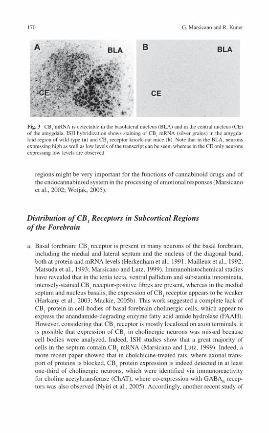
170 G. Marsicano and R. Kuner
regions might be very important for the functions of cannabinoid drugs and of the endocannabinoid system in the processing of emotional responses (Marsicano et al., 2002; Wotjak, 2005).
Distribution of CB1 Receptors in Subcortical Regions
of the Forebrain
a. Basal forebrain: CB1 receptor is present in many neurons of the basal forebrain,
including the medial and lateral septum and the nucleus of the diagonal band, both at protein and mRNA levels (Herkenham et al., 1991; Mailleux et al., 1992; Matsuda et al., 1993; Marsicano and Lutz, 1999). Immunohistochemical studies have revealed that in the tenia tecta, ventral pallidum and substantia innominata, intensely-stained CB
1 receptor-positive fibres are present, whereas in the medial
septum and nucleus basalis, the expression of CB1 receptor appears to be weaker
(Harkany et al., 2003; Mackie, 2005b). This work suggested a complete lack of CB
1 protein in cell bodies of basal forebrain cholinergic cells, which appear to
express the anandamide-degrading enzyme fatty acid amide hydrolase (FAAH). However, considering that CB
1 receptor is mostly localized on axon terminals, it
is possible that expression of CB1 in cholinergic neurons was missed because
cell bodies were analyzed. Indeed, ISH studies show that a great majority of cells in the septum contain CB
1 mRNA (Marsicano and Lutz, 1999). Indeed, a
more recent paper showed that in cholchicine-treated rats, where axonal trans-port of proteins is blocked, CB
1 protein expression is indeed detected in at least
one-third of cholinergic neurons, which were identified via immunoreactivity for choline acetyltransferase (ChAT), where co-expression with GABA
B recep-
tors was also observed (Nyiri et al., 2005). Accordingly, another recent study of
Fig. 3 CB1 mRNA is detectable in the basolateral nucleus (BLA) and in the central nucleus (CE)
of the amygdala. ISH hybridization shows staining of CB1 mRNA (silver grains) in the amygda-
loid region of wild-type (a) and CB1 receptor knock-out mice (b). Note that in the BLA, neurons
expressing high as well as low levels of the transcript can be seen, whereas in the CE only neurons expressing low levels are observed

10 Circuitries and Neurochemistry 171
Degroot and colleagues (2006) identified that the majority of cholinergic hippocampal nerve terminals are CB
1 receptor-positive, and presynaptic CB
1
receptors control the release of acetylcholine in vivo.b. Basal ganglia: The expression of CB
1 mRNA in striatal neurons presents a typical
gradient, with a great majority of medium spiny neurons (more than 95%) of the dorsolateral part of the caudate expressing moderate levels of the transcript and lower levels of expression in the ventral striatum (Matsuda et al., 1993; Marsicano and Lutz, 1999). These neurons are GABAergic, belong to the subpopulations expressing either D
1 or D
2 dopamine receptors (Hermann et al., 2002) and contain
mRNAs for different peptides belonging to the direct as well as the indirect striatal output pathways (Hohmann and Herkenham, 2000). Interestingly, local GABAergic interneurons also seem to contain CB
1 mRNA, whereas large aspiny cholinergic
or somatostatin-positive striatal interneurons are devoid of the receptor (Hohmann and Herkenham, 2000). The gradient of expression observed at mRNA level is also observed in IHC and ligand binding experiments, with the dorsolateral part of caudate putamen expressing higher levels of the receptor both in the matrix and patch structures (Tsou et al., 1998a; Egertová and Elphick, 2000). Early functional studies showed that corticostriatal projection neurons express CB
1 receptors
(Gerdeman and Lovinger, 2001; Huang et al., 2001a; Gerdeman et al., 2002), which was recently confirmed by IHC studies on tissue sections (Uchigashima et al., 2007) and in nerve terminals (Köfalvi et al., 2005). GABAergic striatal pro-jections to the substantia nigra likely contain CB
1 receptor protein (Matyas et al.,
2006). CB1 mRNA, however, is present also in subthalamic neurons (Matsuda
et al., 1993). It is, therefore, possible that glutamatergic subthalamic projections to the substantia nigra also contain CB
1 receptor protein, thereby contributing to the
cannabinoid-mediated control of locomotor activity (Sanudo-Pena et al., 2000; Romero et al., 2002; van der Stelt and Di Marzo, 2003).
c. Nucleus accumbens: In the nucleus accumbens, the expression levels of CB1
mRNA are much lower when compared with the dorsolateral striatum. However, a certain number of neurons in this region do contain detectable levels of the transcript (Matsuda et al., 1993; Monory et al., 2006). Protein expression in this region seems to be associated with glutamatergic transmission derived from prefrontal cortex projections (Robbe et al., 2001). However, the presence of CB
1
receptor on dopaminergic and GABAergic terminals in this region cannot be excluded at the moment (see later).
d. Thalamus: In this brain region, the levels of CB1 receptor are relatively low
(Matsuda et al., 1993; Tsou et al., 1998a; Marsicano and Lutz, 1999; Egertová and Elphick, 2000; Mackie, 2005b). However, some nuclei contain CB
1 mRNA
and protein, such as the lateral habenula, the reticular nucleus, the paraventricu-lar thalamic nucleus and the intermediodorsal thalamic nucleus (Matsuda et al., 1993; Tsou et al., 1998a; Marsicano and Lutz, 1999; Mackie, 2005b). Particularly interesting for the functions of the endocannabinoid system might be the relative abundance of CB
1 mRNA in the lateral habenula, which projects to many differ-
ent brain regions where the receptor might have important functions (Herkenham and Nauta, 1977).
172 G. Marsicano and R. Kuner
e. Hypothalamus: The endocannabinoid system plays a central role in many functions regulated by different hypothalamic nuclei (Pagotto et al., 2006). It is, therefore, not surprising that CB
1 receptor is present at moderate levels in several
hypothalamic areas. Importantly, the hypothalamus is one of the best examples of those regions where the relative amount of CB
1 mRNA and protein does not
correlate with the intensity of the CB1 receptor-mediated signaling events.
Indeed, ligand-induced GTPγ assays revealed that in this region, the relatively low levels of expressed CB
1 receptor are more efficiently coupled with G protein
activation than in many other regions (Breivogel and Childers, 1998). Relatively sparse information is present in the literature concerning the different hypotha-lamic nuclei where CB
1 receptors are expressed. CB
1 mRNA and protein is
present in several subnuclei, including the paraventricular nucleus, and the dor-somedial, ventromedial and lateral hypothalamic nuclei. In the paraventricular nucleus, CB
1 mRNA is present in neurons synthesizing corticotrophin releasing
factor (CRH) and cocaine-amphetamine-regulated transcript (CART) (Cota et al., 2003). In the ventromedial hypothalamus, CB
1 mRNA does not appear to
be present in GABAergic neurons (Marsicano and Lutz, 1999). In the lateral hypothalamus, CB
1 mRNA is expressed in a small fraction of neurons express-
ing pre-pro-orexin and melanin-concentrating hormone (MCH) (Cota et al., 2003). Functional experiments suggest that CB
1 expressed in glutamatergic ter-
minals projecting onto the preoptic anterior hypothalamic nucleus likely mediate hypothermic effects induced by CB
1 agonists (Rawls et al., 2002a,b).
Distribution of CB1 Receptors in the Midbrain
a. Substantia nigra: CB1 protein is highly expressed in the substantia nigra (Tsou
et al., 1998a; Egertova et al., 2000). This expression likely derives from project-ing neurons from other brain regions, such as nuclei of basal ganglia (striatum for GABAergic projections and subthalamic nucleus for glutamatergic ones, see above). These projections likely account for the functional control of CB
1 recep-
tor on nigral activity, e.g., in the control of locomotion (Sanudo-Pena and Walker, 1998; Szabo et al., 2000). Sparse intrinsic nigral neurons might contain very low levels of CB
1 receptor (Matsuda et al., 1993). Nevertheless, this low
amount of CB1 receptor in nigral (and tegmental, see later) neurons might under-
lie a direct control of CB1 receptor on dopaminergic transmission, although this
has not been shown yet (see Chap. 22 further details). Indeed, CB1 receptor pro-
tein was recently identified in a very low but significant proportion of striatal nerve terminals containing tyrosine hydroxylase (TH, a marker of monoaminer-gic neurons) (Köfalvi et al., 2005).
b. Ventral tegmental area: CB1 receptor is centrally involved in the regulation of the
activity of dopaminergic neurons in the ventral tegmental area (Szabo et al., 2002a; Melis et al., 2004a,b; Riegel and Lupica, 2004). These actions might partially explain the role of the endocannabinoid system in rewarding processes.

10 Circuitries and Neurochemistry 173
In this region, CB1 protein is present on glutamatergic as well as GABAergic
terminals. Early studies reported no evidence of CB1 expression in dopaminergic
neurons of the VTA either at protein (Herkenham et al., 1991) or mRNA level (Mailleux et al., 1992; Matsuda et al., 1993). However, some data have been published reporting co-expression of CB
1 receptor and TH in this brain region,
pointing to the possible direct activity of the endocannabinoid system on dopaminergic neurons (Wenger et al., 2003). Further detailed studies are needed to elucidate this issue.
c. Periaqueductal grey: Low to moderate levels of CB1 receptor are found in the
periaqueductal grey (PAG), where the endocannabinoid system might play a central role in the control of pain sensations (Walker et al., 1999; Hohmann et al., 2005). In contrast to opiate receptors, CB
1 receptor is preferentially (but
not exclusively) expressed in the dorsal part of the PAG (Tsou et al., 1998a; Azad et al., 2001).
Distribution of CB1 Receptors in the Hindbrain
a. Brainstem: CB1 receptor is expressed in the brainstem region at relatively low
levels. In contrast to opiate receptors, it is not found in the respiratory control centres of the medulla (Herkenham et al., 1991; Glass et al., 1997), likely explaining the low mortality caused by cannabinoid intoxication in humans and animals. The dorsal motor nucleus of the vagus and the nucleus of the solitary tract involved in central control of the gastrointestinal activity contain relatively high levels of CB
1 receptors (van Sickle et al., 2001; Mackie, 2005b). CB
1
mRNA is also present in neurons belonging to or surrounding the raphe nucleus, which is the main neuronal source of serotonin in the brain. Here, a low but sig-nificant proportion of neurons containing tryptophane hydroxylase 2 mRNA (the rate-limiting enzyme for the synthesis of serotonin) appear to co-express CB
1 mRNA (Haring et al., 2007). Moreover, expression of CB
1 protein was
identified on serotonergic terminals in the hippocampus and amygdala (Haring et al., 2007). Therefore, the endocannabinoid system might control serotonergic transmission both by regulating the activity of afferents onto serotonin-produc-ing neurons (Haj-Dahmane and Shen, 2005) and by directly modulating the functions of a subset of serotonergic neurons (Haring et al., 2007).
b. Cerebellum: The expression of CB1 receptor in the cerebellum is extremely high.
Autoradiographic and IHC imaging shows very strong presence of the protein in the molecular layer (Herkenham et al., 1991; Tsou et al., 1998a; Egertová and Elphick, 2000; Mackie, 2005b), whereas CB
1 mRNA is mainly expressed in the
granule cell layer (though scattered cells in the molecular layer also express the CB
1 receptor transcript) (Matsuda et al., 1993). This complementary expression
of CB1 protein and mRNA suggests that CB
1 receptors are expressed on climb-
ing fibres and parallel fibres and in basket cells. A certain number of Purkinje cells might express low amounts of CB
1 mRNA (Matsuda et al., 1993). These

174 G. Marsicano and R. Kuner
data indicate that the main glutamatergic and GABAergic inputs onto Purkinje cells are under the control of the endocannabinoid system, as exhaustively shown in several electrophysiological studies (Kreitzer and Regehr, 2001a,b, 2002; Maejima et al., 2001; Diana et al., 2002). The functions of the endocan-nabinoid system in the regulation of synaptic transmission in this brain region are well established, whereas functional information regarding a role of the endocannabinoid system in the control of cerebellum-dependent functions still remains sparse.
Distribution of CB1 Receptors in the Spinal Cord
As with the brain, the endocannabinoid system in the spinal cord has attracted a lot of attention and spurred numerous studies. This is not surprising, given its immense therapeutic significance, not only in pain and analgesia, but also in mechanisms governing spinal cord diseases such as multiple sclerosis, traumatic spinal injury and so on. However, the anatomical data concerning the endocannabinoid system in the spinal cord and somatosensory ganglia, such as the dorsal root ganglia (DRG) and trigeminal ganglia, have been somewhat controversial. For instance, although the bulk of current literature favours the view that CB
1 receptors are targeted exclu-
sively to axons and axonal terminals, i.e. a presynaptic localization, the spinal cord represents one of the few regions where postsynaptic localization of CB
1 receptor
has been reported (Farquhar-Smith et al., 2000; Salio et al., 2002b). IHC analyses have revealed anti-CB
1 receptor immunoreactivity in the dorsolateral funiculus,
laminae I and II inner/III transition and lamina X. In addition to neuronal expres-sion, CB
1 receptor immunoreactivity has also been reported in spinal astrocytes
(Salio et al., 2002a). The existence of presynaptic receptors is suggested by the staining of the DRG neurons (see later) as well as the axons of the Lissauer’s tract (Salio et al., 2002b). Because the superficial spinal laminae, which show dense immunoreactivity for CB
1 receptor, constitute major termination sites of primary
afferent terminals of nociceptive DRG neurons, it is important to clarify what pro-portion of CB
1 receptor in these pain-processing laminae derives from the periph-
eral vs. the central nervous system. CB1 receptor immunoreactivity in these spinal
regions has been observed to show little co-localization at the axonal level with primary afferent nociceptive markers. Furthermore, interruption of primary afferent input via dorsal root rhizotomy does not significantly lower CB
1 receptor immuno-
reactivity in these regions (Farquhar-Smith et al., 2000). This is supported by lig-and-binding studies which show that neonatal capsaicin treatment, which leads to an early destruction of nociceptive afferent fibres, reduces ligand binding to CB
1/
CB2 receptors in the superficial dorsal horn by a minor extent only (Hohmann and
Herkenham, 1998). Consistent with the above, a nociceptor-specific deletion of CB
1 receptor using conditional gene targeting approaches was observed to reduce
spinal CB1 receptor-specific ligand binding by approximately 20% only and not
lead to a major change in the CB1 receptor immunoreactivity in spinal laminae I and

10 Circuitries and Neurochemistry 175
II (Agarwal et al., 2007). Taken together, these findings suggest that a significant proportion of CB
1 receptor protein in the superficial spinal laminae is derived from
spinal, not peripheral, neurons. This is also supported by ISH studies, which have revealed labelling of cells throughout the spinal cord (Fig. 4), which is entirely lost in global CB
1 receptor knock-out mice (Agarwal et al., 2007). Postsynaptic CB
1
receptors in the spinal cord are evidenced by the staining of numerous neurons in lamina II outer as well as lamina X, many of which also express GABA, the neuro-nal nitric oxide synthase (nNOS) or the protein kinase C subunit gamma, based upon which they have been suggested to be spinal interneurons (Farquhar-Smith et al., 2000; Salio et al., 2002b). Indeed, the postsynaptic localization of CB
1 receptor
immunoreactivity in somatic as well as dendritic compartments has been confirmed by electron microscopy (Salio et al., 2002b). However, as discussed in the
Fig. 4 In situ hybridization analysis of adult mouse spinal cord and DRG using a riboprobe rec-ognizing CB
1 mRNA. (a) Widespread labelling is seen in all laminae of the spinal cord of adult
wild-type mice, but not of global CB1 receptor knock-out mice. (b) A large number of DRG cells
express CB1 mRNA. (c) The corresponding quantitative size analysis of neurons shows that a large
fraction of DRG neurons expressing CB1 mRNA are small-diameter neurons. Bars, 150 µm in (a)
and 40 µm in (b)

176 G. Marsicano and R. Kuner
introduction, it is important to consider that differences in the origin and specificity of antibodies used in diverse studies and the frequent lack of appropriate controls, e.g., loss of staining in CB
1 receptor knock-out mice, do hinder an unequivocal
interpretation of several of these earlier studies. In a recent study, which imple-mented global CB
1 receptor knock-out mice as a control, spinal CB
1 receptor
immunoreactivity was observed not only in the neuropil, but also in small-sized somata of cells in the dorsal horn (Agarwal et al., 2007). However, it remains unclear whether these somata represent neurons or astrocytes. Thus, further studies are required to clarify whether the spinal cord presents an exception to the general rule of an exclusive presynaptic (axonal) localisation of CB
1 receptor.
Distribution of CB1 Receptors in Dorsal Root Ganglia
and Peripheral Nerves
Although numerous past and recent studies have clearly demonstrated the presence of CB
1 receptor in neuronal somata in the DRG and their axons (primary afferents or
peripheral nerves), the exact nature of the distribution of CB1 mRNA and protein in
different peripheral neuronal subtypes is very controversial – different studies have reported a varying abundance of CB
1 mRNA or protein in different DRG cell types
identified via marker proteins (Table 1). For example, some earlier studies reported that CB
1 receptor is predominantly expressed in non-nociceptive DRG neurons
(medium- and large-diameter cells, distinguished by expression of neurofilament 200) and only very poorly in a small proportion of nociceptive DRG neurons (small-diam-eter cells, distinguished by expression of markers such as substance P, Na
v1.8, TRPV
1
and by binding to labelled Isolectin B4). In contrast, more recent studies have sug-
gested that the distribution of CB1 receptor in nociceptive neurons of the DRG and
trigeminal ganglia is much broader than previously thought and support a role for peripheral CB
1 receptor in pain and analgesia. The most likely reasons for these
Table 1 Percentage of marker-positive cells with CB1 expression in adult DRG
Marker
(HohmannandHerkenham, 1999) (CB
1
mRNA) (%)
(Bridgeset al., 2003) (CB
1 protein)
(%)
(Binzenet al., 2006) (CB
1 protein)
(%)
(Mitrirattanakulet al., 2006) (CB
1 protein)
(%)
(Agarwal et al., 2007) (CB
1
protein) (%)
CGRP or substanceP
13 12 ± 4 Not reported 76–82 40 ± 2
Isolectin B4 Not reported 22 ± 6 Not reported not reported 38 ± 2TRPV
1Not reported 7 ± 2 69 ± 7 76–82 90 ± 2
NF200 Not reported 49 ± 1 Not reported 81 94 ± 1

10 Circuitries and Neurochemistry 177
discrepancies lie in species differences (rats vs. mice), the sensitivity of the detection methods applied (e.g., in situ riboprobes of various lengths), diverse labelling methods, various sensitivities and stringencies of protocols or antibodies recognizing different epitopes on CB
1 receptor. In this regard, it is important to note that CB
1 receptor can
exist as multiple N-terminal splice variants, some of which are not recognised by anti-bodies targeted against the N-terminus of CB
1 receptor (Shire et al., 1995; see Chap.
9 for detailed molecular biology and pharmacology of the splice variants). In general, judging from past and recent studies, it is quite likely that CB
1 receptor is expressed at
high levels in large-diameter neurons and in a small population of small-diameter neurons (and therefore detected readily in all studies) and is, in addition, expressed at lower levels in other small-diameter neurons in the rat (therefore, detected in some, but not all studies depending on sensitivity of assay used). In this regard, the scenario of CB
1 receptor expression in DRG is not different from the controversies that existed
until very recently with respect to CB1 receptor expression in the brain, which is dis-
cussed extensively earlier. Indeed, as with the brain, conditional knockout mice in which CB
1 receptor was deleted specifically in specific populations of DRG neurons
(see later) have revealed that CB1 receptor expressed in peripheral nociceptive neu-
rons, which had been actually missed in initial expression analyses studies, is of very high physiological and therapeutic relevance (Agarwal et al., 2007). These studies have again brought home the message that the abundance of CB
1 receptor expression
is not necessarily equivalent to its functional significance. In contrast to the outcome of a previous ISH study (Hohmann and Herkenham, 1999), a riboprobe, which detects CB
1 mRNA with a high degree of sensitivity (Marsicano and Lutz, 1999; Marsicano
et al., 2003) as well as specificity (complete lack of signals in global CB1 receptor
knockout mice – shown in Fig. 4) revealed that in mouse DRG, more than 50% of cells expressing CB
1 mRNA are small-diameter neurons (diameter of less than 20 µm)
(Agarwal et al., 2007). Consistent with the above, goat-derived C-terminal CB1 recep-
tor antibody, which recognizes all CB1 receptor splice variants in a very specific man-
ner [complete lack of staining in global CB1 receptor knockout mice, see Fig. 5 and
(Coutts et al., 2002)], revealed that more than 40% of CB1 receptor-expressing DRG
neurons are nociceptors (Agarwal et al., 2007). In this study conducted on mouse DRGs, CB
1 protein was detected in more than 80% TRPV
1 receptor-positive neurons,
which is consistent with two very recent studies on rat DRG (Binzen et al., 2006; Mitrirattanakul et al., 2006). Furthermore, 40–45% of Na
v1.8-expressing neurons of the
peptidergic population and of the non-peptidergic Isolectin-B4-binding population of nociceptors were found to express CB
1 protein, in line with previous reports on rat
DRG (Binzen et al., 2006; Mitrirattanakul et al., 2006). Thus, the pattern of expression of CB
1 mRNA as well as protein in the DRG is considerably broader than the pattern
described in older studies. A recent study has shown, moreover, expression as well as peripheral transport of CB
1 receptor in nociceptors that is further increased in states of
peripheral inflammation (Amaya et al., 2006). Taken together, these recent studies suggest that the former view that CB
1 receptor is not expressed to any substantial
extent in nociceptors is incorrect. This is also supported by novel functional data, which show a striking loss of a major proportion of cannabinoid analgesia in mice conditionally lacking CB
1 receptor on nociceptors specifically (Agarwal et al., 2007).

178 G. Marsicano and R. Kuner
Fig. 5 Immunohistochemical analysis of CB1 protein expression in adult mouse DRG. (a) A goat
anti-CB1 antibody yields specific labelling of DRG neurons, which is entirely lost in DRG of
global CB1 knock-out mice. (b) Typical examples of anti-CB
1 receptor immunoreactivity in sub-
populations of DRG neurons labelled using IB4, anti-TRPV1 and anti-neurofilament 200 (NF200)
antibodies in wild-type mice. Note the large overlap between all labelled populations and CB1 expression. Bars, 40 µm

10 Circuitries and Neurochemistry 179
Distribution of CB2 Cannabinoid Receptors and Other Endocannabinoid-Target Receptors in the Nervous System
At the current state of knowledge, CB1 receptors are the most important effectors
of the functions of the endocannabinoid system in the CNS as well as of the phar-macological effects of cannabinoid drugs in the brain and spinal cord. The presence of CB
2 receptors has been mainly described in peripheral immune cells. However,
recent data suggest that CB2 receptors might be present in certain neuronal popula-
tions (see further for details). Moreover, (endo)cannabinoids have been also shown to act in neurons through the activation of different targets than the “conventional” cannabinoid receptors, CB
1 and CB
2. A full discussion of all these putative “can-
nabinoid receptors” is present in several exhaustive reviews (Di Marzo et al., 2002; Mackie and Stella, 2006; Baker et al., 2006; Begg et al., 2005; see Chap. 9) and is beyond the scope of the present chapter. Here, we will focus on the expression pat-tern of some of these potential targets of cannabinoids in the CNS. Among this ever-growing list, we have chosen to elaborate on three interesting candidates: the transient receptor potential vanilloid type 1 receptor (TRPV
1 receptor, previously
known as vanilloid receptor 1 or VR1), the acid sensitive K+ channel TASK-1 and the “orphan” G protein-coupled receptor GPR55 (see Chap. 9).
a. CB2 receptors: Originally, CB
1 receptors were described as the “neuron type”
cannabinoid receptors, whereas CB2 receptors were identified as the “immune
type” members of the family (Matsuda et al., 1990; Munro et al., 1993). This differential distribution has been profoundly challenged in the last years. On one hand, the presence of CB
1 receptors in peripheral tissues, including some types
of immune cells, has been clearly demonstrated (Pacher et al., 2006; Pagotto et al., 2006). On the other hand, CB
2 receptor expression is now known to be not
restricted to peripheral immune cells. Indeed, CB2 receptor was recently
described to be functionally present in neurons of the brainstem, where it can modulate emesis (van Sickle et al., 2005). Some studies even propose a wide distribution of CB
2 receptor in the brain (Gong et al., 2006; Onaivi et al., 2006).
However, on account of a lack of stringent controls, such as lack of staining in corresponding tissues from CB
2 receptor knock-out mice under the same stain-
ing conditions, it is difficult, in our opinion, to unequivocally pinpoint the expression of CB
2 receptors in the brain. One important noteworthy feature of
CB2 receptors is that their expression appears to be inducible (Wotherspoon et
al., 2005). In several studies, CB2 receptor was not detected in neurons in physi-
ological states, but the expression was induced in pathological states, e.g. in neurons of the dorsal root ganglia following nerve injury (Wotherspoon et al., 2005). Because CB
2 receptors are also expressed by neurons under in vitro con-
ditions (Ross et al., 2001), it is possible that CB2 receptor expression is induced
in “stressed” neurons. Therefore, it may be important in future studies address-ing CB
2 receptor expression in neurons to carefully control the conditions at the
moment of killing test animals for expression analysis.

180 G. Marsicano and R. Kuner
b. TRPV1 receptors: The TRPV
1 receptor is the first member of a large family of
transient potential ion channel receptors (TRPs; see Chap. 8). It was originally discovered as the endogenous target of the “hot” component of chilli peppers, capsaicin (Caterina et al., 1997; Tominaga et al., 1998). TRPV
1 receptors are
also activated by low pH, heat and, more recently, were shown to be activated by the endocannabinoid anandamide and by a novel putative endocannabinoid, N-arachidonoyl-dopamine (Zygmunt et al., 1999; Di Marzo et al., 2002; van der Stelt and Di Marzo, 2004; see Chap. 2, 4, 8). The activity of endocannabinoids at TRPV
1 receptors might have very interesting functional consequences. The
presence of TRPV1 in the spinal cord and peripheral neurons is well established
(see later). In the brain, TRPV1 is present at much lower levels than in peripheral
neurons (Sanchez et al., 2001), but the exact distribution of these receptors in the brain is only starting to be elucidated in detail. Original evidence for the pres-ence of TRPV
1 in the brain came from ISH and IHC experiments in rats (Mezey
et al., 2000), which suggested a widespread distribution of the receptor in differ-ent brain regions. Since then, ISH, IHC and ligand-binding studies using the specific radioligand [H3]RTX have partially confirmed previous observations (Cortright et al., 2001; Sanchez et al., 2001; Szabo et al., 2002b; Roberts et al., 2004; Toth et al., 2005; Cristino et al., 2006). Briefly, TRPV
1 is localized in the
cortex, hippocampus, dentate gyrus, central amygdala, striatum, hypothalamus, thalamus, substantia nigra, cerebellum, locus coeruleus and other smaller brain nuclei. This wide expression of TRPV
1 receptor in the brain supports the notion
that this receptor plays broader roles in animal behaviour than the control of pain sensations alone. For instance, it was recently shown that TRPV
1 knock-out
mice display pain-independent lower anxiety in a range of behavioural tests, a phenotype that is essentially opposite to that seen in CB
1 receptor knock-out
mice (Marsch et al., 2007), suggesting that CB1 and TRPV
1 receptors may
potentially play opposite functions in the regulation of certain behaviours. Interestingly, CB
1 and TRPV
1 receptors can influence each other’s function
when the two proteins are co-expressed in the same cells in vitro (Hermann et al., 2003; Sagar et al., 2004), and they have been described to be co-expressed in some neurons of the brain (Cristino et al., 2006). Noteworthy though that at least in the hippocampus, the expression pattern is more likely to be post-synap-tic (Cristino et al., 2006), and a recent studies also failed to demonstrate func-tional presynaptic hippocampal TRPV
1 receptors in the rat hippocampus
(Köfalvi et al., 2006, 2007). Concerning the spinal cord and peripheral neurons, initial studies suggested a selective expression of TRPV
1 receptor in heat-
response small-diameter neurons of the DRG. However, it is now clear that TRPV
1 receptor is also expressed in the spinal cord, in addition to the brain as
described earlier. TRPV1 receptor immunoreactivity has been observed in
somata in lamina II of the spinal dorsal horn and was also prominently seen in dendrites that are contacted by primary afferent endings in electron microscopy studies (Valtschanoff et al., 2001). In double IHC experiments, about 14% of TRPV
1 receptor-expressing dorsal horn cell bodies were also found to express
the NK1 receptor, suggesting that endogenous vanilloids may directly modulate

10 Circuitries and Neurochemistry 181
the activity of spinal projection neurons (Dolly et al., 2004). Furthermore, co-expression of TRPV
1 and CB
1 receptors has also been suggested in the spinal
cord (Cristino et al., 2006).c. TASK-1 K+ channels: The endocannabinoid anandamide was shown to directly
inhibit TASK-1, an acid- and anaesthetic-sensitive K+ channel, which sets the resting membrane potential of some types of central and peripheral neurons (Di Marzo et al., 2002; Maingret et al., 2001; see Chap. 9). Interestingly, this effect results in a depolarization of cerebellar granule neurons, possibly explaining some “paradoxical” excitatory effects of anandamide (Maingret et al., 2001). TASK-1 belongs to the family of the four transmembrane (4TM) channels, which likely form homodimers with four pore domains (Bayliss et al., 2001; Mathie et al., 2003). The distribution of TASK-1 in the brain has been described (Karschin et al., 2001; Kindler et al., 2000; Talley et al., 2001). In Northern blot analysis, TASK-1 RNA was detected in all subregions of CNS, with highest levels in the cerebellum, medulla and subthalamic nucleus (Kindler et al., 2000). ISH studies have elucidated the expression pattern of TASK-1 in more detail. In the brain, TASK-1 mRNA is present in many different regions, with highest levels in the granule cell layer of the cerebellum, arcuate nucleus of the hypotha-lamus, the granule cells of olfactory bulb and the locus coeruleus (Karschin et al., 2001; Talley et al., 2001). High levels were also described in islands of Calleja, in the septum and motor nuclei of cranial nerves, all regions containing cholin-ergic neurons, which appear to be particularly associated with this channel (Karschin et al., 2001). In cortical regions, low to moderate levels of TASK-1 are present in the piriform cortex, neocortex (where few scattered cells express-ing high levels are observed) and in glutamatergic neurons of the hippocampal formation and amygdala (Karschin et al., 2001; Talley et al., 2001). Also many subnuclei of the hypothalamus, thalamus, midbrain and brainstem contain detectable levels of TASK-1 mRNA (Karschin et al., 2001; Talley et al., 2001). In ISH studies, the predominant expression of TASK-1 appears to be in neurons, but IHC experiments came to different conclusions (Kindler et al., 2000). In fact, these authors reported a strong association of TASK-1 with glial cells in the hippocampal formation, in the cerebellar cortex and granular layer (here also neuronal staining was observed) and in the white matter of the spinal cord. Neuronal staining was reported in the neocortex, basal ganglia, amygdala, tha-lamus, midbrain and in Purkinje and granular cells of cerebellum (Kindler et al., 2000). The reasons underlying these discrepancies are not clear yet. Neuronal expression of TASK-1 in serotonergic neurons has also been described (Washburn et al., 2002). Notably though that in the hippocampus, it was recently suggested that the predominant TASK channel, modulated by hybrid endocan-nabinoid/endovanilloid substances, anandamide and N-arachidonoyl dopamine, is the TASK-3 (Köfalvi et al., 2007; see Chap. 9). To date, very few studies have addressed the distribution of TASK channels in the spinal cord and DRG. In peripheral sensory afferents, TASK channels have been implicated in the pain sensory transduction pathway and could constitute a target for anaesthetics and analgesics. TASK-1 has been detected in small- to medium-diameter DRG cell

182 G. Marsicano and R. Kuner
types and it has been proposed that these sensory afferents might contain func-tional heterodimeric channels (Rau et al., 2006). In the spinal cord, strong TASK-1 immunoreactivity has been mainly reported in ependymal cells lining the central canal and in white matter (Kindler et al., 2000).
d. GPR55: In the last few years, two patents proposed that a cloned orphan G pro-tein-coupled receptor might be a bona fide cannabinoid receptor (Baker et al., 2006; see Chap. 9). Despite the fact that a definitive confirmation of this idea has not yet been provided (Petitet et al., 2006), there is accumulating evidence that GPR55 could really be the target of several endogenous and exogenous cannabinoids (Reyes et al., 2006). There is not much information concerning the expression of GPR55 in the brain. The original publication reporting the cloning of the orphan receptor reported the Northern blot analysis of GPR55 expression in human brain (Sawzdargo et al., 1999). The transcript seems to be expressed in human caudate and putamen, but no expression was detected in cortex, hippocampus, thalamus, pons and cerebellum (Sawzdargo et al., 1999). Initial ISH experiments, however, revealed that GPR55 might be present in rat hippocampus, thalamic nuclei and parts of the midbrain (Sawzdargo et al., 1999), as also reported in the Allen Brain Atlas studies (http://www.brainatlas.org/aba/). Recently, immunohistochemical data have also been reported showing that the expression of GPR55 is developmentally regulated in the rat brain. In the embryonic brain, GPR55 expression seems to be strongly associated with various axonal tracts, including the thalamocortical pathway innervating layer IV of the “barrel” cortex. Two weeks after birth, this axonal expression decreases and post-synaptic neuronal staining of the hippocampus, thalamus, striatum and cortex become more evident (Chen et al., 2006). Further studies are mandatory, especially in the soon available GPR55 KO mouse, because of the important implications of this novel putative cannabinoid receptor in the endocannabinoid system.
Distribution of Endocannabinoids and Related Enzymes in the Nervous System
Endocannabinoids: As mentioned earlier and described in details in other chapters of this book, endocannabinoids are lipids in nature, polyunsaturated fatty acid derivatives, which share the ability to modulate the activity of cannabinoid recep-tors. Several endogenous compounds have been shown to interfere with the activity of cannabinoid receptors, at least under in vitro conditions. They are arachidonoyl ethanolamine (AEA, also known as anandamide) (Devane et al., 1992; see Chap. 2), 2-arachidonoylglycerol (2-AG) (Mechoulam et al., 1995; Sugiura et al., 1995; see Chap. 2), noladin ether (2-AGE) (Hanus et al., 2001; see Chap. 4), virodhamine (Porter et al., 2002) and N-arachidonoyldopamine (NADA) (Huang et al., 2001b, 2002; see Chap. 4). Of these compounds, the best studied and the ones clearly shown to act, at least in part, through the activation of cannabinoid receptors are

10 Circuitries and Neurochemistry 183
AEA and 2-AG and this subchapter will deal mainly with their distribution in the CNS. Given the lipophilic nature of endocannabinoids, it is very difficult to identify their presence in biological samples with spatial resolution and the measurement of their levels must necessarily rely on biochemical analysis of tissue extracts. Endocannabinoids are believed to be produced from precursors residing in cell membranes by the activation of more-or-less specific enzymatic machineries, the molecular composition of which is still in the process of elucidation (Piomelli, 2003; De Petrocellis et al., 2004; Di Marzo et al., 2004). The distribution of these agents in the CNS will be the subject of this part of the present chapter. Because they are derived from cell membranes, it is very likely that any cell type potentially possesses the ability to produce endocannabinoids. Indeed, in the CNS, neurons, glial cells and endothelia have been shown to produce endocannabinoids and their levels can be modulated in physiological and pathophysiological conditions (see Chap. 2). Here, we will describe the general distribution of endocannabinoids in the CNS, whereas the pathophysiological regulation of their levels is described in other chapters of this book and in specialized reviews (Di Marzo et al., 2000a, 2004; Piomelli et al., 2000, 2003). Another interesting issue concerning endocannabi-noids is their localization and their ability to be released in the extracellular space. It is currently very well-accepted that endocannabinoids act as retrograde neuro-transmitters at the synaptic level (Alger, 2002; Kreitzer and Regehr, 2002; Chevaleyre et al., 2006; Marsicano and Lutz, 2006; see Chaps. 11 and 21). However, given the lipophilic nature of these compounds, it is not yet fully under-stood how they can leave the membrane and travel through the watery extracellular space. Again, it is very difficult to address this issue because a direct observation of endocannabinoids in different tissue compartments is not currently feasible. Nevertheless, the combination of microdialysis and analytical techniques have enabled the determination of endocannabinoids in extracellular dialysates in the striatum (Giuffrida et al., 1999), in the periaqueductal grey (Walker et al., 1999) and more recently, in the nucleus accumbens of rats self-administering cocaine chronically (Caille et al., 2007). Interestingly, in the first two studies, only AEA was detected in the microdialysates, whereas in the latter both AEA and 2-AG were identified. Therefore, there is indirect evidence that, at least under certain condi-tions, endocannabinoids could be present in extracellular fluids within the CNS. In the brain, endocannabinoids can be found in all brain regions. Early studies revealed that the amount of 2-AG is about two orders of magnitude higher than AEA (Stella et al., 1997). In rat brains, AEA levels range from 10–15 pmols g−1 of tissue in the diencephalon, cortex and cerebellum, to 30–60 pmols g−1 of tissue in the medulla, the limbic forebrain, the hippocampus, the striatum and the brainstem, the last three regions showing the highest amounts (Bisogno et al., 1999a; Di Marzo et al., 2000b). Conversely, 2-AG is present in amounts ranging between 2 and 14 nmols g−1 of tissue, with the highest content (similar to AEA) described in the hippocampus, striatum and brainstem (Bisogno et al., 1999a). Both AEA and 2-AG are also present in the hypothalamus at about 8–9 pmols g−1 of tissue and 8 nmols g−1 tissue, respectively (Di Marzo et al., 2001). Importantly, in this region, the lev-els of endocannabinoids are regulated by feeding and by leptin administration,

184 G. Marsicano and R. Kuner
thereby providing one of the first pieces of evidence that the endocannabinoid system is important in the control of food intake (Di Marzo et al., 2001; see Chap. 14). In general, the levels of endocannabinoids in the brain do not correlate well with the levels of expression of CB
1 receptors in the same brain regions (see
earlier). This apparent discrepancy might be explained by the fact that endocan-nabinoids can additionally bind to yet uncharacterized receptors (Mackie and Stella, 2006). However, it is also possible that similar to the coupling efficiency of CB
1 in different brain regions (Breivogel and Childers, 1998), the expression levels
of the receptor do not correlate with the activity of the endocannabinoid system in a particular region and further that, the actual local concentrations of receptors and ligands may play a more decisive role in certain functions of the endocannabinoid system. As with the brain, several endocannabinoids, such as 2-AG, anandamide as well as the anandamide cogener, palmitoylethanolamide (PEA) have been detected in the spinal cord (Suplita et al., 2005; Agarwal et al., 2007; Petrosino et al., 2007). Interestingly, spinal endocannabinoid levels were found to be elevated following nerve injury (Petrosino et al., 2007) or continuous foot-shock (Suplita et al., 2005), but not following peripheral paw inflammation (Agarwal et al., 2007).
Enzymes for the Synthesis of Endocannabinoids
a. Synthesis of anandamide: Despite intense efforts in the last 15 years, the enzy-matic pathways involved in the synthesis of AEA and other polyunsaturated N-acyl-phosphatidylethanolamines are far from being completely understood in their molecular identities. Since Chap. 2 extensively reviews the question of endocannabinoid synthesis, here we only briefly do so: AEA is believed to be synthesized in a two-step pathway (Piomelli, 2003; Basavarajappa, 2007). First, N-arachidonoyl-phosphatidylethanolamine (NAPE) is synthesized from phos-phatidylethanolamine and arachidonic acid by an N-acyltransferase enzymatic activity. The molecular identity of the specific enzymes involved in this step is not known so far, although, recently, a cytosolic enzyme capable of catalyzing the synthesis of NAPE (i.e. with N-acyltransferase activity) in a Ca2+-independ-ent manner was identified as RLP-1 (Jin et al., 2007). As a second step, a NAPE-specific phospholipase D (NAPE-PLD) catalyzes the cleavage of NAPE to yield AEA (Piomelli, 2003). Recently, a form of PLD able to catalyze the synthesis of AEA in vitro was cloned and proposed as the NAPE-PLD able to mediate the synthesis of AEA in the body (Ueda et al., 2005). However, the recent genera-tion of NAPE-PLD null mutant mice revealed the existence of alternative path-ways leading to the synthesis of AEA (Leung et al., 2006). In particular, the phosphatase PTPN22 (Liu et al., 2006) and the α/β-hydrolase 4 (Abh4) (Simon and Cravatt, 2006) have been recently added to the list of enzymes possibly involved in the synthesis of AEA in vivo. The expression patterns of these enzymes in the CNS are not known in great detail. Western blotting and RT-PCR were used to identify NAPE-PLD expression in different brain regions,

10 Circuitries and Neurochemistry 185
showing that the enzyme is present in all nine brain structures examined, with higher levels in the thalamus. Interestingly, an age-dependent increase in NAPE-PLD expression was also observed between 14-day-old and 56-day-old rats in most brain regions (Morishita et al., 2005). The phosphatase PTPN22, capable of synthesizing AEA by dephosphorylation of an alternative precursor, P-AEA, which is derived from the phospholipase C-mediated cleavage of NAPE, is mainly expressed in lymphoid and hematopoietic tissues and at much lower lev-els in the CNS (Liu et al., 2006). However, Western and Northern blotting experiments have identified the enzyme in brain extracts and initial IHC experi-ments have started revealing its specific expression in neurons of the hippocam-pus (Liu et al., 2006) (see also the Allen Brain Atlas, http://www.brainatlas.org/aba/). Abh4 has not been studied in detail concerning its expression in the CNS. However, RT-PCR studies have suggested that the enzyme is present in the brain and spinal cord (Simon and Cravatt, 2006). Further studies are needed, first to clarify the complete molecular identity of the enzymes involved in the synthesis of AEA and second, to elucidate their expression pattern in the CNS in details.
b. Synthesis of 2-AG: The synthetic pathways of this endocannabinoid, abundantly present in the CNS, are also very complex and their molecular components are just starting to be elucidated (Piomelli, 2003; Basavarajappa, 2007; see Chap. 2). Nevertheless, studies in the last few years allowed identification of some of the enzymes involved in the synthesis of 2-AG and their expression patterns in the brain have been described partially. Briefly, 2-AG can be produced mainly via two distinct pathways. In one route, a phospholipase C (PLC) induces the forma-tion of 1,2-diacylglycerol (DAG), which is, in turn, cleaved by a DAG lipase to finally produce 2-AG (Piomelli, 2003). Alternatively, 2-AG can be synthesized via the phospholipase A
1 (PLA
1)-mediated production of a 2-arachidonoyl-
lysophospholipid, which, in turn, might be hydrolyzed to 2-AG by lyso-PLC activity (Bisogno et al., 1999b; Piomelli, 2003; Basavarajappa, 2007). The former pathway is better known in terms of the precise molecular identification of the enzymes involved. Indeed, one isoform of PLC involved in the synthesis of 2-AG has been recently identified as the PLCβ
1 subtype (Hashimotodani
et al., 2005), and two isoforms of DAG lipase (DAGL α and β) have been recently identified as the ones responsible for the cleavage of DAG (Bisogno et al., 2003). Data are present in the literature concerning the distribution of these enzymes in the CNS. PLCβ
1 is one of the four different isoforms of PLCβ. PLCβ
is activated by Gq/11
proteins, as, for instance, after stimulation of mGuR1 and
mGluR5 metabotropic glutamate receptors and different subclasses of mus-
carinic acetycholine receptors (Gutkind et al., 1991; Abe et al., 1992; Watanabe et al., 1998; see Chapter 11), thereby explaining their influence on endocannabi-noid and, in particular, 2-AG synthesis (Hashimotodani et al., 2005). PLCβ
1
appears to be the main isoform involved in endocannabinoid synthesis (Hashimotodani et al., 2005). The mRNA of PLCβ1 has been shown by ISH experiments to be expressed in the whole rat brain, particularly in the olfactory bulb, the cortex, caudate putamen, piriform cortex, lateral septum, hippocampal

186 G. Marsicano and R. Kuner
formation, with weaker expression in the midbrain, cerebellum (including Purkinje cells), medulla and brainstem (Watanabe et al., 1998). Already at the first glance, this expression pattern is strikingly similar to that of CB
1 receptors
in the brain. In the hippocampal formation, PLCβ1 mRNA is present mainly in
CA1 and CA3 pyramidal neurons, where it likely co-localizes with CB1 recep-
tors and in granule cells of the dentate gyrus. GABAergic interneurons seem to be devoid of the enzyme, though they may express very low amounts of the mRNA (Watanabe et al., 1998). Interestingly, mossy cells in the hilus of the dentate gyrus, expressing CB
1 receptor (Monory et al., 2006), also seem to
express mRNA of the enzyme (Watanabe et al., 1998). Therefore, PLCβ1 is
expressed in locations that could potentially mediate synthesis of 2-AG both in cells neighbouring CB
1 receptor-positive terminals and in neurons containing
CB1 receptors themselves. DAGLα and β are both present in neurons (Bisogno
et al., 2003). Interestingly, the subcellular localization of these proteins appears to undergo a switch during development, with a prominent axonal distribution in early embryonic stages, and a prominent somatodendritic expression in adults (Bisogno et al., 2003). ISH and IHC experiments recently addressed the distribu-tion of these enzymes in the adult rat brain (Yoshida et al., 2006; Uchigashima et al., 2007). DAGLα and β mRNA are expressed in the brain in similar patterns, with the β isoform being expressed at lower levels. DAGLα mRNA is mainly expressed in the olfactory bulb, cortex, caudate putamen, thalamus and Purkinje cells of the cerebellum. Weaker expression is present in other regions, such as midbrain areas and brainstem (Yoshida et al., 2006). Similar patterns were observed for DAGLβ, with the difference that thalamus, caudate putamen, mid-brain and brainstem appeared to express very low (if any) amounts of the tran-script. The subcellular localization of DAGLα protein has been described in detail in immunofluorescence and electron microscopy studies on the hippocam-pal formation, the cerebellum (Katona et al., 2006; Yoshida et al., 2006) and the striatum (Uchigashima et al., 2007). In the hippocampus, DAGLα is present in the head and neck of dendritic spines of pyramidal neurons, with the somatoden-dritic membrane expressing low levels of the protein (Yoshida et al., 2006). In the cerebellum, DAGLα protein is predominantly present in Purkinje cells, both on dendritic and somatic surface (Bisogno et al., 2003; Yoshida et al., 2006). The expression pattern of DAGLα in spines of Purkinje cells is different from the one in hippocampal pyramidal neurons: the enzyme appears to be present on the dendritic membrane, occasionally on the somatic surface and on the membrane forming the neck of dendritic spines, but neither on the main body nor on the head of the spines (Yoshida et al., 2006). In the striatum, DAGLα is also local-ized at the somatodendritic surface of medium spiny neurons, where, impor-tantly, it co-localizes with mGluR
5 and the M
1-type muscarinic acetylcholine
receptor (Uchigashima et al., 2007). However, mGluR5 and DAGLα were
detected in dendritic spines of these neurons, whereas M1 receptor was almost
excluded from this subcellular compartment, suggesting a differential influence of these two receptors on the activity of the 2-AG synthesizing enzyme (Uchigashima et al., 2007). So far, very little is known about the expression of

10 Circuitries and Neurochemistry 187
enzymes involved in the synthesis of endocannabinoids in the spinal cord and DRG. During early embryonic development, DAGLα and β can be detected in axons crossing the floor plate of the spinal cord (Bisogno et al., 2003). RT-PCR analysis suggests that at least DAGLα is expressed at moderate to high levels in the adult mouse spinal cord, but the spatial distribution pattern has not been described (Bisogno et al., 2003). Interestingly, cultured spinal neurons have been reported to contain activities for diacylglycerol and monoacylglycerol lipases, which are further enhanced upon stimulation with glutamate and brady-kinin, suggesting that these enzymes may play an important role in spinal gluta-matergic processes, such as pain (Farooqui et al., 1993). Recent ISH studies have shown that all four members of the PLCβ family are expressed in adult mouse DRG. PLCβ
4 and PLCβ
3 are present at higher levels, whereas PLCβ
1 and
PLCβ2 are only weakly detectable (Han et al., 2006). PLCβ
1 and PLCβ
4 mRNAs
also appear to be expressed in the adult spinal cord, but the identity of cells expressing them has not been clarified (Han et al., 2006).
Enzymes for Degradation of Endocannabinoids
The enzymatic pathways mediating the degradation of endocannabinoids are much better known with respect to molecular identity than endocannabinoid synthetising enzymes (Piomelli, 2003; De Petrocellis et al., 2004; Basavarajappa, 2007; see Chap. 3). An important element of endocannabinoid degradation is the facilitated uptake of the ligands by cell membranes. Despite the accumulating biochemical evidence for the existence of endocannabinoid transporter (ECT) protein(s) (Piomelli, 2003; De Petrocellis et al., 2004; McFarland and Barker, 2004; Basavarajappa, 2007), its (their) molecular identity has not been clarified yet. It is, therefore, yet unfeasible to provide information concerning the neuroanatomical distribution of the ECT(s). However, it can be generally said that all cell types ana-lyzed in vitro so far show a certain degree of ECT activity. Therefore, it could be argued that the distribution of ECT might be ubiquitous, though much more work is necessary, first for its molecular characterization and second, for elucidating the precise localization in different tissues. Interestingly, it was recently suggested that ECT activity might be associated with lipid rafts in specialized membrane compart-ments (McFarland and Barker, 2004; McFarland et al., 2004).
a. Degradation of anandamide: There is a general consensus that the main enzyme involved in the degradation of AEA is the FAAH, which, however, is also involved in the degradation of other polyunsaturated fatty acid amides (Deutsch and Chin, 1993; Piomelli, 2003; Cravatt and Lichtman, 2002; De Petrocellis et al., 2004; Basavarajappa, 2007; see Chap. 3). The distribution of FAAH in the brain has been described through analysis of its enzymatic activity (Thomas et al., 1997) as well as via direct detection of mRNA (Thomas et al., 1997) and protein (Egertova et al., 2003; Gulyas et al., 2004). The enzymatic activity of

188 G. Marsicano and R. Kuner
FAAH is present throughout the brain, with higher levels in the hippocampus and cortex, middle levels in cerebellum, thalamus, olfactory bulb and striatum, and lower levels in the hypothalamus and brainstem (Thomas et al., 1997). FAAH mRNA distribution correlates well with the enzymatic activity, with highest lev-els in the cortex, hippocampus and cerebellum and lower levels in the other brain regions. Interestingly, the subthalamic and pontine nuclei seem to express high levels of the transcript (Thomas et al., 1997). In the hippocampal formation, FAAH mRNA is exclusively detected in CA1/CA3 pyramidal neurons and in granule cells of the dentate gyrus (Thomas et al., 1997). In different brain regions, FAAH protein has been described to be expressed post-synaptically, often juxta-posed to CB
1-containing fibres, supporting the concept of retrograde signalling of
endocannabinoids (Egertova et al., 1998, 2003; Tsou et al., 1998b; Gulyas et al., 2004). In the olfactory bulb, FAAH is present in somata and dendrites of mitral cells (Tsou et al., 1998b; Egertova et al., 2003). In cortical regions, including cortex, amygdala and hippocampus, FAAH mRNA and protein seem to be present exclusively in glutamatergic neurons (Thomas et al., 1997; Egertova et al., 1998, 2003; Tsou et al., 1998b; Gulyas et al., 2004). At subcellular level, FAAH protein is present in the perinuclear cytoplasm, dendrites and dendritic spines of principal cells, whereas axon terminals and glial processes do not appear to contain the enzyme (Gulyas et al., 2004). Several subcortical forebrain regions contain FAAH, including lateral septum, caudate putamen, most of tha-lamic subnuclei and subthalamic nucleus. Interestingly, besides a weak expres-sion of the protein in the anterior hypothalamic area, the hypothalamus appears to show undetectable or very low levels of FAAH protein, suggesting that the functions of the endocannabinoid system in this brain region might be mediated by other ligands than AEA (Egertova et al., 2003). FAAH is present in the mid-brain, in particular in the inferior and superior colliculus, in the rhabdoid nucleus and in mesencephalic raphe and trigeminal nuclei (Egertova et al., 2003). In the cerebellum, FAAH protein is associated with cell bodies and dendrites of Purkinje cells (Tsou et al., 1998b; Egertova et al., 2003). In general, there is good agreement between the expression of FAAH mRNA and protein, indicating that the enzyme is predominantly expressed in cell bodies and is, therefore, unlikely to be localized at axonal terminals (Thomas et al., 1997, 1998b; Egertova et al., 2003). In addition to neurons, FAAH is also expressed by oligodendrocytes, ventricular ependymal cells and the choroid plexus in the rodent brain (Egertova et al., 2000, 2003). FAAH immunoreactivity has been reported in spinal motor neurons (Tsou et al., 1998b). However, most of the insights on the significance of FAAH in modulation of the spinal endocannabinoid system in the context of pain and analgesia have come from biochemical and functional studies. For example, paw inflammation, which leads to enhanced pain via peripheral and central mechanisms, has been reported to be associated with reduced FAAH activity in the affected paw as well as in the spinal cord (Holt et al., 2005). Furthermore, two recent studies have demonstrated a therapeutic potential for FAAH inhibitors in alleviating neuropathic pain (Jhaveri et al., 2006). Therefore, it will be interesting to see in future anatomical and functional studies where and

10 Circuitries and Neurochemistry 189
how exactly FAAH modulates endocannabinoid system under physiological and pathological conditions in the spinal cord.
b. Degradation of 2-AG: Monoacylglycerol lipase (MAGL) activity is responsible for the degradation of 2-AG in biological tissues (Piomelli, 2003; De Petrocellis et al., 2004; Basavarajappa, 2007; see Chap. 3). One form of this enzyme has been recently identified (Dinh et al., 2002) and its distribution in the CNS has been described partially (Dinh et al., 2002; Gulyas et al., 2004). It is important to note, however, that the cloned enzyme, MAGL, does not seem to be the only isoform present in biological samples possessing the ability to degrade 2-AG. Indeed, RNA interference experiments have revealed that the cloned isoform is responsible for approximately only 50% of the enzymatic activity in the rat brain (Dinh et al., 2004). More recently, a novel 2-AG hydrolyzing enzyme was biochemically char-acterized in microglial cells, though its molecular identity is still not known (Muccioli et al., 2007). It is interesting to note that this novel enzymatic activity was identified in nuclear and mitochondrial cellular fractions, possibly revealing novel intracellular locations where endocannabinoid metabolism might be functionally important (Muccioli et al., 2007). The brain distribution of the cloned isoform of MAGL was described via Northern blot analysis, ISH (Dinh et al., 2002) as well as via light and electron IHC experiments in the hippocampus, amygdala and cerebel-lum (Dinh et al., 2002; Gulyas et al., 2004). MAGL mRNA is present in all brain regions analyzed, including the thalamus, cerebellum, cortex, brainstem, striatum, hippocampus and the hypothalamus. In particular, ISH analysis has revealed that the transcript is particularly abundant in different thalamic nuclei (especially anter-odorsal nucleus), in layers IV, V and VI of neocortex, in the hippocampal formation and cerebellum. Moderate signals were also detected in the nucleus accumbens shell, islands of Calleja and the pontine nucleus (Dinh et al., 2002). In the hippoc-ampal formation, MAGL mRNA is associated with pyramidal neurons of CA region (but not with granule cells of dentate gyrus) as well as with sparse neurons within other layers (Dinh et al., 2002). Double ISH-neurochemical data are not available so far concerning the identity of these sparse cells, but electron micros-copy and IHC studies have shown that MAGL protein is partially associated with axon terminals of GABAergic interneurons (mainly basket cells containing CCK), suggesting that this neuronal type might actively participate in the enzymatic deg-radation of 2-AG (Gulyas et al., 2004). The strongest association of MAGL in the hippocampal formation is, however, with presynaptic glutamatergic terminals, whereas cell bodies and dendrites do not appear to express the enzyme (Gulyas et al., 2004). In the amygdaloid nuclei, MAGL is expressed mainly in the basolateral amygdala, whereas the central nucleus appears to be devoid of the protein (Gulyas et al., 2004). In the cerebellum, MAGL does not seem to be present in Purkinje cells, whereas their dendrites in the stratum molecularis are surrounded by intensely stained puncta. Weaker punctate staining was observed in the stratum granulosum (Gulyas et al., 2004). Altogether, the distribution patterns of FAAH and MAGL suggest that there is a strikingly exclusive distribution of the two enzymes, with FAAH mainly expressed postsynaptically and MAGL mainly present on presynaptic terminals of different neuronal populations (Gulyas et al., 2004).

190 G. Marsicano and R. Kuner
c. Distribution of cyclooxygenase-2 (COX-2) in the CNS. In the early 1990s, an inducible isoform of the cyclooxygenase enzyme was cloned and characterized in the brain (Yamagata et al., 1993; Kaufmann et al., 1996). Recently, this enzyme has been implicated in the oxidative metabolism of endocannabinoids, both by direct biochemical evidence (Yu et al., 1997; Kozak et al., 2000,2003; see Chap. 3) and by indirect observations from functional experiments (Kim and Alger, 2004; Slanina and Schweitzer, 2005; see Chap. 3). COX-2 appears to oxygenate 2-AG more efficiently and selectively than anandamide, but the differential role of these two activities is not yet fully elucidated in vivo (Kozak et al., 2004). The COX-2-mediated oxidation of endocannabinoids gives rise to a plethora of bioactive com-pounds belonging to the family of prostaglandins (Kozak et al., 2004). This phenomenon further expands and complicates the biological activity of endocan-nabinoids, but a discussion of these implications is beyond the scope of the present chapter. Here, we will dwell on the anatomical distribution of COX-2 in the CNS, because it might present additional ways of mediating the termination of endocan-nabinoid signalling. COX-2 is encoded by an inducible gene, whose expression can be stimulated by several conditions, including increased neuronal activity, and inhibited by other conditions, such as glucocorticoid activation (Yamagata et al., 1993; Chen et al., 2002). COX-2 mRNA is expressed in the brain, spinal cord as well as sensory ganglia. In particular, high levels were detected in pyramidal and granule cells of the hippocampal formation, in the piriform cortex, in the superficial layers of neocortex and in the amygdala. Lower levels were observed in the caudate putamen, the thalamus and hypothalamus (Yamagata et al., 1993). These observa-tions have been confirmed in IHC experiments, which revealed that COX-2 immunoreactivity is mainly limited to forebrain areas, where the protein localizes to cell bodies, distal dendrites and the spines of excitatory neurons (Kaufmann et al., 1996). Further studies have revealed that COX-2 immunoreactivity is also present in the paraventricular nucleus of the hypothalamus and in the brainstem (dorsal raphe, nucleus of the brachium and subcoeruleus) (Breder et al., 1995). Amongst enzymes currently thought to be involved in the degradation of endocan-nabinoids, much is known about the expression of COX-2 in the spinal cord and DRG, whereas the precise distribution pattern of FAAH and MAGL at these avenues remains to be elucidated. COX-2 is known to be constitutively expressed in the spinal cord and is strongly upregulated in an interleukin-1β-dependent manner following peripheral inflammation, concurrent to the development of pain hypersensitivity (Samad et al., 2001). However, there is no clear evidence for expression and significance of COX-2 in the DRG (Dou et al., 2004).
Concluding Remarks
The endocannabinoid system is becoming one of the most studied modulatory systems in the body, because not only of its intrinsic scientific interest but also of the plethora of potential and actual therapeutic implications of its functions. The

10 Circuitries and Neurochemistry 191
distribution of various elements of the endocannabinoid system is a very important issue in clarifying the mechanisms, through which endocannabinoids exert many different functions in the body and, in particular, in the central and peripheral nerv-ous system. In this chapter, we have tried to present an updated overview of what it is known concerning the tissue and cellular distribution of various molecules consti-tuting the endocannabinoid system. However, this field is in continuous expansion, with an exponential growth of scientific publications during the last years. The daily development of new methods for the detection of endocannabinoid system elements, the discovery of novel molecules possibly participating in the activity of the system, and the steadily growing accumulation of new functional data implying the presence of these elements in previously “unsuspected” locations, make the endocannabinoid field a very exciting and challenging area of research. During the creation of this chapter, we constantly feared missing some new and important piece of information. That this has happened is very likely the case, and we apologize in advance to the readers and the authors of these potentially overlooked studies for these omissions. It could not be helped simply due to the impressive amount of data that are present in the literature. What is known to date concerning the distribution of the endocan-nabinoid system in the nervous systems accounts for many, but not all the different functions assigned to endocannabinoids. We are anxiously awaiting novel and excit-ing discoveries in the immediate future to complete and implement our understand-ing of the fascinating world of endocannabinoid signaling.
Acknowledgements The authors thank Krisztina Monory for reading part of the manuscript and for their useful comments. Giovanni Marsicano is supported by an AVENIR grant of INSERM, in partnership with the Fondation Bettencourt-Schueller.
References
Abe T, Sugihara H, Nawa H, Shigemoto R, Mizuno N, Nakanishi S (1992) Molecular characteri-zation of a novel metabotropic glutamate receptor mGluR5 coupled to inositol phosphate/Ca2+
signal transduction. J Biol Chem 267:13361–13368.Agarwal N, Pacher P, Tegeder I, Amaya F, Constantin C, Brenner GJ, Rubino T, Michalski CW,
Marsicano G, Monory K, Mackie K, Marian C, Batkai S, Parolaro D, Fischer MJ, Reeh P, Kunos G, Kress M, Lutz B, Woolf CJ, Kuner R (2007) Cannabinoids mediate analgesia largely via peripheral type 1 cannabinoid receptors in nociceptors. Nat Neurosci doi: 10.1038/nn1916.
Aguado T, Monory K, Palazuelos J, Stella N, Cravatt B, Lutz B, Marsicano G, Kokaia Z, Guzman M, Galve-Roperh I (2005) The endocannabinoid system drives neural progenitor proliferation. FASEB J 19:1704–1706.
Aguado T, Palazuelos J, Monory K, Stella N, Cravatt B, Lutz B, Marsicano G, Kokaia Z, Guzman M, Galve-Roperh I (2006) The endocannabinoid system promotes astroglial differentiation by acting on neural progenitor cells. J Neurosci 26:1551–1561.
Alger E (2002) Retrograde signaling in the regulation of synaptic transmission: focus on endocan-nabinoids. Prog Neurobiol 68:247–286.
Amaya F, Shimosato G, Kawasaki Y, Hashimoto S, Tanaka Y, Ji RR, Tanaka M (2006) Induction of CB
1 cannabinoid receptor by inflammation in primary afferent neurons facilitates anti-
hyperalgesic effect of peripheral CB1 agonist. Pain 124:175–183.

192 G. Marsicano and R. Kuner
Azad SC, Marsicano G, Eberlein I, Putzke J, Zieglgansberger W, Spanagel R, Lutz B (2001) Differential role of the nitric oxide pathway on delta9-THC-induced central nervous system effects in the mouse. Eur J Neurosci 13:561–568.
Azad SC, Eder M, Marsicano G, Lutz B, Zieglgansberger W, Rammes G (2003) Activation of the cannabinoid receptor type 1 decreases glutamatergic and GABAergic synaptic transmission in the lateral amygdala of the mouse. Learn Mem 10:116–128.
Bacci A, Huguenard JR, Prince DA (2004) Long-lasting self-inhibition of neocortical interneu-rons mediated by endocannabinoids. Nature 431:312–316.
Baker D, Pryce G, Davies WL, Hiley CR (2006) In silico patent searching reveals a new cannabi-noid receptor. Trends Pharmacol Sci 27:1–4.
Basavarajappa BS (2007) Critical enzymes involved in endocannabinoid metabolism. Protein Pept Lett 14:237–246.
Bayliss DA, Talley EM, Sirois JE, Lei Q (2001) TASK-1 is a highly modulated pH-sensitive ‘leak’ K+ channel expressed in brainstem respiratory neurons. Respir Physiol 129:159–174.
Begg M, Pacher P, Batkai S, Osei-Hyiaman D, Offertaler L, Mo FM, Liu J, Kunos G (2005) Evidence for novel cannabinoid receptors. Pharmacol Ther 106:133–145.
Binzen U, Greffrath W, Hennessy S, Bausen M, Saaler-Reinhardt S, Treede RD (2006) Co-expression of the voltage-gated potassium channel K
v1.4 with transient receptor potential
channels (TRPV1 and TRPV
2) and the cannabinoid receptor CB
1 in rat dorsal root ganglion
neurons. Neuroscience 142:527–539.Bisogno T, Berrendero F, Ambrosino G, Cebeira M, Ramos JA, Fernandez-Ruiz JJ, Di Marzo V
(1999a) Brain regional distribution of endocannabinoids: implications for their biosynthesis and biological function. Biochem Bioph Res Co 256:377–380.
Bisogno T, Melck D, De Petrocellis L, Di Marzo V (1999b) Phosphatidic acid as the biosynthetic precursor of the endocannabinoid 2-arachidonoylglycerol in intact mouse neuroblastoma cells stimulated with ionomycin. J Neurochem 72:2113–2119.
Bisogno T, Howell F, Williams G, Minassi A, Cascio MG, Ligresti A, Matias I, Schiano-Moriello A, Paul P, Williams EJ, Gangadharan U, Hobbs C, Di Marzo V, Doherty P (2003) Cloning of the first sn1-DAG lipases points to the spatial and temporal regulation of endocannabinoid signaling in the brain. J Cell Biol 163:463–468.
Breder CD, Dewitt D, Kraig RP (1995) Characterization of inducible cyclooxygenase in rat brain. J Comp Neurol 355:296–315.
Breivogel CS, Childers SR (1998) The functional neuroanatomy of brain cannabinoid receptors. Neurobiol Dis 5:417–431.
Bridges D, Rice AS, Egertova M, Elphick MR, Winter J, Michael GJ (2003) Localisation of can-nabinoid receptor 1 in rat dorsal root ganglion using in situ hybridisation and immunohisto-chemistry. Neuroscience 119:803–812.
Caille S, varez-Jaimes L, Polis I, Stouffer DG, Parsons LH (2007) Specific alterations of extracel-lular endocannabinoid levels in the nucleus accumbens by ethanol, heroin, and cocaine self-administration. J Neurosci 27:3695–3702.
Caterina MJ, Schumacher MA, Tominaga M, Rosen TA, Levine JD, Julius D (1997) The capsai-cin receptor: a heat-activated ion channel in the pain pathway. Nature 398:816–824.
Chen C, Magee JC, Bazan NG (2002) Cyclooxygenase-2 regulates prostaglandin E2 signaling in
hippocampal long-term synaptic plasticity. J Neurophysiol 87:2851–2857.Chen H, Wang Y, Mackie K, Lu H (2006) The expression of GPR55, a putative cannabinoid
receptor, is dynamically regulated during pre- and postnatal brain development.Chen K, Ratzliff A, Hilgenberg L, Gulyas A, Freund TF, Smith M, Dinh TP, Piomelli D, Mackie
K, Soltesz I (2003) Long-term plasticity of endocannabinoid signaling induced by develop-mental febrile seizures. Neuron 39:599–611.
Chevaleyre V, Takahashi KA, Castillo PE (2006) Endocannabinoid-mediated synaptic plasticity in the CNS. Annu Rev Neurosci 29:37–76.
Cortright DN, Crandall M, Sanchez JF, Zou T, Krause JE, White G (2001) The tissue distribution and functional characterization of human VR
1. Biochem Biophys Res Commun
281:1183–1189.
10 Circuitries and Neurochemistry 193
Cota D, Marsicano G, Tschop M, Grubler Y, Flachskamm C, Schubert M, Auer D, Yassouridis A, Thone-Reineke C, Ortmann S, Tomassoni F, Cervino C, Nisoli E, Linthorst AC, Pasquali R, Lutz B, Stalla GK, Pagotto U (2003) The endogenouscannabinoid system affects energy balance via central orexigenic drive and peripheral lipogenesis. J Clin Invest 112:423–431.
Coutts AA, Irving AJ, Mackie K, Pertwee RG, Anavi-Goffer S (2002) Localisation of cannabinoid CB
1 receptor immunoreactivity in the guinea pig and rat myenteric plexus. J Comp Neurol
448:410–422.Cravatt BF, Lichtman AH (2002) The enzymatic inactivation of the fatty acid amide class of sig-
naling lipids. Chem Phys Lipids 121:135–148.Cristino L, De PL, Pryce G, Baker D, Guglielmotti V, Di Marzo V (2006) Immunohistochemical
localization of cannabinoid type 1 and vanilloid transient receptor potential vanilloid type 1 receptors in the mouse brain. Neuroscience 139:1405–1415.
De Petrocellis L., Cascio MG, Di Marzo V (2004) The endocannabinoid system: a general view and latest additions. Br J Pharmacol 141:765–774.
Degroot A, Köfalvi A, Wade MR, Davis RJ, Rodrigues RJ, Rebola N, Cunha RA, Nomikos GG (2006) CB
1 receptor antagonism increases hippocampal acetylcholine release: site and mecha-
nism of action. Mol Pharmacol 70:1236–1245.Deutsch DG, Chin SA (1993) Enzymatic synthesis and degradation of anandamide, a cannabinoid
receptor agonist. Biochem Pharmacol 46:791–796.Devane WA, Hanus L, Breuer A, Pertwee RG, Stevenson LA, Griffin G, Gibson D, Mandelbaum
A, Etinger A, Mechoulam R (1992) Isolation and structure of a brain constituent that binds to the cannabinoid receptor. Science 258:1946–1949.
Di Marzo V, Bisogno T, De Petrocellis L (2000a) Endocannabinoids: new targets for drug devel-opment. Curr Pharm Des 6:1361–1380.
Di Marzo V, Breivogel CS, Tao Q, Bridgen DT, Razdan RK, Zimmer AM, Zimmer A, Martin BR (2000b) Levels, metabolism, and pharmacological activity of anandamide in CB
1 cannabinoid
receptor knockout mice: evidence for non-CB1, non-CB
2 receptor-mediated actions of ananda-
mide in mouse brain. J Neurochem 75:2434–2444.Di Marzo V, Goparaju SK, Wang L, Liu J, Batkai S, Jarai Z, Fezza F, Miura GI, Palmiter RD,
Sugiura T, Kunos G (2001) Leptin-regulated endocannabinoids are involved in maintaining food intake. Nature 410:822–825.
Di Marzo V, De Petrocellis L, Fezza F, Ligresti A, Bisogno T (2002) Anandamide receptors. Prostaglandins Leukot Essent Fatty Acids 66:377–391.
Di Marzo V, Bifulco M, De Petrocellis L (2004) The endocannabinoid system and its therapeutic exploitation. Nat Rev Drug Discov 3:771–784.
Di Marzo V, De PL, Bisogno T (2005) The biosynthesis, fate and pharmacological properties of endocannabinoids. Handb Exp Pharmacol 147–185.
Diana MA, Levenes C, Mackie K, Marty A (2002) Short-term retrograde inhibition of GABAergic synaptic currents in rat purkinje cells is mediated by endogenous cannabinoids. J Neurosci 22:200–208.
Dinh TP, Carpenter D, Leslie FM, Freund TF, Katona I, Sensi SL, Kathuria S, Piomelli D (2002) Brain monoglyceride lipase participating in endocannabinoid inactivation. Proc Natl Acad Sci USA 99:10819–10824.
Dinh TP, Kathuria S, Piomelli D (2004) RNA interference suggests a primary role for monoacylg-lycerol lipase in the degradation of the endocannabinoid 2-arachidonoylglycerol. Mol Pharmacol 66:1260–1264.
Doherty J, Dingledine R (2003) Functional interactions between cannabinoid and metabotropic glutamate receptors in the central nervous system. Curr Opin Pharmacol 3:46–53.
Doly S, Fischer J, Conrath M (2004) The vanilloid receptor-1 (TRPV1) is expressed in some rat
dorsal horn NK1 cells. Brain Res 1004:203–207.Domenici MR, Azad SC, Marsicano G, Schierloh A, Wotjak CT, Dodt HU, Zieglgansberger W,
Lutz B, Rammes G (2006) Cannabinoid receptor type 1 located on presynaptic terminals of principal neurons in the forebrain controls glutamatergic synaptic transmission. J Neurosci 26:5794–5799.

194 G. Marsicano and R. Kuner
Dou W, Jiao Y, Goorha S, Raghow R, Ballou LR (2004) Nociception and the differential expres-sion of cyclooxygenase-1 (COX-1), the COX-1 variant retaining intron-1 (COX-1v), and COX-2 in mouse dorsal root ganglia (DRG). Prostaglandins Other Lipid Mediat 74:29–43.
Egertová M, Elphick MR (2000) Localisation of cannabinoid receptors in the rat brain using anti-bodies to the intracellular C-terminal tail of CB
1. J Comp Neurol 422:159–171.
Egertova M, Giang DK, Cravatt BF, Elphick MR (1998) A new perspective on cannabinoid sig-nalling: complementary localization of fatty acid amide hydrolase and the CB
1 receptor in rat
brain. Proc Biol Sci 265:2081–2085.Egertova M, Cravatt BF, Elphick MR (2000) Fatty acid amide hydrolase expression in rat choroid
plexus: possible role in regulation of the sleep-inducing action of oleamide. Neurosci Lett 282:13–16.
Egertova M, Cravatt BF, Elphick MR (2003) Comparative analysis of fatty acid amide hydrolase and cb
1 cannabinoid receptor expression in the mouse brain: evidence of a widespread role for
fatty acid amide hydrolase in regulation of endocannabinoid signaling. Neuroscience 119:481–496.
Farooqui AA, Anderson DK, Horrocks LA (1993) Effect of glutamate and its analogs on diacylg-lycerol and monoacylglycerol lipase activities of neuron-enriched cultures. Brain Res 604:180–184.
Farquhar-Smith WP, Egertova M, Bradbury EJ, McMahon SB, Rice AC, Elphick MR (2000) Cannabinoid CB1 receptor expression in rat spinal cord. Mol Cell Neurosci 15:510–521.
Freund TF, Katona I, Piomelli D (2003) Role of endogenous cannabinoids in synaptic signaling. Physiol Rev 83:1017–1066.
Galve-Roperh I, Aguado T, Palazuelos J, Guzman M (2007) The endocannabinoid system and neurogenesis in health and disease. Neuroscientist 13:109–114.
Gerdeman G, Lovinger DM (2001) CB1 cannabinoid receptor inhibits synaptic release of gluta-
mate in rat dorsolateral striatum. J Neurophysiol 85:468–471.Gerdeman GL, Ronesi J, Lovinger DM (2002) Postsynaptic endocannabinoid release is critical to
long-term depression in the striatum. Nat Neurosci 5:446–451.Giuffrida A, Parsons LH, Kerr TM, Rodriguez de Fonseca F, Navarro M, Piomelli D (1999)
Dopamine activation of endogenous cannabinoid signaling in dorsal striatum. Nat Neurosci 2:358–363.
Glass M, Dragunow M, Faull RL (1997) Cannabinoid receptors in the human brain: a detailed anatomical and quantitative autoradiographic study in the fetal, neonatal and adult human brain. Neuroscience 77:299–318.
Gong JP, Onaivi ES, Ishiguro H, Liu QR, Tagliaferro PA, Brusco A, Uhl GR (2006) Cannabinoid CB
2 receptors: immunohistochemical localization in rat brain. Brain Res 1071:10–23.
Gross CG (2000) Neurogenesis in the adult brain: death of a dogma. Nat Rev Neurosci 1:67–73.Gulyas AI, Cravatt BF, Bracey MH, Dinh TP, Piomelli D, Boscia F, Freund TF (2004) Segregation
of two endocannabinoid-hydrolyzing enzymes into pre- and postsynaptic compartments in the rat hippocampus, cerebellum and amygdala. Eur J Neurosci 20:441–458.
Gutkind JS, Novotny EA, Brann MR, Robbins KC (1991) Muscarinic acetylcholine receptor sub-types as agonist-dependent oncogenes. Proc Natl Acad Sci USA 88:4703–4707.
Haj-Dahmane S, Shen RY (2005) The wake-promoting peptide orexin-B inhibits glutamatergic transmission to dorsal raphe nucleus serotonin neurons through retrograde endocannabinoid signaling. J Neurosci 25:896–905.
Han SK, Mancino V, Simon MI (2006) Phospholipase Cβ3 mediates the scratching response acti-vated by the histamine H
1 receptor on C-fiber nociceptive neurons. Neuron 52:691–703.
Hanus L, Abu-Lafi S, Fride E, Breuer A, Vogel Z, Shalev DE, Kustanovich I, Mechoulam R (2001) 2-arachidonyl glyceryl ether, an endogenous agonist of the cannabinoid CB
1 receptor.
Proc Natl Acad Sci USA 98:3662–3665.Haring M, Marsicano G, Lutz B, Monory K (2007) Identification of the cannabinoid receptor type
1 in serotonergic cells of raphe nuclei in mice. Neuroscience 146(3):1212–1219.Harkany T, Hartig W, Berghuis P, Dobszay MB, Zilberter Y, Edwards RH, Mackie K, Ernfors P
(2003) Complementary distribution of type 1 cannabinoid receptors and vesicular glutamate

10 Circuitries and Neurochemistry 195
transporter 3 in basal forebrain suggests input-specific retrograde signalling by cholinergic neurons. Eur J Neurosci 18:1979–1992.
Hashimotodani Y, Ohno-Shosaku T, Tsubokawa H, Ogata H, Emoto K, Maejima T, Araishi K, Shin HS, Kano M (2005) Phospholipase Cbeta serves as a coincidence detector through its Ca2+ dependency for triggering retrograde endocannabinoid signal. Neuron 45:257–268.
Herkenham M (1991) Characterization and localization of cannabinoid receptors in brain: an invitro technique using slide-mounted tissue sections. NIDA Res Monogr 112:129–145.
Herkenham M, Nauta WJ (1977) Afferent connections of the habenular nuclei in the rat. A horse-radish peroxidase study, with a note on the fiber-of-passage problem. J Comp Neurol 173:123–146.
Herkenham M, Lynn AB, Little MD, Johnson MR, Melvin LS, De Costa BR, Rice KC (1990) Cannabinoid receptor localization in brain. Proc Natl Acad Sci USA 87:1932–1936.
Herkenham M, Lynn AB, Johnson MR, Melvin LS, De Costa BR, Rice KC (1991) Characterization and localization of cannabinoid receptors in rat brain: a quantitative in vitro autoradiographic study. J Neurosci 11:563–583.
Hermann H, Marsicano G, Lutz B (2002) Coexpression of the cannabinoid receptor type 1 with dopamine and serotonin receptors in distinct neuronal subpopulations of the adult mouse fore-brain. Neuroscience 109:451–460.
Hermann H, De Petrocellis L, Bisogno T, Schiano MA, Lutz B, Di Marzo V (2003) Dual effect of cannabinoid CB1 receptor stimulation on a vanilloid VR
1 receptor-mediated response. Cell
Mol Life Sci 60:607–616.Hill EL, Gallopin T, Ferezou I, Cauli B, Rossier J, Schweitzer P, Lambolez B (2007) Functional
CB1 receptors are broadly expressed in neocortical GABAergic and glutamatergic neurons.
J Neurophysiol 97:2580–2589.Hohmann AG, Herkenham M (1998) Regulation of cannabinoid and mu opioid receptors in rat
lumbar spinal cord following neonatal capsaicin treatment. Neurosci Lett 252:13–16.Hohmann AG, Herkenham M (1999) Localization of central cannabinoid CB
1 receptor messenger
RNA in neuronal subpopulations of rat dorsal root ganglia: a double-label in situ hybridization study. Neuroscience 90:923–931.
Hohmann AG, Herkenham M (2000) Localization of cannabinoid CB1 receptor mRNA in neuro-
nal subpopulations of rat striatum: a double-label in situ hybridization study. Synapse 37:71–80.
Hohmann AG, Suplita RL, Bolton NM, Neely MH, Fegley D, Mangieri R, Krey JF, Walker JM, Holmes PV, Crystal JD, Duranti A, Tontini A, Mor M, Tarzia G, Piomelli D (2005) An endo-cannabinoid mechanism for stress-induced analgesia. Nature 435:1108–1112.
Holt S, Comelli F, Costa B, Fowler CJ (2005) Inhibitors of fatty acid amide hydrolase reduce carrageenan-induced hind paw inflammation in pentobarbital-treated mice: comparison with indomethacin and possible involvement of cannabinoid receptors. Br J Pharmacol 146:467–476.
Howlett AC, Fleming RM (1984) Cannabinoid inhibition of adenylate cyclase. Pharmacology of the response in neuroblastoma cell membranes. Mol Pharmacol 26:532–538.
Howlett AC, Barth F, Bonner TI, Cabral G, Casellas P, Devane WA, Felder CC, Herkenham M, Mackie K, Martin BR, Mechoulam R, Pertwee RG (2002) International Union of Pharmacology. XXVII. Classification of cannabinoid receptors. Pharmacol Rev 54:161–202.
Huang CC, Lo SW, Hsu KS (2001a) Presynaptic mechanisms underlying cannabinoid inhibition of excitatory synaptic transmission in rat striatal neurons. J Physiol 532:731–748.
Huang SM, Bisogno T, Petros TJ, Chang SY, Zavitsanos PA, Zipkin RE, Sivakumar R, Coop A, Maeda DY, De Petrocellis L, Burstein S, Di M, V, Walker JM (2001b) Identification of a new class of molecules, the arachidonyl amino acids, and characterization of one member that inhibits pain. J Biol Chem 276:42639–42644.
Huang SM, Bisogno T, Trevisani M, Al Hayani A, De Petrocellis L, Fezza F, Tognetto M, Petros TJ, Krey JF, Chu CJ, Miller JD, Davies SN, Geppetti P, Walker JM, Di M, V (2002) An endogenous capsaicin-like substance with high potency at recombinant and native vanilloid VR1
receptors. Proc Natl Acad Sci USA 99:8400–8405.

196 G. Marsicano and R. Kuner
Jhaveri MD, Richardson D, Kendall DA, Barrett DA, Chapman V (2006) Analgesic effects of fatty acid amide hydrolase inhibition in a rat model of neuropathic pain. J Neurosci 26:13318–13327.
Jiang W, Zhang Y, Xiao L, Van CJ, Ji SP, Bai G, Zhang X (2005) Cannabinoids promote embry-onic and adult hippocampus neurogenesis and produce anxiolytic- and antidepressant-like effects. J Clin Invest 115:3104–3116.
Jin XH, Okamoto Y, Morishita J, Tsuboi K, Tonai T, Ueda N (2007) Discovery and characteriza-tion of a Ca2+-independent phosphatidylethanolamine N-acyltransferase generating the anan-damide precursor and its congeners. J Biol Chem 282:3614–3623.
Johnston D, Amaral DG (2004) Hippocampus. In: The synaptic organization of the brain (Shepherd GM, ed.), pp 455–498. Oxford: Oxford University Press.
Karschin C, Wischmeyer E, Preisig-Muller R, Rajan S, Derst C, Grzeschik KH, Daut J, Karschin A (2001) Expression pattern in brain of TASK-1, TASK-3, and a tandem pore domain K+ channel subunit, TASK-5, associated with the central auditory nervous system. Mol Cell Neurosci 18:632–648.
Katona I, Rancz EA, Acsady L, Ledent C, Mackie K, Hajos N, Freund TF (2001) Distribution of CB
1 cannabinoid receptors in the amygdala and their role in the control of GABAergic trans-
mission. J Neurosci 21:9506–9518.Katona I, Sperlagh B, Sik A, Köfalvi A, Vizi ES, Mackie K, Freund TF (1999) Presynaptically
located CB1 cannabinoid receptors regulate GABA release from axon terminals of specific
hippocampal interneurons. J Neurosci 19:4544–4558.Katona I, Urban GM, Wallace M, Ledent C, Jung KM, Piomelli D, Mackie K, Freund TF (2006)
Molecular composition of the endocannabinoid system at glutamatergic synapses. J Neurosci 26:5628–5637.
Kaufmann WE, Worley PF, Pegg J, Bremer M, Isakson P (1996) COX-2, a synaptically induced enzyme, is expressed by excitatory neurons at postsynaptic sites in rat cerebral cortex. Proc Natl Acad Sci USA 93:2317–2321.
Kawamura Y, Fukaya M, Maejima T, Yoshida T, Miura E, Watanabe M, Ohno-Shosaku T, Kano M (2006) The CB
1 cannabinoid receptor is the major cannabinoid receptor at excitatory presy-
naptic sites in the hippocampus and cerebellum. J Neurosci 26:2991–3001.Kim J, Alger BE (2004) Inhibition of cyclooxygenase-2 potentiates retrograde endocannabinoid
effects in hippocampus. Nat Neurosci 7:697–698.Kindler CH, Pietruck C, Yost CS, Sampson ER, Gray AT (2000) Localization of the tandem pore
domain K+ channel TASK-1 in the rat central nervous system. Brain Res Mol Brain Res 80:99–108.
Klausberger T, Marton LF, O’Neill J, Huck JH, Dalezios Y, Fuentealba P, Suen WY, Papp E, Kaneko T, Watanabe M, Csicsvari J, Somogyi P (2005) Complementary roles of cholecysto-kinin- and parvalbumin-expressing GABAergic neurons in hippocampal network oscillations. J Neurosci 25:9782–9793.
Köfalvi A, Rodrigues RJ, Ledent C, Mackie K, Vizi ES, Cunha RA, Sperlagh B (2005) Involvement of cannabinoid receptors in the regulation of neurotransmitter release in the rodent striatum: a combined immunochemical and pharmacological analysis. J Neurosci 25:2874–2884.
Köfalvi A, Oliveira CR, Cunha RA (2006) Lack of evidence for functional TRPV1 vanilloid recep-
tors in rat hippocampal nerve terminals. Neurosci Lett 403:151–156.Köfalvi A, Pereira MF, Rebola N, Rodrigues RJ, Oliveira CR, Cunha RA (2007) Anandamide and
NADA bi-directionally modulate presynaptic Ca2+ levels and transmitter release in the hippocampus. Br J Pharmacol doi:10.1038/sj.bjp.0707252.
Kozak KR, Rowlinson SW, Marnett LJ (2000) Oxygenation of the endocannabinoid, 2-arachido-nylglycerol, to glyceryl prostaglandins by cyclooxygenase-2. J Biol Chem 275:33744–33749.
Kozak KR, Prusakiewicz JJ, Rowlinson SW, Prudhomme DR, Marnett LJ (2003) Amino acid determinants in cyclooxygenase-2 oxygenation of the endocannabinoid anandamide. Biochemistry 42:9041–9049.

10 Circuitries and Neurochemistry 197
Kozak KR, Prusakiewicz JJ, Marnett LJ (2004) Oxidative metabolism of endocannabinoids by COX-2. Curr Pharm Des 10:659–667.
Kreitzer AC, Regehr WG (2001a) Cerebellar depolarization-induced suppression of inhibition is mediated by endogenous cannabinoids. J Neurosci 21:RC174.
Kreitzer AC, Regehr WG (2001b) Retrograde inhibition of presynaptic calcium influx by endog-enous cannabinoids at excitatory synapses onto Purkinje cells. Neuron 29:717–727.
Kreitzer AC, Regehr WG (2002) Retrograde signaling by endocannabinoids. Curr Opin Neurobiol 12:324–330.
LeDoux JE (2000) Emotion circuits in the brain. Annu Rev Neurosci 23:155–184.Leung D, Saghatelian A, Simon GM, Cravatt BF (2006) Inactivation of N-acyl phosphati-
dylethanolamine phospholipase D reveals multiple mechanisms for the biosynthesis of endocannabinoids. Biochemistry 45:4720–4726.
Liu J, Wang L, Harvey-White J, Osei-Hyiaman D, Razdan R, Gong Q, Chan AC, Zhou Z, Huang BX, Kim HY, Kunos G (2006) A biosynthetic pathway for anandamide. Proc Natl Acad Sci USA 103:13345–13350.
Lledo PM, Alonso M, Grubb MS (2006) Adult neurogenesis and functional plasticity in neuronal circuits. Nat Rev Neurosci 7:179–193.
Lutz B (2004) On-demand activation of the endocannabinoid system in the control of neuronal excitability and epileptiform seizures. Biochem Pharmacol 68:1691–1698.
Lutz B, Hermann H, Woelfel B, Monory K, Ekker M, Rubenstein JL, Marsicano G (2004) Anatomical and functional localization of CB
1 cannabinoid receptors in principal glutamatergic neurons.
Mackie K (2005a) Cannabinoid receptor homo- and heterodimerization. Life Sci 77:1667–1673.Mackie K (2005b) Distribution of cannabinoid receptors in the central and peripheral nervous
system. Handb Exp Pharmaco l299–325.Mackie K, Stella N (2006) Cannabinoid receptors and endocannabinoids: evidence for new play-
ers. AAPS J 8:E298–E306.Maejima T, Ohno-Shosaku T, Kano M (2001) Endogenous cannabinoid as a retrograde messenger
from depolarized postsynaptic neurons to presynaptic terminals. Neurosci Res 40:205–210.Mailleux P, Vanderhaeghen JJ (1992) Distribution of neuronal cannabinoid receptor in the adult
rat brain: a comparative receptor binding radioautography and in situ hybridization histochem-istry. Neuroscience 48:655–668.
Mailleux P, Parmentier M, Vanderhaeghen JJ (1992) Distribution of cannabinoid receptor mes-senger RNA in the human brain: an in situ hybridization histochemistry with oligonucleotides. Neurosci Lett 143:200–204.
Maingret F, Patel AJ, Lazdunski M, Honore E (2001) The endocannabinoid anandamide is a direct and selective blocker of the background K+ channel TASK-1. EMBO J 20:47–54.
Marsch R, Foeller E, Rammes G, Bunck M, Kossl M, Holsboer F, Zieglgansberger W, Landgraf R, Lutz B, Wotjak CT (2007) Reduced anxiety, conditioned fear, and hippocampal long-term potentiation in transient receptor potential vanilloid type 1 receptor-deficient mice. J Neurosci 27:832–839.
Marsicano G, Lutz B (1999) Expression of the cannabinoid receptor CB1 in distinct neuronal subpopulations in the adult mouse forebrain. Eur J Neurosci 11:4213–4225.
Marsicano G, Lutz B (2006) Neuromodulatory functions of the endocannabinoid system. J Endocrinol Invest 29:27–46.
Marsicano G, Wotjak CT, Azad SC, Bisogno T, Rammes G, Cascio MG, Hermann H, Tang J, Hofmann C, Zieglgänsberger W, Di Marzo V, Lutz B (2002) The endogenous cannabinoid system controls extinction of aversive memories. Nature 418:530–534.
Marsicano G, Goodenough S, Monory K, Hermann H, Eder M, Cannich A, Azad SC, Cascio MG, Gutierrez SO, Van D, Lopez R, Casanova E, Schutz G, Zieglgänsberger W, Di Marzo V, Behl C, Lutz B (2003) CB1 cannabinoid receptors and on-demand defense against excitotoxicity. Science 302:84–88.
Mathie A, Clarke CE, Ranatunga KM, Veale EL (2003) What are the roles of the many different types of potassium channel expressed in cerebellar granule cells? Cerebellum 2:11–25.
198 G. Marsicano and R. Kuner
Matsuda LA, Lolait SJ, Brownstein MJ, Young AC, Bonner TI (1990) Structure of a cannabinoid receptor and functional expression of the cloned cDNA. Nature 346:561–564.
Matsuda LA, Bonner TI, Lolait SJ (1993) Localization of cannabinoid receptor mRNA in rat brain. J Comp Neurol 327:535–550.
Matyas F, Yanovsky Y, Mackie K, Kelsch W, Misgeld U, Freund TF (2006) Subcellular localiza-tion of type 1 cannabinoid receptors in the rat basal ganglia. Neuroscience 137:337–361.
McDonald AJ, Mascagni F (2001) Localization of the CB1 type cannabinoid receptor in the rat
basolateral amygdala: high concentrations in a subpopulation of cholecystokinin-containing interneurons. Neuroscience 107:641–652.
McFarland MJ, Barker EL (2004) Anandamide transport. Pharmacol Ther 104:117–135.McFarland MJ, Porter AC, Rakhshan FR, Rawat DS, Gibbs RA, Barker EL (2004) A role for
caveolae/lipid rafts in the uptake and recycling of the endogenous cannabinoid anandamide. J Biol Chem 279:41991–41997.
Mechoulam R, Ben Shabat S, Hanus L, Ligumsky M, Kaminski NE, Schatz AR, Gopher A, Almog S, Martin BR, Compton DR (1995) Identification of an endogenous 2-monoglycer-ide, present in canine gut, that binds to cannabinoid receptors. Biochem Pharmacol 50:83–90.
Melis M, Perra S, Muntoni AL, Pillolla G, Lutz B, Marsicano G, Di M, V, Gessa GL, Pistis M (2004a) Prefrontal cortex stimulation induces 2-arachidonoyl-glycerol-mediated suppression of excitation in dopamine neurons. J Neurosci 24:10707–10715.
Melis M, Pistis M, Perra S, Muntoni AL, Pillolla G, Gessa GL (2004b) Endocannabinoids mediate presynaptic inhibition of glutamatergic transmission in rat ventral tegmental area dopamine neurons through activation of CB
1 receptors. J Neurosci 24:53–62.
Mezey E, Toth ZE, Cortright DN, Arzubi MK, Krause JE, Elde R, Guo A, Blumberg PM, Szallasi A (2000) Distribution of mRNA for vanilloid receptor subtype 1 (VR
1), and VR
1-like immunore-
activity, in the central nervous system of the rat and human. Proc Natl Acad Sci USA 97:3655–3660.
Mitrirattanakul S, Ramakul N, Guerrero AV, Matsuka Y, Ono T, Iwase H, Mackie K, Faull KF, Spigelman I (2006) Site-specific increases in peripheral cannabinoid receptors and their endogenous ligands in a model of neuropathic pain. Pain 126:102–114.
Monory K, Massa F, Egertova M, Eder M, Blaudzun H, Westenbroek R, Kelsch W, Jacob W, Marsch R, Ekker M, Long J, Rubenstein JL, Goebbels S, Nave KA, During M, Klugmann M, Wolfel B, Dodt HU, Zieglgansberger W, Wotjak CT, Mackie K, Elphick MR, Marsicano G, Lutz B (2006) The endocannabinoid system controls key epileptogenic circuits in the hippoc-ampus. Neuron 51:455–466.
Morales M, Backman C (2002) Coexistence of serotonin 3 (5-HT3) and CB
1 cannabinoid receptors
in interneurons of hippocampus and dentate gyrus. Hippocampus 12:756–764.Morishita J, Okamoto Y, Tsuboi K, Ueno M, Sakamoto H, Maekawa N, Ueda N (2005) Regional
distribution and age-dependent expression of N-acylphosphatidylethanolamine-hydrolyzing phospholipase D in rat brain. J Neurochem 94:753–762.
Muccioli GG, Xu C, Odah E, Cudaback E, Cisneros JA, Lambert DM, Lopez Rodriguez ML, Bajjalieh S, Stella N (2007) Identification of a novel endocannabinoid-hydrolyzing enzyme expressed by microglial cells. J Neurosci 27:2883–2889.
Munro S, Thomas KL, Abu-Shaar M (1993) Molecular characterization of a peripheral receptor for cannabinoids. Nature 365:61–65.
Nyiri G, Cserep C, Szabadits E, Mackie K, Freund TF (2005) CB1 cannabinoid receptors are
enriched in the perisynaptic annulus and on preterminal segments of hippocampal GABAergic axons. Neuroscience 136:811–822.
Onaivi ES, Ishiguro H, Gong JP, Patel S, Perchuk A, Meozzi PA, Myers L, Mora Z, Tagliaferro P, Gardner E, Brusco A, Akinshola BE, Liu QR, Hope B, Iwasaki S, Arinami T, Teasenfitz L, Uhl GR (2006) Discovery of the presence and functional expression of cannabinoid CB
2 recep-
tors in brain. Ann N Y Acad Sci 1074:514–536.Pacher P, Batkai S, Kunos G (2006) The endocannabinoid system as an emerging target of phar-
macotherapy. Pharmacol Rev 58:389–462.

10 Circuitries and Neurochemistry 199
Pagotto U, Marsicano G, Cota D, Lutz B, Pasquali R (2006) The emerging role of the endocan-nabinoid system in endocrine regulation and energy balance. Endocr Rev 27:73–100.
Petitet F, Donlan M, Michel A (2006) GPR55 as a new cannabinoid receptor: still a long way to prove it. Chem Biol Drug Des 67:252–253.
Petrosino S, Palazzo E, de N, V, Bisogno T, Rossi F, Maione S, Di Marzo V (2007) Changes in spinal and supraspinal endocannabinoid levels in neuropathic rats. Neuropharmacology 52:415–422.
Piomelli D (2003) The molecular logic of endocannabinoid signalling. Nat Rev Neurosci 4:873–884.
Piomelli D, Giuffrida A, Calignano A, de Fonseca FR (2000) The endocannabinoid system as a target for therapeutic drugs. Trends Pharmacol Sci 21:218–224.
Porter AC, Sauer JM, Knierman MD, Becker GW, Berna MJ, Bao J, Nomikos GG, Carter P, Bymaster FP, Leese AB, Felder CC (2002) Characterization of a novel endocannabinoid, virodhamine, with antagonist activity at the CB
1 receptor. J Pharmacol Exp Ther
301:1020–1024.Rau KK, Cooper BY, Johnson RD (2006) Expression of TWIK-related acid sensitive K+ channels
in capsaicin sensitive and insensitive cells of rat dorsal root ganglia. Neuroscience 141:955–963.
Rawls SM, Cabassa J, Geller EB, Adler MW (2002a) CB(1) receptors in the preoptic anterior hypothalamus regulate WIN 55212-2 [(4,5-dihydro-2-methyl-4(4-morpholinylmethyl)-1-(1-naphthalenyl-carbonyl)-6H-pyrrolo[3,2,1ij]quinolin-6-one]-induced hypothermia. J Pharmacol Exp Ther 301:963–968.
Rawls SM, Cowan A, Tallarida RJ, Geller EB, Adler MW (2002b) N-methyl-d-aspartate antago-nists and WIN 55212-2 [4,5-dihydro-2-methyl-4(4-morpholinylmethyl)-1-(1-naphthalenyl-carbonyl)-6 H-pyrrolo[3,2,1-i,j]quinolin-6-one], a cannabinoid agonist, interact to produce synergistic hypothermia. J Pharmacol Exp Ther 303:395–402.
Reyes FR, Cudaback E, Hague C, Moeller T, Mackie K, Stella N (2006) Initial characterization of GPR55, a novel cannabinoid receptor. Program No. 791.1 2006 Abstract.
Riegel AC, Lupica CR (2004) Independent presynaptic and postsynaptic mechanisms regulate endocannabinoid signaling at multiple synapses in the ventral tegmental area. J Neurosci 24:11070–11078.
Robbe D, Alonso G, Duchamp F, Bockaert J, Manzoni OJ (2001) Localization and mechanisms of action of cannabinoid receptors at the glutamatergic synapses of the mouse nucleus accumbens. J Neurosci 21:109–116.
Roberts JC, Davis JB, Benham CD (2004) [3H]Resiniferatoxin autoradiography in the CNS of wild-type and TRPV
1 null mice defines TRPV
1 (VR-1) protein distribution. Brain Res 995:176–183.
Romero J, Lastres-Becker I, de Miguel R, Berrendero F, Ramos J, Fernandez-Ruiz J (2002) The endogenous cannabinoid system and the basal ganglia. biochemical, pharmacological, and therapeutic aspects. Pharmacol Ther 95:137.
Ross RA, Gibson TM, Brockie HC, Leslie M, Pashmi G, Craib SJ, Di Marzo V, Pertwee RG (2001) Structure-activity relationship for the endogenous cannabinoid, anandamide, and cer-tain of its analogues at vanilloid receptors in transfected cells and vas deferens. Br J Pharmacol 132:631–640.
Sagar DR, Smith PA, Millns PJ, Smart D, Kendall DA, Chapman V (2004) TRPV1 and CB(1) receptor-mediated effects of the endovanilloid/endocannabinoid N-arachidonoyl-dopamine on primary afferent fibre and spinal cord neuronal responses in the rat. Eur J Neurosci 20:175–184.
Salio C, Doly S, Fischer J, Franzoni MF, Conrath M (2002a) Neuronal and astrocytic localization of the cannabinoid receptor-1 in the dorsal horn of the rat spinal cord. Neurosci Lett 329:13–16.
Salio C, Fischer J, Franzoni MF, Conrath M (2002b) Pre- and postsynaptic localizations of the CB1
cannabinoid receptor in the dorsal horn of the rat spinal cord. Neuroscience 110:755–764.Samad TA, Moore KA, Sapirstein A, Billet S, Allchorne A, Poole S, Bonventre JV, Woolf CJ
(2001) Interleukin-1beta-mediated induction of Cox-2 in the CNS contributes to inflammatory pain hypersensitivity. Nature 410:471–475.

200 G. Marsicano and R. Kuner
Sanchez JF, Krause JE, Cortright DN (2001) The distribution and regulation of vanilloid receptor VR
1 and VR1 5 splice variant RNA expression in rat. Neuroscience 107:373–381.
Sanudo-Pena MC, Walker JM (1998) Effects of intrapallidal cannabinoids on rotational behavior in rats: interactions with the dopaminergic system. Synapse 28:27–32.
Sanudo-Pena MC, Romero J, Seale GE, Fernandez-Ruiz JJ, Walker JM (2000) Activational role of cannabinoids on movement. Eur J Pharmacol 391:269–274.
Sawzdargo M, Nguyen T, Lee DK, Lynch KR, Cheng R, Heng HH, George SR, O’Dowd BF (1999) Identification and cloning of three novel human G protein-coupled receptor genes GPR52, PsiGPR53 and GPR55: GPR55 is extensively expressed in human brain. Brain Res Mol Brain Res 64:193–198.
Shire D, Carillon C, Kaghad M, Calandra B, Rinaldi-Carmona M, Le Fur G, Caput D, Ferrara P (1995) An amino-terminal variant of the central cannabinoid receptor resulting from alterna-tive splicing. J Biol Chem 270:3726–2731.
Simon GM, Cravatt BF (2006) Endocannabinoid biosynthesis proceeding through glycerophos-pho-N-acyl ethanolamine and a role for alpha/beta-hydrolase 4 in this pathway. J Biol Chem 281:26465–26472.
Sjöström PJ, Turrigiano GG, Nelson SB (2003) Neocortical LTD via coincident activation of presynaptic NMDA and cannabinoid receptors. Neuron 39:641–654.
Sjöström PJ, Turrigiano GG, Nelson SB (2004) Endocannabinoid-dependent neocortical layer-5 LTD in the absence of postsynaptic spiking. J Neurophysiol 92:3338–3343.
Slanina KA, Schweitzer P (2005) Inhibition of cyclooxygenase-2 elicits a CB1-mediated decrease
of excitatory transmission in rat CA1 hippocampus. Neuropharmacology 49:653–659.Stella N, Schweitzer P, Piomelli D (1997) A second endogenous cannabinoid that modulates
long-term potentiation. Nature 388:773–778.Sugiura T, Kondo S, Sukagawa A, Nakane S, Shinoda A, Itoh K, Yamashita A, Waku K (1995)
2-Arachidonoylglycerol: a possible endogenous cannabinoid receptor ligand in brain. Biochem Biophys Res Commun 215:89–97.
Suplita RL, Gutierrez T, Fegley D, Piomelli D, Hohmann AG (2005) Endocannabinoids at the spinal level regulate, but do not mediate, nonopioid stress-induced analgesia. Neuropharmacology 50(3):372–379.
Swanson LW, Petrovich GD (1998) What is amygdala? TINS 21:323–331.Szabo B, Wallmichrath I, Mathonia P, Pfreundtner C (2000) Cannabinoids inhibit excitatory neu-
rotransmission in the substantia nigra pars reticulata. Neuroscience 97:89–97.Szabo B, Siemes S, Wallmichrath I (2002a) Inhibition of GABAergic neurotransmission in the
ventral tegmental area by cannabinoids. Eur J Neurosci 15:2057–2061.Szabo T, Biro T, Gonzalez AF, Palkovits M, Blumberg PM (2002b) Pharmacological characteri-
zation of vanilloid receptor located in the brain. Brain Res Mol Brain Res 98:51–57.Takahashi KA, Castillo PE (2006) The CB
1 cannabinoid receptor mediates glutamatergic synaptic
suppression in the hippocampus. Neuroscience 139:795–802.Talley EM, Solorzano G, Lei Q, Kim D, Bayliss DA (2001) Cns distribution of members of the
two-pore-domain (KCNK) potassium channel family. J Neurosci 21:7491–7505.Thomas EA, Cravatt BF, Danielson PE, Gilula NB, Sutcliffe JG (1997) Fatty acid amide hydro-
lase, the degradative enzyme for anandamide and oleamide, has selective distribution in neu-rons within the rat central nervous system. J Neurosci Res 50:1047–1052.
Tominaga M, Caterina MJ, Malmberg AB, Rosen TA, Gilbert H, Skinner K, Raumann BE, Basbaum AI, Julius D (1998) The cloned capsaicin receptor integrates multiple pain-produc-ing stimuli. Neuron 21:531–543.
Toth A, Boczan J, Kedei N, Lizanecz E, Bagi Z, Papp Z, Edes I, Csiba L, Blumberg PM (2005) Expression and distribution of vanilloid receptor 1 (TRPV
1) in the adult rat brain. Brain Res
Mol Brain Res 135:162–168.Tsou K, Brown S, Sanudo-Pena MC, Mackie K, Walker JM (1998a) Immunohistochemical distri-
bution of cannabinoid CB1 receptors in the rat central nervous system. Neuroscience
83:393–411.

10 Circuitries and Neurochemistry 201
Tsou K, Nogueron MI, Muthian S, Sanudo-Pena MC, Hillard CJ, Deutsch DG, Walker JM (1998b) Fatty acid amide hydrolase is located preferentially in large neurons in the rat central nervous system as revealed by immunohistochemistry. Neurosci Lett 254:137–140.
Uchigashima M, Narushima M, Fukaya M, Katona I, Kano M, Watanabe M (2007) Subcellular arrangement of molecules for 2-arachidonoyl-glycerol-mediated retrograde signaling and its physiological contribution to synaptic modulation in the striatum. J Neurosci 27:3663–3676.
Ueda N, Okamoto Y, Morishita J (2005) N-acylphosphatidylethanolamine-hydrolyzing phospholi-pase D: a novel enzyme of the beta-lactamase fold family releasing anandamide and other N-acylethanolamines. Life Sci 77:1750–1758.
Valtschanoff JG, Rustioni A, Guo A, Hwang SJ (2001) Vanilloid receptor VR1 is both presynaptic
and postsynaptic in the superficial laminae of the rat dorsal horn. J Comp Neurol 436:225–235.
van der Stelt M, Di Marzo V (2003) The endocannabinoid system in the basal ganglia and in the mesolimbic reward system: implications for neurological and psychiatric disorders. Eur J Pharmacol 480:133–150.
van der Stelt M, Di Marzo V (2004) Endovanilloids. Putative endogenous ligands of transient receptor potential vanilloid 1 channels. Eur J Biochem 271:1827–1834.
Van Sickle MD, Oland LD, Ho W, Hillard CJ, Mackie K, Davison JS, Sharkey KA (2001) Cannabinoids inhibit emesis through CB
1 receptors in the brainstem of the ferret.
Gastroenterology 121:767–774.Van Sickle MD, Duncan M, Kingsley PJ, Mouihate A, Urbani P, Mackie K, Stella N, Makriyannis
A, Piomelli D, Davison JS, Marnett LJ, Di M, V, Pittman QJ, Patel KD, Sharkey KA (2005) Identification and functional characterization of brainstem cannabinoid CB
2 receptors. Science
310:329–332.Walker JM, Huang SM, Strangman NM, Tsou K, Sanudo-Pena MC (1999) Pain modulation by
release of the endogenous cannabinoid anandamide. Proc Natl Acad Sci USA 96:12198–12203.
Washburn CP, Sirois JE, Talley EM, Guyenet PG, Bayliss DA (2002) Serotonergic raphe neurons express TASK channel transcripts and a TASK-like pH- and halothane-sensitive K+ conduct-ance. J Neurosci 22:1256–1265.
Watanabe M, Nakamura M, Sato K, Kano M, Simon MI, Inoue Y (1998) Patterns of expression for the mRNA corresponding to the four isoforms of phospholipase Cbeta in mouse brain. Eur J Neurosci 10:2016–2025.
Wenger T, Moldrich G, Furst S (2003) Neuromorphological background of cannabis addiction. Brain Res Bull 61:125–128.
Wotherspoon G, Fox A, McIntyre P, Colley S, Bevan S, Winter J (2005) Peripheral nerve injury induces cannabinoid receptor 2 protein expression in rat sensory neurons. Neuroscience 135:235–245.
Wotjak CT (2005) Role of endogenous cannabinoids in cognition and emotionality. Mini Rev Med Chem 5:659–670.
Yamagata K, Andreasson KI, Kaufmann WE, Barnes CA, Worley PF (1993) Expression of a mitogen-inducible cyclooxygenase in brain neurons: regulation by synaptic activity and glu-cocorticoids. Neuron 11:371–386.
Yoshida T, Fukaya M, Uchigashima M, Miura E, Kamiya H, Kano M, Watanabe M (2006) Localization of diacylglycerol lipase-alpha around postsynaptic spine suggests close proximity between production site of an endocannabinoid, 2-arachidonoyl-glycerol, and presynaptic can-nabinoid CB1 receptor. J Neurosci 26:4740–4751.
Yu M, Ives D, Ramesha CS (1997) Synthesis of prostaglandin E2 ethanolamide from anandamide
by cyclooxygenase-2. J Biol Chem 272:21181–21186.Zygmunt PM, Petersson J, Andersson DA, Chuang H, Sorgard M, Di M, V, Julius D, Hogestatt
ED (1999) Vanilloid receptors on sensory nerves mediate the vasodilator action of anandam-ide. Nature 400:452–457.
Chapter 11Endocannabinoids at the Synapse: Retrograde Signaling and Presynaptic Plasticity in the Brain
Gregory L. Gerdeman
Abstract In the study of synaptic transmission, the field of endocannabinoid research is flowering. A wealth of recent findings has revealed critical molecular underpinnings of endocannabinoid generation and signal transduction, and the subcellular localization of these processes within neurons has been mapped with increasing detail. Sophisticated techniques of neuronal imaging and electrophysio-logical recording have combined to yield new insights into the timing and regulation of endocannabinoid signaling at synapses throughout the brain, and these discov-eries are beginning to influence models of synaptic computation and plasticity on a profound level. Triggered by membrane depolarization, intracellular Ca2+ eleva-tion, and/or activation of G
q/11-coupled metabotropic receptors, postsynaptically
released endocannabinoids act at presynaptic CB1 receptors to mediate retrograde
synaptic inhibition at both excitatory and inhibitory synapses, and on timescales that are either transient (on a scale of seconds) or long lasting. By dynamically modulating synapse reliability, synaptic suppression mediated by endocannabi-noids provides a means for postsynaptic neurons to “tune” the sensitivity of their synaptic inputs to afferent patterns of stimulation. This may in turn help to regu-late burst firing, or to generate or maintain synchronous membrane oscillations in interconnected neuronal populations. Endocannabinoid-dependent long-term synaptic depression (LTD) has also been recently demonstrated to underlie multi-ple forms of spike-timing-dependent plasticity (STDP) in the cerebral cortex, long thought to regulate the neuronal representation of sensory maps. In this chapter, I briefly survey updated concepts and mechanisms of endocannabinoid-mediated synaptic plasticity, and discuss the possible functional relevance of these processes to perception and behavior.
Introduction
For decades, the study of cannabinoid effects in the brain was directed by interest in the psychoactive properties of phytocannabinoids derived from Cannabis ssp., espe-cially ∆9-tetrahydrocannabinol (∆9-THC). As elaborated throughout this book, cellu-lar mechanisms discovered in the search for cannabinoid effects have revealed a
A. Köfalvi (ed.), Cannabinoids and the Brain. 203© Springer 2008

204 G.L. Gerdeman
system of membrane-derived, bioactive lipids – the endocannabinoids – with physio-logical roles far more extensive than originally expected. Endocannabinoids, repre-sented primarily by anandamide (AEA) and 2-arachidonoylglycerol (2-AG) at present, are now known to act as a widespread system of retrograde signaling at cen-tral synapses, whereby the stimulus-dependent synthesis of endocannabinoids in postsynaptic neurons leads to the activation of presynaptic CB
1 receptors, and a sub-
sequent inhibition of neurotransmitter release via multiple presynaptic mechanisms. First demonstrated only six years ago ( Wilson and Nicoll, 2001), mechanisms of ret-rograde signaling and endocannabinoid-mediated synaptic plasticity have been stud-ied and elaborated at a rapid pace, and recent findings are altering models of synaptic function and plasticity at many of the numerous brain areas where CB
1 receptors are
expressed. Accordingly, this subject has been covered in several excellent recent reviews, to which I direct the reader for comprehensive mechanistic and historical perspectives (Alger, 2003; Freund et al., 2003; Gerdeman and Lovinger, 2003; Piomelli, 2003; Diana and Marty, 2004; Chevaleyre et al., 2006; Hashimotodani et al., 2007b). As the metabolic and anatomical characteristics of the brain endocan-nabinoid system are reviewed elsewhere in this book (see Chaps. 2, 3, and 10), it is my intention here to provide a point of access into the fast-growing literature on endocannabinoids as retrograde mediators of synaptic suppression, and overview some recent lines of study wherein the “basics” of endocannabinoid physiology are being integrated into models of neuronal function at the level of simple circuits and networks, with considerable implications for mental health and disease.
Endocannabinoids as a Synaptic System
Multiple lines of evidence have led to general consensus that in many brain areas, the molecular synthetic pathways for AEA and 2-AG (as reviewed in Chap. 2) are functionally located postsynaptic to both excitatory and inhibitory synapses (Matyas et al., 2006; Katona et al., 2006; Yoshida et al., 2006; see Chap. 10). A broad schematic depiction of these mechanisms, including details that are known to vary among individual endocannabinoid-releasing synapses, is shown in Fig. 1. The release of AEA from depolarized neurons in primary culture was first described in 1994 (Di Marzo et al., 1994), and was subsequently observed in the striatum of rats in vivo (Giuffrida et al., 1999). Moreover, neuronal activity and elevations of internal Ca2+ were demonstrated to increase the enzymatic synthesis of AEA, enhancing N-acyltransferase (NAT) activity and the production of N- arachidonoyl-phosphatidylethanolamine (NAPE) (Cadas et al., 1996, 1997). Thus, the generation of endocannabinoids was proposed to occur “on demand,” consistent with what is now seen as a system of molecules that convey rapid synaptic feedback from somatodendritic neuronal membranes (Piomelli, 2003). The localization of CB
1 receptors on presynaptic terminals (Herkenham et al.,
1991; Egertova et al., 1998; Katona et al., 1999), preferential coupling to Gi/o
proteins (Howlett et al., 2002), and inhibition of voltage-gated Ca2+ channels (VCCs)

11 Endocannabinoids at the Synapse 205
Fig. 1 Molecular players that can mediate or influence endocannabinoid signaling at central synapses. This overview highlights potential factors controlling the physiology of 2-AG or anan-damide (AEA) at postsynaptic sites of generation and release, as well as presynaptic signaling by CB
1 receptors (CB
1R). In particular, 2-AG synthesis is stimulated by activation of phospholipase
Cβ (PLC), which can serve as a coincidence detector of convergent postsynaptic signaling. Sources of Ca2+ vary in synapse-specific ways, as discussed in the text. AEA synthesis is also activated by internal Ca2+, membrane depolarization (∆Ψ), and certain metabotropic receptors (not shown), but in general 2-AG seems to be more prevalent as a fast retrograde synaptic messenger. Presynaptic inhibition of neurotransmitter release via CB
1 receptors can involve multiple effectors, especially
including voltage-gated Ca2+ channels (VCC), and also K+ channels. In addition, long-term depression of GABAergic synapses (I-LTD) is mediated by decreased phosphorylation of RIM
1αvia a signaling cascade involving adenylyl cyclase (AC) and protein kinase A (PKA). Additional details are described in the text. EMT putative endocannabinoid membrane transporter; COX-2cyclooxygenase-2; DAG diacylglycerol; IP
3 inositol 1,4,5-trisphosphate; NAPE N- acyl- phosphatid
ylethanolamine; NAT N-acyltransferase; PI phosphoinositol; RyR ryanodine receptor
(Twitchell et al., 1997; Shen and Thayer, 1998) all further indicated that endocan-nabinoids would cause suppression of vesicular neurotransmitter release (see Fig. 1). This has indeed been supported by numerous studies showing that CB
1 receptor
activation can inhibit the evoked release of neurotransmitters in a variety of in vitro tissue preparations (Szabo and Schlicker, 2005). Thus, while it is important here to note that there are numerous central neurons that express CB
1 receptors at somatic

206 G.L. Gerdeman
or postsynaptic sites, the function of presynaptic inhibition has emerged as a domi-nant theme in the physiological significance of cannabinoids in the brain.
Cannabinoids, Coincidence Detection, and Long-Term Synaptic Plasticity
Several studies in the late 1990s demonstrated that CB1 receptor activation could have
distinct inhibitory effects on both LTD and long-term potentiation (LTP) of excitatory inputs onto CA1 pyramidal neurons of the hippocampus (Sullivan, 2000) and LTD in the cerebellum (Levenes et al., 1998). As a brief background, LTP and LTD have become rather general terms that describe the ability of neurons to adjust the strength of their synaptic connections in a stable and persistent manner. Throughout the brain, numerous distinct types of LTP and LTD can be experimentally elicited by electrical stimulation of afferent synaptic pathways (Malenka and Bear, 2004). Although the various mechanisms of LTD and LTP are evoked by an equally diverse collection of experimental protocols – not always tightly reflective of neuronal firing patterns known to occur in vivo – it is believed that these mechanisms reflect the molecular physiology underlying various forms of learning and memory (Martin et al., 2000; Ito, 2001; Malenka and Bear, 2004), and are integral to the proper organization of synaptic connections during brain development and homeostasis (Feldman and Brecht, 2005; Dan and Poo, 2006; Turrigiano, 2007). With that said, it is valuable for the present discussion to note some common features of LTP and LTD, which are also relevant to short-term endocannabinoid-mediated presynaptic plasticity. Theories of synaptic plasticity underlying learning have evolved exquisite detail since the landmark propos-als of Donald Hebb (Hebb, 1949), yet all address the means by which coincident neu-ronal firing or associative synaptic activation can generate specific signals that lead to persistent changes in the strength of connection. Induction of long-term synaptic plas-ticity typically involves patterns of stimulation that cause an elevation of internal Ca2+
in the postsynaptic cell, activating various downstream effectors. Rises in Ca2+ have proven to be an integral component of neuronal coincidence detection, and have thus become pivotal to models of LTP and LTD (Bear, 1996; Sjostrom and Nelson, 2002; Malenka and Bear, 2004; Chevaleyre et al., 2006). Sources of Ca2+ influx into the cytoplasm – such as through NMDA-sensitive ionotropic glutamate receptors (NMDARs), VCCs, or from intracellular stores of the endoplasmic reticulum – generally require, or are enhanced by, pronounced levels of membrane depolariza-tion typical of coincident or sustained sources of excitation (Bear et al., 1987). Thus, returning to the present topic, the ability of CB
1 receptor activation to acutely block the
induction of LTP or LTD might be explained entirely by inhibition of presynaptic glutamate release, which will prevent sufficient postsynaptic depolarization required to elevate Ca2+ through voltage-dependent sources like NMDARs and VCCs (Sullivan, 2000; Gerdeman and Lovinger, 2003). This may or may not be a significant physio-logical function of endocannabinoid-mediated retrograde signaling (Stella et al., 1997; Slanina et al., 2005), to which we will now turn our attention.

11 Endocannabinoids at the Synapse 207
General Modes and Mechanisms of Endocannabinoid-Mediated Synaptic Plasticity
The first demonstration of a retrograde synaptic function of endocannabinoids came with the elegant solution to a standing puzzle in neurophysiology (Wilson and Nicoll, 2001). It was known that postsynaptic depolarization of principal neurons in the hip-pocampus or cerebellum, using whole-cell electrophysiology, can lead to a transient but reproducible decrease in the release of GABA from presynaptic terminals of inhibitory interneurons (Llano et al., 1991; Pitler and Alger, 1992). This phenome-non, known as depolarization-induced suppression of inhibition (DSI, Fig. 2a), can last for several seconds, is dependent upon postsynaptic Ca2+ signaling, and requires
Fig. 2 Basics of synaptic suppression by endocannabinoids, as described by electrophysiology. (a) Fast and transient synaptic inhibition by endocannabinoids occurs at many synapses in brain slice or cultured neuronal preparations. In response to particular protocols of postsynaptic stimula-tion (arrow), postsynaptic current (PSC) amplitude is reduced for several seconds, in a manner dependent on CB
1 receptor activation. (b) Endocannabinoid-dependent LTD and I-LTD are
evoked by different specific protocols of synaptic stimulation and somatic depolarization, and persist stably for many minutes to hours, generally longer than the experiment can be maintained. (c) Measurement of paired pulse facilitation (PPF), a form of short-term plasticity that is indica-tive of changes in neurotransmitter release probability, is a very common technique used to infer a presynaptic locus for endocannabinoid-dependent synaptic depression of evoked PSC ampli-tudes. Enhanced PPF occurs when PSC inhibition is associated with an increased ratio between the amplitudes of two closely timed evoked responses (PSC #2/PSC #1 = PPF ratio). Thus, if CB
1
receptor activation by postsynaptically generated endocannabinoids is correlated to increased PPF ratio, a presynaptic or retrograde mechanism is indicated. (d) Experimental manipulations used to characterize endocannabinoid-dependent synaptic plasticity using whole-cell patch clamp tech-niques. Mechanisms underlying endocannabinoid generation have been identified and localized in identified neuronal preparations by the introduction of various molecules directly into recorded neurons via the patch pipette, as indicated (described further in text). ∆Ψ represents current injec-tion; Lighting bolt represents electrical stimulation of afferent axons in the brain slice; Shadedcircles represent local postsynaptic endocannabinoid release

208 G.L. Gerdeman
a retrograde signal to the presynaptic terminal. There is now good evidence that this retrograde messenger is 2-AG, acting at presynaptic CB
1 receptors to inhibit
VCCs and thereby decrease vesicular release of GABA (Wilson and Nicoll, 2002; Alger, 2003; Diana and Marty, 2004; Szabo et al., 2006). Such a mechanism was also found to exist at excitatory synapses, and by analogy to DSI was termed DSE (Kreitzer and Regehr, 2001a; Maejima et al., 2001). Endocannabinoid-mediated DSI and DSE have since been demonstrated at many other synapses throughout the brain, including hippocampus (Isokawa and Alger, 2005; Hofmann et al., 2006), neocortex (Trettel and Levine, 2002; Bodor et al., 2005; Fortin and Levine, 2007), basal ganglia (Chap. 21), amygdala (Zhu and Lovinger, 2005), hypothalamus (Hentges et al., 2005), midbrain (Melis et al., 2004), and brainstem (Kushmerick et al., 2004). Endocannabinoid synthesis and retrograde feedback suppression also can be induced by relatively brief, physiologically relevant patterns of afferent synaptic stimulation (Brown et al., 2003; Fortin et al., 2004). Endocannabinoid signaling at synapses is therefore a highly prevalent mechanism for modulating the efficacy of neurotransmitter release in the brain, and has quickly come to represent the most prominent example of retrograde signaling in the nervous system. In addi-tion to transient modes of inhibition, endocannabinoids have also been found to be critically necessary for multiple forms of LTD (Fig. 2b) (Chevaleyre et al., 2006). As was the case for DSI, the first evidence for endocannabinoid-mediated LTD marked a novel explanation for a highly studied form of plasticity. Corticostriatal LTD, expressed as a lasting presynaptic decrease in glutamate release – and thus requiring a putative retrograde messenger (Choi and Lovinger, 1997) – was shown to be CB
1 receptor dependent and mimicked by postsynaptic introduction of AEA
(Fig. 2d) (Gerdeman et al., 2002). Subsequent studies have provided further evi-dence that AEA is a retrograde messenger in striatal LTD (Ronesi et al., 2004; Ade and Lovinger, 2007). Moreover, similar forms of LTD have been discovered using different induction protocols, such that in the striatum and nucleus accumbens, CB
1 receptor-dependent LTD can be generated by several different patterns of
afferent stimulation (Robbe et al., 2002; Hoffman et al., 2003; Kreitzer and Malenka, 2005; Ronesi and Lovinger, 2005). The CB
1 receptor is highly expressed
at axon terminals of certain populations of GABA-releasing neurons (Katona et al., 1999; Freund et al., 2003; Matyas et al., 2006; and see Chap. 10), where postsyn-aptically released 2-AG mediates DSI, as referenced above. It is also now well established that presynaptic CB
1 receptors can trigger LTD at many inhibitory
synapses, a process most commonly referred to as I-LTD (Chevaleyre et al., 2006). First shown in the basolateral amygdala (BLA), where I-LTD was correlated to the behavioral extinction of conditioned fear responses (but termed “LTDi,” Marsicano et al., 2002; Azad et al., 2004), Chevaleyre and Castillo (2003) also demonstrated I-LTD to occur in the hippocampus. More recently, this group has discovered that the presynaptic mechanisms mediating lasting inhibition of vesicular GABA release are similar in both brain areas (Chevaleyre et al., 2007). This novel finding marks an important step in understanding how the CB
1 receptor appears strategi-
cally expressed to directly transduce both short- and long-lasting mechanisms of presynaptic suppression of neurotransmitter release, depending on the pattern and

11 Endocannabinoids at the Synapse 209
duration of postsynaptic activation. In the proposed model by Chevaleyre and coworkers (2007), the presynaptic DSI pathway involves a classic, membrane-delimited and voltage-dependent inhibition of presynaptic VCCs (Wilson et al., 2001; Brown et al., 2004; Foldy et al., 2006), signaled via the G protein β/γ subu-nit (Ikeda, 1996). The I-LTD pathway, however, is recruited only in situations of relatively prolonged (>5 min) CB
1 receptor activation (Chevaleyre and Castillo,
2003) and involves the Gi/oα subunit, decreased activation of protein kinase A
(PKA), and likely reduced phosphorylation of RIM1α (Chevaleyre et al., 2007),
which is a presynaptic protein intimately associated with the vesicular release machinery (see Fig. 1; Kaeser and Sudhof, 2005). Importantly, RIM
1α is already implicated in a presynaptic form of LTP at a glutamatergic synapse (Castillo et al., 2002). It is therefore compelling to consider that regulation of RIM
1α may likewise mediate CB
1 receptor-dependent LTD at excitatory synapses, which also
is thought to require prolonged CB1 receptor signaling (e.g., Ronesi et al., 2004).
In the striatum, LTD was recently shown to occur only at presynaptic terminals specifically activated by the plasticity-inducing afferent stimulus (Singla et al., 2007), which might relate to a priming of the G
i/o signaling cascade leading to
decreased activity of RIM1α, if the model applies here. In any case, synapse spe-
cificity is an important aspect for any synaptic mechanism to fit within accepted learning theories, and the finding by Singla and coworkers (2007) confirms that CB
1 receptor-dependent LTD evoked by high frequency synaptic activation meets
this requirement.
Electrophysiological Techniques as Windows into Endocannabinoid Synaptic Function
Since the initial descriptions of endocannabinoid mechanisms in DSI/E and LTD, an impressive array of modern neuroscience methodologies has been applied to questions of endocannabinoid-mediated synaptic plasticity in numerous brain areas (Fig. 2d). For example, populations of endocannabinoid-releasing neurons have been exqui-sitely identified, such as by filling postsynaptic neurons with biocytin for post hoc microscopic analysis, or by using sophisticated transgenic techniques to select recorded neurons on the basis of gene expression (Wang et al., 2006; Kreitzer and Malenka, 2007). The whole-cell electrophysiology technique has furthermore proven tremendously valuable for identifying cell-signaling pathways necessary for endo-cannabinoid production in cells. Thus, the involvement of G protein-coupled recep-tors (GPCRs) and intracellular Ca2+ are commonly tested by inclusion of GDPβS or Ca2+ chelators (BAPTA or EGTA) into the postsynaptic recording pipette, respec-tively (e.g., Brown et al., 2003; Galante and Diana, 2004). Elevation of intracellular Ca2+ is often necessary for endocannabinoid formation (Piomelli, 2003), although Ca2+-independent mechanisms also occur (Varma et al., 2001; Kim et al., 2002). Moreover, the subcellular localization of Ca2+ microdomains required to induce endo-cannabinoid release have been visualized and quantified by the postsynaptic infusion

210 G.L. Gerdeman
of fluorescent Ca2+ indicators, a method that has added invaluable perspective to the dendritic induction of retrograde signaling (Brenowitz and Regehr, 2003; Brown et al., 2003; Brenowitz et al., 2006; Nevian and Sakmann, 2006; Rancz and Hausser, 2006). At synapses where phospholipase C (PLC) and diacylglycerol lipase (DAGL) have been identified as important for presynaptic CB
1 receptor activation (thus indi-
rectly identifying 2-AG as a released endocannabinoid), this has largely been demon-strated by introducing specific inhibitors of these enzymes directly into a recording pipette (Hashimotodani et al., 2007c). These techniques have the powerful combined ability to both identify enzymatic players involved in synaptic plasticity, and by vir-tue of being applied only to the single neuron under investigation, also demonstrate convincingly that the target cell supplies the endocannabinoid mediating synaptic plasticity, thereby decisively supporting the retrograde messenger hypothesis. Another important question is the source of Ca2+ often required for endocannabinoid generation, and whole-cell techniques have been essential in elaborating these mech-anisms as well. For example, the intracellular infusion of MK-801, a pore-blocking inhibitor of NMDARs, demonstrated that postsynaptic NMDARs are necessary for DSE in interneurons of the cerebellum (Beierlein and Regehr, 2006), yet excluded this possibility in layer 5 neurons of the cerebral cortex (implicating presynaptic NMDARs instead: Sjostrom et al., 2003; see below). Other ion channel inhibitors routinely present in recording pipettes include cesium (Cs+) and tetraethylammonium (TEA), both K+ channel blockers, which by blocking numerous leak conductances can enhance the ability of depolarizing stimuli to propagate to distal dendrites. While such techniques can be necessary to voltage-clamp distal synapses, it is pertinent to apply careful perspective to experimental results, in the sense that DSE/I protocols in the presence of Cs+ or TEA may elicit greater endocannabinoid release at synapses than would occur in unperturbed neurons with numerous dendritic K+ channels avail-able to shunt active currents. This general approach has also been used to test the involvement of the putative endocannabinoid membrane transporter (EMT) in the release of endocannabinoids from postsynaptic membranes. Although no protein has been cloned representing an EMT, specific inhibitors suggest that one exists, func-tioning by ATP-independent mechanisms of facilitated diffusion (Hillard and Jarrahian, 2000; Piomelli, 2003; see also Chap. 3). Therefore, my colleagues and I reasoned that postsynaptic intracellular application of a competitive blocker for the EMT might prevent the timely release of endocannabinoids needed for retrograde synaptic signaling. Indeed, the inclusion of either AM404 or VDM11 into patch pipettes prevented striatal LTD (Ronesi et al., 2004), a finding that has been also observed in somatosensory cortex (Bender et al., 2006b). This suggests that the rapid and spatially limited release of endocannabinoids (Heinbockel et al., 2005) may be facilitated by a specific EMT activity in postsynaptic membranes, which could be a significant target for therapeutic intervention (Piomelli, 2003). A great deal of infor-mation on endocannabinoid signaling has recently been gained by the increasing fea-sibility of paired whole cell recordings in brain slices (e.g., Sjostrom et al., 2003, 2004, 2007; Freiman et al., 2006). In such experiments, presynaptic inputs of single neurons can be induced by direct current injection to elicit individual action potentials or spike trains. The presynaptic, endocannabinoid-sensitive cell can also be biocytin-filled and

11 Endocannabinoids at the Synapse 211
co-labeled for other neurochemical markers such as cholecystokinin (CCK), allowing a fine degree of identification, and elaborating the function of CB
1 receptor express-
ing neurons within local circuits (e.g., Galarreta et al., 2004; Klausberger et al., 2005; Foldy et al., 2006; Glickfeld and Scanziani, 2006).
Presynaptic Inhibition and Synaptic “Tuning”
At this point, it is valuable to emphasize the presynaptic nature of endocannabi-noid-mediated plasticity. An important and distinctive feature of presynaptic inhi-bition is that it alters the sensitivity of synaptic transmission to the frequency of input stimuli (Abbott and Regehr, 2004). This is reflected by changes in the short-term plasticity expressed at the synapse in response to subsequent afferent impulses, often measured in terms of paired-pulse facilitation (PPF, Fig. 2c). PPF is related to a nonlinear Ca2+ dependence of vesicular release, and the ability of residual pre-synaptic Ca2+ to markedly augment the effectiveness of successively timed stimuli (Zucker and Regehr, 2002). This index is generally greater when the initial proba-bility of release (P
r) in response to an action potential is low, so an increase in the
PPF ratio is a useful diagnostic to test whether a reduction in the amplitude of an evoked postsynaptic current, such as following an LTD- or DSI-inducing protocol, is actually mediated by presynaptic inhibition (a decrease in P
r, see Gerdeman and
Lovinger, 2003). By changing the short-term plasticity characteristics of synapses, presynaptic inhibition can have significant implications for neuronal computational functions such as gain control (Abbot and Regehr, 2004). Specifically, presynaptic CB
1 receptor activation – either transient or long-lasting – can “tune” the filtering
properties of a given synapse such that the reliability of an incoming action poten-tial to evoke a postsynaptic response is decreased for single or low frequency stim-uli, but increased in the context of higher frequency afferent bursts (e.g., Oliet et al., 2007). Note that the aforementioned voltage-dependence of VGCC inhibition by Gβγ subunits (Ikeda, 1996) means that a DSI/E mechanism of presynaptic inhibi-tion might be relieved by sustained high frequency inputs, an observation that has been suggested to underlie the context-dependence of some neuronal and behavio-ral effects of exogenous cannabinoid agonists (Foldy et al., 2006).
Gq/11
-Coupled Metabotropic Receptors and PLCb
Metabotropic receptors play decisive roles to enhance the postsynaptic generation of endocannabinoids and thus their action as retrograde messengers (see Fig. 1). Of particular importance are GPCRs coupled preferentially to G
q/11, especially group
I mGluRs (mGluR subtypes 1 and 5) (Maejima et al., 2001; Varma et al., 2001) and muscarinic acetylcholine receptors (mAChRs) of the M
1 and M
3 subtypes (Kim
et al., 2002; Ohno-Shosaku et al., 2003; Fukudome et al., 2004; Narushima et al.,

212 G.L. Gerdeman
2007). Numerous investigations have now addressed the extent to which metabo-tropic receptor activation activates endocannabinoid signaling, with or without postsynaptic depolarization, and it is clear that mechanisms can vary between syn-aptic pathways (Hashimotodani et al., 2007c). Even within a single class of neuron, the CA1 hippocampal pyramidal cell, generation of endocannabinoids by DSI, mGluRs, or mAChRs, has been shown to proceed by multiple mechanisms (Edwards et al., 2006). An important observation, however, is that coupling postsynaptic depo-larization with simultaneous agonist-induced activation of G
q/11-coupled receptors
leads to greatly enhanced endocannabinoid generation compared to either approach alone (Varma et al., 2001; Kim et al., 2002; Ohno-Shosaku et al., 2002, 2003). In elegant studies, Kano and colleagues demonstrated that the cooperative enhance-ment of 2-AG release by depolarization and G
q/11-coupled receptors is a result of
PLCβ activity (Hashimotodani et al., 2005; Maejima et al., 2005). In particular, PLCβ is activated both by G
q/11 and Ca2+, in a manner that acts as a postsynaptic
coincidence detector of these two signal transduction pathways, a process which these authors have dubbed Ca2+-assisted receptor-driven endocannabinoid release (Ca-RER) (Maejima et al., 2005). Thus, the sharp Ca2+ dependency of PLCβ
1 in hip-
pocampal pyramidal neurons (Hashimotodani et al., 2005), and PLCβ4 in Purkinje
cells of the cerebellum (Maejima et al., 2005), allow for physiologically relevant Ca2+ levels and metabotropic receptor activation to mutually augment their effects on 2-AG generation. Such a mechanism can describe forms of associative synaptic plasticity in which one synaptic input facilitates a decrease in the efficacy (CB
1
receptor-mediated decrease in Pr) of a second, coincidentally active input (Fig. 3a)
(Brenowitz and Regehr, 2005). PLCβ activity leads to both the generation of DAG (then 2-AG, via the activity of DAGL) and IP
3, the latter of which can directly
increase the concentration of cytoplasmic Ca2+ through actions on IP3 receptors on
the endoplasmic reticular membrane (see Fig. 1). Likewise, Ca2+-gated ryanodine receptors, which can modulate 2-AG synthesis in some situations (Isokawa and Alger, 2006), can also further stimulate Ca2+ release from intracellular stores. On these grounds, it has been proposed that certain plasticity-evoking stimuli might activate PLCβ in a sustained manner through a regenerative positive feedback loop (Hashimotodani et al., 2007c). This would enhance the ability of synaptic inputs to reliably recruit endocannabinoid signaling. It can also be emphasized that such sig-naling loops would contain numerous points of interaction with other cellular proc-esses. Although this discussion is perhaps more relevant to 2-AG, which appears to be more frequently utilized by neurons as a retrograde messenger, it is also the case that AEA can be stimulated by either G
q/11 (Azad et al., 2004; Wettschureck et al.,
2006) or Gs-coupled GPCRs (Cadas et al., 1996). In the striatum, AEA is also stimu-
lated by D2 dopamine receptors (Giuffrida et al., 1999), which preferentially couple
to Gi/o
. In the context of LTD induction, this appears to be a heterosynaptic effect of D
2 receptors located on cholinergic interneurons, activation of which leads to
decreased Ca2+ signaling and AEA synthesis in medium spiny neurons through indi-rect mechanisms (Wang et al., 2006; see Fig. 2 in Chap. 21). It is, however, also very compelling to note a recent report of functional D
2/CB
1 receptor heterodimers,
which paradoxically couple to Gs (Kearn et al., 2005; see Chaps. 9, 23), and may

11 Endocannabinoids at the Synapse 213
therefore represent a novel way in which postsynaptic D2 and CB
1 receptors could
stimulate AEA release. In addition to mGluRs and mAChRs, any Gq/11
-coupledreceptor could potentially activate the PLCβ-DAGL pathway to generate 2-AG (Piomelli, 2003; Wettschureck et al., 2006). Indeed, 2-AG can be stimulated by G
q/11-coupled orexin receptors in the dorsal raphe nucleus (Haj-Dahmane and Shen,
2005), oxytocin autoreceptors in magnocellular neurosecretory cells of the hypotha-lamus (Hirasawa et al., 2004; Oliet et al., 2007), and by serotonin acting at 5-HT
2C
receptors in cultured fibroblasts (Parrish and Nichols, 2006). Taken together, the above-described mechanisms reveal that endocannabinoid pathways have properties ideally suited to act as signals of postsynaptic coincidence detection, and that the brain expresses a rich diversity of means to utilize this system for synaptic regula-tion. Either precisely timed release (Brenowitz and Regehr, 2005) or basal release (Narushima et al., 2007) of modulatory neurotransmitters, or “spillover” activation of extrasynaptic mGluRs (Marcaggi and Attwell, 2005, 2007; Wadiche and Jahr, 2005), may facilitate endocannabinoid release in activated regions of the somatoden-dritic membrane (see Fig. 3a), thereby dampening CB
1 receptor-expressing inputs
Fig. 3 Endocannabinoids mediate associative synaptic plasticity and heterosynaptic metaplastic-ity. (a) Local and synapse specific endocannabinoid generation can be influenced by numerous signals of coincident cellular activity. The timing, intensity, and/or cellular source of elevated dendritic Ca2+ (e.g., from active VCC conductances or mGluR-induced Ca2+ release from intracel-lular stores) is particularly critical. Endocannabinoid-mediated synaptic plasticity can therefore be an associative process that might be important for various functions of neuronal computation. (b)Metaplasticity between GABAergic and glutamatergic synapses. Disinhibition of GABAergic interneurons via either DSI or I-LTD facilitates dendritic excitability and the induction of LTP at distal synapses of the CA3-CA1 Schaffer collateral pathway. bAP back propagating action poten-tial; eCB endocannabinoid; EPSP excitatory postsynaptic potential

214 G.L. Gerdeman
(e.g., Neu et al., 2007) and/or tuning cellular responses to synaptic excitation in a stimulus-dependent manner (e.g., Oliet et al., 2007). Quite likely, the field is only scratching the surface of the means by which the endocannabinoid system can serve as a functionally relevant readout of the complex signaling state of a given neuron and its synapses at a given point in time.
Heterosynaptic Metaplasticity
The life of most neurons is a constant interplay between membrane excitation and inhibition. GABAergic interneurons often form large synaptic contacts on soma-todendritic sites of principal output neurons, exerting strong control over the ability of excitatory postsynaptic potentials (EPSPs) to drive cell firing (McBain and Fisahn, 2001; Freund, 2003; Hestrin and Galarreta, 2005). Conversely, these local inhibitory inputs also shunt somatically generated currents, dampening the invasion of back-propagating actions potentials (bAPs) into the dendritic tree (Hausser et al., 2000; Sjostrom and Nelson, 2002). It therefore was hypothesized that a reduced P
r of
GABA from CB1 receptor-expressing inhibitory interneurons would heterosynapti-
cally influence properties of glutamate-mediated excitatory responses in multiple ways (Fig. 3b). In the hippocampus, LTP has been associated with an increased likeli-hood to observe a postsynaptic spike in response to an evoked EPSP (a property termed E–S coupling), and Chevaleyre and Castillo (2003) found evidence that this is due to simultaneously induced I-LTD at interneuron-pyramidal cell synapses. In other words, endocannabinoid-mediated I-LTD elicits a lasting increase in the effi-ciency of EPSPs to drive somatic depolarization to the threshold for firing an action potential. Inversely, the suppression of GABAergic inhibition by 2-AG can facilitatethe induction of LTP at glutamatergic synapses. Specifically, when postsynaptically released endocannabinoids cause a retrograde suppression of inhibition, whether transiently (DSI, Carlson et al., 2002) or long term (I-LTD, Chevaleyre and Castillo, 2004; Zhu and Lovinger, 2007), hippocampal LTP can be induced by stimuli that are otherwise insufficient. Therefore a heterosynaptic effect of endocannabinoid-mediated disinhibition can lead to LTP-enhancing metaplasticity in a circuit critical to many forms of explicit learning (Martin et al., 2000; Malenka and Bear, 2004). This is in contrast to the ability of exogenous cannabinoids to inhibit LTP in other brain slice experiments, as discussed above, or of 2-AG to likewise inhibit LTP induction when the endocannabinoid is generated local to the excitatory inputs (Stella et al., 1997; Slanina et al., 2005). In short, endocannabinoid signaling can play multiple roles within a given neural circuit, with sometimes opposite effects on the valence of synaptic plasticity. The extent to which these physiological roles are mimicked by exogenous cannabinoids depends upon the spatial localization of endocannabinoid release (which varies according to the nature of stimulation; see Brown et al., 2003; Brenowitz et al., 2006), the limits of endocannabinoid diffusion (Hajos et al., 2004; Chevaleyre et al., 2006; Hofmann et al., 2006), and the nature of the pharmacological agent (Whalley et al., 2004; Lauckner et al., 2005; Straiker and Mackie, 2005).

11 Endocannabinoids at the Synapse 215
Endocannabinoid Synapses of the Cerebellum
The diverse utility of endocannabinoid retrograde signaling has been most exten-sively explored in the cerebellum. In addition to being one of the brain areas richest in CB
1 receptor expression (see Chap. 10), the cerebellar cortex is amenable to pre-
cise investigation of synaptic pathways due to the well-defined architecture of its axonal projections (Fig. 4) (Palay and Chan-Palay, 1974). The principal output
Fig. 4 Diversity and ubiquity of endocannabinoid-mediated synaptic depression in the cerebellar cortex. (Left) The neural circuitry of the cerebellar cortex includes excitatory inputs from parallel fibers [PFs, arising from glutamatergic (glu) granule cells GCs)] and climbing fibers (CF), synapsing onto the extensive dendritic tree of Purkinje cell (PC) output neurons. PCs also receive inhibitory input from local interneurons, the stellate cells (SC) and basket cells (BC). Every synapse depicted expresses CB
1 receptors and functional retrograde endocannabinoid signaling. Dashed boxes indicate
areas illustrated in the cartoons at right, and lightning bolts represent different sites of PF stimulation as discussed in the text. (Top right) Mechanisms of SC-LTD and DSE in interneurons. In SC-LTD (induced by four bursts of 25 stimuli at 30 Hz), synapse specificity is achieved by the tightly limited diffusion of Ca2+ in thin dendrites expressing the Ca2+ binding protein parvalbumin (PV). The Ca2+
necessary for SC-LTD is gated through Ca2+-permeable AMPA receptors (CP-AMPAR), whereas NMDA receptors provide Ca2+ influx mediating DSE (induced by a short, 10-stimuli burst at 50 Hz; this DSE also occurs at PF-BC synapses). Lower right: Paired stimulation of CF and PF inputs (or PF stimulation briefly preceding CF) leads to supralinear Ca2+ summation and activation of PLCβ4, lead-ing to DSE or LTD of activated PF synapses. Unlike other forms of eCB-mediated LTD, cerebellar LTD is expressed postsynaptically via a PKC-mediated internalization of AMPA receptors, which is triggered by nitric oxide (NO). The source of NO is not entirely certain, and other explanations have been offered for the role of presynaptic CB
1 receptors in facilitating cerebellar LTD (see text)

216 G.L. Gerdeman
neurons of the cerebellar cortex are the tonically active Purkinje cells (PCs), which have elaborate, planar-oriented dendritic arbors (ideal for Ca2+ visualization) and use GABA as a neurotransmitter. Individual PCs receive many (~100,000) weak glutamatergic inputs from cerebellar granule cells (GCs) via stereotypically arranged parallel fibers (PFs), as well as strong excitatory connections from a sin-gle climbing fiber (CF) arising from the inferior olive (Ito, 2001). The PFs also form excitatory synapses onto two primary types of inhibitory interneuron, the basket cells (BCs) and stellate cells (SCs), which in turn form GABAergic, feed-forward synapses onto PCs (Mittmann et al., 2005; Beierlein and Regehr, 2006). Experiments showing endocannabinoid-mediated DSI and DSE were pioneered in PCs (Kreitzer and Regehr, 2001a,b; Diana et al., 2002), yet these early studies uti-lized depolarizing stimuli that are not likely to occur in vivo. Subsequent studies by Regehr’s group and others have greatly refined models of endocannabinoid retro-grade signaling in this circuitry. Brown and colleagues (2003) first demonstrated that brief bursts (3–5 impulses at 50 Hz) of stimuli delivered to PF axons in the molecular layer, can induce feedback suppression of PF inputs by endocannabi-noids acting at CB
1 receptors. This retrograde inhibition was found to be synapse
specific: suppression did not spread to other PFs activated by a separate stimulating electrode, consistent with the observation (using intracellular Ca2+ indicators) that dendritic Ca2+ influx was only locally enhanced (within <20 µm of the activated PF inputs). It was also confirmed that retrograde inhibition was mediated solely by the recorded neuron (as it was blocked by intracellular BAPTA and GDPβS) (Brown et al., 2003). This is an important distinction from DSI evoked in PCs: DSI induced by strong somatic depolarization (Maejima et al., 2001; Brenowitz and Regehr, 2003; Szabo et al., 2006), or prolonged but physiological Ca2+ signaling (Brenowitz et al., 2006), appears to induce 2-AG release throughout the dendritic arbor, inhibit-ing multiple interneuron inputs. 2-AG release activated by the mGluR
1 agonist
DHPG was also found to act beyond tight spatial restrictions (Galante and Diana, 2004). Moreover, whereas retrograde suppression of PF to PC inputs is mediated by CB
1 receptor inhibition of presynaptic VGCCs (Brown et al., 2004), DSI
appears mediated, at least in part, by an activation of GIRK-like K+ conductances (Kreitzer et al., 2002). This latter mechanism, by inducing a strong hyperpolariza-tion of the CB
1 receptor-expressing GABAergic interneuron, can spread the effect
of DSI far beyond the spatial limits of endocannabinoid diffusion, potentially dis-inhibiting every PC targeted by that interneuron (Kreitzer et al., 2002). A mecha-nistically distinct form of interneuron firing suppression mediated by endocannabinoids has also been demonstrated in neocortex (Bacci et al., 2004).
Associative CF-PF Plasticity and LTD
The cooperative interactions observed between dendritic Ca2+ and mGluR1 activa-
tion to stimulate 2-AG formation led to the proposal that such a mechanism might occur with the proper timing and spatial fidelity to allow for associative mechanisms

11 Endocannabinoids at the Synapse 217
of plasticity between CF and PF synapses (Brenowitz and Regehr, 2005). Associative processes – whereby the postsynaptic neuron integrates convergent inputs to adapt its synaptic weights and/or circuit function – are key to neuronal learning theories (Schultz and Dickinson, 2000). In cerebellar PCs, LTD of PF inputs can be induced by associative pairing of CF activation with PF stimulation (Ito, 2001). Regehr and colleagues have recently shown that CF–PF paired stimulation induces the mGluR
1-
and Ca2+-dependent generation of 2-AG, thereby identifying the associative process of suppressing PF glutamate release (see Fig. 4). In response to brief paired stimuli, this associative plasticity is expressed as a transient retrograde inhibition (Brenowitz and Regehr, 2005), which might be a means for the circuit to achieve rapid correc-tion of errors during fine motor behaviors (Schultz and Dickinson, 2000). Although mechanisms for rapid associative plasticity have been postulated previously, the endocannabinoid system represents the first molecular pathway anywhere in the brain to definitively mediate such synaptic integration and feedback plasticity (Brenowitz and Regehr, 2005). A similar mechanism was also demonstrated to explain cerebellar LTD (Safo and Regehr, 2005). Thus, PF stimulation leads to a modest level of mGluR
1 activation and Ca2+ influx, which is enhanced dramatically
by a closely following CF-evoked EPSP, leading to supralinear Ca2+ summation and the activation of PLCβ
4 and DAGL (Brenowitz and Regehr, 2005; Maejima et al.,
2005; Safo and Regehr, 2005). In the case of LTD, which requires repetitive paired stimuli, nitric oxide (NO) signaling is also recruited, leading to PKC-mediated internalization of postsynaptic AMPARs (Ito, 2001). Thus, the final expression of cerebellar LTD is mechanistically distinct from presynaptic forms of endocannabi-noid-mediated LTD (Chevaleyre et al., 2006). Cerebellar LTD has been studied for many years, and is believed to be necessary for forms of learning requiring the cere-bellum, such as trace eye-blink conditioning (Ito, 2001). This notion has now been further verified by the finding that trace eye-blink conditioning is specifically impaired when CB
1 receptors are inactivated by genetic deletion or with the selective
antagonist SR141716A (Kishimoto and Kano, 2006). Some important caveats to these findings have emerged, however, which have further demonstrated both the complex functionality and descriptive power of retrograde endocannabinoid signal-ing at synapses. For instance, a series of studies by Marcaggi and Attwell (2005, 2007) investigated the stimulation patterns required to elicit retrograde endocannabi-noid inhibition of activated PFs. To activate PFs experimentally, most investigators place their stimulating electrode in the molecular layer of the cerebellar cortex (see the top lightning bolt in the circuit diagram of Fig. 4). As pointed out by Marcaggi and Attwell, however, this approach will activate numerous closely spaced PFs, and result in a synchronous convergence of glutamate release and subsequent activation of extrasynaptic mGluRs. They found that retrograde inhibition of PF synapses (e.g., Brown et al., 2003) did not happen when similar stimulus trains were applied in the granule layer, which is more likely to activate spatially separated PF synapses onto patch-clamped PCs (see Fig. 4). Thus, PC endocannabinoid release was proposed to act as a homeostatic mechanism to restore synaptic independence when the “crosstalk” of adjacent PF inputs leads to spillover of synaptic glutamate onto extrasynaptic mGluRs (Marcaggi and Attwell, 2005). A similar finding was more

218 G.L. Gerdeman
recently demonstrated for CB1 receptor-dependent associative plasticity and cerebellar
LTD (Marcaggi and Attwell, 2007). Consistent with the need for glutamate spillover and extrasynaptic mGluR activation, retrograde 2-AG signaling apparently does not happen at PC dendritic domains where synaptic glutamate is rapidly and efficiently removed by a high expression pattern of the glutamate transporter EAAT4 (Wadiche and Jahr, 2005). Retrograde signaling by endocannabinoids at PF synapses might therefore be somewhat less common than experiments stimulating the molecular layer have suggested, and a mechanism recruited specifically by adjacent, synchro-nously active PF inputs (Rancz and Hausser, 2006). That this is proposed as homeo-static (Marcaggi and Attwell, 2005) does not negate an important computational role (Turrigiano, 2007), and one which is likely to occur in many areas of the brain where axon pathways are less favorably arranged for experimental isolation. Evidence has also been recently presented for an entirely alternative explanation for the role of CB
1
receptors in LTD (van Beugen et al., 2006). Based on their findings, van Beugen and colleagues (2006) propose that retrograde endocannabinoids act not to directly induce LTD, but rather to unmask LTD by inhibiting concurrent presynaptic mecha-nisms of LTP. This scheme, although complicated at first glance, is arguably more parsimonious than the alternative, since there is scant evidence to connect CB
1 recep-
tors with synaptic NO synthesis (see Duguid and Sjostrom, 2006 for commentary). The conclusions of van Beugen and coworkers (2006) are also reminiscent of models emerging to describe findings in other brain areas. That is, LTD and LTP are increas-ingly seen as closely intertwined, perhaps even competing mechanisms, with LTD being favored over LTP when endocannabinoid-generating pathways are activated (Nevian and Sakmann, 2006; Sjostrom and Hausser, 2006; Ade and Lovinger, 2007; Sjostrom et al., 2007; and see below). Again, the discovery of synaptic endocannabi-noids is helping to stir up many novel concepts of neuronal plasticity, highlighting the importance of this system to adaptive brain function.
Interneurons Also Release Endocannabinoids
The cerebellum is exemplary as a circuit “on endocannabinoids,” since every synaptic connection diagrammed in Fig. 4 has been shown to functionally express presynaptic CB
1 receptors. Very recent findings have demonstrated that both BC and
SC interneurons can release 2-AG in response to either depolarization (DSE) or brief trains (10 pulses at50 Hz) of PF inputs (Beierlein and Regehr, 2006; see Fig. 4). In addition, a more prolonged pattern of PF stimulation (25 pulses at 30 Hz, repeated 4 times) induces LTD of PF inputs onto SCs (SC-LTD) (Soler-Llavina and Sabatini, 2006). Important and novel mechanistic differences distinguish the two, especially that the gating of requisite postsynaptic Ca2+ for DSE involves postsyn-aptic NMDARs (Beierlein and Regehr, 2006), whereas SC-LTD depends not on NMDARs but on Ca2+-permeable AMPARs (CP-AMPARs) (Soler-Llavina and Sabatini, 2006). Moreover, SC-LTD was shown to be specific for activated

11 Endocannabinoids at the Synapse 219
synapses, a property previously thought unlikely in neurons lacking dendritic spines. Through techniques of Ca2+ imaging in the postsynaptic neuron, Soler-Llovina and Sabatini (2006) also shed new light on mechanisms defining the spatial domains of endocannabinoid synthesis (the endocannabinoid was not identified). They found that the diffusion of Ca2+ necessary for endocannabinoid formation was limited by the expression of parvalbumin, a Ca2+-binding protein present in these aspiny cells. Both the fast kinetics of CP-AMPARs and the narrow dendritic structure of SCs also contributed to the spatial specificity of endocan-nabinoid-mediated SC-LTD (Soler-Llavina and Sabatini, 2006). DSE is likely to show similar spatial constraints, at least in SCs, and 2-AG release selectively inhib-ited CB
1 receptor-expressing PF inputs, without influencing CB
1 receptor-sensitive
somatic conductances within the interneurons themselves (Beierlein and Regehr, 2006). This is consistent also with the selective targeting of CB
1 receptors to axon
terminals (Leterrier et al., 2006), which would be distant from PF-interneuron inputs. Both BC and SC interneurons play important roles of feed- forward inhibi-tion in the cerebellar cortex, and thereby directly influence PC spike output to the deep cerebellar nuclei (see Fig. 4; Mittmann et al., 2005). DSE and SC-LTD in interneurons might therefore be mechanisms to refine the integrative properties of PC output and thus motor learning or behavior (Ito, 2001; Patel and Hillard, 2001). In summary, brain slices from the CB
1 receptor-rich cerebellar cortex have been a
tremendously instructive setting for elucidating synaptic endocannabinoid func-tion, with discoveries ranging from molecular mechanisms, to novel insights regarding the regulation of circuits and behavior.
Bidirectional Synapses and Spike-Timing-Dependent Plasticity
In proposing a synaptic basis of learning and memory, Hebb postulated that changes in synaptic strength would occur in response to persistent simultaneous activation between neurons (Hebb, 1949). For a given neuron or synapse type, the spatiotemporal parameters describing how patterns of activity will induce synaptic plasticity – and in what direction the change will be expressed (LTP vs. LTD) – is known as a learning rule (Bear, 1996; Yao and Dan, 2005). Hebbian learning rules include frequency-based rules of homosynaptic plasticity – occur-ring for example in neurons which exhibit LTD in response to low frequency repetitive stimuli/LTP in response to high frequency stimulation (Bear et al., 1987). Other synaptic learning rules are based on coincidence of inputs, or on the relative timing of pre- and postsynaptic spikes in a pair of synaptically connected neurons (Feldman and Brecht, 2005; Yao and Dan, 2005). The latter case is referred to as spike-timing-dependent plasticity (STDP) (Sjostrom and Nelson, 2002; Dan and Poo, 2004), or famously: “cells that fire together, wire together” (Hebb, 1949). The bidirectionality of neuronal plasticity is necessary for information storage in the brain (Bear, 1996; Turrigiano, 2007), and the vari-ous synaptic learning rules are believed to describe how patterns of activity

220 G.L. Gerdeman
evoked by sensory experience encode functional modifications in neuronal circuits (Feldman and Brecht, 2005). In an exciting development, recent investigations have discovered that the LTD component of numerous neuronal learning rules – includ-ing multiple expressions of STDP – are mediated by endocannabinoid retrograde signaling (Fig. 5).
Endocannabinoids as an LTD Coincidence Detector in STDP
Sjostrom and colleagues (2003) demonstrated the first example of a CB1 receptor-
dependent learning rule in STDP. In layer 5 (L5) neurons of visual cortex, as in most known forms of STDP (Dan and Poo, 2004), when presynaptic firing occurs closely before a postsynaptic spike, LTP results from postsynaptic NMDAR-dependent
Fig. 5 A model of endocannabinoid-dependent LTD as a coincidence detector in spike-timing-dependent plasticity (STDP) at neocortical glutamatergic synapses. (a) When a postsynaptic spike or subthreshold depolarization precedes a presynaptic spike (post before pre), PLC activity is primed by Ca2+ influx and enhanced by subsequent mGluR
1/5 activation, leading to endocannabi-
noid release and presynaptic LTD. Also, coincident activity of postsynaptic and presynaptic ele-ments is detected by presynaptic CB
1 receptors and NMDARs, respectively, which are both
necessary for LTD using STDP protocols in layer 5 neocortex. (b) When a presynaptic action potential precedes postsynaptic spiking (pre before post) within ~25 ms, glutamate-bound NMDARs lose their voltage-dependent Mg2+ block, and the subsequent influx of Ca2+ leads to postsynaptic LTP through classic mechanisms involving protein kinase A (not shown) and inser-tion of new AMPARs into the dendritic plasma membrane. (c) An idealized “learning rule” for bidirectional synapses. The relative timing of coincident neuronal firing leads to either weakening (LTD) or strengthening (LTP) of synaptic efficacy between the neurons. As depicted in (a) and (b), this model revises influential theories of bidirectional Hebbian plasticity by involving two distinct mechanisms of postsynaptic coincidence detection: for LTD, the coincidence detector is represented by mGluR-eCB-CB
1R signaling. At layer 2/3 synapses of mouse visual cortex (V1),
an analogous learning rule describes bidirectional frequency-dependent plasticity, raising the strong possibility that endocannabinoid-dependent LTD mediates a form of synaptic depression induced by visual deprivation in vivo

11 Endocannabinoids at the Synapse 221
mechanisms (Sjostrom et al., 2003) (see Fig. 5b). Post-before-presynaptic spiking leads to LTD, and these authors showed that in this case it was prevented by a CB
1
receptor antagonist. In addition, the synaptic learning rule was greatly modified to favor LTD in the presence of an EMT blocker or FAAH inhibitor, clearly demon-strating an endocannabinoid mediator (Sjostrom et al., 2003). Endocannabinoid-dependent LTD also occurred when postsynaptic spiking was replaced by subthreshold depolarization (Sjostrom et al., 2004). Further studies by this group have found that when L5 neuron pairs are stimulated to fire coincident, natural spike trains (by a simple current injection), multiple expressions of synaptic plastic-ity are simultaneously induced (Sjostrom et al., 2007). Yet here again, the LTD component of this mix was definitively presynaptic and mediated by CB
1 receptors,
blockade of which biased the system toward LTP. L5 cortical neurons extend long apical dendrites across several cortical layers. In addition to L5–L5 synapses just discussed, L5 neurons also integrate synaptic inputs within these superficial corti-cal layers, especially inputs in L2/3 (Sjostrom and Hausser, 2006). In a clever study, Sjostrom and Hausser (2006) found that when a timing-dependent pairing protocol was used to induce plasticity, the sign of change was dependent on the dendritic location of synapses, and controlled by the success (or failures) of bAPs. Endocannabinoid-dependent LTD was normally favored at more distal L2/3 inputs, whereas at synapses proximal to L5, where bAPs were more robust, the same pro-tocol reliably induced LTP. If, however, the propagation of a bAP further along the apical dendrite was augmented by a distally generated Ca2+ spike, the resulting plasticity switched to LTP, a L2/3 synapses as well (Sjostrom and Hausser, 2006). This scheme is similar to that depicted in Fig. 3a – imagine, however, that the con-vergence of a bAP and EPSP actually “switches” the LTD-expressing synapse to instead express NMDAR-dependent LTP. What do we make of these dynamic inter-relationships between mechanisms of plasticity? It is primarily because both synaptic inputs and bAPs represent active Ca2+ conductances that they influence synaptic plasticity. The finding just described could therefore be based on a long-standing “Ca2+ hypothesis” of bidirectional synaptic plasticity, where the LTD/LTP learning rule is primarily a result of varying levels of internal Ca2+ (Bear et al., 1987; Bear, 1996; Sjostrom and Nelson, 2002). In this influential, single coinci-dence detector model, Ca2+ induces either LTD or LTP based largely on its different affinities for functionally opposing enzymatic processes in the postsynaptic neuron (Bear et al., 1987). An alternative model argues, however, that at least for STDP, a second postsynaptic coincidence detector is required to achieve the learning rules observed in many neuronal circuits (Karmarkar and Buonomano, 2002). Recent findings in somatosensory cortex (S1) (Bender et al., 2006b; Nevian and Sakmann, 2006), as well as in the auditory sensory brainstem (Tzounopoulos et al., 2007), strongly support the latter model, and that the second coincidence detector responsible for signaling LTD is the mGluR-dependent generation of retrograde endocannabinoid inhibition. In L2/3 pyramidal neurons, even when spike pairing achieved comparable concentrations of internal Ca2+, the system was biased toward LTD if endocannabinoids were activated (Nevian and Sakmann, 2006). If postsynaptic VGCC activation was followed closely by mGluR
1/5 and PLC
222 G.L. Gerdeman
signaling (as with a post-before-pre STDP protocol), the resulting surge in endo-cannabinoid release and presynaptic CB
1 receptor activation deterministically
resulted in LTD (see Fig. 5a). If in the same situation CB1 receptors were blocked,
LTP was the outcome – thus in this case, the STDP learning rule was set mostly by the synaptic endocannabinoid system – a switch to induce LTD in a cortical circuit (Nevian and Sakmann, 2006) (Fig. 5c). Likewise at L4 to L2/3 synapses in S1 cor-tex, the STDP learning rule is determined by CB
1 receptor-dependent LTD, such
that application of the CB1 receptor antagonist AM251 results in LTP following
STDP protocols that normally favor LTD (Bender et al., 2006b). In these studies, the post-before-pre induction of LTD was also blocked by (1) a postsynaptic DAGL inhibitor or (2) postsynaptic VDM11, a selective EMT blocker, evidencing the role of 2-AG efflux through an EMT (Bender et al., 2006b).
Implications for Sensory Map Plasticity
Processes such as STDP in S1 cortex are believed to underlie important processes in the formation and activity-dependent plasticity of cortical somatosensory maps (Feldman and Brecht, 2005). An important corollary of this hypothesis is that proc-esses of in vivo sensory map plasticity ought to reflect mechanisms of STDP seen in reduced experimental preparations. Recently, Bender and coworkers (2006a) indeed demonstrated that sensory map plasticity observed in rat S1 barrel fields after whisker trimming (a well-studied methodology of precise sensory depriva-tion) exhibited in vivo electrophysiological changes indicative of presynaptic LTD, such as increases in the PPF ratio. While a direct test of endocannabinoid depend-ence was not reported, the known correlations between whisker deprivation, barrel field plasticity, and STDP seen in S1 slices (Feldman and Brecht, 2005) argue that this form of LTD induced by behavioral experience is a CB
1 receptor-dependent
phenomenon (Bender et al., 2006a). Further indirect evidence comes from the observation that CB
1 receptor null mice exhibit altered S1 barrel field morphology
(Deshmukh et al., 2007). Synaptic depression induced by visual deprivation is one of the earliest and most studied forms of behaviorally induced neuronal plasticity (Bear, 1996). Monocular deprivation occurring early in life results in lost visual responsiveness and plasticity of ocular dominance cortical maps, and it was in fact a model of this phenomenon that led to the original postulation of LTD as an experimentally tractable paradigm (Bear et al., 1987). LTD induced by low fre-quency afferent stimulation has subsequently become a model for deprivation-induced synaptic depression, and shares overlapping characteristics (Crozier et al., 2007). Explicitly motivated by the growing involvement of endocannabinoid- mediated LTD in STDP of visual cortex (V1) (Sjostrom et al., 2003), Crozier and colleagues (2007) reevaluated mechanisms of low frequency stimulated LTD in L2/3 pyramidal neurons of V1, finding that the phenomenon is CB
1 receptor
dependent. Furthermore, this form of endocannabinoid-mediated LTD is mimicked and occluded by prior visual deprivation, indicating redundant mechanisms

11 Endocannabinoids at the Synapse 223
(Crozier et al., 2007). Sensory map plasticity – changes in the cortical representa-tion of the sensory environment – is a wide-ranging set of phenomena that is likely to be as complex as the cortex itself (Feldman and Brecht, 2005). It appears, how-ever, that mechanisms of endocannabinoid-mediated LTD are commonly involved with map plasticity in multiple areas of primary sensory cortex, as well as in earlier sensory centers (Tzounopoulos et al., 2007).
Endocannabinoids and the Control of Neuronal Oscillations and Synchrony
One of the ultimate questions driving neuroscience research is how mechanisms of synaptic plasticity in discrete circuits – and the timing of neuronal firing within and between circuits – actually relate to the encoding of lasting memories, or the bind-ing of sensory experiences into a cognitive perceptual reality. A leading mode of thought is that synchronous firing in neuronal ensembles – measurable as rhythmic oscillations of various frequencies – is critical to such higher order functions of neuronal networks (Varela, 2001). Oscillations are windows of active temporal coherence within cooperating neuronal assemblies, and might therefore reflect the distributed representation, storage or retrieval of information within those assem-blies (Csicsvari et al., 2003). There is now growing evidence that the brain endo-cannabinoid system has particular relevance to the generation or maintenance of hippocampal oscillations (Freund, 2003; Klausberger et al., 2005; Robbe et al., 2006), and this may relate to the pronounced effects of Cannabis on both memory and perception (Iverson, 2000). In brief, the generation of synchronous oscillations in neuronal networks is highly dependent upon patterns of inhibitory modulation. In cortical structures (including hippocampus), the rhythmic firing of GABAergic interneurons can represent a large fraction of field oscillations, and is believed to play critical roles in maintaining network synchrony (McBain and Fisahn, 2001; Freund, 2003; Hestrin and Galarreta, 2005). The exceptional pattern of dense CB
1
receptor expression in subpopulations of GABAergic interneurons in the hippoc-ampus thus led to the first published consideration that these receptors may be vital to the fine-tuning of synchronous rhythms there (Katona et al., 2000). The same group showed in contemporaneous work that cannabinoid agonists could disrupt kainic acid-induced gamma oscillations in hippocampal slices (Hajos et al., 2000). Electrical coupling among functional groups of interneurons is thought to contrib-ute significantly to their role in rhythm generation (Hestrin and Galarreta, 2005), and coupled CB
1 receptor-expressing interneurons might indeed work coopera-
tively in this regard (Galarreta et al., 2004). Other recently discovered properties of CB
1 receptor expressing interneurons seem to place them in a physiological role of
generating oscillations. Compared with the fast-firing, high-fidelity neurons expressing parvalbumin in the hippocampus, the CB
1 receptor- and CCK- expressing
interneurons receive weak afferent excitation, which they integrate relatively slowly, requiring the summation of consecutive EPSPs to trigger a spike (Glickfeld

224 G.L. Gerdeman
and Scanziani, 2006). These neurons therefore require stronger or more global, convergent input to fire. This especially includes feedback excitation from acti-vated pyramidal neurons (Glickfeld and Scanziani, 2006) and from extrinsic inputs related to emotional arousal (Freund, 2003). Both of these observations suggest that endocannabinoid sensitive interneuron networks may be particularly tuned to behaviorally relevant contexts that push them to fire synchronously. In this perspec-tive, the stimuli related to these contexts might transmit emotionally salient inputs – mediated for example, by serotonergic or cholinergic inputs that specifically target this class of endocannabinoid-sensitive interneuron (Freund, 2003). Strong inputs pushing these interneurons to fire may also be related to active exploration, which evokes complex, theta-rhythm burst firing of pyramidal “place cells” occurring when the animal enters a particular area in space (Klausberger et al., 2005). In support of this idea, CB
1 receptor-expressing interneurons have specifically been found to fire
action potentials early in the phase procession of a population theta rhythm, a charac-teristic that phase-locks their firing within place fields (Klausberger et al., 2005). In other words, endocannabinoid-sensitive cells contribute to the behaviorally rele-vant theta rhythm, and might be fundamentally important to encoding information conveyed by the oscillation, such as a representation of physical space. Stimulus-dependent retrograde inhibition by endocannabinoids, driven perhaps by experience-driven glutamatergic activity or LTP (Chevaleyre and Castillo, 2003; Zhu and Lovinger, 2007), may disinhibit pyramidal cells, dropping them out of population synchrony set up the interneuron network (Freund, 2003; Freund et al., 2003; Hestrin and Galarreta, 2005). Such a finding was reported for mAChR- stimulated theta rhythms in CA1 slices (Reich et al., 2005). If this “dropping out” were driven by patterned inputs relevant to perception, such an effect might be an integral part of encoding the natural world. If activated nonspecifically, how-ever, a widespread loss of synchrony may lead to poor encoding of spatial information – consistent with some of the memory impairments elicited by Cannabis, for example. Indeed, there is compelling recent evidence that CB
1
receptor activation by ∆9-THC, or the more potent CP55940, can markedly reduce the power ( synchrony) of hippocampal oscillations in theta (4–12 Hz), gamma (30–80 Hz), and fast ripple (100–200 Hz) bands in awake, behaving rats (Robbe et al., 2006). The cannabinoid-induced reduction in theta power was specifically correlated to impaired performance of a hippocampus-dependent, delayed spatial alternation task (Robbe et al., 2006). Yet there was no effect of CB
1 receptor activa-
tion on the overall firing rates of the neurons – only their synchrony within the ensemble. Perhaps the influence of DSI or other intrinsic forms of endocannabinoid synaptic inhibition is likewise to preferentially regulate synchrony, rather than over-all firing frequencies. It seems a worthy pursuit that is already providing new insight into brain function. This story is considerably more complicated, but my intention is simply to (1) provide a basic discussion of how population rhythms are believed to be generated and (2) emphasize that models increasingly find the CB
1
receptor expressed at synaptic points that are prominent in rhythm generation. Clearly, the two fields of study are productively informing one another. With regard to the complexity of these descriptions, it can be pointed out that endocannabinoids
11 Endocannabinoids at the Synapse 225
have also been found as critical regulators of an important central pattern generator in a simpler motor system (Kettunen et al., 2005). These authors point to an intrin-sic utility of endocannabinoids to mediate rhythmicity, as retrograde molecules that allow postsynaptic cells to become modulators of their own activities.
Future Directions
It has been an exciting decade to be engaged in studying the synaptic physiology of cannabinoids. The growing wealth of evidence, discussed at length in this book, suggests that the endocannabinoid system is fundamental to neurobiology, at least as it has evolved in mammals. This notion is supported by the evolutionary ancientness of this system (McPartland et al., 2006), which has been implicated in presynaptic function and adaptive behavior in diverse species, including very primitive organisms (De Petrocellis et al., 1999; Egertova and Elphick, 2007). Moreover, the excitable membranes of multiple primary sensory systems have been found to utilize endocannabinoids for functional regulation (Yazulla et al., 2000; Czesnik et al., 2007), suggesting mechanisms that predated the evolution of highly developed brains. It is not surprising in this context that the brain itself utilizes endocannabinoid retrograde signaling in myriad ways. The recent pace of discovery has been remarkable, yet many pivotal mysteries remain. In this last section, I will attempt to relate some of the many insightful questions being considered by labs in the field.
Mechanisms Governing the Expression of CB1
Receptor-Dependent Plasticity
When does CB1 receptor expression tip the scale between competing mechanisms
of LTP and LTD within the same or interconnected synapses (Sjostrom and Hausser, 2006; Ade and Lovinger, 2007; Tzounopoulos et al., 2007)? What is the importance of the tremendous density of presynaptic CB
1 receptors in some axons?
Is the apparent reserve of these receptors indicative of dynamic regulation of syn-aptic reliability by the constant recycling of CB
1 receptors to the presynaptic mem-
brane (Leterrier et al., 2006)? Perhaps the neurophysiology of CB1 receptors, as the
prototype receptor for retrograde signaling and plasticity, can be informed by care-ful analogy to mechanisms of regulating AMPAR surface expression at postsynap-tic membranes, now considered a fundamental mechanism of postsynaptic changes in synaptic strength. In other words, insertion of new CB
1 receptors could silence
synapses (Losonczy et al., 2004; Neu et al., 2007), or re-adjust their filtering prop-erties (Oliet et al., 2007) where there is a tonic endocannabinoid release, or where GABA release already fails to saturate postsynaptic receptors (Biro et al., 2006). Such mechanisms could coexist with those of LTD, which do not require sustained

226 G.L. Gerdeman
CB1 receptor signaling (Chevaleyre and Castillo, 2003; Ronesi et al., 2004). How
do synaptic processes adapt to chronic cannabinoid exposure? Recent studies dem-onstrate that chronic ∆9-THC can lead to changes in the induction of LTD and LTP in multiple brain areas (Hoffman et al., 2003, 2007; Tonini et al., 2006). Given the diverse role of endocannabinoids both in directly mediating LTD and indirectly regulating heterosynaptic efficacy (metaplasticity), an equally wide range of mech-anisms is sure to underlie physiological adaptations to repetitive CB
1 receptor acti-
vation by exogenous ligands (Mato et al., 2005). Given the enduring popularity of Cannabis as a mind-altering substance in many cultures, as well as the promising and not-distant future of more cannabinoid-based medicines (both antagonists and agonists), this is clearly not a trivial question.
Plasticity and Localization of Endocannabinoid Generation, Release, and Degradation
Enzymes involved in endocannabinoid signaling have been localized to high reso-lution with electron microscopy, and this has greatly informed models of CB
1
receptor synaptic function and retrograde signaling. The differential expression of DAGLα – for example, at the head vs. the neck of a dendritic spine, or in relative degrees of proximity to G
q/11-coupled receptors – might play a defining role in the
Ca2+ dynamics required to elicit stimulus-dependent release of 2-AG (Katona et al., 2006; Yoshida et al., 2006; Uchigashima et al., 2007). Changing the local expres-sion of Ca2+ binding proteins could similarly influence the interplay between Ca2+
influx and 2-AG release (Rancz and Hausser, 2006; Soler-Llavina and Sabatini, 2006). The nature of lipid microdomains – where NAPE precursors for AEA could hypothetically be enriched – is largely unknown and few tools exist to investigate them within physiological contexts. How plastic are the enzymatic activities responsible for generating and terminating endocannabinoid signaling? In cerebel-lar PCs, the sustained elevation of dendritic Ca2+ for several seconds greatly enhances 2-AG release, a process likened to posttetanic potentiation of presynaptic neurotransmitter release (Brenowitz et al., 2006). It has also recently been shown that endocannabinoid release in the hippocampus can be enhanced in a lasting way following tetanic patterns of synaptic input (Chen et al., 2007; Zhu and Lovinger, 2007). A similar potentiation of DSI has been shown to be an enduring conse-quence of hyperthermia-induced febrile seizures, with possible clinical relevance to epileptic conditions (Chen et al., 2007). Changing functions of endocannabinoid-mediated synaptic plasticity during normal development are a related issue of sig-nificant importance for future investigation (Bernard et al., 2005; Henneberger et al., 2007; Ade and Lovinger, 2007; Crepel, 2007). The ability of synapses to prolong the window of endocannabinoid signaling can clearly enhance short-term plasticity (Hashimotodani et al., 2007a), can bias synapses toward the induction of LTD (Gerdeman et al., 2002; Chevaleyre and Castillo, 2003; Sjostrom et al., 2003), and could influence the stability of network oscillations or the participation of

11 Endocannabinoids at the Synapse 227
specific cells within a stable cortical rhythm (Freund et al., 2003; Klausberger et al., 2005; Glickfeld and Scanziani, 2006; Robbe et al., 2006). As this chapter is intended to emphasize, the dynamic alteration of a neuron’s endocannabinoid sign-aling repertoire – essentially the functional lipidomics of endocannabinoids – is likely to be definitive of its contribution within a neuronal circuit.
Other Targets of Endocannabinoids and Related Lipids
I have deliberately avoided in-depth discussion of non-CB1 receptor targets for endo-
cannabinoids, as covered by earlier chapters in this book (Chaps. 8–10). The study of such effector systems are nonetheless likely to have significant impact on understand-ing the role of endocannabinoids and their relationships to other signaling pathways within and between neurons. In particular, TRPV
1 receptors, now sometimes referred
to as the ionotropic companion to metabotropic cannabinoid receptors, remain rela-tively unknown in terms of their functional significance within the brain. Early stud-ies were equivocal regarding their expression in the brain (see Chaps. 1 and 8), and only a few studies have directly analyzed their function to regulate synaptic transmis-sion. Can endocannabinoids, acting either at presynaptic TRPV
1 receptors (Marinelli
et al., 2003) or background K+ channels (Köfalvi et al., 2007), lead to sufficient axonal depolarization to elicit an antidromic spike? Such a finding would extend the retrograde function of endocannabinoids to a level that would seriously redefine models of information flow in the nervous system. Despite a notable controversy over the last few years, it is now clearly established that CB
1 receptors are functionally
expressed on excitatory, glutamatergic terminals (Domenici et al., 2006; Katona et al., 2006; Kawamura et al., 2006; Takahashi and Castillo, 2006; Yoshida et al., 2006; Köfalvi et al., 2007; see Chap. 10), and previously controversial results with the WIN55212-2 agonist are likely to reflect direct effects of this compound on VCCs (Shen and Thayer, 1998; Köfalvi et al., 2007; see Chap. 9). The novel GPR55 recep-tor, however, is still mostly an unknown player (Mackie and Stella, 2006; see Chaps. 9, 10). Activation of GPR55 by palmitoylethanolamide, a NAE generated in “entou-rage” with AEA, suggests that this receptor might mediate synaptic functions in close coordination with AEA actions at the CB
1 receptor. Lastly, the existence of N-
arachidonoyl dopamine (NADA) and other relatively novel endocannabinoid family lipids (Mackie and Stella, 2006) are sure to grow in prominence (Chaps. 4, 8), but are too new for inclusion in this broad review of synaptic mechanisms.
Concluding Remarks
In many ways, the activity and functional potential of a neural circuit is defined by the moment-to-moment state of its many, many synapses (Hebb, 1949; Bear, 1996; Abbott and Regehr, 2004). Recent years have seen the endocannabinoids emerge

228 G.L. Gerdeman
as a system of modulating synaptic efficacy within a great many brain areas, acting as postsynaptically released retrograde messengers to presynaptic CB
1 receptors.
Short- and long-term synaptic suppression by CB1 receptors is a widespread mech-
anism to fine-tune synaptic filtering properties, and to facilitate associative modes of plasticity as a coincidence detector. In addition, endocannabinoid-mediated LTD has now redefined many known forms of synaptic plasticity, clarifying these proc-esses on a new level of mechanistic detail. The endocannabinoid system is thus critical for a great many cellular mechanisms of stable neuronal plasticity, a reality that has wide-ranging implications for understanding brain function, as well as the etiology and treatment of neurological diseases.
Acknowledgments I am deeply grateful for the support of Dr. Ed French, Dr. Jason Schechter, and Shinai A. Schindler, and for funding by the National Institute on Drug Abuse (DA14263-04). I also thank Drs. Attila Köfalvi, Pablo Castillo, Alberto Pereda, Laurent Venance, Istvan Katona, John McPartland, and Bela Szabo for helpful correspondence, and to Pablo Castillo for sharing unpublished data. Lastly, the form of this chapter was shaped by numerous invaluable discussions at the 2007 Mind & Life Summer Research Institute, and I am very thankful to each of the partici-pants at the discussion.
References
Abbott LF, Regehr WG (2004) Synaptic computation. Nature 431:796–803.Ade KK, Lovinger DM (2007) Anandamide regulates postnatal development of long-term synap-
tic plasticity in the rat dorsolateral striatum. J Neurosci 27:2403–2409.Alger B (2003) Retrograde signaling in the regulation of synaptic transmission: focus on endocan-
nabinoids. Prog Neurobiol 68:247–286.Azad SC, Monory K, Marsicano G, Cravatt BF, Lutz B, Zieglgansberger W, Rammes G (2004)
Circuitry for associative plasticity in the amygdala involves endocannabinoid signaling. J Neurosci 24:9953–9961.
Bacci A, Huguenard JR, Prince DA (2004) Long-lasting self-inhibition of neocortical interneu-rons mediated by endocannabinoids. Nature 431:312–316.
Bear MF (1996) A synaptic basis for memory storage in the cerebral cortex. Proc Natl Acad Sci USA 93:13453–13459.
Bear MF, Cooper LN, Ebner FF (1987) A physiological basis for a theory of synapse modifica-tion. Science 237:42–48.
Beierlein M, Regehr WG (2006) Local interneurons regulate synaptic strength by retrograde release of endocannabinoids. J Neurosci 26:9935–9943.
Bender KJ, Allen CB, Bender VA, Feldman DE (2006a) Synaptic basis for whisker deprivation-induced synaptic depression in rat somatosensory cortex. J Neurosci 26:4155–4165.
Bender VA, Bender KJ, Brasier DJ, Feldman DE (2006b) Two coincidence detectors for spike timing-dependent plasticity in somatosensory cortex. J Neurosci 26:4166–4177.
Bernard C, Milh M, Morozov YM, Ben-Ari Y, Freund TF, Gozlan H (2005) Altering cannabinoid signaling during development disrupts neuronal activity. Proc Natl Acad Sci USA 102:9388–9393.
Biro AA, Holderith NB, Nusser Z (2006) Release probability-dependent scaling of the post- synaptic responses at single hippocampal GABAergic synapses. J Neurosci 26:12487–12496.
Bodor AL, Katona I, Nyiri G, Mackie K, Ledent C, Hajos N, Freund TF (2005) Endocannabinoid signaling in rat somatosensory cortex: laminar differences and involvement of specific interneuron types. J Neurosci 25:6845–6856.

11 Endocannabinoids at the Synapse 229
Brenowitz SD, Regehr WG (2003) Calcium dependence of retrograde inhibition by endocannabi-noids at synapses onto Purkinje cells. J Neurosci 23:6373–6384.
Brenowitz SD, Regehr WG (2005) Associative short-term synaptic plasticity mediated by endo-cannabinoids. Neuron 45:419–431.
Brenowitz SD, Best AR, Regehr WG (2006) Sustained elevation of dendritic calcium evokes widespread endocannabinoid release and suppression of synapses onto cerebellar Purkinje cells. J Neurosci 26:6841–6850.
Brown SP, Brenowitz SD, Regehr WG (2003) Brief presynaptic bursts evoke synapse-specific retrograde inhibition mediated by endogenous cannabinoids. Nat Neurosci 6:1048–1057.
Brown SP, Safo PK, Regehr WG (2004) Endocannabinoids inhibit transmission at granule cell to Purkinje cell synapses by modulating three types of presynaptic calcium channels. J Neurosci 24:5623–5631.
Cadas H, Gaillet S, Beltramo M, Venance L, Piomelli D (1996) Biosynthesis of an endogenous can-nabinoid precursor in neurons and its control by calcium and cAMP. J Neurosci 16:3934–3942.
Cadas H, di Tomaso E, Piomelli D (1997) Occurrence and biosynthesis of endogenous cannabinoid precursor, N-arachidonoyl phosphatidylethanolamine, in rat brain. J Neurosci 17:1226–1242.
Carlson G, Wang Y, Alger BE (2002) Endocannabinoids facilitate the induction of LTP in the hippocampus. Nat Neurosci 5:723–724.
Castillo PE, Schoch S, Schmitz F, Sudhof TC, Malenka RC (2002) RIM1alpha is required for presynaptic long-term potentiation. Nature 415:327–330.
Chen K, Neu A, Howard AL, Foldy C, Echegoyen J, Hilgenberg L, Smith M, Mackie K, Soltesz I (2007) Prevention of plasticity of endocannabinoid signaling inhibits persistent limbic hyper-excitability caused by developmental seizures. J Neurosci 27:46–58.
Chevaleyre V, Castillo PE (2003) Heterosynaptic LTD of hippocampal GABAergic synapses: a novel role of endocannabinoids in regulating excitability. Neuron 38:461–472.
Chevaleyre V, Castillo PE (2004) Endocannabinoid-mediated metaplasticity in the hippocampus. Neuron 43:871–881.
Chevaleyre V, Takahashi KA, Castillo PE (2006) Endocannabinoid-mediated synaptic plasticity in the CNS. Annu Rev Neurosci 29:37–76.
Chevaleyre V, Heifets BD, Kaeser PS, Sudhof TC, Purpura DP, Castillo PE (2007) Endocannabinoid-mediated long-term plasticity requires cAMP/PKA signaling and RIM1alpha. Neuron 54:801–812.
Choi S, Lovinger DM (1997) Decreased probability of neurotransmitter release underlies striatal long-term depression and postnatal development of corticostriatal synapses. Proc Natl Acad Sci USA 94:2665–2670.
Crepel F (2007) Developmental changes in retrograde messengers involved in depolarization-induced suppression of excitation at parallel fiber-Purkinje cell synapses in rodents. J Neurophysiol 97:824–836.
Crozier RA, Wang Y, Liu CH, Bear MF (2007) Deprivation-induced synaptic depression by dis-tinct mechanisms in different layers of mouse visual cortex. Proc Natl Acad Sci USA 104:1383–1388.
Csicsvari J, Jamieson B, Wise KD, Buzsaki G (2003) Mechanisms of gamma oscillations in the hippocampus of the behaving rat. Neuron 37:311–322.
Czesnik D, Schild D, Kuduz J, Manzini I (2007) Cannabinoid action in the olfactory epithelium. Proc Natl Acad Sci USA 104:2967–2972.
Dan Y, Poo MM (2004) Spike timing-dependent plasticity of neural circuits. Neuron 44:23–30.Dan Y, Poo MM (2006) Spike timing-dependent plasticity: from synapse to perception. Physiol
Rev 86:1033–1048.De Petrocellis L, Melck D, Bisogno T, Milone A, Di Marzo V (1999) Finding of the endocannabi-
noid signalling system in Hydra, a very primitive organism: possible role in the feeding response. Neuroscience 92:377–387.
Deshmukh S, Onozuka K, Bender KJ, Bender VA, Lutz B, Mackie K, Feldman DE (2007) Postnatal development of cannabinoid receptor type 1 expression in rodent somatosensory cortex. Neuroscience 145:279–287.

230 G.L. Gerdeman
Diana MA, Marty A (2004) Endocannabinoid-mediated short-term synaptic plasticity: depolarization-induced suppression of inhibition (DSI) and depolarization-induced sup-pression of excitation (DSE). Br J Pharmacol 142:9–19.
Diana MA, Levenes C, Mackie K, Marty A (2002) Short-term retrograde inhibition of GABAergic synaptic currents in rat Purkinje cells is mediated by endogenous cannabinoids. J Neurosci 22:200–208.
Di Marzo V, Fontana A, Cadas H, Schinelli S, Cimino G, Schwartz JC, Piomelli D (1994) Formation and inactivation of endogenous cannabinoid anandamide in central neurons. Nature 372:686–691.
Domenici MR, Azad SC, Marsicano G, Schierloh A, Wotjak CT, Dodt HU, Zieglgänsberger W, Lutz B, Rammes G (2006) Cannabinoid receptor type 1 located on presynaptic terminals of principal neurons in the forebrain controls glutamatergic synaptic transmission. J Neurosci 26:5794–5799.
Duguid I, Sjostrom PJ (2006) Novel presynaptic mechanisms for coincidence detection in synap-tic plasticity. Curr Opin Neurobiol 16:312–322.
Edwards DA, Kim J, Alger BE (2006) Multiple mechanisms of endocannabinoid response initia-tion in hippocampus. J Neurophysiol 95:67–75.
Egertova M, Elphick MR (2007) Localization of CiCBR in the invertebrate chordate Ciona intes-tinalis: evidence of an ancient role for cannabinoid receptors as axonal regulators of neuronal signalling. J Comp Neurol 502:660–672.
Egertova M, Giang DK, Cravatt BF, Elphick MR (1998) A new perspective on cannabinoid sig-nalling: complementary localization of fatty acid amide hydrolase and the CB
1 receptor in rat
brain. Proc R Soc Lond B Biol Sci 265:2081–2085.Feldman DE, Brecht M (2005) Map plasticity in somatosensory cortex. Science 310:810–815.Foldy C, Neu A, Jones MV, Soltesz I (2006) Presynaptic, activity-dependent modulation of can-
nabinoid type 1 receptor-mediated inhibition of GABA release. J Neurosci 26:1465–1469.Fortin DA, Levine ES (2007) Differential effects of endocannabinoids on glutamatergic and
GABAergic inputs to layer 5 pyramidal neurons. Cereb Cortex 17:163–174.Fortin DA, Trettel J, Levine ES (2004) Brief trains of action potentials enhance pyramidal neuron
excitability via endocannabinoid-mediated suppression of inhibition. J Neurophysiol 92:2105–2112.
Freiman I, Anton A, Monyer H, Urbanski MJ, Szabo B (2006) Analysis of the effects of cannabi-noids on identified synaptic connections in the caudate-putamen by paired recordings in trans-genic mice. J Physiol 575:789–806.
Freund TF (2003) Interneuron Diversity series: rhythm and mood in perisomatic inhibition. Trends Neurosci 26:489–495.
Freund TF, Katona I, Piomelli D (2003) Role of endogenous cannabinoids in synaptic signaling. Physiol Rev 83:1017–1066.
Fukudome Y, Ohno-Shosaku T, Matsui M, Omori Y, Fukaya M, Tsubokawa H, Taketo MM, Watanabe M, Manabe T, Kano M (2004) Two distinct classes of muscarinic action on hippoc-ampal inhibitory synapses: M
2-mediated direct suppression and M
1/M
3-mediated indirect sup-
pression through endocannabinoid signalling. Eur J Neurosci 19:2682–2692.Galante M, Diana MA (2004) Group I metabotropic glutamate receptors inhibit GABA release at
interneuron-Purkinje cell synapses through endocannabinoid production. J Neurosci 24:4865–4874.
Galarreta M, Erdelyi F, Szabo G, Hestrin S (2004) Electrical coupling among irregular-spiking GABAergic interneurons expressing cannabinoid receptors. J Neurosci 24:9770–9778.
Gerdeman GL, Lovinger DM (2003) Emerging roles for endocannabinoids in long-term synaptic plasticity. Br J Pharmacol 140:781–789.
Gerdeman GL, Ronesi J, Lovinger DM (2002) Post-synaptic endocannabinoid release is critical to long-term depression in the striatum. Nat Neurosci 5:446–451.
Giuffrida A, Parsons LH, Kerr TM, Rodriguez de Fonseca F, Navarro M, Piomelli D (1999) Dopamine activation of endogenous cannabinoid signaling in dorsal striatum. Nat Neurosci 2:358–363.

11 Endocannabinoids at the Synapse 231
Glickfeld LL, Scanziani M (2006) Distinct timing in the activity of cannabinoid-sensitive and cannabinoid-insensitive basket cells. Nat Neurosci 9:807–815.
Haj-Dahmane S, Shen RY (2005) The wake-promoting peptide orexin-B inhibits glutamatergic transmission to dorsal raphe nucleus serotonin neurons through retrograde endocannabinoid signaling. J Neurosci 25:896–905.
Hajos N, Katona I, Naiem SS, Mackie K, Ledent C, Mody I, Freund TF (2000) Cannabinoids inhibit hippocampal GABAergic transmission and network oscillations. Eur J Neurosci 12:3239–3249.
Hajos N, Kathuria S, Dinh T, Piomelli D, Freund TF (2004) Endocannabinoid transport tightly controls 2-arachidonoyl glycerol actions in the hippocampus: effects of low temperature and the transport inhibitor AM404. Eur J Neurosci 19:2991–2996.
Hashimotodani Y, Ohno-Shosaku T, Tsubokawa H, Ogata H, Emoto K, Maejima T, Araishi K, Shin HS, Kano M (2005) Phospholipase Cβ serves as a coincidence detector through its Ca2+
dependency for triggering retrograde endocannabinoid signal. Neuron 45:257–268.Hashimotodani Y, Ohno-Shosaku T, Kano M (2007a) Presynaptic monoacylglycerol lipase activ-
ity determines basal endocannabinoid tone and terminates retrograde endocannabinoid signal-ing in the hippocampus. J Neurosci 27:1211–1219.
Hashimotodani Y, Ohno-Shosaku T, Kano M (2007b) Endocannabinoids and synaptic function in the CNS. Neuroscientist 13:127–137.
Hashimotodani Y, Ohno-Shosaku T, Kano M (2007c) Ca2+-assisted receptor-driven endocannabi-noid release: mechanisms that associate presynaptic and postsynaptic activities. Curr Opin Neurobiol 17:360–365.
Hausser M, Spruston N, Stuart GJ (2000) Diversity and dynamics of dendritic signaling. Science 290:739–744.
Hebb DO (1949) Organization of Behavior. New York: Wiley.Heinbockel T, Brager DH, Reich CG, Zhao J, Muralidharan S, Alger BE, Kao JP (2005)
Endocannabinoid signaling dynamics probed with optical tools. J Neurosci 25:9449–9459.Henneberger C, Redman SJ, Grantyn R (2007) Cortical Efferent Control of Subcortical Sensory
Neurons by Synaptic Disinhibition. Cereb Cortex 17:2039–2049Hentges ST, Low MJ, Williams JT (2005) Differential regulation of synaptic inputs by constitu-
tively released endocannabinoids and exogenous cannabinoids. J Neurosci 25:9746–9751.Herkenham M, Lynn AB, de Costa BR, Richfield EK (1991) Neuronal localization of cannabinoid
receptors in the basal ganglia of the rat. Brain Res 547:267–274.Hestrin S, Galarreta M (2005) Electrical synapses define networks of neocortical GABAergic
neurons. Trends Neurosci 28:304–309.Hillard CJ, Jarrahian A (2000) The movement of N-arachidonoylethanolamine (anandamide)
across cellular membranes. Chem Phys Lipids 108:123–134.Hirasawa M, Schwab Y, Natah S, Hillard CJ, Mackie K, Sharkey KA, Pittman QJ (2004)
Dendritically released transmitters cooperate via autocrine and retrograde actions to inhibit afferent excitation in rat brain. J Physiol 559:611–624.
Hoffman AF, Oz M, Caulder T, Lupica CR (2003) Functional tolerance and blockade of long-term depression at synapses in the nucleus accumbens after chronic cannabinoid exposure. J Neurosci 23:4815–4820.
Hofmann ME, Nahir B, Frazier CJ (2006) Endocannabinoid-mediated depolarization-induced suppression of inhibition in hilar mossy cells of the rat dentate gyrus. J Neurophysiol 96:2501–2512.
Hoffman AF, Oz M, Yang R, Lichtman AH, Lupica CR (2007) Opposing actions of chronic ∆9-tetrahydrocannabinol and cannabinoid antagonists on hippocampal long-term potentiation. Learn Mem 14:63–74.
Howlett AC, Barth F, Bonner TI, Cabral G, Casellas P, Devane WA, Felder CC, Herkenham M, Mackie K, Martin BR, Mechoulam R, Pertwee RG (2002) International Union of Pharmacology. XXVII. Classification of cannabinoid receptors. Pharmacol Rev 54:161–202.
Ikeda SR (1996) Voltage-dependent modulation of N-type calcium channels by G-protein beta gamma subunits. Nature 380:255–258.

232 G.L. Gerdeman
Isokawa M, Alger BE (2005) Retrograde endocannabinoid regulation of GABAergic inhibition in the rat dentate gyrus granule cell. J Physiol 567:1001–1010.
Isokawa M, Alger BE (2006) Ryanodine receptor regulates endogenous cannabinoid mobilization in the hippocampus. J Neurophysiol 95:3001–3011.
Ito M (2001) Cerebellar long-term depression: characterization, signal transduction, and func-tional roles. Physiol Rev 81:1143–1195.
Iverson LL (2000) The Science of Marijuana. New York: Oxford.Kaeser PS, Sudhof TC (2005) RIM function in short- and long-term synaptic plasticity. Biochem
Soc Trans 33:1345–1349.Karmarkar UR, Buonomano DV (2002) A model of spike-timing dependent plasticity: one or two
coincidence detectors? J Neurophysiol 88:507–513.Katona I, Sperlagh B, Sik A, Köfalvi A, Vizi ES, Mackie K, Freund TF (1999) Presynaptically
located CB1 cannabinoid receptors regulate GABA release from axon terminals of specific
hippocampal interneurons. J Neurosci 19:4544–4558.Katona I, Sperlagh B, Magloczky Z, Santha E, Köfalvi A, Czirjak S, Mackie K, Vizi ES, Freund
TF (2000) GABAergic interneurons are the targets of cannabinoid actions in the human hip-pocampus. Neuroscience 100:797–804.
Katona I, Urban GM, Wallace M, Ledent C, Jung KM, Piomelli D, Mackie K, Freund TF (2006) Molecular composition of the endocannabinoid system at glutamatergic synapses. J Neurosci 26:5628–5637.
Kawamura Y, Fukaya M, Maejima T, Yoshida T, Miura E, Watanabe M, Ohno-Shosaku T, Kano M (2006) The CB
1 cannabinoid receptor is the major cannabinoid receptor at excitatory presy-
naptic sites in the hippocampus and cerebellum. J Neurosci 26:2991–3001.Kearn CS, Blake-Palmer K, Daniel E, Mackie K, Glass M (2005) Concurrent stimulation of can-
nabinoid CB1 and dopamine D
2 receptors enhances heterodimer formation: a mechanism for
receptor cross-talk? Mol Pharmacol 67:1697–1704.Kettunen P, Kyriakatos A, Hallen K, El Manira A (2005) Neuromodulation via conditional release
of endocannabinoids in the spinal locomotor network. Neuron 45:95–104.Kim J, Isokawa M, Ledent C, Alger BE (2002) Activation of muscarinic acetylcholine receptors
enhances the release of endogenous cannabinoids in the hippocampus. J Neurosci 22:10182–10191.
Kishimoto Y, Kano M (2006) Endogenous cannabinoid signaling through the CB1 receptor is
essential for cerebellum-dependent discrete motor learning. J Neurosci 26:8829–8837.Klausberger T, Marton LF, O’Neill J, Huck JH, Dalezios Y, Fuentealba P, Suen WY, Papp E,
Kaneko T, Watanabe M, Csicsvari J, Somogyi P (2005) Complementary roles of cholecysto-kinin- and parvalbumin-expressing GABAergic neurons in hippocampal network oscillations. J Neurosci 25:9782–9793.
Köfalvi A, Pereira MF, Rebola N, Rodrigues RJ, Oliveira CR, Cunha RA (2007) Anandamide and NADA bi-directionally modulate presynaptic Ca2+ levels and transmitter release in the hip-pocampus. Br J Pharmacol 151:551–563.
Kreitzer AC, Malenka RC (2005) Dopamine modulation of state-dependent endocannabinoid release and long-term depression in the striatum. J Neurosci 25:10537–10545.
Kreitzer AC, Malenka RC (2007) Endocannabinoid-mediated rescue of striatal LTD and motor deficits in Parkinson’s disease models. Nature 445:643–647.
Kreitzer AC, Regehr WG (2001a) Retrograde inhibition of presynaptic calcium influx by endog-enous cannabinoids at excitatory synapses onto Purkinje cells. Neuron 29:717–727.
Kreitzer AC, Regehr WG (2001b) Cerebellar depolarization-induced suppression of inhibition is mediated by endogenous cannabinoids. J Neurosci 21:RC174.
Kreitzer AC, Carter AG, Regehr WG (2002) Inhibition of interneuron firing extends the spread of endocannabinoid signaling in the cerebellum. Neuron 34:787–796.
Kushmerick C, Price GD, Taschenberger H, Puente N, Renden R, Wadiche JI, Duvoisin RM, Grandes P, von Gersdorff H (2004) Retroinhibition of presynaptic Ca2+ currents by endocan-nabinoids released via post-synaptic mGluR activation at a calyx synapse. J Neurosci 24:5955–5965.

11 Endocannabinoids at the Synapse 233
Lauckner JE, Hille B, Mackie K (2005) The cannabinoid agonist WIN55,212-2 increases intracel-lular calcium via CB
1 receptor coupling to G
q/11 G proteins. Proc Natl Acad Sci USA
102:19144–19149.Leterrier C, Laine J, Darmon M, Boudin H, Rossier J, Lenkei Z (2006) Constitutive activation
drives compartment-selective endocytosis and axonal targeting of type 1 cannabinoid recep-tors. J Neurosci 26:3141–3153.
Levenes C, Daniel H, Soubrie P, Crepel F (1998) Cannabinoids decrease excitatory synaptic transmission and impair long-term depression in rat cerebellar Purkinje cells. J Physiol 510:867–879.
Llano I, Leresche N, Marty A (1991) Calcium entry increases the sensitivity of cerebellar Purkinje cells to applied GABA and decreases inhibitory synaptic currents. Neuron 6:565–574.
Losonczy A, Biro AA, Nusser Z (2004) Persistently active cannabinoid receptors mute a subpopu-lation of hippocampal interneurons. Proc Natl Acad Sci USA 101:1362–1367.
Mackie K, Stella N (2006) Cannabinoid receptors and endocannabinoids: evidence for new play-ers. AAPS J 8:E298–E306.
Maejima T, Hashimoto K, Yoshida T, Aiba A, Kano M (2001) Presynaptic inhibition caused by retrograde signal from metabotropic glutamate to cannabinoid receptors. Neuron 31:463–475.
Maejima T, Oka S, Hashimotodani Y, Ohno-Shosaku T, Aiba A, Wu D, Waku K, Sugiura T, Kano M (2005) Synaptically driven endocannabinoid release requires Ca2+-assisted metabotropic glutamate receptor subtype 1 to phospholipase Cβ
4 signaling cascade in the cerebellum. J
Neurosci 25:6826–6835.Malenka RC, Bear MF (2004) LTP and LTD: an embarrassment of riches. Neuron 44:5–21.Marcaggi P, Attwell D (2005) Endocannabinoid signaling depends on the spatial pattern of syn-
apse activation. Nat Neurosci 8:776–781.Marcaggi P, Attwell D (2007) Short- and long-term depression of rat cerebellar parallel fibre syn-
aptic transmission mediated by synaptic crosstalk. J Physiol 578:545–550.Marinelli S, Di Marzo V, Berretta N, Matias I, Maccarrone M, Bernardi G, Mercuri NB (2003)
Presynaptic facilitation of glutamatergic synapses to dopaminergic neurons of the rat substan-tia nigra by endogenous stimulation of vanilloid receptors. J Neurosci 23:3136–3144.
Marsicano G, Wotjak CT, Azad SC, Bisogno T, Rammes G, Cascio MG, Hermann H, Tang J, Hofmann C, Zieglgansberger W, Di Marzo V, Lutz B (2002) The endogenous cannabinoid system controls extinction of aversive memories. Nature 418:530–534.
Martin SJ, Grimwood PD, Morris RG (2000) Synaptic plasticity and memory: an evaluation of the hypothesis. Annu Rev Neurosci 23:649–711.
Mato S, Robbe D, Puente N, Grandes P, Manzoni OJ (2005) Presynaptic homeostatic plasticity rescues long-term depression after chronic Delta 9-tetrahydrocannabinol exposure. J Neurosci 25:11619–11627.
Matyas F, Yanovsky Y, Mackie K, Kelsch W, Misgeld U, Freund TF (2006) Subcellular localiza-tion of type 1 cannabinoid receptors in the rat basal ganglia. Neuroscience 137:337–361.
McBain CJ, Fisahn A (2001) Interneurons unbound. Nat Rev Neurosci 2:11–23.McPartland JM, Matias I, Di Marzo V, Glass M (2006) Evolutionary origins of the endocannabi-
noid system. Gene 370:64–74.Melis M, Pistis M, Perra S, Muntoni AL, Pillolla G, Gessa GL (2004) Endocannabinoids mediate
presynaptic inhibition of glutamatergic transmission in rat ventral tegmental area dopamine neurons through activation of CB1
receptors. J Neurosci 24:53–62.Mittmann W, Koch U, Hausser M (2005) Feed-forward inhibition shapes the spike output of cer-
ebellar Purkinje cells. J Physiol 563:369–378.Narushima M, Uchigashima M, Fukaya M, Matsui M, Manabe T, Hashimoto K, Watanabe M,
Kano M (2007) Tonic enhancement of endocannabinoid-mediated retrograde suppression of inhibition by cholinergic interneuron activity in the striatum. J Neurosci 27:496–506.
Neu A, Foldy C, Soltesz I (2007) Post-synaptic origin of CB1-dependent tonic inhibition of GABA
release at cholecystokinin-positive basket cell to pyramidal cell synapses in the CA1 region of the rat hippocampus. J Physiol 578:233–247.

234 G.L. Gerdeman
Nevian T, Sakmann B (2006) Spine Ca2+ signaling in spike-timing-dependent plasticity. J Neurosci26:11001–11013.
Ohno-Shosaku T, Shosaku J, Tsubokawa H, Kano M (2002) Cooperative endocannabinoid pro-duction by neuronal depolarization and group I metabotropic glutamate receptor activation. Eur J Neurosci 15:953–961.
Ohno-Shosaku T, Matsui M, Fukudome Y, Shosaku J, Tsubokawa H, Taketo MM, Manabe T, Kano M (2003) Post-synaptic M
1 and M
3 receptors are responsible for the muscarinic enhance-
ment of retrograde endocannabinoid signalling in the hippocampus. Eur J Neurosci 18:109–116.
Oliet SH, Baimoukhametova DV, Piet R, Bains JS (2007) Retrograde regulation of GABA trans-mission by the tonic release of oxytocin and endocannabinoids governs post-synaptic firing. J Neurosci 27:1325–1333.
Palay SL, Chan-Palay V (1974) Cerebellar Cortex: Cytology and Organization. New York: Springer-Verlag.
Parrish JC, Nichols DE (2006) Serotonin 5-HT2A
receptor activation induces 2-arachidonoylglyc-erol release through a phospholipase c-dependent mechanism. J Neurochem 99:1164–1175.
Patel S, Hillard CJ (2001) Cannabinoid CB1 receptor agonists produce cerebellar dysfunction in
mice. J Pharmacol Exp Ther 297:629–637.Piomelli D (2003) The molecular logic of endocannabinoid signalling. Nat Rev Neurosci
4:873–884.Pitler TA, Alger BE (1992) Post-synaptic spike firing reduces synaptic GABAA responses in
hippocampal pyramidal cells. J Neurosci 12:4122–4132.Rancz EA, Hausser M (2006) Dendritic calcium spikes are tunable triggers of cannabinoid
release and short-term synaptic plasticity in cerebellar Purkinje neurons. J Neurosci 26:5428–5437.
Reich CG, Karson MA, Karnup SV, Jones LM, Alger BE (2005) Regulation of IPSP theta rhythm by muscarinic receptors and endocannabinoids in hippocampus. J Neurophysiol 94:4290–4299.
Robbe D, Kopf M, Remaury A, Bockaert J, Manzoni OJ (2002) Endogenous cannabinoids medi-ate long-term synaptic depression in the nucleus accumbens. Proc Natl Acad Sci USA 99:8384–8388.
Robbe D, Montgomery SM, Thome A, Rueda-Orozco PE, McNaughton BL, Buzsaki G (2006) Cannabinoids reveal importance of spike timing coordination in hippocampal function. Nat Neurosci 9:1526–1533.
Ronesi J, Lovinger DM (2005) Induction of striatal long-term synaptic depression by moderate frequency activation of cortical afferents in rat. J Physiol 562:245–256.
Ronesi J, Gerdeman GL, Lovinger DM (2004) Disruption of endocannabinoid release and striatal long-term depression by post-synaptic blockade of endocannabinoid membrane transport. J Neurosci 24:1673–1679.
Safo PK, Regehr WG (2005) Endocannabinoids control the induction of cerebellar LTD. Neuron 48:647–659.
Schultz W, Dickinson A (2000) Neuronal coding of prediction errors. Annu Rev Neurosci 23:473–500.
Shen M, Thayer SA (1998) The cannabinoid agonist Win55,212-2 inhibits calcium channels by receptor-mediated and direct pathways in cultured rat hippocampal neurons. Brain Res 783:77–84.
Singla S, Kreitzer AC, Malenka RC (2007) Mechanisms for synapse specificity during striatal long-term depression. J Neurosci 27:5260–5264.
Sjostrom PJ, Hausser M (2006) A cooperative switch determines the sign of synaptic plasticity in distal dendrites of neocortical pyramidal neurons. Neuron 51:227–238.
Sjostrom PJ, Nelson SB (2002) Spike timing, calcium signals and synaptic plasticity. Curr Opin Neurobiol 12:305–314.
Sjostrom PJ, Turrigiano GG, Nelson SB (2003) Neocortical LTD via coincident activation of presynaptic NMDA and cannabinoid receptors. Neuron 39:641–654.
Sjostrom PJ, Turrigiano GG, Nelson SB (2004) Endocannabinoid-dependent neocortical layer-5 LTD in the absence of post-synaptic spiking. J Neurophysiol 92:3338–3343.

11 Endocannabinoids at the Synapse 235
Sjostrom PJ, Turrigiano GG, Nelson SB (2007) Multiple forms of long-term plasticity at unitary neocortical layer 5 synapses. Neuropharmacology 52:176–184.
Slanina KA, Roberto M, Schweitzer P (2005) Endocannabinoids restrict hippocampal long-term potentiation via CB
1. Neuropharmacology 49:660–668.
Soler-Llavina GJ, Sabatini BL (2006) Synapse-specific plasticity and compartmentalized signal-ing in cerebellar stellate cells. Nat Neurosci 9:798–806.
Stella N, Schweitzer P, Piomelli D (1997) A second endogenous cannabinoid that modulates long-term potentiation. Nature 388:773–778.
Straiker A, Mackie K (2005) Depolarization-induced suppression of excitation in murine autaptic hippocampal neurones. J Physiol 569:501–517.
Sullivan JM (2000) Cellular and molecular mechanisms underlying learning and memory impair-ments produced by cannabinoids. Learn Mem 7:132–139.
Szabo B, Schlicker E (2005) Effects of cannabinoids on neurotransmission. Handb Exp Pharmacol 327–365.
Szabo B, Urbanski MJ, Bisogno T, Di Marzo V, Mendiguren A, Baer WU, Freiman I (2006) Depolarization-induced retrograde synaptic inhibition in the mouse cerebellar cortex is medi-ated by 2-arachidonoylglycerol. J Physiol 577:263–280.
Takahashi KA, Castillo PE (2006) The CB1 cannabinoid receptor mediates glutamatergic synaptic
suppression in the hippocampus. Neuroscience 139:795–802.Tonini R, Ciardo S, Cerovic M, Rubino T, Parolaro D, Mazzanti M, Zippel R (2006) ERK-dependent
modulation of cerebellar synaptic plasticity after chronic Delta9-tetrahydrocannabinol exposure. J Neurosci 26:5810–5818.
Trettel J, Levine ES (2002) Cannabinoids depress inhibitory synaptic inputs received by layer 2/3 pyramidal neurons of the neocortex. J Neurophysiol 88:534–539.
Turrigiano G (2007) Homeostatic signaling: the positive side of negative feedback. Curr Opin Neurobiol 17:318–324.
Twitchell W, Brown S, Mackie K (1997) Cannabinoids inhibit N- and P/Q-type calcium channels in cultured rat hippocampal neurons. J Neurophysiol 78:43–50.
Tzounopoulos T, Rubio ME, Keen JE, Trussell LO (2007) Coactivation of pre- and post-synaptic signal-ing mechanisms determines cell-specific spike-timing-dependent plasticity. Neuron 54:291–301.
Uchigashima M, Narushima M, Fukaya M, Katona I, Kano M, Watanabe M (2007) Subcellular arrangement of molecules for 2-arachidonoyl-glycerol-mediated retrograde signaling and its physiological contribution to synaptic modulation in the striatum. J Neurosci 27:3663–3676.
van Beugen BJ, Nagaraja RY, Hansel C (2006) Climbing fiber-evoked endocannabinoid signaling heter-osynaptically suppresses presynaptic cerebellar long-term potentiation. J Neurosci 26:8289–8294.
Varela F, Lachaux JP, Rodriguez E, Martinerie J (2001) The brainweb: phase synchronization and large-scale integration. Nat Rev Neurosci 2:229–239.
Varma N, Carlson GC, Ledent C, Alger BE (2001) Metabotropic glutamate receptors drive the endocannabinoid system in hippocampus. J Neurosci 21:RC188:1–5.
Wadiche JI, Jahr CE (2005) Patterned expression of Purkinje cell glutamate transporters controls synaptic plasticity. Nat Neurosci 8:1329–1334.
Wang Z, Kai L, Day M, Ronesi J, Yin HH, Ding J, Tkatch T, Lovinger DM, Surmeier DJ (2006) Dopaminergic control of corticostriatal long-term synaptic depression in medium spiny neu-rons is mediated by cholinergic interneurons. Neuron 50:443–452.
Wettschureck N, van der Stelt M, Tsubokawa H, Krestel H, Moers A, Petrosino S, Schütz G, Di Marzo V, Offermanns S (2006) Forebrain-specific inactivation of Gq/G11
family G proteins results in age-dependent epilepsy and impaired endocannabinoid formation. Mol Cell Biol 26:5888–5894.
Whalley BJ, Wilkinson JD, Williamson EM, Constanti A (2004) A novel component of cannabis extract potentiates excitatory synaptic transmission in rat olfactory cortex in vitro. Neurosci Lett 365:58–63.
Wilson RI, Nicoll RA (2001) Endogenous cannabinoids mediate retrograde signalling at hippoc-ampal synapses. Nature 410:588–592.
Wilson RI, Nicoll RA (2002) Endocannabinoid signaling in the brain. Science 296:678–682.

236 G.L. Gerdeman
Wilson RI, Kunos G, Nicoll RA (2001) Presynaptic specificity of endocannabinoid signaling in the hippocampus. Neuron 31:453–462.
Yao H, Dan Y (2005) Synaptic learning rules, cortical circuits, and visual function. Neuroscientist 11:206–216.
Yazulla S, Studholme KM, McIntosh HH, Fan SF (2000) Cannabinoid receptors on goldfish reti-nal bipolar cells: electron-microscope immunocytochemistry and whole-cell recordings. Vis Neurosci 17:391–401.
Yoshida T, Fukaya M, Uchigashima M, Miura E, Kamiya H, Kano M, Watanabe M (2006) Localization of diacylglycerol lipase-alpha around post-synaptic spine suggests close proxim-ity between production site of an endocannabinoid, 2-arachidonoyl-glycerol, and presynaptic cannabinoid CB
1 receptor. J Neurosci 26:4740–4751.
Zhu PJ, Lovinger DM (2005) Retrograde endocannabinoid signaling in a post-synaptic neuron/synaptic bouton preparation from basolateral amygdala. J Neurosci 25:6199–6207.
Zhu PJ, Lovinger DM (2007) Persistent synaptic activity produces long-lasting enhancement of endocannabinoid modulation and alters long-term synaptic plasticity. J Neurophysiol 97:4386–4389.
Zucker RS, Regehr WG (2002) Short-term synaptic plasticity. Annu Rev Physiol 64:355–405.

Chapter 12Endocannabinoid Functions in Neurogenesis, Neuronal Migration, and Specification
Tibor Harkany, Manuel Guzmán, and Yasmin L. Hurd
Abstract Endocannabinoids act as retrograde messengers thus controlling many synapses in the postnatal brain. In contrast, the concept that endocannabinoid functions are pivotal to fundamental developmental processes, including progeni-tor proliferation and fate specification, lineage segregation, neuronal migration, differentiation and survival, in the embryonic brain has just begun to emerge. Understanding the basic developmental and signaling principles controlled by endocannabinoids is pertinent to defining the molecular mechanisms establishing functional neuronal circuits with particular emphasis on synapse specification and functional diversification. Deciphering the spatial and temporal context of endocannabinoid signaling will also reveal the molecular substrates of permanent modifications to cellular structure and functions imposed by in utero cannabis exposure.Here, we review the ontogeny and recently identified functions of the endocannabi-noid system with emphasis on the neuronal lineage during brain development, and discuss how fetal cannabis exposure may modify neuronal networks such that long-term changes to cognitive functions manifest in the affected offspring.
Introduction
Endocannabinoids from a Developmental Perspective
Our understanding of the structural substrates, spatial composition, and functional sig-nificance of endocannabinoid signaling has recently undergone rapid expansion, because of the continued identification of multiple endocannabinoid ligands, exoge-nous cannabinoids, and other lipid mediators, metabolic enzymes and bioactive inter-mediates, as well as cannabinoid receptors (Piomelli, 2003; Katona et al., 2006; Mackie and Stella, 2006; Harkany et al., 2007). Accumulating evidence indicates that in the central nervous system (CNS) endocannabinoids and ∆9-tetrahydrocannabinol (∆9-THC), the major psychoactive component in cannabis (Cannabis spp.), target can-nabinoid receptors primarily on neurons (Katona et al., 1999, 2000; Harkany et al., 2003, 2005), but also on glia (Fernandez-Ruiz et al., 2007). It is generally accepted that
A. Köfalvi (ed.), Cannabinoids and the Brain. 237© Springer 2008

238 T. Harkany et al.
CB1 cannabinoid receptors (CB
1 receptors) are selectively recruited to presynaptic ter-
minals of both inhibitory and excitatory neurons and are ideally positioned to sense endocannabinoids released from postsynaptic cells (Freund et al., 2003). The activity-dependent release of endocannabinoids and their tuning of synaptic plasticity at many synapses throughout the brain (Kreitzer and Regehr, 2001; Wilson and Nicoll, 2001; Freund et al., 2003) support the doctrine that endocannabinoids mediate retrograde synaptic signaling in the adult CNS (Llano et al., 1991; Pitler and Alger, 1992).
Endocannabinoids and Neurodevelopment
Besides this well-established neuromodulatory role, endocannabinoid signaling also subserves principal mechanisms of CNS development: this family of lipid mediators is pivotal in controlling the proliferation, migration, lineage commitment, and sur-vival of neural progenitors (Galve-Roperh et al., 2006) with a continued control of neurogenesis in neurogenic niches (e.g., dentate gyrus) postnatally (Galve-Roperh et al., 2007). Furthermore, endocannabinoids contribute to the phenotypic differentia-tion of lineage-committed neuronal precursors (Berghuis et al., 2005; Berghuis et al., 2007; Harkany et al., 2007) and the onset of synaptic communication during assem-bly of functional neuronal networks (Bernard et al., 2005; Berghuis et al., 2007) that directly translate into retrograde synaptic signaling once synapse establishment con-cludes. The importance of endocannabinoid signaling during neuronal development is underscored by the pathogenic impact of maternal cannabis smoking or CB
1 recep-
tor agonist administration during pregnancy, causing cognitive, motor, and social deficits enduring into the adulthood of the affected offspring (Richardson et al., 1995; Fried et al., 2003; Mereu et al., 2003; Antonelli et al., 2005; Huizink and Mulder, 2006). Here, we discuss recent key findings with regard to the ontogeny of the endo-cannabinoid signaling system and to its functions during embryogenesis. Moreover, we present findings outlining potential cellular targets of prenatal endocannabinoid actions and pre-/perinatal ∆9-THC exposure. The emerging significance of endocan-nabinoid signaling during CNS formation will be supported by data from mammalian expression systems and will be limited to well-accepted ligands, metabolic pathways, and receptors in the CNS, as only circumstantial evidence is at present available on the involvement of the CB
2 cannabinoid receptor (Van Sickle et al., 2005; Fernandez-
Ruiz et al., 2007) and the endocannabinoid-sensing orphan G protein-coupled receptor GPR55 (Baker et al., 2006) in instructing neuronal specification.
Ontogeny of the Endocannabinoid System
The establishment of endocannabinoid signaling requires the temporal and spatial coincidence of metabolic enzyme and receptor expression during brain develop-ment. In spite of vast efforts directed toward understanding endocannabinoid sign-

12 Endocannabinoid Functions in Neurogenesis, Neuronal Migration 239
aling in the postnatal brain, a surprising lack of data exists with regard to detailed developmental studies on the distribution of sn-1 diacylglycerol lipases (DAGLs), the prime 2-arachydonoylglycerol (2-AG) synthetic enzymes, monoacylglycerol lipases (MAGL
1/2) (Dinh et al., 2002; Muccioli et al., 2007) and fatty acid amide
hydrolase (FAAH) (Cravatt et al., 1996), enzymes hydrolyzing anandamide (AEA) and 2-AG, respectively, and the by far known cannabinoid receptors in the develop-ing brain. This caveat in our knowledge is in part due to the ensuing definition of the precise metabolic functions of recently discovered enzymatic entities and the lack of available immunoreagents with rigorously proven specificities against their cellular substrates in embryonic tissues.
Temporal and Spatial Localization of Major Endocannabinoids During Neurodevelopment
AEA and 2-AG levels vary substantially throughout prenatal development (Berrendero et al., 1999; Fernandez-Ruiz et al., 2000). At the periimplantation period (between days 4–6 of pregnancy), AEA concentrations in the uterus define uterine receptivity for embryo implantation (Paria et al., 2001): transiently reduced uterine AEA levels with coincident down-regulation of CB
1 and CB
2 receptor
expression in the preimplantation embryo synchronize the onset of uterine receptiv-ity and blastocyst activation enabling implantation competence (Paria et al., 2001). Low AEA concentrations are present in the brain at mid-gestation. In contrast, AEA levels gradually increase throughout the perinatal period until adult levels are reached (Berrendero et al., 1999). Strikingly, 2-AG concentrations (2–8 nmol/g tis-sue) largely exceed those of AEA (3–6 pmol/g tissue) throughout brain develop-ment (Berrendero et al., 1999; Fernandez-Ruiz et al., 2000), with fetal 2-AG levels being similar to those in young and adult rodent brains with a remarkably distinct peak in neonates (Berrendero et al., 1999; Fernandez-Ruiz et al., 2000).
The Ontogeny of Enzymes Involved in 2-AG Biosynthesis
Accumulating evidence indicates the existence of several, often temporally and spatially concerting, biosynthetic pathways enabling the generation of 2-AG and AEA. While a consensus exists about the identities of biosynthetic enzymes pivotal for 2-AG synthesis, the metabolic machinery involved in activity-dependent AEA production is much less understood. Recently, a series of elegant experiments in the Doherty and Di Marzo laboratories demonstrated that the α and β DAGL isoforms (DAGLα/β) are required and are sufficient to generate 2-AG both in heterologous expression systems and in vivo (Bisogno et al., 2003). These studies have also revealed a close spatial association between the sites of DAGLα/β and CB
1 receptor
expression during mid-gestation in the mouse embryo thus strongly suggesting the

240 T. Harkany et al.
existence of an autocrine endocannabinoid signaling loop regulating axon specifi-cation and elongation in subcortical and cerebellar projection tracts (Bisogno et al., 2003). In contrast, DAGLα/β exhibit predominant postsynaptic localization in den-drites of, e.g., hippocampal pyramidal cells, Purkinje cells, and striatal medium spiny neurons in the adult (Bisogno et al., 2003; Katona et al., 2006; Uchigashima et al., 2007), underpinning a developmentally regulated, activity-dependent tempo-ral switch in the sites of endocannabinoid production and release in neurons. The concept that on-demand endocannabinoid signaling links axonal specification in the early embryonic brain to synaptogenesis and synaptic plasticity during the neo-natal period is supported by recent evidence identifying endocannabinoids as a novel class of axon guidance cues as shown in chemotropic and galvanotropic growth cone turning assays in vitro (Berghuis et al., 2007). Moreover, the transloca-tion of DAGLα/β coincides with the local navigation and postsynaptic target selec-tion of local inhibitory afferents during corticogenesis, thus demonstrating that the specification and extension of axons toward postsynaptic target areas may require autocrine endocannabinoid signaling (Bisogno et al., 2003; Williams et al., 2003), while the precise positioning of synapses on postsynaptic targets, the establishment of cell-to-cell contacts, and the onset of synaptic communication within target regions are controlled by the spatially compartmentalized actions of target-derived endocannabinoids (Berghuis et al., 2007; Harkany et al., 2007).
The Temporal and Spatial Localization of Enzymes Involved in AEA Biosynthesis
A series of candidate enzymes with considerable AEA biosynthetic activity has recently been identified, with N-acyl-phosphatidylethanolamine-selective phos-pholipase D (NAPE-PLD) being considered as a prime candidate for AEA produc-tion (Piomelli, 2003; see Chap. 2). However, recent evidence supports the contribution of α/β-hydrolase 4, a lyso-NAPE lipase, to form N-acyl ethanolamines including AEA (Simon and Cravatt, 2006) in multistep AEA biosynthetic pathways that proceed through the generation of bioactive intermediaries. Additionally, Liu and colleagues (2006) have identified yet another AEA biosynthetic pathway involving phospholipase C (PLC)-mediated cleavage of NAPE to generate a bio-logically active intermediate, phospho-AEA, which is dephosphorylated by phos-phatases, such as PTPN22, to yield AEA. Clearly, the uncertainty over which enzymes participate in AEA biosynthesis has hampered their histochemical map-ping during embryogenesis. Recent data (Berghuis et al., 2007) demonstrate promi-nent NAPE-PLD expression in dendritic spines of neocortical pyramidal cells in the late-gestational mouse brain that is in stark contrasts with the lack of detectable NAPE-PLD immunoreactivity in earlier stages of CNS development. These find-ings concur with prior neurochemical observations showing low AEA concentra-tions in the pM range during early to mid-gestation with progressive enhancement of AEA biosynthesis in the perinatal brain (Berrendero et al., 1999; Morishita et al.,

12 Endocannabinoid Functions in Neurogenesis, Neuronal Migration 241
2005). In addition, this evidence argues that 2-AG bioavailability exceeds that of AEA during early stages of CNS specification.
The Ontogeny of Endocannabinoid Degradation
Monoacylglycerol lipases 1/2 (MAGL1/2
) (Dinh et al., 2002; Muccioli et al., 2007) and fatty acid amide hydrolase (FAAH) (Cravatt et al., 1996; Freund et al., 2003; Piomelli, 2003) have been established as the major catabolic enzymes of 2-AG and AEA, respectively. Whereas the cellular distribution of MAGL
1/2 during ontogene-
sis of the CNS is as yet unknown, FAAH has been detected in radial glia during late gestation and throughout the neonatal period (Aguado et al., 2006; Harkany et al., 2007). In neurons, however, FAAH expression may be either transient or perma-nent: hippocampal interneurons undergoing intralaminar migration transiently express FAAH in vitro (Berghuis et al., 2005) or during the first postnatal week in vivo (Morozov et al., 2004) with dissipating enzyme levels later. In contrast, FAAH immunoreactivity steadily increases first in proximal then distal dendrites of hip-pocampal and neocortical pyramidal cell dendrites at birth (Tibor Harkany, unpub-lished data) and throughout the neonatal period (Morozov et al., 2004). Overall, the cellular positioning of DAGLs, NAPE-PLD, and FAAH with coincident CB
1
receptor expression in neurons suggests that endocannabinoid signaling networks are operational in neonates.
CB1 Receptors in the Developing Brain: Spatial, Temporal,
and Functional Considerations
Relatively precise expression patterns are available for the CB1 receptor in the develop-
ing CNS. CB1 receptors have been detected as early as day 11 of gestation in the murine
CNS (comparable to 5–6 weeks in the human embryo) with gradually increasing levels for both mRNA and receptor density throughout the prenatal period in the whole brain (Berrendero et al., 1999; Garcia-Gil et al., 1999; Fernandez-Ruiz et al., 2000; Bisogno et al., 2003; Wang et al., 2003; Bernard et al., 2005; Berghuis et al., 2007). Similar CB
1
receptor mRNA expression patterns were found during human pre- and postnatal CNS development by in situ hybridization (Wang et al., 2003): CB
1 receptors were detected
at week 14 of gestation, with selective receptor expression being present in neurons of the CA2-CA3 hippocampal subfields and in the basal nuclear group of the amygdala by week 20. Similar to the rodent brain, gradually increasing CB
1 receptor mRNA levels
were noted in the frontal cortex, hippocampus, basal ganglia, and cerebellum between the fetal period and adulthood in humans. A unique feature of CB
1 receptor distribu-
tion in the fetal mouse and human brains is its association with several developing axonal trajectories in the white matter. This type of CB
1 receptor localization, widely
considered as atypical receptor positioning (Romero et al., 1997), has recently been

242 T. Harkany et al.
identified as a prerequisite for guiding the elongating axons to their targets, and to achieving proper synapse positioning of postsynaptic target cells (Berghuis et al., 2007). The evolving concept of endocannabinoid-driven synapse specification is further sup-ported by the removal of CB
1 receptors from developing axonal tracts coincident with
the conclusion of synaptogenesis and the selection of postsynaptic targets (Fernandez-Ruiz et al., 2000; Berghuis et al., 2005, 2007). Notably, pharmacological studies une-quivocally demonstrated the functionality of CB
1 receptors in embryonic neural tissues
since WIN55212-2, a cannabinoid receptor agonist, significantly stimulated [35S]GTPγSbinding in both the rodent and human brains (Mato et al., 2003; Wang et al., 2003). Overall, neuroanatomical findings furnish the concept that the endocannabinoid system is expressed and positioned during CNS development such that its activity can ideally tune a broad array of developmental processes in both neural progenitors and in line-age-committed neuronal precursors.
Differential Signaling Through the CB1 Cannabinoid Receptor Couples to Second Messenger Pathways Regulating Neuronal Survival and Differentiation
Cannabinoid receptors belong to the family of G protein-coupled receptors (GPCRs) with preferential coupling to G
i/o proteins. Under certain conditions, however, a shift
to signaling through Gq/11
proteins has been reported (Lauckner et al., 2005). Nevertheless, both G proteins can couple CB
1 receptors to signal transduction path-
ways regulating, among others, ion channels, neurotransmitter transporters, metabolic enzymes, and cytoskeletal integrity (Iyengar, 2005) (Fig. 1). Accordingly, on-demandrecruitment of second messengers to the CB
1 receptor, e.g., the Src/Stat3 (Jordan
et al., 2005; He et al., 2005), extracellular signal-regulate kinase (ERK1/2) (Galve-Roperh et al., 2000; Rueda et al., 2002; Berghuis et al., 2007), and PI
3K/Akt pathways
(Molina-Holgado et al., 2002), and the modulation of sphingolipid-derived signaling mediators and cell death pathways (Guzman, 2003) enhance its potential to dynami-cally regulate fundamental developmental processes, including neural progenitor proliferation and migration, fate decision, survival, and lineage specification, in a spatially and temporally coordinated manner (Harkany et al., 2007; Galve-Roperh et al., 2007). Recent studies showing CB
1 receptor functionality in the developing
human fetus (Mato et al., 2003; Wang et al., 2003) highlight the significance of a physiologically adequate endocannabinoid tone during neurodevelopment, and indi-cate the importance of the selective recruitment of downstream effector pathways in determining the contributions of endocannabinoids to generating neuronal diversity.
Context-Dependent Downstream Signaling
Recent advances in receptor biology have moved beyond the classic depiction of the CB
1 receptor as a solitary GPCR coupling solely to G proteins, and implicate

12 Endocannabinoid Functions in Neurogenesis, Neuronal Migration 243
alternative signaling cascades governing critical events during neurodevelopment (Devi, 2000; Wager-Miller et al., 2002; Hart et al., 2004; Kearn et al., 2005; Rios et al., 2006). CB
1 receptors likely signal as homodimers (Wager-Miller et al., 2002), in
agreement with the principle of GPCR signaling that identifies receptor multimers as the key functional signaling units (Devi, 2000). Accumulating evidence suggests that receptor cross-talk may be an essential means to coordinate the coincident actions of multiple ligands, including endocannabinoids, neuropeptides, and neurotransmitters (Pertwee, 2006). Accordingly, signaling interactions of the CB
1 receptor with other
developmentally regulated signaling systems, e.g., growth factor signaling pathways, may be essential in controlling progenitor proliferation, precursor migration, and even morphogenesis: basic fibroblast growth factor has been proposed to regulate neural cell growth by increasing the level of 2-AG generation (Williams et al., 2003). Additionally, brain-derived neurotrophic factor production appears essential in can-nabinoid-mediated neuroprotection after excitotoxicity (Marsicano et al., 2003;
Fig. 1 Second messenger pathways downstream of the Gi/oα-coupled CB
1 receptor and their
physiological output. GPCR signaling relies on receptor homodimers as the functional backbone of signal transduction. Accordingly, agonist binding to CB
1 receptor homodimers recruits distinct
second messenger cascades whose recruitment to this receptor is thought to be determined by the actual cellular context. Grey arrows point to known biological responses with relevance to CNS development. AC adenylyl cyclase; Ask1 apoptosis signal-regulating kinase 1; B-Raf mitogen-activated protein kinase (MAPK) kinase kinase; CAPP ceramide-activated protein phosphatase; DAG 1,2-diacylglycerol; eCB endocannabinoid; FOXO forkhead transcription factors; GAPGTPase-activating protein; GIRK G-protein-gated inwardly rectifying K+ channel; GPCRA GPCR agonist; GSK3 glycogen synthase kinase-3; HBEGF heparin-binding EGF-like growth factor; IP
3 inositol 1,4,5 trisphosphate; JNK c-Jun N-terminal kinase; MEK ERK kinase; mTOR
mammalian target of rapamycin; PI3KIB class IB phosphoinositide 3-kinase; PKA protein kinase
A; PKC protein kinase C; PLC phospholipase Cα; Raf-1 MEK kinase; S1P sphingosine 1-phosphate; Stat3 signal transducer and activator of transcription 3; VDCC voltage-dependent Ca2+ channel

244 T. Harkany et al.
Khaspekov et al., 2004), and transactivation of TrkB receptors mediates CB1 receptor
regulation of interneuron migration during embryonic development (Berghuis et al., 2005). In summary, cross talk between the endocannabinoid and other signaling sys-tems can influence neurodevelopment. The interaction of alternative endocannabinoid signaling pathways, many of them as yet only partially known, converging on the CB
1
receptor provides a unifying mechanistic perspective explaining the diverse develop-mental actions of endocannabinoids on various neuron populations. It also emphasizes that overall effects are critically dependent on the balance between diverse signaling cascades and their relative levels of activity.
Endocannabinoid Signaling Controls Neural Progenitor Proliferation and Lineage Commitment
Fate Decision Points
During brain development, the expression of CB1 and CB
2 receptors, and enzymes
associated with endocannabinoid synthesis and degradation coincides with the expan-sion of neural progenies and their engagement in establishing neuronal diversity (Galve-Roperh et al., 2006; Harkany et al., 2007) (Fig. 2). The presence of functional endocannabinoid signaling networks in neurogenic proliferative zones of the develop-ing brain, and also in neurogenic niches of the adult (see below), suggests that endo-cannabinoid signals could provide extracellular cues instructing the cellular program of neural progenitors such that they generate appropriate contingents of cell lineages required to build the developing brain. A fine-tuned balance between progenitor cell proliferation and programmed death guarantees the generation of adequate quantities of neural cells during brain development. It is evident that endocannabinoids and related lipid mediators regulate neural progenitor commitment and survival (Guzman
Fig. 2 Endocannabinoid actions during lineage specification in the developing CNS. Note the opposing actions of endocannabinoid signaling on neural progenitor fate decisions and their commitment toward a neuronal or glial lineage. Data were modified from: Guzman et al., 2002; Galve-Roperh et al., 2006, 2007; Harkany et al., 2007

12 Endocannabinoid Functions in Neurogenesis, Neuronal Migration 245
et al., 2002; Guzman, 2003; Aguado et al., 2006). Neural progenitors possess a functional endocannabinoid signaling loop: the capacity to synthesize endocannabinoids, functional CB
1 receptors, and catabolic enzyme(s) (Aguado et al., 2005). Notably,
CB1 receptor activation promotes progenitor cell proliferation (Aguado et al., 2006)
and neurosphere generation in vitro. These actions are abrogated in CB1 receptor
knock-out (CNR1−/−) cells in vitro but increased in FAAH-deficient (FAAH−/−) neuro-spheres with robustly elevated AEA levels (Aguado et al., 2005). The endocannabi-noid system also plays a role in regulating the primary fate decision point of neural progenitors, e.g., whether neural precursors commit to generate neurons or glia. Consequently, activation of CB
1 receptors on neural progenitors promotes their dif-
ferentiation into glial cells (Aguado et al., 2006), since in vivo analysis of CNR1−/−
and FAAH−/− mouse brains demonstrated the requirement of an intact endocannabi-noid signaling loop to sustain astrogliogenesis (Aguado et al., 2006).
Neurogenesis
In contrast, the impact of CB1R activation on neurogenesis seems variable.
Methanandamide, a nonhydrolizable AEA analogue, significantly decreases neuro-genesis, as measured by BrdU incorporation, in the adult dentate gyrus (Rueda et al., 2002). In addition, endocannabinoids decrease the expression of selective markers of early and terminally differentiated neurons, β-III-tubulin and neuron-specific nuclear protein, respectively, and inhibit neurite outgrowth in vitro. Alternatively, SR141716, a selective CB
1 receptor antagonist increases neuronal differentiation of
neural progenitors (Rueda et al., 2002; Jin et al., 2004). Collectively, these studies point to the existence of an endogenous cannabinoid tone actively modulating neural progenitor differentiation through the CB
1 receptor. These findings also indicate that
the pharmacodynamic and pharmacokinetic characteristics of a given CB1 receptor
ligand, together with the particular (patho)physiological signaling context in which cannabinoid signaling occurs, may determine the extent of neurogenesis modulated by endocannabinoid signaling, and implicate the endocannabinoid system in main-taining the neuron/glia balance during brain development.
AEA-Specific Actions
Data obtained with NG108-15 neuroblastoma cells indicate that AEA, but not 2-AG or WIN55212-2, may also inhibit neuronal differentiation in a CB
1 receptor-
independent manner (Galve-Roperh et al., 2006). CB1 receptor-independent regulation
of neurogenesis may however be specific for AEA as a ligand, given its propensity to allosterically regulate the activity of a broad variety of receptors and ion channels affecting neuronal fate (van der Stelt and Di Marzo, 2005). Overall, defining the cel-lular identities of neuronal precursors in conjunction with identifying the molecular composition of the signal transduction machineries regulated by endocannabinoid

246 T. Harkany et al.
signaling in vivo are essential to our understanding of how endo- and phytocannabi-noids influence the developmental competence and patterning of neural progenitors.
(Endo) Cannabinoid Regulation of Adult Neurogenesis
Replenishment of neurons continues in neurogenic niches of the postnatal brain: neu-rons are continuously born from adult subventricular zone and dentate progenitors in the cerebral cortex and hippocampus, respectively, with their progenies generating astroglia and functional neurons integrating in adult neuronal circuitries (Gage, 2002; Kempermann et al., 2004; Harkany et al., 2004; Toni et al., 2007). Initially, Rueda and colleagues (2002) identified a cannabinoid regulatory action on adult neurogenesis that was mediated by CB
1 receptors. This discovery was followed by the identification of
endocannabinoid system components in neural progenitor cells (Jin et al., 2004; Aguado et al., 2005): endocannabinoids are produced by neural progenitors and their action on CB
1 receptors of hippocampal and cortical subventricular zone progenitors
is required for their proliferation and lineage segregation. Accordingly, CNR1−/− mice show impaired neural progenitor proliferation and self-renewal (Aguado et al., 2005). Conversely, the multifold increased AEA concentrations in FAAH−/− mice generates excess astroglia (Aguado et al., 2006), while pharmacological stimulation of CB
1
receptors triggers neurogenesis (Jiang et al., 2005). Intriguingly, HU-210, a synthetic CB
1 receptor agonist, has been shown to expand hippocampal neurogenesis and exert
anxiolytic and antidepressant effects that was attributed to the enhancement of newly born neurons to integrate in corticolimbic circuitries (Jiang et al., 2005). Collectively, these findings suggest that different types of agonists (endogenous vs. synthetic), pathophysiological conditions (e.g., developmental, injury-related, or chronic dis-eases), and signal bioavailability (locally generated endocannabinoids vs. systemic administration of synthetic agonists) can differentially modify the fate decision points of telencephalic neural progenitors such that either neurons or glial cells will predomi-nate in the newly generated neural cell lineage. The importance of an endocannabinoid regulatory tone on neurogenesis is also depicted by the finding that CNR1−/− mice (Zimmer et al., 1999) suffer from early onset age-related cognitive impairment (Bilkei-Gorzo et al., 2005), a potential consequence of aging-associated decrease in cortical neurogenesis in the absence of the CB
1 receptor (Lie et al., 2004).
Second Messenger Signaling Underpinning Endocannabinoid Actions on Neural Progenitors
Since neural progenitors are endowed with a variety of endocannabinoid system com-ponents, including TRPV
1, CB
1 and CB
2 receptors, endocannabinoid ligand (AEA, 2-
AG) synthesis capacity, and FAAH-mediated metabolism, it is imperative to understand the ligand specificity and divergence/convergence points of the downstream signal

12 Endocannabinoid Functions in Neurogenesis, Neuronal Migration 247
transduction cascades brought upon by CB1 or CB
2 receptor activation. Endocannabinoids
can modulate the endogenous differentiation program of neural progenitors either directly or by affecting the production of intermediary mediators in neighboring cells (Rueda et al., 2002; Galve-Roperh et al., 2006). Direct cannabinoid-induced fate deci-sions of neural progenitors can be attributed to their ability to activate the ERK1/2 pathway (Rueda et al., 2002; Palazuelos et al., 2006): during neocortical neurogenesis, sustained ERK signaling is required to generate neurons on the expense of glia. In this context, CB
1 receptor activation exerts dual effects on ERK1/2 signaling in neurons
inasmuch as CB1 receptor-mediated inhibition of cortical neural progenitor differentia-
tion involves the attenuation of sustained ERK1/2 activation via inhibition of upstream Rap-1/B-Raf signaling (Rueda et al., 2002), whereas neural precursor proliferation, and neuritogenesis in neuronally committed progenitors are reliant on the coincident activa-tion of the ERK pathway (Jordan et al., 2005). The differential control of ERK1/2 activity could also be a likely reason for the different effects of endocannabinoids on neuro- vs. gliogenesis, since glial cells do not express significant amounts of B-Raf, an essential anchor point of this signaling cascade (Galve-Roperh et al., 2006). Recently, Kim and colleagues (2006) have proposed that exogenous cannabinoids can interfere with nitric oxide production, a signaling pathway closely linked with endocannabinoid signaling (Alger, 2005), such that they stimulate CB
1 receptor-dependent adult neuro-
genesis on the expense of antineurogenic nitric oxide actions. In summary, these data, together with the CB
1 receptor-driven proliferation of human neural progenitors (both
the hNSC1 neural stem cell line and a subpopulation of subependimal layer-derived progenitors) (Rueda et al., 2002; Palazuelos et al., 2006; Curtis et al., 2006), demon-strate that endocannabinoid signaling is critical for the maintenance of adult neural pro-genitor proliferation, self-renewal, and the generation of lineage-committed neuronal precursors, and highlight potential therapeutic implications of endocannabinoid func-tions with regard to human brain development and disease.
Endocannabinoid Actions Shape Neuronal Phenotypes and Connectivity Patterns
The (endo)cannabinoid-induced switch that commits neural progenitors to gliogen-esis at the expense of neurogenesis clearly poses the question whether endocan-nabinoid effects also extend to the regulation of neuronal migration, and the attainment of particular morphological, physiological, and molecular phenotypes occurring during terminal neuronal differentiation.
Endocannabinoid Actions on Cell Migration
Recent evidence indicates that AEA and WIN55212-2 induce the in vitro migration of late-gestational GABAergic interneurons known to undergo long-distance

248 T. Harkany et al.
migration to populate particular neocortical and hippocampal laminae (Berghuis et al., 2005). Notably, endocannabinoid-induced neuronal migration (Song and Zhong, 2000) acts in cooperativity with brain-derived neurotrophic factor (BDNF), a prime migration (Fukumitsu et al., 2006) and prodifferentiation factor (Ventimiglia et al., 1995; Horch and Katz, 2002; Dijkhuizen and Ghosh, 2005), for a variety of CB
1 receptor-expressing neurons, including GABAergic, serotonergic, and
dopaminergic cells (Fig. 2). In vivo support for the chemotaxic actions of CB1
receptor agonists was provided by the finding that prenatal ∆9-THC increases the density of cholecystokinin-expressing interneurons in the neonatal rat hippocampus (Berghuis et al., 2005). These data, along with the CB
1 receptor agonist-induced
migration of HEK-293 cells transfected with a cDNA encoding the CB1 receptor
(Song and Zhong, 2000) indicate that endocannabinoid signaling is instructive and permissive for neuronal migration.
Endocannabinoids and Establishment of Neuronal Connectivity
CB1 receptor activation also controls neurite outgrowth and synaptogenesis;
processes required to generate functionally mature neurons (Fig. 2). AEA and WIN55212-2 inhibit neurite formation and elongation of both GABAergic interneu-rons (Berghuis et al., 2004, 2005) and excitatory pyramidal cells (J. Mulder and Tibor Harkany, unpublished observations) isolated from the embryonic cerebrum such that AEA even abolishes the morphogenic potential of BDNF. Similarly, cannabinoid lig-ands, including ∆9-THC, have been shown to counteract forskolin-induced synap-togenesis in primary hippocampal neurons (Kim and Thayer, 2001). An attractive hypothesis is that autocrine endocannabinoid signaling regulates growth cone differ-entiation and axon guidance (Bisogno et al., 2003). This concept stems from the finding that 2-AG stimulates neurite outgrowth of cerebellar neurons via a mecha-nism dependent on intrinsic DAGLα/β activity within axonal growth cones, while CB
1 receptor antagonists abolish N-cadherin and Fgf8-induced neurite extension
(Williams et al., 2003). Thus, the question emerges whether the endocannabinoids that differentially control dendrite and axon development originate from within the developing neuron itself or represent paracrine, target-derived morphogens in neural circuits. In this regard, Berghuis and colleagues (2007) have identified endo-cannabinoids as target-derived axon guidance cues during corticogenesis in the late-gestation mouse embryo: AEA and WIN55212-2 gradients repulsed the axons of GABAergic interneurons and Xenopus spinal neurons in chemotropic and galvano-tropic growth cone turning assays and this response was dependent on the CB
1 recep-
tor-mediated activation of RhoA guanosine triphosphatases (GTPases) in neuronal growth cones. Intriguingly, AEA and HU-210 were shown to reduce neurogenic dif-ferentiation through the recruitment of the Rho family of small GTPases, whose spa-tially restricted activation controls cytoskeletal integrity (Jaffe and Hall, 2005; Berghuis et al., 2007), thus inducing cell rounding and neurite remodeling in GABAergic neurons and N1E-115 and B103 neuroblastoma cells (Ishii and Chun, 2002;

12 Endocannabinoid Functions in Neurogenesis, Neuronal Migration 249
Galve-Roperh et al., 2006; Berghuis et al., 2007). In contrast, HU-210 promotes neurite outgrowth in Neuro 2A cells by the G
i/oα-mediated degradation of RapGAPII and subsequent activation of Rap1 (Jordan et al., 2005). Overall, the above evidence together with the finding that conditional genetic deletion of CB
1 receptors in cortical
interneurons affects their postsynaptic target selection and affects neuronal con-nectivity in the neocortex unequivocally define a developmental (morphogenic) niche driven by extracellular endocannabinoid signals.
Prenatal Marijuana Impairs CNS Development
Approximately one-third of ∆9-THC in the plasma undergoes crossplacental trans-fer upon cannabis smoking during pregnancy (Hutchings et al., 1989) and affects, besides general fetal growth, development of the CNS. Retrospective longitudinal studies in humans have demonstrated cannabis-related specific long-term abnor-malities: from exaggerated startle response and poor habituation to novel stimuli in the infant to cognitive retardation cognition (in tasks requiring visual memory, analysis, and integration), behavioral (e.g., hyperactivity, impulsivity, attention deficit), and social disturbances in adolescent children prenatally exposed to can-nabis (Richardson et al., 1995; Fried and Smith, 2001; Fried et al., 2003; Goldschmidt et al., 2004; Antonelli et al., 2005; Gray et al., 2005; Jacobsen et al., 2006; Huizink and Mulder, 2006). The pronounced developmental effects of intrau-terine ∆9-THC exposure are not unexpected considering that CB
1 receptors are
preferentially expressed in corticolimbic areas of the human fetal brain where their recruitment to axons participates in neuronal polarization, axon initiation, and post-synaptic target selection (Berghuis et al., 2004, 2005, 2007; Wang et al., 2003, 2004; Spano et al., 2007). Experimental studies clearly substantiate these findings (Mereu et al., 2003; Antonelli et al., 2005; Bernard et al., 2005; Spano et al., 2007) and link behavioral and cognitive deficits and emotional responsiveness to early developmental exposure to cannabis. Notably, prenatal cannabis treatment appears to robustly impact the glutamatergic system since it potently decreases glutamate release from nerve terminals together (Mereu et al., 2003) with diminishing the expression of glutamate transporters, AMPA receptor subunits, and the ability of astroglia to generate glutamine, the major precursor of glutamate in synaptic vesi-cle pools (Suarez et al., 2002, 2004a,b). More importantly, ∆9-THC appears to influence the functions of endocannabinoids by increasing their (particularly AEA) synthesis and release in a concentration-dependent manner that is reliant on phos-pholipase D (PLD) activity (Hunter and Burstein, 1997). The involvement of G protein-coupled cannabinoid receptors in mediating ∆9-THC-induced AEA synthe-sis is indicated by the pertussis toxin sensitivity of this response. In addition, the decreased AEA concentrations in the hippocampus of CNR1−/− mice (Di Marzo et al., 2000) further supports the concept that CB
1 receptor activity regulates
endocannabinoid levels. Furthermore, ∆9-THC down-regulates the expression and also desensitizes (35–65% of control) CB
1 receptors in a region-specific manner

250 T. Harkany et al.
(Sim et al., 1996; Zhuang et al., 1998; Fernandez-Ruiz et al., 2000), which, in the neonatal hippocampus, could trigger the onset of epileptiform activity (Bernard et al., 2005). While data on ∆9-THC-induced changes in CB
1 receptor expression pat-
terns during development are lacking, reports indicate no lasting changes in CB1
receptor mRNA expression or protein levels in adult rats as a consequence of peri-natal ∆9-THC treatment (Garcia-Gil et al., 1999). Nevertheless, recent data show that endocannabinoids released from both interneurons and pyramidal cells in the CA1 region of the hippocampus during the neonatal period activate CB
1 receptors,
thereby inhibiting synaptic GABA release and disrupting coherent neuronal net-work activity (Bernard et al., 2005). The efficacy of CB
1 receptors to form an
anchor point for the establishment of information processing in immature neuronal networks (Berghuis et al., 2007; Harkany et al., 2007) suggests the involvement of this neuromodulatory system in cannabis-related developmental impairments and provides a focus for future research to understand the neuronal basis for ∆9-THC-induced developmental deficits.
Concluding Remarks
Experimental evidence, from the fields of developmental biology, molecular genet-ics, electrophysiology, neuropharmacology and the neurosciences, together with longitudinal case-controlled human studies demonstrate that (1) endocannabinoid signaling through a variety of cannabinoid-sensing receptors (CB
1 and CB
2 recep-
tors, GPR55, TRPV1 receptors) affects the induction and patterning of the CNS by
modulating the phenotypic differentiation program of neural progenitors, the forma-tion of cell–cell contacts, and the onset and efficacy of intercellular synaptic com-munication. (2) Moreover, endocannabinoid signaling networks are sufficiently organized to evolve into feedback loops underlying retrograde synaptic transmission when immature neuronal networks become operational. (3) Conversely, interference with the precise temporal and spatial coordination of endocannabinoid signaling, through, e.g., in utero cannabis exposure, and genetic (CNR1, FAAH) variations (Ujike et al., 2002; Weiser and Noy, 2005), can exert an enduring impact on the establishment and functional specification of synaptic communication in neuronal circuitries subserving learning, memory formation, and motor control. Further iden-tification of endocannabinoid ligands, metabolic enzymes, and receptors, together with defining the cellular context-specific recruitment of second messenger cascades and the affected gene sets, will allow us to understand microenvironmental require-ments necessary for physiological endocannabinoid signaling to occur during brain development and will reveal the neural basis of developmental defects imposed by prenatal drug abuse.
Acknowledgments This work was supported by the Swedish Medical Research Council (T.H.), Alzheimer’s Association (T.H.), CIBERNED Grant CIBER06/05/0005 (M.G.), and NIH Grants DA15446–04, DA12030–06, DA019350–02, and DA019348 (Y.L.H.).

12 Endocannabinoid Functions in Neurogenesis, Neuronal Migration 251
References
Aguado T, Monory K, Palazuelos J, Stella N, Cravatt B, Lutz B, Marsicano G, Kokaia Z, Guzman M, Galve-Roperh I (2005) The endocannabinoid system drives neural progenitor proliferation. FASEB J 19:1704–1706.
Aguado T, Palazuelos J, Monory K, Stella N, Cravatt B, Lutz B, Marsicano G, Kokaia Z, Guzman M, Galve-Roperh I (2006) The endocannabinoid system promotes astroglial differentiation by acting on neural progenitor cells. J Neurosci 26:1551–1561.
Alger BE (2005) Endocannabinoid identification in the brain: studies of breakdown lead to break-through, and there may be NO hope. Sci STKE 2005:e51.
Antonelli T, Tomasini MC, Tattoli M, Cassano T, Tanganelli S, Finetti S, Mazzoni E, Trabace L, Steardo L, Cuomo V, Ferraro L (2005) Prenatal exposure to the CB
1 receptor agonist WIN
55,212-2 causes learning disruption associated with impaired cortical NMDA receptor func-tion and emotional reactivity changes in rat offspring. Cereb Cortex 15:2013–2020.
Baker D, Pryce G, Davies WL, Hiley CR (2006) In silico patent searching reveals a new cannabi-noid receptor. Trends Pharmacol Sci 27:1–4.
Berghuis P, Dobszay MB, Ibanez RM, Ernfors P, Harkany T (2004) Turning the heterogeneous into homogeneous: studies on selectively isolated GABAergic interneuron subsets. Int J Dev Neurosci 22:533–543.
Berghuis P, Dobszay MB, Wang X, Spano S, Ledda F, Sousa KM, Schulte G, Ernfors P, Mackie K, Paratcha G, Hurd YL, Harkany T (2005) Endocannabinoids regulate interneuron migration and morphogenesis by transactivating the TrkB receptor. Proc Natl Acad Sci USA 102:19115–19120.
Berghuis P, Rajnicek AM, Morozov YM, Ross RA, Mulder J, Urban GM, Monory K, Marsicano G, Matteoli M, Canty A, Irving AJ, Katona I, Yanagawa Y, Rakic P, Lutz B, Mackie K, Harkany T (2007) Hardwiring the brain: endocannabinoids shape neuronal connectivity. Science 316:1212–1216.
Bernard C, Milh M, Morozov YM, Ben Ari Y, Freund TF, Gozlan H (2005) Altering cannabinoid signaling during development disrupts neuronal activity. Proc Natl Acad Sci USA 102:9388–9393.
Berrendero F, Sepe N, Ramos JA, Di Marzo V, Fernandez-Ruiz JJ (1999) Analysis of cannabinoid receptor binding and mRNA expression and endogenous cannabinoid contents in the develop-ing rat brain during late gestation and early postnatal period. Synapse 33:181–191.
Bilkei-Gorzo A, Racz I, Valverde O, Otto M, Michel K, Sastre M, Zimmer A (2005) Early age-related cognitive impairment in mice lacking cannabinoid CB1 receptors. Proc Natl Acad Sci USA 102:15670–15675.
Bisogno T, Howell F, Williams G, Minassi A, Cascio MG, Ligresti A, Matias I, Schiano-Moriello A, Paul P, Williams EJ, Gangadharan U, Hobbs C, Di Marzo V, Doherty P (2003) Cloning of the first sn1-DAG lipases points to the spatial and temporal regulation of endocannabinoid signaling in the brain. J Cell Biol 163:463–468.
Cravatt BF, Giang DK, Mayfield SP, Boger DL, Lerner RA, Gilula NB (1996) Molecular charac-terization of an enzyme that degrades neuromodulatory fatty-acid amides. Nature 384:83–87.
Curtis MA, Faull RL, Glass M (2006) A novel population of progenitor cells expressing cannabi-noid receptors in the subependymal layer of the adult normal and Huntington’s disease human brain. J Chem Neuroanat 31:210–215.
Devi LA (2000) G-protein-coupled receptor dimers in the lime light. Trends Pharmacol Sci 21:324–326.
Dijkhuizen PA, Ghosh A (2005) BDNF regulates primary dendrite formation in cortical neurons via the PI3-kinase and MAP kinase signaling pathways. J Neurobiol 62:278–288.
Di Marzo V, Berrendero F, Bisogno T, Gonzalez S, Cavaliere P, Romero J, Cebeira M, Ramos JA, Fernandez-Ruiz JJ (2000) Enhancement of anandamide formation in the limbic forebrain and reduction of endocannabinoid contents in the striatum of delta9-tetrahydrocannabinol-tol-erant rats. J Neurochem 74:1627–1635.

252 T. Harkany et al.
Dinh TP, Carpenter D, Leslie FM, Freund TF, Katona I, Sensi SL, Kathuria S, Piomelli D (2002) Brain monoglyceride lipase participating in endocannabinoid inactivation. Proc Natl Acad Sci USA 99:10819–10824.
Fernandez-Ruiz J, Berrendero F, Hernandez ML, Ramos JA (2000) The endogenous cannabinoid system and brain development. Trends Neurosci 23:14–20.
Fernandez-Ruiz J, Romero J, Velasco G, Tolon RM, Ramos JA, Guzman M (2007) Cannabinoid CB
2 receptor: a new target for the control of neural cell survival? Trends Pharmacol Sci
28:39–45.Freund TF, Katona I, Piomelli D (2003) Role of endogenous cannabinoids in synaptic signaling.
Physiol Rev 83:1017–1066.Fried PA, Smith AM (2001) A literature review of the consequences of prenatal marihuana expo-
sure. An emerging theme of a deficiency in aspects of executive function. Neurotoxicol Teratol 23:1–11.
Fried PA, Watkinson B, Gray R (2003) Differential effects on cognitive functioning in 13- to 16-year-olds prenatally exposed to cigarettes and marihuana. Neurotoxicol Teratol 25:427–436.
Fukumitsu H, Ohtsuka M, Murai R, Nakamura H, Itoh K, Furukawa S (2006) Brain-derived neu-rotrophic factor participates in determination of neuronal laminar fate in the developing mouse cerebral cortex. J Neurosci 26:13218–13230.
Gage FH (2002) Neurogenesis in the adult brain. J Neurosci 22:612–613.Galve-Roperh I, Sanchez C, Cortes ML, del Pulgar TG, Izquierdo M, Guzman M (2000) Anti-
tumoral action of cannabinoids: involvement of sustained ceramide accumulation and extra-cellular signal-regulated kinase activation. Nat Med 6:313–319.
Galve-Roperh I, Aguado T, Rueda D, Velasco G, Guzman M (2006) Endocannabinoids: a new family of lipid mediators involved in the regulation of neural cell development. Curr Pharm Des 12:2319–2325.
Galve-Roperh I, Aguado T, Palazuelos J, Guzman M (2007) The endocannabinoid system and neurogenesis in health and disease. Neuroscientist 13:109–114.
Garcia-Gil L, Romero J, Ramos JA, Fernandez-Ruiz JJ (1999) Cannabinoid receptor binding and mRNA levels in several brain regions of adult male and female rats perinatally exposed to delta9-tetrahydrocannabinol. Drug Alcohol Depend 55:127–136.
Goldschmidt L, Richardson GA, Cornelius MD, Day NL (2004) Prenatal marijuana and alcohol exposure and academic achievement at age 10. Neurotoxicol Teratol 26:521–532.
Gray KA, Day NL, Leech S, Richardson GA (2005) Prenatal marijuana exposure: effect on child depressive symptoms at ten years of age. Neurotoxicol Teratol 27:439–448.
Guzman M (2003) Cannabinoids: potential anticancer agents. Nat Rev Cancer 3:745–755.Guzman M, Sanchez C, Galve-Roperh I (2002) Cannabinoids and cell fate. Pharmacol Ther
95:175–184.Harkany T, Hartig W, Berghuis P, Dobszay MB, Zilberter Y, Edwards RH, Mackie K, Ernfors P
(2003) Complementary distribution of type 1 cannabinoid receptors and vesicular glutamate transporter 3 in basal forebrain suggests input-specific retrograde signalling by cholinergic neurons. Eur J Neurosci 18:1979–1992.
Harkany T, Andang M, Kingma HJ, Gorcs TJ, Holmgren CD, Zilberter Y, Ernfors P (2004) Region-specific generation of functional neurons from naive embryonic stem cells in adult brain. J Neurochem 88:1229–1239.
Harkany T, Dobszay MB, Cayetanot F, Hartig W, Siegemund T, Aujard F, Mackie K (2005) Redistribution of CB
1 cannabinoid receptors during evolution of cholinergic basal forebrain
territories and their cortical projection areas: a comparison between the gray mouse lemur (Microcebus murinus, primates) and rat. Neuroscience 135:595–609.
Harkany T, Guzman M, Galve-Roperh I, Berghuis P, Devi LA, Mackie K (2007) The emerging functions of endocannabinoid signaling during CNS development. Trends Pharmacol Sci 28:83–92.
Hart S, Fischer OM, Ullrich A (2004) Cannabinoids induce cancer cell proliferation via tumor necrosis factor alpha-converting enzyme (TACE/ADAM17)-mediated transactivation of the epidermal growth factor receptor. Cancer Res 64:1943–1950.

12 Endocannabinoid Functions in Neurogenesis, Neuronal Migration 253
He JC, Gomes I, Nguyen T, Jayaram G, Ram PT, Devi LA, Iyengar R (2005) The G alpha(o/i)-coupled cannabinoid receptor-mediated neurite outgrowth involves Rap regulation of Src and Stat3. J Biol Chem 280:33426–33434.
Horch HW, Katz LC (2002) BDNF release from single cells elicits local dendritic growth in nearby neurons. Nat Neurosci 5:1177–1184.
Huizink AC, Mulder EJ (2006) Maternal smoking, drinking or cannabis use during pregnancy and neurobehavioral and cognitive functioning in human offspring. Neurosci Biobehav Rev 30:24–41.
Hunter SA, Burstein SH (1997) Receptor mediation in cannabinoid stimulated arachidonic acid mobilization and anandamide synthesis. Life Sci 60:1563–1573.
Hutchings DE, Martin BR, Gamagaris Z, Miller N, Fico T (1989) Plasma concentrations of delta-9-tetrahydrocannabinol in dams and fetuses following acute or multiple prenatal dosing in rats. Life Sci 44:697–701.
Ishii I, Chun J (2002) Anandamide-induced neuroblastoma cell rounding via the CB1 cannabinoid
receptors. Neuroreport 13:593–596.Iyengar R (2005) Teaching resources. Introduction: overview of pathways and networks and
GPCR signaling. Sci STKE 2005:tr4.Jacobsen LK, Pugh KR, Constable RT, Westerveld M, Mencl WE (2006) Functional correlates of
verbal memory deficits emerging during nicotine withdrawal in abstinent adolescent cannabis users. Biol Psychiatry 61:31–40.
Jaffe AB, Hall A (2005) Rho GTPases: biochemistry and biology. Annu Rev Cell Dev Biol 21:247–269.
Jiang W, Zhang Y, Xiao L, Van Cleemput J, Ji SP, Bai G, Zhang X (2005) Cannabinoids promote embryonic and adult hippocampus neurogenesis and produce anxiolytic- and antidepressant-like effects. J Clin Invest 115:3104–3116.
Jin K, Xie L, Kim SH, Parmentier-Batteur S, Sun Y, Mao XO, Childs J, Greenberg DA (2004) Defective adult neurogenesis in CB
1 cannabinoid receptor knockout mice. Mol Pharmacol
66:204–208.Jordan JD, He JC, Eungdamrong NJ, Gomes I, Ali W, Nguyen T, Bivona TG, Philips MR, Devi LA,
Iyengar R (2005) Cannabinoid receptor-induced neurite outgrowth is mediated by Rap1 activation through Gαo/i
-triggered proteasomal degradation of Rap1GAPII. J Biol Chem 280:11413–11421.Katona I, Sperlagh B, Sik A, Köfalvi A, Vizi ES, Mackie K, Freund TF (1999) Presynaptically
located CB1 cannabinoid receptors regulate GABA release from axon terminals of specific
hippocampal interneurons. J Neurosci 19:4544–4558.Katona I, Sperlagh B, Magloczky Z, Santha E, Köfalvi A, Czirjak S, Mackie K, Vizi ES, Freund
TF (2000) GABAergic interneurons are the targets of cannabinoid actions in the human hip-pocampus. Neuroscience 100:797–804.
Katona I, Urban GM, Wallace M, Ledent C, Jung KM, Piomelli D, Mackie K, Freund TF (2006) Molecular composition of the endocannabinoid system at glutamatergic synapses. J Neurosci 26:5628–5637.
Kearn CS, Blake-Palmer K, Daniel E, Mackie K, Glass M (2005) Concurrent stimulation of can-nabinoid CB
1 and dopamine D
2 receptors enhances heterodimer formation: a mechanism for
receptor cross-talk? Mol Pharmacol 67:1697–1704.Kempermann G, Wiskott L, Gage FH (2004) Functional significance of adult neurogenesis. Curr
Opin Neurobiol 14:186–191.Khaspekov LG, Brenz Verca MS, Frumkina LE, Hermann H, Marsicano G, Lutz B (2004)
Involvement of brain-derived neurotrophic factor in cannabinoid receptor-dependent protec-tion against excitotoxicity. Eur J Neurosci 19:1691–1698.
Kim D, Thayer SA (2001) Cannabinoids inhibit the formation of new synapses between hippoc-ampal neurons in culture. J Neurosci 21:RC146.
Kim SH, Won SJ, Mao XO, Ledent C, Jin K, Greenberg DA (2006) Role for neuronal nitric-oxide synthase in cannabinoid-induced neurogenesis. J Pharmacol Exp Ther 319:150–154.
Kreitzer AC, Regehr WG (2001) Cerebellar depolarization-induced suppression of inhibition is mediated by endogenous cannabinoids. J Neurosci 21:RC174.

254 T. Harkany et al.
Lauckner JE, Hille B, Mackie K (2005) The cannabinoid agonist WIN55,212-2 increases intracel-lular calcium via CB
1 receptor coupling to G
q/11 G proteins. Proc Natl Acad Sci USA
102:19144–19149.Lie DC, Song H, Colamarino SA, Ming GL, Gage FH (2004) Neurogenesis in the adult brain: new
strategies for central nervous system diseases. Annu Rev Pharmacol Toxicol 44:399–421.Liu J, Wang L, Harvey-White J, Osei-Hyiaman D, Razdan R, Gong Q, Chan AC, Zhou Z, Huang
BX, Kim HY, Kunos G (2006) A biosynthetic pathway for anandamide. Proc Natl Acad Sci USA 103:13345–13350.
Llano I, Leresche N, Marty A (1991) Calcium entry increases the sensitivity of cerebellar Purkinje cells to applied GABA and decreases inhibitory synaptic currents. Neuron 6:565–574.
Mackie K, Stella N (2006) Cannabinoid receptors and endocannabinoids: evidence for new play-ers. AAPS J 8:E298–E306.
Marsicano G, Goodenough S, Monory K, Hermann H, Eder M, Cannich A, Azad SC, Cascio MG, Gutierrez SO, van der SM, Lopez-Rodriguez ML, Casanova E, Schutz G, Zieglgansberger W, Di Marzo V, Behl C, Lutz B (2003) CB
1 cannabinoid receptors and on-demand defense against
excitotoxicity. Science 302:84–88.Mato S, Del Olmo E, Pazos A (2003) Ontogenetic development of cannabinoid receptor expres-
sion and signal transduction functionality in the human brain. Eur J Neurosci 17:1747–1754.Mereu G, Fa M, Ferraro L, Cagiano R, Antonelli T, Tattoli M, Ghiglieri V, Tanganelli S, Gessa
GL, Cuomo V (2003) Prenatal exposure to a cannabinoid agonist produces memory deficits linked to dysfunction in hippocampal long-term potentiation and glutamate release. Proc Natl Acad Sci USA 100:4915–4920.
Molina-Holgado E, Vela JM, Arevalo-Martin A, Almazan G, Molina-Holgado F, Borrell J, Guaza C (2002) Cannabinoids promote oligodendrocyte progenitor survival: involvement of cannabi-noid receptors and phosphatidylinositol-3 kinase/Akt signaling. J Neurosci 22:9742–9753.
Morishita J, Okamoto Y, Tsuboi K, Ueno M, Sakamoto H, Maekawa N, Ueda N (2005) Regional distribution and age-dependent expression of N-acylphosphatidylethanolamine-hydrolyzing phospholipase D in rat brain. J Neurochem 94:753–762.
Morozov YM, Ben Ari Y, Freund TF (2004) The spatial and temporal pattern of fatty acid amide hydrolase expression in rat hippocampus during postnatal development. Eur J Neurosci 20:459–466.
Muccioli GG, Xu C, Odah E, Cudaback E, Cisneros JA, Lambert DM, Lopez Rodriguez ML, Bajjalieh S, Stella N (2007) Identification of a novel endocannabinoid-hydrolyzing enzyme expressed by microglial cells. J Neurosci 27:2883–2889.
Palazuelos J, Aguado T, Egia A, Mechoulam R, Guzman M, Galve-Roperh I (2006) Non-psycho-active CB2 cannabinoid agonists stimulate neural progenitor proliferation. FASEB J 20:2405–2407.
Paria BC, Song H, Wang X, Schmid PC, Krebsbach RJ, Schmid HH, Bonner TI, Zimmer A, Dey SK (2001) Dysregulated cannabinoid signaling disrupts uterine receptivity for embryo implan-tation. J Biol Chem 276:20523–20528.
Pertwee RG (2006) The pharmacology of cannabinoid receptors and their ligands: an overview. Int J Obes (Lond) 1:S13–S18.
Piomelli D (2003) The molecular logic of endocannabinoid signalling. Nat Rev Neurosci 4:873–884.
Pitler TA, Alger BE (1992) Postsynaptic spike firing reduces synaptic GABAA responses in hip-
pocampal pyramidal cells. J Neurosci 12:4122–4132.Richardson GA, Day NL, Goldschmidt L (1995) Prenatal alcohol, marijuana, and tobacco use:
infant mental and motor development. Neurotoxicol Teratol 17:479–487.Rios C, Gomes I, Devi LA (2006) Mu opioid and CB1 cannabinoid receptor interactions: recipro-
cal inhibition of receptor signaling and neuritogenesis. Br J Pharmacol 148:387–395.Romero J, Garcia-Palomero E, Berrendero F, Garcia-Gil L, Hernandez ML, Ramos JA,
Fernandez-Ruiz JJ (1997) Atypical location of cannabinoid receptors in white matter areas during rat brain development. Synapse 26:317–323.

12 Endocannabinoid Functions in Neurogenesis, Neuronal Migration 255
Rueda D, Navarro B, Martinez-Serrano A, Guzman M, Galve-Roperh I (2002) The endocannabi-noid anandamide inhibits neuronal progenitor cell differentiation through attenuation of the Rap1/B-Raf/ERK pathway. J Biol Chem 277:46645–46650.
Sim LJ, Hampson RE, Deadwyler SA, Childers SR (1996) Effects of chronic treatment with delta9-tetrahydrocannabinol on cannabinoid-stimulated [35S]GTPgammaS autoradiography in rat brain. J Neurosci 16:8057–8066.
Simon GM, Cravatt BF (2006) Endocannabinoid biosynthesis proceeding through glycerophos-pho-N-acyl ethanolamine and a role for {alpha}/beta-hydrolase 4 in this pathway. J Biol Chem 281:26465–26472.
Song ZH, Zhong M (2000) CB1 cannabinoid receptor-mediated cell migration. J Pharmacol Exp
Ther 294:204–209.Spano MS, Ellgren M, Wang X, Hurd YL (2007) Prenatal cannabis exposure increases heroin
seeking with allostatic changes in limbic enkephalin systems in adulthood. Biol Psychiatry 61:554–563.
Suarez I, Bodega G, Fernandez-Ruiz JJ, Ramos JA, Rubio M, Fernandez B (2002) Reduced glial fibrillary acidic protein and glutamine synthetase expression in astrocytes and Bergmann glial cells in the rat cerebellum caused by delta9-tetrahydrocannabinol administration during devel-opment. Dev Neurosci 24:300–312.
Suarez I, Bodega G, Fernandez-Ruiz J, Ramos JA, Rubio M, Fernandez B (2004a) Down-regula-tion of the AMPA glutamate receptor subunits GluR1 and GluR2/3 in the rat cerebellum fol-lowing pre- and perinatal delta9-tetrahydrocannabinol exposure. Cerebellum 3:66–74.
Suarez I, Bodega G, Rubio M, Fernandez-Ruiz JJ, Ramos JA, Fernandez B (2004b) Prenatal can-nabinoid exposure down-regulates glutamate transporter expressions (GLAST and EAAC1) in the rat cerebellum. Dev Neurosci 26:45–53.
Toni N, Teng EM, Bushong EA, Aimone JB, Zhao C, Consiglio A, van Praag H, Martone ME, Ellisman MH, Gage FH (2007) Synapse formation on neurons born in the adult hippocampus. Nat Neurosci 10:727–734.
Uchigashima M, Narushima M, Fukaya M, Katona I, Kano M, Watanabe M (2007) Subcellular arrangement of molecules for 2-arachidonoyl-glycerol-mediated retrograde signaling and its physiological contribution to synaptic modulation in the striatum. J Neurosci 27:3663–3676.
Ujike H, Takaki M, Nakata K, Tanaka Y, Takeda T, Kodama M, Fujiwara Y, Sakai A, Kuroda S (2002) CNR1, central cannabinoid receptor gene, associated with susceptibility to hebephrenic schizophrenia. Mol Psychiatry 7:515–518.
van der Stelt M, Di Marzo V (2005) Anandamide as an intracellular messenger regulating ion channel activity. Prostaglandins Other Lipid Mediat 77:111–122.
Van Sickle MD, Duncan M, Kingsley PJ, Mouihate A, Urbani P, Mackie K, Stella N, Makriyannis A, Piomelli D, Davison JS, Marnett LJ, Di Marzo V, Pittman QJ, Patel KD, Sharkey KA (2005) Identification and functional characterization of brainstem cannabinoid CB
2 receptors.
Science 310:329–332.Ventimiglia R, Mather PE, Jones BE, Lindsay RM (1995) The neurotrophins BDNF, NT-3 and
NT-4/5 promote survival and morphological and biochemical differentiation of striatal neu-rons in vitro. Eur J Neurosci 7:213–222.
Wager-Miller J, Westenbroek R, Mackie K (2002) Dimerization of G protein-coupled receptors: CB
1 cannabinoid receptors as an example. Chem Phys Lipids 121:83–89.
Wang X, Dow-Edwards D, Keller E, Hurd YL (2003) Preferential limbic expression of the can-nabinoid receptor mRNA in the human fetal brain. Neuroscience 118:681–694.
Wang X, Dow-Edwards D, Anderson V, Minkoff H, Hurd YL (2004) In utero marijuana exposure associated with abnormal amygdala dopamine D2 gene expression in the human fetus. Biol Psychiatry 56:909–915.
Weiser M, Noy S (2005) Interpreting the association between cannabis use and increased risk for schizophrenia. Dialogues Clin Neurosci 7:81–85.
Williams EJ, Walsh FS, Doherty P (2003) The FGF receptor uses the endocannabinoid signaling system to couple to an axonal growth response. J Cell Biol 160:481–486.

256 T. Harkany et al.
Wilson RI, Nicoll RA (2001) Endogenous cannabinoids mediate retrograde signalling at hippoc-ampal synapses. Nature 410:588–592.
Zhuang S, Kittler J, Grigorenko EV, Kirby MT, Sim LJ, Hampson RE, Childers SR, Deadwyler SA (1998) Effects of long-term exposure to delta9-THC on expression of cannabinoid receptor (CB
1) mRNA in different rat brain regions. Brain Res Mol Brain Res 62:141–149.
Zimmer A, Zimmer AM, Hohmann AG, Herkenham M, Bonner TI (1999) Increased mortality, hypoactivity, and hypoalgesia in cannabinoid CB
1 receptor knockout mice. Proc Natl Acad Sci
USA 96:5780–5785.

Part IIThe Endocannabinoid System in Clinical
Neuroscience and Experimental Neuropsychiatry

Abstract With the discovery of the endocannabinoid system, research investi-gating the role that this system plays in the control of nausea and vomiting has accelerated. In this chapter, we review some of the evidence in both human clinical trials literature and animal literature demonstrating the potential of cannabinoids to modify nausea and vomiting.
Introduction
Nausea and vomiting are common symptoms reported by patients. They can occur separately and together in many different diseases and are side effects of many drug treatments. Understanding the neurobiological mechanisms responsible for the sensation of nausea and for the reflex of vomiting is important for the development of antiemetic and antinausea treatments. The emetic reflex is conventionally con-sidered to include vomiting, retching, and the more subjective sensation of nausea. However, the organization of the reflex is very complex, because although nausea, retching, and vomiting usually occur in a temporal sequence, they can be separated experimentally (Andrews and Davis, 1995). Although the physiology of vomiting is well understood, the same is not true of nausea (Andrews and Horn, 2006). Chemotherapy treatment for cancer is often accompanied by the serious side effects of nausea and vomiting which may interfere with the completion of treatment. Chemotherapy patients experience three separate types of emetic episodes: (1) acute nausea and/or vomiting that occurs within minutes to hours of receiving a dose of a toxic chemotherapy drug; (2) delayed nausea and/or vomiting that has been arbitrarily defined as emesis that begins or persists more than 24 h after chemotherapy; (3) anticipatory nausea and/or vomiting that occurs when the patient is reexposed to cues associated with the toxin. Anticipatory nausea/vomiting occurs in nearly half of the patients treated, frequently during later cycles of chemother-apy. The more intense the initial acute emetic episode is, the worse is the resultant anticipatory nausea/vomiting (Aapro et al., 1994). A major advance in the control of acute emesis in chemotherapy treatment was the finding that blockade of one subtype of the 5-hydroxytryptamine (5-HT) receptor, the 5-HT
3 receptor, could
A. Köfalvi (ed.), Cannabinoids and the Brain. 259© Springer 2008
Chapter 13Cannabinoids in the Management of Nausea and Vomiting
Linda A. Parker and Cheryl L. Limebeer
260 L.A. Parker and C.L. Limebeer
suppress the acute emetic response (retching and vomiting) induced by cisplatin in the ferret and the shrew (Costall et al., 1986; Miner and Sanger, 1986; Ueno et al., 1987; Matsuki et al., 1988; Torii et al., 1991). In clinical trials with humans, the treatment with 5-HT
3 antagonists often combined with the corticosteroid, dexame-
thasone, during the first chemotherapy treatment has reduced the incidence of acute vomiting by 70–90% (Reynolds et al., 1991; Tsukada et al., 2001; Bartlett and Koczwara, 2002; Aapro et al., 2003; Ballatori and Roila, 2003; Hickock et al., 2003). If acute vomiting is prevented, the incidence of delayed and anticipatory vomiting is reduced (Aapro et al., 1994). However, the 5-HT
3 antagonists are less
effective in suppressing acute nausea than they are in suppressing acute vomiting (Andrews and Davis, 1995; Morrow and Dobkin, 1988; Barlett and Koczwara, 2002; Ballatori and Roila, 2003; Hickok et al., 2003) and they are ineffective in reducing instances of delayed nausea/vomiting (Morrow and Dobkin, 1988; Grelot et al., 1995; Rudd and Naylor, 1996; Rudd et al., 1996; Tsukada et al., 2001; Hesketh et al., 2003) and anticipatory nausea/vomiting (Nesse et al., 1980; Morrow and Dobkin, 1988; Reynolds et al., 1991; Stockhorst et al., 1993; Ballatori and Roila, 2003; Hickok et al., 2003) when they do occur. Therefore, it is likely that another system may be involved in chemotherapy-induced nausea, delayed nausea/vomiting, and anticipatory nausea/vomiting. Two such systems include the Neurokinin 1 (NK
1) tachykinin receptors for substance P (e.g., Rudd and Naylor,
1996; Rudd et al., 1996; Hesketh et al., 2003) and the endocannabinoid system. The effect of cannabinoids on nausea and vomiting is the subject of this review.
Antiemetic Effects of Cannabinoids in Human Clinical Trials
The marijuana plant has been used for several centuries for a number of therapeutic results, including nausea and vomiting. Ineffective treatment of chemotherapy-induced nausea prompted oncologists to investigate the antiemetic properties of cannabinoids in the late 1970s and early 1980s. In these early studies, several clini-cal trials have compared the effectiveness of ∆9-THC with placebo or other antiemetic drugs. Comparisons of oral ∆9-THC with existing antiemetic agents generally indicated that ∆9-THC was at least as effective as the dopamine antagonist, prochlorperazine (Carey et al., 1983; Ungerleider et al., 1984; Tramer et al., 2001). Three cannabis-based medicines are available: Dronabinol™, Nabilone™, and levonantradol. A systematic review (Tramer et al., 2001) found that oral Nabilone, Dronabinol, and intramuscular levonantradol were more effective than other antiemetics after mild to moderately emetogeneic chemotherapy, but were less effective after highly emetogenic chemotherapy compared with the dopamine antagonist, metoclopramide (Crawford and Buckman, 1986; Cunningham et al., 1988). Withdrawal rates from these trials indicated a narrow therapeutic dose range of effectiveness suggesting a need to carefully titrate the dose. Since these earlier trials, the more effective 5-HT
3 antagonist antiemetic drugs have been developed
that reduce acute vomiting during cancer chemotherapy. Additionally, NK1 receptor

13 Cannabinoids in the Management of Nausea and Vomiting 261
antagonists (Aprepitant) have been developed which decrease both acute and delayed emesis from cisplatin-based chemotherapy (Van Belle et al., 2002). To date, clinical trials have not compared the antiemetic effects of cannabinoids with the newer agents. Furthermore, all of the earlier studies involved oral use of cannabi-noids which may be less effective than sublingual or inhaled cannabinoids, given the need to titrate the dose (Hall et al., 2005). Because the mechanisms of cannabinoid-induced antiemesis differ from other agents, they may benefit unresponsive patients or may even be found to synergistically facilitate the effects of 5-HT
3 antagonists as
is suggested from the animal literature (Kwiatkowska et al., 2004). Cannabinoids produce psychotropic side effects, which partially accounts for their lack of popular-ity in clinical use (Schwartz and Beveridge, 1994). Patients who have not had any experience with cannabis often find the psychotropic effects unpleasant and disturb-ing. Most importantly, the development of 5-HT
3 antagonist antiemetic drugs has
limited clinical use of cannabis-based medicines. However, 5-HT3 antagonist
antiemetic agents are not as effective in inhibiting nausea as they are in inhibiting vomiting and are ineffective in treating delayed nausea/vomiting or anticipatory nausea/vomiting (e.g., Hickok et al., 2003). There is some evidence that cannabis-based medicines may be effective in treating these more difficult-to-control symp-toms. Abrahamov and colleagues (1995) evaluated the antiemetic effectiveness of ∆8-THC, a close but less psychoactive relative of ∆9-THC, in children receiving chemotherapy treatment. The children were given ∆8-THC as oil drops on the tongue or in a bite of food 2 h before the start of each cancer treatment and every 6 h there-after for 24 h. After a total of 480 treatments, the only side effects reported were slight irritability in two of the youngest children (3.5 and 4 years old); both acute and delayed nausea and vomiting were controlled. More recently, Layeeque and coworkers (2006) evaluated the potential of the combination of Dronabinol and prochlorperazine to reduce postoperative nausea and vomiting following general anesthesia. The rate of nausea and vomiting were improved in patients treated pro-phylactically with a combination of Dronabinol and prochlorperazine (59% vs. 15% and 29% and 3%, respectively). Many patients have a strong preference for smoked marijuana over the synthetic cannabinoids delivered orally (Tramer et al., 2001). Several reasons have been suggested: (1) advantages of self-titration with the smoked marijuana; (2) difficulty of swallowing the pills while experiencing emesis; (3) faster speed of onset for the inhaled or injected ∆9-THC than oral delivery; and (4) a combination of the action of other cannabinoids with ∆9-THC that are found in marijuana. Although many marijuana users have claimed that smoked marijuana is a more effective antiemetic than oral ∆9-THC, no controlled studies have yet been published that evaluate this possibility. Smoking marijuana may represent a more efficient and rapid route of administration. In addition, ∆9-THC is only one of over 60 different compounds found in smoked marijuana and some of the additional con-stituents may contribute to the antiemetic/antinausea effect. Another major cannabi-noid found in marijuana is cannabidiol (CBD). Unlike ∆9-THC, CBD does not produce psychomimetic effects (Mechoulam et al., 2002). In shrews, CBD inhibits cisplatin-induced (Kwiatkowska et al., 2004) and lithium-induced (Parker et al., 2003a) emesis and in rats CBD inhibits nausea as reflected by the conditioned

262 L.A. Parker and C.L. Limebeer
gaping response (Parker et al., 2002b). CBD also interferes with anticipatory retching in shrews (Parker et al., 2006) and with anticipatory gaping (reflective of nausea) in rats (Limebeer et al., 2007). This effect does not appear to be mediated by the action of CBD on CB
1 receptors, because, unlike ∆9-THC, CBD does not bind to them. It
may act by blocking the reuptake of anandamide (an endogenous cannabinoid), or by inhibiting enzymatic hydrolysis of anandamide, or binding with some as of the yet unknown cannabinoid receptor (Mechoulam et al., 2002). Recent evidence indi-cates that CBD may act as an agonist on the 5-HT
1A autoreceptors acting to reduce
the availability of 5-HT (Russo et al., 2005). Additionally, CBD has been shown to antagonize the ability of WIN55212-2 to inhibit electrically evoked concentrations of the mouse vas deferens in a manner that appears to be competitive, but does not involve direct competition for CB
1 receptors (Thomas et al., 2004). CBD may also
act as an adenosine reuptake inhibitor (Carrier et al., 2006). In mice, CBD is also a highly effective anti-inflammatory agent (Malfait et al., 2000), as well as a neuro-protective antioxidant (Hampson et al., 1998).
Antiemetic Effects of Cannabinoids: Mechanism of Action
The mechanism of action of the suppression of nausea and vomiting produced by can-nabinoids has only recently been explored with the discovery of the endocannabinoid system and the development of animal models of nausea and vomiting. Recent reviews on the gastrointestinal effects of cannabinoids have concluded that cannabinoid agonists act mainly via peripheral CB
1 receptors to decrease intestinal motility (Pertwee, 2001),
but may act centrally to attenuate emesis (Van Sickle et al., 2001). The dorsal vagal complex (DVC) is involved in the nausea and/or vomiting reactions induced by either vagal gastrointestinal activation or several humoral cytotoxic agents. It is considered the starting point of a final common pathway for the induction of emesis in vomiting spe-cies. The DVC consists of the area postrema (AP), nucleus of the solitary tract (NTS), and the dorsal motor nucleus of the vagus (DMNX) in the brainstem of rats, ferrets, and the least shrews. CB
1 receptors, as well as FAAH, have been found in areas of the brain
involved in emesis, including the DMNX (Van Sickle et al., 2001). CB1 receptors in the
NTS are activated by ∆9-THC, and this activation is blocked by the selective CB1
antagonist/inverse agonists, SR141716A (Rimonabant, Acomplia™; Darmani et al., 2005) and AM251 (Van Sickle et al., 2003). In fact, at higher doses than required to reverse the antiemetic effects of ∆9-THC, SR141716A produces emesis on its own in the least shrew (Darmani, 2001c) and AM251 potentiates cisplatin-induced emesis in the ferret (Van Sickle et al., 2001). Molecular markers of activation also implicate the role of central CB
1 receptors in the antiemetic effects of ∆9-THC. Cisplatin pretreatment
results in c-fos expression in the DMNX, specific subnuclei of the NTS and AP, which is significantly reduced by pretreatment with ∆9-THC (Van Sickle et al., 2001, 2003). Endogenous cannabinoid ligands, such as anandamide, as well as synthetic cannabi-noids, such as WIN55212-2, also act on these receptors (Simoneau et al., 2001). However, Darmani and Johnson (2004) provide evidence that both central and peripheral

13 Cannabinoids in the Management of Nausea and Vomiting 263
mechanisms contribute to the antiemetic actions of ∆9-THC against emesis produced by 5-hydroxytryptophan (5-HTP), the precursor to 5-HT in the least shrew. At lower doses, ∆9-THC acted centrally as an antiemetic, but at higher doses (10 mg/kg) it acted peripherally. Although anandamide has been reported to have antiemetic proper-ties in the ferret (Van Sickle et al., 2001), 2-AG has emetic properties, most likely via its downstream metabolites (arachidonic acid and prostaglandins), because its emetic activity can be blocked by the COX-2 inhibitor, indomethacin (Darmani, 2002). An evaluation of changes in endocannabinoid levels in response elicited by cisplatin revealed that cisplatin increased levels of 2-AG in the brainstem, but decreased intesti-nal levels of both 2-AG and AEA (Darmani et al., 2005). Darmani and colleagues (2005) suggested that the central elevation of 2-AG may contribute to the emetic poten-tial of cisplatin (in addition to mobilizing the release of known emetic stimuli such as serotonin, dopamine, and substance P). Most recently, Van Sickle and coworkers (2005) reported the presence of CB
2 receptors on DMNX neurons in both the rat and the ferret
that were activated by a CB2 receptor agonist, 2-arachidonoylglycerol, and by elevated
levels of endocannabinoids which also act on CB1 receptors. The action of 2-AG on
these receptors was blocked by pretreatment with the CB2 antagonist AM630. The
authors suggest that “the brainstem receptors are functionally coupled to inhibition of emesis when costimulated with CB
1 receptors by an endogenous cannabinoid capable
of activating both receptors.” Recent findings indicate that the cannabinoid system interacts with the serotonergic system in the control of emesis. The DVC not only con-tains CB
1 receptors, but also is densely populated with 5-HT
3 receptors (Himmi et al.,
1996, 1998), potentially a site of antiemetic effects of 5-HT3 antagonists. Anandamide
has also been reported to interact with serotonin (Kimura et al., 1998). Cannabinoid receptors are coexpressed with serotonin 5-HT
3 receptors in some neurons in the CNS
(Hermann et al., 2002; see Chap. 10) and inhibitory functional interactions have been reported between cannabinoid CB
1 and 5-HT
3 receptors (Fan, 1995; Barann et al.,
2002). Additionally, cannabinoids have been shown to reduce the ability of 5-HT3 ago-
nists to produce emesis (Darmani and Johnson, 2004) and this effect was prevented by pretreatment with SR141716A. Cannabinoids may act at CB
1 presynaptic receptors to
inhibit release of newly synthesized serotonin (Schlicker and Kathman, 2001; Howlett et al., 2002; Darmani and Johnson, 2004). Indeed, Darmani and colleagues (2003) report that SR141716A (which produces vomiting in the least shrew) increases brain serotonin and turnover at doses that induced vomiting in the shrew. Furthermore, the antiemetic effects of CBD may be mediated by its ability to act as an agonist on the 5-HT
1A autoreceptors reducing the availability of 5-HT (Russo et al., 2005).
Effects of Cannabinoids on Vomiting in Animal Models
To evaluate the antiemetic potential of drug therapies, animal models have been developed. Since rats and mice do not vomit in response to a toxin challenge, it was necessary to develop other animal models on vomiting. As indicated in Table 1 (from Parker et al., 2005), there is considerable evidence that cannabinoids attenuate

264 L.A. Parker and C.L. Limebeer
Table 1 Effect of cannabinoids on emesis across species
Species Emetogen CannabinoidEffect on emesis (↓:reduced; –: no effect)
Cat Cisplatin (7.5 mg/kg, i.v.) Nabilone (0.025–0.1 mg/kg, i.v.)
↓ McCarthy and Borison (1981)
N-methyllevonan-tradol (0.003–0.02 mg/kg, i.v.)
↓ McCarthy and Borison (1981)
Dog Cisplatin (3 mg/kg, i.v.) Nabilone™ (0.1 mg/kg, i.v.)
– Gylys et al. (1979)
Apomorphine(0.05–5 mg/kg, i.v.)
∆9-THC (0.003–0.3 mg/kg, i.v.)
– Shannon et al. (1978)
Pigeon Cisplatin (10 mg/kg, i.v.) ∆9-THC (5.0 mg/kg) with CuCl
2
↓ Feigenbaum et al. (1989)
Cisplatin (7.5 mg/kg, i.v.) HU-211 (2.5 mg/kg) with CuCl
2
↓ Feigenbaum et al. (1989)
Emetine (20 mg/kg, s.c.) HU-210 (0.012–0.05 mg/kg, s.c.)
↓ Ferrari et al. (1999)
HU-210 (0.012–0.05 mg/kg, s.c.)
↓ Ferrari et al. (1999)
Ferret Morphine (1 mg/kg, s.c.) WIN55212-2 (0.03–0.13 mg/kg, s.c.)
↓ Simoneau et al. (2001)
Morphine-6-glucuronide(M6G)
∆9-THC (1 mg/kg, i.p.)
↓ Van Sickle et al. (2001)
(0.05 mg/kg, s.c.) WIN55212-2 (1 mg/kg, i.p.)
↓ Van Sickle et al. (2001)
Cisplatin (10 mg/kg, i.v.) methanandamide (3 mg/kg, i.p.)
↓ Van Sickle et al. (2001)
∆9-THC (0.1–1.0 mg/kg, i.p.)
↓ Van Sickle et al. (2003)
Cryptotis parva-(least shrew)
SR141716A(20 mg/kg, i.p.)
CP55940 (1 mg/kg, i.p.)
↓ Darmani (2001a)
Cisplatin (20 mg/kg, i.p.) WIN55212-2 (10 mg/kg, i.p.)
↓ Darmani (2001c)
2-AG (2.5–10 mg/kg, i.p.) ∆9-THC (20 mg/kg, i.p.)
↓ Darmani (2001a)
5-HTP (100 mg/kg, i.p.) ∆9-THC (1–10 mg/kg, i.p.)
↓ Darmani (2001b)
5-HT (5 mg/kg, i.p.) WIN55212-2 (1–5 mg/kg, i.p.)
↓ Darmani (2001b)
2-methylserotonin(5-HT
3 agonist)
CP55940 (0.025–0.3 mg/kg)
↓ Darmani et al. (2003)
(5 mg/kg, i.p.) CP55940 (0.05–0.1 mg/kg, i.p.)
↓ Darmani (2002)
WIN55212-2 (1–5 mg/kg, i.p.)
↓ Darmani (2002)
∆9-THC (2.5–5 mg/kg, i.p.)
↓ Darmani (2002)
CBD (10–20 mg/kg, i.p.)
– Darmani (2002)
(continued)

13 Cannabinoids in the Management of Nausea and Vomiting 265
vomiting in emetic species. Cannabinoids have been shown to reduce vomiting in cats (McCarthy and Borrison, 1981), pigeons (Feigenbaum et al., 1989; Ferrari et al., 1999), ferrets (Simoneau et al., 2001; Van Sickle et al., 2001, 2003), least shrews, Cryptotis parva (Darmani, 2001a,b, 2002; Darmani and Johnson, 2004; Darmani et al., 2005), and the house musk shrews, Suncus murinus (Parker et al., 2003a; Kwiatkowska et al., 2004). This data has been reviewed by Parker and colleagues (2005) and is summarized in Table 1.
Effects of Cannabinoids on Nausea in Rats: Conditioned Gaping Model of Nausea
Nausea is more resistant to effective treatment with new antiemetic agents than is vomiting (e.g., Andrews and Horn, 2006) and therefore remains a significant
anandamide (5 mg/kg, i.p.)
↓ Darmani (2002)
methanandamide(10 mg/kg, i.p.)
↓ Darmani (2002)
SR141716A (2.5–5 mg/kg, i.p.)
↓ Darmani (2002)
∆9-THC (5–20 mg/kg, i.p.)
↓ Darmani and Johnson (2004)
∆9-THC (20 mg/kg, i.p.)
↓ Darmani and Johnson (2004)
∆9-THC (20 mg/kg, i.p.)
∆9-THC (20 mg/kg, i.p.)
↓ Darmani and Johnson (2004)
↓ Darmani and Johnson (2004)
Suncus murinus(house musk shrew)
Cisplatin (20 mg/kg, i.p.) ∆9-THC (2.5–10 mg/kg, i.p.)
↓ Kwiatkowska et al. (2004)
Lithium chloride (390 mg/kg, i.p.)
CBD (5–10 mg/kg, i.p.)
↓ Kwiatkowska et al. (2004)
∆9-THC (3–20 mg/kg, i.p.)
↓ Parker et al. (2003a)
CBD (5–10 mg/kg, i.p.)
↓ Parker et al. (2003a)
Table 1 (continued)
Species Emetogen CannabinoidEffect on emesis (↓:reduced; –: no effect)

266 L.A. Parker and C.L. Limebeer
problem in chemotherapy treatment and as a side effect from other pharmacological therapies, such as antidepressants. Even when the cisplatin-induced emetic response is blocked in the ferret by administration of a 5-HT
3 receptor antagonist, c-fos activa-
tion still occurs in the area postrema, suggesting that an action here may be respon-sible for some of the other effects of cytotoxic drugs, such as nausea or reduced food intake (Reynolds et al., 1991). In rats, the gastric afferents respond in the same man-ner to physical and chemical (intragastric copper sulfate and cisplatin) stimulation that precedes vomiting in ferrets (presumably resulting in nausea that precedes vom-iting; Hillsley and Grundy, 1998; Billig et al., 2001). Furthermore, 5-HT
3 antago-
nists that block vomiting in ferrets also disrupt this preceding neural afferent reaction in rats. That is, in the rat, the detection mechanism of nausea is present, but the vomiting response is absent. Nauseogenic doses of CCK and LiCl induce specific patterns of brainstem and forebrain c-fos expression in ferrets that are similar to c-fosexpression patterns in rats (Reynolds et al., 1991; Billig et al., 2001). In a classic review paper, Borrison and Wang (1953) suggest that the rats’ inability to vomit can be explained as a species-adaptive neurological deficit and that, in response to emetic stimuli, the rat displays autonomic and behavioral signs corresponding to the presence of nausea, called the prodromata (salivation, papillary dilation, tachypnea, and tachycardia). Over the past number of years, our laboratory has provided con-siderable evidence that conditioned nausea in rats may be displayed as conditioned rejection reactions (Parker, 1982, 1995, 1998, 2003; Limebeer and Parker, 2000, 2003; Limebeer et al., 2004) using the Taste Reactivity (TR) test (Grill and Norgen, 1978). Rats display a distinctive pattern of rejection reactions (including gaping, chin rubbing, and paw treading) when they are intraorally infused with a bitter tast-ing quinine solution. This rejection pattern is also displayed to a sweet-tasting solu-tion (that normally elicits hedonic reactions of tongue protrusions) when that solution is paired with a drug that produces vomiting (such as lithium chloride or cyclophosphamide) in species capable of vomiting. Only drugs with emetic proper-ties produce this conditioned gaping reaction when paired with a taste. The most reliable conditioned rejection reaction in the rat is that of gaping (Breslin et al., 1992; Parker, 2003). If conditioned gaping reflects nausea in rats, then antinausea drugs should interfere with this reaction. Limebeer and Parker (2000) demonstrated that when administered prior to a saccharin–lithium pairing, the 5-HT
3 antagonist,
ondansetron, prevented the establishment of conditioned gaping in rats, presumably by interfering with lithium-induced nausea. Since ondansetron did not modify unconditioned gaping elicited by bitter quinine solution, the effect was specific to nausea-induced gaping. Subsequently, Limebeer and Parker (2003) demonstrated a very similar pattern following pretreatment with the 5-HT
1A autoreceptor antagonist,
8-OH-DPAT, that also reduces serotonin availability and serves as an antiemetic agent in animal models. Most recently, Limebeer and colleagues (2004) report that lesions of the dorsal and median raphe that reduce forebrain serotonin availability interfere with the establishment of conditioned gaping consistent with reports that reduced serotonin availability interferes with nausea. Since rats are incapable of vomiting, we have argued that the gape represents an “incipient vomiting response.”

13 Cannabinoids in the Management of Nausea and Vomiting 267
As is evident in Fig. 1, the orofacial characteristics of the rat gape are very similar to those of the shrew retch (Parker, 2003). Indeed, Travers and Norgren (1986) sug-gest that the muscular movements involved in the gaping response mimic those seen in species capable of vomiting. Using the conditioned gaping measure of nausea in rats, we have demonstrated that a low dose (0.5 mg/kg, i.p.) of ∆9-THC interferes with the establishment and the expression of cyclophosphamide-induced condi-tioned gaping (Limebeer and Parker, 1999). In addition, the nonintoxicating com-pound found in marijuana smoke, CBD (5 mg/kg, i.p.), as well as its synthetic dimethylheptyl homolog (5 mg/kg, i.p.), suppressed the establishment and the expression of lithium-induced conditioned gaping (Parker et al., 2002a,b). The potent agonist, HU-210 (0.001–0.01 mg/kg), also suppressed lithium-induced condi-tioned gaping (Parker and Mechoulam, 2003; Parker et al., 2003b) and this suppres-sion was reversed by the CB
1 antagonist/reverse agonist, SR141716A, suggesting
that the effect of HU-210 was mediated by its action at CB1 receptors. When admin-
istered 30 min prior to the conditioning trial, SR141716A did not produce condi-tioned rejection on its own, but it did potentiate the ability of lithium to produce conditioned gaping. This same pattern has been reported in the emesis literature. Van Sickle and colleagues (2001) reported that although the CB
1 antagonist/reverse ago-
nist AM251 did not produce vomiting on its own, it potentiated the ability of an emetic stimulus to produce vomiting in the ferret. Although low doses of the CB
1 antagonists/inverse agonists SR141716A (Parker et al., 2003b) and AM251
(McLaughlin et al., 2005) did not produce conditioned gaping on their own, higher doses of AM251 (>8 mg/kg, i.p.) produced conditioned gaping reflective of nausea.
Fig. 1 The rat gape is topographically similar to the shrew retch

268 L.A. Parker and C.L. Limebeer
This finding suggests that the appetite-suppressant effect of the newly marketed CB1
antagonist/inverse agonist, Rimonabant, may be partially mediated by the side effect of nausea which is the most commonly reported side effect in human randomized control trials (Pi-Sunyer et al., 2006). Most recently, we evaluated the effect of the silent CB
1 antagonist, AM4113, that does not have inverse agonist properties and
found that it did not produce conditioned gaping at doses that produced equivalent feeding suppression as evident with AM251 (Sink et al., 2007). AM251-induced conditioned gaping may thus be mediated by its inverse agonist properties. More compelling evidence that the endocannabinoid system may serve as a regulator of nausea is our recent finding that prolonging the duration of action of anandamide by pretreatment with URB597, a drug that inhibits the enzyme FAAH, also disrupts the establishment of lithium-induced conditioned disgust reactions in rats (Cross-Mellor et al., 2007). Rats pretreated with URB597 (0.3 mg/kg, i.p.) 2 h prior to a saccharin-lithium pairing displayed suppressed conditioned gaping reactions in a subsequent drug-free test. Rats given the combination of URB597 (0.3 mg / kg, i.p.) and ananda-mide (5 mg/kg, i.p.) displayed even greater suppression of conditioned gaping reactions.
Conditioned Retching in Shrews and Conditioned Gaping in Rats: A Model for Anticipatory Nausea
Anticipatory nausea often develops over the course of repeated chemotherapy sessions (Nesse et al., 1980; Morrow and Dobkin, 1988; Reynolds et al., 1991; Stockhorst et al., 1993; Aapro et al., 1994; Ballatori and Roila, 2003; Hickok et al., 2003). For instance, Nesse and colleagues (1980) described the case of a patient who had severe nausea and vomiting during repeated chemotherapy treatments. After his third treatment, the patient became nauseated as soon as he walked into the clinic building and noticed a “chemical smell,” that of isopropyl alcohol. He experienced the same nausea when returning for routine follow-up visits, even though he knew he would not receive treatment. The nausea gradually disappeared over repeated follow-up visits. Nesse and colleagues (1980) reported that about 44% of the patients being treated for lymphoma demonstrated such anticipatory nausea. Anticipatory nausea is best understood as a classically conditioned response (Pavlov, 1927). Control over anticipatory nausea could be exerted at the time of conditioning or at the time of reexposure to the conditioned stimulus (CS). If an antiemetic drug is presented at the time of conditioning, then a reduction in antici-patory nausea would be the result of an attenuated unconditioned response (UCR), that is, reduced nausea produced by the toxin at the time of conditioning thereby attenuating the establishment of the conditioned response (CR). Indeed, when administered during the chemotherapy session, the 5-HT
3 antagonist, grani-
setron, has been reported to reduce the incidence of anticipatory nausea in repeat cycle chemotherapy treatment (Aapro et al., 1994). On the other hand, if a

13 Cannabinoids in the Management of Nausea and Vomiting 269
drug is delivered prior to reexposure to cues previously paired with the toxin-induced nausea, then suppressed anticipatory nausea would be the result of attenu-ation of the expression of the CR (conditioned nausea); the 5-HT
3 antagonists are
ineffective at this stage (Nesse et al., 1980; Morrow and Dobkin, 1988; Reynolds et al., 1991; Stockhorst et al., 1993; Aapro et al., 1994; Ballatori and Roila, 2003; Hickok et al., 2003). Anecdotal evidence suggests that ∆9-THC alleviates anticipatory nausea in chemotherapy patients (Grinspoon and Bakalar, 1993; Iverson, 2000). Although there has been considerable experimental investigation of unconditioned retching and vomiting in response to toxins, there have been relatively few reports of conditioned retching, that is, emetic reactions elicited by reexposure to a toxin-paired cue (anticipatory nausea). Conditioned retching has been observed to occur in coyotes, wolves, and hawks upon reexposure to cues previously paired with lith-ium-induced toxicosis (Garcia et al., 1977) and ferrets have been reported to dis-play conditional emetic reactions during exposure to a chamber previously paired with lithium-induced toxicosis (Davey and Biederman, 1998).
Conditioned Retching in the Shrew as a Model of Anticipatory Nausea
We have recently reported that the Suncus murinus (house musk shrew) which is capable of vomiting, displays conditioned retching when returned to a chamber previously paired with a dose of lithium that produced vomiting (Parker and Kemp, 2001). Furthermore, this conditioned retching reaction is suppressed by pretreatment with ∆9-THC. This effect was replicated more recently and extended to demonstrate that the nonpsychoactive compound found in mari-juana, CBD, also interfered with the expression of conditioned retching in the shrew, but the 5-HT
3 antagonist ondansetron was completely ineffective (Parker
et al., 2006). The doses employed were selected on the basis of their potential to interfere with toxin-induced vomiting in the Suncus (Parker et al., 2003a; Kwiatkowska et al., 2004). Therefore, cannabinoids may be potential treatments for anticipatory nausea.
Conditioned Gaping in the Rat as a Model of Anticipatory Nausea
Rodriguez and colleagues (2000) reported that following repeated pairings of a context with lithium, rats will subsequently suppress their consumption of a novel flavored solution when returned to that context. They reasoned that since rats show suppressed consumption of a novel flavored solution when they are ill (Domjan, 1977), the context previously paired with illness must have elicited conditioned nausea promoting suppressed consumption. However, suppressed drinking is not a

270 L.A. Parker and C.L. Limebeer
selective measure of nausea, because rats also suppress consumption as a measure of conditioned fear. If suppressed consumption while in a context previously paired with LiCl is a measure of anticipatory nausea, then rats would also be expected to exhibit conditioned gaping responses when infused with a novel flavored solution in that context. Indeed, a recent study confirmed this expectation (Limebeer et al., 2006). Following four pairings of a distinctive, vanilla odor-laced chamber with LiCl-induced illness, rats were returned to the context for 30 min and received a 1-min intraoral infusion of novel saccharin solution every 5 min. During the infu-sions, the rats displayed gaping reactions. Surprisingly, the rats also gaped during intervals when they were not being infused with saccharin while in the LiCl-paired context. It was further demonstrated that ∆9-THC, but not ondansetron, interfered with the conditioned gaping response during both infusion and interinfusion intervals. The finding that rats express conditioned gaping responses when reex-posed to a context previously paired with LiCl during interinfusion intervals (Limebeer et al., 2006) suggests that LiCl-paired contextual cues in the absence of the flavor can elicit conditioned nausea. Limebeer and colleagues (2007) recently found that even in the absence of a flavored solution, rats display conditioned gap-ing reactions during exposure to a distinctive context laced with vanilla odor previ-ously paired with a high dose of lithium, as well as a low dose of lithium and provocative motion. These results are consistent with those of an earlier report by Meachum and Bernstein (1992), that reexposure to a lithium-paired context laced with an odor cue (but not in the absence of an odor cue) elicited gaping reactions in rats. We further demonstrated that pretreatment with cannabidiol (CBD), a nonpsy-choactive cannabinoid found in marijuana, prior to reexposure to a lithium-paired distinctive context results in rats expressing fewer gaping responses. These results support the proposal that conditioned gaping is a selective measure of nausea, and this rat model of anticipatory nausea provides a valuable preclinical tool for evaluating the effectiveness of antinausea treatments. Furthermore, cannabinoid compounds may reduce anticipatory nausea.
Concluding Remarks
Since the discovery of the mechanism of action of cannabinoids, our understanding to the role of the endocannabinoid system in the control of nausea and vomiting has greatly increased. In the ferret and shrew models, the site of action has been identi-fied in the emetic area of the brainstem, the dorsal vagal complex. The shrew model, in particular, is cost effective for the evaluation of the antiemetic properties of agents. It is clear that many cannabinoids act on the CB
1 receptors to produce
their antiemetic properties; however, it is not known how the nonpsychoactive can-nabinoid, cannabidiol, which does not act at the CB
1 receptor, produces antiemetic
effects within a limited dose range in the Suncus murinus (Parker et al., 2003a; Kwiatkowska et al., 2004). The conditioned gaping response in the rat has provided a glimpse into the antinausea mechanisms of action of cannabinoids, in the absence

13 Cannabinoids in the Management of Nausea and Vomiting 271
of a vomiting response. Since nausea is a more difficult symptom to control than vomiting, the gaping model may serve as a useful tool for the development of new antiemetic treatments, as well as for the evaluation of the potential side effects of nausea in newly developed pharmacological treatments. Recent work has also sup-ported anecdotal reports that cannabis may attenuate anticipatory nausea (anticipa-tory nausea). Using the Suncus murinus and the rat models of anticipatory nausea, both ∆9-THC and CBD effectively prevented conditioned retching and conditioned gaping, respectively, elicited by reexposure to a lithium-paired chamber.
Acknowledgments The authors would like to thank Marion Corrick for care of the shrews. This research was supported by research grants to Linda Parker from the Canadian Institutes of Health Research and the Natural Sciences and Engineering Research Council of Canada.
References
Aapro MS, Kirchner V, Terrey JP (1994) The incidence of anticipatory nausea and vomiting after repeat cycle chemotherapy: the effect of granisetron. Br J Cancer 69:957–960.
Aapro MS, Thuerlimann B, Sessa C, De Pree C, Bernhard J, Maibach R, Swiss Group for Clinical Cancer Research (2003) A randomized double-blind trial to compare the clinical efficacy of granisetron with metoclopramide, both combined with dexamethasone in the prophylaxis of chemotherapy-induced delayed emesis. Ann Oncol 14:291–297.
Abrahamov A, Abrahamov A, Mechoulam R (1995) An efficient new cannabinoid antiemetic in pediatric oncology. Life Sci 56:2097–2102.
Andrews PLR, Davis CJ (1995) The physiology of emesis induced by anti-cancer therapy. In: Reynolds J, Andrews PLR, Davis CJ (eds.), Serotonin and the Scientific Basis of Anti-Emetic Therapy. Oxford: Oxford Clinical Communications, pp. 25–41.
Andrews PLR, Horn CC (2006) Signals for nausea and emesis: implications for models diseases. Auton Neurosci 125:100–115.
Ballatori E, Roila F (2003) Impact of nausea and vomiting on quality of life in cancer patients during chemotherapy. Health Qual Life Outcomes 1:46.
Barann M, Molderings G, Bruss M, Bonisch H, Urban BW, Gothert M (2002) Direct inhibition by cannabinoids of human 5-HT
3A receptors: probable involvement of an allosteric modulatory
site. Br J Pharm 137:589–96.Bartlett N, Koczwara B (2002) Control of nausea and vomiting after chemotherapy: what is the
evidence? Intern Med J 32:401–407.Billig I, Yates BJ, Rinaman L (2001) Plasma hormone levels and central c-Fos expression in fer-
rets after systemic administration of cholecystokinin. Am J Physiol Regul Integr Comp Physiol 281:R1243–R1255.
Borrison HL, Wang SC (1953) Physiology and pharmacology of vomiting. Pharmacol Rev 5:193–230.Breslin PA, Spector AC, Grill HJ (1992) A quantitative comparison of taste reactivity behaviors
to sucrose before and after lithium chloride pairings: a unidimensional account of palatability. Behav Neurosci 106:820–836.
Carey MP, Burish TG, Brenner DE (1983) Delta-9-tetrahydrocannabinol in cancer chemotherapy: research problems and issues. Ann Intern Med 99:106–114.
Carrier EJ, Achampach JA, Hillard CJ (2006) Inhibition of an equilibrative nucleoside transporter by cannabidiol: a mechanism of cannabinoid immunosuppression. Proc Natl Acad Sci USA 103:7895–7900.
Costall B, Domeney AM, Naylor RJ, Tattersall FD (1986) 5-Hydroxytryptamine receptor antago-nism to prevent cisplatin-induced emesis. Neuropharmacology 25:959–961.

272 L.A. Parker and C.L. Limebeer
Crawford SM, Buckman R (1986) Nabilone and metoclopramide in the treatment of nausea and vomiting due to cisplatin: a double blind study. Med Oncol Tumor Pharmacother 3:39–42.
Cross-Mellor SK, Ossenkopp KP, Piomelli D, Parker LA (2007) Effects of the FAAH inhibitor, URB597, and anandamide on lithium-induced taste reactivity responses: a measure of nausea in the rat. Psychopharmacology 190:135–143.
Cunningham D, Bradley CJ, Forrest GJ, Hutcheon AW, Adams L, Sneddon M, Harding M, Kerr DJ, Soukop M, Kaye SB (1988) A randomized trial of oral nabilone and prochlorperazine compared to intravenous metoclopramide and dexamethasone in the treatment of nausea and vomiting induced by chemotherapy regimens containing cisplatin or cisplatin analogues. Eur J Cancer Clin Oncol 24:685–689.
Darmani NA (2001a) Delta-9-tetrahydrocannabinol and synthetic cannabinoids prevent emesis produced by the cannabinoid CB
1 receptor antagonist/inverse agonist SR 141716A.
Neuropsychopharmacology 24:198–203.Darmani NA (2001b) Delta-9-tetrahydrocannabinol differentially suppresses cisplatin-induced
emesis and indices of motor function via cannabinoid CB1 receptor in the least shrew.
Pharmacol Biochem Behav 69:239–249.Darmani NA (2001c) The cannabinoid CB
1 receptor antagonist SR 141716A reverses the
antiemetic and motor depressant actions of WIN 55212-2. Eur J Pharm 430:49–58.Darmani NA (2002) The potent emetogenic effects of the endocannabinoid, 2-AG
(2-arachidonoylglycerol) are blocked by Delta9-tetrahydrocannabinol and other cannabinoids. J Pharmacol Exp Ther 300:34–42.
Darmani NA, Johnson CJ (2004) Central and peripheral mechanisms contribute to the antiemetic actions of delta-9-tetrahydrocannabinol against 5-hydroxytryptophan-induced emesis. Eur J Pharmacol 488:201–212.
Darmani NA, Janoyan JJ, Kumar N, Crim JL (2003) Behaviorally active doses of the CB1 receptor
antagonist SR 141716A increase brain serotonin and dopamine levels and turnover. Pharmacol Biochem Behav 75:777–787.
Darmani NA, McClanahan BA, Trinh C, Petrosino S, Valenti M, Di Marzo V (2005) Cisplatin increases brain 2-arachidonoylglycerol (2-AG) and concomitantly reduces intestinal 2-AG and anandamide levels in the least shrew. Neuropharmacology 49:502–513.
Davey VA, Biederman GB (1998) Conditioned antisickness: indirect evidence from rats and direct evidence from ferrets that conditioning alleviates drug-induced nausea and emesis. J Exp Psychol Anim Behav Process 24:483–491.
Domjan M (1977) Selective suppression of drinking during a limited period following aversive drug treatment in rats. J Exp Psychol Anim Behav Process 3:66–76.
Fan P (1995) Cannabinoid agonists inhibit the activation of 5-HT3 receptors in rat nodose ganglion
neurons. J Neurophysiol 73:907–910.Feigenbaum JJ, Richmond SA, Weissman Y, Mechoulam R (1989) Inhibition of cisplatin-induced
emesis in the pigeon by a non-psychotropic synthetic cannabinoid. Eur J Pharmacol 4:159–165.Ferrari F, Ottanik A, Giuliani D (1999) Cannabimimetic activity in rats and pigeons of HU-210,
a potent antiemetic drug. Pharmacol Biochem Behav 62:75–80.Garcia J, Rusiniak KW, Brett LP (1977) Conditioning food-illness aversions in wild animals:
caveant canonici. In: Davis H, Hurowitz HMB (eds.), Operant Pavlovian Interactions. Hillsdale, NJ: Lawrence Erlbaum, pp. 273–316.
Grelot L, Milano S, LeStunff H (1995) Does 5-HT play a role in the delayed phase of cisplatin-induced emesis? In: Reynolds J, Andrews PLR, Davis CJ (eds.), Serotonin and the Scientific Basis of Anti-Emetic Therapy. Oxford: Oxford Clinical Communications, pp. 181–191.
Grill HC, Norgren R (1978) The taste reactivity test. I. Mimetic responses to gustatory stimuli in neurologically normal rats. Brain Res 143:263–279.
Grinspoon L, Bakalar JB (1993) Marijuana: The Forbidden Medicine. New Haven: Yale University Press.
Gylys JA, Doran KM, Buyniski PJ (1979) Antagonism of cisplatin induced emesis in the dog. Res Commun Chem Pathol Pharmacol 23:61–68.

13 Cannabinoids in the Management of Nausea and Vomiting 273
Hall W, Christie M, Currow D (2005) Cannabinoids and cancer: causation, remediation, and pal-liation. Lancet Oncol 6:35–42.
Hampson AJ, Grimaldi M, Axelrod J (1998) Cannabidiol and delta-9-tetrahydrocannabinol are neuroprotective antioxidants. Proc Natl Acad Sci USA 95:8268–8273.
Hermann H, Marsicano G, Lutz B (2002) Coexpression of the cannabinoid receptor type 1 with dopamine and serotonin receptors in distinct neuronal subpopulations of the adult mouse fore-brain. Neuroscience 109:451–460.
Hesketh PJ, Van Belle S, Aapro M, Tattersall FD, Naylor RJ, Hargreaves R, Carides AD, Evans JK, Horgan KJ (2003) Differential involvement of neurotransmitters through the time course of cisplatin-induced emesis as revealed by therapy with specific receptor antagonists. Eur J Cancer 39:1074–1080.
Hickok JT., Roscoe JA, Morrow GR, King DK, Atkins JN, Fitch TR (2003) Nausea and emesis remain significant problems of chemotherapy despite prophylaxis with 5-Hydroxytryptamine-3 antiemetics. Cancer 97:2880–2886.
Hillsley K, Grundy D (1998) Serotonin and cholecystokinin activate different populations of rat mesenteric vagal afferents. Neurosci Lett 255:63–66.
Himmi T, Dallaporta M, Perrin J, Orsini JC (1996) Neuronal responses to delta9-tetrahyrocannabinolin the solitary tract nucleus. Eur J Pharmacol 312:273–279.
Himmi T, Perrin J, El Ouazzani T, Orsini JC (1998) Neuronal responses to cannabinoid receptor ligands in the solitary tract nucleus. Eur J Pharmacol 359:49–54.
Howlett AC, Barth F, Bonner TI, Cabral P, Casellaa G, Devane WA, Felder CC, Herkenham M, Mackie K, Martin B R, Mechoulam R, Pertwee RG (2002) International Union of Pharmacology. XXVII. Classification of Cannabinoid Receptors. Pharmacol Rev 54:161–202.
Iversen LL (2000) The Science of Marijuana. New York: Oxford University Press.Kimura T, Ohta T, Watanabe K, Yoshimura H, Yamamoto I (1998) Anandamide, an endogenous
cannabinoid receptor ligand, also interacts with 5-hydroxytryptamine (5HT) receptor. Biol Pharm Bull 21:224–226.
Kwiatkowska M, Parker LA, Burton P, Mechoulam R (2004) A comparative analysis of the poten-tial of cannabinoids and ondansetron to suppress cisplatin-induced emesis in the Suncus muri-nus (house musk shrew). Psychopharmacology 174:254–259.
Layeeque R, Siegel E, Kass R, Henry-Tillman RS, Colvert M, Mancino A, Klimberg VS (2006) Prevention of nausea and vomiting following breast surgery. Am J Surg 191:767–772.
Limebeer CL, Parker LA (1999) Delta-9-tetrahydrocannabinol interferes with the establishment and the expression of conditioned disgust reactions produced by cyclophosphamide: a rat model of nausea. Neuroreport 26:371–384.
Limebeer CL, Parker LA (2000) Ondansetron interferes with the establishment and the expression of conditioned disgust reactions: a rat model of nausea. J Exp Psychol Anim Behav Process 26:371–384.
Limebeer CL, Parker LA (2003) The 5-HT1A
agonist 8-OH-DPAT dose-dependently interferes with the establishment and the expression of lithium-induced conditioned rejection reactions in rats. Psychopharmacology 166:120–126.
Limebeer CL, Parker, LA, Fletcher, P (2004) 5,7-dihydroxytryptamine lesions of the dorsal and median raphe nuclei interfere with lithium-induced conditioned gaping, but not conditioned taste avoidance, in rats. Behav Neurosci 118:1391–1399.
Limebeer CL, Hall G, Parker LA (2006) Exposure to a lithium-paired context elicits gaping in rats: a model of anticipatory nausea. Physiol Behav 88:398–403.
Limebeer CL, Krohn JP, Rock EM, Cross-Mellor SK, Parker LA, Ossenkopp KP (2007) Exposure to a context previously associated with toxin (LiCl)- or motion-induced sickness elicits condi-tioned gaping in rats: evidence in support of a model of anticipatory nausea. Behav Brain Res (in press).
Malfait AM, Gallily R, Sumariwalla PF, Malik AS, Andreakos E, Mechoulam R, Feldman M (2000) The non-psychoactive cannabis-constituent cannabidiol is an oral anti-arthritc thera-peutic in murine collagen-induced arthritis. Proc Natl Acad Sci USA 97:9561–9566.

274 L.A. Parker and C.L. Limebeer
Matsuki N, Ueno S, Kaji T, Ishihara A, Wang CH, Saito H (1988) Emesis induced by cancer chemotherapeutic agents in the Suncus murinus: a new experimental model. Jpn J Pharmacol 48:303–306.
McCarthy LE, Borison HL (1981) Anti-emetic activity of N-methyllevonantrobil and naboline in cisplatin treated cats. J Clin Pharmacol 21:30S–37S.
McLaughlin PJ, Winston KM, Limebeer CL, Parker LA, Makriyannis A, Salamone JD (2005) The cannabinoid antagonist AM 251 produces food avoidance and behaviors associated with nau-sea but does not impair feeding efficiency in rats. Psychopharmacology 180:286–293.
Meachum CL, Bernstein IL (1992) Behavioral conditioned responses to contextual and odor stimuli paired with LiCl administration. Physiol Behav 52:895–899.
Mechoulam R, Parker LA, Gallily R (2002) Cannabidiol: an overview of some pharmacological aspects. J Clin Pharmacol 42:11S–19S.
Miner WJ, Sanger GJ (1986) Inhibition of cisplatin-induced vomiting by selective 5-hydroxytryptamineM-receptor antagonism. Br J Pharmacol 88:497–499.
Morrow GR, Dobkin PL (1988) Anticipatory nausea and vomiting in cancer patients undergoing chemotherapy treatment: prevalence, etiology and behavioral interventions. Clin Psychol Rev 8:517–556.
Nesse R M, Carli T, Curtis GC, Kleinman PD (1980) Pretreatment nausea in cancer chemother-apy: a conditioned response? Psychosom Med 42:33–36.
Parker LA (1982) Nonconsummatory and consummatory behavioral CRs elicited by lithium-paired and amphetamine-paired flavors. Learn Motiv 13:281–303.
Parker LA (1995) Rewarding drugs produce taste avoidance, but not taste aversion. Neurosci Biobehav Rev 19:143–151.
Parker LA (1998) Emetic drugs produce conditioned rejection reactions in the taste reactivity test. J Psychophysiol 12:3–13.
Parker LA (2003) Taste avoidance and taste aversion: evidence for two different processes. Learn Behav 31:165–172.
Parker LA, Kemp S (2001) Tetrahydrocannabinol (THC) interferes with conditioned retching in Suncus murinus: an animal model of anticipatory nausea and vomiting (ANV). Neuroreport 12:749–751.
Parker LA, Mechoulam R (2003) Cannabinoid agonists and an antagonist modulate conditioned gaping in rats. Integr Physiol Behav Sci 38:134–146.
Parker LA, Corrick ML, Limebeer CL, Kwiatkowska M (2002a) Amphetamine and morphine produce a conditioned taste and place preference in the house musk shrew (Suncus murinus).J Exp Psychol Anim Behav Process 28:75–82.
Parker LA, Mechoulam R, Schlievert C (2002b) Cannabidiol, a non-psychoactive component of cannabis, and its dimethylheptyl homolog suppress nausea in an experimental model with rats. Neuroreport 13:567–570.
Parker LA, Kwiatkowska M, Burton P, Mechoulam R (2003a) Effect of cannabinoids on lithium-induced vomiting in the Suncus murinus. Psychopharmacology 171:156–161.
Parker LA, Mechoulam R, Shlievert C, Abbott L, Fudge ML, Burton P (2003b) Effects of can-nabinoids on lithium-induced conditioned rejection reactions in a rat model of nausea. Psychopharmacology 166:156–162.
Parker LA, Limebeer CL, Kwiatkowska M (2005) Cannabinoids: effects on vomiting and nausea in animal models. In: Mechoulam R (ed.), Cannabinoids as Therapeutics. Basel: Birkhauser Verlag, pp. 183–200.
Parker LA, Kwiatkowska M, Mechoulam R (2006) Delta-9-tetrahydrocannabinol and cannabidiol, but not ondansetron, interfere with conditioned retching reactions elicited by a lithium-paired context in Suncus murinus: an animal model of anticipatory nausea and vomiting. Physiol Behav 87:61–71.
Pavlov IP (1927) Conditioned Reflexes (G.V. anrep, trans.).London: Oxford University Press.Pertwee RG (2001) Cannabinoids and the gastrointestinal tract. Gut 48:859–867.Pi-Sunyer FX, Aronne LJ, Heshmati HM, Devin J, Rosenstock J, RIO-North American Study
Group (2006) Effect of rimonabant, a cannabinoid-1 receptor blocker, on weight and cardi-ometabolic risk factors in overweight or obese patients. JAMA 295:761–775.

13 Cannabinoids in the Management of Nausea and Vomiting 275
Reynolds DJM, Barber NA, Grahame-Smith DG, Leslie RA (1991) Cisplatin-evoked induction of c-fos protein in the brainstem of the ferret: the effect of cervical vagotomy and the antiemetic 5HT-3 receptor antagonist granisetron. Brain Res 565:321–336.
Rodriguez M, Lopez M, Symonds M, Hall G (2000) Lithium-induced context aversion in rats as a model of anticipatory nausea in humans. Physiol Behav 71:571–579.
Rudd JA, Naylor RJ (1996) An interaction of ondansetron and dexamethasone antagoniz-ing cisplatin-induced acute and delayed emesis in the ferret. Br J Pharmacol 118:209–214.
Rudd JA, Jordan, CC, Naylor, RJ (1996) The action of the NK1 tachykinin receptor antagonist, CP
99,994, in antagonizing the acute and delayed emesis induced by cisplatin in the ferret. Br J Pharmacol 119:931–936.
Russo EB, Burnett, A, Hall B, Parker KK (2005) Agonist properties of cannabidiol at 5-HT1a
receptors. Neurochem Res 30:1037–1043.Schlicker E, Kathman M (2001) Modulation of transmitter release via presynaptic cannabinoid
receptors. TIPS 22:565–571.Schwartz RH, Beveridge RA (1994) Marijuana as an antiemetic drug: how useful is it today?
Opinions from clinical oncologists. J Addict Dis 13:53–65.Shannon HE, Martin WR, Silcox D (1978) Lack of antiemetic effect of ∆9-tetrahydrocannabinol
in apomorphine-induced emesis in the dog. Life Sci 23:49–53.Simoneau II, Hamza MS, Mata HP, Siegel EM, Vanderah TW, Porreca F, Makriyannis A, Malan
P (2001) The cannabinoid agonist WIN 55,212-2 suppresses opioid-induced emesis in ferrets. Anesthesiology 94:882–886.
Sink KS, McLaughlin PJ, Brown C, Xu W, Fan P, Vemuri VK, Wood, JT, Makriyannis A, Parker LA,Salamone JD (2007) The novel cannabinoid CB
1 receptor neutral antagonist AM4113
suppresses food intake and food-reinforced behavior but does not induce signs of nausea in rats. Neuropsychopharmacology (in press).
Stockhorst U, Klosterhalfen S, Klosterhalfen W, Winkelmann M, Steingrueber HJ (1993) Anticipatory nausea in cancer patients receiving chemotherapy: classical conditioning etiology and therapeutical implications. Integr Physiol Behav Sci 28:177–181.
Thomas A, Ross RA, Saha B, Mahadevan A, Razdan RK, Pertwee RJ (2004) 6²-Azidohex-2²-yne-cannabidiol: a potential neutral, competitive cannabinoid CB
1 receptor antagonist.
Eur J Pharmacol 487:213–221.Torii Y, Saito H, Matsuki N (1991) Selective blockade of cytotoxic drug-induced emesis by 5-HT
3
receptor antagonists in Suncus Murinus. Jpn J Pharmacol 55:107–113.Tramer, MR, Carroll D, Campbell FA, Reynolds DJM, Moore RA, McQuay HJ (2001) Cannabinoids
for control of chemotherapy induced nausea and vomiting: quantitative systematic review. BMJ 323:1–8.
Travers JB, Norgren R (1986) Electromyographic analysis of the ingestion and rejection of sapid stimuli in the rat. Behav Neurosci 100:544–555.
Tsukada H, Hirose T, Yokoyama A, Kurita Y (2001) Randomized comparison of ondansetron plus dexamethasone with dexamethasone alone for control of delayed cisplatin-induced eme-sis. Eur J Cancer 37:2398–2404.
Ueno S, Matsuki N, Saito H (1987) Suncus murinus: a new experimental model in emesis research. Life Sci 43:513–518.
Ungerleider JT, Andrysiak TA, Fairbanks LA, Tesler AS, Parker RG (1984) Tetrahydrocannabinol vs. prochlorperazine. The effects of two antiemetics on patients undergoing radiotherapy. Radiology 150:598–599.
Van Belle S, Lichinitser M, Navari R, Garin AM, Decramer ML, Riviere A, Thant M, Brestan E, Bui B, Eldridge K, DeSmet M, Michiels N, Reinhardt RR, Carides AD, Evans JK, Gertz BJ (2002) Prevention of cisplatin-induced acute and delayed emesis by the slective neurokinin-1 antagonists, L-758,298 and MK869. Cancer 94:3032–3041.
Van Sickle MD, Oland LD, HO W, Hillard CJ, Mackie K, Davison JS, Sharkey KA (2001) Cannabinoids inhibit emesis through CB
1 receptors in the brainstem of the ferret. Gastroenterology
121:767–774.

276 L.A. Parker and C.L. Limebeer
Van Sickle MD, Oland LD, Mackie K, Davison JS, Sharkey KA (2003) ∆9-Tetrahydrocannabinol selectively acts on CB
1 receptors in specific regions of dorsal vagal complex to inhibit emesis
in ferrets. Am J Physiol Gastrointest Liver Physiol 285:G566–G576.Van Sickle MD, Cuncan M, Kingsley PJ, Mouihate A, Urbani P, Mackie K, Stella N, Makriyannis A,
Piomelli D, Davison JS, Marnett LJ, Di Marzo V, Pittman QJ, Patel KD, Sharkey KA (2005) Identification and functional characterization of brainstem cannabinoid CB
2 receptors. Science
310:329–332.

Abstract It has long been known that marihuana consumption acutely increases appetite (Hollister, 1971) and decreases body temperature (Borgen et al., 1973). These effects predicted that cannabinoids may control energy homeostasis. Later, the endocannabinoid system was discovered, and its role in energy homeostasis moved into focus. The main psychoactive constituent of marihuana, ∆9-THC,as well as the endocannabinoids, anandamide and 2-arachidonoylglycerol, were shown to stimulate appetite centrally in animal models and in man (Sacks et al., 1990; Williams and Kirkham, 1999; Di Marzo and Matias, 2005). As the ultimate evidence, genetic ablation of the CB
1 receptor was shown to lead to reduced food
intake after food deprivation, leanness, resistance to diet-induced obesity, and enhanced leptin sensitivity (Di Marzo et al., 2001; Cota et al., 2003b; Ravinet-Trillou et al., 2004). Meanwhile, obesity and obesity-related complications have put an increasing burden on Western societies’ health care; therefore, a large effort has been made to map the role and the therapeutic potential of the endocannabinoid system in obesity and cardiometabolic disorders. This research finally resulted in the introduction of the CB
1 receptor antagonist SR141716A (also known as
Rimonabant) in the European market under the name Acomplia™ in the summer of 2006, to treat obesity and its metabolic complications and cardiovascular risk fac-tors (Gelfand and Cannon, 2006). Acomplia™ is the first CB
1 receptor-based medi-
cine. The discussion of the role and clinical impact of endocannabinoids on energy homeostasis, obesity, and diabetes also cannot be limited to the brain. Therefore, although this book is mostly about the CNS, a large part in this chapter will detail peripheral mechanisms as well.
Introduction
Energy homeostasis and metabolism are maintained by multiple mechanisms including the regulation by circulating hormones such as leptin and insulin, for which numerous interactions with the endocannabinoid system have been already documented (Di Marzo et al., 2001; Cota et al., 2003a; Juan-Picó et al., 2006; Matias et al., 2006). Two other classes of molecules that have received increasing
A. Köfalvi (ed.), Cannabinoids and the Brain. 277© Springer 2008
Chapter 14Endocannabinoids in Energy Homeostasis and Metabolic Disorders
Isabel Matias, Vincenzo Di Marzo, and Attila Köfalvi

278 I. Matias et al.
attention in energy homeostasis are circulating glucose and fatty acids, which are the main signals of the body’s nutritional status. Indeed, the involvement of the endocannabinoid system in glucose and fatty acid homeostasis is still unclear. This review will therefore focus not only on recent findings related to the role of the endocannabinoid system in this context in the central nervous system, and in particular, the hypothalamus, but also on the liver, adipose tissue and skeletal muscle.
Glucose Homeostasis
Oxidizable glucose is the major energy source of the body, and 20–50% of this energy is utilized by the brain under resting condition (Gispen and Biessels, 2000; Fehm et al., 2006). Importantly, the majority of this energy is used to maintain the physicochemical properties of neural membranes. Acute focal or systemic short-age of glucose severely damages body cells because their short-term energy store, glycogen, depletes rapidly. Under such conditions, namely ischemia and infarct (of the heart or brain, and so on) or during hyperinsulinemia, it is critical to rees-tablish the normal glucose supply of the affected organs, and to localize necrosis. Since the CNS is the most susceptible organ to changes of systemic glucose levels, it is critical to review here recent advances in the role of the endocannabinoid sys-tem in glucose homeostasis. Notwithstanding, one of the major glucoregulator systems of the body, namely the HPA axis, is also involved in the control of behavior and hormonal homeostasis. Thus, the impact of the endocannabinoid system on these regulatory mechanisms cannot be entirely separated from glucose homeostasis.
Fatty Acid Homeostasis
Emerging evidence from the last few years indicates that circulating free fatty acids and their derivatives, the long-chain fatty acids (LCFAs), should also be considered as nutritional state sensors (Lam et al., 2005). Therefore, the interplay between de novo lipogenesis and fatty acid oxidation might be the key to control energy home-ostasis. In fact, these two phenomena maintain homeostasis by increasing energy expenditure during periods of energy excess and by decreasing it during times of energy deficit. Once the fatty acid is in the cell, a decision must be made whether to direct fatty acid toward mitochondrial oxidation for energy production or toward glycerolipid synthesis for energy storage. Since the endocannabinoid system is upregulated during obesity, and obesity is characterized by atherogenic dyslipi-demia, the endocannabinoid system must be involved in fatty acid homeostasis, as will be discussed below.

14 Endocannabinoids in Energy Homeostasis and Metabolic Disorders 279
Physiology and Biochemistry of Energy Homeostasis
Glucose Homeostasis
The majority of ingested carbohydrates, the carbon atoms of catabolized cellular or ingested proteins, and lactate (originated mainly from the skeletal muscle and erythrocytes) can be all converted to glucose in the liver via the biochemical path-ways comprised in gluconeogenesis (see Fig. 2). Focal and systemic signals main-tain a very complex and delicate balance of systemic glucose level, which has a physiological value of ∼5 mM in the plasma and is crucial for the survival of the mammalian organism. The primary effector of these signals is the liver, which takes up or releases glucose, depending on its circulating concentration. Low blood glucose triggers the release of glucagon from pancreatic α-cells and among others, of ACTH and of growth hormone from the pituitary, of glucocorticoids (primarily cortisol) from the adrenal cortex, and of epinephrine from the adrenal medulla, to increase blood glucose either via inhibiting its uptake or by increasing glycogenol-ysis. Both glucagon and epinephrine, acting on their hepatocyte cell surface recep-tors, activate glycogene phosphorylase via the cAMP-PKA-mediated cascade, leading to free intracellular glucose-6-phosphate, which is hydrolyzed into glucose and released into the blood. Since muscle and brain cells do not have the enzyme for the latter step (i.e., glucose-6-phosphatase), the glucose-6-phosphate product of hexokinases is retained and oxidized by these tissues. High blood glucose triggers the release of insulin from pancreatic β-cells which in turn stimulates extra-hepatic glucose uptake and glycogen synthesis. Importantly, insulin stimulates the recruit-ment of glucose transporter complexes on the surface of nonhepatic cells. Glucose transporters comprise a family of an increasing number of members (Joost et al., 2002). Among the most characterized ones, GLUT1 is ubiquitously distributed in various tissues. GLUT2 is found primarily in the intestine, kidney, and liver. GLUT3 is also found in the intestine and GLUT5 in the brain and testis. GLUT5 is also the major glucose transporter present in the membrane of the endoplasmic reticulum (ER) and serves the function of transporting glucose to the cytosol fol-lowing its dephosphorylation by the ER enzyme glucose-6-phosphatase. Skeletal muscle and adipose tissues contain GLUT4. When the concentration of blood glu-cose increases in response to food intake, pancreatic glucose uptake is increased by GLUT2, which leads to glucose metabolism and in turn, release of insulin from β-cells, both controlled by the enzyme glucokinase. Hepatocytes, by being virtually freely permeable to glucose, are only marginally affected by insulin. When blood glucose is high, the activity of liver glucokinase elevates. The glucose-6-phosphate product of glucokinase is rapidly converted to glucose-1-phosphate by phosphoglu-comutase and then is incorporated into glycogen. Altogether, these data indicate that mainly two peripheral effector organs (namely the liver and the pancreas) and two major peripheral hormones (insulin and glucagon) work hand in hand to main-tain normal glucose levels in the plasma. Virtually, more signals and hormones are

280 I. Matias et al.
available to mobilize plasma glucose than to decrease its levels, which corresponds well to the fact that one may live for a relatively long time with untreated hyperg-lycemia, whereas severe hypoglycemia cannot be managed by the CNS, leading to death in minutes.
Central Regulation of Systemic Glucose Homeostasis
As Fig. 1 indicates, the brain is not a passive participant in glucose homeostasis but actively and bidirectionally communicates with peripheral organs: directly via nerves, and indirectly via hormones. The central regulation of hunger, food seeking, satiation and fatty acid and glucose levels is still ill defined and very complex (Levin, 2006); therefore, its detailed discussion stays beyond the scope of this book. In brief, however, we should mention that the lateral and ventrome-dial hypothalamic areas play a major role in sensing the levels of nutrients and energy-homeostasis-related signaling molecules (Anand and Brobeck, 1951; Stephens, 1980; Minami et al., 1990; Song and Routh, 2005). Neurons in these areas, together with the proopiomelanocortin- and neuropeptide Y-positive arcu-ate nucleus neurons and the nucleus tractus solitarius neurons are either excited or inhibited by glucose (Dallaporta et al., 1999; Ibrahim et al., 2003; Kohno et al., 2003; Burdakov et al., 2005; Levin, 2006). The glucosensing mechanism of these neurons employs GLUT3, which is saturated at normal blood glucose levels, and glucokinase, which is the real glucosensor due to its high K
m and the lack of end-
product inhibition. When taken up, glucose undergoes glycolysis, which results in elevated ATP levels. Eventually, the increase in ATP levels closes the K
ATP
potassium channels, thereby depolarizing the axons (as an explanation for the excitatory action) or may hyperpolarize membranes, assumedly through chloride channel activation (inhibitory action). In fact, these mechanisms are quite similar to those in the pancreatic islets that regulate insulin and glucagon release (Kang et al., 2004, Levin, 2006). Interestingly, only dramatic changes in glucose levels activate or inhibit these neurons. Normally, food seeking and ingestive behavior are regulated by several other factors, such as peripheral chemo- and mechano-sensor signals converging into the nucleus tractus solitarius through the vagus
Fig. 1 (continued) precursor of α-melanocyte stimulating hormone [αMSH; aMR] and adrenocor-ticotropin [ACTH]); NPY neuropeptide Y (YR); CCK cholecystokinin (CCKR); eCBs endocannabinoids(CB
1R); GluCs glucocorticoids (mainly cortisol) (GluCR); CRH corticotropin-releasing hormone
(CRHR); TRH thyrotropin-releasing hormone (TRHR); TRS thyroid-stimulating hormone, AMPK5′’ AMP-activated protein kinase, AGRP agouti-related protein (aMR antagonist/inverse agonist); CART cocaine/amphetamine-related transcript; IGF-1 insulin-like growth factor-1 (IGF1R); MCHmelanin-concentrating hormone (MCHR); GHRH growth-hormone-releasing hormone; GH growth hormone; OX
1R orexin receptor-1; OBR leptin (“obesity”) receptor; OTR oxytocin receptor; CatR
catecholamine receptor; GHSR growth hormone-secretagogue receptor of ghrelin; IR insulin recep-tor; ANR adiponectin receptor. A “pathway inhibiting other pathway” means that the galanine-like peptide-1-positive pathway from the NTSV inhibits the orexigenic action of NPY in the PVNH, whereas AGRP antagonizes αMSH at its receptor

14 Endocannabinoids in Energy Homeostasis and Metabolic Disorders 281
Fig. 1 Schematic Illustration of central regulation of feeding and energy homeostasis, featuring the vagal nuclei, brain stem, hypothalamus, hypophysis, HPA and HPT axes, and the limbic system. For sake of simplicity, only the most important brain areas and pathways are displayed. Ligands without indication of origin are mostly transported by blood to the brain but can be locally released as well. Brain areas: APV area postrema of nervus vagus (n. X.); NTSV nucleus tractus solitarii of n. X.; DMNV dorsal motor nucleus of n. X.; DMNH dorsomedial nucleus of hypothalamus; LNH lateral nucleus of hypothalamus; NAH nucleus arcuatus of hypothalamus; PVNH paraventricular nucleus of hypothalamus. Substances, signals, ligands, and their receptors: POMC proopiomelanocortin (the

282 I. Matias et al.
nerve, by blood levels of fatty acids and ketone bodies, and by hormones from the fat tissue, the gastrointestinal tract and the pancreas such as ghrelin, leptin, cole-cystokinin (CCK), adiponectin or insulin. These signals fine-tune the exact response of glucose-excited and glucose-inhibited neurons, sometimes reversing the direction of the response (Spanswick et al., 2000; Kang et al., 2004; Wang et al., 2004). Once the final direction of the central response is established and fur-ther modulated by higher order brain areas such as the limbic system, striatum and cortex, the effector areas such as the lateral hypothalamus and the paraven-tricular nucleus will be activated and neurohumoral, autonomic and somatomotor information will be passed to the peripheral tissues to regulate energy homeosta-sis (see below).
Glucose Homeostasis of the Brain
As mentioned above, neuronal activity and function is greatly dependent on the availability of glucose (McCall, 2004; Leybaert, 2005). Therefore, it is under-standable why the brain is equipped with specialized glucose uptake mechanisms (McEwen and Reagan, 2004), and why blood flow and transport of glucose through the blood–brain barrier (via the GLUT1), is a function of focal activity (Leybaert, 2005). The brain harvests almost all oxygen and glucose through the blood–brain barrier; therefore, only by increasing blood flow can the glucose sup-ply of the brain increase. Insulin is transported through the blood–brain barrier into the brain with a saturable mechanism, which is thought to be the major source for cerebral insulin. The more the insulin that circulates, the higher are its levels in the brain (Woods and Porte, 1977; Banks, 2004), and most likely, blood is the sole source of cerebral insulin (Woods et al., 2003). Insulin-like growth factor-1 (IGF-1) is homologous to insulin in its structure and amino acid sequence. Its main source is the liver and its circulating levels are very much sustained around the clock. It is also transported to the brain through the blood–brain barrier with higher efficacy (Reinhardt and Bondy, 1994). However, the brain itself also syn-thesizes IGF-1, which works as an autocrine and/or paracrine anabolic mediator. Both insulin and IGF-1 receptors are widely distributed in the brain (Bondy and Cheng, 2004), yet it is noteworthy that the majority of CNS neurons do not seem to use glucose in function of the circulating insulin level (Kyriaki, 2003). In con-trast, in IGF-1 knockout mice, the brain develops to a much smaller size and takes up 30–60% less glucose compared to the brain of wild-type mice (Bondy and Cheng, 2004). This indicates that IGF-1 may be more important in the regulation of glucose uptake than insulin, at least during development. Importantly, both insulin and IGF-1 receptors utilize signaling cascades similar to those activated upon CB
1R activation: (1) the phosphatidylinositol 3-kinase (PI
3K)-PKB/Akt
pathway, leading to increased GLUT4 density in the cell membranes, with con-comitant increase in glucose uptake (Summers and Birnbaum, 1997) and (2) PKB/Akt-glycogen synthase kinase 3β (GSK3β) pathway, leading to facilitated glycogen and protein synthesis (Cheng et al., 2000; Clodfelder-Miller et al., 2005). These

14 Endocannabinoids in Energy Homeostasis and Metabolic Disorders 283
cascades gain importance in neuropsychiatric disorders in which insulin/IGF-1 and CB
1 receptors are implicated in neurodegeneration and hypoplasticity.
Apparently, the variability of brain GLUT isoforms is greater than in the rest of the body (McEwen and Reagan, 2004). For instance, GLUT1 and GLUT3 are responsible for the majority of glucose uptake, whereas GLUT2,4,6,8,10 have more specialized distribution and function. In conclusion, the brain is highly dependent on glucose availability; therefore from its part “it does its best” to main-tain the continuous glucose supply with high affinity and specialized transporters, with storage of glycogen in resting conditions, with local autocrine and paracrine regulation of glucose utilization, and with active bidirectional neuronal and humoral communication with peripheral organs.
Fatty Acid Homeostasis
Fatty acids are a major source of energy in vertebrates. They can be endogenously synthesized from lipids, carbohydrates or amino acids; however, the main source of lipids is the diet (Fig. 2). In the gut lumen, they are absorbed by intestinal villi cells, a process requiring triglyceride hydrolysis in the lumen and reesterification in epithelial cells. Lipids are then delivered into the general circulation via the lymph as chylomicrons, which are transported to various organs either in the form of triacylglycerols associated with lipoproteins, or as unesterified fatty acids com-plexed to serum albumin, produced by the adipose tissue after lipolytic stimulation by glucagon and adrenaline. Outside the cell, triacyglycerols are hydrolyzed by lipoprotein lipase to yield again free fatty acids. Fatty acids will then cross the plasma membrane, and also the blood–brain barrier (Wolfgang and Lane, 2006a). Once in the cell, fatty acids are rapidly esterified to fatty acyl-coenzyme A by an acetyl-CoA synthetase. The utilization of fatty acyl-CoA, and particularly of long-chain fatty acyl-CoA (LC-CoA) derived from plasma fatty acids, for either fatty acid oxidation or lipogenesis depends on the body’s nutritional status and in particular, on the availability of carbohydrates and on the rate of glycolysis. Importantly, glyc-eraldehyde-3-phosphate dehydrogenase (GAPDH) is a key enzyme in glycolysis,catalyzing the conversion of D-glyceraldehyde-3-phosphate to 1,3-bisphosphoglyc-erate in a reaction that is reversible in order to obtain pyruvate. Starting from glucose, phosphofructokinase (PEK), GAPDH, pyruvate kinase (PYK), and lactate dehydrogenase (LDH) transform glucose into pyruvate and lactate. In fact, in fed animals, glucose will be degraded during glycolysis and the resulting acetyl-CoA will be then converted by one of the two isoforms of acetyl-CoA carboxylase, the acetyl-CoA carboxylase 2 (ACC2), into malonyl-CoA, which has been proposed to be a sensor and indicator of the nutritional energy status (Lam et al., 2005). The other isoform of this enzyme, ACC1, is cytosolic and is expressed in different lipogenic cell types and functions in fatty acid synthesis. Both isoforms can be inhibited through their phosphorylation by the AMP-activated protein kinase (AMPK), which is hormonally regulated by the ratio between glucagon and insulin

284 I. Matias et al.
Fig. 2 Major participants in the peripheral regulation of feeding and energy homeostasis, featur-ing (a) the gastrointestinal tract (including the exocrine liver and pancreas), (b) the adrenal cortex, (c) the endocrine pancreas (α and β cells of the Langerhans’ islets), (d) the mature adipocytes of the white adipose tissue (w.a.t.) and the maturation of preadipocytes, (e) the liver, and (f) the skeletal muscle. In contrast to the hypothalamus, in the peripheral tissue, AMPK (5′AMP-activated protein kinase) is activated by leptin and adiponectin, whereas inhibited by ghrelin and endocan-nabinoids, 2-AG (2-arachidonoylglycerol) and AEA (anandamide). SREBP1 sterol regulatory element-binding protein; PPARa,γ peroxisome proliferator-activated receptor α and γ; FAAH fatty acid amidohydrolase, the major anandamide-metabolizing enzyme; A-CoA acetyl coenzyme A; CO
2+cal complete oxidation into carbon dioxide and heat. Other substances, signals, ligands, and
their receptors: NPY neuropeptide Y (YR); CCK cholecystokinin; GluCs glucocorticoids (mainly cortisol in human) (GluCR); ACTH adrenocorticotropin (acthR); IGF-1 insulin-like growth factor-1; TRPV
1R transient release potential receptor vanilloid sybtype-1; OBR leptin (“obesity”) recep-
tor; IR insulin receptor; ANR adiponectin receptor; CB1R and CB
2R cannabinoid receptor type-1
and -2; GLP-1 glucagon-like polypeptide-1

14 Endocannabinoids in Energy Homeostasis and Metabolic Disorders 285
and will also modulate the intracellular levels of cAMP. AMPK, representing two main isoforms (AMPKα1 and AMPKα2), is considered a cellular fuel gauge and plays a key role in the regulation of energy metabolism. Activated by an increase in the AMP/ATP ratio (ATP depletion), AMPK switches on catabolic pathways such as fatty acid oxidation, and switches off anabolic pathways such as lipogenesis or gluconeogenesis in order to limit further ATP utilization by anabolic pathways and fatty acid synthesis (Hardie et al., 2006). In fact, AMPK regulates glucose and fatty acid homeostasis in the whole body. In the skeletal muscle, activation of AMPK increases glucose uptake and lipid oxidation. Instead, in the liver, activation of AMPK inhibits glucose and lipid synthesis but still increases lipid oxidation. Lipolysis and lipogenesis in the adipose tissue are also reduced following stimula-tion of AMPK. Activation of pancreatic AMPK is associated with decreased insulin secretion. Collectively, activation of AMPK in muscle, liver, and adipose tissue results in the decrease of circulating glucose and lipids and of ectopic fat accumula-tion, to maintain the normal homeostasis of glucose and fatty acids and to protect the body against the metabolic syndrome (Rossmeisl et al., 2004). AMPK also integrates hormonal and nutrients signals in multiple tissues. The adipocyte-derived hormones, leptin and adiponectin, activate AMPK in peripheral tissues, including skeletal muscle and liver, thereby increasing energy expenditure and fatty acid oxidation together with glucose uptake (Muoio et al., 1997). In the hypothalamus, AMPK is instead inhibited by leptin and insulin, two hormones that suppress feed-ing, while ghrelin, a hormone that increases food intake, activates this enzyme (Ahima, 2006; Carling, 2005). This means that in fasted animals, when leptin and insulin circulating levels are low and ghrelin levels are high, increased AMPK activity and intracellular cAMP levels will increase the phosphorylation of ACC thereby decreasing its activity and increasing hypothalamic lipid oxidation.
Fatty Acid b-Oxidation
Fatty acid β-oxidation occurs in the mitochondria once fatty acyl-CoA crosses both inner and outer mitochondrial membranes. The inner mitochondrial membrane is impermeable to fatty acyl-CoAs; therefore, these compounds are carried across this membrane by transmembrane proteins, the carnitine acyltransferases (Wolfgang and Lane, 2006a). These enzymes catalyze the exchange of acyl groups between carnitine and coenzyme A (CoA) and include carnitine acetyltransferases (CATs), carnitine octanoyltransferase (COT), and carnitine palmitoyltransferases (CPTs). CPT-1 and CPT-2 are crucial for the β-oxidation of long-chain fatty acids in the mitochondria by enabling their transport across the mitochondrial membrane into the mitochondrial matrix. It is important to notice that malonyl-CoA is a reversible and effective inhibi-tor of CPT1. When this compound is present in high levels, namely, as mentioned above, in fed animals, it will block fatty acid β-oxidation, and behave as a key regula-tor in fatty acid homeostasis (Lam et al., 2005). CTP1, but also the pyruvate dehydro-genase kinase 4 (PDK4), are instead stimulated by a peroxisome proliferators-activated

286 I. Matias et al.
gamma (PPARγ) coactivator-1 (PGC1) to control energy metabolism (Postic et al., 2004; Sugden and Holness, 2006). Substrate competition between fatty acids and glucose occurs at the level of the pyruvate dehydrogenase complex (PDHC), which catalyzes the oxidative decarboxylation of pyruvate to form acetyl-CoA. Thus, it links glycolysis to ATP production and to the tricarboxylic acid cycle (TCA cycle or Krebs’ cycle), which depends mostly on the activity of three principal enzymes: fumarase (FUMA), aconitase, and oxoglutarate dehydrogenase (OGDH) (Sugden and Holness, 2006). Inactivation of PDHC by phosphorylation is catalyzed by the PDK4, whose activity depends on the nutritional and endocrine status (Finck and Kelly, 2006). Mitochondrial β-oxidation contributes to energy production via oxidative phosphorylation generating ATP. Fatty acyl-CoA will enter an oxidative spiral, beginning with the dehydrogenation of the fatty acyl-CoA by an acyl-CoA dehydro-genase. Three different acyl-CoA dehydrogenases function in this process, according to the size of the fatty acyl-CoA: the short, medium, and long chain acyl-CoA dehy-drogenases which are under the regulation of the peroxisome proliferator-activated receptor alpha (PPARα; see also Chap. 9). Instead, the enoyl CoA hydratase (croto-nase) catalyzes the second step of oxidation. The resulting acetyl-CoA units produced at each cycle of fatty acid β-oxidation have three possible destinies: (1) Acetyl-CoA condenses with oxaloacetate, normally provided by the glycolytic pathway via pyru-vate, to form citrate which can enter the TCA cycle for complete oxidation to CO
2
and ATP generation. (2) Acetyl-CoA-derived citrate can also be exported to the cytosol for the synthesis of fatty acids, if necessary. (3) In the case of fasting or dia-betes, acetyl-CoA can be converted into ketone bodies by the main enzyme involved in this process, the hydroxymethylglutaryl-CoA synthase (HMG-CoAS).
Fatty Acid Synthesis
Fatty acid synthesis starts from acetyl-CoA and malonyl-CoA and involves a sequence of six reactions for each two carbons added to the acyl chain. Fatty acid synthase (FAS) generates saturated fatty acids that can be further modified by the addition of a double bond catalyzed by acyl-CoA desaturases, or stored as triglyc-erides (Wolfgang and Lane, 2006a,b). FAS is a multifunctional protein organized into globular domains encoded by a single gene. This process is also regulated by the adipocyte determination (ADD) and differentiation factors, sterol regulatory element-binding proteins (SREBPs), which are intracellular transcription factors that are activated by sterol depletion to stimulate fatty acid synthesis (Eberle et al., 2004). The SREBP family is composed of three isoforms, SREBP-1a, SREBP-1c, and SREBP-2, which all have different roles in lipid synthesis. SREBP-1c is involved in fatty acid synthesis and insulin-induced glucose metabolism (particu-larly, during lipogenesis) and seems to be mainly regulated at the transcriptional level by insulin, whereas SREBP-2 is relatively specific for cholesterol synthesis. In contrast, SREBP-1a seems to be implicated in both pathways (Eberle et al., 2004). The skeletal muscle lacks FAS and serves mostly as a regulatory tissue since

14 Endocannabinoids in Energy Homeostasis and Metabolic Disorders 287
it regulates the levels of malonyl-CoA through the activity of ACC2, which con-verts acetyl-CoA to malonyl CoA, and of malonyl-CoA decarboxylase (MCD), which regenerates acetyl-CoA by decarboxylating malonyl CoA. Again, both enzymes are regulated by AMPK. Instead, in the liver and also in the hypothamus, FAS is dominant (Wolfgang and Lane, 2006a,b).
The Endocannabinoid System Controls Glucose Homeostasis
At the Systemic Level
It is more than forty years that scientists have recognized that marihuana extracts and preparations modulate carbohydrate metabolism in animal models (el-Sourogy et al., 1966). Hashish smoke was shown to increase blood glucose level in rats (Mahfouz et al., 1978), and cannabis resin does the same in dogs together with decreasing glucose tolerance (de Pasquale et al., 1978). Sanz and colleagues (1983, 1985) observed that upon both chronic and single ∆9-THC injection of rats, energetic and detoxifying glucose metabolism increased in liver postmitochondrial fractions, paralleled by a decrease in glycogen levels. Apart from animal experiments, obser-vations in volunteer human subjects indicated that marihuana extracts may control glucose homeostasis: in six hospitalized volunteer male subjects it was shown that 14 days of pretreatment with a 210 mg/day dose of ∆9-THC reduced insulin-induced hypoglycemia-evoked increase of growth hormone blood levels to one third and that of cortisol to half (Benowitz et al., 1976). Although we should consider the doses of ∆9-THC relatively high in the above-listed studies, rediscovering the importance of cannabinoid research in glucose homeostasis with finer pharmacological and molec-ular tools in the last ten years has supplied us with data overall consistent with those from the 1970s and 1980s. A recent report has accordingly demonstrated that CB
1
receptor activation induces glucose intolerance in rats that were injected with 2 g/kg glucose i.p. (Bermúdez-Siva et al., 2006). When animals were injected with the selective CB
1 receptor agonist ACEA or the endogenous agonist anandamide 30 min
prior to glucose administration, significantly higher and prolonged glucose levels were observed compared to control animals. The selective CB
1 receptor antagonist
AM251 not only prevented the effect of anandamide and ACEA but also per se facilitated the clearance of glucose from the blood. The capability of CB
1 receptor
antagonists to increase glucose tolerance stretches beyond antagonizing the effects of externally given CB
1 receptor agonists. High fat diet-induced obesity (DIO) also
results in elevated blood glucose levels in animals, and SR141716A (10 mg/kg for 10 weeks) was shown to reduce leptin, insulin and glucose levels by 67–81% in mice with DIO (Poirier et al., 2005), concomitantly with favorable changes in serum cho-lesterol levels and HDL/LDL cholesterol ratios. Likewise, SR141716A, at the same dose and already after 14 days of treatment, decreased feeding, blood glucose and insulin levels, and increased insulin sensitivity and, possibly, thermogenesis in the

288 I. Matias et al.
brown adipose tissue, all assessed in lean vs. obese Zucker rats (Doyon et al., 2006). As it will be discussed below, these beneficial actions of CB
1 receptor antagonism
in obese animals appear to be effected both peripherally and centrally.
At the Neurohumoral Level
Several lines of evidence indicate the involvement of the endocannabinoid system in the central regulation of systemic glucose homeostasis (Fig. 1). A compelling one is that direct injection of anandamide into the ventromedial hypothalamus induces sig-nificant hyperphagia which can be diminished with pretreatment with SR141716A (Jamshidi and Taylor, 2001). In the paraventricular nucleus, acute fast glucocorticoid feedback on the glutamatergic afferents that stimulate the release of several peptide hormones was shown to be mediated by endocannabinoid retrograde actions on gluta-mate release (Di et al., 2003). This process involves a rapid stimulation of endocan-nabinoid synthesis through a G
sα–cAMP–PKA pathway upon stimulation of the plasma membrane glucocorticoid receptor. This synthesis and release of endocan-nabinoids is counteracted by the peripheral antiobesity hormone leptin via the stimu-lation of phosphodiesterase-3B and in turn, a decrease in cAMP levels (Malcher-Lopes et al., 2006). This mechanism might explain in part why defective leptin signaling is associated with elevated hypothalamic levels of endocannabinoids in obese db/db and ob/ob mice and Zucker rats (Di Marzo et al., 2001). As for ACTH and glucocorti-coids, it has been shown that CB
1 receptor activation increases their plasma levels and
the mRNA levels in the hypothalamus for proopiomelanocortin (POMC) and cortico-trophin releasing hormone (CRH), and that the effect of ∆9-THC on glucocorticoid levels is counteracted by SR141716A (Weidenfeld et al., 1994; Wenger et al., 1997; Corchero et al., 1999; Manzanares et al., 1999; Fig. 1). In CB
1 receptor-knockout
mice, a widespread dysregulation of the HPA axis has been recently discovered. These animals display increased corticosterone levels at the circadian peak and increased CRH mRNA expression in the paraventricular nucleus, as well as a hyper-responsiveness of ACTH release to CRH and forskolin at the pituitary level in vitro, and a paradox increase in the release of ACTH and corticosterone upon treatment with low dose of dexamethasone (Cota et al., 2007). The CB
1 receptor antagonist
SR141716A has also been shown to stimulate basal circulating corticosterone levels and the activity of the HPA axis in food-deprived obese rats (Doyon et al., 2006). Although these recent data on the effect of CB
1 receptor inactivation on the HPA axis
sound somewhat unexpected as they appear to be in contradiction with earlier data on similar effects of CB
1 stimulation, they indicate that one of the central anorexic
mechanisms of CB1 receptor antagonism might exploit the anorexic effects of CRH
and, perhaps, melanocortins (Fig. 1). Ghrelin is one of the major orexigenic hormones that are secreted both centrally and peripherally, and regulates ingestive behavior and energy homeostasis (Gil-Campos et al., 2006). Ghrelin is the endogenous agonist for the growth hormone secretagogue receptor. It stimulates appetite when intracerebrov-entricularly injected, through stimulating the synthesis of neuropeptide Y and agouti-

14 Endocannabinoids in Energy Homeostasis and Metabolic Disorders 289
related protein in the arcuate nucleus and hindbrain. It controls the release of proopiomelanocortin, insulin and leptin, and, in turn, the latter two control ghrelin release (Gil-Campos et al., 2006). When fasted rats were injected with SR141716A (5 mg/kg), diminished food intake was observed in the first 20 min after injection, together with a smaller increase in blood ghrelin levels. In fed rats, however, SR141716A injection diminished blood ghrelin level by 35% vs. vehicle-injected animals (Cani et al., 2004; Fig. 2). A subanorectic dose of SR141716A was shown to prevent intrahypothalamic ghrelin from stimulating appetite (Tucci et al., 2004). Later, it was discovered that both ghrelin and the CB
1 receptor agonists, 2-AG and
∆9-THC, either administered peripherally or centrally, increase AMPK phosphoryla-tion and activity by 50–70% in the hypothalamus (Kola et al., 2005; Fig. 1). At the cellular level, AMPK is an integrator of feeding- and energy-regulator hormonal sig-nals (Hardie, 2004; Xue and Kahn, 2006). Increase in phospho-AMPK, for instance, diminishes the synthesis and increases the oxidation of fatty acids, increases glucose uptake and oxidation and mitochondrial biogenesis. This is what happens when AMPK is stimulated in peripheral tissues (e.g., in the skeletal muscle and adipose tis-sues) by leptin and adiponectins (Hardy et al., 2004; Fig. 2). Ghrelin and ∆9-THC strongly suppress AMPK in adipose tissues and in the liver and, instead, they stimu-late it in the hypothalamus (Kola et al., 2005; Fig. 1). By doing so, they suppress glu-cose uptake and glucose oxidation in the liver and, in parallel, set free from inhibitory control the enzymes of gluconeogenesis. Although the role of CB
1 receptors was not
investigated by the authors, this mechanism might explain why anandamide and ACEA reduce glucose tolerance in vivo in rats (Bermudez-Siva et al., 2006). Intriguingly, this central vs. peripheral dichotomy also exists for leptin, which inhibits AMPK activity in the hypothalamus but increases it in the peripheral tissues (Hardie, 2004; Figs. 1,2). What could be the underlying mechanism for the opposing central and peripheral effects for endocannabinoids, ghrelin and leptin on AMPK, and how could SR141716A prevent the central action of ghrelin (Tucci et al., 2004)? Although several mechanisms can be hypothesized, one interesting and challenging idea is the heterodimerization between CB
1 receptors and receptors for ghrelin or leptin. The
CB1 receptor and the receptor for another orexigenic peptide, the orexin-1 receptor,
also can form a heterodimer which results in a novel functional entity (Ellis et al., 2006), and may be sensitive to cross-desensitization between ligands. Such a hypo-thetical heterodimer of CB
1 ghrelin/leptin receptors may therefore exist in the
hypothalamus but not in the periphery, which would explain the opposing central vs. peripheral effect of endocannabinoids, leptin and ghrelin. Further information on CB
1
receptor heterodimers can be found in Chap. 9.
At the Adipocyte Level
As mentioned above, both ghrelin and ∆9-THC suppress AMPK activity in the adi-pose tissue, resulting in adipogenesis and the suppression of fatty acid and glucose oxidation. In view of the fact that increased endocannabinoid levels have been

290 I. Matias et al.
measured in visceral white adipose tissue removed from obese patients (Matias et al., 2006), it is possible that this phenomenon contributes to the increasing fat deposits (see below and Fig. 2). At the cellular level in the white adipose tissue, systemic CB
1 receptor antagonism has been shown to exert additional beneficial
effects on glucose metabolism. In high-fat diet-fed C57BL/6J mice, SR141716A (10 mg/kg orally for 40 days) induced upregulation of both glycogen phosphorylase and glycogen synthase. The former enzyme facilitates the breakdown of glycogen and, thus, feeds glycolysis, whereas the latter is a rate-determining enzyme for gly-cogen synthesis. The authors hypothesized that this would represent an enhanced “futile cycle” in the white adipose tissue cells with concomitant and constant energy dissipation (Jbilo et al., 2005; Fig. 2). The same study enriched further our knowledge on the impact of CB
1 receptor blockade on glucose metabolism in the
white adipose tissue. SR141716A induced an upregulation of the insulin-responsive glucose transporter, GLUT4, which suggests a facilitated glucose transport and consequently, increased glycolysis. In contrast to this finding, however, Maccarrone and coworkers have demonstrated that anandamide stimulates insulin-induced glu-cose uptake in a white adipocyte cell line, the mouse 3T3L1 adipocytes, and that this effect is blocked by SR141716A (Gasperi et al., 2007). Upregulation by SR141716A of glycolytic enzymes, namely the phosphofructokinase, the glyceral-dehyde-3-phosphate dehydrogenase, the phosphoglycerate mutase, and the β-enolase,was, however, also demonstrated (Jbilo et al., 2005). In conclusion, blockade of CB
1 receptors might increase glucose uptake and metabolism in the white adipose
tissue cells, resulting in an increased energy loss, which might at least in part contribute to the observed 18–27% weight loss in mice with DIO (Ravinet-Trillou et al., 2003; Jbilo et al., 2005; Poirier et al., 2005).
At the Level of Langerhans Islets 1. Cannabinoid Receptors
Juan-Picó and colleagues (2006) observed that CB1 receptors are not present in
insulin-releasing β-cells of pancreatic islets, whereas both β-cells and non-β-cellsare equipped with the CB
2 receptor. Pharmacologically, the CB
2 receptor also
proved to be the major regulator of insulin secretion. Although they concluded that anandamide and 2-AG decrease intracellular Ca2+ oscillations and insulin release via CB
2 receptor activation, paracrine regulation of β-cells by CB
1 receptors in α-
cells cannot be excluded; otherwise there would be a mismatch with the findings showing that CB
1 receptor antagonism increases glucose tolerance and decreases
blood insulin levels (Bermudez-Siva et al., 2006; Doyon et al., 2006). Two of us recently reported, for the first time in the same study (Starowicz et al., submitted), the expression of endocannabinoid receptors and metabolic enzymes in intact mouse pancreatic islets. The general scenario that emerges from the results that we obtained using both DAB and immuno-fluorescence staining is that endocannabi-noid biosynthesizing enzymes are mostly localized in glucagon-secreting β-cells,together with CB
1 and CB
2 receptors, whereas degrading enzymes appear to be

14 Endocannabinoids in Energy Homeostasis and Metabolic Disorders 291
mostly localized in insulin-secreting β-cells, where, staining of CB2, but not CB
1,
receptors was also localized (Fig. 2), in agreement with the work by Juan-Pico and colleagues (2006). However, in the same study we also found that CB
1 receptors are
expressed in a small population of rat β-cells. Therefore, a cautious conclusion that can be reached from these findings is that, although endocannabinoids are certainly produced from β-cells to act mostly on cannabinoid receptors on these cells, they might, depending on the animal species, also act in a paracrine way on both can-nabinoid receptor types expressed in some β-cells, and hence regulate insulin release. Anandamide might also regulate insulin release from α-cells by activating TRPV
1 receptors expressed in these cells (see below). These findings provide a
potential additional explanation to the reduced CB1 receptor-induced glucose intol-
erance observed in vivo (Bermudez-Siva et al., 2006). In fact, autocrine stimulation of CB
1 receptors in α-cells by endocannabinoids might affect glucagon or gluca-
gon-like peptide 1-release from α-cells and, hence, the stimulatory effects of these hormones on insulin release (Moens et al., 1998; Dyachok et al., 2006). Therefore, future studies will have to be aimed at investigating the effect of CB
1 stimulation
on hormone release from α-cells. Interestingly, in cultured rat insulinoma β-cells,where both CB
1 and CB
2 receptors are expressed, conditions mimicking hypergly-
cemia cause elevation of the levels of anandamide and 2-AG (Matias et al., 2006), which implies that in hyperglycemic patients, a dysregulation of pancreatic endo-cannabinoid levels might represent, depending on the yet-to-be-fully-understood effect of endocannabinoids on insulin release, either a maladaptive cause or an adaptive change for hyperglycemia.
At the Level of Langerhans Islets 2. TRPV1 (Vanilloid) Receptors
As emphasized in the pharmacology of the vanilloid system, endocannabinoids such as anandamide and N-arachidonoyl dopamine (NADA) are also capable of activating the TRPV
1 vanilloid receptor thereby causing Ca2+ and Na+ entry and
depolarization of the cell membrane, with a facilitated exocytosis. In this case, these endogenous ligands are expected to work like capsaicin. Notably, systemic capsaicin administration increases glucose-stimulated insulin level in the dog (Tolan et al., 2001), which prompted Akiba and coworkers (2004) to test the pres-ence of the TRPV
1 receptor in β-cells and the effect of capsaicin on insulin release.
Both TRPV1 receptor immunoreactivity and mRNA were detected in rat pancreas
in cells corresponding to β-cells and in the RIN β-cell line; capsaicin induced the release of insulin from RIN cells, and when systemically administered, elevated insulin levels in fasting rats. One should consider, however, that afferent nerve ter-minals in the pancreatic islets are also equipped with TRPV
1 receptors, and activa-
tion of these receptors induces the release of peptide neurohormones which may differently affect the function of the endocrine pancreas. In line with this evidence, neonatal ablation of capsaicin-sensitive afferents in the mouse pancreas increased glucose tolerance in these animals in their adult life (Karlsson et al., 1994).

292 I. Matias et al.
Furthermore, the TRPV1 receptor can be either desensitized or sensitized by direct
interaction with the CB1 receptor upon coactivation cells coexpressing the two
receptors, depending on whether or not the cAMP-dependent protein kinase A is activated (Hermann et al., 2003; Oshita et al., 2005), which may happen in the islets, thereby resulting in a net inhibitory or stimulatory effect, respectively, of anandamide on insulin release. Finally, TRPV
1 activation can cause cell toxicity
due to the heavy Ca2+ and Na+ load (Kim et al., 2005). In inflamed tissues, activa-tors of the TRPV
1 are released from surrounding immune or necrotic cells (Nagy
et al., 2004), and ethanol potentiates the action of agonists at the receptor (Trevisani et al., 2002). Therefore it would be wise to investigate whether or not β-cell loss in pancreatitis, especially when exacerbated with ethanol consumption, is a direct consequence of TRPV
1 receptor overactivation.
At the Neuronal and Astrocytic Level
Several in vivo studies have addressed the impact of systemic cannabinoid ligand treatment on local cerebral glucose utilization. One of the early studies investigated the effects of low to high doses of ∆9-THC on radiolabelled 2-deoxy-d-glucoseuptake using autoradiography in male rats. Interestingly, a low (0.2 mg/kg) dose of ∆9-THC increased 2-deoxy-d-glucose uptake in all examined cortical and limbic structures, but not in the examined diencephalic and brainstem areas. In contrast, ∆9-THC at 2.0 and 10.0 mg/kg inhibited 2-deoxy-d-glucose uptake in most of these regions (Margulies and Hammer, 1991). The anatomic localization of ∆9-THCeffects is therefore consistent with the distribution of CB
1 receptors in the brain (see
Chap. 10). However, the biphasic nature of this effect, namely activation at low and inhibition at high concentrations, although typical of many actions of cannabinoids, is not easy to explain. One may assume that CB
1 receptors in GABAergic neurons
are more sensitive to cannabinoid agonists, perhaps because of their higher density. Therefore, a low ∆9-THC concentration would inhibit GABAergic inhibition lead-ing to a net neuronal excitation and higher glucose utilization. By contrast, at higher ∆9-THC concentrations, glutamate release is also inhibited, resulting in a net inhibi-tion of neuronal excitation and glucose utilization. However, our recent compre-hensive work undermines this hypothesis, because we demonstrated that WIN55212-2, a full and potent CB
1 receptor agonist, inhibits presynaptic release of
GABA and glutamate with equal potency and similar efficacy (EC50
, ~60 nM; Köfalvi et al., 2007). Another early study utilizing positron emission tomography in eight normal human subjects reported that ∆9-THC increases 18F-2-fluoro-2-deoxyglucose metabolism (Volkow et al., 1991). This increase was only observed in the cerebellum, whereas global cerebral glucose metabolism in response to ∆9-THC was variable. In this study, a relatively low amount (2 mg) of ∆9-THC was injected intravenously to the subjects, and furthermore, during a PET scan, the majority of CB
1 receptor-positive forebrain neurons (those regulating motor func-
tions and cognition) are expected to be idle using glucose at baseline level. As we

14 Endocannabinoids in Energy Homeostasis and Metabolic Disorders 293
discuss below, glucose uptake in an idle neuronal network is likely not subject to modulation by CB
1 receptors, questioning the usefulness of a PET scan in this type
of research. Returning to the rat model, Pontieri and colleagues (1999) have reported that intravenously injected low doses of WIN55212-2 modulated 2-deoxy-d-glucose uptake in selected brain areas of awaken rats, and yet failed to affect behavior. At 0.15 mg/kg, WIN55212-2 elevated 2-deoxy-d-glucose uptake in the shell of the nucleus accumbens by 23%, which was interestingly not observed at the 0.3 mg/kg dose. This bell-shaped dose–response curve can be interpreted as a possible outcome of the interaction at the network level between CB
1 receptor-posi-
tive and -negative neurons. At the 0.3 mg/kg dose, however, WIN55212-2 decreased 2-deoxy-d-glucose uptake in the range of 19–33% in the ventromedial thalamus and in all subareas of the hippocampus, whereas other brain areas were still unaf-fected (Pontieri et al., 1999). In contrast to the findings of Margulies and Hammer (1991) and Pontieri and colleagues (1999), Freedland and colleagues (2002) reported that the low (0.25) dose of ∆9-THC failed to affect 2-deoxy-d-glucoseuptake in the rat brain. They found that only moderate (1.0–2.5 mg/kg) doses of ∆9-THC (i.p.) inhibited, dose dependently, 2-deoxy-d-glucose uptake in the rat brain 15 min after the injection of the tracer. At the highest ∆9-THC dose tested, most (28 out of 38) brain areas were affected (in the range of −25 to −42%), and all effects were prevented by pretreatment with SR141716A. Brain areas of the limbic and sensory systems were affected to the highest extent. The same laboratory also reported that a single i.p. injection of ∆9-THC (2.5 and 10 mg/kg) caused an inhibi-tion of 2-deoxy-d-glucose uptake–an effect lasting for hours depending on the brain area. For instance, at 2.5 mg/kg, significantly reduced 2-deoxy-d-glucose uptake was observed in the auditory and infralimbic cortices, in the superior colliculus, in the amygdala, in the shell of nucleus accumbens, and in the ventral caudate nucleus, 24 h after injection (Whitlow et al., 2002). Paradoxically, ∆9-THC at 10 mg/kg caused less profound inhibition 24 h after its injection, and only in a limited number of areas, namely in the infralimbic cortex and the central amygdala. Although this observation clearly indicates the involvement of CB
1 receptor desensitization by the
high ∆9-THC dose, the site of action (neuronal, extraneuronal, cerebral or systemic) is unclear. This question is supported by the following observation of the same lab-oratory. After 7 or 21 days of repeated single injections of ∆9-THC, the last injec-tion of ∆9-THC still induced a decrease of 2-deoxy-d-glucose uptake in a few brain areas encompassing the nucleus accumbens, mediodorsal thalamus, basolateral amygdala, portions of the hippocampus and median raphe (Whitlow et al., 2003). Although 2-deoxy-d-glucose uptake was similar to vehicle treatment in the major-ity of brain areas that were affected by a single dose of 10 mg/kg ∆9-THC in the previous study, it is notable that the areas showing a reduced 2-deoxy-d-glucoseuptake were not restricted only to the infralimbic cortex and the central amygdala – the only two areas that were still affected by ∆9-THC (10 mg/kg) 24 h after its injection (Whitlow et al., 2002). In other words, it is likely that desensitization of cannabinoid receptors and tolerance to cannabinoid agonists include extraneuronal/extraglial components as well. Even more intriguing was the following report of the same laboratory with the CB
1 receptor antagonist/inverse agonist SR141716A.

294 I. Matias et al.
SR141716A dose-dependently inhibited 2-deoxy-d-glucose uptake in the thalamus, hippocampus, and limbic system of operant behaving rats; in other words, it induced changes in the same direction as the agonist in the previous tests. More surprisingly, this inhibition was more prominent and covered a larger number of brain areas in ∆9-THC-tolerant animals, namely in animals in which CB
1 receptor
desensitization (in other words, lower CB1 receptor functionality) is expected.
Therefore, what one can conclude from in vivo studies is that both the agonist and the antagonist inhibit glucose uptake, though the antagonist prevents the effect of the agonist (a phenomenon that would then be an occlusion rather than a classical antagonism); and furthermore, tolerance toward these ligands seems unlikely to be solely a local cerebral phenomenon. The studies listed above, however, carry sev-eral limitations: (1) first of all, usually it is concluded that if glucose uptake is inhibited then glucose utilization/metabolism is also inhibited. We will discuss below how this is not necessarily the case. The term “metabolic mapping” used in these studies also should be avoided since 2-deoxy-d-glucose can only be “metabo-lized” to 2-deoxy-d-glucose-6-phosphate as an end-product; therefore, it cannot be a tool for metabolic mapping. (2) Second, systemic injection with cannabinoid agonists decreases cerebral blood flow (Goldman et al., 1975; Bloom et al., 1997) and concomitantly, the level of available cerebral glucose. Cannabinoids via several target organs and receptors also change systemic levels of glucose and different types of hormones (see above), as well as core body temperature, in other words, systemic energy expenditure, at higher concentrations. (3) Last but not least, cannabinoid ligands may affect behavior, and neurotransmission, consequently – neuronal activity. However, the endogenous modulator role of endocannabinoids in brain cells and circuitries encompasses a mere local (autocrine or paracrine) effect on cell somas, and does not necessarily affect the activity of a larger network or, to the extreme end, the levels of blood glucose. To understand this in detail, we should mention that, when resting, neurons take up and use glucose themselves, but when the circuitry is active, astrocytes take up and metabolize glucose at a higher rate than neurons, and then pass lactate to neurons as an alternative fuel and substrate for biosynthetic pathways. Indeed, astrocytes store the majority of the brain ener-gy store, glycogen, which has a rapid turnover corresponding to synaptic activity (Magistretti and Pellerin, 1996; Wiesinger et al., 1997). Altogether, changes in astrocytic glucose metabolism may influence neuronal functions. Earlier investiga-tions in astrocytic cultures have shown that ∆9-THC and the potent CB
1 receptor
agonist HU-210 increase glucose oxidation into CO2 and glucose incorporation
into phospholipids and glycogen, in an SR141716A-dependent manner (Sánchez et al., 1998). The underlying mechanism suggested consisted of sphingomyelin breakdown into ceramide, which in turn stimulated p42/44 MAPKs, leading to Raf-1 phosphorylation and translocation. In summary, acute experimental hyperg-lycemia, elicited with intravenous or intracerebrovetricular glucose injection, increases neuronal network functions, whereas hypoglycemia obviously induces the opposite effect (Watson and Craft, 2004). Thus, a decrease in cerebral and, more specifically, hippocampal glucose supply attenuates higher order brain functions including memory processes. The above-listed findings together with

14 Endocannabinoids in Energy Homeostasis and Metabolic Disorders 295
those showing that both CB1 receptor agonists and inverse agonists (SR141716A or
AM251) decrease glucose uptake in vivo, call for further, carefully planned experi-ments to unravel the exact contribution of the modulation of glucose uptake and metabolism to the observed – and sometimes controversial – effects of cannabinoid ligands on synaptic transmission, memory, cognition, behavior, as well as in ischemia and neuroprotection.
Possible Roles of CB1 Receptors in Diabetic Encephalopathy
Diabetes mellitus, the most common metabolic disorder in man, affects a plethora of tissues and organs, including the brain. “Diabetic encephalopathy” encompasses characteristic biochemical, electrophysiological, morphological, and cognitive defi-cits in diabetic patients (Trudeau et al., 2004). Type-1 diabetic rats also display a compromised long term potentiation (LTP), cognitive deficits and marked changes in the density of pre- and postsynaptic synaptic markers, decreased cell proliferation, and apoptosis in certain cortical regions (Chabot et al., 1997; Jackson-Guilford et al., 2000; Li et al., 2002; Grillo et al., 2005). As discussed above and in several chapters of this book, CB
1 receptors are intricately involved in this spectrum of physiological
and pathological mechanisms. This prompted one of us to test if the density and the expression of CB
1 receptors are changed in the streptozotocin-induced classic animal
model of type-1 diabetes. We found that in the diabetic animals, CB1 receptor immu-
noreactivity was increased in the nerve terminals and in total hippocampal cell mem-branes as well (Duarte et al., 2007). Binding experiments utilizing [3H]SR141716A have additionally demonstrated (1) that the majority of binding sites is located to the nerve terminals, indicating a major neuromodulator role for the CB
1 receptors; and
(2) that in diabetic animals, the binding density is increased – notably by 2–3 times compared to the increases in total nerve terminals and membrane protein densities. This finding might suggest that type-1 diabetes increases not only protein density but also receptor activity. Real-time quantitative analysis of CB
1 receptor expression
revealed a decrease in the hippocampal level of CB1 receptor mRNA in the diabetic
animals. This suggests an exhaustive (accelerated) translation from mRNA rather than a decreased CB
1 receptor recycling as the underlying mechanism for the
increased CB1 receptor density observed. It also indicates that the expression of CB
1
receptors (namely, the level of CB1 mRNA) does not always change in a way parallel
to that of protein density and function. Indeed, an increase in protein translation was already reported to occur in the hippocampus after traumatic brain injury (Chen et al., 2007), upon activation of a key rate-limiting translational (the mTOR) pathway. It is also notable that our results have been confirmed by Zhang and colleagues (2006): in rat pheochromocytoma (PC12) and human neuroblastoma (SH-SY5Y) cell lines, hyperglycemia induced a decrease in CB
1 receptor expression (mRNA levels), asso-
ciated with a reduction in total neurite length. The increased density of presynaptic CB
1 receptors may reflect an elevated neuromodulator power to restore normal LTP
functions or itself contribute to the impairment of synaptic plasticity – a question to

296 I. Matias et al.
be answered by future functional studies. Elevated CB1 receptor density in these frac-
tions may reflect a role also in the control of metabolism, cell survival, and neurogen-esis. Indeed, CB
1 receptors, forming heterodimers with TrkB BDNF receptors, are
responsible for correct migration and axonal arborization of cortical neurons (Berghuis et al., 2005; see Chap. 12). Therefore, impairment of this “cosignaling” results in impaired neuritogenesis, similar to that observed in cultured cells by Zhang and colleagues (2006). The level of phosphorylated (active) Akt is also increased in the streptozotocin model of diabetes, with a concomitant increase in levels of phos-phorylated (inactive) glycogen synthase kinase 3β (GSK3β) (Clodfelder-Miller et al., 2005). Since CB
1 receptors can activate the survival factor Akt (Gómez del Pulgar
et al., 2002), and thus indirectly inhibit GSK3β, they can promote survival, as well as dendritic arborization, which are normally controlled by the active form of GSK3β.Controlling glucose utilization of hippocampal neurons is another aspect whereby CB
1 receptors may either worsen or improve neuronal functions and survival chances
in the absence of insulin, namely, in type-1 diabetes.
The Endocannabinoid System Controls Fatty Acid Homeostasis
At the Systemic Level
Cota and coworkers have demonstrated that CB1 receptor null mutant (knockout) mice
exhibit a slightly albeit significantly lower body weight than wild-type littermates dur-ing a period of 12 weeks from birth, starting from week 3 (Cota et al., 2003b). This decrease in body weight is accompanied by a decrease in fat mass and by a corre-sponding increase of lean mass. Since CB
1 receptor knockout and wild-type mice
show similar circadian variations in body temperature and locomotor activity, and only a trend toward higher energy expenditure (which corresponds to the energy combus-tion and fat and carbohydrates oxidation), the weight loss observed in CB
1 receptor
knockout mice must be directly connected to the absence of CB1 receptors and to their
role not only in ingestive behavior but also in fat mass accumulation and in fatty acid synthesis (Cota et al., 2003b). Moreover, the blockade of the CB
1 receptor in a 40-day
regimen with SR141716A in the same animal model reduced body weight also persist-ently in a dose-dependent way, and this effect was accompanied by a decrease of white adipose tissue in epidydimal, perirenal, and lumbar tissues (Cota et al., 2003b).
At the Neurohumoral Level
Several lines of evidence indicate the involvement of the endocannabinoid system in the central regulation of fatty acid homeostasis (Fig. 1). Since leptin modulates the activity of AMPK and fatty acid homeostasis, it was interesting to observe that

14 Endocannabinoids in Energy Homeostasis and Metabolic Disorders 297
hypothalamic endocannabinoid levels are decreased after systemic leptin adminis-tration in rats, and increased in rodent models of congenital hyperphagia and obes-ity, such as db/db mice, and Zucker rats, where leptin signaling is defective, as well as in ob/ob mice, where leptin biosynthesis is defective and exogenous leptin can restore the normal (low) levels of endocannabinoids (Di Marzo et al., 2001). Furthermore, it was shown that hypothalamic endocannabinoid levels are increased in rats deprived of food for a short period, whose plasma leptin levels are normally low, and tend to decrease during food consumption, i.e., when plasma leptin levels are higher (Kirkham et al., 2002; Hanus et al., 2003). These data clearly indicate that endocannabinoids play a role in the control of food intake, but has this control anything to do with the effects of leptin on fatty acid homeostasis in the hypothala-mus? Kunos and coworkers were the first to suggest the direct involvement of CB
1
receptors in the hypothalamic stimulation of the lipogenic enzyme, FAS. In fact, treatment of mice with HU-210 induced an increase of both SREBP-1c and FAS expression in this brain area (Osei-Hyiaman et al., 2005), an effect that, in view of the stimulatory action of hypothalamic free fatty acids on food intake, might medi-ate the appetite-inducing action of the cannabinoid receptor agonist. SR141716A was able to prevent the effect of HU-210 on FAS, even though the CB
1 antagonist
did not exhibit any effect per se. However, SR141716A did reduce both the food intake and the expression of hypothalamic SREBP-1c and FAS induced by a cycle of fasting and refeeding constituted by 24-h fasting, followed by a 3-h period of either continued fasting or refeeding with a high-carbohydrate diet. Therefore, SR141716A might inhibit food intake, among other things, also by reducing the expression of SREBP-1c and FAS, in fasted/refed but not in free-feeding mice (Osei-Hyiaman et al., 2005). At the hypothalamic level, another study carried out in rats proposed instead an interaction between cannabinoids and AMPK, another actor involved in fatty acid homeostasis, which stimulates fatty acid oxidation according to the body’s hormonal and nutritional status. Kola and coworkers (2005) demonstrated that cannabinoids act at AMPK at both central and peripheral levels with opposing effects, and suggested that AMPK might mediate the orexi-genic effect of cannabinoids in the rat hypothalamus but also their lipogenic effects in peripheral tissues, as indicated by the previous study of Osei-Hyiaman and col-leagues (2005; see Figs. 1 and 2). Kola and coworkers observed that 2-AG admin-istration to rats increased the total activity of AMPK in the hypothalamus due to an increase of its phosphorylation. Furthermore, and possibly subsequent to AMPK activation, the phosphorylation of ACC1 and ACC2 appeared to be also increased after central injection of cannabinoids. The inactivation of the two ACC isoforms would result in an inhibition of fatty acid synthesis and stimulation of fatty acid oxidation in the hypothalamus. The authors suggested that cannabinoids could potentially increase appetite by central AMPK stimulation or by facilitating the restorative actions of AMPK as the hypothalamus senses fuel deprivation. These data seem to be in contrast with the results of Osei-Hyiaman and coworkers (2005), who observed instead a hypothalamic increase of the expression of FAS. However, (1) these two studies were realized in two different animal species (mouse and rat); (2) in the study of Osei-Hyiaman and colleagues, SR141716A inhibited food intake

298 I. Matias et al.
only in fasted/refed animals but not in free-feeding animals; and (3) no CB1 antago-
nist was used by Kola and colleagues to demonstrate the involvement of CB1 recep-
tors. Last but not least, it is important to keep in mind that the participation and the role of AMPK and also of malonyl-CoA in the hypothalamus is far from being understood and is still under investigation.
At the Adipose Tissue Level
Several studies, carried out either in adipose tissue or in isolated adipocytes, seem to favor the idea that the endocannabinoid system is involved in fatty acid homeos-tasis in this tissue and that the activation of CB
1 receptors increases de novo lipo-
genesis. Cota and coworkers (2003b) suggested for the first time the role of the endocannabinoid system in peripheral lipogenesis by showing that CB
1 receptors
are expressed in mouse epidydimal fat pads and in a primary epidydimal-derived adipocyte cell line. Furthermore, lipoprotein lipase activity in the primary adi-pocyte cell line was increased after treatment with the CB
1 agonist WIN55212, and
this effect was blocked by SR141716A. Bensaid and coworkers (2003) also advanced a hypothesis that could possibly explain the effect of SR141716A on peripheral lipogenesis. They showed that the expression of one of the major adi-pocyte-derived hormones, adiponectin, was enhanced by the CB
1 antagonist
SR141716A (FIG. 2). They showed that CB1 is expressed in rat adipose tissue and
in the mouse adipocyte 3T3F442A cell line. In addition, they also found that CB1
receptor expression is upregulated in the adipose tissue of obese Zucker (fa/fa) rats in comparison to lean rats, and in differentiated mouse 3T3F442A adipocytes in comparison to undifferentiated adipocytes. SR141716A-induced increase of adi-ponectin expression in the adipose tissue of obese Zucker (fa/fa) rats that was sig-nificantly more pronounced that in lean rats. SR141716A also induced adiponectin overexpression in the mouse adipocyte cell line but not on adipocytes from CB
1
receptor knockout mice (Bensaid et al., 2003). In agreement with these results, Matias and coworkers (2006), using the same adipocyte cell line, demonstrated that the activation of CB
1 receptors with HU210 inhibits adiponectin expression in
mature/hypertrophic adipocytes and instead stimulates preadipocyte differentiation and lipogenesis. In fact, the chronic stimulation of cannabinoid receptors with HU-210 accelerated the appearance of PPARγ – an early marker of adipocyte differen-tiation (see Chap. 9 for further reference) – during adipocyte differentiation. Under the same conditions of incubation, HU-210 also stimulated the accumulation of lipid droplets as assessed by Oil Red O-staining. An almost twofold stimulation with HU-210 was observed, also at day 4, when not all preadipocytes are fully dif-ferentiated. All these effects were attenuated or reversed by SR141716A (Matias et al.,2006), pointing to the direct role of CB
1 receptors in increasing lipid accumulation
in adipocytes (Fig. 2). Stimulation of these receptors is known to be coupled to inhibition of adenylyl cyclase and of cAMP formation, an intracellular event coupled to lipolysis and inhibition of lipogenesis in adipocytes. To assess if the

14 Endocannabinoids in Energy Homeostasis and Metabolic Disorders 299
effect of HU-210 on lipid droplets could be due to inhibition of cAMP formation and, hence, inhibition of lipolysis or stimulation of lipogenesis, the authors studied the effect of the compound on forskolin-induced cAMP formation in mature 3T3F442A adipocytes and found that HU-210 dose-dependently inhibited cAMP formation in a way that was significantly attenuated by SR141716A but not by a CB
2 receptor antagonist (Matias et al., 2006). One can then hypothesize that the
CB1 receptor-induced lipogenesis or inhibition of lipolysis might be due to inhibi-
tion of cAMP formation. It is important to note that, also in fat pads, the activation of CB
1 receptors stimulates the expression of the important transcription factor
SREBP-1c and of its targets, the ACC1 and FAS, suggesting that CB1 receptors
might increase lipid levels by increasing fatty acid synthesis (Osei-Hyiaman et al., 2005). These data, together with the observation that ∆9-THC inhibits AMPK and, therefore, fatty acid synthesis in the adipose tissue (Kola et al., 2005), and that CB
1
receptor blockade inhibits adipocyte proliferation (Gary-Bobo et al., 2006), suggest that the endocannabinoid system has several potential mechanisms to increase fatty acid storage into white adipocytes and, hence, the mass of adipose tissue (Fig. 2, Table 1). In both visceral and subcutaneous adipose tissue, ∆9-THC decreases AMPK activity although this effect only in the visceral fat was accompanied by a decrease of AMPK phosphorylation on threonine 172 (Kola et al., 2005). In mouse 3T3F442A adipocytes, SR141716A inhibits cell proliferation in a concentration-dependent manner and stimulates the expression not only of adiponectin, but also
Table 1 Unfavorable cardiometabolic changes in obesity and their reversal by the CB1 receptor
antagonist, SR141716A (Acomplia™)
High-fat (and -sugar) diet (or leptin receptor defect) induces
Serum Increase of: the LDL/HDL cholesterol ratio; the levels of glucose, FA, cholesterol, insulin, anandamide (in women) and 2-AG (in men and women)
White adipose tissue Increase of: adipocyte proliferation and maturation, lipid droplet accumulation, lipogenesis; the level of anandamide (epidydimal fat), anandamide and 2-AG (visceral fat); the expression of PPARγ, SREBP-1c, ACC1, FAS, CB
1 receptor (mainly in visceral fat)
Decrease of: AMPK function, lipolysis, glycolysis, adiponectin release; the expression of phosphofructokinase, glyceraldehyde- 3-phosphate dehydrogenase, phosphoglycerate mutase, β-enolase, CAT, CPT2, crotonase, fumarase, aconitase, oxoglutarate dehydrogenase, cysteine dioxygenase, methylcrotonoyl-CoA carboxylase, methylmalonyl CoA mutase, β
3 adrenergic receptors,
GH receptorsLiver Increase of: the expression of SREBP-1c; the level of anandamide Decrease of: glucose oxidation, FA oxidation, AMPK activation
Long-term treatment with SR141716A (Acomplia™) results inThe reversal of the above detailed unfavorable changes both in animal models and in obese, obese type II diabetic and nonobese type II diabetic patients; and in general, body weight and waist circumference reduction, reduced food-intake, a decrease in cardiometabolic risk factors
300 I. Matias et al.
of GAPDH, which is an enzyme involved in lipid and glucose metabolism (Gary-Bobo et al., 2006). However, in agreement with the results by Matias and col-leagues (2006), the accumulation of lipids droplets was not affected. The intracellular and secreted levels of adiponectin were increased, respectively, 6.5- and 2.5-fold. Regarding GAPDH expression, the levels of GAPDH mRNA but also the level of cellular GAPDH protein, were increased in a concentration-dependent manner by approximately threefold, also according to the time of exposition to SR141716A (Gary-Bobo et al., 2006). Furthermore, also in whole white adipose tissue, SR141716A was shown to increase GAPDH and other glycolytic and lipoly-tic enzymes (Jbilo et al., 2005; Table 1). Using DNA chip technology, the compari-son between the transcriptional profiles of the white adipose tissues of mice with DIO and lean mice, treated or not with SR141716A, revealed that this CB
1 receptor
antagonist enhances lipolysis and energy expenditure in DIO mice possibly by upregulating the expression of the enzymes involved in fatty acid oxidation such as CAT, CPT2, and crotonase, and of enzymes involved in the TCA cycle, such as fumarase, aconitase, and oxoglutarate dehydrogenase (Table 1). Interestingly, the expression of the adenine nucleotide carrier (ANT), which is a carrier protein that exports ATP from mitochondria, together with a component of the respiratory chain, the cytochrome C oxidase subunit Via, were also increased by SR141716A. Lipolysis regulators such as the β
3 adrenergic and growth hormone receptors were
also upregulated. Since fatty acids can be endogenously synthesized from amino acids to increase energy expenditure, Jbilo and coworkers also looked at the expres-sion of three enzymes involved in amino acid degradation and showed that SR141716A in DIO mice increased the expression of the cysteine dioxygenase responsible for the oxidative degradation of the cysteine, and of the methylcrot-onoyl-CoA carboxylase and methylmalonyl CoA mutase responsible for valine, leucine, and isoleucine degradation (Table 1). Also in the brown adipose tissue, SR141716A increased the genes involved in energy storage and expenditure and in the regulation of mitochondrial functions (Jbilo et al., 2005). Overall, the authors observed how chronic treatment of DIO mice with SR141716A restores a pheno-type, in terms of the enzymes involved in glucose and lipid metabolism and in energy expenditure, similar to that of lean mice. Importantly, however, these stud-ies were carried out following the systemic administration of the CB
1 receptor
antagonist. As discussed above and below, SR141716A causes weight loss via other central and peripheral mechanisms; therefore, it is not possible to conclude that the changes observed by Jbilo and colleagues (2005) were only due to direct effects of SR141716A on adipocytes. Since leptin and PPARγ are known to par-ticipate in the regulation of fatty acid homeostasis in adipocytes, it seemed useful to understand the action of these compounds on endocannabinoids levels. Recently, using again the mouse 3T3F442A adipocyte cell line, we observed that endocan-nabinoid levels are controlled by these two important regulators of metabolism (Matias et al., 2006). As previously reported in the rodent hypothalamus (Di Marzo et al., 2001), both anandamide and 2-AG levels in mature adipocytes were decreased after either acute or prolonged stimulation with leptin. By contrast, in partially differentiated preadipocytes, leptin decreased anandamide levels but not

14 Endocannabinoids in Energy Homeostasis and Metabolic Disorders 301
2-AG levels. Ciglitazone, a selective agonist of PPARγ, decreased levels of 2-AG but not of anandamide in partially differentiated preadipocytes, but not in mature adipocytes. Given the fact that leptin and PPARγ levels increase and decrease, respectively, when passing from preadipocytes to hypertrophic adipocytes, it is reasonable to expect that, whereas the extent of the inhibitory effect of leptin on endocannabinoid levels increases with differentiation, that of PPARγ instead decreases. Therefore, these regulatory events probably explain why, during 3T3F442A cell differentiation into adipocytes, the levels of 2-AG, which are under the negative control of PPARγ first and leptin later, peak before maturation (when leptin levels are low and PPARγ levels are not high yet) and remain elevated in hypertrophic adipocytes (when PPARγ levels are starting to decrease) (Fig. 2). By contrast, anandamide levels, which are under the negative control of leptin only, peak before maturation (when leptin levels are low) and decrease to levels similar to those observed in preadipocytes after maturation (when leptin levels are the highest)(Matias et al., 2006). A recent study suggested that CB
1 receptors in the adipose
tissue are involved in the increase of adipocyte size in visceral and subcutaneous tissue, induced in rats by a prolonged high fat diet, and also participate in the decrease of adipocyte size, induced in these rats by exercise (Yan et al., 2007). In fact, the authors observed how high fat diet-induced adipocyte fattening and exer-cise-induced adipocyte size reduction are both accompanied by changes in PPARδlevels opposite to those of CB
1 receptors, in the two types of white adipose tissue
but not in brown adipose tissue. This prompted the authors to suggest an inverse correlation between PPARδ and CB
1 receptor levels in white adipocytes. Accordingly,
when 3T3-L1 adipocytes were submitted to selective silencing of PPARδ mRNA, a significant increase in both CB
1 receptor expression and adipocyte differentiation
was observed, whereas adenovirus-mediated overexpression of PPARδ signifi-cantly reduced both CB
1 expression and adipocyte differentiation. These findings
suggest that adipocyte differentiation is inhibited by PPARδ via actions on CB1
receptor expression (Yan et al., 2007). Finally, in view of the fact that anandamide, as discussed above, is a full agonist at TRPV
1 receptors, it is important to highlight
a recent study in which these channels were detected in 3T3-L1-preadipocytes and in visceral adipose tissue from mice and humans (Li Zhang et al., 2007). The authors reported how, in vitro, the TRPV
1 agonist capsaicin dose-dependently
induces calcium influx and prevents adipogenesis in stimulated 3T3-L1-preadi-pocytes. RNA interference knockdown of TRPV
1 in 3T3-L1-preadipocytes attenuated
capsaicin-induced calcium influx, and adipogenesis in stimulated 3T3-L1-preadi-pocytes was no longer prevented. During regular adipogenesis, the authors observed that TRPV
1 channels are downregulated, and this phenomenon was accompanied
by a significant and time-dependent reduction of calcium influx. A reduced TRVP
1 expression as compared with lean counterparts was found in visceral
adipose tissue from obese db/db and ob/ob mice, and from obese human male subjects. The reduced TRPV
1 expression in visceral adipose tissue from obese
humans was accompanied by reduced capsaicin-induced calcium influx. Finally, oral administration of capsaicin for 120 days prevented obesity in male wild type mice but not in TRPV
1 receptor knockout mice assigned to high fat diet. The

302 I. Matias et al.
authors concluded that the activation of TRPV1 channels by capsaicin prevents
adipogenesis and obesity (Li Zhang et al., 2007; Fig. 2). The peak of anandamide levels previously observed to precede 3T3 preadipocyte differentiation into mature adipocytes (Matias et al., 2006) can be interpreted in the light of these results also as an endogenous mechanism contributing to adipocyte differentiation via cessation of TRPV
1 receptor-mediated signaling.
At the Liver Level
The first study concerning the role of the endocannabinoid system in fatty acid homeostasis at the level of the liver demonstrated, in rat hepatocytes, that ananda-mide, via its metabolite acid arachidonic, but not ∆9-THC, inhibits ACC and a sub-sequent decrease of malonyl-CoA levels, leading to inhibition of de novo fatty acid synthesis (Fig. 2). Furthermore, fatty acid oxidation was also increased since CPT1 and the rate of ketogenesis from palmitate were found to be stimulated. Both effects on fatty acid synthesis and oxidation were prevented by phenylmethylsulfonyl fluo-ride (PMSF), an inhibitor of the anandamide enzymatic hydrolysis via FAAH (Guzmán et al., 1995). Demonstrating that it is arachidonic acid, originating from anandamide metabolism, which stimulates the oxidation of fatty acids and inhibits their synthesis, this pioneer study is of great importance and is only in apparent contrast with results obtained later by other laboratories (for instance, Kola et al., 2005; Osei-Hyiaman et al., 2005). In fact, Osei-Hyiaman and coworkers showed that the activation of CB
1 receptors, localized in Kupffer cells and in hepatocytes
around perivascular areas, simulate de novo fatty acid synthesis (Osei-Hyiaman et al., 2005). Measurement of the incorporation of tritium into fatty acids in the liver following intrahepatic injection of 3H
2O after pretreatment of mice with the can-
nabinoid receptor agonist HU-210 revealed a twofold increase of fatty acid synthe-sis. Furthermore, in hepatocytes isolated from mice and treated with HU-210, an increase of fatty acids synthesis was also observed. Increase in fatty acids synthesis was found neither in the liver nor in isolated hepatocytes from SR141716A-pretreated or CB
1 receptor knockout (CB
1−/−) mice. Furthermore, diet-induced obesity
resulted in decreased liver FAAH activity and in increased liver anandamide levels (and hence probably decreased arachidonic acid levels). Subsequently, an increase in fatty acid synthesis was observed – a phenomenon that was significantly decreased by blocking CB
1 receptors by chronic treatment with SR141716A during
the high fat diet. The authors also showed that activation of CB1 receptors stimu-
lates the expression of the transcription factor SREBP-1c and of its targets, the ACC1 and FAS. This finding was similar to some extent to the effects discussed above for the white adipose tissue and the hypothalamus (Osei-Hyiaman et al., 2005). Accordingly, in the same study, the authors showed that in CB
1−/− mice, the
level of expression of SREBP-1c in the liver (as well as in the adipose tissue) was lower as compared to that found in wild-type (CB
1+/+) mice. These mechanisms
seem to explain the lipogenesis-stimulating action of CB1 receptors in these cells.

14 Endocannabinoids in Energy Homeostasis and Metabolic Disorders 303
The study by Kola and coworkers (2005) instead described an inhibitory effect of ∆9-THC on AMPK phosphorylation and, hence, on its activity – similar to the action of ghrelin. Since AMPK activation increases fatty acid oxidation and inhibits fatty acid synthesis in hepatocytes by decreasing the activity of ACC, this action of cannabinoids is in agreement with the observation that the activation of CB
1 recep-
tors increases ACC and FAS (Osei-Hyiaman et al., 2005). Notably, the authors never reported if ∆9-THC acted through CB
1 receptors. This is not a trivial issue
since ∆9-THC is a putative PPARγ activator (O’Sullivan et al., 2005; and see Chap. 9) and a CB
2 receptor agonist as well.
At the Skeletal Muscle Level
Little is known about the presence of the endocannabinoid system in the skeletal muscle and of its involvement in fatty acid homeostasis in this tissue. Recently, the CB
1 receptor antagonist SR141716A was shown to directly affect glucose
uptake in the isolated soleus muscle of genetically obese mice (Liu et al., 2005). It has also been shown that the expression of CB
1 receptors in the mouse soleus
muscle increases in mice on high-fat diet in comparison to the tissue derived from mice on normal chow (Pagotto et al., 2006). Additional investigations have been carried out by Cavuoto and coworkers (2007) using a primary culture of myotubes from both lean and obese patients. The authors first confirmed the presence of CB
1
receptors in this in vitro model of skeletal muscle and then demonstrated that, in cultures from both lean and obese patients, the CB
1 receptor antagonist AM251
increases the expression of AMPKα1 and decreases that of PDK4 (Cavuoto et al., 2007). Interestingly, anandamide blocked the effect of the antagonist only on the expression of AMPKα1 and not of PDK4, suggesting that the effect of the CB
1
receptor antagonist is CB1 receptor-mediated at least as far as AMPKα1 activation,
and the subsequent activation of lipolysis and fatty acid oxidation, are concerned (Fig. 2). No effect on AMPKα2 was observed with the CB
1 antagonist, which
instead inhibited PGC1α expression in myotubes from lean subjects. Therefore, since PGC1α is an activator of PDK4, the inhibitory effect of AM251 on PDK4 might be due to its inhibition of PGC1α only in lean subjects. Accordingly, anan-damide stimulated both PDK4 and PGC1α only in cultures from lean patients but never significantly affected the levels of AMPK1α. In view of the fact that PDK4 is an important inhibitor of the pyruvate dehydrogenase complex (PDHC), which links glycolysis to ATP production and to the TCA cycle, its inhibition by AM251 will stimulate glucose flux into the mitochondria and subsequently cause an increase of glucose oxidation in the muscle, and the opposite is likely to occur with anandamide (Cavuoto et al., 2007; Fig. 2). Surprisingly, Kola and coworkers did not observe previously any effect of cannabinoids on AMPK in the skeletal muscle even though administration of either 2-AG or ∆9-THC did increase cardiac AMPK activity (Kola et al., 2005). This discrepancy is probably due to the fact

304 I. Matias et al.
that Kola and coworkers looked at the whole AMPK activity and not at the expressionof the different isoforms of this lipid homeostasis regulator.
Effect of the Diet on Tissue Endocannabinoid Levels and Overactivity of the Endocannabinoid System
Since the body’s nutritional status depends on the diet and also on the type of food consumed, it has been suggested that the type of diet and, particularly, of fatty acids contained in the diet might directly influence the levels of the endocannabinoids, pos-sibly by causing a remodeling of the amounts of their biosynthetic phospholipid pre-cursors. In particular, diets rich in ω6-polyunsaturated fatty acids (ω6-PUFAs) and poor in ω3-PUFA – as it is typically the case of many “high fat” diets – have been shown to significantly enhance the levels of anandamide (Berger et al., 2001) or 2-AG (Watanabe et al., 2003) in the postnatal and adult brain, respectively. On the other hand, long-term food deprivation, with the subsequent shortage of essential ω6-fatty acid precursors, was found to reduce the levels of hypothalamic endocan-nabinoids both in adult rodents (Hanus et al., 2003) and in pups from undernourished dams (Matias et al., 2003). In the latter study, a linear correlation was found between hypothalamic anandamide levels in the pups and their body weight at weaning. Two of us recently observed that the levels of ω3-PUFAs and ω6-PUFAs can also influ-ence the endocannabinoid levels not only in the brain but also directly in the mouse 3T3F442A adipocyte cell line. A strong increase in 2-AG levels following 3-day incubations with arachidonic acid (a ω6-PUFA) and a decrease with docosahexaenoic and eicosapentanoic acids (two ω3-PUFAs) were observed (I. Matias and V. Di Marzo, unpublished data). That the availability of certain biosynthetic precursors, rather than the activity of the biosynthesizing enzymes, might be responsible for changes in endocannabinoid levels was elegantly demonstrated in a recent study by Petersen and colleagues (2006), who investigated the differential changes in the lev-els of anandamide and other N-acyl-ethanolamines (NAEs) in the small intestine of rats following food deprivation and refeeding. Whereas intestinal anandamide levels were found to increase following brief food deprivation, those of other NAEs, which are produced by the action of the same biosynthetic enzymes as anandamide but on different precursors, were found to decrease. The authors showed that the total levels of precursor N-acyl-phosphatidyl-ethanolamines (NAPEs) for all NAEs were decreased upon food deprivation, whereas the level of only the anandamide precur-sor, N-arachidonoyl-phosphatidy-ethanolamide (NarPE), increased, with no changes in the activities of the NAPE-PLD and acyl-transferase enzymes, which catalyze NAE biosynthesis, nor of FAAH, which recognizes other NAEs also. The authors concluded that the remodeling of the amide-linked fatty acids of NAPEs (which is likely to be diet dependent) is responsible for the opposite effects of food deprivation and refeeding on the small intestine levels of anandamide and other NAEs (Petersen et al., 2006). Since obesity is in most cases the consequence of sedentary life, in which an increase of food consumption over energy expenditure leads to fat accumulation,
14 Endocannabinoids in Energy Homeostasis and Metabolic Disorders 305
and since high fat diets result in an increase of endocannabinoids levels, one can hypothesize that the endocannabinoid system will be overactive in obesity states in both laboratory animals and in man. Indeed, as anticipated in the previous sections, the endocannabinoid system does become overactive during obesity and hyperglyc-emia (Table 1). Defective leptin signaling is the most likely cause of the permanently elevated endocannabinoid levels found in the hypothalamus of ob/ob and db/db mice and Zucker rats (Di Marzo et al., 2001). The latter animals also exhibit permanently upregulated CB
1 receptors in the adipose tissue (Bensaid et al., 2003). In mice with
DIO, elevated endocannabinoid levels have been reported so far in the epididymal fat (2-AG only) and in the pancreas (both anandamide and 2-AG) (Matias et al., 2006) as well as in the liver (anandamide only) (Osei-Hyiaman et al., 2005). In this latter organ, upregulation of CB
1 receptors was also observed. Also in the visceral and sub-
cutaneous adipose tissue of rats fed with a high fat diet, but not of mice, CB1 receptors
were found to be upregulated (Matias et al., 2006; Yan et al., 2007). In visceral, but not subcutaneous, fat of obese patients, 2-AG, but not anandamide, levels are elevated (Matias et al., 2006), but the same does not seem to always apply to the expression of CB
1 receptors (Engeli et al., 2005; Bluher et al., 2006; Matias et al., 2006; Löfgren
et al., 2007), which however are more abundant in tissue and adipocytes from omental vs. subcutaneous fat (Roche et al., 2006; Löfgren et al., 2007). In the blood of women that became obese because of binge eating or menopausa, the levels of anandamide or of both endocannabinoids, respectively, are also significantly higher than in age-matched nonobese controls (Engeli et al., 2005; Monteleone et al., 2005; Table 1), whereas in the blood of obese, particularly if male, patients, the levels of 2-AG, but not anandamide, correlate with intra-abdominal adiposity and with all the cardiomet-abolic risks associated with ectopic visceral fat (Bluher et al., 2006; Coté et al., 2007; Table 1). Also in the blood of nonobese type II diabetes patients, levels of both anan-damide and 2-AG are permanently elevated with respect to those of age- and gender-matched controls (Matias et al., 2006). Finally, a single aminoacid polymorfism in the CB
1 encoding gene (CNR1), potentially causing CB
1 malfunctioning, was recently
found to be associated to leanness in a special Italian population (Gazzerro et al., 2007). Despite the increasing evidence for an overactive endocannabinoid system in obesity, still very little is known about the possible biochemical mechanisms underly-ing this phenomenon. Two of us have recently participated in a study (Starowicz et al., submitted) investigating, in mice fed for different periods of time with either a standard diet (STD) or a high fat diet (HFD), the expression and localization of can-nabinoid CB
1 and CB
2 receptors and of endocannabinoid metabolizing enzymes
(NAPE-PLD and DAGLα, for anandamide and 2-AG biosynthesis, respectively; FAAH and MAGL, for anandamide and 2-AG hydrolysis, respectively) in Langerhans’ islets. Already 3 weeks, and also 8 and 14 weeks following a HFD, both DAGLα and NAPE-PLD, which in lean mice are expressed only in α-cells, became expressed also in β-cells, whereas FAAH expression in these latter cells significantly decreased starting with 8 weeks of HFD. We observed that these changes in endocannabinoid metabolic enzyme expression are accompanied by increases of both 2-AG and, par-ticularly, anandamide levels at both 3 and 8 weeks after the beginning of the HFD (Starowicz et al., submitted). Interestingly, however, 14 weeks after HFD, despite the

306 I. Matias et al.
changes in the expression of endocannabinoid metabolic enzymes, no change in endo-cannabinoid levels was observed in pancreatic islets at this time point, thus underly-ing again the fact that expression of biosynthetic and degrading enzymes might not be the only factor leading to changes in endocannabinoid levels. At any rate, these findings suggest that following a HFD, endocannabinoids are made also by β-cells, whereupon they might act as autocrine mediators to regulate cell function and insulin secretion via cannabinoid receptors (Juan-Picó et al., 2006; Matias et al., 2006) in an aberrant way. Importantly, also in the liver of DIO mice, increased levels of ananda-mide were accompanied by decreased expression of FAAH (Osei-Hyiaman et al., 2005), whereas in the visceral fat of obese patients, the increase of 2-AG levels (Matias et al., 2006) is often accompanied by the reduction of the expression of FAAH and MAGL (Engeli et al., 2005; Löfgren et al., 2007). Contrasting results have appeared in the literature regarding the possibility that a polymorphism in the FAAH encoding gene (FAAH) is associated with obesity (Sipe et al., 2005; Jensen et al., 2007). These data, taken together, indicate that changes in the activity of biosynthe-sising or degrading enzymes caused by the diet or genetic factors can be one of the causes, but certainly not the only one, of permanently elevated endocannabinoid levels in peripheral organs of obese individuals. In conclusion, many biochemical mechanisms, including (1) up- or downregulation of biosynthetic and degrading enzymes, respectively, due perhaps to changes in the functional activity of hormones like leptin, insulin, ghrelin, and glucocorticoids, which might control their expres-sion, or (2) the dietary abundance of certain fatty acids instead of others, might explain the elevated endocannabinoid levels during obesity and hyperglycemia (Table 1). Regarding the levels of the expression of CB
1 receptors, it is not known
how these can be upregulated (Bensaid et al., 2003; Yan et al., 2007) or downregu-lated (Engeli et al., 2005; Bluher et al., 2006) during obesity. It has been shown that CCK reduces, and ghrelin enhances, CB
1 levels in a plastic way in the nodose gan-
glion (Burdyga et al., 2004, 2006), but whether these effects involve any indirect interaction of CCK and ghrelin receptors with the CNR1 promoter region has not been yet investigated. As outlined in this chapter, endocannabinoids appear to con-trol homeostatic regulation by stimulating the central orexigenic system, inhibiting peripheral lipolysis and modulating glucose metabolism. On the other hand, endo-cannabinoid levels, and hence the extent of food intake stimulation and fat accumu-lation, might directly or indirectly depend on eating habits and on certain nutritional regimes, thus originating a “vicious circle” causing more and more food intake and ectopic fat accumulation (Table 1).
Clinical Impact of Cannabinoids in Energy Homeostasis
As anticipated above, blockade of CB1 receptors with specific antagonists seems to
be a fruitful and relatively safe pharmacological strategy to reduce fasting glucose and triglycerides in obese humans. So far, four phase III “Rimonabant In Obesity” (RIO) clinical trials have been completed with SR141716A (Van Gaal et al., 2005;

14 Endocannabinoids in Energy Homeostasis and Metabolic Disorders 307
Despres et al., 2005; Pi-Sunyer et al., 2006; Scheen et al., 2006) and another one (SERENADE, i.e., “Study Evaluating Rimonabant Efficacy in Drug-Naïve Diabetic Patients”) has been reported at conferences. It is quite impressive to see how three of the five studies carried out in similar populations of obese patients and also the remaining two carried out in diabetic patients reported overlapping results in terms of safety, body weight, and waist circumference reduction and of amelioration of metabolic parameters, such as fasting glucose levels, insulinemia, HDL cholesterol, and triglyceride levels (Table 1). These beneficial metabolic effects had been pre-dicted from animal studies. In particular, the results of a two-year study, known as Rimonabant in Obesity (RIO)-North America, with over 3,400 patients subject to a mild low calory diet (Pi-Sunyer et al., 2006), can be summarized as follows: (1) a 1-year administration with a 20-mg/day oral dose of SR141716A causes in treated patients weight losses ≥5% and ≥10% in over 62% and 32% of the completers, respectively, vs. 33% and 16% in placebos, respectively. The average weight loss and waist reduction were ∼8.8 kg and 8.4 cm, vs. 2.9 kg and 4 cm in placebos, respectively. (ii) After 1-year treatment, the blood triglyceride levels in completers dropped by ∼8.5% (vs. a ∼4.5% increase in placebos) and the HDL cholesterol levels increased by ∼17.5% (vs. ∼6.3% in placebos). Fasting insulin levels decreased by ∼2.7µIU/ml vs. placebos. (3) Following randomization into placebo or drug continuation at one year, the patients that were kept on SR141716A for another year did not lose further weight, but continued to significantly increase their HDL cholesterol levels, whereas the previously treated patients now taking placebo slowly regained weight to eventually become undistinguishable from the “placebo–placebo” group only at the end of the trial. Results identical to the 1-year results of the RIO-North America study were obtained in two other studies, i.e., the RIO-Lipids, a 1-year study in which a high percent of patients with metabolic syn-drome was selected (Després et al., 2005), and the RIO-Europe trials (Van Gaal et al., 2005). In the RIO-Lipids study, it was possible to dissociate ∼50% of the beneficial metabolic effects (i.e., increase of adiponectin or HDL-cholesterol levels) from the observed decrease in body weight, in support of the possible direct action of an overactive endocannabinoid system on peripheral cells and tissues involved in these effects, e.g., the adipose tissue and the liver. The three pooled RIO studies (5,580 patients) at one year yielded reassuring results on the safety side, with a 3.6% increase in patients with any adverse events between treated and placebos, and a 5.9% difference between the two groups in patients who discontinued due to adverse events. These events consisted mostly of nausea (1.3%), diarrhea (1.3%), dizziness (0.6%), depression (1.4%), and anxiety (0.7%), and in most cases showed tolerance after the first weeks of treatment, in agreement with results in animal models. The fourth 1-year study was carried out with 1,045 obese patients with type-2 diabetes cotreated with either metformin or sulfonylureas, and in this case changes in glycosylated hemoglobin (HbA1C) were also monitored (Scheen et al., 2006). Apart from reduction in body weight and waist circumference, an increase of HDL cholesterol and a decrease in triglyceride levels were observed with 20 mg/day of SR141716A, which also caused a further reduction of about 0.7% of HbA1C on top of that induced by metformin or sulfonylureas – an effect which was ∼50%

308 I. Matias et al.
independent from weight loss. Finally, the SERENADE trial was a six-month ran-domized placebo controlled study carried out in 281 participants to confirm the efficacy of SR141716A in type-2 diabetes. The primary objective of this study was to validate the effect of SR141716A on blood glucose measured by its indicator HbA1C in newly diagnosed type-2 diabetes patients not adequately controlled by the diet. After six-month administration with a 20-mg/day oral dose, apart from a reduction in body weight (−6.7 kg vs. −2.7 in placebo) and waist circumference (−6.1 cm vs. −2.4 in placebo), an increase of HDL cholesterol (+10.1% vs. +3.2 in placebo) and a decrease in triglyceride levels (−16.3% vs. −4.4 in placebo), SR141716A also caused a further reduction of about 0.5% of glycosylated hemo-globin (−0.8% vs. −0.3 in placebo). SR141716A (commercial name: Acomplia™,Sanofi-Aventis) is the first CB
1 receptor antagonist/inverse agonist to be approved
for therapeutic use in Europe. This compound might be followed in future by other CB
1 receptor antagonists, developed by other companies, now in Phase I and II
clinical trials. Like SR141716A, these compounds might not only reduce food intake and body weight in obese patients, but also significantly ameliorate the signs of the metabolic syndrome in overweight/viscerally obese and/or type-2 diabetes patients only partly via weight loss, in other words, via directly targeting a poten-tially overactive endocannabinoid system in peripheral cells and organs (Table 1).
Concluding Remarks
CB1 receptor activation increases blood glucose levels via several potential mecha-
nisms, including the inhibition of insulin release and of glucose utilization by peripheral tissue and brain cells, whose actual occurrence still needs to be substan-tiated by future studies. It also stimulates appetite and ingestive behavior through central mechanisms concerning primarily the hypothalamus, but possibly also other brain areas involved in the control of food intake (see Matias and Di Marzo, 2007, for review). Furthermore, CB
1 receptor activation facilitates the growth of fat
deposits rather than burning fat as a fuel for cells. As a malignant factor, circulating and fat tissue endocannabinoid levels are increased in overweight and especially abdominally adipose patients, thus giving rise to a vicious circle. These effects can all be counteracted by the CB
1 receptor antagonist SR141716A (Rimonabant,
Acomplia™) as well as other compounds with similar activity. Since, at the thera-peutically used concentration, SR141716A does not cause major side effects, pos-sibly because it does not block CB
1 receptors throughout the body completely, this
compound will likely represent a very effective and promising drug to fight meta-bolic disorders. On the other hand, under conditions of normal activation, CB
1
receptor activation is certainly beneficial to ensure the optimal energy homeostasis necessary to compensate for the loss of energy occurring following stressful condi-tions (in which, as discussed in other chapters of this book, the endocannabinoid system plays a major role). In fact, in view of the stress recovery function proposed for the endocannabinoid system, helping with energy replenishment represents one
14 Endocannabinoids in Energy Homeostasis and Metabolic Disorders 309
of the crucial physiological functions played by CB1 receptors. Additional studies
are required to understand if CB1 receptor upregulation in the diabetic brain con-
tributes or counteracts encephalopathy, and hence if SR141716A worsens or atten-uates this disorder.
References
Ahima RS (2006) Adipose tissue as an endocrine organ. Obesity (Silver Spring) 14:242S–249S.Akiba Y, Kato S, Katsube K, Nakamura M, Takeuchi K, Ishii H, Hibi T (2004) Transient receptor
potential vanilloid subfamily 1 expressed in pancreatic islet beta cells modulates insulin secre-tion in rats. Biochem Biophys Res Commun 321:219–225.
Anand BK, Brobeck JR (1951) Hypothalamic control of food intake in rats and cats. Yale J Biol Med 24:123–146.
Banks WA (2004) The source of cerebral insulin. Eur J Pharmacol 490:5–12.Benowitz NL, Jones RT, Lerner CB (1976) Depression of growth hormone and cortisol response
to insulin-induced hypoglycemia after prolonged oral delta-9-tetrahydrocannabinol adminis-tration in man. J Clin Endocrinol Metab 42:938–941.
Bensaid M, Gary-Bobo M, Esclangon A, Maffrand JP, Le Fur G, Oury-Donat F, Soubrie P (2003) The cannabinoid CB
1 receptor antagonist SR141716 increases Acrp30 mRNA expression in
adipose tissue of obese fa/fa rats and in cultured adipocyte cells. Mol Pharmacol 63:908–914.
Berger A, Crozier G, Bisogno T, Cavaliere P, Innis S, Di Marzo V (2001) Anandamide and diet: inclusion of dietary arachidonate and docosahexaenoate leads to increased brain levels of the corresponding N-acylethanolamines in piglets. Proc Natl Acad Sci USA 98:6402–6406.
Berghuis P, Dobszay MB, Wang X, Spano S, Ledda F, Sousa KM, Schulte G, Ernfors P, Mackie K, Paratcha G, Hurd YL, Harkany T (2005) Endocannabinoids regulate interneuron migration and morphogenesis by transactivating the TrkB receptor. Proc Natl Acad Sci USA 102:19115–191120.
Bermudez-Siva FJ, Serrano A, Diaz-Molina FJ, Sanchez Vera I, Juan-Pico P, Nadal A, Fuentes E, Rodriguez de Fonseca F (2006) Activation of cannabinoid CB
1 receptors induces glucose
intolerance in rats. Eur J Pharmacol 531:282–284.Bloom AS, Tershner S, Fuller SA, Stein EA (1997) Cannabinoidinduced alterations in regional
cerebral blood flow in the rat. Pharmacol Biochem Behav 57:625–631.Bluher M, Engeli S, Kloting N, Berndt J, Fasshauer M, Batkai S, Pacher P, Schon MR, Jordan J,
Stumvoll M (2006) Dysregulation of the peripheral and adipose tissue endocannabinoid sys-tem in human abdominal obesity. Diabetes 55:3053–3060.
Bondy CA, Cheng CM (2004) Signaling by insulin-like growth factor 1 in brain. Eur J Pharmacol 490:25–31.
Borgen LA, Lott GC, Davis WM (1973) Cannabis-induced hypothermia: a dose-effect compari-son of crude marihuana extract and synthetic 9-tetrahydrocannabinol in male and female rats. Res Commun Chem Pathol Pharmacol 5:621–626.
Burdakov D, Gerasimenko O, Verkhratsky A (2005) Physiological changes in glucose differen-tially modulate the excitability of hypothalamic melanin-concentrating hormone and orexin neurons in situ. J Neurosci 25:2429–2433.
Burdyga G, Lal S, Varro A, Dimaline R, Thompson DG, Dockray GJ (2004) Expression of can-nabinoid CB
1 receptors by vagal afferent neurons is inhibited by cholecystokinin. J Neurosi
24:2708–2715.Burdyga G, Varro A, Dimaline R, Thompson DG, Dockray GJ (2006) Ghrelin receptors in rat and
human nodose ganglia: putative role in regulating CB-1 and MCH receptor abundance. Am J Physiol Gastrointest Liver Physiol 290:G1289–G1297.

310 I. Matias et al.
Cani PD, Montoya ML, Neyrinck AM, Delzenne NM, Lambert DM (2004) Potential modulation of plasma ghrelin and glucagon-like peptide-1 by anorexigenic cannabinoid compounds, SR141716A (rimonabant) and oleoylethanolamide. Br J Nutr 92:757–761.
Carling D (2005) AMP-activated protein kinase: balancing the scales. Biochimie 87:87–91.Cavuoto P, McAinch AJ, Hatzinikolas G, Cameron-Smith D, Wittert GA (2007) Effects of can-
nabinoid receptors on skeletal muscle oxidative pathways. Mol Cell Endocrinol 267:63–69.Chabot C, Massicotte G, Milot M, Trudeau F, Gagne J (1997) Impaired modulation of AMPA
receptors by calcium-dependent processes in streptozotocin-induced diabetic rats. Brain Res 768:249–256.
Chen S, Atkins CM, Liu CL, Alonso OF, Dietrich WD, Hu BR (2007) Alterations in mammalian target of rapamycin signaling pathways after traumatic brain injury. J Cereb Blood Flow Metab 27:939–949.
Cheng CM, Reinhardt RR, Lee WH, Joncas G, Patel SC, Bondy CA (2000) Insulin-like growth factor 1 regulates developing brain glucose metabolism. Proc Natl Acad Sci USA 97:10236–10241.
Clodfelder-Miller B, De Sarno P, Zmijewska AA, Song L, Jope RS (2005) Physiological and pathological changes in glucose regulate brain Akt and glycogen synthase kinase-3. J Biol Chem 280:39723–39731.
Corchero J, Fuentes JA, Manzanares J (1999) Chronic treatment with CP-55,940 regulates corti-cotropin releasing factor and proopiomelanocortin gene expression in the hypothalamus and pituitary gland of the rat. LifeSci 64:905–911.
Cota D, Marsicano G, Lutz B, Vicennati V, Stalla GK, Pasquali R, Pagotto U (2003a) Endogenous cannabinoid system as a modulator of food intake. Int J Obes Relat Metab Disord 27:289–301.
Cota D, Marsicano G, Tschop M, Grubler Y, Flachskamm C, Schubert M, Auer D, Yassouridis A, Thone-Reineke C, Ortmann S, Tomassoni F, Cervino C, Nisoli E, Linthorst AC, Pasquali R, Lutz B, Stalla GK, Pagotto U (2003b) The endogenous cannabinoid system affects energy balance via central orexigenic drive and peripheral lipogenesis. J Clin Invest 112:423–431.
Cota D, Steiner MA, Marsicano G, Cervino C, Herman JP, Grubler Y, Stalla J, Pasquali R, Lutz B, Stalla GK, Pagotto U (2007) Requirement of cannabinoid receptor type 1 for the basal modulation of hypothalamic-pituitary-adrenal axis function. Endocrinology 148:1574–1581.
Cote M, Matias I, Lemieux I, Petrosino S, Almeras N, Despres JP, Di Marzo V (2007) Circulating endocannabinoid levels, abdominal adiposity and related cardiometabolic risk factors in obese men. Int J Obes (Lond) 31:692–699.
Dallaporta M, Himmi T, Perrin J, Orsini JC (1999) Solitary tract nucleus sensitivity to moderate changes in glucose level. Neuroreport 10:2657–2660.
Despres JP, Golay A, Sjostrom L, and the Rimonabant in Obesity-Lipids Study Group (2005) Effects of rimonabant on metabolic risk factors in overweight patients with dyslipidemia. N Engl J Med 353:2121–2134.
Di S, Malcher-Lopes R, Halmos KC, Tasker JG (2003) Nongenomic glucocorticoid inhibition via endocannabinoid release in the hypothalamus: a fast feedback mechanism. J Neurosci 23:4850–4857.
Di Marzo V, Matias I (2005) Endocannabinoid control of food intake and energy balance. Nat Neurosci 8:585–589.
Di Marzo V, Goparaju SK, Wang L, Liu J, Batkai S, Jarai Z, Fezza F, Miura GI, Palmiter RD, Sugiura T, Kunos G (2001) Leptin-regulated endocannabinoids are involved in maintaining food intake. Nature 410:822–825.
Doyon C, Denis RG, Baraboi ED, Samson P, Lalonde J, Deshaies Y, Richard D (2006) Effects of rimonabant (SR141716) on fasting-induced hypothalamic-pituitary-adrenal axis and neuronal activation in lean and obese Zucker rats. Diabetes 55:3403–3410.
Duarte JM, Nogueira C, Mackie K, Oliveira CR, Cunha RA, Köfalvi A (2007) Increase of can-nabinoid CB
1 receptor density in the hippocampus of streptozotocin-induced diabetic rats. Exp
Neurol 204:479–484.

14 Endocannabinoids in Energy Homeostasis and Metabolic Disorders 311
Dyachok O, Isakov Y, Sagetorp J, Tengholm A (2006) Oscillations of cyclic AMP in hormone-stimulated insulin-secreting beta-cells. Nature 439:349–352.
Eberle D, Hegarty B, Bossard P, Ferre P, Foufelle F (2004) SREBP transcription factors: master regulators of lipid homeostasis. Biochimie 86:839–848.
Ellis J, Pediani JD, Canals M, Milasta S, Milligan G (2006) Orexin-1 receptor-cannabinoid CB1 receptor heterodimerization results in both ligand-dependent and -independent coordinated alterations of receptor localization and function. J Biol Chem 281:38812–38824.
Engeli S, Bohnke J, Feldpausch M, Gorzelniak K, Janke J, Batkai S, Pacher P, Harvey-White J, Luft FC, Sharma AM, Jordan J (2005) Activation of the peripheral endocannabinoid system in human obesity. Diabetes 54:2838–2843.
Fehm HL, Kern W, Peters A (2006) The selfish brain: competition for energy resources. Prog Brain Res 153:129–140.
Finck BN, Kelly DP (2006) PGC-1 coactivators: inducible regulators of energy metabolism in health and disease. J Clin Invest 116:615–622.
Freedland CS, Whitlow CT, Miller MD, Porrino LJ (2002) Dose-dependent effects of Delta9-tetrahydrocannabinol on rates of local cerebral glucose utilization in rat. Synapse 45:134–142.
Gary-Bobo M, Elachouri G, Scatton B, Le Fur G, Oury-Donat F, Bensaid M (2006) The cannabinoid CB
1 receptor antagonist rimonabant (SR141716) inhibits cell proliferation and increases markers of
adipocyte maturation in cultured mouse 3T3 F442A preadipocytes. Mol Pharmacol 69:471–478.Gasperi V, Fezza F, Pasquariello N, Bari M, Oddi S, Agro AF, Maccarrone M (2007)
Endocannabinoids in adipocytes during differentiation and their role in glucose uptake. Cell Mol Life Sci 64:219–229.
Gazzerro P, Caruso MG, Notarnicola M, Misciagna G, Guerra V, Laezza C, Bifulco M (2007) Association between cannabinoid type-1 receptor polymorphism and body mass index in a southern Italian population. Int J Obes (Lond) 31:908–912.
Gelfand EV, Cannon CP (2006) Rimonabant: a cannabinoid receptor type 1 blocker for manage-ment of multiple cardiometabolic risk factors. J Am Coll Cardiol 47:1919–1926.
Gil-Campos M, Aguilera CM, Canete R, Gil A (2006) Ghrelin: a hormone regulating food intake and energy homeostasis. Br J Nutr 96:201–226.
Gispen WH, Biessels GJ (2000) Cognition and synaptic plasticity in diabetes mellitus. Trends Neurosci 23:542–549.
Goldman H, Dagirmanjian R, Drew WG, Murphy S (1975) Delta9-tetrahydrocannabinol alters flow of blood to subcortical areas of the conscious rat brain. Life Sci 17:477–482.
Gómez del Pulgar T, De Ceballos ML, Guzmán M, Velasco G (2002) Cannabinoids protect astro-cytes from ceramide-induced apoptosis through the phosphatidylinositol 3-kinase/protein kinase B pathway. J Biol Chem 277:36527–36533.
Grillo CA, Piroli GG, Wood GE, Reznikov LR, McEwen BS, Reagan LP (2005) Immunocytochemical analysis of synaptic proteins provides new insights into diabetes-mediated plasticity in the rat hippocampus. Neuroscience 136:477–486.
Guzmán M, Fernández-Ruiz JJ, Sánchez C, Velasco G, Ramos JA (1995) Effects of anandamide on hepatic fatty acid metabolism. Biochem Pharmacol 50:885–888.
Hanus L, Avraham Y, Ben-Shushan D, Zolotarev O, Berry EM, Mechoulam R (2003) Short-term fasting and prolonged semistarvation have opposite effects on 2-AG levels in mouse brain. Brain Res 983:144–151.
Hardie DG (2004) The AMP-activated protein kinase pathway – new players upstream and down-stream. J Cell Sci 117:5479–5487.
Hardie DG, Hawley SA, Scott JW (2006) AMP-activated protein kinase-development of the energy sensor concept. J Physiol 574:7–15.
Hermann H, De Petrocellis L, Bisogno T, Schiano Moriello A, Lutz B, Di Marzo V (2003) Dual effect of cannabinoid CB
1 receptor stimulation on a vanilloid VR
1 receptor-mediated response.
Cell Mol Life Sci 60:607–616.Hollister LE (1971) Hunger and appetite after single doses of marihuana, alcohol, and dextroam-
phetamine. Clin Pharmacol Ther 12:44–49.
312 I. Matias et al.
Ibrahim N, Bosch MA, Smart JL, Qiu J, Rubinstein M, Ronnekleiv OK, Low MJ, Kelly MJ (2003) Hypothalamic proopiomelanocortin neurons are glucose responsive and express K
ATP chan-
nels. Endocrinology 144:1331–1340.Jackson-Guilford J, Leander JD, Nisenbaum LK (2000) The effect of streptozotocin-induced dia-
betes on cell proliferation in the rat dentate gyrus. Neurosci Lett 293:91–94.Jamshidi N, Taylor DA (2001) Anandamide administration into the ventromedial hypothalamus
stimulates appetite in rats. Br J Pharmacol 134:1151–1154.Jbilo O, Ravinet-Trillou C, Arnone M, Buisson I, Bribes E, Peleraux A, Penarier G, Soubrie P, Le
Fur G, Galiegue S, Casellas P (2005) The CB1 receptor antagonist rimonabant reverses the
diet-induced obesity phenotype through the regulation of lipolysis and energy balance. FASEB J 19:1567–1569.
Jensen DP, Andreasen CH, Andersen MK, Hansen L, Eiberg H, Borch-Johnsen K, Jorgensen T, Hansen T, Pedersen O (2007) The functional Pro129Thr variant of the FAAH gene is not associated with various fat accumulation phenotypes in a population-based cohort of 5,801 whites. J Mol Med 85:445–449.
Juan-Picó P, Fuentes E, Bermudez-Silva FJ, Javier Diaz-Molina F, Ripoll C, Rodriguez de Fonseca F, Nadal A (2006) Cannabinoid receptors regulate Ca2+ signals and insulin secretion in pancreatic beta-cell. Cell Calcium 39:155–162.
Kang L, Routh VH, Kuzhikandathil EV, Gaspers L, Levin BE (2004) Physiological and molecular characteristics of rat hypothalamic ventromedial nucleus glucosensing neurons. Diabetes 53:549–559.
Karlsson S, Scheurink AJ, Steffens AB, Ahren B (1994) Involvement of capsaicin-sensitive nerves in regulation of insulin secretion and glucose tolerance in conscious mice. Am J Physiol 267:1071–1077.
Kim SR, Lee DY, Chung ES, Oh UT, Kim SU, Jin BK (2005) Transient receptor potential vanil-loid subtype 1 mediates cell death of mesencephalic dopaminergic neurons in vivo and in vitro.J Neurosci 25:662–671.
Kirkham TC, Williams CM, Fezza F, Di Marzo V (2002) Endocannabinoid levels in rat limbic forebrain and hypothalamus in relation to fasting, feeding and satiation: stimulation of eating by 2-arachidonoylglycerol. Br J Pharmacol 136:550–557.
Köfalvi A, Pereira MF, Rebola N, Rodrigues RJ, Oliveira CR, Cunha RA (2007) Anandamide and NADA bi-directionally modulate presynaptic Ca2+ levels and transmitter release in the hippoc-ampus. Br J Pharmacol 151:551–563.
Kohno D, Gao H-Z, Muroya S, Kikuyama S, Yada T (2003) Ghrelin directly interacts with neu-ropeptide-Y-containing neurons in the rat arcuate nucleus: Ca2+ signaling via protein kinase A and N-type channel-dependent mechanisms and cross-talk with leptin and orexin. Diabetes 52:948–956.
Kola B, Hubina E, Tucci SA, Kirkham TC, Garcia EA, Mitchell SE, Williams LM, Hawley SA, Hardie DG, Grossman AB, Korbonits M (2005) Cannabinoids and ghrelin have both central and peripheral metabolic and cardiac effects via AMP-activated protein kinase. J Biol Chem 280:25196–25201.
Kyriaki G (2003) Brain insulin: regulation, mechanisms of action and functions. Cell Mol Neurobiol 23:1–25.
Lam TK, Schwartz GJ, Rossetti L (2005) Hypothalamic sensing of fatty acids. Nat Neurosci 8:579–584.
Levin BE (2006) Metabolic sensing neurons and the control of energy homeostasis. Physiol Behav 89:486–489.
Leybaert L (2005) Neurobarrier coupling in the brain: a partner of neurovascular and neurometa-bolic coupling? J Cereb Blood Flow Metab 25:2–16.
Li ZG, Zhang W, Grunberger G, Sima AAF (2002) Hippocampal neuronal apoptosis in type 1 diabetes. Brain Res 946:221–231.
Liu YL, Connoley IP, Wilson CA, Stock MJ (2005) Effects of the cannabinoid CB1 receptor
antagonist SR141716 on oxygen consumption and soleus muscle glucose uptake in Lep(ob)/Lep(ob) mice. Int J Obes (Lond) 29:183–187.

14 Endocannabinoids in Energy Homeostasis and Metabolic Disorders 313
Li Zhang L, Yan Liu D, Qun Ma L, Dan Luo Z, Bing Cao T, Zhong J, Cheng Yan Z, Juan Wang L, Gang Zhao Z, Jun Zhu S, Schrader M, Thilo F, Ming Zhu Z, Tepel M. (2007) Activation of Transient Receptor Potential Vanilloid Type-1 Channel Prevents Adipogenesis and Obesity. Circ Res 100:1063–1070.
Lofgren P, Sjolin E, Wahlen K, Hoffstedt J (2007) Human adipose tissue cannabinoid receptor 1 gene expression is not related to fat cell function or adiponectin level. J Clin Endocrinol Metab 92:1555–1559.
Magistretti PJ, Pellerin L (1996) Cellular bases of brain energy metabolism and their relevance to functional brain imaging. Cereb Cortex 6:50–61.
Mahfouz M, Makar AB, Ghoneim MT, Mikhail MM (1975) Effect of hashish on brain gamma aminobutyric acid system, blood fibrinolytic activity and glucose and some serum enzymes in the rat. Pharmazie 30:772–774.
Malcher-Lopes R, Di S, Marcheselli VS, Weng FJ, Stuart CT, Bazan NG, Tasker JG (2006) Opposing crosstalk between leptin and glucocorticoids rapidly modulates synaptic excitation via endocannabinoid release. J Neurosci 26:6643–6650.
Manzanares J, Corchero J, Fuentes JA (1999) Opioid and cannabinoid receptor-mediated regula-tion of the increase in adrenocorticotropin hormone and corticosterone plasma concentrations induced by central administration of delta9-tetrahydrocannabinol in rats. Brain Res 839:173–179.
Margulies JE, Hammer Jr RP (1991) Delta 9-tetrahydrocannabinol alters cerebral metabolism in a biphasic, dose-dependent manner in rat brain. Eur J Pharmacol 202:373–378.
Matias I, Di Marzo V (2007) Endocannabinoids and the control of energy balance. Trends Endocrinol Metab 18:27–37.
Matias I, Leonhardt M, Lesage J, De Petrocellis L, Dupouy JP, Vieau D, Di Marzo V (2003) Effect of maternal under-nutrition on pup body weight and hypothalamic endocannabinoid levels. Cell Mol Life Sci 60:382–389.
Matias I, Gonthier MP, Orlando P, Martiadis V, De Petrocellis L, Cervino C, Petrosino P, Pasquali R, Maj M, Pagotto U, Di Marzo V (2006) Regulation, function and dysregulation of endocan-nabinoids in models of adipose and β-pancreatic cells and in obesity and hyperglicemia. J Clin Endocrinol Metab 91:3171–3180.
McCall AL (2004) Cerebral glucose metabolism in diabetes mellitus. Eur J Pharmacol 490:147–158.
McEwen BS, Reagan LP (2004) Glucose transporter expression in the central nervous system: relationship to synaptic function. Eur J Pharmacol 490:13–24.
Minami T, Shimizu N, Duan S, Oomura Y (1990) Hypothalamic neuronal activity responses to 3-hydroxybutyric acid, an endogenous organic acid. Brain Res 509:351–354.
Moens K, Flamez D, Van Schravendijk C, Ling Z, Pipeleers D, Schuit F (1998) Dual glucagon recognition by pancreatic beta-cells via glucagon and glucagon-like peptide 1 receptors. Diabetes 47:66–72.
Monteleone P, Matias I, Martiadis V, De Petrocellis L, Maj M, Di Marzo V (2005) Blood levels of the endocannabinoid anandamide are increased in anorexia nervosa and in binge-eating disorder, but not in bulimia nervosa. Neuropsychopharmacology 30:1216–1221.
Muoio DM, Dohm GL, Fiedorek Jr FT, Tapscott EB, Coleman RA (1997) Leptin directly alters lipid partitioning in skeletal muscle. Diabetes 46:1360–1363.
Nagy I, Santha P, Jancso G, Urban L (2004) The role of the vanilloid (capsaicin) receptor (TRPV1)
in physiology and pathology. Eur J Pharmacol 500:351–369. Osei-Hyiaman D, DePetrillo M, Pacher P, Liu J, Radaeva S, Batkai S, Harvey-White J, Mackie K,
Offertaler L, Wang L, Kunos G (2005) Endocannabinoid activation at hepatic CB1 receptors
stimulates fatty acid synthesis and contributes to diet-induced obesity. J Clin Invest 115:1298–1305.
Oshita K, Inoue A, Tang HB, Nakata Y, Kawamoto M, Yuge O (2005) CB1 cannabinoid receptor
stimulation modulates transient receptor potential vanilloid receptor 1 activities in calcium influx and substance P Release in cultured rat dorsal root ganglion cells. J Pharmacol Sci 97:377–385.

314 I. Matias et al.
O’Sullivan SE, Tarling EJ, Bennett AJ, Kendall DA, Randall MD (2005) Novel time-dependent vascular actions of ∆9-tetrahydrocannabinol mediated by peroxisome proliferator-activated receptor gamma. Biochem Biophys Res Commun 337:824–831.
Pagotto U, Marsicano G, Cota D, Lutz B, Pasquali R (2006) The emerging role of the endocan-nabinoid system in endocrine regulation and energy balance. Endocr Rev 27:73–100.
de Pasquale A, Costa G, Trovato A (1978) The influence of cannabis on glucoregulation. Bull Narc 30:33–41.
Petersen G, Sorensen C, Schmid PC, Artmann A, Tang-Christensen M, Hansen SH, Larsen PJ, Schmid HH, Hansen HS (2006) Intestinal levels of anandamide and oleoylethanolamide in food-deprived rats are regulated through their precursors. Biochim Biophys Acta 1761:143–150.
Pi-Sunyer FX, Aronne LJ, Heshmati HM, Devin J, Rosenstock J, RIO-North America Study Group (2006) Effect of rimonabant, a cannabinoid-1 receptor blocker, on weight and cardi-ometabolic risk factors in overweight or obese patients: RIO-North America: a randomized controlled trial. JAMA 295:761–775.
Poirier B, Bidouard JP, Cadrouvele C, Marniquet X, Staels B, O’Connor SE, Janiak P, Herbert JM (2005) The anti-obesity effect of rimonabant is associated with an improved serum lipid pro-file. Diabetes Obes Metab 7:65–72.
Pontieri FE, Conti G, Zocchi A, Fieschi C, Orzi F (1999) Metabolic mapping of the effects of WIN 55212–2 intravenous administration in the rat. Neuropsychopharmacology 21:773–776.
Postic C, Dentin R, Girard J (2004) Role of the liver in the control of carbohydrate and lipid homeostasis. Diabetes Metab 30:398–408.
Ravinet-Trillou C, Arnone M, Delgorge C, Gonalons N, Keane P, Maffrand JP, Soubrie P (2003) Anti-obesity effect of SR141716, a CB
1 receptor antagonist, in diet-induced obese mice. Am
J Physiol Regul Integr Comp Physiol 284:R345–R353.Ravinet-Trillou C, Delgorge C, Menet C, Arnone M, Soubrie P (2004) CB
1 cannabinoid receptor
knockout in mice leads to leanness, resistance to diet-induced obesity and enhanced leptin sensitivity. Int J Obes Relat Metab Disord 28:640–648.
Reinhardt RR, Bondy CA (1994) Insulin-like growth factors cross the blood-brain barrier. Endocrinology 135:1753-1761.
Roche R, Hoareau L, Bes-Houtmann S, Gonthier MP, Laborde C, Baron JF, Haffaf Y, Cesari M, Festy F (2006) Presence of the cannabinoid receptors, CB
1 and CB
2, in human omental and
subcutaneous adipocytes. Histochem Cell Biol 126:177–187.Rossmeisl M, Flachs P, Brauner P, Sponarova J, Matejkova O, Prazak T, Ruzickova J, Bardova K,
Kuda O, Kopecky J (2004) Role of energy charge and AMP-activated protein kinase in adi-pocytes in the control of body fat stores. Int J Obes Relat Metab Disord 28:S38–44.
Sacks N, Hutcheson Jr JR, Watts JM, Webb RE (1990) Case report: the effect of tetrahydrocan-nabinol on food intake during chemotherapy. J Am Coll Nutr 9:630–632.
Sánchez C, Galve-Roperh I, Rueda D, Guzmán M (1998) Involvement of sphingomyelin hydroly-sis and the mitogen-activated protein kinase cascade in the Delta9-tetrahydrocannabinol-induced stimulation of glucose metabolism in primary astrocytes. Mol Pharmacol 54:834–843.
Sanz P, Rodriguez-Vicente C, Repetto M (1985) Alteration of glucose metabolism in liver by acute administration of cannabis. Bull Narc 37:31–35.
Sanz P, Villar P, Repetto M (1983) Effect of cannabis on enzyme induction by phenobarbital. Arch Toxicol Suppl 6:115–120.
Scheen AJ, Finer N, Hollander P, Jensen MD, Van Gaal LF, RIO-Diabetes Study Group (2006) Efficacy and tolerability of rimonabant in overweight or obese patients with type 2 diabetes: a randomised controlled study. Lancet 368:1660–1672.
Sipe JC, Waalen J, Gerber A, Beutler E (2005) Overweight and obesity associated with a missense polymorphism in fatty acid amide hydrolase (FAAH). Int J Obes (Lond) 29:755–759.
Song Z, RouthVH (2005) Differential effects of glucose and lactate on glucosensing neurons in the ventromedial hypothalamic nucleus. Diabetes 54:15–22.
el-Sourogy M, Malek AY, Ibrahim HH, Farag A, el-Shihy A (1966) The effect of Cannabis indica on carbohydrate metabolism in rabbits. J Egypt Med Assoc 49:626–628.

14 Endocannabinoids in Energy Homeostasis and Metabolic Disorders 315
Spanswick D, Smith MA, Mirshamsi S, Routh VH, Ashford ML (2000) Insulin activates ATP-sensitive K+ channels in hypothalamic neurons of lean, but not obese rats. Nat Neurosci 3:757–758.
Starowicz K, Cristino L, Matias I, Capasso R, Racioppi A, Izzo AA, Di Marzo V (2007) Endocannabinoid dysregulation in the pancreas and adipose tissue of mice fed a high fat diet. Obesity submitted.
Stephens DB (1980) The effects of alimentary infusions of glucose, amino acids, or neutral fat on meal size in hungry pigs. J Physiol 299:453–463.
Sugden MC, Holness MJ (2006) Mechanisms underlying regulation of the expression and activi-ties of the mammalian pyruvate dehydrogenase kinases. Arch Physiol Biochem 112:139–149.
Summers SA, Birnbaum MJ (1997) A role for the serine/threonine kinase, Akt, in insulin-stimulated glucose uptake. Biochem Soc Trans 25:981–988.
Tolan I, Ragoobirsingh D, Morrison EY (2001) The effect of capsaicin on blood glucose, plasma insulin levels and insulin binding in dog models. Phytother Res 15:391–394.
Trevisani M, Smart D, Gunthorpe MJ, Tognetto M, Barbieri M, Campi B, Amadesi S, Gray J, Jerman JC, Brough SJ, Owen D, Smith GD, Randall AD, Harrison S, Bianchi A, Davis JB, Geppetti P (2002) Ethanol elicits and potentiates nociceptor responses via the vanilloid receptor-1.Nat Neurosci 5:546–551.
Trudeau F, Gagnon S, Massicotte G (2004) Hippocampal synaptic plasticity and glutamate recep-tor regulation: influences of diabetes mellitus. Eur J Pharmacol 490:177–186.
Tucci SA, Rogers EK, Korbonits M, Kirkham TC (2004) The cannabinoid CB1 receptor antagonist
SR141716 blocks the orexigenic effects of intrahypothalamic ghrelin. Br J Pharmacol 143:520–523.
Van Gaal LF, Rissanen AM, Scheen AJ, Ziegler O, Rossner S (2005) Effects of the cannabinoid-1 receptor blocker rimonabant on weight reduction and cardiovascular risk factors in overweight patients: 1-year experience from the RIO-Europe study. Lancet 365:1389–1397.
Volkow ND, Gillespie H, Mullani N, Tancredi L, Grant C, Ivanovic M, Hollister L (1991) Cerebellar metabolic activation by delta-9-tetrahydro-cannabinol in human brain: a study with positron emission tomography and 18F-2-fluoro-2-deoxyglucose. Psychiatry Res 40:69–78.
Wang R, Liu X, Dunn-Meynell AA, Levin BE, Routh VH (2004) The regulation of glucose-excited (GE) neurons in the hypothalamic arcuate nucleus by glucose and feeding relevant peptides. Diabetes 53:1959–1965.
Watanabe S, Doshi M, Hamazaki T (2003) n-3 Polyunsaturated fatty acid (PUFA) deficiency ele-vates and n-3 PUFA enrichment reduces brain 2-arachidonoylglycerol level in mice. Prostaglandins Leukot Essent Fatty Acids 69:51–59.
Watson GS, Craft S (2004) Modulation of memory by insulin and glucose: neuropsychological observations in Alzheimer’s disease. Eur J Pharmacol 490:97–113.
Weidenfeld J, Feldman S, Mechoulam R (1994) Effect of the brain constituent anandamide, a cannabinoid receptor agonist, on the hypothalamopituitary-adrenal axis in the rat. Neuroendocrinology 59:110–112.
Wenger T, Jamali KA, Juaneda C, Leonardelli J, Tramu G (1997) Arachidonyl ethanolamide (anandamide) activates the parvocellular part of hypothalamic paraventricular nucleus. Biochem Biophys Res Commun 237:724–728.
Whitlow CT, Freedland CS, Porrino LJ (2002) Metabolic mapping of the time-dependent effects of delta 9-tetrahydrocannabinol administration in the rat. Psychopharmacology (Berl) 161:129–136.
Whitlow CT, Freedland CS, Porrino LJ (2003) Functional consequences of the repeated adminis-tration of Delta9-tetrahydrocannabinol in the rat. Drug Alcohol Depend 71:169–177.
Wiesinger H, Hamprecht B, Dningen R (1997) Metabolic pathways for glucose in astrocytes. Glia 21:22–34.
Williams CM, Kirkham TC (1999) Anandamide induces overeating: mediation by central can-nabinoid (CB
1) receptors. Psychopharmacology (Berl) 143:315–317.

316 I. Matias et al.
Wolfgang MJ, Lane MD (2006a) Control of energy homeostasis: role of enzymes and intermedi-ates of fatty acid metabolism in the central nervous system. Annu Rev Nutr 26:23–44.
Wolfgang MJ, Lane MD (2006b) The role of hypothalamic malonyl-CoA in energy homeostasis. J Biol Chem 281:37265–37269.
Woods SC, Porte D Jr (1977) Relationship between plasma and cerebrospinal fluid insulin levels of dogs. Am J Physiol 233:E331–334.
Woods SC, Seeley RJ, Baskin DG, Schwartz MW (2003) Insulin and the blood–brain barrier. Curr Pharm Des 9:795–800.
Xue B, Kahn BB (2006) AMPK integrates nutrient and hormonal signals to regulate food intake and energy balance through effects in the hypothalamus and peripheral tissues. J Physiol 574:73–83.
Yan ZC, Liu DY, Zhang LL, Shen CY, Ma QL, Cao TB, Wang LJ, Nie H, Zidek W, Tepel M, Zhu ZM (2007) Exercise reduces adipose tissue via cannabinoid receptor type 1 which is regulated by peroxisome proliferator-activated receptor-delta. Biochem Biophys Res Commun 354:427–433.
Zhang F, Stone V, Smith PJW (2006) Expression of cannabinoid CB1 receptors in cell culture
models of diabetic neuropathy. FENS Abstr 3:A004.20.
Abstract The majority of neurodegenerative diseases are associated with excessive glutamatergic transmission, oxidative stress and/or inflammatory changes that lead to activation of the apoptotic cascade and subsequent neuronal demise. Cannabinoids have been demonstrated to confer neuroprotection both in vitro and in a number of in vivo paradigms of neurodegeneration including cerebral ischemia, hypoxia, seizures and experimental autoimmune encephalitis. The molecular mechanisms underlying cannabinoid-mediated protection involve both CB
1 receptor-dependent
and receptor-independent events. Anti-oxidant activities and the proclivity to reduce excessive glutamatergic synaptic activity underlie some of the neuroprotective effects of cannabinoids. The attenuation of pro-inflammatory signalling coupled with an induction of pro-survival growth factors and enhanced mitogen-activated protein kinase (MAPK) and phosphatidylinositol 3-kinase (PI
3K) activities have
also been implicated in the ability of exogenous and endogenous cannabinoids to provide neuroprotection.
Introduction
Loss of neurons is a common feature of a number of neurodegenerative conditions including Alzheimer’s disease (AD), Parkinson’s disease (PD), epilepsy and stroke. The neuronal loss may be triggered by oxidative stress and subsequent lipid peroxida-tion, accumulation of misfolded proteins, such as β-amyloid in AD or α-synuclein in PD, or the excessive activation of glutamate receptors. These neuronal insults induce the demise of the cell via a programmed cascade of cellular events that involve, but are not limited to dysregulation of intracellular calcium homeostasis, activation of stress-activated protein kinases, translocation of mitochondrial cytochrome-c, which in turn triggers the caspase cascade that kills the cell. There has been substantial inter-est in the ability of cannabinoids to circumvent the apoptotic (programmed cell death) cascade and offer neuroprotection against a number of insults including ischemia, excitotoxicity, neuroinflammation and experimental models of Alzheimer’s disease and multiple sclerosis. Such neuroprotective properties may be elicited pharmaco-logically, or via manipulation of the endocannabinoid system to confer physiological
A. Köfalvi (ed.), Cannabinoids and the Brain. 317© Springer 2008
Chapter 15Cannabinoids and Neuroprotection
Veronica A. Campbell and Eric J. Downer

318 V.A. Campbell and E.J. Downer
neuroprotection. Thus, while it is now apparent that cannabinoids have neuroprotec-tive properties, both in vitro and in vivo, the mechanisms underlying their protective effects are complex and will be reviewed herewith.
Cannabinoids and Neuroprotection
The bulk of experimental evidence suggests that cannabinoids act as neuroprotect-ants in a number of in vitro and in vivo models of neurodegeneration (Guzmán et al., 2002). The molecular mechanisms underlying the protective effects of cannabi-noids have been determined principally in vitro. Endogenous cannabinoids protect cultured cortical neurons from oxygen and glucose deprivation independently of CB
1 and CB
2 receptor activation (Nagayama et al., 1999; Sinor et al., 2000). In
addition, ∆9-tetrahydrocannabinol (∆9-THC), the principal psychoactive ingredient of marijuana, and the non-psychoactive cannabinoid, cannabidiol, decrease gluta-mate toxicity in rat cortical neuronal cultures in a manner that is not blocked by CB
1
receptor antagonists, an observation suggestive of a receptor-independent process (Hampson et al., 1998). Cannabidiol also protects cultured rat PC12 cells from toxic beta-amyloid (Aβ)-induced toxicity by virtue of its anti-oxidative and anti-apoptotic properties that are not coupled to CB
1 receptor activation (Iuvone et al.,
2004). In contrast, Abood and co-workers (2001) have shown that ∆9-THC protects mouse spinal neurons against kainate toxicity via activation of the CB
1 receptor.
Similarly, the synthetic cannabinoid, WIN55212-2, has been shown to protect cul-tured hippocampal neurons from glutamate-induced excitotoxicity by a mechanism involving CB
1 receptor activation (Shen and Thayer, 1998) and protective effects
mediated through the CB1 receptor have been observed in a mouse hippocampal
cell line and in primary cerebellar cell cultures (Marsicano et al., 2002). The protec-tive effects of some cannabinoids may also be related to the regulation of the NMDA receptor, since the non-psychotropic cannabinoid, HU-211, acts as a stere-oselective inhibitor of the NMDA receptor and protects rat forebrain cultures (Nadler et al., 1993) and cortical neuronal cultures (Eshhar et al., 1993) from NMDA-induced neurotoxicity. Neuroprotective effects of cannabinoids have also been found in vivo. Nagayama and co-workers (1999) have shown that WIN55212-2, acting via the CB
1 receptor, decreases hippocampal loss in adult rats following
transient global cerebral ischemia. ∆9-THC has also been shown to protect the neo-cortex and striatum, but not the hippocampus, from an ischemic insult in adult rats (Louw et al., 2000). Furthermore, ∆9-THC acting through the CB
1 receptor reduces
the neuronal injury that is evoked in neonatal rats following injection of ouabain (van der Stelt et al., 2001). The endocannabinoid, 2-AG, reduces brain oedema, infarct volume and hippocampal damage via the CB
1 receptor in mice following
closed head injury (Panikashvili et al., 2001, 2005, 2006). It is also interesting to note that in infant rat models of in vivo neurodegeneration, anandamide precursors, anandamide concentrations and CB
1 receptor density are increased in the cortex
(Hansen et al., 2001a,b), which may represent a putative neuroprotective response.

15 Cannabinoids and Neuroprotection 319
The finding that WIN55,212-2 protects hippocampal astrocytes from the toxic effects of focal administration of ceramide (Gómez del Pulgar et al., 2002) suggests that the neuroprotective effects of the cannabinoid system may be due to an impact on glial cells; thus maintaining glial support of neurons. The ability of cannabinoids to confer neuroprotection are exerted through a variety of mechanisms, including the scavenging of reactive oxygen species (Hampson et al., 1998; Marsicano et al., 2002; Iuvone et al., 2004), inhibition of caspase-3 processing (Iuvone et al., 2004) or by the closing of voltage-sensitive calcium channels and the reduction of cal-cium influx into the cell (Shen and Thayer, 1996). More recently, the synthetic cannabinoid receptor agonist, WIN55212-2, has been shown to induce morphologi-cal changes consistent with neuronal sprouting in vivo (Taglioferro et al., 2006). Taken together, these experimental findings suggest that cannabinoids may have potential therapeutic value to reverse the cellular changes that contribute to neuro-degeneration and also promote brain repair.
Cannabinoids and Excitotoxicity
A sustained release of glutamate from glutamatergic nerve terminals results in chronic activation of post-synaptic glutamate receptors (NMDA and AMPA/kain-ate subtypes), which in turn evoke a prolonged Ca2+influx and consequent disrup-tion of intracellular calcium homeostasis. It is generally accepted that a sustained elevation in intracellular calcium concentration ([Ca2+]
i) induces apoptosis, in part
by stimulating calcineurin which activates pro-apoptotic caspase-3 (Polster and Fiskum, 2004). Such glutamate receptor overactivation leads to the excitotoxicity that is implicated in both acute conditions, such as stroke, and chronic neurodegen-erative conditions, such as amyotrophic lateral sclerosis and Alzheimer’s disease. A number of in vitro and in vivo approaches lend support to the neuroprotective properties of cannabinoids being due to an ability to reduce excitotoxic damage. Thus, in hippocampal cultures the excitotoxicity evoked by aberrant patterns of glutamatergic activity is abrogated by activation of CB
1 receptors, although the
protection diminishes following long-term treatment with cannabinoid agonists because of receptor desensitization (Gilbert et al., 2006). Similarly, in cultured spi-nal cord neurons, the toxicity evoked by kainic acid is inhibited by ∆9-THC in a CB
1 receptor-dependent manner (Abood et al., 2001). Part of this neuroprotective
property may be related to the ability of the CB1 receptor to suppress glutamatergic
synaptic activity (Shen et al., 1996; Takahashi and Castillo, 2006) via inhibition of presynaptic Ca2+entry through N- and P/Q-type voltage-dependent Ca2+channels(Mackie and Hille, 1992; Twitchell et al., 1997) and a subsequent prevention of excessive glutamate release. The evidence for a Ca2+-dependent synthesis of AEA and 2-AG Di Marzo et al., 1994; Stella et al., 1997) would suggest that endocan-nabinoids are generated in response to an intracellular Ca2+load in an attempt to provide feedback inhibition of the excitotoxicity. In this regard it is notable that endocannabinoid upregulation is a feature of a number of neurotoxic paradigms

320 V.A. Campbell and E.J. Downer
that are associated with elevated intracellular Ca2+concentration (Hansen et al., 1998). Alternative mechanisms of protection against excitotoxicity include inhibi-tion of protein kinase A and reduced nitric oxide generation (Kim et al., 2006a,b) and inhibition of [Ca2+]
i by reducing calcium release from ryanodine-sensitive
stores (Zhuang et al., 2005). Manipulation of the endocannabinoid system is also quite likely to be pertinent in mediating protection against excitotoxicity since inhibition of the endocannabinoid transporter or degrading enzyme, fatty acid amide hydrolase (FAAH), enhanced extracellular regulated kinase (ERK) signal-ling and afforded protection against excitotoxicity both in hippocampal slices and in vivo (Karanian et al., 2005a,b). Of potential clinical relevance are the findings that cannabinoids mediate neuroprotection from excitotoxicity in vivo. In mice lacking the CB
1 cannabinoid receptor (CB
1−/−), kainic acid-induced seizures were
much more severe than those experienced by wild type animals, inferring that the presence of CB
1 exerted a protective influence (Marsicano et al., 2003). The elegant
studies by Monory and colleagues (2006) indicate that the CB1 receptors located on
cortical glutamatergic cells are key elements in the defence against kainic acid-induced excitotoxicity. The exact nature of the endocannabinoid responsible for conferring neuroprotection against excitotoxicity remains to be fully resolved. Stella and co-workers (1997) have demonstrated that glutamate stimulates produc-tion of 2-AG, but not anandamide, in the hippocampus, whilst kainic acid-induced seizures are associated with an upregulation of anandamide, but not 2-AG (Marsicano et al., 2003). In an experimental closed head injury model, 2-AG is elevated and exogenous administration of 2-AG reduced the brain oedema and hip-pocampal cell death, which is a feature of this injury model (Panikashvili et al., 2001, 2005), thereby further supporting a neuroprotective role of endocannabinoids in vivo. The phytocannabinoid ∆9-THC is also neuroprotective in an in vivo model of excitotoxicity (van der Stelt et al., 2001). In that study, ∆9-THC reduced neuronal injury elicited by the inhibition of the Na+/K+-ATPase in 7- to 8-day-old neonatal rats. This effect was CB
1 receptor-dependent and occurred following a 30 min
exposure to ∆9-THC prior to toxin injection. Thus, the excitotoxicity that is evoked in a number of experimental paradigms is attenuated by cannabinoids and this may be relevant to the development of novel therapies for disorders in which excitotox-icity is a feature.
Cannabinoids and Oxidative Stress
There has been substantial interest in the role of cannabinoids in controlling ischemia-induced damage and there has been intense discussion as to whether can-nabinoids act as neuroprotectants or even worsen neuronal damage subsequent to cerebral ischemia. In an experimental stroke model, the CB
1 antagonist, SR141716A,
has been found to reduce infarct volume in spite of failing to downregulate excito-toxic NMDA receptors in the ischemic penumbra (Sommer et al., 2006). Following oxygen and glucose deprivation of forebrain slices, an in vitro model for hypoxic-

15 Cannabinoids and Neuroprotection 321
ischemic brain damage in newborn rats, an increase in glutamate release, cell dam-age and upregulation of pro-inflammatory cytokines and iNOS was observed (Fernandez-Lopez et al., 2006). The finding that WIN55212-2 abrogated these effects provides evidence of a neuroprotective role for CB
1/CB
2 in this system.
Furthermore, occlusion of the middle cerebral artery induces a focal cerebral ischemia that is reduced in volume by WIN55212-2 (Bonfils et al., 2006). This latter form of neuroprotection is dependent upon the WIN55212-2-induced hypothermia suggesting that cannabinoids are candidates for a drug-induced hypothermia that may have therapeutic potential in stroke. In vitro studies also support a role for can-nabinoids as protectants against oxidative injury. The neurodegeneration evoked by oxidative stress is prevented by anandamide and WIN55212-2, in a manner that is dependent upon inhibition of protein kinase A, and mimicked by antioxidants (Kim et al., 2006a). In addition to protecting cells from damage induced by reactive oxy-gen species, cannabinoids also confer protection against reactive nitrogen species. Thus, in retinal neurons, the excitotoxicity evoked by glutamate is associated with excessive formation of peroxynitrite that is proposed to lead to the demise of the retinal ganglion cells. The formation of peroxynitrite and subsequent lipid peroxida-tion and apoptosis is attenuated by both THC and cannabidiol (El-Remessy et al., 2003). The role of cannabinoid receptors in mediating the neuroprotective properties of cannabinoids against oxidative stress is complex. The oxidative stress studies performed in cerebellar granule neurons prepared from CB
1−/− mice would suggest
that the CB1 receptor is not involved in the cellular antioxidant neuroprotective
effects of cannabinoids (Marsicano et al., 2002), and recent reports suggest that the antioxidant capacity of cannabinoids is via an ability to chelate Fe2+and thus limit Fe2+-induced brain lipid peroxidation (Kessiova et al., 2006). In vivo, cannabidiol offers similar protection against alcohol-induced cell death as that offered by the common anti-oxidant α-tocopherol and suggests that cannabinoid-like lipophilic antioxidant agents may have value in preventing binge ethanol-induced neuronal damage (Hamelink et al., 2005). Antioxidant properties of cannabinoids have also been reported to involve CB
1 receptor activation (Kim et al., 2005).
Cannabinoids and Growth Factors
A number of interesting studies have emerged, which indicate that cannabinoids regulate neurotrophin signaling. Brain-derived neurotrophic factor (BDNF) is responsible for the interneuron migration and morphogenesis via activation of the TrkB receptor. Berghuis and colleagues (2005) have shown that endocannabinoids regulate interneuron migration and morphogenesis by transactivation of TrkB, and their work suggests that prenatal exposure to ∆9-THC may disrupt that accurate interneuron placement and integration during corticogenesis (see Chap. 12). Furthermore, chronic administration of ∆9-THC upregulates BDNF expression in the nucleus accumbens, prefrontal cortex and paraventricular nucleus (Butovsky et al., 2005), which may be important in inducing neuroadaptation to cannabinoid
322 V.A. Campbell and E.J. Downer
exposure. Such cannabinoid-mediated regulation of neurotrophic pathways may be pertinent in the neuroprotection exerted by cannabinoids. In support of this conten-tion, genetic ablation of CB
1 receptors abolishes induction of BDNF that is observed
following kainic acid-induced excitotoxicicty and exacerbates the neuronal loss, while application of exogenous BDNF rescues the cells from kainic-acid neurotoxic-ity (Khaspekov et al., 2004). Thus, BDNF is a critical mediator in the CB
1 receptor-
dependent protection against excitotoxicity. In non-neuronal cells the induction of nerve growth factor is also facilitated by cannabinoids, acting through the PI
3K/PKB
pathway (Sanchez et al., 2003). The activation of the CB1 receptor by the endocan-
nabinoid, 2-AG, can also couple to the activated fibroblast growth factor (FGF) receptor to induce an axonal growth response, whilst CB
1 receptor antagonists
inhibit axonal growth stimulated by FGF or N-cadherin (Williams et al., 2003). The ability of cannabinoids to confer neuroprotection may also be related to their role in the regulation of neurogenesis. The synthetic cannabinoid, WIN55212-2, stimulates adult neurogenesis by opposing the anti-neurogenic effect of nitric oxide (NO) (Kim et al., 2006b) and adult neurogenesis is defective in mice lacking CB
1 receptors (Jin
et al., 2004) (see Chap. 12 for further readings). Also, the endocannabinoids have been demonstrated to regulate neurogenesis and neural differentiation (Rueda et al., 2002; Galve-Roperh et al., 2006). Thus, the neuroprotective effects of cannabinoids may involve short-term adaptation to neuronal stress, such as inhibition of glutamate release and oxidative stress, as well as longer-term adaptations associated with de novo neuronal formation and differentiation.
Cannabinoid Signalling Associated with Neuroprotection
The signalling molecules involved in mediating the neuroprotective attributes of cannabinoids are multi-faceted. The control of cell survival is intimately linked to the balance between the activity patterns of members of the mitogen-activated protein kinase (MAPK) family, notably extracellular-regulated protein kinases (ERK) and the stress-activated kinase, p38. The p38 MAPK family mediates cellular responses to stress, such as inflammatory and osmotic insults (Herlarr and Brown, 1999). In contrast, the ERK family members are involved in regulat-ing cell growth and differentiation in response to growth factors and other intra-cellular messengers (Derkinderen et al., 1999). However, there are also data showing that ERK mediates growth arrest and apoptosis under some circum-stances (Grewal et al., 1999). ∆9-THC and endocannabinoids activate p38 MAPK in hippocampal slices (Derkinderen et al., 2001, 2003) and in PC12 cells (Sarker et al., 2003). Both ∆9-THC and endocannabinoids have been shown to activate ERK in hippocampal slices (Derkinderen et al., 2003) and in Chinese hamster ovary cells transfected with CB
1 receptor cDNA (Bouaboula et al.,
1995). Overall, these findings show that MAPK signalling has a role in the can-nabinoid-induced intracellular cascades that may be pertinent in the control of cell fate. In neuronal systems, the endocannabinoid system maintains excitatory
15 Cannabinoids and Neuroprotection 323
synapses in the hippocampus through activation of ERK and integrin-related focal adhesion kinase (FAK) signalling, whereby disruption of either of those kinases has a detrimental affect on the integrity of the synapse (Karanian et al., 2005a). Upregulation of the endocannabinoid system is also associated with enhanced ERK activation and subsequent protection from excitotoxicity (Karanian et al., 2005b). In contrast, the downregulation of p38 MAP kinase is responsible for the neuroprotective and anti-inflammatory effects of cannabidiol in retinal degeneration (El-Remessy et al., 2006) and ∆9-THC confers protection from NMDA-induced excitotoxicity by virtue of its ability to inhibit p38 MAP kinase (Chen et al., 2005). Another kinase pathway implicated in the neuropro-tective role of cannabinoids is the phosphatidylinositol 3-kinase (PI
3K)/Akt
pathway whereby the neuroprotection from excitotoxicity offered by the syn-thetic cannabinoid, HU-210, was associated with activation of Akt, and was reversed by LY294002, an inhibitor of PI
3K (Molina-Holgado et al., 2005). The
pro-survival action of HU-210 has been demonstrated to require CB1 receptor-
induced ERK activation downstream of PI3K/Akt (Galve-Roperh et al., 2002).
The consequences of the cannabinoid-mediated regulation of these kinases are quite likely to include a dampening down of key components of the apoptotic cascade (Gomez Del Pulgar et al., 2002). Exposure of telencepthalon cultures to IFNγ is associated with induction of the pro-apoptotic protease, caspase-3, which is lacking in cultures prepared from CB
1 receptor-deficient mice (Jackson
et al., 2004) and a number of other studies have demonstrated that cannabinoids inhibit the activation of the apoptotic cascade (Iuvone et al., 2004). A number of neurodegenerative situations, particularly those with a neuroinflammatory com-ponent, impact on activity of the transcription factor, NFκB. In closed head injury the increase in 2-AG correlated with a reduction in NFκB transactivation and inhibition of intracellular inflammatory signaling pathways (Panikashvili et al., 2005). Such cannabinoid-mediated inhibition of NFκB may be related to the stabilization of the endogenous inhibitor of NFκB, IKB and prevention of NFκBtranslocation to the nucleus or inhibition of the transactivation potential of NFκB in a CB
1 receptor-independent manner (Curran et al., 2005). The ability
of cannabinoids to regulate neuroinflammatory signalling is quite likely to be critical factor in their ability to prevent AD-like pathology, since cannabinoids have been shown to block the microglial activation and the subsequent cognitive impairment and neuronal loss that is associated with β-amyloid (Ramirez et al., 2005). Also, in PC12 cells exposed to neurotoxic β-amyloid, cannabidiol acts to decrease p38 MAP kinase and NFκB activity, as well as nitrosative stress, to confer neuroprotection (Esposito et al., 2006). Furthermore, cannabinoids pro-vide significant neuroprotection from the consequences of the neuroinflamma-tion that is a feature of experimental allergic encephalomyelitis (EAE), an experimental model of multiple sclerosis (Pryce et al., 2003) in which increased activation of NFκB is observed (Pahan and Schmid, 2000). Thus, the neuropro-tective mechanisms of cannabinoids are most likely to include a downregulation in activity of the transcription factors that are pertinent in induction of the pro-inflammatory proteins that serve as key players in neurodegenerative disease.

324 V.A. Campbell and E.J. Downer
Neuroprotection vs. Neurotoxicity
The bulk of the experimental evidence indicates that cannabinoids may protect neurons from toxic insults both in vitro (Eshhar et al., 1993; Nadler et al., 1993; Hampson et al., 1998; Shen and Thayer, 1998; Nagayama et al., 1999; Abood et al., 2001; Marsicano et al., 2002; Iuvone et al., 2004) and in vivo (Panikashvili et al., 2000; Hansen et al., 2001a; van der Stelt et al., 2001). The evidence for cannabinoid neurotoxicity is limited to some studies in primary neurons (Chan et al., 1998, Campbell, 2001, Downer et al., 2003), where ∆9-THC has been found to evoke apoptosis through generation of reactive oxygen species and activation of the stress-activated kinase, c-Jun N-terminal kinase via CB
1 receptor. Also, in trans-
formed neural cells a number of studies have identified a pro-apoptotic role for cannabinoids (Sánchez et al., 1998; Jacobsson et al., 2000; Maccarrone et al., 2000; Sarker et al., 2003). In particular, anandamide has been shown to induce apoptotic body formation and DNA fragmentation in cultured neuroblastoma cells (Maccarrone et al., 2000). This neurotoxic effect is independent of CB
1 receptor
activation and involves an increase in intracellular calcium concentration, cyto-chrome-c release from the mitochondria and caspase-3 activation. Anandamide also induce apoptosis in cultured PC12 cells via a similar mechanism (Sarker et al., 2003). The observation that anandamide inhibits adult neurogenesis and prevents cortical progenitor cell, neural stem cell and PC12 cell differentiation to the mature neuronal phenotype also highlights the impact of anandamide on neural fate (Rueda et al., 2002). Chronic exposure studies have revealed that THC produces morpho-logical changes in brain structures that are indicative of toxicity (Scallet et al., 1987; Scallet, 1991; Lawston et al., 2000). Specifically, chronic exposure to ∆9-THC or marijuana extracts alters the structure of the rat hippocampus with decreased mean volume of neurons and number of synapses per unit volume in the hippocampal CA3 region; these structural changes persisted up until at least 7 months after treatment (Scallet et al., 1987). Although apoptotic parameters were not identified in those studies, the authors imply that the morphological changes in the hippocampus are indicative of cannabinoid neurotoxicity. Furthermore, Lawston and co-workers (2000) have shown that adult rats injected twice daily with the synthetic cannabinoid, WIN55212-2 (2 mg kg−1), for 21 days exhibit dendritic degradation in CA1 of the hippocampus and retraction from the pyramidal cells. Such morphoregulatory features of cannabinoids in the hippocampus may contrib-ute to cannabinoid-induced memory deficits as changes in the strength of connec-tions between neurons is thought to underlie memory formation (Bolshakov et al., 1997). More recently, the application of functional magnetic resonance imaging (fMRI) has demonstrated a reduction in frontal white-matter volume in substance abusers who abused heroin, cocaine and cannabis (Schlaepfer et al., 2006). Heavy marijuana users were found to have reduced grey matter in the parahippocampal gyrus and reduced white matter in the left parietal lobe, as well as other structural changes (Matochik et al., 2005; see Chap. 22). While the mechanisms responsible for these structural changes have not been identified, the authors suggest a neuro-

15 Cannabinoids and Neuroprotection 325
toxic effect, in part mediated through inhibition of myelination. The recurrent tran-sient cerebral ischemic attacks that occur with cannabis use may also cause detrimental effects on the brain (Haubrich et al., 2005). In contrast, Hollister (1986) and Tzilos and colleagues (2005) have found that marijuana does not cause struc-tural damage to the brains of laboratory animals or long-term damage to the human brain. These findings, in conjunction with many in vitro and in vivo experiments describing differential effects of cannabinoids on neuronal viability (Chan et al., 1998; Nagayama et al., 1999; Galve-Roperh et al., 2000; Maccarrone et al., 2000; Sinor et al., 2000; van der Stelt et al., 2001; Zhou and Song, 2001; Iuvone et al., 2004), highlight the complexities associated with cannabinoids and the control of cell survival/death signals in the brain.
Concluding Remarks
Overall, the role of cannabinoids in controlling neuronal cell fate is a complex issue that is influenced by the nature of the toxic insult, the cell type and the particular cannabinoid under investigation. Although the effect of chronic cannabis use on neuronal viability remains to be fully resolved, it is evident that both synthetic and endogenous cannabinoids have the proclivity to confer neuroprotection against a range of insults that are pertinent in excitotoxicity, neuroinflammation, oxidative damage and Alzheimer’s pathology. Future therapeutic strategies for neurodegen-erative disease may target the endocannabinoid system to offer a cannabinoid-based approach with which to circumvent neurodegeneration.
References
Abood ME, Rizvi G, Sallapudi N, McAllister S (2001) Activation of the CB1 cannabinoid receptor
protects cultured mouse spinal neurons against excitotoxicity. Neuroscience Lett 309:197–201.Bolshakov VY, Golan H, Kandel ER, Siegelbaum SA (1997) Recruitment of new sites of synaptic
transmission during the cAMP-dependent late phase of LTP at CA3-CA1 synapses in the hippocampus. Neuron 19:635–651.
Berghuis P, Dobszay MB, Wang X, Spano S, Ledda F, Sousa KM, Schulte G, Ernfors P, Mackie K, Paratcha G, Hurd YL, Harkany T (2005) Endocannabinoids regulate interneuron migration and morphogenesis by transactivating the TrkB receptor. Proc Natl Acad Sci USA 102:19115–19120.
Bonfils PK, Reith J, Hasseldam H, Johansen FF (2006) Estimation of the hypothermic component in neuroprotection provided by cannabinoids following cerebral ischemia. Neurochem Int 49:508–518.
Bouaboula M, Poinot-Chazel C, Bourrie B, Canat, X Calandra, B Rinaldi-Carmona, M Le Fur G, Casellas P (1995) Activation of mitogen-activated protein kinases by stimulation of the central cannabinoid receptor CB
1. Biochem J 312:637–641.
Butovsky E, Juknat A, Goncharov I, Elbaz J, Eilam R, Zangan A, Vogel Z (2005) In vivo upregula-tion of brain-derived neurotrophic factor in specific brain areas by chronic exposure to tetrahy-drocannabinol. J Neurochem 93:802–811.

326 V.A. Campbell and E.J. Downer
Campbell VA (2001) Tetrahydrocannabinol-induced apoptosis in cultured cortical neurones is associated with cytochrome c release and caspase-3 activation. Neuropharmacol 40:702–709.
Chan GC, Hinds TR, Impey S, Storm DR (1998) Hippocampal neurotoxicity of ∆9-Tetrahydrocannabinol. J Neurosci 18:5322–5332.
Chen J, Lee CT, Errico S, Deg X, Cadet JL, Freed, WJ (2005) Protective effects of tetrahydrocan-nabinol against NMDA-induced AF5 cell death. Brain Res Mol Brain Res 134:215–225.
Curran NM, Griffin BD, O’Toole D, Brady K, Fitzgerald SN, Moynagh PN (2005) the synthetic cannabinoid, R(+)WIN 55, 212-2 inhibits the interleukin-1 signaling pathway in human astro-cytes in a cannabinoid receptor-independent manner. J Biol Chem 280:35797–35806.
Derkinderen P, Enslen H, Girault JA (1999) The ERK/MAP-kinases cascade in the nervous sys-tem. Neuroreport 10:24–34.
Derkinderen P, Ledent C, Parmentier M, Girault JA (2001) Cannabinoids activate p38 mitogen-activated protein kinases through CB
1 receptors in hippocampus. J Neurochem 77:957–960.
Derkinderen P, Valjent E, Toutant M, Corvol JC, Enslen H, Ledent C, Trzaskos J, Caboche J, Girault JA (2003) Regulation of Extracellular Signal-Regulated Kinase by cannabinoids in hippocampus. J Neurosci 23:2371–2382.
Di Marzo V, Fontana A, Cadas H, Schinell S, Clmlno G, Schwartz J, Piomell D (1994) Formaton and inactivation of endogenous cannabinoid anandamide in central neurones. Nature 372:686–691.
Downer E, Fogarty MP, Campbell VA (2003) Tetrahydrocannabinol-induced neurotoxicity depends on CB1 receptor-mediated c-Jun N-terminal kinase activation in cultured cortical neurons. Br J Pharmacol 140:547–557
El-Remessy AB, Khalil IE, Matragoon S, Abou-Mohamed G, Tsai NJ, Roon P, Caldwell RB, Caldwell RW, Green K, Liou GI (2003) Neuroprotective effect of tetrahydrocannabinol and cannabidiol in NMDA-induced retinal neurotoxicity: involvement of peroxynitrite. Am J Pathol 163:1997–2008.
Eposito G, De Filippis D, Maiuri MC, De Stefano D, Carnuccio R, Iuvone T (2006) Cannabidiol inhibits inducible NOS expression and NO production in β-amyloid-stimulated PC12 neurons through p38 MAP kinase and NFκB involvement. Neurosci Lett 399:91–95.
Eshhar N, Streim S, Biegon A (1993) HU-211, a non-psychotropic cannabinoid, rescues cortical neurones from excitatory amino acid toxicity in culture. Neuroreport 5:237–240.
Fernandez-Lopez D, Martinez-Orgado J, Nunez E, Romero J, Lorenzo P, Moro MA, Lizasoain I (2006) Characterization of the neuroprotective effect of the cannabinoid agonist WIN-55212 in an in vitro model of hypoxic-ischemic brain damage in newborn rats. Pediatr Res 60:169–173.
Galve-Roperh I, Rued D, Gómez del Pulgar T, Velasco G, Guzmán M (2002) Mechanism of extracellular signal-regulated kinase activation by the CB
1 cannabinoid receptor. Mol
Pharmacol 62:1385–1392.Galve-Roperh I, Aguado T, Rueda D, Velasco G, Guzman M (2002) Endocannabinoids: a new
family of lipid mediators involved in the regulation of neural cell development. Curr Pharm Des 12:2319–2325.
Gilbert GL, Kim HJ, Waataja JJ, Thayer SA (2006) Tetrahydrocannabinol protects hippocampal neurons from excitotoxicity. Brain Res 128:61–69.
Gómez del Pulgar T, de Ceballos ML, Guzmán M, Velasco G (2002) Cannabinoids protect astro-cytes from ceramide-induced apoptosis through the phosphatidylinositol 3-kinase/Protein Kinse B pathway. J Biol Chem 277:36527–36533.
Grewal SS, York RD, Stork PJS (1999) Extracellular-signal-regulated kinase signalling in neu-rons. Curr Opin Neurobiol 9:544–553.
Guzmán M, Sánchez C, Galve-Roperh I (2002) Cannabinoids and cell fate. Pharmacol Ther 95:175–184.
Hamelink C, Hampson A, Wink DA, Eiden LE, Eskay Rl (2005) Comparison of cannabidiol, antioxidants, and diuretics in reversing binge ethanol-induced neurotoxicity. J Pharmacol Exp Ther 314:780–788.

15 Cannabinoids and Neuroprotection 327
Hampson AJ, Grimaldi M, Axelrod J, Wink D (1998) Cannabidiol and (−)∆9-tetrahydrocannabi-nol are neuroprotective antioxidants. Proc Natl Acad Sci USA 95:8268–8273.
Hansen HH, Ikonomidou C, Bittigau P, Hansen SH, Hansen HS (2001a) Accumulation of the anandamide precursor and other N-acylethanolamine phospholipids in infant rat models of invivo necrotic and apoptotic neuronal death. J Neurochem 76:39–46.
Hansen HH, Schmid PC, Bittigau P, Lastres-Becker I, Berrendero F, Manzanare J, Ikonomidou C, Scmid HHO, Fernandez-Ruiz JJ, Hansen HS (2001b) Anandamide, but not 2-arachido-noylglycerol, accumulates during in vivo neurodegeneration. J Neurochem 78:1415–1427.
Hansen HS, Lauritzen L, Moesgaard B, Strand AM, Hansen HH (1998) Formation of N-acyl-phosphatidylethanolamines and N-acetylethanolamines proposed role in neurotoxicity. Biochem Pharmacol 55:719–725.
Haubrich C, Diehl R, Donges M, Schiefer J, Loos M, Kosinski C (2005) Recurrent transient ischemic attacks in a cannabis smoker. J Neurol 252:369–370.
Herlarr E, Brown Z (1999) p38 MAPK signaling cascades in inflammatory disease. Mol Med Today 5: 439–447.
Hollister LE (1986) Health aspects of cannabis. Pharmacol Rev 38:1–20.Iuvone T, Esposito G, Esposito R, Santamaria R, Di Rosa M, Izzo AA (2004) Neuroprotective
effect of cannabidiol, a non-psychoactive component from Cannabis sativa, on β-amyloid-induced toxicity in PC12 cells. J Neurochem 89:134–141.
Jackson SJ, Pryce G, Diemel LT, Cuzner ML, Baker D (2005) Cannabinoid-receptor 1 null mice are susceptible to neurofilament damage and caspase-3 activation. Neuroscience 134:261–268.
Jacobsson SOP, Rongard E, Stridh M, Tiger G, Fowler CJ (2000) Serum-dependent effects of tamoxifen and cannabinoids upon C6 glioma cell viability. Biochem Pharmacol 69:1807–1813.
Jin K, Xie L, Kim SH, Parmentier-Batteur S, Sun Y, Mao XO, Childs J, Greenberg DA (2004) Defective adult neurogenesis in CB1 caanbinoid receptor knockout mice. Mo Pharmacol 66:204–208.
Karanian DA, Brown QB, Makriyannis A, Bahr BA (2005a) Blocking cannabinoid activation of FAK and ERK1/2 compromises synaptic integrity in hippocampus. Eur J Pharmacol 508:47–56.
Karanian DA, Brown QB, Makriyannis A, Kosten TA, Bahr BA (2005b) Duel modulation of endocannabinoid transport and fatty acid amide hydrolase protects against excitotoxicity. J Neurosci 25:7813–7820.
Kessiova M, Alexandrova A, Gergieva A, Kirkova M, Todorov S (2006) In vitro effects of CB1
receptor ligands on lipid peroxidation and antioxidant defense systems in the rat brain. Pharmacol Rep 58:870–875.
Khaspekov LG, Brenz Verca MS, Frumkina LE, Hermann H, Marsicano G, Lutz B (2004) Involvement of brain-derived neurotrophic factor in cannabinoid receptor-dependent protec-tion against excitotoxicity. Eur J Neurosci 19:1691–1698.
Kim SH, Won SJ, Mao XA, Jin K, Greenberg DA (2005) Involvement of protein kinase A in can-nabinoid receptor-mediated protection from oxidative neuronal injury. J Pharmacol Exp Ther 313:88–94.
Kim SH, Won SJ, Mao XA, Jin K, Greenberg DA (2006a) Molecular mechanisms of cannabinoid protection from neuronal excitotoxicity. Mol Pharmacol 69:691–696.
Kim SH, Won SJ, Mao XA, Ledent C, Jin K, Greenberg DA (2006b) Role for neuronal nitric-oxide synthase in cannabinoid-induced neurogenesis. J Pharmacol Exp Ther 319:150–154.
Lawston J, Borella A, Robinson JK, Whitaker-Azmitia PM (2000) Changes in hippocampal mor-phology following chronic treatment with the synthetic cannabinoid WIN 55,212-2. Brain Res 877:407–410.
Louw DF, Yang FW, Sutherland GR (2000) The effect of δ–9-tetrahydrocannabinol on forebrain ischemia in rat. Brain Res 857:183–187.
Mackie K, Hille B (1992) Cannabinoids inhibit N-type calcium channels in neuroblastoma-glioma cell lines. Proc Natl Acad Sci USA 89:3825–3829.

328 V.A. Campbell and E.J. Downer
Maccarrone M, Lorenzon T, Bari M, Melino G, Finazzi-Agro A (2000) Anandamide induces apoptosis in human cells via vanilloid receptors. J Biol Chem 275:31938–31945.
Marsicano G, Moosmann B, Hermann H, Lutz B, Behl C (2002) Neuroprotective properties of cannabinoids against oxidative stress: role of the cannabinoid receptor CB
1. J Neurochem
80:448–456.Marsicano G, Goodenough S, Monory K, Hermann H, Eder M, Cannich A, Azad SC, Cascio MG,
Gutierrez SO, van der Stelt M, Lopez-Rodriguez ML, Casanova E, Schutz G, Zieglgansberger W, Di Marzo V, Behl C, Lutz B (2003) CB
1 receptors and on-demand defense against excito-
toxicity. Science 302:84–88.Matochik JA, Eldreth DA, Cadet JL, Bolla KI (2005) Altered brain tissue composition in heavy
marijuana users. Drug Alcohol Depend 77:23–30.Molina-Holgado F, Pinteaux E, Heenan L, Moore JD, Rothwell NJ, Gibson RM (2005)
Neuroprotective effects of the synthetic cannabinoid HU-210 in primary cortical neurons are mediated by phosphatidylinositol 3-kinase/Akt signaling. Mol Cell Neurosci 28:189–194.
Monory K, Massa F, Egertova M, Eder M, Blaudzun H, Westenbroek R, Kelsch W, Jacob W, Marsch R, Ekker M, Long J, Rubenstein JL, Goebbels S, Nave KA, During M, Klugmann M, Wolfel B, Dodt HU, Zieglgansberger W, Wotjak CT, Mackie K, Elphick MR, Marsicano G, Lutz B (2006) Endocannabinoid system controls key epileptogenic circuits in the hippocam-pus. Neuron 51:455–466.
Nadler V, Mechoulam R, Sokolovsky M (1993) The non-psychotropic cannabinoid (+)-3S,4S)-7-hydroxy-∆6-tetrahydrocannabinol 1,1-dimethylheptyl (HU-211) attenuates N-methyl-d-aspartate receptor-mediated neurotoxicity in primary cultures of rat forebrain. Neurosci Lett 162:43–45.
Nagayama T, Sinor AD, Simon RP, Chen J, Graham SH, Jin K, Greenberg DA (1999) Cannabinoids and neuroprotection in global and focal cerebral ishcemia and in neuronal cul-tures. J Neurosci 19:2987–2995.
Panikashvili D, Simeonidou C, Ben-Shabat S, Hanus L, Breuer A, Mechoulam R, Shohami E (2001) An endogenous cannabinoid (2-AG) is neuroprotective after brain injury. Nature 413:527–531.
Panikashvili D, Mechoulam R, Beni SM, Alexandrovich A, Shohami E (2005) CB1 cannabinoid receptors are involved in neuroprotection via NFκB. Blood Flow Metab. 25:477–484.
Panikashvili D, Shein NA, Mechoulam R, Trembolver V, Kohen R, Alexandrovich A, Shohami E (2006) The endocannabinoid, 2-AG, protects the blood-brain barrier after closed head injury and inhibits mRNA expression of proinflammatory cytokines. Neurobiol Dis 22:257–264.
Pahan K, Schmid, M (2000) Activation of NFκB in the spinal cord of experimental allergic encephalomyelitis. Neurosci Lett 287:17–20.
Polste BM, Fiskum G (2004) Mitochondrila mechanisms of neural cell apoptosis. J Neurochem 90:1281–1289.
Pryce G, Ahmed Z, Hankey DJ, Jackson SJ, Croxford JL, Pocock JM, Ledent C, Petzold A, Thompson AJ, Giovannoni G, Cuzner ML, Baker D (2003) Cannabinoids inhibit neurodegen-eration in animal models of multiple sclerosis. Brain 126:2191–2102.
Ramirez BG, Blazquez C, Gomez del Pulgar T, Guzman M, de Caballos ML (2005) Prevention of Alzheimer’s disease pathology by cannabinoids: neuroprotection mediated by blockade of microglial activation. J Neurosci 25:1904–1913.
Rueda D, Navarro B, Martinez-Serrano A, Guzmán M, Galve-Roperh I (2002) The endocannabi-noid anandamide inhibits neuronal progenitor cell differentiation through attenuation of the Rap1/B-Raf/ERK pathway. J Biol Chem 277:46645–46650.
Sánchez C, Galve-Roperh I, Canova C, Brachet P, Guzmán M (1998) ∆9-Tetrahydrocannabinol induces apoptosis in C6 glioma cells. FEBS Lett 436:6–10.
Sánchez MG, Ruiz-Llorente L, Sánchez AM, Díaz-Laviada I (2003) Activation of phosphoinosit-ide 3-kinase/PKB pathway by CB
1 and CB
2 cannabinoid receptors expressed in prostate PC-3
cells. Involvement in Raf-1 stimulation and NGF induction. Cell Signal 15:851–859.Sarker KP, Biswas KK, Yamakuchi M, Lee K-Y, Hahiguchi T, Krach M, Kitajima I, Maruyama I
(2003) ASK1-p38 MAPK/JNK signaling cascade mediates anandamide-induced PC12 cell death. J Neurochem 85:50–61.

15 Cannabinoids and Neuroprotection 329
Scallet AC, Uemura E, Andrews A, Ali SF, McMillan DE, Paule MG, Brown RM, Slikker W (1987) Morphometric studies of the rat hippocampus following chronic delta-9-tetrahydrocan-nabinol (THC). Brain Res 436:193–198.
Scallet AC (1991) Neurotoxicology of cannabis and THC: a review of chronic exposure studies in animals. Pharm Biochem Behav, 40:671–676.
Schlaepfer TE, Lancaste E, Heidbreder R, Strain EC, Kosel M, Fisch HU, Pearlson GD (2006) Decreased frontal white-matter volume in chronic substance abuse. Int J Neuropsychopharmacol 9:147–153.
Shen M, Piser TM, Seybold VS, Thayer SA (1996) Cannabinoid receptor agonists inhibit gluta-matergic synaptic transmission in rat hippocampal cultures. J Neurosci 16:4322–4334.
Shen M, Thayer SA (1998) The cannabinoid agonist Win55,212-2 inhibits calcium channels by receptor-mediated and direct pathways in cultured rat hippocampal neurons. Brain Res 783:77–84.
Sinor AD, Irvin SM, Greenberg DA (2000) Endocannabinoids protect cerebral cortical neurons from in vitro ischemia in rats. Neurosci Lett 278:157–160.
Sommer C, Schomacher M, Berger C, Kuhnert K, Muller HD, Schwab S, Schabitz WR (2006) Neuroprotective cannabinoid receptor antagonist SR141716A prevents downregulation of excitotoxic NMDA receptors in the ischemic penumbra. Acta Neuropathol 112:277–286.
Stella N, Schweitzer P, Piomelli D (1997) A second endogenous cannabinoid that modulates long-term potentiation. Nature 388:773–778.
Tagliaferro P, Javier Ramos A, Onaivi ES, Evrard SG, Lujilde J, Brusco A (2006) Neuronal cytoskeleton and synaptic densities are altered after a chronic treatment with the cannabinoid receptor agonist WIN 55, 212-2. Brain Res 1085:163–176.
Takahashi KA, Castillo PE (2006) CB1 cannabinoid receptor mediates glutamatergic synaptic sup-
pression in the hippocampus. Neuroscience 139:795–802.Twitchell W, Brown S, Mackie K (1997) Cannabinoids inhibit N- and P/Q-type calcium channels
in cultured rat hippocamapl neurons. J Neurophysiol 78:43–50.Tzilos GK, Cintron CB, Wood JB, Simpson NS, Young AD, Pope HG, Yurgelun-Todd DA (2005)
Lack of hippocampal volume change in long-term cannabis users. Am J Addict 14:64–72.Van Der Stelt M, Veldeheis WB, Bar PR, Veldink GA, Vliegenthart JFG, Nicolay K (2001)
Neuroprotection by ∆9-Tetrahydrocannabinol, the main active compound in marijuana, against ouabain-induced in vivo excitotoxicity. J Neurosci, 21:6475–6479.
Williams EJ, Walsh FS, Doherty P (2003) The FGF receptor uses the endocannabinoid signalling system to couple to an axonal growth response. J Cell Biol 160:481–486.
Zhunag S-Y, Bridges D, Grigoenko E, McCloud S, Boon A, Hampson RE, Deadwyler SA (2005) Cannabinoids produce neuroprotection by reducing intracellular calcium release from ryanod-ine-sensitive stores. Neuropharmacol 28:1086–1096.

Abstract The remarkable density and wide distribution of cannabinoid CB1
receptors in the central nervous system served to explain many of the well-known pharmacological effects of natural, synthetic and endogenous cannabinoids. This receptor type is one of the most abundant cerebral receptors so far described. Its presynaptic location in neurons allows its participation in a myriad of cerebral func-tions, such as those controlling motor activity or memory and to mediate cannabi-noid-induced neuroprotection. At the same time, the psychoactive effects derived from CB
1 activation limited the development of novel therapeutic approaches on
the use of cannabinoids. However, recent data have raised the possible interest of the endocannabinoid system in neuroinflammation. These new perspectives can be summarized mostly at two levels: (1) the participation of other components of the endocannabinoid system, mainly CB
2 receptors and fatty acid amide hydro-
lase (FAAH), in neuroinflammatory processes; and (2) the predominance of the glial endocannabinoid system over the neuronal endocannabinoid system under pathological conditions. We now know that dramatic changes take place in the endocannabinoid system in the human brain, suggesting its possible involvement in several prevalent diseases, such as Alzheimer’s disease, multiple sclerosis or viral encephalitis. This is the subject of the present review.
Introduction
Recent progress in our understanding of neuroinflammatory processes has evi-denced the critical role of microglia. These cells are derived from bone marrow and are considered as the resident macrophages of the central nervous system (CNS). It is currently well known that they participate in the first reaction to injury or infec-tion and microglia activation generally precedes any other response (Streit, 2005). They are thus involved in a wide number of acute and chronic pathological states affecting the CNS. In their resting state, microglia serve the role of immune surveil-lance and host defense, patrolling the cerebral parenchyma and responding against even subtle changes of their microenvironment (Nimmerjahn et al., 2005). When activated, microglial cells experience a series of morphologic and phenotypic
Chapter 16Neuroinflammation and the Glial Endocannabinoid System
Cristina Benito, Rosa María Tolón, Estefanía Núñez, María Ruth Pazos, and Julián Romero
A. Köfalvi (ed.), Cannabinoids and the Brain. 331© Springer 2008

332 C. Benito et al.
changes that allow them to respond more efficiently against the damaging stimulus. Among these changes, microglia up-regulate cell surface receptors, such as com-plement and major histocompatibility complex receptors, and secrete a number of pro-inflammatory molecules that ultimately collaborate in the neurodegenerative process (Streit, 2005).
The Glial Endocannabinoid System
Endocannabinoid System and Microglia
The possible relevance of the endocannabinoid system in microglial function is an expanding field of research. Tables 1 and 2 summarize much of the current litera-ture regarding cannabinoids and microglia, including in vitro and in vivo studies, respectively. In vitro experiments must face the obvious limitation established by the almost immediate activation of microglial cells in primary culture, but are very useful in providing valuable information on, for instance, cell migration, prolifera-tion or cytokine production. An anti-inflammatory action has been classically attributed to cannabinoids acting on CB
1 and CB
2 receptors, and thus their action
on microglia is gaining increasing attention. It must be also noted that non-receptor mediated effects of cannabinoids on microglial function have also been reported. The existence of other type(s) of cannabinoid receptors is still an open issue and might explain some of these observations. Seminal studies by Cabral and co- workers first established that cannabinoids were indeed able to modulate the production of certain molecules by microglia in culture (Waksman et al., 1999; Puffenbarger et al., 2000; Carlisle et al., 2002). Specifically, nitric oxide (NO) production and mRNAs for IL-1α, IL-1β, IL-6 and TNF-α were decreased after exposure of rat microglial cells to ∆9-THC. Interestingly, while NO production inhibition was a CB
1 receptor-mediated process, cytokine mRNAs modulation was a CB
1 receptor-
independent event. These results were somewhat contradictory to those from Stefano and colleagues (1996), who reported increases in NO production by inver-tebrate microglia and human monocytes after exposure to cannabinoid agonists through a CB
1-mediated mechanism. Cabral’s group was also the first to focus their
research on the possible role of CB2 receptors in macrophage/microglia function.
Thus, activation of CB2 receptors present in murine macrophages was shown to
decrease antigen processing and subsequent T cell activation (McCoy et al., 1999). Remarkably, CB
2 receptor level of expression could be linked to the cellular activa-
tion status, while CB1 expression levels were constantly low and independent of
macrophage activation state. Taken together, these data were highly suggestive of a CB
2 receptor predominant role over CB
1 receptor in macrophages and microglia.
Subsequent work from different laboratories has confirmed this suggestion and CB2
receptor has become a principal target when studying the relationships between the endocannabinoid system and microglial function (see Tables 1 and 2). It seems now
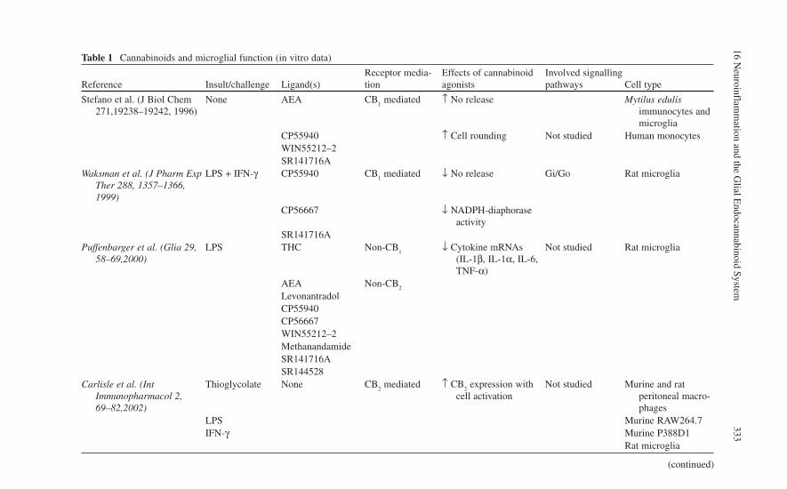
16 Neuroinflam
mation and the G
lial Endocannabinoid System
333
Table 1 Cannabinoids and microglial function (in vitro data)
Reference Insult/challenge Ligand(s)Receptor media-tion
Effects of cannabinoid agonists
Involved signalling pathways Cell type
Stefano et al. (J Biol Chem 271,19238–19242, 1996)
None AEA CB1 mediated ↑ No release Mytilus edulis
immunocytes and microglia
CP55940 ↑ Cell rounding Not studied Human monocytesWIN55212–2SR141716A
Waksman et al. (J Pharm Exp Ther 288, 1357–1366, 1999)
LPS + IFN-γ CP55940 CB1 mediated ↓ No release Gi/Go Rat microglia
CP56667 ↓ NADPH-diaphorase activity
SR141716APuffenbarger et al. (Glia 29,
58–69,2000)LPS THC Non-CB
1↓ Cytokine mRNAs
(IL-1β, IL-1α, IL-6, TNF-α)
Not studied Rat microglia
AEA Non-CB2
LevonantradolCP55940CP56667WIN55212–2MethanandamideSR141716ASR144528
Carlisle et al. (Int Immunopharmacol 2, 69–82,2002)
Thioglycolate None CB2 mediated ↑ CB
2 expression with
cell activationNot studied Murine and rat
peritoneal macro-phages
LPS Murine RAW264.7IFN-γ Murine P388D1
Rat microglia
(continued)

334 C
. Benito et al.
Facchinetti et al. (Glia 41, 161–168,2003
LPS AEA Non-CB1
↓ TNFα release Non Gi/o
mediated Rat microglia
2-AG Non-CB2
CP55940WIN55212–2HU210AM251SR141716ASR144528
Walter et al. (J Neurosci 23, 1398–1405, 2003)
ATP AEA CB2 mediated ↑ Cell migration ERK1/2 BV-2
2-AG abn-CBD mediated Mouse microgliaPEASR141716ASR144528CannabinolCannabidiolO-1918
Klegeris et al. (Br J Pharmacol 139, 775–786, 2003)
LPS + IFN-γ JWH-015 CB2 mediated ↓ IL-1β Not studied THP-1
SR141716A ↓ TNF-α Human microgliaSR144528
Franklin and Stella (Eur J Pharmacol 474, 195–198, 2003)
None ACPA CB2 mediated ↑ Cell migration G
i/G
oBV-2
Cannabinol abn-CBD mediatedCannabidiolO-1918SR141716ASR144528
Table 1 (continued)
Reference Insult/challenge Ligand(s)Receptor media-tion
Effects of cannabinoid agonists
Involved signalling pathways Cell type

16 Neuroinflam
mation and the G
lial Endocannabinoid System
335
Franklin et al. (J Neurosci 23, 7767–7775,2003)
LPS + IFN-γ PEA Non-CB1
↑ Cell migration Gi/G
oBV-2
Non-CB2
Non-abn-CBDNon-WINr
Peterson et al. (J Neuroimmunol 147, 123–126, 2004)
HIV-1SF162
WIN55212–2 Not studied ↓ HIV expression (WIN55212-2) No effect (THC)
Not studied Human fetal microg-lia
THCSR141716ASR144528
Cabral and Marciano-Cabral (J Neuroimmunol 147, 127–130, 2004)
Acanthamoebacastellanii
THC Not studied ↓ Cytokine mRNAs (IL-1β, IL-1α, TNF-α)
Not studied Rat microglia
Gongora et al. (Immunol Lett 91, 11–16, 2004)
IFN-γ CP55,940 CB1 mediated ↓ MHC-II Not studied EOC 20
↓ Class II transactivatorWitting et al. (PNAS 101,
3214–3219, 2004)ATP None Purinergic P2X
7↑ 2-AG PI-PLC Mouse microglia
DG-lipaseERK1 RTMGL1
Carrier et al. (Mol Pharmacol 65, 999–1007, 2004)
M-CSF 2-AG CB2 mediated ↑ Proliferation Not studied Mouse microglia
AEAJWH133SR144528
Ramírez et al. (J Neurosci 25, 1904–1913, 2005)
Aβ25–35 and Aβ1–40
HU-210 CB1 mediated ↓ Morphological
changesWIN55212–2 CB
2 mediated ↓ MTT
JWH-133 ↓ TNT-α↑ Neuronal survival
(continued)

336 C
. Benito et al.
Table 1 (continued)
Reference Insult/challenge Ligand(s)Receptor media-tion
Effects of cannabinoid agonists
Involved signalling pathways Cell type
Ortega-Gutierrez et al. (FASEB J 19, 1338–1340, 2005)
LPS UCM707 CB1 mediated ↓ No Not studied Mouse microglia
OMDM1 CB2 mediated ↓ iNOS
AEA ↓ Cytokines (IL-1β, IL-6, TNF-α)
MethanandamideSR141716ASR144528
Ehrhart et al. (J Neuroinflm 2, 29–42, 2005)
Aβ1–42 JWH-015 CB2 mediated ↓ IFN-γ-mediated CD40
expressionJAK/STATI Mouse microglia
↓ TNF-α↓ NO↑ Phagocytosis
Maresz et al. (J Neurochem 95, 437–445, 2005)
GM-CSF None Not studied ↑ CB2
Not studied Mouse microglia
IFN-γLPS
Eljaschewitsch et al. (Neuron 49, 4–8, 2006)
NMDA AEA CB1 mediated ↑ MKP-1 ERK-1/2 Rat microglia
OGD WIN55212-2 CB2 mediated ↓ No BV-2
AM251 ↓ iNOS OHSCsAM630
Witting et al. (PNAS 103, 6362–6367, 2006)
IFN-γ None Not studied ↓ P2X7–mediated 2-AG DG lipase-α Mouse microglia

16 Neuroinflam
mation and the G
lial Endocannabinoid System
337
Carrier et al. (PNAS 103, 7895–7900, 2006)
None THC Not studied ↓ Nucleoside uptake ENT1 nucleoside transporter
EOC-20
CBD Adenosine A2A
RAW264.7Kreutz et al. (Exp Neurol 203,
246–257, 2007)NMDA THC CB
2 mediated (THC, 2-AG)
↓ Number of microglial cells
Not studied OHSCs
AEA Non-CB2 mediated
(AE)↓ Number of degenerat-
ing neurons (2-AG)2-AGAM630
Mukhopadhyay et al. (J Neuroimmunol 181, 82–92, 2006)
LPS None Not studied ↑ CB2
NF-κB RAW264.7
PKAPKC

338 C
. Benito et al.
Table 2 Cannabinoids and microglial function (in vivo data)
ReferenceInsult/diseasemodel Ligand(s)
Receptormediation
Molecular effects of cannabinoid agonists
Symptomaticeffects of cannabi-noid agonists
Animalspecies
Arevalo-Martin et al. (J Neurosci 23, 2511–2516, 2003)
Theiler’s virus WIN55212 CB1 mediated ¯ Microglial acti-
vationMotor recovery Mouse
ACEA CB2 mediated ¯ MHC-II expres-
sionRemyelination
JWH-015 ¯ CD4+ infiltrationZhang et al. (Eur J
Neurosci 17, 2750–2754, 2003)
Chronic constriction injury
None ↓ CB2 mRNA Not studied Not studied Rat
Freund’s complete adjuvant injection
Spinal nerve ligationFranklin et al. (J
Neurosci 23, 7767–7775, 2003)
Focal cerebral isch-emia
PEA Non-CB1
↓ AEA-induced microglialmigration
Not studied Mouse
Non-CB2
Non-abn-CBDNon-WINr
Cabral and Marciano-Cabral (J Neuroimmunol 147, 127–130, 2004)
Acanthamoeba castel-lanii
THC Not studied ↓ Brain accumula-tion of macro-phages
↓ Mortality Mouse
¯ Cytokine mRNAs (IL-1β, IL-1α,TNF-α)
↓ Severity of dis-ease

16 Neuroinflam
mation and the G
lial Endocannabinoid System
339
Ramírez et al. (J Neurosci 25, 1904–1913, 2005)
Aβ25–35 and Aβ1–40 HU-210 Not studied ¯ Microglial acti-vation
¯ Aβ-induced cog-nitive impair-ment
Rat
WIN55212 ¯ Aβ-induced neu-ronal damage
¯ ¯ Aβ-inducedcognitive impairment
JWH-133Mestre et al. (J
Neurochem 92, 1327–1339, 2005)
Theiler’s virus AEA Not studied ¯ Microglial acti-vation
↓ Motor function Mouse
OMDM1 ¯ MHC-II expres-sion
↓ Motor function
OMDM2Ortega-Gutierrez
et al. (FASEB J 19, 1338–1340, 2005)
Theiler’s virus UCM707 Not studied ¯ Microglial acti-vatioin
↓ Motor function Mouse
¯ MHC-II expression

340 C. Benito et al.
clear that the expression of microglial CB1 receptor remains unaltered after cell
activation, while that of CB2 receptor is dramatically increased when microglial
cells are exposed to many different types of injuries or challenges. Furthermore, CB
2 receptor activation triggers a decrease in the expression and secretion of pro-
inflammatory molecules, such as IL-1 or TNF-α. It must be also considered, however, that other reports suggest a pro-inflammatory CB
2-mediated action on
microglia. Thus, Walter and co-workers (2003) and Franklin and co-workers (2003) reported that CB
2 receptor activation leads to an enhancement of microglial
migration. In addition, Carrier and colleagues (2004) showed a CB2 receptor-
mediated increase in cell proliferation. These responses are classically considered as pro-inflammatory and could then be paradoxical with the previously described anti-inflammatory actions of cannabinoids through their binding to CB
2 receptors.
Nevertheless, it may be argued that these responses are not mutually exclusive, as microglia could proliferate and migrate more rapidly after CB
2 receptor activation
to participate in several processes (such as encapsulation of the site of injury, phagocytosis, etc.) and, at the same time, exhibit a lower ability to produce pro-inflammatory molecules. In this line of reasoning, it can be speculated that up-regulated microglial CB
2 receptors could represent an extraordinarily useful target
for the development of new therapeutics in neuroinflammation. Finally, it is important to note that microglial cells are a major source of endocannabinoids in the brain. According to results obtained by Stella’s group (Stella, 2004), these cells produce almost 20 times more endocannabinoids than astrocytes or neurons do. Furthermore, these authors suggest that microglial cells may be the main produc-ers of endocannabinoids in the inflammatory foci with a subsequent enhancement of local cell recruitment. Interestingly, microglial cells do not exhibit FAAH activity (Franklin et al., 2003).
Endocannabinoid System and Astrocytes
Astrocytes are the most numerous non-neuronal cell type in the CNS (Chen and Swanson, 2003). A well-known function of these cells is to physically structure the brain. A second function is to provide neurons with nutrients such as glucose. The astrocyte end-feet encircling endothelial cells form part of the blood–brain barrier, as they contribute to the formation of tight intercellular junctions between capillary endothelial cells and regulate the expression and function of several endothelial transporters. They perform several essential functions for normal neuronal activity including transmitter’s uptake and release, or modulation of synaptic transmission (Piet et al., 2004). They also serve as intermediaries in neuronal regulation of blood flow (Parri and Crunelli, 2003), glucose metabolism and promotion of the myeli-nating activity of oligodendrocytes. This suggests that astrocytes have an executive-co-ordinating role in the brain (Ishibashi et al., 2006). The presence of CB
1
receptors on astrocytes is well documented. CB1 receptor immunoreactivity has
been described in astrocytes of the nucleus accumbens of Sprague–Dawley rats

16 Neuroinflammation and the Glial Endocannabinoid System 341
(Rodriguez et al., 2001) and also in astrocytes located in the cingulate cortex, the medial forebrain bundle, the amygdala, nucleus accumbens and laminae I and II of the hippocampal dorsal horn of Wistar rats (Moldrich and Wenger, 2000). Perivascular glial fibres have shown moderate to high density of CB
1 protein in
olfactory and limbic structures (Salio et al., 2002) (for further information see Chap. 10). In vitro data have raised some conflicts between findings from different laboratories. Cultures from human astrocytomas tumors of different grades and normal rat astrocytes express the CB
1 receptor (Bouaboula et al., 1995; Sánchez
et al., 1998a). Furthermore, Molina-Holgado and colleagues (2002a) found that cultured astrocytes from CD1 mice express CB
1 receptors, while Walter and Stella
(2003) did not in astrocytes from C57BL/6 mice. Moreover, two studies performed in Swiss-Webster mice astrocytes have shown contradictory results (Sagan et al., 1999; Abood et al., 2001). These discrepancies may reflect differences in the CNS structures used to prepare the astrocytes in culture, differences among species, dis-crepancies in culture systems or conditions, or ages of cultures. It is important to note that CB
1 expression change depending on the differentiation state of cells in
culture (Daaka et al., 1996; Noe et al., 2000). It is still controversial whether astro-cytes express CB
2 receptors. Bouaboula and colleagues (1995) and Walter and
Stella (2003) found that primary rat and mouse astrocytes do not express CB2
receptor. On the contrary, Carlisle and colleagues (2002) and Sheng and colleagues (2005) found CB
2 mRNA on cortical primary rat astrocytes and primary human
astrocytes, respectively. As mentioned for microglial cells, these paradoxical results may be linked to the fact that levels or expression of CB
2 receptor can
change according to cell activation (Carlisle et al., 2002). Immunohistochemical and enzymological data show FAAH expression in human astrocytes. We (Romero et al., 2002) showed FAAH expression in perivascular human astrocytes of the grey and white matter of the human cortex and basal ganglia. In addition, Stella and co-workers (1997) found that mouse astrocytes in culture show FAAH and MAGL protein expression, as well as elicited corresponding enzymatic activities. On the other side, Beltramo and colleagues (1997) found that human astrocytoma cells seem to rapidly metabolize 2-AG. Cannabinoids induce remarkably different effects on astrocytes. When injected into the brain of developing rats, ∆9-THC may interfere with astroglial differentiation in a sex-dependent manner (Suárez et al., 2000, 2002). Interestingly, WIN55212-2 and HU-210 protect primary astrocytes from ceramide-induced apoptosis via activation of PI
3K/Akt and ERK (Gómez del
Pulgar et al., 2002a). It is important to note that, unlike this protective effect on astrocytes, cannabinoids induce apoptosis of glioma cells (Galve-Roperh et al., 2000; Sánchez et al., 2001a; Gómez del Pulgar et al., 2002b). This opposite response to cannabinoids of glioma cells and astrocytes could be based on differ-ences in the regulation of the pathway of de novo ceramide synthesis (Carracedo et al., 2004). Activation of astrocytic CB
1 receptors increases the rate of glucose oxi-
dation to CO2 as well as the rate of glucose incorporation into phospholipids and
glycogen, two phenomena involved in energy supply to the brain (Sánchez et al., 1998b; Blázquez et al., 1999). As perivascular astrocytes are located between cerebral arteries and neurons and regulate energy supply to neighbouring neurons

342 C. Benito et al.
(Magistretti, 2000; Voutsinos-Porche et al., 2003), CB1 receptors present at the
end-feet of astrocytes may regulate energy supply from blood to neurons. In agree-ment with this hypothesis, anandamide and ∆9-THC enhanced the energetic brain metabolism in the rat, probably via the cannabinoid CB
1 receptor (Costa et al.,
2004). Additional data show that CB1 receptor may signal independently of G
i/o
proteins (see Chap. 9 for detailed discussion). Sánchez and colleagues (2001b) reported that ∆9-THC, acting at CB
1 receptors, induced sphingomyelin hydrolysis
in primary astrocytes, an effect not blocked by pertussis toxin. Other reports show that some effects of cannabinoids on astrocytes are not mediated by CB
1 or CB
2
receptors (Venance et al., 1995; Shivachar et al., 1996; Pertwee, 1999; Sagan et al., 1999; Pearlman et al., 2003; Curran et al., 2005). Whether astrocytes produce endo-cannabinoids has remained unknown until recently, mainly because of limited sen-sitivity of the methods used (Di Marzo et al., 1994; Di Tomaso et al., 1997; Stella et al., 1997). However, Walter and colleagues (2002, 2003) recently developed a chemical ionization gas chromatography/mass spectrometry (CI-GC/MS) method that allowed femtomole detection and quantification of anandamide and other acylethanolamides in biological samples. Using this method, these authors detected and quantified anandamide production by mouse astrocytes in culture. Interestingly, endothelin-1 enhanced the production of AEA and 2-AG only (Walter et al., 2002; Walter and Stella, 2003), indicating that activation of different receptor subtypes may selectively increase the production of individual endocannabinoids. Finally, there are a lot of evidence that astrocytes and astrocytoma cell lines inactivate endocannabinoids by uptake and hydrolysis (Beltramo et al., 1997; Deutsch et al., 2000; Muthian et al., 2000; Bisogno et al., 2001; Jonsson et al., 2001).
Endocannabinoid System and Oligodendrocytes
Oligodendrocytes, as myelin-forming cells in the CNS, are responsible for produc-ing the myelin sheath that allows electrical signals to propagate more efficiently. To that end, oligodendroglial processes extend from the cell soma to make contact with axons (Butt and Ransom, 1993). Oligodendrocytes express Ca2+-permeableglutamate receptors and have low resistance to oxidative stress, two factors that make them particularly susceptible to injury (Back et al., 1998). Oligodendrocyte damage compromises brain function, and their injury or death is a prominent fea-ture in demyelinating and neurodegenerative disorders, such as multiple sclerosis. Although the origin of oligodendrocytes capable of remyelinating naked axons is not clear (Levine et al., 2001), oligodendrocyte progenitors exist in the CNS and are recruited to the demyelinated areas to perform the remyelinating process (Keirstead and Blakemore, 1999; Chang et al., 2002). However, survival of prolif-erating oligodendrocyte progenitors and their successful differentiation to myeli-nating oligodendrocytes require an appropriate axon–oligodendrocyte contact (Fernandez et al., 2000) and trophic factors released by neurons and astrocytes (Barres et al., 1992; Gard et al., 1995). Molina-Holgado and co-workers (2002b)
16 Neuroinflammation and the Glial Endocannabinoid System 343
were able to detect CB1 receptor immunoreactivity in all of the different develop-
mental stages of rat oligodendrocytes in vivo and in vitro. In primary culture, oli-godendrocyte progenitors and differentiated oligodendrocytes also expressed CB
2
receptor, whereas CB2 expression was absent in vivo. In addition, they reported that
CB1 and CB
2 receptor activation is involved in protecting oligodendrocyte progeni-
tors from apoptosis, via a mechanism dependent on the PI3K/Akt signaling pathway
(Molina-Holgado et al., 2002b). These results supported data obtained by the same group in an animal model of multiple sclerosis in which cannabinoid treatment reduced the MHC class II-restricted CD4 + T cell response rendering a significant increase in the capacity of remyelinating naked axons (Arévalo-Martin et al., 2003). These authors suggested that cannabinoids may favour myelin repair directly because of both anti-inflammatory actions and effects on oligodendrocyte survival and differentiation. A comparative immunohistochemical study carried out in mouse brain on the distribution of FAAH and CB
1 receptors revealed that only
FAAH is present in fibre tracts, identified as oligodendrocytes (Egertová et al., 2003). In the mid-brain, the expression of FAAH by oligodendrocytes was particu-larly striking and particularly abundant in white matter surrounding the cerebellar nuclei. To date, the functional significance of FAAH expression in these glial cells is unknown. The association of FAAH with oligodendrocytes in fibre tracts is of particular interest considering the reported reduction in FAAH activity in the stria-tum in a rat model of Parkinson’s disease (Gubellini et al., 2002). Egertová and colleagues (2003) speculate that the loss of neuronal inputs may also lead to the loss of associated FAAH-expressing oligodendrocytes, which could account for the reduced levels of striatal FAAH in these experimental rats. Nothing is known about whether oligodendrocytes produce endocannabinoids under basal or stimulated conditions or whether are able to take them up (Witting and Stella, 2004).
Cannabinoids and Neuroinflammation
“Neuroinflammation” may be defined as “chronic, sustained cycles of injury and response, in which the cumulative ill effects of immunological microglial and astrocytic activation contribute to and expand the initial neurodestructive effects, thus maintaining and worsening the disease process through their actions” (Streit et al., 2004). This notion originated in the field of Alzheimer’s disease (Rogers et al., 1988; Griffin et al., 1989), where it revolutionized our understanding of this disease. From then, it has been also applied to other neurodegenerative diseases such as Parkinson’s (McGeer et al., 2001) and Huntington’s diseases (Sapp et al., 2001), HIV encephalopathy (Gendelman et al., 1994), multiple sclerosis (Martino et al., 2002), ischemia (Chopp et al., 1994), traumatic brain injury (Dusart and Schwab, 1994), tumor biology (Graeber et al., 2002) and even to normal brain development. The release of pro-inflammatory and neurotoxic mediators (TNF-α,IL-1β, IL-6, eicosanoids, NO and reactive oxygen species) may induce or aggravate brain damage. These factors are predominantly produced by glial cells (mainly

344 C. Benito et al.
reactive microglia) and can be deleterious to neurons (Boje and Arora, 1992; Chao et al., 1992; McGuire et al., 2001; Liu and Hong, 2003). Neuroinflammation incor-porates a wide spectrum of complex cellular responses that include activation of microglia and astrocytes and elaboration of cytokines and chemokines, comple-ment proteins, acute phase proteins and related molecular processes. These events may have detrimental effects on neuronal function, leading to neuronal injury with further glial activation and, ultimately, neurodegeneration. As described in the pre-vious section, cell types involved in this process express components of the can-nabinoid signalling system that can be endogenously or pharmacologically controlled. Cannabinoid agonists are able to reduce the inflammation that occurs in these diseases. This effect is possibly caused by local effects on glial cells, exerted by either reducing the release of cytotoxic factors or increasing the production of pro-survival molecules (Grundy et al., 2001; Grundy, 2002; Mechoulam et al., 2002; Fowler, 2003; Walter and Stella, 2004). Interestingly, endogenous cannabi-noids are also released under neuroinflammation including brain injury (Hansen et al., 2001; Panikashvili et al., 2001; Franklin et al., 2003; Marsicano et al., 2003; Mechoulam and Lichtman, 2003) and are believed to attenuate neuronal damage by binding to CB
1 receptors and protecting against excitotoxicity. These sustained
increases in endocannabinoid production constitute a defense mechanism prevent-ing the propagation of neuroinflammation and also of cell damage. The neuropro-tective actions of cannabinoids are thought to be mediated through a variety of mechanisms, including antioxidative actions (Hampson et al., 1998), inhibition of NMDA-mediated calcium influx (Mackie and Hille, 1992; Nadler et al., 1993) and inhibition of glutamate release (Shen and Thayer, 1998; Köfalvi et al., 2007). Cannabinoids act on glia and neurons to inhibit the release of proinflammatory molecules, including IL-1, TNF-α and NO (Molina-Holgado et al., 1997,2002a; Shohami et al., 1997; Puffenbarger et al., 2000; Cabral et al., 2001), and enhance the release of the anti-inflammatory cytokines IL-4, IL-10 (Klein et al., 2000), IL-6 (Molina-Holgado et al., 1998) and IL-1ra (Molina-Holgado et al., 2003). Particularly interesting is the inhibitory effect of cannabinoids on the production of TNF-αsince this is a major contributor to the pathophysiology of brain injury (Klein et al., 2000). Croxford and Miller (2003) found that WIN55212-2 decreased CNS mRNA encoding for TNF-α in mice infected with Theiler’s murine encephalomyelitis virus. In another rat model of brain injury, HU-211 was shown to suppress brain levels of TNF-α directly as well as to reduce mortality and improve clinical out-comes (Shohami et al., 1997). Endogenous and synthetic cannabinoids have the ability to ablate the release of TNF-α elicited by LPS in rat primary cortical micro-glial and astroglial cells (Facchinetti et al., 2003; Ortega-Gutierrez et al., 2005). This effect does not appear to be mediated by either CB receptor type 1 or type 2 (Facchinetti et al., 2003). Another important inflammation-related mediator is NO, which is produced in response to immune-mediated cellular toxicity playing a role in neurodegeneration (Guzmán et al., 2001; Walter and Stella, 2004). Different can-nabinoid chemicals inhibit the release of NO in LPS- or TMEV-stimulated astro-cytes, microglia cells or glioma cell line C6 (Molina-Holgado et al., 1997, 2002a, Waksman et al., 1999; Esposito et al., 2001; Ortega-Gutierrez et al., 2005). The

16 Neuroinflammation and the Glial Endocannabinoid System 345
effects on NO levels are, at least in part, due to a direct influence on iNOS expres-sion (Ortega-Gutierrez et al., 2005). However, using BV-2 cells, a mouse microglial cell line, WIN55212-2 did not affect basal release of NO, or modulated the LPS/INF-gamma-induced production of NO (Franklin et al., 2003). IL-1 has been iden-tified as an important mediator of diverse forms of experimentally induced brain damage and is expressed rapidly in response to many forms of experimental brain injury, initially by microglia and later by astrocytes (Davies et al., 1999). Cannabinoid agonists (UCM707 and HU-210) induce a significant reduction in the IL-1β levels produced by LPS-stimulated astrocytes (Ortega-Gutierrez et al., 2005). In mouse mixed glial cultures, Molina-Holgado and colleagues (2003) found that HU-210 and CP55940 increase LPS-induced production of IL-1ra, and SR141716A and SR144528 lowered this response. Interestingly, cannabinoid receptor activation failed to do so in knockout mice for these anti-inflammatory cytokines (Molina-Holgado et al., 2003). IL-1ra is a potent endogenous antagonist of all IL-1 actions in the brain (Dinarello and Thompson, 1991), protecting against ischemic, excito-toxic and traumatic brain insults (Allan and Rothwell, 2001). Furthermore, inhibi-tion or deletion of endogenous IL-1ra enhances ischemic brain injury (Loddick et al., 1997) and increases inflammatory responses (Josephs et al., 2000). Anandamide enhances the release of IL-6 from astrocytes infected with TMEV, the virus that elicits a mouse model of multiple sclerosis, an effect blocked by SR141716A (Molina-Holgado et al., 1998). However, it is opposed to the effect observed in LPS-stimulated astrocytes (Ortega-Gutierrez et al., 2005) where UCM707 dimin-ishes IL-6 levels. This result probably may be in relation with the dual character of this cytokine, which can exhibit either pro- or anti-inflammatory properties depend-ing on different factors such as the simultaneous presence of other cytokines. The production of IL-6 by astrocytes could then be related to the anti-inflammatory and/or neuroprotective roles of this cytokine considering that, for example, astro-cytes secrete nerve growth factor in response to IL-6 (Frei et al., 1989). Recent studies have implicated CB
2 receptors in the neuroprotective activity of cannabi-
noids, mainly through a series of glia-dependent anti-inflammatory actions (Fernández-Ruíz et al., 2005). Several studies show that CB
2 receptor activation
decreases the production of proinflammatory molecules in several neural cell types such as rat microglial cells (Puffenbarger et al., 2000; Facchinetti et al., 2003), human microglial and THP-1 cells (Stella, 2004), and human astrocytes (Sheng et al., 2005). Activation of CB
2 receptors also reduces the release of proinflamma-
tory factors in animal models of perinatal hypoxia-ischemia (Fernández-López et al., 2006) and Huntington’s disease (Fernández-Ruiz and Gonzalez, 2005). The most relevant pro-inflammatory molecules that seem to be under control of the CB
2
receptor include NO, TNF-α, IL-1 and IL-6. In addition, CB2 receptor activation
induce an increase in the release of some anti-inflammatory molecules such as IL-1ra, this molecule may negatively regulate IL-1β (Molina-Holgado et al., 2003). All this data suggest that microglia, astrocytes and oligodendrocytes are sensitive to cannabinoid agonists in different ways, accounting for their anti-inflammatory action. It may be postulated that the beneficial effects on neuroinflammation might be related to several events: inhibition of proinflammatory mediator production,

346 C. Benito et al.
enhancement of anti-inflammatory factor production, inhibition of microglial recruitment and enhancement of astrocyte or oligodendrocyte survival.
The Glial Endocannabinoid System in Human Neurodegenerative Disorders
The distribution of the different components of the endocannabinoid system in the CNS has been extensively studied during the last 15 years. Once the first reports on the existence of a specific receptor protein for cannabinoids were published, the study on its localization was faced. In this sense, elegant autoradiographic studies by Herkenham and co-workers were pioneer (Herkenham et al., 1990, 1991). Thus, by the use of potent and specific radioligands synthesized at Pfizer, these authors obtained evidence on the extensive distribution of cannabinoid CB
1 receptors in the
CNS of several animal species, including humans. Basal ganglia structures, cere-bellar cortex and hippocampus accounted for the most CB
1 receptor-enriched areas
of the brain, explaining some of the most prototypical effects of exogenous can-nabinoids (Herkenham et al., 1991; Glass et al., 1997). Once cloned, mRNA distri-bution studies further confirmed these data and showed that CB
1 receptors were
among the most abundant ones in the CNS and exhibited a presynaptic neuronal distribution (Mailleux and Vanderhaegen, 1992). Herkenham and co-workers were also the first to analyze the distribution of CB
2 receptor, revealing its preferential
distribution in immune cells and tissues and confirming its absence from the CNS under normal conditions (Lynn and Herkenham, 1994). When the first specific antibodies became available, immunohistochemical studies were performed to describe the precise cellular localization of cannabinoid receptors and FAAH in the CNS. Tsou and colleagues (1998a,b) reported immunohistochemical evidence con-firming previous autoradiographic data and describing the predominantly neuronal distribution of these elements of the endocannabinoid system in the murine brain. The first immunohistochemical observations performed in the human brain seemed to corroborate data obtained in other animal species. Both CB
1 receptor and FAAH
were abundantly expressed by neurons throughout the brain, with special relevance in cortical neurons, basal ganglia, cerebellar cortex and large neurons of the spinal cord (see Chap. 10). Interestingly, no glial cells showed CB
1 receptor immunoreac-
tivity, while only scarce white matter astrocytes were positive for FAAH enzyme. Several years later, the preferential expression of FAAH on astrocytes has been extensively corroborated. As mentioned earlier, cannabinoid CB
2 receptors seemed
to be absent from the CNS (Galiegue et al., 1995). Recent data, however, have raised substantial controversy, with some groups reporting a massive presence of this receptor in neuronal elements of the mouse brain (Gong et al., 2006) and others showing a selective, restricted expression of these receptors in neuronal elements of the brainstem of several animal species (Van Sickle et al., 2005). Concerning the human CNS, our group provided immunohistochemical evidence suggesting the presence of CB
2 receptors in a microglial cell subtype, as the perivascular microglia

16 Neuroinflammation and the Glial Endocannabinoid System 347
(Nuñez et al., 2004). These cells exhibit important differences in respect to other types of microglia and are known to play critical roles in blood–brain barrier home-ostasis as well as in pathological states of the CNS (Williams and Hickey, 2002). The selective presence of CB
2 receptors in these cells matched well with the partici-
pation of these receptors in immune-related functions and expanded the field to novel approaches like their possible role in the viral infection of the brain.
Alzheimer’s Disease
Alzheimer’s disease (AD) is one of the most important health challenges in western countries (for details, see Chap. 19). The analysis of human postmortem brain sam-ples from AD patients has provided information on the neuropathology of the endocannabinoid system that raises new hypothesis on the possible role of this sys-tem in the prevention and/or treatment of AD (Benito et al., 2003). Of special rele-vance may be the induction of the expression of CB
2 receptors in microglial cells.
It has only been recently accepted that this type of cannabinoid receptor may be present in the CNS, as previous work circumscribed its presence to peripheral cells and tissues of the immune system (Howlett et al., 2002). Although the functions of these receptors in the CNS are far from clear, they may be now considered as diag-nostic markers for microglial activation and as relevant candidates for the develop-ment of anti-Aβ therapies. The amount of in vitro data in the literature on the anti-inflammatory effects of CB
2 activation, for instance, raises appealing possibili-
ties, as anti-inflammatory compounds are under intense study as putative useful agents for the treatment of AD patients. The modulation of FAAH expression and activity also constitutes an interesting approach. Our current hypothesis suggests that FAAH inhibition may provide benefits for dampening the local inflammatory process triggered by Aβ as it would render more endocannabinoids available for interaction with their receptors. Together with the observed decrease in CB
1 recep-
tor binding and functional coupling in human AD samples (Westlake et al., 1994; Ramirez et al., 2005), the putative psychoactive effects derived from the potentia-tion of the endogenous cannabinoid tone could have a lower impact. In addition, a decrease in FAAH expression and/or activity would also affect the local levels of arachidonic acid, one of the products of FAAH enzymatic activity on AEA, and precursor of a series of potent pro-inflammatory mediators.
HIV-1-Associated Dementia
The syndrome of cognitive and motor dysfunction observed after infection with human HIV-1 has been designated as HIV-associated dementia (HAD). Many experts believe that HAD is now the most common cause of dementia worldwide among people under 40 years of age ( Ellis et al., 1997). There is an incomplete understanding of how HIV

348 C. Benito et al.
infection causes neuronal injury and apoptosis. The principal pathway for HIV entry into the CNS is through infected monocytes, being perivascular macrophages and not the parenchymal microglia the primary cell productively infected (Williams et al., 2001). Some non-productive infection of astrocytes can also occur but it seems well established that neurons are not directly infected. Neuronal injury has been attributed either to a release of neurotoxic factors by HIV-infected microglia/macrophages (and possibly astrocytes) or to neurotoxic HIV proteins (Kaul et al., 2001). HAD is associ-ated pathologically with HIV-1 encephalitis (HIVE). HIVE is characterized by the formation of multinucleated giant cells (through the fusion of inflammatory cells), microglial nodules, infiltration of macrophages from the periphery, widespread astro-cytosis, myelin pallor and neuronal loss (Persidsky and Gendelman, 2003). We have recently addressed the question on the status of the endocannabinoid system after HIV-1 infection of the brain by the analysis of human brain tissue samples from HIVE patients and from macaques infected with the simian variant of this virus (the SIVE model) (Benito et al., 2005). Interestingly, these models allow the direct analy-sis of inflammation in the brain, as samples from infected individuals but without encephalitis are also included. The analysis of human and macaque samples allowed us to conclude that, with little exceptions, a common pattern of inflammation-linked changes in the pattern of expression of cannabinoid receptors and FAAH take place. For the first time, we observed CB
1 receptor positive astrocytes and microglial cells
in HIVE samples. This is an important difference with previous observations in AD tissue samples, where CB
1 receptors remained unchanged in their pattern of expres-
sion. Although the presence of CB1 receptor in astrocytes has been previously
reported in the rat brain (Rodríguez et al., 2001), their induction in astrocytes in the human brain as a consequence of the viral-triggered inflammatory process deserves to be highlighted. In addition, infiltrated T lymphocytes also exhibited high levels of CB
1 receptor immunoreactivity. In contrast, no glial expression of CB
1 receptors
could be noticed in the inflamed macaq ue brain. Possible differences in the inflam-matory response of the human vs. macaque respect CB
1 expression could be explained
by a possible strain-derived virus phenotype. Similar to what seen in AD human samples, the most dramatic changes occur to CB
2 receptors and FAAH. Both ele-
ments of the endocannabinoid system are up-regulated in the inflamed brains of macaques and humans with viral infection. Microglial nodules and infiltrated T lym-phocytes exhibited highest levels of CB
2 expression. Interestingly, perivascular
microglial cells also exhibited elevated levels of CB2 immunoreactivity as a conse-
quence of the viral infection of the brain. These cells are known to play a specific and crucial role in the process of viral entry into the CNS and, thus, it seems reasonable to think that selective CB
2 receptor activation could modify this process. FAAH-
positive astrocytes were found predominantly in perivascular regions and specifically in areas of cellular infiltration. As astrocytes are known to play a regulatory role in HIV-1 encephalitis by dampening the overexpression of eicosanoids, platelet- activating factor, and TNFα by activated HIV-1 monocytes (Minagar et al., 2002), FAAH overexpressed in glial cells could partially counteract some of these benefi-cial processes (Weber et al., 2004).

16 Neuroinflammation and the Glial Endocannabinoid System 349
Multiple Sclerosis
Multiple sclerosis (MS) is an inflammatory and demyelinating disease of the CNS, with unknown ethiology (for details, see Chap. 18). The endocannabinoid system is a current target for the treatment of several diseases, including MS (Pryce and Baker, 2005). Clinical evidence from trials confirms the therapeutic potential of cannabinoids in the treatment of multiple sclerosis symptoms (Zajicek et al., 2003, 2005; Rog et al., 2005). These data are supported by previous results obtained in different animal models of MS that show a relief in certain symptoms of this dis-ease. So far, the treatment of MS has focused only in CB
1 receptor activation.
However, other results (Arevalo-Martin et al., 2003; Yiangou et al., 2006; Benito et al., 2007; Docagne et al., 2007) postulate that other elements, such as CB
2 recep-
tors and FAAH enzyme, are potential therapeutic targets for the treatment of MS. Moreover, the attractive possibility of finding cannabinoid-based therapies for dis-eases devoid of undesired CB
1 receptor-mediated psychotropic side effects is also
opened. Studies performed in human spinal cord and brain MS samples detected strong CB
2 receptor immunoreactivity in microglia/macrophages in white matter
areas, usually within active plaques or in the periphery of chronic lesions (Yiangou et al., 2006; Benito et al., 2007). These results confirm CB
2 expression in glial cells
in the human CNS as previously reported in other neuroinflammatory conditions (Benito et al., 2003, 2005). In the brain lesions of MS donors, abundant CB
1 expres-
sion was also detected in macrophages located within active plaques. Several in vitro studies documented that microglia/macrophages are involved in phagocytosis of myelin debris in MS lesions, and as a result the process triggers release of pro-inflamatory cytokines and NO (Williams et al., 1994; Mosley and Cuzner, 1996; van der Laan et al., 1996). Although little is known on the effects of cannabinoids on myelin phagocytosis, previous reports have shown that the activation of the endocannabinoid system decrease the production of pro-inflammatory cytokines and NO levels in macrophages/microglia, thus accounting for an anti-inflammatory effect that seems to potentiate the neuroprotection induced by cannabinoids (Mestre et al., 2005; Ortega-Gutierrez et al., 2005). The immunohistochemical study carried out by Benito and colleagues (2007) also revealed the expression of the cannabi-noid receptors and the enzyme FAAH in other glial cells. Interestingly, CB
1 recep-
tors were also present in adult oligodendrocytes and OPCs located within MS plaques. These cells are known to be essential to neuroprotection and brain repair since are a key part in the re-myelination process that takes place during the course of the disease (Levine et al., 2001). Previous studies have shown CB
1 and CB
2
expression in the different developmental stages of rat oligodendrocytes in vivo and in vitro (Molina-Holgado et al., 2002b). The activation of these receptors promoted oligodendrocyte survival, via a PI
3K/Akt-dependent mechanism, and thereby
enhanced axonal re-myelination in a MS animal model (Molina-Holgado et al., 2002b; Arevalo-Martin et al., 2003). In addition, CB
1 and CB
2 receptors were
expressed by perivascular T lymphocytes. The myelin-reactive T lymphocytes are thought to be involved in the demyelinating process and to cause inflammation

350 C. Benito et al.
(Frohman et al., 2006). Thus, the presence of both type of receptors in T lym-phocyte is suggestive of a possible role of the endocannabinoid system in MS-linked, T cell-mediated neuroinflammation, since T cells are known to participate in the pathogenesis of MS (Frohman et al., 2006) and cannabinoids decrease CD4+
infiltration into the spinal cord in an animal model of MS through CB1 and CB
2
receptor activation (Arevalo-Martin et al., 2003). In contrast to previous data obtained in other pathologies such as Alzheimer’s disease (Benito et al., 2003; Ramirez et al., 2005), CB
2 expression was also detected in white matter astrocytes,
being the first observation in that type of glial cells in situ in human. There are little data about the role of CB
2 receptors in astrocytes, although in vitro studies suggest
that they may modulate the production of different inflammatory mediators (Ortega-Gutierrez et al., 2005; Sheng et al., 2005). More recently, Docagne and co-workers (2007) have proposed a neuroprotective effect in a MS animal model as a result of the concomitant activation of CB
1 receptor in neurons and CB
2 recep-
tor in astrocytes. As previously reported in other neurinflammatory pathologies (Benito et al., 2003, 2005), the endocannabinoid-degrading enzyme FAAH was overexpressed in reactive astrocytes within MS plaques; therefore, this seems to be a strikingly constant feature of this enzyme. Importantly, other arachidonic acid-related enzymes, such as COX-2 or phospholipase-A
2 are also known to be selec-
tively overexpressed in astrocytes under inflammatory stimuli (Sun et al., 2005). FAAH inhibition could have beneficial effects during inflammation because of decreased local production of arachidonic acid and enhanced endogenous cannabi-noid tone (Benito et al., 2003; Karanian et al., 2005).
Concluding Remarks
It is now accepted that the endocannabinoid system is an endogenous neuro-modulator system that participates in many important processes of the CNS and that acts as a physiological neuroprotectant, both under acute as well as chronic insults. Recent literature shows an increasing attention to in vitro and in vivo animal models of injury in which the activation of the endocannabinoid system results in neuroprotection. In addition, the study of the neuropathology of the endocannabinoid system in the human brain suggests that it may be involved in the neuroinflam-mation that usually takes place in several diseases, such as AD, HIV-1-encepha-litis or MS. Table 3 summarizes some of these findings that confirm a change in the pattern of expression of cannabinoid receptors and FAAH in the chronically damaged human brain. These disease-related modifications suggest a less prominent role for the neuronal CB
1 receptor, the main cannabinoid receptors in the brain,
while are indicative of an emerging role for glial CB2 receptor and FAAH. The
relevance of these findings lies on the possible interest of these elements of the endocannabinoid system as new diagnostic markers as well as possible targets for the development of novel therapies by using cannabinoid chemicals, without undesired psychoactive effects.

16 Neuroinflammation and the Glial Endocannabinoid System 351
Table 3 Endocannabinoid system in neurodegenerative diseases (details and abbreviations in the text)
ControlAlzheimer’s disease
Down’s syn-drome Sive Hive
Multiplesclerosis
CB1
Neurons Neurons Neurons Neurons Neurons neuronsAstrocytes MacrophagesMicroglia Oligodendro-
cytesT lymphocytes
CB2
Perivascular microglia
Activated microglia
Activated microglia
Activated microglia
Activated microglia
Activated microglia
Perivascular microglia
Perivascular microglia
Macrophages
T lympho-cytes
T lympho-cytes
Astrocytes
FAAH Neurons Neurons Neurons Neurons Neurons NeuronsAstrocytes Reactive
astrocytesReactive
astrocytesReactive
astrocytesReactive
astrocytesReactive
astrocytes
References
Abood ME, Rizvi G, Sallapudi N, McAllister S (2001) Activation of the CB1 cannabinoid receptor
protects cultured mouse spinal neurons against excitotoxicity. Neurosci Lett 309:197–201.Allan SM, Rothwell NJ (2001) Cytokines and acute neurodegeneration. Nat Rev Neurosci
2:734–744.Arevalo-Martin A, Vela JM, Molina-Holgado E, Borrell J, Guaza C (2003) Therapeutic action of
cannabinoids in a murine model of multiple sclerosis. J Neurosci 23:2511–2516.Back SA, Gan X, Li Y, Rosenberg PA, Volpe JJ (1998) Maturation-dependent vulnerability of
oligodendrocytes to oxidative stress-induced death caused by glutathione depletion. J Neurosci 18:6241–6253.
Barres BA, Hart IK, Coles HS, Burne JF, Voyvodic JT, Richardson WD, Raff MC (1992) Cell death and control of cell survival in the oligodendrocyte lineage. Cell 70:31–46.
Beltramo M, Stella N, Calignano A, Lin SY, Makriyannis A, Piomelli D (1997) Functional role of high-affinity anandamide transport, as revealed by selective inhibition. Science 277:1094–1097.
Benito C, Nunez E, Tolon RM, Carrier EJ, Rabano A, Hillard CJ, Romero J (2003) Cannabinoid CB2 receptors and fatty acid amide hydrolase are selectively overexpressed in neuritic plaque-associated glia in Alzheimer’s disease brains. J Neurosci 23:11136–11141.
Benito C, Kim WK, Chavarria I, Hillard CJ, Mackie K, Tolon RM, Williams K, Romero J (2005) A glial endogenous cannabinoid system is upregulated in the brains of macaques with simian immunodeficiency virus-induced encephalitis. J Neurosci 25:2530–2536.
Benito C, Romero JP, Tolón RM, Clemente D, Docagne F, Hillard CJ, Guaza C, Romero J (2007) Cannabinoid CB
1 and CB
2 receptors and fatty acid amide hydrolase are specific markers of
plaque cell subtypes in human multiple sclerosis. J Neurosci 27:2396–2402.Bisogno T, Maccarrone M, De Petrocellis L, Jarrahian A, Finazzi-Agrò A, Hillard C, Di Marzo V
(2001) The uptake by cells of 2-arachidonyglycerol, an endogenous agonist of cannabinoid receptors. Eur J Biochem 268:1982–1989.

352 C. Benito et al.
Boje KM, Arora PK (1992) Microglial-produced nitric oxide and reactive nitrogen oxides mediate neuronal cell death. Brain Res 587:250–256.
Bouaboula M, Bourrié B, Rinaldi-Carmona M, Shire D, Le fur G, Casellas P (1995) Stimulation of cannabinoid receptor CB
1 induces krox-24 espression in human astrocytoma cells. J Biol
Chem 270:13973–13980.Butt AM, Ransom BR (1993) Morphology of astrocytes and oligodendrocytes during develop-
ment in the intact rat optic nerve. J Comp Neurol 338:141–158.Cabral GA, Harmon KN, Carlisle SJ (2001) Cannabinoid-mediated inhibition of inducible nitric
oxide production by rat microglial cells: evidence for CB1 receptor participation. Adv Exp
Med Biol 493:207–214.Carlisle SJ, Marciano-Cabral F, Staab A, Ludwick C, Cabral GA (2002) Differential expression
of the CB2 cannabinoid receptor by rodent macrophages and macrophage-like cells in relation
to cell activation. Int Immunopharmacol 2:69–82.Carracedo A, Geelen MJ, Diez M, Hanada K, Guzman M, Velasco G (2004) Ceramide sensi-
tizes astrocytes to oxidative stress: protective role of cannabinoids. Biochem J 380:435–440.
Carrier EJ, Kearn CS, Barkmeier AJ, Breese NM, Yang W, Nithipatikom K, Pfister SL, Campbell WB, Hillard CJ (2004) Cultured rat microglial cells synthesize the endocannabinoid 2- arachidonylglycerol, which increases proliferation via a CB2 receptor-dependent mechanism. Mol Pharmacol 65:999–1007.
Chang A, Tourtellotte WW, Rudick R, Trapp BD (2002) Premyelinating oligodendrocytes in chronic lesions of multiple sclerosis. N Engl J Med 346:165–173.
Chao CC, Hu S, Molitor TW, Shaskan EG, Peterson PK (1992) Activated microglia mediate neu-ronal cell injury via a nitric oxide mechanism. J Immunol 149:2736–2741.
Chen Y, Swanson RA 2003. Astrocytes and brain injury. J Cereb Blood Flow Metab 23:137–149.
Chopp M, Zhang RL, Chen H, Li Y, Jiang N, Rusche JR (1994) Postischemic administration of an anti-Mac-1 antibody reduces ischemic cell damage after transient middle cerebral artery occlusion in rats. Stroke 25:869–875.
Costa B, Colleoni M, Conti S, Parolaro D, Franke C, Trovato AE, Giagnoni G (2004) Oral antiin-flammatory activity of cannabidiol, a non-psychoactive constituent of cannabis, in acute car-rageenan-induced inflammation in the rat paw. Nauyn-Schmiedebergs Arch Pharmacol 369:74–79.
Croxford JL, Miller SD (2003) Immunoregulation of a viral model of multiple sclerosis using the synthetic cannabinoid R + WIN55,212. J Clin Invest 111:1231–1240.
Curran NM, Griffin BD, O’Toole D, Brady KJ, Fitzgerald SN, Moynagh PN (2005) The synthetic cannabinoid R(+)WIN55,212–2 inhibits the interleukin-1 signaling pathway in human astro-cytes in a cannabinoid receptor-independent manner. J Biol Chem 280:35797–35805.
Daaka Y, Friedman H, Klein TW (1996) Cannabinoid receptor proteins are increased in jurkat, human T-cell line after mitogen activation. J Pharmacol Exp Ther 276:776–783.
Davies CA, Loddick SA, Toulmond S, Stroemer RP, Hunt J, Rothwell NJ (1999) The progression and topographic distribution of interleukin-1beta expression after permanent middle cerebral artery occlusion in the rat. J Cereb Blood Flow Metab 19(1):87–98.
Deutsch DG, Glaser ST, Howell JM, Kunz JS, Puffenbarger RA, Hillard CJ, Abumrad N (2000) The cellular uptake of anandamide is coupled to its breakdown by fatty acid amide hydrolase (FAAH). J Biol Chem 276:6967–6973.
Di Marzo V, Fontana A, Cadas H, Shimelli S, Cimino G, Schwart JC, Piommelli D (1994) Formation and inactivation of endogenous cannabinoid anandamide in central neurons. Nature 372:686–619.
Dinarello CA, Thompson RC (1991) Interleukin-1 receptor antagonist competitively inhibits the binding of interleukin-1 to the type II interleukin-1 receptor. J Biol Chem 266:14147–14150.
Di Tomaso E, Cadas H, Gaillet S, Beltramo M, Desarnaud F, Venance L, Piomelli D (1997) Endogenous lipids that activate cannabinoid receptors. Formation and inactivation. Adv Exp Med Biol 407:335–340.

16 Neuroinflammation and the Glial Endocannabinoid System 353
Docagne F, Muneton V, Clemente D, Ali C, Loria F, Correa F, Hernangomez M, Mestre L, Vivien D, Guaza C (2007) Excitotoxicity in a chronic model of multiple sclerosis: neuroprotective effects of cannabinoids through CB
1 and CB
2 receptor activation. Mol Cell Neurosci
34:551–561.Dusart I, Schwab ME (1994) Secondary cell death and the inmflammatory reaction after dorsal
hemisction of the rat spinal cord. Eur J Neurosci 6:712–724.Egertova M, Cravatt BF, Elphick MR (2003) Comparative analysis of fatty acid amide hydrolase
and cb1 cannabinoid receptor expression in the mouse brain: evidence of a widespread role for
fatty acid amide hydrolase in regulation of endocannabinoid signaling. Neuroscience 119:481–496.
Ellis RJ, Deutsch R, Heaton RK, Marcotte TD, McCutchan JA, Nelson JA, Abramson I, Thal LJ, Atkinson JH, Wallace MR, Grant I (1997) Neurocognitive impairment is an independent risk factor for death in HIV infection. Arch Neurol 54:416–424.
Esposito G, Izzo AA, Di Rosa M, and Iuvone T (2001) Selective cannabinoid CB1 receptor-
mediated inhibition of inducible nitric oxide synthase protein expression in C6 rat glioma cells. J Neurochem 78:835–841.
Facchinetti F, Del Giudice E, Furegato S, Passarotto M, Leon A (2003) Cannabinoids ablate release of TNFalpha in rat microglial cells stimulated with lypopolysaccharide. Glia 41:161–168.
Fernandez PA, Tang DG, Cheng L, Prochiantz A, Mudge AW, Raff MC (2000) Evidence that axon-derived neuregulin promotes oligodendrocyte survival in the developing rat optic nerve. Neuron 28:81–90.
Fernandez-Lopez D, Martinez-Orgado J, Nunez E, Romero J, Lorenzo P, Moro MA, Lizasoain I (2006) Characterization of the neuroprotective effect of the cannabinoid agonist WIN-55212 in an in vitro model of hypoxic-ischemic brain damage in newborn rats. Pediatr Res 60:169–73.
Fernandez-Ruiz J, Gonzalez S (2005) Cannabinoid control of motor function at the basal ganglia. Handb Exp Pharmacol 168:479–507.
Fernandez-Ruiz JJ, Gonzalez S, Romero J, Ramos JA (2005) Cannabinoids in neurodegeneration and neuroprotection. In Cannabinoids as Therapeutics, Mechoulam R, ed., Birkhäuser Verlag/Switzerland, pp. 79–109.
Fowler CJ (2003) Plant-derived, synthetic and endogenous cannabinoids as neuroprotective agents. Non-psychoactive cannabinoids, “entourage” compounds and inhibitors of N-acyl eth-anolamine brakdown as therapeutic strategies to avoid pyschotropic effects. Brain res Rev 41:26–43.
Franklin A, Parmentier-Batteur S, Walter L, Greenberg DA, Stella N (2003) Palmitoylethanolamide increases after focal cerebral ischemia and potentiates microglial cell motility. J Neurosci 23:7767–7775.
Frei K, Malipiero Uv, Leist TP, Zinkernagel RM, Schwab ME, Fontana A (1989) On the cellular source and function of interleukin 6 produced in the central nervous system in viral diseases. Eur J Immunol 19:689–694.
Frohman EM, Racke MK, Raine CS (2006) Multiple Sclerosis. The plaque and its pathogenesis. New Eng J Med 354:942–955.
Galiegue S, Mary S, Marchand J, Dussossoy D, Carriere D, Carayon P, Bouaboula M, Shire D, Le Fur G, Casellas P (1995) Expression of central and peripheral cannabinoid receptors in human immune tissues and leukocyte subpopulations. Eur J Biochem 232:54–61.
Galve-Roperh I, Sánchez C, Cortés M, Gómez del Pulgar T, Izquierdo M, Guzmán M (2000) Anti-tumoral action of cannabinoids: involvement of sustained ceramide accumulation and extracel-lular signal-regulated kinase activation. Nat Med 6:313–319.
Gard AL, Burrell MR, Pfeiffer SR, Rudge JS, Williams II WC (1995) Astroglial control of oligodendrocyte survival mediated by PDGF and leukemia inhibitory factor-like protein. Development 121:2187–2197.
Gendelman HE, Genis P, Jett M, Zhai QH, Nottet HS (1994) An experimental model sustem for HIV-1-inudced brain injury. Adv Neuroimmunol 4:189–193.
354 C. Benito et al.
Glass M, Dragunow M, Faull RL (1997) Cannabinoid receptors in the human brain: a detailed anatomical and quantitative autoradiographic study in the fetal, neonatal and adult human brain. Neuroscience 77:299–318.
Gómez del Pulgar T, de CeballoS ML, Guzmán M, Velasco G (2002a) Cannabinoids protect astrocytes from ceramide-induced apoptosis through the phosphatidylinositol 3-kinase B path-way. J Biol Chem 277:36527–36533.
Gómez del Pulgar T, Velasco G, Sánchez C, Haro A, Gumán M (2002b) De novo-synthesized ceramide is involved in cannabinoid-induced apoptosis. Biochem J 363:183–188.
Gong JP, Onaivi ES, Ishiguro H, Liu QR, Tagliaferro PA, Brusco A, Uhl GR (2006) Cannabinoid CB
2 receptors: immunohistochemical localization in rat brain. Brain Res 1071:10–23.
Graeber MB, Scheithauer BW, Kreutzberg GW (2002) Microglia in brain tumors. Glia 40:252–259.
Griffin WS, Stanley LC, Ling C, White L, MacLeod V, Perrot LJ, Whote CL, Araoz C (1989) Brain interleukin 1 and S-100 immunoreactivity are elevated in Down syndrome and Alzheimer disease. Proc Natl Acad Sci 86:7611–7615.
Grundy RI (2002) The therapeutic potential of the cannabinoids in neuroprotection. Expert Opin Investig Drugs 11:1–10.
Grundy RI, Rabuffetti M, Beltramo M (2001) Cannabinoids and neuroprotection. Mol Neurobiol 24:29–51.
Gubellini P, Picconi B, Bari M, Battista N, Calabresi P, Centonze D, Bernardi G, Finazzi-Agro A, Maccarrone M (2002) Experimental parkinsonism alters endocannabinoid degradation: implications for striatal glutamatergic transmission. J Neurosci 22:6900–6907.
Guzmán M, Sanchez C, Galve-Roperh I (2001) Control of the cell survival/death decision by cannabinoids. J Mol Med 78:613–625.
Hampson AJ, Grimaldi M, Axelrod J, Wink D (1998) Cannabidiol and (−)∆9-tetrahydrocannabi-nol are neuroprotective antioxidants. Proc Natl Acad Sci USA 95:8268–8273.
Hansen HH, Schmid PC, Bittigau P, Lastres-Becker I, Berrendero F, Manzanares J, Ikonomidou C, Schmid HH, Fernandez-Ruiz JJ, Hansen HS (2001) Anandamide, but not 2-arachidonoylg-lycerol, accumulates during in vivo neurodegeneration. J Neurochem 78:1415–1427.
Herkenham M, Lynn AB, Little MD, Johnson MR, Melvin LS, De Costa BR, Rice KC (1990) Cannabinoid receptor localization in brain. Proc Natl Acad Sci 87:1932–1936.
Herkenham M, Lynn AB, Johnson MR, Melvin LS, De Costa BR, Rice KC (1991) Characterization and localization of cannabinoid receptors in rat brain: a quantitative in vitro autoradiographic study. J Neurosci 11:563–583.
Howlett AC, Barth F, Bonner TI, Cabral G, Casellas P, Devane WA, Felder CC, Herkenham M, Mackie K, Martin BR, Mechoulam R and Pertwee RG (2002) International Union of Pharmacology. XXVII. Classification of cannabinoid receptors. Pharmacol Rev 54:161–202.
Ishibashi T, Dakin K, Stevens B, Lee P, Kozlov S, Stewart C, Fields R (2006) Astrocytes promote myelination in response to electrical impulses. Neuron 49:823–832.
Jonsson K-O, Vandevoorde S, Lambert DM, Tiger G, Fowler CJ (2001) Effects of homologues and analogues of pamitoylethanolamide upon the inactivation of the endocannabinoid ananda-mide. Br J Pharmacol 133:1263–1275.
Josephs MD, Solorzano CC, Taylor M, Rosenberg JJ, Topping D, Abouhamze A, Mackay SL, Hirsch E, Hirsh D, Labow M, Moldawer LL (2000) Modulation of the acute phase response by altered expression of the IL-1 type 1 receptor or IL-1ra. Am J Physiol Regul Integr Comp Physiol 278:R824–R830.
Karanian DA, Brown QB, Makriyannis A, Kosten TA, Bahr BA (2005) Dual modulation of endo-cannabinoid transport and fatty acid amide hydrolase protects against excitotoxicity. J Neurosci 25:7813–7820.
Kaul M, Garden GA, Lipton SA (2001) Pathways to neuronal injury and apoptosis in HIV- associated dementia. Nature 410:988–994.
Keirstead HS, Blakemore WF (1999) The role of oligodendrocytes and oligodendrocyte progeni-tors in CNS remyelination. Adv Exp Med Biol 468:183–197.

16 Neuroinflammation and the Glial Endocannabinoid System 355
Klein TW, Lane B, Newton CA, Friedman H (2000) The cannabinoid system and cytokine net-work. Proc Soc Exp Biol Med 225:1–8.
Köfalvi A, Pereira MF, Rebola N, Rodrigues RJ, Oliveira CR, Cunha RA (2007) Anandamide and NADA bi-directionally modulate presynaptic Ca2+ levels and transmitter release in the hip-pocampus. Br J Pharmacol doi:10.1038/sj.bjp.0707252.
Levine JM, Reynolds R, Fawcett JW (2001) The oligodendrocyte precursor cell in health and disease. Trends Neurosci 24:39–47.
Liu B, Hong JS (2003) Role of microglia in inflammation-mediated neurodegenerative deseases: mechanisms and strategies for therapeutic intervention. J Pharmacol Exp Ther 304:1–7.
Loddick SA, Wong ML, Bongiorno PB, Gold PW, Licinio J, Rothwell NJ (1997) Endogenous inter-leukin-1 receptor antagonist is neuroprotective. Biochem Biophys Res Commun 234:211–215.
Lynn AB, Herkenham M (1994) Localization of cannabinoid receptors and nonsaturable high- density cannabinoid binding sites in peripheral tissues of the rat: implications for receptor- mediated immune modulation by cannabinoids. J Pharmacol Exp Ther 268:1612–1623.
Mackie K, Hille B (1992) Cannabinoids inhibit N-type calcium channels in neuroblastoma-glioma cells. Proc Natl Acad Sci USA 89:3825–3829.
Magistretti P (2000) Cellular bases of functional bran imaging: insights from neuron-glia meta-bolic coupling. Brain Res 886:108–112.
Mailleux P, Vanderhaeghen JJ (1992) Distribution of neuronal cannabinoid receptor in the adult rat brain: a comparative receptor binding radioautography and in situ hybridization histochem-istry. Neuroscience 48:655–668.
Marsicano G, Goodenough S, Monory K, Hermann H, Eder M, Cannich A, Azad SC, Cascio MG, Gutierrez SO, van der Stelt M, Lopez-Rodriguez ML, Casanova E, Schutz G, Zieglgansberger W, Di Marzo V, Behl C, Lutz B (2003) CB
1 cannabinoid receptors and on-demand defense
against excitotoxicity. Science 302:84–88.Martino G, Adorini L, Rieckmann P, Hillert J, Kallmann B, Comi G, Filippi M. (2002)
Inflammation in multiple sclerosis the good, the bad, and the complex. Lancet Neuro 1:499–509.
McCoy KL, Matveyeva M, Carlisle SJ, Cabral GA (1999) Cannabinoid inhibition of the process-ing of intact lysozyme by macrophages: evidence for CB
2 receptor participation. J Pharmacol
Exp Ther 289:1620–1625.McGeer PL, Yasojima K, McGeer EG (2001) Inflammation in Parkinson’s disease. Adv Neuro
86:83–89.Mc Guire SO, Ling ZD, Lipton JW, Sortwell CE, Collier TJ, Carvey P (2001) Tumor necrosis
factor alpha is toxic to embryonic mesencephalic dopamine neurons. Exp Neurol 169:219–230.
Mechoulam R, Lichtman AH (2003) Neuroscience. Stout guards of the central nervous system. Science 302:65–67.
Mechoulam R, Panikashivili A, Shohami E (2002) Cannabinoids and brain injury: therapeutic implications. Trend Mol Med 8:58–61.
Mestre L, Correa F, Arevalo-Martin A, Molina-Holgado E, Valenti M, Ortar G, Di Marzo V, Guaza C (2005) Pharmacological modulation of the endocannabinoid system in a viral model of multiple sclerosis. J Neurochem 92:1327–1339.
Minagar A, Shapshak P, Fujimura R, Ownby R, Heyes M, Eisdorfer C (2002) The role of macrophage/microglia and astrocytes in the pathogenesis of three neurologic disorders: HIV- associated dementia, Alzheimer disease, and multiple sclerosis. J Neurol Sci 202:13–23.
Moldrich G, Wenger T (2000) Localization of the CB1 cannabinoid recptor in the rat brain. An
immunohistochemical study. Peptides 21:1735–1742.Molina-Holgado F, Lledo A, Guaza C (1997) Anandamide suppresses nitric oxide and TNF-alpha
responses to Theiler’s virus or endotoxin in astrocytes. Neuroreport 8:1929–1933.

356 C. Benito et al.
Molina-Holgado F, Molina-Holgado E, Guaza C (1998) The endogenous cannabinoid anandamide potentiates interleukin-6 production by astrocytes infected with Theiler’s murine encephalo-myelitis virus by a receptor-mediated pathway. FEBS Lett 433:139–142.
Molina-Holgado F, Molina-Holgado E, Guaza C, Rothwell NJ (2002a) Role of CB1 and CB2
receptors in the inhibitory effects of cannabinoids on lipopolysaccharde-induced nitric oxide release in astrocytes cultures. J Neurosci Res 67:829–836.
Molina-Holgado E, Vela JM, Arevalo-Martin A, Almazan G, Molina-Holgado F, Borrell J, Guaza C (2002b) Cannabinoids promote oligodendrocyte progenitor survival: involvement of cannabi-noid receptors and phosphatidylinositol-3 kinase/Akt signaling. J Neurosci 22:9742–9753.
Molina-Holgado F, Pinteaux E, Moore JD, Molina-Holgado E, Guaza C, Gibson RM, Rothwell NJ (2003) Endogenous interleukin-1 receptor antagonist mediates anti-inflammatory and neu-roprotective actions of cannabinoids in neurons and glia. J Neurosci 23:6470–6474.
Mosley K, Cuzner ML (1996) Receptor-mediated phagocytosis of myelin by macrophages and microglia: effect of opsonization and receptor blocking agents. Neurochem Res 21:481–487.
Muthian S, Nithipatikom K, Campbell WB, Hillard CJ (2000) Synthesis and characterization of a fluorescent substrate for the N-arachidonoylethanolamine (anandamide) transmembrane car-rier. J Pharmacol Exp Ther 293:289–295.
Nadler V, Mechoulam R, Sokolovsky M (1993) Blockade of 45Ca2+ influx through the N- methyl-d-aspartate receptor ion channel by the non-psychoactive cannabinoid HU-211. Brain Res 622:79–85.
Nimmerjahn A, Kirchhoff F, Helmchen F (2005) Resting microglial cells are highly dynamic sur-veillants of brain parenchyma in vivo. Science 308:1314–1318.
Noe SN, Newton C, Widen R, Friedman H, Klein TW (2000) Anti-CD40, anti-CD3, and IL-2 stimulation induce contrasting changes in CB
1 mRNA expression in mouse splenocytes.
J Neroimmunol 110:161–167.Nunez E, Benito C, Pazos MR, Barbachano A, Fajardo O, Gonzalez S, Tolon RM, Romero
J (2004) Cannabinoid CB2 receptors are expressed by perivascular microglial cells in the
human brain: an immunohistochemical study. Synapse 53:208–213.Ortega-Gutierrez S, Molina-Holgado E, Guaza C (2005) Effect of anandamide uptake inhibition
in the production of nitric oxide and in the release of cytokines in astrocyte cultures. Glia 52:163–168.
Panikashvili D, Simeonidou C, Ben Shabat S, Hanus L, Breuer A, Mechoulam R, and Shohami E (2001) An endogenous cannabinoid (2-AG) is neuroprotective after brain injury. Nature 413:527–531.
Parri R, Crunelli V (2003) An astrocyte bridge from synapse to blood flow. Nat Neurosci 6:5–6.Pearlman RJ, Aubrey KR, Vandenberg RJ (2003) Arachidonic acid and anandamide have opposite
modulatory actions at the glycine transporter, GLYT1a
. J Neurochem 84:592–601.Persidsky Y, Gendelman HE (2003) Mononuclear phagocyte immunity and the neuropathogene-
sis of HIV-1 infection. J Leukoc Biol 74:691–701.Pertwee RG (1999) Evidence for the presence of CB
1 cannabinoid receptors on peripheral
neurones and for the existence of neuronal non-CB1 cannabinoid receptors. Life Sciences
65:597–605.Piet R, Vargová L, Syková E, Poulain D, Oliet S (2004) Physiological contribution of the astro-
cytic environment of neurons to intersynaptic crosstalk. Proc Natl Acad Sci USA 101:2151–2155.
Pryce G, Baker D (2005) Emerging properties of cannabinoid medicines in management of multi-ple sclerosis. Trends Neurosci 28:272–276.
Puffenbarger RA, Boothe AC, Cabral GA (2000) Cannabinoids inhibit LPS-inducible cytokine mRNA expression in rat microglial cells. Glia 29:58–69.
Ramirez BG, Blazquez C, Gomez GP, Guzman M, De Ceballos ML (2005) Prevention of Alzheimer’s disease pathology by cannabinoids: neuroprotection mediated by blockade of microglial activation. J Neurosci 25:1904–1913.
Rodriguez JJ, Mackie K, Pickel VM (2001) Ultrastructural localization of the CB1 cannabinoid recp-
tor in µopioid recptor patches of the rat caudate putamen nucleus. J Neurosci 21:823–833.

16 Neuroinflammation and the Glial Endocannabinoid System 357
Rog DJ, Nurmikko TJ, Friede T, Young CA (2005) Randomized, controlled trial of cannabis-based medicine in central pain in multiple sclerosis. Neurology 65:812–819.
Rogers J, Luber-Narod J, Styren SD, Civin WH (1988) Expression of immune system-associated antigens by cells of the human central nervous system: relationship to the pathology of Alzheimer’s disease. Neurobiol Aging 9:339–349.
Romero J, Hillard CJ, Calero M, Rabano A (2002) Fatty acid amide hydrolase localization in the human central nervous system: an immunohistochemical study. Brain Res Mol Brain Res 100:85–93.
Sagan S, Venance L, Torrens Y, Cordier J, Glowinski J, Giaume C (1999) Anandamide and WIN 55212–2 inhibit cyclic AMP formation through G-protein-coupled recptors distinct from CB
1
cannabinoid receptors in cultured astrocytes. Eur J Neurosci 11:691–699.Salio C, Doly S, Fischer J, Franzoni MF, Conrath M (2002) Neuronal and astrocytic localization
of the cannabinoid recptor-1 in the dorsal horn of the rat spinal cord. Neurosci Lett 329:13–16.
Sánchez C, Galve-Roperh I, Canova C, Brachet P, Guzmán M (1998a) Delta 9-tetrahydrocannabi-nol induces apoptosis in a C6 glioma cells. FEBS Lett 436:6–10.
Sánchez C, Galve-Roperh I, Rueda D, Guzmán M (1998b) Involvement of sphingomyelin hydrolysis and the mitogen-activated protein kinase cascade in the delta9-tetrahydrocannabi-nol-induced stimulation of glucose metabolism in primary astrocytes. Mol Pharmacol 54:834–843.
Sánchez C, de Ceballos ML, Gómez del Pulgar T, Rueda D, Corbacho C, velasco G, Galve-Roperh I, Huffman JWH, Ramón y Cajal S, Guzmán M (2001a) Inhibition of glioma growth in vivo by selective activation of the CB
2 cannabinoid receptor. Cancer Res
61:5784–5789.Sánchez C, Rueda D, Segui B, Galve-Roperh I, Levade T, Guzmán M (2001b) The CB1 cannabi-
noid receptor of astrocytes in coupled to sphingomyelin hydrolysis through the adaptor protein Fan. Mol Pharmacol 59:955–959.
Sapp E, kegel KB, Aronin N, Hashikawa T, Uchiyama Y, Tohyama K, Bhide PG, Vonsattel JP, DiFiglia M (2001) Early and progressive accumulation of reactive microglia in the Huntington disease brain. J Neuropathol Exp Neurol 60:161–172.
Shen M, Thayer SA (1998) The cannabinoid agonist Win55,212–2 inhibits calcium channels by receptor-mediated and direct pathways in cultured rat hippocampal neurons. Brain Res 783:77–84.
Sheng WS, Hu S, min X, Cabral GA, Lokensgard JR, Peterson P (2005) Synthetic cannabinoid WIN55,212-2 inhibits generation of inflammatory mediators by IL-1beta stimulated human astrocytes. Glia 49:211–219.
Shivachar AC, Martin BR, Ellis EF (1996) Anandamide- and delta9-tetrahydrocannabinol-evoked arachidonic acid mobilization and blockade by SR141716A [N-(Piperidin-1-yl)-5-(4-chlo-rophenyl)-1-(2,4-dichlorophenyl)-4-methyl-1H-pyrazole-3-carboximide hydrochloride]. Biochem Pharmacol 51:669–676.
Shohami E, Gallily R, Mechoulam R, Bass R, Ben-Hur T (1997) Cytokine production in the brain following closed head injury: dexanabinol (HU-211) is a novel TNF-alpha inhibitor and an effective neuroprotectant. J Neuroimmuno 72:169–177.
Stefano GB, Liu Y, Goligorsky MS (1996) Cannabinoid receptors are coupled to nitric oxide release in invertebrate immunocytes, microglia, and human monocytes. J Biol Chem 271:19238–19242.
Stella N (2004) Cannabinoid signaling in glial cells. Glia 48:267–277.Stella N, Schweitzer P, Piomelli D (1997) A second endogenous cannabinoid that modulates long-
term potentiation. Nature 388:773–778.Streit WJ (2005) Microglia and neuroprotection: implications for Alzheimer’s disease. Brain Res
Rev 48:234–239.Streit WJ, Mrak RE, Griffin WS (2004) Microglia and neuroinflammation: a pathological per-
spective. J Neuoinflammation 1:14.

358 C. Benito et al.
Suárez I, Bodega G, Ramos JA, Fernández-Ruiz JJ, Fernández B (2000) Neuronal and astroglial response to pre- and perinatal exposure to delta-9-tetrahydrocannabinol in the rat substantia nigra. Dev Neurosci 22:253–263.
Suárez I, Bodega G, Fernández-Ruiz JJ, Ramos JA, Rubio M, Fernández B (2002) Reduced glial fibrillary acidic protein and glutamine synthase expression in astrocytes and Bergmann glial cells in the rat cerebellum caused by delta-9-tetrahydrocannabinol administration during development. Dev Neurosci 24:300–312.
Sun GY, Xu J, Jensen MD, Yu S, Wood WG, Gonzalez FA, Simonyi A, Sun AY, Weisman GA. (2005) Phospholipase A
2 in astrocytes: responses to oxidative stress, inflammation, and
G protein-coupled receptor agonists. Mol Neurobiol 31:27–41.Tsou K, Brown S, Sanudo-Pena MC, Mackie K, Walker JM (1998a) Immunohistochemical distribu-
tion of cannabinoid CB1 receptors in the rat central nervous sytem. Neuroscience 83:393–411.
Tsou K, Nogueron MI, Muthian S, Sanudo-Pena MC, Hillard CJ, Deutsch DG, Walker JM (1998b) Fatty acid amide hydrolase is located preferentially in large neurons in the rat central nervous system as revealed by immunohistochemistry. Neurosci Lett 254:137–140.
Van der Laan LJ, Ruuls SR, Weber KS, Lodder IJ, Dopp EA, Dijkstra CD (1996) Macrophage phagocytosis of myelin in vitro determined by flow cytometry: phagocytosis is mediated by CR3 and induces production of tumor necrosis factor-alpha and nitric oxide. J Neuroimmunol 70:145–152.
Van Sickle MD, Duncan M, Kingsley PJ, Mouihate A, Urbani P, Mackie K, Stella N, Makriyannis A, Piomelli D, Davison JS, Marnett LJ, Di Marzo V, Pittman QJ, Patel KD, Sharkey KA (2005) Identification and functional characterization of brainstem cannabinoid CB
2 receptors.
Science 310:329–332.Venance L, Piomelli D, Glowinski J, Giaume C (1995) Inhibition by anandamide of gap junctions
and intercellular calcium signalling in striatal astrocytes. Nature 376:590–594.Voutsinos-Porche B, Bonvento G, Tanaka K, Steiner P, Welker E, Chatton J-Y, Magistretti PJ,
Pellerin L (2003) Glial glutamate transporters mediate a functional metabolic crosstalk between neurons and astrocytes in the mouse developing cortex. Neuron 37:275–286.
Waksman Y, Olson J.M, Carlisle SJ, Cabral GA (1999) The central cannabinoid receptor (CB1)
mediates inhibition of nitric oxide production by rat microglial cells. J Pharmacol Exp Ther 288:1357–1366.
Walter L, Stella N (2003) Endothelin-1 increases 2-arachidonyl glycerol (2-AG) production in astrocytes. Glia 44:85–90.
Walter L, Franklin A, Witting A, Moller T, Stella N (2002) Astrocytes in culture produce ananda-mide and other acylethanolamides. J Biol Chem 277:20869–20876.
Walter L, Franklin A, Witting A, Wade C, Xie Y, Kunos G, Mackie K, Stella N (2003) Nonpsychotropic cannabinoid receptors regulate microglial cell migration. J Neurosci 23:1398–1405.
Weber A, Ni J, Ling KH, Acheampong A, Tang-Liu DD, Burk R, Cravatt BF, Woodward D (2004) Formation of prostamides from anandamide in FAAH knockout mice analyzed by HPLC with tandem mass spectrometry. J Lipid Res 45:757–763.
Westlake TM, Howlett AC, Bonner TI, Matsuda LA, Herkenham M (1994) Cannabinoid receptor binding and messenger RNA expression in human brain: an in vitro receptor autoradiography and in situ hybridization histochemistry study of normal aged and Alzheimer’s brains. Neuroscience 63:637–652.
Williams K, Ulvestad E, Waage A, Antel JP, McLaurin J (1994) Activation of adult human derived microglia by myelin phagocytosis in vitro. J Neurosci Res 38:433–443.
Williams KC, Hickey WF (2002) Central nervous system damage, monocytes and macrophages, and neurological disorders in AIDS. Annu Rev Neurosci 25:537–562.
Witting A, Stella N (2004) Cannabinoid signaling in glial cells in health and disease. Current Neuropharmacol 2.
Yiangou Y, Facer P, Durrenberger P, Chessell IP, Naylor A, Bountra C, Banati RR, Anand P (2006) COX-2, CB2
and P2X7-immunoreactivities are increased in activated microglial cells/macro-
phages of multiple sclerosis and amyotrophic lateral sclerosis spinal cord. BMC Neurol 6:12.

16 Neuroinflammation and the Glial Endocannabinoid System 359
Zajicek J, Fox P, Sanders H, Wright D, Vickery J, Nunn A, Thompson A (2003) Cannabinoids for treatment of spasticity and other symptoms related to multiple sclerosis (CAMS study): multi-centre randomised placebo-controlled trial. Lancet 362:1517–1526.
Zajicek JP, Sanders HP, Wright DE, Vickery PJ, Ingram WM, Reilly SM, Nunn AJ, Teare LJ, Fox PJ, Thompson AJ (2005) Cannabinoids in multiple sclerosis (CAMS) study: safety and effi-cacy data for 12 months follow up. J Neurol Neurosurg Psychiatry 76:1664–1669.

Abstract Cannabinoids, the active components of Cannabis sativa L., act in the body by mimicking endogenous substances – the endocannabinoids – that activate specific cell surface receptors. Cannabinoids exert various palliative effects in can-cer patients. In addition, cannabinoids inhibit the growth of different types of tumor cells, including glioma cells, in laboratory animals. They do so by modulating key cell signaling pathways, mostly the endoplasmic reticulum stress response, thereby inducing antitumoral actions such as the apoptotic death of tumor cells and the inhibition of tumor angiogenesis. Of interest, cannabinoids seem to be selective antitumoral compounds as they kill glioma cells but not their nontransformed astro-glial counterparts. On the basis of these preclinical findings, a pilot clinical study of D9-tetrahydrocannabinol (D9-THC) in patients with recurrent glioblastoma mul-tiforme has been recently run. The fair safety profile of D9-THC, together with its possible growth-inhibiting action on tumor cells, may set the basis for future trials aimed at evaluating the potential antitumoral activity of cannabinoids.
Introduction
Gliomas are defined as those tumors that display histological, immunohistochemical, and ultrastructural evidence of glial differentiation. The World Health Organization classifies gliomas according to their cellular features (i.e., resembling astrocytes, oli-godendrocytes, or ependymal cells) and their grade of malignancy (from I to IV) (Kleihues et al., 2002). Glioblastoma multiforme (GBM), or grade IV astrocytoma, is the most frequent class of malignant primary brain tumors and one of the most aggressive forms of cancer. As a consequence, survival after diagnosis is normally just 6–12 months (Kleihues et al., 2002; Reardon and Wen, 2006). This dramatic behavior is mainly due to the high invasiveness and proliferation rate of GBM. In addition, GBM exhibits a high resistance to common chemotherapy and radiotherapy. These malignant features may be related to the varying mutations frequently found in these tumors that impact different key pathways involved in the control of cell prolif-eration, survival, differentiation, and DNA repair (Maher et al., 2001; Kleihues et al., 2002; Reardon and Wen, 2006). Current standard therapeutic strategies for the
Chapter 17Targeting Cannabinoid Receptors in Brain Tumors
Guillermo Velasco, Arkaitz Carracedo, Cristina Blázquez, Mar Lorente, Tania Aguado, Cristina Sánchez, Ismael Galve-Roperh, and Manuel Guzmán
A. Köfalvi (ed.), Cannabinoids and the Brain. 361© Springer 2008

362 G. Velasco et al.
treatment of GBM are only palliative, and include surgical resection and focal radio-therapy. A large number of chemotherapeutic agents (e.g., alkylating agents such as temozolomide and nitrosureas such as carmustine) have also been tested, but no remarkable improvement on patient survival has been achieved as yet (Lonardi et al., 2005; Reardon and Wen, 2006). Likewise, although dendritic cell- and peptide-based immunotherapy strategies appear promising as a safe approach to induce an antitu-mor immune response (Yamanaka, 2006), no immunotherapy or gene therapy trial performed to date has been significantly successful. It is therefore essential to develop new therapeutic strategies for the management of GBM, which will most likely require a combination of therapies to obtain significant clinical results. Here we sum-marize the current knowledge on how a new family of compounds, the cannabinoids, exerts antiglioma actions in laboratory animals, and how a potential cannabinoid-based therapy for GBM might be envisaged.
Cannabinoids and Their Receptors
The hemp plant Cannabis sativa L. produces approximately 70 unique compounds known as cannabinoids, of which D9-THC is the most important owing to its high potency and abundance in cannabis (Gaoni and Mechoulam, 1964). D9-THC exerts a wide variety of biological effects by mimicking endogenous substances – the endo-cannabinoids, anandamide, and 2-arachidonoylglycerol (2-AG) – that bind to and activate specific cannabinoid receptors (see Chaps. 2 and 7). So far, two types of can-nabinoid-specific G
i/o protein-coupled receptors, CB
1 and CB
2, have been cloned and
characterized from mammalian tissues (Howlett et al., 2002). Most of the effects of cannabinoids rely on CB
1 receptor activation. CB
1 receptors are particularly abundant
in discrete areas of the brain and peripheral nerve terminals, where they mediate endo-cannabinoid-dependent neuromodulation (Piomelli, 2003), but are also expressed in many extraneural sites. In contrast, CB
2 receptors were first described in cells and tis-
sues of the immune system and have been long believed to be absent from the brain. Recent data, however, question this notion and support the existence of CB
2 receptors
in the central nervous system, specifically in microglial cells, astrocytes, some neuron subpopulations, and glioma cells (Fernández-Ruiz et al., 2007; see Chap. 10). Extensive molecular and pharmacological studies have demonstrated that cannabi-noids inhibit adenylyl cyclase through CB
1 and CB
2 receptors. The CB
1 receptor also
modulates ion channels, inducing, for example, inhibition of N- and P/Q-type voltage-sensitive Ca2+ channels and activation of G protein-coupled K+ channels (Howlett et al., 2002). Besides these well-established cannabinoid receptor-coupled signaling events, cannabinoid receptors also modulate several pathways that are more directly involved in the control of cell proliferation and survival, including extracellular signal-regulated kinase (ERK) (Bouaboula et al., 1995), c-Jun N-terminal kinase and p38 mitogen-activated protein kinase (Liu et al., 2000; Rueda et al., 2000), phosphatidyli-nositol 3-kinase (PI
3K)/Akt (Gómez del Pulgar et al., 2000), focal adhesion kinase
(Derkinderen et al., 1996), and the sphingomyelin cycle (Sanchez et al., 2001).

17 Targeting Cannabinoid Receptors in Brain Tumors 363
Antitumoral Activity of Cannabinoids
Cannabinoids have been known for several decades to exert palliative effects in cancer patients, and nowadays capsules of D9-THC (Marinol™) and its synthetic analogue nabilone (Cesamet™) are approved to treat nausea and emesis associated with cancer chemotherapy (Tramer et al., 2001; see Chap. 13). In addition, several clinical trials are testing other potential palliative properties of cannabinoids in oncology such as appetite stimulation and analgesia (Guzmán, 2003; Hall et al., 2005). Besides these palliative actions, cannabinoids have been proposed as poten-tial antitumoral agents on the basis of experiments performed both in cultured cells and in animal models of cancer. These antiproliferative properties of cannabis com-pounds were first reported 30 years ago, when it was shown that D9-THC inhibits lung adenocarcinoma cell growth in vitro and after oral administration in mice (Munson et al., 1975). Although these observations were promising, further studies in this area were not performed until the late 1990s, mostly by Di Marzo’s group (Bifulco and Di Marzo, 2002) and Guzmán’s group (Guzmán, 2003). A number of plant-derived (for example, D9-THC and cannabidiol), synthetic (for example, WIN55212-2 and HU-210), and endogenous cannabinoids (for example, anandam-ide and 2-AG) are now known to exert antiproliferative actions on a wide spectrum of tumor cells in culture (Guzmán, 2003). More importantly, cannabinoid adminis-tration to nude mice curbs the growth of various types of tumor xenografts, includ-ing lung carcinoma (Munson et al., 1975), glioma (Galve-Roperh et al., 2000), thyroid epithelioma (Bifulco et al., 2001), lymphoma (McKallip et al., 2002), skin carcinoma (Casanova et al., 2003), pancreatic carcinoma (Carracedo et al., 2006a), and melanoma (Blázquez et al., 2006). The requirement of cannabinoid receptors for this antitumoral activity has been revealed by various biochemical and pharmaco-logical approaches, in particular by determining cannabinoid receptor expression in the tumors and by using selective cannabinoid receptor agonists and antagonists.
Antitumoral Activity of Cannabinoids in Gliomas
Most of our research on cannabinoid antitumoral action has focused on gliomas. Initial experiments in cultured glioma cells showed that incubation with cannabi-noids induces cell death by an apoptotic mechanism (Sánchez et al., 1998). Further studies with animal models showed that local administration of D9-THC or WIN55212-2 reduced the size of tumors generated by intracranial inoculation of C6 glioma cells in rats, leading to complete eradication of gliomas and increased sur-vival in one third of the treated rats (Galve-Roperh et al., 2000). Additional studies used tumor xenografts generated by subcutaneous injection of glioma cells in the flank of immune-deficient mice. Local administration of D9-THC, WIN55212-2, or the selective CB
2 cannabinoid receptor agonist JWH133 decreased the growth of
tumors derived not only from the rat glioma C6 cell line, but also from GBM cells

364 G. Velasco et al.
obtained from tumor biopsies of patients (Galve-Roperh et al., 2000; Sánchez et al., 2001a,b). These and other studies also showed that cannabinoid receptor activation on glioma cells modulates key signaling pathways involved in cell proliferation and survival. Although the downstream events by which cannabinoids exert their anti-tumoral action in gliomas are not completely unraveled, there is substantial evi-dence for the implication of at least two mechanisms: induction of apoptosis of tumor cells and inhibition of tumor angiogenesis (Fig. 1).
Induction of Apoptosis
Cannabinoids induce apoptosis of cultured glioma cells (Sanchez et al., 1998; Galve-Roperh et al., 2000). Different studies have shown that this effect relies on the activation of cannabinoid receptors and the accumulation of the proapoptotic sphingolipid ceramide (Galve-Roperh et al., 2000; Gómez del Pulgar et al., 2002a,b; Ogretmen and Hannun, 2004). However, the molecular mechanisms involved in the triggering of the apoptotic signal by cannabinoids have started to be unraveled only very recently. By using a DNA array approach, we have identi-fied a series of genes that are selectively upregulated in cannabinoid-sensitive but
Fig. 1 Antitumoral effect of cannabinoids in gliomas. Cannabinoid administration to mice decreases the growth of gliomas by several mechanisms, including at least (i) reduction of tumor angiogenesis, (ii) induction of tumor cell apoptosis, and perhaps (iii) inhibition of tumor cell migration and invasiveness

17 Targeting Cannabinoid Receptors in Brain Tumors 365
not cannabinoid-resistant glioma cells upon D9-THC treatment (Carracedo et al., 2006b). One of these genes was the stress-regulated protein p8 (also designated candidate of metastasis 1 – Com-1), that belongs to the family of HMG-I/Y tran-scription factors and was originally described as a gene induced in acute pancreati-tis (Mallo et al., 1997). Different experimental approaches confirmed that p8 upregulation is essential for the proapoptotic and antitumoral action of cannabi-noids in gliomas and pancreatic tumors (Carracedo et al., 2006a,b). The acute increase of p8 levels after cannabinoid treatment triggers a cascade of events that involves the upregulation of the activating transcription factor 4 (ATF-4) and the C/EBP-homologous protein (CHOP, also called DDIT3 and GADD153). These two transcription factors cooperate in the induction of the tribbles homologue 3 (TRB3, also called TRIB3), a pseudokinase that has been implicated in the induc-tion of apoptosis of tumor cells and neurons (Ohoka et al., 2005). In line with this observation, selective knock-down of ATF-4 and TRB3 prevented cannabinoid-induced apoptosis indicating that this signaling route also operates in glioma cells after treatment with cannabinoids (Carracedo et al., 2006b) (Fig. 2). ATF-4, CHOP, and TRB3 (together with other genes selectively induced upon D9-THC treatment of glioma cells) (Carracedo et al., 2006b) participate in the endoplasmic reticulum (ER) stress response. A series of ER alterations such as calcium depletion, protein
Fig. 2 Mechanism of cannabinoid proapoptotic action in glioma cells. Cannabinoid treatment induces apoptosis of glioma cells via ceramide accumulation and activation of an ER stress-related pathway. The stress-regulated protein p8 plays a key role in this effect by controlling the expression of ATF-4, CHOP, and TRB3. This cascade of events triggers the activation of the mitochondrial intrinsic apoptotic pathway through mechanisms that have not been unraveled as yet. Cannabinoids also decrease the expression of various tumor-progression molecules such as VEGF and MMP2

366 G. Velasco et al.
misfolding, and impairment of protein trafficking to the Golgi triggers this response, which involves attenuation of protein synthesis and selective transcrip-tion and translation of a series of genes, mainly involved in favoring correct protein folding (Schroder and Kaufman, 2005). When these ER alterations cannot be repaired by the ER stress response, the damaged cells undergo apoptosis. Several stimuli, including ischemia (Tajiri et al., 2004), viral infection (Li and Holbrook, 2004; Benali-Furet et al., 2005), and drugs such as tunicamycin (Ohoka et al., 2005) or cisplatin (Mandic et al., 2003), induce apoptosis through this pathway. Of interest, cannabinoid-induced ceramide accumulation and ER stress induction seem to be closely linked. Thus, inhibition of ceramide synthesis de novo prevents D9-THC-induced p8, ATF-4, CHOP, and TRB3 upregulation (Carracedo et al., 2006b) as well as ER dilation (authors’ unpublished observations), indicating that ceramide accumulation is an early event in cannabinoid-triggered ER stress and apoptosis in glioma cells. Unlike this proapoptotic action of cannabinoids on transformed cells, treatment of primary cultured astrocytes with these compounds triggers neither ceramide accumulation (Carracedo et al., 2004) nor the induction of the aforemen-tioned ER stress-related genes (Carracedo et al., 2006b) (Fig. 3). Furthermore, can-nabinoids promote the survival of astrocytes (Gómez del Pulgar et al., 2002a,b), oligodendrocytes (Molina-Holgado et al., 2002), and neurons (Mechoulam et al.,
Fig. 3 Synergy of cannabinoids and endoplasmic reticulum stress inducers. Cannabinoid-induced activation of the ER stress proapoptotic pathway is blunted in cannabinoid-resistant glioma cells. This resistance can be overcome by cotreatment with cannabinoids and ER stress-inducing drugs. Cannabinoids protect astrocytes from different proapoptotic stimuli rather than activate the ER stress pathway in them

17 Targeting Cannabinoid Receptors in Brain Tumors 367
2002) in different models of injury, suggesting that the antiproliferative effect of cannabinoids is selective for brain tumor cells, the viability of normal brain cells being unaffected or even favored by cannabinoid challenge. The processes down-stream of ER stress activation involved in the execution of cannabinoid-induced apoptosis of glioma cells are only partially understood. Decreased mitochondrial membrane potential and caspase 3 activation are observed in cannabinoid-treated glioma cells (Ellert-Miklaszewska et al., 2005; Carracedo et al., 2006b), suggesting that execution of apoptosis occurs via activation of the mitochondrial intrinsic pathway (Fig. 2), a mechanism that is involved in the induction of apoptosis by cannabinoids in other types of tumor cells (Lombard et al., 2005; Herrera et al., 2006). Cannabinoid treatment induces loss of mitochondrial membrane potential in p8+/+ but not p8-deficient mouse embryonic fibroblasts, suggesting that the p8- regulated pathway described above is required for the activation of the mitochon-drial proapoptotic pathway. On the other hand, cannabinoids inhibit Akt in glioma cells, an effect that is prevented by pharmacological blockade of ceramide synthesis de novo (Gómez del Pulgar et al., 2002a,b). In addition, cannabinoids lead to decreased phosphorylation of the BH3-only protein Bad (Ellert-Miklaszewska et al., 2005), an Akt and extracellular signal-regulated protein kinase (ERK) cascade target which phosphorylation inhibits apoptosis via the intrinsic pathway. These observations suggest that regulation of Akt could be involved in the connection between the ceramide/p8-regulated pathway and the activation of the mitochondrial proapoptotic route (Fig. 2). Modulation of ERK, as well as of the other mitogen-activated protein kinases, could also participate in the induction of apoptosis by cannabinoids in gliomas (Galve-Roperh et al., 2000). Intriguingly, both inhibition (e.g., Ellert-Miklaszewska et al., 2005) and activation (e.g., Galve-Roperh et al., 2000) of ERK have been proposed to participate in this effect. Further research is therefore necessary to clarify the involvement of this signaling cascade in cannabi-noid-induced apoptosis.
Inhibition of Tumor Angiogenesis
To grow beyond minimal size, tumors must generate a new vascular supply (ang-iogenesis) for purposes of cell nutrition, gas exchange, and waste disposal, and therefore blocking the angiogenic process constitutes one of the most promising antitumoral approaches currently available. Immunohistochemical analyses in mouse models of glioma (Blázquez et al., 2003), skin carcinoma (Casanova et al., 2003), and melanoma (Blázquez et al., 2006) have shown that cannabinoid admin-istration turns the vascular hyperplasia characteristic of actively growing tumors to a pattern of blood vessels characterized by small, differentiated, and impermeable capillaries. This is associated with a reduced expression of vascular endothelial growth factor (VEGF) and other proangiogenic cytokines such as angiopoietin-2 and placental growth factor (Blázquez et al., 2003; Casanova et al., 2003; Portella et al., 2003), as well as of type 1 (Portella et al., 2003) and type 2 (Blázquez et al.,

368 G. Velasco et al.
2004) VEGF receptors, in cannabinoid-treated tumors. Pharmacological inhibition of ceramide synthesis de novo abrogates the antitumoral and antiangiogenic effect of cannabinoids in vivo and decreases VEGF production by glioma cells in vitro and by gliomas in vivo (Blázquez et al., 2004), indicating that ceramide plays a general role in cannabinoid antitumoral action. Other reported effects of cannabi-noids might be related with the inhibition of tumor angiogenesis and invasiveness by these compounds (Fig. 1). Thus, activation of cannabinoid receptors on vascu-lar endothelial cells in culture inhibits cell migration and survival (Blázquez et al., 2003). Endothelial cell apoptosis was also potently triggered by cannabinoid quinonoid derivatives, although this action seems to be cannabinoid receptor- independent (Kogan et al., 2006). In addition, cannabinoid administration to glioma-bearing mice decreases the activity and expression of matrix metallopro-teinase-2 (MMP2), a proteolytic enzyme that allows tissue breakdown and remod-eling during angiogenesis and metastasis (Blázquez et al., 2003). In line with this notion, cannabinoid intraperitoneal injection reduces the number of metastatic nodes produced from paw injection of lung (Portella et al., 2003), breast (Grimaldi et al., 2006), and melanoma (Blázquez et al., 2006) cancer cells in mice.
Other Potential Targets of Cannabinoid Action
The identification of the cell(s) of origin of gliomas is still a matter of debate. Although neoplastic transformation of differentiated glial cells was for many years the most accepted hypothesis to explain the origin of gliomas, recent findings support the existence of a stem cell-derived origin for different types of cancers such as gliomas, hematopoietic, breast, and prostate tumors (Jordan et al., 2006). In particu-lar, glioma-derived stem-like cells, which may represent the consequence of transfor-mation of the normal neural stem cell compartment, seem to be crucial for the malignancy of gliomas (Vescovi et al., 2006). We have recently shown that glioma stem-like cells derived from GBM biopsies and glioma cell lines express CB
1 and
CB2 receptors, which activation promotes cell differentiation and inhibit gliomagene-
sis (Aguado et al., 2007). Interestingly, gene array experiments indicated that cannabinoid receptor activation on glioma stem-like cells downregulates epidermal growth factor (EGF) and fibroblast growth factor (FGF) receptors, in line with the suggestion that cannabinoids mediate at least part of their apoptotic actions on skin and prostate cancer cells by attenuating EGF receptor expression (Casanova et al., 2003; Mimeault et al., 2003) and/or tyrosine kinase activity (Casanova et al., 2003). In addition, the antiproliferative action of cannabinoids in breast, prostate, and thyroid cancer cells may involve a decrease in the activity and/or expression of prolactin (De Petrocellis et al., 1998), nerve growth factor (Melck et al., 2000), and type 1 VEGF receptors (Portella et al., 2003). Furthermore, cannabinoids inhibit type 2 VEGF receptor activation in glioma cells (Blázquez et al., 2004). Taken together, these results indicate that attenuation of the signaling through tyrosine kinase recep-tors may constitute a common mechanism of cannabinoid growth-inhibiting action.

17 Targeting Cannabinoid Receptors in Brain Tumors 369
Cannabinoids as Potential Therapeutic Agents for the Treatment of Gliomas
On the basis of the aforementioned preclinical findings, we have recently conducted a pilot phase I clinical trial in which nine patients with actively growing recurrent GBM were administered D9-THC intratumorally (Guzmán et al., 2006). The patients had pre-viously failed standard therapy (surgery and radiotherapy) and had clear evidence of tumor progression. The primary endpoint of the study was to determine the safety of intracranial D9-THC administration. D9-THC action on length of survival and various tumor cell parameters was also evaluated. A dose escalation regime for D9-THC admin-istration was assessed. The initial dose of D9-THC delivered to the patients was 20–40µg at day 1, increasing progressively for 2–5 days up to 80–180 mg/day. The median duration of D9-THC administration was 15 days. Under these conditions, cannabinoid delivery was safe and could be achieved without significant psychoactive effects. Median survival of the cohort from the beginning of cannabinoid administration was 24 weeks (95% CI: 15–33). D9-THC decreased tumor cell proliferation (as determined by Ki67 immunostaining; (Guzmán et al., 2006) ) and increased tumor cell apoptosis (as determined by active-caspase 3 immunostaining; (Carracedo et al., 2006b) ) when administered to two patients. The fair safety profile observed for D9-THC, together with its possible antiproliferative action on tumor cells, may set the basis for future trials aimed at evaluating the potential antitumoral activity of cannabinoids. These possible new trials could involve one or more of the following modifications:
Patients with Newly Diagnosed Tumors
Pilot placebo-controlled trials for recurrent glioblastoma multiforme with temo-zolomide, a DNA-damaging agent that constitutes the current benchmark for the management of malignant gliomas, showed a very slight impact on overall length of survival (median survival = 24 weeks; 6-month survival = 46–60%) (Dinnes et al., 2002). Further trials in patients with newly diagnosed tumors allowed a clear improvement in the therapeutic efficacy of temozolomide through the development of various administration regimes (Lonardi et al., 2005; Stupp et al., 2005; Reardon and Wen, 2006). It is therefore conceivable that better outcomes could also be obtained with cannabinoid-based therapies in newly diagnosed gliomas.
D9-THC in Combination with Temozolomide
Glioblastoma multiforme – particularly when relapse occurs – is an extremely lethal dis-ease. The success of potential treatments is usually hampered by factors such as the rapid growth, remarkable heterogeneity, high degree of infiltration, and extreme resistance to

370 G. Velasco et al.
chemotherapy displayed by these tumors. It is therefore conceivable that combined ther-apies could provide better results than single-agent therapies. For example, by synergiz-ing via complementary signaling pathways, D9-THC plus temozolomide might exert a more potent clinical impact than either D9-THC or temozolomide alone.
Noninvasive Administration Route
Although intratumoral delivery may allow a high local concentration of the drug in situ, in the case of large tumors, such as actively growing recurrent glioblastoma multifome, the local perfusion through a catheter placed at one point of the tumor constitutes an obvious limitation of the technique. In addition, a noninvasive, less traumatic route would be more desirable in clinical practice. Alternative or complementary options for D9-THC administration would include oral capsules and oro-mucosal sprays.
Other Cannabinoid Ligands
Although the use of cannabinoids in medicine may be limited by their well-known psychotropic effects, it is generally believed that cannabinoids display a fair drug safety profile and that their potential adverse effects are within the range of those accepted for other medications, especially in cancer treatment (Guzmán, 2003; Hall et al., 2005). In line with this idea, D9-THC delivery in the aforementioned clinical study was safe and could be achieved without overt psychoactive effects. As the possi-ble antitumoral action of nabilone has never been evaluated preclinically, D9-THC remains as the unique cannabinoid receptor agonist currently available for cancer clini-cal trials. Nonetheless, most likely, D9-THC is not the most appropriate cannabinoid agonist for future antitumoral strategies owing to its high hydrophobicity, relatively weak agonistic potency, and ability to elicit CB
1 receptor-mediated psychoactivity.
Unfortunately, the current synthetic cannabinoid agonists that have been reported to exert antitumoral actions in animal models and that could theoretically circumvent – at least in part the pharmacokinetic and pharmacodynamic limitations of D9-THC, e.g., WIN55212-2, a more potent and less hydrophobic mixed CB
1/CB
2 receptor agonist
(Galve-Roperh et al., 2000), and JWH133, a more potent CB2 receptor-selective ago-
nist (Sánchez et al., 2001a,b) – are still very far from the clinical application owing to the lack of thorough preclinical toxicology studies.
Other Types of Tumors
As mentioned above, we and others have shown that D9-THC and synthetic cannabi-noids, besides their antiglioma activity, inhibit the growth of different types of tumor
17 Targeting Cannabinoid Receptors in Brain Tumors 371
xenografts in mice (see above). Trials on these and other types of tumors might also be run to test the antitumoral activity of cannabinoids in these malignant diseases.
Concluding Remarks
One of the most striking features of gliomas is their high resistance to conventional chemotherapy. Nowadays, it is widely believed that strategies aimed at reducing the mortality caused by these tumors should consist of targeted therapies capable of providing the most efficacious treatment for each individual patient and tumor. This new therapeutic approach would require not only the utilization of new cock-tails of chemotherapeutic drugs but, more importantly, the identification of the markers associated with the resistance of tumor cells to these new therapies. The significant antiproliferative action of cannabinoids in animal models of gliomas, together with their low toxicity compared with other chemotherapeutic agents, might make these compounds promising new tools for the management of GBM. Studies performed in our laboratory suggest that resistance of glioma cells to can-nabinoid treatment correlates with the ability of these cells to block the activation of the ER stress pathway (authors’ unpublished observations). In addition, we have observed that agents that induce ER stress exert a synergic action when adminis-tered with cannabinoids (Carracedo et al., 2006b). Likewise, overexpression of p8 or TRB3 sensitizes resistant glioma cells to a further treatment with cannabinoids (Carracedo et al., 2006b). These observations suggest that activation of this route may be investigated as a potential strategy to enhance the response of gliomas to chemotherapy. Research to be performed during the next few years should help to clarify which are the optimal conditions of cannabinoid utilization by identifying the factors that confer resistance to cannabinoid treatment as well as the most effi-cient approaches for enhancing their antitumoral activity.
Acknowledgments We are indebted to the rest of our lab colleagues for their collaboration and encouragement. Research in our laboratory is financially supported by Ministerio de Educación y Ciencia, Comunidad Autónoma de Madrid, Ministerio de Sanidad y Consumo, Fundación de Investigación Médica Mutua Madrileña Automovilística, and Santander/Complutense.
References
Aguado T, Carracedo A, Julien B, Velasco G, Milman G, Mechoulam R, Álvarez L, Guzmán M, Galve-Roperh I (2007) Cannabinoids inhibit glioma stem-like cell differentiation and inhibit gliomagenesis. J Biol Chem 282:6854–6862.
Benali-Furet NL, Chami M, Houel L, De Giorgi F, Vernejoul F, Lagorce D, Buscail L, Bartenschlager R, Ichas F, Rizzuto R, Paterlini-Brechot P (2005) Hepatitis C virus core trig-gers apoptosis in liver cells by inducing ER stress and ER calcium depletion. Oncogene 24:4921–4933.

372 G. Velasco et al.
Bifulco M, Di Marzo V (2002) Targeting the endocannabinoid system in cancer therapy: a call for further research. Nat Med 8:547–550.
Bifulco M, Laezza C, Portella G, Vitale M, Orlando P, De Petrocellis L, Di Marzo V (2001) Control by the endogenous cannabinoid system of ras oncogene-dependent tumor growth. FASEB J 15:2745–2747.
Blázquez C, Casanova ML, Planas A, Gómez del Pulgar T, Villanueva C, Fernandez-Acenero MJ, Aragones J, Huffman JW, Jorcano JL, Guzmán M (2003) Inhibition of tumor angiogenesis by cannabinoids. FASEB J 17:529–531.
Blázquez C, Gonzalez-Feria L, Alvarez L, Haro A, Casanova ML, Guzman M (2004) Cannabinoids inhibit the vascular endothelial growth factor pathway in gliomas. Cancer Res 64:5617–5623.
Blázquez C, Carracedo A, Barrado L, Real PJ, Fernandez-Luna JL, Velasco G, Malumbres M, Guzman M (2006) Cannabinoid receptors as novel targets for the treatment of melanoma. FASEB J 20:2633–2635.
Bouaboula M, Poinot-Chazel C, Bourrie B, Canat X, Calandra B, Rinaldi-Carmona M, Le Fur G, Casellas P (1995) Activation of mitogen-activated protein kinases by stimulation of the central cannabinoid receptor CB
1. Biochem J 312:637–641.
Carracedo A, Geelen MJ, Diez M, Hanada K, Guzman M, Velasco G (2004) Ceramide sensitizes astrocytes to oxidative stress: protective role of cannabinoids. Biochem J 380:435–440.
Carracedo A, Gironella M, Lorente M, Garcia S, Guzman M, Velasco G, Iovanna JL (2006a) Cannabinoids induce apoptosis of pancreatic tumor cells via endoplasmic reticulum stress-related genes. Cancer Res 66:6748–6755.
Carracedo A, Lorente M, Egia A, Blázquez C, Garcia S, Giroux V, Malicet C, Villuendas R, Gironella M, Gonzalez-Feria L, Piris MA, Iovanna JL, Guzman M, Velasco G (2006b) The stress-regulated protein p8 mediates cannabinoid-induced apoptosis of tumor cells. Cancer Cell 9:301–312.
Casanova ML, Blazquez C, Martinez-Palacio J, Villanueva C, Fernandez-Acenero MJ, Huffman JW, Jorcano JL, Guzman M (2003) Inhibition of skin tumor growth and angiogenesis in vivoby activation of cannabinoid receptors. J Clin Invest 111:43–50.
De Petrocellis L, Melck D, Palmisano A, Bisogno T, Laezza C, Bifulco M, Di Marzo V (1998) The endogenous cannabinoid anandamide inhibits human breast cancer cell proliferation. Proc Natl Acad Sci USA 95:8375–8380.
Derkinderen P, Toutant M, Burgaya F, Le Bert M, Siciliano JC, de Franciscis V, Gelman M, Girault JA (1996) Regulation of a neuronal form of focal adhesion kinase by anandamide. Science 273:1719–1722.
Dinnes J, Cave C, Huang S, Milne R (2002) A rapid and systematic review of the effectiveness of temozolomide for the treatment of recurrent malignant glioma. Br J Cancer 86:501–505.
Ellert-Miklaszewska A, Kaminska B, Konarska L (2005) Cannabinoids down-regulate PI3K/Akt
and Erk signalling pathways and activate proapoptotic function of Bad protein. Cell Signal 17:25–37.
Fernández-Ruiz J, Romero J, Velasco G, Tolon R, Ramos J, Guzmán M (2007) Cannabinoid CB2
receptor: a new target for controlling neural cell survival? Trends Pharmacol Sci 28:39–45.Galve-Roperh I, Sanchez C, Cortes ML, Gómez del Pulgar T, Izquierdo M, Guzmán M (2000)
Anti-tumoral action of cannabinoids: involvement of sustained ceramide accumulation and extracellular signal-regulated kinase activation. Nat Med 6:313–319.
Gaoni Y, Mechoulam R (1964) Isolation, structure and partial synthesis of an active constituent of hashish. J Am Chem Soc 86:1646–1647.
Gómez del Pulgar T, Velasco G, Guzman M (2000) The CB1 cannabinoid receptor is coupled to
the activation of protein kinase B/Akt. Biochem J 347:369–373.Gómez del Pulgar T, De Ceballos ML, Guzmán M, Velasco G (2002a) Cannabinoids protect
astrocytes from ceramide-induced apoptosis through the phosphatidylinositol 3-kinase/protein kinase B pathway. J Biol Chem 277:36527–36533.
Gómez del Pulgar T, Velasco G, Sánchez C, Haro A, Guzman M (2002b) De novo-synthesized ceramide is involved in cannabinoid-induced apoptosis. Biochem J 363:183–188.

17 Targeting Cannabinoid Receptors in Brain Tumors 373
Grimaldi C, Pisanti S, Laezza C, Malfitano AM, Santoro A, Vitale M, Caruso MG, Notarnicola M, Iacuzzo I, Portella G, Di Marzo V, Bifulco M (2006) Anandamide inhibits adhesion and migra-tion of breast cancer cells. Exp Cell Res 312:363–373.
Guzmán M (2003) Cannabinoids: potential anticancer agents. Nat Rev Cancer 3:745–755.Guzmán M, Duarte MJ, Blazquez C, Ravina J, Rosa MC, Galve-Roperh I, Sanchez C, Velasco G,
Gonzalez-Feria L (2006) A pilot clinical study of delta-9-tetrahydrocannabinol in patients with recurrent glioblastoma multiforme. Br J Cancer 95:197–203.
Hall W, Christie M, Currow D (2005) Cannabinoids and cancer: causation, remediation, and pal-liation. Lancet Oncol 6:35–42.
Herrera B, Carracedo A, Diez-Zaera M, Gomez del Pulgar T, Guzmán M, Velasco G (2006) The CB
2 cannabinoid receptor signals apoptosis via ceramide-dependent activation of the mito-
chondrial intrinsic pathway. Exp Cell Res 312:2121–2131.Howlett AC, Barth F, Bonner TI, Cabral G, Casellas P, Devane WA, Felder CC, Herkenham M,
Mackie K, Martin BR, Mechoulam R, Pertwee RG (2002) International Union of Pharmacology. XXVII. Classification of cannabinoid receptors. Pharmacol Rev 54:161–202.
Jordan CT, Guzmán ML, Noble M (2006) Cancer stem cells. N Engl J Med 355:1253–1261.Kleihues P, Louis DN, Scheithauer BW, Rorke LB, Reifenberger G, Burger PC, Cavenee WK
(2002). The WHO classification of tumors of the nervous system. J Neuropathol Exp Neurol 61:215–225.
Kogan NM, Blázquez C, Alvarez L, Gallily R, Schlesinger M, Guzman M, Mechoulam R (2006) A cannabinoid quinone inhibits angiogenesis by targeting vascular endothelial cells. Mol Pharmacol 70:51–59.
Li J, Holbrook NJ (2004) Elevated gadd153/chop expression and enhanced c-Jun N-terminal pro-tein kinase activation sensitizes aged cells to ER stress. Exp Gerontol 39:735–744.
Liu J, Gao B, Mirshahi F, Sanyal AJ, Khanolkar AD, Makriyannis A, Kunos G (2000) Functional CB
1 cannabinoid receptors in human vascular endothelial cells. Biochem J 346:835–840.
Lombard C, Nagarkatti M, Nagarkatti PS (2005) Targeting cannabinoid receptors to treat leukemia: role of cross-talk between extrinsic and intrinsic pathways in Delta9-tetrahydrocannabinol (THC)-induced apoptosis of Jurkat cells. Leuk Res 29:915–922.
Lonardi S, Tosoni A, Brandes AA (2005) Adjuvant chemotherapy in the treatment of high grade gliomas. Cancer Treat Rev 31:79–89.
Maher EA, Furnari FB, Bachoo RM, Rowitch DH, Louis DN, Cavenee WK, DePinho RA (2001) Malignant glioma: genetics and biology of a grave matter. Genes Dev 15:1311–1333.
Mallo GV, Fiedler F, Calvo EL, Ortiz EM, Vasseur S, Keim V, Morisset J, Iovanna JL (1997) Cloning and expression of the rat p8 cDNA, a new gene activated in pancreas during the acute phase of pancreatitis, pancreatic development, and regeneration, and which promotes cellular growth. J Biol Chem 272:32360–32369.
Mandic A, Hansson J, Linder S, Shoshan MC (2003) Cisplatin induces endoplasmic reticulum stress and nucleus-independent apoptotic signaling. J Biol Chem 278:9100–9106.
McKallip RJ, Lombard C, Fisher M, Martin BR, Ryu S, Grant S, Nagarkatti PS, Nagarkatti M (2002) Targeting CB
2 cannabinoid receptors as a novel therapy to treat malignant lymphoblas-
tic disease. Blood 100:627–634.Mechoulam R, Spatz M, Shohami E (2002) Endocannabinoids and neuroprotection. Sci STKE
2002:RE5.Melck D, De Petrocellis L, Orlando P, Bisogno T, Laezza C, Bifulco M, Di Marzo V (2000)
Suppression of nerve growth factor Trk receptors and prolactin receptors by endocannabinoids leads to inhibition of human breast and prostate cancer cell proliferation. Endocrinology 141:118–126.
Mimeault M, Pommery N, Wattez N, Bailly C, Henichart JP (2003) Anti-proliferative and apop-totic effects of anandamide in human prostatic cancer cell lines: implication of epidermal growth factor receptor down-regulation and ceramide production. Prostate 56:1–12.
Molina-Holgado E, Vela JM, Arevalo-Martin A, Almazan G, Molina-Holgado F, Borrell J, Guaza C (2002) Cannabinoids promote oligodendrocyte progenitor survival: involvement of cannabi-noid receptors and phosphatidylinositol-3 kinase/Akt signaling. J Neurosci 22:9742–9753.

374 G. Velasco et al.
Munson AE, Harris LS, Friedman MA, Dewey WL, Carchman RA (1975) Antineoplastic activity of cannabinoids. J Natl Cancer Inst 55:597–602.
Ogretmen B, Hannun YA (2004). Biologically active sphingolipids in cancer pathogenesis and treatment. Nat Rev Cancer 4:604–616.
Ohoka N, Yoshii S, Hattori T, Onozaki K, Hayashi H (2005) TRB3, a novel ER stress-inducible gene, is induced via ATF4-CHOP pathway and is involved in cell death. EMBO J 24:1243–1255.
Piomelli D (2003) The molecular logic of endocannabinoid signalling. Nat Rev Neurosci 4:873–884.
Portella G, Laezza C, Laccetti P, De Petrocellis L, Di Marzo V, Bifulco M (2003) Inhibitory effects of cannabinoid CB
1 receptor stimulation on tumor growth and metastatic spreading:
actions on signals involved in angiogenesis and metastasis. FASEB J 17:1771–1773.Reardon DA, Wen PY (2006) Therapeutic advances in the treatment of glioblastoma: rationale and
potential role of targeted agents. Oncologist 11:152–164.Rueda D, Galve-Roperh I, Haro A, Guzman M (2000) The CB
1 cannabinoid receptor is coupled
to the activation of c-Jun N-terminal kinase. Mol Pharmacol 58:814–820.Sánchez C, Galve-Roperh I, Canova C, Brachet P, Guzmán M (1998) Delta-9-tetrahydrocannabinol
induces apoptosis in C6 glioma cells. FEBS Lett 436:6–10.Sánchez C, de Ceballos ML, del Pulgar TG, Rueda D, Corbacho C, Velasco G, Galve-Roperh I,
Huffman JW, Ramon y Cajal S, Guzmán M (2001a) Inhibition of glioma growth in vivo by selective activation of the CB
2 cannabinoid receptor. Cancer Res 61:5784–5789.
Sánchez C, Rueda D, Segui B, Galve-Roperh I, Levade T, Guzmán M (2001b) The CB1 cannabi-
noid receptor of astrocytes is coupled to sphingomyelin hydrolysis through the adaptor protein fan. Mol Pharmacol 59:955–959.
Schroder M, Kaufman RJ (2005). The mammalian unfolded protein response. Annu Rev Biochem 74:739–789.
Stupp R, Mason WP, van den Bent MJ, Weller M, Fisher B, Taphoorn MJ, Belanger K, Brandes AA, Marosi C, Bogdahn U, Curschmann J, Janzer RC, Ludwin SK, Gorlia T, Allgeier A, Lacombe D, Cairncross JG, Eisenhauer E, Mirimanoff RO (2005) Radiotherapy plus concomi-tant and adjuvant temozolomide for glioblastoma. N Engl J Med 352:987–996.
Tajiri S, Oyadomari S, Yano S, Morioka M, Gotoh T, Hamada JI, Ushio Y, Mori M (2004) Ischemia-induced neuronal cell death is mediated by the endoplasmic reticulum stress pathway involving CHOP. Cell Death Differ 11:403–415.
Tramer MR, Carroll D, Campbell FA, Reynolds DJ, Moore RA, McQuay HJ (2001) Cannabinoids for control of chemotherapy induced nausea and vomiting: quantitative systematic review. Br Med J 323:16–21.
Vescovi AL, Galli R, Reynolds BA (2006) Brain tumour stem cells. Nat Rev Cancer 6:425–436.Yamanaka R (2006) Novel immunotherapeutic approaches to glioma. Curr Opin Mol Ther
8:46–51.

Abstract In response to patient perceptions that cannabis can control some of the symptoms of multiple sclerosis (MS), scientific studies in experimental models and clinical trials in MS have been undertaken. These studies and recent understanding of the biology of the cannabinoid system and MS have provided a rationale and objective evidence to support these perceptions. Indeed, the first cannabis-based medicine for the treatment of signs has been recently licensed for use in MS. Although most clinical studies have focused on symptom control, experimental evidence also indicates a potential action for cannabinoids in the control of autoim-mune and neurodegenerative processes. These drive the underlying disease pathol-ogy that cause the varied symptomatology, for which cannabis-based medicines may currently be used. In the future it may possible to harness the medical benefits that the cannabis system has to offer to control MS, whilst limiting the adverse effects, both physical and psycho-social, associated with smoking cannabis.
Introduction
There has been much recent interest in the potential use of cannabis or cannabinoid reagents for the control of multiple sclerosis (MS). In the absence of effective disease control, people with MS have been willing to seek alternative therapies. They have self-medicated and perceived benefit from taking cannabis (Consroe et al., 1997); lit-tle did they know that cannabinoid biology was about to blossom and shed light on these opinions. Multiple sclerosis is a major demyelinating disease of the central nervous system (CNS) that can affect up to 1:500 people in areas of high incidence (Compston and Coles, 2002). The disease is typically associated with relapsing–remitting episodes of neurological dysfunction with varying degrees of clinical recov-ery prior to the development of progressive accumulation of increasing disability. As a consequence of nerve damage, people with MS accumulate a variety of additional signs such as tremor, spasticity, pain, bladder and sexual dysfunction that greatly diminish “quality of life” of the affected individual (Compston and Coles, 2002; Confavreux and Vukusic, 2006). These clinical features can be observed in experi-mental autoimmune encephalomyelitis (EAE), which is an autoimmune model of MS
Chapter 18Cannabinoids for the Control of Multiple Sclerosis
Gareth Pryce, Sam J. Jackson, and David Baker
A. Köfalvi (ed.), Cannabinoids and the Brain. 375© Springer 2008

376 G. Pryce et al.
(Pryce et al., 2005; Baker and Jackson, 2007). This and viral models of MS have been used to investigate the potential function of cannabinoids for the control of MS. However, some people fail to appreciate the biology of MS and group all data on cannabinoids from animal models, which largely focus on immune response, into one context and relate this to symptom control in human disease. It is very important that one appreciates that both human disease and its models are both complex diseases and that their pathologies and signs result from distinct processes (Compston and Coles, 2002; Bjartmar and Trapp, 2003; Confavreux and Vukusic, 2006; Baker and Jackson, 2007).
Immune Responses
Immune responses cause the formation of CNS lesions and relapsing clinical attacks in MS and EAE (Compston and Coles, 2002; Pryce et al., 2005; Coles et al., 2006; Polman et al., 2006; Baker and Jackson, 2007). These inflammatory, mononuclear cell lesions lead to a loss in CNS homeostasis and if located in clinically eloquent locations can result in clinical disease (Compston and Coles, 2002). Loss of motor function is due to the transient loss of nerve function resulting from oedema and consequent conduction block. In some instances, damage to the oligodendrocyte and the myelin sheaths, which is the pathological hallmark of MS, occurs also (Compston and Coles, 2002). The conduction block alone may explain the paralysis that develops in many acute EAE models as paralysis can occur in the absence of demyelination. Immunosuppressive agents can block the formation of lesions and the development of paralytic relapses (Compston and Coles, 2002; Pryce et al., 2005; Coles et al., 2006; Polman et al., 2006). Cannabinoids have been reported to inhibit the development of paralysis in models of MS (Lyman et al., 1989). However, it is imperative to realize that this inhibition of paralysis is not because the drug is directly affecting symptoms and restoring nerve conduction to the affected limbs, but because it is modulating the immune response. The immune attack fails to materialize in the CNS and so does not cause the initial inflammatory reaction. Therefore, conduction block, the loss of neurotransmission and the consequent development paralysis due to loss of muscle control do not follow.
Progressive Disability
Progressive disability appears due to neurodegenerative processes that start early in the disease course. Nerves are lost due to inflammatory attack and this can be accommodated at least initially by compensation mechanisms such as redundancy and plasticity of affected neural pathways (Bjartmar and Trapp, 2003). The immune attack creates a damaged environment, containing chronic demyelination and low

18 Cannabinoids for the Control of Multiple Sclerosis 377
level glial cell activation, which appears to trigger a slow neurodegenerative proc-ess (Compston and Coles, 2002; Bjartmar and Trapp, 2003; Confavreux and Vukusic, 2006). For example, toxic ionic imbalances due to the redistribution and function of ion channels and metabolic failure of demyelinated nerves or glutamate excitotoxicity following influences of inflammatory cells or relative loss of inhibi-tory, GABAergic, circuits can lead to nerve death and the development of chronic, irreversible disability (Bjartmar and Trapp, 2003; Kapoor et al., 2003; Bolton and Paul, 2006; Dutta et al., 2006). This neurodegeneration is the substrate for progres-sive MS and is not responsive to immunosuppressive drugs (Pryce et al., 2005; Coles et al., 2006; Confavreux and Vukusic, 2006; Metz et al., 2007). Normal neu-rotransmission is affected and as these lesions occur in sensory or motor pathways they can cause a wide variety of signs (Compston and Coles, 2002). Thus, it is important to realize that symptom control agents will positively influence neuro-transmission and thus have a relatively rapid effect. In contrast, immunomodula-tory and neuroprotective agents aim to affect the underlying causes of the symptomatology and thus the agents will need to be administered long term early in the disease process and will require time for an effect to be manifest.
Clinical Experience
Symptom Control
Coupling knowledge of the problems of MS with the recent understanding of the biology of the cannabinoid system in regulating neurotransmission suggests that can-nabinoids may have a potential beneficial role in symptom control through control of neurotransmission (Fig. 1, Howlett et al., 2002; Wilson and Nicholl, 2002). This sup-ports patient perceptions that cannabis may offer benefit (Consroe et al., 1997; Schnelle et al., 1999; Page et al., 2003; Clark et al., 2004; Ware et al., 2005; Chong et al., 2006; Page and Verhoef, 2006). Symptoms in MS are quite varied and reflect problems related to lesion location within the CNS (Compston and Coles, 2002). However, it is evident that the perceived beneficial effects are not universal across all the varied symptoms of MS and they would suggest that cannabis has value in con-trolling notably spasms, spasticity, and pain and sleep disturbances (Consroe et al., 1997).
Spasticity
Spasticity is an inappropriate increase in muscle stretch reflexes due to amplified reactivity of motor segments compared to sensory input that leads to limb stiff-ness. Spasticity is common in MS and becomes more prevalent as nerves are lost

378 G. Pryce et al.
and disease progresses, and is in part associated with insufficient GABAergic inhibition of neurotransmission (Kesselring and Thompson, 1997). The majority (>90%) of people with MS who use cannabis for symptom control believe that cannabis can alleviate spasticity (Consroe et al., 1997). Indeed, experimentally induced spasticity, as seen as the residual limb stiffness that develops due to nerve loss following repeated neurological, paralytic attacks caused by immune attack, was tonically controlled by the cannabinoid system (Baker et al., 2000, 2001). Further studies indicate that the CB
1 receptor is the major, although perhaps not
exclusive, target for the muscle relaxing effect of cannabis (Baker et al., 2000; Brooks et al., 2002; Wilkinson et al., 2003; Pryce and Baker, 2007). There is lim-ited evidence to suggest that the CB
2 receptor is involved in neuronal function or
that it could be involved in the control of spasticity (Howlett et al., 2002; Van Sickle et al., 2005; Pryce and Baker, 2007). This indicates that optimal therapeutic effect of cannabis will invariably be associated with side effects as the CB
1 recep-
tor expressed in different areas of the brain will be controlling the aberrant neuro-transmission as well as inducing psychoactive effects. Thus, there may be a small therapeutic window for cannabis-based drugs as suggested from recent clinical trials (Table 1). The perception that cannabis can alleviate spasticity has been largely supported by clinical trials using dose-titrated D9-THC or cannabis extracts
Fig. 1 Cannabinoid control of aberrant neurotransmission and excitotoxicity. (a) Symptoms result from aberrant neurotransmission due to demyelination and loss of elements in neural control circuits. This coupled with neuroinflammation can lead to toxic levels of calcium ions, following oxidative and excitotoxic insults, which cause the nerve death that leads to progressive MS. In addition, alterations in neurotransmission, typically glutamatergic transmission, can result in loss of motor control leading to persistent symptoms of disease. (b) These elements may be controlled both by exogenous and endogenous cannabinoids, which may involve stimulation of 2-AG and notably anandamide production to reduce excessive neurotransmission and neurotoxic events

18 Cannabinoids for the Control of Multiple Sclerosis 379
containing D9-THC and cannabidiol such as Sativex™ and Cannador™ (Table 1). However, clinical trials using doses that have been titrated to not induce significant psychoactive effects have largely failed to show objective benefit (Table 1), although benefits in walking were apparent (Zajicek et al., 2003). Furthermore long-term treatment with D9-THC appeared to result in significant change in objective measures of spasticity (Zajicek et al., 2005). Medical cannabis is cur-rently undergoing regulatory assessment for use of cannabis extracts in the control of spasticity.
Pain
Pain occurs in the majority of people with MS (Ehde et al., 2006). The types of pain manifest are varied, such as painful spasm and trigeminal neuralgia, but it is often intractable, chronic neuropathic pain, which may be particularly severe (Compston and Coles, 2002; Ehde et al., 2006). Studies show that cannabinoids inhibit experi-mental pain in animals and neuropathic pain in MS responds to cannabis treatment (Table 1; Walker and Hohmann, 2005; Iskedjian et al., 2007).
Table 1 Clinical trials of cannabinoid use in multiple sclerosis
Spasticity Pain
Reference nObjective outcome
Subjective outcome
Subjective outcome
Petro and Ellenberger, 1981
8 +ve n.d. n.d.
Ungerleider et al., 1987
8 −ve +ve n.d.
Killestein et al., 2002 16 −ve −ve n.d.Wade et al., 2003 18 −ve +ve +veZajicek et al., 2003 660 −ve +ve +veWade et al., 2004 160 −ve +ve +veBrady et al., 2004 15 n.d. +ve +veWade et al., 2006 137 n.d. +ve n.d.Nottcutt et al., 2004 34 n.d. n.d. +veSvendsen et al., 2005 24 n.d. n.d. +veRog et al., 2005 66 n.d. n.d. +veVaney et al., 2004 57 −ve +ve (spasms) n.d.
Clinical trials in MS have used objective (physician-assessed) and subjective (patient-assessed) outcome measures although in some instances these were not determined (n.d.) and report either positive (+ve) or lack (−ve) of efficacy

380 G. Pryce et al.
Tremor
Tremor, which results from aberrant neurotransmission, is a sign of MS that is difficult to treat (Alusi et al., 2001; Compston and Coles, 2002). Although people with MS have claimed benefit of smoked cannabis on tremor, the only large scale study aimed at assessing the effect of oral cannabis (Cannador™)on MS-associated tremor has failed to detect any of the objective improve-ment of upper limb tremor compared to placebo (Consroe et al., 1997; Fox et al., 2004). However, there was also some suggestion of a subjective improvement of tremor of oral cannabinoids, most noticeable with long-term use (Zajicek et al., 2003, 2005). However, there have been reports that some tremors do appear to respond to smoked cannabis or oral D9-THC (Clifford, 1983; Meinck et al., 1989; Schon et al., 1999). Although we have reported that cannabinoids can inhibit some tremors in rodents with EAE, the same agent may even exacerbate other tremors (Baker et al., 2000; Baker and Pryce, 2004). Thus, as tremors may develop due to effects in different neu-ropathological routes, then the cannabinoid may cause different outcomes depending on whether the neurotransmission circuit is intact and whether the cannabinoid receptor is expressed in a pathway that controls stimulatory or inhibitory neural circuits.
Bladder Dysfunction
Bladder dysfunction occurs in the majority of patients with MS, which is associ-ated with degree of spinal cord involvement (Kalsi and Fowler, 2005). Bladder hyperactivity can be caused by nerve damage that influences central inhibitory mechanisms, central sensory or motor pathways or damage that promotes the reorganization of spinal reflex pathways (de Groat, 1998; Kalsi and Fowler, 2005). Experimental studies in rodents suggest that cannabinoids have some potential to limit neurological contractions of bladder and thus the cannabinoid receptors in the bladder, the spinal roots and nervous system are potential pharmacological targets to control, and anecdotal reports suggest that cannabis may alleviate lower urinary tract symptoms (Consroe et al., 1997; Martin et al., 2000. This is supported by results from symptom control trials of Sativex™ in MS and an open label study that reported an inhibitory effect on D9-THC and Sativex™ on incontinence (Wade et al., 2003; Brady et al., 2004). Whilst a study on spasticity failed to detect a sig-nificant influence on bladder dysfunction in a substudy aimed at addressing the influence on bladder function both oral Cannador™ and Marinol™ reduced urge incontinence compared to placebo (Zajicek et al., 2003; Freeman et al., 2006). This suggests that cannabinoids may have some utility in the control of bladder dysfunction and support first line treatments such as anti-cholinergic agents (Kalsi and Fowler, 2005).

18 Cannabinoids for the Control of Multiple Sclerosis 381
Cannabis in Symptom Control
The clinical reality of effects of cannabis in symptom control in recent large scale clinical trials, using cannabis extracts that have been dose-titrated to limit psycho-active influences, have as yet failed to show remarkable differences compared to the effects of cannabis and placebo. Thus the efficacy of cannabis, although gener-ally well tolerated, is at best, modest. However, primary outcomes notably in spas-ticity are based on relatively insensitive, objective scales that have largely been unresponsive to treatment with cannabis-based medicines (Table 1). Furthermore, lack of efficacy may in part relate to route of delivery in some trials such as the oral route, which may limit bioavailability of the drug due to first pass metabolism and compartmentalizing of the active cannabinoids in dietary and body fat due to their hydrophobicity. This makes dosing difficult and reduces the therapeutic window. Drugs such as Sativex™ have yet to gain universal, regulatory approval, despite almost universal, positive effects on spasticity in subjective, patient-oriented scales. Therefore, the millions being spent on trials have merely confirmed what was indi-cated from surveys performed over a decade ago. By contrast, primary outcome measures in pain are based on subjective scales (Table 1). Although cannabis has not been the panacea in pain relief, some have accepted that it has a place in the medical armoury against disease and Sativex™ has recently been licensed in some North American and European countries for the use relief of chronic pain associ-ated with MS. Furthermore, there has been variation of the response to therapy and must be expected as the location and extent of the lesion load, and expression of cannabinoid receptors within excitatory, inhibitory or disinhibitory circuits will vary between individual and may in part account for seemingly contradictory accounts of the effects of cannabis to suppress or induce a number of signs.
Relapse Rate and Progression
In surveys, there was a perception amongst cannabis smokers that cannabis could have a beneficial effect on the incidence of relapsing disease (Consroe et al., 1997). However, the clinical course of MS within an individual is notoriously difficult, if not impossible to predict. The “field of MS” is full of these unsubstantiated anec-dotes, but they have invariably failed to deliver any useful therapies when properly tested in controlled trials. Furthermore, when looking at a generalized population, it appears that relapses are less frequent in later stages of MS, which would corre-spond to the time when people may be taking cannabis for symptom control, com-pared to earlier disease (Compston and Coles, 2002). At present, no studies have been undertaken that have been aimed at detecting an effect of cannabis on relapse rate. Most of the published trials in symptom control using cannabis had too few participants, were short in duration and not designed or sufficiently powered to address the issue of an effect of cannabis on relapse rate. Whilst a recent symptom

382 G. Pryce et al.
control trial of oral cannabis and D9-THC, using people selected to have relatively stable disease, hinted that there may be a reduced relapse rate during treatment, this was not substantiated upon longer follow-up and no affect on relapse rate was detected (Zajicek et al., 2003, 2005). Furthermore, assessment of a subgroup of people taking cannabis failed to detected significant immune alterations (Killestein et al., 2003; Katona et al., 2005). This suggests that cannabis does not exert signifi-cant immunosuppressive effects. However, an effect on progression is suggested from follow-up of long-term oral THC (Zajicek et al., 2005). There appeared to be significant improvements for the subjective and objective measures of control of symptoms not evident during the trial aimed at detecting effects on symptoms (Zajicek et al., 2003, 2005). This suggests that THC can have either a neuroprotec-tive effect by slowing the accumulation of disability or that it is promoting synaptic plasticity that can compensate for the damaging effects of the disease (Tagliaferro et al., 2006). This contrasts to potential worsening of MS following CB
1 receptor
antagonism with rimonabant (van Oosten et al., 2004). These ideas of roles for can-nabinoids in addition to symptom control have come from clinical experiences, understanding of biology of cannabis and the disease and from evidence produced in experimental studies in animals (Lyman et al., 1989; Pryce et al., 2003; Maresz et al., 2007). These animal-based studies are advanced compared to the human studies and have implications for the application of cannabinoid-based medicines for the treatment of MS.
Implications for the Therapy of Multiple Sclerosis
Symptom Control
The experimental findings that D9-THC is the major component in cannabis that mediates control of spasticity and the adverse effects via CB
1 receptors, notably
inhibiting GABAergic signals in cognitive centres, indicates that it will not be pos-sible to truly dissociate positive from adverse events using cannabis (Howlett et al., 2002; Wachtel et al., 2002; Wilkinson et al., 2003; Varvel et al., 2005a,b; Pryce and Baker, 2007). In the long term this will hamper drug development. D9-THC exhibits partial agonist activity at CB
1 receptors and is generally well tolerated for human
use (Howlett et al., 2002). The pharmaceutical industry usually aims to produce synthetic ligands with very high affinity and agonism potential as part of their drug development programs. Currently only Nabilone/Cesamet™ has been licensed for the treatment of human disease (Howlett et al., 2002). However, given the wide variability in the ability of humans to tolerate cannabinoids, high affinity agents will only serve to narrow the therapeutic window and will make dosing difficult and increase the chances of adverse events (Zajicek et al., 2003; Wade et al., 2004; Brady et al., 2004). Low affinity agents are preferable candidates for clinical devel-opment. Some agents with lowest affinity for the cannabinoid receptors are the

18 Cannabinoids for the Control of Multiple Sclerosis 383
endocannabinoids themselves (Howlett et al., 2002). Endocannabinoids are pro-duced “on demand” and are degraded by endogenous mechanisms and are future targets for control of spasticity (Howlett et al., 2002; see Chaps. 2, 3, 11). Indeed in EAE, it was possible to demonstrate that CB
1 receptor antagonism transiently
worsened experimental spasticity and that endocannabinoids appeared to be upreg-ulated in areas of damage in spasticity, possibly as a means to control neurotrans-mission or in response to nerve loss that has accumulated in spastic animals (Baker et al., 2000, 2001; Cabranes et al., 2006). However, the dissection of which endo-cannabinoid pathway is best to target is currently hampered by the lack of availabil-ity of specific agents, which target the different endocannabinoid synthetic and degradation pathways. Although, there is evidence to suggest that anandamide, 2-arachidonoylglycerol and noladin ether can inhibit experimental spasticity (Baker et al., 2000, 2001, and unpublished data), currently only the anandamide degrada-tion pathway is amenable to study and validation. However, most data would sug-gest that the phospholipase C and diacylglycerol lipase pathway, which produces 2-AG, is probably the most important pathway in normal conditions, in providing the retrograde inhibitory signal in controlling synaptic neurotransmission (Fig. 1; Howlett et al., 2002; Makara et al., 2005; Hashimotodani et al., 2007; Szabo et al., 2006). This would suggest that targeting monoglycerol lipase (see Chap. 3) would be a useful target for therapy. However, as anandamide has a greater affinity for the CB
1 receptor than 2-AG but is hundred times less abundant (Howlett et al., 2002),
we suspect that the 2-AG largely serves to maintain the tonicity and underlying signalling of the CB
1 receptor and that it is anandamide, which provides the over-
riding control of neurotransmission during pathological conditions. Indeed, agents which slow anandamide re-uptake (Baker et al., 2001; De Lago et al., 2004, 2006; Ligresti et al., 2006) and hydrolysis via blockage of fatty aid amide hydrolase (Baker et al., 2001, unpublished) inhibit experimental spasticity. These provide tools with which to develop some form of selective targeting to lesions, which are concentrated in the spinal cord at least in animals, where the endocannabinoid sys-tem appears to be dysregulated in contrast to the limbic system which is relatively unaffected in MS (Baker et al., 2001). Furthermore inhibition of excessive neuro-transmission not only serves to limit disease symptoms, but can also serve to stop conditions leading to excessive calcium fluxes, which are ultimately lethal to nerves (Fig. 1) and cause the underlying substrate for the development of progres-sive disability.
Neuroprotection
Nerves are destroyed as a consequence of neuroinflammation that is triggered by immune attack, and also by (auto)immune-independent mechanisms (Compston and Coles, 2002; Bjartmar and Trapp, 2003). Therefore, one route to protect nerves from damage is to induce immunosuppression that prevents neuroinflammation from developing (see below). Another route is to protect the nerves from the damaging

384 G. Pryce et al.
effects of the neuroinflammation. Glutamate excitotoxicity, calcium influxes and oxidative stress in excess can cause neurodegeneration in a variety of neurological diseases, including MS. Furthermore hyperexcitation of nerves from electrical activ-ity within an inflammatory environment can cause neurodegeneration so that factors promoting control of excessive neurotransmission and excessive calcium fluxes may also be neuroprotective (Kapoor et al., 2003; Bolton and Paul, 2006; Dutta et al., 2006; Fig. 1). During MS, there may be a relative loss of GABAergic inhibitory sig-nals in motor control and excessive glutamatergic signalling leading to symptoms of disease and/or nerve damage (Bolton and Paul, 2006; Dutta et al., 2006). There is evidence that cannabinoids can inhibit such glutamate-induced damage (Pryce et al., 2003; Docagne et al., 2007; Howlett et al., 2002). In addition to evidence using exog-enous cannabinoid receptor agonists, endocannabinoid levels are altered during the course of disease and may be neuroprotective (Howlett et al., 2002; Schabitz et al., 2002; Eljaschewitsch et al., 2006; Witting et al., 2006). In contrast to the relative increase of endocannabinoids in the spinal cord of spastic animals during remission from paralytic attacks (Baker et al., 2001), decreases in endocannabinoid levels have been reported in the brain during periods of paralytic, immune attack (Cabranes et al., 2005; Witting et al., 2006). It has been suggested that such a decrease, possibly due to g-interferon-induced reduction of 2-arachidonoylglycerol production by microglia, may induce a loss of neuroprotection leading to nerve damage as a consequence of immune attack (Witting et al., 2006). However, the significance of these changes is not completely clear, as the chief pathology in EAE is in the spinal cord and not the brain, which is relatively unaffected during EAE in rodents (Cabranes et al., 2005; Witting et al., 2006). In response to the paralysis, there are dynamic changes in expression levels and importantly, changes in the signalling potential in cannabinoid receptors in different brain regions in the brain (Cabranes et al., 2006). Thus, the observed changes in endocannabinoid levels during immune attack may be reflective of the lack of neurotransmission during paralysis. Although neuronal endocannabi-noid expression in the grey matter of people with MS has not been addressed, there have been studies in the white matter of MS tissue, which may relate to more axonal protection or the immune response (Yiangou et al., 2006; Eljaschewitsch et al., 2006; Benito et al., 2007). This demonstrates changes in the cannabinoid receptor expres-sion, notably enhanced glial CB
2 receptor expression (Yiangou et al., 2006). Although
it is known that there is significant postmortem generation of endocannabinoids (Schmid et al., 1995; Felder et al., 1996; Kempe et al., 1996), examination of post-mortem CNS white matter tissue, sampled hours after death, suggests that anandam-ide levels are elevated in active lesions compared to normal-appearing white matter in MS/non-MS tissue (Eljaschewitsch et al., 2006). Elevated anandamide levels may stimulate neuroprotection through inhibition of microglial neurotoxicity (Eljaschewitsch et al., 2006). In contrast, some studies have implicated neuroprotec-tion via 2-archidonoyl glycerol-dependent mechanisms, which could be mediated via CB
2 receptor stimulation (Walter et al., 2003; Witting et al., 2006). CB
1 receptor-
deficient animals poorly tolerate glutamate excitotoxicity and the inflammatory insult during EAE and show enhanced nerve loss and clinical deficits (Pryce et al., 2003; Jackson et al., 2005). Furthermore, cannabinoid receptor stimulation with D9-THC or

18 Cannabinoids for the Control of Multiple Sclerosis 385
synthetic cannabinoids can slow the rate of nerve loss in the spinal cords and slow the accumulation of clinical disability, independent of stopping immune-mediated clini-cal attacks (Pryce et al., 2003; Croxford et al., submitted). Although cannabinoid control of autoimmune-independent neurodegeneration (Pryce et al., 2005) has yet to be established, cannabinoids have been shown to inhibit autoimmune-independent neurodegeneration by both CB
1 and CB
2 receptor-dependent mechanisms in models
of amyotrophic lateral sclerosis (Ramen et al., 2004; Weydt et al., 2005; Bilsland et al., 2006; Kim et al., 2006; Shoemaker et al., 2007). Following the study indicating beneficial effects on controlling progression in long-term follow-up in symptom con-trol trials (Zajicek et al., 2005), the potential of D9-THC to affect progression in MS is currently being investigated in a 3-year trial (http://www.pms.ac.uk/cnrg/cupid.php).
Immunomodulation
Immunomodulation was shown to be a properly of high-dose D9-THC in EAE, even before the discovery and identification of the cannabinoid receptors (Lyman et al., 1989). This efficacy in slowing clinical disease was associated with inhibition of the inflammatory response to be recruited into the CNS (Lyman et al., 1989; Ni et al., 2004; Croxford et al., submitted) and thus shows an activity that is different from an effect on spasticity (Baker et al., 2000, 2001). There are an increasing number of stud-ies in EAE which suggest that cannabinoids could have an immunmodulatory role. This could be via both CB
1 and CB
2 receptor-dependent pathways (Fig. 2; Maresz
et al., 2007) as also suggested in viral models of MS (Arevalo-Martin et al., 2003; Croxford and Miller, 2003). Cannabinoid receptor stimulation blocks T cell function and the conditions that lead to microglial activation and migration (Arevalo-Martin et al., 2003; Franklin and Stella, 2003; Walter et al., 2003; Maresz et al., 2007). Recent studies suggest that human T cells do not express significant levels of cannabinoid receptors until activated (Borner et al., 2007; Coopman et al., 2007). However, it is possible to show that CB
2 receptor regulates T cell apoptosis and that may be mediated
by CNS-derived endocannabinoids (Sanchez et al., 2006; Lombard et al., 2007; Maresz et al., 2007). Thus, stimulation of CB
2 receptors reduces pathogenic Th1/ Th17
responses, including inhibition of gamma interferon production, which can be reflected by the failure to upregulate major histocompatibility complex class II antigens on glial cells, thus reducing their capacity to present antigen to T cells (Arevalo-Martin et al., 2003). Likewise, it has been reported that WIN55212-2 inhibits leucocyte migration into brains of mice with EAE by partially a CB
2 receptor-dependent mechanism (Ni et
al., 2004). In contrast, CB2 receptor inverse agonists (see Chap. 7) have been reported
to inhibit leucocyte diapedesis into tissues (Lunn et al., 2006; Oka et al., 2006) and other studies have failed to find evidence for immunomodulatory effects of CB
2 recep-
tor agonists or antagonists (Croxford et al., submitted) and indicate that cannabinoid-mediated immunmodulation by D9-THC in EAE is mediated largely via the CB
1 recep-
tor (Fujiwara and Egashira, 2004; Croxford et al., submitted; Maresz et al., 2007).

386 G. Pryce et al.
However, this form of immunosuppression does not appear to be due to cannabinoid stimulation on T cells, but is secondary to neuronal CB
1 receptor stimulation (Maresz
et al., 2007) resulting in the downstream production of immunosuppressive molecules such as glucocorticosteroids that are known to tonically control the neuroinflammatory response in EAE (Pertwee, 1974; Wirguin et al., 1994; Bolton et al., 1997; Murphy et al., 1998). Furthermore, immunosuppression shown in vitro occurs only at high micromolar cannabinoid concentrations that are unlikely to be reached under normal physiological conditions (Kraft and Kress, 2004) and in vivo at high doses that induce significant cannabimimetic effects, which would be unlikely to be used in the clinic (Wirguin et al., 1994; Croxford and Miller, 2003; Croxford et al., submitted; Maresz et al., 2007). Likewise, arvanil is a cannabinoid with mild cannabinoid receptor affin-ity, which is such a potent TRPV
1 vanillloid receptor agonist that is not effectively
inhibited in mice by the TRPV1 antagonist, capsazepine (Brooks et al., 2002; Correll
et al., 2004; Pryce and Baker, 2007). This agent can induce modest immunosuppres-sion of acute EAE via TRVP
1 responses (Malfitano et al., 2006; Marquez et al., 2006),
but again at doses which will probably induce immunosuppressive stress responses
Fig. 2 Immunoregulation by cannabinoids. Immunoregulation by cannabinoids could influence peripheral (a, b) or (c) central immune responses by either (a, c) direct or (b) indirect effector mechanisms

18 Cannabinoids for the Control of Multiple Sclerosis 387
in vivo, due to the notably noxious/cannabimimetic effects via TRPV1 receptor
responses that this molecule produces (Brooks et al., 2002; Pryce and Baker, 2007). Inhibitors of endocannabinoid degradation have been reported to inhibit the immune-mediated disease in viral and autoimmune models (Cabranes et al., 2005; Mestre et al., 2005); however, in EAE at least this was again associated with their activity at TRPV
1
receptors rather than an action on cannabinoid receptors (Cabranes et al., 2005). Further work is needed to clarify the role of cannabinoids in immunoregulation. Synthetic D9-THC is currently licensed for treatment of chemotherapy-induced nausea and wasting associated with acquired immunodeficiency syndrome (AIDS). If canna-bis were markedly immunosuppressive, as some experimental studies would lead us to believe, then it would be unlikely that Marinol™ would be considered useful or desira-ble for use in AIDS. Therefore the immunomodulatory effect of cannabinoids shown in EAE may represent an artefact of dose, which has limited relevance to human use. Cannabis smokers are not overtly immunosuppressed and as already mentioned, evi-dence for marked immunomodulatory effect that would represent in a reduction in relapse rate has not yet been detected in cannabis trials in MS (Killestein et al., 2003; Katona et al., 2005; Zajicek et al., 2005). Although cannabinoids may have some lim-ited potential for modulating immune responses, this is probably of limited clinical significance and the value of cannabinoids for MS is more likely to be in the control of symptoms and progression than in influencing relapsing immune-mediated disease as an immune modulator.
Concluding Remarks
Current data indicate that whilst cannabis may not be the “wonder drug” that was initially hoped, non-smoked cannabis extracts have a small but significant impact on some symptoms of MS and it is likely that cannabis will enter the pharmaco-poeia to a greater extent in the not too distant future. Currently much has been made of the value of mixing the weak CB
1 receptor antagonist/anandamide uptake inhibi-
tor/weak TRPV1 receptor agonist phytocannabinoid, cannabidiol (CBD) with D9-
THC for medicinal cannabis (Russo and Guy, 2006). Whilst it is argued that CBD positively influences the pharmacokinetics and reduces the psychoactive potential of D9-THC (Russo and Guy, 2006), the reasons for mixing plant cannabinoids com-pared to using pure synthetic cannabinoids are perhaps more based on the need to identify product novelty and a marketing niche than on scientific merit. Both British and American cannabis users with MS claim benefit from smoking cannabis (Consroe et al., 1997), yet cannabis in the USA tends to have a low/marginal CBD content (ElSohly et al., 2000). This supports experimental studies that D9-THC is the major active component of cannabis, where CBD by itself has no demonstrable activity in experimental spasticity (Baker et al., 2000; Wilkinson et al., 2003). Furthermore, it is reported that CBD exhibits minimal influence on the induction of adverse events on D9-THC action or pharmacokinetics in the ratios used clinically (Nadulski et al., 2005; Varvel et al., 2006). Surprisingly it has been reported that

388 G. Pryce et al.
CBD acts as a CB receptor antagonist (Thomas et al., 2007) and may be consistent with the observations that D9-THC may exhibit enhanced therapeutic benefit over cannabis extracts containing CBD (Brady et al., 2004; Zajicek et al., 2005). Although it appears that medical cannabis is well tolerated, with time we will be able to assess whether the consequences of long-term use are acceptable (Iverson, 2005; Wade et al., 2006). Once efficacy is accepted and a market is generated, the real value of cannabinoid therapy will be identified. Agents that target the endocan-nabinoid system are likely to be the future for cannabinoid therapeutics. However, at present only experimental agents are available and we are not aware of an endo-cannabinoid degradation inhibitor that has been shown to be safe for human use. These agents may be able to harness the medical benefits that the cannabis system has to offer, whilst limiting the adverse effects, both physical and psycho-social, associated with smoking cannabis.
Acknowledgements The authors would like to thank the support of the Multiple Sclerosis Society of Great Britain and Northern Ireland, the National Multiple Sclerosis Society and Aims2cure.
References
Alusi SH, Worthington J, Glickman S, Bain PG (2001) A study of tremor in multiple sclerosis. Brain 124:720–730.
Arevalo-Martin A, Vela JM, Molina-Holgado E, Borrell J, Guaza C (2003) Therapeutic action of cannabinoids in a murine model of multiple sclerosis. J Neurosci 23:2511–2516.
Baker D, Jackson SJ (2007) Models of multiple sclerosis. Adv Clin Neurosci Rehab 6:10–12.Baker D, Pryce G (2004) The potential role of the endocannabinoid system in the control of mul-
tiple sclerosis. Curr Med Chem 4:195–202.Baker D, Pryce G, Croxford JL, Brown P, Huffman JW, Pertwee RG, Layward L (2000)
Cannabinoids control spasticity and tremor in an animal model of multiple sclerosis. Nature 404:84–87.
Baker D, Pryce G, Croxford JL, Brown P, Makryiannis A, Pertwee R, Layward L, Di Marzo V (2001) Endocannabinoids control spasticity in a multiple sclerosis model. FASEB J 15:300–302.
Benito C, Romero JP, Clemente D, Docagne F, Hillard C, Guaze C, Tolon RM, Romero J (2007) Cannabinoid CB
1 and CB
2 receptors and FAAH are specific markers of plaque cell subtypes
in human multiple sclerosis. J Neurosci 27:2396–2402.Bilsland LG, Dick JRT, Pryce G, Petrosino S, Di Marzo V, Baker D, Greensmith L (2006)
Manipulation of the endocannabinoid system ameliorates disease symptoms in the SOD1G93A
mouse model of ALS. FASEB J 20:1003–1005.Bjartmar C, Trapp BD (2003) Axonal degeneration and progressive neurologic disability in mul-
tiple sclerosis. Neurotox Res 5:157–164.Bolton C, Paul C (2006) Glutamate receptors in neuroinflammatory demyelinating disease.
Mediators Inflamm 2006:1–12.Bolton C, O’Neill JK, Allen SJ, Baker D (1997) Regulation of chronic relapsing experimental
allergic encephalomyelitis by endogenous and exogenous glucocorticoids. Int Arch Allergy Immunol 114:74–80.
Borner C, Hollt V, Sebald W, Kraus J (2007) Transcriptional regulation of the cannabinoid recep-tor type 1 gene in T cells by cannabinoids. J Leukoc Biol 81:336–343.

18 Cannabinoids for the Control of Multiple Sclerosis 389
Brady CM, DasGupta R, Dalton C, Wiseman OJ, Berkley KJ, Fowler CJ (2004) An open-label pilot study of cannabis-based extracts for bladder dysfunction in advanced multiple sclerosis. Mult Scler 10:425–433.
Brooks JW, Pryce G, Bisogno T, Jagger SI, Hankey DJR, Brown P, Bridges D, Ledent C, Bifulco M, Rice AS, Di Marzo V, Baker D (2002) Arvanil-induced inhibition of spasticity and persist-ent pain: further evidence for additional therapeutic non-CB
1 cannabinoid receptors. Eur
J Pharmacol 439:83–92.Cabranes A, Venderova K, de Lago E, Fezza F, Sanchez A, Mestre L, Valenti M, Garcia-Merino
A, Ramos JA, Di Marzo V, Fernandez-Ruiz J (2005) Decreased endocannabinoid levels in the brain and beneficial effects of agents activating cannabinoid and/or vanilloid receptors in a rat model of multiple sclerosis. Neurobiol Dis 20:207–217.
Cabranes A, Pryce G, Baker D, Fernández-Ruiz J (2006) Changes in CB1 receptors in motor-
related brain structures of chronic relapsing experimental allergic encephalomyelitis mice. Brain Res 1107:199–205.
Chong MS, Wolff K, Wise K, Tanton C, Winstock A, Silber E (2006) Cannabis use in patients with multiple sclerosis. Mult Scler 12:646–651.
Clark AJ, Ware MA, Yazer E, Murray TJ, Lynch ME (2004) Patterns of cannabis use among patients with multiple sclerosis. Neurology 62:2098–2100.
Clifford DB (1983) Tetrahydrocannabinol for tremor in multiple sclerosis. Ann Neurol 13:669–671.
Coles AJ, Cox A, Le Page E, Jones J, Trip SA, Deans J, Seaman S, Miller DH, Hale G, Waldmann H, Compston DA (2006) The window of therapeutic opportunity in multiple sclerosis: evi-dence from monoclonal antibody therapy. J Neurol 253:98–108.
Compston A, Coles A (2002) Multiple sclerosis. Lancet 359:1221–1231.Confavreux C, Vukusic S (2006) Accumulation of irreversible disability in multiple sclerosis:
from epidemiology to treatment. Clin Neurol Neurosurg 108:327–332.Consroe P, Musty R, Rein J, Tillery W, Pertwee R (1997). The perceived effects of smoked can-
nabis on patients with multiple sclerosis. Eur Neurol 38:44–48.Coopman K, Smith LD, Wright KL, Ward SG (2007) Temporal variation in CB
2R levels follow-
ing T lymphocyte activation: evidence that cannabinoids modulate CXCL12-induced chemo-taxis. Int Immunopharmacol 7:360–371.
Correll CC, Phelps PT, Anthes JC, Umland S, Greenfeder S (2004) Cloning and pharmacological characterization of mouse TRPV
1. Neurosci Lett 370:55–60.
Croxford JL, Miller SD (2003) Immunoregulation of a viral model of multiple sclerosis using the synthetic cannabinoid R + WIN55,212. J Clin Invest 111:1231–1240.
Croxford JL, Pryce G, Jackson SJ, Ledent C, Giovannoni G, Pertweee RG, Yamamura T, Baker D (2007) Cannabinoid-mediated neuroprotection, not immunosuppression, may be more relevant to multiple sclerosis. Submitted for publication.
De Groat WC (1998) Anatomy of the central neural pathways controlling the lower urinary tract. Eur Urol 34:2–5.
De Lago E, Ligresti A, de Lago E, Ortar G, Morera E, Cabranes A, Pryce G, Bifulco M, Baker D, Fernadez-Ruiz J, Di Marzo V (2004) In vivo pharmacological actions of two novel inhibitors of anandamide cellular uptake. Eur J Pharmacol 484:249–257.
De Lago E, Fernández-Ruiz J, Ortega-Gutiérrez S, Cabranes A, Pryce G, Baker D, López-Rodríguez, Ramos JA (2006) UCM707, an inhibitor of anandamide uptake, behaves as a symp-tom control agent in models of Huntington’s disease and multiple sclerosis, but fails to delay/arrest the progression of different motor-related disorders. Eur Neuropsychopharmacol 16:7–18.
Docagne F, Muneton V, Clemente D, Ali C, Loria F, Correa F, Hernangomez M, Mestre L, Vivien D, Guaza C (2007) Excitotoxicity in a chronic model of multiple sclerosis: neuroprotective effects of cannabinoids through CB
1 and CB
2 receptor activation. Mol Cell Neurosci
34:551–561.Dutta R, McDonough J, Yin X, Peterson J, Chang A, Torres T, Gudz T, Macklin WB, Lewis DA,
Fox RJ, Rudick R, Mirnics K, Trapp BD (2006) Mitochondrial dysfunction as a cause of axonal degeneration in multiple sclerosis patients. Ann Neurol 59:478–489.

390 G. Pryce et al.
Ehde DM, Osborne TL, Hanley MA, Jensen MP, Kraft GH (2006) The scope and nature of pain in persons with multiple sclerosis. Mult Scler 12:629–638.
Eljaschewitsch E, Witting A, Mawrin C, Lee T, Schmidt PM, Wolf S, Hoertnagl H, Raine CS, Schneider-Stock R, Nitsch R, Ullrich O (2006) The endocannabinoid anandamide protects neurons during CNS inflammation by induction of MKP-1 in microglial cells. Neuron 49:67–79.
ElSohly MA, Ross SA, Mehmedic Z, Arafat R, Yi B, Banahan III BF (2000) Potency trends of delta9-THC and other cannabinoids in confiscated marijuana from 1980–1997. J Forensic Sci 45:24–30.
Felder CC, Nielsen A, Briley EM, Palkovits M, Priller J, Axelrod J, Nguyen DN, Richardson JM, Riggin RM, Koppel GA, Paul SM, Becker GW (1996) Isolation and measurement of the endogenous cannabinoid receptor agonist, anandamide, in brain and peripheral tissues of human and rat. FEBS Lett 393:231–235.
Fox P, Bain PG, Glickman S, Carroll C, Zajicek J (2004) The effect of cannabis on tremor in patients with multiple sclerosis. Neurology 62:1105–1109.
Franklin A, Stella N (2003) Arachidonylcyclopropylamide increases microglial cell migration through cannabinoid CB
2 and abnormal-cannabidiol-sensitive receptors. Eur J Pharmacol
474:195–198.Freeman RM, Adekanmi O, Waterfield MR, Waterfield AE, Wright D, Zajicek J (2006) The
effect of cannabis on urge incontinence in patients with multiple sclerosis: a multicentre, ran-domised placebo-controlled trial (CAMS-LUTS). Int Urogynecol J Pelvic Floor Dysfunct 17:636–641.
Fujiwara M, Egashira N (2004) New perspectives in the studies on endocannabinoid and cannabis: abnormal behaviors associate with CB
1 cannabinoid receptor and development of therapeutic
application. J Pharmacol Sci 96:362–366.Hashimotodani Y, Ohno-Shosaku T, Kano M (2007) Presynaptic monoacylglycerol lipase activity
determines basal endocannabinoid tone and terminates retrograde endocannabinoid signaling in the hippocampus. J Neurosci 27:1211–1219.
Howlett AC, Barth F, Bonner TI, Cabral G, Casellas P, Devane WA, Felder CC, Herkenham M, Mackie K, Martin BR, Mechoulam R, Pertwee RG (2002) International Union of Pharmacology. XXVII. Classification of cannabinoid receptors. Pharmacol Rev 54:161–202.
Iskedjian M, Bereza B, Gordon A, Piwko C, Einarson TR (2007) Meta-analysis of cannabis based treatments for neuropathic and multiple sclerosis-related pain. Curr Med Res Opin 23:17–24.
Iversen L (2005) Long-term effects of exposure to cannabis. Curr Opin Pharmacol 5:69–72.Jackson SL, Pryce G, Diemel DT, Baker D (2005) Cannabinoid receptor null mice are susceptible
to neurofilament damage and caspase 3 activation. Neuroscience 134:261–268.Kalsi V, Fowler CJ (2005) Therapy insight: bladder dysfunction associated with multiple sclero-
sis. Nat Clin Pract Urol 2:492–501.Kapoor R, Davies M, Blaker PA, Hall SM, Smith KJ (2003) Blockers of sodium and calcium entry
protect axons from nitric oxide-mediated degeneration. Ann Neurol 53:174–80.Katona S, Kaminski E, Sanders H, Zajicek J (2005) Cannabinoid influence on cytokine profile in
multiple sclerosis. Clin Exp Immunol 140:580–585.Kempe K, Hsu FF, Bohrer A, Turk J (1996) Isotope dilution mass spectrometric measurements
indicate that arachidonylethanolamide, the proposed endogenous ligand of the cannabinoid receptor, accumulates in rat brain tissue post mortem but is contained at low levels in or is absent from fresh tissue. J Biol Chem 271:17287–17295.
Kesselring J, Thompson AJ (1997) Spasticity, ataxia and fatigue in multiple sclerosis. Baillieres Clin Neurol 6: 429–445.
Killestein J, Hoogervorst EL, Reif M, Kalkers NF, Van Loenen AC, Staats PG, Gorter RW, Uitdehaag BM, Polman CH (2002) Safety, tolerability, and efficacy of orally administered cannabinoids in MS. Neurology 58:1404–1407.
Killestein J, Hoogervorst EL, Reif M, Blauw B, Smits M, Uitdehaag BM, Nagelkerken L, Polman CH (2003) Immunomodulatory effects of orally administered cannabinoids in multiple sclero-sis. J Neuroimmunol 137:140–143.

18 Cannabinoids for the Control of Multiple Sclerosis 391
Kim K, Moore DH, Makriyannis A, Abood ME (2006) AM1241, a cannabinoid CB2 receptor selective compound, delays disease progression in a mouse model of amyotrophic lateral scle-rosis. Eur J Pharmacol 542:100–105.
Kraft B, Kress HG (2004) Cannabinoids and the immune system. Of men, mice and cells. Schmerz 18:203–210.
Ligresti A, Cascio MG, Pryce G, Kulasegram S, Beletskaya I, De Petrocellis L, Saha B, Mahadevan A, Visintin C, Baker D, Wiley J, Martin BR, Razdan RK, Di Marzo V (2006). New potent and selective inhibitors of anandamide re-uptake with anti-spastic activity in a mouse model of multiple sclerosis. Br J Pharmacol 147:83–91.
Lombard C, Nagarkatti M, Nagarkatti P (2007) CB2 cannabinoid receptor agonist, JWH-015, trig-
gers apoptosis in immune cells: potential role for CB2-selective ligands as immunosuppressive
agents. Clin Immunol 122:259–270.Lunn CA, Fine JS, Rojas-Triana A, Jackson JV, Fan X, Kung TT, Gonsiorek W, Schwarz MA,
Lavey B, Kozlowski JA, Narula SK, Lundell DJ, Hipkin RW, Bober LA (2006) A novel can-nabinoid peripheral cannabinoid receptor-selective inverse agonist blocks leukocyte recruit-ment in vivo. J Pharmacol Exp Ther 316:780–788.
Lyman WD, Sonett JR, Brosnan CF, Elkin R, Bornstein MB (1989) Delta 9-tetrahydrocannabinol: a novel treatment for experimental autoimmune encephalomyelitis. J Neuroimmunol 23:73–81.
Makara JK, Mor M, Fegley D, Szabo SI, Kathuria S, Astarita G, Duranti A, Tontini A, Tarzia G, Rivara S, Freund TF, Piomelli D (2005) Selective inhibition of 2-AG hydrolysis enhances endocannabinoid signaling in hippocampus.Nat Neurosci 8:1139–1141.
Malfitano AM, Matarese G, Pisanti S, Grimaldi C, Laezza C, Bisogno T, Di Marzo V, Lechler RI, Bifulco M (2006) Arvanil inhibits T lymphocyte activation and ameliorates autoimmune encephalomyelitis. J Neuroimmunol 171:110–119.
Maresz K, Pryce G, Ponomarev ED, Marsicano G, Croxford JL, Shriver LP, Ledent C, Cheng X, Carrier E, Mann MK, Giovannoni G, Pertwee RG, Yamamura T, Buckley NE, Hillard CJ, Lutz B, Baker D, Dittel BN (2007) Direct suppression of CNS autoimmune inflammation via the cannabinoid receptor CB
1 on neurons and CB
2 on autoreactive T Cells. Nat Med 13:492–497.
Marquez N, De Petrocellis L, Caballero FJ, Macho A, Schiano-Moriello A, Minassi A, Appendino G, Munoz E, Di Marzo V (2006) Iodinated N-acylvanillamines: potential “multiple-target” anti-inflammatory agents acting via the inhibition of t-cell activation and antagonism at vanil-loid TRPV
1 channels. Mol Pharmacol 69:1373–1382.
Martin RS, Luong LA, Welsh NJ, Eglen RM, Martin GR, MacLennan SJ (2000) Effects of can-nabinoid receptor agonists on neuronally evoked contractions of urinary bladder tissues iso-lated from rat, mouse, pig, dog, monkey and human. Br J Pharmacol 129:1707–1715.
Meinck HM, Schonle PW, Conrad B (1989) Effect of cannabinoids on spasticity and ataxia in multiple sclerosis. J Neurol 236:120–122.
Mestre L, Correa F, Arevalo-Martin A, Molina-Holgado E, Valenti M, Ortar G, Di Marzo V, Guaza C (2005) Pharmacological modulation of the endocannabinoid system in a viral model of multiple sclerosis. J Neurochem 92:1327–1339.
Metz I, Lucchinetti CF, Openshaw H, Garcia-Merino A, Lassmann H, Freedman MS, Atkins HL, Azzarelli B, Kolar OJ, Bruck W (2007). Autologous haematopoietic stem cell transplantation fails to stop demyelination and neurodegeneration in multiple sclerosis. Brain 130:1254–1262.
Murphy LL, Munoz RM, Adrian BA, Villanua MA (1998) Function of cannabinoid receptors in the neuroendocrine regulation of hormone secretion. Neurobiol Dis 6:432–446.
Nadulski T, Pragst F, Weinberg G, Roser P, Schnelle M, Fronk EM, Stadelmann AM (2005). Randomized, double-blind, placebo-controlled study about the effects of cannabidiol (CBD) on the pharmacokinetics of Delta9-tetrahydrocannabinol (THC) after oral application of THC versus standardized cannabis extract. Ther Drug Monit 27:799–810.
Ni X, Geller EB, Eppihimer MJ, Eisenstein TK, Adler MW, Tuma RF (2004) Win 55212-2, a cannabinoid receptor agonist, attenuates leukocyte/endothelial interactions in an experimental autoimmune encephalomyelitis model. Mult Scler 10:158–164.

392 G. Pryce et al.
Notcutt W, Price M, Miller R, Newport S, Phillips C, Simmons S, Sansom C (2004) Initial experi-ences with medicinal extracts of cannabis for chronic pain: results from 34 ‘N of 1’ studies. Anaesthesia 59:440–452.
Oka S, Wakui J, Gokoh M, Kishimoto S, Sugiura T (2006) Suppression by WIN55212-2, a can-nabinoid receptor agonist, of inflammatory reactions in mouse ear: interference with the actions of an endogenous ligand, 2-arachidonoylglycerol. Eur J Pharmacol 538:154–162.
Page SA, Verhoef MJ (2006) Medicinal marijuana use: experiences of people with multiple scle-rosis. Can Fam Physician 52:64–65.
Page SA, Verhoef MJ, Stebbins RA, Metz LM, Levy JC (2003) Cannabis use as described by people with multiple sclerosis. Can J Neurol Sci 30:201–205.
Pertwee RG (1974) Tolerance to the effect of delta1-tetrahydrocannabinol on corticosterone levels in mouse plasma produced by repeated administration of cannabis extract or delta1-tetrahydrocannabinol. Br J Pharmacol 51:391–397.
Petro DJ, Ellenberger Jr C (1981) Treatment of human spasticity with delta 9-tetrahydrocannabi-nol. J Clin Pharmacol 21:413S–416S.
Polman CH, O’Connor PW, Havrdova E, Hutchinson M, Kappos L, Miller DH, Phillips JT, Lublin FD, Giovannoni G, Wajgt A, Toal M, Lynn F, Panzara MA, Sandrock AW, AFFIRM Investigators (2006) A randomized, placebo-controlled trial of natalizumab for relapsing mul-tiple sclerosis. N Engl J Med 354:899–910.
Pryce G, Baker D (2007) Control of spasticity in a multiple sclerosis model is CB1, not CB
2, can-
nabinoid receptors. Br J Pharmacol 150:519–525.Pryce G, Ahmed Z, Hankey DRJ, Jackson SL, Croxford JL, Pocock JM, Ledent C, Petzold A,
Thompson AJ, Giovannoni G, Cuzner ML, Baker D (2003) Cannabinoids inhibit neurodegen-eration in multiple sclerosis models. Brain 126:2191–2202.
Pryce G, O’Neill JKA, Croxford JL, Amor S, Hankey DRJ, Giovannoni G, Baker D (2005) Immunological tolerance that eliminates relapses, fails to halt secondary progression in a chronic multiple sclerosis model. J Neuroimmunol 165:41–52.
Raman C, McAllister SD, Rizvi G, Patel SG, Moore DH, Abood ME (2004) Amyotrophic lateral sclerosis: delayed disease progression in mice by treatment with a cannabinoid. Amyotroph Lateral Scler Other Motor Neuron Disord 5:33–39.
Rog DJ, Nurmikko TJ, Friede T, Young CA (2005) Randomized, controlled trial of cannabis-based medicine in central pain in multiple sclerosis. Neurology 65:812–819.
Russo E, Guy GW (2006) A tale of two cannabinoids: the therapeutic rationale for combining tet-rahydrocannabinol and cannabidiol. Med Hypotheses 66:234–246.
Sanchez AJ, Gonzalez-Perez P, Galve-Roperh I, Garcia-Merino A (2006) R-(+)-[2,3-Dihydro-5-methyl-3-(4-morpholinylmethyl)-pyrrolo-[1,2,3-de]-1,4-benzoxazin-6-yl]-1-naphtalenyl-methanone ameliorates experimental autoimmune encephalomyelitis and induces encephalitogenic T cell apoptosis: partial involvement of the CB
2 receptor. Biochem
Pharmacol 72:1697–1706.Schabitz WR, Giuffrida A, Berger C, Aschoff A, Schwaninger M, Schwab S, Piomelli D (2002)
Release of fatty acid amides in a patient with hemispheric stroke: a microdialysis study. Stroke 33:2112–2114.
Schmid P C, Krebsbach R J, Perry S R, Dettmer TM, Maasson JL, Schmid HH (1995) Occurrence and postmortem generation of anandamide and other long-chain N-acylethanolamines in mammalian brain. FEBS Lett 375:117–120.
Schnelle M, Grotenhermen F, Reif M, Gorter RW (1999) Results of a standardized survey on the medical use of cannabis products in the German-speaking area. Forsch Komplementarmed 6:28–36.
Schon F, Hart PE, Hodgson TL, Pambakian AL, Ruprah M, Williamson EM, Kennard C (1999) Suppression of pendular nystagmus by smoking cannabis in a patient with multiple sclerosis. Neurology 53:2209–2210.
Shoemaker JL, Seely KA, Reed RL, Crow JP, Prather PL (2007) The CB2 cannabinoid agonist AM-1241 prolongs survival in a transgenic mouse model of amyotrophic lateral sclerosis when initiated at symptom onset. J Neurochem 101:87–98.

18 Cannabinoids for the Control of Multiple Sclerosis 393
Svendsen KB, Jensen TS, Hansen HJ, Bach FW (2005) Sensory function and quality of life in patients with multiple sclerosis and pain. Pain 114:473–481.
Szabo B, Urbanski MJ, Bisogno T, Di Marzo V, Mendiguren A, Baer WU, Freiman I (2006) Depolarization-induced retrograde synaptic inhibition in the mouse cerebellar cortex is medi-ated by 2-arachidonoylglycerol. J Physiol 577:263–280.
Tagliaferro P, Javier Ramos A, Onaivi ES, Evrard SG, Lujilde J, Brusco A (2006) Neuronal cytoskeleton and synaptic densities are altered after a chronic treatment with the cannabinoid receptor agonist WIN 55,212-2. Brain Res 1085:163–176.
Thomas A, Baillie GL, Phillips AM, Razdan RK, Ross RA, Pertwee RG (2007) Cannabidiol dis-plays unexpectedly high potency as an antagonist of CB
1 and CB
2 receptor agonists in vitro.
Br J Pharmacol 150:613–823.Ungerleider JT, Andyrsiak T, Fairbanks L, Ellison GW, Myers LW (1987) Delta-9-THC in the
treatment of spasticity associated with multiple sclerosis. Adv Alcohol Subst Abuse 7:39–50.Vaney C, Heinzel-Gutenbrunner M, Jobin P, Tschopp F, Gattlen B, Hagen U, Schnelle M, Reif
M (2004) Efficacy, safety and tolerability of an orally administered cannabis extract in the treatment of spasticity in patients with multiple sclerosis: a randomized, double-blind, pla-cebo-controlled, crossover study. Mult Scler 10:417–424.
van Oosten BW, Killestein J, Mathus-Vliegen EM, Polman CH (2004) Multiple sclerosis follow-ing treatment with a cannabinoid receptor-1 antagonist. Mult Scler 10:330–331.
van Sickle MD, Duncan M, Kingsley PJ, Mouihate A, Urbani P, Mackie K, Stella N, Makriyannis A, Piomelli D, Davison JS, Marnett LJ, Di Marzo V, Pittman QJ, Patel KD, Sharkey KA (2005) Identification and functional characterization of brainstem cannabinoid CB2 receptors. Science 310:329–332.
Varvel SA, Anum E, Niyuhire F, Wise LE, Lichtman AH (2005a) Delta9-THC-induced cognitive deficits in mice are reversed by the GABA(A) antagonist bicuculline. Psychopharmacology 178:317–327.
Varvel SA, Bridgen DT, Tao Q, Thomas BF, Martin BR, Lichtman AH (2005b) Delta9-tetrahy-drocannbinol accounts for the antinociceptive, hypothermic, and cataleptic effects of mari-juana in mice. J Pharmacol Exp Ther 314:329–337.
Varvel SA, Wiley JL, Yang R, Bridgen DT, Long K, Lichtman AH, Martin BR (2006) Interactions between THC and cannabidiol in mouse models of cannabinoid activity. Psychopharmacology 186:226–234.
Wachtel SR, ElSohly MA, Ross SA, Ambre J, de Wit H (2002) Comparison of the subjective effects of Delta9-tetrahydrocannabinol and marijuana in humans. Psychopharmacology 161:331–339.
Wade DT, Robson P, House H, Makela P, Aram J (2003) A preliminary controlled study to deter-mine whether whole-plant cannabis extracts can improve intractable neurogenic symptoms. Clin Rehabil 17:21–29.
Wade DT, Makela P, Robson P, House H, Bateman C (2004) Do cannabis-based medicinal extracts have general or specific effects on symptoms in multiple sclerosis? A double-blind, randomized, placebo-controlled study on 160 patients. Mult Scler 10:434–441.
Wade DT, Makela PM, House H, Bateman C, Robson P (2006) Long-term use of a cannabis-based medicine in the treatment of spasticity and other symptoms in multiple sclerosis. Mult Scler 12:639–645.
Walker JM, Hohmann AG (2005) Cannabinoid mechanisms of pain suppression. Handb Exp Pharmacol 168:509–554.
Walter L, Franklin A, Witting A, Wade C, Xie Y, Kunos G, Mackie K, Stella N (2003) Nonpsychotropic cannabinoid receptors regulate microglial cell migration. J Neurosci 23:1398–1405.
Ware MA, Adams H, Guy GW (2005) The medicinal use of cannabis in the UK: results of a nationwide survey. Int J Clin Pract 59:291–295.
Weydt P, Hong S, Witting A, Moller T, Stella N, Kliot M (2005) Cannabinol delays symptom onset in SOD1 (G93A) transgenic mice without affecting survival. Amyotroph Lateral Scler Other Motor Neuron Disord 6:182–184.

394 G. Pryce et al.
Wilkinson JD, Whalley BJ, Baker D, Pryce G, Constanti A, Gibbons S, Williamson EM (2003) Medicinal cannabis: is delta9-tetrahydrocannabinol necessary for all its effects? J Pharm Pharmacol 55:1687–1694.
Wilson RI, Nicoll RA (2002) Endocannabinoid signalling in the brain. Science 296:678–682.Wirguin I, Mechoulam R, Breuer A, Schezen E, Weidenfeld J, Brenner T (1994) Suppression of
experimental autoimmune encephalomyelitis by cannabinoids. Immunopharmacology 28:209–214.
Witting A, Chen L, Cudaback E, Straiker A, Walter L, Rickman B, Moller T, Brosnan C, Stella N (2006) Experimental autoimmune encephalomyelitis disrupts endocannabinoid-mediated neu-roprotection. Proc Natl Acad Sci USA 103:6362–6367.
Yiangou Y, Facer P, Durrenberger P, Chessell IP, Naylor A, Bountra C, Banati RR, Anand P (2006) COX-2, CB
2 and P2X
7-immunoreactivities are increased in activated microglial cells/macro-
phages of multiple sclerosis and amyotrophic lateral sclerosis spinal cord. BMC Neurol 6:12.Zajicek J, Fox P, Sanders H, Wright D, Vickery J, Nunn A, Thompson A, UK MS Research Group
(2003) Cannabinoids for treatment of spasticity and other symptoms related to multiple scle-rosis (CAMS study): multicentre randomised placebo-controlled trial. Lancet 362:1517–1526.
Zajicek JP, Sanders HP, Wright DE, Vickery PJ, Ingram WM, Reilly SM, Nunn AJ, Teare LJ, Fox PJ, Thompson AJ (2005) Cannabinoids in multiple sclerosis (CAMS) study: safety and effi-cacy data for 12 months follow up. J Neurol Neurosurg Psychiatry 76:1664–1669.

Abstract Alzheimer’s disease (AD), the major cause of dementia, is a chronic neuro-degenerative disorder. Although our understanding of the cellular and molecular events involved in the pathophysiology of the disease has greatly advanced, few effective therapies had been introduced into the clinic. The characterization of the cannabinoid system has been defined during the last few years and cannabinoid-based therapies are beginning to be recognized for the treatment of different diseases. According to recent evidence, cannabinoid receptors are localized to senile plaques in AD brain, in particu-lar in activated microglial cell clusters. On the other hand, cortical CB
1 positive neu-
rons are lost and CB1 receptor expression and functioning are markedly decreased in
the neurologic disorder. Furthermore, in AD models, in vivo cannabinoids prevent the cognitive impairment, while reducing the loss of neuronal markers and of markers of gliosis. The beneficial effects of cannabinoids in preventing neurotoxicity induced by β-amyloid (Aβ) may rely on the anti-inflammatory properties of cannabinoids, given that they reduce the effects Aβ on microglial cells and on astrocytes, as judged by in vitro experiments, and can be brought about by both cannabinoid receptor-dependent and -independent mechanisms. These findings may set the basis for the use of these compounds, that combine both anti-inflammatory and neuroprotective actions, as a therapeutic approach for AD.
Introduction
Alzheimer’s disease (AD) is a chronic and progressive neurodegenerative disease, which is the most frequent cause of dementia. Indeed it is considered to be responsible for 50% of cases of dementia. In particular, AD currently affects around 15 million people worldwide. The risk of suffering AD varies with age; thus, the incidence increases from 0.5% per year at the age of 65 to 8% per year after the age of 85. In the opinion of some authors, this high prevalence of the disease may be considered as an epidemic and all the efforts should be made to prevent the disease so as to delay the onset of symptoms and to alleviate those symptoms in already diagnosed patients. This represents a major challenge to researchers in Biomedicine, whether medicinal chem-ists, pharmacologists, cellular/molecular biologists or clinical neurologists. AD patients
Chapter 19Endocannabinoids in Alzheimer’s Disease
María L. de Ceballos
A. Köfalvi (ed.), Cannabinoids and the Brain. 395© Springer 2008

396 M.L. de Ceballos
have problems in recent memory and abstract thinking, spatial and temporal disorienta-tion, altered judgement, problems with speech and alterations in behaviour. Mental function and the activities of daily living progressively worsen. In this disorder, brain regions involved in learning and memory processes are reduced in size. They present the pathological alterations characteristic of the disease and show loss of particular subsets of neurons. Those areas are neocortical regions, the hippocampus, nuclei of the amygdala, the nucleus basalis of Meynert and several monoaminergic nuclei of the brain stem. The pathological lesions which characterize the disease are localized to those brain areas, namely the senile plaques, vascular β-amyloid (Aβ) accumulation (amyloid angiopathy) and neurofibrillary tangles (La-Ferla and Odo, 2005). Neuritic plaques are extracellular deposits comprising a core of aggregated Aβ, surrounded by activated microglia and reactive astrocytes. Aβ, a 4 kDa peptide, is generated from a large molecule, the Aβ precursor protein (APP), through sequential cleavage by β- and γ-secretases. The 40-amino acid form of Aβ predominates in brain, but longer peptides are thought to be the major pathogenic forms in AD. Neurofibrillary tangles are intraneuronal aggregates of paired helical filaments of hyperphosphorylated tau, a microtubule-associated protein. In general, the disorder is sporadic and not inherited, while in other cases is caused by mutations in different genes, that results in familial cases with early onset of the disease. Mutations in three different genes have been found in familial AD. The genes are present in chromosome 21, 14 and 1, and encode APP (Goate et al., 1991), presenilin-1 (PS1; Sherrington et al., 1995) and presenilin-2 (PS2; Levy-Lahad et al., 1995). The origin of sporadic AD, accounting for more than 90% of the cases, appears to be multifactorial and indeed several risk factors have been identi-fied such as advanced age, female gender, diet (high calorie and fat diet), and prior car-diovascular disease or brain trauma. Genetic variants may also confer risk for suffering AD such as apolipoprotein E (ApoE ε4), alpha2 macroglobulin and several mitochon-drial genes. An invariant feature of AD is the marked microglial activation in the vulner-able regions of the brain. Several studies have shown that in AD, microglia is attracted and associates to the plaques surrounding them (McGeer et al., 1987; Dickson et al., 1988; Haga et al., 1989). The role played by activated microglia is under debate, given that they may be either neurotoxic or neuroprotective under different circumstances. Cultured microglial cells, activated by Aβ, release toxic species such as proinflamma-tory cytokines which can cause neurodegeneration. Therefore, neurotoxicity has been observed in co-cultures of microglia with neurons (Combs et al., 1999, 2001; Tan et al., 2000; Xie et al., 2002). The neuroprotective role of microglia is supported by immuni-zation studies with Aβ peptides or with antibodies directed against Aβ in transgenic mouse models. These manipulations effectively reduce Aβ burden in the brain restoring cognitive functions (Bard et al., 2000; Janus et al., 2000). This strategy has been inves-tigated in patients, although severe undesired side effects made the trial to be discontin-ued. The development of different experimental animal models of AD has greatly helped our understanding of the pathophysiology of the disease and the search for new therapeutic strategies. Several decades ago, analogues of glutamic acid were used to destroy particular subsets of neurons known to degenerate in the neurologic disorders (e.g. cholinergic neurons in the nucleus basalis magnocellularis). After Aβ purification and characterization, a great number of studies have demonstrated that the peptide was

19 Endocannabinoids in Alzheimer’s Disease 397
neurotoxic to cultured cells and some of the biochemical and pathological changes were mimicked in animals intraventricularly or focally injected with the peptide (Frautschy et al., 1991; Kowall et al., 1991; Delobette et al., 1997). Finally, the recognition of familial AD due to different mutations supported the development of transgenic mice bearing them (Kobayashi and Chen, 2005). Thus, different APP transgenics with differ-ent human mutations have been described, Aβ levels are markedly increased compared with wild type mice and they show plaques, which progress from diffuse to mature plaques (Games et al., 1995). These plaques are surrounded by activated microglia and reactive astrocytes and have markers of inflammation (Frautschy et al., 1998; Jantzen et al., 2002). More importantly, the APP transgenic mice have pronounced impairment in learning and memory processes, as judged by their performance in different tests, in particular in those involving spatial navigation (Müller et al., 1994; Chen et al., 2000). Although PS1 and PS2 transgenic mice do not have an overt phenotype, whether patho-logical or behavioural, double mutants APP/PS1 (Holcomb et al., 1998) or APP/PS2 show significant changes earlier than the single transgenics. However, these animals do not show one of the hallmarks of AD, namely tau pathology. This has prompted the generation of triple transgenics APP/PS1/tau to mimick more faithfully the pathology observed in AD patients. During the last decades, in light of the high incidence of AD, the research effort made has increased our understanding on the cellular and molecular events associated with the pathology. Animal models, human post-mortem material and genetic analyses have all provided important cues to the etiology of AD and indeed the present search for effective therapies is based on these findings (Martin, 1999; Michaelis, 2003; Mattson, 2004). These may fall into two different kinds of therapies: palliative, i.e. drugs aimed to symptom relief, and disease-modifying agents, which prevent/delay its onset or slow the course of the disease. Drugs aimed at treating AD belong to very different pharmacological subclasses. To maintain ACh levels, acetyl-cholinesterase inhibitors have been developed to block the major ACh-degrading enzyme. Indeed, these drugs are presently approved and introduced into the clinic. Aβ production and deposition – markedly increased in AD – are targets for other series of compounds. Therefore, several drugs which inhibit the production of Aβ (secretase inhibitors), or favour its clearance (including vaccination) or block its aggregation are under active investigation. Other strategies aimed at decreasing neu-rodegeneration and favouring neuroprotection have been sought (Scorer, 2001; Benson, 2005). In this section we may include antioxidants, glutamate antagonists and trophic factors. Antinflammatory drugs are actively studied as well, since they decrease the risk of suffering the disease (Broe et al., 2000) and the impact of inflammation in AD presently recognized (Akiyama et al., 2000).
Changes in the Endocannabinoid System in Alzheimer’s Disease
The first work addressing the study of cannabinoid receptors in AD was that of Westlake and colleagues (1994). By means of autoradiography with the mixed CB
1/CB
2 cannabinoid agonist CP55940 they reported decreases in binding in

398 M.L. de Ceballos
several hippocampal subfields and basal ganglia regions, but no changes in corti-cal regions. They found no actual correlation between the changes in cannabinoid receptors in AD with the pathological alterations, whether senile plaques or neu-rofibrillary tangles. However, it should be mentioned that autoradiography lacks cellular resolution that may explain those results. Therefore the localization of cannabinoid receptors in AD brain has been investigated by immunohistochemis-try. Benito and colleagues (2003) observed the presence of CB
2 receptors in cells
with microglial morphology and co-labelled with CD68, a microglial marker, surrounding plaques, in enthorrinal cortex and hippocampus in AD. These find-ings were confirmed by other authors (Ramírez et al., 2005) that found CB
1 and
CB2 localized to plaques along with microglial activation markers. Interestingly,
CB1 positive neurons, present in high numbers in frontal cortex in control sub-
jects, according to the results in primates (Ong and Mackie, 1999), were mark-edly reduced in AD brains, in particular in areas showing enhanced microglial activation (Ramírez et al., 2005), suggesting their vulnerability to the toxic spe-cies released by activated microglial cells. The presence of CB
2 receptors in neu-
rons has been recently described in brain stem of several species (van Sickle et al., 2005), in the cerebellum (Ashton et al., 2006) and in other brain regions of the rat (Gong et al., 2006), although in previous studies it was not documented (see Chap. 10 for pro and contra arguments). Therefore, it was an unexpected observation to find CB
2 positive neurons with similar morphology to tangle-like
neurons and in dystrophic neurites in AD cortex (Ramírez et al., 2005), a finding deserving further study. The localization and expression of fatty acid amide hydrolase (FAAH), one of the major endocannabinoid hydrolysing enzymes, has been studied in AD as well. While activated microglia in senile plaques express both types of cannabinoid receptors, FAAH positive hypertrophic astrocytes have been described adjacent to plaques in AD brain (Benito et al., 2003), although in controls the labelling was predominantly neuronal. Furthermore, FAAH enzyme activity was detected in individual plaques, but not in similar tissue pieces of healthy subjects. Apart from the above-described overt changes in the cellular localization of cannabinoid receptors in brains from patients with AD, cannabi-noid receptor density and function have been found to be markedly altered. Indeed, we observed that CB
1 receptor immunoreactivity was significantly
decreased (Ramírez et al., 2005). More importantly, their function has been com-promised, as judged by the pronounced reduction in G protein-coupling, observed by experiments with WIN55212-2-stimulated GTPγS binding (Ramírez et al., 2005). The fact that protein nitration was found in AD plaques along with CB
1
and CB2 receptor immunoreactivity and that this biochemical modification inac-
tivates some proteins (Aoyama et al., 2000), prompted us to study if protein nitra-tion of cannabinoid receptors could occur in this neurological disorder. Indeed, both receptors were markedly nitrated in comparison with control subjects – a change that may explain their decreased G protein-coupling. In summary, can-nabinoid receptors present a different localization in AD brain compared to that in normal brain and CB
1 receptor density and function is compromised, suggest-
ing that these changes may be important in the ethiopathology of the disease.

19 Endocannabinoids in Alzheimer’s Disease 399
A Possible Therapeutic Role of Endocannabinoids in Alzheimer’s Disease
Aβ is toxic to different neural and cell lines in culture. After the seminal work by Yankner (1996), overwhelming evidence has been gathered on the characteristics of this toxicity, and it has determined that several mechanisms are responsible for it. In fact, the peptide promotes oxidative stress (Behl et al., 1994; Butterfield et al., 2002), disrupts calcium homeostasis (Mattson et al., 1993, 2000), enhances extra-cellular levels of glutamate due to glutamate transport inhibition, as well as induces apoptotic or necrotic cell death, depending on the experimental conditions or the cell system employed. Cannabinoids are neuroprotective agents as shown in both in vitro and in vivo experiments (see Chap. 15) modelling acute brain damage. In that neuroprotection, different mechanisms appear to be involved such as decreased glutamate release, modulation of calcium channel activity, reduced release of inflammatory molecules (e.g. TNF-α), antioxidant effects or enhancement of trophicfactor support. Therefore this type of drugs may modulate the neurodegeneration occurring in models of AD, an issue only recently investigated. Several recent works have studied the effects of different cannabinoids on the in vitro effects of Aβ (Table 1). Milton (2002) reported that the toxicity of high concentrations (25µM) of Aβ peptides was prevented by endocannabinoids in NT-2 cells, a human teratocarcinoma cell line that can be differentiated into neuronal phenotype, and in myeloma cells. Thus, anandamide and noladin ether protected the cells by a CB
1
and a mixed CB1/CB
2 receptor-mediated effect, respectively, depending on the cell
line used (Milton 2002). These results obtained with cell lines were not reproduced in our neuronal cultures. Thus, different synthetic cannabinoids (HU-210, WIN55212-2
Table 1 Effects of cannabinoids on the cellular/molecular actions of β-amyloid
Peptide Model Drug Effect Reference
Aβ1–40
(fib) primary microglia WIN, HU-210, JWH133
↓TNF-α Ramírez et al., 2005
microglia/neurons WIN, JWH133 ↓neurotoxicityAβ
1–42 (fib) N9 + CD40L JWH015 ↓NO
2− Erhart et al., 2005
↓TNF-αAβ
1–42 (sol) C6 ACEA, WIN ↓NO
2−,↓iNOS Esposito et al., 2006c
C6/PC12 ↓phospho tauAβ
1–42 (sol) PC12 CBD ↓GSK3β,
↑β-catenin↓phospho tau
Esposito et al., 2006a
Aβ1–42
(sol) PC12 CBD ↓NO2−, ↓iNOS,
↓p38 MAPK, ↓NFκB
Esposito et al., 2006b
Aβ AchE + THC ↓aggregation Eubanks et al., 2006
AChE acetilcholinesterase; fib fibrillar; GSK3β glycogen synthase kinase 3β; sol soluble; THC∆9-THC; TNF-α tumour necrosis factor-α; WIN WIN55212-2

400 M.L. de Ceballos
or JWH133) did not prevent direct toxicity of Aβ on primary cortical neurons in culture (Ramírez et al., 2005). Cannabinoids modulate glial activity; in particular, they diminish the reactive phenotype of astrocytes and microglial cells (see Chap. 16).The anti-inflammatory properties of cannabinoids can be observed in Aβ-stimulatedmicroglial cells in culture (Table 1). Aβ challenge induced a morphological acti-vated phenotype and an increase in mitochondrial respiration which was blocked by HU-210. The increased release of the cytokine TNF-α following Aβ addition to the microglia cultures, both at 4 h and at 24 h, was prevented by HU-210, and the effect was mimicked by WIN55212-2, devoid of antioxidant properties (Marsicano et al., 2002), and by the CB
2 selective agonist JWH133 (Ramírez et al., 2005).
These results have been confirmed and extended in primary microglial cell cultures and in the N9 microglial cell line (Ehrhart et al., 2006). Indeed, the CB
2 receptor-
selective agonist JWH015 reduced microglial TNF-α and nitrite generation induced either by IFN-γ or Aβ peptide challenge in the presence of CD40 ligation. As previ-ously mentioned, in our work we found no protection when Aβ toxicity was studied either on neurons or astrocytes in culture. However a significant neuroprotection was observed when we investigated the effects of cannabinoid on the neurotoxicity mediated by Aβ microglial activation. Indeed, co-addition of either WIN55212-2 or JWH133 with Aβ to microglial cells cultured on inserts, to avoid the peptide direct toxicity to neurons, prevented neuron cell death, and the effect was mediated by CB
1 and/or CB
2 receptors (Ramírez et al., 2005; Table 1). Aβ-induced iNOS
expression and nitrite generation by C6 glioma cells in culture has been found to be counteracted by the synthetic cannabinoid WIN55212-2 and the endocannabinoid analogue ACEA in a dose-dependent manner (Esposito et al., 2006c). CB
1 recep-
tors appear to be responsible of the effects, since the CB2 selective agonist JWH015
was ineffective and the CB1 selective antagonist rimonabant blocked the ACEA
action. Importantly, Aβ-induced tau hyperphospholyration – one of the hallmarks of AD – in differentiated PC12 co-cultured with C6 cells, and this change was pre-vented by ACEA but not JWH015 treatment (Esposito et al., 2006c). Therefore, the inflammatory response consequence of Aβ stimulation of C6 cells is effectively counteracted by cannabinoids being beneficial to neuron-like cells. Interesting results have been reported with the non-psychotropic constituent of marijuana can-nabidiol (Table 1), that has no or very low affinity for CB
1 or CB
2 receptors, but that
shows prominent antioxidant properties. In fact this cannabinoid molecule showed antiapoptotic activity in cultured PC12 cells when added prior Aβ exposure and significantly elevated cell survival while it decreased ROS production, lipid peroxi-dation, caspase 3 levels, DNA fragmentation and intracellular calcium (Iuvone et al., 2004). The inhibition of nitrite generation and iNOS expression induced by Aβin PC12 cells was accompanied by a reduction of p38 MAP kinase and in the redox-sensitive transcription factor NFκB (Esposito et al., 2006b). The drug also effectively counteracted the increase in phosphorylation of tau and GSK3β and the decrease in expression of β-catenin observed upon Aβ challenge of PC12 cells (Esposito et al., 2006a). Taken together, these results suggest that cannabidiol, which has anti-inflammatory and antioxidant properties (Hampson et al., 1998), may represent

19 Endocannabinoids in Alzheimer’s Disease 401
an interesting compound that blocks Aβ toxicity and the molecular pathways lead-ing to it. Cannabinoids are also able to prevent different biochemical and behav-ioural changes observed in the rat model of AD following its administration in vivo. This model recapitulates many of the pathological and neurochemical alterations found in AD brain. Interestingly, a reduction in CB
1 receptor expression similar to that
in AD was also found. In rats repeatedly injected with Aβ for 7 days, WIN55212-2 prevented the microglial activation observed at day 8 and the astrogliosis existing at day 15. More importantly, the cognitive impairment and the loss of neuronal markers at 2 months following Aβ administration were counteracted by cannabi-noid treatment (Ramírez et al., 2005). Van der Stelt and colleagues (2006) reported an increase in the hippocampus in 2-AG levels, but not anandamide, at 12 days after a single cortical injection of Aβ, concomitant with a reduction in neuronal markers and an increase in markers of gliosis. Increased endocannabinoid levels in this con-dition resembles that found after other kind of brain injuries (Panikashvili et al., 2001), and may subserve a neuroprotective role. VDM11, an inhibitor of endocan-nabinoid reuptake, enhanced rat hippocampal and mouse brain endocannabinoid levels when administered subchronically, starting either at 3 or 7 days after Aβadministration until the 12th to 14th day. The drug restored neuronal or inflamma-tory markers to basal levels and the loss of memory retention in the passive avoid-ance task in mice, but only when administered at the third day after Aβ injection (van der Stelt et al., 2006). Taken together, the results available so far in the rodent models of AD (e.g. rats or mice injected with Aβ either intraventricularly or focally) suggest an involvement of the cannabinoid system in the effects of Aβ(decreased CB
1 protein expression, increased endocannabinoid levels) and a benefi-
cial action of both endocannabinoids (brought about by reuptake inhibition) or synthetic molecules on the AD-like pathology and symptoms of the animals. The amelioration of the learning and memory impairment by cannabinoids is of note, and appears to contradict the well-known memory-disrupting effects of acute administration of this type of molecules (see Chap. 22). In fact, CB
1 antagonists
show memory-enhancing effects in different paradigms and it has been reported that rimonabant administered before the retention trial counteracted the amnesia induced by Aβ injected 7 days prior to the learning trial (Mazzola et al., 2003), but ineffective when given at other time points. However, cannabinoids may not be detrimental for memory processes when administered chronically, given the tolerance to the effects upon repeated exposure to cannabinoids, including their memory-disruptive effects (Hampson et al., 2003).
Concluding Remarks
Cannabinoids have several pharmacologic properties that make them suitable for the treatment of AD, aimed either at preventing or slowing the progress of AD. Indeed some of them have antioxidant capacity, and oxidative stress is an early process

402 M.L. de Ceballos
induced by Aβ in different experimental conditions in vitro and in vivo, which also exists in AD. Another interesting feature is their anti-inflammatory actions (Klein, 2005; Stella, 2004), which may reduce the ongoing inflammatory process in the neurological disease considered to be a consequence of glial activation, preventing its negative effects on neuronal processes. In this regard, CB
2 receptor-selective
agonists may be a better choice compared with mixed cannabinoids given that the former are devoid of their psychoactivity (Hanus et al., 1999; Sánchez et al., 2001) induced by CB
1 receptor activation, which may cast concern when translating this
kind of therapy to the clinic. Nevertheless, the modulator effects of cannabinoids on immune function may be a double-edged sword that should be taken into account. However, it would be possible to obtain neuroprotection with no accompanying psychoactivity by using low doses of mixed cannabinoids or CB
1 selective agonists,
an issue that should be addressed as well. In addition, ∆9-THC has been recently shown to competitively inhibit the activity of AChE in vitro as well as preventing AChE-induced Aβ aggregation (Eubanks et al., 2006) and both mechanisms would positively impact on the progression of AD. This mechanism is independent of can-nabinoid receptors and relies on the chemical structure of the molecule, as revealed by computational modelling of the ∆9-THC–AChE interaction, but may be well shared by other cannabinoids with a similar chemical structure. Finally, cannabi-noids may be useful for the management of anorexia and disturbed behaviour, major problems in advanced AD, as shown in a clinical trial report (Volicer et al., 1997). In summary, cannabinoids are showing very interesting effects on AD-like pathol-ogy in vivo and their effects in vitro are beginning to delineate the mechanism that underlie the beneficial effects of those drugs (de Ceballos and Guzmán, 2005). Several cellular targets appear to be involved in the actions of cannabinoids against Aβ neurotoxicity, namely microglial cells, astrocytes and the neurons itself. Undoubtedly, present and future studies will contribute to the possible endorsement of cannabinoids in the treatment of AD, a devastating disease.
Acknowledgements Dr. B.G. Ramírez is acknowledged for her collaboration and M.E. Fernández de Molina for editorial help. The work of María L. de Ceballos is funded by the Ministerio de Educación y Ciencia (SAF2002–01566 and SAF2005–02845) and Comunidad
de Madrid (S-BIO/0170/2006).
References
Akiyama H, Barg, S, Barnum S, Bradt B, Bauer J, Cole GM, Cooper NR, Eikelenboom P, Emmerling M, Fiebich BL, Finch CE, Frautschy SA, Griffin WST, Hampel H, Hull M, Landreth G, Lue L-F, Mrak R, Mackenzie IR, McGeer PL, O’Banion K, Pachter J, Pasinetti G, Plata-Salaman C, Rogers J, Rydell R, Shen Y, Sreit W, Strohmeyer R, Tooyoma I, Van Muiswinkel FL, Veerhuis R, Walker D, Webster S, Wegrzyniak B, Wenk G, Wyss-Coray T (2000) Inflammation and Alzheimer’s disease. Neurobiol Aging 21:383–421.
Aoyama K, Matsubara K, Fujikawa Y, Nagahiro Y, Shimizu K, Umegae N, Hayase N, Shiono H, Kobayashi S (2000) Nitration of manganese superoxide dismutase in cerebrospinal fluids is a marker for peroxynitrite-mediated oxidative stress in neurodegenerative diseases. Ann Neurol 47:524–527.

19 Endocannabinoids in Alzheimer’s Disease 403
Ashton JC, Friberg D, Darlington CL, Smith PF (2006) Expression of the cannabinoid CB2 receptor
in the rat cerebellum: an immunohistochemical study. Neurosci Lett 396:113–116.Bard F, Cannon C, Barbour R, Burke R-L, Games D, Grajeda H, Guido T, Hu K, Huang J,
Johnson-Wood K, Khan K, Kholodenko D, Lee M, Lieberbug I, Motter R, Nguyen M, Soriano F, Vasquez N, Weiss K, Welch B, Seubert P, Schenk D, Yednock T (2000) Peripherally adminis-tered antibodies against amyloid β-peptide enter the central nervous system and reduce pathology in a mouse model of Alzheimer disease. Nat Med 6:916–919.
Behl C, Davis JB, Lesley R, Schubert D (1994) Hydrogen peroxide mediates amyloid β protein toxicity. Cell 77:817–827.
Benito C, Núñez E, Tolón RM, Carrier EJ, Rábano A, Hillard CJ, Romero J (2003) Cannabinoid CB
2 receptors and fatty acid amide hydrolase are selectively overexpressed in neuritic plaque-
associated glia in Alzheimer’s disease brains. J Neurosci 23:11136–11141.Benson A (2005) Alzheimer’s disease: a tangled issue. Drug Discov Today 10:749–751.Broe GA, Grayson DA, Creasey HM, Waite LM, Casey BJ, Bennett HP, Brooks WS, Halliday GM
(2000) Anti-inflammatory drugs protect against Alzheimer disease at low doses. Arch Neurol 57:1586–1591.
Butterfield DA, Castegna A, Lauderback CM, Drake J (2002) Evidence that amyloid beta-peptide-induced lipid peroxidation and its sequelae in Alzheimer’s disease brain contributes to neuro-nal death. Neurobiol Aging 23:655–664.
Chen G, Chen KS, Knox J, Inglis J, Bernard A, Martin SJ, Justice A, McConlogue L, Games D, Freedman SB, Morris RG (2000) A learning deficit related to age and beta-amyloid plaques in a mouse model of Alzheimer’s disease. Nature 408:975–979.
Combs CK, Johnson DE, Cannady SB, Lehman TM, Landreth GE (1999) Identification of micro-glial signal transduction pathways mediating a neurotoxic response to amyloidogenic frag-ments of beta-amyloid and prion proteins. J Neurosci 19:928–939.
Combs CK, Karlo JC, Kao SC, Landreth GE (2001) Beta-amyloid stimulation of microglia and monocytes results in TNFalpha-dependent expression of inducible nitric oxide synthase and neuronal apoptosis. J Neurosci 21:1179–1188.
de Ceballos ML, Guzmán M (2005) The role of cannabinois in preventing the neurodegenerative process occurring in Alzheimer’s disease. Drugs Fut 30:807–814.
Delobette S, Privat A, Maurice T (1997) In vitro aggregation facilitates β-amyloid peptide-(25–35)-induced amnesia in the rat. Eur J Pharmacol 319:1–4.
Dickson DW, Farlo J, Davies P, Crystal H, Fuld P, Yen S-H (1988) Alzheimer’s disease: a double-labeling immunohistochemical study of senile plaques. Am J Pathol 132:86–101.
Ehrhart J, Obregón D, Mori T, Hou H, Sun N, Bai Y, Klein T, Fernández F, Tan J (2005) Stimulation of cannabinoid receptor 2 (CB2
) supresses microglial activation. J Neuroinflam 2:1–13.Esposito G, De Filipis D, Carnuccio R, Izzo AA, Iuvone T (2006a) The marijuana component
cannabidiol inhibits β-amyloid-induced tau protein hyperphosphorylation through Wnt/β-cat-enin pathway rescue PC12 cells. J Mol Med 84:253–258.
Esposito G, De Filipis D, Mauiuri MC, De Stefano D, Carnuccio R, Iuvone T (2006b) Cannabidiol inhibits nitric oxide synthase protein expression and nitric oxide production in β-amyloidstimulated PC12 neurons through p38 MAP kinase and NFκB involvement. Neurosci Lett 399:91–95.
Esposito G, De Filipis D, Steardo L, Scuderi C, Savani C, Cuomo V, Iuvone T (2006c) CB1 recep-
tor selective activation inhibits β-amyloid-induced iNOS protein expression in C6 cells and subsequently blunts tau protein hyperphosphorylation in co-cultured neurons. Neurosci Lett 404:342–346.
Eubanks LM, Rogers CJ, Beuscher IV AE, Koob GF, Olson AJ, Dickerson TJ, Janda KD (2006) A molecular link between the active component of marijuana and Alzheimer’s disease pathol-ogy. Mol Pharm 3:773–777.
Frautschy SA, Baird A, Cole GM (1991) Effects of injected Alzheimer beta-amyloid cores in rat brain. Proc Natl Acad Sci USA 88:8362–8366.
Frautschy SA, Cole GM, Baird A (1998) Microglial response to amyloid plaques in APPsw trans-genic mice. Am J Pathol 152:307–317.

404 M.L. de Ceballos
Games D, Adams D, Alessandrini R, Barbour R, Berthelette P, Blackwell C, Carr T, Clemens J, Donaldson T, Gillespie F (1995) Alzheimer-type neuropathology in transgenic mice over-expressing V717F beta-amyloid precursor protein. Nature 373:523–527.
Goate A, Chartier-Harlin MC, Mullan M, Brown J, Crawford F, Fidani L, Giuffra L, Haynes A, Irving N, James L, Mant R, Newton P, Rooke K, Roques P, Talbot C, Pericak-Vance M, Roses A, Williamson R, Rossor M, Owen M, Hardy J (1991) Segregation of a missense mutation in the amyloid precursor protein gene with familial Alzheimer’s disease. Nature 349:704–706.
Gong JP, Onaivi ES, Ishiguro H, Liu QR, Tagliaferro PA, Brusco A, Uhl GR (2006) Cannabinoid CB
2 receptors: immunohistochemical localization in rat brain. Brain Res 1071:10–23.
Haga S, Akai K, Ishii T (1989) Demonstration of microglial cells in and around senile (neuritic) plaques in the Alzheimer brain. An immunohistochemical study using a novel monoclonal antibody. Acta Neuropathol 77:569–575.
Hampson AJ, Grimaldi M, Axelrod J, Wink D (1998) Cannabidiol and (-)Delta9-tetrahydrocan-nabinol are neuroprotective antioxidants. Proc Natl Acad Sci USA 95:8268–8273.
Hampson RE, Simeral JD, Kelly EJ (2003) Tolerance to the memory disruptive effects of cannabi-noids involves adaptation by hippocampal neurons. Hippocampus 13:543–556.
Hanus L, Breuer A, Tchilibon S, Shiloah S, Goldenberg D, Horowitz M, Pertwee RG, Ross RA, Mechoulam R, Fride E (1999) HU-308: a specific agonist for CB
2, a peripheral cannabinoid
receptor. Proc Natl Acad Sci USA 96:14228–14233.Holcomb L, Gordon MN, McGowan MC, Yu X, Benkovic S, Jantzen P, Wright K, Saad I, Mueller R,
Morgan D, Sanders S, Zehr C, O’Campo K, Hardy J, Prada C, Eckman C, Younkin S, Hsiao K, Duff K (1998) Accelerated Alzheimer-type phenotype in transgenic mice carrying both mutant amyloid precursor protein and presenilin 1 transgenes. Nat Med 4:97–100.
Iuvone T, Esposito G, Esposito R, Santamaria R, Di Rossa M, Izzo AA (2004) Neuroprotective effects of cannabidiol, a non-psychoactive component from Cannabis sativa, on β-amyloid-induced toxicity in PC12 cells. J Neurochem 89:134–141.
Jantzen PT, Connor KE, DiCarlo G, Wenk GL, Wallace GL, Rojiani AM, Coppola D, Morgan D, Gordo MN (2002) Microglial activation and β-amyloid deposit reduction caused by nitric oxide-releasing nonsteroidal anti-inflammatory drug in amyloid precursor protein plus prese-nilin-1 transgenic mice. J Neurosci 22:2246–2254.
Janus C, Pearson J, McLaurin J, Mathews PM, Jiang Y, Schmidt SD, Chishti MA, Horne P, Heslin D, French J, Mount HT, Nixon RA, Mercken M, Bergeron C, Fraser PE, St George-Hyslop P, Westaway D (2000) Abeta peptide immunization reduces behavioural impairment and plaques in a model of Alzheimer’s disease. Nature 408:979–982.
Klein TW (2005) Cannabinoid-based drugs and anti-inflammatory therapeutics. Nat Rev Immunol 5:400–411.
Kobayashi DM, Chen KS (2005) Behavioural phenotypes of amyloid-based genetically modified mouse models of Alzheimer’s disease. Genes Brain Behav 4:173–196.
Kowall NW, Beal MF, Busciglio J, Duffy LK, Yankner BA (1991) An in vivo model for the neu-rodegenerative effects of beta amyloid and protection by substance P. Proc Natl Acad Sci USA 88:7247–7251.
LaFerla FM, Oddo S (2005) Alzheimer’s disease: Abeta, tau and synaptic dysfunction. Trends Mol Med 11:170–176.
Levy-Lahad E, Wijsman EM, Nemens E, Anderson L, Goddard KA, Weber JL, Bird TD, Schellenberg GD (1995) A familial Alzheimer’s disease locus on chromosome 1. Science 269:970–973.
Marsicano G, Moosmann B, Hermann HM, Lutz B, Behl C (2002) Neuroprotective properties of cannabinoids against oxidative stress: role of the cannabinoid receptor CB1
. J Neurochem 80:448–456.
Martin JB (1999) Molecular basis of the neurodegenerative disorders. N Engl J Med 340:1970–1980.
Mattson MP (2004) Pathways towards and away from Alzheimer’s disease. Nature 5:631–639.Mattson MP, Barger SW, Cheng B, Lieberburg I, Smith-Swintosky VL, Rydel RE (1993) Beta-
amyloid precursor protein metabolites and loss of neuronal Ca2+ homeostasis in Alzheimer’s disease. Trends Neurosci 16:409–414.

19 Endocannabinoids in Alzheimer’s Disease 405
Mattson MP, La Ferla FM, Chan SL, Leissring MA, Shepel PN, Geiger JD (2000) Calcium signal-ing in the ER: its role in neuronal plasticity and neurodegenerative disorders. Trends Neurosci 23:222–229.
Mazzola C, Micale V, Drago F (2003) Amnesia induced by β-amyloid fragments is counteracted by cannabinoid CB
1 receptor blockade. Eur J Pharmacol 477:219–225.
McGeer PL, Itagak S, Tago H, McGeer EG (1987) Reactive microglia in patients with senile dementia of the Alzheimer type are positive for the histocompatibility glycoprotein HLA-DR. Neurosci Lett 79:195–200.
Michaelis ML (2003) Drugs targeting Alzheimer’s disease: some things old and some things new. J Pharmacol Exp Ther 304:897–904.
Milton NGN (2002) Anandamide and noladin ether prevent neurotoxicity of the human amyloid-βpeptide. Neurosci Lett 332:127–130.
Müller U, Cristina N, Li Z-W, Wolfer DP, Lipp H-P, Rulicke T, Brandner S, Aguzzi T, Weissmann C (1994) Behavioral and anatomical deficits in mice homozygous for a modified β-amyloidprecursor protein. Cell 79:755–765.
Ong WY, Mackie K (1999) A light and electron microscopic study of the CB1 cannabinoid recep-
tor in primate brain. Neuroscience 92:1177–1191.Panikashvili D, Simeonidou C, Ben-Shabat S, Hanus L, Breue A, Mechoulam R, Shohami E (2001)
An endogenous cannabinoid (2-AG) is neuroprotective after brain injury. Nature 413:527–531.Ramírez BG, Blázquez C, Gómez del Pulgar T, Guzmán M, de Ceballos ML (2005) Prevention of
Alzheimer’s disease pathology by cannabinoids: neuroprotection mediated by blockade of microglial activation. J Neurosci 25:1904–1913.
Sánchez C, de Ceballos ML, Gómez del Pulgar T, Rueda D, Corbacho C, Velasco G, Galve-Roperh I, Huffman JW, Ramón y Cajal S, Guzmán M (2001) Inhibition of glioma growth invivo by selective activation of the CB
2 cannabinoid receptor. Cancer Res 61:5784–5789.
Scorer CA (2001) Preclinical and clinical challenges in the development of disease-modifying therapies for Alzheimer’s disease. Drug Discov Today 6:1207–1219.
Sherrington R, Rogaev EI, Liang Y, Rogaeva EA, Levesque G, Ikeda M, Chi H, Lin C, Li G, Holman K, et al. (1995) Cloning of a gene bearing missense mutations in early-onset familial Alzheimer’s disease. Nature 375:754–760.
Stella N (2004) Cannabinoid signalling in glial cells. Glia 48:267–277.Tan J, Town T, Mori T, Saxe Y, Crawford F, Mullan M (2000) CD45 opposes β-amyloid peptide-
induced microglial activation via inhibition of p44/42 mitogen-activated protein kinase. J Neurosci20:7587–7594.
van der Stelt M, Mazzola C, Esposito G, Matias I, Petrosino S, De Filipis D, Micale V, Steardo L, Drago F, Iuvone T, Di Marzo V (2006) Endocannabinoids and β-amyloid-induced nerurotoxicity in vivo:effect of pharmacological elevation of endocannabinoid levels. Cell Mol Life Sci 63:1410–1424.
Van Sickle MD, Duncan M, Kingsley PJ, Mouihate A, Urbani P, Mackie K, Stella N, Makriyannis A, Piomelli D, Davison JS, Marnett LJ, Di Marzo V, Pittman QJ, Patel KD, Sharkey KA (2005) Identification and functional characterization of brainstem cannabinoid CB
2 receptors.
Science 310:329–332.Volicer L, Stelly M, Morris J, McLaughlin J, Volicer BJ (1997) Effects of dronabinol on anorexia and
disturbed behaviour in patients with Alzheimer’s disease. Int J Geriatr Psychiatry 12:913–919.Westlake TM, Howlett AC, Bonner TI, Matsuda LA, Herkenham M (1994) Cannabinoid receptor
binding and messenger RNA expression in human brain: an in vitro receptor autoradiography and in situ hybridization histochemistry study of normal aged and Alzheimer’s brains. Neuroscience 63:637–652.
Xie Z, Wei M, Morgan TE, Fabrizio P, Han D, Finch CE, Longo VD (2002) Peroxynitrite mediates neurotoxicity of amyloid β-peptide
1–42 and lipopolysaccharide-activated microglia. J Neurosci
22:3484–3492.Yankner BA (1996) Mechanisms of neuronal degeneration in Alzheimer’s disease. Neuron
16:921–932.
Abstract A tightly regulated balance between excitatory and inhibitory neu-rotransmission is required for proper functioning of the brain in the long term. Although a strong excitatory drive is necessary for processes such as learning and memory, exaggerated levels of excitation may lead to the neuronal system get-ting out of control, possibly leading to pathological processes. They may range from epileptiform seizures to neurodegenerative disorders, finally resulting in a massive neuronal cell death. The present chapter will discuss the function of the endocannabinoid system in the maintenance of the balance between excitation and inhibition in the brain, and its possible therapeutic exploitation as a target against epilepsy.
Introduction
In the forebrain, large interconnected neuronal networks are able to generate synchronized activities. However, if excessive synchronized bursts of activities take place in cortical, hippocampal and thalamocortical networks, respectively, epileptiform seizures may occur, with possible long-lasting changes of network activities and the development of a state of hyperexcitability, a pathology called epilepsy (McCormick and Contreras, 2001). Epilepsy syndromes can be classi-fied into two major categories, regarding the sites of hyperexcitability. Partial seizures occur within localized forebrain regions, while generalized seizures happen in the entire forebrain. Generalized seizures fall into two categories: A patient with a grand mal seizure becomes unconscious and experiences strong muscular convulsions all over the body, while a petit mal seizure (also called absence seizure) is much milder and leads only to a temporary loss in conscious-ness, often lasting only for a few seconds. But in any case, regardless whether the lapse of consciousness is long or only a few seconds, such seizures can be life threatening in particular circumstances. Epilepsy is a major health burden, affecting about 1% of the human population, with a cumulative lifetime inci-dence of up to 3%. The incidence is particularly high during the first years of life and in elderly persons (LaRoche and Helmers, 2004). There are more than
Chapter 20The Endocannabinoid System as a Therapeutic Target in Epilepsy
Krisztina Monory and Beat Lutz
A. Köfalvi (ed.), Cannabinoids and the Brain. 407© Springer 2008

408 K. Monory and B. Lutz
40 recognized types of epileptic syndromes (McCormick and Contreras, 2001). Although several drugs are available, the current medical treatments are not fully satisfactory, as the drugs are ineffective or not fully effective in a considerable number of patients (up to 30–40%). This problem may be due to the fact that epilepsy constitutes a complex disease of multiple origins. More than 70 epi-lepsy susceptibility genes have been found in humans to date (Noebels, 2003), and numerous small nucleotide polymorphisms (SNPs) have been associated with epilepsy (Clancy and Kass, 2003). Therefore, the quest for novel therapeu-tic targets is an important line of research.
Cannabis and Epilepsy
The use of Cannabis as a possible treatment of a variety of diseases has a long history, dating back about 5,000 years (Iversen, 2000; Mechoulam, 1986; see Chap. 1). Since the identification of the psychoactive compound in Cannabissativa L., ∆9-tetrahydrocannabinol (∆9-THC) (Gaoni and Mechoulam, 1964), and the discovery of the endocannabinoid system (Matsuda et al., 1990; Piomelli, 2003), the mechanisms underlying the reported effects in numerous Cannabisapplications have been started to be elucidated (Di Marzo et al., 2004; Mackie, 2006). However, the possible beneficial effects of Cannabis extracts or of distinct cannabinoid compounds, including ∆9-THC and the non-psychoactive cannabi-noid cannabidiol, in the treatment against epilepsy have not yet been validated by well designed clinical trials that are of a quality which would meet the current standard. In addition, few patients’ reports are available, and altogether, the data available to date is insufficient to make firm conclusions (Keeler and Reifler, 1967; Consroe et al., 1975; Cunha et al., 1980; Ames and Cridland, 1986; Ellison et al., 1990; Ng et al., 1990; Gordon and Devinsky, 2001; Schwenkreis and Tegenthoff, 2003; Lutz, 2004; Pertwee, 2004). Nevertheless, it is important to note that the reported cases and the small clinical trials hint to a possible therapeutic application of Cannabis and its derivatives in the treatment against epilepsy. The most disturbing observation is that cannabinoids may be protective against seizures or they may worsen seizures, and in many cases they had no effects. This is true both for humans with epilepsy, but also in animal models, in particular in rodent models of epilepsy. Therefore, further research aimed at understanding this dichotomy of the action of cannabinoids. In a first step, it is important to under-stand the protective mechanisms of the endocannabinoid system in the control of excitability and epileptiform seizures in animal models. It is hoped that these investigations are able to lead to a more rational design of strategies using the endocannabinoid system as a therapeutic target in appropriate animal models of human epilepsy and, afterwards, in well designed clinical studies in humans. Nevertheless, clearly, it is still a long path to reach this goal. In this chapter, it is discussed how the endocannabinoid system protects the brain from excessive neuronal activities and epileptic seizures.

20 The Endocannabinoid System as a Therapeutic Target in Epilepsy 409
The Endocannabinoid System in the Control of Neuronal Excitability and Epileptiform Seizures
The Control of Neuronal Excitability
In neurons, the typical intracellular effects after binding of agonists to CB1 recep-
tors are (1) inhibition of adenylyl cyclase, leading to decreased levels of intracellu-lar cAMP, (2) stimulation of potassium channels (A-type and inwardly rectifying potassium channels), leading to an increased efflux of potassium ions, and (3) inhi-bition of voltage-dependent calcium channels (N- and P/Q-type), leading to a decreased calcium ion influx. Collectively, CB
1 receptor agonists render neurons
less excitable. At the level of synaptic transmission, the activation of CB1 receptors
at the presynaptic site leads to a transient decrease of neurotransmitter release from the presynaptic site (Chevaleyre et al., 2006). Thus, based on these general charac-teristics of CB
1 receptor signalling, it becomes evident that the type of synapse
where CB1 receptors are expressed and activated is an extremely important aspect
to consider. In fact, the dichotomy of cannabinoid effects on seizure frequency has its counterpart at the level of expression sites. It has clearly been established that CB
1 receptors are present both on inhibitory synapses (i.e. γ-aminobutyric acid-
containing, GABAergic terminals) (Freund et al., 2003) as well as on excitatory synapses (i.e. glutamate-containing terminals) (Marsicano et al., 2003; Piomelli, 2003; Chevaleyre et al., 2006; Monory et al., 2006; see Chap. 10). As a stringent consequence of this observation, the activation of CB
1 receptors on glutamatergic
neurons should be anti-convulsive, while the activation of CB1 receptors on
GABAergic neurons should be pro-convulsive. Within this frame, indeed, the spe-cific loss of receptors on glutamatergic terminals was shown to lead to an increased excitability of hippocampal glutamatergic neurons when treated with the excitatory compound kainic acid, which activates glutamate receptors (Marsicano et al., 2003) (Fig. 1). Similarly, at the behavioural level, loss of CB
1 receptors on glutamatergic
terminals led to a significant increase of kainic acid-induced seizures (Marsicano et al., 2003; Monory et al., 2006). Surprisingly, however, deleting CB
1 receptor
from GABAergic cells did not lead to any changes in seizure behaviour (Monory et al., 2006). This, however, does not contradict the notion that CB
1 activation on
GABAergic cells is pro-convulsive, as most probably, in an acute excitatory situa-tion in vivo, these CB
1 receptors do not get activated by endocannabinoids. It was
shown several years ago that application of both the hippocampal cultures with CB1
receptor agonists leads to a decrease of Ca2+ spiking and neuronal death, consistent with the reduction of neuronal excitability after CB
1 receptor activation. However,
sustained activation of CB1 receptors by ∆9-THC in autaptic hippocampal neurons,
in which glutamatergic axons project back to their own dendrites, leads to an abol-ishment of the transient decrease of glutamatergic transmission, called depolariza-tion-induced suppression of excitation (DSE) (Straiker and Mackie, 2007). This is important to consider when systemic application of CB
1 receptor agonists is used
in the treatment against seizure. In the following paragraphs, the role of the

410 K. Monory and B. Lutz
endocannabinoid system in several animal models of epilepsy will be discussed. Using these model systems, it is hoped to understand the “logic” of the endocan-nabinoid system (Piomelli, 2003) in the protection against epileptiform seizures and to outline possible further investigations in order to finally exploit the endocan-nabinoid system as a therapeutic target against epilepsy in humans.
Febrile Seizure
Febrile (fever-induced) seizure is the most frequent form of seizure in childhood, affecting about 3–5% of humans between the ages of 6 months and 6 years (Shinnar and Glauser, 2002). Most febrile seizures are benign. However, one-third of them are
Fig. 1 CB1 receptors mediate suppression of kainic acid (KA)-induced excitation of CA1 hippocam-
pal glutamatergic (pyramidal) neurons. (a) Representative traces of pyramidal neurons in wild-type mice (WT) before (upper part) and after (lower part) KA treatment. Excitation is non-significantly changed. (b) Mice lacking CB
1 receptors in principal neurons of the forebrain, including hippocampal
glutamatergic neurons (CaMK-CB1−/−), display excessive neuronal activities after KA treatment.
(c) Normalized excitation in the course of KA treatment in the two mutant mice

20 The Endocannabinoid System as a Therapeutic Target in Epilepsy 411
associated with a risk to subsequent epilepsy, in particular if a prolonged duration of seizures (more than 10–20 min) had occurred (French et al., 1993). A very valuable model for this human disorder has been established by exposing early postnatal rats to an increased temperature, comparable to the human situation. Typically, at post-natal day 10, rat pups are exposed to moderately heated air for 30 min, finally lead-ing to an increased core temperature of 41–42°C. Concomitantly, seizures occur and can be quantified. These febrile seizure events induce also long-term hyperexcitabil-ity and a decrease of seizure threshold. Studies revealed that alterations of inhibitory neurotransmission are the hallmarks for the pathological process (Chen et al., 1999). Further studies revealed changes in the endocannabinoid system as a possible mech-anism underlying the observed alterations of inhibitory neurotransmission. Increased levels of CB
1 receptors were found on presynaptic terminals of GABAergic interneu-
rons in the hippocampus, leading to an increase in CB1 receptor-mediated suppres-
sion of GABA transmission. This was measured by using the electrophysiological paradigm of depolarization-induced suppression of inhibition (DSI) (Chen et al., 2003). Interestingly, no changes of CB
1 receptor levels in glutamatergic neurons nor
changes in glutamate transmission were observed, as DSE was not altered after febrile seizure (Chen et al., 2007). Thus, altogether, these studies clearly revealed a link between the decrease in GABA transmission and the increase in excitability with the GABAergic-specific up-regulation of CB
1 receptor function. In another
study, it was found that hyperthermia causes respiratory alkalosis in the immature brain, and the suppression of alkalosis by incubation with 5% CO
2 abolishes seizures
immediately during hyperthermia, and, furthermore, CO2 treatment prevents the up-
regulation of CB1 receptors in GABAergic terminals (Schuchmann et al., 2006). In
an elegant set of experiments, the question was raised whether or not blocking of CB
1 receptor signalling during febrile seizure induction is able to prevent the long-
term hyperexcitability (Chen et al., 2007). Indeed, application of the CB1 receptor
antagonist rimonabant during hyperthermia blocked the increase in CB1 receptor protein
levels and the concomitant enhancement of DSI. Importantly, the long-term changes in hyperexcitability were also abolished. Six weeks after hyperthermia, seizure thresholds were monitored by acute injection of kainic acid. Rimonabant treatment was able to completely abolish the increased seizure frequency as seen in untreated animals after hyperthermia. Thus, although blockade of CB
1 receptors generally
leads to a hyperexcitability of neurons, in this case, the acute CB1 receptor blockade
is beneficial for the prevention of hyperexcitability in the long-term. In future studies, it will be interesting to investigate the effects of CB
1 receptor modulators in
animals that have already acquired febrile seizure.
The Pilocarpine Model of Status Epilepticus and Acquired Epilepsy
Status epilepticus refers to a life threatening condition, in which the brain is in a state of persistent seizure, i.e. either in a continuous or in a recurrent seizure activity

412 K. Monory and B. Lutz
without gaining consciousness for longer than 30 min. Such a state is a major emer-gency and is associated with significant long-term damages of the brain or even immediate mortality (up to 10% of the patients). Furthermore, the long-term con-sequence of a status epilepticus is a process called epileptogenesis, finally resulting in acquired epilepsy, a state of neuronal hyperexcitablity and recurrent seizures (DeLorenzo et al., 2005). These pathological processes can be modelled in rodents. A single injection of pilocarpine, a muscarinic acetylcholine receptor agonist, into rats induces a status epilepticus, which is terminated after 30 min by injections of diazepam to enhance GABA signalling via GABA
A receptor. The resulting neuronal
injury in the hippocampus, as well as the persistent spontaneous recurrent seizures,resembles the human pathology (Mello et al., 1993; Wallace et al., 2003). Importantly, the quest for novel therapeutic targets in this model of epilepsy is of high relevance, as it represents a refractory epileptic condition which is not easily treated by conventional anti-convulsive drugs (Leite and Cavalheiro, 1995). After induction of the status epilepticus with pilocarpine, freely moving rats were moni-tored for several days to weeks by electroencephalography and video monitoring (Wallace et al., 2003). Pharmacological treatments targeting the endocannabinoid system were performed, revealing that blockade of CB
1 receptor function increased
seizure frequency, while CB1 receptor agonist treatment reduced the seizure fre-
quency much more efficiently than the widely used anti-convulsive drugs pheno-barbital, a potentiator of GABA
A-mediated transmission, and phenytoin, an
inhibitor of sodium currents. In addition, an increase of hippocampal levels of the endocannabinoid 2-arachidonoylglycerol (2-AG) and CB
1 receptor mRNA levels
were reported (Wallace et al., 2003). A further study investigated the changes of the endocannabinoid system in this model of acquired epilepsy in more detail (Falenski et al., 2007). In contrast to the changes observed in the febrile seizure model, where an up-regulation of CB
1 receptors was reported in a particular neuronal population,
i.e. the GABAergic interneurons, the situation in the pilocarpine model is much more complex. CB
1 receptor immunoreactivity, CB
1 receptor agonist binding and
CB1 receptor-mediated G protein activity (by using the [35S]GTPγS assay) were
quantified in different subregions of the hippocampus. Both up- and down-regulationwere reported. While, e.g. in stratum radiatum at the hippocampal subregion CA3, a clear up-regulation was detected, stratum pyramidale and inner molecular layer of the dentate gyrus contained lower levels of CB
1 receptor activity. Thus, a region-
specific redistribution of hippocampal CB1 receptor protein occurred. These effects
are not easy to be interpreted. In particular, in this set of experiments, it was not possible to determine exactly in which neuronal populations the up- and down-reg-ulation, respectively, occurred. This is important as CB
1 receptors are expressed
both in glutamatergic and GABAergic neurons (Monory et al., 2006). It is reasona-ble to suggest that an up-regulation in glutamatergic neurons is anti-convulsive, while an up-regulation in GABAergic neurons is pro-convulsive. Further investiga-tions will have to address these issues. In general, however, the question remains whether or not this redistribution of hippocampal CB
1 receptors is beneficial or
harmful for brain function. If it were similar to the case of febrile seizure, the redistribution of CB
1 receptors could constitute a compensatory process which

20 The Endocannabinoid System as a Therapeutic Target in Epilepsy 413
participates in the pathology observed. However, our current knowledge does not yet allow a conclusion. An in vitro model of status epilepticus was recently devel-oped (DeLorenzo et al., 2005; Blair et al., 2006). Early postnatal rat hippocampal neurons are grown on a glial support layer. After two weeks, medium is changed to a saline buffered medium without serum. For induction of the status epilepticus, medium without Mg2+ was used. Addition of 1 mM of Mg2+ stopped the continuous epileptiform high-frequency bursts. This was typically done after 3 h. This treat-ment led to a model of acquired epilepsy, as characterized by spontaneous recurrent epileptiform discharges. Thus, pharmacological treatments could be tested during the time period of these spontaneous discharges, monitoring an aggravation or a mitigation of the spontaneous discharges. Altogether, these experiments were able to show that both synthetic CB
1 receptor agonist (Blair et al., 2006) and endocan-
nabinoids (Deshpande et al., 2007b) were able to alleviate acquired epilepsy. In contrary, pharmacological blockade of CB
1 receptor function worsened the fre-
quency of discharges (Deshpande et al., 2007c). In another set of experiments, the effects on the acute status epilepticus were investigated (Deshpande et al., 2007a). As a large fraction of the status epilepticus cases is refractory to commonly used anti-epileptic treatments, the alleviating effects of CB
1 receptor agonists on high-
frequency spiking were compared with the effects evoked by the widely used ben-zodiazepine. Remarkably, while benzodiazepine lost its effectiveness 30 min after induction of the status epilepticus, the CB
1 receptor agonist WIN55212-2 retained
its efficacy even after 2 h. Thus, this is a very promising result for the treatment of status epilepticus cases with CB
1 receptor agonists.
Acute Model of Kainic Acid-Induced Seizures
Excessive activation of glutamatergic transmission is considered as a key patho-genic event that can lead to epileptiform seizures. Hippocampal glutamatergic cir-cuits are particularly prone to excessive excitatory activities. The model of acute kainic acid-induced seizures has been shown to be a useful experimental paradigm to understand the mechanisms underlying epileptiform seizures of temporal lobe epilepsies (Ben-Ari and Cossart, 2000), although this model represents properly only the acute phase of the seizures. Kainic acid (KA), a drug originally discovered in a red alga, is able to pass the blood–brain barrier and activates efficiently a par-ticular subtype of glutamate receptors, the KA receptors, leading to excessive exci-tatory transmission in the brain. The intensity and frequency of the acutely induced seizures can be monitored and quantified over the course of about 2 h (Schauwecker and Steward, 1997). Using this model, CB
1 receptors were shown to be crucial for
the protection against seizures (Marsicano et al., 2003). Deficiency of CB1 recep-
tors in the mouse leads to a highly increased susceptibility to KA-induced seizures as compared to wild-type littermates. The endocannabinoid system is activated dur-ing KA-induced seizures, as significantly elevated levels of anandamide (Marsicano et al., 2003; Wettschureck et al., 2006) and 2-AG (Wettschureck et al., 2006) were

414 K. Monory and B. Lutz
measured at about the time when first signs of seizure activities are observed, i.e. 20 min after KA injection. This activation requires, at least in part, the function of the heterotrimeric G proteins of the G
q/G
11 family. Genetic deletion of the α-subunits
of Gq and G
11, Gαq
and Gα11, in forebrain glutamatergic neurons revealed that
the KA-induced up-regulation of 2-AG, but not of anandamide was completely abolished (Wettschureck et al., 2006). The deficit in elevated 2-AG production after KA treatment may explain, at least in part, the increased susceptibility of these mutant mice to KA-induced seizures. In fact, pharmacological treatment of mutant mice with the endocannabinoid transport inhibitor OMDM-2 was able to rescue the phenotype of the mutant mice, and alleviated the increased seizure frequency. The elevation of endocannabinoids after KA treatment appears to be transient, as 1 h after KA injection, anandamide levels returned to basal levels (Marsicano et al., 2003). Thus, a distinct temporal course of the changes in the activity of the endo-cannabinoid system seems to be an important feature of this endogenous protective system. The concept of such a transient activation of the endocannabinoid system after KA treatment may explain the apparent contrasting results obtained in the analysis of mice deficient in the endocannabinoid degrading enzyme fatty acid amid hydrolase (FAAH) (Clement et al., 2003). FAAH deficient mice show a 10- to 15-fold increase in anandamide levels in the brain. However, these elevated levels do not mediate enhanced protection against KA-induced seizures. In contrary, the mutant mice displayed an increased susceptibility to KA. Thus, continuously ele-vated levels of endocannabinoids are apparently pro-convulsive and do not provide protection. In addition, systemic application of CB
1 receptor agonists to wild-type
mice prior to KA injection were not able to protect against KA-induced seizures, at least with the agonists and doses tested (Clement et al., 2003; Krisztina Monory and Beat Lutz, unpublished results). Thus, it appears that the activation of all CB
1
receptors in the brain before the excitotoxic event occurs is not beneficial at all. This might be explained by the notion that CB
1 receptors are prematurely activated
and are not able to perceive the endocannabinoid signalling when it is needed and acutely activated after KA-induced seizures. A similar feature is observed in the transient CB
1 receptor-mediated decrease of neurotransmitter release, both at gluta-
matergic and GABAergic synapses, called DSE and DSI, respectively (Chevaleyre et al., 2006). In these electrophysiological paradigms, the application of CB
1 recep-
tor antagonists disrupts suppression of neurotransmitter release, clearly showing a CB
1 receptor-dependent mechanism. However, CB
1 receptor agonists similarly
abolish these processes, suggesting that activation of CB1 receptors prior to the
induction of DSI/DSE occludes the mechanism required for the transient suppres-sion of neurotransmission mediated by endocannabinoids. The protective effects of the endocannabinoid system are characterized not only by a temporal specificity. There is also an important specificity given by the fact that CB
1 receptors are
expressed in functionally opposing neuronal subpopulations. It has been estab-lished for more than ten years that CB
1 receptors are highly abundant on terminals
of GABAergic neurons (Freund et al., 2003). However, it has been a long-lasting quest for showing functional CB
1 receptor proteins on glutamatergic terminals,
although there has been compelling evidence from electrophysiological experiments

20 The Endocannabinoid System as a Therapeutic Target in Epilepsy 415
(Shen et al., 1996) and in situ hybridization studies (Matsuda et al., 1993; Marsicano and Lutz, 1999). To this end, several recent studies convincingly showed the presence and the functional importance of CB
1 receptors on glutamatergic neurons.
Using the Cre/loxP technique to generate mouse lines with neuron subtype-specific deficiencies of CB
1 receptors (Fig. 2), the role of this receptor in the protection
from KA-induced seizures was established (Marsicano et al., 2003; Monory et al., 2006). Three different mouse lines were analysed in the KA model of epileptiform seizures. One line (named CaMK-CB
1−/−) shows a complete loss of CB
1 receptors
on all forebrain projecting neurons, including cortical and hippocampal glutamater-gic neurons as well as GABAergic striatal medium spiny neurons. The second line has a specific loss of CB
1 receptors on cortical and hippocampal glutamatergic
neurons (named Glu-CB1−/−) (Fig. 3). Finally, in the third line, CB
1 receptor expres-
sion is lost on all GABAergic neurons (named GABA-CB1
−/−) (Fig. 3). The phenotypic analyses of the CaMK-CB
1−/− line gave for the first time conclusive
insights into the role of CB1 receptors on glutamatergic neurons as a “stout guard”
Fig. 2 Cre/loxP technique for the generation of tissue-specific gene inactivation in mouse. In the so-called floxed mouse, two loxP sites (open arrowheads) were introduced by homologous recom-bination in embryonic stem cells. The loxP sites, oriented as head-to-tail, are adjacent to the coding region of the CB
1 gene. The insertion of the loxP sites does not influence the expression of the CB
1
gene, and thus, the “floxed” mouse can be considered as a wild-type control mouse line. The loxP sites are recognized by the Cre recombinase and the sequence between the two loxP sites is deleted. The introduction of the Cre recombinase can occur by two different methods, either by crossing the floxed mouse with a transgenic mouse line expressing Cre recombinase in a tissue-specific manner, or by site-specific injection of virus expressing Cre recombinase. These methods lead to a loss of CB
1 receptors in specific neuronal subpopulation and brain regions, respectively

416 K. Monory and B. Lutz
Fig. 3 Deletion of CB1 receptors in glutamatergic or GABAergic neurons, by applying the Cre/
loxP technique. Bright field micrographs of coronal sections of forebrains of wild-type (a), Glu-CB
1−/− (b) and GABA-CB
1−/− (c) mice showing the distribution of CB
1 mRNA, as detected by
in situ hybridization with a [35S]-radiolabelled riboprobe for CB1. Note the complex expression
pattern in the wild-type animal with CB1 mRNA being present in glutamatergic neurons (low
level, “blurry” expression in cortical areas) and GABAergic neurons (strong, spot-like expression in cortex and hippocampus, intense staining in striatum) as well. In Glu-CB
1−/− mice (b), only the
strong spot-like expression remains in the cortex (representing GABAergic interneurons), while in GABA-CB
1−/− mice (c), the remaining, widespread but low level of cortical CB
1 expression
originates from glutamatergic principal neurons

20 The Endocannabinoid System as a Therapeutic Target in Epilepsy 417
of the central nervous system (Mechoulam and Lichtman, 2003). As measured by in vitro patch clamp experiments with slices from the CaMK-CB
1−/− line, loss of
CB1 receptors in hippocampal glutamatergic neurons leads to a hyperexcitability of
these neurons upon KA treatment (Marsicano et al., 2003) (Fig. 1). Consistently, this mutant line displayed increased susceptibility to KA-induced seizures, to a degree that is comparable with the phenotype observed in mice with a loss of CB
1
receptors in all the cells of the body (Marsicano et al., 2003). As discussed above, endocannabinoids appear to provide a protection against seizures, as their levels transiently increase upon KA treatment. Based on this concept, the pharmacologi-cal treatment of KA-injected mice with UCM707, an endocannabinoid transport inhibitor, was able to enhance this endogenous protective mechanism, by prolong-ing and/or intensifying the endocannabinoid signalling at the time and the location where it is needed to mediate protection. UCM707-treated animals showed a lower seizure frequency than untreated mice. This protective effect was missing in the mutant line CaMK-CB
1−/−, consistent with the notion of the importance of CB
1
receptors on glutamatergic terminals. In this mutant line, it was also shown that well established neuroprotective signalling pathways were impaired in its induction after KA treatment. Phosphorylation of p42 and p44 extracellular signal regulated kinases (ERKs), the expression of the transcription factors c-fos and zif268, and the expression of the gene encoding brain-derived neurotrophic factor (BDNF) were not induced to the same extent as observed in wild-type littermate controls. Similar impairments in c-fos and zif268 gene induction were observed in mice with a fore-brain-specific loss of G
qα and G11α (Wettschureck et al., 2006). As discussed above,
this mutant mouse line has also some deficits in endocannabinoid synthesis after KA injection and displayed increased susceptibility to seizures. Thus, this congru-ent phenotype of both mutant mouse lines suggests that c-fos and zif268 are impor-tant protective gene products downstream of the endocannabinoid signalling. Further experiments aimed at characterizing the neuronal cell population and brain regions which mediate the CB
1-dependent protection from KA-induced seizures
(Monory et al., 2006). As CaMK-CB1−/− mice lost CB
1 receptor expression also in
the striatum and in subcortical forebrain regions, it was important to generate another mutant mouse line with a loss of CB
1 receptors more restricted to cortical
and hippocampal glutamatergic neurons (Fig. 3). To this end, the line Glu-CB1
−/−
was generated. Consistent with the results obtained so far, these mutant mice also displayed increased susceptibility to KA-induced seizures (Monory et al., 2006). Furthermore, it was important to investigate the role of CB
1 receptors on GABAergic
neurons. Interestingly, the newly established GABA-CB1
−/− mice (Fig. 3) showed the same responses to KA as littermate wild-type controls. This is in contrast to the expectations, as activation of CB
1 receptors on GABAergic neurons would suppress
GABA transmission and, thus, loss of CB1 receptors on this neuronal population
could, in theory, lead to an enhanced protection against KA-induced seizures. Thus, these results may suggest that in the case of a very strong excitatory burst such as a KA treatment does, CB
1 receptors on GABAergic interneurons are not pro-
convulsive, and the possible CB1 receptor-mediated depression of GABA transmission
is either overridden by the strong glutamatergic drive or the receptors on GABAergic

418 K. Monory and B. Lutz
neurons do not get activated during such an insult. In a next step, it was asked which glutamatergic neurons might be relevant for the protection. We found that the inner molecular layer of the dentate gyrus contains abundantly synaptic terminals that co-express CB
1 receptor protein with the vesicular glutamate transporter 1 (VGlut
1),
a marker for glutamatergic terminals. The molecular layer obtains input from CA3 pyramidal neurons and from the hilus of the hippocampus, where a distinct popula-tion of glutamatergic neurons is located, namely the mossy cells. These neurons are considered as central players when excessive neuronal activities may develop to epileptiform seizures (Ratzliff et al., 2002). Further experiments substantiated the importance of CB
1 receptors in the hippocampal region. Cre recombinase-expressing
virus was specifically injected into the hippocampus, leading to a hippocampal loss of CB
1 receptors. Consistently, these mice displayed increased seizure responses to
KA as compared to control mice, indicating that indeed the CB1 receptors in the
hippocampus mediate the protection from seizures. The current interpretation of all these experiments is depicted in Fig. 4, suggesting a pivotal role of CB
1 receptors
in mossy cells.
The Endocannabinoid System and Other Messenger Systems
Recent investigations revealed that targeting different elements of the endocannabi-noid system might be a promising direction towards the development of novel ther-apeutic approaches for seizure-related pathologies. Thus, directly stimulating or blocking CB
1 receptor, blocking endocannabinoid re-uptake or degradation have
Fig. 4 Proposed circuits involved in the CB1 receptor-mediated suppression of excitatory drive in
the hippocampus. At the inner molecular layer of the dentate gyrus, CB1 receptor-positive gluta-
matergic terminals synapse to granule cells of the dentate gyrus. These terminals represent projec-tions from ipsilateral and contralateral mossy cells, located in the hilus of the dentate gyrus, and from the CA3 subregion of the hippocampus. CB
1 receptor-negative projections are from the
dentate gyrus to the mossy cells and to the CA3 region. There is evidence that the mossy cells are crucially involved in the control of excitatory drive in the hippocampus. Thus, in the case of CB
1
receptor deficiency on glutamatergic terminals, including those in the inner molecular layer of the dentate gyrus, this endogenous dampening mechanism is impaired

20 The Endocannabinoid System as a Therapeutic Target in Epilepsy 419
proved to be beneficial in some or other models of epileptic disorders. Yet, an important notion is that in vivo the neuromodulatory systems always operate in an intricately complex milieu of many other neurotransmitter or neuromodulatory systems. Therefore, they may interact with each other at several levels. Such an interaction has recently been shown to occur with the action of valproate, a rather widely used anti-epileptic drug, probably acting via GABA transmission. The maximal electroshock-induced seizure threshold test was applied in mouse (Luszczki et al., 2006). With this model, the median effective dose ED
50 was deter-
mined. The administration of the specific CB1 receptor agonist ACEA (arachido-
noyl-2′-chloroethylamide) together with the FAAH inhibitor PMSF (phenylmethylsulfonyl fluoride) enhanced the effects elicited by valproate. Thus, the EC
50 value of valproate was shifted to a lower concentration. This is interesting,
as the co-application of different drugs at lower concentrations is more effective than when applied alone. This might be beneficial regarding the reduction of the side effects. The major endocannabinoids are anandamide and 2-AG (Piomelli, 2003). However, several endocannabinoid-like compounds exist, including N-palmitoylethanolamide (PEA). A putative high affinity PEA receptor has not yet been identified (Mackie and Stella, 2006). PEA was tested in several models of seizures. PEA was shown to have potent anti-epileptic effects with EC
50 comparable
to phenytoin, in particular in pentylenetetrazol-induced seizures (Lambert et al., 2001). Another study used the model of kindled amygdaloid seizures, also concluding that PEA produces antiepileptic effects, though it does not suppress them completely(Sheerin et al., 2004).
Concluding Remarks
Recent research on the role of the endocannabinoid system regarding a protective mechanism against seizure and epilepsy in animal model systems has provided several new insights. (1) The long history of Cannabis extracts suggests that they are able to alleviate seizure frequency under certain conditions. However, currently, no clinical trials have substantiated these observations, and in various animal mod-els, the effects are controversial. Therefore, it will be important to identify the active compounds in these extracts regarding the alleviation of seizures. Furthermore, clinical trials with various components from Cannabis extracts should be per-formed. (2) The mere application of CB
1 receptor agonists does not reliably allevi-
ate seizures. The effects may depend on the seizure model and the species (e.g. rat or mouse) used. The psychotropic activity of CB
1 receptor agonists is often per-
ceived by patients as a serious side effect. Furthermore, there is a high risk of the development of tolerance by the treatment with direct CB
1 receptor agonists. As an
alternative approach, application of endocannabinoid re-uptake inhibitors appears to be promising. However, long-term treatments have not yet been performed with such substances. (3) It is important to note that, depending on the origin of the pathology, CB
1 receptors on both hippocampal glutamatergic neurons and

420 K. Monory and B. Lutz
GABAergic interneurons are centrally involved in the state of hyperexcitability and the emergence of seizures. On glutamatergic neurons, CB
1 receptors provide pro-
tection, while it was shown that enhanced expression and activity of CB1 receptors
on GABAergic neurons are pro-convulsive. In further investigations, it will be cru-cial to understand the biochemical and cell biological properties of CB
1 receptors
on glutamatergic and GABAergic neurons, respectively. By this approach, it is expected to find strategies to specifically target CB
1 receptors on glutamatergic
neurons, thereby alleviating exaggerated glutamatergic transmission, or to inhibit CB
1 receptors on GABAergic neurons.
References
Ames FR, Cridland S (1986) Anticonvulsant effect of cannabidiol. S Afr Med J 69:14.Ben-Ari Y, Cossart R (2000) Kainate, a double agent that generates seizures: two decades of
progress. Trends Neurosci 23:580–587.Blair RE, Deshpande LS, Sombati S, Falenski KW, Martin BR, DeLorenzo RJ (2006) Activation
of the cannabinoid type-1 receptor mediates the anticonvulsant properties of cannabinoids in the hippocampal neuronal culture models of acquired epilepsy and status epilepticus. J Pharmacol Exp Ther 317:1072–1078.
Chen K, Baram TZ, Soltesz I (1999) Febrile seizures in the developing brain result in persistent modification of neuronal excitability in limbic circuits. Nat Med 5:888–894.
Chen K, Ratzliff A, Hilgenberg L, Gulyas A, Freund TF, Smith M, Dinh TP, Piomelli D, Mackie K, Soltesz I (2003) Long-term plasticity of endocannabinoid signaling induced by developmental febrile seizures. Neuron 39:599–611.
Chen K, Neu A, Howard AL, Foldy C, Echegoyen J, Hilgenberg L, Smith M, Mackie K, Soltesz I (2007) Prevention of plasticity of endocannabinoid signaling inhibits persistent limbic hyper-excitability caused by developmental seizures. J Neurosci 27:46–58.
Chevaleyre V, Takahashi KA, Castillo PE (2006) Endocannabinoid-mediated synaptic plasticity in the CNS. Annu Rev Neurosci 29:37–76.
Clancy CE, Kass RS (2003) Pharmacogenomics in the treatment of epilepsy. Pharmacogenomics 4:747–751.
Clement AB, Hawkins EG, Lichtman AH, Cravatt BF (2003) Increased seizure susceptibility and procon-vulsant activity of anandamide in mice lacking fatty acid amide hydrolase. J Neurosci 23:3916–3923.
Consroe PF, Wood GC, Buchsbaum H (1975) Anticonvulsant nature of marihuana smoking. JAMA 234:306–307.
Cunha JM, Carlini EA, Pereira AE, Ramos OL, Pimentel C, Gagliardi R, Sanvito WL, Lander N, Mechoulam R (1980) Chronic administration of cannabidiol to healthy volunteers and epilep-tic patients. Pharmacology 21:175–185.
DeLorenzo RJ, Sun DA, Deshpande LS (2005) Cellular mechanisms underlying acquired epi-lepsy: the calcium hypothesis of the induction and maintenance of epilepsy. Pharmacol Ther 105:229–266.
Deshpande LS, Blair RE, Nagarkatti N, Sombati S, Martin BR, DeLorenzo RJ (2007a) Development of pharmacoresistance to benzodiazepines but not cannabinoids in the hippoc-ampal neuronal culture model of status epilepticus. Exp Neurol 204:705–713.
Deshpande LS, Blair RE, Ziobro JM, Sombati S, Martin BR, DeLorenzo RJ (2007b) Endocannabinoids block status epilepticus in cultured hippocampal neurons. Eur J Pharmacol 558:52–59.
Deshpande LS, Sombati S, Blair RE, Carter DS, Martin BR, DeLorenzo RJ (2007c) Cannabinoid CB
1 receptor antagonists cause status epilepticus-like activity in the hippocampal neuronal
culture model of acquired epilepsy. Neurosci Lett 411:11–16.

20 The Endocannabinoid System as a Therapeutic Target in Epilepsy 421
Di Marzo V, Bifulco M, De Petrocellis L (2004) The endocannabinoid system and its therapeutic exploitation. Nat Rev Drug Discov 3:771–784.
Ellison JM, Gelwan E, Ogletree J (1990) Complex partial seizure symptoms affected by marijuana abuse. J Clin Psychiatry 51:439–440.
Falenski KW, Blair RE, Sim-Selley LJ, Martin BR, DeLorenzo RJ (2007) Status epilepticus causes a long-lasting redistribution of hippocampal cannabinoid type 1 receptor expression and func-tion in the rat pilocarpine model of acquired epilepsy. Neuroscience 146:1232–1244.
French JA, Williamson PD, Thadani VM, Darcey TM, Mattson RH, Spencer SS, Spencer DD (1993) Characteristics of medial temporal lobe epilepsy: I. Results of history and physical examination. Ann Neurol 34:774–780.
Freund TF, Katona I, Piomelli D (2003) Role of endogenous cannabinoids in synaptic signaling. Physiol Rev 83:1017–1066.
Gaoni Y, Mechoulam R (1964) Isolation, structure and partial synthesis of an active constituent of hashish. J Am Chem Soc 86:1646–1647.
Gordon E, Devinsky O (2001) Alcohol and marijuana: effects on epilepsy and use by patients with epilepsy. Epilepsia 42:1266–1272.
Iversen LL (2000) The Science of Marijuana. Oxford: Oxford University Press.Keeler MH, Reifler CB (1967) Grand mal convulsions subsequent to marijuana use. Case report.
Dis Nerv Syst 28:474–475.Lambert DM, Vandevoorde S, Diependaele G, Govaerts SJ, Robert AR (2001) Anticonvulsant
activity of N-palmitoylethanolamide, a putative endocannabinoid, in mice. Epilepsia 42:321–327.
LaRoche SM, Helmers SL (2004) The new antiepileptic drugs: scientific review. JAMA 291:605–614.
Leite JP, Cavalheiro EA (1995) Effects of conventional antiepileptic drugs in a model of spontane-ous recurrent seizures in rats. Epilepsy Res 20:93–104.
Luszczki JJ, Czuczwar P, Cioczek-Czuczwar A, Czuczwar SJ (2006) Arachidonyl-2 -chloroethylamide, a highly selective cannabinoid CB
1 receptor agonist, enhances the anticonvulsant action of valproate
in the mouse maximal electroshock-induced seizure model. Eur J Pharmacol 547:65–74.Lutz B (2004) On-demand activation of the endocannabinoid system in the control of neuronal
excitability and epileptiform seizures. Biochem Pharmacol 68:1691–1698.Mackie K (2006) Cannabinoid receptors as therapeutic targets. Annu Rev Pharmacol Toxicol
46:101–122.Mackie K, Stella N (2006) Cannabinoid receptors and endocannabinoids: evidence for new play-
ers. AAPS J 8:E298–E306.Marsicano G, Lutz B (1999) Expression of the cannabinoid receptor CB
1 in distinct neuronal sub-
populations in the adult mouse forebrain. Eur J Neurosci 11:4213–4225.Marsicano G, Goodenough S, Monory K, Hermann H, Eder M, Cannich A, Azad SC, Cascio MG,
Gutierrez SO, van der Stelt M, Lopez-Rodriguez ML, Casanova E, Schütz G, Zieglgänsberger W, Di Marzo V, Behl C, Lutz B (2003) CB
1 cannabinoid receptors and on-demand defense against
excitotoxicity. Science 302:84–88.Matsuda LA, Lolait SJ, Brownstein MJ, Young AC, Bonner TI (1990) Structure of a cannabinoid
receptor and functional expression of the cloned cDNA. Nature 346:561–564.Matsuda LA, Bonner TI, Lolait SJ (1993) Localization of cannabinoid receptor mRNA in rat
brain. J Comp Neurol 327:535–550.McCormick DA, Contreras D (2001) On the cellular and network bases of epileptic seizures.
Annu Rev Physiol 63:815–846.Mechoulam R (1986) The pharmacohistory of Cannabis sativa. In: Mechoulam R (ed.),
Cannabinoids as Therapeutic Agents. Boca Raton, FL: CRC Press, pp. 1–19.Mechoulam R, Lichtman AH (2003) Neuroscience. Stout guards of the central nervous system.
Science 302:65–67.Mello LE, Cavalheiro EA, Tan AM, Kupfer WR, Pretorius JK, Babb TL, Finch DM (1993) Circuit
mechanisms of seizures in the pilocarpine model of chronic epilepsy: cell loss and mossy fiber sprouting. Epilepsia 34:985–995.

422 K. Monory and B. Lutz
Monory K, Massa F, Egertova M, Eder M, Blaudzun H, Westenbroek R, Kelsch W, Jacob W, Marsch R, Ekker M, Long J, Rubenstein JL, Goebbels S, Nave KA, During M, Klugmann M, Wolfel B, Dodt HU, Zieglgansberger W, Wotjak CT, Mackie K, Elphick MR, Marsicano G, Lutz B (2006) The endocannabinoid system controls key epileptogenic circuits in the hippoc-ampus. Neuron 51:455–466.
Ng SK, Brust JC, Hauser WA, Susser M (1990) Illicit drug use and the risk of new-onset seizures. Am J Epidemiol 132:47–57.
Noebels JL (2003) The biology of epilepsy genes. Annu Rev Neurosci 26:599–625.Pertwee RG (2004) The pharmacology and therapeutic potential of cannabidiol. In: Di Marzo V
(ed.), Cannabinoids. New York: Kluwer Academic/Plenum Publishers, pp. 32–42.Piomelli D (2003) The molecular logic of endocannabinoid signalling. Nat Rev Neurosci
4:873–884.Ratzliff AH, Santhakumar V, Howard A, Soltesz I (2002) Mossy cells in epilepsy: rigor mortis or
vigor mortis? Trends Neurosci 25:140–144.Schauwecker PE, Steward O (1997) Genetic determinants of susceptibility to excitotoxic cell
death: implications for gene targeting approaches. Proc Natl Acad Sci USA 94:4103–4108.Schuchmann S, Schmitz D, Rivera C, Vanhatalo S, Salmen B, Mackie K, Sipila ST, Voipio J, Kaila
K (2006) Experimental febrile seizures are precipitated by a hyperthermia-induced respiratory alkalosis. Nat Med 12:817–823.
Schwenkreis P, Tegenthoff M (2003) Therapeutic use of cannabinoids in neurology. Schmerz 17:367–373.
Sheerin AH, Zhang X, Saucier DM, Corcoran ME (2004) Selective antiepileptic effects of N-palmitoylethanolamide, a putative endocannabinoid. Epilepsia 45:1184–1188.
Shen M, Piser TM, Seybold VS, Thayer SA (1996) Cannabinoid receptor agonists inhibit gluta-matergic synaptic transmission in rat hippocampal cultures. J Neurosci 16:4322–4334.
Shinnar S, Glauser TA (2002) Febrile seizures. J Child Neurol 17:S44–S52.Straiker A, Mackie K (2007) Metabotropic suppression of excitation in murine autaptic hippoc-
ampal neurons. J Physiol 578:773–785.Wallace MJ, Blair RE, Falenski KW, Martin BR, DeLorenzo RJ (2003) The endogenous cannabi-
noid system regulates seizure frequency and duration in a model of temporal lobe epilepsy. J Pharmacol Exp Ther 307:129–137.
Wettschureck N, van der SM, Tsubokawa H, Krestel H, Moers A, Petrosino S, Schutz G, Di Marzo V, Offermanns S (2006) Forebrain-specific inactivation of G
q/G
11 family G proteins results in
age-dependent epilepsy and impaired endocannabinoid formation. Mol Cell Biol 26:5888–5894.

Abstract Multiple lines of evidence indicate a prominent role for the cannabinoid signaling system in the control of basal ganglia function, exerted by modulating the activity of various classic neurotransmitters, such as GABA, dopamine, or glutamate, that operate within this circuit. Throughout the basal ganglia, the activity-evoked release of endocannabinoids has been found to directly regulate the release and plas-ticity of both excitatory and inhibitory synapses. These observations, together with the demonstration that different elements (receptors, ligands, enzymes) of the can-nabinoid signaling system are markedly disturbed in basal ganglia disorders, namely Parkinson’s and Huntington’s disease, provide support to the idea that cannabinoid-based medicines, with selectivity for different targets of the cannabinoid signaling system, might have therapeutic benefits in these disorders. These benefits would include the alleviation of specific motor symptoms but they could be also extended to a possible delay or arrest of disease progression based on the neuroprotective and/or neuroregenerative properties of certain cannabinoid compounds. In this chapter we review the anatomical, neurochemical, electrophysiological, and pharmacological bases that sustain the importance of the cannabinoid system in basal ganglia function, attempting also to present current information and future lines for research on the therapeutic potential of this system in basal ganglia disorders.
Introduction
Among the different neurobiological processes in which the cannabinoid system has been implicated, the control of movement deserves special attention. In com-parison to other brain structures, the endocannabinoids and their receptors – including not only the CB
1 receptor subtype, but also the CB
2 receptor and the related
vanilloid TRPV1 receptor – are abundant in the basal ganglia (Mackie, 2005). In the
basal ganglia, the activation or blockade of some of these receptors, in particular the CB
1 receptor, produces motor alterations (Romero et al., 2002). However, the
location of these receptors at different sites in the basal ganglia circuit may some-times produce paradoxical effects. In general, cannabinoid agonists have powerful inhibitory actions on motor activity, although there are differences in magnitude
Chapter 21The Endocannabinoid System in the Physiology and Pathology of the Basal Ganglia
Gregory L. Gerdeman and Javier Fernández-Ruiz
A. Köfalvi (ed.), Cannabinoids and the Brain. 423© Springer 2008

424 G.L. Gerdeman and J. Fernández-Ruiz
and duration for their effects depending on their differences in receptor affinity, potency, and/or metabolic stability (Fernández-Ruiz and González, 2005). These behavioral consequences following the activation or blockade of CB
1 receptors are
certainly related to the capability of these receptors to regulate the activity of sev-eral neurotransmitters, including GABA, dopamine, and glutamate, that are impor-tantly involved in basal ganglia function (Lupica and Riegel, 2005; Chevaleyre et al., 2006). Furthermore, recent investigations have found different elements of the cannabinoid system to be significantly altered in different basal ganglia disorders,phenomena demonstrated either in human patients or in different animal models for these diseases. This opens the possibility that cannabinoid compounds, acting selectively at key targets of this signaling system (e.g., enzymes, receptors, trans-porters), may be used to alleviate specific motor symptoms and/or to delay the pro-gression of different disorders affecting the basal ganglia function, in particular Parkinson’s disease (PD) or Huntington’s disease (HD). The present chapter will address many lines of research evidence pertinent to this hypothesis, which estab-lish a basis to support the future clinical application of cannabinoid-based medicinesin basal ganglia disorders.
The Anatomy and Neurochemistry of the Endocannabinoid System in the Basal Ganglia
That the cannabinoid system is a key modulator of basal ganglia function was first supported by studies dealing with the anatomical identification and the biochemical quantification of the different elements of this signaling system (Romero et al., 2002; Fernández-Ruiz and González, 2005). These studies have strongly demonstrated that endocannabinoids and their receptors, in comparison with other brain structures, are significantly abundant in the basal ganglia. Among the different key proteins (recep-tors, transporter, enzymes) of the cannabinoid signaling system, most studies have concentrated on the expression of the CB
1 receptor, but also more recently, the func-
tionally related TRPV1 receptor (Fig. 1). Further readings can be found in Chap. 10
on the general distribution of these receptors in the basal ganglia.
Receptors for Endocannabinoids in the Basal Ganglia
In the beginning of the 1990s, several authors conducted exhaustive autoradio-graphic studies aimed at establishing the anatomical distribution of the CB
1
receptor in the brain of rats and other laboratory species, studies that were later followed by the characterization of this receptor type in human brains. These studies demonstrated that the basal ganglia are among the brain structures that contain highest levels of both binding (Herkenham et al., 1991a) and mRNA expression (Mailleux and Vanderhaeghen, 1992a) for the CB
1 receptor, a fact
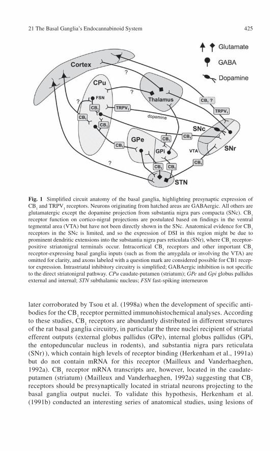
21 The Basal Ganglia’s Endocannabinoid System 425
later corroborated by Tsou et al. (1998a) when the development of specific anti-bodies for the CB
1 receptor permitted immunohistochemical analyses. According
to these studies, CB1 receptors are abundantly distributed in different structures
of the rat basal ganglia circuitry, in particular the three nuclei recipient of striatal efferent outputs (external globus pallidus (GPe), internal globus pallidus (GPi, the entopeduncular nucleus in rodents), and substantia nigra pars reticulata (SNr) ), which contain high levels of receptor binding (Herkenham et al., 1991a) but do not contain mRNA for this receptor (Mailleux and Vanderhaeghen, 1992a). CB
1 receptor mRNA transcripts are, however, located in the caudate-
putamen (striatum) (Mailleux and Vanderhaeghen, 1992a) suggesting that CB1
receptors should be presynaptically located in striatal neurons projecting to the basal ganglia output nuclei. To validate this hypothesis, Herkenham et al. (1991b) conducted an interesting series of anatomical studies, using lesions of
Fig. 1 Simplified circuit anatomy of the basal ganglia, highlighting presynaptic expression of CB
1 and TRPV
1 receptors. Neurons originating from hatched areas are GABAergic. All others are
glutamatergic except the dopamine projection from substantia nigra pars compacta (SNc). CB1
receptor function on cortico-nigral projections are postulated based on findings in the ventral tegmental area (VTA) but have not been directly shown in the SNc. Anatomical evidence for CB
1
receptors in the SNc is limited, and so the expression of DSI in this region might be due to prominent dendritic extensions into the substantia nigra pars reticulata (SNr), where CB
1 receptor-
positive striatonigral terminals occur. Intracortical CB1 receptors and other important CB
1
receptor-expressing basal ganglia inputs (such as from the amygdala or involving the VTA) are omitted for clarity, and axons labeled with a question mark are considered possible for CB1 recep-tor expression. Intrastriatal inhibitory circuitry is simplified; GABAergic inhibition is not specific to the direct striatonigral pathway. CPu caudate-putamen (striatum); GPe and Gpi globus pallidus external and internal; STN subthalamic nucleus; FSN fast-spiking interneuron

426 G.L. Gerdeman and J. Fernández-Ruiz
specific neuronal subpopulations in the basal ganglia with different neurotoxins and analyzing the changes in CB
1 receptor levels. These authors found an almost
complete disappearance of CB1 receptors when they lesioned striatal projection
neurons with ibotenate, but not when they lesioned other neuronal subpopula-tions in the basal ganglia (Herkenham et al., 1991b), thus confirming the loca-tion of the CB
1 receptor in striatal projection neurons. As mentioned above,
these early studies always used autoradiographic analysis of the CB1 receptor
because of the absence of available antibodies for immunolabeling. These tools were available, however, some years later and allowed a more precise analysis of the cellular and subcellular distribution of this receptor type in the basal gan-glia (Tsou et al., 1998a; Marsicano and Lutz, 1999; Egertová and Elphick, 2000; Hohman and Herkenham, 2000; Julian et al., 2003; Köfalvi et al., 2005; Matyas et al., 2006), which essentially corroborated the data reported by Herkenham and coworkers. Thus, CB
1 receptors are located in striatal neurons projecting to the
functional unit formed by the substantia nigra and the GPi – the so-called direct striatal efferent pathway – and also in those projecting to the GPe – the so-called indirect striatal efferent pathway (see Fig. 1). Both neuronal subpopulations use GABA as neurotransmitter and therefore express glutamic acid decarboxylase, but they can be differentiated by specific markers which are selectively coexpressed with CB
1 receptors in either striatonigral (dynorphin/substance P and D
1 receptor)
or striatopallidal (enkephalin and D2 receptors) pathways (Hohmann and
Herkenham, 2000; Julian et al., 2003). Striatal projection neurons are not, however, the only neuronal subpopulations that contain CB
1 receptors in the
basal ganglia. The autoradiographic studies mentioned before also allowed the identification of measurable levels of mRNA for this receptor in the subthalamic nucleus (STN), together with the absence of detectable levels of receptor bind-ing in that structure (Mailleux and Vanderhaeghen, 1992a). This is compatible with the presence of CB
1 receptors in subthalamic neurons projecting to the SNr
(and also to the GPi), preferably at axonal sites. These neurons are glutamater-gic, thus reproducing the classic pattern described for the CB
1 receptor in most
brain regions where it is generally located in neurons with GABAergic or gluta-matergic phenotype (see Chap. 10). Motor control is therefore influenced by CB
1
receptors at both glutamatergic and GABAergic synapses intrinsic to the basal ganglia circuitry. Intrinsic striatal neurons have also been recently identified as expressing CB
1 receptors. For instance, Hohmann and Herkenham (2000)
reported that certain striatal interneurons, containing somatostatin or acetylcho-line (ACh), do not express CB
1 receptors (see also Uchigashima et al., 2007).
However, these authors and others (Fusco et al., 2004; Uchigashima et al., 2007) demonstrated that some subclasses of striatal interneurons are CB
1 receptor-
positive. These included most of the GABAergic interneurons that are labeled with parvalbumin, but also according to Fusco et al. (2004), roughly one-third of cholinergic interneurons. CB
1 receptors are not, however, expressed by nigros-
triatal dopaminergic neurons, as several studies which used selective lesions of these neurons with 6-hydroxydopamine (6-OHDA) (Herkenham et al., 1991b) or double labeling (tyrosine hydroxylase vs. CB
1 receptors) methods (Julian et al.,

21 The Basal Ganglia’s Endocannabinoid System 427
2003) have strongly demonstrated. According to these data, it was generally assumed that the effects of cannabinoids on dopamine transmission in the basal ganglia would be always indirect and presumably exerted through CB
1 receptors
located on GABAergic and/or glutamatergic afferents to the substantia nigra pars compacta (SNc) (Romero et al., 2002; van der Stelt and Di Marzo, 2003; Fernández-Ruiz and González, 2005; and see Chap. 22). However, there is recent evidence indicating that TRPV
1 receptors, for which certain endocannabi-
noids, including anandamide (AEA), are able to act as endogenous ligands, are located in nigrostriatal dopaminergic neurons. This provides an alternative for AEA and other endocannabinoids to act through these receptors to directly con-trol dopamine synthesis and release, as supported by the following observations: (1) TRPV
1 receptors colocalize with tyrosine hydroxylase in the basal ganglia
(Mezey et al., 2000); (2) TRPV1 receptor density is lowered by treatment with
6-OHDA which damages dopaminergic neurons (Lastres-Becker et al., 2005); (3) TRPV
1 receptor binding is enhanced in the striatum of mice deficient in
dopamine transporter (Tzavara et al., 2006); and (4) AEA, but not ∆9-tetrahydro-cannabinol (∆9-THC), was found to inhibit striatal dopamine release in vitro (de Lago et al., 2004a). In contrast to some of the above observations, however, other authors reported that CB
1 and TRPV
1 receptors might colocalize in the
caudate-putamen, the GP and the SN (Cristino et al., 2006). In addition, activa-tion of the TRPV
1 receptor – a nonselective cation channel (see Chaps. 1 and 8)
– may excite, rather than inhibit, SNc neurons through indirect, presynaptic mechanisms (Marinelli et al., 2003; see below). Studies performed during the 1990s indicated that the CB
2 receptor, the other major cannabinoid receptor type,
does not appear to be present in the healthy brain (Felder and Glass, 1998), although it might be induced in response to different damaging stimuli (Fernández-Ruiz et al., 2007). However, recent data from different laboratories have found that the normal brain also contains CB
2 receptors located in different
cellular elements, including neurons and different types of glial cells, of differ-ent species including humans and rodents (Nuñez et al., 2004; Van Sickle et al., 2005; Ashton et al., 2006; Gong et al., 2006) – although see Chaps. 6, 9, 10. This also includes the basal ganglia, in particular the striatum and the SN, where Gong et al. (2006) detected immunoreactivity for this receptor type. Other stud-ies have employed a combination of RT-PCR analysis and immunolabeling techniques to investigate CB
2 receptor expression in the striatum of naive rats,
observing that this receptor subtype is indeed expressed in striatal cells, but pos-sibly located in astrocytes rather than in neurons (Fernández-Ruiz et al., 2007). Its role in the control of the basal ganglia function in normal conditions remains to be elucidated, although there is some evidence that it can be involved in the protection of striatal neurons against different types of damaging stimuli (Fernández-Ruiz et al., 2007). There are, in fact, growing indications that in multiple disease states, CB
2 receptors are induced or upregulated in glial cells
(activated astrocytes, reactive microglia) to regulate the protective and/or cyto-toxic influences that these cells exert on neuronal homeostasis (Benito et al., 2003, 2005, 2007; Pazos et al., 2004). Lastly, some studies have suggested the

428 G.L. Gerdeman and J. Fernández-Ruiz
possibility that a novel receptor type, different from CB1, CB
2, or TRPV
1 recep-
tors but active for certain cannabinoids, may be present in the basal ganglia and participate in motor effects of these compounds under certain circumstances. In general, the pharmacological profile of this unknown receptor would be rela-tively similar to the CB
1 receptor, yet its distinct identity is supported by phar-
macological experiments conducted in mice with genetic deletions of classic cannabinoid receptors (Di Marzo et al., 2000a,b).
Endocannabinoid Ligands in the Basal Ganglia
Endocannabinoid ligands, AEA and 2-arachidonoylglycerol (2-AG), are also abundant in the basal ganglia (Bisogno et al., 1999; Di Marzo et al., 2000c; Fernández-Ruiz and González, 2005), showing concentrations that are in general higher than those mea-sured in other brain regions. Endocannabinoids are particularly abundant in the basal ganglia output structures, the GP and SN (Di Marzo et al., 2000c), which parallels the high densities for CB
1 receptors reported in these two structures (Herkenham et al.,
1991a; Mailleux and Vanderhaeghen, 1992a). In the case of AEA, the GP and the SN are the two regions where the levels of this endocannabinoid are the highest (Di Marzo et al., 2000c). The existence of in situ synthesis for this endocannabinoid can be confirmed by the detection of its metabolic precursor N-arachidonoylphosphatidyleth-anolamine (NAPE; see Chap. 2) in the basal ganglia (Di Marzo et al., 2000d) and of the enzyme responsible for this process, NAPE-phospholipase D (NAPE-PLD), in the whole brain (Okamoto et al., 2004). One important issue is to determine the neuronal phenotype in which this process occurs. There is indirect evidence indicating that AEA would be generated in striatal (Ronesi et al., 2004; Ade and Lovinger, 2007) and nigral (Wallmichrath and Szabo, 2002a,b) neurons, in both cases aimed at controlling CB
1
receptor function in cortical afferents to the striatum, and striatal afferents to the SNr, respectively (see below). It is also important to mention that the levels of AEA are sig-nificantly elevated in the striatum after the stimulation of dopaminergic D
2 receptors
(Giuffrida et al., 1999; Beltramo et al., 2000). A priori this increase would reflect that the generation of AEA would be D
2 receptor-dependent, but it can also reflect a low
rate of degradation, since a recent study reported both increased NAPE-PLD and reduced fatty acid amide hydrolase (FAAH), the enzyme that degrades AEA, in striatal slices following D
2 receptor activation (Centonze et al., 2004). In any case, the eleva-
tion of AEA levels following D2 receptor activation would be compatible with the idea
that the cannabinoid system may serve as an inhibitory feedback mechanism control-ling dopamine-induced motor stimulation (Giuffrida et al., 1999). Striatal medium spiny neurons (MSNs) also express high levels of diacylglycerol lipase α (see Chap. 2), allowing for the activity-dependent generation of 2-AG (Uchigashima et al., 2007). In the basal ganglia, as in other brain areas, 2-AG might be more prominent than AEA as a rapid neuromodulator (see Chap. 11), yet AEA clearly mediates some aspects of striatal function. Most findings of 2-AG activity have been connected to electrophysi-ological studies, and will be discussed below.

21 The Basal Ganglia’s Endocannabinoid System 429
Endocannabinoid Transport in the Basal Ganglia
It is generally assumed that the anandamide membrane transporter (AMT) should be highly concentrated in the basal ganglia (Fernández-Ruiz and González, 2005). However, the evidence supporting this view is indirect and generated mainly by pharmacological data, since to date no AMT has been isolated or cloned (see Chap. 3). Nonetheless, data obtained with several AEA analogues that behave in vitro as putative AMT inhibitors (Giuffrida et al., 2001) produced important effects in several bioassays aimed at functionally detecting changes in the control of movement in laboratory animals (Fernández-Ruiz and González, 2005). This is the case with compounds such as AM404 (González et al., 1999; Beltramo et al., 2000), VDM11 (de Lago et al., 2004b), UCM707 (de Lago et al., 2002), or OMDM2 (de Lago et al., 2004b). These compounds generally inhibited motor activity in rodents, with differ-ent potencies depending on the type of inhibitor, but the most important observation was that they were able to potentiate the motor inhibition exerted by subeffective doses of AEA (Fernández-Ruiz and González, 2005). Based on these pharmacologi-cal data, there is a general consensus that the AMT is present, and possibly abun-dantly concentrated, in the basal ganglia. However, further experiments are certainly necessary to demonstrate the molecular identity of the AMT activity and how it is regulated in the brain. Such experiments should receive significant benefit from the recent development of covalent AMT inhibitors (Moriello et al., 2006), which might serve as novel tools to isolate and quantify the protein involved in this process.
Endocannabinoid Metabolism in the Basal Ganglia
FAAH is the enzyme that catalyzes the hydrolysis of AEA and related N-acyleth-anolamines, and so it plays a key role in the regulation of brain levels of this endocan-nabinoid (see Chap. 3). In general, FAAH is located in cell bodies and dendrites of neurons that are postsynaptic to axonal terminals containing CB
1 receptors (Egertová
et al., 2003; see Chap. 10), thus showing a pattern complementary to these receptors and supportive of the role of endocannabinoids as retrograde signaling molecules. FAAH enzyme is present in numerous brain structures, including structures of the basal ganglia (Desarnaud et al., 1995; Tsou et al., 1998b; Egertová et al., 2003), which is concordant with the subtle motor anomalies found in FAAH null mutant mice (Cravatt et al., 2001; Lichtman et al., 2002). However, while some authors detected high or moderate levels of FAAH in the GP and SN (Desarnaud et al., 1995; Tsou et al., 1998b), more recent studies found that the abundance of CB
1 receptors typical
of these two regions correlated with very little FAAH expression (Egertová et al., 2003). By contrast, these authors detected significant expression of FAAH in the striatum, preferentially located in neurons but also in glial cells, in particular in oligodendro-cytes (Egertová et al., 2003), an observation that these authors related to the reduction of FAAH levels reported in the striatum of parkinsonian rats (Gubellini et al., 2002).

430 G.L. Gerdeman and J. Fernández-Ruiz
Finally, monoacylglycerol lipase (MAGL), the enzyme involved in the degradation of 2-AG, and also other related monoacylglycerols (see Chap. 3), has been also detected in the basal ganglia (Dinh et al., 2002). Its pattern is somewhat different than FAAH, which suggests that AEA and 2-AG might subserve different functional roles in the basal ganglia circuitry (see also discussion in the next section). However, as stated above, both enzymes accept as substrates various N-acylethanolamines or monoacyl-glycerols, respectively; therefore, they should not be considered as definitive markers specific for cannabinoid signaling. In any case, the identification of these two enzymes, FAAH and MAGL, in the basal ganglia supports the notion that endocannabinoids generated in the different neuronal subpopulations of these structures are also degraded locally as a mechanism to efficiently inactivate these signaling molecules.
Endocannabinoids Control Neurotransmitter Release and Synaptic Plasticity in the Basal Ganglia
Physiological studies over the last several years have demonstrated that a principal and widespread function of endocannabinoids in the brain is to modulate fast synaptic neurotransmission (see Chap. 11). Both short- and long-lasting plasticity of synaptic transmission are mediated by activity-dependent release of endocannabinoids, which act in a retrograde manner at presynaptic sites to regulate the vesicular release of classical neurotransmitters (Freund et al., 2003; Chevaleyre et al., 2006). Given the distribution and dense expression levels of the CB
1 receptor within the basal ganglia, it is not surpris-
ing that endocannabinoid signaling has now been found to modulate synaptic function extensively within this network, indicating a prominent and complex role in modulating the physiology of behavioral output (see Fig. 1). Experimentally, the “on-demand” release of endocannabinoids is evoked either by the direct electrical stimulation of affer-ent synaptic pathways, pharmacological manipulations (such as the activation of certain G
q/11-coupled receptors), or by direct depolarization of a neuron using whole-cell voltage-
clamp electrophysiology. In the latter case, retrograde signaling by endocannabinoids is measured as depolarization-evoked suppression of inhibition or excitation (DSI or DSE), when studying inhibitory or excitatory synapses, respectively. The mechanisms underly-ing these phenomena have been studied extensively – especially in the hippocampus and cerebellum – and are the subject of several excellent reviews (Alger, 2002; Freund et al., 2003; Diana and Marty, 2004; Hashimotodani et al., 2007; see also Chap. 11). As of pre-paring this chapter, however, there has not yet been a careful review of the many recently elaborated mechanisms of endocannabinoid-mediated synaptic plasticity in the basal ganglia. As such processes may be highly relevant to basal ganglia function in health and disease, we will discuss the topic at length in this section.
Glutamate
As the primary input nucleus of the basal ganglia, the striatum receives a massive convergence of glutamatergic axons from all areas of the cerebral cortex, as well as

21 The Basal Ganglia’s Endocannabinoid System 431
from the thalamus (Tepper, 2006). Coincident excitatory input from cortical and/or thalamic afferents largely controls striatal output, driving MSNs into a depolarized “up-state” from which these cells fire action potentials (Wilson and Kawaguchi, 1996; Tepper, 2006). The synaptic integration of excitatory inputs in the striatum is therefore a major means by which cortical activity can be translated into motor pat-terns through the basal ganglia (Graybiel et al., 1994). A number of labs have recently confirmed an important role of endocannabinoid signaling in modulating the presynaptic function of glutamatergic synapses in both dorsal and ventral stria-tum (Chevaleyre et al., 2006). The finding that striatal glutamate release is acutely inhibited by CB
1 receptor activation (Gerdeman and Lovinger, 2001; Robbe et al.,
2001; Huang et al., 2001; Gubellini et al., 2002; Köfalvi et al., 2005) was initially confusing, because the loss of CB
1 receptor binding following excitotoxic lesioning
of the striatum suggested the postsynaptic expression of these receptors (Herkenham et al., 1991a). It is important to note, however, that the methods employed to quan-tify these findings focused on medial striatal areas where CB
1 receptor expression
is the lowest (Herkenham et al., 1991b; Glass et al., 1997; Tsou et al., 1998a), and binding was assayed at a time point that could allow for significant degeneration of afferent cortical axons from the site of the lesion (Herkenham et al., 1991a). Nonetheless, immunohistochemical detection techniques using CB
1 receptor-
directed antibodies have yielded inconsistent results, with some authors failing to see labeling of asymmetric, excitatory synapses (Matyas et al., 2006). Others have found CB
1 receptor expression levels that, although modest in comparison to
the very dense expression of this receptor at other synapses, nonetheless support the findings of physiological experiments (Robbe et al., 2001; Rodriguez et al., 2001; Köfalvi et al., 2005; Uchigashima et al., 2007). Efforts to detect CB
1 receptor
mRNA in corticostriatal projection neurons using in situ hybridization techniques were also initially unsuccessful (Marsicano and Lutz, 1999), but recent experiments designed to specifically investigate this neuronal population have yielded positive results (Uchigashima et al., 2007). Considering all the evidence, it is likely that only very low levels of CB
1 receptors are required for functioning in striatal glutamater-
gic axon terminals of mature animals.
Striatal Long-Term Depression (LTD)
Evidence for the physiological activity of presynaptic striatal CB1 receptors
emerged with the finding that endocannabinoids act as retrograde messengers critical for the induction of striatal long-term synaptic depression (LTD) (Gerdeman et al., 2002; Robbe et al., 2002; Kreitzer and Malenka, 2005). LTD is a form of synaptic plasticity that is usually induced by high-frequency stimula-tion of afferent corticostriatal axons, and is thought to play important roles in striatal mnemonic function, including the development of behavioral habits (Gerdeman et al., 2003, 2006; Yin and Knowlton, 2006). In concert with mecha-nisms of long-term potentiation (LTP), striatal LTD may powerfully sculpt the learning and execution of motor repertoires involving the basal ganglia (Graybiel,

432 G.L. Gerdeman and J. Fernández-Ruiz
2005; Yin and Knowlton, 2006), and has received considerable attention in mod-els of basal ganglia pathology (Calabresi et al., 1996, 2000; Graybiel and Rauch, 2000; Gubellini et al., 2002; Gerdeman et al., 2003; Kreitzer and Malenka, 2007). Importantly, endocannabinoids have now been found as necessary for the induc-tion of striatal LTD via several different methods of induction (Chevaleyre et al., 2006), including a protocol meant to mimic the naturally occurring, depolarized up-state transitions of striatal MSNs in vivo (Kreitzer and Malenka, 2005). In dorsal striatum, endocannabinoid-dependent LTD is also dependent on D
2 dopamine
receptors (Calabresi et al., 1992; Tang et al., 2001) and mGluRs1/5
(Gubellini et al., 2001; Sung et al., 2001; Kreitzer and Malenka, 2005). Endocannabinoid synthesis and release appears to represent a coincidence detector in striatal MSNs, signaling not only convergent excitatory inputs releasing glutamate (a likely requirement for sufficient postsynaptic depolarization and mGluR
1/5 activation),
but also the coincident release of dopamine from nigrostriatal boutons (Gerdeman et al., 2003; Yin and Lovinger, 2006). Such an associative role of CB
1 and D
2
receptors in mediating the complex synaptic mechanisms of striatal LTD may have important implications for striatal pathologies and for the function of dopamine as a learning signal critical for motivated behaviors (Graybiel, 2005; Schultz, 2006). As noted above, activation of D
2 receptors has been found to
stimulate release of AEA in the rat striatum in vivo, especially under depolarizing conditions of high external K+ (Giuffrida et al., 1999). Recent findings have led to disagreement regarding the mechanisms by which D
2 receptors may promote
endocannabinoid release during the induction of LTD (Wang et al., 2006; Kreitzer and Malenka, 2007). Wang et al. (2006) reported a mechanism to explain the observation that D
2 receptor-dependent LTD can be regularly induced in striatal
MSNs that do not themselves express D2 receptors (see Wilson, 2006, for com-
mentary). These authors demonstrated that regardless of the verified expression of D
1 or D
2 receptors in recorded MSNs, striatal LTD was highly reproducible and
involved a D2 receptor-mediated inhibition of tonically active cholinergic
interneurons. According to this model, therefore, the presence or lack of D2
receptors on the MSNs themselves are largely irrelevant to the pathways leading to LTD. Rather, D
2 receptor activation on interneurons leads to a decrease in syn-
aptic ACh, which then facilitates postsynaptic endocannabinoid production by deactivating a signaling cascade involving M
1 muscarinic ACh receptors
(mAChRs) and Cav1.3 (L-type) voltage-gated Ca2+ channels (Wang et al., 2006)
(Fig. 2). In other words, the requisite generation of AEA for striatal LTD, driven by influx of extracellular Ca2+, is under a tonic inhibition by M
1 mAChRs, and the
populations of D2 receptors responsible for enhancing endocannabinoid
release are located on cholinergic interneurons, where they induce a pause in the release of ACh (Watanabe and Kimura, 1998; Wang et al., 2006). Kreitzer and Malenka (2007) have provided evidence for a different hypothesis. Using transgenic mice to identify fluorescently labeled medium spiny neurons as either D
1 or D
2
receptor expressing (Wang et al. (2006) also employed this technique), these authors reported that only D
2 receptor-expressing neurons of the indirect striatal
outflow pathway readily express endocannabinoid-dependent LTD. This was attributed

21 The Basal Ganglia’s Endocannabinoid System 433
to a greater intrinsic excitability of indirect pathway neurons. Thus, these neurons are expected to more readily release AEA in response to synaptically driven Ca2+
influx and mGluR1/5
activation, augmented in some way by postsynaptic D2
receptors (Kreitzer and Malenka, 2007; see also Chap. 11). These novel results are difficult to reconcile with the level of reproducibility seen for the induction of striatal LTD in prior studies (e.g., Gerdeman et al., 2002; Ronesi et al., 2004; Kreitzer and Malenka, 2005), and are in direct disagreement with the article just described (Wang et al., 2006). Regardless of how this controversy is resolved, it is compelling to presume that endocannabinoid release may at least occur more readily from striatal neurons of the indirect pathway under physiological condi-tions, where afferent activity is not as robust as during stimuli used to evoke LTD in striatal slices. These findings imply that in the striatum, inhibitors of endocan-nabinoid reuptake or enzymatic degradation may have markedly different effects than direct CB
1 receptor agonists, which inhibit glutamate release at neurons of
both pathways (Gerdeman and Lovinger, 2001; Kreitzer and Malenka, 2007; Uchigashima et al., 2007).
Fig. 2 Model of synaptic endocannabinoid signaling in striatal medium spiny neurons. In all cases, retrograde activation of presynaptic CB
1 receptors leads to decreased neurotransmitter
release. At somatodendritic GABAergic synapses formed by parvalbumin-expressing (PV+) interneurons, DSI is induced by mGluR
5 and M
1 mAChRs paired with postsynaptic depolariza-
tion, involving the activation of DAGLα to generate 2-AG. mGluR5 and DAGLα are also criti-
cally required to elicit DSE at glutamatergic synapses, where the M1 mAChR plays only a
facilitating role. Corticostriatal LTD is mechanistically distinct, and involves anandamide (AEA) rather than 2-AG. LTD induction requires activation of postsynaptic mGluRs
1/5, hypoth-
esized to promote AEA synthesis via elevation of intracellular Ca2+ levels (not shown) to acti-vate n-acyl-phosphatidylethanolamine specific PLD (NAPE-PLD). Dopamine signaling at D
2
receptors is required to transiently inhibit ACh release, thereby removing a tonic blockade of postsynaptic Ca
v1.3 Ca2+ channels. Postsynaptic D
2 receptors (when present) may also enhance
AEA generation or its carrier-facilitated efflux through an endocannabinoid membrane trans-porter (EMT). Mechanisms for DSE and LTD may coexist in dendritic spines, but are depicted separately for the sake of clarity. Presynaptic effectors controlling vesicular release probability also may vary among synapses, and the presynaptic mechanisms underlying the persistent expression of LTD are not well understood. This scheme is an integration of numerous refer-ences described in the text

434 G.L. Gerdeman and J. Fernández-Ruiz
Short-Term Plasticity of Glutamate Release via Endocannabinoids in the Basal Ganglia
Endocannabinoid-mediated retrograde inhibition of glutamate release also occurs in the striatum on a rapid and transient timeframe, and can be evoked by stimulation of afferent corticostriatal synapses in a frequency-dependent manner (Yin and Lovinger, 2006). Similarly to LTD, this phenomenon also requires elevation of internal Ca2+ via mGluR
1/5 and coincident activation of the D
2 class of dopamine
receptors. Frequency-dependent suppression of glutamate release in MSNs there-fore represents another form of endocannabinoid-mediated associative plasticity that may regulate motor behaviors or procedural learning. DSE has also been recently demonstrated in striatal MSNs, and likewise appears to be strongly dependent on direct activation of mGluR
5 (Narushima et al., 2006a; Uchigashima
et al., 2007), closely linking endocannabinoid signaling to synaptic input. The extent of a physiological role for DSE is not entirely clear, and optimal parameters for inducing the phenomenon are not conditions expected to occur in vivo (5 s at a holding potential of 0 mV; Narushima et al., 2006a). Nonetheless, even small influ-ences on glutamate release might have significant influence on synaptic integration, and can be underestimated in distal dendrites using voltage clamp techniques. DSE could function as a purely synapse-specific negative feedback loop, or it might additionally represent a heterosynaptic mechanism by which spillover of glutamate can inhibit transmission at neighboring synapses (see Chap. 11). Such a question will be difficult to determine in the striatum and throughout the basal ganglia, where axo-dendritic arrangements are not nearly as predictable as in hippocampus or cerebellum (Tepper, 2006). An intriguing picture emerging from recent studies is that different endocannabinoids appear to mediate DSE and LTD in the striatum. DSE is prevented by inhibitors of DAGL, demonstrating a critical involvement of 2-AG (Uchigashima et al., 2007). This physiological finding was presented along-side highly consistent anatomical data demonstrating postsynaptic distribution pat-terns of DAGLα and mGluR
5, physically apposed to presynaptic CB
1 receptors
(Uchigashima et al., 2007). In contrast, CB1 receptor-dependent striatal LTD is not
blocked by DAGL inhibitors. Rather, the ability to induce striatal LTD in brain slices has been correlated to a developmental increase in striatal AEA content, and is greatly facilitated by exogenously applied AEA (Ade and Lovinger, 2007). LTD can also be rescued in striatal neurons, either by blocking extracellular AMT activity(Ronesi et al., 2004; Ronesi and Lovinger, 2005), or by extracellular application of URB-597, a specific FAAH inhibitor (Kreitzer and Malenka, 2007). As FAAH is the primary enzymatic pathway for AEA hydrolysis, this latter finding is consistent with the observation that striatal levels of AEA, but not 2-AG, are enhanced by local application of the D
2 receptor agonist quinpirole either in vivo (Giuffrida et al.,
1999) or in acute brain slices (Ade et al., 2003) – and quinpirole also facilitates LTD (Kreitzer and Malenka, 2005, 2007; Wang et al., 2006). Thus, mechanisms of AEA signaling – including an apparent requirement for transport-mediated efflux (Ronesi et al., 2004) – might be specialized for mediating enduring depression of

21 The Basal Ganglia’s Endocannabinoid System 435
synaptic efficacy in response to convergent input (from both glutamate and dopamine-releasing afferents) (see Fig. 2). It is not known which endocannabinoid is primarily responsible for transient, frequency-dependent suppression of excita-tion in these neurons (Yin and Lovinger, 2006), but similarities with LTD are nota-ble. These include a dependence on D
2 receptors and elevations of internal Ca2+
(which has not been directly demonstrated for either DSE or DSI in striatum), sug-gesting that perhaps AEA is also the retrograde messenger for this form of CB
1
receptor-mediated synaptic plasticity. Interestingly, whereas AEA-dependent LTD has been more easily induced in D
2 receptor-expressing MSNs (Kreitzer and
Malenka, 2007), Uchigashima et al. (2007) reported immunolabeling of DAGLα to appear more intensely in neuronal processes expressing the D
1 receptor. While
more research will surely refine these models, the distinction of separate functional pathways of retrograde endocannabinoid signaling in striatal MSNs highlights the possibility that specific pharmacological targeting of these pathways might have differential effects on the extrapyramidal motor system, distinct also from direct activation of CB
1 receptors. Glutamatergic synapses in other nuclei of the basal
ganglia are also sensitive to modulation by endocannabinoids. These include projections from the STN to both the GPe and SNr. Using electrophysiological techniques either in vivo (Sañudo-Peña and Walker, 1997) or in brain slices (Szabo et al., 2000), excitation of SNr neurons by subthalamonigral axons was inhibited by cannabinoids, as suggested by earlier behavioral studies (Sañudo-Peña et al., 1996, 1999). Similarly, direct electrical stimulation of the STN evokes glutamatergic excitatory postsynaptic currents (EPSCs) in GPe neurons, and the CB
1 receptor
agonist WIN55212-2 inhibits these EPSCs by a presynaptic mechanism (Freiman and Szabo, 2005). WIN55212-2 also was shown to decrease the spontaneous firing of GP neurons measured in anesthetized rats, consistent with a decrease in gluta-mate release from tonically active subthalamopallidal inputs (Miller and Walker, 1996, 1998). Thus, in agreement with anatomical data, STN neurons projecting to either the GP or SNr express CB
1 receptors preferentially at axonal sites (Freiman
and Szabo, 2005), where cannabinoids inhibit the release of glutamate. Glutamatergic synapses onto the dopaminergic neurons of the ventral tegmental area (VTA) are modulated by endocannabinoids as well (Melis et al., 2004a,b; Riegel and Lupica, 2004), where it has been elegantly demonstrated that 2-AG acts as a retrograde messenger mediating DSE (Melis et al., 2004b). Moreover, Melis et al. provided evidence that electrical stimulations of the PFC, reflective of in vivo firing, are suf-ficient to elicit 2-AG release and negative feedback to suppress glutamatergic transmission. Riegel and Lupica (2004) demonstrated that endocannabinoid release is enhanced by inducing burst firing of dopamine neurons – activating CB
1 receptors
at both excitatory and inhibitory synapses within the VTA. Space does not permit further discussion of endocannabinoid modulation of VTA activity (Lupica and Riegel, 2005), although it is likely to have numerous implications for the neurobiol-ogy of drug abuse, as well as motor function and the sensitivities of DA neurons to excitotoxicity (Melis et al., 2006). Lastly, there is some evidence that CB
1 receptor
agonists can inhibit glutamate reuptake, either in striatal brain slices (Brown et al., 2003) or synaptosomal preparations (Köfalvi et al., 2005; see Chap. 9). Although

436 G.L. Gerdeman and J. Fernández-Ruiz
these effects are more likely to be CB1 receptor-independent (see Chap. 9), altera-
tions in the transporter-mediated reuptake of glutamate can exert pronounced influ-ence on the excitation of neurons and on the stimulation of 2-AG by extrasynaptic mGluRs (see Chap. 11). Whether or not endocannabinoids inhibit glutamate reuptake in physiological settings remains to be seen. There is, however, evidence that endocannabinoids can enhance synaptic glutamate in the SNc by activating TRPV
1 receptor cation channels on presynaptic sites, which would be expected to
depolarize the axon terminal (Marinelli et al., 2003).
Inhibition of Striatal GABA Release
Pharmacological studies have long demonstrated interactions between cannabi-noids and GABA signaling within the extrapyramidal motor system (Pertwee and Wickens, 1991; Sañudo-Peña et al., 1999). As mentioned above, CB
1 receptors are
highly expressed at symmetric, GABA-releasing synapses throughout the basal ganglia. Accordingly, some of the first uses of brain slice electrophysiology to investigate CB
1 receptor signaling found cannabinoid modulation of GABA release
in the striatum (Szabo et al., 1998) and SNr (Chan and Yung, 1998; Chan et al., 1998). In the striatum, GABAergic inputs to MSNs arise from axon collaterals from other MSNs, and from at least three types of interneuron (Tepper, 2006). Recent studies have indicated that synapses formed by the parvalbumin-expressing (PV+), fast spiking class of interneurons (FSNs) are physiologically regulated by retro-grade endocannabinoids (Freiman et al., 2006; Narushima et al., 2006b, 2007; Matyas et al., 2006; Uchigashima et al., 2007). In technically demanding experi-ments, paired recordings have been used to analyze FSN-MSN and MSN-MSN synaptic connections (Freiman et al., 2006; Narushima et al., 2006b). Both of these synapses were found to be sensitive to the CB
1 receptor agonist WIN55212-2
(Freiman et al., 2006), consistent with a role for endocannabinoid signaling at these synapses. Accordingly, DSI can be induced in MSNs, but only with concomitant activation of either mGluR
1/5 or M
1 mAChRs, and this receptor-driven endocannabi-
noid release was only detected at synapses from PV + FSNs in paired recordings (Freiman et al., 2006; Narushima et al., 2006b). These interneurons are believed to be highly important to information processing in the striatum, providing a source of strong feed-forward inhibition to MSNs (Koos and Tepper, 1999; Tepper, 2006). There is also evidence that short-term synaptic depression of these synapses can significantly influence the likelihood of MSNs to fire action potentials in response to afferent excitatory inputs (Fitzpatrick et al., 2001). Endocannabinoid-mediated suppression of GABA release at FSN-MSN synapses may therefore play an impor-tant role in the modulation of striatal output. Uchigashima et al. (2007) found that DAGLα activation is necessary for DSI, and that this enzyme is localized close to CB
1 receptor-expressing synaptic sites (including PV + GABAergic terminals) and
to Gq/11
-coupled receptors that activate 2-AG signaling (see Fig. 2). This builds a compelling story that 2-AG is the retrograde messenger responsible for both DSE

21 The Basal Ganglia’s Endocannabinoid System 437
and DSI in the striatum (Uchigashima et al., 2007), as also appears to be the case for DSI in the SNr (Szabo et al., 2006), cerebellum (Galante and Diana, 2004; Szabo et al., 2006), and hippocampus (Hashimotodani et al., 2007). Release of 2-AG has also been measured from striatal slice cultures following electrical stimulation(Jung et al., 2005), although other protocols have yielded primarily AEA from acutely prepared slices (Ade et al., 2003), or from the striatum in vivo (Giuffrida et al., 1999). Striatal DSI appears to be tightly under the control of the giant aspiny cholinergic interneurons (Narushima et al., 2007). Although these cells represent only 5% of the total striatal neuronal population, the tonically active cholinergic interneurons extend dense axonal arbors throughout a large region of the striatum, endowing it with the largest concentration of ACh in the mammalian brain (Tepper, 2006). Narushima et al. (2007) recently showed elegantly that DSI in striatal MSNs was enhanced by increasing ambient ACh or by evoking spikes within individual nearby cholinergic interneurons, and this effect required the activation of postsyn-aptic M
1 mAChRs (see Fig. 2). It is fascinating then that ACh release may play
contrasting endocannabinoid-mediated roles between excitatory and inhibitory neurotransmission in the striatum. M
1 mAChR activation may chronically inhibit
AEA synthesis at striatal spines (Wang et al., 2006), yet tonically enhance 2-AG synthesis at somatodendritic GABAergic synapses (Narushima et al., 2007). ACh has not previously been highly implicated in striatal cannabinoid function, given that at least most cholinergic interneurons do not express CB
1 receptors (see above),
and in striatal brain slices, evoked release of ACh is not modulated by CB1 receptor
ligands (Gifford et al., 1997). Yet, based on these recent investigations of DSI and LTD, spontaneous firing activities of cholinergic interneurons may play critical roles in regulating endocannabinoid synthesis and release in MSNs. This could have important mechanistic implications for models of striatal contributions to adaptive behavior, and striatal function in disease states. It is known that the toni-cally active cholinergic neurons recorded in vivo exhibit a distinct pause in firing in response to reward-associated events encountered by the animal (Graybiel et al., 1994; Schultz, 2006). This pause is correlated to dopamine cell firing and activation of D
2 receptors (Watanabe and Kimura, 1998), and is thought to play important
roles in striatal processes of associative learning (Schultz, 2006). The recent find-ings reviewed here suggest that dopamine-induced pauses in interneuron activity and ACh release might act as a cellular switch that regulates endocannabinoid-mediated synaptic plasticity and its control over striatal output (Wang et al., 2006; Narushima et al., 2007; see Fig. 2).
GABA Release in GP and SNr
Experiments measuring rat behavior and neuronal activity in vivo first provided evi-dence that GABA release in the GP and SNr is inhibited by presynaptic CB
1 receptors
densely expressed on the axon terminals of striatal afferent projections (Miller and Walker, 1995, 1996, 1998; Sañudo-Peña et al., 1996, 1999; Tersigni and Rosenberg,

438 G.L. Gerdeman and J. Fernández-Ruiz
1996). Studies using whole-cell patch-clamp methods in brain slice preparations have corroborated this evidence. Thus, in the SNr, activation of CB
1 receptors by
WIN55212-2 decreased spontaneous or locally evoked IPSCs by a presynaptic mechanism (Chan and Yung, 1998; Chan et al., 1998). This finding was supported by studies that more specifically stimulated striatonigral afferent axons (Wallmichrath and Szabo, 2002a,b). DSI was also found to occur in both SNr (Wallmichrath and Szabo, 2002b; Yanovsky et al., 2003) and SNc neurons (Yanovsky et al., 2003), and there is some evidence that endocannabinoids may be tonically released by SNr neurons to inhibit striatonigral inputs (Wallmichrath and Szabo, 2002a). The effects of exogenous cannabinoid agonists on motor behaviors therefore involve influencing the balance of excitatory and inhibitory synapses in the SNr (Sañudo-Peña et al., 1999). In normal situations, the most overt motor effects are likely due to inhibition of the spontaneously active STN inputs releasing glutamate, but this may change according to interactions with neighboring dopamine neurons (Sañudo-Peña et al., 1998a) or following dopamine depletion in a PD model (Sañudo-Peña et al., 1998b). The effects of cannabinoids on GP output are likewise dependent upon the relative activities of excitatory (subthalamopallidal) and inhibitory (striatopallidal) inputs, as both are inhibited by presynaptic CB
1 receptors (Miller and Walker, 1996, 1998; Sañudo-Peña
et al., 1999; see Fig. 1). In a slice preparation preserving inputs to the GPe from the caudate/putamen, intrastriatal stimulation evoked CB
1 receptor-sensitive IPSCs in GP
neurons, and this striatopallidal synapse was found to exhibit a modest DSI mediated by endocannabinoids and associated with robust elevations in intracellular Ca2+
(Engler et al., 2006). It is compelling to speculate that DSI would be enhanced by activation of G
q/11-coupled mGluRs, which could be relevant to associative processes
between STN and striatal inputs to the GP. For example, a recent study found that repetitive activation of subthalamopallidal axons causes an mGluR
1-mediated postsy-
naptic depolarization and a subsequent enhancement in postsynaptic spiking that lasts much longer than the depolarization itself, over 25 s (Kaneda et al., 2007). Although it has not yet been tested, an endocannabinoid-mediated DSI may contribute to this phenomenon – especially if enhanced by mGluR
1/5 as in multiple other brain areas
(see Chap. 11) – thereby allowing STN inputs to more strongly drive GP activity. Such an interaction might suggest an increase in GP endocannabinoid contents in PD, in which STN neurons can exhibit enhanced burst firing that likely correlates to impaired movement and sensory motor processing (Bevan et al., 2002). This is consistent with findings in multiple animal PD models where elevations in GP endo-cannabinoid tone were observed (Di Marzo et al., 2000c; van der Stelt et al., 2005). Whether this relates directly to PD symptoms – or is perhaps reflective of a hyperac-tivated homeostatic response – is presently unknown.
I-LTD in the Basal Ganglia?
In addition to DSI and other transient endocannabinoid effects on GABAergic syn-apses, LTD of inhibitory inputs (I-LTD) has been observed in multiple brain areas

21 The Basal Ganglia’s Endocannabinoid System 439
(Chevaleyre et al., 2006). A recent finding suggests that I-LTD may occur within inhibitory networks of the basal ganglia, particularly in the superior colliculus (SC), where CB
1 receptors can inhibit motor behavior (Sañudo-Peña et al., 2000a). Using
a tissue culture system, mGluR- and CB1 receptor-dependent suppression of inhibi-
tion was observed in SC neurons innervated by cortical explants (Henneberger et al., 2007). This transient effect developed into a lasting, presynaptic I-LTD fol-lowing high frequency activation of excitatory cortical axons and postsynaptic mGluRs. Such endocannabinoid-mediated heterosynaptic plasticity within the SC could contribute to proper sensorimotor visual processing (Henneberger et al., 2007), which is disrupted by ∆9-THC in humans (Ploner et al., 2002). It remains to be seen if this I-LTD occurs in mature SC tissue, or elsewhere among the many collateral GABAergic synapses in the basal ganglia.
GABA Reuptake Transporters
Some authors have found evidence for an inhibition of [3H]GABA uptake by CB1
receptors in the basal ganglia (Maneuf et al., 1996a,b; Romero et al., 1998a), which could provide a homeostatic function to balance the inhibition of GABA release by endocannabinoids. Interpretation of these studies is hampered, however, by the use of drug concentrations that are many times higher than those required to fully activate CB
1
receptors (see Chap. 9). Other reports have indeed contradicted some of these findings (Köfalvi et al., 2005; Venderova et al., 2005; Engler et al., 2006), with one study indicating that CB
1 receptors inhibit GABA uptake in the SNr, but not the GP
(Romero et al., 1998a). In either nucleus, studies utilizing whole-cell voltage-clamp electrophysiology have not shown evidence for alterations in the decay of GABA-mediated currents following CB
1 receptor activation, which would be expected if
there were an inhibition of GABA reuptake (Chan and Yung, 1998; Chan et al., 1998; Yanovsky et al., 2003; Engler et al., 2006; Wallmichrath and Szabo, 2002a). In the GP, Engler et al. (2006) directly tested this hypothesis, finding that WIN55212-2 does not prolong the decay of GABAergic IPSCs in a manner similar to a specific blocker of GABA uptake transporters. Therefore, while an endocannabinoid mechanism for modulating amino acid transporters cannot be ruled out, definitive support for such a model awaits the use of expression systems to describe such a function in molecular detail.
Dopamine
Systemically delivered cannabinoids can influence dopamine cell firing (French et al., 1997; Gessa et al., 1998) and the regulation of tyrosine hydroxylase (Romero et al., 1995a,b, 2002). In accordance with many of the mechanisms just discussed, there is good evidence that cannabinoid agonists can disinhibit SNc

440 G.L. Gerdeman and J. Fernández-Ruiz
cell firing by acting on striatonigral terminals (Yanovsky et al., 2003), which would lead to elevations of striatal dopamine observed following ∆9-THC admin-istration (Castaneda et al., 1991; Ng Cheong Ton et al., 1988; see Chap. 22). Some of these terminals preferentially target postsynaptic metabotropic GABA
B
receptors, which contribute to cell firing in complicated ways (Lupica and Riegel, 2005). This is consistent with the observation that some behavioral effects of can-nabinoids are distinctly modulated by GABA
B receptors (Romero et al., 1996a).
As mentioned, however, endocannabinoids could also excite SNc neurons via TRPV
1 receptors (Marinelli et al., 2003), and the influence of CB
1 receptors on
cell firing may vary in context-dependent ways (Gueudet et al., 1995; Melis et al., 2004b; Lupica and Riegel, 2005; see also Chap. 11 for a broader discussion of this concept). A disinhibition model of cannabinoid effects on dopamine cell function is supported by anatomical data (see above). It is therefore not expected that endocannabinoids within the striatum could influence dopamine release through direct actions at dopamine release sites (see Chap. 22). There are none-theless conflicting reports in the literature in this regard. Notably, Cadogan et al. (1997) found an inhibitory effect of cannabinoids on electrically stimulated release of [3H]dopamine in striatal slices, which has been widely interpreted as suggesting a direct inhibition of dopaminergic transmission by striatal endocan-nabinoids. This careful study used concentrations of CP55940 and AEA that are generally considered specific for CB
1 receptors in ex vivo brain slice experiments
(rife with nonspecific binding opportunities for hydrophobic ligands). Inhibition by CB
1 receptor agonists on the release of preloaded [3H]dopamine was blocked
by the CB1 receptor antagonist Rimonabant at concentrations as low as 10 nM
(Cadogan et al., 1997). However, methods of whole-slice electrical stimulation employed by these authors to evoke the release of dopamine lack both specificity and physiological basis. It is likely that neuronal elements other than the nigros-triatal dopaminergic axons were direct targets of cannabinoids in this study. As mentioned, CB
1 receptors inhibit corticostriatal glutamate release, and glutamate
is likely to directly enhance striatal dopamine release via activation of ionotropic receptors on nigrostriatal afferents (Borland and Michael, 2004). Polysynaptic effects of exogenous n-methyl-d-aspartate (NMDA) may also explain the results of Kathmann et al. (1999), rather than a cannabinoid effect on dopamine release. For instance, neither WIN55212-2 nor CP55940 were found to inhibit the release of endogenous dopamine induced by single, localized electrical pulses (Szabo et al., 1999; see also Köfalvi et al., 2005). There may, however be CB
1 receptor-
independent effects of high-dose cannabinoid agonists on dopamine transporter function in the striatum (Price et al., 2007; see Chap. 9). It is not clear how these findings apply to endocannabinoid function in the basal ganglia in vivo. Price et al. (2007) used bit high ligand concentrations, and curiously saw similar effects with both AM251, a CB
1 receptor antagonist, and AM404, an AMT inhibitor. In
contrast, Fernandez-Espejo et al. (2004) used AM251 to block the effects of AM404 on dopamine-mediated turning behaviors when both drugs were injected directly into the striatum. Other authors failed to see direct effects of either Rimonabant or CB
1 receptor agonists on uptake of radiolabeled extracellular

21 The Basal Ganglia’s Endocannabinoid System 441
dopamine (Cadogan et al., 1997; Köfalvi et al., 2005), further indicating that any effects of these compounds on dopamine transporters are CB
1 receptor indepen-
dent and require high concentrations that are not likely to mimic endocannabinoid signaling (Price et al., 2007). Considering multiple lines of study, effects of endo-cannabinoids on dopamine release in vivo are most likely the result of alterations in SNc cell firing, through presynaptic modulation of GABA release or basal ganglia network activity, rather than a direct effect on dopamine release or reuptake. In summary, given that CB
1 receptors can modulate the release of both
glutamate and GABA in multiple basal ganglia nuclei, it is not surprising that cannabinoid agonists such as ∆9-THC would have complex and dose-dependent effects on motor output (Sañudo-Peña et al., 1999, 2000b; Fernandez-Ruiz et al., 2002). It should also be expected that systemic application of CB
1 receptor
antagonists might preferentially target excitatory or inhibitory transmission, depending on dosage. This highlights the need to develop and characterize phar-macological agents that act specifically on endocannabinoid uptake or metabo-lism. Nonetheless, even regarding endocannabinoids, it is not yet clear whether these molecules exert similar, or imbalanced effects on excitatory vs. inhibitory synapses under normal conditions, and how this might change in basal ganglia pathologies. There is further complexity added by the apparent formation of heteromeric receptor assemblies involving CB
1 receptors, which may allow other
transmitter systems to regulate cannabinoid effects on motor output through allosteric receptor interactions or other cooperative mechanisms (Kearn et al., 2005; Schoffelmeer et al., 2006; Carriba et al., 2007). Endocannabinoid signals appear to be an important component of regulating brain responses to particular contexts or patterns of afferent neuronal activity (see Chap. 11). Neuronal firing throughout the basal ganglia is correlated to behavioral activation, and a great many of the synaptic connections are sensitive to cannabinoids (see Fig. 1). Behavioral consequences of exogenous, systemically delivered cannabinoids may thus vary depending on the situational context and recent history of an individual. As this field advances rapidly, increasingly diverse techniques are being applied to test such hypotheses. It is important to recognize that the widespread role of endocannabinoid signaling in the basal ganglia – sometimes at functionally opposing synaptic inputs to the same neurons – reflects a complex physiology that should be interpreted through multiple approaches to understand systems-level effects of cannabinoid-based medicines. For example, some authors have described cannabinoid signaling as a “brake” on dopaminergic activity (Rodriguez de Fonseca et al., 1998), whereas others have emphasized the integral role of endocannabinoids as part of signaling cascades downstream of D
2 receptor acti-
vation, which may functionally occlude dopamine effects at the same synapses (Chevaleyre et al., 2006; and see above). These differences in emphasis are not exclusive, but largely reflect the particular historical perspectives of various methodologies, and how these are applied to a systems-level of interpretation. Ideally, increasing sophistication in neuroscience allows for multidisciplinary findings to be integrated and reconciled; it is our hope that the overview provided by this chapter is helpful in this regard.
442 G.L. Gerdeman and J. Fernández-Ruiz
Pharmacological Effects of Cannabinoids on Motor Behavior
It is evident from the pronounced activity of the cannabinoid system in the basal ganglia circuitry that this signaling system plays an important role in the control of movement. Accordingly, mice deficient in the gene encoding for FAAH, which exhibit a high and permanent elevation of brain AEA levels, develop a series of subtle motor disturbances, in particular in the response to different pharmacologi-cal stimuli (Cravatt et al., 2001; Lichtman et al., 2002). It is quite likely that the effects found in FAAH-deficient mice are mediated by the activation of CB
1
receptors (Cravatt et al., 2001), and indeed CB2 receptor knockout mice have not
been reported to exhibit any motor disturbances (Buckley et al., 2000). By con-trast, CB
1 receptor-deficient mice have exhibited important motor alterations
(Ledent et al., 1999; Zimmer et al., 1999), despite the fact that the two models developed so far exhibited apparently opposite motor phenotype, i.e., hyperloco-motion was observed in the CB
1 knockout mouse model developed by Ledent et
al. (1999), whereas hypoactivity was evident in the strain developed by Zimmer et al. (1999). Methodological differences, varying anxiety levels in experimental populations, and other reasons have been argued to explain the differences between these two models. It is possible that these differences are related to the multiplicity of sites where these receptors modulate synaptic transmission in the basal ganglia circuitry, subserving a multifactorial regulation of network activity and behavior. As will be detailed below, this could be also associated with the few paradoxical results obtained in pharmacological studies, in particular when experiments employed cannabinoid compounds with notably different pharmaco-dynamic or pharmacokinetic properties, or used very different doses or times of treatment (Fernández-Ruiz and González, 2005). However, these paradoxical results are a minor component within the literature published so far, where there is a general consensus that the activation of CB
1 receptors is usually followed by
an inhibition of motor activity. By contrast, the blockade of CB1 receptors attenu-
ates the effects of agonists and is sometimes even associated with hyperlocomo-tion, due perhaps to the inverse agonist properties displayed by most CB
1 receptor
antagonists in heterologous expression systems (see Chap. 7). In laboratory ani-mals, the administration of cannabinoid agonists produces dose-dependent impairments in the open-field, ring test, actimeter, rotarod, or other tests fre-quently employed to record motor activity (Sañudo-Peña et al., 1999; Romero et al., 2002; Fernández-Ruiz and González, 2005). This is the case of ∆9-THC, the prototypical tricyclic cannabinoid derived from Cannabis sativa, AEA, and a variety of synthetic agonists (Crawley et al., 1993; Fride and Mechoulam, 1993; Wickens and Pertwee, 1993; Smith et al., 1994; Romero et al., 1995a,b). The data in humans reinforce the same idea, since individuals that consume cannabis can experience diverse psychomotor effects reflected by a global impairment of motor performance (especially in complex and demanding tasks) associated with incoordination, ataxia, tremulousness, and weakness (Dewey, 1986; Consroe, 1998; Kalant, 2004).

21 The Basal Ganglia’s Endocannabinoid System 443
Effects of Plant-Derived, Synthetic, or Endogenous Cannabinoid Agonists
Much information on the involvement of the cannabinoid system in the control of movement has been obtained in studies testing phytocannabinoid effects in labora-tory animals. For instance, the administration of ∆9-THC reduced the spontaneous motor activity and provoked catalepsy by itself or enhanced the cataleptic activity of muscimol in mice (Pertwee et al., 1988). In rats, ∆9-THC also reduced the spon-taneous activity and the frequency of stereotypic movements (Navarro et al., 1993; Romero et al., 1995a; Jarbe et al., 1998), increased the inactivity (Rodríguez de Fonseca et al., 1994; Romero et al., 1995a; Jarbe et al., 1998), and disrupted fine motor control (McLaughlin et al., 2000). ∆9-THC also affected the motor effects of classic hypo- and hyperkinetic substances in rats; for example, it potentiated reser-pine-induced motor inhibition (Moss et al., 1981) and muscimol-induced catalepsy (Wickens and Pertwee, 1993), but reduced amphetamine-induced hyperlocomotion (Gorriti et al., 1999). Other plant-derived cannabinoids, like cannabinol (CBN) and cannabidiol (CBD), also produced motor inhibition (Hiltunen et al., 1988), although their effects were weaker than those caused by ∆9-THC in accordance with their lower affinity for CB
1 receptors, in particular, in the case of CBD (see Chap. 9).
Synthetic but somewhat nonspecific cannabinoid agonists, developed in an attempt to improve either pharmacokinetic or pharmacodynamic properties of classic phy-tocannabinoids, produced powerful inhibitory effects in a variety of motor tests and animal models (Consroe, 1998; Sañudo-Peña et al., 1999; Romero et al., 2002). Selective agonists for the CB
1 receptor, such as the metabolically stable arachido-
noyl-chloroethylamide (ACEA), also impaired motor function in laboratory ani-mals (Schuster et al., 2002), although the effects found in that study were relatively quite small. By contrast, selective agonists for the CB
2 receptor, such as HU308 or
JWH133, failed to reproduce these effects (Hanus et al., 1999; Huffman, 2005), thus supporting the possible favorability of CB
2 selective ligands for certain neuro-
logical pathologies (Fernández-Ruiz et al., 2007). The inhibitory effects reported for plant-derived or synthetic cannabinoids have been, in general, reproduced by endocannabinoids, in particular by AEA, which has been the endogenous cannabi-noid ligand most studied for its effects on the control of movement. Thus, one year after the discovery of AEA, Fride and Mechoulam (1993) reported that this endo-cannabinoid decreases rearing behavior and causes immobility in mice, results that were subsequently reproduced by Crawley et al. (1993) and Smith et al. (1994) in rats. In addition, Wickens and Pertwee (1993) found that muscimol-induced cata-lepsy in rats was potentiated by AEA as well as ∆9-THC. The group of Fernandez-Ruiz has also contributed to characterize the motor effects of AEA by measuring the dose-dependent and time-course responses exhibited by this endocannabinoid in rats subjected to open-field analysis. We found that AEA inhibited motor and stereotypic behaviors in a dose-related manner as did ∆9-THC (Romero et al., 1995a), but, compared with the time-course response exhibited by this phytocan-nabinoid, AEA showed a biphasic pattern that is related to its lower metabolic stability

444 G.L. Gerdeman and J. Fernández-Ruiz
(Romero et al., 1995b). This was confirmed after repeating the same type of experi-ments with methanandamide, a more stable analogue of AEA (Romero et al., 1996b; Jarbe et al., 1998). It is possible, however, that the differences between the motor effects of AEA and those caused by ∆9-THC might be originated by the existence of other targets available for AEA, but not for classic cannabinoids, to influence motor behavior (Di Marzo et al., 2000a,b; de Lago et al., 2004a; see Chap. 9). However, most of the literature published in relation with the motor inhibitory effects of the different cannabinoid agonists indicates that these are CB
1
receptor mediated. It is true that authors report subtle differences in magnitude and duration of these motor effects, but they can be attributed to the use of different compounds with differences in receptor affinity, potency, and/or metabolic stability. As already discussed, another key factor is that the CB
1 receptor modulates a diver-
sity of synapses within the basal ganglia circuitry – which are likely to be recruited in different context- and experience-dependent ways. Variable effects of cannabi-noids on excitatory vs. inhibitory synapses, for example – or in functionally oppo-sitional basal ganglia nuclei – may also relate to the biphasic effects of cannabinoids in some studies reporting increased motor behavior in mice (Souilhac et al., 1995) or rats (Sañudo-Peña et al., 2000b) following very low doses.
Effects of Inhibitors of Endocannabinoid Inactivation
Hypokinetic effects may also be achieved by indirect activation of the CB1 receptor
through the modulation of different elements that prolong endocannabinoid activity, such as inhibitors of the AMT, FAAH, or MAGL. Inhibition of these activities pro-duced generally equivalent effects to those observed with direct CB
1 receptor ago-
nists, with the advantage that they might be used with comparatively minimal side effects (Fernández-Ruiz and González, 2005). A guiding hypothesis is that such compounds will not produce motor effects by themselves, or that these will be small, but they can enhance the motor effects of endocannabinoids. Again, this may be an oversimplification, and such effects may be somewhat context-dependent, as sug-gested, for example, by conflicting results with the FAAH inhbitor URB597. In some studies, this inhibitor did not produce hypomotility or catalepsy when admin-istred alone (Jayamanne et al., 2006), whereas in others, URB597 was either sedat-ing (Van Sickle et al., 2005), or it reduced movement in normal rodents but not in those previously treated with reserpine (Lee et al., 2006). Other “indirect agonists,” interesting for their hypokinetic effects, are compounds characterized as inhibitors of the putative AMT. The most important compounds in this category are AM404 (González et al., 1999; Beltramo et al., 2000), VDM11 (de Lago et al., 2004b), OMDM2 (de Lago et al., 2004b), or UCM707 (de Lago et al., 2002). This last com-pound is the most potent and selective AMT inhibitor developed so far. Compared with transport inhibitors like AM404 that have hypokinetic activity by themselves – perhaps due to activity at other targets (González et al., 1999; see Chap. 9) – UCM707
21 The Basal Ganglia’s Endocannabinoid System 445
does not produce any motor effects when administered alone, but it causes a significantpotentiation of hypokinetic effects of subeffective doses of AEA (de Lago et al., 2002). This enables this compound to be an interesting pharmacological tool for those diseases, such as HD or other hyperkinetic disorders, where a hypofunction of the cannabinoid signaling has been documented (see below).
Effects of Cannabinoid Receptor Antagonists
As mentioned before, the motor effects of most of cannabinoid agonists were usu-ally prevented by blockade of CB
1 receptors with Rimonabant or other selective
antagonists (Souilhac et al., 1995; Di Marzo et al., 2001). However, Rimonabant and other CB
1 receptor blockers have in some reports induced stereotypies and
hyperlocomotion in laboratory animals (Compton et al., 1996), which is similar to those data obtained in CB
1 receptor knockout mice that exhibited equivalent motor
disturbances (Ledent et al., 1999). The motor effects of these compounds might be related to their reported inverse agonist properties. This raises an interesting thera-peutic possibility since it suggests CB
1 receptor inverse agonists to be useful for the
treatment of hypokinetic signs associated with overactivity of the cannabinoid sys-tem, which seems to include PD and related disorders (discussed in detail below). By the above rationale, a similar therapeutic benefit might be reached by pharma-cological inhibition of endocannabinoid synthetic enzymes, such as NAPE-PLD or DAGL. These enzymes have been identified and characterized only recently, how-ever, and selective pharmacological tools to study their potential therapeutic appli-cation are still lacking. One interesting compound, however, may be the DAGL inhibitor O3841 (Bisogno et al., 2006), whose motor effects are presently under investigation.
Involvement of TRPV1 Receptors in Motor Effects of Certain
Cannabinoids
Recent data have suggested that certain cannabinoids may also produce motor inhibition through the activation of vanilloid TRPV
1 receptors (de Lago et al.,
2004a). This suggestion is supported by the identification of these receptors in nigrostriatal dopaminergic neurons within the basal ganglia circuitry (Mezey et al., 2000), and also by some pharmacological studies indicating that classic vanilloid agonists or antagonists affected movement. For instance, although some studies, conducted in the 1980s using intranigral injection of capsaicin, described an enhancement of motor activity by this TRPV
1 receptor agonist (Dawbarn et al.,
1981; Hajos et al., 1988), more recent data indicate that the administration of capsaicin was followed by a strong reduction in locomotor activity in rodents and

446 G.L. Gerdeman and J. Fernández-Ruiz
that these effects are reversed by capsazepine (Di Marzo et al., 2001; Lee et al., 2006). In contrast to CB
1 receptor agonists, capsaicin did not enhance the hypokinetic
action of reserpine (Lee et al., 2006). With the above information in mind, several studies have tried to demonstrate that the TRPV
1 receptors located within the
basal ganglia might represent an alternative target for certain cannabinoid ago-nists (eicosanoid-derived cannabinoids with vanilloid-like activity, such as AEA or AM404, but not classic cannabinoids) to improve movement in basal ganglia disorders (Lastres-Becker et al., 2002a, 2003a; de Lago et al., 2004a). Thus, in the case of AEA, there exists recent data that demonstrate that the motor inhibi-tion caused by this endocannabinoid is reversed by the blockade of TRPV
1 recep-
tors with capsazepine but not by the blockade of CB1 receptors with Rimonabant
(de Lago et al., 2004a). In the same line, other studies described that the increase of TRPV
1 receptor binding observed in the striatum of mice deficient in the
dopamine transporter (DAT) would be a mechanism aimed at compensating the spontaneous hyperactivity and low striatal AEA levels exhibited by these mice (Tzavara et al., 2006). The importance of the TRPV
1 receptor is also evident in
studies conducted with AM404 to reduce hyperkinesia in rat models of HD (Lastres-Becker et al., 2002a, 2003a) or with other putative AMT inhibitors that reduced spontaneous hyperlocomotion in DAT knockout mice (Tzavara et al., 2006). In both studies, the reduction in motor hyperactivity was primarily dependent on the capability of these compounds to activate TRPV
1 receptors,
either directly or indirectly through the elevation of AEA levels.
Endocannabinoids and Parkinson’s Disease
The wealth of preclinical research we have described supports the idea that manipu-lations of the cannabinoid signaling system could be a fruitful therapeutic approach to different disorders affecting the function of the basal ganglia. Additional compelling evidence derives from a large number of studies conducted in postmortem tissue or biological fluids obtained from patients with basal ganglia disorders – or using well-described animal models of these conditions – in which disease symptomology was correlated to significant changes in markers of endocannabinoid function within the basal ganglia. This evidence provides definitive support to the possible use of cannabinoid-based medicines to alleviate symptoms and/or provide neuro-protection in basal ganglia disorders. This includes prominent hypokinetic or hyperkinetic disorders affecting specific neuronal subpopulations within the basal ganglia, such as PD (Lastres-Becker and Fernández-Ruiz, 2006) and HD (Lastres-Becker et al., 2003b, Maccarone et al., 2007), respectively, but also other disorders of the basal ganglia, such as primary dystonias or dyskinesias of different origins, and compulsive tic disorders such as Tourette’s Syndrome (Table 1). PD is the most prevalent disorder directly affecting basal ganglia function (de Lau and Breteler, 2006) and there is now considerable interest in the therapeutic possibilities of

21 The Basal Ganglia’s Endocannabinoid System 447
Table 1 Alleviation of motor symptoms and/or delay of the disease progression with cannabinoids in patients or animal models of different basal ganglia disorders
Neurological disorder Symptom relief Disease progression
Huntington’s disease TRPV1 receptor agonists reduce
hyperkinesia (3NP-lesioned rats)CB
1 receptor agonists reduce excitotoxicity (quinolinate-lesioned rats)
CB1 receptor agonists produce only modest effects (3NP-lesioned rats)
CB2 receptor agonists attenu-ate microglial toxicity (malonate-lesioned rats)
Inhibitors of the endocannabinoid uptake are effective only if they also bind TRPV
1 receptors
(3NP-lesioned rats)
CBD and ∆9-THC reduce oxidative injury by mechanisms independent of cannabinoid receptors (3NP-lesioned rats)
Parkinson’s disease CB1 receptor antagonists reduce bradykinesia and restore loco-motion in parkinsonian rats, but they do not work in patients
CBD, ∆9-THC and AM404 reduce oxidative injury by mechanisms independent of cannabinoid receptors (6-hydroxydopamine-lesioned rats)
CB1 receptor agonists reduce tremor in parkinsonian animals
CB1 agonists and inhibitors of the endocannabinoid trans-porter are not effective (6-hydroxydopamine-lesionedrats)
Cannabinoid receptor agonists interact with dopaminergic ago-nists to improve motor deterio-ration in parkinsonian animals
CB2 receptor agonists produce only modest effects (in vitro models)
CB1 receptor agonists and antago-nists delay and reduce levodopa-induced dyskinesia (patients and various animal models)
Tourette’s syndrome Plant-derived cannabinoids and analogues reduce tics (patients)
Dystonia Classic and nonclassic cannabi-noid agonists have antidystonic effects (patients and laboratory animals)
Dyskinesia CB1 agonists and or antagonists delay and reduce levodopa-induced dyskinesia (patients and various animal models)
cannabinoid-based medicines for either alleviating specific symptoms or delaying the progression of this disease (Lastres-Becker and Fernández-Ruiz, 2006). The major clinical neuropathology in PD includes bradykinesia (slowness of move-ment), rigidity and tremor, which develop following the progressive degeneration of

448 G.L. Gerdeman and J. Fernández-Ruiz
dopaminergic neurons of the SNc and a subsequent severe dopaminergic denervationof its target structures (Blandini et al., 2000). Although the etiology of PD is pres-ently unknown, there is consensus that both genetic (i.e., mutations in different PD-related genes: α-synuclein, parkin, PINK, dardarin, etc.; Abeliovich and Beal, 2006) and environmental (i.e., pesticides, antidopaminergic drugs; Di Monte, 2003) insults are important factors to trigger the disease. This progresses through a series of concomitant cytotoxic processes (i.e., altered proteolysis, oxidative stress, exci-totoxicity, mitochondrial failure, and inflammatory stimuli; McGeer et al., 2001; Sherer et al., 2001; Sethi, 2002; Wood-Kaczmar et al., 2006) that synergistically interact to trigger the progressive loss of nigral dopaminergic neurons. Dopaminergic replacement therapy with levodopa represents a useful remedy to release rigidity and bradykinesia in PD patients (Carlsson, 2002; Singh et al., 2007), at least in the early and middle phases of this disease, but patients develop an irreversible state of dyskinesia after 5 or 10 years of prolonged levodopa treatment (Fabbrini et al., 2007). Therefore, major challenges for novel pharmacological therapies in PD are (1) the finding of an alternative symptomatic treatment for those patients that do not respond well to levodopa or to the other treatments enhancing dopaminergic trans-mission; (2) the development of novel medicines for advanced phases of the dis-ease; (3) the treatment of tremor which is a prominent symptom in a third of patients; (4) the attenuation of levodopa-induced dyskinesia with the use of differ-ent types of coadjuvants; and (5) the development of an efficient therapy to arrest or delay the progression of nigral degeneration. It is possible that compounds ele-vating or inhibiting endocannabinoid activity might provide some benefits in all or part of these therapeutic demands.
Changes in the Endocannabinoid System in PD
Thinking that the activation of the cannabinoid signaling reduces movement, one might expect that this system would become overactive following the dopamine denervation that occurs in PD. Some time ago, it was demonstrated that this hypoth-esis is correct, as it was observed that the density of CB
1 receptors, as well as the
capability of these receptors to activate GTP-binding proteins, were significantly increased in postmortem basal ganglia obtained from PD patients (Lastres-Becker et al., 2001a). In accordance with these data, Pisani et al. (2005) also found an increase in endocannabinoid levels in the cerebrospinal fluid of PD patients. However, a frequent problem with the data obtained in patients is that it is difficult to be precise about whether these increases are exclusively related to the selective degeneration of nigrostriatal dopaminergic neurons, or whether they are a conse-quence of, or are associated with, the dopaminergic replacement therapy with levo-dopa that patients receive over the course of several years. For example, in the study conducted by Hurley et al. (2003) in postmortem tissues from normal and parkin-sonian human subjects, the authors found a reduction of CB
1 receptor-mRNA lev-
els, assessed by RT-PCR, in some structures (e.g., caudate nucleus, anterior dorsal

21 The Basal Ganglia’s Endocannabinoid System 449
putamen, GPe) but not in the remaining basal ganglia, and suggested that their data might have resulted from long-term dopamine-increasing treatment received by their patient population prior to death. To determine the relative influence of levo-dopa treatment in the data found in another study in PD patients (Lastres-Becker et al., 2001a), parallel analyses were conducted in MPTP-lesioned nonhuman primatemodels of PD, with or without a chronic treatment with the dopamine precursor. Results strongly indicated that the increase in the number and function of CB
1
receptors was directly related to the degeneration of dopaminergic neurons (Lastres-Becker et al., 2001a), a finding also reported by van der Stelt et al. (2005) for the increase in endocannabinoid levels that these authors found in the striatum and GPe, but not in the GPi or SN, in the same PD primate model. Interestingly, in both studies, the chronic administration of levodopa attenuated the increase in the number of CB
1 receptors (Lastres-Becker et al., 2001a) and in the levels of endo-
cannabinoid ligands (van der Stelt et al., 2005). A similar finding emerged from studies conducted in rats lesioned with 6-OHDA by the group of Mauro Macarrone (Gubellini et al., 2002; Macarrone et al., 2003). These authors found that elevated endocannabinoid levels and other abnormalitites of the cannabinoid system observed following 6-OHDA lesions (Gubellini et al., 2002) were markedly reduced by chronic treatment with levodopa (Macarrone et al., 2003). Collectively, these observations support the existence of an imbalance between dopamine and endocannabinoids at the basal ganglia in PD, which is consistent with the conclu-sions of Mailleux and Vanderhaeghen (1993) that cannabinoid signaling in the basal ganglia is under a negative control exerted by dopamine transmission. Several authors (e.g., van der Stelt et al., 2005) have proposed that the increase in endocan-nabinoid signaling that parallels dopaminergic denervation of the striatum might represent a compensatory mechanism aimed at reducing an excess of glutamate transmission in this structure, in concordance with the more general suggestion that the reduction of excitatory inputs might serve as an antiparkinsonian therapy (Wu and Frucht, 2005). It is important to consider that enhanced endocannabinoid sig-naling might also aggravate parkinsonian symptoms due to the hypokinetic profile of direct or indirect agonists of CB
1 receptors (but see Kreitzer and Malenka, 2007).
Heightened measures of cannabinoid signaling in the basal ganglia, as reflected in upregulation of CB
1 receptors or increases in endocannabinoid levels, has also been
reported in studies using different models of experimental parkinsonism in labora-tory animals (Mailleux and Vanderhaeghen, 1993; Romero et al., 2000; Di Marzo et al., 2000c; González et al., 2005). However, the issue retains certain controversy since there are a few studies that reported no changes in endocannabinoid signaling in PD (Herkenham et al., 1991b), or found a reduction (Silverdale et al., 2001) or a dependency on chronic levodopa cotreatment (Zeng et al., 1999). Despite these conflicting data, it is widely evidenced that the cannabinoid signaling becomes overactive in the basal ganglia in PD. This body of evidence is supportive of a view that a general overactivity of endocannabinoid signaling is an event that develops when the degeneration of nigral neurons has progressed to a certain extent and the major parkinsoniam symptoms are already evident. However, some recent studies suggest that losses and/or malfunctioning of the cannabinoid signaling system,

450 G.L. Gerdeman and J. Fernández-Ruiz
mainly at the level of the CB1 receptor, might be an early event linked to the PD
pathogenesis itself. This can be studied in patients affected by incidental Lewy body disease, an early and presymptomatic phase of PD, in which individuals present Lewy bodies and a low degree of nigral pathology, but not neurological symptoms. Using postmortem tissue from a small population of these patients, it was observed that CB
1 receptors already exhibited a trend toward an increase in
some basal ganglia structures (Lastres-Becker et al., 2001a). The issue can be also studied in recently developed mouse models of deficiency or mutation in specific genes linked in humans with the development of parkinsonism, such as mice defi-cient in the PARK-2 (parkin; see Itier et al., 2003) or PARK-1 (α-synuclein; Cabin et al., 2002) genes, or overexpressing a mutated form of α-synuclein (Gispert et al., 2003). The importance of these genetic models is that they can be considered as representative of early stages of parkinsonism when animals only exhibit small dis-turbances in motor behaviors or markers of dopamine function, but no evidence for protein aggregation or neurodegeneration. Interestingly, these mice show several alterations in the synthesis, density, or function of CB
1 receptors in the SN and
other basal ganglia structures (González et al., 2005; Fernández-Ruiz, unpublished results). It is important to emphasize that these receptor changes occurred in a situ-ation where dopaminergic dysfunction rather than neuronal death is the major extant pathological event, thus indicating that these receptor changes might be an early event presumably involved in the neurodegenerative process. These anomalies (losses or malfunctioning) in CB
1 receptors might trigger excitotoxicity, inflamma-
tion, or other cytotoxic events that are normally under the control of these receptors, contributing to PD disease progression.
Potential of Cannabinoid-Based Therapies in PD
As mentioned above, the present therapy in PD only allows the alleviation of specific parkinsonian symptoms but fails to delay the progression of nigral degeneration. This consists in the use of the dopamine precursor levodopa which is able to release the rigidity and bradykinesia typical of most parkinsonian patients, in particular, during the early and middle phases of this disease (Carlsson, 2002; Singh et al., 2007). However, not all patients respond to levodopa, and, in those that are well responders, levodopa frequently loses efficacy, and ultimately leads to the appear-ance of an irreversible dyskinetic state in a period of 5–10 years. Therefore, the treatment of PD patients is demanding the urgent development of novel medicines (1) that are able to reduce parkinsonian symptoms in patients that do not respond to levodopa; (2) that do not develop dyskinesia after prolonged uses; and (3) that serve as neuroprotective molecules able to delay or arrest the progress of nigral degeneration. Cannabinoid-based compounds, either agonists or antagonists, might serve as novel medicines for PD, either used alone or as coadjuvants with classic therapies, as recent preclinical studies and a few clinical trials have indicated. According to these studies, cannabinoid compounds might be used to alleviate

21 The Basal Ganglia’s Endocannabinoid System 451
parkinsonian symptoms (including the attenuation of levodopa-induced dyskinesia) and to prevent SNc cell death (Brotchie, 2000, 2003; Fernández-Ruiz and González, 2005; Lastres-Becker and Fernández-Ruiz, 2006). However, the type of cannabi-noid compound to be used in each of these two aspects might be different. Parkinsonian bradykinesia might be effectively treated by the use of selective CB
1
receptor antagonists, compounds that could also be recommended for delaying the development of dyskinesia associated with long-term levodopa treatment (Brotchie, 2003). By contrast, antioxidant cannabinoid agonists might be the best option to protect dopaminergic nigral cells from death (Lastres-Becker and Fernández-Ruiz, 2006), although this would cover only one component – oxidative injury – of the complex pathophysiology of PD. It is possible that the antiexcitotoxic and/or anti-inflammatory properties of CB
1 and CB
2 agonists, respectively, may be also used to
protect nigral cells from death (Fernández-Ruiz et al., 2005).
Alleviation of Parkinsonian Symptoms with Cannabinoid-Based Compounds
Studies carried out almost exclusively in laboratory animals revealed that CB1
receptor agonists, despite their hypokinetic profile, may be useful in PD under cer-tain circumstances. For instance, they are able to interact with dopaminergic ago-nists to improve motor impairments (Anderson et al., 1995; Maneuf et al., 1997; Brotchie, 1998; Sañudo-Peña et al., 1998b). In a recent study, Kreitzer and Malenka (2007) proposed that endocannabinoid enhancement, using FAAH or MAGL inhibitors, combined with D
2 receptor agonists may attenuate motor deficits in PD
(see striatal LTD discussion above). Cannabinoid agonists might also reduce tremor associated with overactivity of the STN, based on the capability of CB
1 receptors
to inhibit glutamate release from subthalamonigral axon terminals (Sañudo-Peña et al., 1998, 1999). Lastly, CB
1 receptor agonists have also been related to a decrease
and/or delay in the development of levodopa-induced dyskinesia, as has been described in both laboratory animals (Fox et al., 2002a; Ferrer et al., 2003; Segovia et al., 2003) and human patients (Sieradzan et al., 2001), although a recent clinical trial using a cannabis extract conducted by Carroll et al. (2004) did not replicate the observations in the study by Sieradzan et al. (2001) who used the ∆9-THC analogue Nabilone™. Despite these therapeutic benefits suggested for CB
1 receptor agonists
against certain parkinsonian symptoms, a generally accepted view is that, due to their hypokinetic profile, it is unlikely that these agonists might be useful to allevi-ate bradykinesia, the major symptom in PD patients. In fact, the few completed studies in humans and MPTP-lesioned primates confirm this view, since the administration of phytocannabinoid agonists was interpreted to worsen motor disa-bility (Consroe, 1998; Müller-Vahl et al., 1999c). By contrast, the blockade of CB
1 receptors has been proposed as a better alternative to reduce bradykinesia,
although only in special circumstances which will be described below. CB1
receptor antagonists, used as coadjuvants with the classic dopaminergic

452 G.L. Gerdeman and J. Fernández-Ruiz
replacement therapy, have been proposed as a better alternative than agonists to delay the appearance and to reduce the severity of levodopa-induced dyskinesia (Brotchie, 2000, 2003), a proposal that has been recently evaluated with positive results in nonhuman primate (van der Stelt et al., 2005) and rat (Segovia et al., 2003) models of PD. The use of CB
1 receptor antagonists in PD is supported by
studies describing that cannabinoid signaling becomes overactive in the basal gan-glia of PD patients (Lastres-Becker et al., 2001a; Pisani et al., 2005). It is also sup-ported by data obtained in multiple animal models of parkinsonism, including reserpine depletion (Di Marzo et al., 2000c), chronic dopaminergic blockade (Mailleux and Vanderhaeghen, 1993), neurotoxin lesioning with 6-OHDA (Mailleux and Vanderhaeghen, 1993; Romero et al., 2000; Gubellini et al., 2002; Fernández-Espejo et al., 2004), or MPTP (Lastres-Becker et al., 2001a; van der Stelt et al., 2005), or genetic manipulations (González et al., 2005; Fernández-Ruiz, unpub-lished results). As described above, there are various hypotheses as to how CB
1
receptor antagonists – or agonists – could elicit benefit in a manner specific to dif-ferent aspects of PD expression (bradykinesia vs. tremor, for example). Given the diverse and synapse-specific effects of CB
1 receptors to regulate neurotransmitter
release in the complex circuitry of the basal ganglia (see Fig. 1), it seems premature to espouse any present model too strictly. With that said, although some evaluations of CB
1 receptor blockade on animal PD models have shown positive effects (Di
Marzo et al., 2000c; van der Stelt et al., 2005), others have found no benefits (Meschler et al., 2001). The only clinical trial developed so far with the CB
1 receptor
antagonist Rimonabant in PD patients showed no improvements (Mesnage et al.,2004). This study was conducted, however, with a population of patients that were all well-responders to classic dopaminergic replacement therapy (Mesnage et al., 2004), and it is possible (but unexamined) that Rimonabant might function better in those patients with poor response to levodopa. Preclinical data indeed suggest that the blockade of CB
1 receptors would require special circumstances to be effective
in PD, including attention paid to the following points: (1) the use of low doses of Rimonabant (< 1 mg/kg) in rats (González et al., 2006), although van der Stelt et al. (2005) reported beneficial effects at a dose of 3 mg/kg in MPTP-lesioned primates and (2) that Rimonabant might be effective only at very advanced phases of the disease (Fernández-Espejo et al., 2005). If this hypothesis is correct, it would be possible to have an antiparkinsonian agent for conditions at which classic levo-dopa therapy generally fails: patients with poor response and/or advanced phases of this disease, which would represent an important pharmacological advantage. On the other hand, the description of TRPV
1 receptors in nigrostriatal dopaminergic
neurons (Mezey et al., 2000), as well as the neurochemical data indicating that they might play a role in the regulation of dopamine release from nigral neurons (de Lago et al., 2004a), open an additional therapeutic possibility for the endocannabinoid/endovanilloid systems to be used for the alleviation of parkinsonian symptoms. There is evidence that TRPV
1 receptors are reduced in the striatum of parkinsonian
rats (Lastres-Becker et al., 2005), likely as a consequence of the death of neurons containing this receptor, which would represent a problem for therapeutic manipulation.However, the blockade of remaining receptors in surviving cells might be used to

21 The Basal Ganglia’s Endocannabinoid System 453
enhance dopamine transmission – assuming an inhibitory function of these recep-tors in the regulation of dopamine release (de Lago et al., 2004a) – although this requires further verification.
Neuroprotection with Cannabinoid-Based Compounds in PD
Cannabinoids appear to represent a special class of molecules, in which a multifac-eted combination of complementary effects exert an overall neuroprotective influ-ence that is mediated by multiple mechanisms (van der Stelt and Di Marzo, 2005; see Chaps. 15 and 16). Thus, cannabinoid agonists are able to reduce excitotoxicity, calcium influx, oxidative injury, cerebro-vasoconstriction, body temperature, and/or local inflammatory events – all of which are effects that could contribute to increasing neuronal survival in this and other acute or chronic neurodegenerative disorders (Fernández-Ruiz et al., 2005; Sarne and Mechoulam, 2005; van der Stelt and Di Marzo, 2005). An interesting aspect of this potential is that it does not include exclusively the activation of CB
1 receptors. Other important targets of the
cannabinoid signaling system (e.g., CB2 receptors) seem to also play key roles.
Such diverse neuroprotective potential presents the cannabinoid system as an excel-lent tool for the treatment of neurodegenerative disorders, because it allows the combination of multiple, supplemental strategies. Therapeutic approaches therefore go beyond the exclusive use of CB
1 receptor agonists, the clinical use of which is
confounded by their psychotrophic side effects (Fowler, 2005). Because of their capability to regulate glial influences to neuronal homeostasis, CB
2 receptor ago-
nists are a likely clinical alternative for neuroprotection (Fernández-Ruiz et al., 2007) and possibly, because of its antioxidant and anti-inflammatory properties, CBD may also be of similar use (Mechoulam et al., 2002). Other neuroprotective strategies are being directed toward methods to enhance endocannabinoids, such as targeting the AMT or degradative enzymes (Di Marzo et al., 2004; Fowler et al., 2005). Details on the molecular and cellular pathways underlying the effects of cannabinoids as neuroprotective agents have been discussed in another chapter of this book and will not be addressed here. We will instead concentrate exclusively on the neuroprotective potential of cannabinoids for basal ganglia disorders, for which a schematic diagram of different mechanisms proposed has been outlined in Fig. 3. As regard to PD, recent preclinical studies carried out with ∆9-THC have provided solid evidence that this plant-derived cannabinoid may reduce the degen-eration of nigrostriatal dopaminergic neurons in rats with hemiparkinsonism caused by the unilateral application of 6-OHDA (Lastres-Becker et al., 2005). However, the hypokinetic profile of this phytocannabinoid may represent a disadvantage for any future clinical exploitation of this effect. Interestingly, another phytocannabinoid,CBD, proved to exhibit the same degree of neuroprotection against the toxicity of 6-OHDA in rats (Lastres-Becker et al., 2005). CBD has minimal affinity for CB
1
and CB2 receptors compared to ∆9-THC, and this likely represents some important
advantages for a possible clinical exploitation of this effect: (1) CBD does not

454 G.L. Gerdeman and J. Fernández-Ruiz
activate CB1 receptors (see Chaps. 7 and 9) and so it would not produce any aggravation
of motor disability that compounds activating this receptor type (Mechoulam et al., 2002; Russo and Guy, 2006) and (2) CBD does not elicit tolerance compared to ∆9-THC when used for prolonged treatments (Malfait et al., 2000; Hayakawa et al., 2007). In favor of ∆9-THC, however, this compound, but not CBD, is able to acti-vate CB
2 receptors, which might represent another important neuroprotective target
in PD because of their anti-inflammatory potential; although CBD is also anti-inflammatory by mechanisms that have not yet been identified (Walter and Stella, 2004). On the other hand, the fact that both phytocannabinoids were equally effec-tive in increasing neuronal survival, despite their pharmacodynamic differences, is suggestive that the mechanism providing neuroprotection against 6-OHDA toxicity would be cannabinoid receptor-independent and would emphasize antioxidant properties of these compounds (Lastres-Becker et al., 2005). The same type of antioxidant properties have been proposed for explaining the neuroprotective potential of phytocannabinoids in other in vitro or in vivo models of neurodegener-ation (Hampson et al., 1998; Sagredo et al., 2007). Corroborating this hypothesis, recent in vivo studies – investigating the potential of different cannabinoid-based molecules with selectivity for CB receptor types or for the AMT (García-Arencibia et al., 2007) – indicated that only compounds with antioxidant properties, such as AM404, were able to reduce the nigral toxicity of 6-OHDA (García-Arencibia et al., 2007). This effect was likely originated by the antioxidant potential of the
Fig. 3 Mechanisms suggested for the protection exerted by cannabinoids against the damage of specific basal ganglia structures in HD and PD

21 The Basal Ganglia’s Endocannabinoid System 455
phenolic group present in the chemical structure of AM404, rather than its capability to act as an AMT inhibitor, since other AMT inhibitors devoid of this antioxidant potential, like UCM707, failed to protect dopaminergic neurons (García-Arencibia et al., 2007). Selective agonists for the CB
1 receptor, like ACEA, also failed to
protect these neurons (García-Arencibia et al., 2007), which is in part concordant with the data published by Kim et al. (2005) indicating that CB
1 receptors enhance
rather than reduce the toxic effect of the TRPV1 agonist capsaicin in an in vitro
model of PD. As mentioned above, the cause of dopaminergic cell death in PD is still unknown and possibly involves the combination of different pathogenic mech-anisms. Alterations in the function of glial cells, including the activation of CB
2
receptor-expressing microglia, might be one of these mechanisms, playing a poten-tially important role in the initiation and/or early progression of the dopaminergic denervation (Whitton, 2007). Activated microglia and other glial cells, for example, have been described in close proximity to nigral neurons (Kim et al., 2000; Gao et al., 2002; Whitton, 2007). These cells produce several cytotoxic factors, such as TNFα, IL-1β, nitric oxide, reactive oxygen species, and others, that have been reported to be elevated in the SN and the caudate-putamen of PD patients (Nagatsu et al., 2000; Whitton, 2007). Reactive microgliosis has been also described in dif-ferent animal models of Parkinsonism (Whitton, 2007), and in these animals, the control of the activity of these cells has been reported to provide neuroprotection(Morale et al., 2006). With this idea in mind, some in vitro studies evaluated the importance of glial metabolism in the effects of cannabinoid agonists on 6-OHDA neuronal toxicity. Results indicated that the activation of CB receptors located in glial cells, but not in neurons, was associated with an increase in neuronal survival, presumably exerted by increasing the trophic support exerted by glial cells on neurons (Lastres-Becker et al., 2005). These experiments utilized HU-210, a can-nabinoid with antioxidant potential but certainly not selective for CB
2 receptors.
However, the importance of CB2 receptors in the function of glial cells, and in
particular microglia (Walter and Stella, 2004), supports a possible role for this receptor type to provide neuroprotection in PD (Fernández-Ruiz et al., 2007). Yet, recent in vivo data only reflected a very modest contribution of CB
2 receptors to the
neuroprotective effect of cannabinoids observed in hemiparkinsonian rats (García-Arencibia et al., 2007). Collectively, the different studies conducted so far strongly indicate that the noted neuroprotective properties of certain cannabinoids are mechanistically related to their antioxidant potentials (Lastres-Becker et al., 2005; García-Arencibia et al., 2007). It is important to remark that this fact is of special relevance in a degenerative disorder in which oxidative injury is a major hallmark of pathogenesis (Blandini et al., 2000). CB receptor-independent antioxidant effects are presumably exerted by the phenolic structure of these compounds, which allows them to act as scavengers of reactive oxygen species; although, they might also act by improving the function of endogenous antioxidant enzymes (García-Arencibia et al., 2007). Additionally, cannabinoids may provide neuroprotection through the activation of CB
2 receptors located in glial cells which would regulate the influence
of these cells on neuronal homeostasis (Stella, 2004; Walter and Stella, 2004; Fernández-Ruiz et al., 2007). Therefore, the major challenges for future research in

456 G.L. Gerdeman and J. Fernández-Ruiz
this area are essentially two. First, the development of additional preclinical studies aimed at identifying the true potential of CB
2 receptors in PD, in particular: (1) to
determine whether these receptors are induced or upregulated in reactive microglia or astrocytes, as happens in other neurodegenerative disorders (Fernández-Ruiz et al., 2007) and (2) to identify the molecular mechanism(s) through which these receptors control the cytotoxic or protective influences exerted by glial cells on neuronal homeostasis, as also described in other neurodegenerative disorders (Fernández-Ruiz et al., 2007). The second challenge would be the clinical valida-tion of the neuroprotective potential of (1) CB
2 receptor agonists – if the above
proposed experiments indicate that this receptor type is involved – and in particular (2) the antioxidant cannabinoids CBD and ∆9-THC, for which preclinical studies have already reported solid and promising possibilities. In this sense, CBD com-bined with ∆9-THC is the basis for one of the cannabis-based medicines, Sativex™,which is presently being subjected to clinical testing for its potential in a variety of neurological disorders (Russo and Guy, 2006). This combination appears an excel-lent option for the clinical exploitation of a cannabinoid-based medicine able to control the progression of PD pathogenesis.
Endocannabinoids and Huntington’s Disease
HD is an inherited, progressive, and fatal neurodegenerative disorder caused by the expansion of polyglutamines in the N-terminal of a protein identified in the study of this disease, which was called huntingtin and whose gene (IT15) is located on chromosome 4. The disorder belongs to the family of diseases caused by an excess of CAG repeats in the genes encoding for different proteins, including huntingtin in HD and ataxins in the different spinocerebellar ataxias (Riley and Orr, 2006). In the case of HD, the mode of transmission is autosomal dominant and the limit for CAG repeats is 35. Expansions between 36 and 39 lead to incomplete penetrance, whereas the occurrence of typical adult-onset HD starts with a number of repeats greater than 40. Variables such as an earlier age of onset, the rate of disease pro-gression, and the severity of neuronal damage/neurological deficits mostly corre-lated with the number of glutamines found in huntingtin (Walker, 2007). Although mutated huntingtin is constitutively expressed, only a few cells are sensitive to its toxic effects. Thus, HD is characterized by a dramatic loss of neurons in the stria-tum and the cerebral cortex, resulting in motor abnormalities (chorea), cognitive disturbances (dementia), and early death (10–20 years after diagnosed). The mechanism(s) by which the mutated huntingtin causes the progressive loss of stri-atal and cortical neurons is still pending complete description, but there is consen-sus that HD is a multifactorial disease, and multiple molecular and cellular mechanisms have been reported to be involved in the pathophysiology (Li and Li, 2006). Part of these pathogenic mechanisms, possibly those that operate in the initiation and during the first steps of disease progression, involve a series of confor-mational changes caused by polyglutamine expansion in the huntingtin protein,

21 The Basal Ganglia’s Endocannabinoid System 457
resulting in altered protein–protein interactions, abnormal protein aggregation, and proteolysis (Li and Li, 2004; Borrell-Pagès et al., 2006). These are followed by transcriptional dysregulation affecting some genes involved in neuronal survival (e.g., brain-derived neurotrophic factor, BDNF), excitotoxicity, mitochondrial dys-function (i.e., complex II deficiency), oxidative stress, and local inflammatory events, and culminating in extensive loss of striatal and cortical neurons (Cattaneo et al., 2005; Borrell-Pagès et al., 2006). At present, there is no specific pharmaco-therapy to alleviate symptoms and/or to arrest or delay striatal and cortical degen-eration in HD (see below). In this context, cannabinoid-related compounds have been proposed as potential novel medicines for this disorder considering the data generated in a series of preclinical studies (Lastres-Becker et al., 2003b). The rationale for these studies is based on the suspicion that cannabinoid agonists might be used to alleviate hyperkinetic symptomatology in HD because they are hypoki-netic substances. In addition, they can also protect striatal neurons from death because of their neuroprotective properties.
Changes in the Endocannabinoid System in HD
Contrarily to the case of PD, the hypokinetic function exerted by cannabinoid sig-naling would suggest a priori that this system becomes progressively hypofunctionalin HD due to the hyperkinetic nature of this disorder (Lastres-Becker et al., 2003b). Studies in postmortem basal ganglia from HD patients have confirmed this hypothesis proving a massive loss of CB
1 receptors in the SN and GPe, and a lesser
reduction in the putamen (Glass et al., 1993, 2000; Richfield and Herkenham, 1994). The loss of CB
1 receptors reflects the characteristic neuronal loss observed
in the basal ganglia in HD, which predominantly affects GABAergic MSNs. On this level, the loss of CB
1 receptors have been seen as equivalent to the losses of
other phenotypic markers of striatal neurons, such as substance P, dynorphin, enkephalin, and adenosine and dopamine receptors (Hersch and Ferrante, 1997). The data obtained in different animal models of HD generally corroborated the findings obtained in human postmortem tissues. Thus, marked losses of CB
1 recep-
tors were also evident in the striatum, GP and SN of rats with lesions of striatal GABA projection neurons, induced with mitochondrial or excitotoxic toxins (Page et al., 2000; Lastres-Becker et al., 2001b, 2002a,b). These toxins, in particular 3-nitropropionic acid (3-NP), reproduce in animals the characteristic mitochondrial complex II deficit described in patients (see Gu et al., 1996) and are associated with the same cytotoxic events that have been proposed for the etiology of the human disease, i.e., failure of energy metabolism, glutamate excitotoxicity, and oxidative stress, leading to progressive neuronal death (Alexi et al., 1998; Brouillet et al., 1999, 2005). As in humans, the losses of CB
1 receptors found in animals lesioned
with neurotoxins might represent a mere side effect caused by the progressive and selective destruction of striatal MSNs where these receptors are located. However, there is evidence that these losses happen in early stages where neuronal death does

458 G.L. Gerdeman and J. Fernández-Ruiz
not exist or is still minimal (this important aspect will be addressed below). Interestingly, the losses of CB
1 receptors described in rats lesioned with 3-NP were
accompanied by a reduction in the levels of endocannabinoids that was mostly evi-dent in the caudate-putamen (Lastres-Becker et al., 2001b). Therefore, it is possible that endocannabinoid signaling becomes progressively hypofunctional in the basal ganglia in HD. It is likely that this might contribute to some extent to the hyperki-nesia typical of this disorder and support a hypothesis that CB
1 receptor agonists
might be beneficial to alleviate motor deterioration. In evident contrast with the loss of CB
1 receptors typical of HD, CB
2 receptors seem to be induced or upregulated
in the basal ganglia in response to damaging conditions. This seems to occur in reactive microglial cells that are recruited to lesioned sites, and also in astrocytes, in animal models of HD (Fernández-Ruiz et al., 2005, 2007), in similarity to studies with patients affected for other neurodegenerative or neuroinflammatory disorders (Benito et al., 2003, 2005, 2007; see Chap. 16). In these cells, CB
2 receptors would
presumably play a protective role by reducing the cytotoxic influences of reactive microglial cells on neuronal homeostasis (Fernández-Ruiz et al., 2007). The occur-rence of reactive microgliosis is documented in the striatum of HD patients (Pavese et al., 2006), which would support that they also experience a possible induction or upregulation of CB
2 receptors – this would have interesting pharmacological impli-
cations in a disease with a poor therapeutic outcome. In this sense, a recent microar-ray study conducted in blood samples from HD patients revealed the gene encoding for the CB
2 receptor as one of the genes altered during the development and pro-
gression of HD, although this gene was not included among the nine key genes that authors proposed as potential biomarkers of this disease (Borovecki et al., 2005). On the other hand, in HD, as happens in PD, the changes observed in endocannabi-noid signaling, rather than being mere secondary consequences of striatal injury, might contribute to the pathogenesis and/or early progression of this disease. Thus, the marked losses of CB
1 receptors described in middle and advanced phases of HD
have been already observed at very early phases. They occur in advance of other receptor losses and even before the appearance of major HD symptomatology, therefore in conditions in which cell death is still very low. This has been described by Glass et al. (2000) using postmortem human brains at different grades of HD progression, and further validated in animal models (Lastres-Becker et al., 2003b). For instance, losses of CB
1 receptors were also observed in the basal ganglia of dif-
ferent transgenic mouse models (R6/1, R6/2, and HD94) that express mutated forms of the human huntingtin with different numbers of CAG repeats/poly-glutamine expansions (Denovan-Wright and Robertson, 2000; Lastres-Becker et al., 2002c; Naver et al., 2003; McCaw et al., 2004). Importantly, the magnitude of CB
1 receptor down-regulation correlated with the size of the CAG repeats
(McCaw et al., 2004) and were attenuated when transgenic animals are housed in an enriched environment (Glass et al., 2004), in parallel to a slow progression of HD symptoms (Hockly et al., 2002). One important aspect of these data is that the reductions of CB
1 receptors were evident at ages of the animals at which cell dysfunction
rather than cell death is the major change that takes place. This is absolutely consistent with the results obtained by Glass et al. (2000) in early grades of the

21 The Basal Ganglia’s Endocannabinoid System 459
human disease, supporting the hypothesis that losses of CB1 receptors are an early
event presumably involved in the initiation and/or first phases of HD pathogenesis. With this notion in mind, Centonze et al. (2005) conducted a series of electrophys-iological experiments in the R6/2 transgenic mouse model of HD trying to identify early alterations of endocannabinoid signaling that may relate to the progression of the disease. These authors found a greatly reduced sensitivity of striatal GABAergic synapses to the presynaptic inhibitory effects of CB
1 receptor activation (Centonze
et al., 2005; Maccarone et al., 2007). Again, they found that these alterations were not a consequence of striatal degeneration because they were observed at an early stage in the disease progression in R6/2 mice, before the primary initiation of cell death (Turmaine et al., 2000). The mechanism(s) underlying this effect is presently unknown, but there is some evidence that it might be related to impairment in the efficiency of these receptors to activate certain intracellular signals. One of the ear-liest events following 3-NP intoxication in rats is the occurrence of various anoma-lies in CB
1 receptor agonist-induced stimulation of GTP-binding proteins in striatal
neurons (Lastres-Becker et al., 2004). This occurred in absence of changes in bind-ing and mRNA levels for this receptor and before the first signs of neurodegenera-tion and neurological deterioration (Lastres-Becker et al., 2004). For some authors, this response and the subsequent losses of CB
1 receptors (Glass et al., 2000;
Denovan-Wright and Robertson, 2000; Lastres-Becker et al., 2002c) are interpreted as a compensatory mechanism which might counteract excitoxic damage to MSNs by enhancing GABAergic synaptic function through reduced presynaptic inhibition (see Fig. 2; Maccarrone et al., 2007). An alternative explanation (not necessarily exclusive of the former) could be that the losses or the malfunctioning of CB
1
receptors in specific neuronal subpopulations of the basal ganglia might render these neurons more vulnerable to different cytotoxic stimuli (e.g., oxidative stress, excitotoxicity, inflammation; see Chaps. 15 and 16) that frequently cooperate to produce cell damage in HD (van der Stelt et al., 2002; Fernández-Ruiz et al., 2005). If this were the case, the activation of these receptors might be used as a neuro-protectant strategy in this disease, as will be addressed with more detail in the following section.
Potential of Cannabinoid-Based Therapies in HD
Despite the progression in the elucidation of molecular events involved in the pathogenesis in HD (Cattaneo et al., 2005; Li and Li, 2006; Borrell-Pagès et al., 2006), there is no parallel progression in the development of novel medicines capa-ble of alleviating symptoms and/or delaying degeneration. To date, the only thera-peutic tools used in HD include mainly antidopaminergic drugs to reduce the hyperkinesia characteristic of the first phases of the disease (Factor and Friedman, 1997) and antiglutamatergic agents to reduce excitotoxicity (Kieburtz, 1999). There are some novel pharmacological strategies (e.g., unsaturated fatty acids, coenzyme Q10, minocycline, inhibitors of histone deacetylases) that are still under clinical

460 G.L. Gerdeman and J. Fernández-Ruiz
testing (Bonelli and Wenning, 2006; Butler and Bates, 2006;Walker, 2007). In any case, the outcome of these strategies, measured in terms of improving quality of life for HD patients, is still too limited. In this context, cannabinoid agonists might be a reasonable alternative since they can act as antihyperkinetic substances as well as neuroprotective agents.
Alleviation of Hyperkinetic Symptoms with Cannabinoid-Based Compounds
As antihyperkinetic substances, CB1 receptor ligands may act to acutely recover the
neurochemical deficits typical of the hyperkinetic phase of HD (Lastres-Becker et al., 2002a, 2003a). As the disease progresses, this property may be limited by the massive loss of CB
1 receptors and the occurrence of akinesia rather than hyperki-
nesia as a major symptom during advanced states of this disorder (Lastres-Becker et al., 2002b). Therefore, the activation of the cannabinoid system may serve to alleviate motor disturbances in HD only during the first and intermediate phases of this disease. However, this property seems to be restricted to certain cannabinoids that combine the capability to enhance endocannabinoid signaling but also to directly activate TRPV
1 receptors. AM404, for example, was able to reduce hyper-
kinesia and lead to the recovery from GABAergic and dopaminergic deficits in rats with striatal lesions caused by local application of 3-NP (Lastres-Becker et al., 2002a, 2003a). While activity of this compound as an AMT blocker will enhance the action of endocannabinoids at the CB
1 receptor, the population of CB
1 receptors
in the striatum is progressively reduced in parallel to the progression of HD, so that indirect CB
1 receptor activation is likely to be most relevant only in early grades of
the disease when cell death is still low (Lastres-Becker et al., 2003b). However, AM404 remained efficacious in more advanced phases of the disease characterized by a moderate degree of neuronal death (Lastres-Becker et al., 2002a), which sug-gested the contribution of an additional mechanism – possibly the participation of the TRPV
1 receptor – in the antihyperkinetic potential of AM404. This was corrob-
orated in a series of experiments demonstrating: (1) that the antihyperkinetic effects of AM404 were reversed by antagonists for the TRPV
1 receptor but not for the CB
1
receptor (Lastres-Becker et al., 2003a); (2) that direct agonists of CB1 receptors,
such as CP55940, only produced very modest antihyperkinetic effects (Lastres-Becker et al., 2003a); (3) that inhibitors of endocannabinoid inactivation, devoid of direct capability to activate TRPV
1 receptors, such as VDM11 or AM374, were not
effective (Lastres-Becker et al., 2003a), and the potent AMT inhibitor UCM707 only produced modest effects (de Lago et al., 2006); and (4) that the TRPV
1 recep-
tor agonist capsaicin was also antihyperkinetic (Lastres-Becker et al., 2003a), as was the endocannabinoid/endovanilloid hybrid arvanil, although apparently through distinct mechanisms (de Lago et al., 2005). These data collectively suggest that the TRPV
1 receptor may be a relevant target for the treatment of hyperkinesia, the
major symptom in HD at least during first grades of the disease. This proposal will

21 The Basal Ganglia’s Endocannabinoid System 461
need, of course, further clinical validation, especially considering that the only clinical trials conducted to date with cannabinoids to reduce choreic movements in HD patients have failed. It is important to remark that these negative effects could be explained by the lack of TRPV
1 receptor activation by the phytocannabinoids
(Consroe, 1998) or their synthetic analogues (Müller-Vahl et al., 1999b) used in those clinical trials. The best option for this clinical validation would be the devel-opment of “hybrid” compounds with a dual capability to activate both TRPV
1 and
CB1 receptors, although the relative contribution of both targets would ideally vary
along the course of the disease due to the progressive reduction reported for CB1
receptors (but with no changes reported in TRPV1 receptor expression) (Lastres-
Becker et al., 2003b).
Neuroprotection with Cannabinoid-Based Compounds in HD
As was discussed for PD, the neuroprotective (Romero et al., 2002; Lastres-Becker et al., 2003b; Fernández-Ruiz et al., 2005, 2007) and even neuroregenerative (Galve-Roperh et al., 2007) properties of certain cannabinoids (see Chaps. 15 and 16) may add significantly to the therapeutic utility of these compounds in HD. To date, this neuroprotective potential has been examined only in animal (Lastres-Becker et al., 2003c, 2004; Pintor et al., 2006) and cellular (Aiken et al., 2004; Wang et al., 2005) models of this disease, and although the matter is still far from being completely elucidated, some results have provided promising expectations for a clinical evaluation and future application for patients. It is again valuable to note that glial CB
2 receptors – upregulated in conditions of striatal degeneration
(Fernández-Ruiz et al., 2007), in apparent contrast to the loss of neuronal CB1
receptors in HD – might represent therapeutic targets to attenuate striatal degenera-tion in this disorder. A recent in vitro study screened a library of 1,040 compounds for their capability to protect cultured PC12 cells from death caused by an expanded polyglutamine form of huntingtin exon 1 and found that various cannabinoids, including CBN, CBD, ∆8-THC, and ∆9-THC, showed reproducible protection in this assay (Aiken et al., 2004). However, this was not replicated in a similar study with the same library of compounds (Wang et al., 2005). The issue has been also recently evaluated in vivo using different rat models of striatal damage generated with excitotoxic or mitochondrial toxins that selectively replicate some of the cyto-toxic events that cooperatively contribute to HD pathogenesis. For example, Pintor et al. (2006) hypothesized that HD patients (which present low levels of CB
1 recep-
tors in the striatum (Glass et al., 1993, 2000; Richfield and Herkenham, 1994)) would experience a reduction in CB
1 receptor-mediated inhibition of glutamate
release in this structure, thus resulting in excitotoxicity. To validate this hypothesis, they used rats lesioned with quinolinate and found that the activation of CB
1 recep-
tors indeed reduced the striatal damage caused by this excitotoxin (Pintor et al., 2006). Cannabinoids may also be effective against other types of neurotoxic events that specifically operate in HD patients. An example of this is the complex II deficiency
462 G.L. Gerdeman and J. Fernández-Ruiz
characteristic of HD patients (Gu et al., 1996) that may be experimentally repro-duced by using selective inhibitors of complex II like 3-NP, characteristics of which have been detailed above. Striatal injury in rats lesioned with this toxin progressesby mechanisms that mainly involve nonapoptotic pathways, since neuronal death in this model is reportedly caspase 3-independent and produced instead via the Ca2+-regulated protein calpain (Bizat et al., 2003; Galas et al., 2004). The phytocan-nabinoids, ∆9-THC (Lastres-Becker et al., 2004) and CBD (Sagredo et al., 2007), have been found to protect striatal neurons against the in vivo toxicity of 3-NP in rats, and to a similar extent. Importantly, selective agonists of CB
1, CB
2, or TRPV
1
receptors failed to provide neuroprotection in this animal model (Sagredo et al., 2007), again implicating antioxidant actions as a critical mechanism. This finding is particularly relevant because the generation of reactive oxygen and nitrogen spe-cies seems to be a key process, among others, in the striatal injury provoked by intoxication in rats with 3-NP (Brouillet et al., 2005). CB
2 receptors have also been
proposed as an alternative target for the treatment of HD based on data obtained in another rat model of HD (Fernández-Ruiz et al., 2007). This model was generated by unilateral injections of malonate, another complex II inhibitor that, in contrast with 3-NP, is reversible, allowing certain recovery of the mitochondrial function and producing the death of striatal neurons through the activation of apoptotic machinery (via activation of NMDA receptors and caspase-3) (Toulmond et al., 2004). In this model, selective agonists for the CB
2 receptor, but not for the CB
1
receptor, were able to protect striatal neurons from malonate-induced cell death (Fernández-Ruiz et al., 2007). AMT inhibitors like UCM707 (de Lago et al., 2006) or antioxidant cannabinoids like CBD (Fernández-Ruiz et al., 2007) also failed to reproduce this neuroprotective effect, thus stressing the importance of CB
2 receptors.
This was also supported by experiments conducted with selective CB2 receptor
antagonists (Fernández-Ruiz et al., 2005, 2007). As mentioned above, CB2 receptors are
present in low concentrations in the healthy striatum, presumably located in astrocytes, but in response to malonate-induced damage, they are induced or up-regulated in reactive microglial cells (as several double-labeling immunohisto-chemical analyses have confirmed), which then become activated and migrate to the lesioned sites (Fernández-Ruiz et al., 2007). In these cells, CB
2 receptors might
control the production of cytotoxic factors, such as nitric oxide, reactive oxygen species, and in particular, proinflammatory cytokines released by microglial cells, deteriorating neuronal homeostasis (Stella, 2004; Walter and Stella, 2004; Fernández-Ruiz et al., 2007; see Chaps. 15 and 16). This hypothesis is supported by data obtained in HD patients that show (1) activation of glial cells (i.e., astro-cytes, oligodendroglia or microglia; see Rajkowska et al., 1998; Sapp et al., 2001) associated with the toxicity of mutated huntingtin (Singhrao et al., 1999) and (2) higher levels of TNFα and IL-2 (Bonifati and Kishore, 2007). The activation of CB
2
receptors may in fact reduce the malonate-induced increase in the production of TNFα, which behaves as a proinflammatory mediator aggravating the striatal dam-age generated by the mitochondrial failure caused by the neurotoxin (Fernández-Ruiz et al., unpublished results). On the other hand, it is important to remark that the induc-tion or upregulation of CB
2 receptors in glial cells in response to malonate-induced

21 The Basal Ganglia’s Endocannabinoid System 463
damage is not a phenomenon exclusive of the striatum and of HD, since it has also been observed in other brain regions and in other disorders (Benito et al., 2003, 2005, 2007). Therefore, it should be interpreted as a part of an endogenous response against neuronal death caused by local inflammatory events (Pazos et al., 2004; Fernández-Ruiz et al., 2005, 2007). Importantly, this endogenous response may be the basis for the design of a novel neuroprotective strategy based on selective CB
2
receptor agonists capable of controlling microglial toxic influences of neurons. This therapeutic option is consistent with the general idea that neuroprotective and anti-inflammatory properties should be combined for the therapy of neurodegenera-tive disorders, since both neurodegeneration and neuroinflammation are frequently cooperative events in these disorders (Maccarrone et al., 2007). There are therefore three key mechanisms that enable cannabinoids to provide neuroprotection in HD (see Fig. 3 for a schematic overview): (1) their capability to normalize glutamate release processes via CB
1 receptors, which is expected to mitigate excitotoxic
events that occur in this pathology; (2) the receptor-independent antioxidant poten-tial of certain cannabinoids, which can reduce the oxidative injury that takes place in HD; and (3) their activity at CB
2 receptors to control microglial influences on
neuronal survival, thus reducing the local inflammatory events that are associated with striatal degeneration. The availability of rat models of HD that reproduce these phenomena with certain selectivity has allowed researchers to resolve separable cannabinoid mechanisms that are differentially effective against each of these cyto-toxic processes. However, these cytotoxic events occur in a cooperative manner during the pathogenesis of HD in humans (Borrell-Pages et al., 2006). In this con-text, the use of nonselective or hybrid compounds with relatively broad-spectrum cannabinoid properties might be the best solution. Centonze et al. (2007) have recently reviewed this issue providing excellent ideas that support how different cannabinoids with specific properties may be combined as a rational strategy in HD and in other neurodegenerative disorders, irrespective of the nature of the primary insult. To this point, we have tried to provide all available information to sustain that cannabinoids have neuroprotective potential in HD, exerted by preventing stri-atal cell death caused by different neurotoxic stimuli that operate in this disease. However, this is not the only way for cannabinoids to delay or arrest the progression of the disease. In this regard, it is important to indicate that cannabinoids have been recently involved in the control of adult neurogenesis (Aguado et al., 2005; see also Chap. 12), which occurs mainly in two forebrain regions, the subventricular zone and the hippocampal dentate gyrus (Taupin and Gage, 2002). Although the evi-dence accumulated so far on this potential is still very limited, some studies have already suggested that it could serve as a novel therapy for different neurodegenera-tive disorders, including HD (Galve-Roperh et al., 2006, 2007; Maccarrone et al., 2007). According to such a view, a cannabinoid-sensitive mechanism could allow the replacement of dead neurons through the proliferation of cell progenitors, their differentiation into neurons, and their migration to the damaged striatum, where they might acquire the typical phenotype of the striatal MSNs that are mainly lost in HD. Recent data have actually demonstrated the presence of a population of progenitor cells expressing cannabinoid receptors in the subependymal layer of the

464 G.L. Gerdeman and J. Fernández-Ruiz
adult normal and HD human brain (Curtis et al., 2006), which might represent a suitable source for the replacement of cells lost due to striatal degeneration (Maccarrone et al., 2007). In a general sense, this effect might be comparable to the effects reported for different neurotrophic factors, such as FGF-2 and BDNF in HD models (Barnabe-Heider and Miller, 2003; Curtis et al., 2003; Jin et al., 2005).
Other Disorders Affecting the Basal Ganglia
Cannabinoid-based medicines might also provide certain benefits to alleviate motor or behavioral abnormalities in other disorders affecting directly or indi-rectly the basal ganglia structures. This is the case for tardive dyskinesia, Gilles de la Tourette’s syndrome, primary dystonias, and other disorders (Consroe, 1998; Romero et al., 2002; Müller-Vahl, 2003). In brief, cannabinoid agonists have antidystonic effects reported in humans (Fox et al., 2002b) or animal mod-els (Richter and Löscher, 1994, 2002). Patients with Gilles de la Tourette’s syn-drome – a compulsive tic disorder of proposed striatal etiology – experienced a reduction in the severity of behavioral tics when they were treated with plant-derived cannabinoids; this includes data obtained from patients who self-medicate with cannabis (Hemming and Yellowlees, 1993; Consroe, 1998; Müller-Vahl et al., 1998, 1999a, 2002; Müller-Vahl, 2003). Cannabinoids might also be useful for the treatment of different types of dyskinesias, in particular, in the case of levodopa-induced dyskinesias where both agonists and antagonists of CB
1
receptors have been reported to be beneficial (see key references in the section corresponding to PD in this chapter). However, this pharmacological evidence has progressed with little indications that the endocannabinoid system is specifi-cally altered during the development of these pathologies. In a similar way, the neurochemical substrates underlying the beneficial effects reported for cannabi-noids in these basal ganglia disorders have not yet been identified. There is a growing perspective, however, that disorders of unwanted or habitual behaviors – ranging from dyskinesias and Tourette’s syndrome to drug addiction – may be fundamentally related to the “habit-learning” or procedural memory functions of the basal ganglia (Gerdeman et al., 2003; Yin and Knowlton, 2006), and either pathologically “learned” or reinforced through mechanisms of long-term synap-tic plasticity (which could be overactivated by genetic variation in key proteins). As discussed at length earlier in this chapter, such synaptic learning functions in the striatum and its target nuclei of the basal ganglia are under a remarkable level of control by retrograde endocannabinoid signaling and presynaptic CB
1
receptors, as well as ACh, which has fundamental roles in striatal mnemonic function. It is proposed that CB
1 receptor-dependent LTD in the striatum may be
a substrate for the learning of both adaptive behavioral habits in rats (Gerdeman et al., 2006, unpublished observations) and chronic compulsive behaviors related not only to drug addiction, but also perhaps to disorders of compulsive movement
21 The Basal Ganglia’s Endocannabinoid System 465
(Gerdeman et al., 2003). Two other prevalent disorders with notable basal ganglia malfunctioning, and thus deserving some additional comment, are mul-tiple sclerosis (MS) and Alzheimer’s disease (AD). Both disorders should not be mentioned here with great detail because they are not originated by a primary degeneration of the basal ganglia, and also because they have been extensively addressed in Chaps. 18 and 19. However, the primary cause of both disorders (e.g., autoimmune activation in the case of MS, and β-amyloid-dependent degeneration of cortical and subcortical areas in the case of AD) secondarily induces malfunctioning of the basal ganglia circuitry triggering the occurrence of a series of notable extrapyramidal symptoms. Studies in laboratory animals have convincingly demonstrated that both direct and indirect cannabinoid recep-tor agonists are useful to alleviate motor-related symptoms in MS, including spasticity, tremor, and dystonia as the most relevant (Pertwee, 2002; Baker and Pryce, 2003). These preclinical observations have been mostly validated in a series of clinical trials recently completed using some cannabinoid-based medi-cines (Zajicek et al., 2003, 2005; Vaney et al., 2004; Wade et al., 2004). An important observation for the purpose of the present chapter is that in rodent models of this disease, most changes observed in CB
1 receptors were in large
part restricted to basal ganglia structures (Berrendero et al., 2001; Cabranes et al., 2006), which may be related to the fact that motor deterioration is one of the most prominent neurological signs in MS. The changes in receptors were also accompanied by changes in endocannabinoid levels seen in multiple brain struc-tures, including, but not restricted to, the basal ganglia (Baker et al., 2001; Cabranes et al., 2005). Changes found in basal ganglia endocannabinoid markers (Berrendero et al., 2001; Cabranes et al., 2005, 2006) were, however, not accom-panied by parallel changes in vesicular transmitters such as GABA, dopamine, serotonin, or glutamate (Cabranes et al., 2005). This is an important observation that may in part explain the greater potential of cannabinoid-related compounds to alleviate the motor deterioration (spasticity, tremor, dystonia) observed in patients with MS. AD is likewise not a disorder of the basal ganglia, but the occurrence of extrapyramidal signs, possibly caused by the degeneration of glutamate cortical afferents to the striatum, is frequent in these patients (Kurlan et al., 2000). Studies in postmortem brain regions of patients affected by this disease have revealed a significant loss of CB
1 receptors within the basal gan-
glia, in particular the caudate nucleus, and to a lesser extent, the SN and GP (Westlake et al., 1994). However, these changes did not correlate with the his-topathology (e.g., location of neuritic plaques or fibrillary tangles) suggesting that these results related more to increasing age (Mailleux and Vanderhaeghen, 1992b; Romero et al., 1998b) rather than to an effect selectively associated with the histopathology characteristic of AD (Westlake et al., 1994). Additional stud-ies have analyzed the expression of CB
2 receptors in postmortem tissue from
patients with AD (Benito et al., 2003; Ramírez et al., 2005). These authors described increased CB
2 receptor detection in activated microglia surrounding
senile plaques, in parallel to losses in the number of CB1 receptors located in

466 G.L. Gerdeman and J. Fernández-Ruiz
degenerating neurons (Benito et al., 2003; see details in Chaps. 16 and 19). This observation suggests key relationships of both CB
1 and CB
2 receptors in either
the pathogenesis or treatment of AD (Milton, 2002; Iuvone et al., 2004; Ramírez et al., 2005; Esposito et al., 2007).
Concluding Remarks
The cannabinoid signaling system plays a key role in the control of basal ganglia function, as now supported by a large body of multidisciplinary research. We have extensively reviewed this evidence, and used these findings to propose models for cannabinoid receptor function in health and disease of the basal ganglia. We feel that this body of research has established an excellent rationale for the development of novel pharmacotherapies with compounds that selectively target specific ele-ments of the cannabinoid system, thus increasing or reducing the endogenous activ-ity of this signaling system through various mechanisms. These approaches hold promise not only for the alleviation of specific symptoms of multiple disorders (e.g., hyperkinesia in HD, tremor and bradykinesia in PD, tics in Tourette’s syndrome,or spasticity and dystonia in MS) but they also could provide a remarkable array of benefits in terms of delaying or arresting the progression of these neurodegenera-tive diseases, due to the neuroprotectant or neuroregenerative properties described for certain cannabinoids. However, most of the studies which have examined the therapeutic potential of these compounds in basal ganglia disorders have been con-ducted in animal or cellular models, whereas the number of clinical trials is still too limited. Therefore, we see two major horizons for future research in this field. First – the development of novel compounds with more selectivity for the different pro-teins (CB
1, CB
2, and TRPV
1 receptors, AMT, synthetic enzymes, and the degrading
enzymes, FAAH, MAGL) which altogether constitute the cannabinoid system. This would represent an attempt to have therapeutic compounds that minimize the fre-quent side effects observed when classic cannabinoids are used in patients – yet combinations of different types of cannabinoids might remain the best option in particular cases, and the therapeutic utility of known phytocannabinoids are clearly still being discovered. Second – the development of more clinical studies that vali-date the symptomatic and/or neuroprotective properties of different cannabinoids for the treatment of basal ganglia pathology. This will allow the promising poten-tials of cannabinoid medicines to progress from growing clinical evidence to actual clinical utility.
Acknowledgments Studies included in this chapter have been possible by grants from “Ministerio de Educación y Ciencia, Programa Nacional de Biomedicina” (SAF2003-–08269 and SAF2006-–11333), CIBERNED (CB06/05/0089), and “Comunidad de Madrid” (S-SAL-0261/2006) to Javier Fernández-Ruiz. Gregory L. Gerdeman acknowledges support by the National Institute on Drug Abuse (DA14263-–04), and thanks Shinai A. Schindler for critical reading of an earlier draft of the manuscript.

21 The Basal Ganglia’s Endocannabinoid System 467
References
Abeliovich A, Beal MF (2006) Parkinsonism genes: culprits and clues. J Neurochem 99:1062–1072.
Ade KK, Lovinger DM (2007) Anandamide regulates postnatal development of long-term synap-tic plasticity in the rat dorsolateral striatum. J Neurosci 27:2403–2409.
Ade KK, Kunos G, Lovinger DM (2003) Stimulation of dopamine D2 receptors induces the forma-
tion of anandamide (AEA) but not 2-arachidonoyl glycerol (2-AG) in rat cortical-striatal slices.Symposium on the Cannabinoids International Cannabinoid Research Society (Burlington, VT), p. 88.
Aguado T, Monory K, Palazuelos J, Stella N, Cravatt B, Lutz B, Marsicano G, Kokaia Z, Guzman M, Galve-Roperh I (2005) The endocannabinoid system drives neural progenitor proliferation. FASEB J 19:1704–1706.
Aiken CT, Tobin AJ, Schweitzer ES (2004) A cell-based screen for drugs to treat Huntington’s disease. Neurobiol Dis 16:546–555.
Alexi T, Hughes PE, Faull RLM, Williams LE (1998) 3-Nitropropionic acid’s lethal triplet: coop-erative pathways of neurodegeneration. Neuroreport 9:57–64.
Alger B (2002) Retrograde signaling in the regulation of synaptic transmission: focus on endocan-nabinoids. Prog Neurobiol 68:247–286.
Anderson LA, Anderson JJ, Chase TN, Walters JR (1995) The cannabinoid agonists WIN55,212-2 and CP55,940 attenuate rotational behaviour induced by a dopamine D
1 but not D
2 agonist
in rats with unilateral lesions of the nigrostriatal pathway. Brain Res 691:106–114.Ashton JC, Friberg D, Darlington CL, Smith PF (2006) Expression of the cannabinoid CB
2 recep-
tor in the rat cerebellum: an immunohistochemical study. Neurosci Lett 396:113–116.Baker D, Pryce G (2003) The therapeutic potential of cannabis in multiple sclerosis. Expert Opin
Investig Drugs 12:561–567.Baker D, Pryce G, Croxford JL, Brown P, Pertwee RG, Makriyannis A, Khanolkar A, Layward L,
Fezza F, Bisogno T, Di Marzo V (2001) Endocannabinoids control spasticity in a multiple sclerosis model. FASEB J 15:300–302.
Barnabe-Heider F, Miller FD (2003) Endogenously produced neurotrophins regulate survival and dif-ferentiation of cortical progenitors via distinct signaling pathways. J Neurosci 23:5149–5160.
Beltramo M, Rodríguez de Fonseca F, Navarro M, Calignano A, Gorriti MA, Grammatikopoulos G, Sadile AG, Giuffrida A, Piomelli D (2000) Reversal of dopamine D
2 receptor responses by
an anandamide transport inhibitor. J Neurosci 20:3401–3407.Benito C, Nunez E, Tolon RM, Carrier EJ, Rabano A, Hillard CJ, Romero J (2003) Cannabinoid
CB2 receptors and fatty acid amide hydrolase are selectively overexpressed in neuritic plaque-
associated glia in Alzheimer’s disease brains. J Neurosci 23:11136–11141.Benito C, Kim WK, Chavarria I, Hillard CJ, Mackie K, Tolon RM, Williams K, Romero J (2005)
A glial endogenous cannabinoid system is upregulated in the brains of macaques with simian immunodeficiency virus-induced encephalitis. J Neurosci 25:2530–2536.
Benito C, Romero JP, Tolon RM, Clemente D, Docagne F, Hillard CJ, Guaza C, Romero J (2007) Cannabinoid CB
1 and CB
2 receptors and fatty acid amide hydrolase are specific markers of
plaque cell subtypes in human multiple sclerosis. J Neurosci 27:2396–2402.Bernardi G, et al. (2004) A critical interaction between dopamine D
2 receptors and endocannabinoids
mediates the effects of cocaine on striatal GABAergic transmission. Neuropsychopharmacology 29:1488–1497.
Berrendero F, Sánchez A, Cabranes A, Puerta C, Ramos JA, García-Merino A, Fernández-Ruiz J (2001) Changes in cannabinoid CB
1 receptors in striatal and cortical regions of rats with experi-
mental allergic encephalomyelitis, an animal model of multiple sclerosis. Synapse 41:195–202.Bevan MD, Magill PJ, Terman D, Bolam JP, Wilson CJ (2002) Move to the rhythm: oscillations
in the subthalamic nucleus-external globus pallidus network. Trends Neurosci 25:525–531.Bisogno T, Berrendero F, Ambrosino G, Cebeira M, Ramos JA, Fernández-Ruiz JJ, Di Marzo V
(1999) Brain regional distribution of endocannabinoids: implications for their biosynthesis and biological function. Biochem Biophys Res Commun 256:377–380.

468 G.L. Gerdeman and J. Fernández-Ruiz
Bisogno T, Cascio MG, Saha B, Mahadevan A, Urbani P, Minassi A, Appendino G, Saturnino C, Martin B, Razdan R, Di Marzo V (2006) Development of the first potent and specific inhibitors of endocannabinoid biosynthesis. Biochim Biophys Acta 1761:205–212.
Bizat N, Hermel JMH, Humbert S, Jacquard C, Créminon C, Escartin C, Saudou F, Krajewski S, Hantraye P, Brouillet E (2003) In vivo calpain/caspase cross-talk during 3-nitropropionic acid-induced striatal degeneration: implication of a calpain-mediated cleavage of active caspase-3. J Biol Chem 278:43245–43253.
Blandini F, Nappi G, Tassorelli C, Martignoni E (2000) Functional changes in the basal ganglia circuitry in Parkinson’s disease. Prog Neurobiol 62:63–88.
Bonelli RM, Wenning GK (2006) Pharmacological management of Huntington’s disease: an evi-dence-based review. Curr Pharm Des 12:2701–2720.
Bonifati DM, Kishore U (2007) Role of complement in neurodegeneration and neuroinflamma-tion. Mol Immunol 44:999–1010.
Borland LM, Michael AC (2004) Voltammetric study of the control of striatal dopamine release by glutamate. J Neurochem 91:220–229.
Borovecki F, Lovrecic L, Zhou J, Jeong H, Then F, Rosas HD, Hersch SM, Hogarth P, Bouzou B, Jensen RV, Krainc D (2005) Genome-wide expression profiling of human blood reveals biomarkers for Huntington’s disease. Proc Natl Acad Sci USA 102:11023–11028.
Borrell-Pages M, Zala D, Humbert S, Saudou F (2006) Huntington’s disease: from huntingtin function and dysfunction to therapeutic strategies. Cell Mol Life Sci 63:2642–2660.
Brotchie JM (1998) Adjuncts to dopamine replacement: a pragmatic approach to reducing the problem of dyskinesia in Parkinson’s disease. Mov Disord 13:871–876.
Brotchie JM (2000) The neural mechanisms underlying levodopa-induced dyskinesia in Parkinson’s disease. Ann Neurol 47:S105–S114.
Brotchie JM (2003) CB1 cannabinoid receptor signalling in Parkinson’s disease. Curr Opin
Pharmacol 3:54–61.Brouillet E, Conde F, Beal MF, Hantraye P (1999) Replicating Huntington’s disease phenotype in
experimental animals. Prog Neurobiol 59:427–468.Brouillet E, Jacquard C, Bizat N, Blum D (2005) 3-Nitropropionic acid: a mitochondrial toxin to
uncover physiopathological mechanisms underlying striatal degeneration in Huntington’s dis-ease. J Neurochem 95:1521–1540.
Brown TM, Brotchie JM, Fitzjohn SM (2003) Cannabinoids decrease corticostriatal synaptic transmission via an effect on glutamate uptake. J Neurosci 23:11073–11077.
Buckley NE, McCoy KL, Mezey E, Bonner T, Zimmer A, Felder CC, Glass M, Zimmer A (2000) Immunomodulation by cannabinoids is absent in mice deficient for the cannabinoid CB
2 recep-
tor. Eur J Pharmacol 396:141–149.Butler R, Bates GP (2006) Histone deacetylase inhibitors as therapeutics for polyglutamine disor-
ders. Nat Rev Neurosci 7:784–796.Cabin DE, Shimazu K, Murphy D, Cole NB, Gottschalk W, McIlwain KL, Orrison B, Chen A,
Ellis CE, Paylor R, Lu B, Nussbaum RL (2002) Synaptic vesicle depletion correlates with attenuated synaptic responses to prolonged repetitive stimulation in mice lacking alpha-synu-clein. J Neurosci 22:8797–8807.
Cabranes A, Venderova K, de Lago E, Fezza F, Sánchez A, Mestre L, Valenti M, García-Merino A, Ramos JA, Di Marzo V, Fernández-Ruiz J (2005) Decreased endocannabinoid levels in the brain and beneficial effects of agents activating cannabinoid and/or vanilloid receptors in a rat model of multiple sclerosis. Neurobiol Dis 20:207–217.
Cabranes A, Pryce G, Baker D, Fernández-Ruiz J (2006) Changes in CB1 receptors in motor-
related brain structures of chronic relapsing experimental allergic encephalomyelitis mice. Brain Res 1107:199–205.
Cadogan AK, Alexander SP, Boyd EA, Kendall DA (1997) Influence of cannabinoids on electri-cally evoked dopamine release and cyclic AMP generation in the rat striatum. J Neurochem 69:1131–1137.
Calabresi P, Maj R, Mercuri NB, Bernardi G (1992) Coactivation of D1 and D
2 dopamine receptors
is required for long-term synaptic depression in the striatum. Neurosci Lett 142:95–99.

21 The Basal Ganglia’s Endocannabinoid System 469
Calabresi P, Pisani A, Mercuri NB, Bernardi G (1996) The corticostriatal projection: from synap-tic plasticity to dysfunctions of the basal ganglia. Trends Neurosci 19:19–24.
Calabresi P, Giacomini P, Centonze D, Bernardi G (2000) Levodopa-induced dyskinesia: a patho-logical form of striatal synaptic plasticity? Ann Neurol 47:60–68; discussion:68–69.
Carlsson A (2002) Treatment of Parkinson’s with L-DOPA. The early discovery phase, and a com-ment on current problems. J Neural Transm 109:777–787.
Carriba P, Ortiz O, Patkar K, Justinova Z, Stroik J, Themann A, Müller C, Woods AS, Hope BT, Ciruela F, Casadó V, Canela EI, Lluis C, Goldberg SR, Moratalla R, Franco R, Ferré S (2007) Striatal adenosine A
2A and cannabinoid CB
1 receptors form functional heteromeric complexes
that mediate the motor effects of cannabinoids. Neuropsychopharmacology doi:10.1038/sj.npp.1301375.
Carroll CB, Bain PG, Teare L, Liu X, Joint C, Wroath C, Parkin SG, Fox P, Wright D, Hobart J, Zajicek JP (2004) Cannabis for dyskinesia in Parkinson disease: a randomized double-blind crossover study. Neurology 63:1245–1250.
Castaneda E, Moss DE, Oddie SD, Whishaw IQ (1991) THC does not affect striatal dopamine release: microdialysis in freely moving rats. Pharmacol Biochem Behav 40:587–591.
Cattaneo E, Zuccato C, Tartari M (2005) Normal huntingtin function: an alternative approach to Huntington’s disease. Nat Rev Neurosci 6:919–930.
Centonze D, Battista N, Rossi S, Mercuri NB, Finazzi-Agro A, Bernardi G, et al. (2004) A critical interaction between dopamine D
2 receptors and endocannabinoids mediates the
effects of cocaine on striatal GABAergic transmission. Neuropsychopharmacology 29:1488–1497.
Centonze D, Rossi S, Prosperetti C, Tscherter A, Bernardi G, Maccarrone M, Calabresi P (2005) Abnormal sensitivity to cannabinoid receptor stimulation might contribute to altered gamma-aminobutyric acid transmission in the striatum of R6/2 Huntington’s disease mice. Biol Psychiatry 57:1583–1589.
Centonze D, Finazzi-Agro A, Bernardi G, Maccarrone M (2007) The endocannabinoid system in targeting inflammatory neurodegenerative diseases. Trends Pharmacol Sci 28:180–187.
Chan PK, Yung WH (1998) Occlusion of the presynaptic action of cannabinoids in rat substantia nigra pars reticulata by cadmium. Neurosci Lett 249:57–60.
Chan PK, Chan SC, Yung WH (1998) Presynaptic inhibition of GABAergic inputs to rat substan-tia nigra pars reticulata neurones by a cannabinoid agonist. Neuroreport 9:671–675.
Chevaleyre V, Takahashi KA, Castillo PE (2006) Endocannabinoid-mediated synaptic plasticity in the CNS. Annu Rev Neurosci 29:37–76.
Compton DR, Aceto MD, Lowe J, Martin BR (1996) In vivo characterization of a specific can-nabinoid antagonis (SR141716A): inhibition of ∆9-tetrahydrocannabinol-induced responses and apparent agonist activity. J Pharmacol Exp Ther 277:586–594.
Consroe P (1998) Brain cannabinoid systems as targets for the therapy of neurological disorders. Neurobiol Dis 5:534–551.
Cravatt BF, Demarest K, Patricelli MP, Bracey MH, Giang DK, Martin BR, Lichtman AH (2001) Supersensitivity to anandamide and enhanced endogenous cannabinoid signaling in mice lack-ing fatty acid amide hydrolase. Proc Natl Acad Sci USA 98:9371–9376.
Crawley JN, Corwin RL, Robinson JK, Felder ChC, Devane WA, Axelrod J (1993) Anandamide, an endogenous ligand of the cannabinoid receptor, induces hypomotility and hypothermia invivo in rodents. Pharmacol Biochem Behav 46:967–972.
Cristino L, de Petrocellis L, Pryce G, Baker D, Guglielmotti V, Di Marzo V (2006) Immunohistochemical localization of cannabinoid type 1 and vanilloid transient receptor potential vanilloid type 1 receptors in the mouse brain. Neuroscience 139:1405–1415.
Curtis MA, Penney EB, Pearson AG, van Roon-Mom WM, Butterworth NJ, Dragunow M, Connor B, Faull RL (2003) Increased cell proliferation and neurogenesis in the adult human Huntington’s disease brain. Proc Natl Acad Sci USA 100:9023–9027.
Curtis MA, Faull RL, Glass M (2006) A novel population of progenitor cells expressing cannabi-noid receptors in the subependymal layer of the adult normal and Huntington’s disease human brain.. J Chem Neuroanat 31:210–215.

470 G.L. Gerdeman and J. Fernández-Ruiz
Dawbarn D, Harmar AJ, Pycock CJ (1981) Intranigral injection of capsaicin enhances motor activ-ity and depletes nigral 5-hydroxytryptamine but not substance P. Neuropharmacology 20:341–346.
de Lago E, Fernandez-Ruiz J, Ortega-Gutierrez S, Viso A, Lopez-Rodriguez ML, Ramos JA (2002) UCM707, a potent and selective inhibitor of endocannabinoid uptake, potentiates hypokinetic and antinociceptive effects of anandamide. Eur J Pharmacol 449:99–103.
de Lago E, de Miguel, Lastres-Becker I, Ramos JA, Fernández-Ruiz J (2004a) Involvement of vanilloid-like receptors in the effects of anandamide on motor behavior and nigrostriatal dopaminergic activity: in vivo and in vitro evidence. Brain Res 1007:152–159.
de Lago E, Ligresti A, Ortar G, Morera E, Cabranes A, Pryce G, Bifulco M, Baker D, Fernández-Ruiz J, Di Marzo V (2004b) In vivo pharmacological actions of two novel inhibitors of anan-damide cellular uptake. Eur J Pharmacol 484:249–257.
de Lago E, Urbani P, Ramos JA, Di Marzo V, Fernández-Ruiz J (2005) Arvanil, a hybrid endocan-nabinoid and vanilloid compound, behaves as an antihyperkinetic agent in a rat model of Huntington’s disease. Brain Res 1050:210–216.
de Lago E, Fernández-Ruiz J, Ortega-Gutierrez S, Cabranes A, Pryce G, Baker D, Lopez-Rodriguez M, Ramos JA (2006) UCM707, an inhibitor of the anandamide uptake, behaves as a symptom control agent in models of Huntington’s disease and multiple sclerosis, but fails to delay/arrest the progression of different motor-related disorders. Eur Neuropsychopharmacol 16:7–18.
de Lau LM, Breteler MM (2006) Epidemiology of Parkinson’s disease. Lancet Neurol 5:525–535.
Denovan-Wright EM, Robertson HA (2000) Cannabinoid receptor messenger RNA levels decrease in subset neurons of the lateral striatum, cortex and hippocampus of transgenic Huntington’s disease mice. Neuroscience 98:705–713.
Desarnaud F, Cadas H, Piomelli D (1995) Anandamide amidohydrolase activity in rat brain micro-somes. Identification and partial purification. J Biol Chem 270:6030–6035.
Dewey WL (1986) Cannabinoid pharmacology. Pharmacol Rev 38:151–178.Diana MA, Marty A (2004) Endocannabinoid-mediated short-term synaptic plasticity: depolariza-
tion-induced suppression of inhibition (DSI) and depolarization-induced suppression of exci-tation (DSE). Br J Pharmacol 142:9–19.
Di Marzo V, Breivogel C, Bisogno T, Melck D, Patrick G, Tao Q, Szallasi A, Razdan RK, Martin BR (2000a) Neurobehavioral activity in mice of N-vanillyl-arachidonyl-amide. Eur J Pharmacol 406:363–374.
Di Marzo V, Breivogel CS, Tao Q, Bridgen DT, Razdan RK, Zimmer AM, Zimmer A, Martin BR (2000b) Levels, metabolism, and pharmacological activity of anandamide in CB
1 cannabinoid
receptor knockout mice: evidence for non-CB1, non-CB
2 receptor-mediated actions of ananda-
mide in mouse brain. J Neurochem 75:2434–2444.Di Marzo V, Hill MP, Bisogno T, Crossman AR, Brotchie JM (2000c) Enhanced levels of endo-
cannabinoids in the globus pallidus are associated with a reduction in movement in an animal model of Parkinson’s disease. FASEB J 14, 1432–1438.
Di Marzo V, Berrendero F, Bisogno T, Gonzalez S, Cavaliere P, Romero J, Cebeira M, Ramos JA, Fernandez-Ruiz J (2000d) Enhancement of anandamide formation in the limbic forebrain and reduction of endocannabinoid contents in the striatum of ∆9-tetrahydrocannabinol-tolerantrats. J Neurochem 74:1627–1635.
Di Marzo V, Lastres-Becker I, Bisogno T, De Petrocellis L, Milone A, Davis JB, Fernandez-Ruiz J (2001) Hypolocomotor effects in rats of capsaicin and two long chain capsaicin homologues. Eur J Pharmacol 420:123–131.
Di Marzo V, Bifulco M, De Petrocellis L (2004) The endocannabinoid system and its therapeutic exploitation. Nat Rev Drug Discov 3:771–784.
Di Monte DA (2003) The environment and Parkinson’s disease: is the nigrostriatal system prefer-entially targeted by neurotoxins? Lancet Neurol 2:531–538.
Dinh TP, Freund TF, Piomelli D (2002) A role for monoglyceride lipase in 2-arachidonoylglycerol inactivation. Chem Phys Lipids 121:149–158.

21 The Basal Ganglia’s Endocannabinoid System 471
Egertova M, Elphick MR (2000) Localisation of cannabinoid receptors in the rat brain using anti-bodies to the intracellular C-terminal tail of CB. J Comp Neurol 422:159–171.
Egertova M, Cravatt BF, Elphick MR (2003) Comparative analysis of fatty acid amide hydrolase and CB
1 cannabinoid receptor expression in the mouse brain: evidence of a widespread role
for fatty acid amide hydrolase in regulation of endocannabinoid signaling. Neuroscience 119:481–496.
Engler B, Freiman I, Urbanski M, Szabo B (2006) Effects of exogenous and endogenous cannabi-noids on GABAergic neurotransmission between the caudate-putamen and the globus pallidus in the mouse. J Pharmacol Exp Ther 316:608–617.
Esposito G, Iuvone T, Savani C, Scuderi C, De Filippis D, Papa M, Di Marzo V, Steardo L (2007) Opposing control of cannabinoid receptor stimulation on amyloid-β induced reactive gliosis: in vitro and in vivo evidence. J Pharmacol Exp Ther 322:1144–1152.
Fabbrini G, Brotchie JM, Grandas F, Nomoto M, Goetz CG (2007) Levodopa-induced dyskine-sias. Mov Disord 22:1379–1389.
Factor SA, Friedman JH (1997) The emerging role of clozapine in the treatment of movement disorders. Mov Disord 12:483–496.
Felder CC, Glass M (1998) Cannabinoid receptors and their endogenous agonists. Annu Rev Pharmacol Toxicol 38:179–200.
Fernández-Espejo E, Caraballo I, Rodriguez de Fonseca F, Ferrer B, El Banoua F, Flores JA, Galan-Rodriguez B (2004) Experimental parkinsonism alters anandamide precursor synthesis, and functional deficits are improved by AM404: a modulator of endocannabinoid function. Neuropsychopharmacology 29:1134–1142.
Fernández-Espejo E, Caraballo I, de Fonseca FR, El Banoua F, Ferrer B, Flores JA, Galan-Rodriguez B (2005) Cannabinoid CB
1 antagonists possess antiparkinsonian efficacy only in
rats with very severe nigral lesion in experimental parkinsonism. Neurobiol Dis 18:591–601.Fernández-Ruiz J, González S (2005) Cannabinoid control of motor function at the basal ganglia.
In: Pertwee RG (ed.), Handbook of Experimental Pharmacology – 168 – Cannabinoids. Heidelberg: Springer-Verlag, pp. 479–507.
Fernandez-Ruiz J, Lastres-Becker I, Cabranes A, Gonzalez S, Ramos JA (2002) Endocannabinoids and basal ganglia functionality. Prostaglandins Leukot Essent Fatty Acids 66:257–267.
Fernández-Ruiz J, González S, Romero J, Ramos JA (2005) Cannabinoids in neurodegeneration and neuroprotection. In: Mechoulam R (ed.), Cannabinoids as Therapeutics (MDT). Switzerland: Birkhaüser Verlag, pp. 79–109.
Fernández-Ruiz J, Romero J, Velasco G, Tolón RM, Ramos JA, Guzmán M (2007) Cannabinoid CB
2 receptor: a new target for the control of neural cell survival? Trends Pharmacol Sci
28:39–45.Ferrer B, Asbrock N, Kathuria S, Piomelli D, Giuffrida A (2003) Effects of levodopa on endocan-
nabinoid levels in rat basal ganglia: implications for the treatment of levodopa-induced dyski-nesias. Eur J Neurosci 18:1607–1614.
Fitzpatrick JS, Akopian G, Walsh JP (2001) Short-term plasticity at inhibitory synapses in rat striatum and its effects on striatal output. J Neurophysiol 85:2088–2099.
Fowler CJ (2005) Pharmacological properties and therapeutic possibilities for drugs acting upon endocannabinoid receptors. Curr Drug Targets CNS Neurol Disord 4:685–696.
Fowler CJ, Holt S, Nilsson O, Jonsson KO, Tiger G, Jacobsson SO (2005) The endocannabinoid sig-naling system: pharmacological and therapeutic aspects. Pharmacol Biochem Behav 81:248–262.
Fox SH, Henry B, Hill M, Crossman A, Brotchie J (2002a) Stimulation of cannabinoid receptors reduces levodopa-induced dyskinesia in the MPTP-lesioned nonhuman primate model of Parkinson’s disease. Mov Disord 17:1180–1187.
Fox SH, Kellett M, Moore AP, Crossman AR, Brotchie JM (2002b) Randomised, double-blind, placebo-controlled trial to assess the potential of cannabinoid receptor stimulation in the treat-ment of dystonia. Mov Disord 17:145–149.
Freiman I, Szabo B (2005) Cannabinoids depress excitatory neurotransmission between the sub-thalamic nucleus and the globus pallidus. Neuroscience 133:305–313.

472 G.L. Gerdeman and J. Fernández-Ruiz
Freiman I, Anton A, Monyer H, Urbanski MJ, Szabo B (2006) Analysis of the effects of cannabi-noids on identified synaptic connections in the caudate-putamen by paired recordings in trans-genic mice. J Physiol 575:789–806.
French ED, Dillon K, Wu X (1997) Cannabinoids excite dopamine neurons in the ventral tegmen-tum and substantia nigra. Neuroreport 8:649–652.
Freund TF, Katona I, Piomelli D (2003) Role of endogenous cannabinoids in synaptic signaling. Physiol Rev 83:1017–1066.
Fride E, Mechoulam R (1993) Pharmacological activity of the cannabinoid receptor agonist, anandamide, a brain constituent. Eur J Pharmacol 231:313–314.
Fusco FR, Martorana A, Giampa C, De March Z, Farini D, D’Angelo V, Sancesario G, Bernardi G (2004) Immunolocalization of CB
1 receptor in rat striatal neurons: a confocal microscopy
study. Synapse 53:159–167.Galante M, Diana MA (2004) Group I metabotropic glutamate receptors inhibit GABA release at
interneuron-Purkinje cell synapses through endocannabinoid production. J Neurosci 24:4865–4874.
Galas MC, Bizat N, Cuvelier L, Bantubungi K, Brouillet E, Schiffmann SN, Blum D (2004) Death of cortical and striatal neurons induced by mitochondrial defect involves differential molecular mechanisms. Neurobiol Dis 15:152–159.
Galve-Roperh I, Aguado T, Rueda D, Velasco G, Guzman M (2006) Endocannabinoids: a new family of lipid mediators involved in the regulation of neural cell development. Curr Pharm Des 12:2319–2325.
Galve-Roperh I, Aguado T, Palazuelos J, Guzman M (2007) The endocannabinoid system and neurogenesis in health and disease. Neuroscientist 13:109–114.
Gao HM, Jiang J, Wilson B, Zhang W, Hong JS, Liu B (2002) Microglial activation-mediated delayed and progressive degeneration of rat nigral dopaminergic neurons: relevance to Parkinson’s disease. J Neurochem 81:1285–1297.
García-Arencibia M, González S, de Lago E, Ramos JA, Mechoulam R, Fernández-Ruiz J (2007) Evaluation of the neuroprotective effect of cannabinoids in a rat model of Parkinson’s disease: impor-tance of antioxidant and cannabinoid receptor-independent properties. Brain Res 1134:162–170.
Gerdeman G, Lovinger DM (2001) CB1 cannabinoid receptor inhibits synaptic release of gluta-
mate in rat dorsolateral striatum. J Neurophysiol 85:468–471.Gerdeman GL, Ronesi J, Lovinger DM (2002) Postsynaptic endocannabinoid release is critical to
long-term depression in the striatum. Nat Neurosci 5:446–451.Gerdeman GL, Partridge JG, Lupica CR, Lovinger DM (2003) It could be habit forming: drugs of
abuse and striatal synaptic plasticity. Trends Neurosci 26:184–192.Gerdeman GL, Schechter JB, French ED (2006) Inhibition of stimulus-response (habit) learning
by striatal injection of the CB1 antagonist rimonabant. Symposium on the Cannabinoids
International Cannabinoid Research Society (Burlington, VT), p. 178.Gessa GL, Melis M, Muntoni AL, Diana M (1998) Cannabinoids activate mesolimbic dopamine
neurons by an action on cannabinoid CB1 receptors. Eur J Pharmacol 341:39–44.
Gifford AN, Samiian L, Gatley SJ, Ashby Jr CR (1997) Examination of the effect of the cannabi-noid receptor agonist, CP 55,940, on electrically evoked transmitter release from rat brain slices. Eur J Pharmacol 324:187–192.
Gispert S, Del Turco D, Garrett L, Chen A, Bernard DJ, Hamm-Clement J, Korf HW, Deller T, Braak H, Auburger G, Nussbaum RL (2003) Transgenic mice expressing mutant A53T human alpha-synuclein show neuronal dysfunction in the absence of aggregate formation. Mol Cell Neurosci 24:419–429.
Giuffrida A, Parsons LH, Kerr TM, Rodriguez de Fonseca F, Navarro M, Piomelli D (1999) Dopamine activation of endogenous cannabinoid signaling in dorsal striatum. Nat Neurosci 2:358–363.
Giuffrida A, Beltramo M, Piomelli D (2001) Mechanisms of endocannabinoid inactivation: bio-chemistry and pharmacology. J Pharmacol Exp Ther 298:7–14.
Glass M, Faull RLM, Dragunow M (1993) Loss of cannabinoid receptors in the substantia nigra in Huntington’s disease. Neuroscience 56:523–527.

21 The Basal Ganglia’s Endocannabinoid System 473
Glass M, Dragunow M, Faull RL (1997) Cannabinoid receptors in the human brain: a detailed anatomical and quantitative autoradiographic study in the fetal, neonatal and adult human brain. Neuroscience 77:299–318.
Glass M, Dragunow M, Faull RL (2000) The pattern of neurodegeneration in Huntington’s disease: a comparative study of cannabinoid, dopamine, adenosine and GABA-A receptor alterations in the human basal ganglia in Huntington’s disease. Neuroscience 97:505–519.
Glass M, van Dellen A, Blakemore C, Hannan AJ, Faull RL (2004) Delayed onset of Huntington’s disease in mice in an enriched environment correlates with delayed loss of cannabinoid CB
1
receptors. Neuroscience 123:207–212.Gong JP, Onaivi ES, Ishiguro H, Liu QR, Tagliaferro PA, Brusco A, Uhl GR (2006) Cannabinoid
CB2 receptors: immunohistochemical localization in rat brain. Brain Res 1071:10–23.
González S, Romero J, de Miguel R, Lastres-Becker I, Villanúa MA, Makriyannis A, Ramos JA, Fernández-Ruiz J (1999) Extrapyramidal and neuroendocrine effects of AM404, an inhibitor of the carrier-mediated transport of anandamide. Life Sci 65:327–336.
González S, Mena MA, Lastres-Becker I, Serrano A, de Yebenes JG, Ramos JA, Fernandez-Ruiz J (2005) Cannabinoid CB
1 receptors in the basal ganglia and motor response to activation or
blockade of these receptors in parkin-null mice. Brain Res 1046:195–206.Gonzalez S, Scorticati C, Garcia-Arencibia M, de Miguel R, Ramos JA, Fernandez-Ruiz J (2006)
Effects of rimonabant, a selective cannabinoid CB1 receptor antagonist, in a rat model of
Parkinson’s disease. Brain Res 1073–1074:209–219.Gorriti MA, Rodríguez de Fonseca F, Navarro M, Palomo T (1999) Chronic (−) ∆9-tetrahydrocan-
nabinol treatment induces sensitization to the psychomotor effects of amphetamine in rats. Eur J Pharmacol 365:133–142.
Graybiel AM (2005) The basal ganglia: learning new tricks and loving it. Curr Opin Neurobiol 15:638–644.
Graybiel AM, Rauch SL (2000) Toward a neurobiology of obsessive-compulsive disorder. Neuron 28:343–347.
Graybiel AM, Aosaki T, Flaherty AW, Kimura M (1994) The basal ganglia and adaptive motor control. Science 265:1826–1831.
Gu M, Gash MT, Mann VM, Javoy-Agid F, Cooper JM, Schapira AH (1996) Mitochondrial defect in Huntington’s disease caudate nucleus. Ann Neurol 39:385–389.
Gubellini P, Saulle E, Centonze D, Bonsi P, Pisani A, Bernardi G, Conquet F, Calabresi P (2001) Selective involvement of mGlu
1 receptors in corticostriatal LTD. Neuropharmacology
40:839–846.Gubellini P, Picconi B, Bari M, Battista N, Calabresi P, Centonze D, Bernardi G, Finazzi-Agro A,
Maccarrone M (2002) Experimental parkinsonism alters endocannabinoid degradation: impli-cations for striatal glutamatergic transmission. J Neurosci 22:6900–6907.
Gueudet C, Santucci V, Rinaldi-Carmona M, Soubrie P, Le Fur G (1995) The CB1 cannabinoid
receptor antagonist SR 141716A affects A9 dopamine neuronal activity in the rat. Neuroreport 6:1421–1425.
Hajos M, Engberg G, Nissbrandt H, Magnusson T, Carlsson A (1988) Capsaicin-sensitive vasodilatatory mechanisms in the rat substantia nigra and striatum. J Neural Transm 74:129–139.
Hampson AJ, Grimaldi M, Axelrod J, Wink D (1998) Cannabidiol and (−)∆9-tetrahydrocannabi-nol are neuroprotective antioxidants. Proc Natl Acad Sci USA 95:8268–8273.
Hanus L, Breuer A, Tchilibon S, Shiloah S, Goldenberg D, Horowitz M, Pertwee RG, Ross RA, Mechoulam R, Fride E (1999) HU-308: a specific agonist for CB
2, a peripheral cannabinoid
receptor. Proc Natl Acad Sci USA 96:14228–14233.Hashimotodani Y, Ohno-Shosaku T, Kano M (2007) Ca2+-assisted receptor-driven endocannabi-
noid release: mechanisms that associate presynaptic and postsynaptic activities. Curr Opin Neurobiol 17:360–365.
Hayakawa K, Mishima K, Nozako M, Ogata A, Hazekawa M, Liu AX, Fujioka M, Abe K, Hasebe N, Egashira N, Iwasaki K, Fujiwara M (2007) Repeated treatment with cannabidiol but not

474 G.L. Gerdeman and J. Fernández-Ruiz
∆9-tetrahydrocannabinol has a neuroprotective effect without the development of tolerance. Neuropharmacology 52:1079–1087.
Hemming M, Yellowlees PM (1993) Effective treatment of Tourette’s syndrome with marijuana. J Psychopharmacol 7:389–391.
Henneberger C, Redman SJ, Grantyn R (2007) Cortical efferent control of subcortical sensory neurons by synaptic disinhibition. Cereb Cortex 17:2039–2049.
Herkenham M, Lynn AB, de Costa BR, Richfield EK (1991a) Neuronal localization of cannabi-noid receptors in the basal ganglia of the rat. Brain Res 547:267–274.
Herkenham M, Lynn AB, Johnson MR, Melvin LS, de Costa BR, Rice KC (1991b) Characterization and localization of cannabinoid receptors in rat brain: a quantitative in vitro autoradiographic study. J Neurosci 11:563–583.
Hersch SM, Ferrante RJ (1997) Neuropathology and pathophysiology of Huntington’s disease. In: Watts RL, Koller WC (eds.), Movement Disorders. Neurologic Principles and Practice. New York: McGraw-Hill, pp. 503–518.
Hiltunen AJ, Jarbe TU, Wangdahl K (1988) Cannabinol and cannabidiol in combination: tempera-ture, open-field activity, and vocalization. Pharmacol Biochem Behav 30:675–678.
Hockly E, Cordery PM, Woodman B, Mahal A, van Dellen A, Blakemore C, Lewis CM, Hannan AJ, Bates GP (2002) Environmental enrichment slows disease progression in R6/2 Huntington’s disease mice. Ann Neurol 51:235–242.
Hohmann AG, Herkenham M (2000) Localization of cannabinoid CB1 receptor mRNA in neuronal
subpopulations of rat striatum: a double-label in situ hybridization study. Synapse 37:71–80.Huang CC, Lo SW, Hsu KS (2001) Presynaptic mechanisms underlying cannabinoid inhibition of
excitatory synaptic transmission in rat striatal neurons. J Physiol 532:731–748.Huffman JW (2005) CB
2 receptor ligands. Mini Rev Med Chem 5:641–649.
Hurley MJ, Mash DC, Jenner P (2003) Expression of cannabinoid CB1 receptor mRNA in basal
ganglia of normal and parkinsonian human brain. J Neural Transm 110:1279–1288.Itier JM, Ibanez P, Mena MA, Abbas N, Cohen-Salmon C, Bohme GA, Laville M, Pratt J, Corti
O, Pradier L, Ret G, Joubert C, Periquet M, Araujo F, Negroni J, Casarejos MJ, Canals S, Solano R, Serrano A, Gallego E, Sanchez M, Denefle P, Benavides J, Tremp G, Rooney TA, Brice A, Garcia de Yebenes J (2003) Parkin gene inactivation alters behaviour and dopamine neurotransmission in the mouse. Hum Mol Genet 12:2277–2291.
Iuvone T, Esposito G, Esposito R, Santamaria R, Di Rosa M, Izzo AA (2004) Neuroprotective effect of cannabidiol, a non-psychoactive component from Cannabis sativa, on beta-amyloid-induced toxicity in PC12 cells. J Neurochem 89:134–141.
Jarbe TU, Sheppard R, Lamb RJ, Makriyannis A, Lin S, Goutopoulos A (1998) Effects of ∆9-tet-rahydrocannabinol and (R)-methanandamide on open-field behavior in rats. Behav Pharmacol 9:169–174.
Jayamanne A, Greenwood R, Mitchell VA, Aslan S, Piomelli D, Vaughan CW (2006) Actions of the FAAH inhibitor URB597 in neuropathic and inflammatory chronic pain models. Br J Pharmacol 147:281–288.
Jin K, LaFevre-Bernt M, Sun Y, Chen S, Gafni J, Crippen D, Logvinova A, Ross CA, Greenberg DA, Ellerby LM (2005) FGF-2 promotes neurogenesis and neuroprotection and prolongs sur-vival in a transgenic mouse model of Huntington’s disease. Proc Natl Acad Sci USA 102:18189–18194.
Julian MD, Martin AB, Cuellar B, Rodriguez De Fonseca F, Navarro M, Moratalla R, Garcia-Segura LM (2003) Neuroanatomical relationship between type 1 cannabinoid receptors and dopaminergic systems in the rat basal ganglia. Neuroscience 119:309–318.
Jung KM, Mangieri R, Stapleton C, Kim J, Fegley D, Wallace M, Mackie K, Piomelli D (2005) Stimulation of endocannabinoid formation in brain slice cultures through activation of group I metabotropic glutamate receptors. Mol Pharmacol 68:1196–1202.
Kalant H (2004) Adverse effects of cannabis on health: an update of the literature since 1996. Prog Neuropsychopharmacol Biol Psychiatry 28:849–863.
Kaneda K, Kita T, Kita H (2007) Repetitive activation of glutamatergic inputs evokes a long-lastingexcitation in rat globus pallidus neurons in vitro. J Neurophysiol 97:121–133.

21 The Basal Ganglia’s Endocannabinoid System 475
Kathmann M, Bauer U, Schlicker E, Gothert M (1999) Cannabinoid CB1 receptor-mediated inhi-
bition of NMDA- and kainate-stimulated noradrenaline and dopamine release in the brain. Naunyn Schmiedebergs Arch Pharmacol 359:466–470.
Kearn CS, Blake-Palmer K, Daniel E, Mackie K, Glass M (2005) Concurrent stimulation of cannabinoid CB
1 and dopamine D
2 receptors enhances heterodimer formation: a mechanism
for receptor cross-talk? Mol Pharmacol 67:1697–1704.Kieburtz K (1999) Antiglutamate therapies in Huntington’s disease. J Neural Transm Suppl
55:97–102.Kim WG, Mohney RP, Wilson B, Jeohn GH, Liu B, Hong JS (2000) Regional difference in
susceptibility to lipopolysaccharide-induced neurotoxicity in the rat brain: role of microglia. J Neurosci 20:6309–6316.
Kim SR, Lee DY, Chung ES, Oh UT, Kim SU, Jin BK (2005) Transient receptor potential vanil-loid subtype 1 mediates cell death of mesencephalic dopaminergic neurons in vivo and in vitro.J Neurosci 25:662–671.
Köfalvi A, Rodrigues RJ, Ledent C, Mackie K, Vizi ES, Cunha RA, Sperlagh B (2005) Involvement of cannabinoid receptors in the regulation of neurotransmitter release in the rodent striatum: a combined immunochemical and pharmacological analysis. J Neurosci 25:2874–2884.
Koos T, Tepper JM (1999) Inhibitory control of neostriatal projection neurons by GABAergic interneurons. Nat Neurosci 2:467–472.
Kreitzer AC, Malenka RC (2005) Dopamine modulation of state-dependent endocannabinoid release and long-term depression in the striatum. J Neurosci 25:10537–10545.
Kreitzer AC, Malenka RC (2007) Endocannabinoid-mediated rescue of striatal LTD and motor deficits in Parkinson’s disease models. Nature 445:643–647.
Kurlan R, Richard IH, Papka M, Marshall F (2000) Movement disorders in Alzheimer’s disease: more rigidity of definitions is needed. Mov Disord 15:24–29.
Lastres-Becker I, Fernandez-Ruiz J (2006) An overview of Parkinson’s disease and the cannabi-noid system and possible benefits of cannabinoid-based treatments. Curr Med Chem 13:3705–3718.
Lastres-Becker I, Cebeira M, de Ceballos M, Zeng B-Y, Jenner P, Ramos JA, Fernández-Ruiz J (2001a) Increased cannabinoid CB1
receptor binding and activation of GTP-binding proteins in the basal ganglia of patients with Parkinson’s syndrome and of MPTP-treated marmosets. Eur J Neurosci 14:1827–1832.
Lastres-Becker I, Fezza F, Cebeira M, Bisogno T, Ramos JA, Milone A, Fernández-Ruiz J, Di Marzo V (2001b) Changes in endocannabinoid transmission in the basal ganglia in a rat model of Huntington’s disease. Neuroreport 12:2125–2129.
Lastres-Becker I, Hansen HH, Berrendero F, de Miguel R, Pérez-Rosado A, Manzanares J, Ramos JA, Fernández-Ruiz J (2002a) Alleviation of motor hyperactivity and neurochemical deficits by endocannabinoid uptake inhibition in a rat model of Huntington’s disease. Synapse 44:23–35.
Lastres-Becker I, Gomez M, De Miguel R, Ramos JA, Fernández-Ruiz J (2002b) Loss of cannabi-noid CB
1 receptors in the basal ganglia in the late akinetic phase of rats with experimental
Huntington’s disease. Neurotox Res 4:601–608.Lastres-Becker I, Berrendero F, Lucas JJ, Martín-Aparicio E, Yamamoto A, Ramos JA, Fernández-
Ruiz J (2002c) Loss of mRNA levels, binding and activation of GTP-binding proteins for can-nabinoid CB
1 receptors in the basal ganglia of a transgenic model of Huntington’s disease.
Brain Res 929:236–242.Lastres-Becker I, de Miguel R, De Petrocellis L, Makriyannis A, Di Marzo V, Fernández-Ruiz J
(2003a) Compounds acting at the endocannabinoid and/or endovanilloid systems reduce hyperkinesia in a rat model of Huntington’s disease. J Neurochem 84:1097–1109.
Lastres-Becker I, De Miguel R, Fernández-Ruiz J (2003b) The endocannabinoid system and Huntington’s disease. Curr Drug Targets CNS Neurol Disord 2:335–347.
Lastres-Becker I, Bizat N, Boyer F, Hantraye P, Brouillet E, Fernández-Ruiz J (2003c) Effects of cannabinoids in the rat model of Huntington’s disease generated by an intrastriatal injection of malonate. Neuroreport 14:813–816.

476 G.L. Gerdeman and J. Fernández-Ruiz
Lastres-Becker I, Bizat N, Boyer F, Hantraye P, Fernández-Ruiz J, Brouillet E (2004) Potential involvement of cannabinoid receptors in 3-nitropropionic acid toxicity in vivo. Neuroreport 15:2375–2379.
Lastres-Becker I, Molina-Holgado F, Ramos JA, Mechoulam R, Fernández-Ruiz J (2005) Cannabinoids provide neuroprotection against 6-hydroxydopamine toxicity in vivo and invitro: relevance to Parkinson’s disease. Neurobiol Dis 19:96–107.
Lastres-Becker I, Molina-Holgado F, Ramos JA, Mechoulam R, Fernández-Ruiz J (2005) Cannabinoids provide neuroprotection against 6-hydroxydopamine toxicity in vivo and invitro: relevance to ParkinsonLedent C, Valverde O, Cossu G, Petitet F, Aubert JF, Beslot F, Böhme GA, Imperato A, Pedrazzini T, Roques BP, Vassart G, Fratta W, Parmentier M (1999) Unresponsiveness to cannabinoids and reduced addictive effects of opiates in CB
1 receptor
knockout mice. Science 283:401–404.Lee J, Di Marzo V, Brotchie JM (2006) A role for vanilloid receptor 1 (TRPV
1) and endocannabin-
noid signalling in the regulation of spontaneous and L-DOPA induced locomotion in normal and reserpine-treated rats. Neuropharmacology 51:557–565.
Li SH, Li XJ (2004) Huntingtin and its role in neuronal degeneration. Neuroscientist 10:467–475.
Li S, Li XJ (2006) Multiple pathways contribute to the pathogenesis of Huntington disease. Mol Neurodegener 1:19.
Lichtman AH, Hawkins EG, Griffin G, Cravatt BF (2002) Pharmacological activity of fatty acid amides is regulated, but not mediated, by fatty acid amide hydrolase in vivo. J Pharmacol Exp Ther 302:73–79.
Lupica CR, Riegel AC (2005) Endocannabinoid release from midbrain dopamine neurons: a potential substrate for cannabinoid receptor antagonist treatment of addiction. Neuropharmacology 48:1105–1116.
Maccarrone M, Gubellini P, Bari M, Picconi B, Battista N, Centonze D, Bernardi G, Finazzi-Agro A, Calabresi P (2003) Levodopa treatment reverses endocannabinoid system abnormalities in experimental parkinsonism. J Neurochem 85:1018–1025.
Maccarrone M, Battista N, Centonze D (2007) The endocannabinoid pathway in Huntington’s disease: a comparison with other neurodegenerative diseases. Prog Neurobiol 81:349–379.
Mackie K (2005) Distribution of cannabinoid receptors in the central and peripheral nervous system. In: Pertwee RG (ed.), Handbook of Experimental Pharmacology – 168 – Cannabinoids. Heidelberg: Springer-Verlag, pp. 299–325.
Mailleux P, Vanderhaeghen JJ (1992a) Distribution of neuronal cannabinoid receptor in the adult rat brain: a comparative receptor binding radioautography and in situ hybridization histochem-istry. Neuroscience 48:655–668.
Mailleux P, Vanderhaeghen JJ (1992b) Age-related loss of cannabinoid of cannabinoid receptor binding sites and mRNA in the rat striatum. Neurosci Lett 147:179–181.
Mailleux P, Vanderhaeghen JJ (1993) Dopaminergic regulation of cannabinoid receptor mRNA levels in the rat caudate-putamen: an in situ hybridization study. J Neurochem 61:1705–1712.
Malfait AM, Gallily R, Sumariwalla PF, Malik AS, Andreakos E, Mechoulam R, Feldmann M (2000) The nonpsychoactive cannabis constituent cannabidiol is an oral anti-arthritic therapeu-tic in murine collagen-induced arthritis. Proc Natl Acad Sci USA 97:9561–9566.
Maneuf YP, Crossman AR, Brotchie JM (1996a) Modulation of GABAergic transmission in the globus pallidus by the synthetic cannabinoid WIN 55,212-2. Synapse 22:382–385.
Maneuf YP, Nash JE, Croosman AR, Brotchie JM (1996b) Activation of the cannabinoid receptor by ∆9-THC reduces GABA uptake in the globus pallidus. Eur J Pharmacol 308:161–164.
Maneuf YP, Croosman AR, Brotchie JM (1997) The cannabinoid receptor agonist WIN 55,212-2 reduces D
2, but not D
1, dopamine receptor-mediated alleviation of akinesia in the reserpine-
treated rat model of Parkinson’s disease. Exp Neurol 148:265–270.Marinelli S, Di Marzo V, Berretta N, Matias I, Maccarrone M, Bernardi G, Mercuri NB (2003)
Presynaptic facilitation of glutamatergic synapses to dopaminergic neurons of the rat substan-tia nigra by endogenous stimulation of vanilloid receptors. J Neurosci 23:3136–3144.

21 The Basal Ganglia’s Endocannabinoid System 477
Marsicano G, Lutz B (1999) Expression of the cannabinoid receptor CB1 in distinct neuronal sub-
populations in the adult mouse forebrain. Eur J Neurosci 11:4213–4225.Matyas F, Yanovsky Y, Mackie K, Kelsch W, Misgeld U, Freund TF (2006) Subcellular localiza-
tion of type 1 cannabinoid receptors in the rat basal ganglia. Neuroscience 137:337–361.McCaw EA, Hu H, Gomez GT, Hebb AL, Kelly ME, Denovan-Wright EM (2004) Structure,
expression and regulation of the cannabinoid receptor gene (CB1) in Huntington’s disease
transgenic mice. Eur J Biochem 271:4909–4920.McGeer PL, Yasojima K, McGeer EG (2001) Inflammation in Parkinson’s disease. Adv Neurol
86:83–89.McLaughlin PJ, Delevan CE, Carnicom S, Robinson JK, Brener J (2000) Fine motor control in
rats is disrupted by ∆9-tetrahydrocannabinol. Pharmacol Biochem Behav 66:803–809.Mechoulam R, Parker LA, Gallily R (2002) Cannabidiol: an overview of some pharmacological
aspects. J Clin Pharmacol 42:11S–19S.Melis M, Pistis M, Perra S, Muntoni AL, Pillolla G, Gessa GL (2004a) Endocannabinoids mediate
presynaptic inhibition of glutamatergic transmission in rat ventral tegmental area dopamine neurons through activation of CB
1 receptors. J Neurosci 24:53–62.
Melis M, Perra S, Muntoni AL, Pillolla G, Lutz B, Marsicano G, Di Marzo V, Gessa GL, Pistis M (2004b) Prefrontal cortex stimulation induces 2-arachidonoyl-glycerol-mediated suppression of excitation in dopamine neurons. J Neurosci 24:10707–10715.
Melis M, Pillolla G, Bisogno T, Minassi A, Petrosino S, Perra S, Muntoni AL, Lutz B, Gessa GL, Marsicano G, Di Marzo V, Pistis M (2006) Protective activation of the endocannabinoid sys-tem during ischemia in dopamine neurons. Neurobiol Dis 24:15–27.
Meschler JP, Howlett AC, Madras BK (2001) Cannabinoid receptor agonist and antagonist effects on motor function in normal and 1-methyl-4-phenyl-1,2,5,6-tetrahydropyridine (MPTP)-treated non-human primates. Psychopharmacology 156:79–85.
Mesnage V, Houeto JL, Bonnet AM, Clavier I, Arnulf I, Cattelin F, Le Fur G, Damier P, Welter ML, Agid Y (2004) Neurokinin B, neurotensin, and cannabinoid receptor antagonists and Parkinson’s disease. Clin Neuropharmacol 27:108–110.
Mezey E, Toth ZE, Cortright DN, Arzubi MK, Krause JE, Elde R, Guo A, Blumberg PM, Szallasi A (2000) Distribution of mRNA for vanilloid receptor subtype 1 (VR1), and VR1-like immu-noreactivity, in the central nervous system of the rat and human. Proc Natl Acad Sci USA 97:3655–3660.
Miller AS, Walker JM (1995) Effects of a cannabinoid on spontaneous and evoked neuronal activ-ity in the substantia nigra pars reticulata. Eur J Pharmacol 279:179–185.
Miller AS, Walker JM (1996) Electrophysiological effects of a cannabinoid on neural activity in the globus pallidus. Eur J Pharmacol 304:29–35.
Miller AS, Walker JM (1998) Local effects of cannabinoids on spontaneous activity and evoked inhibition in the globus pallidus. Eur J Pharmacol 352:199–205.
Milton NG (2002) Anandamide and noladin ether prevent neurotoxicity of the human amyloid-beta peptide. Neurosci Lett 332:127–130.
Morale MC, Serra PA, L’episcopo F, Tirolo C, Caniglia S, Testa N, Gennuso F, Giaquinta G, Rocchitta G, Desole MS, Miele E, Marchetti B (2006) Estrogen, neuroinflammation and neu-roprotection in Parkinson’s disease: glia dictates resistance versus vulnerability to neurodegen-eration. Neuroscience 138:869–878.
Moriello AS, Balas L, Ligresti A, Cascio MG, Durand T, Morera E, Ortar G, Di Marzo V (2006) Development of the first potential covalent inhibitors of anandamide cellular uptake. J Med Chem 49:2320–2332.
Moss DE, McMaster SB, Rogers J (1981) Tetrahydrocannabinol potentiates reserpine-induced hypokinesia. Pharmacol Biochem Behav 15:779–783.
Müller-Vahl KR (2003) Cannabinoids reduce symptoms of Tourette’s syndrome. Expert Opin Pharmacother 4:1717–1725.
Müller-Vahl KR, Kolbe H, Schneider U, Emrich HM (1998) Cannabinoids: posible role in the pathophysiology of Gilles de la Tourette-syndrome. Acta Psychiatr Scand 98:502–506.

478 G.L. Gerdeman and J. Fernández-Ruiz
Müller-Vahl KR, Schneider U, Kolbe H, Emrich HM (1999a) Treatment of Tourette-syndrome with ∆9-tetrahydrocannabinol. Am J Psychiatry 156:495.
Müller-Vahl KR, Schneider U, Emrich HM (1999b) Nabilone increases choreatic movements in Huntington’s disease. Mov Disord 14:1038–1040.
Müller-Vahl KR, Kolbe H, Schneider U, Emrich HM (1999c) Cannabis in movement disorders. Forsch Komplementärned 6:23–27.
Müller-Vahl KR, Schneider U, Koblenz A, Jobges M, Kolbe H, Daldrup T, Emrich HM (2002) Treatment of Tourette’s syndrome with ∆9-tetrahydrocannabinol (THC): a randomized crosso-ver trial. Pharmacopsychiatry 35:57–61.
Nagatsu T, Mogi M, Ichinose H, Togari A (2000) Changes in cytokines and neurotrophins in Parkinson’s disease. J Neural Transm 60:277–290.
Narushima M, Hashimoto K, Kano M (2006a) Endocannabinoid-mediated short-term suppression of excitatory synaptic transmission to medium spiny neurons in the striatum. Neurosci Res 54:159–164.
Narushima M, Uchigashima M, Hashimoto K, Watanabe M, Kano M (2006b) Depolarization-induced suppression of inhibition mediated by endocannabinoids at synapses from fast-spiking interneurons to medium spiny neurons in the striatum. Eur J Neurosci 24:2246–2252.
Narushima M, Uchigashima M, Fukaya M, Matsui M, Manabe T, Hashimoto K, et al. (2007) Tonic enhancement of endocannabinoid-mediated retrograde suppression of inhibition by cholinergic interneuron activity in the striatum. J Neurosci 27:496–506.
Navarro M, Fernández-Ruiz J, de Miguel R, Hernández ML, Cebeira M, Ramos JA (1993) Motor disturbances induced by an acute dose of ∆9-tetrahydrocannabinol: possible involvement of nigrostriatal dopaminergic alterations. Pharmacol Biochem Behav 45:291–298.
Naver B, Stub C, Moller M, Fenger K, Hansen AK, Hasholt L, Sorensen SA (2003) Molecular and behavioral analysis of the R6/1 Huntington’s disease transgenic mouse. Neuroscience 122:1049–1057.
Ng Cheong Ton JM, Gerhardt GA, Friedemann M, Etgen AM, Rose GM, Sharpless NS, Gardner EL (1988) The effects of delta 9-tetrahydrocannabinol on potassium-evoked release of dopamine in the rat caudate nucleus: an in vivo electrochemical and in vivo microdialysis study. Brain Res 451:59–68.
Nuñez E, Benito C, Pazos MR, Barbachano A, Fajardo O, González S, Tolón R, Romero J (2004) Cannabinoid CB
2 receptors are expressed by perivascular microglial cells in the human brain:
an immunohistochemical study. Synapse 53:208–213.Okamoto Y, Morishita J, Tsuboi K, Tonai T, Ueda N (2004) Molecular characterization of a phos-
pholipase D generating anandamide and its congeners. J Biol Chem 279:5298–5305.Page KJ, Besret L, Jain M, Monaghan EM, Dunnett SB, Everitt BJ (2000) Effects of systemic
3-nitropropionic acid-induced lesions of the dorsal striatum on cannabinoid and mu-opioid receptor binding in the basal ganglia. Exp Brain Res 130:142–150.
Pavese N, Gerhard A, Tai YF, Ho AK, Turkheimer F, Barker RA, Brooks DJ, Piccini P (2006) Microglial activation correlates with severity in Huntington disease: a clinical and PET study. Neurology 66:1638–1643.
Pazos MR, Nuñez E, Benito C, Tolón R, Romero J (2004) Role of the endocannabinoid system in Alzheimer’s disease: new perspectives. Life Sci 75:1907–1915.
Pertwee RG (2002) Cannabinoids and multiple sclerosis. Pharmacol Ther 95:165–174.Pertwee RG, Wickens AP (1991) Enhancement by chlordiazepoxide of catalepsy induced in rats
by intravenous or intrapallidal injections of enantiomeric cannabinoids. Neuropharmacology 30:237–244.
Pertwee RG, Greentree SG, Swift PA (1988) Drugs which stimulate or facilitate central GABAergic transmission interact synergistically with ∆9-tetrahydrocannabinol to produce marked catalepsy in mice. Neuropharmacology 27:1265–1270.
Pintor A, Tebano MT, Martire A, Grieco R, Galluzzo M, Scattoni ML, Pezzola A, Coccurello R, Felici F, Cuomo V, Piomelli D, Calamandrei G, Popoli P (2006) The cannabinoid receptor

21 The Basal Ganglia’s Endocannabinoid System 479
agonist WIN 55,212-2 attenuates the effects induced by quinolinic acid in the rat striatum. Neuropharmacology 51:1004–1012.
Pisani A, Fezza F, Galati S, Battista N, Napolitano S, Finazzi-Agro A, Bernardi G, Brusa L, Pierantozzi M, Stanzione P, Maccarrone M (2005) High endogenous cannabinoid levels in the cerebrospinal fluid of untreated Parkinson’s disease patients. Ann Neurol 57:777–779.
Ploner CJ, Tschirch A, Ostendorf F, Dick S, Gaymard BM, Rivaud-Pechoux S, et al. (2002) Oculomotor effects of delta-9-tetrahydrocannabinol in humans: implications for the functional neuroanatomy of the brain cannabinoid system. Cereb Cortex 12:1016–1023.
Price DA, Owens WA, Gould GG, Frazer A, Roberts JL, Daws LC, et al. (2007) CB1-independent
inhibition of dopamine transporter activity by cannabinoids in mouse dorsal striatum. J Neurochem 101:389–396.
Rajkowska G, Selemon LD, Goldman-Rakic PS (1998) Neuronal and glial somal size in the pre-frontal cortex: a postmortem morphometric study of schizophrenia and Huntington disease. Arch Gen Psychiatry 55:215–224.
Ramirez BG, Blazquez C, Gomez del Pulgar T, Guzman M, de Ceballos ML (2005) Prevention of Alzheimer’s disease pathology by cannabinoids: neuroprotection mediated by blockade of microglial activation. J Neurosci 25:1904–1913.
Richfield EK, Herkenham M (1994) Selective vulnerability in Huntington’s disease: preferential loss of cannabinoid receptors in lateral globus pallidus. Ann Neurol 36:577–584.
Richter A, Loscher W (1994) (+)-WIN 55,212-2, a novel cannabinoid receptor agonist, exerts antidystonic effects in mutant dystonic hamsters. Eur J Pharmacol 264:371–377.
Richter A, Loscher W (2002) Effects of pharmacological manipulations of cannabinoid receptors on severity of dystonia in a genetic model of paroxysmal dyskinesia. Eur J Pharmacol 454:145–151.
Riegel AC, Lupica CR (2004) Independent presynaptic and postsynaptic mechanisms regulate endocannabinoid signaling at multiple synapses in the ventral tegmental area. J Neurosci 24:11070–11078.
Riley BE, Orr HT (2006) Polyglutamine neurodegenerative diseases and regulation of transcrip-tion: assembling the puzzle. Genes Dev 20:2183–2192.
Robbe D, Alonso G, Duchamp F, Bockaert J, Manzoni OJ (2001) Localization and mechanisms of action of cannabinoid receptors at the glutamatergic synapses of the mouse nucleus accumbens. J Neurosci 21:109–116.
Robbe D, Kopf M, Remaury A, Bockaert J, Manzoni OJ (2002) Endogenous cannabinoids medi-ate long-term synaptic depression in the nucleus accumbens. Proc Natl Acad Sci USA 99:8384–8388.
Rodríguez de Fonseca F, Gorriti MA, Fernández-Ruiz J, Palomo T, Ramos JA (1994) Downregulation of rat brain cannabinoid binding sites after chronic ∆9-tetrahydrocannabinoltreatment. Pharmacol Biochem Behav 47:33–40.
Rodriguez de Fonseca F, Del Arco I, Martin-Calderon JL, Gorriti MA, Navarro M (1998) Role of the endogenous cannabinoid system in the regulation of motor activity. Neurobiol Dis 5:483–501.
Rodriguez JJ, Mackie K, Pickel VM (2001) Ultrastructural localization of the CB1 cannabinoid receptor
in mu- opioid receptor patches of the rat Caudate putamen nucleus. J Neurosci 21:823–833.Romero J, García L, Cebeira M, Zadrozny D, Fernández-Ruiz J, Ramos JA (1995a) The endog-
enous cannabinoid receptor ligand, anandamide, inhibits the motor behaviour: role of nigros-triatal dopaminergic neurons. Life Sci 56:2033–2040.
Romero J, de Miguel R, García-Palomero E, Fernández-Ruiz J, Ramos JA (1995b) Time-course of the effects of anandamide, the putative endogenous cannabinoid receptor ligand, on extrapy-ramidal function. Brain Res 694:223–232.
Romero J, Garcia-Palomero E, Fernandez-Ruiz JJ, Ramos JA (1996a) Involvement of GABAB
receptors in the motor inhibition produced by agonists of brain cannabinoid receptors. Behav Pharmacol 7:299–302.
Romero J, García-Palomero E, Lin SY, Ramos JA, Makriyannis A, Fernández-Ruiz J (1996b) Extrapyramidal effects of methanandamide, an analog of anandamide, the endogenous CB
1
receptor ligand. Life Sci 58:1249–1257.

480 G.L. Gerdeman and J. Fernández-Ruiz
Romero J, de Miguel R, Ramos JA, Fernández-Ruiz J (1998a) The activation of cannabinoid receptors in striatonigral neurons inhibited GABA uptake. Life Sci 62:351–363.
Romero J, Berrendero F, García-Gil L, de la Cruz P, Ramos JA, Fernández-Ruiz J (1998b) Loss of cannabinoid receptor binding and messenger RNA levels and cannabinoid agonist-stimu-lated [35S]-GTPγS binding in the basal ganglia of aged rats. Neuroscience 84:1075–1083.
Romero J, Berrendero F, Pérez-Rosado A, Manzanares J, Rojo A, Fernández-Ruiz J, de Yébenes JG, Ramos JA (2000) Unilateral 6-hydroxydopamine lesions of nigrostriatal dopaminergic neurons increased CB
1 receptor mRNA levels in the caudate-putamen. Life Sci 66:485–494.
Romero J, Lastres-Becker I, de Miguel R, Berrendero F, Ramos JA, Fernandez-Ruiz J (2002) The endogenous cannabinoid system and the basal ganglia. Biochemical, pharmacological, and therapeutic aspects. Pharmacol Ther 95:137–152.
Ronesi J, Lovinger DM (2005) Induction of striatal long-term synaptic depression by moderate frequency activation of cortical afferents in rat. J Physiol 562:245–256.
Ronesi J, Gerdeman GL, Lovinger DM (2004) Disruption of endocannabinoid release and striatal long-term depression by postsynaptic blockade of endocannabinoid membrane transport. J Neurosci 24:1673–1679.
Russo E, Guy GW (2006) A tale of two cannabinoids: The therapeutic rationale for combining tetrahydrocannabinol and cannabidiol. Med Hypotheses 66:234–246.
Sagredo O, Ramos JA, Decio A, Mechoulam R, Fernández-Ruiz J (2007) Cannabidiol reduced the striatal atrophy caused 3-nitropropionic acid in vivo by mechanisms independent of the activa-tion of cannabinoid receptors. Eur J Neurosci Eur J Neuro Sci 26:843–851.
Sañudo-Peña MC, Walker JM (1997) Role of the subthalamic nucleus in cannabinoid actions in the substantia nigra of the rat. J Neurophysiol 77:1635–1638.
Sañudo-Peña MC, Patrick SL, Patrick RL, Walker JM (1996) Effects of intranigral cannabinoids on rotational behavior in rats: interactions with the dopaminergic system. Neurosci Lett 206:21–24.
Sañudo-Peña MC, Force M, Tsou K, Miller AS, Walker JM (1998a) Effects of intrastriatal cannabinoids on rotational behavior in rats: interactions with the dopaminergic system. Synapse 30:221–226.
Sañudo-Peña MC, Patrick SL, Khen S, Patrick RL, Tsou K, Walker JM (1998b) Cannabinoid effects in basal ganglia in a rat model of Parkinson’s disease. Neurosci Lett 248:171–174.
Sañudo-Peña MC, Tsou K, Walker JM (1999) Motor actions of cannabinoids in the basal ganglia output nuclei. Life Sci 65:703–713.
Sañudo-Peña MC, Tsou K, Romero J, Mackie K, Walker JM (2000a) Role of the superior collicu-lus in the motor effects of cannabinoids and dopamine. Brain Res 853:207–214.
Sañudo-Peña MC, Romero J, Seale GE, Fernandez-Ruiz JJ, Walker JM (2000b) Activational role of cannabinoids on movement. Eur J Pharmacol 391:269–274.
Sapp E, Kegel KB, Aronin N, Hashikawa T, Uchiyama Y, Tohyama K, Bhide PG, Vonsattel JP, DiFiglia M (2001) Early and progressive accumulation of reactive microglia in the Huntington disease brain. J Neuropathol Exp Neurol 60:161–172.
Sarne Y, Mechoulam R (2005) Cannabinoids: between neuroprotection and neurotoxicity. Curr Drug Targets CNS Neurol Disord 4:677–684.
Schoffelmeer AN, Hogenboom F, Wardeh G, De Vries TJ (2006) Interactions between CB1 can-
nabinoid and mu opioid receptors mediating inhibition of neurotransmitter release in rat nucleus accumbens core. Neuropharmacology 51:773–781.
Schultz W (2006) Behavioral theories and the neurophysiology of reward. Annu Rev Psychol 57:87–115.
Schuster J, Ates M, Brune K, Guhring H (2002) The cannabinoids R(-)-7-hydroxy-delta-6-tetra-hydrocannabinol-dimethylheptyl (HU-210), 2-O-arachidonoylglycerylether (HU-310) and arachidonyl-2-chloroethylamide (ACEA) increase isoflurane provoked sleep duration by acti-vation of cannabinoids 1 (CB
1)-receptors in mice. Neurosci Lett 326:196–200.
Segovia G, Mora F, Crossman AR, Brotchie JM (2003) Effects of CB1 cannabinoid receptor
modulating compounds on the hyperkinesia induced by high-dose levodopa in the reserpine-treated rat model of Parkinson’s disease. Mov Disord 18:138–149.
Sethi KD (2002) Clinical aspects of Parkinson disease. Curr Opin Neurol 15:457–460.

21 The Basal Ganglia’s Endocannabinoid System 481
Sherer TB, Betarbet R, Greenamyre JT (2001) Pathogenesis of Parkinson’s disease. Curr Opin Investig Drugs 2:657–662.
Sieradzan KA, Fox SH, Hill M, Dick JP, Crossman AR, Brotchie JM (2001) Cannabinoids reduce levodopa-induced dyskinesia in Parkinson’s disease: a pilot study. Neurology 57:2108–2111.
Silverdale MA, McGuire S, McInnes A, Crossman AR, Brotchie JM (2001) Striatal cannabinoid CB
1 receptor mRNA expression is decreased in the reserpine-treated rat model of Parkinson’s
disease. Exp Neurol 169:400–406.Singhrao SK, Neal JW, Morgan BP, Gasque P (1999) Increased complement biosynthesis by micro-
glia and complement activation on neurons in Huntington’s disease. Exp Neurol 159:362–376.Singh N, Pillay V, Choonara YE (2007) Advances in the treatment of Parkinson’s disease. Prog
Neurobiol 81:29–44.Smith PB, Compton DR, Welch SP, Razdan RK, Mechoulam R, Martin BR (1994) The pharma-
cological activity of anandamide, a putative endogenous cannabinoid, in mice. J Pharmacol Exp Ther 270:219–227.
Souilhac J, Poncelet M, Rinaldi-Carmona M, Le-Fur G, Soubrie P (1995) Intrastriatal injection of cannabinoid receptor agonists induced turning behavior in mice. Pharmacol Biochem Behav 51:3–7.
Stella N (2004) Cannabinoid signaling in glial cells. Glia 48:267–277.Sung KW, Choi S, Lovinger DM (2001) Activation of group I mGluRs is necessary for induction
of long-term depression at striatal synapses. J Neurophysiol 86:2405–2412.Szabo B, Dorner L, Pfreundtner C, Norenberg W, Starke K (1998) Inhibition of GABAergic inhibi-
tory postsynaptic currents by cannabinoids in rat corpus striatum. Neuroscience 85:395–403.Szabo B, Muller T, Koch H (1999) Effects of cannabinoids on dopamine release in the corpus
striatum and the nucleus accumbens in vitro. J Neurochem 73:1084–1089.Szabo B, Wallmichrath I, Mathonia P, Pfreundtner C (2000) Cannabinoids inhibit excitatory neu-
rotransmission in the substantia nigra pars reticulata. Neuroscience 97:89–97.Szabo B, Urbanski MJ, Bisogno T, Di Marzo V, Mendiguren A, Baer WU, et al. (2006)
Depolarization-induced retrograde synaptic inhibition in the mouse cerebellar cortex is medi-ated by 2-arachidonoylglycerol. J Physiol 577:263–280.
Tang K, Low MJ, Grandy DK, Lovinger DM (2001) Dopamine-dependent synaptic plasticity in striatum during in vivo development. Proc Natl Acad Sci USA 98:1255–1260.
Taupin P, Gage FH (2002) Adult neurogenesis and neural stem cells of the central nervous system in mammals. J Neurosci Res 69:745–749.
Tepper JMaP, D (2006) Microcircuits in the striatum: striatal cell types and their interaction. In: Grillner SaG, A.M., (ed.), Microcircuits: The Interface between Neurons and Global Brain Function. Cambridge, MA: The MIT Press.
Tersigni TJ, Rosenberg HC (1996) Local pressure application of cannabinoid agonists increases spontaneous activity of rat substantia nigra pars reticulata neurons without affecting response to iontophoretically-applied GABA. Brain Res 733:184–192.
Toulmond S, Tang K, Bureau Y, Ashdown H, Degen S, O’Donnell R, Tam J, Han Y, Colucci J, Giroux A, Zhu Y, Boucher M, Pikounis B, Xanthoudakis S, Roy S, Rigby M, Zamboni R, Robertson GS, Ng GY, Nicholson DW, Fluckiger JP (2004) Neuroprotective effects of M826, a reversible caspase-3 inhibitor, in the rat malonate model of Huntington’s disease. Br J Pharmacol 141:689–697.
Tsou K, Brown S, Sañudo-Peña MC, Mackie K, Walker JM (1998a) Immunohistochemical distribu-tion of cannabinoid CB
1 receptors in the rat central nervous system. Neuroscience 83:393–411.
Tsou K, Nogueron MI, Muthian S, Sañudo-Peña M, Hillard CJ, Deutsch DG, Walker JM (1998b) Fatty acid amide hydrolase is located preferentially in large neurons in the rat central nervous system as revealed by immunohistochemistry. Neurosci Lett 254:137–140.
Turmaine M, Raza A, Mahal A, Mangiarini L, Bates GP, Davies SW (2000) Nonapoptotic neuro-degeneration in a transgenic mouse model of Huntington’s disease. Proc Natl Acad Sci USA 97:8093–8097.

482 G.L. Gerdeman and J. Fernández-Ruiz
Tzavara ET, Li DL, Moutsimilli L, Bisogno T, Di Marzo V, Phebus LA, Nomikos GG, Giros B (2006) Endocannabinoids activate transient receptor potential vanilloid 1 receptors to reduce hyper-dopaminergia-related hyperactivity: therapeutic implications. Biol Psychiatry 59:508–515.
Uchigashima M, Narushima M, Fukaya M, Katona I, Kano M, Watanabe M (2007) Subcellular arrangement of molecules for 2-arachidonoyl-glycerol-mediated retrograde signaling and its physiological contribution to synaptic modulation in the striatum. J Neurosci 27:3663–3676.
van der Stelt M, Di Marzo V (2003) The endocannabinoid system in the basal ganglia and in the mesolimbic reward system: implications for neurological and psychiatric disorders. Eur J Pharmacol 480:133–150.
van der Stelt M, Di Marzo V (2005) Cannabinoid receptors and their role in neuroprotection. Neuromol Med 7:37–50.
van der Stelt M, Veldhuis WB, Maccarrone M, Bar PR, Nicolay K, Veldink GA, Di Marzo V, Vliegenthart JF (2002) Acute neuronal injury, excitotoxicity, and the endocannabinoid system. Mol Neurobiol 26:317–346.
van der Stelt M, Fox SH, Hill M, Crossman AR, Petrosino S, Di Marzo V, Brotchie JM (2005) A role for endocannabinoids in the generation of parkinsonism and levodopa-induced dyskinesia in MPTP-lesioned non-human primate models of Parkinson’s disease. FASEB J 19:1140–1142.
Vaney C, Heinzel-Gutenbrunner M, Jobin P, Tschopp F, Gattlen B, Hagen U, Schnelle M, Reif M (2004) Efficacy, safety and tolerability of an orally administered cannabis extract in the treat-ment of spasticity in patients with multiple sclerosis: a randomized, double-blind, placebo-controlled, crossover study. Mult Scler 10:417–424.
Van Sickle MD, Duncan M, Kingsley PJ, Mouihate A, Urbani P, Mackie K, Stella N, Makriyannis A, Piomelli D, Davison JS, Marnett LJ, Di Marzo V, Pittman QJ, Patel KD, Sharkey KA (2005) Identification and functional characterization of brainstem cannabinoid CB
2 receptors.
Science 310:329–332.Venderova K, Brown TM, Brotchie JM (2005) Differential effects of endocannabinoids on [3H]-
GABA uptake in the rat globus pallidus. Exp Neurol 194:284–287.Wade DT, Makela P, Robson P, House H, Bateman C (2004) Do cannabis-based medicinal extracts
have general or specific effects on symptoms in multiple sclerosis? A double-blind, rand-omized, placebo-controlled study on 160 patients. Mult Soler 10:434–441.
Walker FO (2007) Huntington’s disease. Lancet 369:218–228.Wallmichrath I, Szabo B (2002a) Analysis of the effect of cannabinoids on GABAergic neurotransmis-
sion in the substantia nigra pars reticulata. Naunyn Schmiedebergs Arch Pharmacol 365:326–334.Wallmichrath I, Szabo B (2002b) Cannabinoids inhibit striatonigral GABAergic neurotransmis-
sion in the mouse. Neuroscience 113:671–682.Walter L, Stella N (2004) Cannabinoids and neuroinflammation. Br J Pharmacol 141:775–785.Wang W, Duan W, Igarashi S, Morita H, Nakamura M, Ross CA (2005) Compounds blocking
mutant huntingtin toxicity identified using a Huntington’s disease neuronal cell model. Neurobiol Dis 20:500–508.
Wang Z, Kai L, Day M, Ronesi J, Yin HH, Ding J, Tkatch T, Lovinger DM, Surmeier DJ (2006) Dopaminergic control of corticostriatal long-term synaptic depression in medium spiny neu-rons is mediated by cholinergic interneurons. Neuron 50:443–452.
Watanabe K, Kimura M (1998) Dopamine receptor-mediated mechanisms involved in the expres-sion of learned activity of primate striatal neurons. J Neurophysiol 79:2568–2580.
Westlake TM, Howlett AC, Bonner TI, Matsuda LA, Herkenham M (1994) Cannabinoid receptor binding and messenger RNA expression in human brain: an in vitro receptor autoradiography and in situ hybridization histochemistry study of normal aged and Alzheimer’s brains. Neuroscience 63:637–652.
Whitton PS (2007) Inflammation as a causative factor in the aetiology of Parkinson’s disease. Br J Pharmacol 150:963–976.
Wickens AP, Pertwee RG (1993) ∆9-Tetrahydrocannabinol and anandamide enhance the ability of muscimol to induce catalepsy in the globus pallidus of rats. Eur J Pharmacol 250:205–208.

21 The Basal Ganglia’s Endocannabinoid System 483
Wilson CJ (2006) Striatal D2 receptors and LTD: yes, but not where you thought they were.
Neuron 50:347–348.Wilson CJ, Kawaguchi Y (1996) The origins of two-state spontaneous membrane potential fluc-
tuations of neostriatal spiny neurons. J Neurosci 16:2397–2410.Wood-Kaczmar A, Gandhi S, Wood NW (2006) Understanding the molecular causes of
Parkinson’s disease. Trends Mol Med 12:521–528.Wu SS, Frucht SJ (2005) Treatment of Parkinson’s disease: what’s on the horizon? CNS Drugs
19:723–743.Yanovsky Y, Mades S, Misgeld U (2003) Retrograde signaling changes the venue of postsynaptic
inhibition in rat substantia nigra. Neuroscience 122:317–328.Yin HH, Knowlton BJ (2006) The role of the basal ganglia in habit formation. Nat Rev Neurosci
7:464–476.Yin HH, Lovinger DM (2006) Frequency-specific and D
2 receptor-mediated inhibition of glutamate
release by retrograde endocannabinoid signaling. Proc Natl Acad Sci USA 103:8251–8256.Zajicek J, Fox P, Sanders H, Wright D, Vickery J, Nunn A, Thompson A (2003) Cannabinoids for
treatment of spasticity and other symptoms related to multiple sclerosis (CAMS study): multi-centre randomised placebo-controlled trial. Lancet 362:1517–1526.
Zajicek JP, Sanders HP, Wright DE, Vickery PJ, Ingram WM, Reilly SM, Nunn AJ, Teare LJ, Fox PJ, Thompson AJ (2005) Cannabinoids in multiple sclerosis (CAMS) study: safety and effi-cacy data for 12 months follow up. J Neurol Neurosurg Psychiatry 76:1664–1669.
Zeng BY, Dass B, Owen A, Rose S, Cannizzaro C, Tel BC, Jenner P (1999) Chronic L-DOPA treatment increases striatal cannabinoid CB
1 receptor mRNA expression in 6-hydroxy-
dopamine-lesioned rats. Neurosci Lett 276:71–74.Zimmer A, Zimmer AM, Hohmann AG, Herkenham M, Bonner TI (1999) Increased mortality,
hypoactivity, and hypoalgesia in cannabinoid CB1 receptor knockout mice. Proc Natl Acad Sci
USA 96:5780–5785.

Chapter 22The Endocannabinoid System is a Major Player in Schizophrenia
Attila Köfalvi and Markus Fritzsche
Abstract Converging lines of evidence point to an inextricable role of the endocannabinoid system in schizophrenia. Marijuana consumption (1) elicits psy-chotic symptoms similar to schizophrenia; (2) precipitates schizophrenia in suscepti-ble individuals; (3) worsens psychosis; and (4) is more prevalent among schizophrenia patients. (5) Genetic linkage studies have mapped a locus around the CB
1 cannabinoid
receptor gene (CNR1), which potentially confers susceptibility to schizophrenia, and (6) within CNR1, several polymorphisms reportedly associate with this disease. (7) The endocannabinoid system controls brain areas and signalling systems implicated in schizophrenia, (8) and is overactive in patients, (9). It correlates with symptom severity and is reversible with certain antipsychotics. Finally, (10) the naturally occur-ring CB
1 receptor antagonist cannabidiol exhibits a promising antipsychotic profile in
pharmacological model-psychoses and schizophrenia. In summarizing the pertinent epidemiological and molecular data, we define schizophrenia as a manifestation of aberrant circuitry formation at the synaptic level and propose that the liability to develop psychosis is driven by imbalanced co-signalling between endocannabinoids and other neuromodulatory pathways already implicated in schizophrenia.
Introduction
Schizophrenia – a break between thought and emotion – is a psychiatric disorder characterized by hallucinations, delusions, reduced attention and motivation, as well as deterioration in social functioning (Kraepelin, 1899; Bleuler, 1911). Although family, twin and adoption studies conclusively demonstrate the impor-tance of genetic risk factors, researchers have found it surprisingly difficult to disentangle its mode(s) of transmission, or to obtain some finding evidence for single genes, whilst others suggest that many genes must act in combination or under environmental constraints. The incomplete concordance rate for schizophre-nia in monozygotic twins falls far short of the 100% that would be expected from genetically identical individuals. At the same time, unaffected monozygotic twins and their affected co-twins show an equal proportion of schizophrenic offspring. This strongly implies that an unaffected identical twin possesses, but does not
A. Köfalvi (ed.), Cannabinoids and the Brain. 485© Springer 2008

486 A. Köfalvi and M. Fritzsche
express, a genetic pre-disposition to schizophrenia. Equally diverse are the putative ‘schizophrenic’ traits. These are transmitted as minor deviations such as decreased brain volume, increased ventricular space, or dysfunctional eye tracking that are relatively innocent in themselves. If an individual is unlucky enough to inherit several of these traits, confounded by prenatal infection or obstetric complications, the cumulative effect of these risk factors may propel the individual over a thresholdfor the full expression of psychosis. Schizophrenia is one of the most frequent mental disorders affecting people in the range of 7.7–43.0 per 100,000, as recently reassessed by McGrath (2006). The almost sixfold difference between the lowest and the highest value appears to be influenced by complex epigenetic variables including season of birth, place of habitation and life style, among others. Schizophrenia also occurs with significantly higher frequency in men than in women with the ratio of 1.4:1 (McGrath, 2006). We will frequently use the term ‘schizophrenia’ in singular form for the sake of simplicity, without the intention to refer to it as one well-defined type of disorder. More exactly, schizophrenia is char-acterized by a multiplicity of signs and symptoms, no single one of which is present in all patients. To complicate matters, schizophrenia is frequently combined with symptoms typical of other disorders such as drug dependence, depression and obsessive compulsive disorder. Nevertheless, as this highly prevalent illness is rec-ognized throughout the world, there must be a clinical pattern which gives schizo-phrenia conceptual unity. To correctly recognize and treat the disease presents a significant challenge not only to psychiatrists, but also to the molecular biologists and neuroscientists, where it is particularly difficult to adduce plausible explana-tions for its uniquely human symptomatology, which can hardly be modelled in animals (Boksa, 2007). Here, we comprehensively review the converging lines of molecular evidence which seem to confirm that psychotic mechanisms are in part driven by the endocannabinoid system. Nonetheless, we would also like to add the cautionary remark that the putative modes of action still require further elaborate studies from the cell to the in vivo level, to be conclusively proved.
Neurochemical Factors
Not surprisingly, a ca. twofold increase in D2 receptor density has been reported in
post-mortem brains of untreated schizophrenia patients, since the neuromodulators most commonly implicated in schizophrenia are dopamine and serotonin (Seeman, 1987; Kapur and Remington, 1996, 2001). Among their receptors, blockade of D
2-
like and 5-HT2A
receptors have proved to be highly efficient in therapy. This term, ‘D
2-like’, encompasses the pharmacologically hardly distinguishable D
2, D
3 and D
4
receptors. For the sake of simplicity, we use the term D2 rather than the cumber-
some D2-like when we speak in general. The majority of typical and atypical antip-
sychotics exhibit considerable affinity for D2 receptors, whereas some of them (i.e.,
remoxipiride and amisulpiride) lack comparable affinity for 5-HT2A
receptors. Others such as the atypical quetiapine have high 5-HT
2A and low D
2 receptor affinity,

22 The Endocannabinoid System is a Major Player in Schizophrenia 487
and still, all of them are effective antipsychotics. Quetiapine displays a short and rapidly declining peak of D
2 occupancy, which may explain that it is free from
extrapyramidal and prolactin-associated side effects. This indicates that, rather than a continuous D
2 receptor blockade, which is perhaps not quiescent in the effective
treatment of many schizophrenic patients (Kapur et al., 2000), the modulation of phasic dopaminergic signalling may be preferable. This transient dopaminergic signalling is under endocannabinoid control and is also modulated by substances of abuse in a CB
1 receptor-dependent manner in the ventral tegmental area (VTA;
Szabo et al., 2002; Cheer et al., 2004, 2007), which is a major area involved in schizophrenia (Boksa, 2007). Phasic modulation of dopamine release in the VTA may contribute to a possible maladaptive signalling circle in the disease (see below). To further illustrate the complexity of the picture, we have to point out that in a common animal model of schizophrenia, classical antipsychotics reverse most effects (hyperlocomotion and stereotypic behaviour) of the dopamine-releasing drug amphetamine, whereas atypical antipsychotics do not necessarily do so. In man, amphetamine psychosis is similar to schizophrenia in terms of clear senso-rium, auditory hallucinations and sensitivity to phenothiazines. In contrast, the appearance of strong sexual stimulation, stereotypic compulsive behaviour and the lack of flattened affect and formal thought disorder all indicate that high dopamine levels are responsible only for a part of the symptomatology of schizophrenia. N-methyl-D-aspartate (NMDA) receptor hypofunction is widely accepted to contrib-ute to the pathomechanism of the disease particularly in those areas which are enriched in dopamine and serotonin (Mechri et al., 2001; MacDonald and Chafee, 2006; Mouri et al., 2007). This was originally recognized, because the NMDA blocker phencyclidine induced schizophrenia-like symptoms in animals and humans (Boksa, 2007). As it is suggested, the NMDA-erg hypofunction is due to the dysregulation of the receptor function (indirectly due to mutations in the DTNBP1 and/or NRG1 and/or RGS4 and/or DAOA genes) as well as attributable to a loss of NMDA receptor-positive synaptic contacts (MacDonald and Chafee, 2006;Gu et al., 2007), which all lead to impaired executive processes in the prefrontal cortex. Activation of the D
4 dopamine and the 5-HT
1A serotonin receptors down-
regulates prefrontal cortical NMDA receptors, and for instance, the blockade of RGS4 function augments this effect of the 5-HT
1A receptor (Gu et al., 2007).
Furthermore, this 5-HT1A
receptor-mediated action on prefrontal cortical NMDA receptors was exaggerated by sub-chronic phencyclidine treatment of rats, which down-regulated the RGS4 (Gu et al., 2007). Accordingly, a decreased RGS4 expression is also observed in the cortex of schizophrenia patients (Mirnics et al., 2001). The NMDA hypothesis of schizophrenia is further strengthened by several other observations concerning the other non-competitive NMDA blocker, ketamine. At sub-anaesthetic doses, ketamine elicits the majority of positive and negative symptoms of schizophrenia. It causes defects in smooth pursuit eye tracking, a typi-cal marker of schizophrenia, and elevates cortical and striatal synaptic dopamine levels (Mechri et al., 2001). The third frequently used NMDA channel blocker, MK-801 or dizocilpine, induces the typical schizophrenia syndromes, hyperloco-motion and stereotypy, in rats, which are fully prevented by the antipsychotics,

488 A. Köfalvi and M. Fritzsche
haloperidol and eticlopride, but clozapine counteracts hyperlocomotion only (Hoffman, 1992). MK-801 also elicits another typical symptom of schizophrenia, the impairment of the neurobehavioural phenomenon ‘pre-pulse inhibition’ (PPI), which represents the inhibition of a startle reflex by a preceding sensory stimulus of a lower intensity (see below). Importantly, the NMDA subtype of glutamate receptors was shown to be involved in schizophrenia phenotypes. Blockade of the metabotropic subtype mGluR
5 by its selective antagonist MTEP dose-dependently
induces social isolation in rats without causing stereotypy and hyperlocomotion (Koros et al., 2007). Notably, mGluR
5 is a major post-synaptic receptor whose acti-
vation is required to induce endocannabinoid release in the brain (see below and Chaps. 11 and 21). Therefore, mGluR
5 gains critical importance in CB
1 receptor-
mediated synaptic plasticity (Chevaleyre et al., 2006). These findings highlight that neuromodulators, other than dopamine and serotonin and their receptors, D
2 and
5-HT2A
, also contribute to the manifestation of the disease, which we demonstrate later in relation to the endocannabinoid system.
Developmental Variables
Cognitive, behavioural, emotional and motor anomalies may precede the onset of schizophrenia in a significant number of susceptible individuals (Mäki et al., 2004; Isohanni et al., 2006). It means that the putative anatomical abnormalities in schizo-phrenia should be observed before the onset of psychosis in childhood. Grey matter thinning, for example, was demonstrated by numerous neuroimaging studies in the frontal, occipital, temporal and parietal heteromodal association cortices, among others (Narr et al., 2002, 2004, 2005a,b; Yamasue et al., 2004), and one can find, in fact, hardly any major brain region where abnormalities are not reported (Shenton et al., 2001). The cause and onset of the deficits is unclear, but they are very likely of genetic or developmental origin (Shenton et al., 2001). The epigenetic background is most evident in close relatives, i.e., in case of discordant monozygotic twins when one of the twins is schizophrenic and the other is healthy (Cannon et al., 2002; Narr et al., 2002). The fact that genes are necessary, but not sufficient to provoke the phe-notypic manifestation of schizophrenia, begs a question: Which one of all the numer-ously reported candidate genes contributes to the developmental brain dysfunction? Essentially, all of them may be involved by some means, but there are common proc-esses during the development, which should be highlighted.
Oddities in Schizophrenia at the Systemic Level
Any gene–environment interaction causes schizophrenia, certain oddities coincid-ing in the disorder. Compared to matched controls, rheumatoid arthritis and cancer occur rarely, and visible nail-fold capillaries are more common in schizophrenic patients (Hanson and Gottesman, 2005; Kalkman, 2006). This may mean that

22 The Endocannabinoid System is a Major Player in Schizophrenia 489
genetic and epigenetic factors virtually converge to the same pathways, and also suggest that schizophrenia is not only a brain disease. However, a system-wise view of cellular pathways is required to understand it as a whole. Crucial for the survival of neurons in global cerebral and systemic defence against stress and infections, the phosphatidylinositide 3-kinase (PI
3K) pathway is one of the prime candidates
(Kalkman, 2006). For instance, hypoactivity of the PI3K pathway – as observed in
schizophrenia – may be responsible for the reduced cancer incidence, and for the higher vulnerability of the brain to stressors and infections (Kalkman, 2006) as well. CB
1 receptors are coupled to PI
3K, but the direction of this control, whether
it is negative or positive, depends on the condition of the neuron and on other sig-nalling systems which are directly coupled to the CB
1 receptor (Ellert-Miklaszewska
et al., 2005; Kalkman, 2006; Harkány et al., 2007; and see below). Likewise, the systemic oddities may account for the impairment of the immune system. The can-nabinoid receptors, CB
1 and CB
2, are widely expressed in the immune system, which
has an impact on cell proliferation and immune responses. Through controlling the levels of immune mediators, cannabinoids thereby influence cancer growth, inflam-mation, pain, autoimmune reactivity, brain injury and haematopoiesis, apart from other more direct modes of action (Gladkevich et al., 2004; Massi et al., 2006; Fig. 1). A simple skin flush analysis, for example, demonstrates that both schizophrenic non-cannabis abusers and healthy cannabis consumers respond to methylnicotinate with a reduced sensitivity compared to matched controls (Smesny et al., 2007). This peripheral marker of disturbed arachidonic acid pathways implies that the endocan-nabinoid system is co-affected, as being a major signalling system derived from PI
3K-coupled membrane lipids that control a vast array of immune reactions (Massi
et al., 2006; and see Chap. 16).
The Endocannabinoid System of the Brain
The anatomy, molecular biology, pharmacology and pathophysiology of the mole-cules participating in the brain endocannabinoid cascade are extensively reviewed in the previous chapters. Nonetheless, now we just briefly reiterate the most important points. The main cannabinoid-sensing cell surface receptor on neurons is the CB
1
receptor, whereas the CB2 receptor likely appears only on glia cells under pathologi-
cal conditions (Pertwee, 2006; Köfalvi et al., 2006b, 2007; see Chaps. 10 and 16). TRPV
1 ‘vanilloid’ receptor is also believed to be present in the brain, but perhaps only
post-synaptically (Köfalvi et al., 2006a,b, 2007; see Chaps. 8 and 10). CB1 receptor
is primarily an inhibitory metabotropic receptor. It inhibits adenylyl cyclase activity, especially when signalling as a homodimer coupled to Gαi/o
, and in being negatively coupled to voltage-gated Ca2+ channels and positively to K+ channels, CB
1 receptor
stimulation inhibits the release of transmitters. In addition, CB1 receptor also couple
to several intracellular messengers such as PI3K, PKA, Akt, GSK3β and ERK1/2
(Pertwee, 2006; Harkány et al., 2007; see Chap. 5). CB1 receptors constitute the most
frequently expressed metabotropic receptors in the brain. This density is even more

490 A. Köfalvi and M. Fritzsche
Fig. 1 Endocannabinoids (eCBs) and dopamine (DA) mutually enhance each other’s level resulting in a vicious circle. This malignant cycle may be interrupted by CB
1 receptor antagonists
(e.g., Sativex™) or D2 receptor antagonists. Note that for sake of simplicity, we denote the
participating dopamine receptor as D2 but it rather refers to D
2-like receptors (see text). Panel
(a) demonstrates that dopamine release induces D2 dopamine receptor-mediated endocannabi-
noid release. Endocannabinoids in turn, inhibit dopamine transporters (DAT) resulting in an increased synaptic dopamine level (hyperdopaminergic state). Dopamine, together with endo-cannabinoids, co-activates the CB
1/D
2 heterodimer with a concomitant excitation of the plasma
membrane, inducing further endocannabinoid and dopamine release. In panel (b), the excess endocannabinoid level exerts inhibition on the release of glutamate through CB
1 receptor activa-
tion, as well as facilitates the synaptic clearance of glycine through activating its transporter (GlyT
1). These altogether worsen the hypo-NMDA-ergic state and for instance, contribute to
memory dysfunction and hypofrontality. (c) Excess levels of endocannabinoids directly inhibit the α7 nicotinic acetylcholine receptor (α7 nAChR), contributing to the disruption of phenom-ena ‘sensory gating’ and ‘smooth pursuit eye movements’, as well as to hypofrontality and memory dysfunctions. (d) Overactivation of the CB
1 receptors (which, here, is thought to form
heterodimers with nerve growth factor receptors, e.g., the TrkB) in axonal growth cones and mature axons terminals hampers normal axonal development and synaptic plasticity, resulting in hypoplasticity, decreased grey and white matter volumes, hypofrontality and memory dysfunc-tions. (e) Microglia are also sensitive to excess endocannabinoid levels, and may enter a con-tinuously activated phase in which they may exert detrimental effects on neurons

22 The Endocannabinoid System is a Major Player in Schizophrenia 491
pronounced in the prefrontal cortex, basal ganglia and limbic system (Herkenham et al., 1990), in areas which display major developmental deficits in schizophrenia. CB
1
receptors are virtually present on all types of neurons, with highest expression levels GABA-ergic and glutamatergic terminals. Additionally, CB
1 receptors are abundant
on hippocampal cholinergic terminals, less abundant on hippocampal dopaminergic nerve terminals and the least abundant on mesolimbic and striatal dopaminergic and striatal cholinergic nerve terminals (Katona et al., 1999, 2000; Köfalvi et al., 2005; Degroot et al., 2006; and see Chaps. 10 and 21). Insurmountable evidence demon-strates that under physiological conditions, CB
1 receptors exert tonic inhibition on
hippocampal acetylcholine and dopamine release, as well as phasic inhibition on GABA and glutamate release throughout the brain (Katona et al., 1999, 2000; Köfalvi et al., 2005, 2007; Degroot et al., 2006). By means of phasic modulation of these lat-ter neurotransmitters, endocannabinoids exert a transient indirect facilitatory action on VTA dopaminergic activity (see below). Endocannabinoids, anandamide, 2-ara-chidonoylglycerol (2-AG) and some other arachidonic acid-derivative substances are released via enzymatic cleavage upon post-synaptic Ca2+ entry or upon activation of post-synaptic metabotropic receptors of the D
2 or mGluR
5 subtypes, and then return
to the pre-synaptic side to exert inhibition. This so-called retrograde endocannabinoid transmission is of great importance in synaptic plasticity (Chevaleyre et al., 2006; see Chap. 11). Consistently, D
2 receptor blockade abrogates CB
1 receptor-dependent
synaptic plasticity in the VTA and basal ganglia, whereas D2 receptor activation
enhances it (Melis et al., 2004b; Ronesi and Lovinger, 2005).
Interactions Between the Endocannabinoid and Other Major Signalling Systems
Canonical Interactions
Compared to the relatively simple role of CB1 receptors in the pre-synaptic inhibition
of GABA and glutamate release, the whole endocannabinoid system displays a much more complex interaction with dopaminergic neuromodulation. Our discussion here focuses mainly on the VTA, but several observations are also applicable to the substan-tia nigra. As we mentioned before, CB
1 receptors do not seem to directly control
dopamine release in the VTA and in the striatum because of the low density and fre-quency of CB
1 receptors in dopaminergic cells – though other factors seem to underlie
this phenomenon as well (Köfalvi et al., 2005; Lupica and Riegel, 2005). The primary inputs of VTA neurons are mostly GABA-ergic from several brain areas including the nucleus accumbens (ventral striatum), and glutamatergic from the prefrontal cortex. Conversely, their output is dopaminergic with important innervations to the prefrontal cortex and the nucleus accumbens. Consequently, both inhibitory and excitatory inputs of dopaminergic cells can be inhibited by CB
1 receptor activation in the VTA (Lupica
and Riegel, 2005), but the net effect apparently leads to facilitation in the mesolimbic

492 A. Köfalvi and M. Fritzsche
area. First, this comes as no surprise, taking into consideration that illicit drug abuse, including cannabis, can cause dependence, and drug dependence is known to be posi-tively associated with the activity of the VTA (Lupica and Riegel, 2005). Second, SR141716A (Rimonabant, Acomplia™) as well as other systemically administered CB
1
receptor antagonist counteract drug-seeking behaviour, and drug self-administration in animals and significantly heightens the rate of successful smoking cessation in man (Maldonado et al., 2006). Third, cognitive impairment by CB
1 receptor activation and
cognitive enhancement by SR141716A administration can be also explained by the modulation of dopamine levels in the prefrontal cortex. One of the earliest studies dem-onstrated that systemically administered ∆9-THC increased rat striatal dopamine levels up to 200%. Depending on the timing and site of administration, the serotonin uptake inhibitor fluoxetine could bi-directionally modulate this increase (Malone and Taylor, 1999) predicting a complex interaction between the serotonin, dopamine and endocan-nabinoid systems. Another preliminary study showed that intravenously administered ∆9-THC as well as the potent synthetic cannabinoid, WIN55212-2, dose-dependently increased the firing rate and burst firing in the majority of antidromically identified VTA-prefrontal dopaminergic neurons (Diana et al., 1998). Accordingly, intravenous ∆9-THC dose dependently increased the firing rate of VTA efferens projecting to the nucleus accumbens and neostriatum in an SR141716A-sensitive manner (Melis et al., 2000). As mentioned above, the D
2 receptor antagonist eticlopride inhibits, whereas the
D2 receptor agonist quinpirol enhances CB
1 receptor-dependent synaptic plasticity
(Melis et al., 2004b). The same laboratory additionally illustrated that the stimulation of the medial prefrontal cortex of the rat increases spiking and bursting probability, as well as augments frequency within the bursts in the VTA, in the presence of the CB
1
receptor antagonist SR141716A. Slightly unexpectedly, this study implicates that mGluR
1 and not mGluR
5 or D
2 receptor elicits post-synaptic Ca2+ rise in the VTA cells
resulting in 2-AG deliberation and retrograde endocannabinoid transmission (Melis et al., 2004a). It would mean that low to moderate glutamatergic activation preferentially induces endocannabinoid release via mGluR
1 activation, whereas strong activation may
select D2 receptor-mediated endocannabinoid release (Melis et al., 2004a). Thus in
contrast to the mesoaccumbal pathway, the inhibitory mesoprefrontocortical feedback loop is under a phasic negative indirect endocannabinoid tone, as demonstrated previ-ously (Pistis et al., 2001). Regarding the serotonergic system, hitherto, little is known about how CB
1 receptor activation influences the levels of this neuromodulator. It has
been shown that a low fraction of serotonergic raphe neurons contain CB1 receptor
mRNA, and their axon terminals are endowed with the CB1 receptor in the hippocam-
pus and the amygdala (Haring et al., 2007). Therefore, it is presumed that CB1 receptor
may directly control serotonin release in specific brain areas.
Non-Canonical Interactions
Endocannabinoids can increase the level of dopamine in an additional manner. It is quite intriguing that endocannabinoids possess the ability of direct interaction with some transporters and ligand-gated ion channels (see Chap. 9). The dopamine and

22 The Endocannabinoid System is a Major Player in Schizophrenia 493
serotonin transporters, DAT and SERT, can either directly or indirectly, but always CB
1 receptor-independently, be inhibited by endocannabinoids and WIN55212-2,
respectively (Chen et al., 2003; Steffens and Feuerstein, 2004; Price et al., 2007). As a result, an increase in the level of anandamide will reduce the clearance of extracel-lular dopamine. Apart from monoamines, anandamide and 2-AG facilitate the transport of glycine at the glycine transporter 1A (GlyT
1A) (Pearlman et al., 2003; see Chap. 9).
As a result, an increase in endocannabinoid levels would concomitantly result in NMDA-ergic hypofunction, especially if D-serine level is also decreased (Fig. 1). The overall picture of interactions is exemplified by the ability of anandamide, but not of ∆9-THC, to potentiate the NR1/NR2A NMDA receptors by up to 50% in the hippoc-ampus, cortex and cerebellum (Hampson et al., 1998; see Chap. 9).
CB1 Receptor Heterodimers
To date, it has not been clarified whether signalling at receptor heterodimers is the rule or the exception. Therefore, we regard it as neither canonical nor non-canonical. As broadly discussed in Chap. 9, the frequent CB
1 receptor heterodimerization is a
dynamic process which transiently (or eventually, chronically) couples the endocan-nabinoid signalling to other signalling pathways. Stimulation of such heterodimers often activates alternative signalling cascades through coupling to optional down-stream effectors. The most well known such heterodimer is CB
1/D
2 (Glass and
Felder, 1997; Jarrahian et al., 2004; Kearn et al., 2004), which acutely signals with G
sα, increasing cAMP level and MAPK activation, but chronically may switch back to G
i/oα-mediated signalling. The CB1/A
2A adenosine receptor heterodimer also stimulates
cAMP, and A2A
receptor blockade counteracts the motor depressant effects of intras-triatally administered WIN55212-2 (Carriba et al., 2007). This demonstrates that at least in the rat striatum, CB
1 receptor function is highly dependent on A
2A receptors.
Pharmacological assays propose the existence of a 5-HT2/CB
1 receptor heterodimer,
and on this receptor complex, ligands for each receptor enhance binding to the other receptor (Cheer et al., 1999; Devlin and Christopoulos, 2002). Last but not least, CB
1
receptors can form heterocomplexes with some types of receptor tyrosine-kinases (Harkány et al., 2007). CB
1 receptor activation triggers the migration of progenitor
neurons and attenuates neurotrophin-induced neuronal differentiation and neurite outgrowth. In the context of schizophrenia and developmental miswiring, the trans-activation of the TrkB receptor in the growth cone of developing axons is of utmost importance (Berghuis et al., 2005; see below and Chap. 12 and Fig. 1).
Marijuana Abuse and Schizophrenia
Due to limited space, we were obliged to select only a limited amount from the large number of studies on this topic. We do apologize for omitting many signifi-cant publications, which have attracted our attention though.

494 A. Köfalvi and M. Fritzsche
Similarities Between Marijuana Effects and Schizophrenia Symptoms
As discussed in the previous chapters and this chapter, the CB1 receptor has a
quiescent role in learning and reward in the hippocampus, cortex and mesolimbic area. Specifically, one single dose of ∆9-THC, the main psychoactive CB
1 receptor
agonist constituent of marijuana, impairs synaptic plasticity for three days in the hippocampus and the nucleus accumbens via functional tolerance of the CB
1 receptor,
which does not involve down-regulation or uncoupling (Mato et al., 2004). In con-trast, one week treatment with ∆9-THC already induces a significant uncoupling of the CB
1 receptor in cortico-accumbal synapses (Mato et al., 2005). Although less is
known about the molecular changes upon cannabis exposure in man, it is clear that acute marijuana consumption elicits symptoms resembling the positive symptoms of schizophrenia, while chronic marijuana consumption results in a phenotype that is highly similar to the core negative (or residual) symptoms of schizophrenia. Altogether, common marijuana abuse and schizophrenia symptoms encompass: impaired attention and cognition, perceptual alterations, reduced binocular depth inversion, avolition and lack of motivation, apathy, psychotic episodes, hallucina-tions, altered judgement, false beliefs, and psychomotor anomalies (Halikas et al., 1972; Negrete, 1989; Turner and Tsuang, 1990; Chaudry et al., 1991; Mathers and Ghodse, 1992; McGuire et al., 1994; Emrich et al., 1997; Johns, 2001; Semple et al.,2003, 2005; Solowij and Michie, 2007).
Marijuana Abuse as a Risk Factor for Schizophrenia
Associations between marijuana abuse and schizophrenia have been recognized at least for the last 30 years. It was reported several times that prolonged cannabis abuse (ca. ≥ 50 times, and especially in the young) precipitates psychotic symptoms in vulnerable subjects, also triggers the relapse of psychotic symptoms in schizo-phrenic patients and worsens positive symptoms of schizophrenia (Andreasson et al., 1987; Negrete, 1989; Turner and Tsuang, 1990; Linszen et al., 1994; Arseneault et al., 2002, 2004; van Os et al., 2002; Weiser et al., 2002; Zammit et al., 2002). Curiously, drug abuse – marijuana in 60% of the cases – is significantly more frequent among individuals with schizophrenia than individuals in the general community (Shearn and Fitzgibbons, 1972; Andreasson et al., 1987; Schneier and Siris, 1987; Cuffel, 1992; Linszen et al., 1994; Kovasznay et al., 1997). Perhaps one of the most interesting studies was conducted by McGuire and co-workers (1995), who matched cannabis-positive and -negative acute psychotic patients for urine screening, and then estimated the lifetime morbid risk of schizophrenia among the subjects’ first degree relatives. The result was 7.1% vs. 0.7% in favour of can-nabis-positive patients indicating a strong genetic liability in cannabis abuse and psychosis.

22 The Endocannabinoid System is a Major Player in Schizophrenia 495
Is Cannabis a Risk Factor for Schizophrenia?
As John Macleod, George Davey Smith and Matthew Hickman highlighted in their letter to Lancet (2006), caution is required when interpreting cannabis abuse as a risk factor for schizophrenia. The most important evidence to support this view is that there is no major increase in the frequency of schizophrenia occurrence in the population in spite of the fact that there is a significant growth in the number of cannabis abusers in young people (Drewe et al., 2004). In fact, both the rate of incidence and the prevalence of schizophrenia fluctuate in function of decades, ethnicity, geographical location (countries, towns, villages), economy, life style, migration and several other factors (McGrath, 2006). On the contrary, as discussed here, cannabis abuse is more frequent in the pre-schizophrenic life of newly diag-nosed schizophrenia patients than in aged matched controls. This virtual mismatch can be explained in the following manner: (1) A so-far healthy subject – who cer-tainly becomes schizophrenic eventually – is more prone to try cannabis (and other drugs of abuse) than the one who never develops schizophrenia; or (2) decades ago other factors precipitated schizophrenia in susceptible subjects, but in the recent decades, it is cannabis that substitutes a large percentage of previously unidentified precipitating factors; or (3) cannabis ultimately accelerates the onset of the disease in susceptible subjects who would undoubtedly develop schizophrenia in the end. These three explanations semantically do not differ much from each other, but at point (1) we proposed a reverse correlation. A similar reverse causality was screened and rejected in a meta-analysis of prospective studies, which also deter-mined the pooled odds as 2.1 for marijuana abuse to provoke schizophrenia (Henquet et al., 2005). This result most closely corresponds to the result of another meta-analysis which calculated the pooled odds ratio as 2.9 from seven studies (Semple et al., 2005). Therefore, we are left with the conclusion that only the pre-cipitating factor – if needed – and/or the speed of onset are different in relation to chronic marijuana consumption. A recent longitudinal study investigated subjects who were identified as at risk to develop psychosis in the frame of the Cognitive Assessment and Risk Evaluation (CARE) Program. From the group of participants who did not meet criteria for cannabis abuse/dependence, 3.1% developed schizo-phrenia in one year. From the group of cannabis abusers, however, a significantly (ten times) higher rate, 31%, of the subjects converted to psychosis in one year (Kristensen and Cadenhead, 2007). Although the small sample size does not allow drawing a general solid conclusion on the rate of increase due to cannabis abuse, it warns that cannabis itself can indeed provoke transition to schizophrenia from sub-schizophrenic stages in individuals with pre-existing liability. This theory seems to be bolstered by studies demonstrating that schizophrenia debuts significantly ear-lier in chronic cannabis abusers compared to non-abusers (Jockers-Scherubl et al., 2003, 2004). As further detailed below, it is still an open possibility that heavy marijuana consumption may precipitate schizophrenia in the absence of other sus-ceptibility (genetic) factors. However, all these facts discussed in this paragraph can be substantially modified by environmental factors as well.

496 A. Köfalvi and M. Fritzsche
Peculiarities of the Endocannabinoid System in Schizophrenia
Increased CB1 Receptor Density in Schizophrenia Patients
Three studies have demonstrated in post-mortem brain of schizophrenia patients that CB
1 receptor binding density is increased in areas known to be involved in the disorder.
The first study was performed in the dorsolateral prefrontal cortex (Brodmann’s area 9), which is involved in information processing and planning tasks. Here, a significant 19% increase was found in the binding of the tritium-labelled potent CB
1 receptor
agonist CP55940 in schizophrenia cases (Dean et al., 2001). The dorsolateral prefron-tal cortex has reciprocal connections with the anterior and posterior cingulate regions. The anterior cingulate cortex (Brodmann’s area 24) is involved in cognition, attention and motivation. The impairment of these three higher-order brain functions resembles core negative symptoms of schizophrenia. In this brain area, a significant 64% increase in [3H]SR141716A binding was observed in schizophrenia patients (Zavitsanou et al., 2004). SR141716A is a selective CB
1 receptor antagonist, and binding assay with
the radioligand [3H]SR141716A is a useful tool to demonstrate changes in the CB1
receptor protein density (Duarte et al., 2007). The posterior cingulate cortex (Brodmann’s area 23) has also been implicated in the pathomechanism of schizophrenia. This area is the most sensitive to phencyclidine treatment (Sharp et al., 1994; Olney and Farber, 1995), and shows reduced activation during semantic memory tasks in schizophrenia patients (Tendolkar et al., 2004). Furthermore, Newell and co-workers (2006) reported an age-independent 25% increase in CB
1 receptor density, evaluated
by [3H]CP55940 binding, in layers I–II of the posterior cingulate cortex of the schizo-phrenia group, but not in layers III–VI. Interestingly, this brain area displays a reduced metabolic rate in schizophrenia patients (Haznedar et al., 2004), which may be explained by the inhibitory control of CB
1 receptors, exerted on brain glucose metabo-
lism (unpublished observation by one of the authors, and see Chap. 14).
Increased Endocannabinoid Levels in Schizophrenia Patients
In schizophrenia patients, not only the density of CB1 receptors, but the level of its
endogenous agonists such as anandamide and palmitoylethanolamide was shown to be increased in the cerebrospinal fluid (Leweke et al., 1999). Importantly, this findingwas independent from age, gender and medication, whereas 2-AG levels were below detection in both healthy controls and patients. The same laboratory addi-tionally observed that in drug-naïve first episodic paranoid-type schizophrenia patients,the level of anandamide is eightfold higher (0.057 pmol/ml) than in healthy controls, negatively correlating with psychotic symptoms (Giuffrida et al., 2004). It is of utmost importance that the authors also found that ‘typical’ antipsychotics of D
2-like dopamine receptor antagonist activity, but not ‘atypical’ antipsychotics of
preferential 5-HT2A
antagonist activity, abolished this increase. In the plasma of acute

22 The Endocannabinoid System is a Major Player in Schizophrenia 497
schizophrenic patients, in average, a threefold higher anandamide level (7.8 pmol/ml) was detected compared to healthy controls (De Marchi et al., 2003) independ-ently of symptom scores. This increase reduced by half upon clinical remission, accompanied by a significant decrease in CB
2 receptor and FAAH, but not CB
1
receptor, mRNA expression. Here, we should note that while De Marchi and colleagues measured blood anandamide levels in the picomolar range (namely, 2.58 pmol/ml on average for healthy subjects), Giuffrida and colleagues (2004) found the serum level of anandamide six times lower in healthy controls. In antip-sychotic naïve schizophrenics, this value was only 52% higher, which was not sta-tistically significant (Giuffrida et al., 2004). The reason for this discrepancy may lie in the fact that De Marchi and colleagues assayed plasma and blood cells together. More interestingly, Giuffrida and colleagues (2004) reported a change (a 35% drop vs. control) in palmitoylethanolamide level of the cerebrospinal fluid opposing to that they had published previously (Leweke et al., 1999). But since the same study found control plasma anandamide level 60 times higher than in the cerebrospinal fluid in the same subjects (Giuffrida et al., 2004), the extra blood anandamide in schizophrenia is (1) likely blood-born or at least not due to leakage from the brain and (2) may explain the impairment of the immune system in schizophrenic patients, which normalizes upon remission (Muller et al., 2000; Gladkevich et al., 2004; see above). The attenuated skin-flush reaction observed by Smesny and col-leagues (2007; see above) in patients and healthy marijuana smokers thus could be explained by the inhibition of specific immune responses upon chronic cannabinoid receptor overactivation by endogenous and exogenous ligands (Massi et al., 2006). Intriguing that the more potent and efficacious endocannabinoid, 2-AG, has not yet been evaluated in blood samples of schizophrenic patient. However, much prior to the recognition of 2-AG as the main endocannabinoid molecule (see Chap. 2), Kaiya and colleagues proposed that the platelet level of diacyl-glycerol (the precur-sor of 2-AG; see Chap. 2) may predict the outcome of schizophrenia treatment (Kaiya et al., 1989). This assumption later led to the schizophrenia theory of plate-let 2-AG release (Pryor, 2000), but still, further studies are invited to confirm it.
Mapping Schizophrenia to the Human CB1 Receptor Gene CNR1
The most striking evidence for the involvement of the endocannabinoid system in schizophrenia is found at the level of the hCB
1 receptor gene CNR1 (OMIM114610).
In 1997, Cao and colleagues revealed from two independent data sets with a two-stageapproach and non-parametric linkage analysis that a new locus in the chromosome 6, the 6q13-q26, confers susceptibility to schizophrenia. Coincidentally, as previ-ously reported by Hoehe and colleagues (1991), the CNR1 is located to the 6q14–q15 region. This finding by itself still does not seem to be sufficiently substantial, because, for instance, the 5-HT
1E receptor gene is also mapped to the human chro-
mosome 6q14–q15 (Levy et al., 1994), and serotonin is also believed to play a role in schizophrenia (see above). Nevertheless, the CNR1 locus exhibits several single

498 A. Köfalvi and M. Fritzsche
nucleotide polymorphisms and has an (AAT)n microsatellite at 3′-UTR (Dawson,
1995), 18 kbp away from the exon 4 translational start site (Zhang et al., 2004). This microsatellite is associated with polydrug abuse (Comings et al., 1997), with the P300 event-related potential (Johnson et al., 1997), and with the childhood anteced-ent of attention deficit and hyperactivity disorder (ADHD) in alcoholics (Ponce et al.,2003). Among the possible variations of this 3′-UTR flanking region, the 9-repeat allele is statistically significantly positively, whereas the 17-repeat allele negativelyassociated with a susceptibility to hebephrenic schizophrenia in a surveyed Japanese adult population (Ujike et al., 2002). Notably, the frequency of the 9-repeat allele in the paranoid schizophrenics was also higher (by the factor of 1.8) compared to control subjects, but this failed to reach statistical significance. In addition, the 10-repeat allele was also found to significantly increase susceptibility to schizophrenia, since it was only found in a small number of patients, but not in the control group (Ujike et al., 2002). It is also important to note that hebephrenic schizophrenia is characterized by predominant negative symptoms, which are strik-ingly similar to chronic cannabinoid psychoses (Weiser and Noy, 2005). This encom-passes an amotivational state, decreased information processing and weakened planning tasks. As mentioned above, brain areas which play a crucial role in these mental processes display an increased CB
1 receptor density in schizophrenia.
Therefore, although it is unknown how AAT polymorphism affects the expression and function of the CB
1 receptor, it is quite possible that (AAT)
9 genotype causes
an accelerated transcription from CNR1. In contrast to the findings of Ujike and co-workers (2002), Martínez-Gras and colleagues (2006) found in a smaller size Spanish population that the (AAT)
10 is a protective polymorphism, regarding that
32.9% of healthy controls had this allele, compared to the statistically significantly less 23.5% of schizophrenia patients. Last but not least, it is also of interest that according to another study, no association was found between AAT polymorphism and schizophrenia in a Chinese poplulation (Tsai et al., 2000). These kinds of dif-ferences may be due to disease-related issues and statistical inhomogeneity among the studies. However, it must be highlighted that all studies found different allele frequency distributions in the investigated groups of control and schizophrenic patients. For instance, the (AAT)
15 (34.8%) and the (AAT)
16 (28.7%) were the most
frequent ones in the investigated 296 Japanese controls, whereas the (AAT)10
was virtually absent (none of the controls had it). In contrast, (AAT)
10 prevailed among
the tested 111 Spanish people, and (AAT)14
followed it with 27.0%. Together with similar differences in the other studies (Dawson, 1995; Tsai et al., 2000) and in other studies not listed here, we can arrive at the conclusion that the allele frequency distribution may substantially vary based on ethnicity. Furthermore, as Martínez-Gras and colleagues (2006) assumed, AAT polymorphism alone is perhaps not a single factor predicting someone’s susceptibility to schizophrenia, but it should be in linkage disequilibrium with other functional polymorphisms. As mentioned above, several single point polymorphisms have been described for the CNR1. For instance, a homozygous genotype for one of these single base mutations in the first exon is a significant major pre-disposition factor for intravenous substance abuse in a French Caucasian schizophrenic population (Leroy et al., 2001). Altogether, these

22 The Endocannabinoid System is a Major Player in Schizophrenia 499
suggest that a coincidental appearance of two or more specific polymorphisms in the CNR1 (and maybe in other genes) would render the subject susceptible to the disease.
Altered Endocannabinoid Signalling Behind the Pathomechanisms of Schizophrenia
In conclusion, if any of the plant-, blood- or brain-derived CB1 receptor agonist is
in excess and/or CB1 receptors transduce the cannabinoid signal in excess, the con-
sequence of these actions points to the direction of ethiopathology of schizophrenia.In the following sections, we demonstrate several possible hypotheses which all can represent a pathomechanism leading to schizophrenia or at least contributing to the symptoms of the disease.
Developmental Hypothesis
As discussed in Chap. 12, cannabinoids can profoundly modulate neuronal devel-opment. Yet before disentangling this question of schizophrenia, we need to take a closer look at the major extracellular signalling systems involved in brain develop-ment and plasticity.
(a) Transient disconnection of the CA1 and CA2 regions in rat hippocampus in a critical period of development hampers the neurogenesis of the dentate gyrus, which, in turn, impairs the development of prefrontal cortical wiring, reduces levels of brain-derived neurotrophic factor (BDNF), glutamic acid decarboxilase of 67 kDa (GAD67; the major enzyme involved in GABA synthesis), N-acetylaspartate (NAA) and glycogen synthase kinase-3β (GSK3β) ending up in schizophrenia-like symptoms (Lipska, 2004). Repeated administration of phen-cyclidine and MK-801 (in animal models of schizophrenia) also induces decreased neurogenesis in the dentate gyrus, which is antagonized by clozapine, but not haloperidol (Maeda et al., 2007). Furthermore, the endogenous NMDA receptor enhancers, glycine and D-serine, given exogenously together with phencyclidine also attenuate these impairments. These, altogether, point towards the critical role of NMDA receptors in the developing forebrain; therefore, the gene polymorphisms reported above causing NMDA-ergic deficit may all induce aberrant circuitry development. Importantly, BDNF via the TrkB recep-tor potentiates glutamate release and glutamatergic transmission (Kang and Schuman, 1995; Pereira et al., 2006), and it also facilitates GABA-ergic neuro-transmission (Baldelli et al., 2005). Together with its role in activity-dependent synaptic plasticity, namely, strengthening synaptic contacts, BDNF is primarily implicated in the development of the brain. BDNF is required also for the growth of dopaminergic and serotonergic neurons (Knusel et al., 1991; Mamounas et al., 1995). The single nucleotide Val66Met polymorphism of BDNF does not affect

500 A. Köfalvi and M. Fritzsche
the activity of the protein at the TrkB receptor, but impairs its secretion (Chen et al., 2004). This, for instance, may explain several anatomical and pathophysio-logical alterations in schizophrenia, but the underlying exact mechanisms are yet to be determined. In schizophrenic patients, decreased BDNF and TrkB (but not TrkC) mRNA expression is observed together with prefrontal cortical decrease in GAD67 and parvalbumin, all of which are thought to underlie the cognitive deficit in patients (Hashimoto et al., 2005). Compared to BDNF, the decrease in TrkB expression correlated better with that of GAD67, and down-regulation of TrkB expression, but not that of BDNF, resulted in a similar down-regulation of prefrontal cortical GAD67 and parvalbumin expression in mice (Hashimoto et al., 2005). This indicates that the Val66Met polymorphism of BDNF is less likely to be involved in the reduction of cortical inhibition, which is confirmed by Hashimoto and Lewis (2006). Altogether, there can be several mechanisms which may point to the fact that mutations could cause decrease in grey matter and neuronal wiring.
(b) As for the bad consequences of prolonged cannabis intake in adolescents, the developmental hypothesis seems to be the most suitable model. This implies that a chronically aberrant CB
1 receptor signalling undermines the correct selection
of progenitors, and then their migration, maturation and arborization, which consequently results in hypoplasticity, grey matter deficit and wiring anomalies. First, both embryonic and adult neural stem/progenitor cells are endowed with CB
1 receptors. As a result, chronic treatment of hippocampal stem cell culture
with the potent CB1 receptor agonist HU-210 induce proliferation, while chronic
injection of adult rats promotes neurogenesis in the dentate gyrus (Jiang et al., 2005). Conversely, impaired neurogenesis was observed in adult CB
1 receptor
knockout mice (about 50% less bromodeoxyuridine-positive cells were found in the dentate gyrus and subventricular zone than in the wild-type littermates (Jin et al., 2004) ). In hippocampal culture, WIN55212-2, ∆9-THC and anandamide nearly abolish stimulation-induced new synapse formation in a short timescale (Kim and Thayer, 2001). Curiously, a 12-day injection with ∆9-THC at a contin-uous low or at escalating doses increase the length of the dendrites as well as the number of dendritic branches in the shell of the nucleus accumbens and in the medial prefrontal cortex (Kolb et al., 2006). As a highly conceivable underlying mechanism for these results were observed, the involvement of growth factor receptor (possibly the TrkB) heterodimers with CB
1 receptors is proposed.
Namely, both BDNF and endocannabinoids play an important role in synaptic plasticity, but also in the migration of progenitors, maturation of neurons, syn-apse development and establishment of new synaptic contacts (Berghuis et al., 2005, 2007; Harkány et al., 2007; see Chap. 12). In short, BDNF and endocan-nabinoids induce chemotaxis in foetal interneurons, but CB
1 receptor activation
does the same via trans-activation of the TrkB receptor in the heterodimer, whereas endocannabinoids suppress BDNF-induced morphogenesis of these neurons. Additionally, one of the authors of this review observed that the density of pre-synaptic CB
1 receptors was fairly stable during the post-natal lifespan of
the rat, but that of mGluR5 fell to 60% between the first two and eight weeks of

22 The Endocannabinoid System is a Major Player in Schizophrenia 501
post-natal life, and further declined progressively (AK, unpublished observation). Since mGluR
5 is implicated in the generation of post-synaptic endocannabinoid
release, our data point to the fact that as soon as the vast majority of neuro- and neuritogenesis occur and synaptic contacts are established in the young adult, a down-regulation of mGluR
5 density takes place. As detailed above, the impair-
ment of neurogenesis in the developing dentate gyrus at a critical stage results in adult brain anatomical deficits, resembling those in schizophrenia patients (Lipska, 2004; Maeda et al., 2007). Obviously, a chronic overactivation of CB
1
receptors in the developing brain of adolescents by marijuana abuse – even if the endogenous cannabinoid signal is down-regulated – would continuously sup-press the morphogenesis of the neurons in the dentate gyrus and in the cortex, contributing to hypoplasticity and brain maldevelopment (Fig. 1). To test this hypothesis, Szeszko and colleagues (2007) examined prefrontal grey and white matter regions in cannabis user and non-cannabis user patients hospitalized with first episode of schizophrenia, and found less anterior cingulate grey matter in cannabis users, compared with non-cannabis user patients and healthy volun-teers. As for the nerve growth factors, both serum NGF and BDNF levels were shown to be significantly higher in drug-naïve schizophrenic patients with long-term cannabis abuse than in non-drug-abusing schizophrenic patients or controls (Jockers-Scherubl et al., 2003, 2004), and this increase was normalized upon remission due to antipsychotics (Jockers-Scherubl et al., 2006). The authors concluded that the source of the extra growth factor levels is the brain. Thus it is plausible to assume that extra NGF and BDNF release tries to overcome the impaired functionality of the growth factor receptor/CB
1 receptor heterodimers,
but further studies are invited to corroborate this assumption.
Disrupted Sensory Gating and Smooth Pursuit Eye Movements
As we already discussed above, schizophrenia involves anatomical deficits in pre-frontal regions involved in eye movement and motor planning and in temporal and parietal areas which support multimodal sensory and perceptual integration, audi-tory perception and episodic memory (Cannon et al., 2002). Perhaps this is another major reason for those specific deficits observed in schizophrenia patients called sensorimotor gating deficit and voluntary smooth pursuit eye movement deficit. It is very important to underline that for both deficits, an impaired cholinergic signal-ling at the α7 nicotinic acetylcholine receptor (α7 nAChR) is proposed as a partial or full underlying mechanism, and cigarette smoking and treatment with nicotinic agonists are known to alleviate or prevent these symptoms in man (Adler et al., 1998; Olincy et al., 2003). Furthermore, such an impaired signalling at the α7nAChR is proposed to participate in the pathomechanisms for cognitive anomalies in schizophrenia and therefore, α7 nAChR agonists represent valuable therapeutic tools to recover cognitive impairment (Olincy et al., 2006; Pichat et al., 2007). The sensorimotor gating deficit can be reliably experimented by PPI of the startle

502 A. Köfalvi and M. Fritzsche
response in rodents and man (see above). Cannabidiol, a non-psychoactive constituentof marijuana, possesses weak CB
1 receptor antagonist activity (see Chap. 9).
Cannabidiol (5 mg/kg, i.p.) was shown to reverse disruptions in PPI induced by MK-801, and this effect was mimicked by clozapine as well (Long et al., 2006). Similar findings are reported with the potent and selective CB
1 receptor antagonists,
AM251 and SR141716A, which reduced the PPI-disruptive effects of dizocilpine, phencyclidine and apomorphine (Ballmaier et al., 2007). Interestingly, NRG1 het-erozygotic mice are more sensitive to the effect of ∆9-THC on PPI than their wild-type littermates (Boucher et al., 2007; note that NRG1 null-mutant mice do not survive and therefore can not be tested in this assay). Altogether, these data provide direct evidence that cannabinoid agonist can impair auditory sensory gating and smooth pursuit eye movements via CB
1 receptor-mediated and non-CB
1 receptor-
mediated mechanisms. Although mutations in the α7 nAChR gene are also confirmedto underscore these physiological processes, it is important to mention that the endocannabinoids, anandamide and 2-AG, inhibit currents evoked at α7 nAChR in the nanomolar range (Oz et al., 2003, 2004). Therefore, high endocannabinoid lev-els can directly induce impairment in cognition, sensory gating and smooth pursuit eye movements. Inhibitors of CB
1 and D
2 receptors can indirectly attenuate this impair-
ment via reducing endocannabinoid release, as proposed above and as observed in other studies (Fig. 1).
D2 or D
2/ CB
1?
In the VTA and the basal ganglia, post-synaptic D2 receptors are thought to be
involved in eliciting post-synaptic Ca2+ rise from intracellular stores, which in turn, activates enzymes which cleave endocannabinoids from their precursors, and then endocannabinoids are released into the synaptic cleft (Melis et al., 2004b; Ronesi and Lovinger, 2005, and see below as well). Furthermore, schizophrenia is a hyper-dopaminergic and hypercannabinergic state, as extensively discussed above. Additionally, D
2 receptors are inhibitory receptors, but the D
2/CB
1 receptor het-
erodimer is coupled to Gsα, thereby eliciting facilitatory responses, including Ca2+
level elevations (Glass and Felder, 1997; Jarrahian et al., 2004; Kearn et al., 2004). This prompts us to ask: “Could it be possible that the D
2-mediated synaptic plastic-
ity is rather a D2/CB
1 receptor-mediated synaptic plasticity?” If it was so, then high
transient dopamine and endocannabinoid levels would continually produce endo-cannabinoids in excess. Moreover, high endocannabinoid levels reduce the uptake of dopamine and serotonin, whereas facilitate that of glycine (see above), and inhibit the release of glutamate and GABA via CB
1 receptor activation, and then
everything comes full circle: this excess dopamine and endocannabinoid level will further elevate the levels of endocannabinoids, dopamine and serotonin, and dimin-ish that of glycine, besides inhibiting the release of glutamate, both contributing to the hypo-NMDA-ergic state, and also inhibiting the release of GABA whereby increasing the post-synaptic activity of mesolimbic dopaminergic cells. Now, if all

22 The Endocannabinoid System is a Major Player in Schizophrenia 503
these assumptions were true then (1) (endo)cannabinoids should be able to increase dopamine levels in the brain – and this was already demonstrated by several studies both in animal models and in schizophrenia patients (Melis et al., 2000, 2004b; Malone and Taylor, 1999; Voruganti et al., 2001; and see above) and (2) antipsy-chotics should be able to disrupt endocannabinoid signalling to exert their ben-eficial effects. And indeed, the D
2 receptor antagonist clozapine displaces the
binding of the CB1 receptor agonist [3H]CP55940 in rat nucleus accumbens
(Sundram et al., 2005), indicating that clozapine can interrupt CB1 receptor signal-
ling via D2 receptor blockade. Additional studies are necessary to strengthen or
reject this hypothesis (Fig. 1).
Impaired Working Memory
Anatomical and functional data in rodents and man suggest that the endocannabi-noid system modulates cognitive functions, such as working memory, through depolarization-induced suppression of inhibition (DSI) on cholecystokinin (CCK)-positive terminals in the prefrontal cortex, and these terminals are presumed to fine-tune the network oscillation of parvalbumin containing neurons in the gamma frequency range (Lewis and Hashimoto, 2007). Intriguingly, the working memory deficits commonly observed in schizophrenia (Goldman-Rakic, 2005) are associ-ated with both reduced gamma band power and deficient perisomatic afferents to pyramidal neurons from parvalbumin containing GABA neurons. Activation of CB
1
receptors through the use of ∆9-THC could therefore result in an additional deficit in perisomatic GABAergic input to prefrontal pyramidal neurons in individuals with schizophrenia by inhibiting GABA release from CCK-positive interneurons (Lewis and Hashimoto, 2007). The reported upregulation of CB
1 receptor binding
sites in schizophrenic brains (Ujike and Morita, 2004) might further worsen this deleterious effect on working memory. These observations suggest that endocan-nabinoids play a critical role in the circuitry which subserves cognitive functions such as those which are disturbed in schizophrenia. Likewise, ∆9-THC may induce a host of perceptual distortions indistinguishable from schizophrenia (Iversen, 2003; D’Souza, 2007), one of which is particularly interesting.
Psychotic Time Distortions
Altered time estimation has been reported with considerable consistency in both cannabis intoxication and schizophrenia (Melges, 1982; Elvevag et al., 2003). The characteristic temporal distortion under the influence of ∆9-THC is best described by the poet J.R. Anderson:
The first effect – and this remained true for every subsequent occasion – was the alteration of time values. Time was so immensely lengthened that it practically ceased to exist. But

504 A. Köfalvi and M. Fritzsche
this slowing-down … did not apply to the processes of thought. Those, on the contrary, appeared to be very greatly accelerated (cited in Hicks et al., 1984).
In other words, when ∆9-THC speeds up the physiological processing of time, the subject estimates the passage of physical time to be proportionally longer com-pared to the clock. This phenomenon is also experienced by schizophrenic patients when subjective time seems to be passing more quickly and physical time more slowly than expected.
“Time has stopped; there is no time… The past and future have collapsed into the present, and I can’t tell them apart”. “The world had become timeless.” She knew that the “clocks still marched onward,” but she was “in a different realm” where “everything is happening at once” (Melges, 1982).
This kind of asynchrony, being counter current to our common sense intuition of time, appears to be directly related to other perceptual alteration including spatial distortions and dissolution of the self.
A common hallucination induced by large doses of cannabis is time and space distortion: minutes seem like hours, small rooms yawn into caverns, and every activity is imbued with a sense of timeless grandeur…More importantly, in the ecstatic union of the human and the divine represented by this ritual, the sense of self is transcended by both partners. The role of cannabis in Tantric ceremony is thus to enable the worshippers to feel the divinity within and without themselves (Aldrich, 1977).
Accordingly, Melges (1982) cites a patient with schizophrenia who experienced ‘mystical awareness’ in which she felt she could ‘see beyond’ ordinary reality. But later, her sense of ‘psychic powers’ and revelations dissipated, as she entered the ‘abyss of timelessness’. Along with this, she had lost her ‘grip’ on who she was and felt ‘pushed and pulled’ by ‘strange forces and voices’ that made her do things against her will. While unconscious processes are characterized by timelessness (Freud, 1915), time distortions with disintegration of the subject–object boundary are fundamental aspects of altered states of consciousness (Ludwig, 1966). Likewise, impaired goal-directed behaviour and loss of self control are among the most striking clinical clues that a patient may be psychotic. Objectively, this can be observed in the disorganization of the patient’s thought and actions. Subjectively, when asked about his or her future perspective, the psychotic patient often reports to have completely lost control over what might happen in the future (Melges, 1982). Despite decades of intensive research by psychologists, anatomists and cli-nicians, the physiological substrate of temporal information processing by the brain (Buhusi and Meck, 2005) has remained incompletely understood. One of the major parsimonious findings concerning time perception in the seconds to minutes range is the ability to accelerate subjective time with CB
1 and D
2 receptor agonists, and
to decrease it with the classical antipsychotic dopamine D2 receptor antagonists
(Meck, 1996). Interestingly, genetic polymorphisms within both CB1 receptor
(Johnson et al., 1997) and D2 receptors (Blum et al., 1994) caused alterations of
P300 (see above and below). This event-related potential, known to correlate with attention across time-subsequent goal-directed action (Munson et al., 1984), is among the most robust electroencephalographic indices for schizophrenia

22 The Endocannabinoid System is a Major Player in Schizophrenia 505
(Blackwood, 2000) and long-term cannabis abuse (Solowij et al., 1991). In addition,there is considerable support for a dissociation of the internal clock affected by dopaminergic manipulation from a memory stage affected by cholinergic manipu-lation. As mentioned previously, dopaminergic antagonists produce a deceleration of the subjective clock speed in proportion to their affinity for the dopamine D
2
receptor, while the systematic discrepancies in the accuracy of temporal memory are proportional to pharmacological effect of acetylcholine (ACh) in the frontal cortex (Buhusi and Meck, 2005). Based on this functional dichotomy, a biologi-cally plausible model has been developed. It describes timing as an emergent oscil-latory property of medium spiny (MS) neurons in the striatum activated by coincidental cortical inputs. Glutamatergic afferents from the motor cortex increase synchrony and coincidental activity on MS neurons, when animals are trained to expect a ‘go’ signal at a certain point in time, while attention modulates the coinci-dental activity of somatosensory and visual cortical afferents. These oscillators are assumed to be synchronized at the onset of the trial, and to oscillate at a fixed beta frequency throughout the criterion interval (Meck, 1996; Buhusi and Meck, 2005). Fully in line with this assumption are the reported intrinsic beta2 frequencies (20–30 Hz) of efferent pyramidal neurons in the cortex (Roopun et al., 2006), as well as the 25 Hz timing frequency assessed in humans (Brown et al., 2002). Very intrigu-ing, therefore, are the recently reported findings of electroencephalographic altera-tions in the beta frequency range in schizophrenic patients (Gross et al., 2006) and cannabis abusers (Skosnik et al., 2006). In addition, the total years of cannabis use positively correlate with their schizotypal profile, and those scoring higher in schizo-typy demonstrate larger deficits in neuronal synchronization (Skosnik et al., 2006).
Antagonistic Signal Transduction Converges onto the Calcium Cascade
(a) Dopamine vs. ACh. The prefrontal cortex is involved in this process and made up of areas thought to mediate specific aspects of the temporal organization of behaviour (Luria, 1973; Fuster, 1989; Simmons and Richmond, 2007). In func-tional (Fujii and Graybiel, 2005) and anatomical terms (Eblen and Graybiel, 1995), one of the prime output targets of these neocortical areas is the striatum (nucleus accumbens and caudate-putamen), the main afferent structure of the so-called ‘basal ganglia’ (Hanero et al., 2002). Overall, there are two major inputs to the striatum: the corticostriatal and thalamostriatal pathways; and these path-ways are under direct, mutually antagonistic (Shapovalova, 2000) influence by dopaminergic (Goto and Grace, 2005a,b) and cholinergic neurons (Kimura et al., 2003; Samejima et al., 2005). The effect of these modulatory neurons upon the activity of MS neurons has recently been shown to be critically modulated by endocannabinoid retrograde messengers (Kreitzer and Malenka, 2007; Narushima et al., 2007). Dopamine D
2 receptors and muscarine M
1 receptors thereby play a
role as ‘coincidence detectors’ of dopamine-glutamate and ACh-glutamate

506 A. Köfalvi and M. Fritzsche
co-activation, respectively (Kreitzer and Malenka, 2007; Uchigashima et al., 2007). On MS neurons, several other ‘competing’ coincidence detectors exist at the cellular level involving CB
1/D
2 (Glass and Felder, 1997; Andersson et al.,
2005) and D1/D
2 receptor heterodimerization (Rashid et al., 2007). Particularly
intriguing, in our context, is the fact that in MS neurons, both D1 and the CB
1/D
2
receptor heterodimers converge on the same target, the Gs protein-coupled-
metabotropic receptor subunit of the protein kinase A (PKA) cascade (Andersson et al., 2005). Complementary to this pathway, the D
1/D
2 receptor heterodimers
(Rashid et al., 2007), the CB1 (Lauckner et al., 2005) and M
1 receptors (Narushima
et al., 2007), all converge on the Gq / 11
protein-coupled-metabotropic receptor subunit, which in turn is pivotal for the direct and indirect downstream activation of the inositol-triphosphate (IP
3) calcium cascade (Berridge, 1998), including
phospholipase C (PLC), the calmodulin-dependent protein kinase II and protein kinase C (PKC) (see Fig. 2).
PI PIP PIP2D2
Ca M
2-AG
DAGL
D1
PI4K PIP5K
PIP2
Hom
er
NCS-1IP3
DAG
Ca2+
K+
PKC
P
IP3
PIP2M1
Ca2+
PIP2mGluR5
M1
Ca2+
Ca2+Ca2+
Ca2+
Ca2+Ca2+
Ca2+
Ca2+
Ca2+
IP3R
IP3RCa2+
Ca2+
Ca2+
DAG IP3R
PLCPLC
Hom
er
PLCGq/11Gq/11Gq/11
Fig. 2 Dysbalanced calcium influx at the root of psychosis. Integrated in the phosphoinositide-endocannabinoid system, increases in intracellular free Ca2+ regulate a wide variety of biological processes during fertilization, immune responses, neuronal migration and synaptic plasticity dur-ing development and memory formation. To ensure tight control of the pertinent Ca2+ signalling cascades, complementary effector pathways have evolved in terms of spatial and temporal dif-ferentiation (Berridge, 1998). Within the post-synaptic striatal neuron, scaffolding of D
1/D
2 and
mGluR5 membrane receptors to calcium channels (IP
3R) on the endoplasmatic reticulum (ER)
makes sure that dopaminergic and glutamatergic calcium release from these local Homer-coupled microdomains is effectively segregated from the global elevation of Ca2+ elicited by cholinergic M
1 receptor activation. To engender synaptic plasticity by means of long term potentiation and
depression, calcium signalling depends on temporal patterns of Ca2+ depletion from the ER through burst and tonic firing, respectively (Cui et al., 2007). During tonic firing, M
1 receptor-
mediated stimulation of PLC leads to a closure of Kir2
channels by depleting the membrane bound substrate PIP
2 (Delmas et al., 2004; Carr and Surmeier, 2007; Suh and Hille, 2007). By analogy,
M1 receptor activation could also deprive the two other PLC-dependent receptor complexes of
their pivotal substrate PIP2, resulting in disintegration of D
1/D
2 receptors and the characteristic
NCS-1 coupled upregulation of D2 receptor in schizophrenia (Bergson et al., 2003; Wang and
Goldman-Rakic, 2004). Impairment of D2 receptor-driven behavioural control over M
1 receptor-
mediated sensory inputs (Yeomans, 1995; Shergill et al., 2005) may thus reflect the core of psy-chosis within a broader framework of altered spatiotemporal Ca2+ signal transduction at the synaptic level

22 The Endocannabinoid System is a Major Player in Schizophrenia 507
(b) D1/D
2receptor co-activation of G
q/11subunits. The striatum is not only assumed
to play a principal role in psychostimulant addiction (Hyman et al., 2006), stri-atal dysfunction has also been implicated in several neuropsychiatric disorders, such as schizophrenia (Joyce and Gurevich, 1999). A diminished link between D
1 and D
2 dopamine receptors has been noted in schizophrenic brains, and it has
been proposed that altered calcium signalling may be the central molecular factor in schizophrenia (Bergson et al., 2003; Rashid et al., 2007). Associated with this alleged role, heteromeric D
1/D
2 dopamine receptor signalling is required for
Gq/11
-coupled activation of calmodulin-dependent protein kinase II and subse-quent intracellular calcium release in brain (Rashid et al., 2007). This provides an important hint to the characteristic onset of schizophrenia in late adolescence (Kraepelin, 1899), because the D
1/D
2 signalling-complex can be more readily
detected in mice which are eight months of age, compared to younger animals, and explains why both co-activation of D
1 and D
2 receptors as well as activation
of calmodulin-dependent protein kinase II are necessary for the post-adolescent induction of behavioural sensitization to psychostimulants such as cocaine (Rashid et al., 2007). Given the fact that endocannabinoids facilitate the effects of commonly abused drugs including cocaine (Gardner, 2002; Cheer et al., 2007), it is also noteworthy that a direct inactivation of the G
q/11 subunit in mice
leads to impaired endocannabinoid formation and increasing seizure susceptibil-ity in adolescence (Wettschureck et al., 2006).
(c) Scaffoding of Gq/11
to intracellular calcium release. To understand the full impact of G
q/11-coupled D
1/D
2 co-activation on striatal brain function and dys-
function, its relation in terms of molecular anatomy has to be viewed from yet another perspective: the coupling of G
q/11 to group I metabotropic glutamate
receptors (mGluRs). Cocaine and other psychostimulants, such as methylphenin-date, are known to induce the genetic expression of an ineffective splice variant of Homer (Brakeman et al., 1997; Yano and Steiner, 2005). This splice variant, Homer
1a, competes with Homer
1b/c for binding to mGluRs and uncouples mGluRs
from IP3 receptors. For, by virtue of their ability to form multimers, Homer
1b/c
assembles mGluRs in large macromolecular complexes directly to the IP3 recep-
tor on endoplasmatic reticulum (ER), the main source of intracellular calcium influx (Berridge, 1998). Homer
1a, the short, activity-dependent splice variant of
Homer1b/c
, however, lacks the ability of linking mGluRs to synaptic proteins, and functions as an endogenous negative modulator of the direct PLC-IP
3-mediated
intracellular calcium influx. It has also been proposed that Homer1a
functions as an endogenous antagonist of the mGluR-signalling pathway. Inefficient interac-tion of Homer
1a with striatal post-synaptic G
q/11-coupled receptors such as the
metabotropic glutamate receptor 5 (mGluR5) (Simonyi et al., 2005) results in
major impairment of neural processes involving learning, memory and epilep-togenesis. Additionally, as discussed above, mGluR
1 and mGluR
5 are also critical
receptors to induce post-synaptic endocannabinoid release. Complex motor tasks and amphetamine-induced stereotypy are significantly altered in transgenic mouse lines that over-express Homer
1a in their striata. Since pharmacologically
induced loss of Homer1a
in these mice rescues the normal motor phenotype, it

508 A. Köfalvi and M. Fritzsche
can be safely assumed that Homer1a
, and not other factors such as the genetic background, are responsible for these defects. Furthermore, transgenic mice, which overexpress Homer
1a exclusively in strisomes, display stronger defects
than mice which overexpress Homer1a
in the matrix compartment. This sug-gests that Homer
1a-induced modulation of mGluR
5 signalling (see above) in
striosomal efferents (of the ‘direct’ pathway to dopaminergic neurons in the brainstem) has a greater impact on function than in the matrix (indirect path-way). Consistent with this notion, critical signalling effectors of mGluR
5 such
as PLC and the IP3 receptors have been reported to be enriched in the strio-
somal compartment, although neither mGluR5 themselves nor endogenous
Homer1b/c
appear to display selectivity of expression over the patch–matrix domain of the striatum (Gerfen, 2004; Tappe and Kuner, 2006).
(d) Antagonistic signalling and binary neuronal function. Several studies in rodents have revealed that the matrix and striosomes are highly specific in terms of affer-ent–efferent connectivity: the striosomal neurons process inputs from structures associated with dopamine-mediated motivational behaviour, learning and goal-directed action, on the one hand, and inputs from the intralaminar thalamic nuclei associated with ACh-mediated attention, on the other. In primates, this dual mode is reciprocated in the structure of prefrontal cortex: higher ‘limbic’ efferents from the anterior cingulate and anterior insular cortex project to the striosomes, in addition to intralaminar nuclei of the thalamus and the caudally adjacent tegmental area and periaqueductal grey (PAG). The remaining parts of the prefrontal cortex project preferentially to the matrix compartment which receives inputs associated with somatic locomotor behaviour in response to sen-sory inputs (Eblen and Graybiel, 1995; Tappe and Kuner, 2006). The anterior cigulate and anterior insular cortices, by contrast, are associated with ‘visceral’ emotional behaviour (Damasio 1999; Damasio et al., 2000), and the PAG con-stitutes one of the ultimate effectors of emotional and cardiorespiratory responses to social and environmental challenges (Holstege et al., 2004; Keay and Bandler, 2004; Green et al., 2007). It is, therefore, not surprising that limbic structures have traditionally been implicated in schizophrenia. Functional brain imaging identifies the anterior cingulate gyrus as one of the main dysfunctional regions in schizophrenia. Emotional dysfunction and impaired stress coping, at a phenomenal level (Bleuler, 1911), and diminished heart rate variability (as an indication of anticipatory distress), at a physiological level, are very characteris-tic signs and symptoms in schizophrenia (Malaspina et al., 2002; Bär et al., 2005; Fritzsche et al., 2006). In summary, a specific convergence of inputs into the striatum is closely associated with the emotional processing of attention and motivation, faculties that are typically impaired in schizophrenia (Kraepelin, 1899; Bleuler, 1911) and chronic cannabis psychosis (D’Souza, 2007). Thereby, the striosomal compartment plays a critical role in the dopaminergic (G
q/11-
mGluR5-Homer
1-IP
3 receptor-mediated) trigger of calcium release through the
ER membrane of the MS neuron (Berridge, 1998) on the one hand, and the integration of signals as part of the (G
q/11-mediated) intracellular component
of neural calcium release involves ACh, on the other. The dual mode of

22 The Endocannabinoid System is a Major Player in Schizophrenia 509
dopaminergic–cholinergic signalling in this compartment does not only appear to be functional within different afferent structures from the prefrontal ‘limbic’ and intralaminar thalamic system. The duality also reflects the binary membrane mechanism of neural calcium release (Berridge, 1998). This brings us to the crucial question: “How is the dopaminergic ER-bound calcium signalling mechanism related to cholinergic calcium signalling?” We will see in the sequel that the M
1-G
q/11 receptor complex constitutes the plasma membrane (PM) com-
ponent of the binary system and that its function, in terms of signal integration, is inversely related to the dopaminergic modulation of MS neurons.
(e) Signal integration by depleting membrane-bound substrates. Let us first start with the basic M
1-mediated mechanism of action onto the potassium channel. In
efferents from the rodent limbic cortex, the layer V pyramidal cells, M1 mus-
carinic receptor stimulation depolarizes the membrane and reduces constitu-tively active inwardly rectifying (K
ir2) potassium channel. This results in robust
membrane depolarization and tonic firing. More specifically, M1 receptor-mediated
stimulation of PLC leads to a depletion of membrane phosphatidylinositol-4,5-bisphosphate (PIP
2) and the closure of K
ir2 channels, because binding of PIP
2
to the C-terminus of Kir2
channel subunits is necessary for channel opening (see Fig. 2). In other words, M
1 receptor-triggered signal integration in the membrane
is induced by means of depleting a pivotal, membrane-bound fatty acid substrate through its degrading enzyme PLC (Delmas et al., 2004; Carr and Surmeier, 2007; Suh and Hille, 2007). Consistent with this finding is the effect on retro-grade depolarization-induced suppression of inhibition (DSI), which is prompted by M
1-PLC-mediated release of endocannabinoids in the neocortex (Hill et al.,
2007) and striatum (Uchigashima et al., 2007). In addition to direct membrane depolarization, PLC-mediated DSI and PLC-mediated closure of largely den-dritic K
ir2 potassium channels dramatically enhances the summation of excita-
tory synaptic potentials. Complimented by M2/4
receptor-mediated inhibitory component of the same terminals, ACh thus reduces the post-synaptic conse-quences of single afferent volleys, but potentiates the response to temporally coherent bursts of synaptic activity in vitro. Whether this is also true in a natural setting, awaits demonstration in vivo (Carr and Surmeier, 2007). Nevertheless, the conclusions appear to be complementary to those drawn from the effects of M
1 receptor stimulation on glutamatergic afferents (Uchigashima et al., 2007),
the striatal recipients of the layer V pyramidal cells. At the membrane level, this effect is engendered, at least in part, by depriving other receptors of their sub-strate through PLC. In summary, it is tempting to assume that direct activation of PLC does not only deprive K
ir2 signalling of PIP
2, but could also deprive PLC
dependent D1/D
2-G
q/11 signalling of its pivotal substrate PIP
2. If this is the case,
the latter would disintegrate and uncouple D1/D
2 membrane-bound heterodimer-
ization. M1 receptor-dependent enhancement of the inositol pathway is also
known to affect downstream calcium-PIP3 receptors indirectly through intracel-
lular diffusion, because in the case of M1, G
q/11 is not directly bound to PIP
3
receptors (Delmas et al., 2004). As a consequence, M1-dependent activation
(or alternatively, D1/D
2-dependent deactivation) could counter-regulate the

510 A. Köfalvi and M. Fritzsche
down-regulation of D2 by means of the neuronal calcium sensor-1 (NCS-1),
which possesses molecular docking pockets that bind to the D2 and PIP
3 recep-
tors as well as to a type III phosphatidylinositol-4-kinase (Burgoyne et al., 2004). Irrespective of the precise mechanism involved, NCS-1 and D
2 receptors
have been reported to be up-regulated in schizophrenia (Bergson et al., 2003; Wang and Goldman-Rakic, 2004), and NCS-coupled up-regulation of D
2 recep-
tor would, in a vicious circle, further impair the primarily D1 driven (Rashid et
al., 2007) cohesion of the D1/D
2-G
q/11 receptor complex. Worse still, NCS-cou-
pled activation of the phosphatidylinositol-4-kinase would further deplete upstream substrates of the inositol pathway (Burgoyne et al., 2004), a scenario that is fully in line with the reported deprivation of membrane-bound lipids and related second messengers in schizophrenia (Arvindakshan et al., 2003) and chronic cannabis abuse (Smesny et al., 2007). Taken together, there is ample evidence that these phenomena may play a role in schizophrenia at the molecu-lar level, but the question arises whether the functional consequence of this scenario would also be consistent with what we know about ACh-dopamine interaction in the striatum.
(f) Retrograde signalling at centre stage. Owing to differential cellular distribution of the receptors and their downstream molecular cascades on excitatory and inhibitory synapses on MS neurons and their microcircuits, activation of dopaminergic and cholinergic receptors exert an opposite effect on MS neurons. This anterograde antagonism, which seems to be at the core of striatal function (Shapovalova, 2000; Kimura et al., 2004; Ragozzino and Choi, 2004; Minamimoto et al., 2005), mirrors the dual role of retrograde regulation in the striatum. On excitatory glutamatergic synapses of the indirect pathway, D
2 receptor-mediated
enhancement of retrograde endocannabinoid release suppresses excitatory gluta-matergic transmission, and this results in depolarization-induced suppression of excitation (DSE) diminishing the activity of the MS neuron (Kreitzer and Malenka, 2007). This stands in contrast to the endocannabinoid-mediated effects on inhibitory synapses of both direct and indirect pathways, where M
1 activation
alone strongly enhances 2-AG synthesis by PLC, as reported in the hippocampus and cerebellum. As a result, the retrograde release of 2-AG from the MS neuron suppresses inhibitory GABA transmission onto MS neurons through DSI, which transiently enhances overall striatal output (Narushima et al., 2007; Uchigashima et al., 2007). In the striatum, this is the least we know at present (but see also Surmeier et al., 2007). It is also clear that the site from where neurotransmitters are released at the MS neuron does not always correspond with the location of the respective receptors. ACh released at the shaft of the MS neuron, for example, has to cover a certain distance before it couples to the corresponding M
1 receptor
on the spines (Uchigashima et al., 2007). Dopaminergic release from afferent buttons near the middle of the MS dendritic tree is even further apart from the respective D
2 receptor on its apical segments. For simple anatomical reasons,
therefore, ‘phasic’ volume transmission has been postulated for both ACh and dopamine along the MS neuron. Otherwise, the topological facts would make no sense (Saulskaya, 2000). By contrast, dendritic D
1 and D
2 receptors

22 The Endocannabinoid System is a Major Player in Schizophrenia 511
normally co-localize during tonic transmission inside the synaptic space oppo-site the dopaminergic terminals (Saulskaya, 2000) in close proximity to the adjacent perisynaptic M
1 and mGluR
5 receptor sites (Uchigashima et al., 2007).
Such sub-cellular proximity stipulates close functional interaction of the D1/D
2-
Gq/11
signalling complex, not only with the mGluR5-G
q/11 complex, as discussed
in detail, but also with the M1-G
q/11 signalling complex. Taking into considera-
tion that the afferents to the cholinergic terminals to the striatum stem from the intralaminar thalamus, and the afferents to the mGluRs exclusively from the dopaminergic limbic cortex – reminding us of Meck’s statement that decision andtiming processes reflect two sides of the same coin (Buhusi and Meck, 2005) –it is tempting to assume some kind of attentional switch (Kimura et al., 2004, Minamimoto et al., 2005). If this switch is engendered through the cholinergic interface, what would be the behavioural effect? If dysfunctional, the question immediately arises what the neuropsychiatric sequelae would be.
Basic Reaction to Challenge and Dysfunctional Sensorimotor Integration
At the most basic physiological level, the cholinergic switch would trigger an archaic type of information process or choice pattern: stop moving and watch out, as if one type of signal integration takes precedence over another type of signal integration (Berridge, 1998). As evidenced by experiment (Shapovalova, 2000; Ragozzino and Choi, 2004), this duality is deeply rooted in evolutionary biology. It constitutes the two most basic moieties of structure and function in the nerve axis, one for sensing and deciphering incoming data from the environment and the other for acting in it. In the lowest monocellular forms, behaviour is dependent upon direct contact with the environment and is limited to two reactions: moving towards or withdrawal (Stein and Meredith, 1993). As organisms progress up the phyloge-netic scale, this innate behavioural pattern is integrated into higher levels of the neuraxis and promoted to a potentially infinite number of exploratory, goal-oriented and aversive responses. Consequently, the binary system of signal integration does not only continue to exist at the cellular level (Stein and Meredith, 1993; Berridge, 1998), the functional dichotomy remains preserved in the basic anatomical organi-zation of the mammalian brain (Swanson, 2000) and can be observed during risk assessment behaviour, when foraging animals leave the safe haven of their territoryfor feeding and procreation (Misslin, 2003). Betz (1875) first extrapolated to the motor cortex the posterior–anterior dichotomy that has prevailed in the course of evolution along the nerve axis from the spinal cord on upwards. However, at the pinnacle of evolution, in the associative cortex, there is one fundamental difference between perceptual and motor function. The first essentially receptive mode gets dominated by memory and sensory representations, and the latter mode, in addition to being dopaminergic and motivational, also gets operational in subserving senso-rimotor integration. This dual mode of neural functioning reflects and extends the

512 A. Köfalvi and M. Fritzsche
widely accepted dichotomy in neuropsychology, which has proved so eminently useful for understanding higher associative motor and memory deficits (Luria, 1973; Fuster, 1989).
(a) Delusions of Alien Control. In shedding additional light onto the psychiatric deficits, the serendipitous observations by Giacomo Rizzolatti and colleagues have put the valid scheme of sensorimotor integration into a new perspective. Normally, when individuals observe an action performed by another person, a ‘replica’ of that action is automatically generated in the so-called mirror neurons of the cortex, recruiting the same neuronal circuits that become active when such action is generated by the observers themselves (Rizzolatti and Gallese, 2003). Being mandatory for understanding the intentions of others and motor learning, this mechanism normally does not lead to confusion. Yet, it is only possible to discriminate self-generated action from action made by others, if there exist sig-nals preceding action initiation as well as signals following movement onset, that is, the representation of an extended time dimension. If this temporal repre-sentation is absent or dysfunctional, the mirror neurons produce delusions of alien control by default. Schizophrenic patients have precisely this problem, because incorrect predictions cause the delusion of self-generated actions as externally generated. To be precise, schizophrenics erroneously misattribute their own actions to an external source, because they suffer from a defect in their ability to predict the sensory consequences of their actions (Shergill et al., 2005). In addition to the deficits to anticipate motor sequences, they also have defects in error correction and memory for action, and brain-imaging studies during hallucinatory experiences demonstrate increased activation in associated sen-sory regions (Frith et al., 2000). Consistent with the model described by Jackson (1932), dysfunctional overactivity in these areas results from a lack of incoming inhibition from the frontal cortex, which normally attenuates activity associated with predicted, self-generated re-afferent stimuli. By default, the physiological role of this inhibitory frontal process is to enhance the salience of sensations that have an external cause: if the predicted trajectory is discordant, as when one’s arm is passively deviated by someone else, the respective motion is labelled as foreign. Conversely, if the predicted sensory inputs match the actual sensory consequences of active movement engendered by the frontal lobe, it is labelled as one’s own. In schizophrenia, misattribution of the self to the outside or vice versa, which Schneider included among his first rank symptoms, can be best described as actions created, not by the patients themselves, but by some outside forces. At the root of the psychotic disorder, such a dysfunction relates to higher motor control over sensory inputs (Shergill et al., 2005) and reflects the kind of dysbalance in dopamine-driven motor control over ACh-mediated sensory inputs discussed above. This closes the conceptual loop between the phenome-nological and molecular level and brings us back to the central tenet of the present chapter – the missing link between schizophrenia and cannabis psychosis.
(b) Dreams, nightmares and psychosis. Refashioned in the absence of a direct sensori-motor input, dreaming is widely held to emerge through activated cortical memory
22 The Endocannabinoid System is a Major Player in Schizophrenia 513
networks under the influence of subcortical afferents. Activation of the cholinergic system, with its source in a nuclear complex at the base of the brainstem, is thereby critically implicated; in rapid eye movement (REM) sleep, both the basal forebrain and thalamic cortiocopetal projections are stimulated by cholinergic afferents origi-nating mainly from the pedunculopontine and laterodorsal tegmenta. In this con-text, long-standing speculations about the similarities between dreaming and psychotic conditions are substantiated by the following main arguments. Compared to normal controls, certain patients with schizophrenia show an earlier onset and a decreased latency of REM sleep, as well as a potential increase in the number of cholinergic cells in the tegmentum. There is evidence that administration of antip-sychotic drugs attenuates abnormal increases in cortical ACh release and repeated administration of hallucinogenic psychostimulants augments drug-induced increases in cortical ACh efflux. A substantial body of literature also indicates that ACh exerts its role not alone but by interaction with other neurotransmitters, including dopamine, glutamate, GABA and, not least, the endocannabinoids, which are all involved in schizophrenia (Yeomans, 1995; Sarter and Bruno, 2000). The memory activated by ACh in dreams is not only distorted, often beyond recognition by the awakened dreamer, but it lacks a critical attribute of conscious awareness: temporal-ity. Cut off from sensory inputs and context, the subject cannot but project the events in the dream to any time but the present; the cortical neuronal networks, anchored in the present as they are without time tags and references, seem to lack the associative links to a time frame (Freud, 1915). The dream, though replete with past experience, appears in the present and lacks the phenomenal attributes of past and future. Taken together, it comes with no surprise that the typical time distortions in cannabis and schizophrenic psychoses are closely related to dreamlike episodes. During psychedelic cannabis experience, there are frequent momentous transitions from a ‘dreamy’ state of consciousness to full awareness and vice versa. Typically, the lapses back into the ‘dreamy’ state go unnoticed and are beyond control, whether you sit, stand or walk. In self-experiments with high dosages of Dronabinol™ and ∆9-THC, one of the authors of this chapter (MF) and his medical colleagues could reliably reproduce these strikingly asymmetric shifts in conscious-ness. These can only be noted, i.e., by pressing a button linked to the EEG, at the moment the subjects ‘awake’, but not in the opposite way. In the classic Les paradis artificiels, in which the poet Charles Baudelaire reports about his self-experiments with cannabis, this phenomenon is eloquently brought to the point:
When the personal self … and the notion of time disappear completely … you may from time to time wake up shortly. It seems that you are stepping out of a marvellous and fan-tastic world. Yet, you actually preserve the faculty of observing yourself, and tomorrow you will remember part of your impressions Baudelaire (1966).
Bleuler (1911) observed similar episodic mental changes in early schizophrenia, referring to the Kraepelinian (1899) concept of ‘blocking’ as being of fundamental significance for its initial diagnosis. In his words, the patients exhibited relatively short periods of ‘inattention’, during which even the most powerful stimuli could not influence their train of thought. Sometimes this involves the entire psyche, speech and motility. Being to some extent aware of the disrupting process, the

514 A. Köfalvi and M. Fritzsche
patients often describe the subjective experiences during such episodes as ‘trances’, ‘attacks’, ‘dazes’, ‘blank spells’ or ‘stoppages of the mind’. Some patients say: “it’s a delight – you don’t feel anxious until you come out of it”. Others are ‘breaking up into bits’, and they interpret the dissolution of their identity as an impending death of the self (Chapman, 1966). It is also noteworthy that in the build up of these phenomena, before the block occurs, schizophrenic patients have difficulties in co-ordinating simple motor sequences, namely, they suffer from paroxysmal ideokinetic dyspraxia (Chapman, 1966). We do not know what kinds of associa-tions provide cortical networks with the time tags that are missing in dreams, but we do know that the sense of an extended time is not the only one missing in it; olfactory and gustatory representations are practically unheard of. In fact, most dreams have only two major characteristics. According to the magnitude dedicated to visual and motor representations in the primate cortex, they are mostly visual and they contain movement. It is as if the extended visual and frontal regions of the cortex attracted the lion’s share of brain stem inputs. In addition to moving visual images, kinaesthetic sensations are thus common in dreams, but auditory ones are rare. This point is of importance, as schizophrenic hallucinations are often auditory: voices perceived as originating from someone else or ‘aliens’ in one’s body. Kraepelin (1899) argued that these symptoms are associated with the temporal lobe, the function or structure of which being most probably altered in schizophre-nia (Talbot and Arnold, 2002). It was, therefore, tempting to compare temporal lobe epilepsy and its typical dreamy states (Vignal et al., 2007) with the episodes of altered states of consciousness in schizophrenia (Jackson, 1932). With the advent of depth recording, it was possible to obtain intracerebral EEG abnormalities from schizophrenic patients. These insights have provided a unique contribution to our knowledge of psychosis, since, owing to ethical constraints, studies of this nature will not likely ever be repeated. During periods of acutely psychotic behaviour, Heath (1954) discovered abnormal electrical activity that localized predominantly to the septum and to a lesser extent to the hippocampus proper and amygdala. The particular septal region could be associated with the hippocampal formation, as demonstrated by Heath (1954) in comparative neurophysiological studies with cats and primates. In the meantime, this septal hippocampal system has been disclosed to constitute a functional, mainly cholinergic complex (Swanson, 2000; Risold, 2004) that projects via brainstem tegmental areas and the intralaminar thalamus (Kimura et al., 2004) to the striatum and related ‘limbic’ areas in the anterior cin-gulate gyrus and insula (Nieuwenhuys et al., 1988; Jones, 2007). Hippocampal hyperactivity during intoxication with ∆9-THC is probably induced by DSI. Whether in schizophrenia, septal hyperactivity is caused by developmental malfor-mation, genetic or epigenetic, must for the present remain a matter of conjecture. In any case, Heath (1962) showed that patients with acute psychosis due to clini-cally established temporal lobe epilepsy exhibited higher amplitude spiking and more slow-wave activity, compared to what was typically seen in schizophrenia. Although the anatomical regions of abnormal depth recordings were the same for the two groups, the pattern of activity was different, and this was so even during periods when the epileptic patients were displaying psychotic features indistinguishable
22 The Endocannabinoid System is a Major Player in Schizophrenia 515
from the schizophrenic. In every schizophrenic case, however, drowsiness or sleep intensified either the amplitude of the characteristic spikes or the frequency of their discharge. Strong or frequent ‘spikers’ showed the synchronized discharge, whether they were alert or asleep, but invariably the spiking was intensified with the occur-rence of sleep. According to Heath (1954), this point was of paramount concern to the electrophysiological analysis of schizophrenia, reminding us of Hughlings Jackson’s postulate that you will find out about insanity if you find out about dreams (Jackson, 1932; Gottesmann, 2006).
DARP-32
Downstream to the PLC-IP3-mediated calcium signalling cascade, psychotic dys-
function may likewise emerge at the genetic level. Application of muscarinic and/or glutamatergic agonists to neurons reportedly evokes Ca2+ waves propagating from the dendritic tree into the nucleus. Such rises in nuclear Ca2+ may in turn activate dopamine- and cAMP-regulated phosphoprotein of 32 kDa (DARPP-32)-mediated gene transcription (Power and Sah, 2007), complementary to the well-known DARPP-32-mediated phosphorylation of target proteins. A polymorphism of the DARPP-32 gene, PPP1R1B, has recently been associated with the risk for schizophrenia in a family-based study (Meyer-Lindenberg et al., 2007), and, upon cannabis consumption, DARPP-32-mediated gene transcription and/or phosphoryla-tion of downstream targets may impact on the state of excitability of striatopallidal neurons. More specifically, DARP-32 is abundantly expressed in the MS neurons of the striatum (Ouimet et al., 1998), and activation of CB
1 receptors induces its phos-
phorylation through PKA, while inactivation of the PKA phosphorylation site on DARP-32 impairs the psychomotor effects of CB
1 receptor agonists (Andersson
et al., 2005). It is of importance that disrupted signalling at the D2 and A
2A receptors
also impair the ability of CB1 receptors to exert motor depressant effects through the
phosphorylation of DARP-32. As mentioned above, CB1 receptors form heterodim-
ers with the D2 receptors (Glass and Felder, 1997; Jarrahian et al., 2004; Kearn et al.,
2004), as well as with the A2A
receptors (Carriba et al., 2007), and intact signalling at A
2A receptors is a prerequisite for the motor effects of cannabinoids (Carriba et al.,
2007). At the crossroads of calcium-dependent PKA and PKC phosporylation (Chen et al., 2007), DARP-32 is considered a major integrator of molecular signalling at the nuclear level, and it may be safe to implicate its dysfunction as an additional causative factor in schizophrenia.
The Antipsychotic Cannabidiol (Sativex™)
The weak CB1 receptor antagonist phytocannabinoid, cannabidiol, has been recog-
nized as a possible antipsychotic drug more than a decade ago (Zuardi et al., 1995). It reduces apomorphine-induced stereotypic behaviour in rats, but in contrast to

516 A. Köfalvi and M. Fritzsche
haloperidol, neither elevates prolactine level nor induces catalepsy at high doses. Additionally, cannabidiol reduces both ketamine- and amphetamine-induced hyper-locomotion in mice (Moreira and Guimarães, 2005). As for the safety profile, up to the tested highest dose (daily 0.7 g for six weeks), cannabidiol fails to cause toxicity or significant pathological alterations in healthy volunteers and Huntington’s disease patients. In healthy volunteers under ketamine-induced psychosis, cannabidiol is also effective in reducing psychosis, particularly delusion of alien control, indicat-ing that cannabidiol can be a safe and well-tolerated antipsychotic. Unfortunately, this conjecture has only been tested in a highly limited number of schizophrenic patients. A 19-year-old female, suffering from serious side effects of conventional antipsychotics, improved significantly after a four-week treatment with cannabidiol, but relapsed when cannabidiol was substituted with haloperidol (Zuardi et al., 1995). Among the other 22–23-year-old male patients, one responded well to cannabidiol, while the remaining two were in a refractory phase and responded neither to cannabidiol nor to clozapine. Due to the small number of observations, a substantial effort is invited to explore if cannabidiol could be an alternative medi-cine in non-treatment-resistant schizophrenia (Zuardi et al., 2006).
Concluding Remarks
One of the most remarkable mental capacities that nature has bestowed upon human beings is our sense of time in which we exist. We can reflect on our pro-tracted existence that extends from the present back into the past and forward into the distant future. Sometimes we even fathom at novel, previously unlearned things. Recent evidence has started to elucidate the physiological basis of this faculty being so characteristically distorted in both cannabis psychosis and schizophrenia – the fact that timing by the brain is associated with temporal sensorimotor integration and personal identity. Two other lines of evidence merge into conceptual unity: one being rooted in the dopaminergic and the other in the cholinergic neurotransmitter system, with the endocannabinoid retrograde messenger system at centre stage. This corroborates the long-held hypothesis that cannabis abuse and its psychotic manifestation are closely related to schizophrenia and, more specifically, suggests that the epigenetic liability to developing psychosis is driven by imbalanced calcium co-signalling between endocannabinoid and other neuromodulator pathways already implicated in schizophrenia. The dysfunctional information processing and aberrant formation of neuronal circuitry, as a result, shed new light onto the under-lying physiological process at the synaptic level, reminding us of Virchov’s conclu-sion that ‘diseases represent merely the course of physiological phenomena, yet under altered conditions’ (1847). Apart from the epidemiological data gathered in the last decades, this should prompt the society to (1) help adolescents understand that marijuana abuse is a major risk factor for developing schizophrenia, (2) test the therapeutic efficacy of CB
1 receptor antagonists in psychotic patients and (3) raise
funds for an in-depth scientific and clinical investigation of the physiological role of endocannabinoids in other neuropsychiatric disorders.

22 The Endocannabinoid System is a Major Player in Schizophrenia 517
Acknowledgements Attila Köfalvi is grateful for the III/BIO/56/2005 grant and for the Fundação para a Ciência e Tecnologia of the Portuguese Government (POCI2010/SFRH/BPD/18506/2004).
References
Adler LE, Olincy A, Waldo M, Harris JG, Griffith J, Stevens K, Flach K, Nagamoto H, Bickford P, Leonard S, Freedman R (1998) Schizophrenia, sensory gating, and nicotinic receptors. Schizophr Bull 24:189–202.
Aldrich MR (1977) Tantric cannabis use in India. J Psychedelic Drugs 9:227–233.Andersson M, Usiello A, Borgkvist A, Pozzi L, Dominguez C, Fienberg AA, Svenningsson P,
Fredholm BB, Borrelli E, Greengard P, Fisone G (2005) Cannabinoid action depends on phos-phorylation of dopamine- and cAMP-regulated phosphoprotein of 32 kDa at the protein kinase A site in striatal projection neurons. J Neurosci 25:8432–8438.
Andreasson S, Allebeck P, Engstrom A, Rydberg U (1987) Cannabis and schizophrenia. A longi-tudinal study of Swedish conscripts. Lancet 2:1483–1486.
Arseneault L, Cannon M, Poulton R, Murray R, Caspi A, Moffitt TE (2002) Cannabis use in ado-lescence and risk for adult psychosis: longitudinal prospective study. BMJ 325:1212–1213.
Arseneault L, Cannon M, Witton J, Murray RM (2004) Causal association between cannabis and psychosis: examination of the evidence. Br J Psychiatry 184:110–117.
Arvindakshan M, Sitasawad S, Debsikdar V, Ghate M, Evans D, Horrobin DF, Bennett C, Ranjekar PK, Mahadik SP (2003) Essential polyunsaturated fatty acid and lipid peroxide levels in never-medicated and medicated schizophrenia patients. Biol Psychiatry 53:56–64.
Baldelli P, Hernandez-Guijo JM, Carabelli V, Carbone E (2005) Brain-derived neurotrophic factor enhances GABA release probability and nonuniform distribution of N- and P/Q-type channels on release sites of hippocampal inhibitory synapses. J Neurosci 25:3358–3368.
Ballmaier M, Bortolato M, Rizzetti C, Zoli M, Gessa G, Heinz A, Spano P (2007) Cannabinoid receptor antagonists counteract sensorimotor gating deficits in the phencyclidine model of psychosis. Neuropsychopharmacology 32:2098–2107.
Bär KJ, Letzsch A, Jochum T, Wagner G, Greiner W, Sauer H (2005) Loss of efferent vagal activ-ity in acute schizophrenia. J Psychiatr Res 39:519–527.
Baudelaire C (1966) Les Paradis Artificiels. Paris: Garnier-Flammarion.Berghuis P, Dobszay MB, Wang X, Spano S, Ledda F, Sousa KM, Schulte G, Ernfors P, Mackie
K, Paratcha G, Hurd YL, Harkany T (2005) Endocannabinoids regulate interneuron migration and morphogenesis by transactivating the TrkB receptor. Proc Natl Acad Sci USA 102:19115–191120.
Berghuis P, Rajnicek AM, Morozov YM, Ross RA, Mulder J, Urban GM, Monory K, Marsicano G, Matteoli M, Canty A, Irving AJ, Katona I, Yanagawa Y, Rakic P, Lutz B, Mackie K, Harkany T (2007) Hardwiring the brain: endocannabinoids shape neuronal connectivity. Science 316:1212–1216.
Bergson C, Levenson R, Goldman-Rakic PS, Lidow MS (2003) Dopamine receptor-interacting proteins: the Ca2+ connection in dopamine signaling. Trends Pharmacol Sci 24:486–492.
Berridge MJ (1998) Neuronal calcium signaling. Neuron 21:13–26.Betz VA (1875) Two centers in the human brain cortex.Blackwood D (2000) P300, a state and a trait marker in schizophrenia. Lancet 355:771–72.Bleuler E (1911) Handbuch der Psychiatrie. Dementia praecox oder Gruppe der Schizophrenien.
Leipzig: Deuticke.Blum J, Braverman ER, Dinardo MJ, Wood RC, Sheridan PJ (1994) Prolonged P300 latency in a neu-
ropsychiatric population with the D2 dopamine receptor A1 allele. Pharmacogenetics 4:313–322.
Boksa P (2007) Of rats and schizophrenia. J Psychiatry Neurosci 32:8–10.Boucher AA, Arnold JC, Duffy L, Schofield PR, Micheau J, Karl T (2007) Heterozygous neuregu-
lin 1 mice are more sensitive to the behavioural effects of Delta9-tetrahydrocannabinol.Psychopharmacology (Berl) 192:325–336.

518 A. Köfalvi and M. Fritzsche
Boydell J, van Os J, Caspi A, Kennedy N, Giouroukou E, Fearon P, Farrell M, Murray RM (2006) Trends in cannabis use prior to first presentation with schizophrenia, in South-East London between 1965 and 1999. Psychol Med 36:1441–1446.
Brakeman PR, Lanahan AA, O’Brien R, Roche K, Barnes CA, Huganir RL, Worley PF (1997) Homer: a protein that selectively binds metabotropic glutamate receptors. Nature 368:284–288.
Brown P, Williams D, Aziz T, Mazzone P, Oliviero A, Insola A, Tonali P, Di Lazzaro V (2002) Pallidal activity recorded in patients with implanted electrodes predictively correlates with eventual performance in a timing task. Neurosci Lett 330:188–192.
Buhusi CV, Meck WH (2005) What makes us tick? Functional and neural mechanisms of interval timing. Nat Rev Neurosci 6:755–765.
Burgoyne RD, O’Callaghan DW, Hasdemir B, Haynes LP, Tepikin AV (2004) Neuronal Ca2+-sen-sor proteins: multitalented regulators of neuronal function. Trends Neurosci 27:203–209.
Cannon TD, Thompson PM, van Erp TG, Toga AW, Poutanen VP, Huttunen M, Lonnqvist J, Standerskjold-Nordenstam CG, Narr KL, Khaledy M, Zoumalan CI, Dail R, Kaprio J (2002) Cortex mapping reveals regionally specific patterns of genetic and disease-specific gray-matter deficits in twins discordant for schizophrenia. Proc Natl Acad Sci USA 99:3228–3233.
Cao Q, Martinez M, Zhang J, Sanders AR, Badner JA, Cravchik A, Markey CJ, Beshah E, Guroff JJ, Maxwell ME, Kazuba DM, Whiten R, Goldin LR, Gershon ES, Gejman PV (1997) Suggestive evidence for a schizophrenia susceptibility locus on chromosome 6q and a confir-mation in an independent series of pedigrees. Genomics 43:1–8.
Carr DB, Surmeier DJ (2007) M1 Muscarinic receptor modulation of k
ir2 channels enhances tem-
poral summation of excitatory synaptic potentials in prefrontal cortex pyramidal neurons. J Neurophysiol 97:3432–3438.
Carriba P, Ortiz O, Patkar K, Justinova Z, Stroik J, Themann A, Müller C, Woods AS, Hope BT, Ciruela F, Casadó V, Canela EI, Lluis C, Goldberg SR, Moratalla R, Franco R, Ferré S (2007) Striatal adenosine A
2A and cannabinoid CB
1 receptors form functional heteromeric complexes
that mediate the motor effects of cannabinoids. Neuropsychopharmacol doi:10.1038/sj.npp.1301375.
Chapman J (1966) The early symptoms of schizophrenia. Br J Psychiatry 112:225–251.Chaudry HR, Moss HB, Bashir A, Suliman T (1991) Cannabis psychosis following bhang inges-
tion. Br J Addict 86:1075–1081.Cheer JF, Cadogan AK, Marsden CA, Fone KC, Kendall DA (1999) Modification of 5-HT
2 recep-
tor mediated behaviour in the rat by oleamide and the role of cannabinoid receptors. Neuropharmacology 38:533–541.
Cheer JF, Wassum KM, Heien ML, Phillips PE, Wightman RM (2004) Cannabinoids enhance sub-second dopamine release in the nucleus accumbens of awake rats. J Neurosci 24:4393–4400.
Cheer JF, Wassum KM, Sombers LA, Heien ML, Ariansen JL, Aragona BJ, Phillips PE, Wightman RM (2007) Phasic dopamine release evoked by abused substances requires cannabi-noid receptor activation. J Neurosci 27:791–795.
Chen N, Appell M, Berfield JL, Reith ME (2003) Inhibition by arachidonic acid and other fatty acids of dopamine uptake at the human dopamine transporter. Eur J Pharmacol 478:89–95.
Chen L, Bohanick JD, Nishihara M, Seamans JK, Yang CR (2007) Dopamine D1/5
receptor-medi-ated long-term potentiation of intrinsic excitability in rat prefrontal cortical neurons: Ca2+-dependent intracellular signaling. J Neurophysiol 97:2448–2464.
Chen ZY, Patel PD, Sant G, Meng CX, Teng KK, Hempstead BL, Lee FS (2004) Variant brain-derived neurotrophic factor(BDNF)(Met66) alters the intracellular trafficking and activity-dependent secretion of wild-type BDNF in neurosecretory cells and cortical neurons. J Neurosci 24:4401–4411.
Chevaleyre V, Takahashi KA, Castillo PE (2006) Endocannabinoid-mediated synaptic plasticity in the CNS. Annu Rev Neurosci 29:37–76.
Comings DE, Muhleman D, Gade R, Johnson P, Verde R, Saucier G, MacMurray J (1997) Cannabinoid receptor gene (CNR1): association with i.v. drug use. Mol Psychiatry 2:161–168.
Cuffel BJ (1992) Prevalence estimates of substance abuse in schizophrenia and their correlates. J Nerv Ment Dis 180:589–592.

22 The Endocannabinoid System is a Major Player in Schizophrenia 519
Cui G, Bernier BE, Harnett MT, Morikawa H (2007) Differential regulation of action potential- and metabotropic glutamate receptor-induced Ca2+ signals by inositol 1,4,5-trisphosphate in dopaminergic neurons. J Neurosci 27:4776–4785.
Damasio AR (1999) The Feeling of What Happens – Body and Emotion in the Making of Consciousness. New York: Harcourt Brace.
Damasio AR, Grabowski TJ, Bechara A, Damasio H, Ponto LL, Parvizi J, Hichwa RD (2000) Subcortical and cortical brain activity during the feeling of self-generated emotions. Nat Neurosci 3:1049–1056.
Dawson E (1995) Identification of a polymorphic triplet repeat marker for the brain cannabinoid receptor gene: use in linkage and association studies. Psychiatr Genet 5(S50–S51):850.
De Marchi N, De Petrocellis L, Orlando P, Daniele F, Fezza F, Di Marzo V (2003) Endocannabinoid signaling in the blood of patients with schizophrenia. Lipids Health Dis 2:5.
Dean B, Sundram S, Bradbury R, Scarr E, Copolov D (2001) Studies on [3H]CP-55940 binding in the human central nervous system: regional specific changes in density of cannabinoid-1 receptors associated with schizophrenia and cannabis use. Neuroscience 103:9–15.
Degroot A, Köfalvi A, Wade MR, Davis RJ, Rodrigues RJ, Rebola N, Cunha RA, Nomikos GG (2006) CB
1 receptor antagonism increases hippocampal acetylcholine release: site and mecha-
nism of action. Mol Pharmacol 70:1236–1245.Delmas P, Crest M, Brown DA (2004) Functional organization of PLC signaling microdomains in
neurons. Trends Neurosci 27:41–47.Devlin MG, Christopoulos A (2002) Modulation of cannabinoid agonist binding by 5-HT in the
rat cerebellum. J Neurochem 80:1095–1102.Drewe M, Drewe J, Riecher-Rossler A (2004) Cannabis and risk of psychosis. Swiss Med Wkly
134:659–663.D’Souza DC (2007) Cannabinoids and psychosis. Int Rev Neurobiol 78:289–326.Duarte JM, Nogueira C, Mackie K, Oliveira CR, Cunha RA, Köfalvi A (2007) Increase of can-
nabinoid CB1 receptor density in the hippocampus of streptozotocin-induced diabetic rats. Exp
Neurol 204:479–484.Eblen F, Graybiel AM (1995) Highly restricted origin of prefrontal cortical inputs to striosomes
in the macaque monkey. J Neurosci 15:5999–6013.Ellert-Miklaszewska A, Kaminska B, Konarska L (2005) Cannabinoids down-regulate PI
3K/Akt and
Erk signaling pathways and activate proapoptotic function of Bad protein. Cell Signal 17:25–37.
Elvevag B, McCormack T, Gilbert A, Brown GD, Weinberger DR, Goldberg TE (2003) Duration judgements in patients with schizophrenia. Psychol Med 33:1249–1261.
Emrich HM, Leweke FM, Schneider U (1997) Towards a cannabinoid hypothesis of schizophre-nia: cognitive impairments due to dysregulation of the endogenous cannabinoid system. Pharmacol Biochem Behav 56:803–807.
Freud S (1915) The unconscious. In: Strachey J (ed.), Complete Psychological Works, Vol. 14, translated in 1964. London: Hogarth.
Frith CD, Blakemore S, Wolpert DM (2000) Explaining the symptoms of schizophrenia: abnor-malities in the awareness of action. Brain Res Brain Res Rev 31:357–363.
Fritzsche M, Fritzsche LN, Kosidubova SM, Prognimak AB, Mayorov OY (2006) Asymmetric information-processing in development, evolution and psychopathology. In: Hellige JB, Bogen JE (eds.), Cognition, Brain, Behavior, Special Issue. Napoca: CBB, pp. 311–342.
Fujii N, Graybiel AM (2005) Time-varying covariance of neural activities recorded in striatum and frontal cortex as monkeys perform sequential-saccade tasks. Proc Natl Acad Sci USA 102:9032–9037.
Fuster JM (1989) The Prefrontal Cortex, Anatomy, Physiology, and Neuropsychology of the Frontal Lobe. New York: Raven.
Gardner EL (2002) Addictive potential of cannabinoids: the underlying neurobiology. Chem Phys Lipids 121:267–290.
Gerfen CR (2004) Basal ganglia. In: Paxinos G (ed.), The Rat Nervous System. Amsterdam: Elsevier, pp. 455–508.

520 A. Köfalvi and M. Fritzsche
Giuffrida A, Leweke FM, Gerth CW, Schreiber D, Koethe D, Faulhaber J, Klosterkotter J, Piomelli D (2004) Cerebrospinal anandamide levels are elevated in acute schizophrenia and are inversely correlated with psychotic symptoms. Neuropsychopharmacology 29:2108–2114.
Gladkevich A, Kauffman HF, Korf J (2004) Lymphocytes as a neural probe: potential for studying psychiatric disorders. Prog Neuropsychopharmacol Biol Psychiatry 28:559–576.
Glass M, Felder CC (1997) Concurrent stimulation of cannabinoid CB1 and dopamine D
2 recep-
tors augments cAMP accumulation in striatal neurons: evidence for a Gs linkage to the CB
1
receptor. J Neurosci 17:5327–5333.Goldman-Rakic PS (2005) Working memory dysfunction in schizophrenia. In: Salloway SP,
Malloy PF, Duffy JD (eds.), The Frontal Lobes and Neuropsychiatric Illness. Washington, DC: American Psychiatric PI, pp. 71–82.
Goto Y, Grace AA (2005a) Dopamine-dependent interactions between limbic and prefrontal cortical plasticity in the nucleus accumbens: disruption by cocaine sensitization. Neuron 47:255–266.
Goto Y, Grace AA (2005b) Dopaminergic modulation of limbic and cortical drive of nucleus accumbens in goal-directed behavior. Nat Neurosci 8:805–812.
Gottesmann C (2006) The dreaming sleep stage: a new neurobiological model of schizophrenia? Neuroscience 140:1105–1115.
Green AL, Wang S, Owen SL, Aziz TZ (2007) The periaqueductal grey area and the cardiovascu-lar system. Acta Neurochir Suppl 97:521–528.
Gross A, Joutsiniemi SL, Rimon R, Appelberg B (2006) Correlation of symptom clusters of schizophrenia with absolute powers of main frequency bands in quantitative EEG. Behav Brain Funct 2:23.
Gu Z, Jiang Q, Yan Z (2007) RGS4 modulates serotonin signaling in prefrontal cortex and links to serotonin dysfunction in a rat model of schizophrenia. Mol Pharmacol 71:1030–1039.
Halikas JA, Goodwin DW, Guze SB (1972) Marihuana use and psychiatric illness. Arch Gen Psychiatry 27:162–165.
Hampson AJ, Bornheim LM, Scanziani M, Yost CS, Gray AT, Hansen BM, Leonoudakis DJ, Bickler PE (1998) Dual effects of anandamide on NMDA receptor-mediated responses and neurotransmission. J Neurochem 70:671–676.
Hanson DR, Gottesman II (2005) Theories of schizophrenia: a genetic-inflammatory-vascular synthesis. BMC Med Genet 6:7.
Häring M, Marsicano G, Lutz B, Monory K (2007) Identification of the cannabinoid receptor type 1 in serotonergic cells of raphe nuclei in mice. Neuroscience 146:1212–1219.
Harkány T, Guzman M, Galve-Roperh I, Berghuis P, Devi LA, Mackie K (2007) The emerging functions of endocannabinoid signaling during CNS development. Trends Pharmacol Sci 28:83–92.
Hashimoto T, Bergen SE, Nguyen QL, Xu B, Monteggia LM, Pierri JN, Sun Z, Sampson AR, Lewis DA (2005) Relationship of brain-derived neurotrophic factor and its receptor TrkB to altered inhibitory prefrontal circuitry in schizophrenia. J Neurosci 25:372–383.
Hashimoto T, Lewis DA (2006) BDNF Val66Met polymorphism and GAD67 mRNA expression in the prefrontal cortex of subjects with schizophrenia. Am J Psychiatry 163:534–537.
Haznedar MM, Buchsbaum MS, Hazlett EA, Shihabuddin L, New A, Siever LJ (2004) Cingulate gyrus volume and metabolism in the schizophrenia spectrum. Schizophr Res 71:249–262.
Heath RG (1954) Studies in Schizophrenia. A Multidisciplinary Approach to Mind-Brain Relationships. Cambridge: Harvard University Press.
Heath RG (1962) Common characteristics of epilepsy and schizophrenia: clinical observation and depth electrode studies. Am J Psychiatry 118:1013–1026.
Henquet C, Murray R, Linszen D, van Os J (2005) The environment and schizophrenia: the role of cannabis use. Schizophr Bull 31:608–612.
Herkenham M, Lynn AB, Little MD, Johnson MR, Melvin LS, de Costa BR, Rice KC (1990) Cannabinoid receptor localization in brain. Proc Natl Acad Sci USA 87:1932–1936.
Herrero MT, Barcia C, Navarro JM (2202) Functional anatomy of thalamus and basal ganglia. Childs Nerv Syst 18:386–404.

22 The Endocannabinoid System is a Major Player in Schizophrenia 521
Hicks RE, Gualtieri CT, Mayo JP, Perez-Reyes M (1984) Cannabis, atropine, and temporal infor-mation processing. Neuropsychobiology 12:229–237.
Hill EL, Gallopin T, Ferezou I, Cauli B, Rossier J, Schweitzer P, Lambolez B (2007) Functional CB1 receptors are broadly expressed in neocortical GABAergic and glutamatergic neurons. J Neurophysiol 97:2580–2589.
Hoehe MR, Caenazzo L, Martinez MM, Hsieh WT, Modi WS, Gershon ES, Bonner TI (1991) Genetic and physical mapping of the human cannabinoid receptor gene to chromosome 6q14–q15. New Biol 3:880–885.
Hoffman DC (1992) Typical and atypical neuroleptics antagonize MK-801-induced locomotion and stereotypy in rats. J Neural Transm Gen Sect 89:1–10.
Holstege G, Mouton LJ, Gerrits N (2004). Emotional motor system. In: Paxinos G (ed.), The Human Nervous System. Amsterdam: Elsevier, pp. 1306–1325.
Hyman SE, Malenka RC, Nestler EJ (2006) Neural mechanisms of addiction: the role of reward-related learning and memory, Annu Rev Neurosci. 2006; 29:565–98.
Isohanni M, Miettunen J, Maki P, Murray GK, Ridler K, Lauronen E, Moilanen K, Alaraisanen A, Haapea M, Isohanni I, Ivleva E, Tamminga C, McGrath J, Koponen H (2006) Risk factors for schizophrenia. Follow-up data from the Northern Finland 1966 Birth Cohort Study. World Psychiatry 5:168–171.
Iversen L (2003) Cannabis and the brain. Brain 126:1252–1270.Jackson JH (1932) In: Taylor J (ed.), Selected Writings of John Hughlings Jackson. London:
Hodder & Stoughton.Jarrahian A, Watts VJ, Barker EL (2004) D
2 dopamine receptors modulate Gα-subunit coupling of
the CB1 cannabinoid receptor. J Pharmacol Exp Ther 308:880–886.
Jiang W, Zhang Y, Xiao L, Van Cleemput J, Ji SP, Bai G, Zhang X (2005) Cannabinoids promote embryonic and adult hippocampus neurogenesis and produce anxiolytic- and antidepressant-like effects. J Clin Invest 115:3104–3116.
Jin K, Xie L, Kim SH, Parmentier-Batteur S, Sun Y, Mao XO, Childs J, Greenberg DA (2004) Defective adult neurogenesis in CB
1 cannabinoid receptor knockout mice. Mol Pharmacol 66:204–208.
Jockers-Scherubl MC, Danker-Hopfe H, Mahlberg R, Selig F, Rentzsch J, Schurer F, Lang UE, Hellweg R (2004) Brain-derived neurotrophic factor serum concentrations are increased in drug-naive schizophrenic patients with chronic cannabis abuse and multiple substance abuse. Neurosci Lett 371:79–83.
Jockers-Scherubl MC, Matthies U, Danker-Hopfe H, Lang UE, Mahlberg R, Hellweg R (2003) Chronic cannabis abuse raises nerve growth factor serum concentrations in drug-naive schizo-phrenic patients. J Psychopharmacol 17:439–445.
Jockers-Scherubl MC, Rentzsch J, Danker-Hopfe H, Radzei N, Schurer F, Bahri S, Hellweg R (2006) Adequate antipsychotic treatment normalizes serum nerve growth factor concentrations in schizo-phrenia with and without cannabis or additional substance abuse. Neurosci Lett 400:262–266.
Johns A (2001) Psychiatric effects of cannabis. Br J Psychiatry 178:116–122.Johnson JP, Muhleman D, Mc Murray J, Gade R, Verde R, Ask M, Kelley J, Comings DE (1997)
Association between the cannabinoid receptor gene (CNR1) and the P300 event-related poten-tial. Mol Psychiatry 2:169–171.
Jones EG (2007) The Thalamus. California: Cambridge University Press.Joyce JN, Gurevich EV (1999) D
3 receptors and the actions of neuroleptics in the ventral striat-
opallidal system of schizophrenics. Ann NY Acad Sci 877:595–613.Kaiya H, Nishida A, Imai A, Nakashima S, Nozawa Y (1989) Accumulation of diacylgylcerol in
platelet phosphoinositide turnover in schizophrenia: a biological marker of good prognosis? Biol Psychiatry 26:669–676.
Kalkman HO (2006) The role of the phosphatidylinositide 3-kinase-protein kinase B pathway in schizophrenia. Pharmacol Ther 110:117–134.
Kang H, Schuman EM (1995) Long-lasting neurotrophin-induced enhancement of synaptic trans-mission in the adult hippocampus. Science 267:1658–1662.
Kapur S, Remington G (2001) Atypical antipsychotics: new directions and new challenges in the treatment of schizophrenia. Annu Rev Med 52:503–517.

522 A. Köfalvi and M. Fritzsche
Kapur S, Remington G (1996) Serotonin-dopamine interaction and its relevance to schizophrenia. Am J Psychiatry 153:466–476.
Kapur S, Zipursky R, Jones C, Shammi CS, Remington G, Seeman P (2000) A positron emission tomography study of quetiapine in schizophrenia: a preliminary finding of an antipsychotic effect with only transiently high dopamine D
2 receptor occupancy. Arch Gen Psychiatry
57:553–559.Katona I, Sperlágh B, Magloczky Z, Santha E, Köfalvi A, Czirjak S, Mackie K, Vizi ES, Freund
TF (2000) GABAergic interneurons are the targets of cannabinoid actions in the human hip-pocampus. Neuroscience 100:797–804.
Katona I, Sperlágh B, Sík A, Köfalvi A, Vizi ES, Mackie K, Freund TF (1999) Presynaptically located CB
1 cannabinoid receptors regulate GABA release from axon terminals of specific
hippocampal interneurons. J Neurosci 19:4544–4558.Kearn CS, Blake-Palmer K, Daniel E, Mackie K, Glass M (2004) Concurrent stimulation of can-
nabinoid CB1 and dopamine D
2 receptors enhances heterodimer formation: a mechanism for
receptor cross-talk? Mol Pharmacol 67:1697–1704.Keay KA, Bandler R (2004) Periaqueductal gray. In: Paxinos G (ed.), The Rat Nervous System.
Amsterdam: Elsevier, pp. 244–257.Kim D, Thayer SA (2001) Cannabinoids inhibit the formation of new synapses between hippoc-
ampal neurons in culture. J Neurosci 21:RC146.Kimura M, Yamada H, Matsumoto N (2003) Tonically active neurons in the striatum encode
motivational contexts of action. Brain Dev Suppl 1:S20–23.Kimura M, Minamimoto T, Matsumoto N, Hori Y (2004) Monitoring and switching of cortico-
basal ganglia loop functions by the thalamo-striatal system. Neurosci Res 48:355–360.Knusel B, Winslow JW, Rosenthal A, Burton LE, Seid DP, Nikolics K, Hefti F (1991) Promotion
of central cholinergic and dopaminergic neuron differentiation by brain-derived neurotrophic factor but not neurotrophin 3. Proc Natl Acad Sci USA 88:961–965.
Köfalvi A, Oliveira CR, Cunha RA (2006a) Lack of evidence for functional TRPV1 vanilloid
receptors in rat hippocampal nerve terminals. Neurosci Lett 403:151–156.Köfalvi A, Pereira MF, Rebola N, Rodrigues RJ, Oliveira CR, Cunha RA (2007) Anandamide and
NADA bi-directionally modulate presynaptic Ca2+ levels and transmitter release in the hippoc-ampus. Br J Pharmacol 151:551–563.
Köfalvi A, Rebola N, Rodrigues RJ, Pereira MF, Cunha RA (2006b) Evidence for CB1Rs, but lack
of evidence for presynaptic functional CB2Rs and TRPV
1Rs in the hippocampus. Annual
International Cannabinoid Research Society Meeting, Tihany, Hungary.Köfalvi A, Rodrigues RJ, Ledent C, Mackie K, Vizi ES, Cunha RA, Sperlagh B (2005) Involvement
of cannabinoid receptors in the regulation of neurotransmitter release in the rodent striatum: a combined immunochemical and pharmacological analysis. J Neurosci 25:2874–2884.
Kolb B, Gorny G, Limebeer CL, Parker LA (2006) Chronic treatment with Delta-9-tetrahydrocan-nabinol alters the structure of neurons in the nucleus accumbens shell and medial prefrontal cortex of rats. Synapse 60:429–436.
Kovasznay B, Fleischer J, Tanenberg-Karant M, Jandorf L, Miller AD, Bromet E (1997) Substance use disorder and the early course of illness in schizophrenia and affective psychosis. Schizophr Bull 23:195–201.
Kraepelin E (1899) Psychiatrie, Dementia praecox. Ein Lehrbuch für Studierende und Aerzte. Leipzig: Barth.
Kreitzer AC, Malenka RC (2007) Endocannabinoid-mediated rescue of striatal LTD and motor deficits in Parkinson’s disease models. Nature 445:643–647.
Kristensen K, Cadenhead KS (2007) Cannabis abuse and risk for psychosis in a prodromal sam-ple. Psychiatry Res 151:151–154.
Lauckner JE, Hille B, Mackie K (2005) The cannabinoid agonist WIN55,212-2 increases intracellular calcium via CB
1 receptor coupling to G
q/11 G proteins. Proc Natl Acad Sci USA 102:19144–19149.
Leroy S, Griffon N, Bourdel MC, Olie JP, Poirier MF, Krebs MO (2001) Schizophrenia and the cannabinoid receptor type 1 (CB
1): association study using a single-base polymorphism in
coding exon 1. Am J Med Genet 105:749–752.

22 The Endocannabinoid System is a Major Player in Schizophrenia 523
Levy FO, Holtgreve-Grez H, Tasken K, Solberg R, Ried T, Gudermann T (1994) Assignment of the gene encoding the 5-HT
1E serotonin receptor (S31) (locus HTR1E) to human chromosome
6q14–q15. Genomics 22:637–640.Leweke FM, Giuffrida A, Wurster U, Emrich HM, Piomelli D (1999) Elevated endogenous can-
nabinoids in schizophrenia. Neuroreport 10:1665–1669.Lewis DA, Hashimoto T (2007) Deciphering the disease process of schizophrenia: the contribu-
tion of cortical GABA neurons. Int Rev Neurobiol 78:109–131.Linszen DH, Dingemans PM, Lenior ME (1994) Cannabis abuse and the course of recent-onset
schizophrenic disorders. Arch Gen Psychiatry 51:273–279.Lipska BK (2004) Using animal models to test a neurodevelopmental hypothesis of schizophrenia.
J Psychiatry Neurosci 29:282–286.Long LE, Malone DT, Taylor DA (2006) Cannabidiol reverses MK-801-induced disruption of
prepulse inhibition in mice. Neuropsychopharmacology 31:795–803.Ludwig AM (1966) Altered states of consciousness. Arch Gen Psychiatry 15:225–234.Lupica CR, Riegel AC (2005) Endocannabinoid release from midbrain dopamine neurons:
a potential substrate for cannabinoid receptor antagonist treatment of addiction. Neuropharmacology 48:1105–1116.
Luria AR (1973) The Working Brain: An Introduction to Neuropsychology. New York: Basic Books.
MacDonald III AW, Chafee MV (2006) Translational and developmental perspective on N-methyl-D-aspartate synaptic deficits in schizophrenia. Dev Psychopathol 18:853–876.
Macleod J, Davey Smith G, Hickman M (2006) Does cannabis use cause schizophrenia? Lancet 367:1055.
Maeda K, Sugino H, Hirose T, Kitagawa H, Nagai T, Mizoguchi H, Takuma K, Yamada K (2007) Clozapine prevents a decrease in neurogenesis in mice repeatedly treated with phencyclidine. J Pharmacol Sci 103:299–308.
Mäki P, Veijola J, Rantakallio P, Jokelainen J, Jones PB, Isohanni M (2004) Schizophrenia in the offspring of antenatally depressed mothers: a 31-year follow-up of the Northern Finland 1966 Birth Cohort. Schizophr Res 66:79–81.
Malaspina D, Dalack G, Leitman D, Corcoran C, Amador XF, Yale S, Glassman A, Gorman JM (2002) Low heart rate variability is not caused by typical neuroleptics in schizophrenia patients. CNS Spectr 7:53–57.
Maldonado R, Valverde O, Berrendero F (2006) Involvement of the endocannabinoid system in drug addiction. Trends Neurosci 29:225–232.
Malone DT, Taylor DA (1999) Modulation by fluoxetine of striatal dopamine release following Delta9-tetrahydrocannabinol: a microdialysis study in conscious rats. Br J Pharmacol 128:21–26.
Mamounas LA, Blue ME, Siuciak JA, Altar CA (1995) Brain-derived neurotrophic factor promotes the survival and sprouting of serotonergic axons in rat brain. J Neurosci 15:7929 –7939.
Martinez-Gras I, Hoenicka J, Ponce G, Rodriguez-Jimenez R, Jimenez-Arriero MA, Perez-Hernandez E, Ampuero I, Ramos-Atance JA, Palomo T, Rubio G (2006) (AAT)
n repeat in the cannabinoid
receptor gene, CNR1: association with schizophrenia in a Spanish population. Eur Arch Psychiatry Clin Neurosci 256:437–441.
Massi P, Vaccani A, Parolaro D (2006) Cannabinoids, immune system and cytokine network. Curr Pharm Des 12:3135–3146.
Mathers DC, Ghodse AH (1992) Cannabis and psychotic illness. Br J Psychiatry 161:648–653.Mato S, Chevaleyre V, Robbe D, Pazos A, Castillo PE, Manzoni OJ (2004) A single in-vivo expo-
sure to delta 9THC blocks endocannabinoid-mediated synaptic plasticity. Nat Neurosci 7:585–586.
Mato S, Robbe D, Puente N, Grandes P, Manzoni OJ (2005) Presynaptic homeostatic plasticity rescues long-term depression after chronic Delta 9-tetrahydrocannabinol exposure. J Neurosci 25:11619–11627.
McGrath JJ (2006) Variations in the incidence of schizophrenia: data versus dogma. Schizophr Bull 32:195–197.

524 A. Köfalvi and M. Fritzsche
McGuire PK, Jones P, Harvey I, Bebbington P, Toone B, Lewis S, Murray RM (1994) Cannabis and acute psychosis. Schizophr Res 13:161–167.
McGuire PK, Jones P, Harvey I, Williams M, McGuffin P, Murray RM (1995) Morbid risk of schizophrenia for relatives of patients with cannabis-associated psychosis. Schizophr Res 15:277–281.
Mechri A, Saoud M, Khiari G, d’Amato T, Dalery J, Gaha L (2001) Glutaminergic hypothesis of schizophrenia: clinical research studies with ketamine. Encephale 27:53–59.
Meck WH (1996) Neuropharmacology of timing and time perception. Cogn Brain Res 3:227–242.
Melges FT (1982) Time and the Inner Future – A Temporal Approach to Psychiatric Disorders. New York: Wiley.
Melis M, Gessa GL, Diana M (2000) Different mechanisms for dopaminergic excitation induced by opiates and cannabinoids in the rat midbrain. Prog Neuropsychopharmacol Biol Psychiatry 24:993–1006.
Melis M, Perra S, Muntoni AL, Pillolla G, Lutz B, Marsicano G, Di Marzo V, Gessa GL, Pistis M (2004a) Prefrontal cortex stimulation induces 2-arachidonoyl-glycerol-mediated suppression of excitation in dopamine neurons. J Neurosci 24:10707–10715.
Melis M, Pistis M, Perra S, Muntoni AL, Pillolla G, Gessa GL (2004b) Endocannabinoids mediate presynaptic inhibition of glutamatergic transmission in rat ventral tegmental area dopamine neurons through activation of CB
1 receptors. J Neurosci 24:53–62.
Meyer-Lindenberg A, Straub RE, Lipska BK, Verchinski BA, Goldberg T, Callicott JH, Egan MF, Huffaker SS, Mattay VS, Kolachana B, Kleinman JE, Weinberger DR (2007) Genetic evidence implicating DARPP-32 in human frontostriatal structure, function, and cognition. J Clin Invest 117:672–682.
Minamimoto T, Hori Y, Kimura M (2005) Complementary process to response bias in the centro-median nucleus of the thalamus. Science 308:1798–1801.
Mirnics K, Middleton FA, Stanwood GD, Lewis DA, Levitt P (2001) Disease-specific changes in regulator of G-protein signaling 4 (RGS4) expression in schizophrenia. Mol Psychiatry 6:293–301.
Misslin R (2003) The defense system of fear: behavior and neurocircuitry. Neurophysiol Clin 33:55–66.
Moreira FA, Guimarães FS (2005) Cannabidiol inhibits the hyperlocomotion induced by psychot-omimetic drugs in mice. Eur J Pharmacol 512:199–205.
Mouri A, Noda Y, Noda A, Nakamura T, Tokura T, Yura Y, Nitta A, Furukawa H, Nabeshima T (2007) Involvement of a dysfunctional dopamine-D
1/NMDA-NR1 and CaMKII pathway in the
impairment of latent learning in a model of schizophrenia induced by phencyclidine. Mol Pharmacol doi:10.1124/mol.106.032961.
Muller N, Riedel M, Gruber R, Ackenheil M, Schwarz MJ (2000) Immune system and schizophrenia. An integrative view. Ann NY Acad Sci 917:456–467.
Munson R, Ruchkin DS, Ritter W, Sutton S, Squires NK (1984) The relation of P3b to prior events and future behavior. Biol Psychol 19:1–29.
Narr KL, Bilder RM, Toga AW, Woods RP, Rex DE, Szeszko PR, Robinson D, Sevy S, Gunduz-Bruce H, Wang YP, DeLuca H, Thompson PM (2005a) Mapping cortical thickness and gray matter concentration in first episode schizophrenia. Cereb Cortex 15:708–719.
Narr KL, Cannon TD, Woods RP, Thompson PM, Kim S, Asunction D, van Erp TG, Poutanen VP, Huttunen M, Lonnqvist J, Standerksjold-Nordenstam CG, Kaprio J, Mazziotta JC, Toga AW (2002) Genetic contributions to altered callosal morphology in schizophrenia. J Neurosci 22:3720–3729.
Narr KL, Thompson PM, Szeszko P, Robinson D, Jang S, Woods RP, Kim S, Hayashi KM, Asunction D, Toga AW, Bilder RM (2004) Regional specificity of hippocampal volume reduc-tions in first-episode schizophrenia. Neuroimage 21:1563–1575.
Narr KL, Toga AW, Szeszko P, Thompson PM, Woods RP, Robinson D, Sevy S, Wang Y, Schrock K, Bilder RM (2005b) Cortical thinning in cingulate and occipital cortices in first episode schizophrenia. Biol Psychiatry 58:32–40.
22 The Endocannabinoid System is a Major Player in Schizophrenia 525
Narushima M, Uchigashima M, Fukaya M, Matsui M, Manabe T, Hashimoto K, Watanabe M, Kano M (2007) Tonic enhancement of endocannabinoid-mediated retrograde suppression of inhibition by cholinergic interneuron activity in the striatum. J Neurosci 27:496–506.
Negrete JC (1989) Cannabis and schizophrenia. Br J Addict 84:349–351.Newell KA, Deng C, Huang XF (2006) Increased cannabinoid receptor density in the posterior
cingulate cortex in schizophrenia. Exp Brain Res 172:556–560.Nieuwenhuys R, Voogd J, van Huijzen Ch (1988) The Human Central Nervous System. Berlin:
Springer.Olincy A, Harris JG, Johnson LL, Pender V, Kongs S, Allensworth D, Ellis J, Zerbe GO, Leonard
S, Stevens KE, Stevens JO, Martin L, Adler LE, Soti F, Kem WR, Freedman R (2006) Proof-of-concept trial of an alpha7 nicotinic agonist in schizophrenia. Arch Gen Psychiatry 63:630–638.
Olincy A, Johnson LL, Ross RG (2003) Differential effects of cigarette smoking on performance of a smooth pursuit and a saccadic eye movement task in schizophrenia. Psychiatry Res 117:223–236.
Olney JW, Farber NB (1995) Glutamate receptor dysfunction and schizophrenia. Arch Gen Psychiatry 52:998–1007.
Ouimet CC, Langley-Guillion KC, Greengard P (1998) Quantitative immunochemistry of DARPP-32-expressing neurons in the rat caudatoputamen. Brain Res 808:8–12.
Oz M, Ravindran R, Zhang L, Morales M (2003) Endogenous cannabinoid, anandamide inhibits neuronal nicotinic acethylcholine receptor-mediated responses in Xenopus oocytes. J Pharmacol Exp Ther 306:1003–1010.
Oz M, Zhang L, Ravindran A, Morales M, Lupica CR (2004) Differential effects of endogenous and synthetic cannabinoids on alpha7-nicotinic acetylcholine receptor-mediated responses in Xenopus Oocytes. J Pharmacol Exp Ther 310:1152–1160.
Pearlman RJ, Aubrey KR, Vandenberg RJ (2003) Arachidonic acid and anandamide have opposite modulatory actions at the glycine transporter, GLYT
1a. J Neurochem 84:592–601.
Pereira DB, Rebola N, Rodrigues RJ, Cunha RA, Carvalho AP, Duarte CB (2006) Trkb receptors modulation of glutamate release is limited to a subset of nerve terminals in the adult rat hip-pocampus. J Neurosci Res 83:832–844.
Pertwee RG (2006) Cannabinoid pharmacology: the first 66 years. Br J Pharmacol 147:S163–S171.
Pichat P, Bergis OE, Terranova JP, Urani A, Duarte C, Santucci V, Gueudet C, Voltz C, Steinberg R, Stemmelin J, Oury-Donat F, Avenet P, Griebel G, Scatton B (2007) SSR180711, a novel selective alpha7 nicotinic receptor partial agonist: (II) efficacy in experimental models predic-tive of activity against cognitive symptoms of schizophrenia. Neuropsychopharmacology 32:17–34.
Pistis M, Porcu G, Melis M, Diana M, Gessa GL (2001) Effects of cannabinoids on prefrontal neuronal responses to ventral tegmental area stimulation. Eur J Neurosci 14:96–102.
Ponce G, Hoenicka J, Rubio G, Ampuero I, Jimenez-Arriero MA, Rodriguez-Jimenez R, Palomo T, Ramos JA (2003) Association between cannabinoid receptor gene (CNR1) and childhood attention deficit/hyperactivity disorder in Spanish male alcoholic patients. Mol Psychiatry 8:466–467.
Power JM, Sah P (2007) Distribution of IP3-mediated calcium responses and their role in nuclear signaling in rat basolateral amygdala neurons. J Physiol 580:835–857.
Price DA, Owens WA, Gould GG, Frazer A, Roberts JL, Daws LC, Giuffrida A (2007a) CB1-
independent inhibition of dopamine transporter activity by cannabinoids in mouse dorsal striatum. J Neurochem 101:389–396.
Price G, Cercignani M, Parker GJ, Altmann DR, Barnes TR, Barker GJ, Joyce EM, Ron MA (2007) Abnormal brain connectivity in first-episode psychosis: A diffusion MRI tractography study of the corpus callosum. Neuroimage 35:458–466.
Pryor SR (2000) Is platelet release of 2-arachidonoyl-glycerol a mediator of cognitive deficits? An endocannabinoid theory of schizophrenia and arousal. Med Hypotheses 55:494–501.
Ragozzino ME, Choi D (2004) Dynamic changes in acetylcholine output in the medial striatum during place reversal learning. Learn Mem 11:70–77.

526 A. Köfalvi and M. Fritzsche
Rashid AJ, So CH, Kong MM, Furtak T, El-Ghundi M, Cheng R, O’Dowd BF, George SR (2007) D
1-D
2 dopamine receptor heterooligomers with unique pharmacology are coupled to rapid
activation of Gq/11
in the striatum. Proc Natl Acad Sci USA 104:654–659.Risold PY (2004) The septal region. In: Paxinos G (ed.), The Rat Nervous System. Amsterdam:
Elsevier, pp. 602–636.Rizzolatti G, Gallese V (2003) Mirror neurons. In: Nadel L (ed.), Encyclopedia of Cognitive
Science. London: Nature PG, Vol. III, pp. 37–42.Ronesi J, Lovinger DM (2005) Induction of striatal long-term synaptic depression by moderate
frequency activation of cortical afferents in rat. J Physiol 562:245–256.Roopun AK, Middleton SJ, Cunningham MO, LeBeau FE, Bibbig A, Whittington MA, Traub RD
(2006) A beta2-frequency (20–30 Hz) oscillation in nonsynaptic networks of somatosensory cortex. Proc Natl Acad Sci USA 103:15646–15650.
Samejima K, Ueda Y, Doya K, Kimura M (2005) Representation of action-specific reward values in the striatum. Science 310:1337–1340.
Sarter M, Bruno JP (2000) Cortical cholinergic inputs mediating arousal, attentional processing and dreaming: differential afferent regulation of the basal forebrain by telencephalic and brain-stem afferents. Neuroscience 95:933–952.
Saulskaya NB (2000) Volume transmission in the striatum as constituting information processing In: Miller R, Ivanitsky AM, Balaban PM (eds.), Complex Brain Functions – Conceptual Advances in Russian Neuroscience. Singapore: OPA, pp. 1–19.
Schneier FR, Siris SG (1987) A review of psychoactive substance use and abuse in schizophrenia. Patterns of drug choice. J Nerv Ment Dis 175:641–652.
Seeman P (1987) Dopamine receptors and the dopamine hypothesis of schizophrenia. Synapse 1:133–152.
Semple DM, McIntosh AM, Lawrie SM (2005) Cannabis as a risk factor for psychosis: systematic review. J Psychopharmacol 19:187–194.
Semple DM, Ramsden F, McIntosh AM. (2003) Reduced binocular depth inversion in regular cannabis users. Pharmacol Biochem Behav 75:789–793.
Shapovalova KB (2000) The striatal cholinergic system and instrumental behaviour. In: Miller R, Ivanitsky AM, Balaban PM (eds.), Complex Brain Functions – Conceptual Advances in Russian Neuroscience. Singapore: OPA, pp. 263–288.
Sharp FR, Butman M, Koistinaho J, Aardalen K, Nakki R, Massa SM, Swanson RA, Sagar SM (1994) Phencyclidine induction of the hsp 70 stress gene in injured pyramidal neurons is medi-ated via multiple receptors and voltage gated calcium channels. Neuroscience 62:1079–1092.
Shearn CR, Fitzgibbons DJ (1972) Patterns of drug use in a population of youthful psychiatric patients. Am J Psychiatry 128:1381–1387.
Shenton ME, Dickey CC, Frumin M, McCarley RW (2001) A review of MRI findings in schizo-phrenia. Schizophr Res 49:1–52.
Shergill SS, Samson G, Bays PM, Frith CD, Wolpert DM (2005) Evidence for sensory prediction deficits in schizophrenia. Am J Psychiatry 162:2384–2386.
Simmons JM, Richmond BJ (2007) Dynamic changes in representations of preceding and upcom-ing reward in monkey orbitofrontal cortex. Cereb Cortex doi:10.1093/cercor/bhm034.
Simonyi A, Schachtman TR, Christoffersen GR (2005) The role of metabotropic glutamate recep-tor 5 in learning and memory processes. Drug News Perspect 18:353–361.
Skosnik PD, Krishnan GP, Aydt EE, Kuhlenshmidt HA, O’Donnell BF (2006) Psychophysiological evidence of altered neural synchronization in cannabis use: relationship to schizotypy. Am J Psychiatry 163:1798–1805.
Smesny S, Rosburg T, Baur K, Rudolph N, Sauer H (2007) Cannabinoids influence lipid-arachidonic acid pathways in schizophrenia. Neuropsychopharmacology 32:2067–2073.
Solowij N, Michie PT (2007) Cannabis and cognitive dysfunction: parallels with endophenotypes of schizophrenia? J Psychiatry Neurosci 32:30–52.
Solowij N, Michie PT, Fox AM (1991) Effect of long-term cannabis use on selective attention: an event-related potential study. Pharmacol Biochem Behav 40:683–688.

22 The Endocannabinoid System is a Major Player in Schizophrenia 527
Steffens M, Feuerstein TJ (2004) Receptor-independent depression of DA and 5-HT uptake by cannabinoids in rat neocortex – involvement of Na+/K+-ATPase. Neurochem Int 44:529–538.
Stein BE, Meredith MA (1993) The Merging of the Senses. Cambridge: MIT.Suh BC, Hille B (2007) Regulation of KCNQ channels by manipulation of phosphoinositides.
J Physiol 582:911–916.Sundram S, Copolov D, Dean B (2005) Clozapine decreases [3H]CP55940 binding to the cannabi-
noid 1 receptor in the rat nucleus accumbens. Naunyn Schmiedebergs Arch Pharmacol 371:428–433.
Surmeier DJ, Ding J, Day M, Wang Z, Shen W (2007) D1 and D
2 dopamine-receptor modulation of
striatal glutamatergic signaling in striatal medium spiny neurons. Trends Neurosci 30:228–235.Swanson LW (2000) Cerebral hemisphere regulation of motivated behavior. Brain Res 886:113–164.Szabo B, Siemes S, Wallmichrath I (2002) Inhibition of GABAergic neurotransmission in the
ventral tegmental area by cannabinoids. Eur J Neurosci 15:2057–2061.Szeszko PR, Robinson DG, Sevy S, Kumra S, Rupp CI, Betensky JD, Lencz T, Ashtari M, Kane
JM, Malhotra AK, Gunduz-Bruce H, Napolitano B, Bilder RM (2007) Anterior cingulate grey-matter deficits and cannabis use in first-episode schizophrenia. Br J Psychiatry 190:230–236.
Talbot K, Arnold SA (2002) The parahippocampal region in schizophrenia. In: Witter M, Wouterlood F (eds.), The Parahippocampal Region – Organization and Role in Cognitive Function. Oxford: Oxford University Press, pp. 297–320.
Tappe A, Kuner R (2006) Regulation of motor performance and striatal function by synaptic scaf-folding proteins of the Homer1 family. Proc Natl Acad Sci USA 103:774–779.
Tendolkar I, Weis S, Guddat O, Fernandez G, Brockhaus-Dumke A, Specht K, Klosterkotter J, Reul J, Ruhrmann S (2004) Evidence for a dysfunctional retrosplenial cortex in patients with schizophrenia: a functional magnetic resonance imaging study with a semantic-perceptual contrast. Neurosci Lett 369:4–8.
Tsai SJ, Wang YC, Hong CJ (2000) Association study of a cannabinoid receptor gene (CNR1) polymorphism and schizophrenia. Psychiatr Genet 10:149–151.
Turner WM, Tsuang MT (1990) Impact of substance abuse on the course and outcome of schizo-phrenia. Schizophr Bull 16:87–95.
Uchigashima M, Narushima M, Fukaya M, Katona I, Kano M, Watanabe M (2007) Subcellular arrangement of molecules for 2-arachidonoyl-glycerol-mediated retrograde signaling and its physiological contribution to synaptic modulation in the striatum. J Neurosci 27:3663–3676.
Ujike H, Morita Y (2004) New perspectives in the studies on endocannabinoid and cannabis: can-nabinoid receptors and schizophrenia. J Pharmacol Sci 96:376–381.
Ujike H, Takaki M, Nakata K, Tanaka Y, Takeda T, Kodama M, Fujiwara Y, Sakai A, Kuroda S (2002) CNR1, central cannabinoid receptor gene, associated with susceptibility to hebephrenic schizophrenia. Mol Psychiatry 7:515–518.
van Os J, Bak M, Hanssen M, Bijl RV, de Graaf R, Verdoux H (2002) Cannabis use and psychosis: a longitudinal population-based study. Am J Epidemiol 156:319–327.
Vignal JP, Maillard L, McGonigal A, Chauvel P (2007) The dreamy state: hallucinations of auto-biographic memory evoked by temporal lobe stimulations and seizures. Brain 130:88–99.
Virchow R (1847) Standpoints in scientific medicine. In: Rather LJ (ed.), Diseases, Life and Man; Selected Essays by Rudolf Virchow. Stanford: Stanford University Press.
Voruganti LN, Slomka P, Zabel P, Mattar A, Awad AG (2001) Cannabis induced dopamine release: an in-vivo SPECT study. Psychiatry Res 107:173–177.
Wang Y, Goldman-Rakic PS (2004) D2 receptor regulation of synaptic burst firing in prefrontal
cortical pyramidal neurons. Proc Natl Acad Sci USA 101:5093–5098.Weiser M, Knobler HY, Noy S, Kaplan Z (2002) Clinical characteristics of adolescents later hos-
pitalized for schizophrenia. Am J Med Genet 114:949–955.Weiser M, Noy S (2005) Interpreting the association between cannabis use and increased risk for
schizophrenia. Dialogues Clin Neurosci 7:81–85.Wettschureck N, van der Stelt M, Tsubokawa H, Krestel H, Moers A, Petrosino S, Schutz G, Di
Marzo V, Offermanns S (2006) Forebrain-specific inactivation of Gq/G11
family G proteins

528 A. Köfalvi and M. Fritzsche
results in age-dependent epilepsy and impaired endocannabinoid formation. Mol Cell Biol 26:5888–5894.
Yamasue H, Iwanami A, Hirayasu Y, Yamada H, Abe O, Kuroki N, Fukuda R, Tsujii K, Aoki S, Ohtomo K, Kato N, Kasai K (2004) Localized volume reduction in prefrontal, temporolimbic, and paralimbic regions in schizophrenia: an MRI parcellation study. Psychiatry Res 131:195–207.
Yano M, Steiner H (2005) Methylphenidate (Ritalin) induces Homer 1a and zif 268 expression in specific corticostriatal circuits. Neuroscience 132:855–865.
Yeomans JS (1995) Role of tegmental cholinergic neurons in dopaminergic activation, antimus-carinic psychosis and schizophrenia. Neuropsychopharmacology 12:3–16.
Zammit S, Allebeck P, Andreasson S, Lundberg I, Lewis G (2002) Self reported cannabis use as a risk factor for schizophrenia in Swedish conscripts of 1969: historical cohort study. BMJ 325:1199.
Zavitsanou K, Garrick T, Huang XF (2004) Selective antagonist [3H]SR141716A binding to can-nabinoid CB
1 receptors is increased in the anterior cingulate cortex in schizophrenia. Prog
Neuropsychopharmacol Biol Psychiatry 28:355–360.Zhang PW, Isighuro H, Ohtsuki T, Hess J, Carillo F, Walther D, Onaivi ES, Arinami T, Uhl GR
(2004) Human cannabinoid receptor 1:5¢ exons, candidate regulatory regions, polymorphisms, haplotypes and association with polysusbstance abuse. Mol Psychiatry 9:916–931.
Zuardi AW, Crippa JA, Hallak JE, Moreira FA, Guimaraes FS (2006) Cannabidiol, a Cannabis sativa constituent, as an antipsychotic drug. Braz J Med Biol Res 39:421–429.
Zuardi AW, Morais SL, Guimaraes FS, Mechoulam R (1995) Antipsychotic effect of cannabidiol. J Clin Psychiatry 56:485–486.

Chapter 23The Cannabinoid Controversy: Cannabinoid Agonists and Antagonists as Potential Novel Therapies for Mood Disorders
Eleni T. Tzavara and Jeffrey M. Witkin
Abstract The last twenty years have been characterized by a huge progress in the field of cannabinoids. Metabotropic (CB
1 and CB
2) and ionotropic (TRPV
1) recep-
tors for cannabinoids were discovered and their endogenous ligands (endocannabinoids)were isolated. Cannabinoid research has evolved from studying the effects of exogenous cannabinoid substances to unraveling the functional role of the endocan-nabinoid system. Potent and selective cannabinoid agonists and antagonists, as well as endocannabinoid inhibitors, have been and are synthesized and characterized for their therapeutic potential. Since cannabis preparations have historically been abused for their psychotropic and mood-altering properties, many research efforts focused on endocannabinoids, regulation of affect, and mood disorders. Interesting but also apparently discordant results and hypothesis have thus emanated. In this chapter we will first review the link between cannabinoids and affective disorders as evidenced by clinical studies on cannabis users or abusers, as well as by genetic, postmortem, and biomarker studies in relevant populations. We will critically dis-cuss the current neurobiological hypotheses of affective disorders and the functional role of the endocannabinoid system in the regulation and dysregulation of neuronal networks mediating emotional responses. We will finally examine the potential value of endocannabinoid targets in the search for novel and improved medications, in particular preclinical behavioral results with CB
1 receptor antagonists, and with
indirect cannabinoid agonists/endocannabinoid catabolism inhibitors, as well as recent findings from clinical studies with Rimonabant.
Introduction
In the middle of the nineteenth century, French and English romantic poets were using cannabis to experience “Artificial Paradises.” In the middle of the twentieth century, marijuana use inevitably accompanied the stereotyped blissful but unpro-ductive picture of the hippie culture (see Chap. 1). Today, many mainstream pro-fessional and lay people alike smoke the occasional joint as a social drug. Thus, cannabis preparations have been used over centuries and cultures for recreational purposes. From these collective experiences along with clinical and preclinical
A. Köfalvi (ed.), Cannabinoids and the Brain. 529© Springer 2008

530 E.T. Tzavara and J.M. Witkin
studies, we know that cannabinoid consumption can change perception and expression of emotions. The discovery of an endogenous cannabinoid system that operates largely in brain circuits associated with “pleasure,” thought, and regula-tion of emotions offered scientific basis to this knowledge. The historical link of marijuana and subjective state prompted research on cannabinoids and the neuro-biology of mood, and on the possibility of discovering compounds that target endocannabinoid neurotransmission (mainly through CB
1 receptors) for the treat-
ment of mood and anxiety disorders. Ironically, there is probably no other field in cannabinoid research that has generated such a vivid debate as this one. Effects of cannabinoids on a number psychomotor domains including mood can be very different, ranging from a pleasant intoxication to intense anxiety, dysphoria, and negative mood. In line, pharmacological studies in animal models of mood regulation and mood disorders have also led to apparently opposite and contra-dicting findings. Work from different laboratories suggests endocannabinoid agonism as a novel target for depression (Gobbi et al., 2006; Hill and Gorzalka, 2005a,b; Naidu et al., 2007). On the other hand, we (Tzavara et al., 2001, Tzavara et al., 2003a, Witkin et al., 2005a,b) and others (Griebel et al., 2005; Shearman et al., 2003) proposed that cannabinoid antagonists or inverse agonists may have an antidepressant action. What are the effects of cannabinoids on psy-chomotor function, motivation, and affect? What is the functional role of endog-enous cannabinoids in mood regulation and mood disorders? Are there endocannabinoid-related drugable targets with therapeutic relevance for affec-tive pathologies? The present chapter reviews the state of the art knowledge, focusing on the involvement of the endocannabinoid system in mood disorders and the potential value that cannabinoid agonists and antagonists/inverse ago-nists might have in the treatment of these disorders. Previous reviews in this and related areas have been published (Ashton et al., 2005; Hill and Gorzalka, 2005a; Viveros et al., 2005; Watjak, 2005; Witkin et al., 2005a; Felder et al., 2006; Pacher et al., 2006; Piomelli et al., 2006; Vinod and Hungund, 2006).
Current Classification and Therapeutics of Mood Disorders
Mood disorders cause serious problems for individuals, families, and societies, and also place large economic pressures on health care systems. Although we have a host of medicines that treat mood disorders to some degree or other, their preva-lence continues to be staggering with estimates of 9–20% of the population in the Western world being affected and with increased projections for the future. According to the DSM-IV, mood disorders are characterized by (1) depressed mood, (2) greatly diminished interest and pleasure in life events, (3) weight gain or loss, (4) sleep alterations, (5) agitation, (6) fatigue, loss of energy, (7) thoughts of worthlessness or inappropriate guilt, (8) decreased capacity to concentrate, think, and make decisions, and (9) suicidal ideation. Medical classification as a mood disorder requires that at least five of these manifestations exist, are not transient,

23 Cannabinoids in Mood Disorders 531
and are not due to external causes such as bereavement, other medical conditions, or drug use. Age of onset, duration, frequency, and severity of the episodes permit further refinements in classification within the mood disorders. The current diag-nostic criteria of the DSM-IV catalog major depression, dysthymic disorder, psy-chotic depression, cyclothymic disorder, and a variety of other disorders and secondary mood disorders (e.g., seasonal affective disorder and substance-induced mood disorder) (Dubovsky and Buzan, 1999) as well as bipolar disorder (also referred to as manic depression), for which at least one episode of mania or hypo-mania (elevated, expansive, irritable mood) must have occurred and for which subtypes are also recognized. Another classification according to DSM-IV distin-guishes catatonic, melancholic, and atypical depression based on the predominant symptomatology and the intrinsic value of negative thoughts, as discussed below. The rate of affective disorders among first degree relatives of patients suffering from unipolar or bipolar disorder ranges from 10 to 33%, but is only 4.5–6.5% for control subjects. It is now believed that predisposing genes confer inheritable vulnerability; however, the manifestation of the disease is the result of complex interactions between genetic load and environmental risk factors. Among these risk factors, the most prominent are adverse life effects, exposure to persistent, unpre-dictable and uncontrollable stressors (especially in early life), cultural patterns, and the existence of social network and support. Personality traits (themselves also shaped by genetic–environmental interaction) such as stress-coping strategies and cognitive styles also affect the genesis and course of the affective disorders. Although ancient remedies have been described and are still in use, modern medici-nal treatments for depression have been in use only since the introduction of the monoamine oxidase inhibitors and the tricyclic compounds into clinical practice in the 1950s. In addition to changing remarkably the management of these disorders, a great deal of our understanding of the neurobiology of depression derives from analysis of the biochemical mechanism of action of drugs effective in the treatment of depression. The observation that a structurally and pharmacologically diverse group of antidepressant molecules (as well as electroconvulsive therapy (ECT) ) all increase the concentration of biogenic amines (norepinephrine (NE), serotonin (5-HT), and/or dopamine (DA) is one of the foundations of the biogenic amine the-ory of depression (Iversen, 2005). Although marked improvements in the safety and side effect profile of antidepressants have been engineered into modern medicines, there are still a number of critical dimensions along which improvements are needed. One important dimension is efficacy. Older and yet more toxic or controversial antidepressant treatments such as the tricyclic molecules, monoam-ine oxidase inhibitors, or electroconvulsive therapy have generally shown better efficacy over the safer and more widely prescribed selective monoamine uptake inhibitors (cf., Dubovsky and Buzan, 1999). However, even with the former agents, some patients continue to be treatment resistant. In addition, although antide-pressant effects of compounds can be seen rather soon after dosing, the full efficacy of these compounds is generally observed only after several weeks of treatment (Katz et al., 2004), leaving the risk of suicide incompletely managed. Finally, unwanted side effects, including weight gain and sexual dysfunction, continue to

532 E.T. Tzavara and J.M. Witkin
be a problem. Because of these limitations in efficacy, rate of onset of therapeutic effect, and side effect profile, there remains a large unmet need for improved medi-cines for the treatment of mood disorders.
Clinical Findings on Cannabinoids and Mood Disorders
Evidence from Marihuana Users
Much of our understanding of the role of cannabinoid receptors on mood comes from individual reports after cannabis use for recreational or automedication pur-poses and from clinical studies with marijuana (cf., Paton and Pertwee, 1973). It is generally agreed that the primary psychoactive substance is (−)-trans-∆9-tetrahy-drocannabinol (∆9-THC), a direct CB
1 receptor agonist and direct comparisons of
the subjective effects of marijuana smoking or eating and ∆9-THC in humans have confirmed this idea (cf., Isbel et al., 1967; Wachtel et al., 2002). Marijuana is typi-cally smoked, resulting in subjective effects that may include euphoria, as well as depersonalization, altered time sense, but also lethargy, drowsiness, confusion, and changes in mood and anxiety. Marijuana users often vividly paint pleasant but at times also distressing and frightening emotions the drug provokes (for instance, as quoted in Iversen, 2000). Positive and negative drug effects have also been repeat-edly measured in clinical studies with drug-experienced or naïve volunteers (Hart et al., 2001; Wachtel et al., 2002; Ilan et al., 2005). It is widely accepted that the subjective and neurobiological consequences of marijuana depend to a great extent on the history of drug use and the dose used, i.e., the exposure to the drug, as well as the context of use and the genetic makeup of the individual. Still, the data reviewed here point to the conclusion that chronic, heavy use of marijuana and cannabis is often associated with dysphoric states, and cognitive and neurobiologi-cal effects that are also encountered in major depressive disorders. Neurobiological correlates of the behavioral effects of marijuana and ∆9-THC have been investi-gated with the subjective reports of euphoria having been given the most scrutiny. These effects occur during the rising phase of plasma ∆9-THC. Correlated with behavioral signs and verbal reports of euphoria were increases in EEG alpha power (Lukas et al., 1995) and bilateral increases in cerebral blood flow most prominent in frontal structures (Mathew and Wilson, 1993) as well as in paralimbic brain areas (O’Leary et al., 2002). On the other hand, significantly lower mean hemi-spheric and frontal blood flow values were reported in long-term heavy cannabis smokers compared to normal controls (e.g., Tunving et al., 1986; Lundqvist et al., 2001). To this point, the amotivational symptoms that are often observed in heavy marijuana users seem to correlate with symptoms of depression (Musty and Kaback, 1995), a condition that is also associated with hypofrontality, which is a decrease in the function and neuronal activation of the frontal cortex (Galynker et al., 1998). Self-reports from some bipolar patients suggest that marijuana smoking

23 Cannabinoids in Mood Disorders 533
might help to alleviate mania and depression symptoms but controlled studies do not exist (Ashton et al., 2005). In contrast, some of the most compelling data in opposition of cannabinoid agonists having antidepressant effects comes from reports in patients suffering from mood disorders. Cannabis has been reported to induce dysphoria in patients with mood disorders (Ablon and Goodwin, 1974) as well as in recreational cannabis users (Tunving, 1985). Review of the current lit-erature shows a modest association between heavy or problematic cannabis use and depression in cohort studies and well-designed cross-sectional studies in the general population (Degenhardt et al., 2003; Konings and Maharajh, 2006). Prenatal cannabis use is also associated with anxiety and depression in children (Goldschmidt et al., 2004) as well as with downregulation in mesolimbic dopamine (DA) D
2 receptors in fetuses, a potential mechanism for emotional dysregulation
(Wang et al., 2004). Marijuana has also been shown to promote risk-taking behav-ior (Lane et al., 2005) and to increase aspects of impulsivity (McDonald et al., 2003) often seen in mood and anxiety disorders. The CB
1 receptor inverse agonist
rimonabant has shown clinical efficacy in decreasing obesity and reducing tobacco smoking, two conditions associated with impulsivity as well as with depression and anxiety.
Genetic Studies
Numerous sites of genetic variance of the CNR1 gene, the gene that encodes the CB
1 receptor, have been characterized in different pathologies and ethnic back-
grounds. These include exonic and intonic single nucleotide polymorphism (SNPs), an alternative promoter producing a novel 5′-untranslated region, and AAT triplet repeats ((AAT)n) in the 3′ flanking region which is considered to modify the tran-scription of the gene. Surprisingly, up to now, there has been no large systematic screening of the CNR1/CB
1 locus in depressive disorders. However, there is evi-
dence linking CNR1 alleles with conditions and disorders comorbid with depres-sion as well as with polysomnographic symptom clusters relative to affect. Thus, an association of the (AAT)
n repeat with schizophrenia in a Spanish (Martinez-Gras
et al., 2006) and a Japanese (Ujike et al., 2002) population, was shown. Schizophrenia is characterized by severe affective disruption and shares genetic factors including overlap in confirmed linkages with bipolar disorder (see Chap. 22). Eating disorders and substance abuse are heavily comorbid with unipolar and bipolar depression. An association between the 3813G allele of the 3813A/G SNP and obesity-related phenotypes was found in Western European males (Russo et al., 2007). A preferential transmission of the (AAT) trinucleotide repeat allele of CNR1gene was seen in the binging/purging type of anorexia nervosa (Siegfried et al., 2004). CNR1/CB
1 variance has been repeatedly associated with substance (canna-
bis, alcohol, cocaine) abuse (Schmidt et al., 2002; Ballon et al., 2006; Herman et al., 2006). Two recently published articles call attention to a primary link between the

534 E.T. Tzavara and J.M. Witkin
CNR1 gene and emotional responsiveness. In the first one, occurrence of depression in elderly subjects with Parkinson’s disease was related to the length of the poly-morphic triplet (AAT)
n (Barrero et al., 2005). The second shows an exciting,
although preliminary, association between CNR1 variance and striatal response to faces exhibiting happiness (Chakrabarti et al., 2006).
Neuroanatomical Studies
Compelling evidence for a role of endocannabinoid dynamics in depression and in particular in suicide comes from postmortem studies in suicide victims. CB
1 recep-
tor expression and coupling efficacy (as evidenced by agonist-stimulated [35S]GTPγbinding) are increased in the prefrontal cortex of depressed suicide victims (Hungund et al., 2004). Similarly, elevated levels of endocannabinoids and increased CB
1 receptor-mediated G protein signaling were seen in the prefrontal
cortex of alcoholic suicide victims (Vinod et al., 2005). The authors acknowledge that it is not known whether these increases reflect a primary causal pathology or a compensatory adaptation (Vinod and Hungund, 2006). They argue, however, that hyperactivity of the endocannabinoid/CB
1 system in the prefrontal cortex might
lead to a reduction of overall neurochemical prefrontal activity and hypofrontality. This is because CB
1 receptor activation is coupled negatively to adenylyl cyclase
and globally leads to inhibitory effects on neurotransmission. As discussed above, hypofrontality often accompanies administration of high doses of exogenous can-nabinoid agonists, exemplified in chronic heavy marijuana users. Whether overac-tivation of the endocannabinoid tone can lead to diminished cortical activity, is not known. Hypofrontality impacts the ability to monitor changes in reinforcement contingencies, a disruption that may be related to affective inflexibility and mood alterations. Interestingly, expression and activity of cyclic AMP (cAMP) signaling effectors are diminished in postmortem samples from depressed patients prone to suicide. On the other hand, cannabinoid antagonists increase cAMP production and [3H]DA release in human cortical slices (Mato et al., 2002; Steffens et al., 2004). Increases in both cAMP signaling and DA are predictive of antidepressant activity.
Neurobiological Preclinical Evidence Linking the Endocannabinoid Systems to Mood Disorders
The presynaptic localization of CB1 receptors to presynaptic terminals and axons of
glutamatergic, dopaminergic, and cholinergic primary projection neurons, GABAergic interneurons, and in postsynaptic processes in the basal ganglia, extended amygdala, cerebral cortex, and hippocampus indicates that endocannabi-noids regulate the activity of major neural networks involved in the regulation and dysregulation of mood and anxiety (Fig. 1).

23 Cannabinoids in Mood Disorders 535
Fig. 1 Predominant neurobiological hypotheses of depression and current targets for conventional and putative antidepressants. Dysregulated monoaminergic neurotransmission, intracellular phos-phorylation–dephosphorylation equilibrium, impaired neurotrophin production, and neurogenesis lead to deficits in cellular resilience and synaptic plasticity. In some forms of depression, a dys-regulation of the HPA axis leads to hypercortisolemia that contributes to the depressive phenotype. Consequent dysregulation of systems regulating motivation/reward, emotional valence and memory, attention, executive function, and cognitive flexibility contribute to the pathophysiology of the disease. Conventional antidepressants and electroconvulsive therapy impact central neuro-transmission of monoamines by increasing their synaptic availability. Other compounds target second messenger cascades such as the phosphodiesterase type 4 inhibitor rolipram with demon-strated efficacy in clinical investigations. Rolipram increases postsynaptic cell responsiveness by shifting the steady-state of the cAMP-PKA cascade. GSK-3 inhibitors have been proposed as mood regulators. Antiglucocorticoids could have a therapeutic value in depressive states associ-ated with HPA hyperactivity. A dysregulation of the HPA axis leads to hypercortisolemia that contributes to the depressive phenotype. Ligands that target the endocannabinoid treatment are predicted to act on the same neurochemical substrates as the above-mentioned antidepressants (see text). 5HT serotonin; AC adenylyl cyclase; cAMP adenosine 3′,5′-cyclic phosphate; DAdopamine; DARPP-32 dopamine and cyclic AMP regulated phosphoprotein of 32 kilodaltons; ECT electroconvulsive treatment; GSK-3 glycogen synthase kinase 3; MAPK mitogen-activated protein kinase, MAO-I monoamine oxidase inhibitor; NE norepinephrine; PDE phosphodieste-rase; PKA protein kinase A; PI
3K phosphatidylinositol-3 kinase; PLA2 phospholipase A2; SERT
serotonin uptake inhibitor; SNRI selective serotonin/norepinephrine inhibitor; SSRI selective sero-tonin uptake inhibitor

536 E.T. Tzavara and J.M. Witkin
Monoaminergic Neurotransmission
Monoaminergic neurotransmission has received the greatest attention in neurobio-logical studies of affective disorders, essentially because the first effective antide-pressant drugs were shown to act by regulating synaptic levels of monoamines. This hypothesis was further endorsed by metabolomic and imaging studies in patients (Nutt, 2006), and by preclinical data establishing a crucial role for DA, NE, and serotonin 5-HT in energy/interest, reinforecement/pleasure, impulse (cf., Nemeroff, 2002). It has been argued that by increasing synaptic availability of monoamines, antidepressants restore the neurochemical milieu of the brain with an environment more conducive to normal affective tone and adaptability (cf., Duman, 2004). In all brain regions, CB
1 receptor agonists presynaptically inhibit the release
of most known neurotransmitters (cf., Schlicker and Kathmann, 2001). Endocannabinoids also inhibit presynaptic activity and neurotransmitter release acting as retrograde homeostatic signals (cf., Diana and Marty, 2004). Therefore, a CB
1 receptor antagonist/inverse agonist is predicted to enhance neurotransmitter
release. Indeed, the CB1 receptor, rimonabant increases NE and 5-HT efflux in the
medial prefrontal cortex of rats, and selectively the efflux of DA and acetylcholine (ACh) in the same region, whereas it does not have an effect on DA and ACh in a subcortical dopaminergic region, the nucleus accumbens (Tzavara et al., 2003a). Similar findings of cortical selectivity of DA action are observed with classical antidepressants and selective serotonin reuptake inhibitors (SSRIs) that also increase 5-HT and NE efflux in the brain to varying degrees (Tanda et al., 1994; Bymaster et al., 2002). These effects of established and experimental antidepressant medicines are considered as a neurochemical landmark for clinically effective anti-depressant activity, notwithstanding the fact that their primary mechanism of action may be different. Another CB
1 receptor antagonist, SLV-319, also increased cortical
DA, and the stimulatory action of rimonabant on cortical monoamine efflux was prevented in CB
1 KO mice (Witkin et al., 2005b). Cannabinoid agonists adminis-
tered at low doses also increased cortical DA (Chen et al., 1990) and ACh (Acquas et al., 2000). On the other hand, prolonged agonist administration, mimicking heavy cannabis consumption in humans, resulted in a reduction of DA metabolism in the prefrontal cortex of rats (Verrico et al., 2003). As we will see dose-dependent actions of cannabionoid agonists are recurrent to a number of neurochemical and behavioral readouts.
Synaptic Plasticity and Neuronal Resilience: Effects on Intacellular Signaling Cascades
Despite rapid changes in monoamine content, the full-blown clinical effects of antidepressants occur only after chronic administration, suggesting that down-stream effectors are ultimately responsible for therapeutic effects. These downstream

23 Cannabinoids in Mood Disorders 537
cascades regulate synaptic plasticity and cellular resilience, defined as diverse processes by which the brain perceives, adapts, and responds to a variety of inter-nal and external stimuli by altered activity, synaptic remodeling, long-term poten-tiation, and even neurogenesis (cf., Manji and Duman, 2001). Intracellular phosphorylation/dephosphorylation balance and underlying kinase/phosphatase signaling pathways appear as important mediators of the action of known and potential antidepressants (e.g., Svenningsson et al., 2002). Glycogen synthase kinase (GSK3) and protein kinase C (PKC) inhibitors have been proposed as puta-tive mood regulators (cf., Payne et al., 2004) and activation of protein kinase A (PKA) and its downstream cascade is associated with antidepressant activity (cf., Duman, 2004). Rolipram, an inhibitor of phosphodiesterase 4 (enzyme that cat-abolizes cAMP) has demonstrated antidepressant activity in a number of experi-mental models and in humans. CB
1 receptor agonists are negatively coupled to
adenylyl cyclase: CB1 receptor activation via the G
i/oα protein subunit, reduces cAMP production and inhibits PKA, but increases mitogen-activated protein kinases (MAPK) phosphorylation via beta-gamma G-protein subunits in vitro (Bouaboula et al., 1995) and in vivo (Wade et al., 2004; Derkinderen et al., 2003). Rimonabant activates PKA in naïve and ∆9-THC-dependent animals, though with different patterns (Rubino et al., 2000; Tzavara et al., 2000; Mato et al., 2002). While cAMP/PKA activation by CB
1 receptor antagonists is consistent with anti-
depressant-like effects, the effect of rimonabant on kinase/phosphatase cascades needs to be investigated.
Synaptic Plasticity and Neuronal Resilience: Effects on Trophic Factors and on Neurogenesis
A major prevailing hypothesis of the etiology of mood disorders or the triggering of mood disorder relapse postulates that neurotrophic factors, in particular brain-derived neurotrophic factor (BDNF), is a primary regulator of mood through its control of neurogenesis (Santarelli et al., 2003; Duman, 2004). One of the most frequently cited structural changes in major depressive disorders is decreased hippocampal volume (Videbach and Ravnkilde, 2004; Campbell and MacQueen, 2006). Stress, known to increase neurotoxic glucocorticoids and reduce the gen-eration of BDNF is postulated to increase the probability of mood disorders. On the other hand, antidepressants have been shown to increase BDNF levels and neurogenesis (Alt et al., 2006). It has been known for some time that cannabi-noids may play a neuroprotective role. In fact, BDNF mRNA and protein expres-sion is increased after chronic administration with ∆9-THC in rat nucleus accumbens, ventral tegmental area, paraventricular nucleus, and medial prefron-tal cortex, but not in hippocampus (Butovsky et al., 2005). The lack of induction in hippocampus sets a limitation in the potential antidepressant profile of CB
1
agonists. It is not consistent with studies showing hippocampal BDNF induction

538 E.T. Tzavara and J.M. Witkin
with antidepressant agents (cf., Duman, 2004), the fact that BDNF intrahippoc-ampally induces antidepressant-like effects in rats (Shirayama et al., 2002), or findings that neurogenesis in the hippocampal subventricular zone is necessary for antidepressant-like effects (Santarelli et al., 2003). Likewise, the induction of BDNF in the nucleus accumbens is an effect that may oppose antidepressant-like effects (cf., Eisch et al., 2003). Nonetheless, demonstrations of neuroprotective-like effects in hippocampal neurons through endocannabinoid induction have been seen. For example, the excitotoxin kainate induces rapid increases in anan-damide levels in mouse hippocampal pyramidal neurons in vitro and engendered cell protective mechanisms. This protective effect was absent in conditional mutant mice that lack CB
1 receptors in principal forebrain neurons but not in
adjacent inhibitory cells (Marsicano et al., 2003). More direct evidence support-ing a role for hippocampal neurogenesis in cannabinoid regulation of mood comes from a study reporting that hippocampal X-irradiation, which disrupts neurogenesis, prevented the antidepressant-like effects of low doses of HU-210 in rats (Jiang et al., 2005). However, in another study, AM404 failed to enhance neurogenesis per se, although it blocked stress-induced decreases in hippocampal neurogenesis; surprisingly, the CB
1 antagonist AM251 induced robust cell prolif-
eration (Hill et al., 2006a). A similar effect was shown for rimonabant; interest-ingly, this effect persisted in CB
1 KO mice but not in TRPV
1 receptor KO mice,
suggesting TRPV1 receptors might be implicated in some of the effects of
rimonabant (Jin et al., 2004).
Regulation of the Hypothalamus–Pituitary Axis and Effects of Glucocorticoids
Stress and stress-associated stimuli can engender effects behaviorally and neuro-chemically that might be related to mood disorders, anhedonia, learned help-lessness, and decreased neurogenesis to name a few (cf., Rasmusson et al., 2002; Duman, 2004). Presentation or anticipation of a stressor releases corticotropin-releasing hormone (CRH) from the hypothalamus; CRH acts on the anterior pituitary which starts to secrete adrenocorticotropic hormone (ACHT); ACTH triggers release of adrenal glucocorticoids. In prolonged uncontrollable stress, the hypothalamic–pituitary–adrenal (HPA) axis homeostasis is disrupted, resulting in abnormally elevated glucocorticoid secretion. Increased glucocor-ticoid secretion in turn inhibits activity in the hippocampus and prefrontal cor-tex (Gold et al., 2002). Since the HPA axis itself is under corticohippocampal inhibitory control (Sapolsky et al., 1991), this will cause further increase in glucocorticoids. Activity of the amygdala, a region highly involved in the processing of aversive emotions, is also under tonic inhibitory control of corti-cal circuits; cortical deficiency will cause functional activation of the amygdala (Gold et al., 2002) which in turn will aggravate hypofrontality. Thus, recurrent

23 Cannabinoids in Mood Disorders 539
stress results in unbalanced emotional loops that become more and more inflexible, operating in a feed-forward pattern (Gold and Chrousos, 2002) to sustain depression symptoms. Increased salience of aversive memories due to the activation of the amygdala on one hand, and poor executive control, attention, cognitive inhibition, and cognitive flexibility due to ablated cortical activity on the other could provide one explanatory mechanism for the debilitating, persist-ent, compulsive rumination of negative thoughts and the increase in negative biasing that parasitize every other activity in depressed individuals. Increases in glucocorticoids, abnormal HPA activation, and increased amygdala activity occur only in a subgroup of depressed patients, who according to DSM-IV are characterized as melancholic (Gold and Chrousos, 2002). Melancholic depres-sion is defined by (1) profound anhedonia; (2) loss of interest in previously rewarding or pleasant activities; (3) depressed mood with suicidal thoughts; (4) depressed mood accentuated in the morning; (5) insomnia or early morning wakening; (6) agitation; (7) anorexia, loss of weight; and (8) excessive guilt. This condition is to be considered in contrast to atypical depression character-ized by (1) mood responsive to pleasant incidents; (2) hyperphagia, weight gain; (3) leaden paralysis; and (4) rejection sensitivity. Restraint-stress-induced corti-costerone increases were dampended by cannabinoid direct or indirect agonists (Patel et al., 2004), showing clearly that the endocannabinoid system has a large involvement in the control of the physiological reactions to stress and of HPA activity. Endocannabinoids appear to be involved in stress-induced analgesia (Hohmann et al., 2005; Vaughan, 2006), and chronic stress was recently shown to downregulate CB
1 receptors and levels of endocannabinoid 2-arachidonylg-
lycerol within the hippocampus but not in the limbic forebrain (Hill et al., 2006b). On the contrary, disruption of the CB
1 receptor gene was shown to result
in HPA axis hyperactivity (Barna et al., 2004), hypophagia (Cota et al., 2003), and reduced neurogenesis (Jin et al., 2004). These findings lead to propose a role for endocannabinoid activation in the therapeutics of melancholic depression (Hill and Gorzalka, 2005a). Nevertheless, the causal link of these changes has not been demonstrated. Also, anxiety-related responses and affective adaptations in response to chronic stress described in CB
1 receptor KO animals are extremely
dependent on the experimental context (Haller et al., 2004) and strain used. It should also be emphasized that CB
1 receptor antagonists/inverse agonists reduce
corticosterone concentration and corticotrophin release factor (CRF) mRNA expression in control animals (Gonzalez et al., 2004), suggesting a mechanism for the antidepressant effects of rimonabant, similar to that exhibited by CRF antagonists. However, a new perspective, supporting a role for endocannabinoid activation in the therapeutics of melancholic depression, comes from the fact that agonist activation of CB
1 receptors decreases the aversive memory of shock
exposure as evidenced by increases in the extinction of fear-related behaviors (Chhatwal et al., 2005) and that the extinction of aversive memories is enhanced in FAAH KO mice (Azad et al., 2004). In contrast, CB
1 receptor blockade has
the opposite effect increasing storage of aversive but not positive emotional memories (Marsicano et al., 2002; Holter et al., 2005).

540 E.T. Tzavara and J.M. Witkin
Behavioral Preclinical Evidence Linking the Endocannabinoid Systems to Mood Disorders
Preclinical Data Used to Predict Antidepressant Efficacy
One source of preclinical data implicating the endocannabinoid system in mood disor-ders comes primarily from data in animal models where effects of compounds are used to predict antidepressant effects in humans. Cryan and colleagues (2002) and McArthur and Borsini (2006) have provided reviews of such models and an equally comprehen-sive discussion of such models is also available (O’Neill and Moore, 2003). Like most, if not all, of the models used in psychiatry, the depression models are predictive models. That is, although sometimes based upon hypothetical, neural, and/or behavioral predis-positions or etiological factors in depression, the models are not functional models of the human disease state. Nonetheless, the different models described below provide predictive power for the potential of a novel chemical entity to produce antidepressant effects in humans. It is also important to note from the outset, that mood disorders, like all psychiatric disorders, are not homogenous. Individuals and patients at different times can have wide-ranging symptoms that encompass not only those of mood but of cogni-tion, anxiety, appetite, sleep, drug, and other dependencies, and the general state of behavioral engagement. The use of behavioral and neurological tests specific to these symptoms could be valuable in supporting the discovery of novel agents for depression. Two of the most commonly employed animal models of antidepressant action include the rodent forced swim test and the mouse tail suspension test (Porsolt et al., 1977; Steru et al., 1985). Although highly predictive of antidepressant efficacy in humans, the pre-dictive validity is based upon the current antidepressants that act through more or less the same mechanism (increasing synaptic monoamine levels). The ability of these mod-els for predicting antidepressant effects of compounds acting through a different mecha-nism is therefore difficult to estimate. These two methods also detect antidepressant-like effects after acute dosing, whereas full antidepressant effects are observed only after several weeks of dosing. These limitations led to the development of alternative animal models that require repeated dosing before antidepressant-like effects are uncovered. These include the reduction of submissive behavior paradigm (Malatynska et al., 2002), learned helplessness (Maier, 1984), the novelty-suppressed feeding assay that also detects anxiolytics (Bodnoff et al., 1989), the chronic unpredictable stress assay (Willner, 2005), and the chronic social defeat model (Tsankova et al., 2006). Although these assays require subchronic dosing for efficacy to be achieved, their predictive validity has not been better established than the acute assays discussed above.
Cannabinoid Agonists and Mood Disorders
As already noted in the discussion of the endocannabinoid system elsewhere in this book, several molecular targets through agonist effects can be generated. Pharmacologically, the two major classes of cannabinoid agonists are direct-acting

23 Cannabinoids in Mood Disorders 541
and indirect-acting in reference to the CB1 and CB
2 receptors. Thus, compounds
like ∆9-THC, WIN55212-2, and CP55940 act as direct agonist at CB1 receptors.
Indirect agonists can activate CB1 and CB
2 receptors and other targets indirectly by
increasing the concentration of anandamide and other endogenous molecules at relevant proximities to the target proteins. Compounds that block the uptake of anandamide are one such class of indirect agonists for which compounds like LY2077885 and OMDM-1 have been categorized. Compounds that inhibit FAAH also increase concentrations of anandamide; URB597 is perhaps the best character-ized putative selective FAAH inhibitor. Although the indirect agonists have been classified on the basis of their selectivity for anandamide uptake inhibition or FAAH inhibition, the line demarking these actions appears to be hazier than previ-ously reported (Dickason-Chesterfield et al., 2006). If the interpretation of in vitro selectivity is not ambiguous enough, there is almost nothing known about the selec-tivity of these molecules for their putative targets in vivo. It must also be noted that the independent activity of the transporter (which remains to be cloned) and FAAH also does not appear a viable idea since several pieces of data point to their dynamic interaction (Dickason-Chesterfield et al., 2006; Felder et al., 2006). Discriminative stimulus effects of drugs predict subjective effect profiles in humans (cf., Wiley, 1999). It has been universally clear that the discriminative stimulus effects of ∆9-THC are qualitatively reproduced by other direct-acting and in a number of cases indirect-acting CB
1 receptor agonists. In rhesus monkeys, CP55940, WIN55212-2,
and R-methanandamide produced full ∆9-THC-like discriminative stimulus effects; in contrast, the CB
2 receptor agonist AM1241 and the noncannabinoids cocaine,
ketamine, midazolam, and morphine did not (McMahon, 2006). Antagonism stud-ies against these CB
1 agonists with both rimonabant and AM251 revealed competi-
tive antagonism as apparent pA2 values derived from Schild analysis did not differ
from one another (McMahon, 2006). In rats trained to discriminate either ∆9-THCor R-methanandamide from vehicle, both drugs fully substituted for one another and their discriminative stimulus effects were prevented by the CB
1 inverse agonists,
rimonabant and AM251, but not by the CB2 receptor antagonist SR144528 (Jarbe
et al., 2005). Anandamide is metabolically unstable and therefore experiments with exogenous administration have utilized stable analogs. In one such study, ∆9-THCand the stable anandamide analog, O-1812, were used as discriminative stimuli in separate groups of rats. Both compounds substituted for one another and were antagonized by rimonabant. In contrast, a TRPV
1 receptor agonist, O-1839, did not
substitute (Wiley et al., 2004). In addition to predicting the subjective effects such compounds are likely to have in humans, these assays also have relevance to the prediction of abuse potential as discussed below. There has been some evaluation of effects of cannabinoid agonists in assays predictive of antidepressant efficacy. This work has been carried out to date only in acute models. Direct-acting agonists of CB
1 receptors such as HU-210 and indirect agonists such as AM404 and
URB597 have shown efficacy in the forced swim test in rats and the tail suspension test in mice (Table 1). Table 1 also shows that there have also been negative findings in antidepressant tests with this mechanism. Electrophysiological data are also consistent with an antidepressant interpretation of biological activity. URB597

542 E.T. Tzavara and J.M. Witkin
Table 1 Effects of cannabinoid agonists in tests predictive of an impact in mood disorders
Compound Procedure Effect Reference
Positive ImpactURB597 Modified tail suspen-
sion mouseDecreased immobility Gobbi et al., 2006
URB597 Modified tail suspen-sion mouse
Decreased immobility Naidu et al., 2007
None Modified tail suspen-sion mouse
FAAH−/− mice have decreased immobility compared to wild-type
Naidu et al., 2007
URB597 Forced swim rat Decreased immobility Gobbi et al., 2005AM404 HU-210 Forced swim rat Decreased immobility as
with DMIHill and Gorzalka,
2005a,bURB597 Forced swim rat Decreased immobility Hill et al., 2007HU-210 Forced swim rat Decreased immobility Jiang et al., 2005HU-210 Forced swim rat Hippocampal X-irradiation
prevented antidepressant-like effects
Jiang et al., 2005
ACEA Forced swim mouse Decreased immobility Rutkowska and Jachimczuk,2004
CP55940 AM404 URB597
Restraint rat Blockade of corticosterone increases
Patel et al., 2004
CP55940 URB597 Restraint rat Blockade of decreased sucrose intake and preference
Rademacher and Hillard, 2007
URB597 Electrophysiology rat Increased firing of dorsal raphe 5-HT and locus ceruleus NE neurons
Gobbi et al., 2005
No ImpactAnandamide Tail suspension mouse Increases in immobility Naidu et al., 2007URB597 Tail suspension mouse No effect from 1–10 mg/kg Naidu et al., 2007CP47,497 Tetrabenazine-induced
ptosis mouseNo blockade up to 10 mg/kg Weissman et al.,
1982
Single dose data are not included in the table. DMI desipramine; 5-HT serotonin; NEnorepinephrine
induces increased rates of firing of serotonergic and noradrendergic processes from their origin in the dorsal raphe nucleus and the locus ceruleus, respectively. These effects are associated with increased brain anandamide levels, are sensitive to CB
1
receptor blockade, and are not subject to tolerance by subacute dosing (Gobbi et al., 2006). Understanding that these effects might be due to their actions at CB
1 recep-
tors has been achieved in some cases. For example, the antidepressant-like effects of HU-210 and AM404 in the rat forced swim test are attenuated by the CB
1 inverse
agonist AM251 (Hill and Gorzalka, 2005b) used at a dose at which it does not affect immobility by itself (Tzavara et al., 2003a). Additional support for the hypothesis that cannabinoid agonism can drive antidepressant-like responses comes

23 Cannabinoids in Mood Disorders 543
from interaction studies with conventional antidepressants. The CB1 agonists ACEA
when given in conjunction with fluoxetine produced greater decreases in immobil-ity in the mouse forced swim test than by doses of either drug alone (Rutkowska and Jachimczuk, 2004). In another report, desipramine treatment for 3 weeks increased densities of CB
1 receptors in rat hippocampus and hypothalamus. These
biochemical changes were associated with decreases in forced swim-induced corti-costerone secretion and c-fos induction in the paraventricular nucleus. Both the corticosterone and c-Fos effects were prevented by AM251 pretreatment indicating a CB
1 receptor mediation of these effects (Hill et al., 2006c). A compelling case for
the induction of antidepressant-like effects through CB1 receptor agonism comes
from data linking neurogenesis and behavioral effects of the CB1 receptor agonist
HU-210, as noted above. Hippocampal neurogenesis has been linked to antidepres-sant effects (Santarelli et al., 2003). Jiang and coworkers (2005) demonstrated that HU-210 induced neurogensis and also induced reductions in immobility in the rat forced swim test. X-irradiation of the hippocampus disrupted both the neurogenesis and antidepressant-like effects of HU-210.
Cannabinoid Agonists: Side Effect Liability
Cannabinoid agonists produce a spectrum of behavioral effects that are often con-sidered liabilities. These include sedative-like effects, class-related subjective effects, abuse liability, tolerance, and dependence (Wiley, 1999; Tanda and Goldberg, 2003; Wiley and Martin, 2003; Lichtman and Martin, 2005). Another effect of cannabinoids is their propensity to increase food intake (Wiley et al., 2005), an effect that could be associated with weight gain, already an unwanted side effect in some patients with some conventional antidepressant agents. Cannabis use may also be associated with sexual dysfunction although these data do not cleanly address the causal chain (Johnson et al., 2004). As discussed previously, cannabis is considered an abused substance and ∆9-THC is also self-administered by pri-mates (cf., Tanda and Goldberg, 2003). Like a host of drugs of abuse, anandamide increases dopamine efflux in rat nucleus accumbens (Murillo-Rodriguez et al., 2007), an effect that is enhanced by URB597 (Solinas et al., 2006). In contrast, cannabinoid agonists are not typical of other drugs of abuse when evaluated for their effects on brain stimulation reinforcement thresholds. Three compounds that increase brain anandamide levels were evaluated for their ability to alter the thresh-old for reinforcing effects of electrical stimulation of the medial forebrain bundle. Drugs of abuse generally decrease the current required to maintain behavior; in contrast, phenylmethylsulfonyl fluoride and URB597, characterized as FAAH inbibitors, and OMDM-2, characterized as an anandamide transport inhibitor, all increased the reinforcement current thresholds (Vlachou et al., 2006). These effects were CB
1 receptor dependent as demonstrated by their prevention by the CB
1 recep-
tor inverse agonist rimonabant. These data are comparable to those obtained by the same group with the direct-acting CB
1 receptor agonists WIN55212-2 and CP55940.

544 E.T. Tzavara and J.M. Witkin
However, under other procedures which assess abuse potential, direct-acting CB1
receptor agonists such as ∆9-THC (Tanda et al., 2000) and WIN55212-2 (Martellotta et al., 1998; Fattore et al., 1999) are self-administered in experimental animals as marijuana and the active consitiuent ∆9-THC are abused by humans (Chait and Zacney, 1992). Recent findings have suggested that abuse potential of direct- and indirect-acting CB
1 receptor agonists may be different. Under progressive ratio sched-
ules of heroin self-administration of rats, ∆9-THC and WIN55212-2 but not AM404 or URB597 increased the breakpoint (a measure used to assess reinforcing strength) (Solinas et al., 2005). As noted above, URB597 has been reported to produce antide-pressant-like effects (Table 1). Under conditions that engender increases in brain anandamide, URB597 does not substitute for the discriminative stimulus effects of ∆9-THC in rats or produce conditioned place preference (Gobbi et al., 2006), two indicators of abuse potential. These findings point to the possibility of dissociating antidepressant-like and abuse-related effects of cannabinoid agonists. Given the meta-bolic liability of anandamide and its often-reported lack of ∆9-THC-like effects when administered exogenously, the possible lack of abuse liability of compounds like URB597 may be expected. However, if dosed appropriately, anandamide does substi-tute for ∆9-THC and its effects are enhanced by URB597 (Solinas et al., 2007). It was surprising that these same authors did not find comparable enhancements with the putative anandamide transport inhibitors AM404 and UCM-707, although the effects of these compounds alone in full dose ranges were not reported nor was there infor-mation from the same report on the increases these compounds produce in brain anandamide levels or CB
1 receptor occupancy upon systemic dosing. Profound toler-
ance develops to the effects of ∆9-THC and to other cannabionoid agonists. For example, tolerance in mice is engendered to the hypoactivity, hypothermia, antinoci-ception, and cataleptic effects induced by ∆9-THC, WIN55212-2, and CP55940 (e.g., Fan et al., 1996; Hutcheson et al., 1998). Physical dependence to cannabinoid agonists is also now being appreciated both preclinically and in humans (Lichtman and Martin, 2005). Administration of the cannabinoid inverse agonist rimonabant (i.p. or i.c.v.) induced a profound precipitated withdrawal syndrome in ∆9-THC-tol-erant animals exemplified by alterations in motor sequences but without autonomic signs (Tsou et al., 1995; Hutcheson et al., 1998; Tzavara et al., 2000).
Cannabinoid Antagonists/Inverse Agonists and Mood Disorders
Rimonabant (SR141716A) is the first selective CB1 receptor antagonist/inverse
agonist to be reported (Rinaldi-Carmona et al., 1994) and along with other lig-ands such as AM251, these agents provide an opportunity to provide additional insight into the potential involvement of endocannabinoid systems in mood. To date, most so-called CB
1 receptor antagonists demonstrate inverse agonist effects in
vitro but are all antagonists in the functional sense (Gatley et al., 1998; Felder et al., 1998; Plummer et al., 2005; Stoit et al., 2002). The biological significance of inverse agonism will not be known until a neutral antagonist is fully characterized

23 Cannabinoids in Mood Disorders 545
in vivo. Although a case was made above for the potential for cannabinoid ago-nists to serve a role as antidepressants, antidepressant potential of CB
1 receptor
antagonist/inverse agonists has also been supported by a number of preclinical findings. Rimonabant has EEG-activating effects and produces decreases in REM sleep (Santucci et al., 1996) as seen with conventional antidepressants. Two CB
1 receptor antagonists/inverse agonists have also demonstrated efficacy in a
number of models used to predict antidepressant effects in humans (Table 2). Efficacy has been observed in models utilizing both acute and chronic dosing and in several different species. Antidepressant-like effects have been observed in mice, rats, and gerbils, and in models utilizing different dependent measures. However, it is also important to recognize that both rimonabant and AM251, the only inverse agonists well characterized in behavioral experiments, are close structural analogs and therefore, increased confidence in this mechanism would be derived from data on structurally novel molecules. Critical pharmacological experiments have been conducted that support the hypothesis that CB
1 receptor
antagonism is responsible for the antidepressant-like activity of these compounds
Table 2 Effects of cannabinoid receptor antagonist/inverse agonists in tests predictive of an impact in mood disorders
Compound Procedure Effect Reference
Positive ImpactRimonabant Forced swim mouse Decreased immobility Tzavara et al., 2003aRimonabant Forced swim rat Decreased immobility
as with fluoxetineGriebel et al., 2005
AM251 Forced swim mouse Decreased immobility as with DMI
Shearman et al., 2003
AM251 Forced swim mouse CB1 receptor deletion prevented antidepres-sant-like effects
Shearman et al., 2003
AM251 Tail suspension mouse
Decreased immobility as with DMI
Shearman et al., 2003
Rimonabant Tonic immobility gerbil
Decreased immobility as with fluoxetine
Griebel et al., 2005
Rimonabant Chronic mild stress mouse
Decreased fur deterioration.Decreased immobility in forced swim test
Griebel et al., 2005
Rimonabant EEG Antidepressant-like EEG activation
Santucci et al., 1996
AM281 Sexual behavior male, rough-skinnednewts
Blockade of stress and CORT-induced suppression of sexual behavior
Coddington et al., 2007
Rimonabant Neurochemistry rat Increased efflux of 5-HT, NE, DA, Ach
Tzavara et al., 2003a
Single dose data are not included in the table. ACh acetylcholine; CORT corticosterone; DA dopamine; DMI desipramine; 5-HT serotonin; NE norepinephrine

546 E.T. Tzavara and J.M. Witkin
and define the CB1 receptor as a mechanism for these effects: the CB
1 receptor ago-
nist CP55940 prevented the antidepressant-like effects of AM251 and the anti-depressant-like effects of AM251 are absent in CB
1 receptor null mice (Shearman
et al., 2003). Other studies with CB1 receptor KO mice have not provided a fully
consistent picture. CB1 receptor KO mice did not differ significantly from control
mice in the control levels of immobility in the forced swim test (Shearman et al., 2003). However, an increase in depressive-like behaviors has also been reported in CB
1 receptor KO mice compared to their wild type controls in studies with the
chronic mild stress paradigm (Martin et al., 2002) but opposite findings (i.e., reduced depressive-like behaviors) were seen with chronic rimonabant (Griebel et al., 2005). Some data from CB
1 receptor KO mice have also suggested that
CB1 receptors are important regulators of stress responses in vivo (cf., Barna
et al., 2004). CB1 receptors were downregulated in the hippocampus of rats
exposed to repeated stress (Hill et al., 2006b). There have also been limited data suggesting a protective role of CB
1 receptor blockade against stress (Degroot
and Nomikos, 2004), but there is also a body of data suggesting the opposite, that an increase in endocannabinoid neurotransmission instead may tonicaly reduce stress reactivity (cf., Tasker, 2004). Again these effects seem to be dose dependent since high doses of exogenous cannabinoid agonists appear to be anxiogenic (Hill et al., 2007). Recent findings have also been disclosed on the interactions of rimonabant with desipramine. Rimonabant did not interfere with the antidepressant-like effects of desipramine in the mouse forced swim test and yet was able to reduce the body weight gain produced by repeated desipramine treatment (Gobshtis et al., 2007). These data have implications for treatment of antidepressant-emergent weight gain (see Chap. 14). In summary, there are several pieces of data implicating CB
1 receptor blockade as a potential novel antidepres-
sant mechanism: the clinical pharmacology of cannabis has commonalities with mood disorders, the CNS localization of endocannabinoid protein targets, CB
1
receptor alterations in brains of suicide victims, CB1 receptor involvement in related
neuropsychiatric disorders, antidepressant-like increases in cortical monoamines produced by CB
1 receptor inverse agonists, antidepressant-like behavioral effects
of CB1 receptor inverse agonists, antidepressant-like EEG effects of CB
1 receptor
inverse agonists, procognitive effects of CB1 receptor inverse agonists that might
be of value for mood disorder symptoms, and efficacy of rimonabant against comorbid substance abuse and obesity, disorders often associated with impulsivity, anxi-ety, and depression.
Cannabinoid Antagonists/Inverse Agonoists: Side-effect Liability
There is little reason to anticipate weight gain, sexual dysfunction, or abuse liability of CB
1 receptor antagonists/inverse agonists. The lack of effect of rimonabant on
subcortical DA and ACh release also lends credence to the possibility that overt actions on mood, emotionality, pleasure, and psychomotor regulation in the gen-eral population may be negligible. In addition, whereas the agonist WIN55212-2

23 Cannabinoids in Mood Disorders 547
altered cocaine self-administration by rats, suggesting that its reinforcing effects added with those of cocaine, rimonabant did not alter cocaine self-administration (Fattore et al., 1999). Depressed mood and anxiety were among common adverse effects that affected continuation in a study of effects of rimonabant on weight reduction and cardiovascular risk factors in overweight patients (Van Gaal et al., 2005). It is not clear what factors were responsible for the changes reported in these subjects. However, there were no significant changes in depression or anxi-ety scores in patients that completed the trial (Van Gaal et al., 2005). Also, in a previous clinical trial with rimonabant in schizophrenic patients (Meltzer et al., 2004), no adverse effects on mood and anxiety were reported. It is also important to note that the subjective effects of a particular treatment do not necessarily have implication for the mood-altering or antidepressant potential of the treatment. Acute dosing with a host of conventional antidepressants such as the tricylic agents or the monoamine uptake blockers does not produce positive mood effects. The NMDA receptor antagonist ketamine induces a dysphoric mood state (Krystal et al., 2006) and yet has been reported to produce antidepressant effects in humans (Berman et al., 2000; Zarate et al., 2006).
Endocannabinoid Agonism and CB1 Antagonism in Mood Disorders: Contradictory or Complementary Strategies?
A recurrent theme in this review is that with respect to many biochemical (e.g., cortical ACh and DA release) and behavioral (e.g., forced swim) readouts, CB
1 antago-
nism/inverse agonism and CB1 agonism (induced by endocannabinoid catabolism
inhibition or by low doses of exogenous cannabinoids) seem to have identical and not opposing effects, as one might expect in a classical model of agonism/antagonism.Only, endocannabinoid neurotransmission cannot be considered as a simple, straight-forward, one-input, one-output model of neuronal activity. First, endocannabinoids operate as a retrograde feedback system that homeostatically regulates neuronal plasticity. Thus the impact of alterations in endocannabinoid activity depends on the actual steady-state and previous history of the endocannabinoid synaptic mod-ule. Second, CB
1 receptors are localized on and presynapticaly inhibit, opposing
neuronal populations, GABAergic interneurons on the one hand and projection neurons, including glutamatergic neurons on the other. These express high and low levels of CB
1 receptors, respectively (Marsicano and Lutz, 1999), suggesting that
GABAergic circuits might be more sensitive to cannabinoids. In other words, the effects of cannabinoid agonists in experimental models largely depend on the dose used and on the responsiveness of the underlying neuronal circuit. Thus, low levels of cannabinoid agonism would lead in neuronal activation via disinhibition of GABAergic interneurons, while CB
1 antagonism would induce a similar effect by
inhibiting direct inhibitory control in projection neurons. Finally, high doses of exogenously applied CB
1 agonists would lead in nonspecific profound dampening
of neuronal activity, an undesirable feature for the treatment of affective disorders.

548 E.T. Tzavara and J.M. Witkin
This bimodal regulation has been clearly illustrated with respect to ACh release in the hippocampus, where underlying mechanisms have been demonstrated and decorticated (Tzavara et al., 2003b). A second take-home message is that the diverse biological effects of endocannabinoid catabolism inhibitors and of CB
1
antagonists/inverse agonists might be relevant to the need for differential treatment in different aspects/subtypes of mood disorders. Although it is not easy to under-stand how seemingly opposite neurochemical modulation could have a common overall therapeutic impact, or if this indeed will be proven by clinical test, it should be emphasized that the mood disorders represent a heterogeneous set of disorders. As stated above, major depressive disorders may present with psychotic symptoms, melancholia, or atypical features. It should be noted that pure melancholic or atypi-cal depression constitute less than 25% of depressive pathologies, and that classical antidepressants fail to differentiate between these two conditions, with the notable exception of ECT efficacious in severe melancholia. However, one might speculate that the different mood syndromes might be best treated by different medicines. From the perspective of melancholic depression, mild endocannabinoid activation might help alleviate some of the unbearable and debilitating tension created by recurrent negative stereotyped thoughts and ideations. Flexibility in affect and cog-nition relies on an activated prefrontal cortex but also from an optimally fine-tuned stress and amygdalar system. Mild endocannabinoid stimulation might reverse the disruptive unpatterned HPA hyperactivity thus restoring a minimal prefrontal-mediated control on amygdala-dependent bias toward the negative interpretation of emotional information. Endocannabinoid activation might also trigger subcortical circuits to reinstate a sense of pleasure and responsiveness to rewarding stimuli. By alleviating the excessive anxiety imposed by HPA-amygdala overactivation, mild endocannabinoid stimulation might help reinstate a vast palette of thoughts and emotions. On the other hand, CB
1 receptor antagonist-induced increase in monoam-
inergic neurotransmission might be directly responsible for normalizing a dysregu-lated subcortical dopaminegic function that contributes to hedonic and motivational allostasis and consequently to affective disorders, impulsivity, and drug dependence. In addition, enhanced dopaminergic neurotransmission in the prefrontal cortex might normalize the persistence of unfulfilling pleasure seeking, fear of rejection as well as overeating, seen in atypical depression. CB
1 antagonist-mediated stimulation of
neuronal activity in corticohippocampal circuits, might be a key factor to enhance cognitive ability and clarity; ACh and DA-mediated attentional focus and executive inhibitory control might help to reinstate forceful conscious control of behavior and alleviate the prevalent mental weariness. A third point that we would like to stress, as we examine the antidepressant-like potential of cannabinoid ligands, is the importance of global management of affective disorders, to include comorbid con-ditions. Mood disorders present with a significant comorbidity with psychiatric and neuroendocrine or metabolic pathologies such as cognitive dysfunction, drug abuse, agitation, impulsivity, obesity, metabolic syndrome, cardiovascular risks, and oste-oporosis. These conditions and their relationship to endocannabinoids have been extensively discussed in this book. As discussed, CB
1 antagonists/inverse agonists
could be helpful in the management of drug abuse (Cohen et al., 2005; De Vries and

23 Cannabinoids in Mood Disorders 549
Schoffelmeer, 2005; Fattore et al., 2007). These compounds also effectively decreaseweight, obesity, and related cardiometabolic risks (Matias and Di Marzo, 2007; see Chap. 14), and show some promise in the management of osteoporosis (Idris et al., 2005). CB
1 antagonists (Tzavara et al., 2003a) as well as endocannabinoid catabo-
lism inhibitors (Tzavara et al., 2006) reduce hyperlocomotion in pharmacological or genetic rodent models, thought to reflect manic state in humans. We stress again the importance of cognitive function in psychiatric prognosis and management (Fig. 2). Executive deficits (in episodic memory, response to novelty, inhibiting incorrect responses, strategy selection) and cognitive inhibition deficits in depressed patients could reduce their ability to control transient mood changes. We also stress the importance of minimizing side effects in patient compliance. In this respect CB
1
antagonists/inverse agonists that show procognitive potential (see Witkin et al., 2005b) and help reduce weight gain (seen often in depressed patients due to either the condition itself or to antidepressant treatment) might add therapeutic value in depressive pathologies in monotherapy or as adjuvant treatments. Finally we would like to point out the scarcity of literature on TRPV
1 receptors, affect, and emotional
regulation. However, recent studies implicate TRPV1 receptors in the regulation of
Fig. 2 A procognitive pharmacological profile may be desirable for future antidepressants. Impaired cognition, in particular attention and executive function, are a part of the clinical profile of the mood disorders, probably underpinned by hypofrontality. CB
1 receptor antagonists that
increase cortical neurotransmission and arousal could have a role in the treatment of the cognitive dimension of depression

550 E.T. Tzavara and J.M. Witkin
neurochemical functions and of behavioral patterns related to mood disorders. TRPV
1 receptors are present in the brain (Toth et al., 2005; see Chap. 10) where
they regulate mesocorticolimbic neurotransmission (Marinelli et al., 2005). We have demonstrated that TRPV
1 receptors could restore DA-related behavioral defi-
cits, in particular pathological abberant hyperlocomotion (Tzavara et al., 2006). On the other hand, TRPV
1 receptor KO mice showed reduced anxiety, loss of condi-
tioned fear, and impairments in hippocampal long-term potentiation (Marsch et al., 2007). We contend that there is a necessity for preclinical and clinical studies on TRPV
1 receptor-mediated signaling in depressive disorders.
Concluding Remarks
A broad body of data exists to support the hypothesis that endocannabinoid neurotrans-mission and neuromodulation are involved in mood disorders. Further, recent data from pharmacological and behavioral studies has suggested the potential of both cannabinoid agonists as well as cannabinoid antagonists/inverse-agonists in the therapeutic manage-ment of mood disorders. For both agonists and antagonist/inverse agonists, the data sets are relatively restricted either from the limited compounds that have been explored and/or the limited number of models in which these compounds have been assessed. Data in support of agonist therapy have gained ground in recent years with reports that some indirect activators of cannabinoid receptors may be able to transduce their antidepres-sant-like effects without inducing the side effects typically observed with direct-acting agonists. The broader base of preclinical support for a CB
1 receptor antagonist/inverse
agonist antidepressant is weighted against clinical findings in one of the several clinical reports on rimonabant that a select population of people might be susceptible to mood and anxiety problems with this compound. These data are limited to an imperfect CB
1
receptor antagonist/inverse agonist but suggest the possibility that endocannabinoid tone may be responsible for disparate effects across individuals. Combined with the fact that mood disorders represent a very heterogeneous group of symptoms and comorbidi-ties, a place for both agonist and antagonist/inverse agonist therapies for major depres-sive disorders would also not be surprising. It is hoped that direct clinical tests of these ideas will be forthcoming in the near future with the promise of novel and improved medicines for the debilitating disorders of mood.
Acknowledgments We are thankful to Marie-Anne El Khoury for critical comments and to the cartoonist ArKas for Fig. 2.
References
Ablon SL, Goodwin FK (1974) High frequency of dysphoric reactions to tetrahydrocannabinol among depressed patients. Am J Psychiatry 13:448–453.
Acquas E, Pisanu A, Marrocu P, Di Chiara G (2000) Cannabinoid CB1 receptor agonists increase rat cortical and hippocampal acetylcholine release in vivo. Eur J Pharmacol 401:179–185.

23 Cannabinoids in Mood Disorders 551
Alt A, Nisenbaum ES, Bleakman D, Witkin JM (2006) A role for AMPA receptors in mood disorders. Biochem Pharmacol 71:1273–1288.
Ashton CH, Moore PB, Gallagher P, Young AH (2005) Cannabinoids in bipolar affective disorder:a review and discussion of their therapeutic potential. J Psychopharmacol 19:293–300.
Azad SC, Monory K, Marsicano G, Cravatt BF, Lutz B, Zieglgansberger W, Rammes G (2004) Circuitry for associative plasticity in the amygdala involves endocannabinoid signaling. J Neurosci 24:9953–9961.
Ballon N, Leroy S, Roy C, Bourdel MC, Charles-Nicolas A, Krebs MO, Poirier MF (2006) (AAT)n
repeat in the cannabinoid receptor gene (CNR1): association with cocaine addiction in an African-Caribbean population. Pharmacogenomics J 6:126–130.
Barna I, Zelena D, Arszovszki AC, Ledent C (2004) The role of endogenous cannabinoids in the hypothalamo-pituitary-adrenal axis regulation: in vivo and in vitro studies in CB
1 receptor
knockout mice. Life Sci 75:2959–2970.Barrero FJ, Ampuero I, Morales B, Vives F, de Dios Luna Del Castillo J, Hoenicka J, Garcia
Yebenes J (2005) Depression in Parkinson’s disease is related to a genetic polymorphism of the cannabinoid receptor gene (CNR1). Pharmacogenomics J 5:135–141.
Berman RM, Cappiello A, Anand A, Oren DA, Heninger GR, Charney DS, Krystal JH (2000) Antidepressant effects of ketamine in depressed patients. Biol Psychiatry 47:351–354.
Bodnoff SR, Suranyi-Cadotte B, Quirion R, Meaney MJ (1989) A comparison of the effects of diazepam versus several typical and atypical anti-depressant drugs in an animal model of anxi-ety. Psychopharmacology (Berl) 97:277–279.
Bouaboula M, Poinot-Chazel C, Bourrie B, Canat X, Calandra B, Rinaldi-Carmona M, Le Fur G, Casellas P (1995) Activation of mitogen-activated protein kinases by stimulation of the central cannabinoid receptor CB
1. Biochem J 312:637–641.
Butovsky E, Juknat A, Goncharov I, Elbaz J, Eilam R, Zangen A, Vogel Z (2005) In vivo up-regu-lation of brain-derived neurotrophic factor in specific brain areas by chronic exposure to Delta-tetrahydrocannabinol. J Neurochem 93:802–811.
Bymaster FP, Zhang W, Carter PA, Shaw J, Chernet E, Phebus L, Wong DT, Perry KW (2002) Fluoxetine, but not other selective serotonin uptake inhibitors, increases norepinephrine and dopamine extracellular levels in prefrontal cortex. Psychopharmacology 160:353–361.
Campbell S, MacQueen G (2006) An update on regional brain volume differences associated with mood disorders. Curr Opin Psychiatry 19:25–33.
Chait LD, Zacny JP (1992) Reinforcing and subjective effects of oral delta 9-THC and smoked marijuana in humans. Psychopharmacology (Berl) 107:255–262.
Chakrabarti B, Kent L, Suckling J, Bullmore E, Baron-Cohen S (2006) Variations in the human cannabinoid receptor (CNR1) gene modulate striatal responses to happy faces. Eur J Neurosci 23:1944–1948.
Chen J, Paredes W, Lowinson JH, Gardner EL (1990) Delta 9-tetrahydrocannabinol enhances presynaptic dopamine efflux in medial prefrontal cortex. Eur J Pharmacol 190:259–262.
Chhatwal JP, Davis M, Maguschak KA, Ressler KJ (2005) Enhancing cannabinoid neurotransmis-sion augments the extinction of conditioned fear. Neuropsychopharmacology 30:516–524.
Coddington E, Lewis C, Rose JD, Moore FL (2007) Endocannabinoids mediate the effects of acute stress and corticosterone on sex behavior. Endocrinology 148:493–500.
Cohen C, Kodas E, Griebel G (2005) CB1 receptor antagonists for the treatment of nicotine addic-
tion. Pharmacol Biochem Behav 81:387–395.Cota D, Marsicano G, Tschöp M, Grübler Y, Flachskamm C, Schubert M, Auer D, Yassouridis A,
Thöne-Reineke C, Ortmann S, Tomassoni F, Cervino C, Nisoli E, Linthorst AC, Pasquali R, Lutz B, Stalla GK, Pagotto U (2003) The endogenous cannabinoid system affects energy bal-ance via central orexigenic drive and peripheral lipogenesis. J Clin Invest 112:423–431.
Cryan JF, Markou A, Lucki I (2002) Assessing antidepressant activity in rodents: Recent develop-ments and future needs. Trends Pharmacol Sci 23:238–245.
Degenhardt L, Hall W, Lynskey M (2003) Exploring the association between cannabis use and depression. Addiction 98:1493–1504.

552 E.T. Tzavara and J.M. Witkin
Degroot A, Nomikos GG (2004) Genetic deletion and pharmacological blockade of CB1 receptors
modulates anxiety in the shock-probe burying test. Eur J Neurosci 20:1059–1064.Derkinderen P, Valjent E, Toutant M, Corvol J-C, Enslen H, Ledent C, Trzaskos J, Caboche J,
Girault J-A (2003) Regulation of extracellular signal-regulated kinase by cannabinoids in hippocampus. J Neurosci 23:2371–2382.
De Vries TJ, Schoffelmeer AN (2005) Cannabinoid CB1 receptors control conditioned drug seek-
ing. Trends Pharmacol Sci 26:420–426.Diana MA, Marty A (2004) Endocannabinoid-mediated shortterm synaptic plasticity: depolariza-
tion-induced suppression of inhibition (DSI) and depolarization-induced suppression of exci-tation (DSE). Br J Pharmacol 142:9–19.
Dickason-Chesterfield AK, Kidd SR, Moore SA, Schauss JM, Liu B, Nomikos GG, Felder CC (2006) Pharmacological characterization of endocannabinoid transport and fatty acid amino-hydrolase inhibitors. Cell Mol Neurobiol 26:407–423.
Dubovsky SL, Buzan R (1999). Mood disorders. In: Hales RE, Yudofsky SC, Talbott JA (eds.), Textbook of Psychiatry, 3rd edn. Washington, DC: American Psychiatric Press, pp. 479–565.
Duman RS (2004) The neurochemistry of depressive disorders: preclinical studies. In: Charney DS, Nestler EJ (eds.), Neurobiology of Mental Illness, 2nd edn. Oxford: Oxford University Press, pp. 421–439.
Eisch AJ, Bolanos CA, de Wit J, Simonak RD, Pudiak CM, Barrot M, Verhaagen J, Nestler EJ (2003) Brain-derived neurotrophic factor in the ventral midbrain-nucleus accumbens pathway: a role in depression. Biol Psychiatry 54:994–1005.
Fan F, Tao Q, Abood M, Martin BR (1996) Cannabinoid receptor down-regulation without altera-tion of the inhibitory effect of CP55,940 on adenylyl cyclase in the cerebellum of CP55,940-tolerant mice. Brain Res 706:13–20.
Fattore L, Martellotta MC, Cossu G, Mascia MS, Fratta W (1999) CB1 cannabinoid receptor ago-
nist WIN 55,212-2 decreases intravenous cocaine self-administration in rats. Behav Brain Res 104:141–146.
Fattore L, Spano MS, Deiana S, Melis V, Cossu G, Fadda P, Fratta W (2007) An endocannabinoid mechanism in relapse to drug seeking: a review of animal studies and clinical perspectives. Brain Res Rev 53:1–16.
Felder CC, Joyce KE, Briley EM, Glass M, Mackie KP, Fahey KJ, Cullinan GJ, Hunden DC, Johnson DW, Chaney MO, Koppel GA, Brownstein M (1998) LY320135, a novel cannabinoid CB
1 receptor antagonist, unmasks coupling of the CB1 receptor to stimulation of cAMP accu-
mulation. J Pharmacol Exp Ther 284:291–297.Felder CC, Dickason-Chesterfield AK, Moore SA (2006) Cannabinoids biology: the search for
new therapeutic targets. Mol Interv 6:149–161.Galynker II, Cai J, Ongseng F, Finestone H, Dutta E, Serseni D (1998) Hypofrontality and nega-
tive symptoms in major depressive disorder. J Nucl Med 39:608–612.Gatley SJ, Lan R, Volkow ND, Pappas N, King P, Wong CT, Gifford AN, Pyatt B, Dewey SL,
Makriyannis A (1998) Imaging the brain marijuana receptor: development of a radioligand that binds to cannabinoid CB1 receptors in vivo. J Neurochem 70:417–423.
Gobbi G, Bambico FR, Mangieri R, Bortolato M, Campolongo P, Solinas M, Cassano T, Morgese MG, Debonnel G, Duranti A, Tontini A, Tarzia G, Mor M, Trezza V, Goldberg SR, Cuomo V, Piomelli D (2006) Antidepressant-like activity and modulation of brain monoam-inergic transmission by blockade of anandamide hydrolysis. Proc Natl Acad Sci USA 102: 18620–18625.
Gobshtis N, Ben-Shabat S, Fride E (2007) Antidepressant-induced undesirable weight gain: pre-vention with rimonabant without interference with behavioral effectiveness. Eur J Pharmacol 554:155–163.
Gold PW, Chrousos GP (2002) Organization of the stress system and its dysregulation in melan-cholic and atypical depression: High vs. low CRH/NE states. Mol Psychiatry 7:254–275.
Gold PW, Drevets WC, Charney DS (2002) New Insights into the role of cortisol and the gluco-corticoid receptor in severe depression. Biol Psychiatry 52:381–385.

23 Cannabinoids in Mood Disorders 553
Goldschmidt L, Richardson GA, Cornelius MD, Day NL (2004) Prenatal marijuana and alcohol exposure and academic achievement at age 10. Neurotoxicol Teratol 26:521–532.
Gonzalez S, Fernandez-Ruiz J, Di Marzo V, Hernandez M, Arevalo C, Nicanor C, Cascio MG, Ambrosio E, Ramos JA (2004) Behavioral and molecular changes elicited by acute administra-tion of SR141716 to Delta9-tetrahydrocannabinol-tolerant rats: an experimental model of can-nabinoid abstinence. Drug Alcohol Depend 74:159–170.
Griebel G, Stemmelin J, Scatton B (2005) Effects of the cannabinoid CB1 receptor antagonist
rimonabant in models of emotional reactivity in rodents. Biol Psychiatry 57:261–267.Haller J, Varga B, Ledent C, Barna I, Freund TF (2004) Context-dependent effects of CB
1 can-
nabinoid gene disruption on anxiety-like and social behaviour in mice. Eur J Neurosci 19:1906–1912.
Hart CL, van Gorp W, Haney M, Foltin RW, Fischman MW (2001) Effects of acute smoked mari-juana on complex cognitive performance. Neuropsychopharmacology 25:757–765.
Herman AI, Kranzler HR, Cubells JF, Gelernter J, Covault J (2006) Association study of the CNR1 gene exon 3 alternative promoter region polymorphisms and substance dependence. Am J Med Genet B Neuropsychiatr Genet 141:499–503.
Hill MN, Gorzalka BB (2005a) Is there a role for the endocannabinoid system in the etiology and treatment of melancholic depression? Behav Pharmacol 6:333–352.
Hill MN, Gorzalka BB (2005b) Pharmacological enhancement of cannabinoid CB1 receptor activ-
ity elicits an antidepressant-like response in the rat forced swim test. Eur Neuropsychopharmacol 15:593–599.
Hill MN, Kambo JS, Sun JC, Gorzalka BB, Galea LA (2006a) Endocannabinoids modulate stress-induced suppression of hippocampal cell proliferation and activation of defensive behaviours. Eur J Neurosci 24:1845–1849.
Hill MN, Patel S, Carrier EJ, Rademacher DJ, Ormerod BK, Hillard CJ, Gorzalka BB (2006b) Downregulation of endocannabinoid signaling in the hippocampus following chronic unpre-dictable stress. Neuropsychopharmacology 31:471–472.
Hill MN, Ho WS, Sinopoli KJ, Viau V, Hillard CJ, Gorzalka BB (2006c) Involvement of the endocannabinoid system in the ability of long-term tricyclic antidepressant treatment to suppress stress-induced activation of the hypothalamic-pituitary-adrenal axis. Neuropsychopharmacology 31:2591–2599.
Hill MN, Karacabeyli ES, Gorzalka BB (2007) Estrogen recruits the endocannabinoid system to modulate emotionality. Psychoneuroendocrinology 32:350–357.
Hohmann AG, Suplita RL, Bolton NM, Neely MH, Fegley D, Mangieri R, Krey JF, Walker JM, Holmes PV, Crystal JD, Duranti A, Tontini A, Mor M, Tarzia G, Piomelli D (2005) An endo-cannabinoid mechanism for stress-induced analgesia. Nature 435:1108–1112.
Holter SM, Kallnik M, Wurst W, Marsicano G, Lutz B, Wotjak CT (2005) Cannabinoid CB1
receptor is dispensable for memory extinction in an appetitively-motivated learning task. Eur J Pharmacol 510:69–74.
Hungund BL, Vinod KY, Kassir SA, Basavarajappa BS, Yalamanchili R, Cooper TB, Mann JJ, Arango V (2004). Upregulation of CB
1 receptors and agonist-stimulated [35S]GTPgammaS
binding in the prefrontal cortex of depressed suicide victims. Mol Psychiatry 9:184–190.Hutcheson DM, Tzavara ET, Smadja C, Valjent E, Roques BP, Hanoune J, Maldonado R (1998)
Behavioural and biochemical evidence for signs of abstinence in mice chronically treated with delta-9-tetrahydrocannabinol. Br J Pharmacol 125:1567–1577.
Idris AI, van’t Hof RJ, Greig IR, Ridge SA, Baker D, Ross RA, Ralston SH (2005) Regulation of bone mass, bone loss and osteoclast activity by cannabinoid receptors. Nat Med 11:774–779.
Ilan AB, Gevins A, Coleman M, ElSohly MA, de Wit H (2005) Neurophysiological and subjective profile of marijuana with varying concentrations of cannabinoids. Behav Pharmacol 16:487–496.
Isbell H, Gorodetzky CW, Jasinski DR, Claussen U,Von Spulak F, Korte F (1967) Effects of (−)∆,9-trans-tetrahydrocannabinol in man. Psychopharmacologia 11:184–188.
Iversen L (2000) The effects of cannabis in the central nervous system. In: Iversen L (ed.), The Science of Marijuana. Oxford: Oxford University Press, pp. 78–119.

554 E.T. Tzavara and J.M. Witkin
Iversen L (2005) The monoamine hypothesis of depression. In: Licinio J, Wong ML (eds.), Biology of Depression. Weinheim: Wiley-VCH, pp. 71–86.
Jarbe TU, Liu Q, Makriyannis A (2005) Antagonism of discriminative stimulus effects of delta9-THC and (R)-methanandamide in rats. Psychopharmacology (Berl) 184:36–45.
Jiang W, Zhang Y, Xiao L, Van Cleemput J, Ji SP, Bai G, Zhang X (2005) Cannabinoids promote embryonic and adult hippocampus neurogenesis and produce anxiolytic- and antidepressant-like effects. J Clin Invest 115:3104–3116.
Jin K, Xie L, Kim SH, Parmentier-Batteur S, Sun Y, Mao XO, Childs J, Greenberg DA (2004) Defective adult neurogenesis in CB
1 cannabinoid receptor knockout mice. Mol Pharmacol
66:204–208.Johnson SD, Phelps DL, Cottler LB (2004) The association of sexual dysfunction and substance
use among a community epidemiological sample. Arch Sex Behav 33:55–63.Katz MM, Tekell JL, Bowden CL, Brannan S, Houston JP, Berman N, Frazer A (2004) Onset and
early behavioral effects of pharmacologically different antidepressants and placebo in depres-sion. Neuropsychopharmacology 29:566–579.
Konings M, Maharajh HD (2006) Cannabis use and mood disorders: patterns of clinical presenta-tions among adolescents in a developing country. Int J Adolesc Med Health 18:221–233.
Krystal JH, Madonick S, Perry E, Gueorguieva R, Brush L, Wray Y, Belger A, D’Souza DC (2006) Potentiation of low dose ketamine effects by naltrexone: potential implications for the pharmacotherapy of alcoholism. Neuropsychopharmacology 31:1793–1800.
Lane SD, Cherek DR, Tcheremissine OV, Lieving LM, Pietras CJ (2005) Acute marijuana effects on human risk taking. Neuropsychopharmacology 30:800–809.
Lichtman AH, Martin BR (2005) Cannabinoid tolerance and dependence. Handb Exp Pharmacol 168:691–717.
Lukas SE, Mendelson JH, Benedikt R (1995) Electroencephalographic correlates of marijuana-induced euphoria. Drug Alcohol Depend 37:131–140.
Lundqvist T, Jonsson S, Warkentin S (2001) Frontal lobe dysfunction in long-term cannabis users. Neurotoxicol Teratol 23:437–443.
Maier SF (1984) Learned helplessness and animal models of depression. Prog Neuropsycho-pharmacol Biol Psychiatry 8:435–446.
Malatynska E, Goldenberg R, Shuck L, Haque A, Zamecki P, Crites G, Schindler N, Knapp RJ (2002) Reduction of submissive behavior in rats: a test for antidepressant drug activity. Pharmacology 64:8–17.
Manji HK, Duman RS (2001) Impairments of neuroplasticity and cellular resilience in severe mood disorders: implications for the development of novel therapeutics. Psychopharmacol Bull 35:5–49.
Marinelli S, Pascucci T, Bernardi G, Puglisi-Allegra S, Mercuri NB (2005) Activation of TRPV1
in the VTA excites dopaminergic neurons and increases chemical- and noxious-induced dopamine release in the nucleus accumbens. Neuropsychopharmacology 30:864–870.
Marsch R, Foeller E, Rammes G, Bunck M, Kossl M, Holsboer F, Zieglgansberger W, Landgraf R, Lutz B, Wotjak CT (2007) Reduced anxiety, conditioned fear, and hippocampal long-term potentiation in transient receptor potential vanilloid type 1 receptor-deficient mice. J Neurosci 27:832–839.
Marsicano G, Lutz (1999) Expression of the cannabinoid receptor CB1 in distinct neuronal sub-
populations in the adult mouse forebrain. Eur J Neurosci 11:4213–4325.Marsicano G, Wotjak CT, Azad SC, Bisogno T, Rammes G, Cascio MG, Hermann H, Tang J,
Hofmann C, Zieglgansberger W, Di Marzo V, Lutz B (2002) The endogenous cannabinoid system controls extinction of aversive memories. Nature 418:530–534.
Marsicano G, Goodenough S, Monory K, Hermann H, Eder M, Cannich A, Azad SC, Cascio MG, Gutierrez SO, van der Stelt M, Lopez-Rodriguez ML, Casanova E, Schutz G, Zieglgansberger W, Di Marzo V, Behl C, Lutz B (2003) Cannabinoid receptors and on-demand defense against excitotoxicity. Science 302:84–88.
Martellotta MC, Cossu G, Fattore L, Gessa GL, Fratta W (1998) Self-administration of the can-nabinoid receptor agonist WIN 55,212-2 in drug-naive mice. Neuroscience 85:327–330.

23 Cannabinoids in Mood Disorders 555
Martin M, Ledent C, Parmentier M, Maldonado R, Valverde O (2002) Involvement of CB1 can-
nabinoid receptors in emotional behaviour. Psychopharmacology (Berl) 159:379–387.Martinez-Gras I, Hoenicka J, Ponce G, Rodriguez-Jimenez R, Jimenez-Arriero MA, Perez-
Hernandez E, Ampuero I, Ramos-Atance JA, Palomo T, Rubio G (2006) (AAT)n repeat in the cannabinoid receptor gene, CNR1: association with schizophrenia in a Spanish population. Eur Arch Psychiatry Clin Neurosci 256:437–441.
Mathew RJ, Wilson WH (1993) Acute changes in cerebral blood flow after smoking marijuana. Life Sci 52:757–767.
Matias I, Di Marzo V (2007) Endocannabinoids and the control of energy balance. Trends Endocrinol Metab 18:27–37.
Mato S, Pazos A, Valdizan EM (2002) Cannabinoid receptor antagonism and inverse agonism in response to SR141716A on cAMP production in human and rat brain. Eur J Pharmacol 443:43–46.
McArthur R, Borsini F (2006) Animal models of depression in drug discovery: a historical per-spective. Pharmacol Biochem Behav 84:436–452.
McDonald J, Schleifer L, Richards JB, de Wit H (2003) Effects of THC on behavioral measures of impulsivity in humans. Neuropsychopharmacology 28:1356–1365.
McMahon LR (2006) Characterization of cannabinoid agonists and apparent pA2 analysis of can-nabinoid antagonists in rhesus monkeys discriminating Delta9-tetrahydrocannabinol. J Pharmacol Exp Ther 319:1211–1218.
Meltzer HY, Arvanitis L, Bauer D, Rein W, Meta-Trial Study Group (2004) Placebo-controlled evaluation of four novel compounds for the treatment of schizophrenia and schizoaffective disorder. Am J Psychiatry 161:975–984.
Murillo-Rodriguez E, Vazquez E, Millan-Aldaco D, Palomero-Rivero M, Drucker-Colin R (2007) Effects of the fatty acid amide hydrolase inhibitor URB597 on the sleep-wake cycle, c-Fos expression and dopamine levels of the rat. Eur J Pharmacol 562:82–91.
Musty RE, Kaback L (1995) Relationships between motivation and depression in chronic marijuana users. Life Sci 56:2151–2158.
Naidu PS, Varvel SA, Ahn K, Cravatt BF, Martin BR, Lichtman AH (2007) Evaluation of fatty acid amide hydrolase inhibition in murine models of emotionality. Psychopharmacology (Berl) 192:61–70.
Nemeroff CB (2002) Recent advances in the neurobiology of depression. Psychopharmacol Bull 36:6–23.
Nutt DJ (2006) The role of dopamine and norepinephrine in depression and antidepressant treatment. J Clin Psychiatry 67:3–8.
O’Leary DS, Block RI, Koeppel JA, Flaum M, Schultz SK, Andreasen NC, Ponto LB, Watkins GL, Hurtig RR, Hichwa RD (2002) Effects of smoking marijuana on brain perfusion and cognition. Neuropsychopharmacology 26:802–816.
O’Neill MF, Moore NA (2003) Animal models of depression: Are there any? Hum Psychopharmacol Clin Exp 18:239–254.
Pacher P, Batkai S, Kunos G (2006) The endocannabinoid system as an emerging target of phar-macotherapy. Pharmacol Rev 58:389–462.
Patel S, Roelke CT, Rademacher DJ, Cullinan WE, Hillard CJ (2004) Endocannabinoid signaling negatively modulates stress-induced activation of the hypothalamic-pituitary-adrenal axis. Endocrinology 45:5431–5438.
Paton WDM, Pertwee RG (1973) Actions of cannabis in man. Marijuana 287–333.Payne JL, Quiroz JA, Gould TD, Zarate Jr CA, Manji HK (2004) The cellular neurobiology of
bipolar disorder. In: Charney DS, Nestler EJ (eds.), Neurobiology of Mental Illness, 2nd edn. Oxford: Oxford University Press, pp. 397–420.
Piomelli D, Tarzia G, Duranti A, Tontini A, Mor M, Compton TR, Dasse O, Monaghan EP, Parrott JA, Putman D (2006) Pharmacological profile of the selective FAAH inhibitor KDS-4103 (URB597). CNS Drug Rev 12:21–38.
Plummer CW, Finke PE, Mills SG, Wang J, Tong X, Doss GA, Fong TM, Lao JZ, Schaeffer MT, Chen J, Shen CP, Stribling DS, Shearman LP, Strack AM, Van der Ploeg LH (2005) Synthesis

556 E.T. Tzavara and J.M. Witkin
and activity of 4,5-diarylimidazoles as human CB1 receptor inverse agonists. Bioorg Med
Chem Lett 15:1441–1446.Porsolt RD, Le Pichon M, Jalfre M (1977) Depression: a new animal model sensitive to antide-
pressant treatments. Nature 266:730–732.Rademacher DJ, Hillard CJ (2007) Interactions between endocannabinoids and stress-induced
decreased sensitivity to natural reward. Prog Neuropsychopharmacol Biol Psychiatry 31:633–641.
Rasmusson AM, Shi L, Duman R (2002) Downregulation of BDNF mRNA in the hippocampal dentate gyrus after re-exposure to cues previously associated with footshock. Neuropsychopharmacology 27:133–142.
Rinaldi-Carmona M, Barth F, Heaulme M, Shire D, Calandra B, Congy C, Martinez S, Maruani J, Neliat G, Caput D, et al. (1994) SR141716A, a potent and selective antagonist of the brain cannabinoid receptor. FEBS Lett 350:240–244.
Rubino T, Vigano D, Zagato E, Sala M, Parolaro D (2000) In vivo characterization of the specific cannabinoid receptor antagonist, SR141716A: behavioral and cellular responses after acute and chronic treatments. Synapse 35:8–14.
Russo P, Strazzullo P, Cappuccio FP, Tregouet DA, Lauria F, Loguercio M, Barba G, Versiero M, Siani A (2007) Genetic variations at the endocannabinoid type 1 receptor gene (CNR1) are associated with obesity phenotypes in men. J Clin Endocrinol Metab 92: 2382–2386.
Rutkowska M, Jachimczuk O (2004) Antidepressant-like properties of ACEA (arachidonyl-2-chloroethylamide), the selective agonist of CB
1 receptors. Acta Pol Pharm 61:165–167.
Santarelli L, Saxe M, Gross C, Surget A, Battaglia F, Dulawa S, Weisstaub N, Lee J, Duman R, Arancio O, Belzung C, Hen R (2003) Requirement of hippocampal neurogenesis for the behavioral effects of antidepressants. Science 301:805–809.
Santucci, V Storme JJ, Soubrie P, Le Fur G (1996) Arousal-enhancing properties of the CB1 can-
nabinoid receptor antagonist SR 141716A in rats as assessed by electroencephalographic spectral and sleep-waking cycle analysis. Life Sci 58:PL103–PL110.
Sapolsky RM, Zola-Morgan S, Squire LR (1991) Inhibition of glucocorticoid secretion by the hippocampal formation in the primate. J Neurosci 11:3695–3704.
Schlicker E, Kathmann M (2001) Modulation of transmitter release via presynaptic cannabinoid receptors. Trends Pharmacol Sci 22:565–572.
Schmidt LG, Samochowiec J, Finckh U, Fiszer-Piosik E, Horodnicki J, Wendel B, Rommelspacher H, Hoehe MR (2002) Association of a CB
1 cannabinoid receptor gene (CNR1) polymorphism
with severe alcohol dependence. Drug Alcohol Depend 65:221–224.Shearman LP, Rosko KM, Fleischer R, Wang J, Xu S, Tong XS, Rocha BA (2003) Antidepressant-
like and anorectic effects of the cannabinoid CB1 receptor inverse agonist AM251 in mice.
Behav Pharmacol 14:573–582.Shirayama Y, Chen AC, Nakagawa S, Russell DS, Duman RS (2002) Brain-derived neurotrophic
factor produces antidepressant effects in behavioral models of depression. J Neurosci 22:3251–3261.
Siegfried Z, Kanyas K, Latzer Y, Karni O, Bloch M, Lerer B, Berry EM (2004) Association study of cannabinoid receptor gene (CNR1) alleles and anorexia nervosa: differences between restricting and binging/purging subtypes. Am J Med Genet 125B:126–130.
Solinas M, Panlilio LV, Tanda G, Makriyannis A, Matthews SA, Goldberg SR (2005) Cannabinoid agonists but not inhibitors of endogenous cannabinoid transport or metabolism enhance the reinforcing efficacy of heroin in rats. Neuropsychopharmacology 30:2046–2057.
Solinas M, Justinova Z, Goldberg SR, Tanda G (2006) Anandamide administration alone and after inhibition of fatty acid amide hydrolase (FAAH) increases dopamine levels in the nucleus accumbens shell in rats. J Neurochem 98:408–419.
Solinas M, Tanda G, Justinova Z, Wertheim CE, Yasar S, Piomelli D, Vadivel SK, Makriyannis A, Goldberg SR (2007) The endogenous cannabinoid anandamide produces ∆9-tetrahydrocan-nabinol-like discriminative and neurochemical effects that are enhanced by inhibition of fatty acid amide hydrolase but not by inhibition of anandamide transport. J Pharmacol Exp Ther 321:370–380.

23 Cannabinoids in Mood Disorders 557
Steffens M, Engler C, Zentner J, Feuerstein TJ (2004) Cannabinoid CB1 receptor-mediated modu-
lation of evoked dopamine release and of adenylyl cyclase activity in the human neocortex. Br J Pharmacol 141:1193–1203.
Steru L, Chermat R, Thierry B, Simon P (1985) The tail suspension test: a new method for screen-ing antidepressants in mice. Psychopharmacology (Berl) 85:367–370.
Stoit AR, Lange JH, Hartog AP, Ronken E, Tipker K, Stuivenberg HH, Dijksman JA, Wals HC, Kruse CG (2002) Design, synthesis and biological activity of rigid cannabinoid CB
1 receptor
antagonists. Chem Pharm Bull 50:1109–1113.Svenningsson, P, Tzavara ET, Witkin JM, Fienberg AA, Nomikos GG, Greengard P (2002)
Involvement of striatal and extrastriatal DARPP-32 in biochemical and behavioral effects of fluoxetine (Prozac). Proc Natl Acad Sci 99:3182–3187.
Tanda G, Goldberg SR (2003) Cannabinoids: reward, dependence, and underlying neurochemical mechanisms – a review of recent preclinical data. Psychopharmacology (Berl) 169:115–134.
Tanda G, Carboni E, Frau R, Di Chiara G (1994) Increase of extracellular dopamine in the pre-frontal cortex: a trait of drugs with antidepressant potential? Psychopharmacology 115:285–288.
Tanda G, Munzar P, Goldberg SR (2000) Self-administration behavior is maintained by the psychoactive ingredient of marijuana in squirrel monkeys. Nat Neurosci 3:1073–1074.
Tasker J (2004) Endogenous cannabinoids take the edge off neuroendocrine responses to stress. Endocrinology 145:5429–5430.
Toth A, Boczan J, Kedei N, Lizanecz E, Bagi Z, Papp Z, Edes I, Csiba L, Blumberg PM (2005) Expression and distribution of vanilloid receptor 1 (TRPV
1) in the adult rat brain. Brain Res
Mol Brain Res 135:162–168.Tsankova NM, Berton O, Renthal W, Kumar A, Neve RL, Nestler EJ (2006) Sustained hippoc-
ampal chromatin regulation in a mouse model of depression and antidepressant action. Nat Neurosci 9:519–525.
Tsou K, Patrick SL, Walker JM (1995). Physical withdrawal in rats tolerant to D-9 tetrahydro-cannabinol precipitated by a cannabinoid receptor antagonist. Eur J Pharmacol 280:R13–R15.
Tunving K (1985) Psychiatric effects of cannabis use. Acta Psychiatr Scand 72:209–217.Tunving K, Thulin SO, Risberg J, Warkentin S (1986) Regional cerebral blood flow in long-term
heavy cannabis use. Psychiatry Res 17:15–21.Tzavara ET, Valjent E, Firmo C, Mas M, Beslot F, Defer N, Roques BP, Hanoune J, Maldonado
R (2000) Cannabinoid withdrawal is dependent upon PKA activation in the cerebellum. Eur J Neurosci 12:1038–1046.
Tzavara ET, Perry KW, Rodriguez DE, Bymaster FP, Nomikos GG (2001) The cannabinoid CB1
receptor antagonist SR141716A increases norepinephrine outflow in the rat anterior hypotha-lamus. Eur J Pharmacol 426:R3–R4.
Tzavara ET, Davis RJ, Perry KW, Li X, Salhoff C, Bymaster FP, Witkin JM, Nomikos GG (2003a) The CB
1 receptor antagonist SR141716A selectively increases monoaminergic neuro-
transmission in the medial prefrontal cortex: implications for therapeutic actions. Br J Pharmacol 138:544–553.
Tzavara ET, Wade M, Nomikos GG (2003b) Biphasic effects of cannabinoids on acetylcholine release in the hippocampus: site and mechanism of action. J Neurosci 23:9374–9384.
Tzavara ET, Li DL, Moutsimilli L, Bisogno T, Di Marzo V, Phebus LA, Nomikos GG, Giros B (2006) Endocannabinoids activate transient receptor potential vanilloid 1 receptors to reduce hyper-dopaminergia-related hyperactivity: therapeutic implications. Biol Psychiatry 59:508–515.
Ujike H, Takaki M, Nakata K, Tanaka Y, Takeda T, Kodama M, Fujiwara Y, Sakai A, Kuroda S (2002) CNR1, central cannabinoid receptor gene, associated with susceptibility to hebephrenic schizophrenia. Mol Psychiatry 7:515–518.
Van Gaal LF, Rissanen AM, Scheen AJ, Ziegler O, Rossner S, RIO-Europe Study Group (2005) Effects of the cannabinoid-1 receptor blocker rimonabant on weight reduction and cardiovas-cular risk factors in overweight patients: 1-year experience from the RIO-Europe study. Lancet 365:1389–1397.

558 E.T. Tzavara and J.M. Witkin
Vaughan CW (2006) Stressed-out endogenous cannabinoids relieve pain. Trends Pharmacol Sci 27:69–71.
Verrico CD, Jentsch JD, Roth RH (2003) Persistent and anatomically selective reduction in pre-frontal cortical dopamine metabolism after repeated, intermittent cannabinoid administration to rats. Synapse 49:61–66.
Videbech P, Ravnkilde B (2004) Hippocampal volume and depression: a meta-analysis of MRI studies. Am J Psychiatry 161:1957–1966.
Vinod KY, Hungund BL (2006) Role of the endocannabinoid system in depression and suicide. Trends Pharmacol Sci 27:539–545.
Vinod KY, Arango V, Xie S, Kassir SA, Mann JJ, Cooper T, Hungund BL (2005) Elevated levels of endocannabinoids and CB
1 receptor-mediated G-protein signaling in the prefrontal cortex
of alcoholic suicide victims. Biol Psychiatry 57:480–486.Viveros MP, Marco EM, File SE (2005) Endocannabinoid system and stress and anxiety
responses. Pharmacol Biochem Behav 81:331–342.Vlachou S, Nomikos GG, Panagis G (2006) Effects of endocannabinoid neurotransmission modu-
lators on brain stimulation reward. Psychopharmacology (Berl) 188:293–305.Wachtel SR, ElSohly MA, Ross SA, Ambre J, De Wit H (2002) Comparison of the subjective
effects of ∆9-tetrahydrocannabinol and marijuana in humans. Psychopharmacology 161:331–339.
Wade MR, Tzavara ET, Nomikos GG (2004) Cannabinoids reduce cAMP levels in the striatum of freely moving rats: an in vivo microdialysis study. Brain Res 1005:117–123.
Wang X, Dow-Edwards D, Anderson V, Minkoff H, Hurd YL (2004) In utero marijuana exposure associated with abnormal amygdala dopamine D2 gene expression in the human fetus. Biol Psychiatry 56:909–915.
Watjak CT (2005) Role of endogenous cannabinoids in cognition and emotionality. Mini Rev Med Chem 5:659–670.
Weissman A, Milne GM, Melvin LS Jr (1982) Cannabimimetic activity from CP-47, 497, a derivative of 3-phenylcyclohexanol. J Pharmacol Exp Ther 223:516–23.
Wiley JL (1999) Cannabis: discrimination of “internal bliss”? Pharmacol Biochem Behav 64:257–260.
Wiley JL, Martin BR (2003) Cannabinoid pharmacological properties common to other centrally acting drugs. Eur J Pharmacol 471:185–193.
Wiley JL, LaVecchia KL, Karp NE, Kulasegram S, Mahadevan A, Razdan RK, Martin BR (2004) A comparison of the discriminative stimulus effects of delta9-tetrahydrocannabinol and O-1812, a potent and metabolically stable anandamide analog, in rats. Exp Clin Psychopharmacol 12:173–179.
Wiley JL, Burston JJ, Leggett DC, Alekseeva OO, Razdan RK, Mahadevan A, Martin BR (2005) CB
1 cannabinoid receptor-mediated modulation of food intake in mice. Br J Pharmacol
145:293–300.Willner P (2005) Chronic mild stress (CMS) revisited: consistency and behavioural-neurobiologi-
cal concordance in the effects of CMS. Neuropsychobiology 52:90–110.Witkin JM, Tzavara ET, Nomikos GG (2005a) A role for cannabinoid CB
1 receptors in mood and
anxiety disorders. Behav Pharmacol 16:315–331.Witkin JM, Tzavara ET, Davis RJ, Li X, Nomikos GG (2005b) A therapeutic role for cannabinoid
CB1 receptor antagonists in major depressive disorders. Trends Pharmacol Sci 26:609–617.Zarate Jr CA, Singh JB, Carlson PJ, Brutsche NE, Ameli R, Luckenbaugh DA, Charney DS, Manji
HK (2006) A randomized trial of an N-methyl-D-aspartate antagonist in treatment-resistant major depression. Arch Gen Psychiatry 63:856–864.

Chapter 24Role of Cannabinoid Receptors in Anxiety Disorders
Aldemar Degroot
Abstract Cannabinoid agents modulate anxiety, although their effects vary and depend on regional endogenous tone, basal anxiety levels, environmental context, species differences, type of anxiety, prior exposure, and dose. Cannabinoid recep-tors are densely located in brain areas that are involved in the regulation of emo-tional states and induce neurochemical responses that are congruent with anxiolyt ic/anxiogenic effects. The effects on emotion mediated by cannabinoid compounds are believed to be due to a regulation of activity at the cannabinoid CB
1 receptors
although there is some limited evidence implicating the cannabinoid CB2 and a
putative novel cannabinoid receptor (GPR55?) in some of the observed emotional responses. Effects on emotion are likely the result of a net effect of the summated neurochemical responses. Compounds that indirectly regulate activity at the can-nabinoid receptors more consistently reduce anxiety both in preclinical and clinical models. Consequently, these compounds may be the focus of future pharmaceutical development of anxiolytic compounds.
Introduction
Controversy: Anxiolytic or Anxiogenic?
Numerous studies have demonstrated the effects of cannabinoid agents on the modulation of anxiety (Viveros et al., 2005; Witkin et al., 2005; Hill and Gorzalka, 2006). Typically, increased transmission at CB
1 receptors decreases anxiety,
whereas inhibition at CB1 receptors induces an anxiogenic effect (e.g., Biscaia et
al., 2003; Uriguen et al., 2004). However, both human and animal data indicate that cannabinoid agents have inconsistent effects on emotional behavior (see Table 1). Consequently, compounds that facilitate transmission at cannabinoid CB
1 receptors
have both anxiolytic and anxiogenic effects in preclinical models of anxiety (e.g., Pamplona et al., 2006). Similarly, smoked marijuana can either increase or decrease anxiety in man. Oral administration of ∆9-tetrahydrocannabinol (∆9-THC) in the form of Dronabinol™ induces similar contradictory findings on anxiety modulation.
A. Köfalvi (ed.), Cannabinoids and the Brain. 559© Springer 2008

560A
. Degroot
Table 1 Effects of direct CB1 receptor activation and inactivation on preclinical models of anxiety
Cannabinoid receptor activation Animal model References
Cannabinoid receptor inactivation Animal model References
«anxiety Fear-potentiated startle Chhatwal et al., 2005 «anxiety T-maze model of anxiety Takahashi et al., 2005«anxiety Elevated plus-maze Hill and Gorzalka, 2006 «anxiety Elevated plus-maze Rodgers et al., 2003↓anxiety Fear conditioning Pamplona et al., 2006 «anxiety Light–dark box Rutkowska et al., 2006↓anxiety Elevated plus-maze Biscaia et al., 2003;
Haller et al., 2004; Hill and Gorzalka, 2004; Marco et al., 2004; Moreira et al.,2006; Patel and Hillard, 2006
↓anxiety Elevated plus-maze Haller et al., 2002; Griebel et al., 2005
↓anxiety Novelty-suppressed feeding
Jiang et al., 2005 ↓anxiety Vogel conflict test Griebel et al., 2005
↓anxiety Open field test Biscaia et al., 2003 ↓anxiety Mouse defence test battery
Griebel et al., 2005
↓anxiety Restraint-induced corti-costerone release
Patel et al., 2004; Patel and Hillard, 2006
↓anxiety Shock-probe burying test
Degroot and Nomikos, 2004
↑anxiety Light–dark box Rutkowska et al., 2006 ↑anxiety Elevated plus-maze Navarro et al., 1997; Haller et al., 2002, 2004; Uriguen et al., 2004; Rodgers et al., 2005
↑anxiety Elevated plus-maze Hill and Gorzalka, 2004; Marin et al., 2003
↑anxiety Light–dark box Martin et al., 2002; Uriguen et al., 2004

24 Role of C
annabinoid Receptors in A
nxiety Disorders
561↑anxiety Defensive withdrawal
testRodriguez de Fonseca
et al., 1996↑anxiety Fear-potentiated startle Chhatwal et al., 2005
↑anxiety Open field test Schneider et al., 2005; Hill and Gorzalka, 2006; Pamplona et al., 2006
↑anxiety Social interaction Uriguen et al., 2004
↑anxiety Social interaction test Genn et al., 2004 ↑anxiety Novelty stress Haller et al., 2004Up and down arrows indicate increases and decreases in anxiety, respectively, whereas no change is indicated by «

562 A. Degroot
Generally, cannabis-induced anxiety occurs more frequently in drug-naïve subjects and in novel/stressful environments. In addition, whereas large quantities of smoked marijuana induce an anxiogenic effect, smaller quantities tend to decrease anxiety. Relative “large” or “small” quantities of smoked marijuana are subject to individual differences, which likely result from inherent variations in cannabinoid receptor density in key brain structures (Weiser and Noy, 2005). Contradictory effects on anxiety have also been found following decreased transmission at CB
1 receptors
(inverse agonists, CB1 knockout (KO) mice; e.g., Haller et al., 2002; see Table 1).
Clinical evidence is limited to the CB1 inverse agonist rimonabant, which induces
an anxiogenic effect in 6–9% of patients that use the drug (http://www.nyrdtc.nhs.uk/docs/nde/NDE_78_Rimonabant.pdf). There is no evidence to date of a rimona-bant-induced anxiolytic effect in man. A more subtle stimulation of the cannabi-noid receptors may be required for a more consistent anxiolytic effect. In support of this notion, fatty acid amide hydrolase (FAAH; see Chap. 3) inhibitors, which indirectly increase the levels of the endocannabinoid anandamide, have consistently produced anxiolytic effects. Cannabidiol (CBD) which can act as a FAAH inhibitor (Rakhshan et al., 2000) has also repeatedly reduced anxiety in both preclinical and clinical models (see Table 2). Thus, whereas an inconsistent regulation of anxiety limits the clinical usefulness of compounds that directly target activity at the CB
1
receptors, both preclinical and clinical data suggest that an indirect modulation of cannabinoid receptors through increased endocannabinoid tone could represent a novel therapeutic approach for the treatment of clinical anxiety.
Table 2 Effects of indirect cannabinoid receptor activation on preclinical and clinical models of anxiety
Preclinical
Effect on anxiety (compound) Model References
↓anxiety (CBD) Avoidance learning Musty, 1984↓anxiety (CBD) Punished response task Musty, 1984↓anxiety (CBD) Taste aversion Musty et al., 1984↓anxiety (CBD and CBD
derivatives)Elevated plus-maze Guimaraes et al., 1990, 1994;
Onaivi et al., 1990↓anxiety (CBD) Vogel conflict test Moreira et al., 2006↓anxiety (CBD) Contextual fear paradigm Resstel et al., 2006↓anxiety (AM404, URB597) Elevated plus-maze Patel and Hillard, 2006↓anxiety (AM404, URB597) Restraint-induced
corticosterone releasePatel et al., 2004
Clinical↓anxiety (CBD) Effect of CBD on
THC-induced anxietyZuardi et al., 1982
↓anxiety (CBD) Simulated public speaking test Zuardi et al., 1993a↓anxiety (CBD) Subjective anxiety Crippa et al., 2004
Up and down arrows indicate increases and decreases in anxiety, respectively, whereas no change is indicated by «

24 Role of Cannabinoid Receptors in Anxiety Disorders 563
Why the Discrepancy?
There are several plausible possibilities that could explain the often contradictory findings obtained with cannabinoid compounds in anxiety models. For instance, contradictory findings may be due to the level of regional endogenous tone, basal anxiety levels, environmental context, species differences, the type of anxiety that is being examined, prior use, or dose levels. Endocannabinoid tone can be modu-lated by state- and disease-dependent neurochemical and/or neurophysiological events (Howlett, 2005; Pertwee, 2005). In addition, endocannabinoids can be affected by different neurotransmitter systems (Jung et al., 2005; Kreitzer and Malenka, 2005; Maejima et al., 2005). Endocannabinoid tone, in turn, may alter basal anxiety levels (Bari et al., 2006). Specifically, endocannabinoid production in the amygdala during periods of stress may affect emotion by regulating trans-mission from the amygdala (Patel et al., 2005). A change in endogenous tone and subsequent emotional state alters the assessment of the environmental stressor and modulates the environmental context. The emotional consequences of can-nabinoid agents, in turn, may be dependent upon this environmental context (Patel et al., 2005). Specifically, cannabinoid receptor facilitation is more likely to induce an anxiogenic effect in novel or stressful environmental situations. This notion is supported by clinical evidence in that it has been previously demonstrated that anxio-genic effects mediated by ∆9-THC can be further exacerbated by oral surgery or cognitive tests accompanied by experimenter harassment (Gregg et al., 1976; Naliboff et al., 1976). Similarly, in animal models, cannabinoid-induced anxio-genic effects are more likely to occur following exposure to novel or stressful environments (Ng et al., 1973; MacLean and Littleton, 1977; Haller et al., 2004). The effect of dosing on anxiety is particularly relevant for cannabinoid agonists. Specifically, cannabinoid agonists, but not antagonists, induce a dose-dependent bidirectional modulation of anxiety that involves different brain regions and perhaps different neurotransmitter systems. These agonists affect neurochemis-try and behavior in a bimodal fashion. Consequently, low doses of cannabinoid agonists induce anxiolytic effects in preclinical models, whereas higher doses result in an anxiogenic effect (Manzanares et al., 1999; Giuliani et al., 2000; Berrendero and Maldonado, 2002; Marin et al., 2003). This bimodal effect may be due to distinct CB
1 receptors with different neuroanatomical localizations that
have differential sensitivity to cannabinoid agents (Viveros et al., 2005). For instance, a low cannabinoid agonist dose increases hippocampal ACh efflux indirectly through the medial septum, whereas a high agonist dose decreases hippocampal ACh efflux directly through the hippocampus (Tzavara et al., 2003). This differential regulation, in turn, may result in a bimodal regulation of anxiety. Haller et al. (2006) demonstrated the importance of species differences in the effect of cannabinoid agents on emotional behavior. Specifically, enhanced transmission at the cannabinoid receptor has effects on anxiety that are diametri-cally opposite in rats and mice. Haller et al. (2006) postulated that these conflict-ing findings resulted from species differences in their relative responsiveness of

564 A. Degroot
GABA and glutamate to cannabinoids. A differential regulation of GABA and glutamate transmission can result in differences in anxiety since both GABA and glutamate have been widely implicated in emotional behavior (Millan, 2003). Inconsistent effects on anxiety following CB
1 receptor blockade have
been observed in studies that used conflict tasks to measure anxiety. For instance, in the elevated plus-maze, to elicit anxiolytic behavior, animals have to go against their natural inclination to remain in enclosed, dark places. More consistent results have been obtained with an animal model of anxiety that encompasses multiple aspects of anxiety behavior such as the shock-probe bury-ing t est (Degroot and Nomikos, 2004).
Cannabinoid Receptor Facilitation and Anxiety
A facilitation of transmission at cannabinoid receptors can occur through specific agonists or through compounds that counteract the reuptake mechanism of endo-cannabinoids (i.e., FAAH inhibitors). As mentioned, direct facilitation of cannabi-noid receptors yield variable effects, whereas an indirect activation of these same receptors consistently reduces anxiety (compare Tables 1 and 2). The fact that enhanced transmission at cannabinoid receptors induces anxiolytic effects in animal models of anxiety that use both painful and nonpainful stressors (see Table 1) suggests that the anxiolytic effect induced by a facilitation of transmission at can-nabinoid receptors is not due to the analgesic properties of cannabinoid receptor agonism.
The Brain’s Own Marijuana and Anxiety
Endocannabinoid tone can be altered by an anxiety provoking stimulus. Typically anxiety raises endocannabinoid tone, and endocannabinoids, in turn, decrease anxi-ety (coping mechanism). This was demonstrated in an elegant paper by Marsicano et al. (2002). They showed that anandamide levels in the amygdala increase when the animal is conditioned to expect a foot shock after hearing a tone. Endocannabinoids are crucial for the extinction of aversive memories and this process is likely medi-ated through the amygdala (Azad et al., 2004).
Relation to Neurochemistry/Neuroanatomy
The cannabinoid receptors are prominent in anxiety-related brain regions such as the hippocampus, amygdala, prefrontal cortex, and the nucleus accumbens. A modulation of ACh, GABA, glutamate, and monoamine levels in these brain structures is likely associated with the regulation of anxiety by cannabinoid agonists.

24 Role of Cannabinoid Receptors in Anxiety Disorders 565
For instance, an anxiolytic effect induced by CB1 receptor stimulation can be
blocked with a 5-HT1A
antagonist (Braida et al., 2007). In addition, CB1 receptor
stimulation modulates hippocampal ACh efflux, which has been shown to be asso-ciated with an effect on emotional behavior (Degroot and Nomikos, 2005). Differential receptor density or the type of anxiety may result in cannabinoid-induced activation of specific brain regions upon exposure to an anxiety provoking situation. Specifically, Rubino et al. (2007) demonstrated that ∆9-THC treatment significantly decreased c-Fos amounts in the prefrontal cortex and amygdala of rats exposed to the elevated plus-maze without affecting the other cerebral areas inves-tigated. This effect was mediated through the CB
1 receptors since it was reversed
by the CB1 inverse agonist AM251.
CB1 Receptor Blockade and Anxiety
Table 1 demonstrates that similar to cannabinoid receptor agonism, cannabinoid receptor antagonism yields variable effects on emotional behavior in preclinical models. Clinical data is limited, but indicates that cannabinoid receptor antagonism increases anxiety in a small percentage of the population. It is possible that CB
1
receptor antagonism decreases anxiety in certain situation by acting as a coping mechanism through an interaction with the cholinergic system. CB
1 receptor block-
ade increases hippocampal ACh efflux (Degroot et al., 2006) and it has been pre-viously proposed that increased hippocampal ACh efflux may allow for an enhanced coping strategy in a fearful situation, thus reducing anxiety (Degroot and Treit, 2002).
Relation to Neurochemistry/Neuroanatomy
An inactivation of activity at CB1 receptors may regulate anxiety through an
increase in neocortex monoamine or hippocampal ACh levels (Degroot et al., 2006; Tzavara et al., 2003). Specifically, pharmacological blockade of CB
1 receptors
increases both neocortical and hippocampal ACh efflux, whereas genetic deletion does not affect basal, but increases stress-induced hippocampal ACh efflux. Pharmacological blockade also increases monoamine levels in the prefrontal cortex and increases those of 5-HT, but not norepinephrine or dopamine, in the nucleus accumbens (Degroot and Nomikos, 2007). The nature of the emotional response may be the result of a summation of these neurochemical effects. The septo-hippocampal cholinergic system may be particularly involved in the ultimate emotional response and may serve as the nodal point for the cannabinoid-induced neurochemical events.

566 A. Degroot
CB2 and CB3 Receptors?
Since the recent suggestions about the neuronal presence of CB2 receptors in the
CNS under physiological conditions (see Chap. 10), these receptors have been implicated in anxiety, depression, and drug addiction (Onaivi, 2006). In addition, there is evidence that a novel, yet to be identified receptor dubbed GPR55 (Chap. 10)is involved in anxiety modulation.
CB2 Receptors
To date, few studies have examined the effect of CB2 receptor modulation on
behavioral responses. Onaivi (2006) demonstrated that direct intracerebroventricu-lar microinjection of CB
2 antisense oligonucleotide into the mouse brain reduced
anxiety in the elevated plus-maze test. Future studies will need to further explore the potential significance of CB
2 receptors in anxiety modulation.
CB3 Receptors?
There is evidence that a novel, yet to be identified cannabinoid receptor dubbed GPR55 (Petitet et al., 2006) may be involved in anxiety modulation by cannabinoid compounds. Evidence comes from an array of behavioral studies where CB
1 antag-
onists can have anxiolytic effects in mice void of CB1 receptors. For instance, the
CB1 inverse agonist SR141716A decreased anxiety in the elevated plus-maze in
both wild-type and CB1 KO mice (Haller et al., 2002).
Cannabidiol and Anxiety
Both preclinical and clinical studies suggest that the phytocannabinoid cannabidiol (CBD; see Chap. 9) is effective in the treatment of anxiety disorders (see Table 2). An early study by Musty et al. (1984) indicated that a prior administration of CBD effectively reduced anxiety in an avoidance learning task, a punished response task, and a taste aversion model. In addition, Musty found that CBD reduced stress-induced ulcers in mice. Various experiments displayed that CBD effectively reduced anxiety in the elevated plus-maze in rats (Guimarães et al., 1990, 1994; Onaivi et al., 1990). Specifically, Guimarães et al. (1990) indicated that an i.p. administration of CBD reduced anxiety in the elevated plus-maze in a biphasic manner. Consequently, intermediate, but not low or high, doses of CBD signifi-cantly increased open arm entries. In addition, Guimarães et al. (1994) tested the dimethylheptyl homolog of CBD (HU-219) in the elevated plus-maze. HU-219 was

24 Role of Cannabinoid Receptors in Anxiety Disorders 567
found to be even more potent in reducing anxiety than CBD. Lastly, CBD increased the number of licks in the Vogel conflict test in rats (Moreira et al., 2006) and decreased anxiety in a contextual fear model (Resstel et al., 2006). Clinical evi-dence for the anxiolytic effects of CBD comes from four independent studies dating back as far as 1982. CBD at a dose of 1.0 mg/kg was found to counteract ∆9-THC-induced anxiety (Zuardi et al., 1982). In a later study by the same group, a single oral administration of 300 mg/kg of CBD reduced anxiety in the public speaking test (Zuardi et al., 1993a). In fact, the effect was found to be comparable to that of both diazepam and ipsapirone. In another study, a single administration of 300 or 600 mg was found to interfere with cortisol secretion (Zuardi et al., 1993b). Lastly, in a more recent study, Crippa et al. (2004) determined that a single oral administra-tion of 400 mg CBD significantly decreased subjective anxiety and increased over-all mental sedation as determined through imaging techniques.
Method of Action of CBD in Anxiety Disorders
CBD has very low affinity for both cannabinoid receptors 1 and 2 (Pertwee, 1997). In addition, CBD has been reported to inhibit both the FAAH-mediated degradation of the endogenous cannabinoid ligand anandamide (Watanabe et al., 1996) and the RBL-2H3 cell anandamide transporter activity (Rakhshan et al., 2000). Therefore, some of the pharmacological actions of CBD may be due to enhanced anandamide levels, which indirectly modulate activity at the CB
1 receptor. There are several
studies that suggest that decreased FAAH activity and a consequent rise in endo-cannabinoids reduces anxiety (see Table 2). For instance, the FAAH inhibitor URB597 elicits significant anxiolytic, antidepressant, and analgesic effects that can be prevented by pretreatment with a CB
1 antagonist. These therapeutic effects
occurred in the absence of typical cannabinoid-related adverse effects such as cata-lepsy, hypothermia, and hyperphagia. In addition, the compound did not induce place preference, thus limiting abuse potential (Piomelli et al., 2006). Similarly, Gaetani et al. (2003) showed that pharmacological blockade of FAAH produces anxiolytic-like effects in rats without causing the wide spectrum of behavioral responses typical of direct-acting cannabinoid agonists. Therefore, it appears that CBD may reduce anxiety through an indirect increase in anandamide levels. An increase in CBD-induced anandamide tone does not explain why CBD can coun-teract ∆9-THC-induced anxiety. However, CBD partially inhibits the CYP 2C cata-lyzed hydroxylation of ∆9-THC to 11-OH-THC (Nadulski et al., 2005). 11-OH-THC has been postulated to induce some of the adverse effects associated with cannabi-noid consumption such as an increase in anxiety. Therefore, the fact that CBD lim-its the production of this metabolite serves as a plausible explanation for the effect of CBD on ∆9-THC-induced anxiety (Zuardi et al., 1982). Another potential molec-ular mechanism of action underlying the anxiolytic activity of CBD may be medi-ated through serotonin (5-HT) receptors. It has been proposed that CBD may act as an agonist at 5-HT
1A receptors (Russo et al., 2005). Since a facilitation of

568 A. Degroot
neurotransmission at the 5-HT1A
receptor has been demonstrated to alleviate anxiety (Kataoka et al., 1991; Meneses and Hong, 1993; Dekeyne et al., 2000), this may be another plausible mechanism through which CBD exerts its anxiolytic effect.
Concluding Remarks
Currently, selective-serotonin-reuptake-inhibitors (SSRIs) constitute the most widely prescribed treatment for anxiety disorders. SSRIs are effective and have fewer side effects than the traditionally used benzodiazepines. However, SSRIs also induce sexual dysfunction and require 4–6 weeks of chronic use before induc-ing an anxiolytic effect. In fact, early use is often associated with a paradoxical increase in anxiety. Therefore, early treatment with SSRIs is often combined with benzodiazepines to counteract a potential anxiogenic response. Moreover, the use of SSRIs may increase the risk of suicide or self harm in certain patient populations that suffer from both anxiety and depression (Gunnell et al., 2005). Therefore, SSRI use in this population needs to be carefully monitored especially during the early stages of treatment. Unlike SSRIs, a careful modulation of the cannabinoid system may be able to alleviate anxiety disorders immediately and with fewer side effects. The rapid onset of action of cannabinoid agents may also make it an effective alter-native treatment to β blockers for performance anxiety. Since β blockers must be used with caution in people suffering from asthma, certain heart complications, or diabetes, cannabinoid-based products may provide a more safe treatment strategy for performance anxiety in this population. Modulation at the cannabinoid recep-tors may regulate anxiety through various neurochemical processes including an effect on serotonergic, cholinergic, or glutamatergic receptors. The net effect of cannabinoid compounds on emotion may be the result of a summation of the neurochemical effects.
References
Azad SC, Monory K, Marsicano G, Cravatt BF, Lutz B, Zieglgansberger W, Rammes G (2004) Circuitry for associative plasticity in the amygdala involves endocannabinoid signaling. J Neurosci 24:9953–9961.
Bari M, Battista N, Fezza F, Gasperi V, Maccarrone M (2006) New insights into endocannabinoid degradation and its therapeutic potential. Mini Rev Med Chem 6:257–268.
Berrendero F, Maldonado R (2002) Involvement of the opioid system in the anxiolytic-like effects induced by Delta9-tetrahydrocannabinol. Psychopharmacology 163:111–117.
Biscaia M, Marin S, Fernandez B, Marco EM, Rubio M, Guaza C, Ambrosio E, Viveros MP (2003) Chronic treatment with CP 55,940 during the peri-adolescent period differentially affects the behavioural responses of male and female rats in adulthood. Psychopharmacology 170:301–308.
Braida D, Limonta V, Malabarba L, Zani A, Sala M (2007) 5-HT1A receptors are involved in the anxiolytic effect of Delta9-tetrahydrocannabinol and AM 404, the anandamide transport inhibitor, in Sprague-Dawley rats. Eur J Pharmacol 555:156–163.

24 Role of Cannabinoid Receptors in Anxiety Disorders 569
Chhatwal JP, Davis M, Maguschak KA, Ressler KJ (2005) Enhancing cannabinoid neurotrans-mission augments the extinction of conditioned fear. Neuropsychopharmacology 30:516–524.
Crippa JA, Zuardi AW, Garrido GE, Wichert-Ana L, Guarnieri R, Ferrari L, Azevedo-Marques PM, Hallak JE, McGuire PK, Filho Busatto G (2004) Effects of cannabidiol (CBD) on regional cerebral blood flow. Neuropsychopharmacology 29:417–426.
Degroot A, Nomikos GG (2004) Genetic deletion and pharmacological blockade of CB1 receptors
modulates anxiety in the shock-probe burying test. Eur J Neurosci 20:1059–1064.Degroot A, Nomikos GG (2005) Fluoxetine disrupts the integration of anxiety and aversive mem-
ories. Neuropsychopharmacology 30:391–400.Degroot A, Nomikos GG (2007) In vivo neurochemical effects induced by changes in endocan-
nabinoid neurotransmission. Curr Opin Pharmacol 7:62–68.Degroot A, Treit D (2002) Dorsal and ventral hippocampal cholinergic systems modulate anxiety
in the plus-maze and shock-probe tests. Brain Res 949:60–70.Degroot A, Köfalvi A, Wade MR, Davis RJ, Rodrigues RJ, Rebola N, Cunha RA, Nomikos GG
(2006) CB1 receptor antagonism increases hippocampal acetylcholine release: site and mecha-
nism of action. Mol Pharmacol 70:1236–1245.Dekeyne A, Brocco M, Adhumeau A, Gobert A, Millan MJ (2000) The selective serotonin
(5-HT)1A
receptor ligand, S15535, displays anxiolytic-like effects in the social interaction and Vogel models and suppresses dialysate levels of 5-HT in the dorsal hippocampus of freely-moving rats. A comparison with other anxiolytic agents. Psychopharmacology 152:55–66.
Gaetani S, Cuomo V, Piomelli D (2003) Anandamide hydrolysis: a new target for anti-anxiety drugs? Trends Mol Med 9:474–478.
Genn RF, Tucci S, Marco EM, Viveros MP, File SE (2004) Unconditioned and conditioned anxio-genic effects of the cannabinoid receptor agonist CP 55,940 in the social interaction test. Pharmacol Biochem Behav 77:567–573.
Giuliani D, Ferrari F, Ottani A (2000) The cannabinoid agonist HU 210 modifies rat behavioural responses to novelty and stress. Pharmacol Res 41:45–51.
Gregg JM, Small EW, Moore R, Raft D, Toomey TC (1976) Emotional response to intravenous delta9tetrahydrocannabinol during oral surgery. J Oral Surg 34:301–313.
Griebel G, Stemmelin J, Scatton B (2005) Effects of the cannabinoid CB1 receptor antagonist
rimonabant in models of emotional reactivity in rodents. Biol Psychiatry 57:261–267.Guimaraes FS, Chiaretti TM, Graeff FG, Zuardi AW (1990) Antianxiety effect of cannabidiol in
the elevated plus-maze. Psychopharmacology 100:558–559.Guimaraes FS, de Aguiar JC, Mechoulam R, Breuer A (1994) Anxiolytic effect of cannabidiol
derivatives in the elevated plus-maze. Gen Pharmacol 25:161–164.Gunnell D, Saperia J, Ashby D (2005) Selective serotonin reuptake inhibitors (SSRIs) and suicide
in adults: meta-analysis of drug company data from placebo controlled, randomised controlled trials submitted to the MHRA’s safety review. BMJ 330:385.
Haller J, Bakos N, Szirmay M, Ledent C, Freund TF (2002) The effects of genetic and pharmaco-logical blockade of the CB1 cannabinoid receptor on anxiety. Eur J Neurosci 16:1395–1398.
Haller J, Varga B, Ledent C, Freund TF (2004) CB1 cannabinoid receptors mediate anxiolytic
effects: convergent genetic and pharmacological evidence with CB1-specific agents. Behav
Pharmacol 15:299–304.Haller J, Soproni K, Varga B, Nemeth B, Mikics E, Freund TF, Matyas F, Hajos N (2006)
Correlated species differences in the effects of cannabinoid ligands on anxiety and on GABAergic/glutamatergic synaptic transmission. Sixteenth Annual Symposium on the Cannabinoids, ICRS, Tihany, Hungary.
Hill MN, Gorzalka BB (2004) Enhancement of anxiety-like responsiveness to the cannabinoid CB
1 receptor agonist HU-210 following chronic stress. Eur J Pharmacol 499:291–295.
Hill MN, Gorzalka BB (2006) Increased sensitivity to restraint stress and novelty-induced emo-tionality following long-term, high dose cannabinoid exposure. Psychoneuroendocrinology 31:526–536.
Howlett AC (2005) Cannabinoid receptor signaling. Handb Exp Pharmacol 168:53–79.
570 A. Degroot
Jiang W, Zhang Y, Xiao L, Van Cleemput J, Ji SP, Bai G, Zhang X (2005) Cannabinoids promote embryonic and adult hippocampus neurogenesis and produce anxiolytic- and antidepressant-like effects. J Clin Invest 115:3104–3116.
Jung KM, Mangieri R, Stapleton C, Kim J, Fegley D, Wallace M, Mackie K, Piomelli D (2005) Stimulation of endocannabinoid formation in brain slice cultures through activation of group I metabotropic glutamate receptors. Mol Pharmacol 68:1196–1202.
Kataoka Y, Shibata K, Miyazaki A, Inoue Y, Tominaga K, Koizumi S, Ueki S, Niwa M (1991) Involvement of the dorsal hippocampus in mediation of the antianxiety action of tandospirone, a 5-hydroxytryptamine
1A agonistic anxiolytic. Neuropharmacology 30:475–480.
Kreitzer AC, Malenka RC (2005) Dopamine modulation of state-dependent endocannabinoid release and long-term depression in the striatum. J Neurosci 25:10537–10545.
MacLean KI, Littleton JM (1977) Environmental stress as a factor in the response of rat brain cat-echolamine metabolism to delta8-tetrahydrocannabinol. Eur J Pharmacol 41:171–182.
Maejima T, Oka S, Hashimotodani Y, Ohno-Shosaku T, Aiba A, Wu D, Waku K, Sugiura T, Kano M (2005) Synaptically driven endocannabinoid release requires Ca2+-assisted metabotropic glutamate receptor subtype 1 to phospholipase Cbeta4 signaling cascade in the cerebellum. J Neurosci 25:6826–6835.
Manzanares J, Corchero J, Fuentes JA (1999) Opioid and cannabinoid receptor-mediated regulation of the increase in adrenocorticotropin hormone and corticosterone plasma concentrations induced by central administration of delta9-tetrahydrocannabinol in rats. Brain Res 839:173–179.
Marco EM, Perez-Alvarez L, Borcel E, Rubio M, Guaza C, Ambrosio E, File SE, Viveros MP (2004) Involvement of 5-HT
1A receptors in behavioural effects of the cannabinoid receptor
agonist CP 55,940 in male rats. Behav Pharmacol 15:21–27.Marin S, Marco E, Biscaia M, Fernandez B, Rubio M, Guaza C, Schmidhammer H, Viveros MP
(2003) Involvement of the kappa-opioid receptor in the anxiogenic-like effect of CP 55,940 in male rats. Pharmacol Biochem Behav 74:649–656.
Marsicano G, Wotjak CT, Azad SC, Bisogno T, Rammes G, Cascio MG, Hermann H, Tang J, Hofmann C, Zieglgansberger W, Di Marzo V, Lutz B (2002) The endogenous cannabinoid system controls extinction of aversive memories. Nature 418:530–534.
Martin M, Ledent C, Parmentier M, Maldonado R, Valverde O (2002) Involvement of CB1 can-nabinoid receptors in emotional behaviour. Psychopharmacology (Berl) 159:379–387.
Meneses A, Hong E (1993) Modification of the anxiolytic effects of 5-HT1A
agonists by shock intensity. Pharmacol Biochem Behav 46:569–573.
Millan MJ (2003) The neurobiology and control of anxious states. Prog Neurobiol 70:83–244.Moreira FA, Aguiar DC, Guimaraes FS (2006) Anxiolytic-like effect of cannabidiol in the rat
Vogel conflict test. Prog Neuropsychopharmacol Biol Psychiatry 30:1466–1471.Musty R (1984) Possible anxiolytic effects of cannabidiol. In: Agurell S, Dewey WL, Willette RE
(eds.), The Cannabinoids: Chemical, Pharmacological and Therapeutic Aspects. New York: Academic Press, pp. 795–813.
Musty R, Conti LH, Mechoulam R (1984) Anxiolytic properties of cannabidiol. In: Harvey DJ (ed.), Marihuana 84: Proceedings of the Oxford Symposium on Cannabis. Oxford: IRL Press, pp. 713–719.
Nadulski T, Sporkert F, Schnelle M, Stadelmann AM, Roser P, Schefter T, Pragst F (2005) Simultaneous and sensitive analysis of THC, 11-OH-THC, THC-COOH, CBD, and CBN by GC-MS in plasma after oral application of small doses of THC and cannabis extract. J Anal Toxicol 29:782–789.
Naliboff BD, Rickles WH, Cohen MJ, Naimark RS (1976) Interactions of marijuana and induced stress: forearm blood flow, heart rate, and skin conductance. Psychophysiology 13:517–522.
Navarro M, Hernandez E, Munoz RM, del Arco I, Villanua MA, Carrera MR, Rodriguez de Fonseca F (1997) Acute administration of the CB
1 cannabinoid receptor antagonist SR
141716A induces anxiety-like responses in the rat. Neuroreport 8:491–496.Ng LK, Lamprecht F, Williams RB, Kopin IJ (1973) Delta9-tetrahydrocannabinol and ethanol:
differential effects on sympathetic activity in differing environmental setting. Science 180:1368–1369.

24 Role of Cannabinoid Receptors in Anxiety Disorders 571
Onaivi ES (2006) Neuropsychobiological evidence for the functional presence and expression of cannabinoid CB
2 receptors in the brain. Neuropsychobiology 54:231–246.
Onaivi ES, Green MR, Martin BR (1990) Pharmacological characterization of cannabinoids in the elevated plus maze. J Pharmacol Exp Ther 253:1002–1009.
Pamplona FA, Prediger RD, Pandolfo P, Takahashi RN (2006) The cannabinoid receptor agonist WIN 55,212-2 facilitates the extinction of contextual fear memory and spatial memory in rats. Psychopharmacology 188:641–649.
Patel S, Hillard CJ (2006) Pharmacological evaluation of cannabinoid receptor ligands in a mouse model of anxiety: further evidence for an anxiolytic role for endogenous cannabinoid signal-ing. J Pharmacol Exp Ther 318:304–311.
Patel S, Roelke CT, Rademacher DJ, Cullinan WE, Hillard CJ (2004) Endocannabinoid signaling negatively modulates stress-induced activation of the hypothalamic-pituitary-adrenal axis. Endocrinology 145:5431–5438.
Patel S, Cravatt BF, Hillard CJ (2005) Synergistic interactions between cannabinoids and environ-mental stress in the activation of the central amygdala. Neuropsychopharmacology 30:497–507.
Pertwee RG (1997) Pharmacological of cannabinoid CB1 and CB
2 receptors. Pharmacol Ther
74:129–180.Pertwee RG (2005) Pharmacological actions of cannabinoids. Handb Exp Pharmacol 168:1–51.Petitet F, Donlan M, Michel A (2006) GPR55 as a new cannabinoid receptor: still a long way to
prove it. Chem Biol Drug Des 67:252–253.Piomelli D, Tarzia G, Duranti A, Tontini A, Mor M, Compton TR, Dasse O, Monaghan EP,
Parrott JA, Putman D (2006) Pharmacological profile of the selective FAAH inhibitor KDS-4103 (URB597). CNS Drug Rev 12:21–38.
Rakhshan F, Day TA, Blakely RD, Barker EL (2000) Carrier-mediated uptake of the endogenous cannabinoid anandamide in RBL-2H3 cells. J Pharmacol Exp Ther 292:960–967.
Resstel LB, Joca SR, Moreira FA, Correa FM, Guimaraes FS (2006) Effects of cannabidiol and diazepam on behavioral and cardiovascular responses induced by contextual conditioned fear in rats. Behav Brain Res 172:294–298.
Rimonabant. New Drug Evaluation. Regional Drug & Therapeutic Centre (November 2006): No 78. Available at http://www.nyrdtc.nhs.uk/docs/nde/NDE_78_Rimonabant.pdf.
Rodgers RJ, Haller J, Halasz J, Mikics E (2003) ‘One-trial sensitization’ to the anxiolytic-like effects of cannabinoid receptor antagonist SR141716A in the mouse elevated plus-maze. Eur J Neurosci 17:1279–1286.
Rodgers RJ, Evans PM, Murphy A (2005) Anxiogenic profile of AM-251, a selective cannabinoid CB
1 receptor antagonist, in plus-maze-naive and plus-maze-experienced mice. Behav
Pharmacol 16:405–413.Rodriguez de Fonseca F, Rubio P, Menzaghi F, Merlo-Pich E, Rivier J, Koob GF, Navarro M
(1996) Corticotropin-releasing factor (CRF) antagonist [D-Phe12,Nle21,38,C alpha MeLeu37]CRF attenuates the acute actions of the highly potent cannabinoid receptor agonist HU-210 on defensive-withdrawal behavior in rats. J Pharmacol Exp Ther 276:56–64.
Rubino T, Sala M, Vigano D, Braida D, Castiglioni C, Limonta V, Guidali C, Realini N, Parolaro D (2007) Cellular mechanisms underlying the anxiolytic effect of low doses of peripheral delta9-tetrahydrocannabinol in rats. Neuropsychopharmacology 32:2036–2045.
Russo EB, Burnett A, Hall B, Parker KK (2005) Agonistic properties of cannabidiol at 5-HT1a
receptors. Neurochem Res 30:1037–1043.Rutkowska M, Jamontt J, Gliniak H (2006) Effects of cannabinoids on the anxiety-like response
in mice. Pharmacol Rep 58:200–206.Schneider M, Drews E, Koch M (2005) Behavioral effects in adult rats of chronic prepubertal
treatment with the cannabinoid receptor agonist WIN 55,212-2. Behav Pharmacol 16:447–454.
Takahashi RN, Pamplona FA, Fernandes MS (2005) The cannabinoid antagonist SR141716A facilitates memory acquisition and consolidation in the mouse elevated T-maze. Neurosci Lett 380:270–275.

572 A. Degroot
Tzavara ET, Wade M, Nomikos GG (2003) Biphasic effects of cannabinoids on acetylcholine release in the hippocampus: site and mechanism of action. J Neurosci 23:9374–9384.
Uriguen L, Perez-Rial S, Ledent C, Palomo T, Manzanares J (2004) Impaired action of anxiolytic drugs in mice deficient in cannabinoid CB
1 receptors. Neuropharmacology 46:966–973.
Viveros MP, Marco EM, File SE (2005) Endocannabinoid system and stress and anxiety responses. Pharmacol Biochem Behav 81:331–342.
Watanabe K, Kayano Y, Matsunaga T, Yamamoto I, Yoshimura H (1996) Inhibition of anandam-ide amidase activity in mouse brain microsomes by cannabinoids. Biol Pharm Bull 19:1109–1111.
Weiser M, Noy S (2005) Interpreting the association between cannabis use and increased risk for schizophrenia. Dialogues Clin Neurosci 7:81–85.
Witkin JM, Tzavara ET, Nomikos GG (2005) A role for cannabinoid CB1 receptors in mood and
anxiety disorders. Behav Pharmacol 16:315–331.Zuardi AW, Shirakawa I, Finkelfarb E, Karniol IG (1982) Action of cannabidiol on the anxiety
and other effects produced by delta 9-THC in normal subjects. Psychopharmacology 76:245–250.
Zuardi AW, Cosme RA, Graeff FG, Guimareas FS (1993a) Effects of ipsapirone and cannabidiol on human experimental anxiety. J Psychopharmacol 7:82–88.
Zuardi AW, Guimaraes FS, Moreira AC (1993b) Effect of cannabidiol on plasma prolactin, growth hormone and cortisol in human volunteers. Braz J Med Biol Res 26:213–217.

Index
AAAT polymorphisms. See CNR1ABN-CBD. See abnormal cannabidiolAbnormal cannabidiol (ABN-CBD), 50,
133–134, 140–142, 145, 152, 334Abuse (drug / polysubstance / alcohol abuse),
38, 62, 250, 435, 487, 492, 494, 533, 541, 543–544, 546, 548, 567
of marijuana (cannabis): 492–495, 498, 501, 505, 510, 516, 533, 543
ACEA, 92–94, 287, 289, 338, 399–400, 419, 443, 455, 542–543
Acetylcholine (ACh), 144, 171, 397, 426, 432–433, 437, 464, 491, 505, 508–510, 512–513, 536, 545–548, 563–565
Acetylcholine (ACh) receptor, 16, 162muscarinic, 412M
1, 133, 135, 142, 186, 211, 432–433, 436–437, 506, 509–511
M3, 211
M4, 133, 135, 142
nicotinic α7, 133, 135, 144, 490, 501–502ACh. See acetylcholineAcomplia™ (See also: SR141716A), 7, 35,
95–96, 262, 268, 277, 299, 307, 308, 382, 400, 401, 411, 440, 445–446, 452, 492, 529, 533, 536–539, 541, 543–547, 550, 562
Acquired immunodefi ciency syndrome (AIDS) (See also: encephalopathy), 96, 387
ACTH. See adrenocorticotrop hormone2-Acyl glycerols, 47, 53Addiction (See also: abuse), 140, 464, 507, 566Adenosine receptor A
1, 133, 142
A2A
, 139, 337, 493, 515Adenylyl cyclase, 64–65, 78–79, 83, 85,
137, 205, 243, 298, 362, 409, 489, 534–535, 537
subtypes, 137–138
Adipogenesis, 121, 289, 301–302Adiponectin, 282, 284, 285, 289,
298–300, 307Adipose tissue, 121, 278–279, 283, 285, 289,
298–302, 305white, 284, 290, 296, 299–302brown, 288, 300–301
Adrenocorticotrop hormone, 279–280, 284, 288, 538
ADTR. See agonist-directed traffi cking of response
AEA. See anandamideAffective disorder. See mood disorder2-AG. See 2-arachidonoylglycerolAgonist-directed traffi cking of response
(ADTR), 75, 77, 83, 85AIDS. See acquired immunodefi ciency
syndromeAkt (PKB), 65, 79–81, 85, 242, 282,
296, 322–323, 341, 343, 349, 362, 367, 489
Alpha-synuclein, 317, 448, 450Alzheimer’s disease, 64, 96, 317, 319, 325,
331, 343, 347, 350–351, 395–402AM251, 92, 95, 134, 136–137, 141–142,
146–147, 152, 222, 262, 267–268, 287, 295, 303, 334, 336, 440, 502, 538, 541–545, 565
AM404, 32, 39, 134–136, 147, 210, 429, 440, 444, 446–447, 454–455, 460, 538, 541–542, 544, 562
AM630, 92, 95, 137, 141, 263, 336–337Amiloride-sensitive epithelial Na+ channel
(ENaC), 134–135, 151Aminoalkylindole, 64, 92–94AMP-activated protein kinase α1 and α2, 280,
283–285, 287, 289, 296–299, 303–304Amphetamine (and methamphetamine), 38,
443, 487, 507, 516
573

574 Index
AMPKα1 and α2. See AMP-activated protein kinase a1 and a2
AMT. See anandamide membrane transporterAmygdala, 39, 169–170, 173, 180–181,
188–190, 208, 241, 293, 341, 396, 425, 492, 514, 534, 538–539, 548, 563–565
Amyotrophic lateral sclerosis, 96, 319, 385Anandamide (AEA), 7, 10, 15, 39–40, 47–52,
55, 66, 81, 84, 92–94, 102, 108–109, 116–117, 121, 131–132, 133–134, 137–138, 141–142, 144–145, 147–153, 161, 181 204–205, 208, 212–213, 226–227, 245–249, 262–263, 265, 268, 277, 284, 287–292, 299–305, 318–321, 324, 333–339, 342, 345, 347, 362–363, 378, 383–384, 387, 399, 401, 413–414, 419, 427–430, 432–435, 437, 440, 442–446, 492–493, 496–497, 500, 502, 538, 541–544, 562, 564, 567
degradation/ metabolism, 31–32, 35–38, 187–188, 190, 241, 429–430, 567
AEA membrane transporter (AMT)/ AEA uptake, 32–35, 52, 429, 543, 567
synthesis/ release, 15–21, 24–25, 64, 109, 184–185, 239–241, 428, 433
tissue-specifi c distribution, 182–184, 239, 428
Antidepressant, 246, 266, 530–531, 533–550, 567
Antioxidant, 262, 321, 397, 400–401, 451, 453–456, 462–463
Antipsychotics, 485–487, 496–497, 501, 503–504, 513, 515–516
Anxiety, 5, 31, 36, 38, 122, 180, 307, 442, 532–534, 539–540, 546–548, 550, 559–568
anxiety disorder, 96, 530, 533, 559–568Anxiolysis, anxiolytics, 246, 540, 559–568Apoptosis, 37, 79, 81, 106, 295, 319, 321–322,
324, 341, 343, 348, 364–369, 385Appetite. See feedingArachidonic acid, 3, 7, 15–17, 20–25,
31–32, 37, 47, 49, 54, 111–112, 131–132, 148, 152, 184, 263, 302, 304, 347, 350, 489, 491
Arachidonoylethanolamine. See anandamide2-Arachidonoylglycerol (2-AG), 7, 15–16,
21, 47, 54–55, 64, 80, 82–84, 92–93, 96, 132–134, 137–138, 141, 143–145, 148–150, 153, 204–205, 208, 214, 216–219, 245–246, 248, 263–264, 278, 284, 289–291, 297, 299–301, 303–306, 318–320, 322–323, 334–337, 341–342,
362–363, 378, 383–384, 401, 412–414, 419, 428, 430, 433–437, 491–493, 496–497, 502, 506, 510
degradation/ metabolism, 31, 38–40, 189–190, 241, 430
synthesis/ release, 15, 21–25, 185–186, 210, 212–213, 222, 226, 239, 243, 428, 433
tissue-specifi c distribution, 182–184, 239, 428
2-Arachidonoyl lysophosphatidic acid (LPA), 23–24
2-Arachidonylglycerol ether. See noladin etherArcuate nucleus, 181, 280, 289Astrocyte, 17, 22, 60, 149, 174, 176, 294,
319, 340–342, 344–346, 348, 350–351, 361–362, 366, 395–398, 400, 402, 427, 456, 458, 462
Attention, 485, 494, 496, 504–505, 508, 511, 513, 535
Attention defi cit hyperactivity disorder, 96, 249, 498, 539, 548–549
Autoimmune encephalomyelitis (EAE), 317, 323, 344, 375–376, 380, 383–387
Aβ. See beta-amyloid
BBasal ganglia, 49, 59, 62, 116, 139, 146,
152, 169, 171–172, 180–183, 186, 188–189, 204, 208–210, 212, 240–241, 282, 318, 341, 343, 346, 398, 415–417, 423–466, 487, 491–493, 502, 505–511, 514–515, 534
BDNF. See brain-derived neurotrophic factor
BDNF receptor. See receptor-tyrosine kinase B
Benzodiazepine, 413, 568Beta cells. See Langerhans isletBeta-amyloid (Aβ), 318, 335–336, 339, 347,
395–397, 399–402Beta-arrestin, 66, 69Bipolar disorder, 531, 533Bladder, 50, 120, 380
dysfunction, 375, 380hyper-refl exia, 10, 120
Blood fl ow (cerebral), 282, 294, 340, 532Bradykinesia, 447–448, 450–452, 466Bradykinin, 21, 110–112, 119, 150, 187Brain-derived neurotrophic factor (BDNF),
140, 243, 248, 296, 321–322, 417, 457, 464, 499–501, 537–538

Index 575
CCa2+ channel, 64–65, 79, 111, 117, 145–147,
204–205, 243, 489Ca
v1 (L-type), 133, 135, 145–146, 432–433
Cav2 (N, P/Q, R-types), 65, 133, 135, 145–146, 319, 362
Cav3 (T-type), 133, 135, 145
CAG repeats (See also: huntingtin), 456, 458cAMP (cyclic adenosine monophosphate), 6,
64–65, 79, 137, 139, 148–149, 151, 279, 285, 288, 292, 298–299, 409, 493, 515, 534–535, 537
Cancer (and tumor; See also: glioma), 4, 64, 76, 81, 96–97, 116, 140, 259–261, 343, 361–371, 488–489
Cannabidiol (CBD), 5–6, 96, 133–134, 139, 141, 261–265, 267, 269–271, 318, 321, 323, 334, 337, 363, 379, 387–388, 399–400, 408, 443, 447, 453–454, 456, 461–462, 485, 502, 515–516, 562, 566–568
Cannabimimetic, 15, 47–48, 95, 386–387Cannabinol (CBN), 6, 133–134, 150, 334,
443, 461Cannabis, 3–6, 38, 9, 60, 75, 91–92, 223–224,
226, 237, 238, 249–250, 260–261, 267, 269–271, 287, 324–325, 362, 363, 375, 377–382, 387–388, 400, 408, 419, 451, 456, 464, 485, 489, 494–495, 497, 500, 502–505, 508, 512–513, 515, 529, 532–534, 536, 543–544, 546, 559, 562
Cannabis spp. (sativa/ indica), 3–4, 6, 59, 75, 203, 237, 362, 408, 442
Capsaicin, 49–50, 102, 119–121, 134–135, 142, 144, 146–147, 174, 180, 291, 301–302, 445–446, 455, 460
Capsaicin receptor (See also: TRPV1
receptor),9, 10, 150
Capsazepine, 9, 49, 108, 119–120, 134–136, 144, 146–147, 149, 151, 386, 446
Carcinoma. See cancerCaspase-3, 319, 323, 324, 462CB
1 receptor agonist, 24–25, 64–66, 79,
91–94, 96–97, 133–134, 137–138, 143, 243, 246
antagonist/ inverse agonist, 7–8, 35, 37, 39, 68, 95–97, 133–134, 137, 545
cloning, 7, 60–62, 91, 346, 362desensitization, 66–69, 137, 242, 289,
293–294, 319dimerization, 63, 137, 139, 289, 493internalization, 66–69mRNA expression, 62, 164–173, 175–178,
241, 250, 295, 346, 412, 416, 424–426, 431, 448, 459, 492, 497
signal transduction, 64–66, 242–243splice variants, 61–62, 133–135, 138, 177structure, 60–62traffi cking, 67–69
CB2 receptor agonist, 24–25, 76–77, 81–82,
84, 91–94, 96–97, 133–134antagonist/ inverse agonist, 77, 80, 82,
84, 95–97cloning, 76, 91, 362desensitization, 82internalization, 82–83mRNA expression, 77–78, 338, 341, 497signal transduction, 78–82structure, 76–77traffi cking, 82–84
CB3 receptor, 132, 146, 566
CBD. See cannabidiolCCK. See cholecystokininCeramide, 79, 81, 85, 243, 294, 319, 341,
364–368Cerebellum, 49, 62, 104, 151, 173–174,
180–183, 186, 188–189, 206, 207, 210, 212, 215–219, 241, 292, 398, 434, 437, 493, 510
Cesamet™ (See also: Nabilone™), 96, 143, 363, 382
Channel blockade, 133–135, 143–150Chemotherapy, 259–261, 266, 268–269,
370, 387Chili (or chilli) pepper, 4–5, 8, 107, 180Cholecystokinin (CCK), 166, 167, 169, 189,
211, 223, 248, 266, 280–282, 284, 306, 503
Cholesterol, 33, 287, 299, 307–308Cingulate cortex, 341, 496Cisplatin, 260, 260–266, 366c-Jun N-terminal kinase (JNK), 65, 81, 243,
324, 362Clathrin-dependent endocytosis, 66, 69Clozapine, 488, 499, 502, 503, 516CNR1, (CB
1 receptor gene), 62, 497,
alleles, 61–62polymorphism, 250, 305, 407–499,
533–534Cocaine abuse, 324, 307, 533Cognitive impairment (and cognitive/ memory
defi cit/ disturbance/ dysfunction/ impairment), 224, 238, 246, 249, 295, 323, 339, 347, 395, 401, 456, 488, 490, 492, 500–501, 503, 532, 548–549
Coincidence detector, 205, 212, 220–221, 432, 505–506
Corticosterone. See glucocorticoidCorticotrophin releasing hormone (CRH), 172,
280, 288, 538–539

576 Index
COX-2. See cyclooxygenase-2CP55940, 62, 64, 77, 82–84, 92–94, 133–134,
137, 138, 140, 141, 144–146, 148, 149, 151, 224, 264, 333–334, 397, 440, 460, 496, 503, 541–544, 546
Cre/loxP technique, 415CRF. See corticotrophin releasing hormoneCRH. See corticotrophin releasing hormoneCyclooxygenase-2 (COX-2), 31–32, 37,
39–40, 49, 111, 147, 190, 205, 263, 350Cytochrome-c release, 81, 317, 324
DDAGL. See diacylglycerol lipaseDARP-32. See dopamine- and cAMP-
regulated phosphoprotein of 32 kDaDAT. See dopamine transporterdb/db mouse, 288, 297, 301, 305Delta8-tetrahydrocannabinol (D8-THC), 6,
91–93, 96, 261, 461Delta9-tetrahydrocannabinol (D9-THC),
5–6, 64, 75, 91–94, 96–97, 133–134, 137–138, 141, 143–146, 148, 150, 224, 226, 237–238, 248–225, 260, 263–265, 269–271, 287–289, 292–294, 303, 318–325, 333, 335, 337–338, 341, 362–363, 365–366, 369–370, 378–380, 382, 385, 387–388, 399, 402, 408, 439–444, 447, 451, 453–454, 456, 461–462, 492–494, 500, 502–504, 513–514, 532, 537, 541
Delusion of alien control, 512, 516Dependence. See abuse and see addictionDepolarization-induced suppression of
excitation (DSE), 208, 210, 215–216, 218–219, 409, 411, 414, 430, 433–436, 510
Depolarization-induced suppression of inhibition (DSI), 207–209, 211–214, 216, 224, 226, 411, 414, 425, 430, 433, 435, 436–438, 503, 509–510, 514
Depression (See also: mood disorder), 36, 38, 144, 307, 486, 530–535, 539–540, 546–549, 566, 568
Development, neuronal, 59, 238, 499, 238–239, 242–244, 246, 248–249, 490, 499–501, 506
of the brain/ CNS, 19, 140, 186–187, 206, 237–245, 247, 249–250, 343, 349, 488, 491, 493, 499–501, 506, 514
DG lipase. See diacylglycerol lipaseDiabetes, 121, 286, 295
type-I, 295–296type-II, 121, 305, 307–308
Diacylglycerol, 15, 22, 25, 185, 205, 212, 243, 506
Diacylglycerol lipase (DAGLα and β),22–23, 25, 185–187, 210, 212–213, 217, 222, 226, 239–241, 248, 305, 383, 428, 433–436, 445, 501
Diet-induced obesity, 287, 290, 300–308
Dihomo-γ-linolenoylethanolamide, 50–51
Dizocilpine. See MK-801Docosatetraenoylethanolamide, 50–51Dopamine- and cAMP-regulated
phosphoprotein of 32 kDa (DARP-32), 515, 535
Dopamine receptor D1, 171, 426, 432, 435,
506–507, 509–511D
2, 171, 432, 506–507, 509–511
D2-like, 486, 490, 496
Dopamine transporter (DAT), 134–135, 152, 427, 440–441, 446, 490
Dream, 512–515Drug abuse. See abuseDSE. See depolarization-induced suppression
of excitationDSI. See depolarization-induced suppression
of inhibitionDSM-IV, 530–531, 539Dynamin, 67–68Dyskinesia (See also: levodopa), 446–448,
450–452, 464tardive dyskinesia, 96, 464
Dysphoria, 5, 530, 533Dystonia, 446–447, 464–466
EEAE. See autoimmune encephalomyelitisECT. 1) See endocannabinoid transporter
2) See electroconvulsive treatmentedg-like lysophospholipid receptor, 142EEG. See electroencephalogramEGF. See epidermal growth factorEicosanoid, 64, 77, 92, 343, 348, 446Electroconvulsive treatment, 535, 548Electroencephalogram (EEG), 513–514,
532, 545–546Emesis. See nausea/ vomitingEncephalitis (See also: neuroinfl ammation
and autoimmune encephalomyelitis),348, 350
Encephalopathy, diabetic, 295HIV-associated encephalopathy
(HIVE), 343

Index 577
Endocannabinoid. See anandamide and 2-arachidonoylglycerol
endocannabinoid release, see anandamide synthesis/ release and see 2-arachido-noylglycerol synthesis/ release
endocannabinoid system, 5–8Endocannabinoid transporter (ECT), 187
(See also: anandamide uptake)Endovanilloid system, 8–10, 49, 131–132, 452Epidermal growth factor (EGF), 140, 243, 368Epilepsy, (epileptiform) seizure, 22, 145, 164,
250, 317, 407–419, 514Epileptiform seizure. See epilepsy,
(epileptiform) seizureEpinephrine (adrenaline), 279, 283Epithelioma, thyroid (See also: cancer), 363ERK. See extracellular signal-regulated kinaseExcitability, 147, 408
dendritic excitability, 213hyperexcitability, 407, 411, 417membrane excitability, 149neuronal excitability, 102, 409, 411,
433, 515Excitotoxicity, 320–323, 344–345, 377–378,
384, 431, 435, 447–448, 450–451, 453, 457, 459, 461, 463, 538
Extracellular signal-regulated kinase (ERK), 65, 80–81, 83–85, 113, 243, 247, 320, 322–323, 336, 341, 362, 367
Ffa/fa (Zucker) rat, 121, 211, 288, 297–298, 305FAAH. See fatty acid amide hydrolaseFAK. See focal adhesion kinaseFatty acid, 17, 19–20, 31, 47, 51–55, 278, 280,
283, 296–300, 302, 509β-oxidation, 278, 283, 285, 289, 297, 300,
302, 303polyunsaturated (and ω3 and ω6 PUFA),
182, 304synthesis, 283, 285, 286–287, 297, 299,
302, 303fatty acid amide hydrolase (FAAH)
(See also phenylmethylsulfonyl fl uoride), 15, 17, 31–40, 54, 150, 170, 187–190, 221, 239, 241, 245–246, 250, 262, 268, 284, 302, 304–306, 320, 331, 340–341, 343, 346–351, 388, 398, 414, 419, 428–430, 434, 442, 444, 451, 466, 497, 539, 541–543, 562, 564, 567
Febrile seizure (See also: epilepsy, (epileptiform) seizure), 226, 410–412
Feeding (and appetite), 37, 52–54, 96, 143, 183, 268, 277, 281, 284–285, 287–289, 297–298, 304, 308, 363, 511, 540, 560
FGF. See fi broblast growth factorFibroblast growth factor (FGF), 140, 243, 322,
368, 464Focal adhesion kinase (FAK), 65, 323, 362
GG protein-coupled receptor kinase 3, 66GABA (and GABA-ergic; see also DSI, LTP),
60, 66, 115, 139, 146–148, 175, 207–208, 214, 216, 225, 250, 292, 411, 417, 419, 423–424, 426, 436–437, 439, 441, 457, 465, 491, 499, 502–503, 510, 513, 564
GABA receptor GABAA, 412
GABAB, 64, 170, 440
Gap junction, 149GPCR-associated sorting protein (GASP1), 67GDNF. See glial cell-derived neurotrophic
factorGH. See growth hormoneGhrelin, 280–282, 284–285, 288–289, 303, 306Glial cell-derived neurotrophic factor (GDNF),
114, 119Glioblastoma multiforme (GBM). See gliomaGlioma, 79, 81, 341, 344, 361–371Globus pallidus, 62, 116, 425–426, 435, 438,
449, 457Gucagon, 279–280, 283, 290–291Glucocorticoid, 22, 190, 270, 280, 284, 288,
306, 537–539, 542–543Gluconeogenesis, 279, 285, 289Glucose, 278–280, 282, 282–283, 285–296,
299–300, 303, 306–308, 318, 320, 340–341, 496
oxidation, 289, 294, 299, 303, 341tolerance, 287, 289–291uptake/ transporter, 279, 282–283, 285,
289–290, 292–295, 303Glucosensing, 280GLUT. See glucose transporterGlutamate (and glutamatergic; see also DSE,
LTD, LTP, excitotoxicity), 60, 66, 139–140, 146–148, 152–153, 164, 166–169, 171–174, 187–189, 206, 208–209, 213–218, 220, 224, 227, 249, 288, 292, 317–322, 344, 377–378, 384, 397, 399, 409–419, 423–427, 430–436, 438, 440–441, 449, 451, 457, 461, 463, 465, 490–492, 499, 502, 505–506, 509–510, 513, 515, 534, 547, 564

578 Index
Glutamate receptor, 162, 317, 342, 409, 413, 488, 568
AMPA/kainate, 215, 217–220, 225, 249, 319
GluR1,2,3
, 149mGluR
1,5, 185–186, 191, 211–213,
216–217, 220–221, 432–434, 436, 438–439, 488, 491–492, 500–501, 506–508, 511
NMDA, 16, 134–135, 149, 151, 153, 206, 210, 215, 218, 220–221, 318–320, 344, 462, 487–488, 490, 493, 499, 502, 547
Glutamate transporter, (re)uptake, 136, 152, 166, 218, 249, 399, 418, 435–436
Glycine, 145, 149, 151, 490, 493, 499, 502Glycine receptor, 133, 135, 145, 149, 151,Glycine transporter, 49, 134, 136, 153,
490, 493Glycogen, 278–279, 282–283, 287, 290,
294, 341Glycogen phosphorylase, 279, 290Glycogen synthase kinase 3 (GSK3), 65, 243,
282, 296, 399, 499, 535, 537Glycogenolysis, 279Glycolysis, 280, 283, 286, 290, 299, 303GPR119, 37, 53GPR55, 60, 133–134, 141, 179, 182, 227, 238,
250, 559, 566Growth hormone, 279–280, 287, 300Growth hormone secretagogue receptor.
See ghrelinGSK3. See glycogen synthase kinase 3
HHallucination, 485, 487, 494, 504, 514Hashish, 3–5, 287hCB
1A and hCB
1B. See CB
1receptor splice
variantsHeat receptor. See TRPV
1receptor
Hebbian learning, 219Hemp. See Cannabis spp. (sativa/ indica)Hepatocyte. See liverHippocampus, 21, 39–40, 49, 53, 62, 66–68,
116, 122, 137–138, 140, 144–146, 148, 151, 164, 167–169, 171, 173, 180–183, 185–186, 188–190, 206–208, 212, 214, 223–224, 226, 240–241, 246, 248–250, 293–296, 318–320, 322–324, 341, 346, 396, 398, 401, 407, 409–413, 415–419, 430, 434, 437, 463, 491–494, 499–500, 510, 514, 534, 537–539, 542–543, 546, 548, 550, 563–565
HIV-1-associated dementia. Seeencephalopathy
Homer1, 506–508
HPA axis. See hypothalamo-pituitary-adrenal axis
5-HT. See serotoninHU-210, 64, 77, 81–82, 92–94, 133–134,
137–138, 140, 145, 150, 246, 248–249, 264, 267, 294, 297–299, 302, 323, 335, 339, 341, 345, 363, 399–400, 455, 500, 538, 541–543
Huntingtin, 456, 458, 461–462Huntington’s disease, 343, 345, 423–424,
445–447, 454, 456–464, 466, 516
6-Hydroxydopamine (6-OHDA), 426–427, 447, 449, 452–455
Hyperexcitability. See excitabilityHyperpolarization-activated cyclic nucleotide-
gated channel type 1 (HCN1), 134–135, 149
Hypofrontality, 490, 534, 538, 549Hypothalamo-pituitary-adrenal (HPA) axis,
278, 281, 288, 535, 538–539, 548Hypothalamus, 9–10, 21–22, 115–116,
172, 180–181, 183, 188–190, 208, 213, 278, 280–282, 284–285, 288–289, 297–298, 300, 302, 304–305, 308, 538, 543
IIGF-1. See insulin-like growth factor-1Imidazoline receptor, 133–134, 141–142Immune cell/ system/ tissue (See also:
leukocyte and see also: microglia), 7, 75–76, 78–80, 82–85, 150, 163, 179, 292, 331, 346–347, 362, 497
function/ modulator/ response, 50, 60, 79, 85, 331, 344, 347, 362, 376, 378, 382–387, 402, 489, 497, 506
In utero cannabis exposure, 237, 250Indomethacin, 37, 263Infl ammation (See also: neuroinfl ammation
and see also: pain), 5, 17, 22, 31, 35–36, 38, 52, 59, 76, 78, 85, 96–97, 106–107, 111, 113–114, 118–120, 122, 177, 184, 188, 190–191, 344, 348–350, 397, 402, 450, 459, 489
iNOS, 321, 336, 345, 399–400Insulin, 49, 121, 277, 279–280, 282–287,
289–292, 296, 299, 306–308insulin-like growth factor-1 (IGF-1), 280,
282–284

Index 579
interleukin IL-1ra, 344–345IL-1α, 332–333, 335, 338IL-1β, 190, 332–336, 338, 343, 345IL-2, 37, 50, 80, 143, 462IL-4, 85, 344IL-6, 332–333, 336, 343–345,IL-10, 344
inverse agonist (See also: AM251, AM630, SR141716A, SR144528), 84, 91–92, 94–97, 136, 268, 442, 445, 544
iodoresiniferatoxin (I-RTX), 50, 117, 134, 136, 146
I-RTX. See iodoresiniferatoxinIschemia, 16, 278, 295, 317–318, 320–321,
325Itch, 120JNK. See c-Jun N-terminal kinaseK+ channel, 8, 64, 66, 147–148, 205, 210, 489
A-type (Kir and K
v), 66, 147–148
Ca2+-activated BKCa
, 151delayed rectifi er K
v, 133, 135
GIRK, 243, 362K
ATP, 280
Shaker family Kv1.2, 133, 135, 148
two-pore domain acid-sensitive TASK, 134–135, 148, 179, 181–182, 227
Kainate (model), 16, 149, 318–319, 538knockout (-/-), CaMK-CB
1 receptor, 410,
415, 417CB
1 receptor (CNR1-/-), 8, 53, 177, 245–246, 249, 288, 296, 298, 302, 320–321, 442, 445, 500, 562
CB2 receptor, 8, 137, 442
DAT, 446FAAH, 35, 37–38, 245–246, 542GABA-CB
1 receptor, 415–417
Glu-CB1 receptor, 415–417
IGF-1, 282TASK-1, 148TRPV
1 receptor, 118–119, 301
LLangerhans islet, 49, 115, 121, 284, 290,
291, 305L-DOPA. See levodopaLearned helplessness, 538, 540Leptin, 183, 277, 280, 282, 284–285,
287–289, 296–297, 299–301, 305–206
Leukocyte, 54, 76, 84Levodopa (L-DOPA), 448–450, 452
levodopa induced dyskinesia (LID), 97, 447–448, 450–452, 464
Limbic system, 53, 183, 246, 249, 281, 282, 292–294, 342, 383, 491, 508–509, 511, 514, 539
2-Linoleoylglycerol, 24Lipogenesis, 278, 283, 285–286, 298–299, 302Lipolysis, 285, 298–300, 303, 306Lipopolysaccharide (LPS), 16, 22, 50, 78, 122,
333–337, 344–345Lipoxygenase, 31–32, 37, 39, 108, 112Liver, 35, 97, 137, 278–279, 282, 284–285,
287, 289, 299, 302–303, 305–307, 371Locomotor activity. See Motor behaviour/
control/ dysfunction/ effectLong-term depression (LTD), 203, 205–223,
225–226, 431–435, 437–439, 451, 464, 506
Long-term potentiation (LTP), 15, 206, 209, 213–214, 218–226, 295, 325, 431, 537, 550
LPA. See 2-arachidonoyl lysophosphatidic acid
LPS. See lipopolysaccharideLTD. See long-term depressionLTP. See long-term potentiation
MMAGL. See monoacylglycerol lipaseMAPK. See mitogen-activated protein kinaseMarijuana. See cannabisMarinol™ (See also delta9-
tetrahydrocannabinol), 96, 363, 380, 387
Medium spiny neuron (MSN), 171, 186, 212, 240, 415, 428, 431–437, 457, 459, 463, 505
Melanoma (See also: cancer), 363, 367–368Membrane fl uidity (bilayer stiffness), 6, 147Memory dysfunction. See cognitive
impairmentmeso(cortico)limbic area, 491, 494, 502,
533, 550microglia, 22, 24, 60, 78, 82–84, 140, 163,
189, 323, 331–341, 343–349, 351, 362, 384–385, 395–402, 427, 447, 455–456, 458, 462–463, 465, 490
Migration, neuronal, 247–248, 506Mitogen-activated protein kinase (MAPK), 65,
79–81, 83–85, 139, 243, 294, 317, 322, 362, 367, 399, 493, 535, 537
MK-801 (dizocilpine), 210, 487–488, 499, 502monoacylglycerol lipase (MAGL), 31, 37,
39–40, 187, 189–190, 239, 241, 305–306, 341, 430, 444, 451, 466

580 Index
Mood disorder, 529–531, 533–534, 536, 547–548
Motivation, 485, 494, 496, 498, 508, 511, 530, 532, 535, 548
Motor behaviour/ control/ dysfunction/ effect, 5–6, 15, 55, 97, 139, 171, 217, 219, 238, 250, 292, 296, 331, 338–339, 376, 384, 423–424, 426, 428–429, 431, 434, 438–439, 442–447, 450–451, 454, 456, 458, 460, 464–465, 488, 493, 511–512, 514–515
MPTP, 449, 451–452Multiple sclerosis, 35, 64, 96, 174, 317, 323,
331, 342–343, 345, 349–350, 351, 375–388, 465
Myelin (also demyelination, myelination, remyelination), 325, 338, 340, 342–343, 349, 375–378, 388
NNa+ channel, 8, 32, 133, 135, 147, 412Nabilone, 96, 143–144, 260, 264, 363, 370,
382, 451N-acyl ethanolamine, 18, 47, 50–51, 54,
240, 304NADA. See N-arachidonoyldopamineNAPE. See N-
arachidonoylphosphatidylethanolamineN-arachidonoyldopamine (NADA), 49–50,
108, 116, 133–134, 148–149, 180–182, 227, 291
N-arachidonoylethanolamine. See anandamideN-arachidonoylglycine, 16, 25, 47–48, 143N-arachidonoylphosphatidylethanolamine
(NAPE), 18–21, 24–25, 109, 184–185, 204–205, 240–241, 304–305, 428, 433, 445
N-arachidonoylserine, 47–48, 50,N-arachidonoylserotonin, 36Nausea/ vomiting (and emesis), 96–97,
179, 259–271anticipatory, 259–262, 268–271
Necrosis, 278, 292, 399Nerve growth factor (NGF), 106, 111–114,
119, 121 322, 345, 368, 490, 501Neurodegeneration, 23, 283, 317–319, 321,
323, 325, 332, 342–344, 346, 351, 375–377, 384–385, 395–397, 399, 402, 407, 450, 453–454, 456, 458–459, 463, 466
Neurogenesis, 59, 237–239, 241, 243, 245–247, 296, 322, 324, 463, 499–501, 535, 537–539, 543
Neuroinfl ammation, 78, 80, 84, 317, 323, 325, 331–351, 378, 383–384, 386, 388, 458, 463
Neurokinin 1 (NK1) tachykinin receptor/
receptor for substance P, 180, 260Neuropeptide Y, 280, 284, 288Neuroprotection, 148, 243, 295, 317–325,
349–350, 383–384, 397, 399–400, 402, 446, 453–455, 461–463
NFκB, 323, 399–400NGF. See nerve growth factorNGF receptor. See receptor-tyrosine kinase ANightmare. See dreamNitric oxide, 215, 217, 247, 320, 322, 332,
455, 4623-Nitropropionic acid (3-NP), 457–460, 462N-linolenoylethanolamide, 50–51, 54N-linoleoylethanolamide, 51, 54NO. See nitric oxideNociceptive neuron. See sensory neuronnoladin ether 53–55, 82–84, 92, 94, 134–136,
138, 141, 182, 383, 399N-oleoyldopamine (OLDA), 108N-oleoylethanolamide (OEA), 32, 36–37, 50,
52–54, 134, 136Noradrenaline. See norepinephrineNorepinephrine (NE), 141, 531, 535–536, 542,
545, 565N-palmitoylethanolamide (PEA), 25, 36–37,
51–54, 134, 136, 141, 143, 147, 184, 227, 334–335, 338, 419, 496–497
Nucleus tractus solitarius. See solitary tract
OO-arachidonoylethanolamine.
See virodhamineob/ob mouse, 288, 297, 301, 305Obesity, 62, 64, 76, 96, 121, 277–278, 280,
284, 287–288, 290, 297–299, 301–308, 533, 546, 548–549
OEA. See N-oleoylethanolamide6-OHDA. See 6-hydroxydopamineOLDA. See N-oleoyldopamineOleamide, 36, 92, 94, 140Oligodendrocyte (See also: progenitor), 188,
340, 342–343, 345–346, 349, 351, 361, 366, 376, 429
OMDM-1: 336, 339, 541OMDM-2, 339, 414, 429, 444, 543OMIM114610. See CNR1opioid receptor, δ, 83, 85
µ, 139Orexin receptor, 213, 280, 289

Index 581
Oscillation, neuronal, 223–224, 226, 503Oxidative stress, 32, 317, 320–322, 342, 351,
384, 399, 401, 448, 457, 459
Ppain (See also: analgesia), 4–5, 9, 35, 49,
52, 59, 76, 96–97, 102, 107, 109, 112, 118–120, 140, 164, 173–174, 176, 180–181, 187–188, 375, 377, 379, 381, 489, 564
neuropathic, 35–36, 52, 76, 97, 119–120, 149, 188, 379
infl ammatory, 5, 36–38, 107, 118–119Pain receptor. See TRPV
1receptor
Paired-pulse facilitation (PPF), 207, 211, 2222-Palmitoylglycerol, 24Pancreas. See Langerhans isletParkinson’s disease, 64, 76, 97, 317,
343, 423–424, 429, 446, 447–453, 455, 534
Partial agonist, 24, 92–94, 97, 382PEA. See N-palmitoylethanolamidePerceptual alteration/ distortion, 5, 203,
223–224, 494, 501, 503–504, 511, 530Peroxisome proliferator-activated receptor
(PPAR), 143α, 37, 53, 133, 135, 143, 284, 286γ, 37, 133, 135, 143, 286, 298–301, 303δ, 143, 301
Phencyclidine, 487, 496, 499, 502Phenylmethylsulfonyl fl uoride (PMSF), 35,
302, 419, 543Phosphatidic acid, 24Phosphatidylinositol, 22–23Phosphatidylinositol-4,5-bisphosphate (PIP
2),
509, 112–113, 118, 506, 509–510Phosphoinositide-3-kinase (PI
3K), 79–81, 85,
106, 112, 242, 282, 317, 322–323, 341, 343, 349, 362, 489, 535
Phospholipase A1, 22–23, 185
Phospholipase A2, 120, 350, 535
Phospholipase Cα, 243Phospholipase Cβ(
1–4), 20, 22–24, 80,
185–187, 205, 210–213, 215, 217, 220–221, 335, 383
Phospholipase Cγ, 106, 112Phospholipase D, 15, 16, 18–20, 24, 109,
184, 240, 249, 428Phytocannabinoid, 5, 6, 59, 203, 246, 320,
387, 443, 451, 453, 454, 461, 462, 466, 515, 566
PI3K. See phosphoinositide-3-kinase
Pilocarpine (model), 22, 411–412
PIP2. See phosphatidylinositol-4,5-
bisphosphatePKA. See protein kinase APKB. See AktPKC. See protein kinase CPLC. See phospholipase CPMSF. See phenylmethylsulfonyl fl uoridepolysubstance (ab)use. See abusePOMC. See proopiomelanocortinPPARα,γ,δ. See peroxisome proliferator-
activated receptor α, γ, and δPrefrontal cortex (PFC), 17, 38, 165, 171,
321, 435, 487, 491–492, 496, 499–501, 503, 505, 508–509, 534, 536–538, 548, 564–565
Pre-pulse inhibition (PPI), 488, 501–502
Progenitor, neuronal, 140, 169, 237–238, 242–247, 250, 324, 342, 463, 493, 500
oligodendrocyte, 79, 81, 343Proopiomelanocortin (POMC), 280–281,
288–289Prostaglandin, 111, 190, 263Prostaglandin-amid, 32, 37Protein kinase A (PKA), 64, 78, 112–113,
118, 148, 205, 209, 220, 243, 279, 288, 292, 320–321, 337, 489, 506, 515, 535, 537
Protein kinase B (PKB). See AktProtein kinase C (PKC), 49, 53, 106,
111–114, 118, 175, 215, 217, 243, 337, 506, 515, 537
Psychomotor, 6, 442, 494, 515, 530, 546Psychosis, 485–488, 494–495, 506, 508, 512,
514, 516PTPN22 (phosopho-anandamide phosphatase),
184–185, 240PUFA. See fatty acid
RRadioligand binding, 142, 180, 346, 496Rap1, 249Ras, 113Reactive oxygen species, 50, 319, 321, 324,
343, 455, 462Receptor convergence, 64Receptor-tyrosine kinase A (TrkA), 106,
112, 119Receptor-tyrosine kinase B (TrkB), 140, 244,
296, 321, 490, 493, 499–500resiniferatoxin (RTX), 8, 9, 104, 107, 108,
115, 116, 142, 180

582 Index
Retrograde (endocannabinoid) signaling, 39, 142, 183, 188, 191, 203–208, 210–212, 214–218, 220–221, 224–227, 237–238, 250, 288, 383, 429–431, 433–436, 464, 491–492, 505, 509–510, 516, 536, 547
Rho family of small GTPases, 248Rigidity, 447–448, 450RIM
1α, 205, 209Rimonabant. See Acomplia™ and see
SR141716ARIO (Rimonabant in Obesity), 306–307R-methanandamide, 92–94, 133–134, 138,
141–142, 144–145, 148, 151–153, 265, 444, 541
RTX. See resiniferatoxin
SSarc kinase (Src), 112–113, 139, 242Sativex™ (See also: delta9-
tetrahydrocannabinol), 96, 379–381, 456, 490, 515
Schizophrenia, 4, 38, 144, 153, 485–516, 533, 547
core negative (residual) symptoms, 487, 494, 496, 498
positive symptoms, 494Sensory fi bres. See sensory neuronSensory neuron, 8–10, 17, 25, 50, 101–102,
105, 114–117, 120, 122, 146, 174, 176–177
SERENADE (Study Evaluating Rimonabant Effi cacy in Drug-Naïve Diabetic Patients), 307–308
serotonin, 531, 536, 542, 545, 565serotonin receptor, 567
5-HT1, 263, 266, 487, 497, 565, 567–568
5-HT2, 140, 213, 486, 488, 493, 496
5-HT3, 97, 133, 135, 144, 167, 259–264,
266, 268–269Serotonin transporter (SERT), 134, 136, 152,
493, 535SERT. See serotonin transporterSexual dysfunction, 375, 531, 543, 546, 568Signal transducer and activator of transcription
(STAT), 85, 139, 242–243, 336Skeletal muscle, 278–279, 284–286, 289, 303Social (functioning and defi cits), 238, 249,
485, 488, 561Solitary tract, 173, 242, 262, 280–281Spasticity, 375, 377–383, 385, 387–388,
465–466Spillover of glutamate, 213, 217–218, 434
SR141716A (See also: Acomplia™andrimonabant), 7, 50, 54, 92, 95–96, 134, 136, 139, 141–142, 145–146, 148, 150, 152, 217, 262–265, 267, 287–290, 293–300, 302–303, 306–308, 320, 333–336, 345, 492, 496, 502, 544, 566
SR144528, 52, 77, 80, 82, 92, 95, 141, 333–336, 345, 541
Src. See Sarc kinaseSREBP. See sterol regulatory element-binding
proteinStartle refl ex. See pre-pulse inhibitionSTAT. See signal transducer and activator
of transcriptionSterol regulatory element-binding protein
(SREBP), 284, 286, 297, 299, 302Striatum. See basal gangliaSubstance P (See also: neurokinin 1 (NK1)
tachykinin receptor), 121, 176, 260, 263, 426, 457
Substance use. See abuse and addictionSubstantia nigra, 115–116, 164, 171–172,
180, 426, 491pars compacta, 425, 427pars reticulata, 62, 425
Subthalamic nucleus (STN), 171–172, 181, 188, 425–426, 435, 438, 451
Suicide, 531, 534, 546, 568Synaptic plasticity. See DSE, DSI, LTD, LTP,
metaplasticity, paired-pulse facilitation and retrograde (endocannabinoid) signaling
TTemperature sensor. See TRPA
1(formerly
ANKTM1) receptor, TRPM
8receptor,
and TRPV1
receptorTetrahydrocannabinol. See delta8-
tetrahydrocannabinol and/or delta9-tetrahydrocannabinol
THC(s). See delta8-tetrahydrocannabinol and/or delta9-tetrahydrocannabinol
Thermoregulation, 9, 115, 122TNFα. See tumor necrosis factor alphaTobacco dependence/ smoking. See abuseTourette’s syndrome, 96, 446–447,
464, 466Transient release potential family vanilloid-
type 1 receptor. See TRPV1
receptorTraumatic brain injury, 21, 295, 343Tremor, 97, 375, 380, 388, 447–448, 451–452,
465–466

Index 583
TrkA. See receptor-tyrosine kinase ATrkB. See receptor-tyrosine kinase BTRPA
1 (formerly ANKTM
1) receptor,
134–135, 150TRPC
1 receptor, 134–135, 150
TRPM8 receptor, 110
TRPV1 receptor activator/ agonist, 9, 102, 106–114, 117, 120, 133–134, 301
antagonist, 9, 36, 50, 108, 133–134, 146, 292
auxilliary molecule, 105–106cloning, 9desensitization, 8, 109, 116–118,
120, 150mRNA expression, transcriptional
regulation, 109, 113–116, 291structure, 102–105splice variants, 103–105
TRPV4 receptor, 105, 134–135, 150
Tumor necrosis factor alpha (TNFα), 50, 332–334, 336, 338, 340, 343–345, 348, 399–400, 455, 462
Tumor. See cancerType 1 cannabinoid receptor. See CB
1receptor
Type 2 cannabinoid receptor. See CB2
receptor
UUCM707, 336, 339, 345, 417, 429, 444,
455, 460, 462URB597, 34, 36, 38–39, 268, 444, 541–544,
562, 5673′-UTR fl anking region. See CNR1
Vvagal, 144
nerve, 173, 280–281dorsal vagal complex, 262, 270, 281
Vanilloid. See endovanilloid system and see TRPV
1receptor
Vascular endothelial growth factor (VEGF), 365, 367–368
VCC or VGCC (voltage-gated Ca2+ channel). See Ca2+ channel
VDM11, 39, 147, 210, 222, 401, 429, 444, 460VEGF. See vascular endothelial growth factorVentral tegmental area (VTA), 145, 172–173,
425, 435, 487, 491–492, 502, 508, 514, 537
Viral infection, 347–348, 366Viral model, 338–339, 344, 345, 348, 376, 385Virodhamine, 64, 134, 136, 138, 141, 182Visceral fat. See adipose tissue, whiteVomiting. See nausea/ vomitingVR
1. See TRPV
1receptor
VTA. See ventral tegmental area
WWeight gain, 530–531, 539, 543, 546, 549WIN55212–2, 77, 81, 84, 92–94, 133–134,
137–142, 144–150, 152, 227, 242, 245, 247–248, 262, 264, 292–293, 318–319, 321–322, 324, 333–336, 341, 344–345, 363, 370, 385, 398–401, 413, 435–436, 438–440, 492–493, 500, 541, 543–544, 546
WIN55212–3, 94, 133–134, 146, 152