Viscoelastic properties of individual glial cells and neurons in the CNS
Upload
khangminh22Category
view
2download
0
RESEARCH Open Access
Glial cells are functionally impaired injuvenile neuronal ceroid lipofuscinosisand detrimental to neuronsLotta Parviainen1†, Sybille Dihanich1†, Greg W. Anderson1, Andrew M. Wong1, Helen R. Brooks1, Rosella Abeti2,Payam Rezaie3, Giovanna Lalli4, Simon Pope5, Simon J. Heales5, Hannah M. Mitchison5, Brenda P. Williams1†
and Jonathan D. Cooper1,6*†
Abstract
The neuronal ceroid lipofuscinoses (NCLs or Batten disease) are a group of inherited, fatal neurodegenerative disorders ofchildhood. In these disorders, glial (microglial and astrocyte) activation typically occurs early in disease progression andpredicts where neuron loss subsequently occurs. We have found that in the most common juvenile form of NCL (CLN3disease or JNCL) this glial response is less pronounced in both mouse models and human autopsy material, withthe morphological transformation of both astrocytes and microglia severely attenuated or delayed. To investigatetheir properties, we isolated glia and neurons from Cln3-deficient mice and studied their basic biology in culture.Upon stimulation, both Cln3-deficient astrocytes and microglia also showed an attenuated ability to transformmorphologically, and an altered protein secretion profile. These defects were more pronounced in astrocytes,including the reduced secretion of a range of neuroprotective factors, mitogens, chemokines and cytokines, inaddition to impaired calcium signalling and glutamate clearance. Cln3-deficient neurons also displayed an abnormalorganization of their neurites. Most importantly, using a co-culture system, Cln3-deficient astrocytes and microglia had anegative impact on the survival and morphology of both Cln3-deficient and wildtype neurons, but these effects werelargely reversed by growing mutant neurons with healthy glia. These data provide evidence that CLN3 disease astrocytesare functionally compromised. Together with microglia, they may play an active role in neuron loss in this disorder andcan be considered as potential targets for therapeutic interventions.
Keywords: Juvenile batten disease, CLN3 disease, Neuronal ceroid lipofuscinosis, Neuron-glial interactions, Astrocyte andmicroglial dysfunction
IntroductionThe neuronal ceroid lipofuscinoses (NCLs) or Battendisease are a group of fatal lysosomal storage disorders,and are collectively the most common cause of childhooddementia [91]. Each form of NCL is caused by mutationsin a different gene, which determines the age of diseaseonset, symptoms and rate of disease progression, but all
are fatal after a period of prolonged disability [79, 96].Very little is known about how mutations in these geneslead to devastating effects upon the brain, but thesediseases share common pathological features, includingaccumulation of autofluorescent storage material withinthe lysosome and profound neuronal loss [2, 25, 63].Clues to understanding NCL pathogenesis have come
from studying mouse models [13, 23, 24, 82]. Althoughneuron loss is widespread at the end stages of disease, thereis remarkable selectivity in its earlier stages with theseeffects being most prominent within the thalamocorticalsystem and the cerebellum, as reviewed in [24, 25, 63].However, no direct relationship has been found betweenthis pattern of neuron loss and storage material accumula-tion [24, 25, 63]. Instead, localized glial activation
* Correspondence: [email protected]†Equal contributors1Department of Basic and Clinical Neuroscience, King’s College London,Institute of Psychiatry, Psychology & Neuroscience, Maurice Wohl ClinicalNeuroscience Institute, 5 Cutcombe Road, London SE5 9RX, UK6Department of Pediatrics, Harbor-UCLA Medical Center, Los AngelesBiomedical Research Institute and David Geffen School of Medicine UCLA,1124 West Carson Street, Hanley Hardison Building, Torrance, CA 90502, USAFull list of author information is available at the end of the article
© The Author(s). 2017 Open Access This article is distributed under the terms of the Creative Commons Attribution 4.0International License (http://creativecommons.org/licenses/by/4.0/), which permits unrestricted use, distribution, andreproduction in any medium, provided you give appropriate credit to the original author(s) and the source, provide a link tothe Creative Commons license, and indicate if changes were made. The Creative Commons Public Domain Dedication waiver(http://creativecommons.org/publicdomain/zero/1.0/) applies to the data made available in this article, unless otherwise stated.
Parviainen et al. Acta Neuropathologica Communications (2017) 5:74 DOI 10.1186/s40478-017-0476-y

consistently occurs early in NCL disease progression,and its distribution accurately predicts where neuronloss subsequently occurs, as reviewed in [25, 63]. There isalso evidence from human autopsy material that neuronloss is greatest where astrocytosis and microglial activa-tion is most pronounced [2, 37, 90].In the most common juvenile form of NCL (JNCL or
CLN3 disease) the activation of both astrocytes andmicroglia appears to be attenuated compared to otherearlier onset forms of NCL [68, 69, 90]. We have ex-plored this issue in more detail in this study, as suchobservations raise the possibility that normal glial func-tion may be compromised by CLN3 deficiency. Sinceboth astrocytes [66, 85] and microglia [5] are crucialfor proper neuron function and survival, as well asplaying a pivotal role in the pathogenesis of CNS dis-eases, any deficits in the biology of these cells couldsignificantly impact upon neuronal health. Indeed, re-cent evidence suggests that this could be the case inCLN3 disease, with a potential influence of both micro-glia and astrocytes [16, 99]. There is also evidence inCNS disease and injury that astrocytes may be primedby microglia to directly harm neurons [49], raising thepossibility that glia may actively contribute to the patho-genesis of a range of disorders. Furthermore, astrocytedysfunction is suggested to trigger neurodegeneration spe-cifically in lysosomal storage disorders, for example inmultiple sulfatase deficiency [28], and in Niemann-Pickdisease type C [21].In this study, we have explored the role of glia in
CLN3 disease using primary cultures of microglia,astrocytes and neurons derived from Cln3-deficientmice. Grown in isolation, both types of glia respondedatypically to stimulation and displayed altered proteinsecretion profiles. These differences were more pro-found in astrocytes, which displayed a disrupted actinand intermediate filament cytoskeleton and an im-paired ability to propagate a calcium signal and clearglutamate, suggesting that neuron-glial communicationmay be impaired in the JNCL brain. Cortical neuronsfrom these mice displayed altered neurite branching, sug-gesting neurons are also compromised by Cln3 deficiency.In a mixed glial-neuron co-culture system, we found thatCln3-deficient glial cells had a significant negative impactupon the survival and morphology of both Cln3-deficientand wild type neurons, but that the defects found in mutantneurons could be markedly improved by the presence ofhealthy astrocytes and microglia.These findings provide further new information on
how both glia and neurons are compromised in this dis-order and the negative role that glial cells appear to playin the pathogenesis of CLN3 disease, and also highlightastrocytes and microglia as novel potential targets forfuture therapeutic approaches.
Materials and methodsAnimalsHomozygous Cln3Δex1–6 mice (Cln3−/−) were used as amodel of CLN3 disease [56] and cells isolated from earlypostnatal mice for tissue culture, as described below,and were also assessed histologically. For histologicalcomparisons of the level of glial activation, homozygousTpp-1-deficient mice (Tpp-1−/−) were used as a model ofCLN2 disease (Late Infantile NCL) [84]. Wild type (WT)mice on the same strain (C57BL/6 J) background wereused as controls. All animal housekeeping and proce-dures were carried out according to the UK ScientificProcedures (Animals) Act (1986). Cln3−/− mice were an-alyzed histologically at 6.5 months (early symptomatic),12 months (disease mid stage), and 22 months of age(severely affected), and Tpp-1−/− mice histologically at4 months of age (severely affected).
Human tissuesHuman specimens were obtained from the Human Brainand Spinal Fluid Resource Centre, Los Angeles and theMRC London Neurodegenerative Diseases Brain Bank,Institute of Psychiatry, King’s College London followingroutine autopsies of NCL patients with informed writtenconsent from their families. At autopsy, tissues werefixed immediately by immersion in 4% neutral bufferedformaldehyde and subsequently processed and embed-ded in paraffin wax. These cases included NCL patientswith CLN2 (n = 2; 6 years old Female, 26 years Male),CLN3 (n = 2; 20 years old Male, 24 years old Female),neurologically normal controls (n = 2 ages 25 yearsMale, 26 years Female). Study protocols for the use ofhuman material were approved by the Ethical ResearchCommittees of the Institute of Psychiatry (approvalnumbers 223/00, 181/02).
Histological analysisTo investigate glial activation in the mouse brain, frozensections from Cln3−/−, Tpp-1−/− and WT mice wereprepared as previously described [8, 40, 68, 69]. To in-vestigate glial activation in the human NCL brain,paraffin-embedded tissue blocks were prepared fromthe primary visual cortical region of human CLN2 andCLN3 autopsy tissue (n = 2 for each type of NCL), andcut into 8 μm sections, as previously described [18, 90].Both mouse and human sections containing the primaryvisual cortex were immunostained with antibodies to glialfibrillary acidic protein (GFAP, 1:1000 for mouse tissue,1:5000 for human tissue, rabbit polyclonal, Dako) to iden-tify activated astrocytes and Cluster of Differentiation 68(CD68, 1:150, Rat monoclonal, Serotec) to identify activatedmicroglia [54, 59, 71]. Immunostaining was detected usingVECTASTAIN Elite ABC Reagent (Vector Laboratories)
Parviainen et al. Acta Neuropathologica Communications (2017) 5:74 Page 2 of 21

and DAB substrate (Sigma) and human sections counter-stained with hematoxylin [18, 90].
Tissue cultureGlial culturesMixed glial cells were isolated from post-natal day 1–4(P1-P4) Cln3−/− or WT mouse cerebral cortices, as previ-ously described [52, 97]. Once these cultures reached con-fluence they were composed of a base layer of non-dividingastrocytes and an upper layer of dividing microglia and afew oligodendrocytes. Microglial cultures were isolatedfrom these P2-P4 mixed glial cultures by shaking at180 rpm for 10-12 h at 37°C in a humidified incubator 5%CO2 [97]. Cells were harvested, re-suspended in RPMI1640 (Gibco, Invitrogen) supplemented with penicillin/streptomycin (100 U/mL, 100 mg/mL, Sigma, UK), 5% FBS(Gibco, Invitrogen) and 2 mM L-Glutamine (Sigma), plusmacrophage colony-stimulating factor (M-CSF, 10 ng/ml)and granulocyte macrophage colony-stimulating factor(GM-CSF, 10 ng/ml) (both R&D Systems, Minneapolis,MN) to promote proliferation [35, 87], then plated at a con-centration of 1–2 × 10⁵ cells per flask on poly-D-lysine(PDL, 25μg/ml, Sigma) coated T25 (Corning, Costar) flasks.To generate astrocyte cultures (from P1-P2 mice), micro-glia were removed, as described above, and these confluentastrocyte monolayers were treated with Ara-C (Arabinofur-anosyl Cytidine, 2 × 10⁻⁵ mol/l) for 7 days to abolish anyremaining dividing cells. As such, at the start of all experi-ments described, these glial cells had been cultured for ap-proximately 21 days (astrocytes) or 12–14 days (microglia).All cultures used in these studies exhibited a purity of >98%(astrocytes) or >99% (microglia) at one week after plating,as determined by immunofluorescence staining, but theircomposition may subsequently vary over time under someculture conditions.
Neuronal culturesCells were isolated from P0 WT or Cln3−/− mouse cerebralcortices as described previously [10, 11] and plated on PDLcoated (50 μg/ml), 13 mm glass coverslips (VWR) in 24well plates (Corning, Costar) at a concentration of 2.5–3 × 10⁵ cells per coverslip.
Neuron-glia co-culturesCo-cultures were generated by plating 50,000 mixed WTor Cln3−/− glial cells from 3 to 4 week old culturesdirectly on top of 7 day old neuronal cultures.
Pharmacological activation of glial cellsMicroglial cells were activated by exposure to lipopoly-saccharide (LPS, 1 μg/ml LPS, Sigma), while astrocyteswere activated by exposure to LPS plus interferon-gamma(IFN-γ, 100 U/ml, Thermo Scientific) [12, 14]. The abilityof mutant and WT glia to respond similarly to LPS and
IFN-γ was assessed by studying the nuclear translocationof the downstream phosphorylated proteins, NF-κβ sub-unit P65 (P-P65) [22] or STAT1 (P-STAT1) [39] respect-ively, using phospho-specific primary antibodies (P-P65,1:100; P-STAT1, 1:50, both from Cell Signaling).
Immunofluorescence stainingCultures were immunostained using standard protocols(see [9]). Where appropriate, nuclei were counterstainedwith DAPI (4′-6-Diamidino-2-phenylindole, 0.5-1 μg/ml,Sigma) and coverslips mounted using either FluoromountG or Prolong gold (Southern Biotech). The composition ofall cultures was assessed using cell-type specific markers.GFAP (rabbit polyclonal, 1:500, Dako) or glutamate synthe-tase (rabbit polyclonal, 1:500, Abcam) was used to identifyastrocytes, O4 (monoclonal antibody, 1:100, Covance) toidentify oligodendrocytes, CD68 (Rat monoclonal, 1:500,Serotec) to identify microglia and MAP2 (monoclonal anti-body, 1:1000, Abcam) and/or NeuN (monoclonal antibody,1:100, Chemicon) to identify neurons. For cytoskeletal ana-lysis, phalloidin was used to visualize F-actin filaments andα- and β- tubulin antibodies to visualize microtubularorganization (monoclonal and polyclonal antibodies re-spectively, both from Sigma and used at 1:1000). Allsecondary antibodies were obtained from Invitrogen,and used at a dilution of 1:1000 (Alexa 488, 546, 633and biotinylated antibodies) or 1:5000 (Alexa 790, 680).Immunofluorescently stained cells were visualized usinga Zeiss AxioImager Z1 fluorescence microscope (CarlZeiss, Ltd) with a monochrome AxioCamMR3 camerausing AxioVision 4.8. Imaging software (Carl Zeiss,Welwyn Garden City).
Cell death assaysThe overall cytotoxicity in co-cultures was evaluated bymeasuring lactate dehydrogenase (LDH) release using aCytotox 96 assay kit (Promega) according to manufac-turer’s instructions. Total LDH content (100% LDH) wasdetermined by lysing cultures in 0.1% Triton X-100 for30 min, and LDH release from cells was expressed as apercentage of total LDH (%LDH) in each sample. Toreveal the identity of the cells undergoing cell death, alive/dead fixable cellular marker conjugated to a redfluorochrome (Invitrogen) was used, according to themanufacturer’s instructions, in association with relevantcell-type specific markers.
Assessment of morphological changes following activationAstrocyte cultures were immunostained with GFAP andimages of 10 random fields of cells, whose processeswere not overlapping, were taken and cell soma sizemeasured using ImageJ (National Institutes of Health,Bethesda, MD). The average cell soma size of Cln3−/−
astrocytes was normalized to the corresponding values
Parviainen et al. Acta Neuropathologica Communications (2017) 5:74 Page 3 of 21
from WT astrocytes. To assess the morphological re-sponse of microglia to activation, cells were classifiedinto 3 subcategories [98]: type 1 cells – microglia withextended processes (non-activated); type 2 cells – micro-glia with retracted processes (partly activated); type 3cells– rounded cells with a small soma (fully activated),and the percentage of each morphological type present(determined from counting 10 random fields per cul-ture) was calculated for each culture condition.
Protein secretion analysisThe quantitative analysis of the levels of proteinssecreted by Cln3−/− and WT glial cells grown underbasal conditions and at various time points (between 6 hand 96 h) after activation with LPS/IFNγ was carried outby Myriad RBM (Austin, TX, USA, RodentMAP version2.0 cytokine analysis). Simultaneous analysis of 59 differ-ent proteins was carried out on three different biologicalsamples for each treatment per genotype using an auto-mated quantification system. The values obtained werenormalized to the relative number of cells in the culturefrom which the medium was collected, as determined bycounting DAPI stained nuclei.
Glutathione measurementsThe intracellular levels of reduced (GSH) and oxidized(GSSG) glutathione was determined in WT and Cln3−/−astrocytes. Samples for these measurements were gen-erated from stimulated (for 24 or 48 h) and non-stimulated cultures by trypsinization and resuspension ofcells in 300μl of isolation medium (320 mM sucrose,10 mM Tris, 1 mM EDTA, pH 7.4), and the sample splitinto two for testing. One half was used to quantify GSHlevels by separating this antioxidant from other compo-nents within the sample using reverse-phase high per-formance liquid chromatography (HPLC) followed bydetection using an electrochemical method [33]. The GSHlevels obtained were normalized to the total amount ofprotein, as determined using a Lowry protein assay(Thermo Scientific). The other half of the sample was usedto determine the presence of GSSG. This was carried outby treating samples with glutathione reductase (GR) in thepresence of reduced nicotinamide adenosine dinucleotidephosphatase (NADPH) to convert GSSG to GSH [86], andthe level determined by HPLC as before. This gives ameasure of the total glutathione within the cell. The dif-ference between total glutathione concentration andGSH concentration was then used to calculate the con-centration of GSSG. Finally, glutathione levels in theculture medium were measured using the GSH-Gloglutathione assay kit (Promega), according to manufac-turer’s instructions. In some experiments, the effect ofactin depolymerisation on glutathione secretion was
assessed by treating WT astrocytes with Cytochalasin D(1 μM), an inhibitor of actin polymerization.
Intracellular calcium measurementsFluctuations in the levels of intracellular Ca2+ ([Ca2+]i) wereexamined to determine the ability of WTand Cln3−/− astro-cytes to generate calcium waves when exposed to ATP(100 μM) as described previously [60]. To measure intracel-lular Ca2+ levels, cells were loaded with 5 μM of Fura-2-acetoxymethyl ester (Fura-2 AM, Invitrogen), which is amembrane permeable derivative of the ratiometric calciumindicator Fura-2, for 30 min at room temperature and ex-cess reagent removed by washing. Fluorescence measure-ments were carried out at room temperature using anepifluorescence inverted microscope equipped with a 20Xfluorite objective over a 30–45 min period. [Ca2+]i wasmonitored in single cells, with the excitation light providedby a Xenon arc lamp, using a monochromator (CairnResearch) to excite fluorescence sequentially at 340,380 nm (all at 10 nm bandwidth). Using a long pass filterfrom 510 nm, the emitted fluorescence light was reflectedand then transferred to a frame transfer cooled CCD cam-era (Hamamatsu Orca ER).
Glutamate uptake assayThe glutamate clearance capacity of WT and Cln3−/− as-trocytes was determined using a Glutamate Assay kit(Abcam), according to manufacturer’s instructions. Valueswere normalized to the amount of total protein in eachsample, determined using a BCA protein assay kit(Thermo Scientific).
Cell mobility assayThe ability of Cln3−/− astrocytes to migrate was assessedby performing a scratch wound assay. A scratch wasmade in confluent astrocyte cultures grown on EssenImage Lock 24-well plates using an Essen Wound-maker, generating an 800–900 μm wide cell-free region.Cultures were then placed in the IncuCyte live cell im-aging system (Essen) and the wound width measuredevery hour for 24 h [62]. The rate of migration was ob-tained by measuring the width of the existing woundover time.
Neurite complexity measurementsNeurite complexity was analyzed in P0 cortical neuroncultures from WT and Cln3−/− mice after 7 DIV usingImageJ software to analyze immunofluorescence imagesof MAP2-positive cortical neurons, measuring 40 cellsper genotype per experiment. The number of primary,secondary, and tertiary neurites present on each neuronwas counted, the area of its cell soma measured, to-gether and the total length of all primary neurites andthe length of the longest primary neurite (assumed to
Parviainen et al. Acta Neuropathologica Communications (2017) 5:74 Page 4 of 21
represent the axon). Similar measurements of neuritecomplexity and soma size were also obtained from neu-rons co-cultured with glial cells.
StatisticsAll quantitative data was collected using Microsoft Excelspreadsheets, and analyzed using Graphpad PRISM.Where appropriate the data were normalized to valuesfrom untreated WT cultures. Most frequently, to allowcomparisons of groups, one-way ANOVA with Bonferronicorrection was used to test for statistical significance.However, when two groups were compared with eachother a Student’s T-test was used. In general, three tech-nical replicates were used, and independent experimentswere repeated at least three times (unless otherwisestated). Data was presented as mean ± SEM and changeswere considered significant with a p-value of ≤0.05. P-values ≤0.05 marked with *, P-values ≤0.01 marked with**, P-value ≤0.001 marked with ***.
ResultsAttenuated glial response in human CLN3 diseaseIn moderately affected Cln3−/− mice the reactive re-sponse of glia, judged by hallmark morphologicalchanges, appears attenuated compared to earlier onsetforms of NCL [68, 69]. To investigate this possibility fur-ther we extended our analysis to more aged and severelyaffected Cln3−/− mice, and also investigated the extent ofglial activation in the same cortical region in hu-man CLN3 disease autopsy tissue.We compared the extent of gliosis in the primary
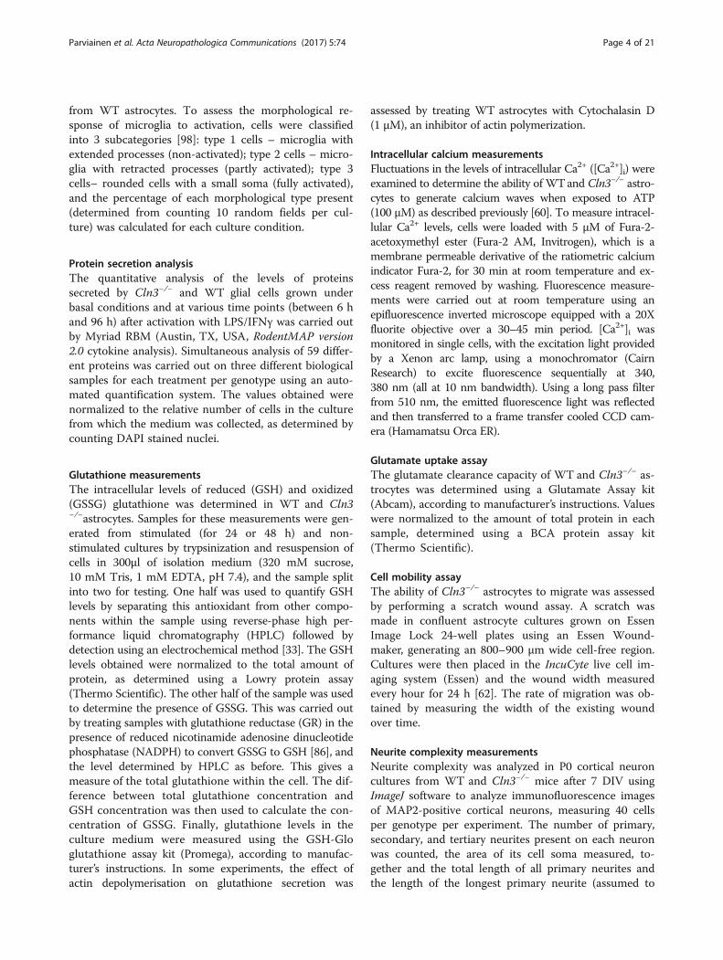
visual cortex (V1) of Cln3−/− mice, (from 6.5–21 monthsof age) with that of wildtype controls and Tpp-1−/− mice(at 4 months of age, representing disease end stage), amodel for CLN2 disease [84], an earlier onset and morerapidly progressing type of NCL [4]. In V1 of theseseverely affected Tpp-1−/− mice, the morphological fea-tures characteristic of reactive astrocytosis were evidentwithin these astrocytes, with intense GFAP immunoreac-tivity, thickened processes and pronounced hypertrophy(Fig. 1a). This astrocytosis in Tpp-1−/− mice displayedlaminar specificity, being most pronounced in laminae IIand III, V and VI.In comparison, in V1 of Cln3−/− mice, GFAP immuno-
reactivity revealed a markedly different extent of astrocy-tosis, with substantial differences in astrocyte morphology.Apart from a small population of darkly immunostainedastrocytes present in the most ventral portion of laminaVI, the majority of astrocytes in V1 of presymptomatic6.5 month old Cln3−/− mice displayed a protoplasmicmorphology with numerous thin processes, compared tothe characteristic appearance of fully activated astrocytesin Tpp-1−/− mice (Fig. 1a). Initially largely confined tolaminae I and IV, these protoplasmic astrocytes became
Fig. 1 Attenuated glial responses in Cln3−/− mouse tissue and in humanJNCL. Cortical sections from wild type (WT), Cln3−/− and Tpp-1−/− mice (a,b) or from LINCL and JNCL human cases (c) were immunostained withGlial Fibrillary Acid Protein (GFAP) or Cluster of Differentiation 68 (CD68)to investigate the level of reactive astrocytosis or microglial activation,respectively. Compared to the very low level of glial activationpresent in WT mice (shown at 6.5 months of age, but changes verylittle over time), marked astrocytosis (a) and microglial activation(b) was apparent in severely affected 4-month-old Tpp-1−/− mousesections, with hypertrophied astrocytes with intense GFAP immunostainingand thickened processes, and morphologically transformed microglia withintense CD68 immunostaining being observed. a In contrast, astrocytosisappeared different in nature in the Cln3−/− cortex, and although manyastrocytes displayed intense GFAP immunoreactivity, especially in thedeeper laminae, most these astrocytes still retained many thin processesreminiscent of protoplasmic astrocytes. Even at the end stages of thedisease (21-month-old Cln3−/− mice) very few astrocytes became fullyhypertrophied. b A similar attenuated morphological transformation ofmicroglial cells was also apparent in the cortex of Cln3−/− mice. Althoughmicroglia became more intensely CD68 immunoreactive and moreswollen with increased age, many of these cells retaining longbranched processes, even in 21-month-old Cln3−/− mice. (c) Reactiveastrocytosis was observed in both human INCL and JNCL (c), butto very different extents. In LINCL cases astrocytes were intenselyimmunostained, with hypertrophied cell bodies and numerousthickened processes. In JNCL cases GFAP immunostaining was palerand far fewer, less hypertrophied astrocytes with thinner processeswere evident. Only a few CD68 positive microglia were observed inJNCL cases (c), compared to the relative abundance of activated CD68immunoreactive microglia activation present in the cortex of LINCLcases. Scale bars = 120 μm (a, b) 20 μm in higher magnification views;50 μm (c). Roman numerals in a, b indicate cortical laminar boundaries
Parviainen et al. Acta Neuropathologica Communications (2017) 5:74 Page 5 of 21
more widespread within V1 of Cln3−/− mice with diseaseprogression, being present in lamina II and most of lam-inae V and VI at 12 months of age, and additionally inlamina IV by 21 months of age. Although the intensity ofGFAP immunoreactivity progressively increased withtime, only a small proportion of these astrocytes (mostlywithin lamina VI) displayed thickened processes or anenlarged cell soma, with most retaining a protoplasmicappearance with thin processes, even in severely affected21 month old Cln3−/− mice. These data suggest that atleast a subset of Cln3−/− astrocytes do retain the ability totransform morphologically, but only in a protracted man-ner towards disease end-stage.A similarly attenuated activation of microglia was also
evident in the cortex of Cln3−/− mice (Fig. 1b). AlthoughCD68 immunoreactive microglia were clearly moredarkly immunostained in V1 of Cln3−/− mice than in WTcontrols at all ages examined, these microglia retained arelatively small cell soma and numerous long thin pro-cesses. Only in severely affected Cln3−/− mice by 21 months,were more overtly swollen and activated microglia wereseen, and these were largely restricted to the more ventralportion of lamina VI and V. Even then, many of these swol-len CD68 positive cells retained long thickened processes.This is in marked contrast to the V1 of 4-month-old Tpp-1−/− mice that were at disease end-stage, where numerousintensely immunostained, completely rounded, and fully ac-tivated amoeboid microglia were evident.Taken together, these data suggest that loss of Cln3
impairs the morphological transformation of both astro-cytes and microglia, which is limited and only occurs latein the disease, in more severely affected Cln3−/− mice.Having described a relatively limited glial response in thehippocampus of human CLN3 disease cases [90], we nextinvestigated whether a similar phenotype was also evidentin the primary visual cortex of CLN3 disease patient brainautopsy material (Fig. 1c). Numerous intensely immuno-stained GFAP positive hypertrophied astrocytes with manythickened processes were observed in V1 of CLN2 cases.In contrast, only a few weakly immunostained GFAP ex-pressing astrocytes with thin processes were present inthis region of CLN3 disease cases (Fig. 1c, top row). Simi-larly, dramatically fewer CD68 positive microglia were ob-served in V1 of CLN3 disease cases compared with CLN2disease cases (Fig. 1c, bottom row).These morphological observations suggest that the
basic biology of glia, the neuronal support cells, may beimpaired in CLN3 disease. To begin investigating howloss of CLN3 expression could influence glial cell func-tion, we compared the properties of Cln3−/− and WTglial cells using primary cultures of either astrocytes ormicroglia, having first defined the composition of ourastrocyte (Additional file 1: Figure S1 and Additionalfile 2: Figure S2) and microglial monocultures
(Additional file 3: Figure S3). One week after platingmicroglial cultures showed over 99% of DAPI stainedcells expressed CD68 (99.97 ± 0.02% and 99.97 ± 0.02%CD68 + ve in WT and Cln3−/−, respectively; with only0.03 ± 0.02% and 0.03 ± 0.03% being GFAP + ve). Oneweek after plating over 98% of DAPI positive cells inour astrocyte cultures were positive for GFAP (WT:98.80 ± 0.28% GFAP + ve, 1.90 ± 0.17% CD68 + ve, and0.10 ± 0.10% O4 + ve; Cln3−/−: 98.86 ± 0.10% GFAP + ve,1.05 ± 0.13% CD68 + ve, and 0.10 ± 0.03% O4 + ve).With subsequent time in culture an increased fraction ofDAPI stained, but GFAP negative cells were apparent inour astrocyte cultures, especially those from WT mice(e.g. see Fig. 3A.). However, at these later time points vir-tually all these DAPI labelled cells immunostained posi-tively for a second astrocyte marker glutamine synthetase(see Additional file 2: Figure S2 for an example taken48 h later), suggesting a dynamic down-regulation ofGFAP over time under some culture conditions. Never-theless, a minor contamination of our astrocyte cul-tures with ependymal or endothelial cells cannot beexcluded.
Morphological responses of Cln3−/− glia to activation areattenuatedTo determine whether the attenuated morphologicalresponse of Cln3−/− glia observed in vivo was mimickedin vitro, we stimulated monocultures of either microgliaand astrocytes and assessed their ability to undergo mor-phological changes. To do this we exposed cultures tostandard inflammatory stimuli that up-regulate pathwaysassociated with immune and injury-related functions[38], either the bacterial endotoxin lipopolysaccharide(LPS) alone to activate microglia, or to LPS combinedwith interferon-γ (IFN), which synergizes with the ef-fects of LPS, to activate astrocytes [26].We first confirmed that Cln3−/− glia were able to acti-
vate relevant downstream signaling pathways (phosphor-ylated NF-κβ subunit p65 and phosphorylated STAT-1,as downstream effectors of LPS and IFNγ stimulationrespectively, [22, 94]) (Additional file 4: Figure S4). Next,to characterize the morphological transformation ofmicroglia, WT and Cln3−/− microglial cultures wereimmunostained with CD68 at various time-points afteractivation (2, 6, 12, 24, 48, 72 and 96 h), dividing thesecells according to their morphology into Type 1 (non-ac-tivated), Type 2 cells (partly activated) and Type 3 cells(fully activated) (see methods). Even under basal condi-tions, CD68 immunoreactivity appeared more intense inCln3−/− vs. WT microglial cultures, with more roundedcells present (Fig. 2A). Upon stimulation, microglia ofboth genotypes morphologically transformed, but manyfewer Cln3−/− microglia changed shape after 24 h (Fig.2A). When these changes were quantified (see Fig. 2B),
Parviainen et al. Acta Neuropathologica Communications (2017) 5:74 Page 6 of 21

there were more type 2 cells in Cln3−/− vs. WT microglialcultures under basal conditions (Fig. 2C, comparepanels a and b), suggesting a higher level of basal activa-tion, but a slower morphological transformation of CLN3disease microglia. A transformation of Type 1 cells intoType 2 cells occurred in microglial cultures of both geno-types upon stimulation, however, Cln3−/− microgliaresponded more slowly than WT microglia, with a slowerdecline in the number of Type 1 cells (Fig. 2C, a, b) and aslower increase in the number of Type 2 cells (Fig. 2C,compare panels c and d). Until 48 h very little change was
observed in the percentage of Type 3 cells under anycondition (Fig. 2C), but by 72 h there was a dramaticincrease in the proportion of this fully activated cell typewithin both WT and Cln3−/− microglial cultures under allconditions (Fig. 2c–f ). This change was accompanied by areduction in the percentage of both Type 1 and Type 2,suggesting a morphological transformation into Type 3cells with increased time in culture.The morphological response of astrocytes to stimulation
(LPS/INFγ treatment for 24 h or 48 h) was assessed inGFAP immunostained cultures. Even under basal
Fig. 2 Attenuated morphological transformation of Cln3−/− microglia. The morphology of wild type (WT) and Cln3-deficient (Cln3−/−) microglia studiedunder basal conditions and after stimulation with LPS was revealed by CD68 immunostaining (red). A Cultures of unstimulated WT microglia were mainlybipolar but upon LPS stimulation for 24 h these cells rapidly changed shape. In contrast, while cultures of Cln3−/− microglia exhibited a heterogeneousmorphology and had intense immunostaining for CD68 under basal conditions these cells failed to dramatically change shape upon stimulation for 24 h.B To quantify morphological changes over time, cells were divided into three categories: Type 1 cells (resting microglia); Type 2 cells (migrating/activatedmicroglia); type 3 (amoeboid activated microglia). C The transformation of type 1 cells into type 2 cells initially occurred more slowly in Cln3−/− microglialcultures upon stimulation, with type 3 cells first appearing in cultures of both genotypes around 48 h regardless of treatment. Scale bars = 50 μm (A, B)
Parviainen et al. Acta Neuropathologica Communications (2017) 5:74 Page 7 of 21
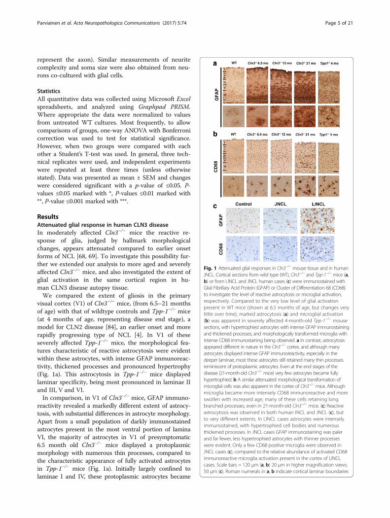
conditions, untreated Cln3−/− astrocytes had a strikinglydifferent morphology to WT astrocytes, appearing largerand flatter, with disrupted intermediate filaments (Fig. 3).Upon stimulation, WTastrocytes already began to morpho-logically transform after 24 h; changing from broad, non-process bearing, flat cells into cells with a shrunken somaand multiple branched processes (as described in [53]) (Fig.3A, c arrowheads). These changes become more apparentwith time (Fig. 3A, e). In contrast, no significant morpho-logical transformation of Cln3−/− astrocytes could be de-tected until 48 h stimulation, when soma size began todecrease and some cells developed processes (Fig. 3A, f).To quantify these changes the soma size of WT and Cln3−/− astrocytes were compared (Fig. 3B). After activation for24 h or 48 h, the cell soma of WT astrocytes becamesmaller, and this was statistically significant after 24 h(30.5% ± 3.3 decrease). After 24 h of stimulation the somasize of Cln3−/− astrocytes remained unchanged, but after48 h of stimulation was not statistically different to that ofstimulated WTastrocytes (Fig. 3C).These data demonstrate that Cln3−/− astrocytes and
microglia are attenuated in their ability to change theirmorphology upon stimulation, suggesting that these cellsretain at least some of their in vivo disease characteris-tics when cultured.
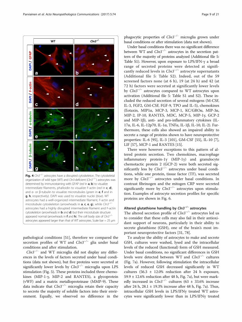
Cln3−/− astrocytes, but not Cln3−/− microglia, have a disruptedcytoskeletonSince morphological changes require cytoskeletal rear-rangements, and GFAP immunostaining suggested thatintermediate filament organization was perturbed inCln3−/− astrocytes (Fig. 3A), we also immunostained as-trocytes for α- and β-tubulin to visualize microtubulesand with phalloidin to visualize F-actin filaments and asimilar cytoskeletal analysis was performed with microglia.Both intermediate filaments and F-actin filaments ap-
peared less defined and highly disorganized in Cln3−/−
vs. WT astrocytes (Fig. 4, a, b for GFAP, and c, d for F-actin, examples marked with arrowheads). Most Cln3−/−
astrocytes lacked F-actin filaments that spanned the cellbody, a common morphological feature of cultured WTastrocytes (Fig. 4c, d). However, the α- and β-microtubular organization of WT and Cln3−/− astrocytesappeared similar (Fig. 4, e, f and g, h, examples markedwith arrows). These data reveal that enlarged Cln3−/−-astrocytes have an abnormally organized actin and inter-mediate filament cytoskeleton, whilst their microtubuleorganization appears normal. No overt changes were ob-served in the cytoskeletal organization of Cln3−/−micro-glia (data not shown).
Cln3−/− glia show altered protein secretion profilesThe secretion of soluble factors is a key feature of bothmicroglia and astrocytes under both physiological and
Fig. 3 Attenuated morphological transformation of Cln3−/−
astrocytes. The morphology of wild type (WT) and Cln3-deficient(Cln3−/−) astrocytes was studied under basal conditions and afterstimulation with LPS/IFNγ for 24 or 48 h by immunostaining withGFAP (red in A, green in B). DAPI (blue) was used to visualize allnuclei. A WT astrocytes changed their morphology dramatically aftera 24 h exposure to LPS/INFγ to display characteristic branchedprocesses (arrowheads) and these changes became enhanced overtime. In contrast Cln3−/− astrocytes remained relativelymorphologically unchanged after 24 h of stimulation remaining aslarge flat cells with no processes, but showed morphologicalchanges after 48 h activation. B ImageJ was used to quantifyastrocyte cell body size under all experimental conditions bydrawing around the soma of GFAP positive cells (dashed lines, withcontained area shaded red). C The mean cell soma sizes weredetermined by quantifying 10 random fields per coverslip and aminimum of two coverslips per experiment. Scale bar = 50 μm
Parviainen et al. Acta Neuropathologica Communications (2017) 5:74 Page 8 of 21
pathological conditions [51], therefore we compared thesecretion profiles of WT and Cln3−/− glia under basalconditions and after stimulation.Cln3−/− and WT microglia did not display any differ-
ences in the levels of factors secreted under basal condi-tions (data not shown), but five proteins were secreted atsignificantly lower levels by Cln3−/− microglia upon LPSstimulation (Fig. 5). These proteins included three chemo-kines (MIP-1-γ, MIP-2 and RANTES), a glycoprotein(vWF) and a matrix metalloproteinase (MMP-9). Thesedata indicate that Cln3−/− microglia retain their capacityto secrete the majority of soluble factors into their envir-onment. Equally, we observed no difference in the
phagocytic properties of Cln3−/− microglia grown underbasal conditions or after stimulation (data not shown).Under basal conditions there was no significant difference
between WT and Cln3−/− astrocytes in the secretion pat-tern of the majority of proteins analyzed (Additional file 5:Table S1). However, upon exposure to LPS/IFN-γ a broadrange of secreted proteins were detected at signifi-cantly reduced levels in Cln3−/− astrocyte supernatants(Additional file 5: Table S2). Indeed, out of the 59screened factors none (at 6 h), 19 (at 24 h) and 42 (at72 h) factors were secreted at significantly lower levelsby Cln3−/− astrocytes compared to WT astrocytes uponactivation (Additional file 5: Table S1 and S2). These in-cluded the reduced secretion of several mitogens (M-CSF,IL-3, FGF2, GM-CSF, FGF-9, TPO and IL-5), chemokines(Eotaxin, MIP1α, MCP-3, MCP-1, KC/GROα, MIP-3α,MIP-2, IP-10, RANTES, MDC, MCP-5, MIP-1γ, GCP-2and MIP-1β), anti- and pro-inflammatory cytokines (IL-17α, IL-6, IL-12p70, IL-1α, TNFα, IL-1β, IL-10, IL-2). Fur-thermore, these cells also showed an impaired ability tosecrete a range of proteins shown to have neuroprotectiveproperties: IL-6 [95], IL-3 [101], GM-CSF [55], IL-10 [7],LIF [57], MCP-1 and RANTES [15].There were however exceptions to this pattern of al-
tered protein secretion. Two chemokines, macrophageinflammatory protein-1γ (MIP-1γ) and granulocytechemotactic protein 2 (GCP-2) were both secreted sig-nificantly less by Cln3−/− astrocytes under basal condi-tions, while one protein, tissue factor (TF), was secretedmore by Cln3−/− astrocytes under basal conditions. Incontrast fibrinogen and the mitogen CRP were secretedsignificantly more by Cln3−/− astrocytes upon stimula-tion. Examples of astrocyte secretion profiles for specificproteins are shown in Fig. 6.
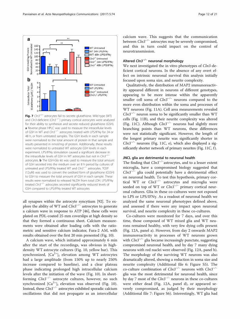
Altered glutathione handling by Cln3−/− astrocytesThe altered secretion profile of Cln3−/− astrocytes led usto consider that these cells may also fail in their antioxi-dant support of neurons, particularly in their ability tosecrete glutathione (GSH), one of the brain’s most im-portant neuroprotective factors [31, 78].To analyze the ability of astrocytes to make and secrete
GSH, cultures were washed, lysed and the intracellularlevels of the reduced (functional) form of GSH measured.Under basal conditions, no significant differences in GSHlevels were detected between WT and Cln3−/− cultures(Fig. 7a). However, following stimulation the intracellularlevels of reduced GSH decreased significantly in WTcultures (56.3 ± 12.0% reduction after 24 h exposure,59.9 ± 12.6% reduction after 48 h, Fig. 7a), but were mark-edly increased in Cln3−/− cultures (63 ± 33.6% increaseafter 24 h, 28.1 ± 19.3% increase after 48 h, Fig. 7a). Thus,intracellular GSH levels in LPS/IFNγ treated WT astro-cytes were significantly lower than in LPS/IFNγ treated
Fig. 4 Cln3−/− astrocytes have a disrupted cytoskeleton. The cytoskeletalorganization of wild type (WT) and Cln3-deficient (Cln3−/−) astrocytes wasdetermined by immunostaining with GFAP (red in a, b) to visualizeintermediate filaments, phalloidin to visualize F-actin (red in c, d),and α- or β-tubulin to visualize microtubules (green in e, f and ing, h, respectively). DAPI was used to visualize nuclei (blue). WTastrocytes had a well-organized intermediate filament, F-actin andmicrotubule cytoskeleton (arrowheads in a, c, e, g), while Cln3−/−
astrocytes had a highly disrupted intermediate filament and F-actincytoskeleton (arrowheads in b and d) but their microtubule structureappeared normal (arrowheads in f and h). The cell body size of Cln3−/−
astrocytes appeared larger than that of WT astrocytes. Scale bar = 25 μm
Parviainen et al. Acta Neuropathologica Communications (2017) 5:74 Page 9 of 21

Cln3−/− astrocytes after 24 (59.6 ± 12.2% decrease in WTvs. Cln3−/−) and 48 h (51.9 ± 14.3% decrease in WT vs.Cln3−/−). These data suggest a failure of Cln3−/− astrocytesto secrete GSH, consistent with the observation thatsignificantly higher levels of extracellular GSH could bedetected in the medium from WT vs. Cln3−/− astrocytesafter stimulation (Fig. 7b).Since the method described above only measures the
reduced form of glutathione, it is conceivable that theseresults could be explained by an opposing change in thelevel of oxidized glutathione (GSSG). This possibilitywas excluded by indirectly measuring intracellular GSSGlevels by converting GSSG to GSH using glutathionereductase (GR). Subsequent HPLC analysis revealed nomeasurable difference between the intracellular levels ofGSH and total glutathione (after GR treatment) in anyof these samples, suggesting that all the intracellularglutathione in both WT and Cln3−/− astrocytes is presentin the reduced form (data not shown).The actin cytoskeleton is important for exocytosis in
astrocytes [70], and it appears abnormally organized inCln3−/− astrocytes (Fig. 3A). Therefore, the possible con-nection between the disrupted actin cytoskeleton and
impaired glutathione secretion was examined by treatingWT astrocytes with cytochalasin D (1uM for 30 min be-fore the 8 h measurement period) to inhibit thepolymerization of actin. This resulted in a significant reduc-tion (50.1 ± 12.7%) in the levels of extracellular glutathionein LPS/IFNγ treated WT astrocytes (Additional file 6:Figure S5). Thus, a normal actin cytoskeleton is essen-tial for glutathione secretion by astrocytes.Since the defects in Cln3−/− astrocytes appeared more
profound than those in microglial cells, and the cytoskeletaldisruption observed in these cells could impact many oftheir functions, we investigated whether Cln3−/− astrocytescould perform other key tasks effectively.
An impaired ability of Cln3−/− astrocytes to migrate, clearglutamate and signal via Ca2+
Slower migration of Cln3−/− astrocytesAstrocyte migration is associated with local inflamma-tion [17] and requires cytoskeletal rearrangements, rais-ing the likelihood that this process may be impaired inCln3−/− astrocytes. To test this possibility, an EssenWound-maker was used to create a cell-free area in con-fluent cultures of WT and Cln3−/− astrocytes, and the
Fig. 5 Altered protein secretion profiles of Cln3−/− microglia. Secreted protein levels were quantified from supernatants collected from WT andCln3−/− microglial cultures grown under basal conditions, or after stimulation with LPS. Following quantitative analysis of 59 soluble factors, 5were found to be secreted at significantly lower levels by Cln3-deficient (Cln3−/−) microglia over time in culture compared to wild type (WT)microglial cultures. These included the chemokines MIP-1-γ, MIP-2 and RANTES, the glycoprotein vWF, and the matrix metalloproteinase MMP-9
Parviainen et al. Acta Neuropathologica Communications (2017) 5:74 Page 10 of 21

ability of the cells to migrate and fill this space assessed.WT astrocytes migrated into the cell free area rapidlyand nearly closed the gap within 24 h (Fig. 8a). The dis-tance covered by Cln3−/− astrocytes over this same timewas significantly reduced (Fig. 8b). The rate of migrationwas significantly decreased in the absence of CLN3 (WT5.3 ± 0.6 μm/h vs. 2.3 ± 0.6 μm/h for Cln3−/− astrocytes)(Fig. 8c), and WT astrocytes migrated further and fasterthan Cln3−/− astrocytes.
Cln3−/− astrocytes show impaired glutamate clearanceA feature of JNCL pathogenesis is an elevated level ofglutamate in the brains of Cln3−/− mice [65]. A glutam-ate assay kit revealed that Cln3−/− astrocytes take-up
significantly less glutamate from the medium than WTastrocytes (48.0% ± 14.0% reduction in glutamate uptake,Fig. 9), suggesting that Cln3−/− astrocytes may not beable to scavenge excess extracellular glutamate as effect-ively as WT astrocytes.
Cln3−/− astrocytes do not form a synchronized calcium waveCalcium signaling forms the basis for astrocyte-astrocyteand astrocyte-neuron communication in the CNS [103].Indeed, Ca2+ is exploited by astrocytes as an intercellularsignal for long distance communication through func-tionally connected astrocyte networks. This synchronouscalcium wave is propagated via gap junctions and hasthe potential to coordinate neurotransmitter release at
Fig. 6 Cln3−/− astrocytes show differences in their ability to secrete proteins. Secreted protein levels were quantified from supernatants collectedfrom wild type (WT) and Cln3-deficient (Cln3−/−) astrocyte cultures grown under basal conditions or after stimulation with LPS/IFNγ. Examples ofchemokines (a), neuroprotective factors (b) and proteins secreted at elevated levels by Cln3−/− astrocytes following activation (c), are shown. aCln3−/− astrocytes secreted significantly less MIP-1ß, MIP-1α and MCP-1 when treated with LPS/IFNγ than did WT astrocytes. There was no significantdifference between untreated WT and Cln3−/− samples. b Cln3−/− astrocytes secreted significantly less IL-6, TNF-α and VEGF upon activation than didWT astrocytes. There was no significant difference between untreated samples. c Cln3−/− astrocytes secreted significantly more CRP and IgA after 24 hexposure to LPS/IFNγ, no such change was observed in WT astrocytes
Parviainen et al. Acta Neuropathologica Communications (2017) 5:74 Page 11 of 21
all synapses within the astrocyte syncytium [92]. To ex-plore the ability of WT and Cln3−/− astrocytes to generatea calcium wave in response to ATP [46], these cells wereplated on PDL-coated 25 mm coverslips at high density sothat they formed a continuous sheet. Calcium measure-ments were obtained after loading cells with the ratio-metric and sensitive calcium indicator, Fura-2 AM, withresults obtained over the first 20 min presented (Fig. 10).A calcium wave, which initiated approximately 6 min
after the start of the recordings, was obvious in high-density WT astrocyte cultures (Fig. 10, yellow bar). Thissynchronized, [Ca2+]I elevation among WT astrocyteshad a large amplitude (from 150% up to nearly 250%increase compared to baseline), and a clear plateauphase indicating prolonged high intracellular calciumlevels after the initiation of the wave (Fig. 10). In sheet-forming Cln3−/− astrocyte cultures, however, no suchsynchronized [Ca2+]I elevation was observed (Fig. 10).Instead, these Cln3−/− astrocytes exhibited sporadic calciumoscillations that did not propagate as an intercellular
calcium wave. This suggests that the communicationbetween Cln3−/− astrocytes may be severely compromised,and this in turn could impact on the control ofneurotransmission.
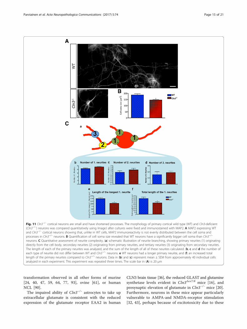
Altered Cln3−/− neuronal morphologyWe next investigated the in vitro phenotypes of Cln3-de-ficient cortical neurons. In the absence of any overt ef-fect on intrinsic neuronal survival this analysis initiallyfocused upon soma size, and neurite complexity.Qualitatively, the distribution of MAP2 immunoreactiv-
ity appeared different in neurons of different genotypes,appearing to be more intense within the apparentlysmaller cell soma of Cln3−/− neurons compared to themore even distribution within the soma and processes ofWT neurons (Fig. 11A). Cell area measurements revealedCln3−/− neuron soma to be significantly smaller than WTcells (Fig. 11B), and their neurite complexity was altered(Fig. 11C). Although Cln3−/− neurons had slightly morebranching points than WT neurons, these differenceswere not statistically significant. However, the length ofthe longest primary neurite was significantly shorter inCln3−/− neurons (Fig. 11C, e), which also displayed a sig-nificantly shorter network of primary neurites (Fig. 11C, f).
JNCL glia are detrimental to neuronal healthThe finding that Cln3−/−astrocytes, and to a lesser extentmicroglia, have a compromised biology suggested thatCln3−/− glia could potentially have a detrimental effecton neuronal health. To test this hypothesis, primary cor-tical WT or Cln3−/− astrocytes and microglia wereseeded on top of WT or Cln3−/− primary cortical neur-onal cultures. Glia in these co-cultures were not exposedto LPS or LPS/IFNγ. As a readout of neuronal health weanalyzed the same neuronal phenotypes defined above,and assessed if there were any impact upon neuronalsurvival, and neurite complexity in these co-cultures.Co-cultures were monitored for 7 days, and over this
time, those composed of WT mixed glia and WT neu-rons remained healthy, with very few dying cells present(Fig. 12A, panel a). However, from day 2 onwards MAP2immunoreactivity in processes of WT neurons grownwith Cln3−/− glia became increasingly punctate, suggestingcompromised neuronal health, and by day 7 many dyingneurons with red nuclei were observed (Fig. 12A, panel b).The morphology of the surviving WT neurons was alsodramatically altered, showing a reduction in soma size andneurite complexity (Additional file 6: Figure S5). Theco-culture combination of Cln3−/− neurons with Cln3−/−
glia was the most detrimental for neuronal health, sinceby day 7 most of the Cln3−/− neurons in these co-cultureswere either dead (Fig. 12A, panel d), or appeared se-verely compromised, as judged by their morphology(Additional file 7: Figure S6). Interestingly, WT glia had
Fig. 7 Cln3−/− astrocytes fail to secrete glutathione. Wild type (WT)and Cln3-deficient (Cln3−/−) primary cortical astrocytes were analyzedfor their ability to synthesize and secrete reduced glutathione (GSH).a Reverse phase HPLC was used to measure the intracellular levelsof GSH in WT and Cln3−/− astrocytes treated with LPS/IFNγ for 24 or48 h, or from untreated samples. The GSH levels in each samplewere normalized to the total amount of protein in that sample andresults presented in nmol/mg of protein. Additionally, these resultswere normalized to untreated WT astrocyte GSH levels in eachexperiment. LPS/IFNγ stimulation caused a significant decrease inthe intracellular levels of GSH in WT astrocytes but not in Cln3−/−
astrocytes. b The GSH-Glo kit was used to measure the total amountof GSH secreted into the medium over an 8 h period by cultures ofuntreated and LPS/IFNγ treated WT and Cln3−/− astrocytes. TCEP(12uM) was used to convert the oxidised form of glutathione (GSSH)to GSH to measure the total amount of GSH in each sample. Theseresults were normalized to released %LDH from total LDH. LPS/IFNγtreated Cln3−/− astrocytes secreted significantly reduced levels ofGSH compared to LPS/IFNγ treated WT astrocytes
Parviainen et al. Acta Neuropathologica Communications (2017) 5:74 Page 12 of 21

a positive influence on both the survival and morph-ology of Cln3−/− neurons (Fig. 12A, panel c).In these co-cultures, we observed that in the presence of
Cln3−/− neurons, Cln3−/− astrocytes had smaller cell bodiesand longer, more numerous processes (reminiscent of acti-vated astrocytes in culture), when compared to Cln3−/−-astrocytes grown with WT neurons (Additional file 8:Figure S7). No such morphological change was evident
when Cln3−/− neurons were co-cultured with WT glia,suggesting that Cln3−/− astrocytes are more sensitive tothe environment than their WT counterparts. Under allculture conditions the morphology of microglia wereheterogeneous with some cells bearing processes andothers being fully rounded (data not shown).These morphological findings correlated well with
measurements of released LDH from the different co-cultures (Fig. 12B). The lowest LDH levels were ob-served when WT glia and neurons were co-cultured, butthese levels increased dramatically when Cln3−/− gliawere co-cultured with WT or Cln3−/− neurons (Fig. 12B).However, when Cln3−/−neurons were co-cultured withWT, rather than Cln3−/− glia, a lower level of LDH re-lease was observed, possibly due to the supportive influ-ence of the WT cells (Fig. 12B). As might be expected,there was significantly more LDH released in Cln3−/−
glia/Cln3−/− neuron co-cultures than in WT glia/WTneuron co-cultures (Fig. 12B).These results suggest that Cln3−/− glia are detrimental
to the health of both WT and Cln3−/− neurons, withCln3−/− neurons being the most vulnerable. In contrast,WT glia appeared to have a positive influence on Cln3−/−neurons, not just on survival, but also upon neuritecomplexity.
DiscussionThis study highlights the attenuated morphologicaltransformation of astrocytes and microglia in both humanand murine CLN3 disease. From studying cultured Cln3−/−
astrocytes and microglia we have provided further supportthat their biology is impaired (see 16 [99]). Although
Fig. 9 Glutamate clearance is altered in Cln3−/− astrocytes. Theability of wild type (WT) and Cln3-deficient (Cln3−/−) astrocytes toclear glutamate from the medium was assessed using a GlutamateAssay Kit. WT and Cln3−/− astrocytes were incubated with 2 mMglutamate for 2 h, and wells without astrocytes were used ascontrols. The glutamate remaining in the medium was quantifiedand normalized to the total amount of protein, and the glutamateuptake values of Cln3−/− astrocytes were normalized to those of WTastrocyte samples. Cln3−/− astrocytes took up significantly lessglutamate than did WT astrocytes over the 2 h period
Fig. 8 Cln3−/− Astrocytes have a migration defect. Wild type (WT) and Cln3-deficient (Cln3−/−) primary cortical astrocytes were plated on EssenImage Lock 24 well plates, grown to confluence then scratched using an Essen wound maker. a Representative pictures of the wound at threetime points. b The distance migrated by WT and Cln3−/− astrocytes every 4 h was calculated by comparing wound widths between the start andthe different time points. WT astrocytes migrated significantly further than did Cln3−/− astrocytes. c The rate of migration was measured bycalculating the distance migrated by these cells/h. Cln3−/− astrocytes migrated significantly slower than WT astrocytes. In each experiment threewound widths were measured per well and three wells quantified per experiment
Parviainen et al. Acta Neuropathologica Communications (2017) 5:74 Page 13 of 21

specific microglial defects are certainly evident, astrocytesappear more severely affected, and these astrocyte defectsmay be due to the cytoskeletal abnormalities they display.Most importantly, we show that while Cln3−/− neurons arethemselves compromised, the combined presence of Cln3−/− astrocytes and microglia exacerbate these phenotypesand have a detrimental effect on neuronal organization andhealth. Taken together, these data provide novel informa-tion that these glial cells exert a negative influence uponneurons and may directly influence neurodegeneration inCLN3 disease.
Defects in glial biology could underlie components ofCLN3 disease pathogenesisDespite concerted efforts, the normal function of CLN3remains poorly understood and it is unclear how its defi-ciency relates to cellular dysfunction, including that ofastrocytes or microglia. Despite microglia accumulatinglarge amounts of storage material, which is also presentin astrocytes, the current view is that it is not the accu-mulation of storage material per se that directly causes
cellular dysfunction and death. Instead it appears thatother, as yet unknown, consequences of Cln3-deficiencyare responsible. Our data suggest that these negativeconsequences of Cln3-deficiency are also evident in glia,rather than being confined to neurons, and it will beimportant to gain in vivo correlates of the data we havefound in tissue culture.Nevertheless, all the biological defects we found associ-
ated with cultured Cln3−/− astrocytes and microglia canplausibly be linked to known features of CLN3 diseasepathogenesis, including the potential involvement ofglutamate mediated excitotoxicity and oxidative stress. In-deed, although in vitro systems do not necessarily accur-ately reflect the in vivo situation, a series of similaritiesbetween our tissue culture observations and other reportsexist. For example, the attenuated ability of Cln3−/− glia torespond morphologically to stimulation is also evident inthe Cln3−/− mouse brain in vivo (Fig. 1, and [68, 69]), anda comparatively lower level of glial activation is evident inhuman CLN3 disease ([90], this study). This is in markedcontrast to the robust glial activation and morphological
Fig. 10 Cln3−/− Astrocytes Show Altered Calcium Signalling. Recordings of Fura-2 fluorescence were made from high density, sheet formingcultures of wild type (WT) and Cln3-deficient (Cln3−/−) astrocytes grown under basal conditions over a period of 30-45 min, from which the first20 min are shown. The figure illustrates changes in [Ca2+]I in three randomly selected WT and Cln3−/− astrocytes. In response to treatment with100 μM ATP, a propagating [Ca2+]I wave was generated by WT astrocytes (marked with yellow bar). This synchronized [Ca2+]I wave had a largeamplitude, and a prolonged plateau persisting for several minutes after initiation. The Cln3−/− astrocytes did not exhibit any propagating calciumwaves, instead, Cln3−/− astrocytes had non-synchronized, spontaneous [Ca2+]I elevations. Data is presented as 340 nm/380 nm ratio, which directlycorrelates with the change in intracellular free Ca2+ levels
Parviainen et al. Acta Neuropathologica Communications (2017) 5:74 Page 14 of 21
transformation observed in all other forms of murine[24, 40, 47, 59, 64, 77, 93], ovine [61], or humanNCL [90].The impaired ability of Cln3−/− astrocytes to take up
extracellular glutamate is consistent with the reducedexpression of the glutamate receptor EAA2 in human
CLN3 brain tissue [36], the reduced GLAST and glutaminesynthetase levels evident in Cln3Δex7/8 mice [16], andpresynaptic elevation of glutamate in Cln3−/− mice [20].Furthermore, neurons in these mice appear particularlyvulnerable to AMPA-and NMDA-receptor stimulation[32, 43], perhaps because of excitotoxicity due to these
Fig. 11 Cln3−/− cortical neurons are small and have shortened processes. The morphology of primary cortical wild type (WT) and Cln3-deficient(Cln3−/−) neurons was compared quantitatively using ImageJ after cultures were fixed and immunostained with MAP2. A MAP2 expressing WTand Cln3−/− cortical neurons showing that, unlike in WT cells, MAP2 immunoreactivity is not evenly distributed between the cell soma andprocesses in Cln3−/− neurons. B Quantification of cell soma size revealed that WT neurons have a significantly bigger cell soma than Cln3−/−
neurons. C Quantitative assessment of neurite complexity, (a) schematic illustration of neurite branching, showing primary neurites (1) originatingdirectly from the cell body, secondary neurites (2) originating from primary neurites, and tertiary neurites (3) originating from secondary neurites.The length of each of the primary neurites was analyzed, and the sum of the length of all of these neurites calculated. (b, c and d the number ofeach type of neurite did not differ between WT and Cln3−/− neurons. e WT neurons had a longer primary neurite, and (f) an increased totallength of the primary neurites compared to Cln3−/− neurons. Data in (b) and (c) represent mean ± SEM from approximately 40 individual cellsanalyzed in each experiment. This experiment was repeated three times. The scale bar in (A) is 20 μm
Parviainen et al. Acta Neuropathologica Communications (2017) 5:74 Page 15 of 21

elevated levels of glutamate, and different classes ofglutamate antagonists provide some therapeutic benefitin Cln3−/− mice [41–44].Another pivotal astrocyte function is the synthesis and
secretion of the anti-oxidant glutathione (GSH) thatplays a crucial role in protecting neurons against oxidativestress [31, 33]. Our data reveal that Cln3−/− astrocytes canstill make, but fail to secrete, glutathione. Indeed,Drosophila lacking CLN3 function are hypersensitiveto oxidative stress [89], and oxidative damage has alsobeen reported in both mouse [6] and human CLN3disease [3].Our calcium signaling studies revealed that Cln3−/− as-
trocytes fail to generate a calcium wave after exposure toATP, providing further evidence that intercellular signal-ing between CLN3 astrocytes may be compromised [16],and it will be important to study calcium signaling inacute slice preparations. This could impact upon thecontrol of neurotransmission in the JNCL brain and
perhaps contribute to the seizure activity observed inthis disease [58, 67, 88].All these functional problems associated with Cln3−/−
astrocytes and some of the phenotypes seen in vivo mayat least partially be explained by their disrupted cytoskel-eton, since expression of glutamate receptors at the cellsurface [48], calcium signaling among astrocytes [27]and secretion by astrocytes [45] have all been shown torequire a functional actin cytoskeleton. Indeed, the alteredshape of Cln3−/− astrocytes, along with the difficulties theyexhibit in changing their morphology in vivo and in ourculture, may plausibly results from their disrupted cyto-skeleton and it will be important to study this in moredetail and determine their importance in vivo. How thesedefects in the cytoskeleton are related to Cln3-deficiencyis unclear, but a functional interaction of CLN3 with non-muscle myosin-IIB has been reported [34], and a migra-tion defect in Cln3−/− mouse embryonic fibroblasts that isconsistent with our novel data for the impaired migrationof Cln3−/− astrocytes.
Alteration in protein secretion could impair cell-cellinteractionsThe protein secretion profiles of both Cln3−/− astrocytesand microglia was altered following activation, with as-trocytes being more severely affected, showing signifi-cantly reduced levels of secretion of a range of proteins(Additional file 5: Tables S1 and S2). Our data are con-sistent with the reported evidence that LPS stimulationalso results in a lower level of cytokine secretion bymicroglia derived from Cln3Δex7/8 mice bearing the 1 kbdeletion that is present in most CLN3 disease cases [99].Intriguingly, these authors suggest that the responses ofCln3-deficient microglia are stimulus-dependent, withceramide or neuronal cell lysates resulting in an in-creased inflammasome activation and expression of awide array of proinflammatory cytokines and chemo-kines [99]. However, it should be borne in mind thatthese authors used Cln3Δex7/8 ‘knock-in’ mice ratherthan the Cln3−/− mice used in our study, and this mayinfluence the different phenotypes observed.The biological significance of our data showing altered
secretion profiles of Cln3-deficient glia remains unclear,but the downstream effects are likely to be complexgiven the multiple and possibly synergistic effects of se-creted proteins on different cell types under both physio-logical and pathological situations [1, 19, 73, 74, 83]. It isalso important to emphasize that what we have detectedin vitro may not reflect the in vivo situation. Nevertheless,our data raise the possibility that cell-cell communicationvia secreted factors may potentially be perturbed in theCLN3 disease brain. In addition, many of the neuroprotec-tive proteins routinely secreted by WT astrocytes after ac-tivation [15, 30, 57, 76, 95, 100], are also significantly
Fig. 12 Cln3−/− cells negatively impact WT cells. P0 cortical wild type(WT) and Cln3-deficient (Cln3−/−) neuronal cultures were combinedwith either WT or Cln3−/− mixed glia cultures to study the impact ofthese glial cells on neuronal health. A After 7 days of co-culture, WTco-cultures were healthy (a) but Cln3−/− mixed glia appeared tohave a detrimental effect when cultured with both WT (b) andCln3−/− neurons (d) with the latter being more dramatically affected.When WT mixed glia were co-cultured with Cln3−/− neurons, neuronalsurvival improved. B Significantly less LDH released was observed inWT neuron/WT mixed glia co-cultures compared to Cln3−/− neuron/Cln3−/− mixed glia co-cultures. Scale bar in (A) = 20 μm
Parviainen et al. Acta Neuropathologica Communications (2017) 5:74 Page 16 of 21
reduced in cultures of activated Cln3−/− astrocytes, withtwo of these proteins (MCP-1 and RANTES) also beingsecreted at significantly lower levels by Cln3−/− microglia.The reduced expression of IL-6, RANTES and MCP-1,which can protect neurons against NMDA receptor-mediated excitotoxicity, may be especially relevant giventhe increased sensitivity of Cln3-deficient neurons toAMPA receptor-mediated excitoxicity [43, 65]. Thus, de-fects in glial-glial and glial-neuronal interactions have thepotential to have a significant impact on neuronal healthin CLN3 disease, a suggestion that prompted us to growmixed glial co-cultures with neurons.Until in vivo data regarding the relative levels of chemo-
kines and cytokines become available, our in vitro datademonstrating altered secretion levels should be interpretedwith caution, especially as these data come from pharmaco-logically stimulated cultures. However, the reduction inchemokine secretion by stimulated Cln3−/− glia in culturemay also have a detrimental effect on the recruitment ofmicroglia to sites of inflammation [72, 81], and partly ex-plain the limited infiltration of monocytes and lymphocytesin CLN3 disease [50]. This reduced chemokine expressionmay also be associated with the attenuated microglial acti-vation observed in vivo ([68, 69, 90], this study). Conversely,Cln3−/− astrocytes showed a reduced ability to secrete anti-inflammatory cytokines, such as IL-4, IL-10 and IL-2, whichcould also prove harmful. Both genetic and pharmaceuticalapproaches to attenuate the adaptive immune responsehave been shown to result in a significant improvement inthe pathology of Cln3−/− mice [80].
Cln3−/− glia are detrimental to neuronal healthDefects in glial biology have been associated with neuronaldysfunction and loss in many neurodegenerative diseases,see [28, 29, 66, 75, 85]. Both positive and negative roles forastrocytes have been proposed, and recently, more activeroles for astrocytes and microglia in directly influencingneuron survival have been postulated [49]. Using a co-culture approach, we have shown here that Cln3−/− astro-cytes and microglia can indeed influence neuronal health,affecting the size and neurite complexity of both WT andCln3−/− neurons, but also causing the death of the latter,which appear to be inherently compromised by Cln3deficiency. From our data it is not clear whether it isthe Cln3−/− astrocytes or microglia, or a combinationof both cell types that negatively influence neuronalheath. It has been suggested that astrocytes can be primedby microglia to become toxic to neurons [49], and it will beimportant to determine if similar mechanisms operate inCLN3 disease, especially in an in vivo context. However, itis apparent that despite any overt intrinsic survival defect inthese short-term cultures, Cln3−/− neurons appeared to becompromised in terms of their morphology, and it will beimportant to investigate their functional status.
In other lysosomal storage disorders, introducingastrocyte-specific gene mutations is sufficient to harmneurons [21, 28], and correcting these defects is benefi-cial [102]. In our studies co-culturing Cln3−/− neuronswith healthy glia improved many of their morphologicaldefects, and resulted in an increase in their survival,suggesting that healthy glia appear to have a neuroprotec-tive effect. This is in marked contrast to the apparentlynegative influence of Cln3−/−glia upon both healthy andmutant neurons. It remains to be seen how accurately ourdata from cultures reflect the in vivo situation, and for thisreason we have generated cell-type specific mutant micein which we can inactivate Cln3 in defined cell types inour future studies. Nevertheless, our data from this invitro study suggests that therapies that target glia inaddition to neurons may be an important step forward intreating this devastating disease.
ConclusionIn summary, this study has provided evidence that bothastrocytes and microglia derived from Cln3-deficientmice are dysfunctional, and this may contribute to dir-ectly harming neurons in this disorder. It will be import-ant to investigate the underlying mechanisms and theextent of pathological involvement of each cell type invivo. Given the close association between glial activationand neuron loss in these disorders, it will be importantto determine whether glia also contribute to neuron lossin the other forms of NCL. This will information will becrucial for determining whether strategies that target gliawill be of therapeutic value.
Additional files
Additional file 1: Figure S1. Composition of Astrocyte Cultures.Primary cortical astrocyte cultures generated from P1–2 wild type (WT)and Cln3-deficient (Cln3−/−) mice were grown for one week after theaddition of Ara C, and in this example stimulated for a further 48 hbefore being immunostained with CD68 to identify microglia, O4 toidentify oligodendrocytes, MAP2 together with NeuN to identify neuronsand GFAP to identify astrocytes. DAPI was used to visualize all nuclei. WTand Cln3−/− astrocyte cultures contained few microglia oroligodendrocytes (A) and no neurons (B), and the vast majority of cells inCln3−/− astrocyte cultures were GFAP-expressing astrocytes (C), with ahigher proportion of DAPI + ve cells showing much weaker or no GFAPimmunostaining in WT astrocyte cultures. Scale bar in (A) and(C) = 50 μm, and in (B) = 20 μm. (TIFF 13274 kb)
Additional file 2: Figure S2. Astrocyte Cultures stained with GlutamineSynthetase. Since GFAP expression can be down-regulated by astrocytesin culture, we also immunostained a parallel series of primary corticalastrocyte cultures from P1–2 wild type (WT) and Cln3-deficient (Cln3−/−)mice with glutamine synthetase as an additional marker of astrocytephenotype, after an additional 48 h in culture. DAPI was used to visualizeall nuclei. Virtually all the DAPI stained cells (blue) were also immunoreactivefor glutamine synthetase (red) in both WT and Cln3−/− cultures, and this wasquantified as being 99.71 ± 0.15% (WT) and 99.29 ± 0.21% (Cln3−/−) of theDAPI stained cells, respectively. Scale bar = 20 μm. (TIFF 2129 kb)
Additional file 3: Figure S3. Composition of Microglial Cultures.Primary cortical microglial cultures generated from P2–4 wild type (WT)
Parviainen et al. Acta Neuropathologica Communications (2017) 5:74 Page 17 of 21
and Cln3-deficient (Cln3−/−) mice were immunostained with CD68 toidentify microglia, O4 to identify oligodendrocytes, TuJ1 to identify neuronsand GFAP to identify astrocytes. DAPI was used to visualize all nuclei.Practically all cells were CD68 expressing microglial cells (A), with virtually nocells expressing GFAP or O4 (B). Scale bar = 20 μm. (TIFF 8572 kb)
Additional file 4: Figure S4. LPS and INFγ induced signaling is notaltered in Cln3−/− glia. Wild type (WT) and Cln3-deficient (Cln3−/−)astrocytes were immunostained with GFAP and microglia with CD68.DAPI was used to visualize all nuclei. Few WT or Cln3−/− glia with nuclear-located P-p65 (A, C) and WT or Cln3−/− astrocytes with nuclear-located P-STAT1 (B) were observed under basal conditions, while the vastmajority of both WT and Cln3−/− glia had P-STAT1 (B) and/or P-p65 (A,C) expressed in the nucleus upon stimulation. The percentage of cellsexpressing P-STAT1 and/or P-p65 in the nucleus was determined bycounting 5 random fields per coverslip and a minimum of threecoverslips per experiment. The means ±SEM shown are from threeseparate experiments. (TIFF 11278 kb)
Additional file 5: Table S1. Protein secretion profile of WT and Cln3−/−
astrocytes under basal conditions. Differences between levels of secretedproteins in supernatants collected after 6 h, 24 h and 72 h from Cln3−/−and WT astrocyte cultures grown under basal conditions. Datapresented as % change (values from Cln3−/− astrocyte samplescompared to corresponding WT astrocyte values) ± SEM from threebiological replicates. (−) indicates proteins whose levels were belowquantifiable detection levels. Table S2. Protein secretion profile of WTand Cln3−/− astrocytes after stimulation. Differences between levels ofsecreted proteins in supernatants collected from Cln3−/− and WTastrocytes after activation with LPS/IFNγ for 6 h, 24 h and 72 h. Datapresented as % change (Cln3−/− astrocyte sample values compared tocorresponding WT astrocyte values) ± SEM from three biologicalreplicates. (−) indicates proteins whose levels were below quantifiabledetection levels. (PDF 676 kb)
Additional file 6: Figure S5. An intact actin cytoskeleton is essential forglutathione secretion. To study the importance of the actin cytoskeletonfor GSH secretion in astrocytes Cytochalasin D (1uM) was added to wildtype (WT) astrocytes for 30 min prior to the start of the 8 h period overwhich the accumulation of secreted GSH in the medium was measured.Cells were then fixed and the actin cytoskeleton visualized with phalloidin.DAPI was used to visualize all nuclei. (A) Cytochalasin D clearly disrupted theF-actin filament organization in WT astrocytes. (B) Perturbing actin cytoskeletalpolymerization significantly inhibited GSH secretion by WT astrocytes. Scalebar in (A) = 10 um. (TIFF 13687 kb)
Additional file 7: Figure S6. Cln3−/− mixed glia negatively impactneuronal morphology. Representative images of MAP2 expressing Wildtype (WT) and Cln3-deficient (Cln3−/−) neurons co-cultured with WT orCln3−/− mixed glia are shown in (A) and quantification of neuronal somasize and neurite complexity under these different growth conditions areshown in (B-D). Cln3−/− neurons co-cultured with Cln3−/− mixed glia hada significantly smaller cell soma than did WT neurons co-cultured withWT glia (Aa, Ad, quantified in B). The substitution of WT mixed glia forCln3−/− mixed glia significantly increased the soma size of Cln3−/− neurons (Ac,Ad, quantified in B). The total length of primary neurites was significantlyreduced when Cln3−/− neurons were co-cultured with Cln3−/− mixed gliacompared to when WT neurons were co-cultured with WT glia (Aa, Ad,quantified in C). The presence of Cln3−/− mixed glia also significantlyreduced the length of the longest primary neurite in both WT and Cln3−/− neurons, and the length of the longest primary neurite was greaterwhen WT neurons were co-cultured with WT glia than when Cln3−/−
neurons were co-cultured with Cln3−/− mixed glia (C). The number ofprimary neurites (1. neurites that are extended from cell bodies) did notdiffer among the different co-cultures, but Cln3−/− mixed glia significantlyreduced the number of both secondary neurites (2. neurites that branch offfrom primary neurites) and tertiary neurites (3. neurites that branch off fromsecondary neurites) in Cln3−/− neurons (D). The presence of Cln3−/− mixedglia also significantly reduced the number of tertiary neurites in WT neurons(D). Scale bar in A = 20 μm. (TIFF 5626 kb)
Additional file 8: Figure S7. Altered astrocyte morphology in co-cultures with Cln3−/− neurons. When co-cultured with Cln3-deficient(Cln3−/−) neurons, Cln3−/− astrocytes (immunostained with GFAP, green)
changed shape, having smaller cell bodies and longer more numerousprocesses (reminiscent of activated astrocytes in culture). No suchchange was observed when Cln3−/− astrocytes were grown with wildtype (WT) neurons or when Cln3−/− neurons were grown with WTastrocytes. Scale bar = 20 μm. Nuclear stain DAPI (blue), Live/dead stain(red). (TIFF 10686 kb)
AcknowledgementsThe generous assistance of Myriad RBM in running the protein secretionassays is gratefully acknowledged. Natalie Masento (human section staining)and Sashya De Silva (glutamine synthetase staining) are acknowledged fortheir skilled assistance. Sybille Dihanich and Helen Brooks were recipients ofMedical Research Council DTA studentships, and Lotta Parviainen by anInstitute of Psychiatry, Psychology & Neuroscience departmental studentship.Prof. Tammy Kielian, Dr. Jill Weimer, Dr. Alison Barnwell, Dr. Allison Najafi andDr. Hemanth Ramesh Nelvagal are thanked for their very useful commentson the manuscript.
FundingThis study was supported by the Beyond Batten Disease Foundation, theBatten Disease Support and Research Association (USA), the Batten DiseaseFamily Association (UK), the Saoirse Foundation and Irish Health ResearchBoard, The NCL Stiftung, The Children’s Brain Disease Foundation, TheNatalie Fund and the Bletsoe Family.
Authors’ contributionsThe study was designed and supervised by JDC and BPW, with expert adviceand guidance from PR and HMM, and the input of all the authors. LP, SDand GWA performed all aspects of the tissue culture experiments, andanalysed these data; AMS and HRB performed the pathology experiments;the glutathione measurements were performed with and supervised by SPand SJH; the calcium imaging experiments were performed with andsupervised by RA; the scratch assays were performed with and supervised byGL; HMM also generated and provided the Cln3 deficient mice. Themanuscript was written by JDC, BPW, LP and SD with input from all theauthors, who approved the final version of the manuscript.
Ethics approval and consent to participateAll applicable international, national, and/or institutional guidelines for thecare and use of animals were followed. Specifically, all animal procedureswere performed in accordance with the UK Scientific Procedures (Animals)Act of 1996, under the UK Home Office Project License number 70/7364. Thestudies involving human autopsy material were in accordance with theethical standards of the Institute of Psychiatry Ethical Research Committee(approval numbers 223/00, 181/02), and with the 1964 Helsinki declarationand its later amendments or comparable ethical standards.
Competing interestsThe authors declare that they have no competing interests.
Publisher’s NoteSpringer Nature remains neutral with regard to jurisdictional claims inpublished maps and institutional affiliations.
Author details1Department of Basic and Clinical Neuroscience, King’s College London,Institute of Psychiatry, Psychology & Neuroscience, Maurice Wohl ClinicalNeuroscience Institute, 5 Cutcombe Road, London SE5 9RX, UK. 2Departmentof Molecular Neuroscience, Institute of Neurology, University CollegeLondon, Queen’s Square, London WC1N, 3BG, UK. 3Department of Life,Health and Chemical Sciences, The Open University, Walton Hall, MiltonKeynes MK7 6AA, UK. 4Wolfson Centre for Age-Related Diseases, King’sCollege London, London SE1 1UL, UK. 5Genetics and Genomic Medicine UCLGreat Ormond Street Institute of Child Health, University College London, 30Guilford Street, London WC1N 1EH, UK. 6Department of Pediatrics,Harbor-UCLA Medical Center, Los Angeles Biomedical Research Institute andDavid Geffen School of Medicine UCLA, 1124 West Carson Street, HanleyHardison Building, Torrance, CA 90502, USA.
Parviainen et al. Acta Neuropathologica Communications (2017) 5:74 Page 18 of 21
Received: 19 September 2017 Accepted: 23 September 2017
References1. Allan SM, Rothwell NJ (2003) Inflammation in central nervous system injury.
Philos Trans R Soc Lond Ser B Biol Sci 358:1669–16772. Anderson GW, Goebel HH, Simonati A (2013) Human pathology in NCL.
Biochim Biophys Acta 1832:1807–18263. Anzai Y, Hayashi M, Fueki N, Kurata K, Ohya T (2006) Protracted juvenile
neuronal ceroid lipofuscinosis–an autopsy report and immunohistochemicalanalysis. Brain and Development 28:462–465
4. Autti T, Raininko R, Santavuori P, Vanhanen SL, Poutanen VP, Haltia M (1997)MRI of neuronal ceroid lipofuscinosis. II. Postmortem MRI andhistopathological study of the brain in 16 cases of neuronal ceroidlipofuscinosis of juvenile or late infantile type. Neuroradiology 39:371–377
5. Benarroch EE (2013) Microglia: Multiple roles in surveillance, circuit shaping,and response to injury. Neurology 81:1079–1088
6. Benedict JW, Sommers CA, Pearce DA (2007) Progressive oxidative damagein the central nervous system of a murine model for juvenile Batten disease.J Neurosci Res 85:2882–2891
7. Bethea JR, Nagashima H, Acosta MC, Briceno C, Gomez F, Marcillo AE, LoorK, Green J, Dietrich WD (1999) Systemically administered interleukin-10reduces tumor necrosis factor-alpha production and significantly improvesfunctional recovery following traumatic spinal cord injury in rats. JNeurotrauma 16:851–863
8. Bible E, Gupta P, Hofmann SL, Cooper JD (2004) Regional and cellularneuropathology in the palmitoyl protein thioesterase-1 null mutantmouse model of infantile neuronal ceroid lipofuscinosis. Neurobiol Dis 16:346–359
9. Bithell A, Finch SE, Hornby MF, Williams BP (2008) FGF2 maintains theneurogenic capacity of embryonic neural progenitor cells in vitro butchanges their neuronal sub-type specification. Stem Cells 26:1565–1574
10. Björkblom B, Ostman N, Hongisto V, Komarovski V, Filén JJ, Nyman TA, KallunkiT, Courtney MJ, Coffey ET (2005) Constitutively active cytoplasmic c-Jun N-terminal kinase 1 is a dominant regulator of dendritic architecture: role ofmicrotubule-associated protein 2 as an effector. J Neurosci 25:6350–6361
11. Björkblom B, Padzik A, Mohammad H, Westerlund N, Komulainen E, HollosP, Parviainen L, Papageorgiou AC, Iljin K, Kallioniemi O, Kallajoki M, CourtneyMJ, Mågård M, James P, Coffey ET (2012) c-Jun N-terminal kinasephosphorylation of MARCKSL1 determines actin stability and migration inneurons and in cancer cells. Mol Cell Biol 32:3513–3526
12. Bolaños JP, Peuchen S, Heales SJ, Land JM, Clark JB (1994) Nitric oxide-mediated inhibition of the mitochondrial respiratory chain in culturedastrocytes. J Neurochem 63:910–916
13. Bond M, Holthaus SM, Tammen I, Tear G, Russell C (2013) Use of modelorganisms for the study of neuronal ceroid lipofuscinosis. Biochim BiophysActa 1832:1842–1865
14. Brown GC, Bolaños JP, Heales SJ, Clark JB (1995) Nitric oxide produced byactivated astrocytes rapidly and reversibly inhibits cellular respiration.Neurosci Lett 193:201–204
15. Bruno V, Copani A, Besong G, Scoto G, Nicoletti F (2000) Neuroprotectiveactivity of chemokines against N-methyl-D-aspartate or beta-amyloid-induced toxicity in culture. Eur J Pharmacol 399:117–121
16. Burkovetskaya M, Karpuk N, Xiong J, Bosch M, Boska MD, Takeuchi H, SuzumuraA, Kielian T (2014) Evidence for aberrant astrocyte hemichannel activity inJuvenile Neuronal Ceroid Lipofuscinosis (JNCL). PLoS One 9(4):e95023
17. Cayre M, Canoll P, Goldman JE (2009) Cell migration in the normal andpathological postnatal mammalian brain. Prog Neurobiol 88:41–63
18. Chang M, Cooper JD, Sleat DE, Cheng SH, Dodge JC, Passini MA, Lobel P,Davidson BL (2008) Intraventricular enzyme replacement improves diseasephenotypes in a mouse model of late infantile neuronal ceroidlipofuscinosis. Mol Ther 16:649–656
19. Chao CC, Hu S, Ehrlich L, Peterson PK (1995) Interleukin-1 and tumor necrosisfactor-alpha synergistically mediate neurotoxicity: involvement of nitric oxideand of N-methyl-D-aspartate receptors. Brain Behav Immun 9:355–365
20. Chattopadhyay S, Ito M, Cooper JD, Brooks AI, Curran TM, Powers JM, PearceDA (2002) An autoantibody inhibitory to glutamic acid decarboxylase in theneurodegenerative disorderBatten disease. Hum Mol Genet 11:1421–1431
21. Chen G, Li H-M, Chen Y-R, Gu X-S, Duan S (2007) Decreased estradiolrelease from astrocytes contributes to the neurodegeneration in a mousemodel of Niemann-Pick disease type C. Glia 55:1509–1518
22. Chow JC, Young DW, Golenbock DT, Christ WJ, Gusovsky F (1999) Toll-likereceptor-4 mediates lipopolysaccharide-induced signal transduction. J BiolChem 274:10689–10692
23. Cooper JD (2010) The neuronal ceroid lipofuscinoses: the same, butdifferent? Biochem Soc Trans 38:1448–1452
24. Cooper JD, Russell C, Mitchison HM (2006) Progress towards understandingdisease mechanisms in small vertebrate models of neuronal ceroidlipofuscinosis. Biochim Biophys Acta 1762:873–889
25. Cooper JD, Tarczyluk MA, Nelvagal HR (2015) Towards a new understandingof NCL pathogenesis. Biochim Biophys Acta 1852:2256–2261
26. Cortés-Vieyra R, Bravo-Patiño A, Valdez-Alarcón JJ, Cajero Juárez M, Finlay BB,Baizabal-Aguirre VM (2012) Role of glycogen synthase kinase-3 beta in theinflammatory response caused by bacterial pathogens. J Inflamm (Lond) 9:23
27. Cotrina ML, Lin JH-C, Nedergaard M (1998) Cytoskeletal Assembly and ATPRelease Regulate Astrocytic Calcium Signaling. J Neurosci 18:8794–8804
28. Di Malta C, Fryer JD, Settembre C, Ballabio A (2012a) Astrocyte dysfunctiontriggers neurodegeneration in a lysosomal storage disorder. Proc Natl AcadSci U S A 109:E2334–E2342
29. Di Malta C, Fryer JD, Settembre C, Ballabio A (2012b) Autophagy in astrocytes:A novel culprit in lysosomal storage disorders. Autophagy 8:1871–1872
30. Dietrich WD, Busto R, Bethea JR (1999) Postischemic hypothermia and IL-10treatment provide long-lasting neuroprotection of CA1 hippocampusfollowing transient global ischemia in rats. Exp Neurol 158:444–450
31. Dringen R (2000) Metabolism and functions of glutathione in brain. ProgNeurobiol 62:649–671
32. Finn R, Kovács AD, Pearce DA (2011) Altered sensitivity of cerebellar granulecells to glutamate receptor overactivation in the Cln3(Δex7/8)-knock-inmouse model of juvenile neuronal ceroid lipofuscinosis. Neurochem Int58:648–655
33. Gegg ME, Clark JB, Heales SJR (2005) Co-culture of neurones withglutathione deficient astrocytes leads to increased neuronal susceptibilityto nitric oxide and increased glutamate-cysteine ligase activity. Brain Res1036:1–6
34. Getty AL, Benedict JW, Pearce DA (2011) A Novel interaction of CLN3 withnonmuscle myosin-IIB and defects in cell motility of Cln3−/− cells. Exp CellRes 317:51–69
35. Giulian D, Ingeman JE (1988) Colony-stimulating factors as promoters ofameboid microglia. J Neurosci 8:4707–4717
36. Hachiya Y, Hayashi M, Kumada S, Uchiyama A, Tsuchiya K, Kurata K (2006)Mechanisms of neurodegeneration in neuronal ceroid-lipofuscinoses. ActaNeuropathol 111:168–177
37. Haltia M, Herva R, Suopanki J, Baumann M, Tyynelä J (2001) Hippocampallesions in the neuronal ceroid lipofuscinoses. Eur J Paediatr Neurol. 5(Suppl A):209–211
38. Hamby ME, Coppola G, Ao Y, Geschwind DH, Khakh BS, Sofroniew MV (2012)Inflammatory mediators alter the astrocyte transcriptome and calcium signalingelicited by multiple G-protein-coupled receptors. J Neurosci 32:14489–14510
39. Horvath CM (2004) The Jak-STAT pathway stimulated by interferon gamma.Sci STKE 2004(260):tr8
40. Kielar C, Maddox L, Bible E, Pontikis CC, Macauley SL, Griffey MA, Wong M,Sands MS, Cooper JD (2007) Successive neuron loss in the thalamus andcortex in a mouse model of infantile neuronal ceroid lipofuscinosis.Neurobiol Dis 25:150–162
41. Kovács AD, Pearce DA (2008) Attenuation of AMPA receptor activityimproves motor skills in a mouse model of juvenile Batten disease. ExpNeurol 209:288–291
42. Kovács AD, Saje A, Wong A, Szénási G, Kiricsi P, Szabó E, Cooper JD, PearceDA (2011) Temporary inhibition of AMPA receptors induces a prolongedimprovement of motor performance in a mouse model of juvenile Battendisease. Neuropharmacology 60:405–409
43. Kovács AD, Weimer JM, Pearce DA (2006) Selectively increasedsensitivity of cerebellar granule cells to AMPA receptor-mediatedexcitotoxicity in a mouse model of Batten disease. Neurobiol Dis 22:575–585
44. Kovács AD, Saje A, Wong A, Ramji S, Cooper JD, Pearce DA (2012) Age-dependent therapeutic effect of memantine in a mouse model of juvenileBatten disease. Neuropharmacology 63:769–775
45. Kreft M, Potokar M, Stenovec M, Pangrsic T, Zorec R (2009) Regulatedexocytosis and vesicle trafficking in astrocytes. Ann N Y Acad Sci 1152:30–42
46. Kuga N, Sasaki T, Takahara Y, Matsuki N, Ikegaya Y (2011) Large-scale calciumwaves traveling through astrocytic networks in vivo. J Neurosci 31:2607–2614
Parviainen et al. Acta Neuropathologica Communications (2017) 5:74 Page 19 of 21
47. Kuronen M, Lehesjoki A-E, Jalanko A, Cooper JD, Kopra O (2012) Selectivespatiotemporal patterns of glial activation and neuron loss in the sensorythalamocortical pathways of neuronal ceroid lipofuscinosis 8 mice.Neurobiol Dis 47:444–457
48. Lau CL, O’Shea RD, Broberg BV, Bischof L, Beart PM (2011) The Rho kinaseinhibitor Fasudil up-regulates astrocytic glutamate transport subsequentto actin remodelling in murine cultured astrocytes. Br J of Pharmacol 163:533–545
49. Liddelow SA, Guttenplan KA, Clarke LE, Bennett FC, Bohlen CJ, Schirmer L,Bennett ML, Münch AE, Chung W-S, Peterson TC, Wilton DK, Frouin A,Napier BA, Panicker N, Kumar M, Buckwalter MS, Rowitch DH, Dawson VL,Dawson TM, Stevens B, Barres BA (2017) Neurotoxic reactive astrocytes areinduced by activated microglia. Nature 541:481–487
50. Lim MJ, Alexander N, Benedict JW, Chattopadhyay S, Shemilt SJ, Guérin CJ,Cooper JD, Pearce DA (2007) IgG entry and deposition are components ofthe neuroimmune response in Batten disease. Neurobiol Dis 25:239–251
51. Lucas S-M, Rothwell NJ, Gibson RM (2006) The role of inflammation in CNSinjury and disease. Br J Pharmacol 147(Suppl 1):S232–S240
52. McCarthy KD, de Vellis J (1980) Preparation of separate astroglial andoligodendroglial cell cultures from rat cerebral tissue. J Cell Biol 85:890–902
53. McMillian M, Thai L, Hong JS, O’Callaghan JP, Pennypacker KR (1994) Braininjury in a dish: a model for reactive gliosis. Trends in Neuroscience 17:138–142
54. Macauley SL, Roberts MS, Wong AMS, McSloy F, Reddy AS, Cooper JD,Sands MS (2012) Synergistic effects of CNS-directed gene therapy and bonemarrow transplantation in the murine model of infantile neuronal ceroidlipofuscinosis. Ann Neurol 71:797–804
55. Mangano EN, Peters S, Litteljohn D, So R, Bethune C, Bobyn J, Clarke M,Hayley S (2011) Granulocyte macrophage-colony stimulating factor protectsagainst substantia nigra dopaminergic cell loss in an environmental toxinmodel of Parkinson's disease. Neurobiol Dis 43:99–112
56. Mitchison HM, Bernard DJ, Greene ND, Cooper JD, Junaid MA, Pullarkat RK,de Vos N, Breuning MH, Owens JW, Mobley WC, Gardiner RM, Lake BD,Taschner PE, Nussbaum RL (1999) Targeted disruption of the Cln3 geneprovides a mouse model for Batten disease. The Batten Mouse ModelConsortium [corrected]. Neurobiol Dis 6:321–334
57. Moidunny S, Dias RB, Wesseling E, Sekino Y, Boddeke HWGM, Sebastião AM,Biber K (2010) Interleukin-6-type cytokines in neuroprotection andneuromodulation: oncostatin M, but not leukemia inhibitory factor, requiresneuronal adenosine A1 receptor function. J Neurochem 114:1667–1677
58. Mole SE, Williams R, Goebel H (2011) The Neuronal Ceroid Lipofuscinoses(Batten Disease). Oxford Univ Press, Oxford
59. Morgan JP, Magee H, Wong A, Nelson T, Koch B, Cooper JD, Weimer JM(2013) A murine model of variant late infantile ceroid lipofuscinosisrecapitulates behavioral and pathological phenotypes of human disease.PLoS One 8:e78694
60. Nobile M, Monaldi I, Alloisio S, Cugnoli C, Ferroni S (2003) ATP-induced,sustained calcium signalling in cultured rat cortical astrocytes: evidence fora non-capacitative, P2X7-like-mediated calcium entry. FEBS Lett 538:71–76
61. Oswald MJ, Palmer DN, Kay GW, Shemilt SJA, Rezaie P, Cooper JD (2005)Glial activation spreads from specific cerebral foci and precedesneurodegeneration in presymptomatic ovine neuronal ceroid lipofuscinosis(CLN6). Neurobiol Dis 20:49–63
62. Oudin MJ, Gajendra S, Williams G, Hobbs C, Lalli G, Doherty P (2011)Endocannabinoids regulate the migration of subventricular zone-derivedneuroblasts in the postnatal brain. J Neurosci 31:4000–4011
63. Palmer DN, Barry LA, Tyynelä J, Cooper JD (2013) NCL disease mechanisms.Biochim Biophys Acta 1832:1882–1893
64. Partanen S, Haapanen A, Kielar C, Pontikis C, Alexander N, Inkinen T, Saftig P,Gillingwater TH, Cooper JD, Tyynelä J (2008) Synaptic changes in thethalamocortical system of cathepsin D deficient mice, a model of humancongenital neuronal ceroid- lipofuscinosis. J Neuropathol Exp Neurol 67:16–29
65. Pears MR, Cooper JD, Mitchison HM, Mortishire-Smith RJ, Pearce DA, GriffinJL (2005) High resolution 1H NMR-based metabolomics indicates aneurotransmitter cycling deficit in cerebral tissue from a mouse model ofBatten disease. J Biol Chem 280:42508–42514
66. Pekny M, Pekna M, Messing A, Steinhäuser C, Lee JM, Parpura V, Hol EM,Sofroniew MV, Verkhratsky A (2016) Astrocytes: a central element inneurological diseases. Acta Neuropathol 131(3):323–345
67. Pinto DJ, Patrick SL, Huang WC, Connors BW (2005) Initiation, propagation,and termination of epileptiform activity in rodent neocortex in vitro involvedistinct mechanisms. J Neurosci 25:8131–8140
68. Pontikis CC, Cella CV, Parihar N, Lim MJ, Chakrabarti S, Mitchison HM, MobleyWC, Rezaie P, Pearce DA, Cooper JD (2004) Late onset neurodegeneration inthe Cln3−/− mouse model of juvenile neuronal ceroid lipofuscinosis ispreceded by low level glial activation. Brain Res 1023:231–242
69. Pontikis CC, Cotman SL, MacDonald ME, Cooper JD (2005) Thalamocorticalneuron loss and localized astrocytosis in the Cln3Deltaex7/8 knock-inmouse model of Batten disease. Neurobiol Dis 20:823–836
70. Potokar M, Kreft M, Li L, Daniel Andersson J, Pangrsic T, Chowdhury HH,Pekny M, Zorec R (2007) Cytoskeleton and vesicle mobility in astrocytes.Traffic 8:12–20
71. Pressey SN, Smith DA, Wong AMS, Platt FM, Cooper JD (2012) Early glialactivation, synaptic changes and axonal pathology in the thalamocorticalsystem of Niemann-Pick type C1 mice. Neurobiol Dis 45:1086–1100
72. Ransohoff RM (2009) Chemokines and chemokine receptors: standing at thecrossroads of immunobiology and neurobiology. Immunity 31:711–721
73. Ransohoff RM, Brown MA (2012) Innate immunity in the central nervoussystem. J Clin Invest 122:1164–1171
74. Rossi F, Bianchini E (1996) Synergistic induction of nitric oxide by beta-amyloid and cytokines in astrocytes. Biochem Biophys Res Commun 225:474–478
75. Rossi D, Volterra A (2009) Astrocytic dysfunction: insights on the role inneurodegeneration. Brain Res Bull 80:224–232
76. Schäbitz WR, Krüger C, Pitzer C, Weber D, Laage R, Gassler N, Aronowski J,Mier W, Kirsch F, Dittgen T, Bach A, Sommer C, Schneider A (2008) Aneuroprotective function for the hematopoietic protein granulocyte-macrophage colony stimulating factor (GM-CSF). J Cereb Blood Flow Metab28:29–43
77. Schmiedt M-L, Blom T, Blom T, Kopra O, Wong A, Schantz-Fant von C,Ikonen E, Kuronen M, Jauhiainen M, Cooper JD, Jalanko A (2012) Cln5-deficiency in mice leads to microglial activation, defective myelination andchanges in lipid metabolism. Neurobiol Dis 46:19–29
78. Schulz JB, Lindenau J, Seyfried J, Dichgans J (2000) Glutathione, oxidativestress and neurodegeneration. Eur J Biochem 267:4904–4911
79. Schulz A, Kohlschütter A, Mink J, Simonati A, Williams R (2013) NCL diseases- clinical perspectives. Biochim Biophys Acta 1832:1801–1806
80. Seehafer SS, Ramirez-Montealegre D, Wong AM, Chan C-H, Castaneda J,Horak M, Ahmadi SM, Lim MJ, Cooper JD, Pearce DA (2011)Immunosuppression alters disease severity in juvenile Batten disease mice.J Neuroimmunol 230:169–172
81. Semple BD, Kossmann T, Morganti-Kossmann MC (2010) Role of chemokines inCNS health and pathology: a focus on the CCL2/CCR2 and CXCL8/CXCR2networks. J Cereb Blood Flow Metab 30:459–473
82. Shacka JJ (2012) Mouse models of neuronal ceroid lipofuscinoses: usefulpre-clinical tools to delineate disease pathophysiology and validatetherapeutics. Brain Res Bull 88:43–57
83. Singh S, Swarnkar S, Goswami P, Nath C (2011) Astrocytes and microglia:responses to neuropathological conditions. Int J Neurosci 121:589–597
84. Sleat DE, Wiseman JA, El-Banna M, Kim K-H, Mao Q, Price S, Macauley SL, SidmanRL, Shen MM, Zhao Q, Passini MA, Davidson BL, Stewart GR, Lobel P (2004) Amouse model of classical late-infantile neuronal ceroid lipofuscinosis based ontargeted disruption of the CLN2 gene results in a loss of tripeptidyl-peptidase Iactivity and progressive neurodegeneration. J Neurosci 24:9117–9126
85. Sofroniew MV, Vinters HV (2010) Astrocytes: biology and pathology. ActaNeuropathol 119:7–35
86. Stewart VC, Stone R, Gegg ME, Sharpe MA, Hurst RD, Clark JB, Heales SJ(2002) Preservation of extracellular glutathione by an astrocyte derivedfactor with properties comparable to extracellular superoxide dismutase.J Neurochem 83:984–991
87. Suzumura A, Sawada M, Yamamoto H, Marunouchi T (1990) Effects ofcolony stimulating factors on isolated microglia in vitro. J Neuroimmunol30:111–120
88. Trevelyan AJ, Sussillo D, Watson BO, Yuste R (2006) Modular propagation ofepileptiform activity: evidence for an inhibitory veto in neocortex. J Neurosci26:12447–12455
89. Tuxworth RI, Chen H, Vivancos V, Carvajal N, Huang X, Tear G (2011) TheBatten disease gene CLN3 is required for the response to oxidative stress.Hum Mol Genet 20:2037–2047
90. Tyynelä J, Cooper JD, Khan MN, Shemilt SJA, Haltia M (2004) Hippocampalpathology in the human neuronal ceroid—lipofuscinoses: distinct patternsof storage deposition, neurodegeneration and glial activation. Brain Pathol14:349–357
Parviainen et al. Acta Neuropathologica Communications (2017) 5:74 Page 20 of 21
91. Verity C, Winstone AM, Stellitano L, Will R, Nicoll A (2010) The epidemiologyof progressive intellectual and neurological deterioration in childhood. ArchDis Child 95:361–364
92. Verkhratsky A (2010) Physiology of neuronal-glial networking. NeurochemInt 57:332–343
93. von Schantz C, Kielar C, Hansen SN, Pontikis CC, Alexander NA, Kopra O,Jalanko A, Cooper JD (2009) Progressive thalamocortical neuron loss in Cln5deficient mice: distinct effects in Finnish variant late infantile NCL. NeurobiolDis 34:308–319
94. Wang D, Baldwin AS (1998) Activation of nuclear factor-kappaB-dependenttranscription by tumor necrosis factor-alpha is mediated throughphosphorylation of RelA/p65 on serine 529. J Biol Chem 273:29411–29416
95. Wang XQ, Peng YP, Lu JH, Cao BB, Qiu YH (2009) Neuroprotection ofinterleukin-6 against NMDA attack and its signal transduction by JAK andMAPK. Neurosci Lett, 450:122–126
96. Warrier V, Vieira M, Mole SE (2013) Genetic basis and phenotypic correlationsof the neuronal ceroid lipofusinoses. Biochim Biophys Acta 1832:1827–1830
97. Williams BP, Price J (1995) Evidence for multiple precursor cell types in thedeveloping rat cerebral cortex. Neuron 14:1181–1188
98. Witting A, Möller T (2011) Microglia cell culture: a primer for the novice.Methods Mol Biol 758:49–66
99. Xiong J, Kielian T (2013) Microglia in juvenile neuronal ceroid lipofuscinosis areprimed toward a pro-inflammatory phenotype. J Neurochem 127:245–258
100. Zambrano A, Otth C, Mujica L, Concha II, Maccioni RB (2007) Interleukin-3prevents neuronal death induced by amyloid peptide. BMC Neurosci 8:82
101. Zambrano A, Otth C, Maccioni RB, Concha II (2010) IL-3 controls taumodifications and protects cortical neurons from neurodegeneration. CurrAlzheimer Res 7:615–624
102. Zhang M, Strnatka D, Donohue C, Hallows JL, Vincent I, Erickson RP (2008)Astrocyte-only Npc1 reduces neuronal cholesterol and triples life span ofNpc1−/− mice. J Neurosci Res 86:2848–2856
103. Zorec R, Araque A, Carmignoto G, Haydon PG, Verkhratsky A, Parpura V(2012) Astroglial excitability and gliotransmission: an appraisal of Ca2+ as asignalling route. ASN Neuro 4:103–119
• We accept pre-submission inquiries
• Our selector tool helps you to find the most relevant journal
• We provide round the clock customer support
• Convenient online submission
• Thorough peer review
• Inclusion in PubMed and all major indexing services
• Maximum visibility for your research
Submit your manuscript atwww.biomedcentral.com/submit
Submit your next manuscript to BioMed Central and we will help you at every step:
Parviainen et al. Acta Neuropathologica Communications (2017) 5:74 Page 21 of 21
Copyright © 2022 FDOKUMEN