Connexin43 and Bergmann glial gap junctions in cerebellar function
LETTER Communicated by Hideo Hasegawa
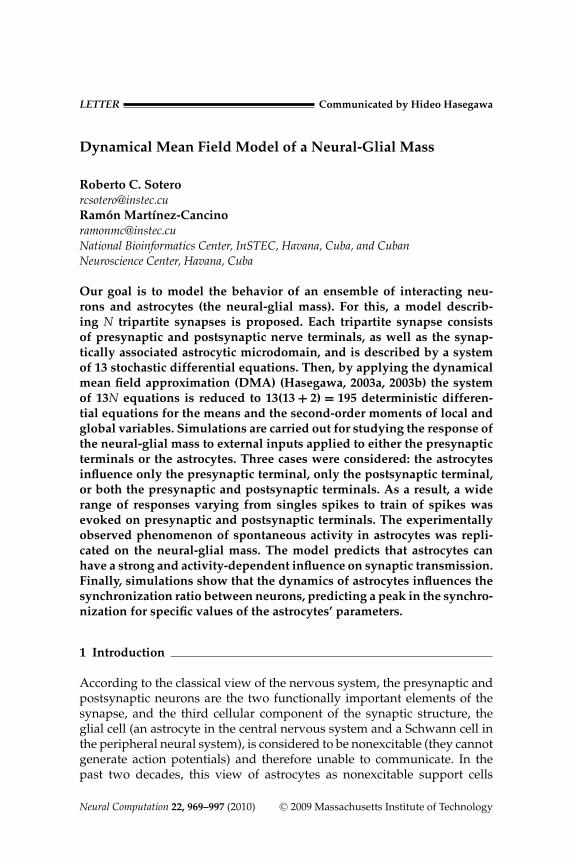
Dynamical Mean Field Model of a Neural-Glial Mass
Roberto C. [email protected] Martı[email protected] Bioinformatics Center, InSTEC, Havana, Cuba, and CubanNeuroscience Center, Havana, Cuba
Our goal is to model the behavior of an ensemble of interacting neu-rons and astrocytes (the neural-glial mass). For this, a model describ-ing N tripartite synapses is proposed. Each tripartite synapse consistsof presynaptic and postsynaptic nerve terminals, as well as the synap-tically associated astrocytic microdomain, and is described by a systemof 13 stochastic differential equations. Then, by applying the dynamicalmean field approximation (DMA) (Hasegawa, 2003a, 2003b) the systemof 13N equations is reduced to 13(13 + 2) = 195 deterministic differen-tial equations for the means and the second-order moments of local andglobal variables. Simulations are carried out for studying the response ofthe neural-glial mass to external inputs applied to either the presynapticterminals or the astrocytes. Three cases were considered: the astrocytesinfluence only the presynaptic terminal, only the postsynaptic terminal,or both the presynaptic and postsynaptic terminals. As a result, a widerange of responses varying from singles spikes to train of spikes wasevoked on presynaptic and postsynaptic terminals. The experimentallyobserved phenomenon of spontaneous activity in astrocytes was repli-cated on the neural-glial mass. The model predicts that astrocytes canhave a strong and activity-dependent influence on synaptic transmission.Finally, simulations show that the dynamics of astrocytes influences thesynchronization ratio between neurons, predicting a peak in the synchro-nization for specific values of the astrocytes’ parameters.
1 Introduction
According to the classical view of the nervous system, the presynaptic andpostsynaptic neurons are the two functionally important elements of thesynapse, and the third cellular component of the synaptic structure, theglial cell (an astrocyte in the central nervous system and a Schwann cell inthe peripheral neural system), is considered to be nonexcitable (they cannotgenerate action potentials) and therefore unable to communicate. In thepast two decades, this view of astrocytes as nonexcitable support cells
Neural Computation 22, 969–997 (2010) C© 2009 Massachusetts Institute of Technology

970 R. Sotero and R. Martınez-Cancino
has changed dramatically. The finding that astrocytes can be excitednonelectrically (their excitability is based on variations of the C2+
a con-centration in the cytosol) has expanded our knowledge of the complexityof the brain (Volterra & Meldolesi, 2005). Today astrocytes are seen as inte-gral modulatory elements of tripartite synapses that can generate variousregulatory signals and bridge structures and networks that are otherwisedisconnected (Araque, Parpura, Sanzgiri, & Haydon, 1999). Experimentalstudies show that astrocytes establish anatomical domains that are largelynonoverlapping and can vary greatly with cell layer, brain region, andspecies. For example, in rodents, one astrocyte ensheathes thousands ofsynapses (Bushong, Martone, Jones, & Ellisman, 2002), while in the humancortex, it might ensheath around 2 million (Oberheim, Wang, Goldman, &Nedergaard, 2006). The interaction between astrocytes is primarily confinedto their domain borders where they form gap-junctional connections. Ad-ditionally, subcellular compartments termed “microdomains” have beenidentified in astrocytes (Grosche et al., 1999). An astrocyte may consist ofthousands of microdomains capable of autonomous interactions with theparticular group of synapses that they ensheath.
Release of neurotransmitters from the presynaptic terminal not onlystimulates the postsynaptic neuron but also activates the perisynaptic glia(see Figure 1). The activated glial cell in turn releases gliotransmitters thatcan directly stimulate the postsynaptic neuron and can feedback to thepresynaptic terminal (Newman, 2003). This modulation of neuronal ex-citability and synaptic transmission by astrocytes is mediated by glutamaterelease (with an excitatory effect) and by ATP and its derivative, adenosine(with an inhibitory effect) (Volterra & Meldolesi, 2005). Another form ofastrocyte excitation occurs independent of neuronal input and is knownas spontaneous excitation (Volterra & Meldolesi, 2005). Additionally, astro-cytes have physiological relevance in the control of blood flow, which canbe continuously adapted to local needs through the induction of local vaso-constriction or vasodilation responses (Iadecola, 2004; Takano et al., 2006).The correct functioning of astrocytes and their partnership with neighbor-ing cells seems to be essential for normal brain function, which has changedour view of several brain diseases (Volterra & Meldolesi, 2005).
In the past few years, several papers have approached the neuron-astrocyte interaction from a modeling point of view. Nadkarni and Jung(2003) incorporated the influence of a surrounding glia in a neuron model.For describing voltage variations in the neuron, they used Hodgkin-Huxley(HH) equations (Hodgkin & Huxley, 1952) and modeled the intracellularinositol (1,4,5)-trisphosphate (IP3) production in astrocytes due to neuronalfiring. Production of IP3 then evokes the release of C2+
a from internal stores.Finally, they used experimental data for relating the C2+
a concentrationin the astrocyte environment to the inward currents in neurons. Postnov,Ryazanova, and Sosnovtseva (2007) proposed a mathematical model of theneural-glial interaction by modeling the presynaptic neuron, the synaptic

Dynamical Mean Field Model of a Neural-Glial Mass 971
Figure 1: The tripartite synapse. Glutamate (Glu) is released from the presy-naptic terminal into the synaptic cleft to communicate with the postsynapticneuron. The neurotransmitter also activates glial receptors, eliciting the intra-cellular production of inositol 1,4,5-triphosphate (IP 3), which in turn evokesincreases in C2+
a concentration in the glial cells and the release of glutamate. Thisfeedback on presynaptic receptors regulates neurotransmitter release, while ac-tivation of postsynaptic receptors directly depolarizes neurons. Stimulation ofglia also elicits the release of ATP, which can inhibit both neurons.
terminal itself, a postsynaptic neuron, and a glial cell. For modeling thepostsynaptic and the presynaptic neurons, they used a Fitzhugh-Nagumomodel (FitzHugh, 1961; Nagumo, Arimoto, & Yoshizawa, 1962). They as-sumed that the glial cell could be activated by two different mechanisms: thefast increase of intercellular potassium produced by the spiking activity ofthe postsynaptic neuron and the slow production of a mediator triggered bythe synaptic activity. On the other hand, Di Garbo, Barbie, Chilemi, Alloisio,

972 R. Sotero and R. Martınez-Cancino
and Nobile (2007) proposed a biophysical model of ATP-evoked calcium re-sponses in astrocytes, while Volman, Ben-Jacob, and Levine (2007) exploredthe consequences of the regulation of synaptic transmission by astrocyticcalcium dynamics in the case of the autaptic oscillator (Seung, Lee, Reis, &Tank, 2000).
All models mentioned describe the dynamics of one tripartite synapse.Although simulations in networks of astrocytes have been carried out forstudying the propagation of calcium waves (Hofer, Venance, & Giaume,2002; Goto, Kinoshita, & Natsume, 2004; Bellinger, 2005; Stamatakis &Mantzaris, 2006), there are no realistic simulations of a network of inter-acting neurons and glial cells using a biophysical-based model of the tri-partite synapse. This would involve a large number of both parameters andvariables, with the disadvantage of the high computational power neededfor solving the system of stochastic differential equations (SDEs) involvedand the additional difficulty of determining the influence of each modelparameter on the generated average network characteristics.
For dealing with this problem, mass models could be used. Neural massmodels (NMMs) are mean field models developed by Freeman (1972) andWilson and Cowan (1972) for dealing with populations of interconnectedneurons. That is, NMMs can describe the dynamics of cortical columnsand brain areas by using only a few parameters, but without much de-tail (Lopes da Silva, Hoeks, Smits, & Zetterberg, 1974; Jansen & Rit, 1995;Sotero, Trujillo-Barreto, Iturria-Medina, Carbonell, & Jimenez, 2007). In thisapproach, spatially averaged magnitudes are assumed to characterize thecollective behavior of populations of neurons of a given type instead ofmodeling single cells and their interactions in a detailed network. In the dy-namical mean field approximation (DMA) proposed by Hasegawa (2003a,2003b), an ensemble of N neurons expressed by coupled K-dimensionalSDEs is replaced by K (K + 2) deterministic differential equations, ex-pressed in terms of means and the second-order moments of local and globalvariables. Interestingly, the equations of the Wilson and Cowan model canbe obtained by applying the DMA to a network of Fitzhugh-Nagumo neu-rons and by neglecting variance and covariance terms (Hasegawa, 2003a).
This letter is an extension of the work on NMMs to an ensemble ofinteracting neurons and astrocytes. We first model each tripartite synapseas consisting of presynaptic and postsynaptic neuronal terminals and asynaptically associated astrocyte microdomain. Each tripartite synapse isthen described by a system of 13 SDEs. For modeling an ensemble of N suchsynapses (referred to as the neural-glial mass from this point onward), wethen apply the DMA theory, thus obtaining a system of 195 deterministicequations for the means and second-order moments of local and globalvariables.
In the following sections, we describe the proposed biophysical model.First, the SDEs describing one tripartite synapse; then the DMA as-sumptions are briefly described, and the equations for the means and

Dynamical Mean Field Model of a Neural-Glial Mass 973
second-order moments of local and global variables of the neural-glial massare obtained. In section 3, simulations are carried out for testing the model.
2 Methods
In this section we present the equations describing the structural subunits ofthe tripartite synapse: the presynaptic and postsynaptic neuronal terminalsand the associated astrocytic microdomain. Finally, we obtain the equationsfor the neural-glial mass after applying the DMA theory.
2.1 Presynaptic Terminal. To model the presynaptic terminal in thetripartite synapse i of the neural-glial mass, we use the HH model:
CmdVi
pre
dt= −g pre
K
(ni
pre
)4(Vi
pre − V preK
) − g preNa
(mi
pre
)3h pre
(Vi
pre − V preNa
)− g pre
l
(Vi
pre − V prel
) +
+ I iastr1 + w
N − 1
∑j(�=i)
Q(Vi j
pre
) + ξ ipre + I i
ext pre (2.1)
dmipre
dt= α pre
m
(1 − mi
pre
) − β prem mi
pre (2.2)
dnipre
dt= α pre
n
(1 − ni
pre
) − β pren ni
pre (2.3)
dhipre
dt= α
preh
(1 − hi
pre
) − βpreh hi
pre , (2.4)
where Vipre is the membrane potential, (mi
pre )3h pre is the fraction of openNa+ channels, (ni
pre )4 is the fraction of open potassium channels, and V preNa ,
V preK , V pre
l are the reversal potentials for the sodium, potassium, and leakagesystem, respectively. The maximal conductances of sodium, potassium, andleakage channels are g pre
Na , g preK , g pre
l , and Cm is the membrane capacitance.The opening and closing rates of the respective gates are given by
αm (V) = 0.1 (V + 40)
1 − e− (V+40)10
(2.5)
αn (V) = 0.01 (V + 55)
1 − e− (V+55)10
(2.6)
αh (V) = 0.07e− (V+65)20 (2.7)
βm (V) = 4e− (V+65)18 (2.8)

974 R. Sotero and R. Martınez-Cancino
βn (V) = 0.125e− (V+65)80 (2.9)
βh (V) = 1
1 + e− (V+35)10
. (2.10)
The current I iext pre in equation 2.1 stands for an applied external stimu-
lus, and the fifth term at the right side describes the coupling betweensynapses, where w is the coupling strength and Q (V) is the sigmoid func-tion (Hasegawa, 2003b):
Q (V) = 1
1 + e− (V−θ )ε
. (2.11)
The self-coupling terms are excluded. N is the number of tripartite synapsesin the neural-glial mass. In equation 2.1, ξ i
pre denotes the spatially correlatedwhite noises given by
⟨ξ i
pre (t)⟩ = 0 (2.12)⟨
ξ ipre (t)ξ j
pre (t′)⟩ = [
β20 preδi j + β2
1 pre
(1 − δi j
)]δ(t − t′)
= (β2
I preδi j + β2C pre
)δ(t − t′), (2.13)
where βC pre = β1 pre and βI pre =√
β20 pre − β2
1 pre denote the magnitudes ofcommon and independent noises, respectively (Hasegawa, 2003b), andbrackets 〈〉 express the expectation value (Hasegawa, 2003a). Finally, I i
astr1denotes the current induced on the presynaptic neuron by the astrocyticactivity.
2.2 Synaptic Coupling. Following Postnov et al. (2007) and Kopell,Ermentrout, Whittington, and Traub (2000), two properties of the synapticcoupling are modeled: the delayed response of the postsynaptic neuronactivity and the threshold activation:
τsdzi
dt= (
1 + tanh(ss
(Vi
pre − hs)))
(1 − zi ) − zi
ds, (2.14)
where zi is a synaptic activation variable, which is a nonlinear func-tion of the presynaptic membrane voltage and represents the number ofneurotransmitter vesicles docked on the presynaptic terminal relative tothe maximum that can dock. Parameter τs denotes the time delay, whereasparameters hs, ss , and ds are responsible for the activation and relaxation of

Dynamical Mean Field Model of a Neural-Glial Mass 975
zi . The current I isyn applied to the postsynaptic terminal is then calculated
as (Kopell et al., 2000)
I isyn = ks (zi − z0)
(Vi
post − Vrev), (2.15)
where ks is the conductivity, Vrev is the reversal potential, and z0 is thereference level, which is calculated from the assumption that when thepresynaptic terminal is silent, z = z0 (Postnov et al., 2007).
2.3 Postsynaptic Terminal. As in the presynaptic terminal, we use theHH formalism for describing voltage variations at the postsynaptic ter-minal. All variables and parameters have the same interpretation as insection 2.1, but in this case, a subindex post is employed. The inputs are thecurrent I i
syn given by equation 2.15 and the current induced by the astrocyticactivity I i
astr2:
CmdVi
post
dt=−g post
K
(ni
post
)4 (Vi
post − V postK
)−g post
Na
(mi
post
)3h post
(Vi
post − V postNa
) −−g post
l
(Vi
post − V postl
) + I isyn + I i
astr2 + ξ ipost (2.16)
dmipost
dt=αm
(1 − mi
post
) − βmmipost (2.17)
dnipost
dt=αn
(1 − ni
post
) − βnnipost (2.18)
dhipost
dt=αh
(1 − hi
post
) − βhhipost. (2.19)
As in the presynaptic neuron, white noise is also considered, in this case bymeans of ξ i
post :⟨ξ i
post(t)⟩= 0 (2.20)⟨
ξ ipost(t)ξ
jpost(t
′)⟩= [
β20 postδi j + β2
1 post
(1 − δi j
)]δ(t − t′)
= (β2
I postδi j + β2C post
)δ(t − t′). (2.21)
2.4 Astrocytic Microdomain. A large variety of mathematical modelshave been developed for studying the intracellular calcium oscillations.However, as pointed out by Schuster, Marhl, and Hofer (2002), to simulateself-oscillations by a system of kinetic equations, at least two variables areneeded. Important two-variable models have been proposed by Goldbeter,Dupont, and Berridge (1990), Li and Rinzel (1994), and Marhl, Schuster,

976 R. Sotero and R. Martınez-Cancino
Brumen, and Heinrich (1997). As was shown recently, these minimal mod-els can be viewed as complementary parts of a six-variable model (Shusteret al., 2002). In this work, we employ the Goldbeter model (Goldbeter et al.,1990; Girard, Luckhoff, Lechleiter, Sneyd, & Clapham, 1992) to model thecalcium dynamics in a single astrocytic microdomain, with additional termsfor the interaction with other astrocytic domains and the external input:
τcdci
dt= ν0 − f (ci , li ) + KE Rli − Kleakci
+M∑
j=1
b j S jm + ξ i
astr + I iext astr (2.22)
τcεcdli
dt= f (ci , li ) − KE Rli , (2.23)
where ci is the calcium concentration within the astrocytic microdomain cor-responding to the tripartite synapse i , li is the calcium concentration withinthe endoplasmic reticulum (ER), I i
ext astr accounts for external stimulation,and τc defines the characteristic time for C2+
a oscillations together with timeseparation parameter εc (Postnov et al., 2007). Parameter ν0 is the sum ofthe calcium influx across the plasma membrane and the rate of release fromIP3-sensitive stores (Girard et al., 1992). The term
∑Mj=1 b j S j
m represents cal-cium variations due to the IP3 (Sj
m) production, where j = 1, . . . , M countsthe tripartite synapse i and the synapses in its neighborhood (for the sakeof simplicity, we will take b j = b). That is, calcium concentration in theastrocytic microdomain corresponding to tripartite synapse i also dependson IP3 production in nearby microdomains. Additionally, we will restrictthis interaction to all microdomains belonging to the same astrocyte. Thus,in this letter, we do not model the interaction between astrocytes, which ismediated by gap junctions through which calcium (Volterrra & Meldolesi,2005) and possibly sodium (Bernardinelli, Magistretti, & Chatton, 2004)waves are transmitted.
The calcium exchange between the ER and the cytoplasm is given by(Girard et al., 1992)
f (ci , li ) = Vp
(c2
i
c2i + K 2
p
)− Vm
(c4
i
c4i + K 4
c
) (l2i
l2i + K 2
l
)(2.24)
Astrocyte noise is included by means of ξ iastr :
⟨ξ i
astr (t)⟩ = 0 (2.25)⟨
ξ iastr (t)ξ j
astr (t′)⟩ = [
β20 astrδi j + β2
1 astr
(1 − δi j
)]δ(t − t′)
= (β2
I astrδi j + β2C astr
)δ(t − t′) (2.26)

Dynamical Mean Field Model of a Neural-Glial Mass 977
Thus, equations 2.22 and 2.23 define a system of stochastic differentialequations with additive noise, similar to the stochastic Li-Rinzel model(Shuai & Jung, 2003).
As in Postnov et al. (2007), the dynamics of Sim and the glutamate, Gi
m,
are modeled by
τSm
d Sim
dt= (
1 + tanh(sSm
(zi − hSm
))) (1 − Si
m
) − Sim
dSm
(2.27)
τGm
dGim
dt= (
1 + tanh(sGm
(ci − hGm
)))(1 − Gm) − Gi
m
dGm
. (2.28)
Finally, the astrocyte-induced currents on presynaptic and postsynapticterminals are proportional to Gi
m:
I iastr1 = γ1Gi
m, I iastr2 = γ2Gi
m. (2.29)
2.5 Compact Form of the Equations Describing a Tripartite Synapse.First, we define the vector upi of the model variables for the tripartitesynapse i:
upi = [u1i , . . . , u13i ]
= [Vi
pre , mipre , ni
pre , hipre , zi , Vi
post, mipost, ni
post, hipost, ci , li , Si
m, Gim
],
(2.30)
where p = 1, . . . , 13. Then the system of K = 13 SDEs described in theprevious sections can be written in compact form as
dupi
dt= F (p)({uqi }) + δp1
⎛⎝ w
N − 1
∑j(�=i)
Q(u j
1i
) + I iext pre + ξ i
pre
⎞⎠
+ δp6ξipost + δp10
(I iext astr + ξ i
astr
), (2.31)
where F (p) is given by
F (1) =− 1Cm
[g pre
K u43i
(u1i − V pre
K
) + g preNa u3
2i u4i(u1i − V pre
Na
)+ g pre
l
(u1i − V pre
l
) + γ1u13i]
F (p) =− [αup (u1i ) + βup (u1i )
]upi + αup (u1i ) (p = 2 − 4)
F (5) = 1τs
[[1 + tanh (ss (u1i − hs))] (1 − u5i ) − u5i
ds
]

978 R. Sotero and R. Martınez-Cancino
F (6) =− 1Cm
[g post
K u48i
(u6i − V post
K
) + g postNa u3
7i u9i(u6i − V post
Na
)+ g pre
l
(u6i − V post
l
) + ks (u6i − Vrev) (u5i − z0) + γ2u13i ]
F (p) =− [αup (u6i ) + βup (u6i )
]upi + αup (u6i ) (p = 7 − 9)
F (10) = 1τc
⎡⎣ν0 − Kleaku10i + KE Ru11i − f (u10i , u11i ) +
M∑j=1
b j uj11i
⎤⎦
F (11) = 1εcτc
[ f (u10i , u11i ) − KE Ru11i ]
F (12) = 1τSi
m
[[1 + tanh
(sSi
m
(u5i − hSi
m
))](1 − u12i ) − u12i
dSim
]
F (13) = 1τGi
m
[[1 + tanh
(sGi
m
(u10i − hGi
m
))](1 − u13i ) − u13i
dGim
]. (2.32)
2.6 Mean Field Model for the Neural-Glial Mass. The neural-glialmass we model in this letter is an ensemble of N-coupled tripartitesynapses. This system is described by 13N SDEs. The goal of this letteris to obtain a mean field model for this neural-glial population. For this,we will use the DMA theory proposed by Hasegawa (2003a, 2003b).
Given the local variables upi , according to Hasegawa (2003a, 2003b), theglobal variables for the ensemble are defined as
Up(t) = 1N
∑i
upi (t) (2.33)
and their averages by
μp(t) = μup (t) = 〈Up(t)〉. (2.34)
Deviations of local variables from the averages are
δupi (t) = upi (t) − μp(t). (2.35)
And those of global variables are calculated as
δUp(t) = Up(t) − μp(t). (2.36)

Dynamical Mean Field Model of a Neural-Glial Mass 979
Variances and covariances of local variables are defined by
γp,q = γup,uq = 1N
∑i
⟨δupiδuqi
⟩, (2.37)
and those of global variables by
ρp,q = ρup,uq = ⟨δUpδUq
⟩. (2.38)
Following Hasegawa (2003a, 2003b), the synchronization ratio (SR) is de-fined as
SRp,q (t) =ρp,q
γp,q− 1
N
1 − 1N
. (2.39)
For an equation of the type of 2.31, the DMA approximation gives thefollowing equations for the means (μp) and variances and covariances oflocal (γp,q ) and global (ρp,q ) variables (see Hasegawa, 2003b):
dμup
dt= F (up) + 1
2
∑q
∑r
F (up)uq ,ur γuq ,ur + δp1
[wU0 + I pre
ext] + δp10 Iext astr
(2.40)
dγup,uq
dt=
∑r
[F (up)
ur γuq ,ur + F (uq )ur γup,ur
]+β2
0preδp1δq1 + β20postδp6δq6 + β2
0astrδp10δq10 +
+ 16
∑r
∑s
∑t
[F (up)
ur us ut
(γuq ,ur γus ,ut + γuq ,us γur ,ut + γuq ,ut γur ,us
)
+ F (uq )ur us ut
(γup,ur γus ,ut + γup,us γur ,ut + γup,ut γur ,us
)]+wU1
[δp1ζuq ,u1 + δq1ζup,u1
](2.41)
dρup,uq
dt=
∑r
[F (up)
ur ρuq ,ur + F (uq )ur ρup,ur
]
+[
1N
β20pre +
(1 − 1
N
)β2
1pre
]δp1δq1
+[
1N
β20post +
(1 − 1
N
)β2
1post
]δp6δq6
+[
1N
β20astr +
(1 − 1
N
)β2
1astr
]δp10δq10 +

980 R. Sotero and R. Martınez-Cancino
Figure 2: Diagram summarizing the different models employed for simulatingthe dynamics of the neural-glial mass.
+ 16
∑r
∑s
∑t
[F (up)
ur us ut
(ρuq ,ur γus ,ut + ρuq ,us γur ,ut + ρuq ,ut γur ,us
)
+ F (uq )ur us ut
(ρup,ur γus ,ut + ρup,us γur ,ut + ρup,ut γur ,us
)]+wU1
[δp1ρuq ,u1 + δq1ρup,u1
](2.42)
ζup,uq =(
1N − 1
) (Nρup,uq − γup,uq
)(2.43)
U0 = 1N
∑j
〈Q(u1 j
)〉 = Q + 12
Qu1u1γu1,u1 (2.44)
U1 = Qu1 + 12
Qu1u1u1γu1,u1 , (2.45)
where q , r, s, and t run from 1 to 13, and Qu1 = ∂ Q∂u1
, Qu1u1u1 = ∂3 Q∂u3
1, F (up)
ur =∂ F (p)
∂ur, F (up)
ur us = ∂2 F (p)
∂ur ∂us, and F (up)
ur us ut = ∂3 F (p)
∂ur ∂us∂utare evaluated at the means (μp).
With this, the original 13N-dimensional SDEs for the neural-glial mass havebeen replaced by 13(13 + 2) = 195 deterministic differential equations.
A diagram summarizing the different models used for describing thetripartite synapse and the DMA is shown in Figure 2.

Dynamical Mean Field Model of a Neural-Glial Mass 981
3 Results
In this section, computational simulations are used to explore the proposedmean field model (equations 2.40 to 2.45) for a neural-glial mass.
3.1 Input to the Presynaptic Terminals. In this section we study theresponse of the neural-glial mass to external inputs in the presynaptic neu-ron Iext pre (t). There will be no input to the astrocytes. Inputs will consist ofindividual spikes or trains of spikes and are applied to all the synapses inthe ensemble. Each spike is given by the equation
Iext pre (t) = I0
Cmα (t − t0) , (3.1)
with the alpha function
α(t) = tτ
e1− tτ �(t), (3.2)
where � (x) = 1 for x ≥ 0 and 0 otherwise; I0 = 5 μA/cm2 stands for themagnitude of an input spike, t0 the input time of a spike, and τ = 1 ms thetime constant. The values of all the parameters used in the simulationsare displayed in Table 1. Some of these values were obtained from previousstudies (Postnov et al., 2007; Hasegawa, 2003b; Girard et al., 1992), andothers were manually tuned in order to approximately reproduce reportedexperimental results.
In this letter, system 2.40 to 2.42 is solved by using the fourth-orderRunge-Kutta method with an integration step size of 0.1 ms and initialconditions
μ0 = [−65, 0.053, 0.32, 0.59, 0.39,
−65, 0.053, 0.32, 0.59, 0.34, 2.26, 0.10, 0.14] and
γup,uq = ρup,uq = 0. The first 4000 points of the simulated signals were dis-carded in order to avoid transient behavior.
In the first simulation, the input to the presynaptic terminal consists ofa single spike at t = 500 ms. Figure 3 shows the means of model variablesμp. In this simulation, the astrocyte influences both the presynaptic and thepostsynaptic terminals (γ1 = 7, γ2 = 7). In Figure 3C, notice that μ1 has afirst peak evoked by the external inputs, which is followed by two peaksevoked by the feedback from astrocytes (due to increases in the mediatorproduction μ13). This phenomenon also causes a very small plateau untilt = 1196 ms. This feedback from astrocytes can be avoided by making γ1 = 0or hGm < 1. In the case of the postsynaptic terminal in Figure 3H, μ6 alsoshows three peaks and a small plateau until t = 1186 ms. There is a 2.5 ms

982 R. Sotero and R. Martınez-Cancino
Table 1: Parameters and Typical Values Used in the Simulations.
Parameters and Typical Values Interpretation
V preNa = V post
Na = 50 mV (Hasegawa, 2003b) Reversal potentials for sodium at pre-and postsynapses
V preK = V post
K − 77 mV (Hasegawa, 2003b) Reversal potentials for potassium at pre-and postsynapses
V prel = V post
l − 54.5 mV (Hasegawa, 2003b) Reversal potentials for leakage system atpre- and postsynapses
Vrev = −80 mV (Kopell et al., 2000) Reversal potential for the synapseg pre
Na = g postNa = 120 mS/cm2 (Hasegawa,
2003b)Maximal conductance of sodium at pre-
and postsynapses
g preK = g post
K = 36 mS/cm2 (Hasegawa,2003b)
Maximal conductance of potassium atpre- and postsynapses
g prel = g post
l = 0.3 mS/cm2 (Hasegawa,2003b)
Maximal conductance of leakage systemat pre- and postsynapses
Cm = 1 μF/cm2 (Hasegawa, 2003b) Membrane capacitance
w = 0.5 , θ = 0, ε = 10 (Hasegawa, 2003b) Parameters of the coupling betweensynapses.
ds = 3 (Postnov et al., 2007)τs = 5, SS = 0.05, hs = −40, ks = 25,ϑ = 0, z0 = 0.312
Parameters of the synaptic terminal
τc = 1, ν0 = 2.9 μM/s, KE R = 1s−1, εc = 1,Kleak = 0.01 ms−1 , b = 0.05, Kl = 2 μM,Kc = 0.9 μM, K p = 1 μM, Vp = 65 μM/s,Vm = 455 μM/s (Girard et al., 1992)
Calcium dynamics in astrocytes
sGm = 100, τGm = 100, dGm = 3, hGm = 1.2(Postnov et al., 2007)
Dynamics of astrocyte mediator(glutamate)
sSm = 100, τSm = 100, dSm = 3, hSm = 0.45(Postnov et al., 2007)
Dynamics of IP3 production
γ1 = 7, γ2 = 7 Weights in astrocyte-induced currentson presynaptic and postsynapticneurons, respectively
βI pre = 0.02, βI post = 0.02, βI astr = 0.001 Independent noises for presynapticneurons, postsynaptic neurons,and astrocytes
βC pre = 0.01, βC post = 0.01, βC astr = 0.0005 Common noises for presynapticneurons, postsynaptic neurons,and astrocytes
N = 500,000 Number of tripartite synapses
delay in the transmission of the response between pre- and postsynapses,which is controlled by equation 2.14.
In the next simulation, the external input to the presynapses consistedof two spikes separated 1000 ms in time. In this case, we set γ1 = γ2 = 0,which means that the astrocyte exerts no feedback on pre- and postsynapses.Figure 4 shows the means of the model variables. From now on, for claritywe will show only six variables—μ1, μ5, μ6, μ10 μ12, and μ13—which are

Dynamical Mean Field Model of a Neural-Glial Mass 983
Figure 3: Time courses of means and external inputs: (A) Iext pre , (B) Iext astr .(C) μ1. (D) μ2. (E) μ3. (F) μ4. (G) μ5. (H) μ6. (I) μ7. (J) μ8. (K) μ9. (L) μ10.(M) μ11. (N) μ12. (O) μ13. The external input to the presynapses is a single spike.The astrocyte influences both the presynaptic and the postsynaptic neurons(γ1 = 7, γ2 = 7).
necessary for understanding the behavior of the neural-glial mass. Resultsshow that two spikes are generated in the presynapses (see Figure 4C)and transmitted to the postsynapses (see Figure 4E). Two responses are alsoobtained at μ13 (see Figure 4H). If we run the same simulation with γ1 = γ2 =7, the system exhibits a more complex behavior, resulting in a train of spikesat the postsynaptic cell (see Figure 5). After the first external spike, threespikes are generated at the presynapses, while after the second externalspike, only one spike is evoked. This is because the voltage variation due tothe first external spike has not returned to its baseline value when the secondexternal spike is provided. In the postsynapses, three spikes are obtained

984 R. Sotero and R. Martınez-Cancino
Figure 4: Time courses of means and external inputs: (A) Iext pre . (B) Iext astr .(C) μ1. (D) μ5. (E) μ6. (F) μ10. (G) μ12. (H) μ13. The external input to the presy-napses consists of two spikes separated 1000 ms in time. The astrocyte does notexert feedback on the neurons (γ1 = γ2 = 0).
after the first input and a train of spikes after the second input. If the intervalbetween the external spikes is increased so that the interaction is avoided,we also obtain three spikes at the postsynapses after the second externalinput. Note that in the IP3 production (see Figure 5G), the second responsehas a lower amplitude than the first one due to the dependence on thesynaptic activation variable (see Figure 5D).
Figure 6 shows the means of model variables when the external inputto the presynapses consists of a train of spikes of 200 ms of duration andindividual spikes separated 25 ms in time, and γ1 = γ2 = 0. Due to the slowrelaxation of the cytoplasm calcium concentration in astrocytes (μ10), themediator production μ13 behaves as a plateau for 535 ms and then beginsto decrease until reaching baseline.

Dynamical Mean Field Model of a Neural-Glial Mass 985
Figure 5: Time courses of means and external inputs: (A) Iext pre . (B) Iext astr .(C) μ1. (D) μ5. (E) μ6. (F) μ10. (G) μ12. (H) μ13. The external input to the presy-napses consists of two spikes separated 1000 ms in time. The astrocyte influencesboth neurons (γ1 = γ2 = 7).
3.2 Input to the Astrocytes. In this section, we study the response of theneural-glial mass to external inputs in the astrocyte Iext astr (t). There will beno input to the presynaptic terminals. As in section 3.1, inputs will consistof individual spikes or trains of spikes, given by equation 3.1, and appliedto all microdomains within the neural-glial mass.
Figure 7 shows the means of model variables when the input to the astro-cyte consists of a single spike at t = 500 ms. The influence of the astrocyticmicrodomain on the presynaptic terminal is given by Iastr1 (γ1 = 7), and weare considering no influence on the postsynaptic neuron (γ2 = 0). As shownin the figure, the excitation in the astrocytes evokes two spikes in presy-naptic and postsynaptic terminals. Figure 8 displays the means of modelvariables when the input to the astrocyte consists of a single spike, but in this

986 R. Sotero and R. Martınez-Cancino
Figure 6: Time courses of means and external inputs: (A) Iext pre . (B) Iext astr .(C) μ1. (D) μ5. (E) μ6. (F) μ10. (G) μ12. (H) μ13. The external input to the presy-napses consist of a train of spikes of 200 ms duration and individual spikesseparated 25 ms in time, and we set γ1 = γ2 = 0.
case, the astrocyte influences the postsynaptic terminal (γ2 = 7) while thereis no influence in the presynaptic terminal (γ1 = 0). This results in no activ-ity at the presynapses and a train of spikes at the postsynapses. In Figure 9the input is again a spike in the astrocytic microdomain at t = 500 ms, but inthis case, γ1 = γ2 = 7, resulting in two spikes at the presynapses and a trainof spikes at the postsynapses. Figure 10 shows the means of model variableswhen the external input to the astrocyte consists of two spikes separatedby 200 ms. In the case, two peaks are obtained at the presynapses at timest = 512.5 ms and t = 532.3 ms. In the postsynapses, there is a first peak att = 509.5 ms; then after 24.8 ms, a train of spikes with spike separation of15.8 ms begins. Figure 11 shows the means of model variables when the

Dynamical Mean Field Model of a Neural-Glial Mass 987
Figure 7: Time courses of means and external inputs: (A) Iext pre . (B) Iext astr .(C) μ1. (D) μ5. (E) μ6. (F) μ10. (G) μ12. (H) μ13. The external input to the astrocyteconsists of a single spike. For this simulation, γ1 = 7 and γ2 = 0.
external input to the astrocytes consists of a train of spikes of 200 ms ofduration and individual spikes separated 25 ms in time. This result is verysimilar to the one obtained in the previous simulation; the main differenceis in μ10
(Figure 11F
), which has several peaks instead of two peaks as in
Figure 10.
3.3 Spontaneous Oscillations in Astrocytes. Astrocytic activity can oc-cur independent of neuronal activity or external inputs due to spontaneousintracellular C2+
a oscillations (Newman, 2003; Skupin et al., 2008). In thissection, we reproduce this phenomenon. The critical parameters for elicitingthe spontaneous oscillations in astrocytes are ν0, hGm , and μ0 (10). Figure 12shows a simulation when there are no external inputs to the neural-glial

988 R. Sotero and R. Martınez-Cancino
Figure 8: Time courses of means and external inputs: (A) Iext pre . (B) Iext astr .(C) μ1. (D) μ5. (E) μ6. (F) μ10. (G) μ12, (H) μ13. The external input to the astrocyteconsists of a single spike. For this simulation, γ1 = 0 and γ2 = 7.
mass, astrocytes influence both neuronal terminals (γ1 = γ2 = 7), and thecritical parameters were set to ν0 = 3.1 μM/s, hGm = 1.4, μ0 (10) = 0.1.This produces intracellular C2+
a oscillations in the astrocytes as shown inFigure 12C. As seen in the figure, this spontaneous excitation in the as-trocytes results in the increase of the glutamate (see Figure 12D) and thesubsequent excitation of presynaptic (see Figure 12A) and postsynaptic (seeFigure 12B) terminals.
The simulations presented in section 3.2 and in this section predict thatastrocytes can have a strong and very diverse influence in neuronal electricalactivity. Particularly, the repetitive action potentials evoked in neurons dueto astrocytic activity, as shown from Figures 8 to 12 (due to different causes),have been reported in experimental settings (Araque, Sanzgiri, Parpura, &Haydon, 1999).

Dynamical Mean Field Model of a Neural-Glial Mass 989
Figure 9: Time courses of means and external inputs: (A) Iext pre . (B) Iext astr .(C) μ1. (D) μ5, (E) μ6. (F) μ10. (G) μ12. (H) μ13. The external input to the astrocyteconsists of a single spike. For this simulation, γ1 = γ2 = 7.
An advantage of our approach over neural mass models is that variancesand covariances of the model variables can be obtained. This also allowsus to calculate the synchronization ratio (see equation 2.39) between vari-ables of interest. In Figure 13A, the temporal behavior of the synchronizationSR1,1(t) is presented for the case of the previous simulation of spontaneousoscillations in astrocytes, showing several peaks. In Figure 13B, parameterν0 was varied between 2.5 μM/s and 4.5 μM/s with an step size of 0.05 μM/sand the maximum of SR1,1(t) was plotted versus ν0. As shown in the fig-ure, the synchronization reaches a maximum for ν0 = 3.2 μM/s. In Figure13C, parameter hGm was varied between 0 and 2 with a step size of 0.1,and the maximum of SR1,1(t) is plotted versus hGm . The figure shows thesynchronization has a maximum for hGm = 1.1. Finally, Figure 13D shows

990 R. Sotero and R. Martınez-Cancino
Figure 10: Time courses of means and external inputs: (A) Iext pre . (B) Iext astr .(C) μ1. (D) μ5, (E) μ6. (F) μ10. (G) μ12, (H) μ13. The external input to the astrocyteconsists of two spikes separated 200 ms in time. The astrocyte influences bothneurons (γ1 = γ2 = 7).
the synchronization versus the initial condition μ0 (10), which was variedbetween 0 and 1 with a step size of 0.05, and a maximum was obtained forμ0 (10) = 0.25. The three critical parameters therefore have a specific valuefor which the synchronization in the presynaptic neuron reaches a maxi-mum. A similar result was obtained for the synchronization of postsynapticneurons SR6,6(t) (not shown in this letter).
4 Discussion
Based on previous biophysical models of each subunit of the tripartitesynapse, and using the DMA theory (Hasegawa, 2003a, 2003b), we have

Dynamical Mean Field Model of a Neural-Glial Mass 991
Figure 11: Time courses of means and external inputs: (A) Iext pre . (B) Iext astr .(C) μ1. (D) μ5, (E) μ6. (F) μ10. (G) μ12, (H) μ13. The external input to the astrocyteconsists of a train of spikes of 200 ms duration and an interval between spikesof 25 ms. The astrocyte influences both neurons (γ1 = γ2 = 7).
proposed a mean field model of a neural-glial mass. In our approach,both the presynaptic and postsynaptic terminals were modeled using theHodgkin-Huxley formalism, while the synaptic coupling between themwas modeled with a first-order differential equation as in Postnov et al.(2007) and Kopell et al. (2000). The calcium dynamics within the astrocyticmicrodomain was described with a two-variable model (Goldbeter et al.,1990; Girard et al., 1992), while glutamate production was modeled follow-ing Postnov et al. (2007). Thus, each tripartite synapse was described by asystem of 13 SDEs. The neural-glial mass was then formed with N coupledtripartite synapses. We then applied the dynamical mean field approxima-tion (DMA) proposed by Hasegawa (2003a, 2003b), obtaining a system of

992 R. Sotero and R. Martınez-Cancino
Figure 12: Spontaneous oscillations in astrocytes. Time courses of means: (A) μ1.(B) μ6. (C) μ10. (D) μ13. There is no external input to the neural-glial mass. Theastrocyte influences both neuronal terminals (γ1 = γ2 = 7).
195 deterministic differential equations for the means and second-ordermoments of local and global variables.
A controversial question in glial biology has been whether neuronalactivity, by inducing calcium waves in astrocytes, induces secretion ofneuroactive substances from astrocytes back onto synapses. Numerousexperiments in the past decade support this notion (Volterra & Meldolesi,2005; Araque, Parpura, Sanzgiri, & Haydon, 1999; Araque, Sanzgiri,Parpura, & Haydon, 1999; Newman, 2003; Parri, Gould, & Crunelli, 2001;Newman & Zahs, 1998; Angulo, Kozlov, Charpak, Audinat, 2004; Fellinet al., 2004; Perea & Araque, 2005, 2007; Jourdain et al., 2007; Wetherington,Serrano, & Dingledine, 2008). Nevertheless, recent results show that theastrocytic gliotransmitter release that affects neuronal activity may not beinduced under certain conditions (Fiacco et al., 2007). So it has been sug-gested that an unidentified variable could determine whether an astrocytic

Dynamical Mean Field Model of a Neural-Glial Mass 993
Figure 13: Neuronal synchronization due to astrocytes’ activity: (A) Temporaldynamics of SR1,1(t) for the simulation of the spontaneous oscillations in astro-cytes. (B) Maximum of SR1,1(t) versus ν0. (C) Maximum of SR1,1(t) versus hGm .(D) Maximum of SR1,1(t) versus μ0 (10).
C2+a transient will influence local synaptic transmission (Wetherington et al.,
2008). In this context, the development of computational models has provento be a useful way of testing hypotheses and making predictions aboutprocesses where experimental data are insufficient (De Schutter, 2008). Webelieve that the model presented here will be helpful for achieving this goal.
For testing the model, three types of simulations were performed. In thesimulations of the first type, we gave an external input to the presynap-tic neurons in the neural-glial mass, and we were able to study how thestimulus was propagated to the other subunits of the tripartite synapse. In-puts consisted of one spike, two spikes, and a train of spikes. Results showcomplex patterns of spikes in the presynaptic and postsynaptic neurons asthe result of astrocyte feedback. In the simulations of the second type, theinputs were given to the astrocyte population and consisted also of onespike, two spikes, and a train of spikes. As a result, the neurons were acti-vated, which shows that even if the neural population is not directly influ-enced by an external stimulus, it could still be activated through astrocytes’

994 R. Sotero and R. Martınez-Cancino
feedback if the glial population is activated. This is radically different fromthe common idea that information is generated or processed by neuronsand travels through neuronal circuits before reaching the glia. Related tothis, in the simulation of the third type, we did not give external inputsto the neural-glial mass but set the parameter values so that oscillationsin intracellular calcium concentration in astrocytes were obtained, whichreplicates the experimentally observed phenomenon of spontaneous oscil-lations in astrocytes (Araque, Parpura, Sanzgiri, & Haydon, 1999; Skupinet al., 2008). This simulation shows how information can be generated bythe astrocytes and transmitted to the neurons.
The model predicts that astrocyte activity can trigger repetitive actionpotentials in neurons, which is in agreement with experimental results(Araque, Sanzgiri, Parpura, & Haydon, 1999). Additionally, it is knownthat astrocyte C2+
a waves can modulate the light-evoked spike activity inretinal neurons (Newman & Zahs, 1998), a phenomenon they associatedwith glutamate-mediated activation of inhibitory interneurons.
Finally, simulations showed that astrocytes, activity influences the syn-chronization ratio in neurons. Synchronization of neuronal activity has beenproposed to contribute to information processing in the brain (Engel, Fries,& Singer, 2001). Then, according to the model proposed here, astrocyte-induced synchronization could be a relevant underlying mechanism.
It is known that development of an epileptic seizure requires physio-logical changes that lead to both increased neuronal excitability and ab-normal synchronization of discharge within the affected neuronal network(Wetherington et al., 2008), so an interesting question is whether exuberantneuron-astrocyte-neuron transmitter-mediated signaling can promote thisabnormal neuronal synchronization. According to our model, the answerto this question is affirmative, predicting a peak in neuronal synchroniza-tion for specific values of astrocytes, parameters related to the calcium andthe released glutamate dynamics. This result was preserved when insteadof the Goldbeter model for the calcium dynamics, we used the Li-Rinzelmodel (Li & Rinzel, 1994) as a robustness test (not shown in the letter).
To our knowledge, this is the first time a mean field model for aneuron-astrocyte network has been proposed. In addition to the inclusionof astrocytes, dynamics, an advantage of our approach over the establishedneural mass model approach (Freeman, 1972; Wilson & Cowan, 1972; Lopesda Silva et al., 1974; Jansen & Rit, 1995; Sotero et al., 2007) is that it allows forthe calculation of not only variables means but also variances, covariances,and the synchronization ratio. A disadvantage of our model is the largenumbers of equation necessary for describing the neural-glial mass, so fur-ther simplifications are needed. The absence of delays in the mathematicalformulation is another limitation of this work. Since the astrocyte-to-neuronsignaling is not instantaneous, delays must be included in equations 2.29.
Although the model proposed here describes only the dynamics of aneural-glial mass, it is of interest to consider the coupling of several masses,

Dynamical Mean Field Model of a Neural-Glial Mass 995
where some of them can have excitatory or inhibitory neurons such as inthe neural mass models literature. This will be the subject of a separatepublication.
Acknowledgments
We thank Alfonso Araque, Nelson Trujillo-Barreto, and Eduardo Martinezfor their scientific input.
References
Angulo, M. C., Kozlov, A. S., Charpak, S., & Audinat, E. (2004). Glutamate releasedfrom glial cells synchronizes neuronal activity in the hippocampus. J. Neurosci.24, 6920–6927.
Araque, A., Parpura, V., Sanzgiri, R., & Haydon, P. (1999). Tripartite synapses: Glia,the unacknowledged partner. Trends in Neurosci., 22(5), 208–215.
Araque, A., Sanzgiri, R. P., Parpura, V., & Haydon, P. G. (1999). Astrocyte-inducedmodulation of synaptic transmission. Canadian Journal of Physiology and Pharma-cology, 77 699–706.
Bellinger, S. (2005). Modeling calcium wave oscillations in astrocytes. Neurocomput-ing, 65, 843–850.
Bernardinelli, Y., Magistretti, P. J., & Chatton, J. Y. (2004). Astrocytes generate N+a -
mediated metabolic waves. PNAS, 101, 14937–14942.Bushong, E. A., Martone, M. E., Jones, Y. Z., & Ellisman, M. H. (2002). Protoplasmic
astrocytes in CA1 stratum radiatum occupy separate anatomical domains. Journalof Neuroscience, 22, 183–192.
De Schutter, E. (2008). Reviewing multi-disciplinary papers: A challenge in neuro-science? Neuroinform., 6, 253–255.
Di Garbo, A., Barbie, M., Chilemi, S., Alloisio, S., & Nobile, M. (2007). Calciumsignalling in astrocytes and modulation of neuronal activity. BioSystems, 89, 74–83.
Engel, A. K., Fries, P., & Singer, W. (2001). Dynamic predictions: Oscillationsand synchrony in top-down processing. Nature Review Neuroscience, 2(10), 704–716.
Fellin, T., Pascual, O., Gobbo, S., Pozzan, T., Haydon, P. G., & Carmignoto, G. (2004).Neuronal synchrony mediated by astrocytic glutamate through activation of ex-trasynaptic NMDA receptors. Neuron, 43, 729–743.
Fiacco, T. A., Agulhon, C., Taves, S. R., Petravicz, J., Casper, K. B., Dong, X., et al.(2007). Selective stimulation of astrocyte calcium in situ does not affect neuronalexcitatory synaptic activity. Neuron, 54, 611–626.
FitzHugh, R. (1961). Impulses and physiological states in theoretical models of nervemembrane. Biophysical J., 1, 445–466.
Freeman, W. J. (1972). Linear analysis of the dynamics of neural masses. Annu. Rev.Biophys. Bioeng., 1, 225–256.
Girard, S., Luckhoff, A., Lechleiter, J., Sneyd, J., & Clapham, D. (1992). Two-dimensional model of calcium waves reproduces the patterns observed in Xeno-pus oocytes. Biophys. J., 61(2), 509–517.

996 R. Sotero and R. Martınez-Cancino
Goldbeter, A., Dupont, G., & Berridge, M. J. (1990). Minimal model for signal-inducedCa2+ oscillations and for their frequency encoding through protein phosphory-lation. PNAS, 87, 1461–1465.
Goto, I., Kinoshita, S., & Natsume, K. (2004). The model of glutamate-inducedintracellular Ca2+ oscillation and intercellular Ca2+ wave in brain astrocytes.Neurocomputing, 58, 461–467.
Grosche, J., Matyash, V., Moller, T., Verkhratsky, A., Reichenbach, A., & Kettenmann,H. (1999). Microdomains for neuron-glia interaction: Parallel fiber signalling toBergmann glial cells. Nature Neuroscience, 2, 139–143.
Hasegawa, H. (2003a). Dynamical mean-field theory of spiking neuron ensembles:Response to a single spike with independent noises. Phys. Rev. E, 67, 041903.
Hasegawa, H. (2003b). Dynamical mean-field theory of noisy spiking neuron en-sembles: Application to the Hodgkin-Huxley model. Phys. Rev. E, 68, 041909.
Hodgkin, A. L., & Huxley, A. F. (1952). A quantitative description of membranecurrent and its application to conduction and excitation in nerve. J. Physiol., 117,500–544.
Hofer, T., Venance, L., & Giaume, C. (2002). Control and plasticity of intercellularcalcium waves in astrocytes. J. Neurosci., 22, 4850–4859.
Iadecola, C. (2004). Neurovascular regulation in the normal brain and in Alzheimer’sdisease. Nat. Rev. Neurosci., 5, 347–360.
Jansen, B. H., & Rit, V. G. (1995). Electroencephalogram and visual evoked potentialgeneration in a mathematical model of coupled cortical columns. Biol. Cybern.,73, 357–366.
Jourdain, P., Bergersen, L. H., Bhaukaurally, K., Bezzi, P., Santello, M., Domercq, M.,et al. (2007). Glutamate exocytosis from astrocytes controls synaptic strength. Nat.Neurosci., 10, 331–339.
Kopell, N., Ermentrout, G. B., Whittington, M. A., & Traub, R. D. (2000). Gammarhythms and beta rhythms have different synchronization properties. PNAS,97(4), 1867–1872.
Li, Y., & Rinzel, J. (1994). Equations for inositol-triphosphate receptor-mediatedcalcium oscillations derived from a detailed kinetic model: A Hodgkin-Huxley-like formalism. J. Theor. Biol., 166, 461–473.
Lopes da Silva, F. H., Hoeks, A., Smits, H., & Zetterberg, L. H. (1974). Model of brainrhythmic activity: The alpha-rhythm of the thalamus. Kybernetik, 15, 27–37.
Marhl, M., Schuster, S., Brumen, M., & Heinrich, R. (1997). Modelling the inter-relations between calcium oscillations and ER membrane potential oscillations.Biophys. Chem., 63, 221–239.
Nadkarni, S., & Jung, P. (2003). Spontaneous oscillations of dressed neurons: A newmechanism for epilepsy? Phys. Rev. Lett., 91, 268101.
Nagumo, J., Arimoto, S., & Yoshizawa, S. (1962). An active pulse transmission linesimulating nerve axon. Proc IRE., 50, 2061–2070.
Newman, E. (2003). New roles for astrocytes: Regulation of synaptic transmission.Trends in Neurosci., 26(10), 536–542.
Newman, E. A., & Zahs, K. R. (1998). Modulation of neuronal activity by glial cellsin the retina. J. Neurosci., 18, 4022–4028.
Oberheim, N. A., Wang, X., Goldman, S., & Nedergaard, M. (2006). Astrocytic com-plexity distinguishes the human brain. Trends in Neuroscience, 29, 547–553.

Dynamical Mean Field Model of a Neural-Glial Mass 997
Parri, H. R., Gould, T. M., & Crunelli, V. (2001). Spontaneous astrocytic Ca2+ os-cillations in situ drive NMDAR-mediated neuronal excitation. Nat. Neurosci., 4,803–812.
Perea, G., & Araque, A. (2005). Properties of synaptically evoked astrocyte calciumsignals reveal synaptic information processing by astrocytes. J. Neurosci., 25, 2192–2203.
Perea, G., & Araque, A. (2007). Astrocytes potentiate transmitter release at singlehippocampal synapses. Science, 317, 1083–1086.
Postnov, D. E., Ryazanova, L. S., & Sosnovtseva, O. V. (2007). Functional modelingof neural-glial interaction. BioSystems, 89, 84–91.
Schuster, S., Marhl, M., & Hofer, T. (2002). Modelling of simple and complex calciumoscillations. From single-cell responses to intercellular signalling. Eur. J. Biochem.,269, 1333–1355.
Seung, H., Lee, D., Reis, B., & Tank, D. (2000). The autapse: A simple illustration ofshort-term analog memory storage by tuned synaptic feedback. J. Comp. Neurosci.,9, 171–185.
Shuai, J., & Jung, P. (2003). Langevin modeling of intra-cellular calcium dynamics.In M. Falcke & D. Malchow (Eds.), Understanding calcium dynamics: Experimentsand theory (pp. 231–252). Berlin: Springer.
Skupin, A., Kettenmann, H., Winkler, U., Wartenberg, M., Sauer, H., Tovey, S. C.,et al. (2008). How does intracellular C2+
a oscillate: By chance or by the clock?Biophysical Journal, 94, 2404–2411.
Sotero, R. C., Trujillo-Barreto, N. J., Iturria-Medina, Y., Carbonell, F., & Jimenez,J. C. (2007). Realistically coupled neural mass models can generate EEG rhythms.Neural Comput., 19, 478–512.
Stamatakis, M., & Mantzaris, N. V. (2006). Modeling of ATP-Mediated signal trans-duction and wave propagation in astrocytic cellular networks. Journal of Theoret-ical Biology., 241(3), 649–668.
Takano, T., Tian, G., Peng, W., Lou, N., Libionka, W., Han, X., et al. (2006). Astrocyte-mediated control of cerebral blood flow. Nat. Neurosci., 9(2), 260–267.
Volman, V., Ben-Jacob, E., & Levine, H. (2007). The astrocyte as a gatekeeper ofsynaptic information transfer. Neural Comput., 19, 303–326.
Volterra, A., & Meldolesi, J. (2005). Astrocytes, from brain glue to communicationelements: The revolution continues. Nat. Neurosci., 6, 626–640.
Wetherington, J., Serrano, G., & Dingledine, R. (2008). Astrocytes in the epilepticbrain. Neuron, 58(2), 168–178.
Wilson, H. R., & Cowan, J. D. (1972). Excitatory and inhibitory interactions in local-ized populations of model neurons. Biophys. J., 12, 1–24.
Received April 29, 2009; accepted August 4, 2009.
Copyright © 2022 FDOKUMEN