Glutamate-mediated glial injury: Mechanisms and clinical importance
13
Glutamate-Mediated Glial Injury: Mechanisms and Clinical Importance CARLOS MATUTE, * MAR IA DOMERCQ, AND MAR IA-VICTORIA S ANCHEZ-G OMEZ Departamento de Neurociencias, Universidad del Pa ıs Vasco, Leioa, Vizcaya, Spain KEY WORDS glutamate receptor; glutamate transporter; cell death; oligo- dendrocyte; pathology ABSTRACT Primary and/or secondary glial cell death can cause and/or aggravate human diseases of the central nervous system (CNS). Like neurons, glial cells are vulnerable to glutamate insults. Astrocytes, microglia, and oligodendrocytes express a wide variety of glutamate receptors and transporters that mediate many of the deleterious effects of glutamate. Astro- cytes are responsible for most glutamate uptake in synaptic and nonsynaptic areas and consequently, are the major regu- lators of glutamate homeostasis. Microglia in turn may secrete cytokines, which can impair glutamate uptake and reduce the expression of glutamate transporters. Finally, oli- godendrocytes, the myelinating cells of the CNS, are very sensitive to excessive glutamate signaling, which can lead to the apoptosis or necrosis of these cells. This review aims at summarizing the mechanisms leading to glial cell death as a consequence of alterations in glutamate signaling, and their clinical relevance. A thorough understanding of these events will undoubtedly lead to better therapeutic strategies to treat CNS diseases affecting glia and in particular, those that involve damage to white matter tracts. V V C 2005 Wiley-Liss, Inc. INTRODUCTION Glutamate is the principal excitatory neurotransmitter in the central nervous system (CNS), but it is also a potent neurotoxin that can kill nerve cells. Excessive glu- tamate signaling can lead to excitotoxicity, a phenomenon whereby overactivation of glutamate receptors (GluRs) initiates cell demise. Excitotoxic cell death was first described during the late 1950s in retinal neurons (Lucas and Newhouse, 1957) and was found later to occur in vir- tually all neurons that express GluRs (Olney et al., 1969). Thereafter, glutamate excitotoxicity has been implicated in acute injury to the CNS and in chronic neurodegenera- tive disorders (Choi, 1988; Lipton and Rosenberg, 1994; Lee et al., 1999). During the past few years, it has been shown that glutamate can also be toxic to glial cells, including astrocytes (Haas and Erdo, 1991) and oligoden- drocytes (Yoshioka et al., 1995; Matute et al., 1997; McDonald et al., 1998). Moreover, glutamate can damage nerve cells by mechanisms that do not involve receptor activation, but rather glutamate uptake (Oka et al, 1993). To understand how aberrant glutamate signaling can cause glial injury, we will first summarize current knowl- edge of GluRs and glutamate transporters (GluTs) as well as glutamate metabolism in glial cells. The mechanisms of glutamate toxicity in glia and its relevance to CNS dis- eases will then be discussed. GLUTAMATE SIGNALING IN GLIAL CELLS Glutamate signaling is carried out by GluRs and GluTs. Glutamate activates ionotropic receptors, which gate membrane ion channels permeable to cations and metabotropic receptors or seven-transmembrane (7TM) receptors, which are coupled to G proteins (for review, see Michaelis, 1998; Dingledine et al., 1999; Cull-Candy and Leszkiewicz, 2004; Swanson et al., 2005; Mayer, 2005). Functional AMPA receptors are composed of GluR1-4 subunits, whereas kainate receptors are composed of GluR5-7 and KA1-2 subunits. Similarly, N-methyl-D- aspartate (NMDA) receptors consist of an NR1 subunit, together with NR2A-D subunits and NR3A-B (Dingledine et al., 1999; Cull-Candy and Leszkiewicz, 2004). AMPA receptors are activated by AMPA and kainate whereas kainate receptors are also activated by kainate and are best functionally isolated in the presence of GYKI53655, a selective AMPA receptor antagonist (Lerma, 2003). In turn, metabotropic GluRs (mGluRs) can be classified as group I (mGluR1, mGluR5), group II (mGluR2, mGluR3), and group III (mGluR4, mGluR6-8) 7TM receptors (Swanson et al., 2005). Functional GluRs are expressed by glial cells in gray as well as in white matter (Verkhratsky and Steinh€ auser, 2000; Belachew and Gallo, 2004; Kettenmann and Steinh€ auser, 2005). Ionotropic GluRs, mainly of the AMPA and kainate type, are expressed by astrocytes and oligodendrocytes and cultured microglia also possesses functional GluRs of these classes (Table 1). In particular, cells of the oligodendrocyte lineage express functional AMPA/kainate type receptors throughout a wide range of developmental stages, although it is not clear yet whether human mature oligodendrocytes express these receptors Grant sponsor: Ministerio de Educaci on y Ciencia, Ministerio de Sanidad y Con- sumo; Grant sponsor: Gobierno Vasco; Grant sponsor: Universidad del Pa ıs Vasco. *Correspondence to: Carlos Matute, Departamento de Neurociencias, Universi- dad del Pa ıs Vasco, E-48940 Leioa, Vizcaya, Spain. E-mail: [email protected] Received 31 May 2005; Accepted 4 August 2005 DOI 10.1002/glia.20275 Published online 3 October 2005 in Wiley InterScience (www.interscience. wiley.com). V V C 2005 Wiley-Liss, Inc. GLIA 53:212–224 (2006)
-
Upload
independent -
Category
Documents
-
view
3 -
download
0
Transcript of Glutamate-mediated glial injury: Mechanisms and clinical importance

Glutamate-Mediated Glial Injury: Mechanismsand Clinical ImportanceCARLOS MATUTE,* MAR�IA DOMERCQ, AND MAR�IA-VICTORIA S�ANCHEZ-G �OMEZDepartamento de Neurociencias, Universidad del Pa�ıs Vasco, Leioa, Vizcaya, Spain
KEY WORDSglutamate receptor; glutamate transporter; cell death; oligo-dendrocyte; pathology
ABSTRACTPrimary and/or secondary glial cell death can cause and/oraggravate human diseases of the central nervous system(CNS). Like neurons, glial cells are vulnerable to glutamateinsults. Astrocytes, microglia, and oligodendrocytes express awide variety of glutamate receptors and transporters thatmediate many of the deleterious effects of glutamate. Astro-cytes are responsible for most glutamate uptake in synapticand nonsynaptic areas and consequently, are the major regu-lators of glutamate homeostasis. Microglia in turn maysecrete cytokines, which can impair glutamate uptake andreduce the expression of glutamate transporters. Finally, oli-godendrocytes, the myelinating cells of the CNS, are verysensitive to excessive glutamate signaling, which can lead tothe apoptosis or necrosis of these cells. This review aims atsummarizing the mechanisms leading to glial cell death as aconsequence of alterations in glutamate signaling, and theirclinical relevance. A thorough understanding of these eventswill undoubtedly lead to better therapeutic strategies to treatCNS diseases affecting glia and in particular, those thatinvolve damage to white matter tracts. VVC 2005 Wiley-Liss, Inc.
INTRODUCTION
Glutamate is the principal excitatory neurotransmitterin the central nervous system (CNS), but it is also apotent neurotoxin that can kill nerve cells. Excessive glu-tamate signaling can lead to excitotoxicity, a phenomenonwhereby overactivation of glutamate receptors (GluRs)initiates cell demise. Excitotoxic cell death was firstdescribed during the late 1950s in retinal neurons (Lucasand Newhouse, 1957) and was found later to occur in vir-tually all neurons that express GluRs (Olney et al., 1969).Thereafter, glutamate excitotoxicity has been implicatedin acute injury to the CNS and in chronic neurodegenera-tive disorders (Choi, 1988; Lipton and Rosenberg, 1994;Lee et al., 1999). During the past few years, it has beenshown that glutamate can also be toxic to glial cells,including astrocytes (Haas and Erdo, 1991) and oligoden-drocytes (Yoshioka et al., 1995; Matute et al., 1997;McDonald et al., 1998). Moreover, glutamate can damagenerve cells by mechanisms that do not involve receptoractivation, but rather glutamate uptake (Oka et al, 1993).To understand how aberrant glutamate signaling cancause glial injury, we will first summarize current knowl-
edge of GluRs and glutamate transporters (GluTs) as wellas glutamate metabolism in glial cells. The mechanismsof glutamate toxicity in glia and its relevance to CNS dis-eases will then be discussed.
GLUTAMATE SIGNALING IN GLIAL CELLS
Glutamate signaling is carried out by GluRs andGluTs. Glutamate activates ionotropic receptors, whichgate membrane ion channels permeable to cations andmetabotropic receptors or seven-transmembrane (7TM)receptors, which are coupled to G proteins (for review, seeMichaelis, 1998; Dingledine et al., 1999; Cull-Candy andLeszkiewicz, 2004; Swanson et al., 2005; Mayer, 2005).Functional AMPA receptors are composed of GluR1-4subunits, whereas kainate receptors are composed ofGluR5-7 and KA1-2 subunits. Similarly, N-methyl-D-aspartate (NMDA) receptors consist of an NR1 subunit,together with NR2A-D subunits and NR3A-B (Dingledineet al., 1999; Cull-Candy and Leszkiewicz, 2004). AMPAreceptors are activated by AMPA and kainate whereaskainate receptors are also activated by kainate and arebest functionally isolated in the presence of GYKI53655,a selective AMPA receptor antagonist (Lerma, 2003). Inturn, metabotropic GluRs (mGluRs) can be classified asgroup I (mGluR1, mGluR5), group II (mGluR2, mGluR3),and group III (mGluR4, mGluR6-8) 7TM receptors(Swanson et al., 2005).
Functional GluRs are expressed by glial cells in gray aswell as in white matter (Verkhratsky and Steinh€auser,2000; Belachew and Gallo, 2004; Kettenmann andSteinh€auser, 2005). Ionotropic GluRs, mainly of theAMPA and kainate type, are expressed by astrocytes andoligodendrocytes and cultured microglia also possessesfunctional GluRs of these classes (Table 1). In particular,cells of the oligodendrocyte lineage express functionalAMPA/kainate type receptors throughout a wide range ofdevelopmental stages, although it is not clear yet whetherhuman mature oligodendrocytes express these receptors
Grant sponsor: Ministerio de Educaci�on y Ciencia, Ministerio de Sanidad y Con-sumo; Grant sponsor: Gobierno Vasco; Grant sponsor: Universidad del Pa�ıs Vasco.
*Correspondence to: Carlos Matute, Departamento de Neurociencias, Universi-dad del Pa�ıs Vasco, E-48940 Leioa, Vizcaya, Spain. E-mail: [email protected]
Received 31 May 2005; Accepted 4 August 2005
DOI 10.1002/glia.20275
Published online 3 October 2005 in Wiley InterScience (www.interscience.wiley.com).
VVC 2005 Wiley-Liss, Inc.
GLIA 53:212–224 (2006)

(Wosik et al., 2004; P�erez-Cerd�a et al., 2005). The hetero-geneity of the native receptors expressed in glia is verylarge, since they are formed by homomeric and hetero-meric combinations of virtually all the AMPA and kainatereceptor subunits (Matthias et al, 2003). This complexityof this heterogeneity is further enhanced by the fact thatpatterns of expression of GluRs are spatially (regionally)and temporally distinct, thus providing a signaling sub-strate that can subserve a highly complex signaling sys-tem. In addition, recent data point to the expression ofNMDA receptors both in astrocytes and in oligodendro-cytes (Table 1; see also Kettenmann and Steinh€auser,2005). However, the presence of this family of receptors inglial cells appears to be very restricted, and the subunitsforming the receptors have not yet been characterized.Activation of ionotropic GluRs on glial cells induces Ca21
influx, which is coupled to a large variety of biologicalresponses, including the release of neurotransmitters andgrowth factors and modulation of neurotransmission (Vol-terra and Steinh€auser, 2004). Glutamate receptor signal-ing may thus play an active role in brain signaling andrepair. Overall, the receptors expressed by glial cells havethe same general properties as their neuronal counter-parts. However, they are edited to a lesser extent in thewhite matter (Matute et al., 1999) and are therefore morepermeable to Ca21 (Burnashev, 1996). In addition, theelectrophysiological properties of AMPA receptors in oli-godendroglial progenitors (OPCs) and in Bergmann gliastrongly suggest that they lack the edited GluR2 subunit,a feature that is relevant to neuron-to-glia communica-tion, since they provide a link between axonal activityand internal Ca21 levels in progenitors (Bergles et al.,2000), and AMPA receptor-mediated Ca21 influx is impor-tant in generating and maintaining the appropriate struc-tural and functional association between the neuronalelements of glutamatergic synapses in the cerebellum andBergmann glia (Iino et al, 2001). However, plastic changessubsequent to intense electrical activity may incorporatethe GluR2 subunit to preexisting receptors, as reported inneurons (Liu and Cull-Candy, 2000), which may exert aprotective switch to excitotoxity. It would be important tocheck whether glial cells have a similar regulatorymechanism. Interestingly, ischemia induces changes inthe expression of ionotropic GluRs in astrocytes as well asde novo expression in microglia (Gottlieb and Matute,1997; Krebs et al., 2003).
Glial cells also express group I, II, and III-type mGluRs(Kettenmann and Steinh€auser, 2005). Astrocytes expresspredominantly mGluR3 and mGluR5, which are coupledto adenylate cyclase and phospholipase C, respectively.Activation of mGluRs in astrocytes leads to Ca21-depen-dent, prostaglandin-mediated glutamate release (Bezziet al., 1998). Notably, the expression of these receptors inastrocytes is altered after traumatic injury, in amyo-trophic lateral sclerosis, in epilepsy, and in multiplesclerosis (MS) (Geurts et al., 2003; Kettenmann andSteinh€auser, 2005). In addition, oligodendrocytes alsoexpress receptors of all three groups of mGluRs but theirlevels are developmentally regulated and are very low inmature cells of this lineage (Deng et al., 2004). Finally,
microglia is endowed with group I, II, and III mGluR(Biber et al., 1999; Taylor et al., 2003, 2005).
Glutamate uptake from the extracellular space by spe-cific glutamate transporters (GluTs) is essential for theshaping of excitatory postsynaptic currents (Auger andAttwell, 2000) and for the prevention of excitotoxic deathdue to overstimulation of GluRs (Rothstein et al., 1996).At least five GluTs have been cloned (Danbolt, 2001;Huang and Bergles, 2004). Of these, glutamate transpor-ter 1 (GLT-1; EAAT2 in the modern nomenclature) exhi-bits the highest level of expression and is responsible formost glutamate transport (Danbolt, 2001). GluTs areexpressed by astrocytes and oligodendrocytes (Table 2).The main transporters expressed by astrocytes and oligo-dendrocytes are EAAT2 and the glutamate aspartatetransporter (GLAST; EAAT1 in the modern nomencla-ture). The neuronal transporter, termed excitatory aminoacid carrier 1 (EAAC1; EAAT3 in the modern nomencla-ture), is present in a subpopulation of adult oligodendro-cyte progenitor cells (Domercq et al., 1999) and also in asubpopulation of NG21 cells that do not belong to the oli-godendroglial lineage (Matthias et al., 2003). It thusappears that all macroglial cells differentially express thethree major GluTs present in the CNS. These transpor-ters maintain basal levels of extracellular glutamate inthe range of 1–2 lM and thus prevent overactivation ofGluRs under physiological conditions. Interestingly, acti-vated microglia and brain invading macrophages alsoexpress glutamate transporters after mechanical stimula-tion (van Landeghem et al., 2001) and HIV-induced CNSdamage (reviewed by Gras et al., 2003) (Table 2).
Glutamate is also a key metabolite that does not crossthe blood-brain barrier. Most of the glutamate present inthe brain is synthesized de novo by astrocytes (Hertzet al., 1999). These cells have somewhat lower levels ofcytosolic glutamate than neurons (2–3 mM vs. 5 mM)because they have glutamine synthase, an enzyme thatconverts glutamate into glutamine as part of the recy-cling of synaptically released glutamate (Hertz et al.,1999; Nedeergard et al., 2002). Glutamate released atnerve terminals is taken up by astrocytes via GluTs andis converted into glutamine or a-ketoglutarate. The gluta-mate–glutamine cycle is completed by shuttling of gluta-mine to neurons and its conversion into glutamate. Amajor source of extracellular glutamate is that which isinitially stored in synaptic vesicles. However, recent evi-dence shows that astrocytes have a vesicular compart-ment that is competent to exocytose glutamate (Bezziet al., 2004).
Alternatively, glutamate can be released from astrocytesby channel-mediated efflux, reversal of membrane gluta-mate transport subsequent to depolarization, or throughvolume-sensitive organic anion channels activated duringswelling (Nedergaard et al., 2002). Other as yet unknownregulated release mechanisms may also contribute to theextracellular concentration of glutamate. Together withglutamate leakage ensuing cellular damage, efflux of cyto-solic glutamate may account for the increased levels ofglutamate found in cerebrospinal fluid following strokeand traumatic injury (Nedergaard et al., 2002).
213GLUTAMATE-MEDIATED GLIAL INJURY

TABLE
1.Glutamate
Recep
tors
inGlialCells
oftheCNS
Cells
Recep
tortype
Preparation
Experim
entaltech
nique
Properties
Referen
ces
Astrocytes
AMPA/kainate
Rat/cerebral/cu
lture
Electrophysiolog
yDirectdep
olariza
tion
and
cation
influx
Bow
manandKim
elberg,1984;
Kettenmanet
al.,1984,
Son
theimer
etal.,1988
Bergmannglia
AMPA,
noGluR2
Rat/cerebelum/culture
Electrophysiolog
y,in
situ
hybridization
HighCa21permea
bility
Burn
ash
evet
al.,1992;
M€ uller
etal.,1992
Astrocytes
AMPA/kainate
Rat/hippocampus/cu
lture
Ca21im
aging
Ca21influxandCa21wave
gen
eration
CornellBellet
al.,1992
Astrocytes
GluR1-4
Mou
se/CA1region/acu
tely
isolatedcells
Electrophysiolog
y,single
cellRT-PCR
Preferentialcoex
pressionof
GluR1/G
luR2/G
luR3
Seifert
etal.,1997
Astrocytes
AMPA
Rabbit/retina/acu
teslices
Electrophysiolog
yglutamate
EC505
21lM
Clark
andMob
bs,
1992
Astrocytes
AMPA
Rat/op
ticnerve/acu
tepreparation
Ca21im
aging
Low
Ca21permea
bility
Krieg
lerandChiu,1993
Astrocytes
AMPA
Human/hippocampus/acu
tepreparation
Electrophysiolog
yLow
Ca21permea
bility
Seifert
etal.,1999
Astrocytes
GluR1-4
Ratandmou
se/
hippocampus/acu
tely
isolatedcellsandslices
Electrophysiolog
y,single
cellRT-PCR
Only
astrocyteslack
ing
glutamate
transp
orter
curren
tsresp
ondto
AMPA
Zhou
andKim
elberg,2001;
Matthiaset
al.,2003
Astrocytes
GluR5-7/
KA1-2
Bov
ineandrat/white
matter/tissu
eRT-PCR,im
munochem
istry
Expressionin
fibrous
astrocytes
Garc� ıa-B
arcinaand
Matute,1996
M€ uller
cells
NR1
Human/retina/culture
Electrophysiolog
y,im
munochem
istry
Dep
ressionof
inward
lyrectifyingK
1cu
rren
tsPuro
etal.,1996
Astrocytes
NMDA
Rat/hippocampus/acu
tely
isolatedcells
Electrophysiolog
y,im
munochem
istry
Functionalreceptors
inastrocytesafter
isch
emia
Sch
ipkeet
al.,2001;
Krebset
al.,2003
Astrocytes
GroupI
mGluR
Rat/brain/culture
Ca21im
aging,RT-PCR,
insitu
hybridization
Activation
ofmGluR5variants
inducesrelease
ofCa21from
intern
alstores
Pea
rceet
al.,1986;
Biber
etal.,1999
Astrocytes
GroupIand
IImGluR
Rat/brain/culture
Pharm
acology
Inhibitionof
CamP
form
ation
Prezeauet
al.,1994
Astrocytes
GroupImGluR
Rat/brain/culture
and
acu
teslices
Glutamate
release,
electrop
hysiolog
y,Ca21im
aging
Activation
oftypeImGluR
leadsto
Ca21dep
enden
t,prostaglandin-
med
iatedglutamate
release
Bezzi
etal.,1998
Astrocytes
GroupIand
IImGluR
Human/CNS/con
trols
andmultiple
sclerosis
Immunochem
istry
Increa
sedex
pressionof
mGluR1,
mGluR2/3,andmGluR5in
astrocytesin
multiple
sclerosis
Geu
rtset
al.,2003
OPCs
AMPA/kainate
Rat/op
ticnerve/acu
tely
isolatedcells
Electrophysiolog
yRespon
sesin
OPCsnot
inoligod
endrocytes
Barres
etal.,1990
OPCs
AMPA/kainate
Rat/op
ticnerve/cu
lture
Co2
1uptake
Influxof
Co2
1via
receptor
Fulton
etal.,1992
Oligod
endrocytes
AMPA/kainate
Mou
se/brain/culture
Electrophysiolog
y,Ca21im
aging
Ca21permea
ble
Borges
andKettenmann,1995;
Borges
etal.,1994
OPCs,
oligod
endrocytes,
andCG-4
cellline
GluR2-4,
GluR6-7,
KA1-2
Rat/cortex
/culture
Electrophysiolog
y,im
munochem
istry
Expressionof
heterom
eric
functional
AMPA
andkainate
receptors
Patnea
uet
al.,1994;
Puch
alskiet
al.,1994
OPCs
AMPA
Rat/cortex
/culture
Electrophysiolog
y,proliferation
assay
Inhibitionof
proliferation
andlinea
ge
progression
Galloet
al.,1996
Oligod
endrocytes
andCG-4
cellline
AMPA
and
kainate
Ratandrabbit/brain
and
opticnerve/cu
lture
and
invivoandCG-4
Tox
icityassays,
immunochem
istry
ExcitotoxicityinitiatedbyAMPA
and
kainate
receptors
Yoshiokaet
al.,1995;
Matute
etal.,1997;
McD
onald
etal.,1998
OPCs
AMPA
Ratand
mou
se/hippocampusand
cerebellum/acu
teslices
Electrophysiolog
y,im
munochem
istry
Pyramidalneu
ronsandclim
bing
fibersmakefunctionalsynapses
onOPCs
Bergleset
al.,2000;
Lin
etal.,2005
Oligod
endrocytes
AMPA
and
kainate
Humanbrain
andop
tic
nerve
Immunochem
istry
Absence
orpresence
ofAMPA
andkainate
receptors
Wosik
etal.,2004;
P� erez-C
erd� aet
al.,2005
OPCsand
oligod
endrocytes
AMPA
and
kainate
Ratandmou
se/brain
and
opticnerve/cu
lture
Tox
icityassays,
immunochem
istry,
oxidativestress
Overactivation
ofAMPA
andkainate
receptors
inducesOPCsand
oligod
endrocyte
dea
th
Liu
etal.,2002;
S� anch
ez-G
� omez
etal.,2003;
Leu
chtm
annet
al.,2003
Oligod
endrocytes
AMPA/kainate
Ratandhuman/
brain/culture
Tox
icityassays,
immunochem
istry,
electrop
hysiolog
y
Oligod
endrocytesare
insensitiveto
kainate
toxicity
Rosen
berget
al.,2003;
Wosik
etal.,2004
214 MATUTE ET AL.

GLIAL VULNERABILITY TO EXCESSIVEGLUTAMATE SIGNALING
Glutamate can damage nerve cells in at least two ways:by excitotoxicity, which is caused by sustained activation ofionotropic GluRs, and by receptor-independent mechanisms,which are secondary to glutamate uptake. In neurons,NMDA receptors, which are highly permeable to Ca21 anddistributed widely on CNS neurons, are the major initiatorsof excitotoxicity. However, activation of Ca21-permeableAMPA or kainate receptors can also trigger neuronal celldeath (reviewed in Weiss and Sensi, 1999) and antagonistsof these receptors are more efficient neuroprotectants thanNMDA receptor antagonists in some experimental neuro-degenerative paradigms (Gill and Lodge, 1997).
Numerous studies carried out over the past few yearshave shown that, in addition to neurons, glial cells can dieby excitotoxicity. The glial cell types that are most vulner-able to excitotoxicity are those of the oligodendrocyte line-age. However, there is evidence that sustained activationof ionotropic GluRs can also kill astrocytes and microglia.
Although astrocytes express functional GluRs, they aregenerally resistant to excitotoxic insults, and their vul-nerability to this cell death mechanism varies from oneregion to another. Thus, prolonged exposure of culturedhypothalamic astrocytes to high concentrations of gluta-mate and AMPA/kainate receptor agonists does not affecttheir viability (Prieto and Alonso, 1999). In contrast,similar insults are harmful to neocortical astrocytes, afeature that is greatly potentiated by blocking AMPAreceptor desensitization (David et al., 1996). Therefore,overactivation of AMPA receptors can be rapidly lethal toastrocytes, but the receptor desensitization mechanismnormally limits this toxicity. In addition, it has beenshown that brief glutamate exposure causes astrocyteswelling, whereas sustained incubation injures these cellsvia oxidative stress (Chen et al., 2000). Instead of an exci-tatory mechanism, glutamate-induced toxicity is me-diated predominantly by a reduction in glutathione con-tent (Fig. 1) and its effects are almost completely blockedby antioxidants and GluT inhibitors.
Microglial cells appear to be the glial cell type that is leastsusceptible to excitotoxicity. This is due to the fact thatmicroglial cells only express GluRs when they are reactive,as occurs in the post-ischemic brain (Gottlieb and Matute,1997) and in Alzheimer’s disease (Kingham et al., 1999).Thus, senile plaques in the Alzheimer’s brain are character-ized by activated microglia and immunoreactivity for thepeptide chromogranin A. Incubation of primary cultures ofrat brain-derived microglia with this peptide triggers nitricoxide production and enhanced microglial glutamate re-lease, a feature that is attenuated by inhibiting iNOS, micro-glial activation, free radical generation, and the cystine/glu-tamate antiport (Kingham et al., 1999). In turn, glutamaterelease causes microglial death, which is reduced by ionotro-pic GluR antagonists (Kingham et al., 1999).
The first evidence that oligodendrocytes are highly vul-nerable to glutamate was obtained in primary culturesmore than ten years ago (Oka et al., 1993). After a 24-hexposure to glutamate, oligodendroglial death was com-
TABLE
1.(con
tinued
).
Cells
Recep
tortype
Preparation
Experim
entaltech
nique
Properties
Referen
ces
OPCs
NR1
Rat/neu
rophypop
hysis/
culture
Electrophysiolog
y,Ca21
imaging,
immunocytoch
emistry
Recep
torblock
adedim
inishes
OPC
migration
Wanget
al.,1996
OPCsand
oligod
endrocytes
NMDA
Rat/whitematter/acu
teslices
Electrophysiolog
yDirectdep
olariza
tion
and
cation
influx
Ziaket
al.,1998;
Karadottiret
al.,2005
OPCsand
oligod
endrocytes
TypeI,
IIand
IIImGluR
Rat/forebrain/culture
Pharm
acology,
Ca21
uptake,
immunochem
istry,
toxicityassays
Expressionof
alltypes
ofmGluRsin
OPCs,
butnot
inoligod
endrocytes;
activation
oftypeImGluRs
protectsOPCsfrom
insu
lts
gen
eratingox
idativestress
Den
get
al.,2004
Microglia
GluR4andNR1
Rat/hippocampus/
isch
emia
Immunochem
istry
Expressionin
reactivemicroglia
GottliebandMatute,1997
Microglia
AMPA
and
kainate
Rat/cortex
/culture
Electrophysiolog
y,RT-PCR
Low
Ca21permea
bility,
GluR2-7,
KA1-2
Nod
aet
al.,2000
Microglia
GroupI
mGluR
Rat/brain/culture
Ca21im
aging,RT-PCR,in
situ
hybridization
Activation
ofmGluR5variants
inducesrelease
ofCa21
from
intern
alstores
Biber
etal.,1999
Microglia
GroupII
mGluR
Rat/brain/culture
Microglia/neu
ronco-cultures,
toxicityassays
MGluR2activation
triggersTNFa
release
from
microgliaand
neu
ronaldea
th
Tayloret
al.,2005
Microglia
GroupIII
mGluR
Rat/brain/culture
Microglia/neu
ronco-cultures,
viabilityassays
Activation
ofgroupIIImGluR
inducesneu
roprotection
Tayloret
al.,2003
215GLUTAMATE-MEDIATED GLIAL INJURY

TABLE
2.Glutamate
Tra
nsp
orters
inGlialCells
oftheCNS
Cells
Transp
orter
Preparation
Experim
entaltech
nique
Properties
Referen
ces
astrocytesand
Bergmannglia
EAAT1
rat/brain/tissu
eNorthern/insitu
hybridization
highex
pressionin
Bergmann
gliaandless
inthecerebru
mStorcket
al.,1992;Shash
idharan
andPlaitakis,1993
astrocytes
EAAT2
rat/brain
andsp
inalcord
/tissu
eim
munochem
istry
highex
pressionin
astrocytes
throughou
ttheCNS
Danboltet
al.,1992
astrocytes
EAAT2b
rat/brain
andsp
inalcord
/tissu
eim
munochem
istry
expressionin
astrocytesandneu
rons
Chen
etal.,2002
astrocytes
EAAT1and
EAAT2
ratandmou
se/hippocampus/
acu
tely
isolatedcellsandslices
electrop
hysiolog
yastrocyteslack
ingAMPA
receptors
displayglutamate
transp
ortcu
rren
tsZhou
andKim
elberg,2001;
Matthiaset
al.,2003
astrocytes
EAAT2
human/opticnerveand/
postm
ortem
tissue
RT-PCR,functionalassays,
immunochem
istry
expressionin
fibrousastrocytes
Vallejo-Illarramen
diet
al.,2005
oligod
endrocytes
EAAT1and
EAAT2
rat/op
ticnerve/cu
lture
andtissue
uptakeassays,
electrop
hysiolog
y,im
munochem
istry
oligod
endrocytesex
press
functional
glutamate
transp
orters
Dom
ercq
etal.,1999;
Dom
ercq
etal.,2005
oligod
endrocytes
EAAT1and
EAAT2
human/spinalcord
/culture
andtissue
uptakeandim
munochem
istry
uptakeandex
pressionis
neg
atively
regulatedbyTNFa
Werner
etal.,2001;
Pittet
al.2003
oligod
endrocytes
EAAT1
human/opticnerve/tissue
immunochem
istry
expressionin
interfascicular
oligod
endrocytes
Vallejo-Illarramen
diet
al.,2005
OPCs
EAAT3
ratandbov
ine/whitematter/tissu
ein
situ
hybridization
/im
munochem
istry
expressionof
EAAT3in
PDGFRa1
cellsof
theadult
corp
uscallosum
Dom
ercq
andMatute,1999;
Dom
ercq
etal.,1999;
KuglerandSch
mitt,
1999
activatedmicroglia
EAAT1
human/brain/tissu
eim
munochem
istry
expressionin
microgliaand
macrop
hages
inHIV
-infected
patien
tsandprion
disea
ses
Vallat-Decou
velaereet
al.,2003;
Chretien
etal.,2004
microglia
EAAT2
rat/brain/culture
uptake/im
munochem
istry
expressionof
functionalEAAT2
transp
orter
Nakajimaet
al.,2001
activatedmicroglia
EAAT2
rat/facialnucleu
s/axotom
yim
munochem
istry
Expresi� on
inactivatedmicroglia
surrou
ndingmoton
eurons
Lop
ez-R
edon
doet
al.,2000
earlyramified
microglia
EAAT1and
EAAT2
rat/brain/injury
immunochem
istry
expressiondenov
oin
microglia
atea
rlystages
postlesion
vanLandeg
hem
etal.,2001
NG21
cells
EAAT3
rat/hippocampus/acu
teslices
single
cell,RT-PCR,
electrop
hysiolog
yEAAT3transcripts
expressed
butnot
glialtransp
ortcu
rren
tsMatthiaset
al.,2003
216 MATUTE ET AL.

parable to that described in neurons. However, oligoden-droglial toxicity was not mediated by GluRs, as in neu-rons, but rather by a transporter-related mechanisminvolving the inhibition of cystine uptake, which resultsin glutathione depletion and cellular vulnerability totoxic free radicals (Oka et al., 1993; see also Fig. 1). Thistoxicity appears to occur predominantly in young cul-tures, since similar neurotoxic effects of glutamate havebeen observed in the early stages of neuronal cortical cul-tures (Choi et al., 1987; Schubert and Piasecki, 2001). Asimilar mechanism involving glutathione depletion andoxidative stress has also been proposed for astrocytes(Chen et al., 2000).
More recently, it was shown that prolonged activationof GluRs is toxic to cells of an oligodendroglial cell line(Yoshioka et al., 1996) and to oligodendrocytes in vitroand in vivo (Matute et al., 1997; McDonald et al., 1998; Liand Stys, 2000). Strikingly, oligodendrocytes, whichrarely express NMDA receptors, are highly vulnerable toglutamate excitotoxicity. Thus, short exposure to agonistsof AMPA and kainate receptors causes oligodendrocytedeath in vitro. This toxicity is directly related to Ca21
influx subsequent to receptor activation, and it is greatlyattenuated in the absence of Ca21 in the culture medium(S�anchez-G�omez and Matute, 1999).
Glutamate can also cause glial demise indirectly byinducing the release of toxic agents. In microglia, activa-tion of AMPA and kainate receptors results in the releaseof tumor necrosis factor-a (TNF-a), which can potentiateglutamate neurotoxicity and kill oligodendrocytes, des-troy myelin and damage axons (Merrill and Benveniste,1996). In turn, stimulation of the mGluR2 receptor onprimary rat microglia induces microglial activation andresults in a neurotoxic phenotype that secretes TNF-a(Taylor et al., 2005). This toxicity can be neutralized bythe activation of the mGluR3 receptor, which is also pre-sent in microglia (Taylor et al., 2005). Moreover, inflam-matory cytokines including TNF-a and interleukin-1b,which are commonly released by reactive microglia canimpair glutamate uptake and trigger excitotoxic oligoden-drocyte death (Takahashi et al., 2003). Indeed, inhibitionof the expression and functioning of GluTs in axonaltracts is sufficient to induce oligodendroglial loss anddemyelination, which demonstrates that the integrity of
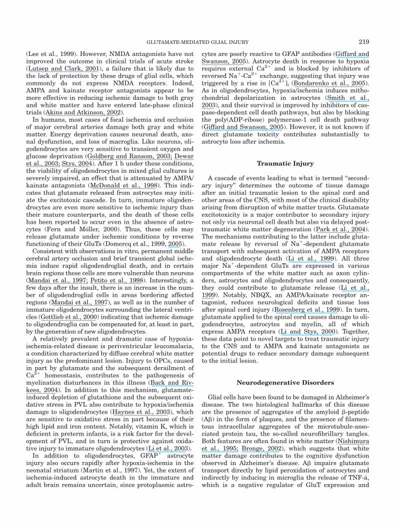
Fig. 1. Signaling cascades triggered by glutamate-induced glialinjury. Selective activation of AMPA receptors (AMPA-R) and kainatereceptors (Kai-R) leads to Na1 and Ca21 influx through the receptorchannel complex. Subsequent depolarization activates voltage-gatedCa21 channels (VGCC) and reverse operation of the Na1/Ca21 exchan-ger (NCX), which contributes to [Ca21]i increase. Ca
21 overload inducesrapid uptake by mitochondria, which results in attenuation of mito-chondrial potential and an increase in the production of reactive oxygenspecies (ROS). The latter can also be generated by reversal of the gluta-mate-cystine exchanger, which results in cystine (CySS) efflux, andthereby glutathione (GSH) depletion. Cytochrome c (Cyt c) is released
from depolarized mitochondria, interacts with apoptotic protease acti-vating factor 1 (Apaf-1) and activates caspases. Other pro-apoptotic fac-tors include apoptosis-inducing factor (AIF), which activates poly(ADP-ribose)-1 (PARP-1). In oligodendrocytes, insults channeled through Kai-R activate caspases 9 and 3, whereas those activating AMPA-R induceapoptosis by recruiting caspase 8, which truncates Bid, caspase 3, andPARP-1, or cause necrosis. In addition, Ca21 influx triggered by Kai-Rstimulation, but not by AMPA-R activates calcineurin (CdP), whichdephosphorylates Bad and facilitates apoptosis. FADD, Fas-associateddeath domain; 14-3-3, phosphoserine-binding protein 14-3-3.
217GLUTAMATE-MEDIATED GLIAL INJURY

oligodendrocytes and white matter depends on properGluT function (Domercq et al., 2005).
MECHANISMS OF GLUTAMATE TOXICITY INGLIAL CELLS
Ionotropic GluRs mediating excitotoxicity are perme-able to Ca21. The degree of Ca21 permeability variesdepending on the subunit composition of the receptor.Excessive activation of AMPA, kainate, and NMDA recep-tors results in the influx of sodium and Ca21 ions throughthe channels formed by these receptors. Sodium influxcan in turn trigger a secondary increase in the concentra-tion of intracellular Ca21 through the activation of vol-tage-gated Ca21 channels and reverse operation of Na1/Ca21 exchange (Lee et al., 1999).
In oligodendrocytes, selective activation of AMPA andkainate receptors leads to Ca21 influx (Fig. 1), an effectthat is totally abolished by non-NMDA receptor antago-nists or by removing this cation from the culture medium.Blockade of voltage-gated Ca21 channels substantiallyreduces the amplitude of the Ca21 current triggered byAMPA receptor activation, but not that initiated by low-and high-affinity kainate receptors (Alberdi et al., 2002).In contrast, inhibition of the plasma membrane Na1/Ca21 exchanger only weakly attenuates the rise in thecytosolic Ca21 concentration ([Ca21]i) caused by activa-tion of kainate receptors. Surprisingly, excitotoxicity trig-gered by GluR overactivation is not reduced by Ca21
channel blockers or by inhibition of the Na1-Ca21
exchanger. This indicates that Ca21 influx via AMPA andkainate receptors alone is sufficient to initiate cell deathin oligodendrocytes, and it does not require the entry ofCa21 via other routes such as voltage-activated Ca21
channels or the plasma membrane Na1-Ca21 exchanger.Overall, these results are reminiscent of those that havebeen reported for neurons, indicating that rapid Ca21
influx through Ca21-permeable AMPA/kainate channelsmay result in mitochondrial Ca21 overload and in theproduction of oxygen radicals, which leads to neuronaldeath (Carriedo et al., 1998).
Several features may render oligodendrocytes vulner-able to overactivation of AMPA and kainate receptors.First, the subunit composition of the receptors endowsthem with higher Ca21 permeability. Thus, AMPA recep-tors in oligodendrocytes are homomeric and/or heteromericentities formed by the GluR3 and GluR4 subunits (Matuteet al., 2002), without GluR2, a configuration that permitsCa21 entry (Hollmann and Heineman, 1994). In addition, amajor constituent of kainate receptors in oligodendrocytesis the unedited version of GluR6, which again displayshigher Ca21 permeability (Burnashev, 1996). Second, oligo-dendrocytes do not express the major Ca21-binding pro-teins that are present in neurons and that contribute tomaintaining intracellular Ca21 homeostasis and prevent-ing cytosolic Ca21 overload (Baimbridge et al., 1992).
The types of excitotoxic cell death observed depend onthe intensity and duration of glutamate exposure andinvolve two temporally distinct phases of necrosis and
apoptosis, a feature that relies on mitochondrial physiology.Mitochondria accumulate much of the Ca21 that enters thecell during excitotoxic insult, and blockade of this processprevents cell death (Stout et al., 1998; Rego et al., 2001).Thus it seems that mitochondrial Ca21 accumulation is acritical event leading to the dysfunction of this organelle.Mitochondrial depolarization, increased production of oxy-gen free radicals, and release of proapoptotic factors thatactivate caspases (see Fig. 1) have all been reported to beconsequences of excitotoxic stimuli (Luetjens et al., 2000;Atlante et al., 2001). The relative contribution of theseevents determines whether ensuing cell death is via necro-sis or apoptosis (Ankarcrona et al., 1995). Consistent withthis, mitochondrial alterations subsequent to excitotoxicinsult in oligodendrocytes are reminiscent of those ob-served in neurons, in that they can trigger apoptotic andnecrotic death (S�anchez-G�omez et al., 2003). Thus, excito-toxic insults to these cells lead to massive Ca21 entry, mito-chondrial depolarization, and a rise in the level of reactiveoxygen species that correlates with a decrease in the levelsof reduced glutathione and ultimately causes caspase-dependent and -independent cell death.
Dysfunction of mitochondria leads to translocation ofcytochrome c to the cytoplasm, where it can activate cas-pases and trigger apoptosis. Alternatively, cytochrome crelease can also increase free radical production by themitochondrial electron transport chain at the levels ofcomplex I, III, and ubiquinone, causing nonspecific cellu-lar damage and necrotic-like morphology (Murphy et al.,1999). Depending on the levels of cytochrome c released bymitochondria, the amount of ATP may be insufficient tocomplete the apoptotic program. Consequently, cells candie by apoptosis or necrosis, as is indeed observed in exci-totoxic models (Bonfoco et al., 1995). Cell death by mildexcitotoxic insults can be prevented by the overexpressionof anti-apoptotic Bcl-2 and Bcl-xl (Lawrence et al., 1996;S�anchez-G�omez et al., 2003), whereas pro-apoptotic mem-bers such as Bad or Bax are involved in excitotoxin-induced mitochondrial dysfunction (Xiang et al., 1998).
CLINICAL RELEVANCE OF GLUTAMATE-MEDIATED GLIAL INJURY
Glial cells constitute the vast majority of CNS cellsand, as described above, are vulnerable to enhanced glu-tamate signals. In particular, glutamate toxicity in glialcells is implicated in most modes of white matter injurystudied so far (Stys, 2004). Since in humans white matterconstitutes about 50% of brain volume, glutamate-induced glial cell death is highly relevant to the patho-physiology of CNS diseases. In addition, primary and/orsecondary glutamate damage to glial cells in gray mattermay also contribute to the onset and progression of acuteand chronic neurodegenerative diseases.
Brain Ischemia
Glutamate excitotoxicity appears to be the predomi-nant mechanism underlying ischemic damage in the CNS
218 MATUTE ET AL.
(Lee et al., 1999). However, NMDA antagonists have notimproved the outcome in clinical trials of acute stroke(Lutsep and Clark, 2001), a failure that is likely due tothe lack of protection by these drugs of glial cells, whichcommonly do not express NMDA receptors. Indeed,AMPA and kainate receptor antagonists appear to bemore effective in reducing ischemic damage to both grayand white matter and have entered late-phase clinicaltrials (Akins and Atkinson, 2002).
In humans, most cases of focal ischemia and occlusionof major cerebral arteries damage both gray and whitematter. Energy deprivation causes neuronal death, axo-nal dysfunction, and loss of macroglia. Like neurons, oli-godendrocytes are very sensitive to transient oxygen andglucose deprivation (Goldberg and Ransom, 2003; Dewaret al., 2003; Stys, 2004). After 1 h under these conditions,the viability of oligodendrocytes in mixed glial cultures isseverely impaired, an effect that is attenuated by AMPA/kainate antagonists (McDonald et al., 1998). This indi-cates that glutamate released from astrocytes may initi-ate the excitotoxic cascade. In turn, immature oligoden-drocytes are even more sensitive to ischemic injury thantheir mature counterparts, and the death of these cellshas been reported to occur even in the absence of astro-cytes (Fern and M€oller, 2000). Thus, these cells mayrelease glutamate under ischemic conditions by reversefunctioning of their GluTs (Domercq et al., 1999, 2005).
Consistent with observations in vitro, permanent middlecerebral artery occlusion and brief transient global ische-mia induce rapid oligodendroglial death, and in certainbrain regions these cells are more vulnerable than neurons(Mandai et al., 1997; Petito et al., 1998). Interestingly, afew days after the insult, there is an increase in the num-ber of oligodendroglial cells in areas bordering affectedregions (Mandai et al., 1997), as well as in the number ofimmature oligodendrocytes surrounding the lateral ventri-cles (Gottlieb et al., 2000) indicating that ischemic damageto oligodendroglia can be compensated for, at least in part,by the generation of new oligodendrocytes.
A relatively prevalent and dramatic case of hypoxia-ischemia-related disease is periventricular leucomalacia,a condition characterized by diffuse cerebral white matterinjury as the predominant lesion. Injury to OPCs, causedin part by glutamate and the subsequent derailment ofCa21 homeostasis, contributes to the pathogenesis ofmyelination disturbances in this illness (Back and Riv-kees, 2004). In addition to this mechanism, glutamate-induced depletion of glutathione and the subsequent oxi-dative stress in PVL also contribute to hypoxia/ischemiadamage to oligodendrocytes (Haynes et al., 2003), whichare sensitive to oxidative stress in part because of theirhigh lipid and iron content. Notably, vitamin K, which isdeficient in preterm infants, is a risk factor for the devel-opment of PVL, and in turn is protective against oxida-tive injury to immature oligodendrocytes (Li et al., 2003).
In addition to oligodendrocytes, GFAP1 astrocyteinjury also occurs rapidly after hypoxia-ischemia in theneonatal striatum (Martin et al., 1997). Yet, the extent ofischemia-induced astrocyte death in the immature andadult brain remains uncertain, since protoplasmic astro-
cytes are poorly reactive to GFAP antibodies (Giffard andSwanson, 2005). Astrocyte death in response to hypoxiarequires external Ca21 and is blocked by inhibitors ofreversed Na1-Ca21 exchange, suggesting that injury wastriggered by a rise in [Ca21]i (Bondarenko et al., 2005).As in oligodendrocytes, hypoxia/ischemia induces mitho-chondrial depolarization in astrocytes (Smith et al.,2003), and their survival is improved by inhibitors of cas-pase-dependent cell death pathways, but also by blockingthe poly(ADP-ribose) polymerase-1 cell death pathway(Giffard and Swanson, 2005). However, it is not known ifdirect glutamate toxicity contributes substantially toastrocyte loss after ischemia.
Traumatic Injury
A cascade of events leading to what is termed ‘‘second-ary injury’’ determines the outcome of tissue damageafter an initial traumatic lesion to the spinal cord andother areas of the CNS, with most of the clinical disabilityarising from disruption of white matter tracts. Glutamateexcitotoxicity is a major contributor to secondary injurynot only via neuronal cell death but also via delayed post-traumatic white matter degeneration (Park et al., 2004).The mechanisms contributing to the latter include gluta-mate release by reversal of Na1-dependent glutamatetransport with subsequent activation of AMPA receptorsand oligodendrocyte death (Li et al., 1999). All threemajor Na1-dependent GluTs are expressed in variouscompartments of the white matter such as axon cylin-ders, astrocytes and oligodendrocytes and consequently,they could contribute to glutamate release (Li et al.,1999). Notably, NBQX, an AMPA/kainate receptor an-tagonist, reduces neurological deficits and tissue lossafter spinal cord injury (Rosenberg et al., 1999). In turn,glutamate applied to the spinal cord causes damage to oli-godendrocytes, astrocytes and myelin, all of whichexpress AMPA receptors (Li and Stys, 2000). Together,these data point to novel targets to treat traumatic injuryto the CNS and to AMPA and kainate antagonists aspotential drugs to reduce secondary damage subsequentto the initial lesion.
Neurodegenerative Disorders
Glial cells have been found to be damaged in Alzheimer’sdisease. The two histological hallmarks of this diseaseare the presence of aggregates of the amyloid b-peptide(Ab) in the form of plaques, and the presence of filamen-tous intracellular aggregates of the microtubule-asso-ciated protein tau, the so-called neurofibrillary tangles.Both features are often found in white matter (Nishimuraet al., 1995; Bronge, 2002), which suggests that whitematter damage contributes to the cognitive dysfunctionobserved in Alzheimer’s disease. Ab impairs glutamatetransport directly by lipid peroxidation of astrocytes andindirectly by inducing in microglia the release of TNF-�a,which is a negative regulator of GluT expression and
219GLUTAMATE-MEDIATED GLIAL INJURY

function (Mattson and Chan, 2003). In addition, Ab killsoligodendrocytes (Xu et al., 2001) and can increase theirvulnerability to being killed by glutamate (Pak et al.,2003). In turn, oligodendrocytes express presenilin 1(PS1) and oligodendrocytes from PS1 mutant knock-inmice exhibited increased vulnerability to excitotoxicitycompared to oligodendrocytes from wild-type mice (Paket al., 2003). Finally, when exposed to the demyelinatingagent cuprizone, PS1 mutant mice exhibit enhanceddamage in the white matter and learning and memorydeficits not seen in wild-type mice exposed to cuprizone(Pak et al., 2003).
Human immunodeficiency virus type 1 (HIV-1) infec-tion causes cognitive dysfunction in about 10–20% ofAIDS patients, a syndrome that may be due to excitotoxicneuronal damage (Kaul et al., 2005). Interestingly, HIVencephalitis is also characterized by myelin pallor, whichsuggests that oligodendrocyte excitotoxicity also occurs inthis condition as a consequence of an inhibition of gluta-mate uptake by eicosanoids and free radicals produced byHIV-infected brain mononuclear phagocytes (Lipton,1998) and/or of excessive glutamate release from astro-cytes (Bezzi et al., 2001). Indeed, the expression and func-tion of the glutamate transporter EAAT2 is reduced inhuman astrocytes exposed to the effects of HIV-1 and itsenvelope glycoprotein (Wang et al, 2003).
Multiple Sclerosis
In MS, the immune system attacks the white matter ofthe brain and spinal cord, leading to disability or paraly-sis, or both. Myelin and oligodendrocytes are lost due tothe release by immune cells of cytotoxic cytokines, auto-antibodies, and toxic amounts of glutamate (Matuteet al., 2001; Srinivasan et al. 2005). Excitotoxins such askainate infused onto the optic nerve cause glutamatereceptor-mediated MS-like lesions (Matute, 1998). Inturn, experimental autoimmune encephalomyelitis (EAE),an animal model that exhibits the clinical and pathologi-cal features of MS, is alleviated by AMPA and kainatereceptor antagonists (Smith et al., 2000; Pitt et al., 2000;Groom et al., 2003). Remarkably, blockade of these recep-tors in combination with anti-inflammatory agents iseffective even at an advanced stage of unremitting EAE,as assessed by increased oligodendrocyte survival andremyelination, and corresponding decreased paralysis,inflammation, CNS apoptosis and axonal damage (Kan-war et al., 2004).
The concentration of glutamate in cerebrospinal fluid(CSF) is higher in patients with acute rather than silentMS and in controls (Stover et al., 1997), and it is asso-ciated with the severity and course of the disease (Bar-khatova et al., 1998). Notably, glutamate levels areincreased in acute MS lesions and in normal-appearingwhite matter in MS patients (Srinivasan et al., 2005).Potential cellular sources contributing to enhanced gluta-mate levels in CSF include activated microglia, which canrelease glutamate via the reversal of GluT function, a pro-cess that is potentiated under pathological conditions
(Noda et al., 1999). In addition, oxidative stress may alsocontribute to the increase in glutamate concentrations inthe extracellular space, since free radicals reduce the effi-ciency of GluTs (Volterra et al., 1994). Other factors thatmay contribute to perturbing glutamate homeostasis in-clude altered activity of the glutamate producing enzymeglutaminase in activated macrophages/microglia in closeproximity to dystrophic axons (Werner et al., 2001), andreduced expression of the glutamate transporters EAAT-1and EAAT-2 in oligodendrocytes as a consequence of en-hanced exposure to the proinflammatory cytokine TNF-a(Pitt et al., 2003). Overall, these alterations likely lead tohigh extracellular glutamate levels and an increased riskof oligodendrocyte excitotoxicity in MS.
CONCLUSIONS AND FUTURE PROSPECTS
Glial cells are sensitive to glutamate-induced damage.Among them, oligodendrocytes display great vulnerabil-ity to overactivation of AMPA and kainate receptors. Thefailure of drugs aimed at preventing neuronal death todemonstrate significant improvement in the outcome ofacute and chronic neurodegeneration in humans is likelydue to the lack of protection by these drugs for the glialcells, which constitute the vast majority of cells withinthe CNS.
The proper functioning of glutamate uptake is criticalto prevent glutamate-induced damage to glia and drugsthat regulate the function and expression of GluTs havethe potential to attenuate glutamate insults to glial cells.Thus, reversal of GluTs can generate toxic extracellularglutamate levels during anoxia and trauma and signifi-cant protection can be achieved with specific inhibitors ofGluTs (Stys, 2004). Likewise, positive regulators of theexpression of GluTs also have a glioprotective potential,as they contribute to ischemic tolerance after ischemicpreconditioning (Romera et al., 2004). These includetumor growth factor-a (TGF-a) and epidermal growth fac-tor (EGF), which by signaling through EGF receptors andactivation of phosphoinositol-3-kinase and nuclear factor-jB, strongly enhance EAAT2 expression (Su et al., 2003).Remarkably, clinically used b-lactam antibiotics are alsopotent activators of GluT expression and thus hold greattherapeutic potential (Rothstein et al., 2005).
Another set of molecular targets to prevent glutamateinsults to glia lie downstream of GluR activation. Thus,the vulnerability of OPCs to glutamate excitotoxicity canbe attenuated with agonists of group I metabotropicGluRs by controlling oxidative stress generated down-stream of AMPA and kainate receptor activation (Denget al., 2004). Similarly, the clinically available anticonvul-sant topiramate prevents excitotoxicity in developing oli-godendrocytes (Follet et al., 2004). Together, these find-ings offer good prospects for future treatments for peri-ventricular leucomalacia, a condition in which there ismassive loss of immature oligodendrocytes.
In addition, the neuroprotective properties of tetracy-clines, which attenuate mitochondrial damage subse-quent to insults including excitotoxicity, have been
220 MATUTE ET AL.

expanded to oligodendrocytes and to white matter as awhole, making these antibiotics promising candidates forthe treatment of acute and chronic diseases with glial loss(Domercq and Matute, 2004). In contrast, drugs support-ing the management of Ca21 overload subsequent toGluR activation may improve glial cell viability. Notably,the major plasma membrane Ca21 extruding system, theNa1/Ca21 exchanger, is cleaved during brain ischemiaand in neuronal excitotoxic settings by calpains. Inhibi-tion of this process has been reported to enhance cell via-bility (Bano et al., 2005). Conceivably, similar interven-tions may prevent excitotoxic glial cell death.
In summary, knowledge about the mechanisms leadingto glutamate-mediated glial injury will open the way tonew pharmacological strategies for the treatment of CNSdisorders and in particular, those ailments in which gliaare primarily affected. Glioprotectants that prevent glu-tamate-induced glial cell death are about to emerge as anovel class of drugs with important clinical applications.Their use in conjunction with neuroprotectants is likelyto increase the effectiveness of current treatments forneurological dysfunction in acute and chronic neurode-generative diseases.
ACKNOWLEDGMENTS
The authors express our thanks to the agency ACTS(http://www.euskalnet.net/acts) for having improved theEnglish of this work.
REFERENCES
Alberdi E, S�anchez-G�omez MV, Marino A, Matute C. 2002. Ca21 influxthrough AMPA or kainate receptors alone is sufficient to initiate exci-totoxicity in cultured oligodendrocytes. Neurobiol Dis 9:234–243.
Akins PT, Atkinson RP. 2002. Glutamate AMPA receptor antagonisttreatment for ischaemic stroke. Curr Med Res Opin 18:s9–s13.
Ankarcrona M, Dypbukt JM, Bonfoco E, Zhivotovsky B, Orrenius S,Lipton SA, Nicotera P. 1995. Glutamate-induced neuronal death: asuccession of necrosis or apoptosis depending on mitochondrial func-tion. Neuron 15:961–973.
Atlante A, Calissano P, Bobba A, Giannattasio S, Marra E, PassarellaS, 2001. Glutamate neurotoxicity, oxidative stress and mitochondria.FEBS Lett 497:1–5.
Auger C, Attwell D. 2000. Fast removal of synaptic glutamate by post-synaptic transporters. Neuron 28:547–558.
Back SA, Rivkees SA. 2004. Emerging concepts in periventricular whitematter injury. Semin Perinatol 28:405–414.
Baimbridge KG, Celio MR, Rogers JH. 1992. Calcium-binding proteinsin the nervous system. Trends Neurosci 15:303–308.
Bano D, Young KW, Guerin CJ, Lefeuvre R, Rothwell NJ, Naldini L,Rizzuto R, Carafoli E, Nicotera P. 2005. Cleavage of the plasma mem-brane Na1/Ca21 exchanger in excitotoxicity. Cell 120:275–285.
Barkhatova VP, Zavalishin IA, Askarova LS, Shavratskii VK, DeminaEG. 1998. Changes in neurotransmitters in multiple sclerosis. Neu-rosci Behav Physiol 28:341–344.
Barres BA, Koroshetz WJ, Swartz KJ, Chun LL, Corey DP. 1990. Ionchannel expression by white matter glia: the O-2A glial progenitorcell. Neuron 4:507–524.
Belachew S, Gallo V. 2004. Synaptic and extrasynaptic neurotrans-mitter receptors in glial precursors’ quest for identity. Glia 48:185–196.
Bergles DE, Roberts JD, Somogyi P, Jahr CE. 2000. Glutamatergicsynapses on oligodendrocyte precursor cells in hippocampus. Nature405:187–191.
Bezzi P, Carmignoto G, Pasti L, Vesce S, Rossi D, Rizzini BL, Pozzan T,Volterra A. 1998. Prostaglandins stimulate calcium-dependent gluta-mate release in astrocytes. Nature 391:281–285.
Bezzi P, Domercq M, Brambilla L, Galli R, Schols D, De Clercq E, Ves-covi A, Bagetta G, Kollias G, Meldolesi J, Volterra A. 2001. CXCR4-activated astrocyte glutamate release via TNFa: amplification bymicroglia triggers neurotoxicity. Nat Neurosci 4:702–710.
Bezzi P, Gundersen V, Galbete JL, Seifert G, Steinhauser C, Pilati E,Volterra A. 2004. Astrocytes contain a vesicular compartment that iscompetent for regulated exocytosis of glutamate. Nat Neurosci 7:613–620.
Biber K, Laurie DJ, Berthele A, Sommer B, Tolle TR, Gebicke-HarterPJ, van Calker D, Boddeke HW. 1999. Expression and signaling ofgroup I metabotropic glutamate receptors in astrocytes and microglia.J Neurochem 72:1671–1680.
Bondarenko A, Svichar N, Chesler M. 2005. Role of Na1-H1 and Na1-Ca21
exchange in hypoxia-related acute astrocyte death. Glia 49:143–152.Bonfoco E, Krainc D, Ankarcrona M, Nicotera P, Lipton SA. 1995.
Apoptosis and necrosis: two distinct events induced, respectively, bymild and intense insults with N-methyl-D-aspartate or nitric oxide/superoxide in cortical cell cultures. Proc Natl Acad Sci USA 92:7162–7166.
Borges K, Kettenmann H. 1995. Blockade of K1 channels induced byAMPA/kainate receptor activation in mouse oligodendrocyte precursorcells is mediated by Na1 entry. J Neurosci Res 42:579–593.
Borges K, Ohlemeyer C, Trotter J, Kettenmann H. 1994. AMPA/kainatereceptor activation in murine oligodendrocyte precursor cells leads toactivation of a cation conductance, calcium influx and blockade ofdelayed rectifying K1 channels. Neuroscience 63:135–149.
Bowman CL, Kimelberg HK. 1984. Excitatory amino acids directly de-polarize rat brain astrocytes in primary culture. Nature 311:656–659.
Bronge L. 2002. Magnetic resonance imaging in dementia. A study ofbrain white matter changes. Acta Radiol 428(suppl):1–32.
Burnashev N. 1996. Calcium permeability of glutamate-gated channelsin the central nervous system. Curr Opin Neurobiol 6:311–317.
Burnashev N, Khodorova A, Jonas P, Helm PJ, Wisden W, Monyer H,Seeburg PH, Sakmann B. 1992. Calcium-permeable AMPA-kainatereceptors in fusiform cerebellar glial cells. Science 256:1566–1570.
Carriedo SG, Yin HZ, Sensi SL, Weiss JH. 1998. Rapid Ca21 entrythrough Ca21-permeable AMPA/Kainate channels triggers markedintracellular Ca21 rises and consequent oxygen radical production.J Neurosci 18:7727–7738.
Chen W, Aoki C, Mahadomrongkul V, Gruber CE, Wang GJ, BlitzblauR, Irwin N, Rosenberg PA. 2002. Expression of a variant form of theglutamate transporter GLT1 in neuronal cultures and in neurons andastrocytes in the rat brain. J Neurosci 22:2142–2152.
Chen CJ, Liao SL, Kuo JS. 2000. Gliotoxic action of glutamate on cul-tured astrocytes. J Neurochem 75:1557–1565.
Choi DW, Maulucci-Gedde M, Kriegstein AR. 1987. Glutamate neuro-toxicity in cortical cell culture. J Neurosci 7:357–368.
Choi DW. 1988. Glutamate neurotoxicity and diseases of the nervoussystem. Neuron 1:623–634.
Chr�etien F, Le Pavec G, Vallat-Decouvelaere AV, Delisle MB, Uro-CosteE, Ironside JW, Gambetti P, Parchi P, Creminon C, Dormont D, MikolJ, Gray F, Gras G. 2004. Expression of excitatory amino acid trans-porter-1 (EAAT-1) in brain macrophages and microglia of patientswith prion diseases. J Neuropathol Exp Neurol 63:1058–1071.
Clark B, Mobbs P. 1992. Transmitter-operated channels in rabbit ret-inal astrocytes studied in situ by whole-cell patch clamping. J Neu-rosci 12:664–673.
Cornell-Bell AH, Thomas PG, Caffrey JM. 1992. Ca21 and filopodialresponses to glutamate in cultured astrocytes and neurons. Can JPhysiol Pharmacol 70(suppl):S206–218.
Cull-Candy SG, Leszkiewicz DN. 2004. Role of distinct NMDA receptorsubtypes at central synapses. Sci STKE;2004:255:re 16.Review.
Danbolt NC. 2001. Glutamate uptake. Prog Neurobiol 65:1–105.Danbolt NC, Storm-Mathisen J, Kanner BI. 1992. An [Na1 1
K1]coupled L-glutamate transporter purified from rat brain is locatedin glial cell processes. Neuroscience 51:295–310.
David JC, Yamada KA, Bagwe MR, Goldberg MP. 1996. AMPA receptoractivation is rapidly toxic to cortical astrocytes when desensitizationis blocked. J Neurosci 16:200–209.
Deng W, Wang H, Rosenberg PA, Volpe JJ, Jensen FE. 2004. Role ofmetabotropic glutamate receptors in oligodendrocyte excitotoxicityand oxidative stress. Proc Natl Acad Sci USA 101:7751–7756.
Dewar D, Underhill SM, Goldberg MP. 2003. Oligodendrocytes andischemic brain injury. J Cereb Blood Flow Metab 23:263–274.
Dingledine R, Borges K, Bowie D, Traynelis SF. 1999. The glutamatereceptor ion channels. Pharmacol Rev 51:7–61.
Domercq M, Matute C. 1999. Expression of glutamate transporters inthe adult bovine corpus callosum. Brain Res Mol Brain Res. 67:296–302.
Domercq M, S�anchez-G�omez MV, Areso P, Matute C. 1999. Expressionof glutamate transporters in rat optic nerve oligodendrocytes. Eur JNeurosci 11:2226–2236.
221GLUTAMATE-MEDIATED GLIAL INJURY

Domercq M, Matute C. 2004. Neuroprotection by tetracyclines. TrendsPharmacol Sci 25:609–612.
Domercq M, Etxebarria E, Perez-Samartin A, Matute C. 2005. Excito-toxic oligodendrocyte death and axonal damage induced by glutamatetransporter inhibition. Glia, May 12; [Epub ahead of print].
Fern R, M€oller T. 2000. Rapid ischemic cell death in immature oligoden-drocytes: a fatal glutamate release feedback loop. J Neurosci 20:34–42.
Follett PL, Deng W, Dai W, Talos DM, Massillon LJ, Rosenberg PA,Volpe JJ, Jensen FE. 2004. Glutamate receptor-mediated oligodendro-cyte toxicity in periventricular leukomalacia: a protective role fortopiramate. J Neurosci 24:4412–4420.
Fulton BP, Burne JF, Raff MC. 1992. Visualization of O-2A progenitorcells in developing and adult rat optic nerve by quisqualate-stimu-lated cobalt uptake. J Neurosci 12:4816–4833.
Gallo V, Zhou JM, McBain CJ, Wright P, Knutson PL, Armstrong RC.1996. Oligodendrocyte progenitor cell proliferation and lineage pro-gression are regulated by glutamate receptor-mediated K1 channelblock. J Neurosci 16:2659–2670.
Garc�ıa-Barcina JM, Matute C. 1996. Expression of kainate-selectiveglutamate receptor subunits in glial cells of the adult bovine whitematter. Eur J Neurosci 8:2379–2387.
Geurts JJ, Wolswijk G, Bo L, van der Valk P, Polman CH, Troost D,Aronica E. 2003. Altered expression patterns of group I and II meta-botropic glutamate receptors in multiple sclerosis. Brain 126:1755–1766.
Giffard RG, Swanson RA. 2005. Ischemia-induced programmed celldeath in astrocytes. Glia 50:299–306.
Gill R, Lodge D. 1997. Pharmacology of AMPA antagonists and theirrole in neuroprotection. Int Rev Neurobiol 40:197–232.
Goldberg MP, Ransom BR. 2003. New light on white matter. Stroke34:330–332.
Gottlieb M, Matute C. 1997. Expression of ionotropic glutamate recep-tor subunits in glial cells of the hippocampal CA1 area followingtransient forebrain ischemia. J Cereb Blood Flow Metab 17:290–300.
Gottlieb M, Domercq M, Matute C. 2000. Altered expression of the glu-tamate transporter EAAC1 in neurons and immature oligodendro-cytes after transient forebrain ischemia. J Cereb Blood Flow Metab20:678–687.
Gras G, Chretien F, Vallat-Decouvelaere AV, Le Pavec G, Porcheray F,Bossuet C, Leone C, Mialocq P, Dereuddre-Bosquet N, Clayette P, LeGrand R, Creminon C, Dormont D, Rimaniol AC, Gray F. 2003. Regu-lated expression of sodium-dependent glutamate transporters andsynthetase: a neuroprotective role for activated microglia and macro-phages in HIV infection? Brain Pathol 13:211–222.
Groom AJ, Smith T, Turski L. 2003. Multiple sclerosis and glutamate.Ann NY Acad Sci 993:229–275.
Haas J, Erdo SL. 1991. Quisqualate-induced excitotoxic death of glialcells: transient vulnerability of cultured astrocytes. Glia 4:111–114.
Haynes RL, Folkerth RD, Keefe RJ, Sung I, Swzeda LI, Rosenberg PA,Volpe JJ, Kinney HC. 2003. Nitrosative and oxidative injury to pre-myelinating oligodendrocytes in periventricular leukomalacia. J Neu-ropathol Exp Neurol 62:441–450.
Hertz L, Dringen R, Schousboe A, Robinson SR 1999. Astrocytes: gluta-mate producers for neurons. J Neurosci Res 5:417–428.
Hollmann M, Heineman S. 1994. Cloned glutamate receptors. AnnuRev Neurosci 17:31–108.
Huang YH, Bergles DE. 2004. Glutamate transporters bring competi-tion to the synapse. Curr Opin Neurobiol 14:346–352.
Iino M, Goto K, Kakegawa W, Okado H, Sudo M, Ishiuchi S, Miwa A,Takayasu Y, Saito I, Tsuzuki K, Ozawa S. 2001. Glia–synapse inter-action through Ca21-permeable AMPA receptors in Bergmann glia.Science 292:926–929.
Kanwar JR, Kanwar RK, Krissansen GW. 2004. Simultaneous neuro-protection and blockade of inflammation reverses autoimmune ence-phalomyelitis. Brain 127:1313–1331.
Karadottir R, Cavelier P, Bergersen L, Attwell D. 2005.NMDA receptormediated responses in oligodendrocytes. In: Seventh European meet-ing on glial cell function in health and disease; abstract. p 104.Amsterdam, 17-21 May, 2005.
Kaul M, Zheng J, Okamoto S, Gendelman HE, Lipton SA. 2005. HIV-1infection and AIDS: consequences for the central nervous system.Cell Death Differ Apr 15; [Epub ahead of print].
Kettenmann H, Steinhauser C. 2005.Receptors for neurotransmittersand hormones. In: Kettenmann H, Ransom BR, editors. Neuroglia.2nd Ed. New York: Oxford University Press. p 131–145.
Kettenmann H, Backus KH, Schachner M. 1984. Aspartate, glutamateand gamma-aminobutyric acid depolarize cultured astrocytes. Neu-rosci Lett 52:25–29.
Kingham PJ, Cuzner ML, Pocock JM. 1999. Apoptotic pathways mobi-lized in microglia and neurones as a consequence of chromogranin A-induced microglial activation. J Neurochem 73:538–547.
Krebs C, Fernandes HB, Sheldon C, Raymond LA, Baimbridge KG. 2003.Functional NMDA receptor subtype 2B is expressed in astrocytes afterischemia in vivo and anoxia in vitro. J Neurosci 23:3364–3372.
Kriegler S, Chiu SY. 1993. Calcium signaling of glial cells along mam-malian axons. J Neurosci 13:4229–4245.
Kugler P, Schmitt A. 1999. Glutamate transporter EAAC1 is expressedin neurons and glial cells in the rat nervous system. Glia 27:129–142.
Lawrence MS, Ho DY, Sun GH, Steinberg GK, Sapolsky RM. 1996.Overexpression of Bcl-2 with herpes simplex virus vectors protectsCNS neurons against neurological insults in vitro and in vivo. J Neu-rosci 16:486–496.
Lee JM, Zipfel GJ, Choi DW. 1999. The changing landscape of ischae-mic brain injury mechanisms. Nature 399:7–14.
Lerma J. 2003. Roles and rules of kainate receptors in synaptic trans-mission. Nat Rev Neurosci. 4:481–495.
Leuchtmann EA, Ratner AE, Vijitruth R, Qu Y, McDonald JW. 2003.AMPA receptors are the major mediators of excitotoxic death inmature oligodendrocytes. Neurobiol Dis 14:336–348.
Li J, Lin JC, Wang H, Peterson JW, Furie BC, Furie B, Booth SL, VolpeJJ, Rosenberg PA. 2003. Novel role of vitamin k in preventing oxida-tive injury to developing oligodendrocytes and neurons. J Neurosci23:5816–5826.
Li S, Mealing GA, Morley P, Stys PK. 1999. Novel injury mechanism inanoxia and trauma of spinal cord white matter: glutamate release viareverse Na1-dependent glutamate transport. J Neurosci 19:RC16.
Li S, Stys PK. 2000. Mechanisms of ionotropic glutamate receptor-mediated excitotoxicity in isolated spinal cord white matter. J Neu-rosci 20:1190–1198.
Lin SC, Huck JH, Roberts JD, Macklin WB, Somogyi P, Bergles DE.2005. Climbing fiber innervation of NG2-expressing glia in the mam-malian cerebellum. Neuron 46:773–785.
Lipton SA. 1998. Neuronal injury associated with HIV-1: approaches totreatment. Annu Rev Pharmacol Toxicol 38:159–177.
Lipton SA, Rosenberg PA. 1994. Excitatory amino acids as a final com-mon pathway for neurologic disorders. N Engl J Med 330:613–622.
Liu HN, Giasson BI, Mushynski WE, Almazan G. 2002. AMPA recep-tor-mediated toxicity in oligodendrocyte progenitors involves freeradical generation and activation of JNK, calpain and caspase 3.J Neurochem 82:398–409.
Liu SQ, Cull-Candy SG. 2000. Synaptic activity at calcium-permeableAMPA receptors induces a switch in receptor subtype. Nature405:454–458.
L�opez-Redondo F, Nakajima K, Honda S, Kohsaka S. 2000. Glutamatetransporter GLT-1 is highly expressed in activated microglia follow-ing facial nerve axotomy. Brain Res Mol Brain Res 76:429–435.
Lucas DR, Newhouse JP. 1957. The toxic effect of sodium L-glutamateon the inner layers of the retina. AMA Arch Ophthalmol 58:193–201.
Luejtens CM, Bui NT, Sengpiel B, M€unstermann G, Poppe M, KrohnAJ, Bauerbach E, Krieglstein J, Prehn JHM. 2000. Delayed mito-chondrial dysfunction in excitotoxic neuron death: cytochrome crelease and a secondary increase in superoxide production. J Neu-rosci 20:5715–5723.
Lutsep H, Clark W. 2001. An update of neuroprotectants in clinicaldevelopment for acute stroke. Curr Opin Invest Drugs 2:1732–1736.
Mandai K, Matsumoto M, Kitagawa K, Matsushita K, Ohtsuki T,Mabuchi T, Colman DR, Kamada T, Yanagihara T. 1997. Ischemicdamage and subsequent proliferation of oligodendrocytes in focal cer-ebral ischemia. Neuroscience 77:849–861.
Martin LJ, Brambrink AM, Lehmann C, Portera-Cailliau C, Koehler R,Rothstein J, Traystman RJ. 1997. Hypoxia-ischemia causes abnorm-alities in glutamate transporters and death of astroglia and neuronsin newborn striatum. Ann Neurol 42:335–348.
Matthias K, Kirchhoff F, Seifert G, Huttmann K, Matyash M, Ketten-mann H, Steinhauser C. 2003. Segregated expression of AMPA-typeglutamate receptors and glutamate transporters defines distinctastrocyte populations in the mouse hippocampus. J Neurosci23:1750–1758.
Mattson MP, Chan SL. 2003. Neuronal and glial calcium signaling inAlzheimer’s disease. Cell Calcium 34:385–397.
Matute C. 1998. Characteristics of acute and chronic kainate excitotoxicdamage to the optic nerve. Proc Natl Acad Sci USA 95:10229–10234.
Matute C, S�anchez-G�omez MV, Mart�ınez-Mill�an L, Miledi R. 1997. Glu-tamate receptor-mediated toxicity in optic nerve oligodendrocytes.Proc Natl Acad Sci USA 94:8830–8835.
Matute C, Domercq M, Fogarty DJ, Pascual de Zulueta M, S�anchez-G�omez MV. 1999. On how altered glutamate homeostasis may contri-bute to demyelinating diseases of the CNS. In: Matsas R, TsacopoulosM, editors. The function of glial cells in health and disease: dialoguebetween glia and neurons. New York: Plenum. p 97–108.
Matute C, Alberdi E, Domercq M, P�erez-Cerd�a F, P�erez-Samart�ın A,S�anchez-G�omez MV. 2001. The link between excitotoxic oligodendro-glial death and demyelinating diseases. Trends Neurosci 24:224–230.
222 MATUTE ET AL.

Matute C, Alberdi E, Ibarretxe G, S�anchez-G�omez MV. 2002. Excito-toxicity in glial cells. Eur J Pharmacol 447:239–246.
Mayer ML. 2005. Glutamate receptor ion channels. Curr Opin Neuro-biol 3:282–288.
McDonald JW, Althomsons SP, Hyrc KL, Choi DW, Goldberg MP. 1998.Oligodendrocytes from forebrain are highly vulnerable to AMPA/kai-nate receptor-mediated excitotoxicity. Nat Med 4:291–297.
Merrill JE, Benveniste EN. 1996. Cytokines in inflammatory brainlesions: helpful and harmful. Trends Neurosci 19:331–338.
Michaelis EK. 1998. Molecular biology of glutamate receptors in thecentral nervous system and their role in excitotoxicity, oxidativestress and aging. Prog Neurobiol 54:369–415.
Muller T, Moller T, Berger T, Schnitzer J, Kettenmann H. 1992. Cal-cium entry through kainate receptors and resulting potassium-chan-nel blockade in Bergmann glial cells. Science 256:1563–1566.
Murphy AN, Fiskum G, Beal MF. 1999. Mitochondria in neurodegen-eration: bioenergetic function in cell life and death. J Cereb BloodFlow Metab 19:231–245.
Nakajima K, Tohyama Y, Kohsaka S, Kurihara T. 2001. Ability of ratmicroglia to uptake extracellular glutamate. Neurosci Lett 307:171–174.
Nedergaard M, Takano T, Hansen AJ. 2002. Beyond the role of gluta-mate as a neurotransmitter. Nat Rev Neurosci 3:748–755.
Nishimura M, Tomimoto H, Suenaga T, Namba Y, Ikeda K, Akiguchi I,Kimura J. 1995. Immunocytochemical characterization of glial fibril-lary tangles in Alzheimer’s disease brain. Am J Pathol 146:1052–1058.
Noda M, Nakanishi H, Akaike N, 1999. Glutamate release from micro-glia via glutamate transporter is enhanced by amyloid-beta peptide.Neuroscience 92:1465–1474.
Noda M, Nakanishi H, Nabekura J, Akaike N. 2000. AMPA-kainatesubtypes of glutamate receptor in rat cerebral microglia. J Neurosci20:251–258.
Oka A, Belliveau MJ, Rosenberg PA, Volpe JJ. 1993. Vulnerability ofoligodendroglia to glutamate: pharmacology, mechanisms, and pre-vention. J Neurosci 13:1441–1453.
Olney JW, Sharpe LG. 1969. Brain lesions in an infant rhesus monkeytreated with monosodium glutamate. Science 166:386–388.
Pak K, Chan SL, Mattson MP. 2003. Presenilin-1 mutation sensitizesoligodendrocytes to glutamate and amyloid toxicities and exacerbateswhich matter damage and memory impairment in mice. NeuromolMed 3:53–64.
Park E, Velumian AA, Fehlings MG. 2004. The role of excitotoxicity insecondary mechanisms of spinal cord injury: a review with an empha-sis on the implications for white matter degeneration. J Neurotrauma21:754–774.
Patneau DK, Wright PW, Winters C, Mayer ML, Gallo V. 1994. Glialcells of the oligodendrocyte lineage express both kainate- and AMPA-preferring subtypes of glutamate receptor. Neuron 12:357–371.
Pearce B, Albrecht J, Morrow C, Murphy S. 1986. Astrocyte glutamatereceptor activation promotes inositol phospholipid turnover and cal-cium flux. Neurosci Lett 72:335–340.
P�erez-Cerd�a F, Torre I, Alberdi E, Matute C. 2005.Expresi�on of AMPAand kainate receptor subunits in oligodendrocytes of the adult humanand rat optic nerve in situ. In: Seventh European meeting on glialcell function in health and disease; abstract. p 105. Amsterdam, 17-21 May, 2005.
Petito CK, Olarte JP, Roberts B, Nowak TS, Pulsinelli WA. 1998. Selec-tive glial vulnerability following transient global ischemia in ratbrain. J Neuropathol Exp Neurol 57:231–238.
Pitt D, Werner P, Raine CS, 2000. Glutamate excitotoxicity in a modelof multiple sclerosis. Nat Med 6:67–70.
Pitt D, Nagelmeier IE, Wilson HC, Raine CS. 2003. Glutamate uptakeby oligodendrocytes: implications for excitotoxicity in multiple sclero-sis. Neurology 61:1113–1120.
Prezeau L, Carrette J, Helpap B, Curry K, Pin JP, Bockaert J. 1994.Pharmacological characterization of metabotropic glutamate receptorsin several types of brain cells in primary cultures. Mol Pharmacol45:570–577.
Prieto M, Alonso F. 1999. Differential sensitivity of cultured tanycytesand astrocytes to hydrogen peroxide toxicity. Exp Neurol 155:118–127.
Puchalski RB, Louis JC, Brose N, Traynelis SF, Egebjerg J, Kukekov V,Wenthold RJ, Rogers SW, Lin F, Moran T, Morrison JH, HeinemannSF. 1994. Selective RNA editing and subunit assembly of native glu-tamate receptors. Neuron 13:131–147.
Puro DG, Yuan JP, Sucher NJ. 1996. Activation of NMDA receptor-channels in human retinal M€uller glial cells inhibits inward-rectify-ing potassium currents. Vis Neurosci 13:319–326.
Rego AC, Ward MW, Nicholls DG. 2001. Mitochondria control AMPA/kainate receptor-induced cytoplasmic calcium deregulation in rat cer-ebellar granule cells. J Neurosci 21:1893–1901.
Romera C, Hurtado O, Botella SH, Lizasoain I, Cardenas A, Fernan-dez-Tome P, Leza JC, Lorenzo P, Moro MA. 2004. In vitro ischemic
tolerance involves upregulation of glutamate transport partlymediated by the TACE/ADAM17-tumor necrosis factor-alpha path-way. J Neurosci 24:1350–1357.
Rosenberg LJ, Teng YD, Wrathall JR. 1999. 2,3-Dihydroxy-6-nitro-7-sul-famoyl-benzo(f)quinoxaline reduces glial loss and acute white matterpathology after experimental spinal cord contusion. J Neurosci19:464–475.
Rosenberg PA, Dai W, Gan XD, Ali S, Fu J, Back SA, Sanchez RM,Segal MM, Follett PL, Jensen FE, Volpe JJ. 2003. Mature myelinbasic protein-expressing oligodendrocytes are insensitive to kainatetoxicity. J Neurosci Res 71:237–245.
Rothstein JD, Dykes-Hoberg M, Pardo CA, Bristol LA, Jin L, KlunclRW, Kanai Y, Hediger MA, Wang Y, Schielke JP, Welty DF. 1996.Knock out of glutamate transporters reveals a major role for astro-glial transport in excitotoxicity and clearance of glutamate. Neuron16:675–686.
Rothstein JD, Patel S, Regan MR, Haenggeli C, Huang YH, BerglesDE, Jin L, Dykes Hoberg M, Vidensky S, Chung DS, Toan SV, BruijnLI, Su ZZ, Gupta P, Fisher PB. 2005. Beta-lactam antibiotics offerneuroprotection by increasing glutamate transporter expression.Nature 433:73–77.
S�anchez-G�omez MV, Matute C. 1999 AMPA and kainate receptors eachmediate excitotoxicity in oligodendroglial cultures. Neurobiol Dis 6:475–485.
S�anchez-G�omez MV, Alberdi E, Ibarretxe G, Torre I, Matute C. 2003.Caspase-dependent and caspase-independent oligodendrocyte deathmediated by AMPA and kainite receptors. J Neurosci 23:9519–9528.
Schipke CG, Ohlemeyer C, Matyash M, Nolte C, Kettenmann H,Kirchhoff F. 2001. Astrocytes of the mouse neocortex express func-tional N-methyl-D-aspartate receptors. FASEB J 15:1270–1272.
Schubert D, Piasecki D. 2001. Oxidative glutamate toxicity can be acomponent of the excitotoxicity cascade. J Neurosci 21:7455–7462.
Seifert G, Zhou M, Steinh€auser C. 1997. Analysis of AMPA receptorproperties during postnatal development of mouse hippocampal astro-cytes. J Neurophysiol 78:2916–2923.
Seifert G, Schumacher T, Sch€oder W, Hinterkeuser S, Schramm J,Steinh€auser C. 1999. AMPA receptor properties of astrocytes in humanepilepsy. Joint Meeting of the International Society for Neurochemistry(ISN) and the European Society for Neurochemistry (ESN). Berlin,Germany, Aug. 8-14, 1999, poster number 538.
Shashidharan P, Plaitakis A. 1993. Cloning and characterization of aglutamate transporter cDNA from human cerebellum. Biochim Bio-phys Acta 1216:161–164.
Smith T, Groom A, Zhu B, Turski L. 2000. Autoimmune encephalomye-litis ameliorated by AMPA antagonists. Nat Med 6:62–66.
Smith IF, Plant LD, Boyle JP, Skinner RA, Pearson HA, Peers C. 2003.Chronic hypoxia potentiates capacitative Ca21 entry in type-I corticalastrocytes. J Neurochem 85:1109–1116.
Sontheimer H, Kettenmann H, Backus KH, Schachner M. 1988. Gluta-mate opens Na1/K1 channels in cultured astrocytes. Glia 1:328–336.
Srinivasan R, Sailasuta N, Hurd R, Nelson S, Pelletier D. 2005. Evi-dence of elevated glutamate in multiple sclerosis using magneticresonance spectroscopy at 3 T. Brain 128:1016–1025.
Storck T, Schulte S, Hofmann K, Stoffel W. 1992. Structure, expression,and functional analysis of a Na1-dependent glutamate/aspartatetransporter from rat brain. Proc Natl Acad Sci USA 89:10955–10959.
Stout AK, Raphael HM, Kanterewicz BI, Klann E, Reynolds IJ. 1998.Glutamate-induced neuron death requires mitochondrial calciumuptake. Nat Neurosci 1:366–373.
Stover JF, Pleines UE, Morganti-Kossmann MC, Kossmann T, Low-itzsch K, Kempsky OS. 1997. Neurotransmitters in cerebrospinalfluid reflect pathological activity. Eur J Clin Invest 27:1038–1043.
Stys PK. 2004. White matter injury mechanisms. Curr Mol Med 4:113–130.
Su ZZ, Leszczyniecka M, Kang DC, Sarkar D, Chao W, Volsky DJ,Fisher PB. 2003. Insights into glutamate transport regulation inhuman astrocytes: cloning of the promoter for excitatory amino acidtransporter 2 (EAAT2). Proc Nat Acad Sci USA 100:1955–1960.
Swanson CJ, Bures M, Johnson MP, Linden AM, Monn JA, SchoeppDD. 2005. Metabotropic glutamate receptors as novel targets foranxiety and stress disorders. Nat Rev Drug Disc 4:131–144.
Takahashi JL, Giuliani F, Power C, Imai Y, Yong VW. 2003. Interleu-kin-1beta promotes oligodendrocyte death through glutamate excito-toxicity. Ann Neurol 53:588–595.
Taylor DL, Diemel LT, Pocock JM. 2003. Activation of microglial groupIII metabotropic glutamate receptors protects neurons against micro-glial neurotoxicity. J Neurosci 23:2150–2160.
Taylor DL, Jones F, Kubota ES, Pocock JM. 2005. Stimulation of micro-glial metabotropic glutamate receptor mGlu2 triggers tumor necrosisfactor alpha-induced neurotoxicity in concert with microglial-derivedFas ligand. J Neurosci 25:2952–2964.
223GLUTAMATE-MEDIATED GLIAL INJURY

Vallat-Decouvelaere AV, Chretien F, Gras G, Le Pavec G, Dormont D,Gray F. 2003. Expression of excitatory amino acid transporter-1 inbrain macrophages and microglia of HIV-infected patients. A neuro-protective role for activated microglia? J Neuropathol Exp Neurol62:475–485.
Vallejo-Illarramendi A, Domercq M, P�erez-Cerd�a F, Ravid R,Matute C. 2005. Increased expression and function of glutamatetransporters in multiple sclerosis. Neurobiol Dis (Jul 29 [E-pub aheadof print]).
van Landeghem FK, Stover JF, Bechmann I, Bruck W, Unterberg A,Buhrer C, von Deimling A. 2001. Early expression of glutamatetransporter proteins in ramified microglia after controlled corticalimpact injury in the rat. Glia 35:167–179.
Verkhratsky A, Steinh€auser C. 2000. Ion channels in glial cells. BrainRes Rev 32:380–412.
Volterra A, Steinh€auser C. 2004. Glial modulation of synaptic transmis-sion in the hippocampus. Glia 47:249–257.
Volterra A, Trotti D, Tromba C, Floridi S, Racagni G, 1994. Glutamateuptake inhibition by oxygen free radicals in rat cortical astrocytes.J Neurosci 14:2924–2932.
Wang Z, Pekarskaya O, Bencheikh M, Chao W, Gelbard HA,Ghorpade A, Rothstein JD, Volsky DJ. 2003. Reduced expression ofglutamate transporter EAAT2 and impaired glutamate transport inhuman primary astrocytes exposed to HIV-1 or gp120. Virology 312:60–73.
Wang C, Pralong WF, Schulz MF, Rougon G, Aubry JM, Pagliusi S,Robert A, Kiss JZ. 1996. Functional N-methyl-D-aspartate receptorsin O-2A glial precursor cells: a critical role in regulating polysialicacid-neural cell adhesion molecule expression and cell migration.J Cell Biol 135:1565–1581.
Weiss JH, Sensi SL. 1999. Ca21-Zn21 permeable AMPA or kainatereceptors: possible key factors in selective neurodegeneration. TrendsNeurosci 23:365–371.
Werner P, Pitt D, Raine CS. 2001. Multiple sclerosis: altered glutamatehomeostasis in lesions correlates with oligodendrocyte and axonaldamage. Ann Neurol 50:169–180.
Wosik K, Antel J, Kuhlmann T, Bruck W, Massie B, Nalbantoglu J.2003. Oligodendrocyte injury in multiple sclerosis: a role for p53.J Neurochem 85:635–644.
Wosik K, Ruffini F, Almazan G, Olivier A, Nalbantoglu J, Antel JP.2004. Resistance of human adult oligodendrocytes to AMPA/kainatereceptor-mediated glutamate injury. Brain 127(Pt 12):2636–2648.
Xiang H, Kinoshita Y, Knudson CM, Korsmeyer SJ, Schawrtzkroin PA,Morrison RS. 1998. Bax involvement in p53-mediated neuronal celldeath. J Neurosci 18:1363–1373.
Xu J, Chen S, Ahmed SH, Chen H, Ku G, Goldberg MP, Hsu CY. 2001.Amyloid-beta peptides are cytotoxic to oligodendrocytes. J Neurosci21:RC118.
Yoshioka A, Hardy M, Younkin DP, Grinspan JB, Stern JL, Pleasure D.1995. Alpha-amino-3-hydroxy-5-methyl-4-isoxazolepropionate (AMPA)receptors mediate excitotoxicity in the oligodendroglial lineage.J Neurochem 64:2442–2448.
Yoshioka A, Bacskai B, Pleasure D. 1996. Pathophysiology of oligoden-droglial excitotoxicity. J Neurosci Res 46:427–437.
Zhou M, Kimelberg HK. 2001. Freshly isolated hippocampal CA1 astro-cytes comprise two populations differing in glutamate transporterand AMPA receptor expression. J Neurosci 21:7901–7908.
Ziak D, Chvatal A, Sykova E. 1998. Glutamate-, kainate- and NMDA-evoked membrane currents in identified glial cells in rat spinal cordslice. Physiol Res 47:365–375.
224 MATUTE ET AL.