Loss of astrocytic glutamate transporters in Wernicke encephalopathy

In: ISBN ------------------------Editor: -------------------, pp. © 2005 Nova Science Publishers, Inc.
Chapter
Neurochemistry of Epileptic Seizures:The Role of f-Actin,
Intercellular Glutamate, and GlutamateIonotropic Receptors Location
Germán Sierra-Paredes, Araceli Vázquez-López,
Teresa Oreiro-García, Alejandra Núñez-Rodríguez and GermánSierra-Marcuño
Neuroscience Division, Department of Biochemistry and Molecular Biology, Schoolof Medicine, University of Santiago, San Francisco, Santiago de Compostela, Spain
Abstract
The molecular basis for developing epilepsy remains ill defined. It is hypothesizedthat increased excitatory synaptic activity may activate the N-methyl-D-aspartate (NMDA)receptor- Ca
2+ transduction pathway, which induces long-lasting plasticity changes
leading to recurrent epileptiform discharges. In epileptic patients and several experimentalmodels of epileptogenesis, the integrity of dendritic spines and the molecularorganization of the postsynaptic density, both essential for normal neurotransmission andcontrolled neuronal excitability, are thought to be dramatically altered. In order todetermine in vivo if these effects are due to the disruption of f-actin filaments in thedendritic spines and the subsequent changes in receptor location, we have perfused thehippocampus of conscious freely-moving rats with the F-actin depolimeryzing agentlatrunculin-A and the actin filaments stabilizer jasplakinolide. Single perfusions oflatrunculin-A and jasplakinolide through microdialysis probes decrease and increasepicrotoxin seizure threshold respectively. Rats treated with latrunculin A become

G. Sierra-Paredes, A. Vázquez-López, T. Oreiro-García, A. Núñez-Rodríguez, et al.2
permanently susceptible to increased glutamate and glicine concentrations. Furthermore,repeated perfusions of both latrunculin-A and jasplakinolide induce acute epilepticseizures and a variable number of spontaneous seizures during a period of six monthsafter the treatment. In those animals a long-term increase in neuronal excitability asmeasured by picrotoxin seizure threshold is observed. We have also investigated theeffect of protein kinase and protein phosphatese inhibitors on experimental epilepticseizures. We found that ascomycin (100µM), a specific calcineurin inhibitor, preventspicrotoxin-induced seizures. H-9 dihydrochloride (100µM), an inhibitor of several proteinkinases, also shows antiepileptic effect.
Our results show that in vivo neurochemical research methods such as chronicmicrodialysis are a good tool to explore the dynamic molecular changes associated withneuronal plasticity and epileptogenesis, complementing in vitro studies. They alsosuggest that actin disruption, and the subsequent loss of dendritic spines followed bydramatic changes in AMPA and NMDA receptor location, is not just a consequence butmight be one of the molecular basis of epileptic seizures and epileptogenesis. Finally, wepropose a new, complementary hypohesis for epileptogenesis, and a new experimentalmodel in rats to study the biochemical changes that may lead to chronic seizures and thechronic effect of potential antiepileptic drugs.
Introduction
The neurochemical events underlying the hyperexcitability of neurons and neuronal networkswhich may lead to epileptic seizures are still under debate. Despite of the significant progress inepilepsy research during the last decade, even in individuals known to be at a high risk fordeveloping epilepsy there is currently no effective method of preventing the development of thedisease. Current therapies are entirely symptomatic. Understanding the molecular mechanisms ofepileptogenesis, as well as the conditions for seizure start and arrest, may provide new moleculartargets for antiepileptic drugs (Lees et al., 2000).
There is a considerable volume of scientific data indicating that the pathogenesis of epilepticseizures may result from alterations of the synaptic function and several intrinsic properties ofneurons. Although development of circuitry with recurrent excitatory synapses is emerging as acommon theme in many experimental models of epilepsy, it seems probable that the intrinsicproperties of the neurons within a network will have a powerful influence on its excitability(McNamara, 1999, Scharfman, 2002).
Much basic research on epileptogenesis has focused in the role of intracellular Ca2+
concentration mediated by N-methyl-D-aspartate receptor (NMDAR) receptor activation. In severalanimal models, prolonged activation of the NMDAR-Ca
2+ transduction pathway induces long-
lasting plasticity changes in hippocampal neurons causing increased excitability leading to theoccurrence of recurrent epileptiform discharges (Stasheff et al., 1989; Dingledine et al., 1990;DeLorenzo et al., 1998).
Ca2+
influx through NMDARs causes a depolymerization of F-actin (Bonfoco et al., 1996;Shorte, 1997) and inhibits its interaction with NMDARs through the competitive inhibition of α-
∗ Address correspondence to Dr. G. Sierra-Paredes at Department of Biochemistry and Molecular
Biology, School of Medicine, University of Santiago, San Francisco 1, 15782 Santiago deCompostela, Spain. Telephone: +43-981-582382. Fax: +43-981-587801. E-mail: [email protected]

Neurochemistry of Epileptic Seizures … 3
actinin binding by Ca2+
-calmodulin (Wyszynski et al., 1997; Zhang et al., 1998). Although thisinduces a Ca
2+-dependent inactivation of NMDA currents (Rosenmund and Westbrook, 1993;
Krup et al., 1999) which may be responsible for seizure arrest (Sierra-Paredes et al., 1999), it iscommonly accepted that NMDAR-mediated Ca
2+ influx is the trigger for long-term potentiation
and other neurochemical events increasing neuronal excitability (DeLorenzo et al., 1998).The postsynaptic density is a highly dynamic structure which is constantly reorganized in an
activity-dependent manner. The molecular organization of the postsynaptic density is thought tobe essential for the fidelity and precission of signaling events. Clustering and immobilization ofneurotransmitter receptors and ion channels is maintained by an intrincate system of protein-protein interactions. It has been shown (Wyneken et al., 2001) that intense synaptic activityassociated with seizures modifies the protein composition of postsynaptic densities, and hasprofound consequencies on the function of several receptors present in them.
The integrity of dendritic spines seems also to be important for neuronal excitability.NMDARs and other glutamate receptor subtypes are clustered in dendritic spines (Craig et al.,1994; Kornau et al., 1995; Rao and Craig, 1997; O'Brien et al., 1998), which serve as integrativeunits in synaptic circuitry and participate in synaptic plasticity (for review, see Harris and Kater,1994; Yuste and Denk, 1995; Matus 2000). The accumulation of glutamate receptor clusters inspines is governed by excitatory synaptic activity (Rao and Craig, 1997; O'Brien et al., 1998),and F-actin, a cytoskeletal protein that is concentrated in dendritic spines (Matus et al., 1982;Kaech et al., 1997), provides the main structural basis for their cytoskeletal organization (Halpain2000; Matus 2000). The shape of a spine depends largely on the degree of actin polymerization.Dendritic spines are highly enriched with actin filaments (Fifkova and Delay,1982; Matus etal.,1982), which are thought to be involved in spine plasticity (Matus, 2000; Rao andCraig,2000).
The localization of NMDARs at synaptic sites is achieved through interactions between theirintracellular domains, cytoskeletal elements (Zhang et al., 1998; Allison et al., 1998; Ehlers etal., 1998) and other cytoplasmically located submembrane proteins in the postsynaptic density(Ponting et al., 1997), many of them scaffolding proteins (Smythies, 2002). F-actin may beresponsible for targeting NMDARs to synaptic sites, because treatment with actin-depolymerizingagents selectively reduces the numbers of synaptic NMDAR clusters without affecting nonsynapticclusters (Allison et al., 1998; Allison et al., 2000; Sattler et al., 2000).
Furthermore, cultured hippocampal neurons exposed to NMDA, L-glutamate, AMPA, andionomycin exhibit a rapid and extensive loss of dendritic spines (Halpain et al., 1998). The closerelationship between NMDA receptor signaling and dendrite development is strenghten by recentresearch (Aizawa et al., 2004) showing that the lasting effects of neuronal activity on dendritedevelopment involve calciun-dependent gene expression via CREST, a calcium responsivetransactivator.
When neurons from both hippocampus and neocortex are examined from patients with chronicfocal epilepsy, they often show dramatic dendritic abnormalities (Sheibel and Sheibel, 1973;Sheibel et al., 1994; Isokawa and Levesque, 1991; Müller et al., 1993; Multani et al., 1994;Belichenko and Dahlstrom, 1995). Dendritic spine loss has been repeatedly reported and issuggested to be more severe with increasing duration of a seizure disorder (Multani et al., 1994).A decrease in dendritic branching is commonly observed. Also, redistribution of glutamatereceptors and glutamate receptor subunits has been reported (de Lanerolle et al., 1998). However,it is difficult to know if these alterations are a cause or a consequence of epileptic seizures. Thesequence of biochemical events leading to increase neuronal excitability is still unknown. Thereare multiple sites by which Ca
2+ may enter the neuron, and Ca
2+ can activate numerous specific

G. Sierra-Paredes, A. Vázquez-López, T. Oreiro-García, A. Núñez-Rodríguez, et al.4
enzyme systems or trigger specific Ca2+
transduction pathways (Bading et al., 1993; Ghosh andGreenberg, 1995). Also, other stimuli apart from Ca
2+ influx through NMDARs may induce
alterations in dendritic spines and cytoskeletal proteins (Halpain, 2000).
The Importance of Ion Channelsand Receptors Localization
Much recent research has focused in the role of ion channels and receptors localization andmobility. Misonou et al. (2004), report activity-dependent changes in the localization and
biophysical properties of Kv2.1 K+
channels in the kainate model of continuous seizures in rats.
After seizures, there is an in vivo loss of Kv2.1 clustering in pyramidal neurons, as well as amarked dephosphorilation of Kv2.1. In rat hippocampal pyramidal neurons, glutamate stimulationrapidly causes dephosphorilation of Kv2.1 and channel traslocation from original clusters to amore uniform localization, accompanied by a shift in the voltage-dependent activation. Groc et al.(2004), using a single-molecule tracking technique, found that changes in neuronal altivitymodifies AMPA receptor mobility, whereas PKC activation modified both AMPA and NMDAreceptors mobility. These results suggest an important link between excitatory neurotransmission,channel location, and the intrinsec excitability of pyramidal neurons, and also that receptor andion channel diffussion to different cell domains might be involved in synaptic plasticity.
Glutamate receptors are mainly concentrated in the postsynaptic complex of central synapses.This implies a highly organized and stable postsynaptic membrane with tightly anchoredreceptors. The identification of many anchorin proteins such as rapsyn, gephyrin and PSD-95, andthe association of postsynaptic density proteins into a molecular scaffold has strenghten this view(Smythies, 2002). Nevertheless, recent evidence suggests that AMPA and NMDA receptors atsynapses are highly dynamic (Liu and Cull-Candy, 2000; Tovar and Westbrook, 2002). It isbecoming evident that scaffolding proteins have much more dynamic properties than simply act asinnert scaffolds for other molecules (Smythies, 2002). Both the rapid incorporation of AMPA andNMDA receptors into synapses and their removal by endocytosis are activity dependent,suggesting that this exchange may underlie several forms of synaptic plasticity. Tovar andWestbrook (2002) demonstrated that NMDA receptors move laterally between synaptic andextrasynaptic pools, providing an alternate mechanism for altering synaptic strenght on the timecourse of minutes. Synaptic and extrasynaptic NMDA receptors have been show to play a differentrole in excititoxicity (Sattler et al., 2000; Sinor et al., 2000). NMDA receptors expressed in asingle neuron can be differentially regulated based on subcellular localization. Li et al. (2002)proposed that distinct regulation of synaptic versus extrasynaptic NMDA receptors provides amechanism for receptor adaptation in response to a variety of stimuli. Furthermore, Hardingham etal. (2002) provided evidence that a key determinant of the nature of NMDA receptor signaling isthe location of the activated receptor. They reported that synaptic and extrasynaptic NMDAreceptors have opposite effects on CREB (cAMP response element binding protein) function, generegulation and neuron survival. Calcium entry through synaptic NMDA receptors induced CREBactivity and brain-derived neurotrophic factor gene expression as strongly as did stimulation of L-type calcium channels. In contrast, calcium entry through extrasynaptic NMDA receptors,triggered by bath glutamate exposure or hypoxic/ischemic conditions, activated a general anddominant CREB shut-off pathway that blocked induction of BDNF expression. Synaptic NMDA
Neurochemistry of Epileptic Seizures … 5
receptors have anti-apoptotic activity, whereas stimulation of extrasynaptic NMDA receptorscaused loss of mitochondrial membrane potential and cell death.
The previous data suggest a sequence of events which may be involved in the long-termresponse of a postsynaptic neuron: increased excitatory activity in the presynaptic neuron, calciuminflux, phosphatases and/or protein kinase activation, phosphorilation/ dephosphorilation ofreceptors, ion channels and other proteins, cytoskeletal changes, receptor and ion channelsmobilization, and, finally, long term changes in postsynaptic excitability. If this reflects thephysiological sequence leading to biochemical and morphological changes involved in the normalfunction of neuronal networks, it is possible that biochemical and/or morphological alterations inany of these steps might produce pathological consequences. For instance, deregulated mobilitycaused by alterations in receptor subunit composition, protein kinases/phosphatases or cytoskeletalproteins is likely to be involved in hyperexcitability leading to epileptic seizures.
¿How might hippocampal neurons distinguish between synaptic and extrasynaptic NMDAreceptor signals? Synaptic and extrasynaptic receptors may be composed of different subunits(Riccio and Ginty, 2002; Tovar and Westbrook, 1999), each potentially endowed with a uniquesignaling capacity. Lu et al. (2001) have shown that activation of synaptic NMDA receptorspromotes LTP, whereas activation of extrasynaptic receptors supports LTD. Recent research (Liuet al., 2004) indicates that changes in neural excitability are dependent on both stimulationfrequency and NMDA receptor subunit composition.
Glutamate in the Intercellular Space
The other important question to be adressed is the origin of intercellular glutamate which maybind to extrasynaptic receptors.
In vivo microdialysis technique has been used to quantify, in a defined brain region, apresumed increase in extracellular glutamate release involved in the onset of epileptic seizures inseveral animal models and in humans (Rogawski, 1995; Chapman, 1997). The relevance of thesestudies lies in the assumption that excessive exocytosis of glutamate at the pre-synaptic level mayescape from the uptake in the synaptic cleft and diffuse to the neuronal extracellular milieu.However, experimental data suggest that no link exists between seizure activity and the increase inextracellular glutamate concentrations (Miece et al., 1996; Obrenovitch et al., 1996; Sierra-Paredeset al., 1999, 2000).
Evidence supporting the role of AMPA and NMDA receptors in the development andexpression of epileptic seizures has been documented (Rogawski, 1995; Chapman, 1997). Onthese grounds, the modifications of extracellular glutamate and aspartate levels may be related toionotropic glutamate receptors activity or it may depend on the interplay between both AMPA andNMDA receptors. On the other hand, there is a little doubt that both neurons and glia releasesubstances in the brain extracellular microenvironment (Martin, 1992). Some transmitter aminoacids (glutamate, aspartate, glycine and taurine) are present at high concentrations in astrocytes(Levy and Patrizio, 1992), which have the enzymatic machinery for their synthesis (Hertz, 1982).Release of neuroactive amino acids from glia can be induced by receptor stimulation (Lehmannand Hansson, 1988; Barnes, 1991; Martin, 1992) or other stimuli that evoke Ca
2+ oscillations
(Fellin et al., 2004). It is also well known that astroglia expresses ion channels and membranereceptors for most of the known neurotransmitters and neuromodulators (Murphy and Pearce,1987; Seifert and Steinhäuser, 1995; Farb et al., 1995; Steinhäuser and Gallo, 1996). These data

G. Sierra-Paredes, A. Vázquez-López, T. Oreiro-García, A. Núñez-Rodríguez, et al.6
suggest that astrocytes play some role in the regulation of the concentration of amino acids, ionsand other neuroactive substances in the extracellular space. It is possible, also, that glutamate inthe neuronal microenvironment may be released by axon terminals mismatched from the receptorlocalization. Furthermore, the active role of astrocytes in synaptic plasticity (Yang et al., 2003)and neuronal synchronization (Fellin et al., 2004) has been recently demostrated
On these grounds, the decrease of glutamate and aspartate in the brain extracellularmicroenvironment related to seizures (Sierra-Paredes et al., 1998) may be the result of either areduced release or an increased reuptake by neurons or glia.
Recent research suggest that the brain extracellular environment appears to be a dynamicentity which maintains a steady intracellular milieu with regard to ions and small molecules,critical for the neuronal function, under the control of neurons and glial cells (Nicholson, 1995).
While we have no clear picture of the role of neuronal and non-neuronal cells in keeping thelevels of neuroactive amino acids in the extracellular milieu in vivo, these problems have beenapproached in vitro using neuronal and astroglial cultures, and also brain slice preparations(Lehmann and Hanson, 1988; Nakanishi, 1992; Steinhauser et al., 1994).
However, it is difficult to interpret the data obtained in experiments in vivo through in vitroresearch because the tools for this comparison have not yet been developed. Occasionally, some ofthe results from in vivo experiments are contrary to those obtained in brain slice preparations(Sierra-Paredes et al., 1999). An interesting approach to this problem was that used by Rothsteinet al (1996), which compared the levels of extracellular glutamate between knockout animals forneuronal or glial glutamate transporters.
Bearing in mind all these problems, the analysis of our results is simplified if we take alsointo account the concept of non-synaptic transmission (Fuxe and Agnati, 1991; Bach-y-Rita,1991a; Vizzi and Kiss, 1998) as a complementary hypothesis to the glutamate action on synaptictransmission.
At the synaptic level, there is much experimental data to support the role of glutamate andglutamate receptors in the genesis of epileptiform discharges in the hippocampus (Rogawski,1995; Wheal et al., 1991).
The excitatory post-synaptic potentials mediated by glutamate receptors may be increased byrecurrent stimulation through a mecanism of glutamate exocytosis positive feedback (Herrero etal., 1992, a, b) which is mediated by post-synaptically generated arachidonic acid (Lynch et al.,1989) and which induces an enhancement in the amount of glutamate release by synaptic vesicles.The effects of the increased release of glutamate is also potentiated by blocking the GABAergicinhibition with picrotoxin. Both NMDA and non-NMDA receptors participate in this enhancedsynaptic stimulation (Andreasen et al., 1989)
The epileptiform synaptic potentials in the hippocampus are clearly dependent on thevesicular glutamate pool. However, it is difficult to understand the role of extracellular glutamateand aspartate concentrations based only on its synaptic effect.
Astroglial cells encapsulate synapses, and astroglial glutamate carriers have sufficient capacityto remove all glutamate released from the presynaptic site (Hertz, 1979). Glial cells respond tosynaptically released glutamate by activation of electrogenic transporters, which generate a currentthat is directly proportional to the amount of glutamate released (Diamond et al., 1998) The highcapacity and affinity glutamate uptake by astrocytes in the synaptic region maintains synaptictransmission possible for long periods of time with a high signal-to-noise ratio (Rönnback andHansson, 1997).
The need for astroglial cells working at the synaptic region as effective system for clearance ofthe presynaptically released glutamate may be clearly understood taking into account the role of
Neurochemistry of Epileptic Seizures … 7
the glutamate-glutamine cycle in both the presynaptic region and the glial cells (Schousboe,1981). Upon release into the synaptic cleft, the glutamate diffuses away, and the glial glutamatetransporters internalize the extracellular glutamate. Inside the glial cells, glutamate is metabolizedinto glutamine, which is transported out of the glial cells and into the synaptic terminal forsubsequent resynthesis of glutamate. Since glutamine does not act on the glutamate receptor, thisrelease does not interfere with synaptic transmission (Schousboe et al., 1977). Also, usingknockout rats (Rothstein et al., 1996), it was shown that extracellular glutamate levels wereunmodified in the animals which had lost neuronal glutamate transport, while the animals whichhad lost the glutamate transport in glia showed an increase in extracellular glutamateconcentrations, suggesting a major role for glial glutamate transporters in the clearance ofextracellular glutamate.
This special role of the astrocytes at the synaptic region may be explained by the astroglialheterogeneity (Hansson, 1990). We also need to take into account the morphological (DeRobertis,1965) and the pharmacological (Curtis and Eccles, 1958) evidence of synaptic barriers, and alsothe fact that during the period in which picrotoxin induced only inter-ictal discharges withoutconcomitant seizures, no modifications in the extracellular glutamate and aspartate were observed(Sierra-Paredes et al., 1998).
Taken together, all these data suggest that the epileptiform synaptic potential is generated bythe glutamate synaptic pool without the participation of the extracellular glutamate pool.
It also explains why the increase in the extracellular glutamate concentration does not produceelectrophysiological changes indicative of excessive excitation (Obrenovitch et al., 1996), andappears insufficient to induce neuronal degeneration (Massieu et al., 1995). The increase inextracellular glutamate observed during seizures was related to neuronal damage (Liu et al., 1997;Peña and Tapia, 1999).
A different picture is observed on the role of glutamate and aspartate in non-synaptictransmission.
Fagni et al. (1983) suggested that the L-glutamate added to the bath stimulated differentglutamate receptors to those activated by the endogenous neurotransmitter, and postulated theexistence of extrasynaptic receptors.
Evidence of extrasynaptic glutamate receptors has been reported (Aoki et al., 1994; Kullmanet al., 1996), and a comparison of some properties of synaptic and non-synaptic NMDA receptorshas been published (Clark et al., 1997, Hardingham et al., 2002). Some data suggest that theextracellular glutamate from astrocytic origins may act on neuronal and glial NMDA non-synapticreceptors (Farb et al., 1995; Seifert and Steinhäser, 1995, Fellin et al., 2004).
The decrease in extracellular glutamate and aspartate levels related to seizures is not preventedby THA (Sierra-Paredes et al., 1999), a potent inhibitor of glutamate and aspartate reuptake. Thisfact may have two explanations. The first is that recurrent paroxysmal depolarization induced byglutamate at the synaptic level may block the glutamate release by glial cells. The ability of theglial cells to change the extracellular environment has been postulated as mechanism whichsimultaneously modulates the excitability of many neurons (Olsson et al., 1997). Thus, thedecrease in extracellular glutamate and aspartate may be a way to reduce the neuronal clusterexcitability.
It has been stated that seizures are always the consequence of an abnormal synchronizations ofmillions of neuronal discharges (Traub and Wong, 1982). A second possibility would be that theparoxismal depolarization generated at the synapses induces a massive binding of extracellularglutamate and aspartate at non-synaptic glutamate receptors and it is this which induces abnormalneuronal synchronization. Repolarization or hyperpolarization of the mass of neurons by diazepam

G. Sierra-Paredes, A. Vázquez-López, T. Oreiro-García, A. Núñez-Rodríguez, et al.8
stops the seizures and produces a rapid return to basal glutamate and aspartate levels (Sierra-Paredes et al., 1997a). Furthermore, these mechanisms explain the effect of MK-801 preventingthe seizures.
The role of non-synaptic receptors in mass sustained convulsive activity has been postulated(Bach-y-Rita, 1991 b). An increase in glutamate binding in surgically removed brain tissue frompatients with intractable seizures has also been reported (Hosford et al., 1991). Recently, Fellin etal. (2004) have shown that neuronal synchrony can be achieved by activation of extrasynapticNMDA receptors with astrocytic glutamate.
Taking into account the decrease of extracellular glutamate observed before the start of theseizures (Sierra-Paredes et al., 1999), it is possible that both processes may work in a temporalsequence: firstly, an increase in synaptic activity might increase extracellular glutamate released byastrocytes and activate extrasynaptic NMDA receptors on both neurons and glia. Extracellularglutamate then binds massively to extrasynaptic NMDA receptors to induce an abnormalsynchrony, and later, a decrease of glial release may decrease neuronal activity in order to stop theseizures.
Protein Phosphatases and Epileptic Seizures
Modulation of intracellular calcium levels is important in a number of normal neuronalprocesses including regulation of gene expression, development of learning and memory, andneurotransmitter synthesis and release (Churn 1995; Chittajallu et al. 1998). However, alterationsin calcium-regulated systems and loss of calcium homeostasis have also been implicated in manypathological conditions, such as ischemia (Choi 1987; Parsons et al. 1997, 1999), traumatic braininjury (Rzigalinski et al. 1998), and status epilepticus (Pal et al. 1999; Parsons et al. 2000).Specifically, influx of calcium through the NMDA subtype of glutamate receptor is suspected tobe important for physiological changes occurring after status epilepticus (Rice and DeLorenzo1998; Pal et al. 1999). NMDA-linked increases in intracellular calcium affect a number ofcalcium-controlled cellular mechanisms and enzymes including calcium/calmodulin-dependentkinase II (Churn et al. 1995; Kochan et al. 2000), calpain I (del Cerro et al. 1994), and calcineurin(Montoro et al. 1993).
Calcineurin is a calcium/calmodulin-stimulated phosphatase highly enriched in neural tissue(Pallen and Wang 1985). Calcineurin-mediated dephosphorylation is an important modulatoryfactor in several cellular processes, including development of learning and memory (Riedel 1999),regulation of long-term potentiation (LTP) and long-term depression (LTD) (Groth et al., 2003),and induction of apoptosis (Springer et al. 2000). Additionally, calcineurin may depress theactivity of the GABAA receptor (Huang and Dillon 1998). These characteristics make calcineurinan intriguing enzyme for study in epileptic seizures.
Kurz et al., (2001) have reported a significant increase in calcineurin activity in the ratpilocarpine model of status epilepticus, occurring through a NMDA-dependent mechanism.
Protein Kinases and Epileptic Seizures
Regulation of NMDA (Westphal et al., 1999; Lan et al., 2001) and AMPA receptors bycAMP-dependent protein kinase (PKA) is essential for synaptic transmission. Ca
2+ influx through

Neurochemistry of Epileptic Seizures … 9
NMDA receptors is closely related with protein kinase activity. PKC phosphorilates the NR1 C-terminal domain upregulating Ca
2+-calmodulin dependent inactivation, but it can also indirectly
enhance NMDA evoked currents (Lu et al., 2000). PKA phosphorilation of the AMPA receptorsubunits GluR4 and GluR1 directly controlls the synaptic incorporation of AMPA receptors in therat hippocampus (Esteban et al., 2003), indicating that PKA phosphorilation of AMPA receptorsubunits contributes to diverse mechanisms underlying synaptic plasticity. Lan et al (2001)reported that PKC increases NMDA channel opening rate and delivers new NMDA channels to theplasma membrane through regulated exocytosis. It was shown that increased excitability inneurons underlying epilepsies would be maintained by abnormalities in protein phosphorylationsystems (Walaas and Greengard, 1991), however, most reserch about the role of PKA in epilepticseizures has been done exclusively on seizure-prone genetic models in mice and rats. Tehrani andBarnes (1995) showed that an abnormal elevation of protein kinase A activity in tottering mousebrain contributes to an impairment of GABAA receptor function, and suggested that the resultingloss of inhibition could play a role in induction of the seizures. Recently, Yechikhov et al. (2001)compared the functioning of the cAMP-dependent system of protein phosphorylation inhomogenates of neocortex and hippocampus in three animal groups: genetically prone toaudiogenic seizures (GPAS) rats, GPAS rats exposed to daily repeated audiogenic seizures andnonepileptic Wistar ones. They found significant differences in phosphorylation of 270, 58, 54and 42 kDa proteins in neocortex and hippocampus of GPAS rats in comparison with Wistarones. Daily repeated seizures induced modifications of phosphorylation of these proteins in thehippocampus.
Purpose
Due to methodological reasons, many of the molecular events related to receptor and synapticdinamics have been exclusively studied on in vitro preparations such as neuronal cultures,synaptosomes or hippocampal slices. However, it has been pointed out that most in vitroparadigms do not work in vivo preparations (Baudry and Davis, 1997).
To adress these questions in vivo, we have used a new experimental design on a whole-animalmodel in which partial seizures can be elicited repeatedly on different days without changes inthreshold or seizure patterns. Picrotoxin seizure thresholds remain constant in the same animal inrepeated experiments for time periods as long as six months, thus providing a good model tostudy possible modifications in neuronal excitability (Sierra-Paredes et al., 1996). In vivomicrodialysis has been widely used for measuring extracellular neurotransmitter concentrations asa direct or indirect method to evaluate changes in neuronal transmitter release. However, someneurotransmitters found in the brain extracellular fluid do not only have a neuronal origin, butthey are released and reuptaked by glial cells. Thus, measuring the brain extracellular fluidneurotransmitter concentrations does not allow us know the origin of the analysed transmitters,but it allows us to study de brain internal milieu and how it is modified by drugs.
In order to investigate the neurochemical mechanisms of epileptic seizures and to find newpharmacological targets for potential antiepileptic drugs, we have been using in vivo microdialysisas a “two way road” which allows us to study, in the same animal, aspects related to theextracellular and intracellular regulation of neurotransmitter receptors and reuptake systems.
We have been working on the hypothesis that extracellular amino acids, such as glutamate,aspartate and glycine, may play a role in modulating neuronal excitability, probably by binding to

G. Sierra-Paredes, A. Vázquez-López, T. Oreiro-García, A. Núñez-Rodríguez, et al.10
extrasynaptic NMDA receptors. Hippocampal microperfusion in freely moving rats has beendemonstrated to be a good method to investigate the in vivo effect of substances with intracellularor extracellular action on neurons (Sierra-Paredes et al., 1999, 2000a, 2001). Using themicrodialysis method with a new experimental design on a new chronic whole-animal model(Sierra-Paredes and Sierra-Marcuño, 1996a; Sierra-Paredes and Sierra-Marcuño, 1996b) we haveshown that during the interictal discharges, no changes in the extracellular levels of glutamate,aspartate, γ -amminobutyric acid (GABA), glycine or taurine occur. However, during ictaldischarges a significant decrease in the levels of extracellular glutamate and aspartate was found(Sierra-Paredes et al., 1998). These data suggest that modifications in extracellular glutamate andaspartate may be related to seizures rather than to paroxysmal activity, supporting theneurophysiological differences between ictal and non-ictal activity (Matsumoto and Ajmone-Marsans, 1964 a, b; Wyler, 1982).
We have found that microdialysis combined with electrophysiological recording techniquesallows to study in vivo some aspects of the receptor regulation which had been studied only invitro.
In order to test if the integrity of the dendritic spine cytoskeleton affects directly to neuronalexcitability, we have tested, for the first time, the effects of the actin-depolymerizing agentlatrunculin-A and actin filaments stabilizer jasplakinolide in the hippocampus of freely movingrats.
Latrunculin A JasplakinolideFigure 1. Chemical structure of Latrunculin A and Jasplakinolide.
Latrunculin-A (Spector et al., 1983), affects actin polymerization by the formation of a 1:1molar complex with G-actin, causing net actin depolymerization (Spector et al., 1989).Latrunculin-A was recently used (Allison et al., 1998; Halpain et al., 1998; Sattler et al., 2000) tostudy the role of F-actin in anchoring NMDARs to synaptic sites. Jasplakinolide stabilizes actinfilaments in vitro and induces in vivo polymerization of actin Bubb et al., 2000). We have alsostudied the effect of increasing glutamate and glycine concentrations on rats previously treatedwith latrunculin A.
Latrunculin-A, picrotoxin, jasplakinolide as well as amino-acids were dialyzed through theprobe to avoid possible dynamic effects imposed by the blood-brain barrier on certain systemicadministered drugs (Aguilar-Veiga et al., 1991).

Neurochemistry of Epileptic Seizures … 11
Methods
Drugs and Solutions
Latrunculin-A and jasplakinolide which were purchased from Molecular Probes (Eugene,Oregon, USA). Stock solutions of latrunculin-A and jasplakinolide were prepared in ethanol (100µg/µl) and kept at -20°C until used. Solutions in Ringer's fluid appropiate for microperfusioncontaining a maximum of 1:18750 ethanol were prepared weekly at concentrations of 2, 4 and 6µg/ml. Control Ringer's fluid and picrotoxin solutions containing 1:18750 ethanol were preparedweekly. Ascomycin and H-9 dihydrochloride were obtained from Tocris (UK).
Animals and Surgical Procedure
Twenty adult male Sprague-Dawley rats, initially weighing 250-300 g were used. They werehoused in groups of three under controlled environmental conditions (ambient temperature 21±1.8°C, humidity 50-60%, 12:12 h light/dark cycle) with free access to food and water except duringtesting. Rats were obtained from the animalary of the University of Santiago. All experimentswere performed in a laboratory with controlled environmental conditions and at the same time inthe morning to avoid circadian variations. All efforts were made to minimize animal suffering,and our chronic animal protocols were designed to reduce the number of animals used (Sierra-Paredes et al., 1996). Animal care followed Spanish legislation on Protection of Animals Used inExperimental and Other Scientific Purposes, in agreement with the European Union regulations(O.J. of E.C. L358/1 18/12/1986)
The rats were anaesthetized with pentobarbital (40 mg/kg) and placed in a stereotaxicinstrument (D. Kopf, Tujunga, CA, USA). Under aseptic conditions, 2 stainless steel microscrewsto be used as electrodes for EEG recording were positioned in the skull above the frontal andoccipital areas of each hemisphere; one screw, used as a reference electrode, was anchored in themid-line, 7-9 mm rostrally to the coronal suture. The intracerebral guide for the microdialysisprobe (CMA/12, CMA/Microdialysis AB, Stockholm, Sweden) was implanted vertically into theventral hippocampus. Stereotaxis coordinates derived from the atlas of Paxinos and Watson (1997)were 5 mm posterior, 4.8 mm lateral and 4 mm ventral for the tip of the cannula relative tobregma and dural surface.
Wires from the microscrews were soldered to a miniature plug (Cannon MD1-9SL1, ITTCannon, Santa Ana, USA) and fixed firmly to the skull with dental cement. After surgery, the ratswere housed in individual cages and received antibiotic therapy for 4-5 days.
The experiments were carried out on conscious, freely moving rats 10 days after surgery.From the fourth day the animals were placed daily for three hours in the experimental unit for

G. Sierra-Paredes, A. Vázquez-López, T. Oreiro-García, A. Núñez-Rodríguez, et al.12
habituation. Bipolar cortical EEGs were recorded on magnetic tapes using a holter-EEG system(Oxford-Medilog 9200, Oxford, U.K.), and also with a Minihuit electroencephalograph (AlvarElectronic, Paris, France).
Seizure Thresholds
In our experiments, seizure threshold was defined as the lowest picrotoxin concentrationwhich produced a specific pattern of EEG and/or behavioural seizures after a 5 minute perfusionthrough the rat hippocampus. Only one picrotoxin dose was perfused in each experimentalsession. The lowest picrotoxin concentration used was 100 µM, and the dose was slowly increased(+25µM each step) in each animal in successive experimental sessions at 7 day intervals until anEEG-behavioral seizure was induced. This seizure was defined as the threshold seizure. Eachanimal was randomly perfused 4 times with the same picrotoxin concentrations which had inducedseizures to ensure that these doses would produce the same type of seizure on different days.Seizure types and rest periods between experimental sessions were described previously in detail(Sierra-Paredes et al., 1996).
Figure 2. Schematic diagram showing the microdialysis and recording systems

Neurochemistry of Epileptic Seizures … 13
Microdialysis
We used CMA/120 system for freely moving animals (CMA/Microdialysis AB, Stockholm,Sweden) and CMA/12 microdialysis probes with 4 mm of membrane length. The probe wasconnected via polyethylene tubing to a syringe selector (CMA/111), and to 1 ml syringesmounted on a microinjection pump (CMA/100). Before starting each experiment, the probe wasperfused with ethanol and distilled water. After checking the integrity of the probe under a lightmicroscope, it was perfused with a sterile Ringer's solution (NaCl 147 mM, KCl 4.0 mM, CaCl22.4 mM) for 10 min, tested routinely for in vitro recovery before every experiment and thenintroduced into the rat hippocampus through the chronically implanted intracerebral guide.
For the control experiments, Ringer's solution was perfused at a constant flow rate of 2µl/min throughout the experiment.
Latrunculin-A and Jasplakinolide Experiments
We used latrunculin-A for a selective actin depolymerization at the dendrite level (Allison etal., 1998), and jasplakinolide for its two possible actions: stabilization of actin filaments (Halpainet al., 1998), and disruption of actin filaments inducing polymerization of monomeric actin inamorphous masses (Bubb et al., 2000.)
For the latrunculin-A experiments, rats were divided into four groups: In the first group, afterthe picrotoxin seizure threshold was established, latrunculin-A was perfused for two hours, andthen a subthreshold dose of picrotoxin was administrated. In the second group, in order to test theacute effect of latrunculin-A alone, it was dissolved in Ringer and perfused daily for 8 hours/daywith continuous EEG monitoring for five consecutive days. In the third group, in order to test thechronic effect of a single latrunculin-A perfusion, the picrotoxin threshold was established, and,after a week of rest, latrunculin-A was perfused for 8 hours with continuous EEG monitoring. Inthe fourth group, latrunculin-A was perfused weekly for 8 hours for a maximum period of 8weeks. The picrotoxin threshold was checked every month and latrunculin-A concentrations of 2,4 and 6 µg/ml were used. Frequent control EEGs were recorded in all the animals during all theexperimental period (at least twice a week), and they were directly observed and videotaped insearch for spontaneous seizures.
The same protocol was repeated in order to test the effect of jasplakinolide.
Increased Glutamate and Glycine Concentrations
In order to test the effect of glutamate and glycine on animals pretreated with latrunculin A,each was dissolved in Ringer's solution at concentration 200 µM and 1mM( Sierra-Paredes et al.,2001a). The microdialysis probe recovery was 16.8 % ± 2.5% for glutamate, and 18,2% ± 3.1%for glycine as measured by HPLC and fluorescence derivatization af amino acids (Sierra-Paredes etal., 1999). This means estimated maximal intercelular concentrations of 168 ± 25 µM forglutamate y 182 ±31 µM for glycine. Each solution was perfused continuously throughout the

G. Sierra-Paredes, A. Vázquez-López, T. Oreiro-García, A. Núñez-Rodríguez, et al.14
experiment in all the animals on different days, following the same protocol for Ringer's solutionand picrotoxin administration in the control experiments.
PKA and Calcineurin Inhibitors
Ascomycin and H-9 dihydrochloride were dissolved in Ringer at a 100 µM concentration andperfused continuously throughout the experiment in all the animals on different days, followingthe same protocol for Ringer's solution and picrotoxin administration in the control experiments.H-9 dihydrochloride is a cell-permeable inhibitor of protein kinase A (Ki = 1.9 mM), proteinkinase C (Ki = 18 mM), and protein kinase G (Ki = 870 nM) (Hidaka et al., 1984). Ascomycin isa FK 506 analog with high affinity to FKBP which inhibits calcineurin phosphatase in the nMrange (Meadows, 1993).
Ascomycin H-9 dihydrochloride
Figure 3. Chemical structure of ascomycin and H-9 dihydrochloride
EEG Recording
EEG records were analyzed using the Medilog 9200 software, version 7.2. Spike and wavedischarges duration, seizure duration, and seizure latencies were evaluated after picrotoxinadministration. Statistical significance of the difference in number of seizures, seizure duration,and seizure onset times was determined by Student’s paired t test.
At the end of the experiments rats were anaesthetized with Nembutal and killed bydecapitation. A probe was introduced and perfused with Sudan black to help locate the position ofthe probe. Then, the brain was removed and placed in 4% phosphate buffered formaldehidesolution. A week later 50 µm coronal sections were cut and stained with cresyl violet, and theposition of the probe was checked under light microscopy.
Results

Neurochemistry of Epileptic Seizures … 15
In vivo Alterations of Actin Dynamics Induces Epileptic Seizures
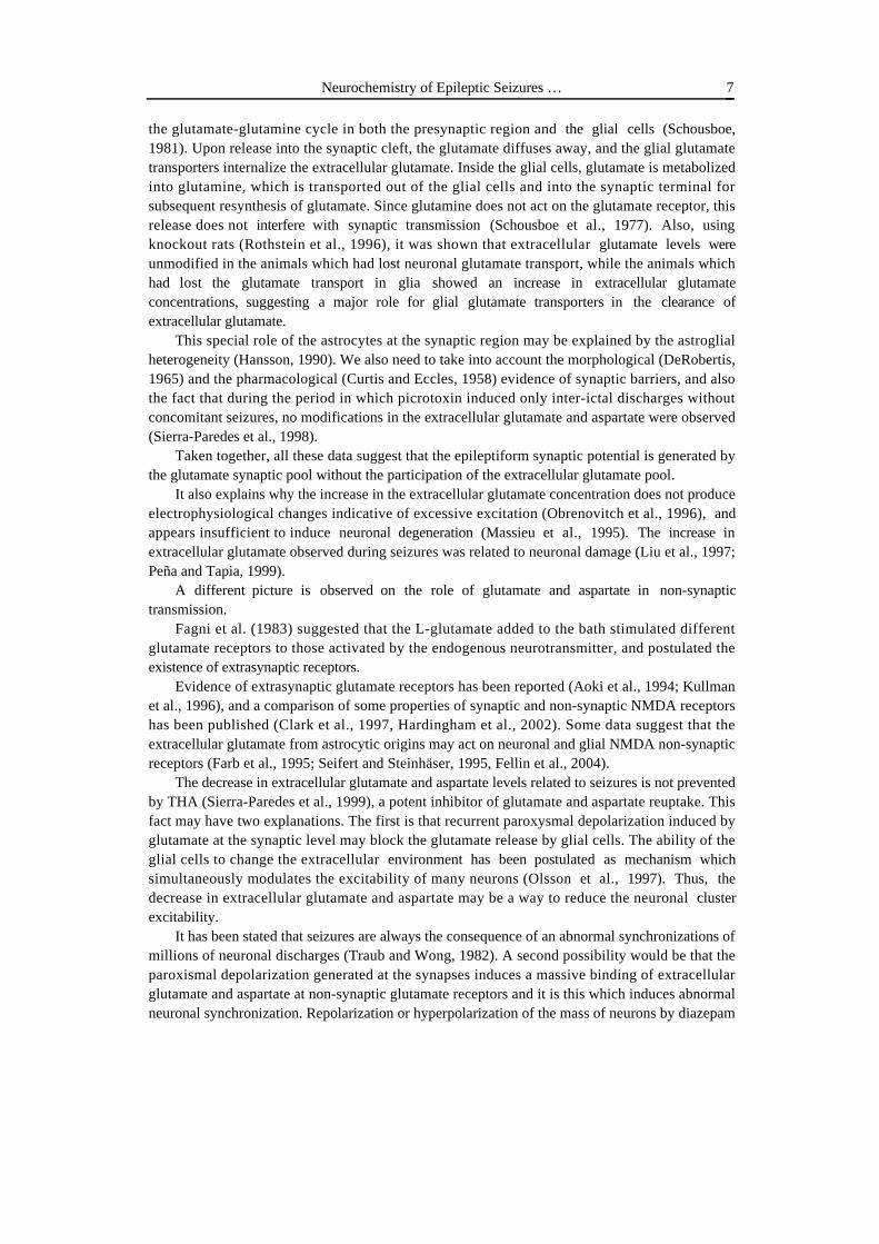
Latrunculin-A induces a short-term increase on neuronal excitability in the rat hippocampuswhich leads to epileptic seizures, and a permanent effect as measured by picrotoxin seizurethreshold (PCTS threshold). The degree of hyperexcitability is not dose-dependent at theconcentrations studied (2, 4 and 6 µg/ml), but related to the perfusion duration. In animals nottreated with picrotoxin, no modifications were detected in basal EEG during wakefulness andsleep periods during a single 2 hour perfusion of latrunculin-A. However, latrunculin-inducedseizures were observed after 6 hours of continuous perfusion in 20% of the animals studied(Figure 4a). When latrunculin-A was perfused for several consecutive days (8 hours a day, seeMethods), seizures were observed in 80% of the animals studied (Figure 4a, c). The severity,duration and onset time of the seizures showed considerable variation among animals, but90%±6% of the seizures were observed during the second and third days of consecutive perfusion(Figure 4a). No seizures were observed after the fourth day of perfusion (Figure 4a). Weeklyperfusions of latrunculin-A induced seizures during perfusion after the 4th week. As in theconsecutive perfusion experiments, the onset of the first seizures showed considerable variation,but all the animals studied presented several seizures between weeks 4 and 6 (Figure 5 a, c). Thoseanimals also show an abnormal EEG response to handling and tapping with slow waves and asignificant increase in sleep periods as compared to control EEGs in the same animals (Figure 5e). Other sensory stimuli, such as auditory random stimuli did not elicit any EEG response. Afew sporadic spontaneous seizures (0.4±0.3 seizures/month) were observed during controls in thethree months following the last latrunculin-A perfusion (Fig 7a). The effect observed here seemsto be receptor activation-independent and related solely to actin depolymerization. In all theanimals in which PCTS threshold was tested, a significant decrease up to 56%±7% of the basalPCTS theshold was observed after a 2 hours perfusion (Figure 6). The chronic experimentsshowed that the decrease in PCTS threshold is permanent and progressive for a period of threemonths in all the animals studied (Figure 7b). Seizures induced by latrunculin A can be preventedby the use of several antiepileptic drugs (unpublished data).

G. Sierra-Paredes, A. Vázquez-López, T. Oreiro-García, A. Núñez-Rodríguez, et al.16
Figure 4. Consecutive daily 8 hours hippocampal microperfusion of latrunculin-A and jasplakinolidein the hippocampus of conscious rats induces epileptiform seizures. (a) Average number of seizuresinduced by latrunculin-A (n=10) and jasplakinolide (n=10) from days 1-4 of consecutive perfusion. (b)Control EEG recording (FOR fronto-occipital right; FOL fronto-occipital left). (c) EEG seizure inducedby latrunculin-A (4 µg/ml), correlated with rearing, alimentary automatism and forelimb clonus. (d) EEG
seizure induced by jasplakinolide (4 µg/ml), with a similar behavioural outcome.
Repeated and consecutive jasplakinolide perfusions (see Methods) into the hippocampus ofliving rats induces an increase on neuronal excitability in the rat hippocampus which leads toepileptic seizures in 30% of the animals studied, a permanent effect as measured by picrotoxinseizure threshold (PCTS threshold) in 80% of the rats, and sporadic spontaneous seizures in 60%.The degree of hyperexcitability is also related to the perfusion duration and is not dose-dependentat the concentrations studied (2, 4 and 6 µg/ml). In animals not treated with picrotoxin, nomodifications were detected in basal EEG during wakefulness and sleep periods during a singlejasplakinolide perfusion. Jasplakinolide-induced seizures were observed after three consecutivedays in 30% of the animals studied (Figure 1a,d). In all the animals in which PCTS thresholdwas tested, a significant increase up to 33% of the basal PCTS theshold was observed after a 2hour perfusion (Figure 3). However, chronic experiments showed that a permanent decrease inPCTS threshold is detected one week after jasplakinolide administrations, lasting for a period ofthree months in 80% of the animals studied (Figure 4b). Weekly perfusions of jasplakinolideinduced seizures during perfusion after the 3rd week (Figure 2a, d), and 80% of the animalsstudied presented several sporadic seizures between weeks 3 and 12 (1,7±1,3 seizures/month, Fig4a). The severity, duration and frequency of the seizures showed considerable variation among

Neurochemistry of Epileptic Seizures … 17
animals, ranging from rapidly recurrent seizures during 24 hours to short (48±21 seconds) partialseizures. During the three months following the last jasplakinolide administration, spontaneousfocal discharges and abnormal EEG responses to handling, tapping and whisk stimulation wereobserved in the 80% of the animals (Figure 2f), and also a significant increase in sleep periods ascompared to control EEGs in the same animals (data not shown).
Figure 5. Consecutive weekly 8 hours hippocampal microperfusion of latrunculin-A and jasplakinolidein the hippocampus of freely moving rats induces epileptiform seizures with a variable frequency. (a)Summary of the average number of weekly perfusions of latrunculin -A(n=10) and jasplakinolide(n=10) to induce seizures in rats. (b) Control EEG recording (FOR fronto-occipital right; FOL fronto-occipital left). (c) EEG seizure induced by latrunculin-A (4 µg/ml). (d) EEG seizure induced by
jasplakinolide (4 µg/ml). (e) EEG response to handling (this pattern appears 10-15 sec after
stimulation) in a rat treated with latrunculin-A. (f) EEG response 5 seconds after whisk stimulation in arat treated with jasplakinolide.
Previous studies in vitro and cell cultures showed that AMPA and NMDA receptors areanchored in the dendritic spines by actin cytoskeleton (Allison et al., 1998; Halpain, 2000).Several findings support the use of latrunculin-A to perturb NMDA and AMPA receptor clusters

G. Sierra-Paredes, A. Vázquez-López, T. Oreiro-García, A. Núñez-Rodríguez, et al.18
(Allison et al., 1998; Sattler et al., 2000), but do not establish a functional significance for thiseffect. Because the actin-depolymerizing agents failed to show any effect on macroscopic whole-cell NMDA currents (Sattler et al., 2000), it was supposed that although NMDAR localizationmight be rearranged, function may be grossly unaffected (Allison et al., 2000). Furthermore, directevidence of the in vivo effect of actin cytoskeleton disruption was lacking. We show here, for thefirst time, that actin disrupting agents can be perfused in conscious, freely moving animals inorder to investigate the in vivo actin dynamics, which through depolymerization or abnormalpolymerization, modifies neuronal excitability and leads to epileptic seizures.
Figure 6. A single 2 hour perfusion of a 4 mg/ml latrunculin-A solution decreases significantly (*p<0.01) the picrotoxin seizure threshold in freely moving rats (n=8). A 2 hour jasplakinolide perfusion (4
µg/ml), however, sigificantly increases (*p< 0.01) picrotoxin seizure threshold (n=8).

Neurochemistry of Epileptic Seizures … 19
Figure 7. Repeated latrunculin-A and jasplakinolide administrations (a minimum of 3 perfusions, 4
µg/ml, daily or weekly) produce a long-lasting effect in neuronal excitability. (a) Average number of
seizures per month observed the three months following weekly latrunculin-A (n=10) andjasplakinolide (n=10) administration. (b) Repeated latrunculin-A perfusions induce a permanent andprogressive decrease in picrotoxin seizure threshold (*p< 0.01) which lasts for three months after thelast latrunculin-A administration (n= 10). Repeated jasplakinolide perfusions produce a significant(*p< 0.01) decrease which lasts unmodified for three months (n=10).
Much experimental evidence shows that an increase in glutamate excitatory action modifiesthe excitability of postsynaptic neurons via the actin cytoskeleton. Alterations in other actinfilaments related proteins, such as acidic calponin (Ferhat et al., 2003), have been linked toexperimental epileptic seizures. It is well known that excitatory synaptic activity modulates thedistribution of AMPA and NMDA receptors in the postsynaptic sites of hippocampal neurons.The mechanism for receptor redistribution involves calcium-mediated actin depolymerization(Bonfoco et al., 1996; Halpain et al., 1998; Lin et al., 2000) and is related to rapid dendritic spineplasticity (Halpain, 2000; Fischer et al., 1998; Shi et al., 1999). This receptor redistribution hasbeen shown to participate in mechanisms such as LTP and LTD (Carroll et al., 1999; Lin et al.,2000), as well as in pathological processes leading to epileptogenesis (de Lanerolle rt al., 1998).Most of these studies have centered on the increased or decreased number of postsynapticreceptors; however, receptor location may be also an important factor in neuronal excitability, andmay be changed by alterations in the actin cytoskeleton (Sattler et al., 2000). Our results showthat this effect can be induced in living animals by perfusing latrunculin-A and jasplakinolide inthe hippocampus. Alterations in receptor density or distribution on dendritic segments would be

G. Sierra-Paredes, A. Vázquez-López, T. Oreiro-García, A. Núñez-Rodríguez, et al.20
expected to significantly modify the effectiveness of synaptic transmission. Furthermore,alterations in the distribution of recurrent excitatory synapses on dendrites could lead to anenhanced ability of these synapses to produce action potentials and, in turn, promote thereverberation of recurrent excitation in networks of mutually excitatory pyramidal cells.
Spontaneous seizure activity was never present in the first month after latrunculin A orjasplkinolide administration. This “latency” period is consistent with the time needed forplasticity changes in the hippocampus such as sprouting. It is not completely clear whensprouting occurs after seizures, although in models involving a period of status epilepticus(i.e.,kainic acid or pilocarpine), it appears that sprouting begins sometime in the first 1 to 2weeks,and is most robust after many weeks (Scharfman, 2002). Eventhough latrunculin A andjasplakinolide do not induce status epilepticus, this latter condition is characterized by massiveactin disruption in dendritic spines (Isokawa 2000; Ferhat et al., 2003).
A differential action of actin disrupting agents among excitatory pyramidal cells andGABAergic interneurons cannot be ruled out. F-actin is necessary for the proper synapticlocalization of AMPA receptors in GABA cells (Allison et al., 1998), and the receptor relocationand complete recovery may be more difficult in GABA cells than in the dendritic spines ofpyramidal cells, maintaining a permanent lack of recurrent inhibition.
We observed a diferent effect between latrunculin-A and jasplakinolide in the acuteadministration. Depolymerization of actin by latrunculin-A has been reported to transientlypromote neurotransmitter release by a mechanism independent of extracellular Ca
2+, and this effect
is antagonized by jasplakinolide (Morales et al., 2000). However, this presynaptic effect does notexplain the chronic effect of both latrunculin-A and jasplakinolide. In cell cultures, a 24 htreatment with latrunculin-A is required for maximum effect (Allison et al., 1998), and we obtainthe maximum effect after three perfusions of 8 hours each. For jasplakinolide, it has been shownthat it can disrupt actin filaments and induce polymerization of monomeric actin in amorphousmasses (Bubb et al., 2000), which may explain the similar effect when both compunds areperfused for several consecutive days. The permanent decrease in seizure threshold is more likelyto be related to morphological changes in the number or shape of dendritic spines, or a permanentreorganization in the location of glutamate receptors or other proteins within the post-synapticdensity which are highly dependent on F-actin for their localization, such as CaMKII, spectrin,myosin V, α-adducin, neurabin, neurabinII/spinophilin, cortactin, and many others (Allison et al.,2000). The major function of those actin-associated components proteins of the post-synapticmembrane appears to be in signal transduction and modification of the microfilament arrays inresponse to synaptic activation, events thought to mediate long-term synaptic plasticity.
In the rat pilocarpine model, dendrites of dentate granule cells displayed a generalized spineloss immediately after the status epilepticus induced by pilocarpine injection. This spine loss wastransient and was followed by a recovery in spine density that started 3 days after statusepilepticus and reached a plateau level 15 days later (Isokawa, 2000). The subsequent recovery ofdendritic spine density probably reflects the formation of new spines on preexisting granule celldendrites and/or development of spines on outgrowing dendrites of newly formed granule cellssubsequent to epilepsy-induced neurogenesis (Parent et al.,1997). Using this model, Ferhat et al.(2003) showed that an increase in acidic calponin is observed during this period of recovery ofspine density, and they suggest that acidic calponin is involved in the formation of new dendriticspines. Those new spines might be involved in the formation of aberrant synapses with newmossy fiber terminals. Actin filament disruption induced by latrunculin A or jasplakinolide mightrigger molecular mechanisms for important remodeling of dendritic spine shape through theaction of acidic calponin and/or synaptopodin (Roth et al., 2001).

Neurochemistry of Epileptic Seizures … 21
The F-actin cytoskeleton is vulnerable to disruption by elevated intracellular calcium, acondition observed in several neuropathologies such as epilepsy, neurotrauma, and otherdegenerative neurological diseases. The results reported here support the idea that NMDARs andvoltage-dependent Ca
2+ channels remain suitable targets for future antiepileptic drugs, but they
also support the search for other intracellular pharmacological targets such as calcineurin (Halpainet al., 1998), calpain (Sierra-Paredes et al., 1999), PKC, CaMKII (MacDonald et al., 1996) andseveral other proteins related to actin dynamics (Allison et al., 2000; Roth et al., 2001; Ferhat etal. 2003). Furthermore, the seizures induced by latrunculin-A or jasplakinolide present distinctivecharacteristics, such as low frequency and a permanent threshold decrease, that are completelydifferent from the chemical convulsants widely used today, and could provide a new alternativeexperimental model of epilepsy for testing antiepileptic drugs.
Increased Intercellular Glutamate and Glycine Concentrations areEpileptogenic in Rats Pretereated with Latrunculin A
We have previously reported the effect of high concentrations of glutamate and glycine onpicrotoxin-induced seizure thresholds on freely moving rats (Sierra-Paredes et al., 2000). Ourresults showed that high extracellular glutamate and glycine levels during prolongedmicroperfusion do not lead to excitatory neuronal damage or paroxismal activity in the living ratbrain.
We found, however, that elevating glutamate and glycine concentrations in the hippocampussignificantly increases neuronal excitability, as reflected by a significant reduction in picrotoxinseizure threshold (Sierra-Paredes et al., 2001). Glutamate and glycine acting alone are not enoughto induce epileptiform seizures, but their continuous action may activate extrasynaptic receptorswhich facilitate seizure onset when GABAergic inhibition is impaired. The fact that glutamate andglycine produce almost the same effect indicates that their action is likely to be mediated byNMDA receptors. This is also confirmed by the fact that 5,7 DKA, a competitive inhibitor of theglycine binding site in the NMDA receptor, completely prevents the effect of both glutamate andglycine (Sierra-Paredes et al., 2001).
When rats are pretreated with latrunculin A, both glycine and glutamate induce seizures when1mM concentrations are perfused into the hippocampus (table 1, fig 8). This effect can beobserved from one to six months after latrunculin A administration. However, rats pretreated withpicrotoxin do not show any seizures or EEG discharges when treated with the same glutamate orglycine concentrations (table 1). This means that latrunculin A, not seizure activity, inducespermanent changes in neuronal excitability.
Table 1. Probability of Observing a Seizure in Controls and Rats Pretreatedwith Picrotoxin or Latrunculin A during Microperfusion of Vehicle,
1mM Glutamate and 1mM Glycine
Treatment Vehicle 1mM Glu 1mM Gly
None 0% 0% 0%Picrotoxin 0% 0% 0%Latrunculin A 5,1% 78% 86%

G. Sierra-Paredes, A. Vázquez-López, T. Oreiro-García, A. Núñez-Rodríguez, et al.22
Figure 8. Probablility of recording a seizure in rats pretreated with latrunculin A when perfusing Ringersolution (control), 1mM glutamate (Glu) and 1mM glycine (Gly). Data are mean ± s.d. from 12 rats. EEGactivity recorded in a rat perfused with 1mM glutamate.
As discussed above, latrunculin A induces long-term changes in neuronal excitability leadingto the onset of spontaneous seizures and a permanent decrease in picrotoxin seizure threshold. Onehypothesis which might explain the action of latrunculin A is the displacement of synapticreceptors to extrasynaptic locations, where they may be easily activated by intercellular glutamate.Our results are compatible with two usual findings in the brain of epileptic patients:morphological alterations in dendritic spines, and the increase in the concentrations of glutamate,glycine and other amino acids.
These results, taken together with previous data about the lack of effect of increasedextracellular glutamate in normal rats (Sierra-Paredes et al., 2000 a, 2001 a; Obrenovitch et al.,1997 a,b) and the evidences of dendrite isolation from intercellular space (Ventura and Harris,1999), support the proposal that exogenous glutamate never reaches postsynaptic space, and itsaction is only produced in special conditions by activating extrasynaptic receptors (Sinor et al.,2000; Sierra-Paredes et al., 1999).
Several studies suggest that NMDA receptors compatimentalization and distribution, as wellas the NMDA receptor interaction with other proteins, are essential for excitotoxic effects (Sattleret al., 1998, 1999; Hardingham et al., 2002). It is possible that relationships among NMDAreceptors and the intracellular processes which lead to increased excitability or cellular death maybe different in extrasinaptic locations (Sinor et al., 2000; Hardingham et al., 2002). This suggeststhat glutamate activity may be different with the increase in the number of extrasynaptic receptors.
The evidence suggesting that AMPA and NMDA receptors at synapses are highly dynamicand able to move laterally between synaptic and extrasynaptic pools (Tovar and Westbrook, 2000;Carroll et al., 1999; Hayashi et al., 2000; Liu and Cull-Candy, 2000), taken together with the in
Neurochemistry of Epileptic Seizures … 23
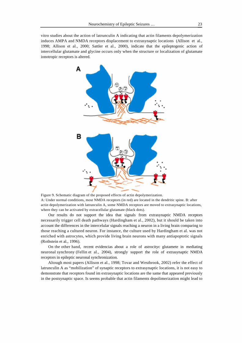
vitro studies about the action of latrunculin A indicating that actin filaments depolymerizationinduces AMPA and NMDA receptors displacement to extrasynaptic locations (Allison et al.,1998; Allison et al., 2000; Sattler et al., 2000), indicate that the epileptogenic action ofintercellular glutamate and glycine occurs only when the structure or localization of glutamateionotropic receptors is altered.
Figure 9. Schematic diagram of the proposed effects of actin depolymerization.A: Under normal conditions, most NMDA receptors (in red) are located in the dendritic spine. B: afteractin depolymerization with latrunculin A, some NMDA receptors are moved to extrasynaptic locations,where they can be activated by extracellular glutamate (black dots).
Our results do not support the idea that signals from extrasynaptic NMDA receptorsnecessarily trigger cell death pathways (Hardingham et al., 2002), but it should be taken intoaccount the differences in the intercelular signals reaching a neuron in a living brain comparing tothose reaching a cultured neuron. For instance, the culture used by Hardingham et al. was notenriched with astrocytes, which provide living brain neurons with many antiapoptotic signals(Rothstein et al., 1996).
On the other hand, recent evidencias about a role of astrocityc glutamete in mediatingneuronal synchrony (Fellin et al., 2004), strongly support the role of extrasynaptic NMDAreceptors in epileptic neuronal synchronization.
Altough most papers (Allison et al., 1998; Tovar and Westbrook, 2002) refer the effect oflatrunculin A as “mobilization” of synaptic receptors to extrasynaptic locations, it is not easy todemonstrate that receptors found im extrasynaptic locations are the same that appeared previouslyin the postsynaptic space. Is seems probable that actin filaments depolimerization might lead to

G. Sierra-Paredes, A. Vázquez-López, T. Oreiro-García, A. Núñez-Rodríguez, et al.24
receptor internalization and proteolysis (Zhou et al., 2000), while new receptors, probably with adifferent subunit composition, would be carried to the cell membrane without the precise locationcontrol which happens under physiological conditions. Those receptors may be activated moreeasily by the glutamate perfused through microdialysis probes producing biochemicalmodifications leading to epileptic seizures. Actin filament depolymerization by latrunculin Amigh also alter protein anchoring to NMDA receptors, modifying the intracellular signalinginduced by both synaptic and extrasynaptic receptors.
The proconvulsant effect of amino acids is higher in those rats which presented seizuresduring latrunculin A microperfusion. It is possible that seizures potentiate the effect of latrunculinA, because during seizures, massive calcium entry through NMDA receptors produces by itselfactin filament depolymerization.
The higher effect of glycine as comapred with glutamate is intriguing. It may be due to thefact that we have used the same concentration for both amino acids, while physiologicalintercellular levels of glycine are lower than glutamate levels (Sierra-Paredes et al., 1998).Furthermore, both glial and vascular transport mechanisms are much more active for glutamate(Hertz, 1979; O'Kane et al., 1999).
Our results, taken together with previous research discussed above, favor the hypothesis thatextrasynaptic glutamate receptors might be involved in diverse pathological consequences linkedto this neurotransmitter. An interesting conclusion is that selective inhibition of extrasynapticNMDA receptors might have interesting therapeutic consequencies in the treatment of epilepsy, aswell as in prevention of epileptogenesis in patientes with brain lesions, cerebral isquemia orstroke. During these injuries, it seems probable that the excess of extracellular glutamate of anysource (including blood-brain barrier alterations), migh activate extrasynaptic NMDA receptors(Riccio and Ginty, 2002) contributing to epileptogenesis.
Generalized inhibition of NMDA receptors is highly problematic, because receptor funcion isrequired for normal synaptic transmission. The use of NMDA inhibitors, both competitive andnon-competitive, with strong antiepileptic effect in experimental models of epilepsy, has notshown to be useful in the clinical trials performed to these moment (Rogawsky, 1995; Vizzi andKiss, 1998). One of the future proposals in the search for new antiepileptic drugs might involvethe development of selective methods for blocking extrasynaptic NMDA receptors withoutinactivating synaptic receptors.
Understanding the cellular and molecular changes that take place during latrunculin A-inducedseizures is essential to understanding the physiological mechanisms that cause this activity.
PKA and Calcineurin Inhibitors Show Antiepileptic Effects
We have found that ascomycin (100 µM), a calcineurin inhibitor, has a good antiepilepticaffect against picrotoxin seizures when perfused into the rat hippocampus (figs 10, 11). Duringpicrotoxin seizures, significant changes in calcineurin activity were not found, however, asignificant increase in calcineurin activity was observed during latrunculin A induced seizures(unpublished data). These differences might be due to the biochemical changes leading to thepermanent changes produced by latrunculin A, and also to the biochemical consequencies ofrepeated seizures. Kurtz et al. (2001) demonstrated a significant increase in calcineurin activity incortical and hippocampal homogenates during status epilepticus induced by pilocarpine.
Calcineurin is a calcium/calmodulin-stimulated enzyme (Klee et al. 1998) and would bestimulated by increased intracellular calcium concentrations. An increase in intracellular free

Neurochemistry of Epileptic Seizures … 25
calcium has been found during and after status epilepticus (Pal et al. 1999). The increasedintracellular calcium associated with status epilepticus could be responsible for activatingcalcineurin above its normal physiological level. Status epilepticus induces a loss of function ofthe endoplasmic reticulum Mg 21 /Ca 21 ATPase (Parsons et al. 2000). This enzyme sequesterscalcium ions into the microsomes of the smooth endoplasmic reticulum, providing a high affinitymechanism for regulating intracellular calcium concentration (Carafoli 1987; Miller 1991). Afterstatus epilepticus, ATPase-mediated uptake of calcium into the microsomes is less efficient(Parsons et al. 2000), which could potentially result in higher than normal resting calciumconcentrations inside the cell did not affect the status epilepticus induced increase in calcineurindephosphorylation.
Another explanation of the increase in calcineurin activity during status epilepticus andlatrunculin seizures might be related with the activity of the calcium-stimulated neutral protease,calpain I. Calpain cleaves the catalytic calcineurin A subunit, removing the calmodulin bindingdomain and creating a highly active, 43 kDa, calcium-independent form of the enzyme (Kurtz etal., 2001; Tallant et al. 1988; Wang et al. 1989). Some researchers also propose that calpain-mediated proteolysis removes the autoinhibitory domain present in the A subunit (Wang et al.1989). Calpain may be activated by the increased intracellular calcium concentrations presentduring status epilepticus, activating calcineurin. We have previously shown that calpain isinvolved in seizure arrest (Sierra-Paredes et al., 1999), so during status epilepticus calpain mightbe overactive, being responsible for the increase in calcineurin activity.
Calcineurin activity might also be involved in the biochemical changes leading to picrotoxin-induced epileptic seizures, but changes could bo too small to be easily detected. The results of onekindling study already suggest that calcineurin may have an epileptogenic role (Moia et al. 1994).Neuronal mechanisms which regulate calcineurin dephosphorylation under normal conditionscould be highly altered as a result of impaired GABAergic activity. One important calcineurin-mediated mechanism is modulation of the GABAA receptor. GABA receptors are the primaryreceptor responsible for the fast inhibitory response in neuronal tissue (Macdonald and Olsen1994), and play a major role in preventing the neuronal hyperexcitability associated with epilepsy.Several recent studies have demonstrated an inhibitory modulation of GABAA receptor function bycalcineurin (Chen and Wong 1995; Amico et al. 1998; Lu et al. 2000). Picrotoxin binding toGABAA receptors may result in increased dephosphorylation, producing a net disinhibition ofcellular excitability.
Increased excitability in neurons can be maintained by abnormalities in proteinphosphorilation systems (Yechikhov et al., 2001). It was shown that PKC modulates receptortrafficking and gating (Lan et al., 2001) underlying synaptic plasticity (Esteban et al., 2003), andit is possible that in our latrunculin model receptor mobilization would be a direct effect of actinfilaments depolymerization associated to changes in the phosphorilation mechanisms. In vivoPKA inhibition showed a slight antiepileptic effect on picrotoxin induced seizures (figures 10,11), supporting the idea that normal phosphorilation and dephosphorilation are involved in seizureactivity. Picrotoxin and latruculin-induced seizures are followed by a non significant increase inthe activity of several protein kinases in the rat hippocampus (unpublished data). It seems possiblethat some long-term epileptogenic changes involve modifications in protein kinase and/or proteinphosphatase activity, and the chronic biochemical or morphological changes induced by directactin depolymerization might require the activation of those mechanisms. It is also possible thatthe location of enzimes would be critical for their action, modifying neuronal response(Hardingham et al., 2003). Further research is required in order to elucidate which biochemicaland/or morphological alterations are epileptogenic in the latrunculin A/jasplakinolide model.

G. Sierra-Paredes, A. Vázquez-López, T. Oreiro-García, A. Núñez-Rodríguez, et al.26
Figure 10. Ascomycin (100µM) and H-9 dihydrochloride (100µM) significantly reduce the number of
seizures (mean ± s.d. from 8 rats) when perfused in the hippocampus of freely moving rats.
Figure 11. Ascomycin (100µM) significantly the mean seizure duration in picrotoxin treated rats. H-9
dihydrochloride (100µM) effect was not significant when compared with control rats.
Conclusions
Our results, showing that epileptic seizures may be caused by the direct disruption of theactin cytoskeleton, indicate that the biochemical study of actin-dependent receptor anchoring,related transduction pathways and many other PSD proteins seems to be an important approach tothe neuropathology and neuropharmacology of epilepsy. Is seems crear now that neurotransmitterreceptors and synapses are not fixed structures, but are subjected to continuous changes and

Neurochemistry of Epileptic Seizures … 27
replacements. Receptor and enzyme location seems to be a decisive feature for the transmission ofneural messages among neurons, and it is very probable that cahanges in receptor location mightbe involved in learning associated plasticity changes as well as in some brain diseases. Oneimportant question remains to be answered: How and when these molecular dynamics producelong term changes in neuronal circuits, and why sometimes the changes do not seem to be easilyreversible?. In the case of epileptogenesis, cell death and/or excessive axon prunning is onepossible answer. However, further research is required to define the mechanisms of the permanentneuronal alterations induced by in vivo actin cytoskeleton disruption.
These results, taken together with previous research discussed above, favor the hypothesis thatextrasynaptic NMDA and AMPA glutamate receptors might be involved in diverse pathologicalconsequences linked to this neurotransmitter, including epileptic seizures. Altered biochemicalconditions which may lead to an increase in extrasynaptic receptor number or activity can berelated with epileptogenic processes. An interesting conclusion is that selective inhibition ofextrasynaptic NMDA receptors and/or intracellular molecules involved in directing receptorlocation might have interesting therapeutic consequencies in the treatment of epilepsy, as well asin prevention of epileptogenesis in patientes with brain lesions, cerebral isquemia or stroke.
References
Aguilar-Veiga, E. Sierra-Paredes, G., Galán-Valiente, J., Soto-Otero, R., Méndez-Alvarez, E. andSierra-Marcuño, G. (1991) Correlation between ethosuximide brain levels measured by highperformance liquid chromatography and its antiepileptic potential. Res. Com. Chem, Pathol.Pharmacol. 71, 351-364.
Aizawa, H., Hu, S.C., Bobb, K., Balakrishnan, K., Ince, G., Gurevich, I., Cowan, M. AndGhosh, A. (2004) Dendrite development regulated by CREST, a calcium-regulatedtranscriptional activator. Science 303: 197-202.
Allison, D.W., Gelfand, V.I., Spector, I. and Craig, A.M. (1998) Role of actin in anchoringpostsynaptic receptors in cultured hippocampal neurons: differential attachment of NMDAversus AMPA receptors. J Neurosci. 18, 2423–2436.
Allison, D.W., Chervin, A.S., Gelfand, V.I. and Craig, A.M. (2000) Postsynaptic scaffolds ofexcitatory and inhibitory synapses in hippocampal neurons: maintenance of core componentsindependent of actin filaments and microtubules. J. Neurosci. 20, 4545-4554.
Amico, C., Cupello, A., Fossati, C. and Robello, M. (1998) Involvement of phosphataseactivities in the run-down of GABAA receptor function in rat cerebellar granule cells inculture. Neuroscience 84, 529-535.
Andreasen, M., Lambert, J.D.C. and Jensen, M.S., 1989. Effects of new non-N-methyl-D-aspartate antagonists on synaptic transmission in the in vitro rat hippocampus. J. Physiol(Lond.) 414, 317-336.
Aoki, C., Venkatesan, C., Go, C.G., Mong, J.A. and Dawson, T.M., 1994. Cellular andsubcellular localization of NMDA-R1 subunit immunoreactivity in the visual cortex of adultand neonatal rats. J. Neurosci. 14, 5202-5222.
Bading, H., Ginty, D.D. and Greenberg, M.E. (1993) Regulation of gene expression inhippocampal neurons by distinct calcium signaling pathways. Science 260, 181-186.
Bach-y-Rita, P., 1991 Thoughts on the role of volumen transmission in normal and abnormalmass sustained functions. In: Fuxe, K. and Agnati, L.F. (Eds) Volumen Transmission in theBrain . New York: pp: Raven Press 489-496.

G. Sierra-Paredes, A. Vázquez-López, T. Oreiro-García, A. Núñez-Rodríguez, et al.28
Bach-y-Rita, P., 1993 Nonsynaptic diffusion neurotransmission (NDN) in the brain. Neurochem.Int., 23, 297-318.
Barnes, B.A., 1991. New roles for glia. J. Neurosci., 11, 3685-3694.Belichenko, P.V. and Dahlstrom, A. (1995) Studies on the 3-dimensional architecture of dendritic
spines and varicosities in human cortex by confocal laser scanning microscopy and Luciferyellow microinjections. J. Neurosci. Meth. 57, 55-61.
Bonfoco, E., Leist, M., Zhivotovsky, B., Orrenius, S., Lipton, S.A., and Nicotera, P. (1996)Cytoskeletal breakdown and apoptosis elicited by NO donors in cerebellar granule cellsrequire NMDA receptor activation. J. Neurochem. 67, 2484-2493.
Bubb, M.R. Spector, I., Beyer, B.B. and Fosen, K.M. (2000) Effects of jasplakinolide on thekinetics of actin polymerization. J. Biol. Chem. 275, 5163-5170.
Carafoli, E. (1987) Intracellular calcium homeostasis. Annu. Rev. Biochem. 56, 395-433.Carroll, R.C., Lissin, D.V., von Zastrow, M., Nicoll, R.A. and Malenka, R.C. (1999) Rapid
redistribution of glutamate receptors contributes to long-term depression in hippocampalcultures. Nat. Neurosci. 2, 454-460.
Chapman A.G., 1997. Microdialysis. In: Engel Jr, J. and Pedley, T.A. (Eds.) Epilepsy: AComprehensive Textbook. Lippincott-Raven Publishers, Philadelphia, pp. 1067-1072.
Chen, Q.X. and Wong, R.K.S. (1995) Suppression of GABAA receptor responses by NMDAapplication in hippocampal neurons acutely isolated from the adult guinea-pig. J. Physiol.(Lond.) 482, 353-362.
Chittajallu, R., Alford, S. and Collingridge, G.L. (1998) Ca2+
and synaptic plasticity. CellCalcium 24, 377-385.
Choi, D.W. (1987) Ionic dependence of glutamate neurotoxicity. J. Neurosci. 7, 369-379._____ . (1988) Calcium-mediated neurotoxicity: relationship to specific channel types and role in
ischemic damage. Trends Neurosci. 11, 465-467.Churn, S.B., Limbrick, D., Sombati, S. and De-Lorenzo, R.J. (1995) Excitotoxic activation of
the NMDA receptor results in inhibition of calcium/calmodulin kinase II activity in culturedhippocampal neurons. J. Neurosci. 15, 3200-3214.
Clark, B.A., Farrant, M. and Cull-Candy, S.G., 1997. A direct comparison of the single-channelproperties of synaptic and extrasynaptic NMDA receptors. J. Neurosci. 17, 107-116.
Conti, F. and Weinberg, R.J., 1999. Shaping excitation at glutamatergic synapses. TrendsNeurosci., 22, 451-458.
Craig, A.M., Blackstone, C.D., Huganir, R.L. and Banker, G. (1994) Selective clustering ofglutamate and γ -aminobutyric acid receptors opposite terminals releasing the correspondingneurotransmitters. Proc. Natl. Acad. Sci. USA 91, 12373-12377.
Curtis, D. R. and Eccles, R. M., 1958. The effect of diffusional barriers upon the pharmacologyof cells within the central nervous system. J. Physiol (Lond.) 141, 446-463.
de Lanerolle, N.C., Eid, T., von Campe, G., Kovacs, I., Spencer, D.D., and Brines, M. (1998)Glutamate receptor subunits GluR1 and GluR2/3 distribution shows reorganization in thehuman epileptogenic hippocampus. Eur. J. Neurosci. 10, 1687-1703.
Del-Cerro, S., Arai, A., Kessler, M., Bahr, B.A., Vanderklish, P., Rivera, S. and Lynch, G.(1994) Stimulation of NMDA receptors activates calpain in cultured hippocampal slices.Neurosci. Lett. 167, 149-152.
DeLorenzo, R.J., Pal, S. and Sombati, S. (1998) Prolonged activation of the N-methyl-D-aspartate receptor-Ca
2+ transduction pathway causes spontaneous recurrent epileptiform
discharges in hippocampal neurons in culture. Proc. Natl. Acad. Sci. USA 95, 14482-14487.

Neurochemistry of Epileptic Seizures … 29
De Robertis, E., 1965. Some new electron microscopical contribution to the biology of neuroglia.In: De Robertis, E. and Carrera, R. (Eds) Progress in Brain Research, vol 15: Biology ofNeuroglia New York: Elsevier pp: 1-11.
Diamond, J.S., Bergles, D.E., and Jahr, C.E. , 1998. Glutamate release monitored with astrocytetransporter currents during LTP. Neuron, 21, 425-433.
Dichter, M. and Ayala, G.F., 1987. Cellular mechanisms of epilepsy: a status report. Science,237, 157-164.
Dingledine, R., McBain, C.J. and McNamara, J.O. (1990) Excitatory amino acids in epilepsy.Trends Pharmacol. Sci. 11, 334-338.
Ehlers, M.D., Fung, E.T., O'Brien, R.J. and Huganir, R.L. (1998) Splice variant-specificinteraction of the NMDA receptor subunit NR1 with neuronal intermediate filaments. JNeurosci 18, 720-730.
Esteban, J.A., Shi, S.H., Wilson, C., Nuriya, M., Huganir, R.L. and Malinow, R. (2003) PKAphosphorylation of AMPA receptor subunits control synaptic trafficking underlyingplasticity. Nature Neurosci. 6, 2, 136-143.
Fagni, L., Baudry, M and Lynch, G., 1983. Clasification and properties of excitatory amino acidreceptors in the hippocampus I. Electrophysiological studies of an apparent desensititazionand interactions with drugs which block transmission. J. Neurosci., 3, 1538-1546.
Farb, C.R., Aoki, T. and LeDoux, J.E., (1995). Differential localization of NMDA and AMPAreceptor subunits in the lateral and basal nuclei of amygdala: a light and electron microscopestudy. J. Comp. Neurol., 362, 86-108.
Fellin, T., Pascual, O., Gobbo, S., Pozzan, T., Haydon, P.G. and Carmignoto, G. (2004)Neuronal synchrony mediated by astrocytic glutamate through activation of extrasynapticNMDA receptors. Neuron, 43:729-743.
Fifkova, E. And Delay, R.J. (1982) Cytoplasmic actin in neuronal processes as a possiblemediator of synaptic plasticity. J. Cell. Biol. 95:345-350.
Fischer, M., Kaech, S., Knutti, D. and Mattus, A. (1998) Rapid actin-based plasticity in dendriticspines. Neuron 20, 847-854.
Fuxe, K. and Agnati, L.F., 1991. Two principal modes of electrochemical communications in thebrain: volume versus wiring transmission. Adv. Neurosci. 1, 1-9.
Ghosh, A. and Greenberg, M.E. (1995) Calcium signaling in neurons: molecular mechanisms andcellular consequences. Science 268, 239-247.
Gloor, P. and Fariello, R.G., 1988. Generalized epilepsy: some of its cellular mechanisms differfrom those of focal epilepsy. Trends Neurosci., 11, 63-68.
Groc, L., Heine, M., Cognet, L., Brickley, K., Stephenson, F. A., Lounis, B. and Choquet, D.(2004) Differential activity-dependent regulation of the lateral mobilities of AMPA andNMDA receptors. Nature Neurosci. 7, 695-696.
Groth, R.D., Dunbar, R.L. and Mermelstein, P.G. (2003) Calcineurin regulation of neuronalplasticity. Biochem. Biophys. Res. Com. 311,1159-1171.
Halpain, S., Hipolito, A. and Saffer, L. (1998) Regulation of F-actin stability in dendritic spinesby glutamate receptors and calcineurin. J. Neurosci. 18, 9835-9844.
Halpain, S. (2000) Actin and the agile spine: how and why do dendritic spines dance? TrendsNeurosci. 23, 141-146.
Hardingham, G.E., Fukunaga, Y. and Bading, H. (2002) Extrasynaptic NMDARs opposesynaptic NMDARs by triggering CREB shut-off and cell death pathways. Nature Neurosci.5:5, 405-414.
G. Sierra-Paredes, A. Vázquez-López, T. Oreiro-García, A. Núñez-Rodríguez, et al.30
Harris, K.M. and Kater, S.B. (1994) Dendritic spines: cellular specializations imparting bothstability and flexibility to synaptic function. Annu. Rev. Neurosci. 17, 341-371.
Herkenham, M., 1991. Mismatches between neurotransmitter and receptor localizations:Implications for endocrine functions in the brain. In: Fuxe, K. and Agnati, L.F. (Eds)Advances in Neuroscience, Vol 1: Volume Transmission in the Brain. Novel Mechanisms forNeural Transmission. New York, Raven Press, 1991, pp: 63-87.
Herrero, I., Miras-Portugal, M.T. and Sanchez-Prieto, J. 1992a. Positive feedback of glutamateexocytosis by metabotropic presynaptic receptor stimulation. Nature. 360, 163-166.
_____ . 1992b Activation of protein kinase C by phorbol esters and arachidonic acid required forthe optimal potentiation of glutamate exocytosis. J. Neurochem. 59, 1574-1577.
Hertz, I. 1979. Functional interactions between neurons and astrocytes I. Turnover andmetabolism of putative amino acids transmitters. Prog. Neurobiol. 13, 277-323.
_____ . 1982. Astrocytes. In: Lajtha, A. (Ed.) Handbook of Neurochemistry. New York: PlenumPress, pp: 319-355.
Hidaka, H., Inagaki, M., Kawamoto, S. and Saaki, Y. (1984) Isoquinolinesulfonamides: noveland potent inhibitors of cyclic nucleotide dependent protein kinase and protein kinase C.Biochemistry 23, 50365041.
Hosford, D.A., Crain, B.J., Cao, Z., Bonhaus, D.W., Friedman, A.H., Okazaki, M.M., Nadler,J.V. and McNamara, J.O., 1991. Increased AMPA-sensitive quisqualate receptor binding andreduced NMDA receptor binding in epileptic human hippocampus. J. Neurosci. , 11, 428-434.
Huang, R. and Dillon, G.H. (1998) Maintenance of recombinant type A g-aminobutyric acidreceptor function: role of tyrosine phosphatases and calcineurina. J. Pharmacol. Exp. Ther.286, 243-255.
Isokawa, M. and Levesque, M.F. (1991) Increased NMDA responses and dendritic degeneration inhuman epileptic hippocampal neurons in slices. Neurosci. Lett. 132, 212-216.
Isokawa, M., Levesque, M.F., Fried I. and Engel J. Jr. (2000) Glutamate Currents inMorphologically Identified Human Dentate Granule Cells in Temporal Lobe Epilepsy. JNeurophysiol 77, 3355-3369.
Kaech, S., Fischer, M., Doll, T. and Matus, A. (1997) Isoform specificity in the relationship ofactin to dendritic spines. J. Neurosci. 17, 9565-9572.
Klee, C.B., Ren, H. and Wang, X. (1998) Regulation of the calmodulin-stimulated proteinphosphatase, calcineurin. J. Biol. Chem. 273, 13367-13370.
Kochan, L.D., Churn, S.B., Omojokun, O., Rice, A. and De-Lorenzo, R.J. (2000) Statusepilépticus results in an N-metyl-D-aspartate receptor-dependent inhibition ofCa
2+/calmodulin-dependent kinase II activity in the rat. Neuroscience 95, 735-743.
Kornau, H.C., Schenker, L.T., Kennedy, M.B. and Seeburg, P.H. (1995) Domain interactionbetween NMDA receptor subunits and the postsynaptic density protein PSD-95. Science 269,1737-1740.
Krupp, J.J., Vissel, B., Thomas, C.G., Heinemann, S.F. and Westbrook, G.L. (1999)Interactions of calmodulin and alpha-actinin with the NR1 subunit modulate Ca
2+-dependent
inactivation of NMDA receptors. J. Neurosci. 19, 1165-1178.Kullman, D.M., Erdemli, G. and Asztély, F., 1996. LTP of AMPA and NMDA receptor-
mediated signals: Evidence for presynaptic expression and extrasynaptic glutamate spill-over.Neuron, 17, 461-474.
Kurz, J.E., Sheets D., Travis-Parsons, J., Rana, A., Delorenzo, R.J. and Churn, S.B. (2001) Asignificant increase in both basal and maximal calcineurin activity in the rat pilocarpinemodel of status epilepticus. J. Neurochem. 78, 304-315.

Neurochemistry of Epileptic Seizures … 31
Lan, J., Skeberdis, V.A., Jover, T., Grooms, S.Y., Lin, Y., Araneda, R.C., Zheng, X., Bennett,M.V.L. and Zukin, S. (2001) Protein kinase C modulates NMDA receptor trafficking andgating. Nature Neurosci. 4:4, 382-390.
Lees, G.V., and Jones, E.G. (2000) Epilepsy. Neurobiol. Dis. 7, 549-551.Lehmann, A. and Hansson, E., 1988. Kainate-induced stimulation of amino acids release from
primary astroglial cultures of the rat hippocampus. Neurochem. Int. , 13, 557-561.Levi, G. and Patrizio, M., 1992. Astrocytes heterogeneity: endogenous amino acid levels and
release evoked by non-N-methyl-aspartate receptor agonists and by potassium-inducedswelling in type-1 and type-2 astrcytes. J. Neurochem., 58, 1943-1952.
Li, B., Chen, N., Luo, T., Otsu, Y., Murphy, T.H. and Raymond, L.A. (2002) Differentialregulation of synaptic and extra-synaptic NMDA receptors. Nature Neurosci. 5:9, 833-834.
Lin, J.W., Ju, W., Foster, K., Lee, S.H., Ahmadian, G., Wyszynski, M., Wang, Y.T., andSheng, M. (2000) Distinct molecular mechanisms and divergent endocytotic pathways ofAMPA receptor internalization. Nat. Neurosci. 3, 1282-1290.
Liu, S.J. and Cull-Candy, S.G. (2000) Activity at calcium-permeable AMPA receptors induces aswitch in receptor subtype. Nature 405, 454-458.
Liu, L., Wong, T.P. Pozza, M.F., Lingenhoehl, K. Wang, Y., Sheng, M., Auberson, Y. P. andWang, T.Y. (2004) Role of NMDA receptor subtypes in governing the direction ofhippocampal synaptic plasticity. Science 304, 1021-1024.
Liu, Z., Stafstrom, C.E., Sarkisian, M.R., Yang, Y., Hori, A., Tandon, P. and Holmes, G.L.,1997. Seizure-induced glutamate release in mature and immature animals: in vivomicrodialysis. Neuroreport, 8, 2019-2023.
Löscher, W. and Hönack, D., 1994. Effects of non-NMDA antagonist NBQX and the 2,3-benzodiazepine GYKI 52466 on different seizure types in mice: comparison with diazepamand flumazenil. Br. J. Pharmacol., 113, 1349-1357.
Lynch, M.A., Errington, M.L. and Bliss, T.V.P., 1989. Nordihydroguaiaretic acid blocks thesynaptic component of long-term potentiation and the associated increase in release ofglutamate and arachidonate: an in vivo study in the dentate gyrus in the rat. Neuroscience 30,693-701.
Lu W, Man H, Ju W, Trimble WS, MacDonald JF, Wang YT. (2001) Activation of synapticNMDA receptors induces membrane insertion of new AMPA receptors and LTP in culturedhippocampal neurons. Neuron. 29(1):243-54.
MacDonald, J.F., Browning, M.D. and Wang, L.Y. (1996) Long-term enhancement ofexcitability and the regulation of glutamate receptors by protein kinases. In ProgressiveNature of Epileptogenesis (U. Heinnemann, J. Engel Jr., G. Avanzini, B.S., Meldrum, A.Mouritzen-Dam, and C. Wasterlain, Eds) pp.275-282. Elsevier, Amsterdam.
Macdonald, R.L. and Olsen, R.W. (1994) GABAA receptor channels. Annu. Rev. Neurosci. 17,569-602.
Matus, A., Ackermann, M., Pehling. G., Byers, H.R. and Fujiwara, K. (1982) High actinconcentrations in brain dendritic spines and postsynaptic densities. Proc. Natl. Acad. Sci.USA 79, 7590 –7594.
Matus, A., Brinkhaus, H. And Wagner, U. (2000) Actin dynamics in dendritic spines: A form ofregulated plasticity at excitatory synapses. Hippocampus 10: 555-560.
Matus, A. (2000) Actin-based plasticity in dendritic spines. Science 290, 754-758.McNamara, J.O. (1999) Emerging insights into the genesis of epilepsy. Nature 399, A15-A22.Martin, D.L., 1992. Synthesis and release of neuroactive substances by glial cells. Glia 5, 81-94.

G. Sierra-Paredes, A. Vázquez-López, T. Oreiro-García, A. Núñez-Rodríguez, et al.32
Massieu, L., Morales-Villagrán, A. and Tapia, R., 1995 Accumulation of extracellular glutamateby inhibition of its uptake is not sufficient for inducing neuronal damage: An in vivomicrodialysis study. J. Neurochem. 64, 2262-2272.
Matsumoto, H. and Ajmone-Marsan, C. 1964a. Cortical cellular phenomena in experimentalepilepsy: Inter-ictal manifestations. Exp. Neurol 9, 286-304.
_____ . 1964b. Cortical cellular phenomena in experimental epilepsy: Ictal manifestations. Exp.Neurol 9, 305-326.
Meadows R.P., Nettesheim D.G., Xu R.X., Olejniczak E.T., Petros A.M., Holzman T.F.,Severin J., Gubbins E., Smith H. and Fesik S.W. (1993) Three-dimensional structure of theFK506 binding protein/ascomycin complex in solution by heteronuclear three- and four-dimensional NMR. Biochemistry. 32(3):754-765.
Millan, M.H., Obrenovitch, T.P., Sarna, G.S., Lok, S.Y., Symon, L. and Meldrum, B.S., 1991Changes in rat brain extracellular glutamate concentration during seizures: an in vivo dialysisstudy with on line enzimatic detection. Epilepsy Res. 9, 86-91.
Miece, M., Boutelle, M.G., and Fillenz, M., 1996. The source of physiologically stimulatedglutamate efflux from the striatum of conscious rats. J. Physiol. (Lond.) 497, 745-751.
Miller, R.J. (1991) The control of neuronal Ca2+
homeostasis. Annu. Rev. Biochem. 56, 395-433.Misonou, H., Mohapatra, D.P., Park, E.W., Leung, V., Zhen, D., Misonou, K., Anderson, A.E.
and Trimmer, J.S. (2004) Regulation of ion channel localization and phosphorylation byneuronal activity. Nature Neurosci. 7, 711-718.
Moia, L., Matsui, H., de Barrose, G.A.M., Tomizawa, K., Miyamoto, K., Kuwata, Y., Tokuda,M., Itano, T. and Hatase, O. (1994) Immunosupressants and calcineurin inhibitorscyclosporin A and FK506, reversibly inhibit epileptogenesis in amigdaloid kindled rat. BrainRes. 648, 337-341.
Montoro, R.J., Diaz-Nido, J., Avila, J. and Lopez-Barneo, J. (1993) N-metyl-D-aspartatestimulates the dephosphorylation of the microtubule-associated protein 2 and potentiatesexcitatory synaptic pathways in the rat hippocampus. Neuroscience 54, 859-871.
Morales, M., Colicos, M.A. and Goda, Y. (2000) Actin-dependent regulation of neurotransmitterrelease at central synapses. Neuron 27, 539-550.
Müller M, Gähwiler BH, Rietschin L and Thompson SM (1993) Reversible loss of dendriticspines and altered excitability after chronic epilepsy in hippocampal slice cultures. Proc.Natl. Acad. Sci. USA 90, 257-261.
Multani, P. Myers, R.H., Blume, H.W., Schomer, D.L. and Sotrel, A. (1994) Neocorticaldendritic pathology in human partial epilepsy: a quantitative Golgy study. Epilepsia 35, 728-736.
Murphy, S. and Pearce, B., 1987. Functional receptors for neurotransmitters on astroglial cells.Neuroscience, 2, 381-394.
Nakanishi, S. 1992. Molecular diversity of glutamate receptors and implications for brainfunction. Science, 258, 597-603.
Nicholson, C.H., 1995. Extracellular space as the pathway for neuron-glial cell interactions. In:Ketternmann, H and Ransom, B.R. (Eds) Neuroglia. New York, Oxford: Oxford UniversityPress, pp: 387-397.
Obrenovitch, T.P., Urenjak, J., and Zilkha, E., 1996. Evidence disputing the link between seizureactivity and high extracellular glutamate. J.Neurochem. 66, 2446-2454.
O'Brien, R.J., Kamboj, S., Ehlers, M.D., Rosen, K.R., Fischbach, G.D., and Huganir, R.L.(1998) Activity-dependent modulation of synaptic AMPA receptor accumulation. Neuron 21,1067-1078.

Neurochemistry of Epileptic Seizures … 33
O'Kane, R.L., Martínez-López, I., DeJoseph, M.R., Viña, J.R. and Hawkins, R.A. (1999) Na+-
dependent glutamate transporters (EAAT1, EAAT2, and EAAT3) of the blood-brain barrier. J.Biol. Chem. 274, 31891-31895.
Olsson, T., Rönnback, L. and Hansson, E., 1997. Astroglia and brain function. In: Hansson, E.,Olsson, T., Rönnback, L. (Eds) On astroglia and glutamate neurotransmission. New York:Springer- R.G. Landes. pp: 1-14.
Pal, S., Sombati, S., Limbrick, D.D. and De-Lorenzo, R.J. (1999) In vitro status epilepticuscauses sustained elevation of intracellular calcium levels in hippocampal neurons. Brain Res.851, 20-31.
Pallen, C.J. and Wang, J.H. (1985) A multifunctional calmodulin-stimulated phosphatase. Arch.Biochem. Biophys. 237, 281-291.
Parent J.M., Yu, T.W., Leibowitz, R.T., Geschwind, D.H., Sloviter, R.S., Lowenstein, D.H.(1997) Dentate granule cells neurogenesis is increased by seizures and contributes to aberrantnetwork reorganization in the adult rat hippocampus. J. Neurosci. 17, 3727-3728.
Parsons, J.T., Churn, S.B. and De-Lorenzo, R.J. (1997) Ischemia-induced inhibition of calciumuptake into rat brain microsomes mediated by Mg
2+/Ca
2+ ATPase. J. Neurochem. 68, 1124-
1134.Parsons, J.T., Churn, S.B. and De-Lorenzo, R.J. (1999) Global ischemia-induced inhibition of
the coupling ration of calcium uptake and ATP hydrolysis by rat whole brain microsomalMg
2+/Ca
2+ ATPase. Brain Res. 834, 32-41.
Parsons, J.T., Churn, S.B., Kochan, L.D. and De-Lorenzo, R.J. (2000) Pilocarpine-induced statusepilépticus causes N-metyl-D-aspartate receptor-dependent inhibition of microsomal Mg
2+/Ca
2+
ATPase mediated Ca2+
uptake. J. Neurochem. 74, 1209-1218.Paxinos, G., and Watson, C., (1997) The Rat Brain in Stereotaxic Coordinates Academic Press,
London.Ponting, C. P., Phillips, C., Davies, K.E. and Blake, D.J. (1997) PDZ domains: targeting
signalling molecules to sub-membranous sites. BioEssays 19, 469-479.Peña, F and Tapia, R., 1999. Relation among seizures, extracellular amino acid changes and
neurodegeneration induced by 4-aminopyridine in rat hippocampus: A microdialysis andelectroencephalographic study. J. Neurochem., 72, 2006-2014.
Rao, A. and Craig, A.M. (1997) Activity regulates the synaptic localization of the NMDAreceptor in hippocampal neurons. Neuron 19, 801-812.
_____ . (2000) Signaling between the actin cytoskeleton and the postsynaptic density of dendriticspines. Hippocampus.10(5):527-41.
Riccio, A and Ginty, D.D. (2002) What a privilege to reside at the synapse: NMDA receptorsignalling to CREB. Nature Neurosci. 5:5, 389-390.
Rice, A.C. and De-Lorenzo R.J. (1998) NMDA receptor activation during status epilepticus isrequired for the development of epilepsy. Brain Res. 782, 240-247.
Rogawski, M.A., 1995. Excitatory amino acids and seizures. In: Stone, T.W. (Ed) CNSNeurotransmitters and Neuromodulators: Glutamate. New York: CRC Press. pp: 219-37.
Rönnback, L. and Hansson, E., 1997. Does astroglial network perform qualitative modificationsof neuronal messages? In: Hansson, E., Olsson, T., Rönnback, L. (Eds) On astroglia andglutamate neurotransmission. New York: Springer- R.G. Landes. pp: 155-187.
Rosenmund, C. and Westbrook, G.L. (1993) Calcium-induced actin depolymerization reducesNMDA channel activity. Neuron 10, 805– 814.
G. Sierra-Paredes, A. Vázquez-López, T. Oreiro-García, A. Núñez-Rodríguez, et al.34
Roth, S.U., Sommer, C., Mundel, P and Kiessling, M. (2001) Expression of synaptopodin, anactin-associated protein, in the rat hippocampues after limbic epilepsy. Brain Pathol. 11,169-181.
Rothstein, J.D., Dykes-Hobert, M., Pardo, C.A., Bristol, L.A., Jin. L., Kuncl. R.W., Kanai, Y.,Hediger, M.A., Wang, Y.F., Schtelke, J.P. and Welty, D.F., 1996. Knockout of glutamatetransporters reveals a major role from astroglial transport in exocitotoxicity and clearance ofglutamate. Neuron, 16, 675-686.
Rzigalinski, B.A., Weber, J.T., Willoughby, K.A. and Ellis, E.F. (1998) Intracellular freecalcium dynamics in strech-injured astrocytes. J. Neurochem. 70, 2377-2385.
Sattler, R., Xiong, Z., Lu, W.Y., MacDonald, J.F. and Tymianski, M. (2000) Distinct roles ofsynaptic and extrasynaptic NMDA receptors in excitotoxicity. J. Neurosci. 20, 22-33.
Seifert, G., and Steinhäuser, C., 1995. Glial cells in the mouse hippocampus express AMPAreceptors with an intermediate Ca
2+ permeability. Eur. J. Neurosci., 7, 1872-1881.
Schab, R.S., 1967. Intravenous diazepam in the treatment of prolonged seizure activity. N. Engl.J. Med., 276, 779-784.
Scharfman, H.E. (2002) Epilepsy as an example of neural plasticity. The Neuroscientist, 8 (2),154-173.
Schousboe, A., 1981. Transport and metabolism of glutamate and GABA in neurons and glialcells. Int. Rev. Neurobiol., 22, 1-45.
Schousboe, A., Svenneby, G. and Hertz, L., 1977. Uptake and metabolism of glutamate inastrocytes cultured from dissociated mouse brain hemispheres. J. Neurochem., 29, 999-1005.
Sheibel, M.E. and Scheibel, A.B. (1973) Hipocampal pathology in temporal lobe epilepsy. AGolgi survey. In Epilepsy: its phenomena on man (M.A. Brazier, Ed.) pp. 311-337 AcademicPress, New York.
Sheibel, M.E., Crandall, P.H. and Sheibel, A.B. (1974) The hippocampal-dentate complex intemporal lobe epilepsy. Epilepsia 15, 55-88.
Shi, S.H., Hayashi, Y., Petralia, R.S., Zaman, S.H., Wenthold, R.J., Svoboda, K., andMalinow, R. (1999) Rapid spine delivery and redistribution of AMPA receptors after synapticNMDA receptor activation. Science 284, 1811-1816.
Shorte, S.L. (1997) N-methyl-D-aspartate evokes rapid net depolymerization of filamentous actinin cultured rat cerebellar granule cells. J. Neurophysiol. 78, 1135-1143.
Sierra, G., Acuña, C., Otero, J. and Docampo, G., 1969. Experimental study of a newpsychosedative drug: HS-2314. Int. J. Neuropharmac., 8, 153-160.
Sierra-Paredes, G., and Sierra-Marcuño, G., (1996) Microperfusion of picrotoxin in thehippocampus of freely moving rats through microdialysis probes: a new method of inducepartial and secondary generalized seizures. J. Neurosci. Meth. 67, 113-120.
Sierra-Paredes, G., Cornes, J.M. and Sierra-Marcuño, G. (1999) Calpain inhibitor I reatardsseizure arrest in the hippocampus of freely moving rats. Neurosci. Lett. 263, 165-168.
Sierra-Paredes, G., Galán-Valiente, J.,.Vazquez-Illanes, M. D., Aguilar-Veiga, E. and Sierra-Marcuño, G. (2000a) Effect of ionotropic glutamate receptors antagonists on themodifications in extracellular glutamate and aspartate levels during picrotoxin seizures: amicrodialysis study in freely moving rats Neurochem. Int. 37, 377-386.
Sierra-Paredes, G., Oreiro-García, M.T., Cornes J.M., and Sierra-Marcuño, G. (2000b) Long-termdecrease in picrotoxin seizure threshold induced by in vivo actin depolymerization in the rathippocampus. Epilepsia 41, suppl 7, 23.

Neurochemistry of Epileptic Seizures … 35
Sierra-Paredes, G., Senra-Vidal, A, and Sierra-Marcuño, G. (2001) Effect of extracellular highconcentrations of glutamate and glycine on picrotoxin seizure thresholds: a microdialysisstudy in freely moving rats. Brain Res. 888, 19-25.
Sinor, J.D., Du, S., Venneti, S., Blitzblau, R.C., Leszkiewicz, D.N., Rosenberg, P-A. andAizenman, E. (2000) NMDA and glutamate evoke excitotoxicity at distinct cellular locationsin rat control neurons in vitro. J. Neurosci. 20: 8831-8837.
Smythies, J(2002). The Dynamic Neuron. Cambridge, Massachusetts. The MIT Press.Soto-Otero, R., Méndez-Alvarez, E., Galán-Valiente, J., Aguilar-Veiga, E., and Sierra-Marcuño,
G., 1994. Quantitative analysis of neuroactive amino acids in brain tissue by liquidchromatography using fluorescent pre-column labelling with o -phthalaldehide and N-acetyl-L-cysteine. Biomed. Chromatogr., 8, 114-118.
Spector, I., Shochet, N.R., Kashman, Y. and Groweiss, A. (1983) Latrunculins: novel marinetoxins that disrupt microfilament organization in cultured cells. Science 219, 493-495.
Spector, I., Shochet N.R., Blasberger, D. and Kashman, Y. (1989) Latrunculins– novel marinemacrolides that disrupt microfilament organization and affect cell growth: I. Comparison withcytochalasin D. Cell. Motil. Cytoskeleton 13, 127–144.
Stasheff, S.F., Anderson, W.W., Clark, S. and Wilson, W.A. (1989) NMDA antagonistsdifferentiate epileptogenesis from seizures in an in vitro model. Science 245, 648-651.
Steinhäuser, C and Gallo, V., 1996. News on glutamate receptors in glial cells. Trend. Neurosci.,19, 339-345.
Steinhäuser, C., Jabs, R. and Kettenmann, H., 1994. Glutamate activates a cationic conductanceand blocks potassium currents in identified glial cells of the mouse hippocampal slice.Hippocampus 4, 19-36.
Tallant, E.A., Brumley, L.M. and Wallace, R.W. (1988) Activation of a calmodulin-dependentphosphatase by a Ca
2+ dependent protease. Biochemestry 27, 2205-2211.
Taylor, C.P. and Dudek, F.E., 1982. Synchronous neural afterdischarges in rat hippocampal sliceswithout active chemical synapsis. Science, 218, 810-812.
Tehrani, M.H.J. and Barnes E.M. (1995) Reduced function of g-aminobutyric acidA receptors intottering mouse brain: Role of cAMP-dependent protein kinase. Epilepsy Res. 22, 13-21.
Tovar, K.R. and Westbrook, G.L. (1999) The incorporation of NMDA receptors with a distinctsubunit composition at nascent hippocampal synapses in vitro. J. Neurosci. 19, 4180-4188.
Traub, R.D. and Wong, R.K.S., 1982. Cellular mechanisms of neuronal synchronization inepilepsy. Science, 216, 745-747.
Ventura, R. and Harris, K.M. (1999) Three-dimensional relationships between hippocampalsynapses and astrocytes. J. Neurosci. 19(16), 6897-6906.
Vizi, E.S. and Kiss, J.P., 1998. Neurochemistry and pharmacology of the major hippocampaltransmitter systems: Synaptic and nonsynaptic interactions. Hippocampus 8, 566-607.
Vizi, E.S., Mike, A., and Tarnawa, I., (1996). 2,3-Benzodiazepines (GYKI 52466 and analogs):Negative allosteric modulators of AMPA receptors. CNS Drug Reviews , 2, 91-126.
Walaas, S.I. and Greengard P. (1991) Protein phosphorylation and neuronal function. Pharmacol.Rev., 43, 299-349.
Wang, K.K.W., Roufogalis, B.D. and Villalobo, A. (1989) Characterization of the fragmentedforms of calcineurin produced by calpain I. Biochem. Cell. Biol. 67, 703-711.
Westphal, R.S., Tavalin, S.J., Lin, J.W., Alto, N.M., Fraser, I.D.C., Langeberg, L.K., Sheng,M. and Scott, J.D. (1999) Regulation of NMDA receptors by an associated phosphatase-kinase signaling complex. Science 285, 93-96.

G. Sierra-Paredes, A. Vázquez-López, T. Oreiro-García, A. Núñez-Rodríguez, et al.36
Wheal, H.V., Simpson, L., Phelps, S. and Stockley, E., 1991. Excitatory amino acid receptorsubtypes and their roles in epileptiform synaptic potentiation in the hippocampus. In: WhealH. V. and Thomson A. (Eds), Excitatory Amino Acids and Synaptic Transmission NewYork: Academic Press. pp: 239-264.
Wilkin, G.P., Marriot, D.R. and Cholewinsky, A.J., 1990. Astrocyte heterogeneity. TrendsNeurosci., 13, 43-46.
Wyler, A.R., 1982. Neuronal activity during seizures in monkeys. Exp. Neurol., 76, 574-585.Wyneken, U., Smalla, K.H., Marengo J.J., Soto, D., de la Cerda, A., Tischmeyer, W., Grimm,
R., Boeckers, T.M., Wolf, G., Orrego, F., Gundelfinger, E.D. (2001) Kainate inducedseizures alter protein composition and N-methyl-D-aspartate receptor function of rat forebrainpostsynaptic densities. Neuroscience, 102:65-74.
Wyszynski, M., Lin, J., Rao, A., Nigh, E., Beggs, A.H., Craig, A.M., Weinberg, R.J., andSheng, M. (1997) Competitive binding of alpha-actinin and calmodulin to the NMDAreceptor. Nature 385, 439-442.
Yang, Y., Ge,W., Chen,Y., Zhang,Z., Shen,W., Wu,C., Poo,M. and Duan, S (2003)Contribution of astrocytes to hippocampal long-term potentiation through release of D-serine.Proc Natl Acad Sci U S A. 100(25): 15194–15199.
Yechikhov, S., Morenkov, E., Chulanova, T., Godukhin, O. and Shchipakina, T. (2001)Involvement of cAMP and Ca
+2/calmodulin-dependent neuronal protein phosphorylation in
mechanisms underlying genetic predisposition to audiogenic seizures in rats. Epilesy Res. 46,15-25.
Yuste, R. and Denk, W. (1995) Dendritic spines as basic fundamental units of neuronalintegration. Nature 375, 682-684.
Zhang, S., Ehlers, M.D., Bernhardt, J.P., Su, C.T. and Huganir, R.L. (1998) Calmodulinmediates calcium-dependent inactivation of N-methyl-D-aspartate receptors. Neuron 21, 682-453.
Acknowledgements
The authors gratefully acknowledge the work of the laboratory of Neuroanatomy (Director:Prof. J.L. Labandeira) who performed the histological study and Prof. J.L. Otero-Cepeda(Department of Statistics) for statistical analysis.
This work was supported by grants XUGA PGIDT00PXI20807PR and XUGAPGIDIT03PXIB20803PR from the Consellería de Educación e Ordenación Universitaria, Xunta deGalicia, Galicia, Spain.
Copyright © 2022 FDOKUMEN