CNF1 Improves Astrocytic Ability to Support Neuronal Growth and Differentiation In vitro
Loss of Astrocytic Glutamate Transporters in WernickeEncephalopathy
Alan S. Hazell1,*, Donna Sheedy2, Raluca Oanea1, Meghmik Aghourian1, Simon Sun1, JeeYong Jung1, Dongmei Wang1, and Chunlei Wang1
1Department of Medicine, University of Montreal, Montreal, Quebec, Canada2Department of Pathology, University of Sydney, Sydney, New South Wales, Australia
AbstractWernicke encephalopathy (WE), a neurological disorder caused by thiamine deficiency (TD), ischaracterized by structural damage in brain regions that include the thalamus and cerebral cortex.The basis for these lesions is unclear, but may involve a disturbance of glutamatergicneurotransmission. We have therefore investigated levels of the astrocytic glutamate transportersEAAT1 and EAAT2 in order to evaluate their role in the pathophysiology of this disorder.Histological assessment of the frontal cortex revealed a significant loss of neurons inneuropathologically confirmed cases of WE compared with age-matched controls, concomitantwith decreases in α-internexin and synaptophysin protein content of 67 and 52% byimmunoblotting. EAAT2 levels were diminished by 71% in WE, with levels of EAAT1 alsoreduced by 62%. Loss of both transporter sites was confirmed by immunohistochemical methods.Development of TD in rats caused a profound loss of EAAT1 and EAAT2 in the thalamusaccompanied by decreases in other astrocyte-specific proteins. Treatment of TD rats with N-acetylcysteine prevented the downregulation of EAAT2 in the medial thalamus, and amelioratedthe loss of several other astrocyte proteins, concomitant with increased neuronal survival. Ourresults suggest that (1) loss of EAAT1 and EAAT2 glutamate transporters is associated withstructural damage to the frontal cortex in patients with WE, (2) oxidative stress plays an importantrole in this process, and (3) TD has a profound effect on the functional integrity of astrocytes.Based on these findings, we recommend that early treatment using a combination of thiamineAND antioxidant approaches should be an important consideration in cases of WE.
Keywordsthiamine deficiency; glutamate; astrocyte; vitamin B1; EAAT; excitotoxicity; oxidative stress
IntroductionEver since the initial report of Wernicke encephalopathy (WE) describing a disorder in threepatients characterized by ataxia, ophthalmoplegia, and mental changes (Wernicke, 1881),attempts have been made to determine the underlying processes that lead to its occurrence.One of two components of the Wernicke–Korsakoff syndrome, a neuropsychiatric disorder,WE represents the consequence of a compromise of vitamin B1 (thiamine) status, occurringin disorders of generalized malnutrition, including chronic alcoholism and in nonalcoholicconditions that include malignant disease (Miyajima et al., 1993; Shah and Wolff, 1973),
© 2009 Wiley-Liss, Inc.*Correspondence to: Alan S. Hazell, NeuroRescue Laboratory, Hôpital Saint-Luc (CHUM), 1058 St-Denis, Montreal, Quebec, CanadaH2X 3J4. [email protected].
NIH Public AccessAuthor ManuscriptGlia. Author manuscript; available in PMC 2012 July 02.
Published in final edited form as:Glia. 2010 January 15; 58(2): 148–156. doi:10.1002/glia.20908.
NIH
-PA Author Manuscript
NIH
-PA Author Manuscript
NIH
-PA Author Manuscript

hyperemesis gravidarum (Ebels, 1978; Ohkoshi et al., 1994), a variety of gastrointestinaldisorders (Lindboe and Loberg, 1989), and AIDS (Soffer et al., 1989). Neuropathology dueto chronic WE can develop following repeated bouts of “subclinical” thiamine deficiency(TD) (Harper, 1983), suggesting that appropriate intervention during these episodes maydelay or prevent the development of major histological lesions.
During life, WE is surprisingly difficult to diagnose, with the classical triad of clinicalfeatures often being absent in both alcoholic and nonalcoholic cases, leading to only a 20%success rate (Harper et al., 1986). In addition, evidence from studies in a large Australianpopulation suggest that the incidence of WE is higher than anticipated (1.7%), with 88% ofthese cases being alcohol-related (Harper, 1979). These findings suggest a higher frequencythan that of epilepsy or Parkinson's disease, making WE an important health care issue.Although structures such as the mammillary bodies and thalamus are characterized asselectively vulnerable in WE, the cerebral cortex is normally considered to be lesssusceptible to damage. However, studies have demonstrated that the cortex also sustainshistological damage in this disorder (Parkin et al., 1993; Yamashita and Yamamoto, 1995).At present, the cause of these cortical lesions is unclear.
A major function of astrocytes is the efficient removal of glutamate from the extracellularspace (Drejer et al., 1983), a process that is instrumental in maintaining normal interstitiallevels of this neurotransmitter (Nicholls and Attwell, 1990). This is effected primarily as aresult of the activity of two subtypes of glutamate transporter, EAAT1 and EAAT2 (alsoknown as GLAST and GLT-1, respectively), both localized predominantly in astrocytes (forreview, see Robinson, 1998), with EAAT2 being considered the major transporter involvedin glutamate clearance from the extracellular space. A third transporter subtype, EAAT3 hasa postsynaptic neuronal localization (Rothstein et al., 1994), but appears to play less of arole in regulation of external glutamate levels. Decreased expression and function ofastrocyte glutamate transporters have the potential to lead to increased extracellularglutamate levels resulting in excitotoxicity and ultimately neuronal cell death (Rothstein etal., 1996). Indeed, mutant mice with a knockout of the EAAT2 gene show more damagefollowing cortical injury than their wild-type counterparts (Tanaka et al., 1997). In addition,a number of neurological disorders have been shown to be associated with astrocyteglutamate transporter dysfunction including ischemia (Martin et al., 1997; Torp et al., 1995),amyotrophic lateral sclerosis (Lin et al., 1998), hepatic encephalopathy (Knecht et al., 1997),and epilepsy (Mathern et al., 1999; Tanaka et al., 1997).
In previous studies, we have demonstrated that loss of astrocytic glutamate transporters is animportant feature of TD (Hazell et al., 2001, 2003). Now, for the first time, we haveinvestigated levels of astrocytic glutamate transporters in the frontal cortex of patients withWE. In addition, we have assessed the possible involvement of oxidative stress in loss ofglutamate transporter integrity in rats with TD, a well-characterized animal model of WE,using the well-established antioxidant N-acetylcysteine (NAC).
Materials and MethodsMaterials
Pyrithiamine hydrobromide, protease inhibitor cocktail, 3,3′-diaminobenzidine (DAB),mouse antiserum against β-actin, and DAPI (4′,6-diamidino-2-phenylindole) werepurchased from Sigma-Aldrich (Oakville, ON, Canada). Goat polyclonal antisera againstglial fibrillary acidic protein (GFAP), α-internexin, synaptophysin, δ-aminobutyric acid(GABA) transporter-3 (GAT-3), and anti-mouse glutamine synthetase (GS), biotinylateddonkey anti-rabbit, -mouse and -goat IgG secondary anti-bodies, and HRP-coupled anti-rabbit, mouse and -goat IgG secondary antibodies were purchased from Santa Cruz
Hazell et al. Page 2
Glia. Author manuscript; available in PMC 2012 July 02.
NIH
-PA Author Manuscript
NIH
-PA Author Manuscript
NIH
-PA Author Manuscript

Biotechnology (Santa Cruz, CA) and PerkinElmer Life And Analytical Sciences(Woodbridge, ON, Canada). Anti-mouse CD68 antibody was purchased from AbDSer-otec(Raleigh, NC). NAC (“Parvolex”) was purchased from Bioniche Pharma (Toronto, ON,Canada). Streptavidin-horseradish peroxidase (HRP) conjugate was purchased from JacksonImmunoResearch Laboratories (West Grove, PA). Alexa Fluor 488 (green) secondaryantibodies were purchased from Invitrogen Canada, Burlington, ON, Canada).Polyvinylidene difluoride (PVDF) membranes and broad-range protein markers werepurchased from Bio-Rad Laboratories (Hercules, CA). Enhanced chemiluminescence (ECL)kits were purchased from New England Nuclear (Boston, MA) and X-OMATautoradiography film was purchased from Kodak (Ile des Soeurs, QC, Canada). All othermaterials and chemicals were purchased from Amersham Canada (Oakville, ON, Canada).
Human Tissue SamplesSamples of frontal cortex were obtained at autopsy from a total of 10 patients, including 5cases with a diagnosis of WE either neuropathologically or during life and 5 control cases(Table 1). Tissue was obtained from the New South Wales Tissue Resource Centre that issupported by the University of Sydney, Neuroscience Institute of Schizophrenia and AlliedDisorders, National Institutes of Alcohol Abuse and Alcoholism, and New South WalesDepartment of Health. Pathological diagnosis of WE was based on classical periventricularchanges, notably degenerative changes in the mamillary bodies (Harper, 1979). These casesincluded a clinical history of consumption of greater than 80 g of absolute alcohol per day.Control cases did not meet any criteria for a neurological disease (Alzheimer's disease,stroke, etc) either clinically or neuropathologically, with a history that included consumptionof less than 20 g of absolute alcohol per day. Informed consent from the next of kin wasobtained in all cases. Studies were performed in accordance with guidelines set out by theHuman Ethics Committee of the University of Montreal.
Rat Model of TDAll procedures were undertaken with the approval of the Animal Ethics Committee ofHôpital Saint-Luc and the University of Montreal, and were conducted in accordance withguidelines set out by the Canadian Council on Animal Care. Male Sprague–Dawley rats(225 g) were weighed daily and housed under constant conditions of temperature, humidity,and 12/12 h day/night cycles. Assessment for rotational and backward movements of theanimals (Hazell et al., 1998) preceding neurological signs of TD (ataxia, opisthotonus, lossof righting reflexes, convulsions, nystagmus) were made on a daily basis. Experiments werecarried out on the following groups of animals: (A) Acute symptomatic group. Rats weremade thiamine-deficient by feeding them a diet lacking in thiamine (Ralston Purina,Richmond, VA) supplemented by daily administration of pyrithiamine (0.5 mg/kg bodyweight, i.p.), and were allowed to progress to a stage of TD characterized by a loss ofrighting reflexes on Day 14. Animals were studied within 6 h following the appearance ofthis condition, and none exhibited obvious seizures at this stage. (B) Acute symptomaticgroup + NAC. Rats were treated as in group (A) plus daily administration of NAC (163 mg/kg body weight, i.p.). (C) Pair-fed control group. Rats were placed on an identical diet tothat of groups (A) and (B), but limited in quantity to that consumed by their TD counterpartswith daily injections of thiamine (100 μg in 0.2 mL saline, i.p.) that exceed therecommended daily allowance of this vitamin.
Immunoblotting StudiesAt the appropriate time, animals (all groups, n = 7) were sacrificed and the brains removed,followed by dissection of the medial thalamus at the level of the medial habenulae andfrontal parietal cortex on dry ice. The tissue was stored at −80°C until ready for study.Samples of human and rat brain tissue were homogenized in buffer containing 50 mM Tris,
Hazell et al. Page 3
Glia. Author manuscript; available in PMC 2012 July 02.
NIH
-PA Author Manuscript
NIH
-PA Author Manuscript
NIH
-PA Author Manuscript

150 mM NaCl, 0.1% sodium dodecyl sulfate (SDS), 1% NP-40, 0.5% sodium deoxycholate(pH 8.0) and protease inhibitor cocktail, and centrifuged at 10,000g for 10 min, 4°C.Preliminary studies carried out on the pellet and supernatant indicated that both EAAT1 andEAAT2 were completely soluble in this buffer under our conditions. Thus, the supernatantwas retained and used for study of both transporters. For extraction of GFAP, buffercontaining 2% SDS was used. Protein content of all samples was determined by the methodof Lowry et al. (1951) using bovine serum albumin (BSA) as the standard. Sample bufferwas added to aliquots of the tissue (30 μg) and the samples boiled for 5 min. Aliquots weresubjected to (SDS)-polyacrylamide gel electrophoresis (8% polyacrylamide) and theproteins subsequently transferred to PVDF membranes by wet transfer at 20 V over 24 h.The transfer buffer consisted of 48 mM Tris (pH 8.3), 39 mM glycine, 0.037% SDS, and20% methanol. Membranes were subsequently incubated in blocking buffer (10 mM Tris,100 mM NaCl, 5% nonfat dried milk, and 0.1% Tween-20) followed by incubations withrabbit polyclonal antisera directed against EAAT1 {A522 (Ab #314); 0.05 μg/mL}, EAAT2{B493 (Ab #96); 0.2 μg/mL, and B563 (Ab #355); 0.05 μg/mL}, goat polyclonal antiseraagainst α-internexin (0.4 μg/mL) or GFAP (0.2 μg/mL), or mouse polyclonal antiseradirected against synaptophysin (0.2 μg/mL), CD68 (1:1,000) or β-actin (120,000). Details ofpreparation of rabbit polyclonal antisera to the C-terminal domains of EAAT1 and EAAT2have previously been described (Beckstrøm et al., 1999; Lehre et al., 1995). Reblocking wasfollowed by incubation with HRP-coupled anti-rabbit IgG (0.01 μig/mL) secondaryantiserum. Each incubation step was of 1-h duration following which blots were washedseveral times with buffer (10 mM Tris, 100 mM NaCl, and 0.1% Tween-20). For thedetection of specific antibody binding, the membranes were treated in accordance with theECL-kit instructions and apposed to photosensitive X-OMAT film for 30–60 s. Signalintensities were subsequently measured by densitometry using a microcomputer-basedimage display system (Imaging Research, St. Catherines, ON, Canada). Linearity of therelationship between optical density and protein concentration was verified usingappropriate standard curves. Blots were reversibly stained with Ponceau-S to assist indetermination of uniformity of protein loading and to assess protein transfer efficiency.Negative controls comprised experiments in which transferred proteins were incubated withblocking buffer in which the primary antiserum was replaced with an appropriate amount ofBSA protein.
Immunohistochemistry and HistologyRats (all groups, n = 4) were deeply anesthetized with pentobarbital (60 mg/kg) and perfusedtranscardially as described previously (Hazell et al., 2001). Brains were removed andpostfixed overnight in neutral-buffered formalin containing 4% formaldehyde, 0.5% sodiumphosphate buffer, and 1.5% methanol, pH.7.0. Coronal sections (−3.8 to −5.8 mm relative tobregma) of 40-μm thickness were cut using a vibrotome according to the rat brain atlas ofPaxinos and Watson (1998). Immunohistochemistry was performed according to Hazell etal. (2001). Frozen sections of human frontal cortex were thawed to room temperature andthen fixed for 3 min in acetone. Briefly, both human and rat brain sections were thenincubated for 10 min in phosphate-buffered saline (PBS) containing 0.3% hydrogenperoxide to block endogenous peroxidase activity. Tissue sections were washed in PBS (3 ×10 min), blocked for 20 min in 0.5% Triton X-100 and 5% donkey serum, and incubatedwith or without 0.5% Triton X-100, along with 5% donkey serum and primary rabbitantisera directed against EAAT1 {A522 (Ab #314); 0.25 μg/mL}, EAAT2 {B493 (Ab #96);0.2 μg/mL, and B563 (Ab #355); 0.5 μg/mL}, goatderived antisera against α-internexin(1:250), the GABA transporter GAT-3 (1:250), GS (1:250), GFAP (1:250), synaptophysin(1:200), or mouse-derived antiserum against CD68 (1:250) at room temperature or 4°C for 1or 24 h. Sections were then washed (3 × 10 min) and incubated in PBS with 0.5% TritonX-100 containing biotinylated donkey anti-rabbit/goat/mouse IgG (1:100). Sections were
Hazell et al. Page 4
Glia. Author manuscript; available in PMC 2012 July 02.
NIH
-PA Author Manuscript
NIH
-PA Author Manuscript
NIH
-PA Author Manuscript

then incubated for 1 h in streptavidin-HRP conjugate (1:100) followed by washing (3 × 10min), and then incubation with DAB (0.05%) in PBS containing in some cases 25 mg/mLnickel ammonium sulfate for signal enhancement and in the presence of H2O2 (0.03%) for2–10 min. For immunofluorescence experiments, sections were incubated with Alexa Fluor488 secondary antibodies (1:200). Sections were then mounted on Superfrost Plus slides(Fisher Scientific, Ottawa, ON, Canada), dehydrated in graded alcohols, cleared in xylene,and coverslipped with permount or, in the case of immunofluorescence studies, with ProlongGold antifade reagent (Invitrogen). Negative controls consisted of omission of primary orsecondary antibody, resulting in loss of immunoreactivity. Immunohistochemicalassessment of NAC treatment was performed using a 0 (no improvement) to 3+ (markedimprovement) grading system. Frozen sections of human frontal cortex (6-μm thickness)and vibratome-cut sections of rat brain were stained with cresyl violet for histologicalevaluation. DAPI staining was used for detection of cell nuclei. Neuronal cell numbers werecounted using Image-Pro Plus (V6.2) software (Media Cybernetics, Bethesda, MD) in fouradjacent boxes (0.06 mm2 each) at a magnification of 400× using an Olympus BX51microscope and attached Spot RT digital camera.
ResultsFigure 1 shows bands corresponding to the astrocyte glutamate transporters EAAT1 andEAAT2, the astrocytic cytoskeletal protein GFAP, the neurofilament protein α-internexin,and the nerve terminal protein marker synaptophysin from postmortem frontal cortex ofcontrol and WE subjects by immunoblotting. Tissue from WE patients showed a 62 and71% decrease in EAAT1 and EAAT2 levels compared with controls (Fig. 1A,B). Inaddition, levels of the astrocytic protein GFAP were dramatically reduced by 70% in WEcases (Fig. 1C). Assessment of synaptophysin in WE revealed a 52% loss in content, whileα-internexin levels were decreased by 67% compared with control cases (Fig. 1D,E).Histological assessment of cresyl violet-stained sections of frontal cortex from WE casesshowed a decrease in neuronal cell numbers compared with control individuals (57.4% ±8.4%, P < 0.01) (Fig. 2A–C), with increased numbers of glial cells present in WE brainsconsistent with a reactive gliosis. Assessment of EAAT1 (Fig. 3A,B) and EAAT2 (Fig.3C,D) immunoreactivity revealed staining that was localized to astrocytes and theirprocesses, with profound loss of this staining in the cortex of WE cases for both transporters.GFAP immunoreactivity was also considerably decreased in astrocytes, along with reducedsynaptophysin and α-internexin immunostaining in WE patients relative to controls,consistent with the immunoblotting findings (not shown).
Assessment of GFAP immunoreactivity in the posterior medial thalamus of rats treated withTD revealed a profound loss of staining compared with control animals (Fig. 4A,B). EAAT1and EAAT2 immunostaining were also decreased considerably in this brain region (Fig. 4C–F). GS and GAT-3 immunoreactivity also displayed a pattern indicative of considerablereduction in the levels of these proteins in the medial thalamus (Fig. 4G–J). DAPI stainingof brain sections indicated viability of this area of the thalamus, with no evidence ofpannecrosis, although neuronal cell numbers were reduced. In support of this, induction ofCD68 was detected by immunoblotting and immunohistochemistry in this vulnerable brainregion, indicative of microgliosis (see Fig. 5).
Examination of levels of EAAT2 in the medial thalamus of rats with TD revealed that levelsof this transporter were decreased by 68% compared with pair-fed control animals, andwhich was associated with a 42.5% ± 5.1% loss of neuronal cell numbers (P < 0.01, Mann–Whitney U-test) (see Fig. 6). When similar groups of TD rats were co-treated with NAC,however, downregulation of EAAT2 was prevented in this part of the thalamus, concomitantwith increased neuronal survival (see Fig. 6). Immunohistochemical staining for EAAT2 and
Hazell et al. Page 5
Glia. Author manuscript; available in PMC 2012 July 02.
NIH
-PA Author Manuscript
NIH
-PA Author Manuscript
NIH
-PA Author Manuscript

GS showed marked improvement with NAC treatment (see Fig. 7), while EAAT1 andGAT-3 immunostaining were moderately improved. However, loss of GFAPimmunoreactivity due to TD did not respond to NAC treatment (see Fig. 7).
DiscussionResults of the present study demonstrate a downregulation of the astrocytic glutamatetransporters EAAT1 and EAAT2 in the frontal cortex of patients with WE. In recent years, agrowing body of evidence has suggested that the pathophysiology of TD involves adisturbance of glutamatergic neurotransmission. Previous reports indicate that loss of theastrocytic glutamate transporters EAAT1 and EAAT2 in the thalamus is a major feature ofthis disorder (Hazell et al., 2001). These findings are consistent with evidence of increasedextracellular glutamate levels in this brain region (Hazell et al., 1993; Langlais and Zhang,1993), and add credence to the possibility that glutamate-mediated excitotoxicity is animportant feature of TD. Such reports are also consistent with earlier studies in which theappearance and development of the central thalamic lesion was found to be similar to thatobserved following intrathalamic administration of excitatory amino acids (Armstrong-James et al., 1988). In addition, treatment with the noncompetitive NMDA glutamatereceptor antagonist MK-801 reduced the extent of cell death in brain regions affected in TD(Langlais and Mair, 1990), providing further evidence of an involvement of glutamate instructural damage in this disorder.
Although the cerebral cortex is normally considered to be less susceptible to damage in TDand WE, several studies have demonstrated that this area of the brain also sustainshistological damage under these circumstances in which there is atrophy and white matterregions of the cortex are affected (Kril et al., 1997; Langlais and Zhang, 1997; Yamashitaand Yamamoto, 1995). The cause of these lesions is unclear, but may involve anexcitotoxic-related process similar to that in the thalamus. Indeed, recent studies havereported evidence for glutamate-mediated excitotoxicity involving white matter injuryfollowing stroke and spinal cord injury (Agrawal and Fehlings, 1997; Li and Stys, 2000;Tekkök et al., 2007).
Although the brains of alcoholic patients with WE show evidence of neuronal loss (Kril etal., 1997), studies indicate that TD in the absence of alcohol exposure decreases glutamateuptake in the prefrontal cortex (Carvalho et al., 2006), indicating that a deficit in glutamatetransport function is a feature of the cerebral cortex under TD conditions. Thus, the neuronalloss in cases of WE with a history of alcoholism is likely to be due at least partly to theaccompanying TD. Since TD results in a loss of EAAT1 and EAAT2 in vulnerable brainregions, it is probable that loss of these glutamate transporters in the frontal cortex ofalcoholic patients with WE is also a major contributor to the neuronal cell death occurring inthis brain region. Although the Carvalho study described a reduction in glutamate uptake insynaptosomes, recent evidence has been provided for the presence of EAAT2 in the plasmamembranes of synaptic terminals (Furness et al., 2008), suggesting that loss of EAAT2 insynaptosomes in TD could at least partly account for their findings. Our results showing aloss of both EAAT1 and EAAT2 in alcoholics with WE suggest that the cortex alsoexperiences a dysfunction of glutamatergic neurotransmission in these cases, possiblyleading to increased extracellular glutamate concentration, similar to what is observed in thethalamus of the TD rat model.
Along with a downregulation of astrocytic glutamate transporters, histological assessmentrevealed the presence of neuronal loss in the frontal cortex of WE patients. We investigatedthis further using α-internexin and synaptophysin as supporting indices of neuronal damageand discovered that levels of both proteins were also decreased in this area of the brain. In
Hazell et al. Page 6
Glia. Author manuscript; available in PMC 2012 July 02.
NIH
-PA Author Manuscript
NIH
-PA Author Manuscript
NIH
-PA Author Manuscript

addition, we have previously demonstrated that EAAT2 levels in the thalamus of TD rats arecorrelated with that of α-internexin (Hazell et al., 2001); thus, loss of EAAT2 may be amajor contributing factor to the neuronal loss detected in the frontal cortex of these cases ofWE. In contrast, it is possible that loss of EAAT2 may be a consequence of the neuronalinjury process itself; such effects have been observed previously (Levy et al., 1995). It isalso conceivable that the EAAT2 and neuronal cell loss may simply be correlated with nodirect cause–effect relationship. Recently, we demonstrated that experimental WE results ina downregulation of complexin I and complexin II in the medial thalamus (Hazell andWang, 2005). These two nerve terminal proteins, which are intimately associated with thesynaptic vesicle release machinery of the cell and are specifically localized in inhibitory andexcitatory synapses respectively (Harrison and Eastwood, 1998; Yamada et al., 1999), arethought to play an important role in the modulation of neurotransmitter release and themaintenance of normal synaptic function (Hu et al., 2002; Pabst et al., 2000). Of particularsignificance, downregulation of complexin II suggests that a dysregulation of the glutamaterelease process occurs in experimental WE, and may therefore be an important additionalcontributing factor to increased extracellular levels of glutamate in vulnerable brain regions.
Understanding the basis of the downregulation of astrocytic glutamate transporters is animportant objective in studying the pathophysiology of WE. Recently, we reported adownregulation of EAAT1 protein in cultured astrocytes exposed to TD conditions in whichphosphorylation of the transporter protein and Group II metabotropic glutamate receptorsare contributing factors (Hazell et al., 2003). In the present study, we have examined the roleof oxidative stress in the loss of EAAT2 and EAAT1 by co-treating rats with TD and theantioxidant NAC. Previous studies have demonstrated an increase in oxidative stress in thethalamus of TD rats (Calingasan et al., 1999; Langlais et al., 1997), and several reports haveindicated an involvement of oxidative stress in glutamate transporter dysfunction (Allen etal., 2001; Volterra et al., 1994). Studies have also demonstrated that NAC can protectagainst ROS through restoration of intracellular glutathione (Juurlink and Paterson, 1998;Ratan et al., 1994), a naturally occurring antioxidant predominantly located in astrocytes.Thus, NAC may protect against neuronal death by improving antioxidant status of the tissue.Our present findings that NAC treatment markedly blocked downregulation of EAAT2 andmoderately prevented loss of EAAT1, concomitant with increased neuronal survival in thethalamus of TD rats, thus indicate a likely role for oxidative stress in the loss of theseglutamate transporters, and a contributing factor to the injury process in WE.
Such an involvement of oxidative stress could also underlie the basis of our findings in theTD rat of a reduction in levels of the other astrocyte-specific proteins GFAP and GS, alongwith GAT-3, which in the thalamus is localized predominantly in these glial cells (De Biasiet al., 1998). Although NAC afforded marked protection of GS against TD, antioxidanttreatment resulted in only moderate protection of GAT-3 immunoreactivity. In contrast, lossof GFAP staining was unresponsive to NAC treatment. These findings suggest that impairedastrocyte function in TD likely has a multifactorial basis, in which other mechanisms inaddition to oxidative stress (e.g., inflammation) may be involved. Indeed, previous reportshave indicated that inflammatory responses are a feature of both WE (Schwenk et al., 1990)and TD (Karuppagounder et al., 2007; Todd and Butterworth, 1999), consistent with theresults of the present study, in which induction of CD68 was observed, indicative of thepresence of microgliosis in this damaged area of the thalamus. In addition, although thecause of this focal compromise in astrocyte integrity is unclear, it is possible that theimpaired oxidative metabolism due to decreased activity of the thiamine-dependent enzymeα-ketoglutarate dehydrogenase may be a major contributor to this effect in the thalamus. Ifthis turns out to be the case, it would suggest that the astrocyte is targeted metabolically inTD. Indeed, it has been known for decades that a major consequence of TD is theaccumulation of lactic acid in brain (Holowach et al., 1968; Kinnersley and Peters, 1930),
Hazell et al. Page 7
Glia. Author manuscript; available in PMC 2012 July 02.
NIH
-PA Author Manuscript
NIH
-PA Author Manuscript
NIH
-PA Author Manuscript

with the major source of cerebral lactate production being the astrocyte (Pellerin et al.,2007). In contrast, since neurons are net consumers of lactate (Lovatt et al., 2007), and focalaccumulation of lactate is associated with areas of neuronal loss in TD, it is arguable thatlactate accumulation may be due, at least in part, to decreased neuronal consumption. Sinceneuronal functional integrity (and survival) is dependent on the presence of astrocytes andefficient intercellular trafficking between these two cell types, it is conceivable that impairedastrocytic function could play a major role in the neuronal loss observed in vulnerable brainregions such as the thalamus in TD, and possibly WE.
In conclusion, our findings suggest that astrocytic glutamate transporter downregulationplays a role in damage to the frontal cortex in WE. As in TD, loss of these transporters maylead to elevated interstitial glutamate levels. These changes in glutamate transporter levelsmay be due to major astrocyte dysfunction, since levels of GFAP were also decreased. Lossof neurons in the frontal cortex was associated with decreased levels of α-internexin andsynaptophysin, consistent with a diminished number of axons and synaptic terminals. Lossof EAAT2 transporter in the vulnerable medial thalamus of TD rats, a model of WE, waspreventable by co-treatment with NAC, suggesting that oxidative stress may play animportant role in the loss of astrocytic glutamate transporters in cases of WE. Together,these results support the concept of excitotoxic damage being a major contributor to thepathophysiology of WE, with oxidative stress having a major role to play in this process. Onthe basis of these findings, we recommend that antioxidant treatment be an importantconsideration in conjunction with thiamine administration in cases of acute WE, andparticularly in cases of recurrent bouts of the disorder, in which the underlying damagingprocesses may be having a cumulative effect.
AcknowledgmentsThe authors are grateful to Dr. Niels C. Danbolt (University of Oslo) for kindly providing the EAAT1 and EAAT2antibodies used in this investigation.
Grant sponsor: Canadian Institutes of Health Research; Grant number: MOP-84497.
ReferencesAgrawal SK, Fehlings MG. Role of NMDA and non-NMDA ionotropic glutamate receptors in
traumatic spinal cord axonal injury. J Neurosci. 1997; 17:1055–1063. [PubMed: 8994060]
Allen JW, Mutkus LA, Aschner M. Methylmercury-mediated inhibition of 3H-D-aspartate transport incultured astrocytes is reversed by the antioxidant catalase. Brain Res. 2001; 902:92–100. [PubMed:11376598]
Armstrong-James M, Ross DT, Chen F, Ebner FF. The effect of thiamine deficiency on the structureand physiology of the rat forebrain. Metab Brain Dis. 1988; 3:91–124. [PubMed: 2460728]
Beckstrøm H, Julsrud L, Haugeto O, Dewar D, Graham DI, Lehre KP, Storm-Mathisen J, Danbolt NC.Interindividual differences in the levels of the glutamate transporters GLAST and GLT, but no clearcorrelation with Alzheimer's disease. J Neurosci Res. 1999; 55:218–229. [PubMed: 9972824]
Calingasan NY, Chun WJ, Park LC, Uchida K, Gibson GE. Oxidative stress is associated with region-specific neuronal death during thiamine deficiency. J Neuropathol Exp Neurol. 1999; 58:946–958.[PubMed: 10499437]
Carvalho FM, Pereira SR, Pires RG, Ferraz VP, Romano-Silva MA, Oliveira-Silva IF, Ribeiro AM.Thiamine deficiency decreases glutamate uptake in the prefrontal cortex and impairs spatialmemory performance in a water maze test. Pharmacol Biochem Behav. 2006; 83:481–489.[PubMed: 16687165]
De Biasi S, Vitellaro-Zuccarello L, Brecha NC. Immunoreactivity for the GABA transporter-1 andGABA transporter-3 is restricted to astrocytes in the rat thalamus. A light and electron-microscopicimmunolocalization. Neuroscience. 1998; 83:815–828. [PubMed: 9483565]
Hazell et al. Page 8
Glia. Author manuscript; available in PMC 2012 July 02.
NIH
-PA Author Manuscript
NIH
-PA Author Manuscript
NIH
-PA Author Manuscript

Drejer J, Larsson OM, Schousboe A. Characterization of uptake and release processes for D- and L-aspartate in primary cultures of astrocytes and cerebellar granule cells. Neurochem Res. 1983;8:231–243. [PubMed: 6856028]
Ebels EJ. How common is Wernicke-Korsakoff syndrome? Lancet. 1978; 2:781–782. [PubMed:80700]
Furness DN, Dehnes Y, Akhtar AQ, Rossi DJ, Hamann M, Grutle NJ, Gundersen V, Holmseth S,Lehre KP, Ullensvang K, Wojewodzic M, Zhou Y, Attwell D, Danbolt NC. A quantitativeassessment of glutamate uptake into hippocampal synaptic terminals and astrocytes: New insightsinto a neuronal role for excitatory amino acid transporter 2. Neuroscience. 2008; 157:80–94.[PubMed: 18805467]
Harper C. Wernicke's encephalopathy: A more common disease than realised. A neuropathologicalstudy of 51 cases. J Neurol Neurosurg Psychiatry. 1979; 42:226–231. [PubMed: 438830]
Harper C. The incidence of Wernicke's encephalopathy in Australia—A neuropathological study of131 cases. J Neurol Neurosurg Psychiatry. 1983; 46:593–598. [PubMed: 6886695]
Harper CG, Giles M, Finlay-Jones R. Clinical signs in the Wernicke-Korsakoff complex: Aretrospective analysis of 131 cases diagnosed at necropsy. J Neurol Neurosurg Psychiatry. 1986;49:341–345. [PubMed: 3701343]
Harrison PJ, Eastwood SL. Preferential involvement of excitatory neurons in medial temporal lobe inschizophrenia. Lancet. 1998; 352:1669–1673. [PubMed: 9853440]
Hazell AS, Butterworth RF, Hakim AM. Cerebral vulnerability is associated with selective increase inextracellular glutamate concentration in experimental thiamine deficiency. J Neurochem. 1993;61:1155–1158. [PubMed: 8103080]
Hazell AS, Hakim AM, Senterman MK, Hogan MJ. Regional activation of L-type voltage-sensitivecalcium channels in experimental thiamine deficiency. J Neurosci Res. 1998; 52:742–749.[PubMed: 9669323]
Hazell AS, Pannunzio P, Rama Rao KV, Pow DV, Rambaldi A. Thiamine deficiency results indownregulation of the GLAST glutamate transporter in cultured astrocytes. Glia. 2003; 43:175–184. [PubMed: 12838509]
Hazell AS, Rama Rao KV, Danbolt NC, Pow DV, Butterworth RF. Selective down-regulation of theastrocyte glutamate transporters GLT-1 and GLAST within the medial thalamus in experimentalWernicke's encephalopathy. J Neurochem. 2001; 78:560–568. [PubMed: 11483659]
Hazell AS, Wang C. Downregulation of complexin I, complexin II in the medial thalamus is blockedby N-acetylcysteine in experimental Wernicke's encephalopathy. J Neurosci Res. 2005; 79:200–207. [PubMed: 15573404]
Holowach J, Kauffman F, Ikossi MG, Thomas C, McDougal DB Jr. The effects of a thiamineantagonist, pyrithiamine, on levels of selected metabolic intermediates and on activities ofthiamine-dependent enzymes in brain and liver. J Neurochem. 1968; 15:621–631. [PubMed:5675587]
Hu K, Carroll J, Rickman C, Davletov B. Action of complexin on SNARE complex. J Biol Chem.2002; 277:41652–41656. [PubMed: 12200427]
Juurlink BH, Paterson PG. Review of oxidative stress in brain and spinal cord injury: Suggestions forpharmacological and nutritional management strategies. J Spinal Cord Med. 1998; 21:309–334.[PubMed: 10096045]
Karuppagounder SS, Shi Q, Xu H, Gibson GE. Changes in inflammatory processes associated withselective vulnerability following mild impairment of oxidative metabolism. Neurobiol Dis. 2007;26:353–362. [PubMed: 17398105]
Kinnersley HW, Peters RA. Brain localization of lactic acidosis in avitaminosis B1 and its relation tothe origin of symptoms. Biochem J. 1930; 24:711–722. [PubMed: 16744412]
Knecht K, Michalak A, Rose C, Rothstein JD, Butterworth RF. Decreased glutamate transporter(GLT-1) expression in frontal cortex of rats with acute liver failure. Neurosci Lett. 1997; 229:201–203. [PubMed: 9237493]
Kril JJ, Halliday GM, Svoboda MD, Cartwright H. The cerebral cortex is damaged in chronicalcoholics. Neuroscience. 1997; 79:983–998. [PubMed: 9219961]
Hazell et al. Page 9
Glia. Author manuscript; available in PMC 2012 July 02.
NIH
-PA Author Manuscript
NIH
-PA Author Manuscript
NIH
-PA Author Manuscript

Langlais PJ, Anderson G, Guo SX, Bondy SC. Increased cerebral free radical production duringthiamine deficiency. Metab Brain Dis. 1997; 12:137–143. [PubMed: 9203158]
Langlais PJ, Mair RG. Protective effects of the glutamate antagonist MK-801 on pyrithiamine-inducedlesions and amino acid changes in rat brain. J Neurosci. 1990; 10:1664–1674. [PubMed: 1970604]
Langlais PJ, Zhang SX. Extracellular glutamate is increased in thalamus during thiamine deficiency-induced lesions and is blocked by MK-801. J Neurochem. 1993; 61:2175–2182. [PubMed:8245970]
Langlais PJ, Zhang SX. Cortical and subcortical white matter damage without Wernicke'sencephalopathy after recovery from thiamine deficiency in the rat. Alcohol Clin Exp Res. 1997;21:434–443. [PubMed: 9161603]
Lehre KP, Levy LM, Ottersen OP, Storm-Mathisen J, Danbolt NC. Differential expression of twoglutamate transporters in rat brain; quantitative and immunocytochemical observations. JNeurosci. 1995; 15:1835–1853. [PubMed: 7891138]
Levy LM, Lehre KP, Walaas SI, Storm-Mathisen J, Danbolt NC. Down -regulation of glial glutamatetransporters after glutamatergic denervation in the rat brain. Eur J Neurosci. 1995; 7:2036–2041.[PubMed: 8542061]
Li S, Stys PK. Mechanisms of ionotropic glutamate receptor-mediated excitotoxicity in isolated spinalcord white matter. J Neurosci. 2000; 20:1190–1198. [PubMed: 10648723]
Lin CL, Bristol LA, Jin L, Dykes-Hoberg M, Crawford T, Clawson L, Rothstein JD. Aberrant RNAprocessing in a neurodegenerative disease: The cause for absent EAAT2, a glutamate transporter,in amyotrophic lateral sclerosis. Neuron. 1998; 20:589–602. [PubMed: 9539131]
Lindboe CF, Loberg EM. Wernicke's encephalopathy in non-alcoholics. An autopsy study. J NeurolSci. 1989; 90:125–129. [PubMed: 2723677]
Lovatt D, Sonnewald U, Waagepetersen HS, Schousboe A, He W, Lin JH, Han X, Takano T, Wang S,Sim FJ, Goldman SA, Nedergaard M. The transcriptome and metabolic gene signature ofprotoplasmic astrocytes in the adult murine cortex. J Neurosci. 2007; 27:12255–12266. [PubMed:17989291]
Lowry OH, Rosebrough NJ, Farr AL, Randall RJ. Protein measurement with the folin phenol reagent.J Biol Chem. 1951; 193:263–275.
Martin LJ, Brambrink AM, Lehmann C, Portera-Cailliau C, Koehler R, Rothstein J, Traystman RJ.Hypoxia-ischemia causes abnormalities in glutamate transporters and death of astroglia andneurons in newborn striatum. Ann Neurol. 1997; 42:335–348. [PubMed: 9307255]
Mathern GW, Mendoza D, Lozada A, Pretorius JK, Dehnes Y, Danbolt NC, Nelson N, Leite JP,Chimelli L, Born DE, Sakamoto AC, Assirati JA, Fried I, Peacock WJ, Ojemann GA, Adelson PD.Hippocampal GABA and glutamate transporter immunoreactivity in patients with temporal lobeepilepsy. Neurology. 1999; 52:453–472. [PubMed: 10025773]
Miyajima Y, Fukuda M, Kojima S, Matsuyama T, Shylaja N, Aso K. Wernicke's encephalopathy in achild with acute lymphoblastic leukemia. Am J Pediatr Hematol Oncol. 1993; 15:331–334.[PubMed: 8328648]
Nicholls D, Attwell D. The release and uptake of excitatory amino acids. Trends Pharmacol Sci. 1990;11:462–468. [PubMed: 1980041]
Ohkoshi N, Ishii A, Shoji S. Wernicke's encephalopathy induced by hyperemesis gravidarum,associated with bilateral caudate lesions on computed tomography and magnetic resonanceimaging. Eur Neurol. 1994; 34:177–180. [PubMed: 8033946]
Pabst S, Hazzard JW, Antonin W, Sudhof TC, Jahn R, Rizo J, Fasshauer D. Selective interaction ofcomplexin with the neuronal SNARE complex. Determination of the binding regions. J BiolChem. 2000; 275:19808–19818. [PubMed: 10777504]
Parkin AJ, Dunn JC, Lee C, O'Hara PF, Nussbaum L. Neuropsy-chological sequelae of Wernicke'sencephalopathy in a 20-year-old woman: Selective impairment of a frontal memory system. BrainCogn. 1993; 21:1–19. [PubMed: 8424858]
Paxinos, G.; Watson, C. The rat brain in stereotaxic coordinates. San Diego: Academic Press; 1998.
Pellerin L, Bouzier-Sore AK, Aubert A, Serres S, Merle M, Costalat R, Magistretti PJ. Activity-dependent regulation of energy metabolism by astrocytes: An update. Glia. 2007; 55:1251–1262.[PubMed: 17659524]
Hazell et al. Page 10
Glia. Author manuscript; available in PMC 2012 July 02.
NIH
-PA Author Manuscript
NIH
-PA Author Manuscript
NIH
-PA Author Manuscript

Ratan RR, Murphy TH, Baraban JM. Macromolecular synthesis inhibitors prevent oxidative stress-induced apoptosis in embryonic cortical neurons by shunting cysteine from protein synthesis toglutathione. J Neurosci. 1994; 14:4385–4392. [PubMed: 8027786]
Robinson MB. The family of sodium-dependent glutamate transporters: A focus on the GLT-1/EAAT2subtype. Neurochem Int. 1998; 33:479–491. [PubMed: 10098717]
Rothstein JD, Dykes-Hoberg M, Pardo CA, Bristol LA, Jin L, Kuncl RW, Kanai Y, Hediger MA,Wang Y, Schielke JP, Welty DF. Knockout of glutamate transporters reveals a major role forastroglial transport in excitotoxicity and clearance of glutamate. Neuron. 1996; 16:675–686.[PubMed: 8785064]
Rothstein JD, Martin L, Levey AI, Dykes-Hoberg M, Jin L, Wu D, Nash N, Kuncl RW. Localizationof neuronal and glial glutamate transporters. Neuron. 1994; 13:713–725. [PubMed: 7917301]
Schwenk J, Gosztonyi G, Thierauf P, Iglesias J, Langer E. Wernicke's encephalopathy in two patientswith acquired immunodeficiency syndrome. J Neurol. 1990; 237:445–447. [PubMed: 2273415]
Shah N, Wolff JA. Thiamine deficiency: Probable Wernicke's encephalopathy successfully treated in achild with acute lymphocytic leukemia. Pediatrics. 1973; 51:750–751. [PubMed: 4512248]
Soffer D, Zirkin H, Alkan M, Berginer VM. Wernicke's encephalopathy in acquired immunedeficiency syndrome (AIDS): A case report. Clin Neuropathol. 1989; 8:192–194. [PubMed:2776385]
Tanaka K, Watase K, Manabe T, Yamada K, Watanabe M, Takahashi K, Iwama H, Nishikawa T,Ichihara N, Kikuchi T, Okuyama S, Kawashima N, Hori S, Takimoto M, Wada K. Epilepsy andexacerbation of brain injury in mice lacking the glutamate transporter GLT-1. Science. 1997;276:1699–1702. [PubMed: 9180080]
Tekkök SB, Ye Z, Ransom BR. Excitotoxic mechanisms of ischemic injury in myelinated whitematter. J Cereb Blood Flow Metab. 2007; 27:1540–1552. [PubMed: 17299453]
Todd KG, Butterworth RF. Early microglial response in experimental thiamine deficiency: Animmunohistochemical analysis. Glia. 1999; 25:190–198. [PubMed: 9890633]
Torp R, Lekieffre D, Levy LM, Haug FM, Danbolt NC, Meldrum BS, Ottersen OP. Reducedpostischemic expression of a glial glutamate transporter, GLT1, in the rat hippocampus. Exp BrainRes. 1995; 103:51–58. [PubMed: 7615037]
Volterra A, Trotti D, Tromba C, Floridi S, Racagni G. Glutamate uptake inhibition by oxygen freeradicals in rat cortical astrocytes. J Neurosci. 1994; 14:2924–2932. [PubMed: 7910203]
Wernicke, C. Lehrbuch der Gehirnkrankheiten fur Aerzte und Studirende. Vol. 2. Kassel: TheodorFischer; 1881. p. 229-242.
Yamada M, Saisu H, Ishizuka T, Takahashi H, Abe T. Immunohistochemical distribution of the twoisoforms of synaphin/complexin involved in neurotransmitter release: Localization at the distinctcentral nervous system regions and synaptic types. Neuroscience. 1999; 93:7–18. [PubMed:10430466]
Yamashita M, Yamamoto T. Wernicke encephalopathy with symmetric pericentral involvement: MRfindings. J Comput Assist Tomogr. 1995; 19:306–308. [PubMed: 7890861]
Hazell et al. Page 11
Glia. Author manuscript; available in PMC 2012 July 02.
NIH
-PA Author Manuscript
NIH
-PA Author Manuscript
NIH
-PA Author Manuscript

Fig. 1.Levels of the astrocytic glutamate transporters EAAT1 and EAAT2 and other astrocytic andneuronal-specific proteins in WE. Representative immunoblots of EAAT1 and EAAT2 aredisplayed along with the astrocyte-specific cytoskeletal protein GFAP, the synaptic terminalprotein synaptophysin, the intermediate neurofilament protein α-internexin, and β-actin inthe frontal cortex of control and WE patients. Graphs A–E show densitometric results forEAAT1, EAAT2, GFAP, synaptophysin, and α-internexin proteins following normalizationto β-actin. Levels of EAAT1 and EAAT2 along with the other three proteins were decreasedin the frontal cortex of WE patients. *P < 0.01 compared with control group (Mann–Whitney U-test). [Color figure can be viewed in the online issue, which is available atwww.interscience.wiley.com.]
Hazell et al. Page 12
Glia. Author manuscript; available in PMC 2012 July 02.
NIH
-PA Author Manuscript
NIH
-PA Author Manuscript
NIH
-PA Author Manuscript

Fig. 2.Photomicrographs of representative sections of frontal cortex in Wernicke's encephalopathy(WE). Cresyl violet staining revealed substantial neuronal loss in WE brain (B) comparedwith controls (A). Graph (C) shows neuronal cell counts for control (Ctrl) and WE samples.*P < 0.01 compared with control group (Mann–Whitney U-test). Bar, 125 lm (A,B). [Colorfigure can be viewed in the online issue, which is available at www.interscience.wiley.com.]
Hazell et al. Page 13
Glia. Author manuscript; available in PMC 2012 July 02.
NIH
-PA Author Manuscript
NIH
-PA Author Manuscript
NIH
-PA Author Manuscript
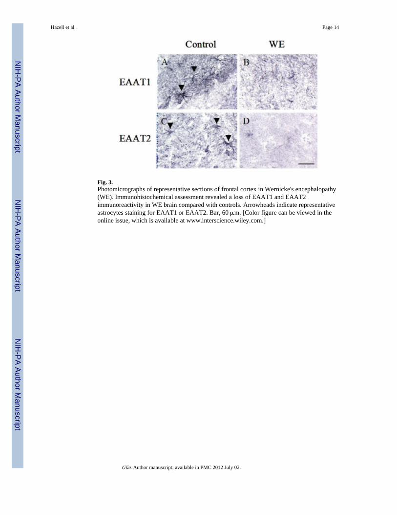
Fig. 3.Photomicrographs of representative sections of frontal cortex in Wernicke's encephalopathy(WE). Immunohistochemical assessment revealed a loss of EAAT1 and EAAT2immunoreactivity in WE brain compared with controls. Arrowheads indicate representativeastrocytes staining for EAAT1 or EAAT2. Bar, 60 μm. [Color figure can be viewed in theonline issue, which is available at www.interscience.wiley.com.]
Hazell et al. Page 14
Glia. Author manuscript; available in PMC 2012 July 02.
NIH
-PA Author Manuscript
NIH
-PA Author Manuscript
NIH
-PA Author Manuscript

Fig. 4.Photomicrographs of representative sections of brain from TD rats at the level of theposterior thalamus. Immunohistochemical assessment revealed a loss of GFAP, EAAT1,EAAT2, GS, and GAT-3 in the area of the medial thalamus of TD rats compared with pair-fed control (PFC) animals. Staining with DAPI indicated the presence of decreased cellnuclei but no pannecrosis in the medial thalamic region. MT, medial thalamus; 3V, thirdventricle. Bars: A–J, 600 μm; K and L, 200 μm.
Hazell et al. Page 15
Glia. Author manuscript; available in PMC 2012 July 02.
NIH
-PA Author Manuscript
NIH
-PA Author Manuscript
NIH
-PA Author Manuscript

Fig. 5.Microgliosis in the medial thalamus of pair-fed controls (PFC) and TD rats. Results showrepresentative immunoblots of CD68 and β-actin at the level of the posterior medialthalamus, and photomicrographs of CD68 immunoreactivity in the same area of brain, inwhich TD rats (B) display a considerable induction of CD68 compared with PFC animals(A). Bar, 60 μm.
Hazell et al. Page 16
Glia. Author manuscript; available in PMC 2012 July 02.
NIH
-PA Author Manuscript
NIH
-PA Author Manuscript
NIH
-PA Author Manuscript

Fig. 6.Comparison of EAAT2 levels in the medial thalamus in pair-fed controls (PFC), TD rats,and in TD rats co-treated with NAC. Results show representative blots of EAAT2 and β-actin along with quantitative analysis in which NAC prevented downregulation of thetransporter protein. *P < 0.05 compared with PFC group (one-way ANOVA with posthocDunnett's test for multiple comparisons). Photomicrographs show representative cresylviolet staining of medial thalamus from a PFC (A), TD (B), and TD rat co-treated with NAC(C). Note neuronal loss in TD and prevention of this effect following NAC treatment. Bar,300 lm. [Color figure can be viewed in the online issue, which is available atwww.interscience.wiley.com.]
Hazell et al. Page 17
Glia. Author manuscript; available in PMC 2012 July 02.
NIH
-PA Author Manuscript
NIH
-PA Author Manuscript
NIH
-PA Author Manuscript

Fig. 7.Photomicrographs of representative sections of brain from TD and TD + NAC treated rats atthe level of the posterior thalamus. Panels show EAAT2 (A,B), GS (C,D), EAAT1 (E,F),GAT-3 (G,H), and GFAP (I,J) immunostaining in the medial thalamus of animals treatedwith TD only (A,C,E,G,I) and TD + NAC (B,D,F,H,J). Treatment with NAC prevented lossof EAAT2, GS, EAAT1, and GAT-3 immunoreactivity. No NAC-mediated protection wasafforded against the loss of GFAP. MT, medial thalamus; 3V, third ventricle. Grading key:no protection (0), mild protection (+), moderate protection (++), marked protection (+++).Bar: 600 μm. [Color figure can be viewed in the online issue, which is available atwww.interscience.wiley.com.]
Hazell et al. Page 18
Glia. Author manuscript; available in PMC 2012 July 02.
NIH
-PA Author Manuscript
NIH
-PA Author Manuscript
NIH
-PA Author Manuscript

NIH
-PA Author Manuscript
NIH
-PA Author Manuscript
NIH
-PA Author Manuscript
Hazell et al. Page 19
Tabl
e 1
Pat
ient
Inf
orm
atio
n
Cas
e N
o.A
ge (
yr)
PM
I (h
r)Se
xN
DC
OD
146
25M
Nor
mal
Car
diac
arr
est
266
22M
Nor
mal
Res
pira
tory
arr
est
344
24M
Nor
mal
Acu
te m
yoca
rdia
l inf
arct
ion
453
26M
Nor
mal
Non
-Hod
gkin
's ly
mph
oma
562
9M
Nor
mal
Ade
noca
rcin
oma
646
28.5
MW
ESe
ptic
aem
ia
747
19M
WE
Thr
oat c
ance
r
854
12M
WE
Sept
icae
mia
960
7M
WE
Isch
aem
ic h
eart
dis
ease
1066
39M
WE
Cor
onar
y ar
tery
dis
ease
PMI,
pos
tmor
tem
inte
rval
; M, m
ale;
ND
, neu
ropa
thol
ogic
al d
iagn
osis
; CO
D, c
ause
of
deat
h.
Glia. Author manuscript; available in PMC 2012 July 02.
Copyright © 2022 FDOKUMEN