Clostridium butyricum Improves Rumen Fermentation ... - MDPI
Upload
independentCategory
view
0download
0
CNF1 Improves Astrocytic Ability to Support NeuronalGrowth and Differentiation In vitroFiorella Malchiodi-Albedi1*, Silvia Paradisi1, Michela Di Nottia2, Daiana Simone1, Sara Travaglione2,
Loredana Falzano2, Marco Guidotti3, Claudio Frank4, Alessandro Cutarelli4, Alessia Fabbri2,
Carla Fiorentini2*
1Department of Cell Biology and Neuroscience, Istituto Superiore di Sanita, Rome, Italy, 2Department of Therapeutic Research and Medicines Evaluation, Istituto
Superiore di Sanita, Rome, Italy, 3Departmrent of Veterinary Public Health and Food Safety, Istituto Superiore di Sanita, Rome, Italy, 4National Centre for Rare Diseases,
Istituto Superiore di Sanita, Rome, Italy
Abstract
Modulation of cerebral Rho GTPases activity in mice brain by intracerebral administration of Cytotoxic Necrotizing Factor 1(CNF1) leads to enhanced neurotransmission and synaptic plasticity and improves learning and memory. To gain moreinsight into the interactions between CNF1 and neuronal cells, we used primary neuronal and astrocytic cultures from ratembryonic brain to study CNF1 effects on neuronal differentiation, focusing on dendritic tree growth and synapseformation, which are strictly modulated by Rho GTPases. CNF1 profoundly remodeled the cytoskeleton of hippocampal andcortical neurons, which showed philopodia-like, actin-positive projections, thickened and poorly branched dendrites, anda decrease in synapse number. CNF1 removal, however, restored dendritic tree development and synapse formation,suggesting that the toxin can reversibly block neuronal differentiation. On differentiated neurons, CNF1 had a similareffacing effect on synapses. Therefore, a direct interaction with CNF1 is apparently deleterious for neurons. Since astrocytesplay a pivotal role in neuronal differentiation and synaptic regulation, we wondered if the beneficial in vivo effect could bemediated by astrocytes. Primary astrocytes from embryonic cortex were treated with CNF1 for 48 hours and used asa substrate for growing hippocampal neurons. Such neurons showed an increased development of neurites, in respect toage-matched controls, with a wider dendritic tree and a richer content in synapses. In CNF1-exposed astrocytes, theproduction of interleukin 1b, known to reduce dendrite development and complexity in neuronal cultures, was decreased.These results demonstrate that astrocytes, under the influence of CNF1, increase their supporting activity on neuronalgrowth and differentiation, possibly related to the diminished levels of interleukin 1b. These observations suggest that theenhanced synaptic plasticity and improved learning and memory described in CNF1-injected mice are probably mediatedby astrocytes.
Citation: Malchiodi-Albedi F, Paradisi S, Di Nottia M, Simone D, Travaglione S, et al. (2012) CNF1 Improves Astrocytic Ability to Support Neuronal Growth andDifferentiation In vitro. PLoS ONE 7(4): e34115. doi:10.1371/journal.pone.0034115
Editor: Nicoletta Landsberger, University of Insubria, Italy
Received July 25, 2011; Accepted February 22, 2012; Published April 16, 2012
Copyright: � 2012 Malchiodi-Albedi et al. This is an open-access article distributed under the terms of the Creative Commons Attribution License, which permitsunrestricted use, distribution, and reproduction in any medium, provided the original author and source are credited.
Funding: This work was partially supported by ‘‘Italia-USA Collaboration Program’’, Fascicolo 11US/1 (to Dr. Fiorentini) and by the Cure_FXS project (www.cure-fxs.eu) under the E-Rare program (to Dr. Fiorentini and Dr. Malchiodi-Albedi). No additional external funding received for this study. The funders had no role instudy design, data collection and analysis, decision to publish, or preparation of the manuscript.
Competing Interests: The authors have declared that no competing interests exist.
* E-mail: [email protected] (CF); [email protected] (FMA)
Introduction
Proteins belonging to the Rho GTPases’ family, including Rho,
Rac and Cdc42 subfamilies, act as molecular switches that cycle
between a GDP-bound inactive and a GTP-bound active state to
transduce extracellular signals to the actin cytoskeleton. Their
ability to modulate the organization of the actin network [1] plays
important roles in the morphogenesis of the dendritic spines of
neurons in the brain [2,3,4] and synaptic plasticity [5,6,7,8,9,10].
Although the picture is not fully resolved yet, it appears that Rac
and Cdc42, which induce actin polymerization, meshwork
formation and bundling, promote spine formation and maturation
[11], whereas RhoA activation, which promotes actin contraction,
results in spine retraction [12]. Dendritic spines are small, actin-
rich protrusions, and actin dynamics regulates their shape and
morphological plasticity. Importantly, activation of NMDA
receptors (as occurs in LTP) affects dendritic spine morphogenesis
by activation of Rac1 and actin remodeling [13], linking activity-
dependent synaptic plasticity to Rho GTPases. In the nervous
system, the Rho GTPases play a key role in several processes, and
mutations in proteins involved in Rho GTPase signaling may be
causative in some forms of mental retardation.
We have found that a bacterial protein toxin from Escherichia
coli, which activates the Rho GTPases, can improve learning and
memory in mice [14,15]. This toxin, named cytotoxic necrotizing
factor 1 (CNF1), acts by blocking the Rho GTPases in their
activated, GTP-bound state by catalyzing the deamidation of
a single glutamine residue of the Rho molecules, thus impeding
GTP hydrolysis and leading to their persistent activation [16,17].
CNF1 modulation of cerebral RhoA and Rac1 activity in mice re-
arranges cerebral actin cytoskeleton, enhances neurotransmission
and synaptic plasticity and improves cognitive performances
[14,15]. Also, CNF1 counteracts the formalin-induced inflamma-
tory pain in mice, after both peripheral and central administration,
further sustaining its ability in modulating CNS pathophysiology
PLoS ONE | www.plosone.org 1 April 2012 | Volume 7 | Issue 4 | e34115

[18]. Very recently, we have demonstrated that CNF1, by directly
modulating the brain Rho GTPases, triggers structural remodeling
and functional plasticity into the adult rat visual cortex [19] and
improves the behavioral phenotype in a mouse model of Rett
syndrome [20].
Therefore, CNF1 can be viewed as a new pharmacological
agent able to enhance the changes in neuronal connectivity
associated with memory for by which the toxin can ameliorate the
neuronal function. To address this question, we have performed
a study on primary neuronal cultures of rat embryonic brain with
the aim of investigating the effects of CNF1 on in vitro neuronal
growth and differentiation, focusing on the development of
dendritic tree and synapse formation. Our data show that while
direct administration of CNF1 to neuronal cultures has a harmful
effect on neuronal maturation, hippocampal neurons conditioned
by CNF1-treated mation and to improve neuronal plasticity. It
remains, however, to define the mechanisms astrocytes show an
increase in dendrite growth and synapse formation, sustaining
a role for CNF1 in improving astrocytic neurosupportive activity.
Results
CNF1 modifies neuritic tree and synapse development inneurons during differentiationTo analyze the effects of CNF1 on neuronal differentiation,
hippocampal cultures were treated at day-in-vitro (DIV) 2 with
CNF1 and fixed at DIV 14. At this stage, staining with the nuclear
dye Hoechst 33342 showed that, although cell density varied
among different cell cultures, there was no significant difference in
the neuronal cell number, within the same culture, between
control and CNF1 treatment (data not shown). However, neuronal
cell differentiation was profoundly affected. Indeed, while in
mature control neurons actin-labeled neurites were long, thin and
well defined, in CNF1-treated cultures, the neuritic tree and the
cell bodies were covered with numerous and short protrusions,
which gave the cells a spiny appearance (Figure 1A). CNF1-
induced cytoskeletal changes were accompanied by a lack of
synapse formation, as demonstrated by immunolabeling of
synaptophysin, an integral protein of synaptic vesicles. In fact, in
control cultures, at DIV 14, synaptophysin-positive synapses
appeared as discrete dots, regularly distributed along the MAP2-
positive dendrites (Figures 1A, B). In contrast, in CNF1-treated
hippocampal cultures, synaptophysin immunolabeling was more
dispersed and lacked the typical punctuated appearance along
dendrites (Figure 1A, B). In addition, while in control neurons,
PSD95, a marker of post-synaptic densities, and synaptophysin
were separately compartmentalized in the pre- and post-synaptic
districts, respectively, in CNF1-treated neurons, the pre- and post-
synaptic markers often lost their dot-like appearance and co-
localized (Figure 1C). Synaptophysin also showed a positivity in
growth cones, which were frequently observed in CNF1-treated
cultures (Figure 1A, inset). Counts of synaptophysin-positive
puncta along dendrites confirmed a decrease in synaptic density
in CNF1-treated cultures (Figure 1D).
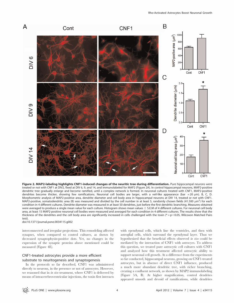
Labeling of MAP2, a marker of dendritic cytoskeleton, also
highlighted CNF1-induced changes of the neuritic tree. In control
pure hippocampal neurons, during differentiation, MAP2-positive
dendritic tree gradually enlarged and became ramified, with thin
and smooth projections, until a complex network was formed
(Figure 2A, left column). Neuronal cell bodies maintained a round
shape, with limited dimensions. When exposed to CNF1 from
DIV 2 MAP2-positive dendrites appeared thicker and more
tortuous. Thin ramifications were lacking. Neuronal cell bodies
were larger, with a veil-like appearance (Figure 2A, right column).
Morphometric analysis confirmed that the dendrite diameter
(Figure 2C) and cell body area (Figure 2D) increased in cells
challenged with the toxin. Total somatodendritic MAP2-positive
area (Figure 2B), however, although showing a trend towards an
increase, did not reach significant values in CNF1-treated cultures,
possibly due to the loss of finer ramifications. Similar results were
obtained in pure cortical neuronal cultures (data not shown).
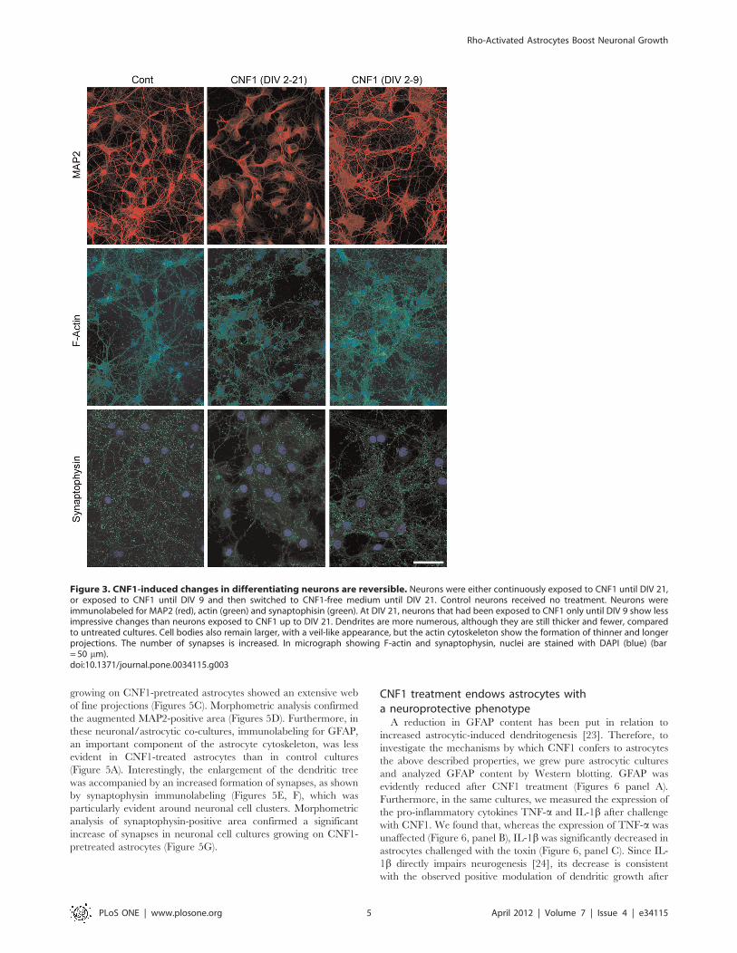
CNF1-induced changes in differentiating neurons arereversibleIn the experiments so far described, CNF1 was administered at
DIV 2 and persisted in the medium until fixation. We asked if
removal of the toxin during differentiation could modify the
observed changes in the growth of neuritic tree and synapse
formation (Figure 3). We compared the development of MAP2
positive dendritic tree, actin cytoskeleton and synaptophysin-
labeled synapses in neurons continuously exposed to CNF1 until
DIV 21, to neurons treated with CNF1 until DIV 9 and then
switched to CNF1-free medium until DIV 21. At the end of the
differentiation process, these neurons showed less impressive
changes than neurons continuously exposed to CNF1. Dendrites
increased in number, although they were still thicker and fewer,
compared to untreated cultures. Cell bodies also remained larger,
with a veil-like appearance, but the actin cytoskeleton showed the
formation of thinner and longer projections. The number of
synapses increased. These results suggest that block of maturation
induced by CNF1 is partially reversible once the toxin is removed.
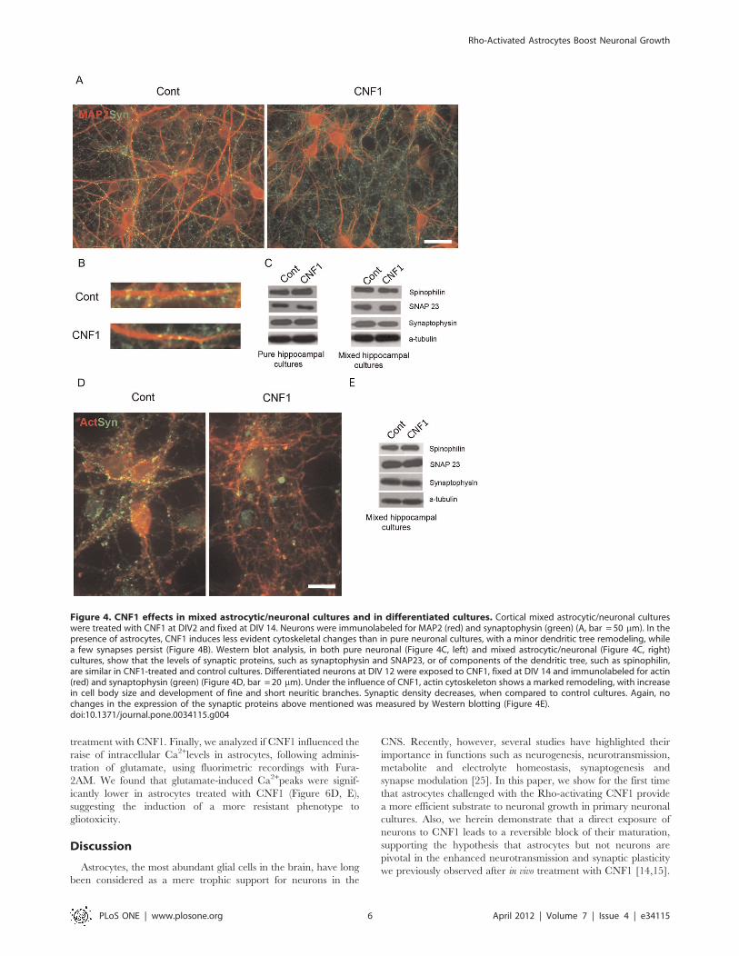
CNF1 effects in mixed astrocytic/neuronal culturesAstrocytes have a pivotal role in neuronal differentiation. In
several neuronal cell models, including hippocampal cultures, the
presence of astrocytes improves and speeds up neuronal
differentiation [21,22]. We asked therefore if the presence of
astrocytes could modify CNF1 effects on neuronal cell growth. At
DIV14, in both hippocampal (data not shown) and cortical mixed
astrocytic/neuronal cultures (Figure 4A, B), the presence of
astrocytes rendered CNF1-induced neuronal cytoskeletal changes
less evident than in pure neuronal cultures, with a minor dendritic
tree remodeling (Figure 4A) and a decreased loss in synapses
(Figure 4B). Still, neurons treated with CNF1 in vitro, even in the
presence of astrocytes, had a less differentiated appearance than
control cultures, a finding that could hardly contribute to explain
the cognitive improvement observed after CNF1 treatment in vivo.
Western blot analysis, conducted in both pure neuronal (Figure 4C,
left) and mixed astrocytic/neuronal (Figure 4C, right) cultures,
showed that the levels of synaptic proteins, such as synaptophysin
and SNAP23, or of components of the dendritic tree, such as
spinophilin, were similar in CNF1-treated and control cultures.
This suggests that the observed changes in neuronal morphology
in CNF1-treated cells were not due to a different expression of key
molecules but probably depended on cytoskeletal remodeling.
CNF1 effects in differentiated culturesSo far, we had analyzed neuronal cell development in vitro under
the influence of CNF1 from the first stages of cell growth. We
wondered if the toxin had different effects on differentiated
neurons. For this purpose, we exposed to CNF1 neuronal cultures
at DIV 12, a stage where polarity has been achieved and synapse
formation is completed (Figure 4D). Again, under the influence of
CNF1, actin cytoskeleton underwent a profound morphological
remodeling. The cell body area increased and the neuritic network
lost its typical structure, composed of long, thin and smooth
neurites, and assumed a spider web-like appearance, with short,
Rho-Activated Astrocytes Boost Neuronal Growth
PLoS ONE | www.plosone.org 2 April 2012 | Volume 7 | Issue 4 | e34115

Figure 1. CNF1 modifies actin cytoskeleton and synapse development in hippocampal neurons. Pure hippocampal neurons weretreated or not with CNF1 at DIV2 and fixed at DIV 14. A. Neurons were immunolabeled for F-actin, upper panel, or co-immunostained forsynaptophysin (green) and MAP2 (red), lower panel. In mature (DIV 14) control neurons, actin-labeled neurites are long, thin and well defined, whileafter CNF1 treatment, the neuritic tree and the cell bodies have a spiny appearance, due to the presence of numerous short protrusions (bar= 20 mm). In control cultures, at DIV 14, synaptophysin-positive synapses appear as small puncta, regularly distributed along the MAP2-positivedendrites. In CNF1-treated hippocampal cultures, fewer synaptophysin-positive dots can be observed along dendrites (bar = 20 mm). Asynaptophysin-labeled growth cone is visible in the inset (bar = 10 mm). B. At higher magnification, the lower number of synaptophysin-positivepuncta along dendrites, induced by treatment with CNF1, can be better appreciated. C. While in control neurons, PSD95 and synaptophysin arecompartmentalized in the pre- and post-synaptic districts, respectively, in CNF1-treated neurons, the pre- and post-synaptic markers often co-localizeand loose their dot-like appearance. D. Synapse density was measured as number of synaptophysin-positive puncta along dendrites, immunostainedwith MAP2. At least 30 images of dendrites (20 mm-long), obtained from 3 different cultures, were analyzed for each condition. The histogramrepresents the mean values 6 S.E.M. *** = p,0.001 Statistical analysis was conducted by the nonparametric Mann-Witney U test.doi:10.1371/journal.pone.0034115.g001
Rho-Activated Astrocytes Boost Neuronal Growth
PLoS ONE | www.plosone.org 3 April 2012 | Volume 7 | Issue 4 | e34115
interconnected and irregular projections. This remodeling affected
synapses, when compared to control cultures, as shown by
decreased synaptophysin-positive dots. Yet, no changes in the
expression of the synaptic proteins above mentioned could be
measured (Figure 4E).
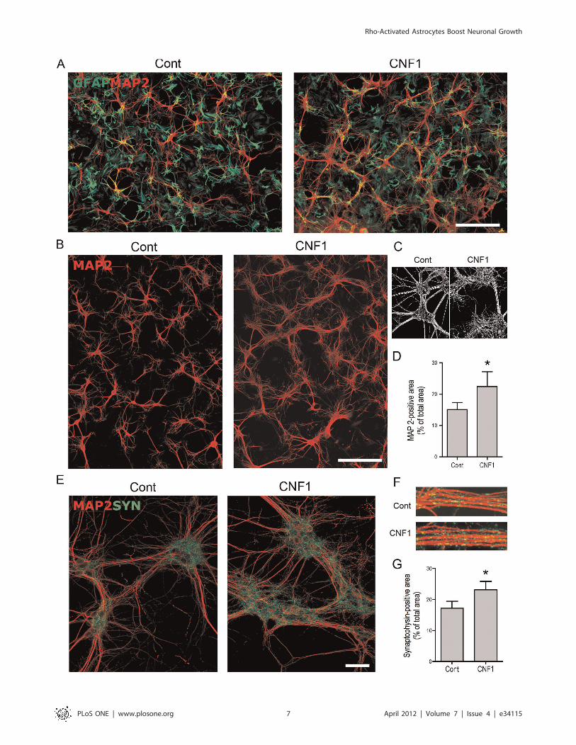
CNF1-treated astrocytes provide a more efficientsubstrate to neuritogenesis and synaptogenesisIn the protocols so far described, CNF1 was administered
directly to neurons, in the presence or not of astrocytes. However,
we reasoned that in in vivo treatment, where CNF1 is delivered by
means of intracerebroventricular injections, the toxin first interacts
with ependymal cells, which line the ventricles, and then with
astroglial cells, which surround the ependymal layer. Thus we
hypothesized that the beneficial effects observed in vivo could be
mediated by the interaction of CNF1 with astrocyes. To address
this question, we treated pure astrocytic cell cultures with CNF1
and analyzed how this treatment affected astrocytic ability to
support neuronal cell growth. At a difference from the experiments
so far conducted, hippocampal neurons, growing on CNF1-treated
astrocytes, but in absence of direct CNF1 influence, produced
a much more abundant dendritic tree, with richer branching,
creating a confluent network, as shown by MAP2 immunolabeling
(Figure 5A, B). At higher magnification, control dendrites
appeared smooth and devoid of ramifications, while dendrites
Figure 2. MAP2-labeling highlights CNF1-induced changes of the neuritic tree during differentiation. Pure hippocampal neurons weretreated or not with CNF1 at DIV2, fixed at DIV 6, 9, and 14, and immunolabeled for MAP2 (Figure 2A). In control hippocampal neurons, MAP2-positivedendritic tree gradually enlarge and become ramified, until a complex network is formed. In neuronal cultures treated with CNF1, MAP2-positivedendrites become thicker, showing few ramifications. Neuronal cell bodies are larger, with a veil-like appearance (bar = 20 mm). B, C, D.Morphometric analysis of MAP2-positive area, dendrite diameter and cell body area in hippocampal neurons at DIV 14, treated or not with CNF1.MAP2-positive, somatodendritic area (B) was measured and divided by the cell number in at least 5, randomly chosen fields (41.500 mm2) for eachcondition in 4 different cultures. Dendrite diameter was measured in at least 50 dendrites, just before the first dendritic branching. Measures obtainedwere averaged to produce a single mean value for each culture. Histogram shows mean values6 S.E.M of 4 different cultures. For neuronal cell bodyarea, at least 15 MAP2-positive neuronal cell bodies were measured and averaged for each condition in 4 different cultures. The results show that thethickness of the dendrites and the cell body area are significantly increased in cells challenged with the toxin (* = p,0.05, Wilcoxon Matched Pairstest).doi:10.1371/journal.pone.0034115.g002
Rho-Activated Astrocytes Boost Neuronal Growth
PLoS ONE | www.plosone.org 4 April 2012 | Volume 7 | Issue 4 | e34115
growing on CNF1-pretreated astrocytes showed an extensive web
of fine projections (Figures 5C). Morphometric analysis confirmed
the augmented MAP2-positive area (Figures 5D). Furthermore, in
these neuronal/astrocytic co-cultures, immunolabeling for GFAP,
an important component of the astrocyte cytoskeleton, was less
evident in CNF1-treated astrocytes than in control cultures
(Figure 5A). Interestingly, the enlargement of the dendritic tree
was accompanied by an increased formation of synapses, as shown
by synaptophysin immunolabeling (Figures 5E, F), which was
particularly evident around neuronal cell clusters. Morphometric
analysis of synaptophysin-positive area confirmed a significant
increase of synapses in neuronal cell cultures growing on CNF1-
pretreated astrocytes (Figure 5G).
CNF1 treatment endows astrocytes witha neuroprotective phenotypeA reduction in GFAP content has been put in relation to
increased astrocytic-induced dendritogenesis [23]. Therefore, to
investigate the mechanisms by which CNF1 confers to astrocytes
the above described properties, we grew pure astrocytic cultures
and analyzed GFAP content by Western blotting. GFAP was
evidently reduced after CNF1 treatment (Figures 6 panel A).
Furthermore, in the same cultures, we measured the expression of
the pro-inflammatory cytokines TNF-a and IL-1b after challenge
with CNF1. We found that, whereas the expression of TNF-a was
unaffected (Figure 6, panel B), IL-1b was significantly decreased in
astrocytes challenged with the toxin (Figure 6, panel C). Since IL-
1b directly impairs neurogenesis [24], its decrease is consistent
with the observed positive modulation of dendritic growth after
Figure 3. CNF1-induced changes in differentiating neurons are reversible. Neurons were either continuously exposed to CNF1 until DIV 21,or exposed to CNF1 until DIV 9 and then switched to CNF1-free medium until DIV 21. Control neurons received no treatment. Neurons wereimmunolabeled for MAP2 (red), actin (green) and synaptophisin (green). At DIV 21, neurons that had been exposed to CNF1 only until DIV 9 show lessimpressive changes than neurons exposed to CNF1 up to DIV 21. Dendrites are more numerous, although they are still thicker and fewer, comparedto untreated cultures. Cell bodies also remain larger, with a veil-like appearance, but the actin cytoskeleton show the formation of thinner and longerprojections. The number of synapses is increased. In micrograph showing F-actin and synaptophysin, nuclei are stained with DAPI (blue) (bar= 50 mm).doi:10.1371/journal.pone.0034115.g003
Rho-Activated Astrocytes Boost Neuronal Growth
PLoS ONE | www.plosone.org 5 April 2012 | Volume 7 | Issue 4 | e34115
treatment with CNF1. Finally, we analyzed if CNF1 influenced the
raise of intracellular Ca2+levels in astrocytes, following adminis-
tration of glutamate, using fluorimetric recordings with Fura-
2AM. We found that glutamate-induced Ca2+peaks were signif-
icantly lower in astrocytes treated with CNF1 (Figure 6D, E),
suggesting the induction of a more resistant phenotype to
gliotoxicity.
Discussion
Astrocytes, the most abundant glial cells in the brain, have long
been considered as a mere trophic support for neurons in the
CNS. Recently, however, several studies have highlighted their
importance in functions such as neurogenesis, neurotransmission,
metabolite and electrolyte homeostasis, synaptogenesis and
synapse modulation [25]. In this paper, we show for the first time
that astrocytes challenged with the Rho-activating CNF1 provide
a more efficient substrate to neuronal growth in primary neuronal
cultures. Also, we herein demonstrate that a direct exposure of
neurons to CNF1 leads to a reversible block of their maturation,
supporting the hypothesis that astrocytes but not neurons are
pivotal in the enhanced neurotransmission and synaptic plasticity
we previously observed after in vivo treatment with CNF1 [14,15].
Figure 4. CNF1 effects in mixed astrocytic/neuronal cultures and in differentiated cultures. Cortical mixed astrocytic/neuronal cultureswere treated with CNF1 at DIV2 and fixed at DIV 14. Neurons were immunolabeled for MAP2 (red) and synaptophysin (green) (A, bar = 50 mm). In thepresence of astrocytes, CNF1 induces less evident cytoskeletal changes than in pure neuronal cultures, with a minor dendritic tree remodeling, whilea few synapses persist (Figure 4B). Western blot analysis, in both pure neuronal (Figure 4C, left) and mixed astrocytic/neuronal (Figure 4C, right)cultures, show that the levels of synaptic proteins, such as synaptophysin and SNAP23, or of components of the dendritic tree, such as spinophilin,are similar in CNF1-treated and control cultures. Differentiated neurons at DIV 12 were exposed to CNF1, fixed at DIV 14 and immunolabeled for actin(red) and synaptophysin (green) (Figure 4D, bar = 20 mm). Under the influence of CNF1, actin cytoskeleton shows a marked remodeling, with increasein cell body size and development of fine and short neuritic branches. Synaptic density decreases, when compared to control cultures. Again, nochanges in the expression of the synaptic proteins above mentioned was measured by Western blotting (Figure 4E).doi:10.1371/journal.pone.0034115.g004
Rho-Activated Astrocytes Boost Neuronal Growth
PLoS ONE | www.plosone.org 6 April 2012 | Volume 7 | Issue 4 | e34115
Rho-Activated Astrocytes Boost Neuronal Growth
PLoS ONE | www.plosone.org 7 April 2012 | Volume 7 | Issue 4 | e34115

Figure 5. CNF1-treated astrocytes provide a more efficient substrate to neuritogenesis and synaptogenesis. Pure astrocytic cellcultures, at confluence, were treated with CNF1 for 48 h. After change of the medium, hippocampal neurons were seeded on the astrocyticmonolayer, fixed at DIV 14, and immunolabeled for MAP2 (red) and GFAP (green) or synaptophysin (green). The exposure of astrocytes to CNF1causes a decrease in GFAP staining (A) whereas hippocampal neurons, growing on CNF1-treated astrocytes but in absence of direct CNF1 influence,produce a much more abundant dendritic network, as shown by MAP2 immunolabeling (A,B, bars = 200 mm). In C, a detail of a black and whiteimage used for morphometric analysis emphasizes the change in the dendritic tree, which, growing on control astrocytes, appear smooth and poorlybranching, while on CNF1-pretreated astrocytes, show much wider ramifications (C). D. Morphometric analysis of MAP2-positive area. In hippocampalneurons co-cultured with astrocytes, after background subtraction, MAP2-positive area was measured as percentage of the total field area(0.15 mm2). Values obtained for each field were pooled to obtain a single mean value for each neuronal culture (n = 5). Bars represent mean values6S.E.M (Figure 5C, * = p,0.05, Wilcoxon Matched Pairs test). The richer dendritic tree is accompanied by an increased formation of synapses, as shownby synaptophysin immunolabeling, particularly enriched around cell bodies (E, bar = 50 mm). Synaptophysin-positive puncta are also visible alongdendrite bundles (F). G. Morphometric analysis of synaptic density. Synaptophysin-positive area was measured in 41.500 mm2-large fields obtainedfrom 3 different experiments, conducted in duplicate from 2 different cultures. At least 16 images were analyzed for each condition and the resultspooled. Histogram represents the values + S.E.M. Statistical analysis was conducted by the nonparametric Wilcoxon Matched Pairs tests.doi:10.1371/journal.pone.0034115.g005
Figure 6. CNF1 treatment reduces GFAP and IL-1b levels and glutamate-dependent intracellular Ca2+ rise in pure astrocyticcultures. The CNF1-induced decrease in GFAP level, observed by immunofluorescence (Figure 5, panel A), is confirmed by Western blot analysis(panel A). In the supernatants from control and CNF1-treated astrocytes, the expressions of TNF-a and IL-1b were measured by ELISA. While the levelsof TNF-a are unaffected (panel B), IL-1b is significantly decreased in astrocytes challenged with the toxin (panel C). Intracellular Ca2+ levels wereanalyzed in astrocytes, following administration of glutamate (after a 3 min baseline), using fluorimetric recordings with Fura 2 AM (panels D,E). In D,the time course of a representative experiment is shown. The values of at least 6 cells were recorded and averaged. In E, Ca2+ levels were compared atthe baseline and in the peak region. Data for three different experiments were pooled and analyzed. At least 6 cells were evaluated in eachexperiment. Bars represent mean values 6 S.E.M. Glutamate induces significantly lower Ca2+ peaks in astrocytes treated with CNF1 than in controlcells (panels C and E *p,0.05 unpaired Student’s t-test).doi:10.1371/journal.pone.0034115.g006
Rho-Activated Astrocytes Boost Neuronal Growth
PLoS ONE | www.plosone.org 8 April 2012 | Volume 7 | Issue 4 | e34115

The improvement of learning and memory that follows the
intracerebral administration of CNF1 suggests that the toxin can
influence the physiology of CNS by modulating Rho GTPases,
which play a fundamental role in the development of dendritic
spines and synapses [26,27,28]. To closely monitor CNF1 effects
on neuronal growth and differentiation, in search of a morphologic
counterpart of the improvement in learning and memory of
CNF1-treated mice, we exposed to CNF1 primary hippocampal
cultures from rat embryonic brains. Neurons treated with CNF1,
especially from the early stages of development, underwent
profound modifications, including the development of filopodia-
like actin-positive protrusions along neurites and around cell
bodies, thickening of dendrites, lack of synapse formation, poor
dendritic branching. Similar results were obtained in the presence
of astrocytes or for neuronal cultures obtained from a different
brain region, such as the cortex. Analogous, although less evident,
effects were observed after CNF1 treatment of mature neurons.
Synaptic loss was not due to decreased synthesis of proteins of
synaptic vesicles, whose levels were similar to that of control
cultures, as shown by Western blot experiments. Thus, far from
enriching the neuritic network as expectable from in vivo results,
CNF1-treated neurons reduced the complexity of dendritic tree
with respect to control neurons and, furthermore, inhibited the
formation of synapses. This pattern of growth suggested a block of
neuronal maturation, which was also suggested by the persistence
of growth cones, normally disappearing after the first days in
culture, in advanced stages of development (DIV 14). Consistently,
removal of the toxin from the cell culture medium allowed the
neuronal cultures to regain the differentiation process, with
recovered growth of dendrite network and synapse formation.
The results of these experiments, where CNF1 was administered
directly to neurons, in the presence or not of astrocytes, did not
explain the beneficial effects of CNF1 on behavioral tasks observed
in in vivo administration of the toxin. However, we reasoned that in
in vivo treatment, where CNF1 is delivered by means of
intracerebroventricular injections, the toxin first interacts with
ependymal cells, which line the ventricles, and then with astroglial
cells, which are in close contact with the ependymal layer. Thus
we hypothesized that the effects observed in vivo could be mediated
by the interaction of CNF1 with astrocyes. To address this issue,
we analyzed the growth of hippocampal neurons on astrocytes that
had been previously treated with CNF1. Intriguingly, when seeded
on CNF1-treated astrocytes, neurons showed a much more
abundant development of dendritic tree, as compared to neurons
grown on untreated astrocytes. Furthermore, synapses developed
normally and were more numerous. It is reasonable to speculate
that the enhanced synaptic plasticity and improved learning and
memory observed in vivo could be mediated by the interaction of
CNF1 with astrocyes.
It has long been known that astrocytes, when co-cultured with
neurons, have the ability to improve neuronal growth, differen-
tiation and synaptic formation [29]. How did CNF1 improve this
ability? First of all, we found that the toxin induced a decrease in
GFAP expression. GFAP is the main intermediate filament protein
in mature astrocytes, but also an important component of the
cytoskeleton in astrocytes during development. GFAP has been
shown to be involved in astrocyte functions relevant to CNS
regeneration and synaptic plasticity. Several lines of evidence
suggest that the observed reduction in GFAP content in CNF1-
treated astrocytes could be related to the increased dendritogen-
esis. GFAP has in fact been found to be a negative regulator of
astrocytic ability to improve neuronal growth and neuritogenesis
[30]. In addition, highly reactive astrocytes, as shown by GFAP
immunostain, induce the formation of fewer synaptic contacts in
co-cultured neurons, compared to less reactive astrocytes [31].
Recent studies showed that increased astrocytic GFAP expression
can be related to neuron atrophy whereas diminished GFAP
content restores neurite outgrowth in certain conditions [32]. It is
worthwhile mentioning that various pathologic conditions of CNS
are accompanied by reactive gliosis, which is characterized by an
increase in the expression of GFAP and is considered to have a role
in neurodegeneration [23]. Therefore, the capacity of CNF1 of
modulating GFAP content could result crucial in those neurolog-
ical diseases where astrocytosis contributes to neuronal damage.
It is well known that in GFAP-overexpressing, activated
astrocytes, the secretion of pro-inflammatory cytokines is up-
regulated and it is believed to contribute to neurodegeneration
[33]. We wondered if CNF1-treated astrocytes, where low levels of
GFAP are detected, also showed decreased levels of cytokines. In
our model, when exposed to CNF1, astrocytes reduced the
secretion of IL-1b while that of TNF-a was unvaried, suggesting
a selective effect of CNF1 on the cytokine network. This result is in
line with our results, since IL-1b can significantly reduce dendrite
development and complexity in neuronal cultures [34]. In
addition, upregulation of IL-1b was observed to negatively
influence neurogenesis [24] and neurodevelopment [35], possibly
by interfering with the signaling of BDNF, a major trophic factor
in the CNS, critical for the development and survival of certain
neuronal populations [36]. Thus, it is reasonable to expect that
low levels of this cytokine may be related to an improvement of
neuronal growth. In addition, it is worth noting that IL-1b is
considered to contribute to neurotoxicity in several neurodegen-
erative diseases. This observation further prompts to consider
CNF1 as a candidate therapeutic option in neurodegenerative
conditions where upregulation of proinflammatory cytokines is
considered of pathogenetic relevance.
Like in neurons, overstimulation of glutamate receptors induce
in astrocytes an increase in intracellular Ca2+concentrations,
which may be at origin of gliotoxic effects, resulting in dysfunction
and death of astrocytes [37]. Since astrocytes are the major sink of
glutamate in the brain, being mainly responsible for glutamate
removal from the extracellular space [38], they represent one of
the important components of CNS defense against glutamate
excitotoxicity. For this reason, dysfunctional astrocytes may
exacerbate excitotoxic cell damage [39]. We checked if CNF1
treatment could modulate glutamate response in astrocytes and
found that intracellular Ca2+peak, following glutamate adminis-
tration, was significantly reduced, compared to untreated astro-
cytes, suggesting an increased Rho-dependent ability of astrocytes
to counteract excitotoxic stimuli.
As a whole, these results suggest that the changes observed in
astrocytes after CNF1 treatment (decrease in GFAP and IL-1blevels, reduction of glutamate-driven intracellular Ca2+concentra-
tions) i) provide a rationale to the observed improvement of
neuritic tree growth in co-cultured neurons; ii) depict a neuropro-
tective astrocytic phenotype. In conclusion, considering the
pathogenetic link between astrocytes and neurological disorders,
through pathways that include inflammation, oxidative stress and
excitotoxicity [40], our results encourage further studies on CNF1-
astrocyte interactions, also in view of a possible development of
new therapeutic approaches.
Materials and Methods
Ethics StatementAll primary cultures used in this study were obtained from
Wistar rat embryos at gestational day 18 (Charles River). This
study was carried out in strict accordance with the recommenda-
Rho-Activated Astrocytes Boost Neuronal Growth
PLoS ONE | www.plosone.org 9 April 2012 | Volume 7 | Issue 4 | e34115

tions in the Guide for the Care and Use of Laboratory Animals of
the National Institutes of Health. The protocol was approved by
the Committee on the Ethics of Animal Experiments of the
University of Minnesota (Permit Number: 27-2956).
Primary culturesAfter dissection, hippocampi were dissociated in trypsin and
plated on poly-L-lysine-coated glass coverslips in Minimum
Essential Medium (MEM), containing 10% fetal calf serum; after
two hours, the medium was replaced with Neurobasal Medium
(NBM) supplemented with B27. To obtain pure neuronal cultures,
hippocampal neurons were treated at day-in-vitro (DIV) 1 with
1.5 mM Arabinosyl-Cytosine (Ara-C). In these conditions, neuro-
nal cultures contain 1–2% of Glial Fibrillary Acidic Protein-
positive astrocytes [41]. To analyze neuronal cell number, nuclei
were stained with Hoechst 33342 in pure neuronal cell cultures at
DIV 14. Five different fields (20x) were randomly chosen in
coverslips obtained from four different cultures. Nuclei were
counted and averaged. To obtain mixed neuronal/astrocytic
cultures, no Ara-C was added. At DIV 14, in mixed cultures
astrocyte number ranged from 20 to 40% of the cell population
(data not shown). For pure and mixed cortical cultures, cortices
were dissected, dissociated and plated, as described for hippo-
campal cultures. In all neuronal cultures (with the exception of
cultures undergoing CNF1 interrupted treatment, see below), cell
culture media were never totally substituted and small aliquots of
NBM-B27 (5–10% in volume) were added once a week. In the
protocol for CNF1 interrupted treatment, where CNF1-containing
cell culture medium was substituted with CNF1-free medium at
DIV 9, NBM-27 conditioned by untreated cultures grown in
parallel was used. Primary astrocytic cultures were obtained from
the cortex of rat embryos. After dissection and dissociation, as
already described, cortical cell suspension was seeded in flasks in
MEM, containing 10% fetal calf serum and let grow until
confluence. Cells were replated twice to obtain a cell culture highly
enriched in astrocytes. Contamination of microglial cells was
below 1%, as shown by staining with Bandeiraea simplicifolia lectin-
peroxidase conjugate (data not shown). For primary astrocytic-
neuronal co-cultures, astrocytes were first seeded on glass cover-
slips and let grow until confluent. Hippocampal neuron suspen-
sion, obtained as described above, was seeded on the astrocytic
monolayer and treated at DIV 1 with Ara-C, to block further
growth of astrocytes. All cell cultures were grown at 37u in 5%
CO2.
CNF1 preparation and treatmentsCNF1 was obtained from the 392 ISS strain (kindly provided by
V. Falbo, Rome, Italy) and purified as previously described [42].
CNF1 was used at the concentration of 10210 M.
Cell cultures were treated according different protocols. A)
Treatment of immature neurons: CNF1 was administered to pure
or mixed neuronal cultures at DIV2 until fixation (5 or 9 or 14 or
21 DIV). B) Treatment of mature neurons: hippocampal neurons
at DIV 12 were treated for 48 h and then fixed. C) Interrupted
treatment of immature neurons: pure hippocampal neurons were
treated at DIV 2; at DIV 9, CNF1-containing medium was
substituted with CNF1-free medium. At this stage of development,
neuronal survival deeply depends on autocrine production of
growth factors, thus abrupt change of medium with fresh NBM-
B27 is deleterious for neurons in vitro. To overcome this problem,
NMB-B27 derived from untreated cultures grown in parallel was
used instead of fresh medium. Hippocampal cultures were fixed at
DIV 21. D) Treatment of astrocytes to be co-cultured with
neurons: confluent primary astrocytic cell cultures were treated for
48 h with CNF1. At the end of treatment, CNF1-containing
medium was changed with CNF1-free NBM-B27 and primary
hippocampal neurons were seeded on the astrocytic monolayer. In
control cultures, hippocampal neurons were seeded on untreated
astrocytes. Neuronal-astrocytic co-cultures were fixed at DIV 14.
ImmunocytochemistryCell cultures were fixed in 4% paraformaldehyde in phosphate
buffered saline (PBS), 0.12 M in sucrose, and permeabilized with
Triton X-100 (0.2%, Sigma). For F-actin detection, cells were
stained with FITC (fluoresceine isothyocianate)-phalloidin (Sigma;
working dilution 0.5 mg/ml in PBS) for 30 min at 37uC.Immunostaining was performed with the following primary
antibodies: anti-microtubule-associated protein MAP2, a marker
of dendrites, anti-synaptophysin, a synaptic vesicle-associated
protein, PSD95, a marker of post-synaptic densities, glial fibrillary
acidic protein (GFAP), specifically identifying astrocytic cytoskel-
eton. All primary antibodies were purchased from Millipore, MA,
USA. After washing, samples were double-labeled with anti-mouse
Alexa Fluor 488 and anti-rabbit 594 (Molecular Probes). In some
experiments, nuclei were counterstained with DAPI or Hoechst
33342. Finally, after extensive washes, samples were mounted and
observed with an Olympus BX51 fluorescence microscope or an
Eclipse 80i Nikon Fluorescence Microscope, equipped with
a VideoConfocal (ViCo) system.
Morphometric analysis. Morphometric analysis was
conducted with the Optilab software (Graftek, Austin, TX) on
images obtained at the Nikon Fluorescence Microscope, equipped
with the ViCo system. In MAP2-immunostained, pure
hippocampal neurons at DIV 14, mean somatodendritic area,
mean dendritic diameter and mean cell body area were measured
in control and CNF1-treated neurons. For somatodendritic area,
total MAP2-positive area was measured and divided by the cell
number in at least five, randomly chosen fields (41.500 mm2) for
each condition in 4 different cultures. Dendrite thickness was
measured before the first dendritic branching. At least 60 dendrites
were randomly chosen from two separate coverslips of the same
culture, measured and averaged, to produce a single mean value
for each condition in 4 different cultures. For neuronal cell body
area, at least 15 MAP2-positive neuronal cell bodies were
measured and averaged for each condition in 4 different
cultures. For synaptic density, synaptophysin-positive puncta
were counted along MAP2-positive dendrites. At least 30 images
of dendrites (20 mm-long), obtained from 3 different cultures, were
analyzed for each condition. In hippocampal neurons co-cultured
with astrocytes, after background subtraction, MAP2-positive area
was measured as percentage of the total field area. In this case,
positive area could not be normalized by cell number, since in
neuronal/astrocytic co-cultures neurons grow in tight clusters and
single cells are difficult to identify. Values obtained for each field
(0.15 mm2) were pooled to obtain a single mean value for each
neuronal culture (n = 5). A different procedure was also used to
measure synaptic density. Again the reason was the peculiar
growth modality of neuronal/astrocytic co-cultures, where
dendrites develop as tightly packed bundles and single dendrites
are difficult to discern. This did not allow counting synapses along
dendrites, as performed in pure neuronal cultures. In addition, in
neuronal/astrocytic co-cultures, synapses are particularly enriched
around neuronal cell bodies, away from dendrites. For these
reasons, synapse density was measured as synaptophysin-positive
area. At least 16 images (41.500 mm2-large fields), obtained from 3
different experiments, conducted in duplicate from 2 different
cultures, were captured and measured for each condition and the
results pooled.
Rho-Activated Astrocytes Boost Neuronal Growth
PLoS ONE | www.plosone.org 10 April 2012 | Volume 7 | Issue 4 | e34115

Statistical analyses were conducted by the nonparametric
Mann-Witney U or Wilcoxon Matched Pairs tests.
Western blot analysisCells were lysed in boiled sample buffer 1x (50 mM Tris-HCl,
pH 6.8, 2% SDS, 10% glycerol, and 100 mM dithiothreitol).
Twenty-five micrograms of total protein extracts were resolved by
SDS-polyacrylamide gel electrophoresis (PAGE) and electrically
transferred onto polyvinylidene difluoride membranes (Bio-Rad).
Membranes were blocked with Tris-buffered saline-Tween 20
(TBS-T) (20 mM Tris-HCl, pH 7.4, 150 mM NaCl, and 0.02%
Tween 20) containing 5% skimmed milk (Bio-Rad) for 30 min at
room temperature, and then they were incubated overnight at 4uCwith primary antibodies diluted in TBS-T containing 2% milk.
The following primary antibodies were used: mouse monoclonal
anti-synaptophysin (Chemicon; 1:1000), rabbit polyclonal anti-
spinophilin (Upstate; 1:1000), mouse monoclonal anti-SNAP-23
(Sy-Sy; 1:10000), rabbit polyclonal anti-GFAP (Millipore; 1:5000),
mouse monoclonal anti-alpha-tubulin (Sigma; 1:10000). After
extensive washing, immune complexes were detected with
horseradish peroxidase-conjugated species-specific secondary anti-
bodies (Jackson’s) followed by enhanced chemiluminescence
reaction (Amersham).
Enzyme-linked immunosorbent assay (ELISA)For detecting IL-1b and TNF-a, ILs ELISA kits were used
following the manifacturer’s instructions (BioVendor-Laboratorni,
a.s.). Briefly, after having washed the microtiterplate with Wash
Buffer, 50 ml of each sample were pipetted in duplicate to the
sample wells. After the Biotin-Conjugate addition to all wells and
a subsequent Streptavidin-HRP incubation, the plate was carefully
washed and the chromogenic substrate solution was added.
Following appropriate incubation, the enzymatic reaction was
stopped and the absorbance was red on a spectro-photometer
(BIORAD) using 450 nm as the primary wave length, with a sub-
wavelength of 650 nm.
Fura-2AM experimentsExperiments were performed on astrocytes treated with CNF1
for 72 h. Optical fluorimetric recordings with Fura-2AM were
used to evaluate the intracellular calcium concentration ([Ca2+]i).
Fura-2AM stock solutions were obtained by adding 50 mg of Fura-2AM to 50 ml of 75% DMSO plus 25% pluronic acid. Cells were
bathed for 60 min at room temperature with 5 ml of stock solutiondiluted in 1 ml of extracellular solution (in mM: 125 NaCl, 1 KCl,
5 CaCl2, 1 MgCl2, 8 glucose, and 20 HEPES, pH 7.35) for a final
Fura concentration of 5 mM. This solution was then removed and
replaced with extracellular solution, and the dishes were quickly
placed on the microscope stage. To measure fluorescence changes,
a Hamamatsu (Shizouka, Japan) Argus 50 computerized analysis
system was used, recording every 6 s the ratio between the values
of light intensity at 340 and 380 nm stimulation. The basal level of
[Ca2+]i was estimated as approximately 80 nM using the
calibration standard kit (Molecular Probes), equivalent to a ratio
value of about 0.8.
Acknowledgments
The authors are grateful to Dr. Simonetta Battisti for critical discussion and
helpful comments.
Author Contributions
Conceived and designed the experiments: FMA SP AF C. Fiorentini.
Performed the experiments: MDN DS ST LF C. Frank AC. Analyzed the
data: FMA SP AF C. Fiorentini. Contributed reagents/materials/analysis
tools: MG. Wrote the paper: FMA AF C. Fiorentini.
References
1. Hotulainen P, Hoogenraad CC (2010) Actin in dendritic spines: Connecting
dynamics to function. J Cell Biol 189(4): 619–629.
2. Luo L (2000) Rho GTPases in neuronal morphogenesis. Nat Rev Neurosci 1(3):
173–180.
3. Saneyoshi T, Fortin DA, Soderling TR (2010) Regulation of spine and synapse
formation by activity-dependent intracellular signaling pathways. Curr Opin
Neurobiol 20(1): 108–115.
4. Tashiro A, Yuste R (2008) Role of rho GTPases in the morphogenesis and
motility of dendritic spines. Methods Enzymol 439: 285–302.
5. Fortin DA, Davare MA, Srivastava T, Brady JD, Nygaard S, et al. (2010) Long-
term potentiation-dependent spine enlargement requires synaptic Ca2+-perme-
able AMPA receptors recruited by CaM-kinase I. J Neurosci 30(35):
11565–11575.
6. O’Kane EM, Stone TW, Morris BJ (2003) Activation of rho GTPases by
synaptic transmission in the hippocampus. J Neurochem 87(5): 1309–1312.
7. Rex CS, Chen LY, Sharma A, Liu J, Babayan AH, et al. (2009) Different rho
GTPase-dependent signaling pathways initiate sequential steps in the consoli-
dation of long-term potentiation. J Cell Biol 186(1): 85–97.
8. Asrar S, Meng Y, Zhou Z, Todorovski Z, Huang WW, et al. (2009) Regulation
of hippocampal long-term potentiation by p21-activated protein kinase 1
(PAK1). Neuropharmacology 56(1): 73–80.
9. Wang HG, Lu FM, Jin I, Udo H, Kandel ER, et al. (2005) Presynaptic and
postsynaptic roles of NO, cGK, and RhoA in long-lasting potentiation and
aggregation of synaptic proteins. Neuron 45(3): 389–403.
10. Nadif Kasri N, Van Aelst L (2008) Rho-linked genes and neurological disorders.
Pflugers Arch 455(5): 787–797.
11. Penzes P, Beeser A, Chernoff J, Schiller MR, Eipper BA, et al. (2003) Rapid
induction of dendritic spine morphogenesis by trans-synaptic ephrinB-EphB
receptor activation of the rho-GEF kalirin. Neuron 37(2): 263–274.
12. Fu WY, Chen Y, Sahin M, Zhao XS, Shi L, et al. (2007) Cdk5 regulates EphA4-
mediated dendritic spine retraction through an ephexin1-dependent mechanism.
Nat Neurosci 10(1): 67–76.
13. Tolias KF, Bikoff JB, Burette A, Paradis S, Harrar D, et al. (2005) The Rac1-
GEF Tiam1 couples the NMDA receptor to the activity-dependent development
of dendritic arbors and spines. Neuron 45(4): 525–538.
14. Diana G, Valentini G, Travaglione S, Falzano L, Pieri M, et al. (2007)Enhancement of learning and memory after activation of cerebral rho GTPases.
Proc Natl Acad Sci U S A 104(2): 636–641.
15. De Viti S, Martino A, Musilli M, Fiorentini C, Diana G (2010) The rho GTPaseactivating CNF1 improves associative working memory for object-in-place.
Behav Brain Res 212(1): 78–83.
16. Flatau G, Lemichez E, Gauthier M, Chardin P, Paris S, et al. (1997) Toxin-induced activation of the G protein p21 rho by deamidation of glutamine.
Nature 387(6634): 729–733.
17. Schmidt G, Sehr P, Wilm M, Selzer J, Mann M (1997) Gln 63 of rho isdeamidated by escherichia coli cytotoxic necrotizing factor-1. Nature 387(6634):
725–729.
18. Pavone F, Luvisetto S, Marinelli S, Straface E, Fabbri A, et al. (2009) The racGTPase-activating bacterial protein toxin CNF1 induces analgesia up-regulating
mu-opioid receptors. Pain 145(1–2): 219–229.
19. Cerri C, Fabbri A, Vannini E, Spolidoro M, Costa M, et al. (2011) Activation ofRho GTPases triggers structural remodeling and functional plasticity in the adult
rat visual cortex. J Neurosci 31(42): 15163–72.
20. De Filippis B, Fabbri A, Simone D, Canese R, et al. (2011) Modulation ofRhoGTPases improves the behavioral phenotype and reverses astrocytic deficits
in a mouse model of Rett syndrome. Neuropsychoparmachol 1–12 (doi10.1038/npp.2011.301).
21. Cordero-Llana O, Scott SA, Maslen SL, Anderson JM, Boyle J, et al. (2011)
Clusterin secreted by astrocytes enhances neuronal differentiation from humanneural precursor cells. Cell Death Differ 18(5): 907–913.
22. Stipursky J, Romao L, Tortelli V, Neto VM, Gomes FC (2011) Neuron-glia
signaling: Implications for astrocyte differentiation and synapse formation. LifeSci 89(15–16): 524–31.
23. Middeldorp J, Hol EM (2011) GFAP in health and disease. Prog Neurobiol
93(3): 421–443.
24. Kuzumaki N, Ikegami D, Imai S, Narita M, Tamura R, et al. (2010) EnhancedIL-1beta production in response to the activation of hippocampal glial cells
impairs neurogenesis in aged mice. Synapse 64(9): 721–728.
25. Barres BA (2008) The mystery and magic of glia: A perspective on their roles inhealth and disease. Neuron 60(3): 430–440.
26. Linseman DA, Loucks FA (2008) Diverse roles of rho family GTPases inneuronal development, survival, and death. Front Biosci 13: 657–676.
Rho-Activated Astrocytes Boost Neuronal Growth
PLoS ONE | www.plosone.org 11 April 2012 | Volume 7 | Issue 4 | e34115
27. Tolias KF, Duman JG, Um K (2011) Control of synapse development and
plasticity by rho GTPase regulatory proteins. Prog Neurobiol 94(2): 133–148.28. Murakoshi H, Wang H, Yasuda R (2011) Local, persistent activation of rho
GTPases during plasticity of single dendritic spines. Nature 472(7341): 100–104.
29. Vernadakis A (1988) Neuron-glia interrelations. Int Rev Neurobiol 30: 149–224.30. Menet V, Gimenez y Ribotta M, Chauvet N, Drian MJ, Lannoy J, et al. (2001)
Inactivation of the glial fibrillary acidic protein gene, but not that of vimentin,improves neuronal survival and neurite growth by modifying adhesion molecule
expression. J Neurosci 21(16): 6147–6158.
31. Emirandetti A, Graciele Zanon R, Sabha M Jr., de Oliveira AL (2006) Astrocytereactivity influences the number of presynaptic terminals apposed to spinal
motoneurons after axotomy. Brain Res 1095(1): 35–42.32. Rozovsky I, Wei M, Morgan TE, Finch CE (2005) Reversible age impairments
in neurite outgrowth by manipulations of astrocytic GFAP. Neurobiol Aging26(5): 705–715.
33. Whitney NP, Eidem TM, Peng H, Huang Y, Zheng JC (2009) Inflammation
mediates varying effects in neurogenesis: Relevance to the pathogenesis of braininjury and neurodegenerative disorders. J Neurochem 108(6): 1343–1359.
34. Gilmore JH, Fredrik Jarskog L, Vadlamudi S, Lauder JM (2004) Prenatalinfection and risk for schizophrenia: IL-1beta, IL-6, and TNFalpha inhibit
cortical neuron dendrite development. Neuropsychopharmacology 29(7):
1221–1229.
35. Garay PA, McAllister AK (2010) Novel roles for immune molecules in neural
development: Implications for neurodevelopmental disorders. Front Synaptic
Neurosci 2: 136.
36. Tong L, Balazs R, Soiampornkul R, Thangnipon W, Cotman CW (2008)
Interleukin-1 beta impairs brain derived neurotrophic factor-induced signal
transduction. Neurobiol Aging 29(9): 1380–1393.
37. Chen CJ, Liao SL, Kuo JS (2000) Gliotoxic action of glutamate on cultured
astrocytes. J Neurochem 75(4): 1557–1565.
38. Kanai Y, Smith CP, Hediger MA (1993) The elusive transporters with a high
affinity for glutamate. Trends Neurosci 16(9): 365–370.
39. Rossi D, Volterra A (2009) Astrocytic dysfunction: Insights on the role in
neurodegeneration. Brain Res Bull 80(4–5): 224–232.
40. Glass CK, Saijo K, Winner B, Marchetto MC, Gage FH (2010) Mechanisms
underlying inflammation in neurodegeneration. Cell 140(6): 918–934.
41. Malchiodi-Albedi F, Domenici MR, Paradisi S, Bernardo A, Ajmone-Cat MA,
et al. (2001) Astrocytes contribute to neuronal impairment in beta A toxicity
increasing apoptosis in rat hippocampal neurons. Glia 34(1): 68–72.
42. Falzano L, Fiorentini C, Donelli G, Michel E, Kocks C, et al. (1993) Induction of
phagocytic behaviour in human epithelial cells by Escherichia coli cytotoxic
necrotizing factor type 1. Mol Microbiol 9: 1247–1254.
Rho-Activated Astrocytes Boost Neuronal Growth
PLoS ONE | www.plosone.org 12 April 2012 | Volume 7 | Issue 4 | e34115
Copyright © 2022 FDOKUMEN


![Astrocytic tracer dynamics estimated from [1-11C]-acetate PET measurements](https://static.fdokumen.com/doc/165x107/6334cca03e69168eaf070c95/astrocytic-tracer-dynamics-estimated-from-1-11c-acetate-pet-measurements.jpg)