Astrocytic adhesion molecules are increased in HIV-1-associated cognitive/motor complex
Upload
khangminh22Category
view
0download
0
Facultat de BiociènciesDepartament de Biologia Cel·lular, Fisiologia i Immunologia
Universitat Autònoma de Barcelona
Role of astrocytic IL-6 and IL-6R in normalphysiology and neuroinflammation
Maria Erta CañabateBellaterra, Març de 2014
Memoria de tesis presentada por Maria Erta Cañabate para optar al grado de Doctor en Neurociencias por la Universitat Autònoma de Barcelona.
Este trabajo ha sido realizado bajo la dirección del Doctor Juan Hidalgo Pareja, catedrático de universidad del Departament de Biologia Cel·lular, Fisiologia i Immunologia.
Director de tesis Doctoranda
Dr. Juan Hidalgo Pareja Maria Erta Cañabate


Mientras la ciencia a descubrir no alcance las fuentes de la vida,
y en el mar o en el cielo haya un abismo que al cálculo resista,
mientras la humanidad siempre avanzando no sepa a dónde camina,
mientras haya un misterio para el hombre, ¡habrá poesía!
-Gustavo Adolfo Bécquer-


ACKNOWLEDGEMENTS
Bernard of Chartres said that “We are like dwarfs on the shoulders of giants, so that wecan see more than they, and things at a greater distance, not by virtue of any sharpnesson sight on our part, or any physical distinction, but because we are carried high andraised up by their giant size.” [Bernard of Chartres, 1130 AD]
The acknowledgements section of a Thesis is the opportunity to look down, see allthose giants pushing us higher and, of course, acknowledge them for their merits andhelp.
First of all, I would like to appreciate the work done by my scientific colleagues in thiswork area. It was pretty amazing to notice that the key molecule I was studying was intu-ited and discovered the year I was born and since then to the moment I started working inthis area 4 years ago, there appear more than 40.000 publications in Pubmed when “Inter-leukin 6” is typed, and during those four years it has been more than 18.000 publications,indicating it haven’t loss any piece of its interest. To my dismay, I couldn’t read them allbut I feel a deep gratitude for their contribution to the field and not only for the answersthey gave to explain former questions but also (and most importantly) for the new andunexpected questions they pointed, making this investigation line really challenging andamazing.
A continuación quiero agradecer a uno de estos "gigantes", mi director de tesis, elDr. Juan Hidalgo, no solo el darme la oportunidad de trabajar en su grupo sino tambiénsu incalculable interés, guía y ayuda durante todos estos años. No cal ni dir que la Dra.Merce Giralt mereix menció especial, no només he d’agrair-li la immensa dosi de feinaque ha realitzat en aquesta tesis i totes les tècniques que m’ha ensenyat sinó que és im-prescindible reconèixer-li la seva disposició incondicional d’ajuda, les seves deliciosespumpkin pies que ensucraven els dies més grisos i el seu bon humor perpètu i contagiósque, juntament amb el dels altres membres del grup, feien que anar a treballar fos mésun hobby que no pas una obligació. Per descomptat tambe a tots els altres membres delgrup, la Dra. Amalia Molinero per la seva valiosa ajuda i consells tant per la tesis com en ladocència; a la Gemma Comes no només per tota l’ajuda prestada i el seu optimisme sinótambé per tots els bons (i grans) moments viscuts dins i fora del laboratori; al Javier per laseva ajuda i consells; a la Olaya pel seu suport i totes les converses frikis que hem tingut;i, per últim, també a les que tot i que ja no estan al laboratori, Yas, Bea y Raquel, tambéhan deixat una petjada en aquesta tesis gràcies a tot el que vaig aprendre d’elles. Tambévull agrair a tots els membres del grup dels Doctors Antonio Armario i Roser Nadal la sevaajuda en la resolució de dubtes sorgits tant de docència com d’investigació, en especialel Xavi, la Núria, la Cristina Rabasa i l’Almudena. També agrair a la Mar tota la ajuda ambel món de les immunos i amb els bons moments passats al lab i als vinachos i al JuanCarles per haver-me ensenyat (millor dit deixar-me gaudir) de com es feien les seves su-perbes classes. Per descomptat agrair també a la Pilar el tenir cura dels animals i la ajudaincondicional i bon humor de les meravelloses secretàries que hem tingut: Olga, Carlota
v

i Paqui. A tots ells moltes gracies per deixar-me formar part d’aquesta gran família.
I also want to thank Dr Iain Campbell to let me work in his lab in Sydney and all histeam-mates, specially Sue Ling, Vicky, Laura, Meng and Magdalena for the amazing ex-perience it was. Fue allí, tan lejos de casa, donde tuve el placer de conocer a nuestros"vecinos de arriba" Bernardo Castellano y Nàdia Villacampa, que junto con los otrosmiembros de su grupo Berta González y Beatriz Almolda han sido y son de gran ayudaenseñándome y ayudándome con técnicas de IHQ i Facs, además de muchísimos buenosmomentos dentro y fuera del laboratorio.
También es un honor poder agradecer el haberme podido iniciar en la ciencia en elgrupo del Dr. Rafael Maldonado, en el PRBB, un lugar precioso con gente aún mejor.Agradecer especialmente las maravillosas enseñanzas y horas de trabajo con Aurelijus yElena.
No puc oblidar-me tampoc dels amics de tota la vida que sempre m’han apoiat i com-près durant tot aquest temps i han soportat tots els "plantons" que els hi he donat perculpa de la feina i també per tots els moments genials que hem passat. Gràcies Laura,David, Seila, Rubén, Marc S, Iván, Marc V, Lluís i Samuel. Gràcies també a les gransamigues de veterinària Nausica, I-chan, Ariadna, Esther i Marisa perque, tot i que ensveiem poc, sempre és un gran plaer i desconnexió retrobar-nos i tambe als grans amicsde telecos (i cia) Carlos, Cris, Fran, Judith, Uri, Glòria, Toni, Saray, Alex, Helena, Alberto,Aris i Javi, moltes gracies per totes les quedades i aconteixements mítics que hem fet ifarem.
Per descomptat també donar les gracies a la meva família, sobretot als meus pares iavis pel seu amor i suport incondicional, i en especial al meu pare, per la seva ajuda i ideescientífiques desde que era ben petita i fins ara. També agrair a tots aquells que tot i queno hi siguin fisicament segueixen estant molt presents. Al meu avi Pepe per deleitar-mesempre amb el seu món fascinant ple de llocs, històries, costums i, fins i tot m’atreviria adir llenguatges, sorprenents i captivadors; i a la meva àvia Pepita, donar-li les gracies pertots els moments viscuts amb ella que formen part de la meva memoria i per inculcarmela importancia dels estudis, recordo la seva cara d’il·lusió quan vaig aprovar la selectivitat,sé que li hagués agradat molt veure com em licenciava i ara defensant la tesis.
Per tu Jordi no sé si trobaré les paraules adients per agrairte tot el que has fet i fas permi. No només has estat i ets el meu millor amic i suport desde que ens vam conèixer sinóque ets una gran persona amb qui estic orgullosa de compartir aquestes aventures quesón la vida i la tesis. Perque, en realitat aquesta tesis és tant teva com meva ja que sense tuni tan sols s’assemblaria al que és ara. No només has deixat la teva emprenta en el format iles figures ajudant-me i guiant-me amb el Latex sinó que també has estat imprescindibleen els continguts: has dissenyat i creat els programes conductuals, m’has fet tot tipusd’artilugis per optimitzar la meva feina que m’han estalviat un munt de temps, ens hasarreglar aparells del laboratori que ja es donaven per perduts, t’has quedat nits senceresal laboratori ajudant-me (fins i tot a l’altre punta de món) i per sobre de tot, sempre m’hasdonat tot el teu suport i ànims. Gràcies de tot cor
A tots, moltíssimes gràcies per ser-hi!!
vi

CONTENTS vii
CONTENTS
Contents vii
List of Figures xi
List of Tables xv
Acronyms xvii
1 Introduction 11.1 Interleukin-6, the founding member of the neuropoietins . . . . . . . . . . 1
1.1.1 The IL-6 Receptor complex . . . . . . . . . . . . . . . . . . . . . . . . 21.1.2 Signal transduction of IL-6 . . . . . . . . . . . . . . . . . . . . . . . . 41.1.3 IL-6 signaling pathways: classic versus trans-signalling . . . . . . . 41.1.4 IL-6 gene expression . . . . . . . . . . . . . . . . . . . . . . . . . . . . 71.1.5 IL-6 functions . . . . . . . . . . . . . . . . . . . . . . . . . . . . . . . . 7
1.2 CNS immunity . . . . . . . . . . . . . . . . . . . . . . . . . . . . . . . . . . . . 81.2.1 Blood Brain Barrier . . . . . . . . . . . . . . . . . . . . . . . . . . . . . 91.2.2 Resident cells in nervous parenchyma . . . . . . . . . . . . . . . . . 91.2.3 Circulating cells . . . . . . . . . . . . . . . . . . . . . . . . . . . . . . . 14
1.3 IL-6 role in the CNS. Data gathered from humans and animal models . . . 161.3.1 Expression . . . . . . . . . . . . . . . . . . . . . . . . . . . . . . . . . . 161.3.2 Role of IL-6 as a pro-inflammatory and anti-inflammatory cytokine 181.3.3 Role of IL-6 in development and normal physiology . . . . . . . . . 191.3.4 Role of IL-6 in pathology . . . . . . . . . . . . . . . . . . . . . . . . . 30
2 Hypothesis and objectives 412.1 Hypothesis . . . . . . . . . . . . . . . . . . . . . . . . . . . . . . . . . . . . . . 412.2 Objectives . . . . . . . . . . . . . . . . . . . . . . . . . . . . . . . . . . . . . . . 41
2.2.1 Objective 1 . . . . . . . . . . . . . . . . . . . . . . . . . . . . . . . . . 412.2.2 Objective 2 . . . . . . . . . . . . . . . . . . . . . . . . . . . . . . . . . 422.2.3 Objective 3 . . . . . . . . . . . . . . . . . . . . . . . . . . . . . . . . . 42
3 Materials and Methods 433.1 Animals . . . . . . . . . . . . . . . . . . . . . . . . . . . . . . . . . . . . . . . . 43
3.1.1 IL-6 floxed mice . . . . . . . . . . . . . . . . . . . . . . . . . . . . . . 433.1.2 IL-6R α-chain floxed mice . . . . . . . . . . . . . . . . . . . . . . . . 433.1.3 GFAP promoter-specific Cre recombinase expressing mice (GFAP-
Cre) . . . . . . . . . . . . . . . . . . . . . . . . . . . . . . . . . . . . . . 433.1.4 CD-1 mice . . . . . . . . . . . . . . . . . . . . . . . . . . . . . . . . . . 44

viii CONTENTS
3.1.5 Generation of Astrocyte IL-6 KO (Ast-IL-6 KO) and Astrocyte IL-6Receptor KO (Ast-IL-6R KO) mice . . . . . . . . . . . . . . . . . . . . 44
3.1.6 GFAP-IL6/sgp130 bigenic mice . . . . . . . . . . . . . . . . . . . . . . 443.2 Tail DNA extraction and genotyping . . . . . . . . . . . . . . . . . . . . . . . 45
3.2.1 Cre recombinase PCR . . . . . . . . . . . . . . . . . . . . . . . . . . . 463.2.2 IL-6 Floxed PCR . . . . . . . . . . . . . . . . . . . . . . . . . . . . . . . 463.2.3 IL-6Rα Floxed PCR . . . . . . . . . . . . . . . . . . . . . . . . . . . . . 463.2.4 Gene recombination PCR . . . . . . . . . . . . . . . . . . . . . . . . . 47
3.3 Behavioural tests battery . . . . . . . . . . . . . . . . . . . . . . . . . . . . . . 493.3.1 Hole-Board . . . . . . . . . . . . . . . . . . . . . . . . . . . . . . . . . 503.3.2 Elevated Plus-Maze . . . . . . . . . . . . . . . . . . . . . . . . . . . . 513.3.3 Tail Suspension test . . . . . . . . . . . . . . . . . . . . . . . . . . . . 523.3.4 Morris Water Maze . . . . . . . . . . . . . . . . . . . . . . . . . . . . . 523.3.5 Dominance tube test . . . . . . . . . . . . . . . . . . . . . . . . . . . . 553.3.6 Resident-Intruder test . . . . . . . . . . . . . . . . . . . . . . . . . . . 55
3.4 Body weight . . . . . . . . . . . . . . . . . . . . . . . . . . . . . . . . . . . . . 563.5 Body temperature . . . . . . . . . . . . . . . . . . . . . . . . . . . . . . . . . . 573.6 Experimental autoimmune encephalomyelitis (EAE) . . . . . . . . . . . . . 573.7 Cryolesion . . . . . . . . . . . . . . . . . . . . . . . . . . . . . . . . . . . . . . 593.8 LPS response . . . . . . . . . . . . . . . . . . . . . . . . . . . . . . . . . . . . . 603.9 Animal sacrifice and sample storage . . . . . . . . . . . . . . . . . . . . . . . 603.10 RNA extraccion and Real Time PCR . . . . . . . . . . . . . . . . . . . . . . . . 603.11 Cell cultures . . . . . . . . . . . . . . . . . . . . . . . . . . . . . . . . . . . . . 62
3.11.1 Astrocyte-enriched cultures from cerebellum . . . . . . . . . . . . . 623.11.2 Astrocyte-enriched cultures from cortex . . . . . . . . . . . . . . . . 623.11.3 Splenocytes cultures . . . . . . . . . . . . . . . . . . . . . . . . . . . . 63
3.12 ELISA . . . . . . . . . . . . . . . . . . . . . . . . . . . . . . . . . . . . . . . . . 633.13 Histochemistry (HC) and Immunohistochemistry (IHC) . . . . . . . . . . . 633.14 Statistical analysis . . . . . . . . . . . . . . . . . . . . . . . . . . . . . . . . . . 66
4 Characterization of astrocyte-derived IL-6 deficiency 674.1 Gene recombination and IL-6 brain levels . . . . . . . . . . . . . . . . . . . . 674.2 Frequencies at weaning . . . . . . . . . . . . . . . . . . . . . . . . . . . . . . . 674.3 Body weight . . . . . . . . . . . . . . . . . . . . . . . . . . . . . . . . . . . . . 684.4 Behavioural tests . . . . . . . . . . . . . . . . . . . . . . . . . . . . . . . . . . . 70
4.4.1 Hole-Board . . . . . . . . . . . . . . . . . . . . . . . . . . . . . . . . . 704.4.2 Elevated Plus-Maze . . . . . . . . . . . . . . . . . . . . . . . . . . . . 714.4.3 Tail Suspension . . . . . . . . . . . . . . . . . . . . . . . . . . . . . . . 744.4.4 Morris water maze . . . . . . . . . . . . . . . . . . . . . . . . . . . . . 754.4.5 Dominance tube . . . . . . . . . . . . . . . . . . . . . . . . . . . . . . 794.4.6 Resident Intruder . . . . . . . . . . . . . . . . . . . . . . . . . . . . . . 80
4.5 Body temperature . . . . . . . . . . . . . . . . . . . . . . . . . . . . . . . . . . 814.6 CNS histochemistry and immunohistochemistry . . . . . . . . . . . . . . . 854.7 EAE . . . . . . . . . . . . . . . . . . . . . . . . . . . . . . . . . . . . . . . . . . 86

CONTENTS ix
4.7.1 Symptomatology . . . . . . . . . . . . . . . . . . . . . . . . . . . . . . 864.7.2 HC and IHC of spinal cord and encephalon in 5th backcrossing group 894.7.3 IL-6 levels . . . . . . . . . . . . . . . . . . . . . . . . . . . . . . . . . . 944.7.4 Splenocytes nitrite production . . . . . . . . . . . . . . . . . . . . . . 95
4.8 Cryolesion . . . . . . . . . . . . . . . . . . . . . . . . . . . . . . . . . . . . . . 954.8.1 Mortality rate and injury size . . . . . . . . . . . . . . . . . . . . . . . 954.8.2 HC and IHC of encephalon from 5th backcrossing group . . . . . . 96
4.9 LPS response . . . . . . . . . . . . . . . . . . . . . . . . . . . . . . . . . . . . . 1004.10 Astrocyte-enriched cell cultures . . . . . . . . . . . . . . . . . . . . . . . . . . 100
5 Characterization of astrocytic IL-6 receptor deficiency 1035.1 Frequencies at weaning . . . . . . . . . . . . . . . . . . . . . . . . . . . . . . . 1035.2 Body weight . . . . . . . . . . . . . . . . . . . . . . . . . . . . . . . . . . . . . 1045.3 Behavioural tests . . . . . . . . . . . . . . . . . . . . . . . . . . . . . . . . . . . 104
5.3.1 Hole-Board . . . . . . . . . . . . . . . . . . . . . . . . . . . . . . . . . 1045.3.2 Elevated Plus-Maze . . . . . . . . . . . . . . . . . . . . . . . . . . . . 1065.3.3 Tail Suspension . . . . . . . . . . . . . . . . . . . . . . . . . . . . . . . 1075.3.4 Morris water maze . . . . . . . . . . . . . . . . . . . . . . . . . . . . . 1075.3.5 Resident Intruder . . . . . . . . . . . . . . . . . . . . . . . . . . . . . . 110
5.4 CNS immunohistochemistry . . . . . . . . . . . . . . . . . . . . . . . . . . . . 1115.5 EAE . . . . . . . . . . . . . . . . . . . . . . . . . . . . . . . . . . . . . . . . . . 111
5.5.1 Symptomatology . . . . . . . . . . . . . . . . . . . . . . . . . . . . . . 1115.6 Cryolesion . . . . . . . . . . . . . . . . . . . . . . . . . . . . . . . . . . . . . . 114
5.6.1 Mortality rate and injury size . . . . . . . . . . . . . . . . . . . . . . . 1145.6.2 IL-6 levels in blood . . . . . . . . . . . . . . . . . . . . . . . . . . . . . 115
6 Characterization of trans-signaling in mediating the biological actions of IL-6 1176.1 The severity of gliosis is diminished in GFAP-IL6/sgp130 mice . . . . . . . . 1176.2 Reduced vascular alterations and blood-brain barrier leakage in GFAP-IL6-
spgp130 mice . . . . . . . . . . . . . . . . . . . . . . . . . . . . . . . . . . . . . 1196.3 Hippocampal neurogenesis is rescued in GFAP-IL6/sgp130 mice . . . . . . 1196.4 Reduced neurodegeneration in GFAP-IL6-spgp130 mice . . . . . . . . . . . 122
7 Discussion 1257.1 Validation of astrocyte-derived IL-6 and IL-6R KO mice . . . . . . . . . . . . 1257.2 Prosurvival role . . . . . . . . . . . . . . . . . . . . . . . . . . . . . . . . . . . 1267.3 Role of astrocyte-derived IL-6 and IL-6R in physiological conditions . . . . 127
7.3.1 Body weight, food intake and energy expenditure . . . . . . . . . . . 1277.3.2 Body temperature effect of astrocyte-secreted IL-6 in adult males . 1297.3.3 Behaviour . . . . . . . . . . . . . . . . . . . . . . . . . . . . . . . . . . 130
7.4 Role of astrocyte-derived IL-6 and IL-6R in pathological conditions . . . . . 1397.4.1 EAE . . . . . . . . . . . . . . . . . . . . . . . . . . . . . . . . . . . . . . 1397.4.2 Traumatic brain injury . . . . . . . . . . . . . . . . . . . . . . . . . . . 141
7.5 Role of trans-signaling in mediating IL-6 actions . . . . . . . . . . . . . . . . 144

x CONTENTS
8 Conclusions 147
A Supplementary Data 149A.1 Behavioral programs guide . . . . . . . . . . . . . . . . . . . . . . . . . . . . . 149A.2 Additional results . . . . . . . . . . . . . . . . . . . . . . . . . . . . . . . . . . 153A.3 Behavioral tests . . . . . . . . . . . . . . . . . . . . . . . . . . . . . . . . . . . 153
A.3.1 Ast-IL-6 mice . . . . . . . . . . . . . . . . . . . . . . . . . . . . . . . . 153A.3.2 Ast-IL-6R mice . . . . . . . . . . . . . . . . . . . . . . . . . . . . . . . 154
A.4 Brain IHC at physiological conditions . . . . . . . . . . . . . . . . . . . . . . 154A.5 EAE . . . . . . . . . . . . . . . . . . . . . . . . . . . . . . . . . . . . . . . . . . 154
Bibliography 161

LIST OF FIGURES
1.1 Schematic representation of hexameric IL-6 receptor complex. . . . . . . . . . 31.2 Signal transduction pathways activated by IL-6. . . . . . . . . . . . . . . . . . . 51.3 Microglia activity states. . . . . . . . . . . . . . . . . . . . . . . . . . . . . . . . . . 111.4 Schematic representations summarizing different gradations of reactive as-
trogliosis. . . . . . . . . . . . . . . . . . . . . . . . . . . . . . . . . . . . . . . . . . 131.5 IL-6 has a major role in the response of the brain to injury. . . . . . . . . . . . . 331.6 IL-6 is related to many brain diseases. . . . . . . . . . . . . . . . . . . . . . . . . 36
3.1 Generation of conditional Knock-out mice for astrocytary IL-6 and astrocytaryIL-6 receptor. . . . . . . . . . . . . . . . . . . . . . . . . . . . . . . . . . . . . . . . 45
3.2 Gender identification and ear marking. . . . . . . . . . . . . . . . . . . . . . . . 463.3 GFAP-Cre mice PCR. . . . . . . . . . . . . . . . . . . . . . . . . . . . . . . . . . . . 473.4 IL-6 floxed mice PCR. . . . . . . . . . . . . . . . . . . . . . . . . . . . . . . . . . . 473.5 IL-6Ra floxed mice PCR. . . . . . . . . . . . . . . . . . . . . . . . . . . . . . . . . . 483.6 Astrocytary IL-6 recombination PCR. . . . . . . . . . . . . . . . . . . . . . . . . . 483.7 Schematic chronology for behavioral tests. . . . . . . . . . . . . . . . . . . . . . 503.8 Hole-board (left) and Plus-maze (right) test apparatus. . . . . . . . . . . . . . . 513.9 Tail Suspension test. . . . . . . . . . . . . . . . . . . . . . . . . . . . . . . . . . . . 523.10 Morris Water Maze test. . . . . . . . . . . . . . . . . . . . . . . . . . . . . . . . . . 533.11 Dominance tube test. . . . . . . . . . . . . . . . . . . . . . . . . . . . . . . . . . . 553.12 Resident-Intruder test. . . . . . . . . . . . . . . . . . . . . . . . . . . . . . . . . . 563.13 Body temperature assessment in light and dark conditions. . . . . . . . . . . . 573.14 EAE induction and symptomatology assessment. . . . . . . . . . . . . . . . . . . 583.15 Injured volume assessment. . . . . . . . . . . . . . . . . . . . . . . . . . . . . . . 593.16 Demyelination assessment. . . . . . . . . . . . . . . . . . . . . . . . . . . . . . . 65
4.1 Characterization of IL-6 brain and spleen levels in Ast-IL-6 KO. . . . . . . . . . 684.2 Body weight growth is affected by astrocyte IL-6 deficiency in a sex-dependent
manner. . . . . . . . . . . . . . . . . . . . . . . . . . . . . . . . . . . . . . . . . . . 694.3 Behavioral traits analyzed in the Hole-board apparatus. . . . . . . . . . . . . . . 724.4 Behavioral traits analyzed in the EPM apparatus. . . . . . . . . . . . . . . . . . . 734.5 Time spent in motion during tail suspension test. . . . . . . . . . . . . . . . . . 754.6 Time to climb to the visible platform in MWT. . . . . . . . . . . . . . . . . . . . . 774.7 Mean time needed to reach the submerged platform. . . . . . . . . . . . . . . . 784.8 Percentage of time spent in the platform quadrant during Probe trials. . . . . . 784.9 Mean time needed to reach the submerged platform in a new location. . . . . 794.10 Percentage of time spent in both new and older platform quadrant during re-
versal probe trials. . . . . . . . . . . . . . . . . . . . . . . . . . . . . . . . . . . . . 804.11 Percentage of victories in the dominance tube test. . . . . . . . . . . . . . . . . 81
xi

xii List of Figures
4.12 Behavioral traits analyzed in the Resident-intruder test. . . . . . . . . . . . . . . 824.13 Body temperature and weight during cold exposure. . . . . . . . . . . . . . . . . 834.14 Body temperature variation after either 1 mg/kg of LPS or saline injection. . . 844.15 Body temperature variation after 10 μg/kg of LPS. . . . . . . . . . . . . . . . . . 854.16 Circadian rhythm analysis. . . . . . . . . . . . . . . . . . . . . . . . . . . . . . . . 854.17 Lectin, GFAP and IL-6Rα stainings and quantifications in Ast-IL-6 KO and floxed
mice. . . . . . . . . . . . . . . . . . . . . . . . . . . . . . . . . . . . . . . . . . . . . 874.18 Clinical score and body weight changes following EAE induction. . . . . . . . . 884.19 Cellular infiltrates and demyelination. . . . . . . . . . . . . . . . . . . . . . . . . 914.20 Gliosis and vasogenesis in spinal cord of EAE-induced animals. . . . . . . . . . 934.21 Gliosis and vasogenesis in encephalon of EAE-induced and non-induced ani-
mals. . . . . . . . . . . . . . . . . . . . . . . . . . . . . . . . . . . . . . . . . . . . . 944.22 Nitrite production by splenocytes from EAE animals stimulated with 10μg/ml
of either MOG or LPS. . . . . . . . . . . . . . . . . . . . . . . . . . . . . . . . . . . 954.23 Total injured volume in Ast-IL-6 KO and floxed mice after cryolesion procedure. 964.24 Astrogliosis assessment in 5th backcrossing Ast-IL-6 animals after cryolesion
procedure. . . . . . . . . . . . . . . . . . . . . . . . . . . . . . . . . . . . . . . . . . 974.25 Microgliosis and number of vessels assessment in 5th backcrossing Ast-IL-6
animals after cryolesion procedure. . . . . . . . . . . . . . . . . . . . . . . . . . . 994.26 Lymphocytary infiltration in 5th backcrossing Ast-IL-6 animals after cryole-
sion procedure. . . . . . . . . . . . . . . . . . . . . . . . . . . . . . . . . . . . . . . 1004.27 IL-6 levels in blood and tissues after 1 mg/kg LPS injection. . . . . . . . . . . . 1014.28 Stimulated astrocyte-enriched cell culture stainings . . . . . . . . . . . . . . . . 1014.29 IL-6 production in astrocyte-enriched cultures. . . . . . . . . . . . . . . . . . . . 102
5.1 Body weight growth was not affected by astrocyte IL-6R deficiency. . . . . . . . 1045.2 Behavioral traits analyzed in the Hole-board apparatus in Ast-IL-6 Receptor
animals. . . . . . . . . . . . . . . . . . . . . . . . . . . . . . . . . . . . . . . . . . . 1055.3 Behavioral traits analyzed in the Plus-maze apparatus for Ast-IL-6R animals. . 1065.4 Time spent in motion during tail suspension test in Ast-IL-6R animals. . . . . . 1075.5 Time to reach the platform in cued, spatial and reversal learning in MWM. . . 1085.6 Percentage of time spent in the platform quadrant during Probe trials in Ast-
IL-6R animals. . . . . . . . . . . . . . . . . . . . . . . . . . . . . . . . . . . . . . . 1095.7 Behavioral traits analyzed in the Resident-intruder test for Ast-IL-6R mice. . . 1115.8 IL-6Rα, Lectin and GFAP stainings and quantifications in Ast-IL-6R KO and
floxed mice. . . . . . . . . . . . . . . . . . . . . . . . . . . . . . . . . . . . . . . . . 1125.9 Clinical score and body weight changes following EAE induction. . . . . . . . . 1135.10 Total injured volume in Ast-IL-6R KO, floxed and WT mice after cryolesion pro-
cedure. . . . . . . . . . . . . . . . . . . . . . . . . . . . . . . . . . . . . . . . . . . . 1155.11 IL-6 blood levels at 3 dpl. . . . . . . . . . . . . . . . . . . . . . . . . . . . . . . . . 116
6.1 Astrogliosis assessment in GFAP-IL6, GFAP-IL6/sgp130, GFAP-sgp130 and WTmice. . . . . . . . . . . . . . . . . . . . . . . . . . . . . . . . . . . . . . . . . . . . . 118

List of Figures xiii
6.2 Microgliosis assessment in GFAP-IL6, GFAP-IL6/sgp130, GFAP-sgp130 and WTmice. . . . . . . . . . . . . . . . . . . . . . . . . . . . . . . . . . . . . . . . . . . . . 120
6.3 Blood vessels and BrdU+ endothelial cells assessment in cerebellum of GFAP-IL6, GFAP-IL6/sgp130, GFAP-sgp130 and WT mice. . . . . . . . . . . . . . . . . 121
6.4 Neurogenesis assessment in dentate gyrus of GFAP-IL6, GFAP-IL6/sgp130, GFAP-sgp130 and WT mice. . . . . . . . . . . . . . . . . . . . . . . . . . . . . . . . . . . 123
6.5 Neurodegeneration assessment in cerebellum of GFAP-IL6, GFAP-IL6/sgp130,GFAP-sgp130 and WT mice. . . . . . . . . . . . . . . . . . . . . . . . . . . . . . . 124
7.1 Schematic illustration of IL-6 involvement in learning and memory . . . . . . . 135
A.1 Accessing Visual Basic for Applications environment. . . . . . . . . . . . . . . . 149A.2 Creating an empty module in Visual Basic environment. . . . . . . . . . . . . . 150A.3 Pasting the code in the empty module. . . . . . . . . . . . . . . . . . . . . . . . . 150A.4 Form example of our HoleBoard test. . . . . . . . . . . . . . . . . . . . . . . . . . 151A.5 Example of HB testing procedure. . . . . . . . . . . . . . . . . . . . . . . . . . . . 151A.6 Results for HB as shown in Excel sheet. . . . . . . . . . . . . . . . . . . . . . . . . 152A.7 Example of EPM testing procedure. . . . . . . . . . . . . . . . . . . . . . . . . . . 152A.8 Results for EPM as shown in Excel sheet. . . . . . . . . . . . . . . . . . . . . . . . 152A.9 Example of the data saved for the EPM route. . . . . . . . . . . . . . . . . . . . . 153A.10 Example of one mouse performance in both behavioral tests. . . . . . . . . . . 153A.11 Additional behavioural traits analysed in the EPM apparatus. . . . . . . . . . . 155A.12 Additional behavioural traits analysed in the EPM apparatus. . . . . . . . . . . 156A.13 IL-6Rα, Lectin and GFAP quantifications in encephalon of Ast-IL-6 KO, floxed,
WT and heterozygous mice. . . . . . . . . . . . . . . . . . . . . . . . . . . . . . . 157A.14 Clinical scores of EAE induced animals from 5th backcrossing group . . . . . . 158A.15 Body weight of EAE induced animals from 5th backcrossing group . . . . . . . 159A.16 Number of infiltrates and lymphocytes occupied area in brain. . . . . . . . . . 160A.17 Areas with increased microgliosis or macrophage infiltrates in EAE-induced
animals. . . . . . . . . . . . . . . . . . . . . . . . . . . . . . . . . . . . . . . . . . . 160


LIST OF TABLES
3.1 Reagents and proportions per DNA sample for PCR reaction. . . . . . . . . . . 493.2 Primer sequences for real time PCR. . . . . . . . . . . . . . . . . . . . . . . . . . 61
4.1 Genotype and sex frequencies at weaning. . . . . . . . . . . . . . . . . . . . . . . 684.2 Summary of significances obtained in Hole-board and Plus-maze behavioral
tests. . . . . . . . . . . . . . . . . . . . . . . . . . . . . . . . . . . . . . . . . . . . . 714.3 Summary of obtained significances in Morris water maze test. . . . . . . . . . . 764.4 Summary of obtained significances in Resident-intruder test. . . . . . . . . . . 834.5 EAE features in induced animals. . . . . . . . . . . . . . . . . . . . . . . . . . . . 90
5.1 Genotype and sex frequencies at weaning in Ast-IL-Receptor animals. . . . . . 1035.2 Summary of obtained significances in Resident-intruder test for Ast-IL-6R an-
imals. . . . . . . . . . . . . . . . . . . . . . . . . . . . . . . . . . . . . . . . . . . . . 1125.3 EAE features in Ast-IL-6R induced animals. . . . . . . . . . . . . . . . . . . . . . 114
A.1 Summary of significances obtained regarding additional parameters analyzedin EPM behavioral test. . . . . . . . . . . . . . . . . . . . . . . . . . . . . . . . . . 154
xv


ACRONYMS
α-MSH alpha melanocyte-stimulating hormone. 8
Aβ β-amyloide peptide. 14, 35
ACTH adrenocorticotropic hormone. 24
AD Alzheimer’s disease. 32
ALS amyotrophic lateral sclerosis. 32
AP anterior pituitary. 24
APCs antigen-presenting cells. 10
APP beta-amyloid precursor protein. 36
Ast-IL-6 Ast-IL-6 KO mice and their littermate controls. 55
Ast-IL-6R Ast-IL-6R KO mice and their littermate controls. 55
AVP arginin-vasopressin. 24
BB Blocking buffer. 63
BBB blood brain barrier. 9
BDNF brain-derived neurotrophic factor. 11
BMI body-mass index. 22
BrdU Bromodeoxyuridine. 60
BSA Bovine serum albumin. 62
CFA Freund’s Complete Adjuvant. 57
CHR cytokine binding homology region. 2
CLC cytokine cardiotrophin-like. 2
CNS Central Nervous System. 8
CREB cAMP response element binding protein. 17
CRH corticotrophin-releasing hormone. 23
CSF cerebrospinal fluid. 10
xvii

xviii ACRONYMS
CT-1 cardiotrophin-1. 2
DAB 3,3-diaminobenzidine-tetrahydrochloride. 63
DG dentate gyrus. 21
DMEM Dulbecco’s Modified Eagle Medium. 62
DNase Deoxyribonuclease. 62
DRG dorsal root ganglia. 27
EAE Experimental Autoimmune Encephalomyelitis. 34
ECM extracellular matrix. 11
ELISA Enzyme-linked immunosorbent assay. 63
EPM Elevated Plus-Maze. 51
FGF fibroblast growth factor. 11
FN III fibronectin type III. 2
GABA gamma aminobutyric acid. 12
GC glucocorticoids. 24
GEE Generalized Estimating Equations. 66
GFAP Glial fibrillary acid protein. 12
GFAP-IL6 mice with GFAP-targeted production of IL-6. 18
GFAP-IL6/sgp130 mice with GFAP-targeted production of IL-6 and sgp130. 44
GFAP-sgp130 mice with GFAP-targeted production of sgp130. 44
GMCSF granulocyte macrophage colony stimulating factor. 18
gp130 glycoprotein 130. 2
GzLM Generalized Linear Model. 65
HB Hole-Board. 50
HC Histochemistry. 60
HD Head-dippings. 50
HPA hypothalamic-pituitary-adrenal. 22

ACRONYMS xix
HuD Huntington’s disease. 32
Iba-1 ionized calcium-binding adaptor protein-1. 10
ICAM-1 intracellular adhesion molecule-1. 18
icv intracerebroventricular. 23
IHC Immunohistochemistry. 60
IL-6 Interleukin 6. 1
IL-6R Interleukin 6 receptor. 2
IL-6Rα IL-6 receptor alpha, CD126. 45
INF-γ Interferon gamma. 14
iNOS inducible nitric oxide synthases. 16
ip intraperitoneal. 25
JAK Janus-tyrosine kinases-1 and 2. 4
KRB Kreps Ringer buffer. 62
LFB Luxol Fast Blue. 63
LIF leukemia inhibitory factor. 2
LPS lipopolysaccharide. 14, 57
LSD Fisher’s least significant difference. 66
LTP long term potentiation. 29
MAPK mitogen activated protein kinase. 5
MCAO middle cerebral artery occlusion. 31
MCH melanin concentrating hormone. 24
MCP-1 monocyte chemoattractant protein 1. 15
MHC major histocompatibility complex. 8
mIL-6R IL-6R membrane bound form. 4
MMPs matrix metalloproteinases. 11
MOG35−55 Myelin Oligodendrocyte Glycoprotein fragment 35-55. 57

xx ACRONYMS
MS Multiple sclerosis. 32
MT Metallothionein. 46
MWM Morris water maze. 52
NaOH sodium hydroxide. 46
NF-κB nuclear factor κB. 17
NGF nerve growth factor. 11
NK natural killer cells. 14
NMDA N-methyl-D-aspartate. 21
NO nitric oxide. 16
NSCs neural stem cells. 21
NSE-IL6 mice with neuronal specific enolase-targeted production of IL-6. 18
NT-3 neurotrophin-3. 11
OSM oncostatin M. 2
PB Phosphate buffer. 60
PBS Phosphate buffered saline. 60
PCR polymerase chain reaction. 46
PD Parkinson’s disease. 32
PFA Paraformaldehyde. 60
PGs prostaglandins. 17
PI3K phosphatidylinositol 3-kinase. 5
PIAS protein inhibitors of activated STATs. 4
PTSD post-traumatic stress disorder. 38
PVN paraventricular nucleus. 24
qRT-PCR Real-time reverse-transcription PCR. 60
REM rapid-eye-movement. 27
ROS reactive oxygen species. 14

ACRONYMS xxi
RT Room temperature. 63
sgp130 soluble form of gp130. 6
SHP SH2-containing phosphatases. 4
sIL-6R soluble form of IL-6 receptor. 5
SNS somatic nervous system. 24
SOCS suppressors of cytokine signaling proteins. 4
STATs Signal Transducer and Activation of Transcription proteins. 4
SZ Schizophrenia. 38
TB Tris buffered solution. 63
TBI Traumatic brain injury. 30
TBS Tris buffered saline solution. 63
TBS-T TBS with 1% triton X-100. 65
TBS-t TBS with 0.5% triton X-100. 63
TF transcription factors. 5
TGF-β1 transforming growth factor beta 1. 8
TNF tumor necrosis factor. 2
TST Tail suspension test. 52
VCAM-1 vascular cell adhesion molecule-1. 18
VIP vasoactive intestinal peptide. 8
WSXWS tryptophan-serine-X-tryptophan-serine. 2
WT wild-type. 23


CH
AP
TE
R
INTRODUCTION
1.1 Interleukin-6, the founding member of the neuropoietins
Cytokines are small, usually secreted, cell-signaling proteins; initially believed to be ex-clusive of the immune system, nowadays they have been found to play a much broaderrole in physiology; regulating cell growth and differentiation, tissue homeostasis and re-pair, and, of course, many aspects of inflammatory and immune responses [1]. They areusually pleiotropic, redundant and induce production of other cytokines which compli-cates their understanding. They act mostly in an autocrine/paracrine way although theycan enter blood circulation and act far from the site of production in an endocrine way[2].
Interleukin 6 (IL-6) is a cytokine so pleiotropic that has caused confusion to researcherssince the very beginning. Tadamitsu Kishimoto, an immunologist critical for our currentunderstanding of the function and structure of IL-6 [3], originally characterized it in 1985as B-cell differentiation factor (BSF-2)[4] after speculating that T cells must produce cer-tain factors that induce growth and differentiation of B cells into antibody-producing cells[5]. Various other labs were studying what was presumed unrelated growth factors at thetime. These factors had several names and functions, interferon β2 [6, 7], hybridomagrowth factor [8], hepatocyte-stimulatory factor [9], cytotoxic T-cell differentiation factor[10], β2-fibrinogen [11], amyloid protein, haptoglobin and hemopexin, to name a few[12]. It was not until 1987 that human IL-6 was cloned [13] (followed by murine andrat IL-6 [14, 15]) and a few years later the protein was purified and utilized in studiesto show that these seemingly different factors were the same molecule, hightlighting thatIL-6 had various interesting biological activities not limited to B cell immunology; reg-ulation of hepatocytes and liver regeneration, hematopoiesis, the acute phase response,bone remodeling and also actions in the cardiovascular system, the placenta and the ner-vous and endocrine systems [16]. Furthermore, its expression is dysregulated in manydiseases like atherosclerosis and pulmonary diseases; in autoimmune diseases (Crohn’sdisease, rheumatoid arthritis, diabetes and multiple sclerosis); in some neurological dis-orders; and in various cancers [17, 18, 19, 20].
Nowadays, the number of known cytokines is enormous. Classification can be made
1

1. INTRODUCTION
based in structural analysis grouping these proteins in different classes such as the heli-cal cytokines [21], the trimeric tumor necrosis factor (TNF) family [22], the cysteine knotgrowth factors [23] and the β2-trefoil growth factors [24]. Cytokines can also be groupedaccording to the type of receptor they bind, which comprise six major families: class I cy-tokine receptors (the largest family, also known as the hematopoietin receptors, to whichIL-6 belongs), class II cytokine receptors, TNF receptors, tyrosine kinase receptors, andchemokine receptors [25, 26]. Moreover, cytokine families can be named according toother aspects like the sharing of a receptor subunit (i.e. the gp130 family) or its physi-ological roles (i.e. the neuropoietic family, for its effects on nervous and hematopoieticsystems).
IL-6 gene is located in chromosome 7 in humans [27] and in chromosome 5 in mice[28]; in both of them it contains 4 introns and 5 exons [29]. Human IL-6 is a single chainglycoprotein with a molecular weight ranging 21-30 kDa [30], while murine IL-6 is a 22-29kDa protein [31]. This heterogeneity is because of extensive post-translational modifica-tion, due to variations in glycosylation, phosphorylation at multiple serine residues, andsulfation, depending on the cellular source, having little effect on its biological activity[30, 32].
Structurally, IL-6 is a prototypical long-chain class of four-helix bundle cytokine fam-ily, arranged in an up-up-down-down topology, that is the founder member of the neu-ropoietins, a group of cytokines structurally related, which include IL-6, IL-11, IL-27, IL-31, leukemia inhibitory factor (LIF), oncostatin M (OSM), cardiotrophin-1 (CT-1), neu-ropoietin and cytokine cardiotrophin-like (CLC) (also known as new neurotrophin 1 andB cell stimulatory factor-3), and two viral analogs of IL-6 (one from HHV-8 IL-6 and an-other from the Rhesus macaque rhadinovirus, Rm IL-6) [26].
1.1.1 The IL-6 Receptor complex
Interleukin 6 receptor (IL-6R) is a 468 amino acid protein that binds IL-6 with nanomolaraffinity, first isolated soon after IL-6 cloning, in 1988 [33]. However, binding of IL-6 to theIL-6R does not lead to signaling because IL-6R do not have intrinsic enzymatic activity[33].
Interestingly, it was observed that when IL-6 was bound with the 80 kDa IL-6R (alsoknown as gp80 and CD126), it induced the recruitment of another cell surface protein of130 kDa, and thus was called glycoprotein 130 (gp130), also known as CD130 or IL-6 R β-chain, [34, 35]. Subsequent studies revealed that gp130 functioned as a shared receptorcomponent not only for IL-6, but for all other neuropoietins as well [16], except for IL-31 that binds to a gp130-like receptor [36]. The sharing of gp130 explains in part theredundancy of the actions of these cytokines, besides, it gives name to the family: gp130family of cytokines [16].
Both gp130 and IL-6R have a modular architecture made of fibronectin type III (FNIII) modules of approximately 100 amino acids. The most distant extracellular domainof IL-6R has an immunoglobulin G (Ig)-like domain at the N-terminus followed by thecytokine binding homology region (CHR) [37], which comprises two FN III modules. Theclosest to the cell membrane has a tryptophan-serine-X-tryptophan-serine (WSXWS) mo-
2

1.1. Interleukin-6, the founding member of the neuropoietins
tif and the intermediate FN III domain carries a set of four conserved cysteine residues.Gp130 has a similar structure but contains three additional FN III modules before the cel-lular membrane and an intracellular cytoplasmic domain that is involved in signal trans-duction. The IL-6R contains a short cytoplasmatic domain not implicated in signalingevents [16, 38] (see Figure 1.1).
Figure 1.1: Schematic representation of hexameric IL-6 receptor complex. The extracellular region of IL-6R consists of three fibronectin type III modules (FN III), the first one is the Ig-like domain, followed bythe cytokine binding homology region (CHR) which is composed of two FN III modules, one carrying fourconserved cysteine residues, marked with a “C” and the other carrying WSXWS conserved motif. The extra-cellular region of gp130 is similar as IL-6R but with three additional FN III modules just before the cellularmembrane. Unlike IL-6R, gp130 has a long intracellular domain implicated in signaling events. Finally, twoIL-6 molecules completes the hexameric IL-6 receptor complex. These IL-6 bind to IL-6R and gp130 via sitesI, II and III as marked in the figure. Based in [16].
The structure of the complex of gp130, IL-6R and IL-6 has been solved by X-ray crys-tallography and consists of a hexamer of two IL-6, two IL-6R and two gp130 proteins[39, 38] (see Figure 1.1). However, it has been argued that the signaling complex is builtof a tetramer of one IL-6/IL-6R complex bound to two gp130 proteins [40, 41]. Func-tional structure studies have identified three conserved epitopes (sites I, II and III) on IL-
3

1. INTRODUCTION
6 molecule important for binding to gp130. Site I on IL-6 N-terminal part between helix Aand helix B is involved in binding to IL-6R and gp130 [42]. However, it only binds gp130 inthe presence of IL-6R [43]. Site II is formed by residues within helix A and C which inter-acts with CHR of gp130 [44]. Site III is unique to gp130-cytokines and contains residuesin the terminal part of the loop of helix D which binds to immunoglobulin-like activationdomain of the other gp130 molecule [44, 45] (see Figure 1.1). Finally, a site 4 determinesthe assembly of the functional human hexameric structure (2 IL-6, 2 IL-6R, and 2 gp130)of the competent signaling IL-6 receptor complex [46, 38].
1.1.2 Signal transduction of IL-6
The formation of the IL-6-IL-6R-gp130 complex is the initial step in IL-6 intracellular sig-nalling [47]. Once the hexameric complex is formed, conformational changes lead togp130 activation. However, it is interesting to note that gp130 seems to be also present asa preformed but inactive dimer in the cellular membrane, ready for activation by ligandaddition [48]. Gp130 activation leads to activation of associated Janus-tyrosine kinases-1 and 2 (JAK) and phosphorylation of several tyrosine residues of intracellular gp130portion, providing recruitment sites for Signal Transducer and Activation of Transcrip-tion proteins (STATs), molecules containing a src homology 2 (SH2) domain. From allseven known members of STATs, IL-6 mainly activates STAT3 and to a lesser extent STAT1[49, 47], especially in vivo [50]. Once phosphorylated by JAK, STATs dimerise and translo-cate into the nucleus where they bind target gene promoters [51]. In addition to JAK/STATpathway, IL-6 also activates the Ras-Raf-MAPK (mitogen activated protein kinase) path-way through a gp130-associated SHP2-containing protein phosphatase-2 . A third signal-ing cascade activated by IL-6 is phosphatidylinositol 3-kinase (PI3K) pathway.
To prevent over-stimulation, mechanisms of signal attenuation are necessary in orderto ensure an adequate and controlled cellular response to IL-6. Three families of proteinsinhibit specific and distinct aspects of this cytokine signal transduction. Firstly the SH2-containing phosphatases (SHP), which are protein tyrosine phosphatases that dephos-phorylate receptors and JAKs. Secondly, the protein inhibitors of activated STATs (PIAS);interestingly, some PIAS proteins present small ubiquitin-related modifier (SUMO)-ligaseactivity which may play a critical role in targeting transcription factors to nuclear bodiesand thereby become transcriptionally active or inactive. Finally, there are the suppres-sors of cytokine signaling proteins (SOCS), (previously named as SSI by Kishimoto’s group[54]), which inhibit the kinase activity of JAKs, being rapidly upregulated by IL-6 and cre-ating a negative feedback loop [47, 52]. Furthermore, internalization by the endocytoticmachinery and degradation of the receptor-ligand complex also allows IL-6 signal ter-mination [55], as well al the usage of the ubiquitin-proteasome pathway, which causesSTATs proteosome-mediated degradation [56].
1.1.3 IL-6 signaling pathways: classic versus trans-signalling
A special feature of IL-6 signaling is the existence of two differents pathways to accom-plish it, either by the classic one or by the more recently discovered, trans-signalling. IL-
4

1.1. Interleukin-6, the founding member of the neuropoietins
Figure 1.2: Signal transduction pathways activated by IL-6: JAK/STAT pathway (blue), MAPK pathway (green)and PI3K pathway (orange) in cells responding by IL-6 classic signalling (left); or by IL-6 trans-signalling(right). Main inhibitors of the pathways are marked in red hexagons. TF. Based in [47, 52, 53].
6R is found in two forms, a IL-6R membrane bound form (mIL-6R) with a transmem-brane and intracellular region and a soluble form of IL-6 receptor (sIL-6R) with only theextracelular region, that can bind IL-6 with a similar affinity as the mIL-6R [57, 58].
Signal transduction of IL-6 via the aforementioned IL-6R membrane complex (seeSection 1.1.1) is named classic signaling. In this pathway, IL-6 binds directly to mIL-6Rand recruits gp130 signaling subunits, which are ubiquitously expressed [59], whereas themIL-6R expression is restricted to some cell populations like hepatocytes, neutrophils,monocytes/macrophages and certain other leukocytes [60] as well as some brain regions(see Section 1.3.1). Cells, which only express gp130 do not respond to IL-6 [61]. As a
5

1. INTRODUCTION
consequence, IL-6 would only bind to and stimulate cells, which express the IL-6R, whichit is not the case.
Signal transduction of IL-6 via trans-signaling involves the binding of IL-6 to sIL-6R inextracellular space and the formation of an active complex with gp130 signaling subunits(see Figure 1.2). Therefore, trans-signaling is a powerful mechanism which confers IL-6responsiveness to virtually all cells in the body, even the ones lacking IL-6R [62]. This isin contrast to other soluble cytokine receptors which exert inhibitory effects [63].
The sIL-6R was originally found in urine and plasma [64, 65]. Under non-pathologicalconditions, sIL-6R is present in moderate levels in human plasma around 77 ng/ml, incerebrospinal fluid at 0.8-1.6 ng/ml and in brain extracts, among others [66, 67], beingsignificantly increased after infections or cerebral trauma [65, 68]. These studies imply akey role of sIL-6R in IL-6 sensitivity in CNS cells that do not express membrane-boundIL-6R (see Section 1.3.1).
It is known that sIL-6R is formed physiologically, either by limited proteolysis of theextracellular domain of mIL-6R by metalloproteases such as ADAM10 and ADAM17 (90%)[69], or by alternative splicing of IL-6R mRNA (10%) [70]. Different stimuli are capable ofinducing the proteolytic cleavage of mIL-6R, like bacterial pore-forming toxins [71], c-reactive protein [72], cholesterol depletion [69] and apoptosis [73], among others.
The activity of the IL-6/sIL-6R complex is tightly controlled by a molar excess of asoluble form of gp130 (sgp130) present in human serum at 100-300 ng/ml levels [74];it is generated only by mRNA splicing [74, 75, 76], at least efficiently [77]. sgp130 is acompetitive inhibitor of trans-signaling, but without affecting classic signaling, becauseof its uncapability to bind neither IL-6 nor IL-6- mIL-6R complex [74, 53]. Despite allIL-6 family cytokines signal throughout gp130, sgp130 is quite specific for IL-6 signalingalthough it is able to partially inhibit at high concentrations LIF- and OSM-signalling [53].
For a long time there has been controversy between pro- versus anti-inflammatoryroles of IL-6 in the body (see Section 1.3.2). Different studies, mostly by Dr Rose-Johngroup, indicate that trans-signaling would be responsible of pro-inflammatory processesand disease models while classic signaling would mostly mediate regenerative actions orthe activation of anti-inflammatory pathways [78].
The detailed knowledge of IL-6 signaling pathways has important consequences fortherapeutic strategies aimed to block IL-6 effects. Recombinant designed proteins aremolecular tools that mimic specific components of IL-6 pathways, helping to distinguishbetween IL-6 classic and trans-signalling effects both in vitro and in vivo. Firstly, Hyper-IL-6, which consists of a fusion between IL-6 and sIL-6R, much more effective than thecombination of these two components unlinked [61]; secondly, there is also the solublefusion protein sgp130-Fc, which specifically inhibits trans-signaling pathway 10 timesmore effectively than native sgp130 and seems a promising tool for therapeutic applica-tion [53]. Also, there is the L-gp130 protein, in which the entire extracellular portion ofgp130 is replaced by the leucine zipper of the Jun protein [79].
6

1.1. Interleukin-6, the founding member of the neuropoietins
1.1.4 IL-6 gene expression
IL-6 is expressed by numerous cell types, by T cells but also by a panoply of cells includingmacrophages, fibroblasts, synovial cells, endothelial cells, glia cells, keratinocytes amongothers. Stimuli that regulate the expression of IL-6 are physical injury, heat shock, othercytokines and growth factors, glucocorticoids, PAMPs and toxins [47, 3].
Initial studies of IL-6 gene showed the existence of at least three transcriptional initi-ation sites [13]. Soon thereafter, a sequence motif in the IL-6 promoter region that con-ferred IL-1-induced IL-6 expression was identified, as well as a nuclear factor binding tothis motif, called NF-IL-6. A cis-sequence was found in IL-6 promotor where NF-IL-6binds. THis sequence was found to be similar to that of C/EBP site. C/EBP (CCAATT/enhancer binding protein) is constitutive expressed, whereas NF-IL-6 is only expressedafter stimulation with inflammatory signals like LPS (Lipopolysaccharide), IL-1, TNF, andIL-6. These two transcription factors have opposing roles in the control of acute-phaseproteins [80].
1.1.5 IL-6 functions
IL-6 is a multifunctional cytokine involved in the regulation of the immune response,inflammation, hematopoiesis, regeneration, metabolism and nervous system functions.
1.1.5.1 Periphery
• Immune responses and inflammation:
– It is a B cell differentiation factor on activated B cells or B lymphoblastoid celllines inducing Ig production [4].
– It is a myeloma and hybridoma-plasmacytoma growth factor [81, 7].
– It is a costimulant factor for thymocytes and mature T lymphocytes [82] andregulates Th17 differentiation [83].
– Induces neutrophil differentiation and controls the extent of local or systemicacute inflammatory responses [84].
– Directs transition from innate to acquired immunity [85].
• Metabolism and regeneration:
– Its a hepatocyte-stimulating factor and stimulates liver cells to induce acute-phase proteins [9].
– Promotes hepatic regeneration [86]
– Stimulates glucose production in liver [87].
– Inhibits liver glycogen synthesis and insulin receptor signal transduction, alltogether contributing to insulin resistance [88].
– Ameliorates fatty liver condition by stimulating hepatic triglyceride secretionand fatty acid oxidation [89].
7

1. INTRODUCTION
– In pancreas stimulates α-cell proliferation, prevents apoptosis due to meta-bolic stress, and regulates glucagon secretion [90].
– Increases adipose tissue lipid metabolism (increasing lipolysis and fat secre-tion and oxidation) and leptin production, while reduces lipoprotein lipaseactivity. IL-6 also induces insulin resistance in adipocytes [91, 92, 93].
– IL-6 directly promotes skeletal muscle differentiation and regulates musclesubstrate utilization, promoting glycogen synthesis and storage (increasingglucose uptake, expression of glucose transporter-4 and AMP-activated pro-tein kinase) and lipid oxidation [94, 95].
1.1.5.2 CNS
As this is the aim of the thesis it will be developed in more depth in the following sections,first a briefly description of the particularities of CNS immunity (Section 1.2) is made andthen specific IL-6 functions in the CNS will be reviewed (Section 1.3).
1.2 CNS immunity
The Central Nervous System (CNS), composed by brain and spinal cord, has been classi-cally considered as an immunologically “privileged” organ because it was thought to beisolated from the immune system and excluded from its surveillance. This idea is basedin Medawar’s work in late 40s which showed that allograft placed in brain suffered lessrejection than in other body part [96]. Medawar explained it due to the lack of lymphaticsystem in the brain, but later studies demonstrated that this immune “privilege” was dueto multiple anatomical, physiologycal and immunoregulating mechanisms [97]. Nowa-days, it is accepted that “this privilege” is confined to CNS parenchyma, mainly activeand non absolute, as it is no preserved after immunization or age, among other factors(for a review see [98]). It is believed that this different regulation in CNS’s immunity isdue to the limited capacity for regeneration of this organ, to which potentially damag-ing molecules, secreted after immune-mediated inflammation, could have devastatingconsequences [99].
The most important isolation mechanism of the CNS parenchyma is the Blood BrainBarrier, which will be explained in the following subsection, but another important mech-anisms of inflammation control are the reduced expression of immune system-activatingmolecules like major histocompatibility complex (MHC), that mediate cellular lysis by T-lymphocites [100]; and also by expressing factors which impairs the correct function ofimmunity cells like Fas ligand (a factor inducing apoptosis in T-lymphocytes and neu-trophils) [101], transforming growth factor beta 1 (TGF-β1) [102], vasoactive intestinalpeptide (VIP) [103] or the alpha melanocyte-stimulating hormone (α-MSH) [104].
However, despite immunological particularities of CNS, it does not escape to immunesystem surveillance [105]. Most differences are quantitative rather than qualitatives, as itcontains a reduced number of macrophages and lymphocytes, as well as resident cells
8

1.2. CNS immunity
like astrocytes and microglia which will be able to recognize danger situations for tissueintegrity that require immune cells infiltration.
1.2.1 Blood Brain Barrier
The blood brain barrier (BBB) is the interface between blood and brain, protecting thebrain against undesirable penetration of compounds or cells and it is the most character-istic feature of CNS immune privilege. It is an structure formed by tight junctions betweenendothelial cells of the blood vessels, the basal lamina in which pericytes are embeddedand, finally, astrocytes end feet covering the vessels, in combination with intra- and ex-tracellular enzymes that represent a metabolic barrier [106].
Despite this apparent shield, cells are able to enter the CNS using the minimal levelof attachment molecules that exist, and BBB is able to respond to a number of sub-stances in the circulation; but not only endothelial cells of the CNS vessels can respond toblood substance and become activated upregulating adhesion molecules but, surprins-ingly, LPS and proinflammatory cytokines like IFN-γ and TNF-α injected into the circu-lation, can also activate cells behind the intact BBB [107, 108]. Although the mechanismis not fully understood these molecules act on the brain through neural, humoral anddiffusive routes [109]. Endothelial cells of the BBB have polarized sides that respond tocytokines in blood on their luminal side and secrete their own immune molecules intobrain parenchyma [110]. Cytokines are also actively transported across the BBB throughenergy-dependent pumps [111].
BBB can nearly be considered as an organ that protects and maintains the homeosta-sis of the brain as when diseases with an inflammatory component like multiple sclerosis,meningitis or encephalitis among others change the functionality and/or integrity of theBBB then brain homeostasis also change [106]. It has key functions as keeping the ioniccomposition of the brain at optimal levels for synaptic signaling functions, tightly con-trolling neurotransmitters concentration in CNS, allowing brain nutrition and avoidingmacromolecules and neurotoxins entering in nervous parenchyma [112].
1.2.2 Resident cells in nervous parenchyma
Glial cells (macro- and microglia) represent 90% of CNS cells. They are included in theterm neuroglia or Nervenkitt, as Virchow firstly named it, to describe connective cellularelements from white matter. Later on it has been seen that neuroglial cells have manymore roles than being a connective tissue between neurons as they have key functions inimmune control, homeostasis and even in synaptic transmission in both physiological orpathological conditions [113, 114].
After a CNS damage, glial cells show phenotypic changes, referred to as reactive glio-sis, which is one of the most characteristic features of neuroinflammation [115, 116]. Glialactivation is found after traumatic brain injury, ischemia, infections, inflammatory dis-eases, psychiatric disorders, brain tumors and neurodegenerative diseases, among others[117, 118]
9

1. INTRODUCTION
1.2.2.1 Microglia
Microglial cells or microglia represents 10-20% of total glial cells, they were discoveredby Del Rio-Hortega using silver impregnation techniques to visualize non-neuronal cells[119]. They are mainly considered the immune cells of the CNS due to observationslike their ability to secrete and respond to cytokines (characteristic of immune accessorycells), to serve as antigen-presenting cells (APCs) and the detection of MHC antigens, T-and B- lymphocyte markers and another immune cell antigens in their surface [120, 121].
Microglia have been described as mesodermal, hematomonocytic, or ectodermal ori-gin [122, 123, 124], but the prevailing view is that blood monocytes enter brain during em-bryonic stage and differentiate into brain resident microglia, sharing many phenotypicmarkers and effector molecules with peripheral macrophages like CD11b/CD18 complex,IgG receptor (CD16/CD32), ionized calcium-binding adaptor protein-1 (Iba-1) and theMHC among others [125] which support the initial theory of Del Rio-Hortega that devel-opmentally, microglia cells are descendants of the monocytic lineage that invade the CNSin embrionary stage.
In resting conditions, microglia present a ramified morphology characterized by asmall soma and ramified processes, with an elaborate tertiary and quaternary branchstructure, which allow them to cover between 30-40μm without overlapping and beingable to cover and survey the whole CNS parenchyma [126, 127].
“Everything is all right” signals maintain microglia in this ramified state, like the inter-action with CD200 glycoprotein present in functional neurons [128] but small changes inthe CNS microenvironment (as for example brain injury or pathogens) trigger an immedi-ate, focal and transient activation of microglial cells, resulting in morphology and physio-logical changes [127] with a different response depending upon the stimulation provided[129, 130]. Key signs of microglia activation are proliferation, shortening of the ramifiedprocesses, extending new pseudopodia that enable active migration and swelling the cellsoma to an “amoeboid” state with phagocytic capacity for removing dead cells and celldebris [130] (see Figure 1.3).
This morphology change comes with a heightened metabolism with the upregulationof the aforementioned cell surface molecules that make microglia the immune cells ofthe CNS (e.g. MHC, receptors for cytokines and chemokines, etc.) as well as cytokineproduction, drastically altering their physiology [125].
1.2.2.2 Astrocytes
Astrocytes are the most abundant cellular type in the CNS, outnumbering neurons byover fivefold [118]. They were first discovered in 1863 by Otto Deiters and initially namedDeiters cells, but due to their star-shaped morphology with numerous processess sur-rounding neighboring neurons and blood vessels, they have been finally called astro-cytes. Morphologically, they can be classified as fibrous (with a few but long processesfound in white matter) or protoplasmic (with a lot of short branched ramifications lo-cated in grey matter) [131]. Astrocytes are, as well, the most abundant type of macroglialcell but not the only: oligodendrocytes (myelin producers in CNS), ependymal cells (cere-
10

1.2. CNS immunity
Figure 1.3: Microglia activity states. When “resting” microglia, constantly scanning the nervous parenchyma,are faced with “activating” signals (indicating a threat to the homeostasis) or a loss of “calming” stimulus,they get activated triggering morphologycal changes from ramified state to an “ameboid”-like state shiftinginto different reactive phenotypes (several cytokines secretion, phagocytic activity, etc.) depending on thecells and media environment and challenging threats. Activated cells could eventually return to a restingstate or stay experienced.
brospinal fluid (CSF) producers), radial glia (found in the developing CNS), Schwann cells(myelin producers in perypheral nervous system), satellite cells (found surrounding neu-rons in sensory, sympathetic and parasympathetic ganglia) and enteric glial cells (foundin the intrinsic ganglia of the digestive system) are also considered macroglia.
Astrocytes are the major source of extracellular matrix (ECM) proteins and adhesionmolecules in the CNS promoting or inhibiting neurite outgrowth depending on ECM bal-ance and adhesion molecules. They also synthesize and secrete proteolytic enzymes, likematrix metalloproteinases (MMPs) which shape ECM. They are also capable of neuronalmaturation and survival by releasing growth factors like nerve growth factor (NGF), brain-derived neurotrophic factor (BDNF), neurotrophin-3 (NT-3), and fibroblast growth factor(FGF) [132], as well as controlling neuronal differentiation by neurotrophic factor release[133] and promoting synaptogenesis and synapses activity between neurons [134].
In relation with BBB induction and maintenance, it should be known that astrocytesinduce angiogenesis, the formation of blood vessels, [135], synthesize laminin [136] andsend specialized processes, the endfeet, to form the BBB [137]. Moreover, they regulatethe induction of the BBB by tight junction formation and expression of transport systems,among others, [138] which also maintain in the adult brain. Reciprocally, astrocytes arealso influenced by endothelial cells [139].
As astrocytes are in contact with both blood vessels and synapses they are able tochange local CNS blood flow and blood vessels diameter in response to changes in neu-
11

1. INTRODUCTION
ronal activity by releasing molecular mediators [140, 141].In relation with house-keeping actions, as astrocyte surround synapses they can main-
tain the fluid, ion, pH, and transmitter homeostasis of the synaptic fluid [142, 143]. Dur-ing normal neuronal activity, neurotransmission leads to the increase of extracellularK+ which results in hyperexcitability and seizures, if not corrected. Astrocytes networkslinked by gap junctions dissipate small molecules such as potassium, gamma aminobu-tyric acid (GABA) and glutamate [144] and prevent their detrimental accumulation [145].In addition, glutamate uptaken by astrocytes is critical for the glutamate-glutamine cycle[146] in which astrocytes collect and metabolize glutamate into glutamine to be redis-tributed to neurons for denovo synthesis of glutamate. Capturing excess ammonia andglutamate and converting them into glutamine also protects neurons against excitotoxic-ity. As well as the uptake of heavy metals, by metal binding proteins like metallothioneins,contained in astrocytes with both neuroprotective and neuroregenerative properties fol-lowing injury or CNS disorders [147].
Moreover, astrocytes are the principal storage sites of glycogen in the CNS, being ableto sustain neuronal activity during hypoglycemia and during periods of high neuronalactivity [148] regulated by glutamate and neuronal activity [149].
Interesting newly-discovered roles of astrocytes showed that these cells actively par-ticipate and modulate neurotransmission due to the presence of calcium waves in astro-cytes and gliotransmission, suggesting astrocytes as excitable cell capable of rapid cellcomunication, especially with neurons (for review [150]).
Another fascinating, newly assessed, characteristic of astrocytes is that Glial fibrillaryacid protein (GFAP)-expressing cells can contribute to cell genesis as stem cells in theadult neurogenic zone or as cellular elements of the neurogenic microenvironment (orniche) [151] redefining astrocyte definition, although there is still controversy whetherthese stem cells can be called astrocyte or not [132, 152]
Finally, astrocytes can serve as a bridge between the CNS and immune system, be-coming an effector immune cell and being able to phagocytose cells and act as antigen-presenting cells, helping microglia in their immune tasks. They can present antigens tot-lymphocytes by the MHC or activating myelin basic protein -specific encephalitogenicT-cell lines [153], they also express costimulatory molecules critical for antigen presenta-tion and T-cell activation [116, 154]. Moreover, they express Toll-like receptors, scavengerreceptors, the complement system and cytokines and chemokines [154].
Astrocytes respond to CNS insults (like infection, trauma, ischemia or neurodegen-erative diseases) by a process referred to as reactive astrogliosis (see Figure 1.4), definedby Sofroniew with four key features as: 1) the spectrum of changes in astrocytes that oc-cur in response to all forms and severities of CNS injury and disease; 2) a variation ofthe changes undergone by reactive astrocytes with the nature and severity of the insultin gradated alterations of molecular expression, progressive cellular hypertrophy and,in severe cases, proliferation and scar formation; 3) astrogliosis changes regulation in acontext-specific manner by specific signaling events able to modify both the nature anddegree of those changes; 4) ability of the changes undergone during reactive astrogliosisto alter astrocyte activities both through gain and loss of functions that can impact bothbeneficially and detrimentally on surrounding neural and non-neural cells [155].
12

1.2. CNS immunity
Figure 1.4: Schematic representations summarizing different gradations of reactive astrogliosis. 1. Astrocytesin healthy CNS tissue. 2. Diffuse reactive astrogliosis underlying variable changes in molecular expressionand functional activity with variable degrees of cellular hypertrophy. Such changes are found surround-ing severe focal lesions, infections or areas responding to chronic neurodegenerative triggers. 3. Severereactive astrogliosis with compact glial scar formation includes newly proliferated astrocytes and other celltypes such as fibromeningeal or other glia cells, as well as deposition of extracellular matrix. Astrocytes havedensely overlapping processes acting as barriers protecting healthy tissue from intense inflammation. It oc-curs along borders to areas with tissue damage and inflammation. Photomicrographs of GFAP IHC fromdifferent areas remote (and presumably healthy, 1) and near a traumatic brain injury (2) and in glial scar (3)trying to illustrate astrogliosis characteristics.
The most typical and demonstrated response of reactive astrocytes after an insult istheir increase of GFAP expression and other intermediate filament proteins like vimentinand nestin [156]. This increase is mediated by cytokines expression, being induced byIL-6 [157] and TGF-α [158] and being inhibited by TGF-β [159]. Those changes in the cy-toskeleton proteins leads to morphology changes. After moderate insults, like axotomyor nerve crush, there is an hypertrophy of the cell body without overlapping neighbor-ing astrocytes, called isomorphic astrogliosis or astrocyte activation, in which astrocyteadopt an stellate form and are able to return to their original state when the insult isremoved [160]. Even in moderate astrogliosis, up regulation of GFAP expression in as-trocytes that in normal conditions do not express detectable levels of GFAP, lead to theincrease of stained cells, giving the false impression of proliferation [118]. In severe in-sults there is an hypertrophy of both the cell body and processes, with overlapping neigh-boring astrocytes domains, called anisomorphic astrogliosis or reactive gliosis, which isaccompanied with proliferation and migration and results, after gross tissue damage, inthe formation of a permanent strongly compacted limiting glial margin, named the glial
13

1. INTRODUCTION
scar [161, 162, 155] which also incorporate other glial cells and a deposition of a denseextracellular matrix without the possibility of returning to the previous state, unlike iso-morphic astrogliosis [163] (see Figure 1.4). The main role of the glial scar is the physicalisolation of the damaged tissue from the healthy one and the reparation of the BBB, actingas neuroprotective barriers [164, 155]. However, although it promotes neuronal survival[165], proteoglycans present in the extracellular matrix also inhibits axonal and cellularmigration preventing axonal regeneration [161]. Glial scar formation is induced by BBBbreakage [166] being directly proportional the degree of breakage with the leukocyte in-filtration. Activated macrophages induce astrocyte migration from the lesion outbreak tothe margins helping to glial scar formation and secretion of proteoglycans [161].
Other triggering insults rather than trauma are invasive infections, neoplasm, chronicneurodegeneration and systemically triggered inflammatory challenges. Molecular trig-gers and regulators include growth factors such as FGF2; cytokines like IL-6, LIF, CNTF,TNF-α, Interferon gamma (INF-γ) and TGF-β; neurotransmitters like glutamate and no-radrenalin; purines like ATP and reactive oxygen species (ROS). As external factors wewould include innate immunity mediators such as lipopolysaccharide (LPS) and otherToll-like receptor ligands, hypoxia and glucose deprivation, neurodegeneration associ-ated products like β-amyloide peptide (Aβ), molecules associated with metabolic toxicitysuch as NH4; and cell proliferation regulators like endothelin-1 [118].
1.2.3 Circulating cells
Until some years ago, due to the fact that the CNS was considered immunologically priv-ileged, leukocytes present in CNS were thought to always imply pathology, because oftheir supposed incapacity to cross BBB in healthy conditions. Nowadays it is well knownthat CNS is not excluded from regular surveillance by the immune system, not only bycontaining resident immune cells, such as astrocytes, microglia, endothelial cells andpericytes, which are capable of antigen presentation [167]; but also by immune circu-lating cells surveying CNS.
Leukocyte migration into and through tissues is fundamental to normal physiology,immunopathology and host defence and it also occurs in CNS although restricted in someways. Two groups of leukocytes: lymphocytes and members of the macrophage/monocytefamily must be considered as they appear in the CNS either in the normal state or duringvarious diseases [168].
Lymphocytes, derived from bone marrow stem cells, are the key cells of adaptive im-munity. They are small (5-12μm) round-shaped with a big circular nucleus which occu-pies nearly the whole cell. There are 3 types known: T-lymphocytes, B-lymphocytes andnatural killer cells (NK). B-lymphocytes mature in spinal cord and produce antibodieswhen stimulated by specific antigens or helper T-cells, carrying out the humoral responseof the adaptive immunity. Oppositely, T-lymphocytes perform the cellular response, theyare produced in the bone marrow but mature in the thymus. They can be grouped be-tween helper T-cells (which express CD4 in the membrane) and cytotoxic T-cells (whichexpress CD8 in the membrane and induce death of pathogen-infected cells by releasingtoxic granules). Once activated, T-cells multiply and enter in blood circulation, survey-
14

1.2. CNS immunity
ing tissues until finding and recognizing their specific antigen and initiate a completeimmune response [2].
Activated CD4 T-lymphocytes possess the ability to cross the CNS parenchyma insearch of specific antigen [105] in an apparently random manner and in low numbersin healthy animals [169, 170]. Only activated T-lymphocytes are able to enter the normalCNS, and they leave within a couple of days from entry if fail to encounter antigen [107].However, if T-cells find CNS parenchyma hostile, they die rapidly via an apoptotic mech-anism [171]. B-lymphocytes’ ability to patrol CNS is less well understood than T-cells;however, they can enter CNS in pathological processes and differentiate into plasma cellssecreting antibodies, but they seem to be also capable to enter CNS in healthy condi-tions to seek their antigen [172, 173]. It is described that leukocyte migration across theBBB can occur without disruption of the complex tight junctions [174], although thereare other routes that bypass the BBB entirely [168].
The macrophage/monocyte family play an important role in immunological surveil-lance of the healthy CNS, as scavengers, they phagocyte and digest bacteria, cellular de-bris and other particles and present the antigen (normally a protein found on the surfaceof the pathogen) to the corresponding helper T-cell. Monocytes circulate in the blood-stream and then typically move into different tissues throughout the body, where theyget established and then differentiate into macrophages [2]. Despite promoting inflam-mation, they also control T-cell responses [175] and in some cases determine T-cells pen-etration into CNS parenchyma [176]. Few monocytes can enter CNS parenchyma as partof normal physiology, [177] although perivascular cells and macrophages from meningesare continuously being repopulated and thus, under permanent immune surveillance[178]. They are active presenting cells, more efficient than microglia. When macrophagesmake contact with inflammatory mediators or any other stimulating signal they get acti-vated, increasing their metabolic rate, their mobility and their phagocytosis rate. For thispurpose they have numerous receptors for cytokines, chemokines and other factors im-plicated in inflammation, with great heterogeneity between different macrophage sub-types [179] which will determine preferred target and activation method [180]. However,their fate is controversial, some data suggest that they return to the lymph nodes andspleen, carrying the antigens with them [181].
After a CNS insult or injury, an enhanced entry of leukocytes occurs, mostly neu-trophils, macrophages and lymphocytes, which differ in its number and extension pro-portionally to the severity of the brain pathology [126]. Cytokines, such as TNF-α, IL-1 and IL-6, secreted by astrocytes and microglia under inflammatory conditions, up-regulate adhesion molecules expression and chemokines in endothelial cells facilitat-ing leukocytary infiltration [182]. These chemokines (a chemotactic type of cytokines)and their receptors are constitutively expressed at low or negligible levels in various celltypes in the brain but, their expression is rapidly induced by inflammatory stimulus.Chemokines have a crucial role in directing inflammatory-cell recruitment in host de-fense, generating the adaptive immune response and contributing to the pathogenesis ofmany diseases; as well as maintenance of CNS homeostasis and potential mediators ofneuroinflammation. There are about 50 different chemokines classified into four fam-ilies depending on differences in structure and function. The largest families are CC
15

1. INTRODUCTION
chemokines, such as monocyte chemoattractant protein 1 (MCP-1) (also called CCL2),which is a potent agonist for monocytes, dendritic cells, memory T-cells, and basophils;and CXC family, like interleukin-8 (CXCL8), which attracts polymorphonuclear leuko-cytes and activates monocytes [183].
Neutrophils enter the inflammation site, mostly induced by CXCL8, CXCL1 and CXCL2[184], and degradate tissues due to their important phagocytic capacity and secretion ofthe matrix metallopeptidase 9 (MMP-9) [185], an enzyme able to degrade BBB [186]. Theyalso regulate T-cell responses [187] and produce other chemokines like CCL2 and CCL3which induce following monocytes and macrophages migration [188]. Macrophages ar-rival in the first stage is beneficial as they phagocyte cellular debris allowing injured arearemodelation and regeneration, but, in CNS, both the number of infiltrated macrophagesand their phagocytic activity, are diminished in comparison with other tissues [189]. How-ever, macrophages have a dual role in neuroinflammation and also produce a bunch offactors potentially neurotoxic when released in big amounts, like free radicals as reac-tive oxygen species or nitric oxide (NO) (also due to activation of inducible nitric oxidesynthases (iNOS)), hydroxyl radicals, proteases and glutamate [190].
1.3 IL-6 role in the CNS. Data gathered from humans andanimal models
The discovery that IL-6 was expressed in astrocytes led to speculation that it could playa role in the CNS [13]. When 10 years later it was suggested that IL-6 could function as aneurotrophic factor [191], it’s role in CNS really emerged.
1.3.1 Expression
After IL-6 discovery, it was demonstrated that some astrocytoma and glioma lines ex-pressed IL-6 when stimulated with IL-1β, which pointed out that IL-6 could have a rolein the CNS [13]. The same group demonstrated that IL-6 was also capable of inducingneuronal differentiation of the rat pheochromocytoma PC12 cell line, in a similar way tothe prototypical neurotrophin NGF [192]. It was therefore not surprising the finding thatboth glial and neuronal cells expressed IL-6 and IL-6R to various degrees throughout thebrain [193, 194, 195, 196, 197, 198]. In vitro, microglia, astrocytes and the neuronal lineN18, but not oligodendrocytes, expressed IL-6R [199]; nevertheless, in vivo, oligodendro-cytes may express IL-6 and IL-6R [200]. It is worth to take into account that despite IL-6Rexpression in astrocytes, they seem to have low level expression of the membrane-boundreceptor, since they can modestly respond to IL-6 alone, needing sIL-6R for optimal re-sponsiveness [201]. Also, IL-6 and IL-6R were expressed in sympathetic and sensory gan-glia, predominantly in neurons [202], as well as in adrenal chromaffin cells [203], of adultrats. In line with these results, in vitro studies demonstrated that dissociated sympatheticneurons and PC12 cells expressed IL-6 and the two receptors, IL-6R and gp130 [204, 205].
Besides neurons and glial cells, endothelial cells produce copious amounts of IL-6,which can act on surrounding cells but also in an autocrine manner regulating a number
16

1.3. IL-6 role in the CNS. Data gathered from humans and animal models
of adhesion proteins and IL-6 synthesis, particularly in the presence of sIL-6R [206, 207].Moreover, trans-signaling confers IL-6 responsiveness to all cells in CNS, even the oneslacking IL-6R [208, 62, 209].
Many cytokines and inflammatory factors, as well as neurotranmitters and neuropep-tides, have been shown to affect IL-6 regulation in brain cells; some of them studied in invitro assays will be commented here. IL-1β and TNFα induced IL-6 in cultured corticalneurons [210] and astrocytes [211, 212], in the latter involving NFκB [213] and the PKCpathway [214]. The AMPc-PKA pathway may also induce astrocytic IL-6 [214, 215]. It islikely that membrane depolarization is one of the main mechanisms for neuronal up-regulation of IL-6, where Ca2+ currents (such as those elicited by the glutamate agonistNMDA) and Ca2+/calmodulin-dependent kinases are critical factors [216]. The majorbacterial pathogen, LPS, normally induces IL-6 in both astrocytes and microglia [217], butTNF-α induces IL-6 in astrocytes but not microglia [218]. There may be species-specificeffects since in human cells in vitro, LPS mostly affect microglia rather than astrocytes;regarding TNF-α, IL-1β and IL-6 production, although IL-1β is a potent stimulator of IL-6 production in astrocytes, in microglia the three cytokines are upregulated [217, 219].GM-CSF stimulates microglial IL-6 but not that of astrocyte [220], whereas IFN-γ inducesIL-6 (and NO) in the murine microglial cell line 6-3 [221]; this cytokine does not induceIL-6 in astrocytes unless it is coincubated with IL-1β [211]. Interestingly, adult human as-trocyte cultures subjected to mechanical injury upregulated IL-6 [222]. IL-6 productionby astrocytes is subjected to autocrine regulation by IL-6, and the addition of sIL-6R syn-ergizes dramatically with IL-1β and TNF-α to induce IL-6 [67]. OSM induced IL-6 aloneand synergized with TNF-α to induce IL-6 [67]. IL-17 functioned in a synergistic mannerwith IL-6 (+ sIL-6R) to induce IL-6 expression in astrocytes [223].
Norepinephrine, VIP and pituitary adenylate cyclase activating polypeptide (PACAP38)stimulate IL-6 in astrocytes, and may synergize with IL-1β and TNF-α [224, 225, 226]; incontrast any of these factors have a major effect in microglia [224]. VIP may induce IL-6through the PKA pathway and independently of prostaglandins (PGs) [227]. ProstaglandinE1 (PGE1) and PGE2, but not PGD2 and PGF2 alphaE2, induce IL-6 in human astrocy-toma cells; PGE2 potentiates IL-1β induction of IL-6 [228, 229]. The synthetic ceramidesC2- and C6-ceramide as well as the enzyme sphingomyelinase were able to induce IL-6 in astrocytes [230]. IL-1β, substance P and histamine induced IL-6 expression in hu-man astrocytoma cells [215, 231], through NF-κB for IL-1β and through NF-IL-6 for SPand histamine [232]. Bradykinin stimulates IL-6 expression through activation of NF-κBin murine astrocytes [233]. Serotonin and adenosine agonists are also effective induc-ers in human astrocytoma cells [234, 235]; in mouse astrocytes, adenosine induces IL-6through activation of PKA and NF-IL-6 [236]. TGF-β inhibits microglia proliferation andactivation, including IL-6 production [237]; in contrast, it stimulates IL-6 production inastrocytes [238].
The main transcription factors regulating IL-6 expression, both in the periphery andthe CNS, are nuclear factor κB (NF-κB), activator protein 1 (AP-1), cAMP response ele-ment binding protein (CREB) and CCAAT/enhancer-binding protein β (NF-IL-6) [239].
17

1. INTRODUCTION
1.3.2 Role of IL-6 as a pro-inflammatory and anti-inflammatory cytokine
Inflammation consists on a strictly orchestrated process designed to combat infection ortissue injury. When effective, it ensures successful resolution of the damage and restora-tion of normal tissue architecture. However, inappropriate control of this defense mech-anism ultimately contributes to disease worsening [85, 240]. A key event in the resolutionof any inflammatory episode is the transition from innate to acquired immunity [241]and, IL-6 has arisen as an essential regulator of this immunological switch by means ofdifferential control of leukocyte recruitment, activation and apoptosis [85]. Moreover, IL-6 also influences other vital branches of neuroinflammation process, affecting microglio-sis, astrogliosis and BBB integrity (for neuroinflammation hallmarks see Section 1.2).
Astrocytes are major responders to IL-6, which is able to affect them in many ways:inducing their proliferation and chemotaxis by different mechanisms [242, 243]; induc-ing astrocyte secretion of inflammatory mediators (like PGs, cytokines, chemokines oracute phase proteins) [244, 245, 246] and, finally, by driving astrogliogenesis (see sections1.3.3.2) in both the developing and the mature CNS [247, 248]. Microglia is also stimu-lated by IL-6, which induces its proliferation [249], morphological changes and upregu-lation of MHC expression and phagocytic activity [250].
Astrocytes are also considered to be the main CNS producers of IL-6 (followed by neu-rons [249]) with lots of stimulus leading to astrocitary IL-6 production such as neuro-transmitters, neuropeptides, inflammatory cytokines (like IL-6 itself, TNF-α and IL-1β)and viral and bacterial pathogens [251, 182, 67].
The key role of IL-6 in astrogliosis and microgliosis has been assessed in several in vivomodels. Transgenic mice with chronic expression of pathophysiological levels of IL-6 un-der the astrocytary promoter GFAP (GFAP-IL6) or the neuronal promoter NSE (NSE-IL6),overexpressing IL-6 in astrocytes and neurons respectively, showed an important brainastrogliosis and microgliosis [252, 157, 253] although, in opposite with GFAP-IL6 mice,NSE-IL6 do not show histological or behavioural signs of neuronal damage [252, 253]. Adouble transgenic mice with systemic overexpression of both IL-6 and sIL-6Rα (IL-6/sIL-6Rα mice) also present astrogliosis and microgliosis [254]. Moreover, both direct infusionof IL-6 in brain and indirectly in the striatum produce massive astrogliosis [255, 256]. Bythe opposite aproximation, animals without IL-6 (IL-6 KO) present a severe decrease inastrocytic and neuroglial activation after injury [257] and reduced early microglial acti-vation [258]. In addition, rats intracerebrally injected with anti-IL-6 antibody presentedreduced astrogliosis and microgliosis after LPS induction [259].
Astrocytic hypertrophy and enhanced expression of GFAP protein are linked to ac-tivation of the JAK2/STAT3 signaling pathway [260]; thus STAT3 pathway is considereda critical regulator of astrogliosis and have a considerable role in the reparation of in-jured tissue and the recovery of motor function after injury [261]; again pointing out thefact that astrogliosis is not detrimental per se. In line with this dual effect, IL-6 can in-duce both neuroprotective [262] and neurotoxic [263] microglial responses. IL-6 alonehas direct effects on microglia by activating STAT3 and kinase pathways. Those differ-ent responses of microglia to IL-6 are context-dependent, specifically on the presence orabsence of sIL-6R, IFNγ or granulocyte macrophage colony stimulating factor (GMCSF)
18

1.3. IL-6 role in the CNS. Data gathered from humans and animal models
which will alter the type and amplitude of microglial response [264].
The last vital branch of neuroinflammation processes altered by IL-6 is BBB degra-dation. IL-6 performs a vital role maintaining its integrity influencing both astrocytesand endothelial cells by inhibiting vascular cell adhesion molecule-1 (VCAM-1), but notintracellular adhesion molecule-1 (ICAM-1) upregulation; this unexpected negative ef-fect on adhesion molecule expression, appear to function as an anti-inflammatory cy-tokine, preventing leukocytes infiltration and helping to maintain BBB integrity in neu-roinflammatory conditions [265]. GFAP-IL6 mice also presented elevated β4 and α5 inte-grin subunits expression in astrocytes and increased laminin and fibronectin expressionin endothelial cells, the authors proposed that during neuroinflammation, astrocytes at-tempt to increase adhesive interactions at the BBB in order to increase barrier integrity[266]. However, in GFAP-IL6 mice, the BBB does not fully develop, presenting an exten-sive breakdown with massive vascular proliferation that could cancel the previous actionon integrins, leading to parenchymal inflammation, gliosis, spongiform change, axonaldegeneration and macrophage accumulation [252, 267]. Those findings suggest that IL-6increase above physiological levels can influence the development and physiologic func-tion of the BBB and contribute to CNS injury by abnormal iron metabolism [268]. Curi-ously, mice with systemic IL-6/sIL-6R overexpression did not present either vascular pro-liferation or inflammatory infiltration and, although BBB presented some changes mani-fested by hydropic astrocytic end-feet, there was no major leakage and the BBB remainedintact [254].
In conclusion, based on its major influence on directing astrogliosis, microgliosis,BBB integrity and leukocyte infiltration, IL-6 can be considered one of the main orches-trators of the inflammatory response in the brain (together with TNF-α and IL-1β) and inthe whole body, as it can be seen in IL-6 deficient mice presenting an incomplete inflam-matory response against infections [269], yet, under certain circumstances IL-6 can alsoact as an anti-inflammatory cytokine [270, 84] by inhibiting TNF-α expression [271] andinducing the expression of soluble TNF-α receptors and IL-1β receptor antagonist [270].However, directing neuroinflammation has marked IL-6 as detrimental, although thoseinflammatory processes, as already said, can be beneficial and help to return to normalhomeostasis after injury [155].
1.3.3 Role of IL-6 in development and normal physiology
In basal conditions, IL-6 levels are so low in CNS that they cannot be measured by tradi-tional ways [198] and led to the initial conclusion that IL-6 would not be involved in thedaily “housekeeping” functions of the body but only to orchestrate host defenses againstinfections or damage, stated in the previous section. However, this has not been sup-ported by further studies, and it is now widely accepted that both IL-6 and its receptorare found in several brain regions in basal conditions (see Section 1.3.1); thus indicatinga role of IL-6 in physiological processes [272].
As explained in this section, IL-6 is involved in the control of body weight and metab-olism, pain perception, body temperature, stress response, synaptic plasticity, learning,
19

1. INTRODUCTION
sleep and nervous system development; being eligible to be classified as a neurotrophicfactor as well.
1.3.3.1 Differentiation, survival and regeneration of neurons
Soon after its discovery, IL-6 was shown to induce the neuronal differentiation of PC12cells [192]. Both NGF and IL-6/sIL-6R synergise and induce the differentiation of PC12but by independent pathways [273]. Moreover, NGF induces IL-6R expression, creatinga positive feedback loop [274]. IL-6 also stimulates differentiation of primary dorsal rootganglion neurons and Schwann cells [275, 276].
Besides inducing differentiation, IL-6 also promote basal survival of catecholaminer-gic, cholinergic and dopaminergic neurons [277, 278], acetylcholinesterase-positive neu-rons [279], enteric neurons [280], sympathetic neurons [205], sensory neurons [281] andoligodendrocytes [282]; those neuron and oligodendrocytes responses are strongly de-pendent on IL-6R or sIL-6R presence [205, 280, 281, 282] and IL-6 concentration [283]. Ithas been suggested that a survival mechanism could be the inhibition of neuronal activityand release of glutamate [284].
Moreover, IL-6 promotes neuronal survival in the presence of oxidative stress [285],hypoxia [286], ionizing radiation [287], serum or calcium deprivation [288] and after in-jury. IL-6 increases with development but levels are low in CNS; however, after an injurylike drop weight model of cortex lesion (closed skull) or axotomy its expression increasesdramatically, mainly in neurons [289], which facilitates its role in survival depending onthe type of neuron. After axotomy, IL-6 increases neuronal survival [290]. In the sciaticmodel, IL-6 expression increases potently within large and medium-sized axotomizedneurons [289], whose survival is decreased by 50% in IL-6 KO mice [291]. IL-6 proba-bly promotes survival through inducing BDNF, and indeed, in vivo IL-6 KO mice do notupregulate this neurotrophin in dorsal root ganglia (DRG) following nerve injury, pre-senting increased neuronal death [291]. In contrast, in the facial nerve axotomy modelno differences in motoneuronal survival were observed [258], whereas IL-6 was shown tobe detrimental in a model of optic nerve injury [292] and in a spinal cord injury modeldelivering Hyper-IL-6 [293]. Moreover, acute administration of an anti-IL-6 antibody af-ter spinal cord injury favors regeneration; however, total absence of IL-6 in IL-6 deficientmice do not show these regenerative effects [294]. IL-6 effects are greatly dependent onthe type of cell and microenvironment context, with huge differences among regions, asfor example IL-6 administration induces spinal cord injury, via a JAK–STAT induction ofiNOS in microglia, but it is protective in hippocampus and cortex, potentially due to in-creased expression of sIL-6R in the brain relative to the spinal cord that may antagonizeIL-6 signaling in this context [295].
IL-6 also affect neuronal functionality, such as the electrical properties of Purkinje[296] and enteric neurons [297]; additionally, it modulates synaptic release in neurons[298] and suppresses presynaptic release of norepinephrine in sympathetic enteric neu-rons [297]. Finally, it induces the cholinergic phenotype of sympathetic neurons [205].Sensory neurons are particularly affected by IL-6 deficiency, since in normal conditions
20

1.3. IL-6 role in the CNS. Data gathered from humans and animal models
IL-6 KO mice show a 60% reduction of the compound action potential of the sensorybranch of the sciatic nerve [299].
Finally, IL-6 plays an important role in neuronal regeneration after peripheral nervelesion. Experiments after injury of the hypoglossal nerve in mice demonstrated this nerveregenerating role of IL-6 [275]. Sensory branch of the sciatic nerve neurons are also highlydependent on IL-6 for functional recovery following injury, being retarded in IL-6 KO[299], possibly due to the absence of STAT-3-mediated GFAP induction in Schwann cells[300]. Results with IL-6 KO mice imply a role for IL-6 on sympathetic sprouting inducedby nerve injury [301, 302]. It also promotes sprouting and functional recovery of organ-otypical cultures of hippocampus [303]. Moreover, Hyper-IL-6 promotes nerve remyeli-nation after sciatic nerve injury [304] while IL-6 KO mice show reduced regeneration ofthe injured facial nerve [258].
IL-6 is sufficient, but not necessary, to mimic both cAMP effect and peripheral con-ditioning lesion responses (a first peripheral lesion promote the regeneration of a centralsecond lesion [305]) on axonal growth; as it can be observed that intrathecal IL-6 allowneuronal growing despite the presence of inhibitors of regeneration found in myelin, al-though there were no differences in conditioning lesion responses between IL-6 KO andwild-type mice [306]. However, another study showed that IL-6 KO mice did not presentaxonal conditioned lesion-induced regeneration [307].
Despite IL-6, as already said, does not increase motoneurons survival of adult neu-rons, it does induce microgliosis and neuronal regeneration after facial axotomy [249,258] and enhances neurite regeneration from transected nerve terminals [308]. IL-6 inconjunction with sIL-6R, also delay degeneration in a mouse model of wobbler motorneuron disease [309].
1.3.3.2 Neurogenesis, astrogliogenesis and oligodendrogenesis
Adult neurogenesis, the process of creating new neurons and glial cells from neural stemcells (NSCs) in the developed brain, was considered imposible until its discovery almost50 years ago [310]. The most active neurogenic regions are the dentate gyrus (DG) ofthe hippocampus, the subventricular zone of the lateral ventricles and the olfactory bulb[311].
Neurogenesis is now known to be dramatically affected by a myriad of factors suchas exercise, environmental enrichment, stress, reproductive behaviour or aging, amongothers; as well as in many neuropathological situations like stroke, status epilepticus,mechanical damage, and Alzheimer, Parkinson and Huntington diseases (for review see[312]); in all these cases a detrimental role of inflammation has usually been suggested[313], and thus, IL-6 will be upregulated and could have a detrimental role on neurogen-esis.
Indeed, GFAP-IL6 mice show a diminished hippocampal neurogenesis [314], and invitro IL-6 clearly decreases the differentiation of neural stem/progenitor cells into neu-rons [315, 316]. In contrast, IL-6 is involved in oligodendrogliogenesis via JAK/STAT path-way [282] and astrogliogenesis [316]. Yet, other studies claimed that IL-6 fused with IL-6R-
21

1. INTRODUCTION
α promotes both gliogenesis (through the STAT-3 pathway) and neurogenesis (throughMAPK/CREB pathway) [317].
1.3.3.3 Regulation of Excitatory and Inhibitory Transmission
Neurons communicate through chemical synapses in which information flows by meansof electrochemical codes translated into neurotransmitters release. Glutamic and GABAacids are neurotransmitters that are able to facilitate or difficult the transmission of infor-mation through synapses. A balance between glutamate’s excitatory actions and GABA’sinhibitory actions determines whether information will circulate through CNS circuits[318].
Cytokines affect neuronal synaptic communication modulating GABA and glutamate-mediated neuronal transmission. Glutamatergic agonists such as N-methyl-D-aspartate(NMDA) and kainic acid have been used extensively to investigate neurodegenerationresulting from excessive glutamatergic excitation. GFAP-IL6 mice are more sensitive toNMDA, but not to pilocarpine-induced seizures, and they are also sensitive to low dosesof kainic acid, which provoke to them severe tonic-clonic seizures and death [319]. IL-6stimulates GABA release from both hypothalamus and posterior pituitary by a mecha-nism mediated by PGs, while TNF-α inhibits GABA release from both tissues [298]. Thiseffect is mediated by PGs and is abolished by indomethacin [298]. The effects of IL-6 onneuronal functioning are not restricted to the CNS, as it also inhibits nociceptive fiberresponses to heat both in vivo and in vitro (see Section 1.3.3.8); by being administeredsystemically to anesthetized rats it inhibits all naturally evoked neuronal responses, but,only animals with nerve ligation presented heat responses, while intraplantar IL-6 injec-tion leads to ipsilateral thermal hypoalgesia. In conclusion, interleukin-6 inhibits normalperipheral nociceptive transmission, yet such anti-nociceptive effects are attenuated fol-lowing nerve injury in a modality-specific manner [320, 321].
1.3.3.4 Control of body weight
The mechanisms that balance food intake and energy expenditure determine who willbe obese and who will be lean. Insulin, a pancreatic hormone, was the first hormoneknown to control body weight in CNS entering from the periphery [322] and controllingenergy expenditure. Soon after, leptin (or the product of Obese gene), an hormone se-creted mainly by adipocytes, was described to be able to enter to CNS and control foodintake [323] by exerting an inhibitory feedback effect on fat mass by acting on hypotha-lamic nuclei that express its signal-transducing receptor, ObRb [324]. Both hormones areconsidered adiposity signals [325] as they circulate at levels proportional to body fat con-tent [326] and enter the CNS in proportion to their plasma level [327]. Receptors of bothleptin and insulin are expresssed by brain neurons involved in energy intake [328, 329];consequently, administration of these peptides directly into the brain reduces food intake[330, 331], whereas the deficiency of these hormones increases it [323, 332].
IL-6, like leptin, is also secreted by adipose tissue during non-inflammatory condi-tions and its serum levels are correlated with body-mass index (BMI) and percent body fat
22

1.3. IL-6 role in the CNS. Data gathered from humans and animal models
[333]. However, newer studies shown that IL-6 levels correlated negatively with total bodyweight and total body fat [334]. Unlike leptin, IL-6 and its receptor are also expressed inneurons of hypothalamic nuclei that regulate BMI [195] as well as in glial and brain en-dothelial cells (see Section 1.3.1) being produced both peripherally and centrally duringnon-inflammatry conditions. Circulating IL-6 levels also correlate with other metabolicparameters like insulin sensitivity [335].
IL-6 inhibit lipoprotein lipase (LPL) activity and decrease its production in murineadipocyte cell lines [336] as well as increase lipolysis [337] and acutely enhance circu-lating levels of triglycerides and glucose [338, 339], thus helping to limit obesity [340].TNFα and IL-6 cause weight loss in mice, which is inhibited by pretreatment with ei-ther anti-TNFα or anti-IL-6 monoclonal antibodies, respectively [341]. However, IL-6 canalso stimulate the hypothalamic-pituitary-adrenal (HPA) axis (see Section 1.3.3.5) whoseover-activity may be important for obesity-associated morbidity [342]. Additionally, somestudies indicate that high circulating IL-6 levels may exert pathogenic functions in obe-sity and atherosclerosis, and that suppression of IL-6 would be beneficial [343, 344].
Studies with deficient IL-6 mice showed that endogenous IL-6 exerts a tonic suppressof fat mass, as IL-6 KO mice developed obesity, that was partly reversed by IL-6 replace-ment at low doses, which did not affect fat mass in wild-type (WT) mice or produce acute-phase reaction, arguing against a pharmacological effect [345]. These mice also showndecreased glucose tolerance and increased circulating triglyceride levels in females, aswell as increased leptin levels and leptin insensitivity in older mice of both sexes [345].Those metabolic changes might be secondary to obesity or due to enhanced leptin effectsby IL-6 interaction with leptin signalling like IL-1β does [346], showing that total deple-tion of IL-6 causes adverse rather than beneficial effects on fat and glucose metabolism.This anti-obesity effect of IL-6 is believed to be mainly exerted at the CNS level, specifi-cally in the hypothalamus [195, 345] as intracerebroventricular (icv) administration of IL-6 increased energy expenditure at a dose without effects when given peripherally, whichrequired considerably higher dose to decrease body weight [347, 345]. Moreover, chronicicv treatment with IL-6 can suppress body fat mass in rats decreasing body weight by 8.4%and decreasing the relative weight of mesenteric and retroperitoneal fat pads [348].
However, some authors claim that IL-6 deficient mice did not show an increase inobesity or in total body fat with aging, even when they were fed a high-fat diet, also, notpresented consistent changes suggesting insulin resistance [349]. Moreover, whether ornot IL-6 has a beneficial role in insulin sensitivity and glucose homeostasis is a hot debate[350, 351].
In line with Wallenius et al. results, GFAP-IL6 transgenic mice expressing IL-6 un-der the GFAP promoter are resistant to both control and high-fat diet-induced obesityin a gender-specific manner; showing GFAP-IL6 a significantly smaller body weight gain,with a IL-6 role more prominent during obesity. This study also suggest that in GFAP-IL6 animals insulin signalling is normal, while insulin regulation is not, as they respondnormally to exogenous insulin but presented an altered control of blood glucemia [352].
The role of IL-6 in food intake is controversial. Some authors claimed that IL-6 actsin the CNS provoking anorexia [348] while others described no change [353, 352]. It maybe due to the need of higher levels than the ones necessary for body weight control [354].
23

1. INTRODUCTION
IL-1β, IL-6, IL-8, and TNF-α induced acute anorexia by direct action in CNS at estimatedpathophysiological concentrations in the CSF [355]. IL-6 injected icv decreased food in-take, but it did not affect water consumption [58]. When analysing effect duration it wasseen that single IL-6 icv injection can suppress 2h food intake [356], but not 24h food in-take following chronic icv treatment [355] although others have described a reduction infood intake during a two weeks period with daily icv injections [348]. In line with theseresults, obese IL-6 KO mice had increased absolute food intake [345] but GFAP-IL6 micedid not present altered food intake in either control or high-fat diet [352].
The mechanism explaining food intake increase in IL-6 deficient mice may be a de-creased expression of corticotrophin-releasing hormone (CRH) mRNA in the paraventric-ular nucleus of the hypothalamus, which is known to be important in the control of foodintake and energy expenditure, playing a role for the late onset obesity observed in theseIL-6 KO mice. Moreover, they also have decreased mRNA expression of the anorexigenicand obesity-suppressing peptide oxytocin. Co-localization of IL-6Rα with CRH, oxytocinand arginine-vasopressin means that IL-6 could stimulate the expression of these pep-tides directly increasing energy expenditure and decreasing food intake [357]. Also, inCRH-deficient animals, both baseline and LPS-stimulated hypothalamic IL-6 mRNA lev-els were elevated, as a compensatory effect [358].
Moreover, IL-6Rα is also co-localized in the same neurons as the neuropeptide, mel-anin concentrating hormone (MCH) in the mouse hypothalamus [359] which is known tohave profound impact in food intake as mice lacking MCH are hypophagic and lean [360]while MCH overexpression leads to increased food intake, obesity and insulin resistance[361].
IL-6 prevents obesity not only by food intake suppression but also by increasing theenergy expenditure. It has been demonstrated that IL-6 injected icv, but not periph-erally, increases oxygen consumption, carbon dioxide production and thermogenesis,likely through the autonomic system [347, 353, 345]. The results in IL-6 KO mice are con-sistent with that assumption, showing that lack of endogenous IL-6 is associated with adecreased fat utilization during baseline conditions at room temperature and a bluntedincrease in energy expenditure during exposure to new-cage stress and cold ambienttemperature in young IL-6 KO mice, which presented lower temperature levels even inbasal conditions [362]. These findings seem to corroborate the role of endogenous IL-6in stimulation of the somatic nervous system (SNS) in mice [205, 353, 362] although thereare some discrepancies [363]. In addition, GFAP-IL6 mice showed an impaired thermoge-nesis rather than the opposite, not supporting an increased sympathetic tone [352], as itis essential for thermogenesis control [364]. Regardless of the type of diet, GFAP-IL6 miceshowed a decreased liver weight similar to IL-6 KO mice, but that might have to do withhepatic local role of IL-6 rather than a CNS mechanism [365, 352].
Moreover, IL-6 KO mice have decreased endurance and energy expenditure duringexercise, suggesting that it is necessary for normal exercise capacity [366]. IL-6 is releasedfrom skeletal muscle, specially during exercise, mediating effects of physical exercise onmetabolism [367] and also from adipose tissue [368], being necessary to achieve brain-specific IL-6 KO mice to rule out peripheral effects of IL-6 and help to understand theroles of this pleiotropic cytokine.
24

1.3. IL-6 role in the CNS. Data gathered from humans and animal models
1.3.3.5 HPA axis stimulation
The hypothalamic–pituitary–adrenal (HPA) axis is a complex neuroendocrine system in-volved in several central and peripheral physiological functions, most of them related toappropriate adaptation to stressful situations. Its activation is the consequence of theconvergence of stimulatory inputs from different brain regions into the paraventricu-lar nucleus (PVN) of the hypothalamus, where corticotrophin-releasing factor/hormone(CRF or CRH) and arginin-vasopressin (AVP), the most important neuropeptides for HPAaxis regulation, are formed. Their reach to the anterior pituitary (AP) activates adreno-corticotropic hormone (ACTH) release by corticotrope cells, activating the synthesis andsecretion of glucocorticoids (GC) in the adrenal cortex [369].
The concept of bidirectional communication between immune and neuroendocrinesystems is quite old. Thomas Addison documented that a patient with adrenal insuffi-ciency had an increase of circuting lymphocytes [370]; adrenal gland removal was foundto produce hypertrophy of the thymus (the organ responsible of lymphocyte production)[371] and Hans Selye found that animal’s response to stress was an adrenal gland hyper-trophy and involution of the thymus [372]. The isolation of GC from adrenal cortex bykendall and Reichstein and the assesment of their anti-inflammatory actions noted thatinflammatory symptoms were alleviated when there was an increase in steroids in thebody [373]. Besedovsky and Munck demonstrated a physiological role for GC in prevent-ing over-activity of immune/inflammatory reactions [374, 375] and finally, it was sug-gested that additionally to the fact that GC had an sizeable impact on immune activity, inturn, immune activity did influence GC secretion [376] giving name to the concept of aneurocrine-immune regulatory loop (reviewed in [377]).
Cytokines, specially IL-1, IL-6 and TNF-α are well known to affect and stimulate HPAaxis acting synergistically and driving the release of ACTH and corticosterone by potenti-ating each other’s actions [378, 379]. The HPA response to Intraperitoneal (ip) IL-6 is lesspotent, fast and short-lived compared to that to IL-1, requiring higher doses and causinglower maximum responses [380] but it acts in a synergistic way enhancing IL-1 effects[381, 382].
IL-6 initiates the release of CRF and AVP from isolated hypothalamic tissue and ad-ministration of IL-6 into the third ventricle causes a dose-dependent hypersecretion ofACTH and corticosterone [383]. IL-6 also have a direct stimulatory effect on ACTH andcorticosterone secretion from the pituitary [384].
1.3.3.6 Control of body temperature
IL-6 meets all the criteria necessary to be an important endogenous pyrogen [347, 385],regulating the acute phase reaction in peripheral tissues and governing its central com-ponents including fever, anorexia, and apathy [386, 387]. On the other hand, TNF-α actedas an endogenous antipyretic, as mice deficient in TNF receptors developed exacerbatedfevers after LPS injection [388]. Local production of IL-6 in medial preoptic nucleus andthe medial hypothalamus activates the HPA axis (see Section 1.3.3.5) and regulates a vari-ety of central effects such as sleep, food intake and fever; and all those IL-6 central actions
25

1. INTRODUCTION
are potentiated by sIL-6R administration [58], specifically in fever, where trans-signalingseems to be of major interest [389]. The essential role of IL-6 in controlling the fever re-sponse was shown in IL-6 deficient mice in which LPS and IL-lβ failed to induce fever[386]. Moreover, those IL-6 KO mice presented lower temperature levels at thermoneu-trality (30°C) conditions respect control animals [362]. Unexpectedly, GFAP-IL6 mice alsoshowed, if anything, an impaired thermogenesis in a cold environment [352] pointingthat there is still a lot of work to do.
Psychological stress, which modulates the thermoregulatory centre through a revers-ible IL-6-dependent pathway, induces an increase in both body temperature [390] andplasma IL-6 levels [391]. Normalization of the increased body temperature after 2 h ofpsychological stress in mice is accelerated after injecting antibodies against IL-6, indi-cating that IL-6 is a thermoregulatory factor during psychological stress [392]. IL-6 maycross the BBB [393] activating neurons of the thermoregulatory centre directly [394] or in-directly, by local production of thermogenic substances by means of activating endothe-lial cells and monocytes among others, to produce these thermogenic substances, suchas PGs [395] or NO [396].
1.3.3.7 Sickness behaviour
Sickness behavior refers to the non-specific symptoms (anorexia, depressed activity andfatigue, coldness, loss of interest in usual activities, disappearance of body-care activi-ties) that accompany the response to infection [397]. Despite their negative impact onwell-being, on the past years, these symptoms of sickness had usually been ignored, per-ceived as uncomfortable but banal components of infections [398]. However, this viewhas turned out to be incorrect as the psychological and behavioural components of sick-ness represent, combined with the fever response and the associated neuroendocrinechanges, a highly organized strategy of the organism to fight infection. That changes aretriggered by pro-inflammatory cytokines, produced at the site of infection by activatedaccessory immune cells, such as IL-1 (α and β), TNF-α and IL-6; which coordinate to-gether the local and systemic inflammatory response to microbial pathogens [109]. Thoseperipherally produced cytokines can act on the brain, causing the behavioural symptomsof sickness, by two communication pathways: a neural route represented by the primaryafferent neurones innervating the body site where the infection takes place; and a hu-moral pathway involving cytokine diffusion to brain target areas and its possible relay byPG intermediates [399].
Systemic or central infusion of IL-1β or TNF-α induces the full spectrum of behav-ioral signs of sickness (decreased locomotor activity and exploration of their physical andsocial environment, reduced food and water intake, and impaired learning and memory)in a dose and time-dependent manner [400, 401, 402]. The same effects are obtained inresponse to the administration of molecules that induce the synthesis of endogenous cy-tokines, like LPS, which induces the expression of pro-inflammatory cytokine in the brainat subseptic doses [403].
In contrast to IL-1β and TNF-α, IL-6 administered systemically or centrally is not ableto induce complete sickness behaviour effect despite its ability to induce a fever response
26

1.3. IL-6 role in the CNS. Data gathered from humans and animal models
[404, 402] but it contributes to the expression of brain cytokines in response to immunestimuli as LPS-induced sickness behaviour and cognitive impairment were less notice-able in IL-6 KO mice respect controls [387, 405]. However, after a turpentine abscess, lackof IL-6 completely prevented fever, anorexia, and cachexia and, although it did not pre-vent lethargy, IL-6 KO mice recovered to normal motor activity levels sooner than wild-type mice [406]. During influenza virus infection in IL-6 KO mice, symptoms of sicknesswere only slightly modified indicating that IL-6 seems more critical in induction of thesymptoms of sickness behavior during sterile tissue abscess than during influenza infec-tion although it is induced in both cases [406].
Anti-inflammatory cytokines, such as IL-10, regulate the intensity and duration ofsickness behaviour, possibly by decreasing pro-inflammatory cytokines production andsignalling, such as IL-6 [407].
1.3.3.8 Pain
IL-6-like immunoreactivity was found in the peripheral nerves in normal and inflamedhuman skin [408] and the peripheral nociceptors lack IL-6R but constitutively expressgp130 [409]. A correlation between the number of IL-6-positive cells and the degree of al-lodynia (pain due to a stimulus which does not normally provoke pain) was found [410];indicating that IL-6 could modulate the development of neuropathic pain. IL-6 interfer-ences with pain are greatly reviewed in Jongh et al [411].
Some authors claimed that an IL-6 increase in plasma produced analgesia due to alocal release of endogenous opioid peptides by immune cells [412] whereas others de-scribed an hyperalgesic effect [413]. If injected in combination with IL-6R, it could sensi-tise nociceptors to heat [414]. Injection of IL-6 in a rat hind paw induced dose-dependentmechanical hyperalgesia (mechanoallodynia) in both hind paws and pretreatment withanti-IL-6 antibodies reduced the hyperalgesia [413] although anti-IL-6 antibodies alonehad no effect on nociception [412, 413]. In the same line, blocking IL-6 trans-signaling inperiphery decreased heat sensitivity in transgenic mice [415] and IL-6 KO mice also pre-sented a lesser degree of thermal hyperalgesia and mechanoallodynia via the PGs path-way only when genetically related wild-type mice were used [302, 416].
Regarding dorsal root ganglia (DRG), it was found a decrease in both sympatheticinvasion of the fiber tract and cell layer of the DRG after sciatic nerve ligation in IL-6 de-ficient mice respect controls [301] with a delayed mechanoallodynia but no differencesin thermal allodynia [301, 417]. Intrathecal IL-6 produced tactile allodynia in normal ratsand thermal hyperalgesia in rats previously lesioned in sciatic nerve [418] while intrathe-cal administration of anti-IL-6 antibodies decreased mechanical allodynia [419]. Also, icvinjection of IL-6 induced thermal hyperalgesia in rats [420].
However, IL-6 deficient mice showed a reduced analgesic response to restraint stressor to the administration of morphine [421] as immune cells found in inflamed tissue syn-thesize β-endorphins [422] and, in tissue undergoing inflammation processes, IL-6 ad-ministration is analgesic as it recruits inflammatory cells and induces their endorphin orenkephalin release [412].
27

1. INTRODUCTION
1.3.3.9 Sleep-wake behaviour
Sleep is a fundamental physiologic brain process for health and well-being; it has re-covery and restorative functions including cellular, endocrine, immune, metabolic, ther-moregulatory and behavioural processes like learning and synaptic plasticity [423]. Twostages of sleep can be identified: non-rapid-eye-movement sleep (non-REM sleep; sub-divided into light and deep sleep) and rapid-eye-movement (REM) sleep [424].
Homeostatic, circadian, neuronal and humoral processes are linked in a very com-plex network regulating sleep-wake behaviour. The circadian control by the clock locatedin the suprachiasmatic nucleus of the hypothalamus receives retinal input, and coordi-nates the timing of the sleep-wake cycle [425]. Hormones of the HPA axis like ACTH,CRH and cortisol regulate interaction between sleep and stress system [426]. As alreadystated, IL-6 stimulate the HPA-axis (see Section 1.3.3.5) having a role in sleep-wake be-havior. Moreover, cytokines are implicated in the humoral link of the interaction betweenimmune and CNS; interacting with other cytokines, neurotransmitters and hormones af-fecting sleep regulation in a circadian phase- and dose-dependent way and also beingcytokine expression affected by sleep itself (greatly reviewed in [427]).
IL-6 is a sleep modulatory factor [428]. It is secreted in a circadian rhythm and cor-relates with the sleep-wake cycle, being increased during the major sleeping period (thelight cycle in rodents) in brain (cortex, hippocampus and hypothalamus), adipose tissue,and blood; and also after sleep deprivation [429].
Central administration of rat IL-6 to rats altered non-REM sleep pattern and frag-mented sleep in a dose-dependent manner without changes in total REM sleep amount[430]. However, central administration of anti-rat IL-6 antibodies did not alter any of theparameters studied, which may indicate that although IL-6 possesses sleep modulatoryproperties it may not be involved in the regulation of spontaneous sleep in healthy ani-mals [430]. IL-6 deficient mice showed normal non-REM sleep but 30% more REM sleepand, following sleep deprivation, they required 6 h longer to accumulate the additionalsleep amount than WT mice did [431]. Hypersecretion of IL-6 together with high cortisollevels led to poor sleep and fatigue [432] but elevated IL-6 and low cortisol led to sleepand sleepiness [433]. Also, disorders and pathologies associated with excessive daytimesleepiness (like insomnia, narcolepsy, sleep apnea, obesity or depression) are related withelevated IL-6 levels (reviewed in [434].
Regarding IL-6 receptors, sleep, compared with continuous wakefulness, increasedabout 70% concentrations of sIL-6R in the period of predominant REM sleep [435]. In-creases in circulating sIL-6R were completely dependent on sleep, in contrast to mIL-6R,implicating that sleep enhances the capacity for IL-6 trans-signaling, enabling the activa-tion of cell types that normally would remain unresponsive to IL-6 [435].
1.3.3.10 Emotional reactivity and other behavioural traits
When exposed to an unknown environment, animals have a motivation to explore it, butthey also experience fear, which tends to increase immobility (freezing) and to reduceexploratory activity in a novel environment. Low activity levels, a high defecation rate,
28

1.3. IL-6 role in the CNS. Data gathered from humans and animal models
and low exploratory activity in novel environments, like in the hole-board and plus-mazeapparatus, have been considered sings of fear in rodents [436, 437, 438]. Thus, the ex-ploratory activity is reduced in emotional animals.
Regarding the role of IL-6 in behavioral parameters, there is a lot of controversy. Ourgroup previously demonstrated that total IL-6 KO mice had an increased emotional reac-tivity in novel environments (Hole-board and Plus-maze test), exhibiting less deambula-tion, rearings and head-dipping in the Hole-Board test and less exploration of the openarms (thus increased anxiety) in the elevated plus-maze test [439]. However, using thesame IL-6 KO mice, other authors confirmed the decrease in rearings and the increasein anxiety, but regarding locomotion they found instead an increase (although using anOpen Field apparatus or activity-meter instead of Hole-Board) [440, 441]. To complicatethings further, other authors observed altered exploration and anxiety levels of IL-6 KOmice only in specifics settings [442, 440], or not at all in a different genetic background[443] or with a different mouse line [444, 445].
Trangenic expression of astrocitic IL-6 in mice has been shown to cause severe neu-rological disease characterized by runting, tremor, ataxia and seizure [252] as well as pro-gressive decline in avoidance learning [446] while transgenic expression of IL-6 underneuronal promoter NSE produced no neuronal deficits [253] although alterations in so-cial behavior were described [444]. Those NSE-IL6 animals showed a greater amount ofaffiliative social interactions respect controls while IL-6 KO mice presented a higher fre-quency and longer duration of offensive-type behavioral events [444].
Other behavioral parameters that are not exempt of controversy are those related tolearning and memory. Exogenous administration of IL-6 led to impaired spatial learningin rats [447], and, as already said, GFAP-IL6 mice exhibited a decrease in avoidance learn-ing [446]. Also, increased IL-6 was suggested to be a biomarker for the risk of future cog-nitive decline [448]. Specifically, IL-6 influences long term potentiation (LTP), which is aparadigm for the cellular mechanisms explaining learning and memory. LTP upregulatesIL-6 expression in an NMDAR-dependent manner [449] and neutralization of IL-6 with ananti-IL-6 antibody prolongs LTP and improves long term memory. Moreover, GFAP-IL6mice also have a reduced LTP in the dentate gyrus [450]. These observations showing adetrimental role of excessive IL-6 in learning and memory would predict that the absenceof IL-6 would lead to improved memory. Indeed, in one study it was reported that IL-6 KOmice exhibited a facilitation of radial maze learning, in terms of a lower number of work-ing memory errors and a higher percentage of animals reaching the criterion as comparedwith WT animals [445]; but, it should be commented that in that study it could be criti-cized the fact that the radial maze measures spatial learning only when transparent wallsare used [451], which was not the situation. In any case, other behavioural studies foundno change in spatial learning [405], or saw impaired novel object recognition in IL-6 KO[452], or found impaired memory processes for both hippocampus-independent (novelobject recognition memory test) and -dependent learning (Morris Water Maze test) inIL-6 KO mice, when compared to controls [442].
Regarding despair behaviour, GFAP IL-6 mice demonstrated increased glucocorticoidlevels after stress [453], suggesting an increased stress sensitivity with a potential vulnera-bility for the development of depressive-like behavior. Consequently, IL-6 deficient mice
29

1. INTRODUCTION
showed reduced despair behavior in the forced swim test [442, 454] and in the tail sus-pension test, as well as enhanced hedonic behavior (seen by the increased sensitivity tothe reinforcing properties of sucrose) [454]. Moreover, IL-6 KO mice presented a strongresistance to develop learned helplessness following intense stress consisting of two con-secutive days of inescapable and unpredictable footshocks [454]. Further evidence re-lating this resistance to the lack of IL-6 is found by the fact that in WT mice, the stresspreceding the helplessness procedure significantly increases IL-6 levels in the hippocam-pus, thought to be involved in murine depression-like behaviors [455, 456]. It is worthto note that increased anxiety in IL-6 KO as a consequence of stress is independent fromdepressive-like behaviour [457, 458] not being contradictory with the fact that IL-6 KOmice showed this reduced despair behaviour in the force swimming test but an increasedanxiety in novel environments.
In summary, our knowledge of the roles of IL-6 on behavior obtained by using total IL-6 KO mice is very limited. There are many putative reasons for the stated discrepancies,but undoubtedly the fact that IL-6 can eventually be produced by neurons, astrocytes,microglia and endothelial cells, and that in turn these cells can respond to IL-6, may bea significant factor. To give some insight into this problem, it is mandatory to study therole of IL-6 in a cell-specific fashion.
1.3.4 Role of IL-6 in pathology
Due to its importance, it is worth to point out that injuries to the CNS, including the brainand spinal cord, are major health problems around the globe.
Traumatic brain injury (TBI) is the leading cause of death and severe disability inpeople under 45 years in Western industrialized countries, mainly due to motor vehicleaccidents; which causes a major worldwide social, economic, and health problem, beingresponsible in Europe for more years of disability than any other cause [459, 460].
Stroke or cerebrovascular accident, which is the the sudden death of brain cells in alocalized area caused by inadequate blood flow due to ischemia after a vessel blockageor haemorrhage; it is the third leading cause of death in the Western world and also aleading cause of permanent disability [461].
Autoimmune diseases are one of the leading causes of death among young and middle-aged women in the United States, resulting in a significant impact in terms of direct andindirect economic costs, and quality of life. Incidence rates vary among the autoimmunediseases, with estimates ranging from less than one newly-diagnosed case of systemicsclerosis to more than 20 cases of adult-onset rheumatoid arthritis per 100 000 person,per years [462].
As explained before (see Section 1.3.2), IL-6 is a critical molecule during a CNS in-jury/inflammation/autoimmune diseases with a very complex role due to its plurifunc-tionality and outcome’s duality. Thus, it is mandatory the study of IL-6 role during theinflammatory response after a CNS injury or disease and the mechanism of action thatwill ultimately determine either a beneficial or a detrimental role of this cytokine to thefinal output.
30

1.3. IL-6 role in the CNS. Data gathered from humans and animal models
1.3.4.1 Traumatic brain injury
IL-6 levels in CSF were significantly higher than plasma levels in patients who had suf-fered TBI [463], and recently, an IL-6 polymorphism (-174C/G) has been associated withfatal outcome in patients with severe traumatic brain injury [464]. As expected, IL-6 isupregulated in several animal models of brain injury [465, 466, 467] and shows a myriadof actions as suggested by studies in GFAP-IL6 and IL-6 KO mice.
GFAP-IL6 mice showed more rapid healing and recovery after TBI [468, 469] becauseof extensive revascularization [468] and an early upregulation of inflammatory cells re-cruitment [469]. Also, anti-oxidant and neuroprotective proteins, metallothioneins I andII, are upregulated in GFAP-IL6 mice together with a decreased oxidative stress and apop-tosis [470, 469]. Consequently, IL-6 KO mice presented a compromised inflammatory re-sponse [471, 472], increased oxidative stress [472], impaired neuroglial activation [257],decreased lymphocyte recruitment [258] and a slower rate of recovery and healing, dueto the slower revascularization and loss of integrity of the BBB [468] (see Figure 1.5).
The transcriptomic analysis of IL-6 KO mice versus WT mice [473] and that of GFAP-IL6 mice [474] in a model of brain cortex cryoinjury revealed that IL-6 modulates the ex-pression of many genes involved in inflammation, apoptosis, oxidative stress and synap-tic activity among others. Although we could conclude that IL-6 seems to improve re-covery in vivo, it is worth pointing out that other studies reported that icv administrationof anti IL-6 antibody did not influence the edema or the acute cognitive and neurolog-ical motor deficits after injury [475] and one study showed that IL-6 KO mice showedno changes compared to WT after TBI in BBB integrity, neurological impairment andinflammatory cell recruitment [476]. However, clinically, IL-6 levels in CSF and brainparenchyma correlate with improved outcome after severe TBI in humans [477, 478] con-firming IL-6 neuroprotective effects after TBI. NGF appeared in the CSF, correlating withthe severity of the injury, when IL-6 levels reached high concentrations; simultaneouslywith or following an IL-6 peak. Also, NGF induction was completely inhibited by anti-IL-6antibodies, suggesting that IL-6 neuroprotective effects could be mediated by the releaseof neurotrophic factors by astrocytes [479].
1.3.4.2 Ischemia
Ischemic brain injury involves inflammation, excitotoxicity, oxidative damage and apop-tosis, and thus to some extent it is a similar scenario as that following traumatic brain in-jury; it is therefore not surprising that IL-6 may be an important factor orchestrating theresponses elicited by stroke. IL-6 seems to be associated with the occurrence of stroke,as IL-6 elevations in plasma correlate with short-term adverse prognosis reflecting highprevalence of coronary instability [480, 481]. Moreover, higher serum IL-6 levels appear tobe associated with unstable carotid plaques, suggesting a potential risk for stroke [482].The IL-6 polymorphism (-174G/C) is also associated with carotid artery atherosclerosisand coronary heart disease (reviewed in [483]). Stroke patients also show significant el-evations of CSF and serum IL-6 shortly after the ischemic event that correlate with braininfarct volume, and IL-6 haplotype affects both infarct size and IL-6 levels [484, 485]. The
31

1. INTRODUCTION
authors showed that the increase in plasma IL-6 started within 24 h and peaked between2 and 4 days after the onset of stroke.
In animal models of cerebral ischemia, it has been a consistent finding the upregula-tion of IL-6, mostly in neurons but also in glial cells and vascular endothelium [486, 487].Also, systemically produced IL-6 was thought to be potentially able to enter into CSFthrough the damaged BBB, but, significantly lower levels of serum IL-6 compared to CSFlevels were reported during the first week after the onset of the stroke, thus indicating thatCNS is the main source of IL-6 after a stroke [488].
Exogenously administered IL-6 in lateral ventricle of gerbils subjected to transientforebrain ischemia prevented learning disabilities and delayed neuronal loss [489]; alsoicv administration of IL-6 in rats with permanent middle cerebral artery occlusion (MCAO)reduced ischemic brain damage 24 h after the insult [486]. These, and many other [490,491] results from different experimental models show the neuroprotective effects of IL-6on cerebral ischemia; controlling oxidative stress [492] and angiogenesis [493] are amongthe attributed functions of IL-6 during stroke. In accordance with those results, anti-mouse IL-6 receptor antibody injection aggravates ischemic cerebral damage in mice byblocking Stat3 activation pathway [494]. Surprisingly, an study with IL-6 KO mice resultedin no differences in neither the infarct size nor neurologic function between IL-6 deficientmice and WT after cerebral ischemia [495]. However, as explained before (see Section3.5), IL-6 is a major regulator of body temperature and postsurgical hypothermia in IL-6-deficient mice was being ignored; when body temperature was controlled by externalwarming after MCAO, IL-6 KO mice showed reduced survival, worse neurologic status,and larger infarcts than WT animals [491, 493]; thus, the dual role of IL-6 in ischemia ishighlighted, with not only beneficial effects (see Figure 1.5).
1.3.4.3 Exitotoxicity and neurotoxicity
Excitotoxicity is the pathological process by which neuronal cells are damaged or killed byexcessive stimulation by neurotransmitters such as glutamate. Its receptors (like NMDAand AMPA), become over-activated, high levels of calcium ions are allowed to enter thecell and, together with the ROS production, are the main mechanisms by which it dam-ages cells. Excitotoxicity is involved in stroke, CNS injury, neurodegenerative diseaseslike Multiple sclerosis (MS), Alzheimer’s disease (AD), amyotrophic lateral sclerosis (ALS),Parkinson’s disease (PD), alcoholism or alcohol withdrawal, epilepsy and Huntington’sdisease (HuD) [497, 498]. It is a major contributor to neuronal cell death in those condi-tions.
Excitotoxicity is a potent inducer of IL-6 production in the brain [499, 500, 490] andit is thought to be an endogenous protective mechanism in cerebral ischemia [490]. IL-6 protects against excitotoxicity in different neuronal populations (see Section 1.3.3.1).Epileptic seizures often involve excitotoxicity, and thus it was expected to find that pa-tients suffering of epilepsy show increased CSF levels of IL-6 after seizures [501], and thatwell-known animal models of epilepsy such as the glutamate analog kainic acid (KA) up-regulate CNS IL-6 [499]. IL-6 KO mice are more susceptible to various convulsant stimuliincluding several glutamate analogs and show clear signs of increased hippocampal dam-
32

1.3. IL-6 role in the CNS. Data gathered from humans and animal models
Figure 1.5: IL-6 has a major role in the response of the brain to injury. To some extent the response of thebrain to trauma and stroke is similar. Stroke may be caused by an embolus/thrombus occlusion, an hemor-rhage or a vasospasm, resulting in ischemia. Hypoxia initiates a biochemical cascade leading to cell death,involving excitotoxicity, oxidative stress and apoptosis, in where IL-6 has a protective effect. In the early re-sponse, neutrophils extravasate to nervous parenchyma involving a process of rolling, activation and trans-migration due to an upregulation of P and E-selectin, followed by an upregulation of ICAM and VCAM. Neu-trophils are a rich source of sIL-6R, and damaged resident cells produce IL-6, TNF-α, IL-1β and chemokines,enhancing leukocyte migration to parenchyma. TNF-α and IL-1β lead to neutrophil degranulation and tis-sue destruction by means of MMP and TGF-β, while IL-6 inhibits TNF-α and neutrophils’ diapedesis. More-over, it induces apoptosis in neutrophils in a negative feedback loop. In the later phase, IL-6 orchestratesthe transition between innate and adaptive immune response, not only inhibiting neutrophils but recruitingmonocytes and T-cells for a late inflammatory response. Besides, it induces astrogliosis and angiogenesisneeded for the tissue remodelation and recovering. However, IL-6 exhibits a detrimental effect in relationwith body temperature increase, critical in the patient outcome. If deregulated, chronic IL-6 may cause sig-nificant brain damage. Figure reprinted from [496].
age [502, 503]. Yet, this is a complex system, since intranasal administration of IL-6 to rats[504] and transgenic IL-6 expression in the brain [319] are pro convulsive. Some authorsalso claim a damaging role of IL-6 on neuron development in culture and on the responseto NMDA [505, 506]. There could be several reasons for such dual effects of IL-6, probablythe context has a dramatic effect, as often is the case with growth factors, which is influ-enced by either the dosage of both IL-6 and NMDA [507], the presence of both IL-6R and
33

1. INTRODUCTION
sIL-6R [508, 280], the timing of IL-6 administration [505], the physiological state of tissueand the brain region or the cell type, among others [505].
IL-6 would enhance excitotoxicity, on the one hand, via its enhancement of the Ca2+
currents in some brain areas [509] and protect against excitotoxicity on the other hand.This protection would be reached via induction of growth factors; suppression and ear-lier termination of calcium elevations [507, 510], by both the gp130/JAK/STAT-3 and thegp130/Ras/ERK pathway [511]; induction of the adenosine A1 receptor [512]; and sup-pression of the production of ROS [502]. Protection against NMDA-induced excitotoxic-ity conferred by IL-6 can be blocked by an IL-1R antagonist and partially blocked by ananti-NGF antibody [513]. STAT-3 and ERK activation were also suggested to be involvedin the protective effect of IL-6 [284, 508], but up to now, the exact molecular mechanisminvolved in IL-6 actions regarding excitotoxicity are not completely understood.
IL-6 also protects against other neurotoxic insults induced by a wide array of stimuli.It reversed the chemotherapy-induced neuropathy in different animal models [514]. IL-6also decreased diabetes-related neuropathy (which is the most severe complication of di-abetes) [515]. Moreover, in GFAP-IL6 mice, the cytotoxicity due to 6-aminonicotinamide(6-AN), an antimetabolite that induces astroglial toxicity, is relieved, showing increasedangiogenesis and a reduction of oxidative stress and cell death [469]. However, IL-6 KOmice are more resistant to methamphetamine-induced neurotoxicity [516], having IL-6,in this case, a detrimental effect.
1.3.4.4 Multiple Sclerosis
Multiple sclerosis (MS) is an inflammatory demyelinating autoimmune disease of theCNS mediated by CD4+ T cells and soluble inflammatory mediators, in which IL-6 has animportant role. While there is significant controversy on whether or not MS is correlatedwith either plasma or CSF IL-6, sIL6R and sgp130 levels [517, 518, 519, 520], it has beendemonstrated the presence of IL-6 in acute and chronic active plaques of MS patients,mainly associated with astrocytes rather than macrophages or mononuclear infiltratingcells [521]. Yet, the association of MS with IL-6 polymorphisms is again not conclusive[522].
One of the most common animal model of MS is Experimental Autoimmune En-cephalomyelitis (EAE) [523]. IL-6 is upregulated in CNS during EAE [524], and differentapproaches have demonstrated a major role of this cytokine. Thus, it was soon estab-lished that neutralization of IL-6 with antibodies led to a reduced disease [525] (althoughanother group found no effect [526]), which later on was seen to be due to the suppres-sion of the MOG-induced differentiation of naive T-cells into Th17 and Th1 cells [527].However, some controversy arise when EAE is induced by a virus instead of myelin anti-gen in which recombinant IL-6 reduced demyelination and inflammation in spinal cord[528].
Another approach was using IL-6 deficient mice, early studies demonstrated that thesemice were resistant to EAE [529, 530, 531, 532, 533], highlighting the essential role of thiscytokine. Absence of infiltrating cells in the CNS, reduction of lymphocyte proliferation,blockage of the activation and differentiation of T-cells, change of the cytokine profile and
34

1.3. IL-6 role in the CNS. Data gathered from humans and animal models
failure to stimulate endothelial VCAM-1 were some of the reasons suggested to be respon-sible for resistance to EAE at the time. Trans-signaling is also crucial for EAE inductionas its blockade with gp130-Fc fusion protein delayed the onset of adoptively transferredEAE compared to controls due to a reduction in VCAM-1 expression on spinal cord micro-vessels [534]. Since the discovery of two novel subsets of T helper cells, named Treg andTh17 cells, and its importance in MS [535, 536], it has been seen that a balance betweenTh17 and Treg is crucial for immune homeostasis, and TGF-β is required for both Th17and Treg differentiation being able to induce both Foxp3 and RORγt expression [537].
IL-6 has also a major role in Th17 cell differentiation from naive CD4+ T-cells (re-viewed in [538]), particularly in the EAE model [527, 83, 539]. Th17 cells produce IL-17(among other cytokines) which enhances IL-6 production by astrocytes, which in turn in-duces differentiation of Th17 cells in a positive feedback loop between IL-17 and IL-6 viaactivation of NF-κB and STAT-3 [223, 540]. Oppositely, TGF-β1 can promote Treg differ-entiation, suppresing adaptive T-cell responses and preventing autoimmunity [541, 542].Although initial reports claimed that IL-23 is required for the generation of Th17 cellsfrom naive T-cells [543, 544], it was demonstrated that IL-23R is not expressed on thisnaive T-cells [545]. Instead, IL-23 and TNF-α, act as a survival signals for Th17 cells[546, 544]. But, it seems clear that IL-6 induces Th17 differentiation together with TGF-β[83, 545] (although IL-21 can also initiate Th17 differentiation combined with TGF-β [547]in an alternative pathway); also, additional signals may be required to induce fully func-tional Th17 cells in vivo [548]. Although IL-6 activates both STAT3 and STAT1, it has beendemonstrated that STAT3 activation is maintained while STAT1 activation is suppressedin Th17 cells [549]. EAE-resistant IL-6 KO mice demonstrated a deficiency in Th17 cellsinfiltrated in the CNS [83]. When responsiveness to IL-6 is eliminated only in T helpercells there is resistance to EAE, as IL-21 pathway is intact but not active in the absence ofIL-6 [550].
Although EAE is considered a disease mostly induced peripherally, the fact is that theCNS local milieu seems to have dramatic effects (see Figure 1.6).
IL-6 production by astrocytes after contact with T-cells was suggested to be mediatedby a different pathway, involving integrin heterodimers and NF-κB [551]. The importanceof glial production of IL-6 have been studied by our group in GFAP-IL6 mouse model ofchronic transgenic IL-6 expression, which presents an atypical EAE due to a retargetingof the immune attack, with no signs of spinal cord damage while showing a prominentcerebellar damage [552]. The mechanisms responsible for these responses to IL-6 in thecerebellum are complex and likely to include an increased vascular activation, loss of in-tegrity of the BBB and the induction of specific cytokines and chemokines. Consequently,targeted inhibition of NF-κB in astrocytes improves the outcome of EAE by suppressingchronic CNS inflammation [553]. On the other hand, neuronal deletion of the inhibitorof NF-κB induces a more severe outcome of EAE [554]. We also have generated miceproducing IL-6 essentially only in the brain (by crossing the GFAP-IL6 mice with IL-6 KOmice) which showed a milder but almost identical phenotype as the GFAP-IL6 mice, indi-cating that not only is cerebellar IL-6 production and eventual leakage into the peripheralcompartment the dominating factor controlling this type of EAE, but that it can also fa-cilitate induction of autoimmunity in the absence of normal systemic IL-6 production
35

1. INTRODUCTION
Figure 1.6: IL-6 is related to many brain diseases. In MS, IL-6 influences T-cell function inducing its prolifer-ation and infiltration into CNS by upregulation of VCAM-1 on the vascular endothelial cells. In the presenceof TGF-β, it also induces T-cell differentiation into Th17 cells, which secrete IL-17 that stimulates IL-6 pro-duction in astrocytes in a positive feedback loop. Besides, T-cell direct contact induces production of IL-6,ROS and NO in astrocytes, contributing to damage myelin sheath and neurons leading to ascending paralysisand, if IL-23 is present, the fully development of MS. In AD, Aβ peptide produced by cleavage of APP, inducesmicrogliosis, astrogliosis and triggers IL-6 production in both types of cells upregulating APP and hyper-phosphorylating tau in neurons. Aβ is accumulated in the extracellular space forming senile plaques andinducing neuronal death. However, IL-6 can play a protective role differentiating microglia into phagocyticmacrophages capable of degrading Aβ. Mutant Huntingtin (mHtt), associated with HuD, is a CAG expansiontranslated into intracellular polyglutamine inclusions which are toxic for the cell due to different pathways:increase in intracellular Ca2+ (iCa2+) due to NMDA receptor binding, increase mitochondrial dysfunctionwith ROS production, and axonal transport disruption due to mHtt/HAP1 complexes. Elevated iCa2+ ac-tivates caspases and calpains, cleaving mHtt into toxic N-terminal fragments and triggering apoptosis in apositive feed-back loop. Also, calpain causes autophagy inhibition resulting in high levels of mHtt in anotherloop. Moreover, microglia expressing mHtt contribute to neuronal cells degeneration. PD is considered asynucleinopathy due to an abnormal intracellular accumulation of insoluble α-synuclein aggregations in theform of Lewy bodies in dopaminergic neurons. Neuronal death is thought to be as a result of mitochondrialdysfunction with ROS production, an increase of iCa2+, oxidative stress and alterations in the ubiquitinpro-teasomal system that become incapable to degrade α-synuclein, triggering microglia to produce ROS. Alltogether produce neurodegeneration and PD symptomatology. MPTP is metabolized in MPP+ by glial cellsaffecting dopaminergic neurons by interfering with mitochondrial metabolism, producing toxic PD in ani-mals. IL-6 protects against Ca2+ and ROS excitotoxicity decreasing neuronal death. Figure reprinted from[496].
[555].
36

1.3. IL-6 role in the CNS. Data gathered from humans and animal models
1.3.4.5 Alzheimer’s disease
Alzheimer disease is an age-dependent neurodegenerative disorder that results in pro-gressive loss of cognitive function. It is characterized by the accumulation of the Aβ pep-tide into amyloid plaques in the extracellular brain parenchyma [556] and the formationof tangles inside neurons as a result of abnormal phosphorylation of the microtubule-associated protein tau [557].
IL-6 expression is altered in the brains of AD patients, being increased in general,around amyloid plaques (even preceding neuropathological changes [558]) and in cere-brospinal fluid [559, 560, 561], as well as in several AD animal models [562, 563]. Trans-genic animals evidenced a connection between IL-6 and AD, GFAP-IL6 mice showed neu-rodegeneration and cognitive impairment but NSE-IL6 mice did not present behavioralabnormalities (see Section 1.3.3.10), emphasizing once more the importance of IL-6 cel-lular source of production. In humans, attempts to find important interactions betweenpolymorphisms in specific alleles, such as the IL-6-174 G/C promoter allele, and geno-type frequencies, are not conclusive [564, 565]. The -572C/G polymorphism of IL-6 genepromoter region, is another polymorphism that might be associated with AD [566].
Both IL-6 and sIL-6R/IL-6 complex stimulated the synthesis of the AD beta-amyloidprecursor protein (APP) [567, 568] and, conversely, IL-6 was upregulated in cultured glialcells upon stimulation with the carboxy-terminal 105 amino acids of APP [569] and inbrain cells upon induction of Aβ [569]. Moreover, IL-6 also enhances neuronal damageinduced by Aβ peptide in cultured rat cortical neurons [506] and influences APP pro-cessing [570]. In addition, IL-6 cause an increase in tau hyperphosphorylation due toJAK-STAT pathway [571].
Despite this detrimental role of IL-6 seen in vitro, in vivo studies with AD transgenicmouse models rather show a beneficial role of IL-6, in principle due to a massive glio-sis which attenuated beta-amyloid peptide deposition and enhanced plaque clearance[572]. Thus, treatment with IL-6 leads to acidification of the lysosomes and microglia ac-tivation to a macrophage-like state, enabling them to degrade fibrillar Aβ [573]. This isnot unexpected, as activated microglia can efficiently phagocytize Aβ peptide and delaypathology course in transgenic models [574, 575, 576]. Astrocytes also may be involvedin the clearance of Aβ peptide [577], also by regulating microglial phagocytosis [578] (seeFigure 1.6). Regarding the influence of IL-6 on learning and memory see Section 1.3.3.10.
1.3.4.6 Parkinson’s disease
Parkinson’s disease (PD) is a degenerative disorder of the CNS with severe motor impair-ment, like shaking or rigidity among many others, due to the death of dopaminergic neu-rons in the substantia nigra. IL-6 is upregulated in PD [579] and IL-6 levels in the CSFof PD patients are elevated [580], although an inverse correlation between severity of PDand IL-6 levels is observed [581]. IL-6 induces the expression, and changes the distribu-tion, of the Parkinson-associated proteins ubiquitin, α-synuclein and tau in cultured glialcells [582]. Also, mutant forms of α-synuclein, that are associated with autosomal PD, in-duce IL-6 expression in astrocytes [583]. IL-6 KO mice showed increased vulnerability to
37

1. INTRODUCTION
MPTP, a molecule that is metabolized into the complex I inhibitor MPP+ by astrocytesand is a selective dopaminergic toxin [584], whereas, in vitro, IL-6 is capable of protectingrat dopaminergic neurons from the neurotoxicity of MPP+ [585]. Altogether, the resultswith these animal models of PD suggest that IL-6 could be exerting a neuroprotective roleduring PD (see Figure 1.6).
1.3.4.7 Huntington’s disease
Huntington’s disease (HuD) is an inherited progressive neurodegenerative disorder withboth psychiatric, cognitive and motor disturbances, due to the progressive loss of striatalGABAergic neurons and atrophy of the neocortex, it is characterized by abnormalities inthe huntingtin gene [586]. IL-6 expression is dramatically elevated in the striatum of HuDpatients, and in general these patients showed clear signs of abnormal immune activation[587]. Lentiviral administration of IL-6 and, to a higher extent a chimeric molecule of IL-6/sIL-6R, rescued striatal neurons from a quinolonic acid induced model of degeneration[588], showing a neuroprotective role of IL-6 in HuD (see Figure 1.6).
1.3.4.8 Mental disorders
IL-6 has also been related to some psychiatric disorders. IL-6 in plasma is elevated andcorrelated with major depression [20]. Also, IL-6 levels are modulated by anti-depressants[589, 590]. Some studies have pointed out that IL-6 levels in CSF but not in plasmawere increased in combat veterans with post-traumatic stress disorder (PTSD) in com-parison with those of controls [591], and that IL-6 and/or sIL-6R levels in plasma butnot sgp130 were significantly higher in PTSD patients respect to controls, and higher inPTSD patients with concurrent major depression than in other PTSD patients or con-trols [592]. The link between IL-6 and depression seems to be both the stimulation ofthe activity of the HPA-axis and the modulation of tryptophan and serotonin metabolism[592, 380, 109]. Studies with IL-6 KO mice have been contradictory, while some statedthat IL-6 deficient mice were resistant to stress and less prone to depression-like behav-ior [454], others reported no enhanced depression-related behavior or enhanced suscep-tibility to stress [443].
IL-6 has also been related with Schizophrenia (SZ), a complex neurological disordercharacterized by a breakdown of thought processes and by poor emotional responsive-ness, commonly manifested with hallucinations, delusions, paranoid and mental dete-rioration [593, 594]. There is a significant increase of IL-6 but not in sIL-6R levels inSZ patients [593]. IL-6 production due to exposure to Ketamine (a NMDA receptor an-tagonist and model of SZ) is responsible for the activation of NADPH-oxidase in brainleading to dysfunction of a subset of fast-spiking inhibitory interneurons [595]. Also, asingle maternal injection of IL-6 during pregnancy causes schizophrenia-like behavioralabnormalities in WT mice but not in IL-6 KO mice [596]. Although IL-6 levels in SZ aredysregulated transmitting abnormal inflammatory signals to brain tissue in the so-calledcytokine hypothesis of schizophrenia, the exact contribution of IL-6 to the pathogenesisof this disease still remains unclear [597].
38

1.3. IL-6 role in the CNS. Data gathered from humans and animal models
Moreover, IL-6 has been found to be increased in the cerebellum of autistic brain [598]and has been suggested to mediate autism-like behaviors [599].
Inflammation and IL-6 undoubtedly contributes to some other chronic CNS disor-ders, not to talk about the periferical diseases, which are not visited in this thesis andhave been reviewed elsewhere [600].
1.3.4.9 Viral infections
Encephalic viral infections with leukemia virus [601], lymphocytic choriomeningitis virus[602], human immune deficiency virus (HIV) [603, 604] and Theiler’s virus [605], amongothers [272], increases IL-6 expression in the brain. Also, IL-6 KO mice infected withTheiler’s virus show severe neurological deficits and mortality in comparison to WT mice,showing a neuroprotective effect of IL-6 on the survival of spinal motor neurons [605].Regarding herpes simplex virus, lack of IL-6 enhanced susceptibility to infection but didnot alter latency or reactivation of the virus in IL-6 KO mice [606].
1.3.4.10 Cancer
As first suggested by Virchow, inflammation seems a critical component of tumor pro-gression [607, 608]. However, molecular mechanisms connecting inflammation and can-cer are just beginning to emerge, like NF-κB critical role in in cancer development andprogression [609]. At least 15–20% of cancers are attributed to infections or inflammation[610]. The start of a neoplasia requires several elements like self-sufficiency in growthsignals, insensitivity to growth-inhibitory signals, evasion of apoptosis, limitless replica-tive potential, tissue invasion/metastasis and sustained angiogenesis [611]. Most of thesefunctions occur through activation of NF-κB [612]. In chronic inflammation, NF-κB pre-vents apoptosis and initiates signals enhancing the inflammatory process [613]. IL-6blocks apoptosis and is one of the effector signals of activated NF-κB in the promotion ofneoplasia; IL-6 signaling through STAT3 also activate diverse cancers [614] by regulatinggenes mediating cell proliferation, promoting angiogenesis and suppressing apoptosis[615].
The more obvious cancer in which IL-6 involvement is found is multiple myeloma,which is a neoplasm, dependent on IL-6, of terminally differentiated B-cells [616]. Also,IL-6 KO mice were resistant to the development of murine plasmacytomas [617]. IL-6trans-signaling, instead of the classical pathway, seems to be the mechanism by whichIL-6 promotes colon cancer as TGF-β suppressed the formation of cancer by inhibitingIL-6 trans-signaling [618], and IL-6R is down-regulated in inflamed and tumorous colonsamples [619]. IL-6 receptor antagonism slowed tumor growth but did not change thenumber of tumors that developed suggesting that IL-6 has its major effect on tumor pro-motion and progression rather than initiation [618].
Finally, in an experiment involving IL-6 floxed mice generated by our group, it wasdemonstrated that in hepatocellular carcinoma, premalignant lesions contained tumorprogenitors cells that were yet to become fully malignant needing a damaged liver whichhad activated STAT3 and IL-6, among others [620]. Also IL-6 expression was elevated
39

1. INTRODUCTION
in isolated hepatocellular carcinoma progenitor cells and in fully transformed malignantcells and IL-6 was important for malignant progression and for tumorigenic growth [620].
1.3.4.11 Aging
Both IL-6 plasma levels [621] and IL-6 expression in the brain increases with age [622,343], and microglia from aged mice express higher levels of IL-6 both basally and inresponse to LPS treatment [622, 623]. Those age-related increments in IL-6 are not ex-plained by differential prevalence of IL-6 gene polymorphisms [624], although the exces-sive production or reduced clearance of oxygen free radicals, which stimulate IL-6 pro-duction, may be important [625]. Moreover, sIL-6R and sgp130 also seem to increasewith age, followed then by a gradual decline [626].
There is an exaggerated brain IL-6 response during infection in aged animals becausethere is excessive cytokine production in the brain in response to a peripheral immunestimulus [627]. This pathological production of cytokines are coupled with an increaseof behavioral deficits [628]. Excessive pro-inflammatory IL-6 in the brain plays a criticalrole in the development of these infection-related alterations in sickness behavior andcognition [387]. Another study showed that aged microglia expressed a higher level of IL-6R and the aged brain expressed higher ADAM17 sheddase respect to adult animals, andco-administration with sgp130 attenuated LPS-induced exaggerations in sickness and IL-6 protein production in microglia and astrocytes of aged animals [629].
40

CH
AP
TE
R 2HYPOTHESIS AND OBJECTIVES
2.1 Hypothesis
IL-6 is a highly plurifunctional cytokine, with many pleitropic actions and consideredone of the main cytokines controlling the immune system and coordinating it with thenervous and endocrine systems. It is produced in multiple cell types in the CNS, and inturn, many cells do respond to it. Astrocytes are major responders to IL-6 as well as one ofits main CNS producers. IL-6 signals via binding to either the membrane bound IL-6Rα(classical signaling) or soluble sIL-6Ra (trans-signaling), being able to signal to virtuallyall cells in the body. Due to the enormous controversy regarding its context-dependenteffects it is therefore important to ascertain which is the contribution of each cell typeand major pathways in the overall role of IL-6 during both physiological and pathologicalconditions.
Our hypothesis is that, being astrocytes major producers of and responders to IL-6 in CNS, the suppression of either astrocyte-derived IL-6, astrocyte IL-6 receptor orcentral IL-6 trans-signaling, could have important consequences in both normal phys-iological states and during neuroinflammation or CNS injury.
2.2 Objectives
To test our hypothesis, we propose the following objectives:
2.2.1 Objective 1
To obtain mice lacking astrocyte-derived IL-6 and proper littermate controls and phe-notype them in several physiological and behavioural traits; response to LPS administra-tion and to cold exposure, as well as phenotyping them in neuroinflammatory conditionssuch as EAE and traumatic brain injury.
41

2. HYPOTHESIS AND OBJECTIVES
2.2.2 Objective 2
To obtain mice lacking astrocyte IL-6 receptor and proper littermate controls and pheno-type them in several physiological and behavioural traits, as well as phenotyping them inneuroinflammatory conditions such as EAE and traumatic brain injury.
2.2.3 Objective 3
To obtain mice with impaired IL-6 trans-signaling pathway in the CNS and phenotypethem for several traits (neurogenesis, neurodegeneration, vasogenesis and gliosis) in thetransgenic neuroinflammatory model of astrocyte-targeted production of IL-6.
42

CH
AP
TE
R 3+1+1+1
MATERIALS AND METHODS
3.1 Animals
Mice were maintained with food (Harlan global diet 2918) and water available ad libitumunder temperature controlled conditions with a 12:12 light-dark cycle. All experimentsperformed were approved by either the UAB Animal Care Committee or the University ofSydney Animal Care and Ethics Committee.
3.1.1 IL-6 floxed mice
Il-6lox/l ox (floxed) mice were generated by gene targeting technology in our group, as de-scribed by Quintana et al [630]. During the generation of IL-6 floxed mice they were in-tercrossed with C57BL/6 background up to ten times.
3.1.2 IL-6R α-chain floxed mice
IL-6R floxed mice were generously provided by Dr. Angela Drew, and were generated asdescribed elsewhere [631]. These mutant mice possess two loxP sites flanking exons 4–6of the interleukin 6 receptor alpha chain (IL-6Rα) gene in C57BL/6 genetic background.
3.1.3 GFAP promoter-specific Cre recombinase expressing mice (GFAP-Cre)
GFAP-Cre mice (01XN3, C57BL/6-Tg(GFAP-Cre)8Gtm) were obtained from the NationalCancer Institute Mouse Models of Human Cancers Consortium (MMHCC) at Frederick,MD 21702, USA. This mouse line expresses Cre recombinase and beta-galactosidase (LacZ)driven by the human glial fibrillary acidic protein (GFAP) promoter, showing a robust ex-pression in astrocytes but not in MAP-positive neurons or APC-positive oligodendrocytes[632].
43

3. MATERIALS AND METHODS
3.1.4 CD-1 mice
Three month-old CD-1 Swiss male mice (N = 16) purchased from Harland by means ofUAB estabulari were kindly gifted by Cristina Rabasa and Antonio Armario after a restraintexperiment.
3.1.5 Generation of Astrocyte IL-6 KO (Ast-IL-6 KO) and Astrocyte IL-6Receptor KO (Ast-IL-6R KO) mice
To obtain conditional KO mice where either IL-6 or IL-6 receptor expression were bluntedin astrocytes (see figure 3.1) together with appropriated controls, we proceeded as fol-lows: heterozygous GFAP-Cre mice were first crossed with each of the two floxed mice,and from the offspring GFAP-Cre positive animals were selected and crossed again with aproper floxed mice (for either IL-6 or IL-6 receptor) to get the subjects to be studied. Withthis strategy, four possible genotypes are obtained which are littermate and thus assur-ing genetic homogeneity: GFAP-Cre+/− IL-6 (or IL-6R)l ox/lox , which will lack either IL-6or IL-6R expression in astrocytes (GFAP-Cre+/− IL-6Δ/Δ or GFAP-Cre+/− IL-6RΔ/Δ mice);GFAP-Cre+/− IL-6 (or IL-6R)lox/+ which will be heterozygous for the conditional deletionin astrocytes (GFAP-Cre+/− IL-6Δ/+ or GFAP-Cre+/− IL-6RΔ/+); GFAP-Cre−/− IL-6 (or IL-6R)lox/lox ; and GFAP-Cre−/− IL-6 (or IL-6R)lox/+. For the sake of simplicity, we will callthese groups Ast-IL-6 KO/Ast-IL-6R KO, heterozygous, floxed and wild-type mice, respec-tively. Although all genotypes were studied, emphasis will be given to the comparisonsbetween Ast-IL-6 KO and Ast-IL-6R KO with their respective floxed mice as the propercontrols due to the fact that conditional KOs present the target gene floxed and knockedin the desired cell population but, in the rest of the cells, the gene is still floxed, insteadof WT (see figure 3.1), being eventually able to alter the normal regulation of the gene,making floxed mice a more reliable control.
Ast-IL-6 KO animals and their littermate controls were initially phenotyped in partialC57BL/6 background (5th backcrossing group) at both physiological and under neuroin-flammatory conditions. Also, IL-6 floxed mice were backcrossed with C57BL/6 mice 10times and the same crossing strategy as aforementioned was followed, although in thiscase, heterozygous mice were not used. Ast-IL-6 KO animals and their littermate controlsin complete C57BL/6 background (10th backcrossing group) were also deeper pheno-typed at both physiological and under neuroinflammatory conditions.
3.1.6 GFAP-IL6/sgp130 bigenic mice
GFAP-IL6 transgenic mice, previously generated and characterized by Dr Campbell [252].GFAP-sgp130 transgenic mice were generated by Dr. Scheller [633]. GFAP-IL6/sgp130bigenic mice heterozygous for each transgene, GFAP-IL6 and GFAP-sgp130 single trans-genic and WT mice were derived by interbreeding of the GFAP-IL6 and GFAP-sgp130parental lines. All mice used in this study were fed ad libitum and were maintained underSPF conditions in the University of Sydney. Different age groups comprised from 1.5 to 9month-old were used.
44
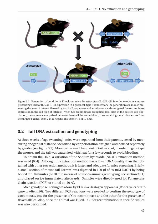
3.2. Tail DNA extraction and genotyping
Figure 3.1: Generation of conditional Knock-out mice for astrocytary IL-6/IL-6R. In order to obtain a mousepresenting a lack of IL-6 or IL-6R expression in a given cell type it is necessary the generation of a mouse pre-senting the gene of interest flanked by two loxP sequences and another one with a targeted Cre recombinaseexpression in the cell type of interest. When Cre recombinase recognizes loxP sites in the desired cell pop-ulation, the sequence comprised between them will be recombined, thus knocking-out critical exons fromthe targeted genes, exon 2 in IL-6 gene and exons 4-6 in IL-6Rα.
3.2 Tail DNA extraction and genotyping
At three weeks of age (weaning), mice were separated from their parents, sexed by mea-suring anogenital distance, identified by ear perforation, weighed and housed separatelyby gender (see figure 3.2). Moreover, a small fragment of tail was cut, in order to genotypethe mouse, and the tail was cauterized with heat for a few seconds to avoid bleeding.
To obtain the DNA, a variation of the Sodium hydroxide (NaOH) extraction methodwas used [634]. Although this extraction method has a lower DNA quality than that ob-tained with other extraction methods, it is faster and adequate for mice screening. Briefly,a small section of mouse tail (<1mm) was digested in 100 μl of 50 mM NaOH by beingboiled for 10 minutes (or 30 min in case of newborn animals genotyping, see section 3.11)and placed on ice immediately afterwards. Samples were directly used for Polymerasechain reaction (PCR) or stored at -20 ºC.
Mice genotype screening was done by PCR in a Stratagen apparatus (RoboCycler Strata-gene gradient 96). Two different PCR reactions were needed to confirm the genotype ofeach mouse, one for the presence of Cre-recombinase and the other for the presence offloxed alleles. Also, once the animal was killed, PCR for recombination in specific tissueswas also performed.
45

3. MATERIALS AND METHODS
Figure 3.2: Gender identification and ear marking. Mice are sexed on the basis of their anogenital distance,which is the distance between the anus and genital papilla. The anogenital distance is smaller in females (A)than in males (B) at all ages (newborns, juveniles and adults). Image C illustrates how ears are marked.
After amplification, samples were maintained at 4 ºC until 3μl of loading buffer (0.25%of bromophenol blue and 40% of sucrose in milliQ water plus a bit of sodium azide toavoid bacterial contamination) were added to each sample. Then samples were loadedand ran in 2 or 2.8% agarose gels depending on the expected distance between bands.Finally, gels were stained (SYBR Safe DNA gel stain, Invitrogen) and bands observed witha uv transiluminator (Safe Imager 2.0, Invitrogen).
3.2.1 Cre recombinase PCR
In order to identify mice containing a Cre recombinase gene under the control of anastrocyte-specific human GFAP promoter element, the chosen genotyping strategy wasto amplify the hGFAP/Cre junction using a primer combination G001/G002 (see table3.1) with a product size of 600 bp. Also, as it is a band/non-band PCR it is useful to havea PCR internal control to know that the PCR has worked correctly, specially in animalswithout the transgenic band. For this purpose, primers for Metallothionein (MT) exon 1detection, p8/p9, a constitutive gene [635], were added to the PCR mix (see table 3.1) andtherefore a band of 178 bp was seen in all samples if the PCR worked well (see figure 3.3).
3.2.2 IL-6 Floxed PCR
In order to identify mice containing loxP sequences surrounding exon 2 of IL-6 gene, thechosen genotyping strategy is to amplify the exon 1/exon 2 junction, containing a loxPsequence, using a primer combination Exon 1F/Exon 2R (see table 3.1) with a productsize of 317 bp in WT alleles and 420 bp in floxed alleles (see figure 3.4).
3.2.3 IL-6Rα Floxed PCR
In order to identify mice containing loxP sequences flanking exons 4-6 of IL-6Rα gene,the chosen genotyping strategy is to amplify the intronic sequence where loxP is locatedusing a primer combination IL-6RaF/ IL-6RaR (see table 3.1) with a product size of 527bp in WT alleles and 671 bp in floxed alleles (see figure 3.5).
46

3.2. Tail DNA extraction and genotyping
Figure 3.3: GFAP-Cre mice PCR. A. This scheme illustrates the genotyping strategy, which is to amplify thehGFAP/Cre junction. B. Optimal cycling conditions for the PCR. C. Photograph of a 2% agarose gel showinga transgenic animal with GFAP-Cre recombinase expression (Tg) and thus, a band of 600 bp, and a non-transgenic animal (c) without the recombinase expression and therefore without the 600 bp band. Bothanimals present a PCR control band for MT exon 1 gene of 178 bp.
Figure 3.4: IL-6 floxed mice PCR. A. This scheme illustrates the genotyping strategy, which is to amplify theexon 1/exon 2 junction containing a loxP sequence in floxed alleles but not in WT alleles. B. Optimal cyclingconditions for the PCR. C. Photograph of a 2% agarose gel showing a IL-6 floxed animal for both allelespresenting a band only at 420 bp (lox/lox) and an animal with a floxed and a WT allele of 420 and 317 bprespectively (lox/wt).
3.2.4 Gene recombination PCR
Several primers were designed for identifying the deletion of the exon 2 upon Cre recom-bination. By using primers Exon 1F/Delta R (see table 3.1) in CNS tissues (see section3.9), the PCR products were aproximately 900, 1000, and 260 bp for the WT, floxed andrecombined alleles, respectively. Recombination was observed in brain and spinal cordbut not in spleen, tail or muscle (see figure 3.6).
47

3. MATERIALS AND METHODS
Figure 3.5: IL-6Ra floxed mice PCR. A. This scheme illustrates the genotyping strategy, amplifying the loxPsequence. B. Optimal cycling conditions for the PCR. C. Photograph from a 2.8% agarose gel showing a IL-6Ra floxed animal for both alleles presenting a band only at 671 bp (lox/lox) and an animal with a floxed anda WT allele of 671 and 527 bp respectively (lox/wt).
Figure 3.6: Astrocytary IL-6 recombination PCR. A. This scheme illustrates the genotyping strategy, amplify-ing the sequence between exon 1 and exon 3 alone (WT allele; 900 bp) or flanked by loxP sequences (floxedallele; 1000 bp) or evidence of recombination (knock out allele; 260 bp). B. Optimal cycling conditions forthe PCR. C (Top). Photograph of a 2.8% agarose gel showing cerebellum tissue samples from left to right ofan heterozygous mice containing both alleles WT and floxed plus the recombination band; a WT/floxed an-imal containing both alleles WT and floxed without recombination; an Ast-IL-6 KO animal showing a strongrecombination band (which in cerebellum frequently overshadows the floxed 1000 bp band); a floxed animalpresenting only the 1000 bp band; and another WT/floxed mice. No recombination band is seen in the samegenotypes in tail (Bottom).
48

3.3. Behavioural tests battery
Table 3.1: Reagents and proportions per DNA sample for PCR reaction. For each PCR reaction, a mix of com-mon different reagents and specific primers are added to 1μl DNA sample and finally a drop of mineral oilis added to the tube to avoid evaporation. Samples are then placed in an automatic thermocycler apparatusfrom Stratagene with specific amplification conditions for each PCR.
3.3 Behavioural tests battery
Prior to starting the behavioral test battery, animals were transferred from the colonyroom to the colony testing room, at least one week before starting any behavioral test,in order to habituate to the new location (see figure 3.7). During this habituation period,mice were moved daily in the home cages to the behavioral testing rooms (located in frontof the colony testing room) and were handled during the first part of the light cycle (8 to12 a.m.) for 3 or 7 days (depending on the experiment). Handling consisted not only inthe habituation to the behavioral testing room for 15-30 min but also in experiencing thesame procedure routine as they were going to experience on test day, as well as being ex-posed to the sound of timers, stopwatches and other mice. Between different behavioraltests mice remained in the colony testing room.
49

3. MATERIALS AND METHODS
Figure 3.7: Schematic chronology for behavioral tests. Animals from 5th backcrossing Ast-IL-6 group onlyperformed HB and PM tests with a separation of 2-days between both tests. In Ast-IL-6 receptor group,animals followed this same scheme. In Ast-IL-6 group from 10th backcrossing, both young and adults/oldwere tested. Young animals were tested as in the scheme. Adult animals group were weaned as normal andthen kept in the colony room being weighed once a week until 8 month-old, when they were transferred tothe colony testing room for handling and habituation. The day after the last handling session they startedthis same behavioral battery but with all tests separated one week from each other. In all cases only malesperformed Resident-intruder test.
Behavioral testing was not performed the same day or the immediate following dayswhen home cages were scheduled for bed changing, as it causes an increase in generalactivity and stress levels which could alter test results. Mice were brought to the test-ing room in their home cages at least 30 minutes prior to the start of behavioral testing.Individual mice were then transported into the testing apparatus. Test room lighting,temperature, and noise levels were consistent for all subjects. Prior to test each mouse,apparatus were cleaned with 5% ethanol and completely dried.
All behavioral tests were carried out and analyzed blindly. The order of animals wascounterbalanced across genotypes and sexes.
3.3.1 Hole-Board
Hole-Board (HB) was originally devised by Boissier and Simon [636], it allows to sep-arately assess locomotion and exploration (measured by Head-dippings (HD)) in miceconfronted with a new environment; it is also a generally used method for screening thepotential anxiolytic character of drugs. HB apparatus consists of a white box (40 x 40 x20 cm) with four 3-cm diameter equidistant holes on the floor. The floor is divided in 16squares of 10 x 10 cm marked with black lines, conforming 12 external squares (aroundthe walls) and 4 internal squares (see figure 3.8). A video camera recorder (Everio G cam-era, JVC) was set up to allow recording of the whole session. This test was performed oneday after the final handling session, during the first part of the light cycle (8 to 12 a.m.).
On the test day, animals were taken to the behavioural room in the same manner as inthe previous handling days, and mice remained in the room for 30 min. Mice were placed
50

3.3. Behavioural tests battery
Figure 3.8: Hole-board (left) and Plus-maze (right) test apparatus.
in a corner of the box. Animals were tested during 5 min; defecations and frequency andduration of head-dipping events (considered as such when an animal inserted the headinto the holes at least to its ear level) were assessed in situ by the investigator. Locomo-tion (number of areas crossed) and the number of rearings (considered as such when ananimal stood completely erect on their hind legs and sniffing) were measured by visual-izing the recorded video with a self developed Excel program (found in supplementarymaterial A.1).
3.3.2 Elevated Plus-Maze
Elevated Plus-Maze (EPM) test, originally devised by File and co-workers [637], is a widelyused rodent model for assessing putative anxiolytic drugs and to characterize geneticand environmental influences on anxiety [638]. It relays on the preference of rodentsfor closed spaces instead of open ones. EPM apparatus consists of four arms (20 cm longand 5 cm wide; divided in 3 rectangles of 6 x 5 cm) connected by a central square of 5 x 5cm. The apparatus is elevated 50 cm above the floor by two metallic legs. Two oppositearms have walls of 15 cm tall (closed arms), whereas the other two only have a small ridgeto prevent slipping of the mice (open arms) (see figure 3.8).
Two, three or seven days (depending on the experiment) after performing the HB test,animals were subjected to the EPM, which was carried out in the same room and with thesame habituation time, during the first part of the light cycle (8 to 12 a.m.). Two animalswere simultaneously tested in two different EPM separated with a black screen to preventsight between mice. The entire procedure (which lasted 5 min) was recorded by camerasplaced above each EPM apparatus.
Mice were gently placed in the center square oriented towards a closed arm to avoidconfounding flight response as open arm exploration [638]. Like in the HB test, the in-vestigator was in the test room during all tests. Different behavioral traits were analyzedusing a self-developed application for Excel software (found in supplementary materialsof A.1) from recorded data. Parameters analyzed were: number of entries and time spentin both open and closed arms (both absolute and as a percentage of total arm entries ortotal time spent in the arms); time spent in the central square, latency of central square
51

3. MATERIALS AND METHODS
first escape, locomotion (crossed squares), number of defecations, number of rearingsand number of protected, unprotected and total head-dips, a valuable ethological de-fensive behavior [639]. Head-dipping behaviour is considered when animal’s head andshoulders are over and below the ridge present in the open arms, in an attempt to exploreunder the maze. Protected HD are recorded when the animal makes a head-dip from thecentre or an adjacent close square of the maze around the edge of a closed arm wall andunprotected HD are those made by mice staying in open arms (open HD).
3.3.3 Tail Suspension test
The Tail suspension test (TST) is a simple screening test for either the behavioral effects ofantidepressants or the individual differences in stress responses in rodents. It is based onthe observation that after initial escape-oriented movements, mice develop an immobileposture when placed in an inescapable stressful situation (mainly is the haemodynamicstress of being hung by their tail) [640].
This behavioral test was performed between 2:00 and 6:00 p.m. Mice were suspendedby the tail (with adhesive tape) in the TST apparatus. The test lasted 6 min and all processwas recorded; results were analyzed in three 2 minutes periods. Up to five simultaneousmice were performed at the same time without visualization between them (see figure3.9).
Figure 3.9: Tail Suspension test.
3.3.4 Morris Water Maze
The Morris water maze (MWM) test is one of the most widely used tasks in behaviouralneuroscience for studying spatial memory and learning in rodents, relying on distal cuesto navigate from random start locations around the perimeter of an open swimming areato locate a submerged escape platform. Spatial learning is assessed across repeated trialsand reference memory is determined by preference for the platform area when the plat-form is absent. Different mechanisms of navigation, learning and memory can be testedin this maze using different training protocols [641].
The MWM tank consists of a propylene round pool (120 cm diameter x 60 cm deep),which is virtually divided in 4 equal quadrants, with a removable platform that can be
52

3.3. Behavioural tests battery
adjusted at different heights (8 cm diameter). It contains water at 22 ºC ±1 made opaqueby the addition of approximately 1cl/l non-toxic liquid latex (Látex Compound EspañolaS.A). Four black cues (12x12 cm) and four white cues (24x24 cm) were placed in the poolwall of each quadrant and in a black curtain surrounding the pool, respectively (see figure3.10). A video camera was placed above the the pool to record the test. It was performedbetween 2:00 and 6:00 p.m.
Figure 3.10: Morris Water Maze test. Spatial learning acquisition
3.3.4.1 Visible platform Test/ Cued learning test (Day 1)
It is a control condition frequently used in the MWM which consist in testing the abilityto learn to swim to a cued goal. The platform was the same as in the hidden platform test,except that it was elevated above the water surface and a visible black ‘flag’ was mountedon it making it easily identifiable from across the pool. The ‘cue’ is designed to allowanimals a direct line-of-sight to the platform. To ensure that animals were using thisproximal cue to locate the platform, both the location of the goal and the starting pointwere moved to new positions in each trial. Mice were given 60 seconds to swim and reachthe visible platform and then allowed to remain on it for 15 seconds. Mice unable tofind or reach the platform in that time, were guided to it and/or helped to climbed it andallowed to remain on it for 15 seconds.
In this test, the only cue that reliably indicates the location of the platform relative tothe start is the cue that is attached to the platform. If subjects are impaired in cued learn-ing, there could be spatial deficits as well, because cued learning requires the same basicabilities (good eyesight, swimming, climbing on the platform, etc.) and the same motiva-tion (escape from water) as in the spatial learning test. This test can be performed beforeor after the spatial test, but doing it before allows the identification of those animals notacquiring the appropriate subordinate skills (swimming ability, climbing the platform,not jumping from it to find another escape route, etc.) before they are presented with thehidden platform test. Therefore, it was performed the first day (only one day) with 4 trials
53

3. MATERIALS AND METHODS
per day separated approximately one hour. Latency to reach the platform was measuredin each trial.
3.3.4.2 Hidden platform test (Days 2-5)
The following day to the visible platform test, the hidden test was initiated. In this phase,mice were challenged to escape from the pool by finding a hidden platform (submerged 1cm below opaque water) using distal cues as spatial references. It is the most basic MWMprocedure. The animal must learn to use distal cues to find the hidden platform.
The platform was in a fixed location relative to the cues across days and mice wereplaced into the water facing the edge of the pool at random start positions across trials (4trials per day). Animals were allowed to freely explore the pool for 60 seconds and afterthat, in case they failed to find the hidden platform, they were guided to it and requiredto remain on it for 15 seconds in all cases. Trials were separated by 60-80 min; duringintervals mice were dried with paper and kept into their cages in the test room. Animalswere trained for 4 consecutive days and the latency to find the hidden platform in eachtrial was recorded.
3.3.4.3 Probe trial test (Days 5-6)
To assess reference memory at the end of learning, a probe trial (without the platform) isgiven. The most common method is to administer one probe trial 24 h after the last ac-quisition day. However, in order to be able to differentiate between short- and long-termmemory, we gave each animal two probe trials, one immediately after the last acquisitiontrial and the other 24 h later [641].
During probe trials, animals were placed in the tank for thirty seconds and then im-mediately removed from water.
3.3.4.4 Spatial reversal test (Days 6-7)
To relocate the platform to another quadrant (usually the opposite one) and subject miceto another set of trials after the spatial learning test is called reversal learning. It revealswhether or not animals can extinguish their initial learning of the platform’s position andacquire a direct path to the new goal position (i.e., cognitive learning flexibility).
In this phase, the protocol was basically the same used in the hidden platform phasebut the platform was relocated to the opposite quadrant of the pool. Mice were trainedon this task only for 2 consecutive days with 4 trials per day (1 min each one) and escapelatency was measured in each trial.
3.3.4.5 Spatial reversal probe trial test (Days 7-8)
As in the hidden platform test, at the end of the reversal test, a 30 seconds reversal probetrial test was given immediately after the last acquisition trial and 24 h later.
54

3.3. Behavioural tests battery
3.3.5 Dominance tube test
As an initial screen for normal social behaviour, mice were tested, between 2:00 and 6:00p.m., in a tube test for social dominance adapted from Lindzey et al. [642] without pre-vious food deprivation nor reward. In this test, two mice of different genotypes (Ast-IL-6KO, floxed or WT) but the same gender were placed head first at opposite ends of a Plexi-glas transparent plastic tube (3 cm inner diameter, 30 cm in length; a size just sufficient topermit one mouse to pass through without reversing the direction) and released simulta-neously (see figure 3.11). The test ended when one of the two mice completely retreatedfrom the tube. The mouse remaining in the tube was designated as the winner (score= 1), and the retreating mouse the loser (score = 0). In some cases neither mouse wasdominant or subordinate, and both mice were considered equivalent. Tests in which onemouse backed up and left the tube without being pushed by the other were not scored.With at least 15 minutes between tests, animals were faced with another opponent ina counterbalanced order in a maximum of five different encounters per animal and thepercentage of victories over the other two genotypes is assessed for each animal.
Figure 3.11: Dominance tube test. Two animals are released at the same time at opposite ends of the tube,they meet in the center and the dominant animal push back the other until the submissive animal is com-pletely retreated from the tube.
3.3.6 Resident-Intruder test
In the resident-intruder test, two rodents are introduced to each other in the home cage ofone of the animals (the resident) and then allowed to interact for a set period of time withthe new animal (the intrudent). The resident-intruder paradigm can be used to studyoffensive aggression, defensive behavior, violence and social stress in rodents [643].
For this test only male subjects were employed. They were all individually housed for15 days before the first aggressive encounter. Body weight and food intake was monitoredduring this two weeks individualised. Bedding was not changed the week before testingin order to left olfactory cues from the resident allowing him to establish its own territory.All encounters were recorded and had a duration of 5 minutes. This test was performedbetween 2:00 and 6:00 p.m. (see figure 3.12).
55

3. MATERIALS AND METHODS
In the first encounter, Ast-IL-6 KO mice and their littermate controls (Ast-IL-6) andAst-IL-6R KO mice and their littermate controls (Ast-IL-6R) mice acted as residents anda CD1 mouse was introduced as an intruder to the same cage and left with the residentmouse for 5 min. In a second encounter one week later, Ast-IL-6 and Ast-IL-6R miceacted, this time, as intruders and were placed with a CD1 male mouse in the cage of thelater. The second CD1 mice confronted was always different to the one from the firstencounter.
Behavior observed was classified between aggressive (latency to the first attack, num-ber of attacks, number of chases, number of tail rattles and number of upright and crou-ched postures) and affiliative (number of anogenital/body/nose sniffs, number of al-logroomings (grooming the partner) and crawl-under behavior were assessed) behaviours.
Figure 3.12: Resident-Intruder test. Different behaviours performed can be observed. A. Anogenital sniffing,B. Upright posture, C. Attack.
3.4 Body weight
Regarding body weight, mice from both genders were weighed at weaning and once aweek thereafter (at the same day and time) until they were sacrificed.
Regarding Ast-IL-6 mice, two experiments were performed. The first one with animalsfrom the fifth back-crossing which were weighed until 15 weeks (N = 45-91 at 3 week-oldand N = 6-18 at 15 week-old). The number of animals per group decreases along time asthey were used in EAE and other experiments, specially around week 10. Also, all animalswere transferred from colony room to the colony testing room at 5 week-old; they alsoreceived handling once a day at 6 week-old and performed Hole-board and Plus-Mazetests at 6 and 7 week-old, respectively.
In a second set of animals from the tenth back-crossing group, they were weigheduntil 25 week-old (N = 15-39), maintaining all animals in the same room along time. Inthis case, animals were not subjected to any location change or any manipulation otherthan cage cleaning and weighing handling. When 25 week-old those mice underwentdifferent behavioural tests (Hole-board, Plus-maze, tail-suspension, Morris water maze,dominance tube test and, just in males, resident-intruder test) and were not weighed
56

3.5. Body temperature
during this period. After 2-week rest, when they were 35 week-old, they were weighedagain before they underwent further experiments. This weight is also added to the study.
Regarding Ast-IL-6R mice, animals were weighed until they were 15 week-old in thesame conditions as for 5th backcrossing Ast-IL-6 mice (N = 21-48 at 3 week-old and N =3-15 at 15 week-old).
3.5 Body temperature
Core body temperature was measured by a lubricated rectal probe (Cibertec) in variousconditions in Ast-IL-6 mice from tenth backcrossing (see figure 3.13). In the first exper-iment it was measured, after proper handling, in seven month-old mice in basal condi-tions between 03:00 to 06:00 p.m., at room temperature (23 ºC±1)(N = 14-33 per genotypeand gender). At 9 months of age, some of these animals (N = 8-10, only males) were keptindividualized in a cold room (8±1 ºC) starting at 9:00 a.m. with no access to water andfood and their temperature was recorded 2, 4 and 6 hours later.
Moreover, other 9 month-old animals (N = 3-8 per genotype and gender) were in-jected ip with either 1 mg/kg LPS (L2880, Sigma) in the morning or saline solution in theafternoon, and temperature was measured just before the injection and 3 hours later. Inanother group of 4-6 month-old animals (N = 3-8 per genotype and gender), their tem-perature in basal conditions was recorded every 3-hours in a 24-hour period (except forthe 3 a.m. measure). They were also injected with 10 μg/kg LPS ip in the morning (8 a.m.)of a different day, and temperature was measured 1, 2 and 3 hours later. Animals werealways kept at room temperature.
Figure 3.13: Body temperature assessment in light and dark conditions.
3.6 Experimental autoimmune encephalomyelitis (EAE)
For the induction of EAE, an animal model of multiple sclerosis, two month-old maleand female mice from Ast-IL-6 5th backcrossing group (N = 11-28, as a summatory of 3
57

3. MATERIALS AND METHODS
separate experiments), three month-old male and female mice from Ast-IL-6 10th back-crossing group (N = 9-19) and three month-old Ast-IL-6R male and female mice (N = 12-14) were used. EAE was induced by active immunization with Myelin OligodendrocyteGlycoprotein fragment 35-55 (MOG35−55) peptide. On day 0, all mice, under isofluraneanesthesia, were injected subcutaneously into the hind flanks with an emulsion of 100 μlMOG35−55 (3 mg/ml) and 100 μl Freund’s Complete Adjuvant (CFA) (Sigma-Aldrich, St.Louis, MO) supplemented with 4 mg/ml Mycobacterium tuberculosis H37RA (Difco, De-troit, MI). In addition, animals received an ip injection of 500ng pertussis toxin (NativeAntigen Company, PT-TNL-100), which was repeated two days after immunization (seeFigure 3.14).
Figure 3.14: EAE induction and symptomatology assessment. A. Examples of non-induced mice perfor-mance: tail from healthy mice is elevated when walking, they have enough strength in the hind legs to sup-port their body and also tail is responsive when a finger is approach in order to avoid falling. B. Schemeof EAE induction showing subcutaneous injections of MOG and M.tuberculosis emulsion (made by repeat-edly passing by two syringes or by placing the solution in a sonicator until ready). C-G. Example of differentclinical symptoms and the clinical score of MOG-induced mice: C. Tail paralysis (1); D. Moderate hind limbparaparesis (2); E. Severe hind limb paraparesis (2.5); F. Partial hind limb paralysis (3); G. hind limb paralysis(3.5).
After immunization, mice were examined daily, weighed and scored for EAE accord-ing to the following clinical criteria: 0 = no signs of disease, 0.5 = partial loss of tail tonus,1 = loss of tail tonus, 2 = moderate hind limb paraparesis, 2.5 = severe hind limb para-paresis, 3 = partial hind limb paralysis, 3.5 = hind limb paralysis, 4 = hind limb paralysisplus partial front leg paralysis, 4.5 = moribund and 5 = death (see Figure 3.14). Finally, foreach animal we determined the time to disease onset (clinical score or ataxia ≥ 1), timeto peak disease, peak-score, cumulative score (sum of all scores from disease onset to day20 or 46), outcome (final score) and grade of remission (difference between peak scoreand outcome).
58

3.7. Cryolesion
3.7 Cryolesion
Two month-old male and female mice from 5th backcrossing group (N = 19-25) and threemonth-old male and female mice from 10th backcrossing group (N = 20-21) of Ast-IL-6KO animals plus controls and four month-old Ast-IL-6R KO plus controls (N = 13-24) wereanalyzed after being lesioned under isoflurane anaesthesia (both genders considered to-gether). Cortical cryogenic injury was performed as previously described [644]. Basically,the skull over the right fronto-parietal cortex was exposed and a cryoinjury produced onthe surface of the brain (without craniotomy) by placing a size-controlled pellet of dry iceonto the skull during 60 seconds. The skin incision was then sutured and treated withiodine solution and animals monitored until they were completely awaken from surgery.Non-lesioned controls were anaesthetized and their skulls exposed but cryoinjury wasnot performed.
To study the volume of brain lesion, damaged area was cut from the beginning to theend in sagittal 8 μm-wide sections in the microtome and one of every 25 sections wasmounted in superfrost slides. Dry sections without staining were examined in a brightfield microscope (Nikon Eclipse 90i) and images were acquired using a Nikon Digitalcamera DXM 1200F and Nikon Act-1 v. 2,70 software. Injured area was measured withthe software ImageJ (NIH) in 20X images (Rasband, 1997). The summation of the injuredarea, in millimeters, of all sections of an animal was performed and multiplied by 0.008(tissue width in mm) and by 25 (number of sections cut between two sections analyzed)to obtain a total injury volume estimation (see figure 3.15).
Figure 3.15: Injured volume assessment. A. Damaged area was cut from the beginning to the end in 8 μm-thick sagittal sections in the microtome and one of every 25 sections was mounted in Superfrost slides. B-C.The lesion of the cortex is clearly identifiable at the microscope without the need of staining and the area ismeasured with ImageJ software.
59

3. MATERIALS AND METHODS
3.8 LPS response
Ast-IL-6 males, from 5th backcrossing (N = 5-9) were injected ip with 1 mg/kg LPS fromEscherichia coli 055:B5 (L2880, Sigma) and killed 1 hour later. IL-6 levels in serum andIL-6 mRNA in brain were assessed by ELISA (see 3.12) and RT-PCR (see 3.10) respectively.
3.9 Animal sacrifice and sample storage
For EAE experiments, mice were sacrificed at days 19, 20, 22 or 46 days post-immunization(dpi) and cryoinjured mice were killed at 3 or 10 days post-lesion (dpl). All mice weresacrificed by rapid decapitation and, if necessary, trunk blood was collected in cold 2ml eppendorfs, centrifuged and serum stored at -80 ºC. Upon brain dissection, the righthemisphere of all mice was fixed for 24h in 4% Paraformaldehyde (PFA) in PBS at 4 ºC,then washed with Phosphate buffered saline (PBS) 1x and stored in 70% ethanol at 4 ºCuntil dehydration and paraffin embedding for Immunohistochemistry (IHC) and Histo-chemistry (HC) analyses; while the left hemisphere was frozen with liquid nitrogen andstored at -80 ºC. Spinal cord was also removed together with the vertebral column andfixed in PFA for 24h, then it was dissected from the column and fixed for 24h more innew PFA and finally washed with PBS and stored in 70% ethanol as in brain storage. Insome experiments spleen and liver were also extracted and frozen with liquid nitrogenand stored at -80 ºC.
Regarding the Australian experiment, six days after an ip Bromodeoxyuridine (BrdU)injection (100 mg/kg in 0.9% saline solution, every 2 h over a 24 h period; Sigma-Aldrich)mice were anaesthetized with xylazine/ketamine and subjected to transcardial perfusionwith 4% PFA in PBS for 10 min. Brain, spinal cord, spleen, liver and a fragment of gutwere resected and post-fixed in the same fixative for four hours, then rinsed with 0.1 MPhosphate buffer (PB) and placed in 30% sucrose solution in 0.1 M PB for 48 h. Follow-ing sucrose equilibration tissues were frozen with 2-methylbutane on dry ice and storedat -80 ºC pending the preparation of frozen sections (30 μm) using a cryomicrotome(Leica). Sections were then placed in OLMOS anti-freezing solution (20% sucrose, 1%polyvinylpyrroline, 30% ethylene glycol in 0.2 M phosphate buffer) and stored at -20 ºC.
3.10 RNA extraccion and Real Time PCR
Total RNA was isolated from frozen cerebellum and spleen tissue of 7 week-old male andfemale mice in basal conditions (only subjected to normal handling procedures) usingTripure reagent (Roche Diagnostics Corporation, Indianapolis, IN) according to the man-ufacturer’s instructions.
Briefly, 90 mg of frozen tissue were homogenized by being placed in a tube containing2 ml of Tripure reagent together with two 3 mm stainless steel balls. High speed oscilla-tion was carried out for a total of 4 min in brains and 6 min in spleens, comprising 30Hz cycles of 2 min separated by a 60 s recovery period in a MM400 (Retsch, Germany).Then, the samples were centrifuged at 12000xg for 10 min and supernatants were ex-
60

3.10. RNA extraccion and Real Time PCR
Gene Forward primer Reverse primer
IL-6 5’-GCTTAATTACACATGTTCTCTGGGAAA-3’ 5’-CAAGTGCATCATCGTTGTTCATAC-3’
TNF-α 5’-ATGGCCCAGACCCTCACA-3’ 5’-TTGCTACGACGTGGGCTACA-3’
IL-1β 5’-GGGCTGCTTCCAAACCTTTG-3’ 5’-TGATACTGCCTGCCTGAAGCTC-3’
β-actin 5’-TACCACAGGCATTGTGATGG-3’ 5’-TTTGATGTCACGCACGATTT-3’
Table 3.2: Primer sequences for real time PCR.
tracted with chloroform. RNA was recovered by isopropanol precipitation, washed in75% ethanol and dissolved in DEPC treated water. Concentration of RNA was measuredspectrophotometrically with a NanoDrop 1000 (Thermo Scientific, CA) and stored at -80 ºC until used. Then, 2.5 μg of total RNA was reverse transcribed into cDNA in a 19μl reaction without RNase Inhibitor using a first-strand cDNA synthesis kit, SuperScriptIII Reverse transcriptase (Invitrogen Corp., Carlsbad, CA) following manufacturer’s direc-tions and stored at -20 ºC. Correct generation of cDNA was assessed by conventional PCRran in a 1% agarose gel with reference gene’s primers (β-actin) and was diluted fivefoldprior to Real-time reverse-transcription PCR (qRT-PCR) amplification.
Analysis of IL-6 expression was carried out in the cerebellum and in the spleen; Table3.2 shows the primer sequences used. Possible compensatory effects of IL-6 deficiencyon other cytokines were analyzed by measuring the expression of IL-1β and TNFα. RT-qPCR for IL-6 and β-actin genes was performed in 96-well plates in a final volume of 20μl containing 131 ng cDNA (5 μl), SYBR Green Master Mix (10 μl)(Bio Rad), 500 nM offorward and reverse primers (Sigma–Aldrich) and sterile distilled water. Reactions wererun in the iCycler MyIQ real-time PCR detection system (Bio-Rad Laboratories, Inc., CA).Subsequent RT-qPCR for TNF-α, IL-1β and β-actin in the cerebellum were performed in384-well plates in a final mix volume of 10 μl containing the exact half proportions ofthe above. Reactions were run in CFX384 real-time PCR detection system (Bio-Rad Lab-oratories, INc.,CA). The cycling conditions comprised a first stage of 5 min at 95 ºC withoptics off, a second stage of 40 cycles at 95 ºC for 10 sec (optics off) and 60 ºC for 30 sec(optics on) and a melt curve of 81 repeats of 10 sec starting from 55 ºC with 0.5 ºC temper-ature increments (optics on). Samples were run in duplicates and non-template controlswere included in each assay. PCR efficiency for both groups of primers and tissues wasassessed to be near 100% by a duplicated standard curve of serial dilutions of cDNA.
To confirm amplification specificity the PCR products from each primer pair weresubjected to agarose gel electrophoresis and resulted in a single product with the desiredlength (β-actin, 200 bp; IL-6, 100 bp). Moreover, a melting curve analysis was performedwhich resulted in single product specific melting temperatures (β-actin, 88 ºC; IL-6, 78.4ºC; IL-1β, 83.5 ºC; TNFα, 81 ºC) and no primer-dimers generation. RT-qPCR data wasobtained and analyzed with the Bio-Rad iQ5 v2.1 software for IL-6 and with Bio-Rad CFXManager 2.1 software for IL-1β and TNFα which automatically determined baseline andthreshold values and used the measured expression level of our reference gene (β-actin)as a normalization factor for cytokine genes; the lower expressor was given a value of 1and the rest of values worked out as a fold-change of that reference.
61
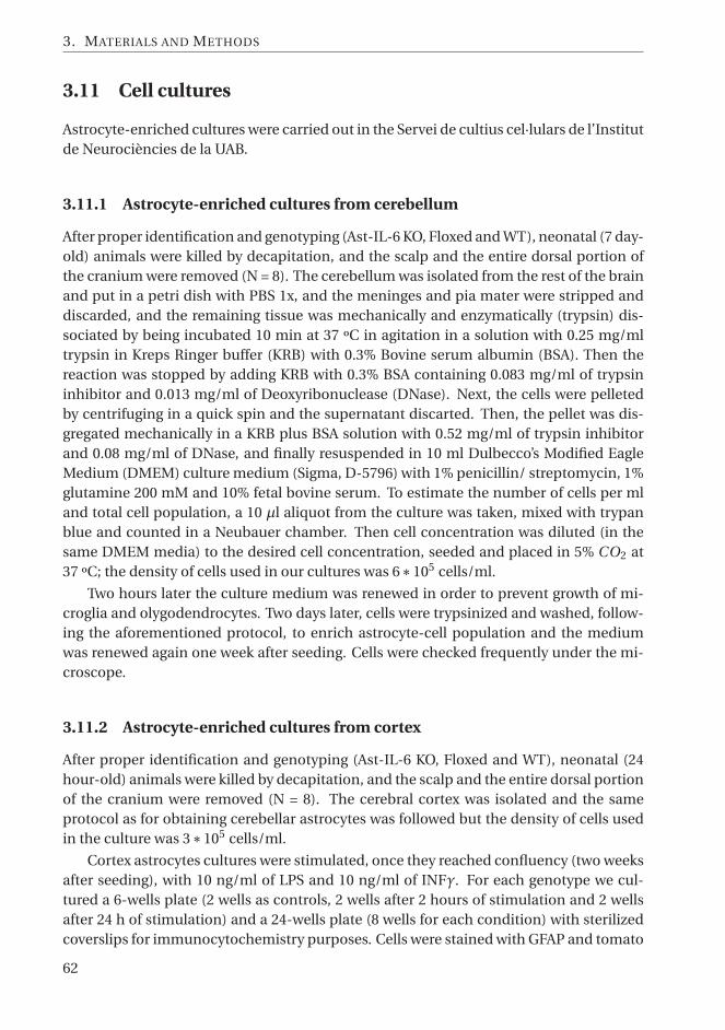
3. MATERIALS AND METHODS
3.11 Cell cultures
Astrocyte-enriched cultures were carried out in the Servei de cultius cel·lulars de l’Institutde Neurociències de la UAB.
3.11.1 Astrocyte-enriched cultures from cerebellum
After proper identification and genotyping (Ast-IL-6 KO, Floxed and WT), neonatal (7 day-old) animals were killed by decapitation, and the scalp and the entire dorsal portion ofthe cranium were removed (N = 8). The cerebellum was isolated from the rest of the brainand put in a petri dish with PBS 1x, and the meninges and pia mater were stripped anddiscarded, and the remaining tissue was mechanically and enzymatically (trypsin) dis-sociated by being incubated 10 min at 37 ºC in agitation in a solution with 0.25 mg/mltrypsin in Kreps Ringer buffer (KRB) with 0.3% Bovine serum albumin (BSA). Then thereaction was stopped by adding KRB with 0.3% BSA containing 0.083 mg/ml of trypsininhibitor and 0.013 mg/ml of Deoxyribonuclease (DNase). Next, the cells were pelletedby centrifuging in a quick spin and the supernatant discarted. Then, the pellet was dis-gregated mechanically in a KRB plus BSA solution with 0.52 mg/ml of trypsin inhibitorand 0.08 mg/ml of DNase, and finally resuspended in 10 ml Dulbecco’s Modified EagleMedium (DMEM) culture medium (Sigma, D-5796) with 1% penicillin/ streptomycin, 1%glutamine 200 mM and 10% fetal bovine serum. To estimate the number of cells per mland total cell population, a 10 μl aliquot from the culture was taken, mixed with trypanblue and counted in a Neubauer chamber. Then cell concentration was diluted (in thesame DMEM media) to the desired cell concentration, seeded and placed in 5% CO2 at37 ºC; the density of cells used in our cultures was 6∗105 cells/ml.
Two hours later the culture medium was renewed in order to prevent growth of mi-croglia and olygodendrocytes. Two days later, cells were trypsinized and washed, follow-ing the aforementioned protocol, to enrich astrocyte-cell population and the mediumwas renewed again one week after seeding. Cells were checked frequently under the mi-croscope.
3.11.2 Astrocyte-enriched cultures from cortex
After proper identification and genotyping (Ast-IL-6 KO, Floxed and WT), neonatal (24hour-old) animals were killed by decapitation, and the scalp and the entire dorsal portionof the cranium were removed (N = 8). The cerebral cortex was isolated and the sameprotocol as for obtaining cerebellar astrocytes was followed but the density of cells usedin the culture was 3∗105 cells/ml.
Cortex astrocytes cultures were stimulated, once they reached confluency (two weeksafter seeding), with 10 ng/ml of LPS and 10 ng/ml of INFγ. For each genotype we cul-tured a 6-wells plate (2 wells as controls, 2 wells after 2 hours of stimulation and 2 wellsafter 24 h of stimulation) and a 24-wells plate (8 wells for each condition) with sterilizedcoverslips for immunocytochemistry purposes. Cells were stained with GFAP and tomato
62

3.12. ELISA
lectin with the same products and concentrations as for embedded paraffin tissues (seesection 3.13).
3.11.3 Splenocytes cultures
Spleens from EAE animals (see section 3.6) were mechanically dissociated and cells wereflushed out from the spleens gently using a syringe plunger and a 21G needle with sterileDulbecco’s PBS 1x with 2% antibiotics (penicillin/ streptomycin) and centrifuged at 4 ºC.The pellet was resuspended and left in RBC lysis buffer (17 mM Tris, 140 mM N H4C l ) toremove erythrocytes. After washing with PBS and further centrifuged, cells were resus-pended in complete medium (RPMI with L-glutamine supplemented with 10% fetal calfserum, 1% antibiotics, 1% sodium piruvate and 0.1% β-mercaptoethanol).
Splenocytes were seeded in 24-well plates at a density of 4∗ 106 cells/ml and somewells were exposed to MOG35−55 (10 μg/ml, 72 h) or LPS (10 μg/ml, 24 h). A colorimetricassay to measure the levels of nitrites in splenocytes culture media was performed usingGriess reagent kit (G-7921, Molecular Probes) following manufacturer’s instructions.
3.12 ELISA
Enzyme-linked immunosorbent assay (ELISA) was used to quantify IL-6 levels in astro-cyte cultures and blood serum samples from mice.
IL-6 determination was done using a commercial murine IL-6 Sandwich ELISA kit(Diaclone) according to manufacturer’s instructions. Absorbance at 450 nm was mea-sured with Labsystems Multiskan Bichromatic microtiter plate reader.
3.13 Histochemistry (HC) and Immunohistochemistry (IHC)
Ast-IL-6 and Ast-IL-6R mice
Embedded paraffin tissues were cut in 8 μm-thick sections in a microtome (Leyca, Ger-many) and mounted in superfrost slides (Thermo scientific).
Before proceeding with the staining protocol, the slides were deparaffinized with xy-lene and passed through a series of decreasing ethanol concentrations until placed in Trisbuffered saline solution (TBS).
For both cryolesioned and EAE-induced mice, microglia was demonstrated by lectinHC (biotinylated tomato lectin from Lycopersicon esculentum, Sigma–Aldrich 1:500 inTBS with 0.5% triton X-100 (TBS-t)). Astrocytes were demonstrated by GFAP IHC (rabbitanti-Glial Fibrillary Acidic Protein from DakoCytomation Denmark, 1:1200 in Blockingbuffer (BB) (0.5% BSA in TBS-t)). Lymphocytes were demonstrated by CD3 IHC (rabbitanti-human CD3, Dako A0452, 1:100 in BB). Spinal cord sections from the EAE experi-ment mice were also stained with Hematoxylin-Eosin and with Luxol Fast Blue (LFB) so-lution (0.1%, overnight at 37 ºC) counterstained with Cresyl violet (0.1%, 1 min) in orderto assess the number of cellular infiltrates and demyelination in white matter.
63

3. MATERIALS AND METHODS
Sections for IHC and HC were preincubated for 1 h with the BB, and then incubatedwith the primary antibodies (GFAP, CD3) or tomato lectin overnight at 4 ºC, followed by1 h at Room temperature (RT) (GFAP, CD3) or at 37 ºC (lectin). For CD3 IHC, a pre-vious antigen retrieval step was performed with protease type XIV (Sigma P5147) 0.1%in TBS at 38 ºC for 10 min. After washing in TBS, sections were incubated with eitherhorseradish peroxidase-coupled streptavidin (Vector Labs, SA-5004, 1:600, 1 h at RT) forHC or biotinylated secondary antibody (Vector Labs, BA-1000, 1:300, 1 h at RT) followedby washes and horseradish peroxidase-coupled streptavidin (Vector Labs, SA-5004, 1:600,1 h at RT) for IHC. The immunoreactivity was visualized by using 0.033% H2O2 in 0.5mg/ml 3,3-diaminobenzidine-tetrahydrochloride (DAB) in Tris buffered solution (TB)0.05 M, pH 7.4 for 4–30 min at RT. Reaction was stopped with TB, washed, dehydrated andmounted in DPX (Sigma, USA). Images at 100x (GFAP) or 200x (Lectin, CD3) were takenin a bright field Nikon Eclipse 90i microscope and acquired with Nikon digital cameraDXm1200F and Nikon Act-1 v. 2,70 software from different brain areas and spinal cord.Finally, to quantify staining areas, intensity and number of cells, images were analyzedusing ImageJ software (NIH) [645]. Histological analyses were performed on at least twosections per mouse. Control sections for non-specific binding analysis (where primaryantibody or tomato lectin was not used) were included routinely. For GFAP IHC, percent-age of stained area or stain quantity was measured in different areas of encephalon (cere-bellum, cortex and hippocampus) from cryolesioned and EAE-induced mice at 100X, be-tween 6 and 12 images per animal depending on the area of study. In the later, white andgrey matter of spinal cords were also analyzed (12 images per animal and area). Thresholdin each area was adapted to better define cells from tissue background. Tomato lectin HCstains both microglia and vessels, so quantification of staining must be complementedwith manual countings of basal (ramified), reactive (protacted cell processes) and fullyactivated (round) microglia and vessels at 200X.
Also, total numbers of microgliosis/ macrophages infiltration areas in spinal cordwere recorded and analyzed. For CD3 IHC percentage of stained area was measured inbrain and spinal cord (10 images per area and animal). Moreover, in cryolesioned micethe number of infiltrating lymphocytes around the lesion was manually counted in twobrain sections using a bright field microscope (Nikon 50i) and for EAE-induced mice, totalnumbers of infiltrates in the white matter of spinal cord were also assessed in two repli-cates. In LFB/cresyl violet staining, total numbers of cellular infiltrates in white matterof spinal cord were counted and afterwards, a color deconvolution plugin developed forImageJ software [646] was used in order to separate colors from LFB and Cresyl Violet tobe able to quantify percentage of LFB staining in spinal cord (see figure 3.16).
GFAP-IL6/sgp130 mice
For IHC and HC, free-floating cryosections (30μm) stored in OLMOS solution, were wa-shed, endogenous peroxidase inhibited by 10 min incubation in 70% methanol plus 2%hydrogen peroxide, washed and pre-incubated with BB (10% fetal bovine serum and 0.3%bovine serum albumin in TBS with 1% triton X-100 (TBS-T) for one hour at RT followedby incubation with rabbit anti-GFAP antibody (Abcam) at 1:2000 dilution in BB overnight
64

3.13. Histochemistry (HC) and Immunohistochemistry (IHC)
Figure 3.16: Demyelination assessment. A. Example of a spinal cord section from a EAE-induced micestained with Luxol Fast Blue and counterstained with Cresyl violet at 100x. Deconvolution plugin [646] isapplied in order to separate cresyl violet dye (B) from LFB color (C). After separation, a threshold is appliedto LFB staining and percentage of occupied area is measured in white matter.
at 4 ºC followed by 1 h at RT. After washing, samples were incubated with a biotinylatedanti-rabbit IgG secondary antibody (Vector Laboratories) at 1:500 dilution in BB for 1 hat RT, washed again and incubated with streptavidin-peroxidase (Vector Laboratories) at1:500 in BB for 1 h at RT. For visualization of GFAP IHC, sections were incubated with DABsolution kit for 3 min according to the Manufacturer’s directions (Vector Laboratories).Following washing, the sections were dehydrated and mounted onto slides with Eukittmounting media (Fluka). For BrdU/doublecortin double staining, a similar IHC protocolwas used, with incubation with doublecortin antibody (Abcam) at 1:8000 in BB and DABincubation, but followed by BrdU IHC. The latter included an initial DNA denaturationstep, with the sections being treated with 1N HCl for 10 min on ice followed by 30 min in2N HCl at 37 ºC, and finally with 0.1M borate buffer (pH 8.5) for 12 min at RT to neutral-ize the HCl. After washing, sections were incubated with rat anti-BrdU antibody (OxfordBiotechnology) at 1:800 in BB overnight at 4 ºC followed by 1 h at RT and, after wash-ing, incubated with a biotinylated anti-rat IgG secondary antibody (Vector Laboratories)at 1:500 in BB for 1 h at RT. After incubation with streptavidin-peroxidase, the sectionswere developed using nickel-DAB following the manufacturer directions (Vector Labora-tories), and mounted onto slides, as above. For lectin HC a similar protocol was followedbut without BB; samples were incubated with biotin-labelled tomato lectin (1:150 in TBS-T) overnight at 4 ºC followed by 1 h at RT, and visualized as above. After mounting ontoslides, these lectin samples were counterstained with Nissl staining.
Stained sections were examined in UAB with a bright field microscope (Nikon Eclipse90i) and images were acquired using a Nikon Digital camera DXM 1200F and Nikon Act-1v. 2,70 software. GFAP and Nissl staining were quantified with the NIH ImageJ (NIH) anal-ysis software. The number of lectin-positive, doublecortin-positive, and BrdU-positivecells, as well as that of blood vessels, was manually counted in specific CNS areas in ablinded manner. In all cases at least three tissue sections per animal were used. For stain-ing quantifications, several images (3-5) per section were taken depending on the brainarea. Since a double staining for lectin and Nissl was carried out, a color deconvolutionplugin for ImageJ [646] was used with all images to separate lectin (DAB) staining fromthat of Nissl.
65

3. MATERIALS AND METHODS
3.14 Statistical analysis
Statistical calculations were performed using the Statistical Package for Social Sciences(SPSS) v. 17.0 for Windows (SPSS Inc).
To analyze genotype frequency at weaning, we performed a Chi-Square test. In allother analysis and for the sake of simplicity, only floxed and knock-out animals were in-cluded in the statistical analysis. However, WT animals are also showed in graphical data.
Data was analysed using a Generalized Linear Model (GzLM) [647] and for the anal-ysis of repeated measurements, such as clinical evaluation, body weight changes, coldresistance or MWM learning acquisition (among others), a GzLM test for repeated mea-sures, the Generalized Estimating Equations (GEE) [648] test was used. In both cases,genotype, gender and age or sacrifice day (when applicable) were used as main factors.In addition, in GEE analysis effect of time (day) was used as within-subject factor. GzLMand GEE tests are a more flexible statistical tool than the standard general lineal model(GLM) because several types of data distribution can be chosen; the analysis can run evenif there are missing data in the repeated measures; and they do not need homogeneity ofvariance.
In all cases, if interaction between any factor was found significant, it was decom-posed pairwise and corrected by Sequential Bonferroni for multiple comparisons [649].In few cases where either gender or age factors were found significant without any inter-action, then data was split for this factor and GzLM or GEE were performed separately ineach group. In all cases Ast-IL-6 and Ast-IL-6R groups were analysed separately and, inAst-IL-6 mice, 5th and 10th backcrossing groups were also analysed independently.
To validate the GzLM, several parameters were also analyzed using ANOVA and Stu-dent’s t test, with similar results obtained. Occasionally, results after sequential Bonfer-roni post-hoc analysis were compared with those obtained with Fisher’s least significantdifference (LSD) post-hoc test.
Values in text and figures shown are mean ± standard error of the mean (SEM) andthe number of animals per group (N). Statistical significance was defined as P ≤ 0.05 andmarginal significance as P ≤ 0.1. Data presentation was carried out using Graph PadPrism Software (version 5). In figures, for the sake of simplicity, we show the most relevantdata, indicating only differences with the appropriate control for genotype (�), age (�)and day of sacrifice (�) factors, as well as the decomposition of significant interactionswith genotype (∗).
66

CH
AP
TE
R 4444444444444444444444444444444444CHARACTERIZATION OF ASTROCYTE-DERIVED
IL-6 DEFICIENCY
4.1 Gene recombination and IL-6 brain levels
Recombination of IL-6 exon 2 gene (see section 3.2.4) was observed in CNS tissues, brainand spinal cord, of Ast-IL-6 KO and heterozygous mice from both 5th and 10th C57BL/6backcrossings, but not in spleen, tail or muscle.
Additionally, analysis of IL-6 expression by RT-qPCR were carried out in the cerebel-lum and in the spleen of young naive animals from 5th backcrossing; Table 3.2 showsthe primer sequences used. We observed a decrease of IL-6 expression in the cerebellumbut not in the spleen of Ast-IL6 KO mice, although this was statistically significant onlyin comparison with WT mice (4.1). Possible compensatory effects of IL-6 deficiency onother cytokines were analyzed by measuring the expression of IL-1β and TNFα in bothcerebellum and spleen; there was a non-significant tendency for both cytokines to be up-regulated in the cerebellum of Ast-IL-6 KO mice and a significant increase of TNFα inspleen of Ast-IL-6 KO respect floxed mice.
IL-6 levels in brain and spinal cord were also intended to examine by IL-6 IHC (sc-1265-R, Santa Cruz Biotechnology) in both paraffin and free-floating sections without anysuccess.
4.2 Frequencies at weaning
A total of 587 and 517 Ast-IL-6 mice from fifth and tenth backcrossings, respectively, wereproduced and genotyped (Table 4.1). There were no abnormalities in the distribution ofsexes, but the distribution of genotypes did not follow a Mendelian ratio, being Ast IL-6KO and heterozygous mice clearly under-represented at the moment of weaning at both5th and 10th backcrossings (P ≤ 0.01).
In general, each breeding pair had 5–7 offspring. Mortality between birth and wean-ing, assessed in the 10th backcrossing group, was equally distributed among genotypes(~10%). After weaning and up to four or ten months of age, very few deaths were ob-
67
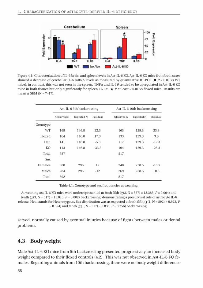
4. CHARACTERIZATION OF ASTROCYTE-DERIVED IL-6 DEFICIENCY
Figure 4.1: Characterization of IL-6 brain and spleen levels in Ast-IL-6 KO. Ast-IL-6 KO mice from both sexesshowed a decrease of cerebellar IL-6 mRNA levels as measured by quantitative RT-PCR (■ P < 0.01 vs WTmice); in contrast, this was not seen in the spleen. TNFα and IL-1β tended to be upregulated in Ast-IL-6 KOmice in both tissues but only significantly for spleen TNFα. � P at least < 0.01 vs floxed mice. Results aremean ± SEM (N = 7–17).
Ast-IL-6 5th backcrossing Ast-IL-6 10th backcrossing
Observed N Expected N Residual Observed N Expected N Residual
Genotype
WT 169 146.8 22.3 163 129.3 33.8
Floxed 164 146.8 17.3 133 129.3 3.8
Het. 141 146.8 -5.8 117 129.3 -12.3
KO 113 146.8 -33.8 104 129.3 -25.3
Total 587 517
Sex
Females 308 296 12 248 258.5 -10.5
Males 284 296 -12 269 258.5 10.5
Total 592 517
Table 4.1: Genotype and sex frequencies at weaning.
At weaning Ast IL-6 KO mice were underrepresented at both fifth (χ(3, N = 587) = 13.388, P = 0.004) andtenth (χ(3, N = 517) = 15.015, P = 0.002) backcrossing, demonstrating a prosurvival role of astrocyte IL-6
release. Het. stands for Heterozygous. Sex distribution was as expected at both fifth (χ(1, N = 592) = 0.973, P= 0.324) and tenth (χ(1, N = 517) = 0.835, P = 0.356) backcrossing.
served, normally caused by eventual injuries because of fights between males or dentalproblems.
4.3 Body weight
Male Ast-IL-6 KO mice from 5th backcrossing presented progressively an increased bodyweight compared to their floxed controls (4.2). This was not observed in Ast-IL-6 KO fe-males. Regarding animals from 10th backcrossing, there were no body weight differences
68

4.3. Body weight
between genotypes in females and, in males, the effect of IL-6 deficiency was weaker andoccurred later, from week 23 onward.
The GEE statistical analysis for floxed and Ast-IL-6 KO mice revealed that both timeand sex had a very significant effect in both backcrossing groups (P ≤ 0.001 in all cases,increasing weight with age and weighting males more than females). Genotype effect wasonly significant in fifth backcrossing group (P ≤ 0.001) but interaction between genotype,age and time was significant in both 5th and 10th backcrossings (P ≤ 0.01 and 0.001, re-spectively). After decomposing the genotype and time interaction in 5th backcrossinggroup, we can observe that differences between Ast-IL-6 KO and floxed mice are only sta-tistically significant from week 12 onwards in the sequential bonferroni test (4.2, for thesake of clarity, significant genotype effects have not been marked in the figure). However,if a less rigid test like LSD post-hoc is applied, significant differences start from week 8onwards. When decomposing the genotype per time interaction in the 10th backcrossinggroup, there are no significant differences regardless the post-hoc test used, presumablybecause differences are only in the latter age while all other weeks body weights are iden-tical. A GEE test considering body weight only from week 23 onwards is performed inmales, resulting in a significant effect of genoype (P ≤ 0.05) (4.2).
Figure 4.2: Body weight growth is affected by astrocyte IL-6 deficiency in a sex-dependent manner. Mice wereweighed weekly (N = 49–91 at 3 weeks and N = 6–18 at 15 weeks in the fifth backcrossing, and N = 15-39 attenth backcrossing).∗ P at least ≤ 0.05 vs floxed mice after decomposition of genotype and time interactionwith sequential Bonferroni post-hoc; � P at least ≤ 0.05 vs floxed mice.
Food intake was measured in a small group of 2 month-old males (N = 7-11) from
69

4. CHARACTERIZATION OF ASTROCYTE-DERIVED IL-6 DEFICIENCY
tenth backcrossing after individualization over a 15-days period before resident-intruderbehavioural tests. Average daily food intake was only marginally increased in Ast-IL-6 KOmale respect floxed but not WT mice (Ast-IL-6 KO: 3.59 ± 0.07; Floxed: 3.24 ± 0.17; WT:3.50 ± 0.11; P = 0.075) in a Generalized Linear model. Unfortunately, data from individu-alized older mice was not assessed.
4.4 Behavioural tests
4.4.1 Hole-Board
The behavioral traits observed in the Hole-Board are shown in Figure 4.3 and statisticalresults are shown in Table 4.2.
Horizontal exploratory activity was measured by counting total squares crossed byanimals and it is shown separated by external (squares adjacent to a wall) and internalambulations. Total ambulation was clearly decreased in Ast-IL-6 KO mice compared tofloxed mice (Ast-IL-6 KO: 75.95 ± 4.58 and 47.13 ± 3.14; floxed: 88.94 ± 3.70 and 69.47 ±3.13; P ≤ 0.05 and 0.001 for 5th and 10th backcrossing, respectively). The separate analy-sis of the external and internal activities revealed a consistent decrease in external activityof Ast-IL-6 KO animals in all conditions, regardless of sex, age and backcrossing. Inter-nal deambulations, more related to anxiety, were also decreased in Ast-IL-6 KO animalsrespect to floxed mice, especially in animals from the 10th backcrossing group. No differ-ences due to sex effects was seen in 5th backcrossing animals but in the 10th backcrossinggroup external deambulations were increased in females and both internal and externalambulations were increased in old-adults compared to young mice.
In contrast to horizontal activity, vertical exploratory activity (rearings) was only sig-nificantly decreased in Ast-IL-6 KO males from 10th backcrossing. Sex did not have aclear-cut effect, as females from 5th backcrossing showed less rearings compared to malesbut in 10th backcrossing the inverse situation was seen.
The exploratory activity was further analyzed by looking at the number of head-dipsand the time head-dipping. A clear sex-, age- and backcrossing-dependent effect of as-trocyte IL-6 deficiency was observed, since in 5th backcrossing group head-dipping be-havior was significantly altered in male mice only in 10th backcrossing group and the op-posite was observed in young female mice. There were some differences between malesand females in the overall head-dipping behavior in 5th backcrossing group (floxed fe-males presented increased numbers compared to floxed males but Ast-IL-6 KO presentedsame levels in both genders); but again this does not seem a consistent sex difference asit was not observed in animals from 10th backcrossing. The latter did show differencesdue to age, as young animals performed increased numbers and time of head-dipping.However, this age-related differences in the number and time of head-dippings occurredexclusively in floxed mice, specifically in females, as showed after decomposing genotypeand age; and genotype, age and sex interactions.
Other important parameters to assess emotionality are the number of defecationsand those of squares not explored. Ast-IL-6 KO showed, in general, a significant increasein defecations compared to floxed mice, especially at younger ages in 10th backcrossing.
70

4.4. Behavioural tests
Ast-IL-6 5th backcrossing Ast-IL-6 10th backcrossing
Genotype (G) Sex (s) Gs Genotype (G) Sex (s) Age (a) Interactions
Hole-Board
Deambul. * - - *** * *** Gs = *
External * - - *** * *** -
Internal - - * *** - * -
Rearings - * - - * - Gs = *
HDn - * ** * - ** Gsa = **
HDt ** *** * - - *** Gsa = *
Def. - * - *** * ** Ga = **
Unexpl. * - * *** - * Gs = *
Plus-Maze
CEn ** - - *** - - -
TO - - * - - - Ga = ***
TCe * - - *** - - Gsa = **
Def. - * - *** - - -
Rearings - - * * - *** sa = *
HDprot * - - *** - - Gsa = *
HDopen * - ** - - - Ga = ***
Table 4.2: Summary of significances obtained in Hole-board and Plus-maze behavioral tests.
Gs, Gsa, Ga and sa denote interaction between genotype and sex; genotype, sex and age; genotype and age;or sex and age, respectively. Deambul. stands for total deambulation, HDn and HDt stand for number andtime of head dipping, Def for defecations, Unexpl. for number of non-visited squares, CEn for number of
close arm entries, TO for time spent in open arms, TCe for time spent in central square, HDprot for numberof protected head-dips and HDopen for number of unprotected head-dips. *, ** and *** P at least ≤ 0.05, ≤
0.01 and ≤ 0.001 respectively.
Defecation was increased in young mice compared to 8-month animals but only in Ast-IL-6 KO, as shown after the decomposition of genotype and age interaction. Ast-IL-6 KOmice, mostly males, showed a significant increase in the number of unexplored squares,which were also affected by age, leaving young mice more unexplored squares comparedto adults.
4.4.2 Elevated Plus-Maze
The behavioral traits observed in the EPM are shown in Figure 4.4 and statistical resultsare shown in Table 4.2.
In accordance with the hole-board results, the general locomotor activity assessed asnumber of entries into the closed arms [650] (Figure 4.4) and total number of squarescrossed (Supplementary Figure A.11) was significantly decreased in the Ast-IL-6 KO micecompared to floxed mice regardless of sex, age and backcrossing number.
71

4. CHARACTERIZATION OF ASTROCYTE-DERIVED IL-6 DEFICIENCY
Figure 4.3: Behavioral traits analyzed in the Hole-board apparatus. Ambulation (External and internal, asnumber of external or internal squares crossed), vertical activity (number of rearings), exploratory behavior(number of head-dips (HD nº) and time (seconds) spent head-dipping (HD t)), defecations (Def.) and num-ber of non-explored squares are shown for Ast-IL-6 KO, floxed and WT mice for both 5th (6 week-old) and10th backcrossings (5 week-old and 8 month-old). (N = 11–37). � and genotype effect P at least ≤ 0.05 or≤ 0.1 vs floxed mice, � P at least ≤ 0.05 vs young mice and∗ P at least ≤ 0.05 vs floxed mice after genotypeinteraction decomposition.
72

4.4. Behavioural tests
Figure 4.4: Behavioral traits analyzed in the Plus-maze apparatus. Number of entries to closed arms, timespent in open arms, time spent in the central square, defecations, number of rearings and number of bothprotected and unprotected (open) head-dips are shown for Ast-IL-6 KO, floxed and WT mice for both 5th(6 week-old) and 10th backcrossings (5 week-old and 8 month-old). (N = 11–37). � and � P at least ≤0.05 vs floxed or young mice respectively. ∗ P at least ≤ 0.05 vs floxed mice after genotype interactiondecomposition.
73

4. CHARACTERIZATION OF ASTROCYTE-DERIVED IL-6 DEFICIENCY
The anxiety level was assessed by measuring the number of entries and time spentinto the open arms and calculating the percentage they represent respect total entriesor total time spent in all arms of the EPM [650], as well as the time spent in the centralsquare of the plus-maze apparatus. Due to the redundancy of these data only time spentin open arms and time spent in the center are plotted in Figure 4.4, the percentage of timeand entries in open arms as well as time spent in close arms are presented in Supplemen-tary materials (Figure A.11 and Table A.1). There is a strong interaction between age andgenotype in time and percentage of time spent and entries in the open arms, tendingto be increased in young Ast-IL-6 KO mice (significant for Ast-IL-6 males from 5th back-crossing group) but significantly decreased only in 8 month-old mice compared to floxedmice; showing a clear age effect as 8 months-old mice presented an opposite phenotypeas young (5-6 week-old) mice regardless their backcrossing group. In relation with timespent in the central square, it was decreased in Ast-IL-6 KO mice respect to floxed animalsindependent of sex, age and backcrossing. However, no differences in latency time of thefirst escape from central square were found between genotypes (see Figure A.11).
Although the EPM is normally used for testing anxiety, other less conventional traitscan be assessed, such as rearings and head-dips (total, protected and unprotected) [651],which can be compared with the results observed in the hole-board. Overall, the resultsof the EPM were quite consistent with those of the hole-board despite the different na-ture of the two apparatus. Thus, the number of rearings, which in hole-board was onlydecreased in Ast-IL-6 KO males, in EPM was decreased in both genders and ages in Ast-IL-6 KO mice from 10th backcrossing group, parallelling deambulation results. However,only in EPM test we observed an age effect in rearing behaviour, being increased in 8-months males in relation with 6 week-old ones. Regarding HD behavior, the total num-ber was decreased only in 8 month-old Ast-IL-6 KO mice respect to floxed animals (seeFigure A.11), but when differentiating them into protected and unprotected head-dips,we can see that Ast-IL-6 KO mice showed a decrease of protected head-dips regardless ofage, sex or backcrossing group. Oppositely, young Ast-IL-6 KO mice show an increase ofthe unprotected ones (only males in the 5th backcrossing but both genders in the 10thbackcrossing), whereas old Ast-IL-6 KO mice show a decrease respect to floxed controls,showing again a clear age effect. This is suggestive that young Ast-IL-6 KO mice are moreprone to exploration in dangerous situations than floxed males, while old/adults Ast-IL-6KO are less willing to do it.
Finally, defecations were strongly increased in both genders and ages of tenth back-crossing Ast-IL-6 KO mice compared to their respective floxed mice. In relation to thattrait, we are not able to truly compare 5th with 10th backcrossing groups as, due to meth-odological differences, 5th backcrossing animals were weighed just before EPM and thusdefecating in the other "new" environment likely altering our results.
4.4.3 Tail Suspension
Time mice spent in motion (agitation time) while being suspended by their tail through-out the experimental condition is shown in Figure 4.5. There are no significant effects ofgenotype or sex but a significant effect of time (P ≤ 0.001) showing that all animals stayed
74

4.4. Behavioural tests
in motion significantly more time in the first 2-min block than in the other two, as usu-ally described for this test. We also found a significant interaction between age and timeof the test (P ≤ 0.001) showing that during first 2 minutes 8 month-old animals agitatedmore than young animals (P ≤ 0.001) while in the middle and last 2 minutes blocks thissituation was reversed (P ≤ 0.01 and 0.001, respectively), being young animals the oneswho stayed in motion more time.
Moreover, there was also a significant interaction between genotype and time (P ≤0.05) and a nearly significant interaction between genotype, sex, age and time (P = 0.052).Decomposition of these interactions with Sequential Bonferroni did not show any signif-icant differences between Ast-IL-6 KO and floxed due to the severity of this post-hoc test.However, it is noteworth to point that with LSD post-hoc test a small significant increasein agitation was found in 8 month-old Ast-IL-6 KO male respect to floxed controls in thefirst and second blocks of 2-minutes test (P ≤ 0.05).
Finally, we applied a GEE test for each condition (young males, adult males, youngfemales and adult females) showing a significant effect of genotype in adult males (P ≤0.05), being Ast-IL-6 KO males more time in agitation compared to floxed controls (4.5).
Figure 4.5: Time spent in motion during tail suspension test. Total duration of the test is shown in 3 blocksof two minutes and agitation time (in seconds) is presented (N = 11–32). � P at least ≤ 0.05 vs floxed mice.
4.4.4 Morris water maze
The behavioral traits observed in the Morris water maze are shown in figures 4.6 - 4.10;and several statistical results are shown in Table 4.3. Young (7 week-old, N= 11-23 for each
75

4. CHARACTERIZATION OF ASTROCYTE-DERIVED IL-6 DEFICIENCY
Ast-IL-6 10th backcrossing
Genotype (G) Sex (s) Age (a) Day (d) Interactions
Cued learning
Time plat. *** - - n/a Ga = ***
Spatial learning
Time plat. ** - *** *** -
Probe 1h * - *** n/a -
Probe 24h - - *** n/a -
Reversal learning
Time plat. * - *** *** Gad = *
Probe new 1h - - - n/a Gs = *
Probe new 24h * - - n/a -
Probe old 1h * - - n/a -
Probe old 24h ** - - n/a -
Table 4.3: Summary of obtained significances in Morris water maze test.
Ga, Gad and Gs denote interaction between genotype and age; genotype, age and day or genotype and sex,respectively. Time plat. stand for time needed to reach the platform and, in reversal learning, probe new
refers to percent of time spent in the quadrant that allocated the platform during reversal learning, whereasold refers to the quadrant that allocated it during spatial learning. *, ** and *** P at least ≤ 0.05, ≤ 0.01 and ≤
0.001. n/a not applicable.
sex) and old-adults (8 month-old, N= 11-21 for each sex) from 10th backcrossing grouphave been studied.
As stated in Section 3.3.4, the first test protocol that we performed in MWM was Cuedlearning, testing the animals for their ability to learn to swim to a cued goal. Animalswere given four trials; a mean value was calculated and is presented in Figure 4.6. Ast-IL-6 KO mice showed a significant impairment in cued learning needing more time to findthe platform. Also, a significant interaction between sex and age was found, showing thatonly in females, 8 month-old animals performed significantly worse than young ones.Moreover, 7 Ast-IL-6 KO mice out of 69, 4 floxed mice out of 89 and 0 WT out of 74 wereincapable of climbing the platform in any trial. However, these non-performers animalswere not excluded from the test because in the following test protocols, all of them wereable to find and climb the submerged platform. We did try to avoid poor eyesight by ex-cluding animals with visible alterations of the eyes (one eye permanently closed, cataractor excessive rheum), we also discarded locomotor deficits altering swimming and pooreyesight by checking mean swimming speed in a subset of animals (N = 10-14 per geno-type and sex in young animals) with SMART v2.5.21 (Panlab S.L.U) with no differencesbetween genotypes (Ast-IL-6 KO: 23.81 ± 0.88; Floxed: 23.80 ± 0.68; P = 0.841) but a sexeffect, with a significant increase in swimming speed in males respect to females (males:24.72 ± 0.65, females: 22.84 ± 0.77 cm/s; P ≤ 0.05).
In first trials many animals did not want to swim away from the wall or quickly jumpedback into the water once they climbed the platform, but this was clearly improved at the
76

4.4. Behavioural tests
end of cued learning test. Animals performing floating behavior at some point one ormore test-days were equally distributed among genotypes (20% in Ast-IL-6 KO mice and18% in Floxed mice) and, as they also presented periods of swimming, they were not ex-cluded from the experiment.
Figure 4.6: Time to climb to the visible platform in MWT. Tenth backcrossing animals are showed separatedby sex and age (7 weeks and 8 month) (N = 11–23). � P at least ≤ 0.05 vs floxed mice.
The second protocol was to test spatial learning acquisition. Animals were given fourtrials a day during four days and the mean for each trial is showed in Figure 4.7, wherewe can observe that animals are learning the spatial location of the platform as the timeto reach it is significantly diminished daily. Although they were all learning, Ast-IL-6 KOmice showed a significant increase in the amount of time needed to find the platformrespect to floxed mice. Moreover, an important age effect is seen showing that 8 month-old mice spent significant more time to reach the platform compared to 7 week-old ones.There is a marginally significant interaction between sex and day (P = 0.073) that togetherwith the visible absence of genotype effect in males made us analysed both genders sepa-rately applying a GEE for each sex, confirming that only in females there was a significanteffect of genotype (P ≤ 0.01), although both genders still present significant age and dayeffects (P ≤ 0.01).
To assess reference memory at the end of those 4 days learning, a probe trial (a 30seconds trial without platform) was carried out 1 hour after last trial and 24 hours later, inorder to differentiate between short- and long-term memory. Percentage of time spent inthe quadrant which previously allocated the platform was presented in Figure 4.8; theseresults show a clear deficit in spatial memory in older animals, which stayed in the 25%borderline limit of chance, in relation with younger animals. Regarding genotype differ-ences, Ast-IL-6 KO mice showed impaired memory only with probe trial carried out 1hour after the last training session but not with that 24 hours later.
Immediately after the second probe trial, animals performed two days of reversallearning with four trials a day, with the platform located in the opposite quadrant to thatin initial spatial learning acquisition. Mean results for learning days are shown (4.9). Micedid learn the new location as they needed significant less time to find the platform in thesecond day. Again, we see important differences between younger and older animals, be-ing learning impaired in the latter, and Ast-IL-6 KO mice also showed a significant poorer
77

4. CHARACTERIZATION OF ASTROCYTE-DERIVED IL-6 DEFICIENCY
Figure 4.7: Mean time needed to reach the submerged platform. (N = 11–23). Data analyzed separately bysex. � P at least ≤ 0.05 vs floxed mice.
Figure 4.8: Percentage of time spent in the platform quadrant during Probe trials. (N = 11–23). In the topdata is shown from the probe trial given 1 hour after last trial and, at the bottom, data from probe trial made24 hours later. � and � P at least ≤ 0.05 vs floxed mice and young animals, respectively.
performance than floxed controls. However, the decomposition of the significant inter-action between genotype, age and day found, shown that learning deficits in Ast-IL-6 KOmice were present only in 8 month-old animals in the second day of training.
Once reversal learning was finished animals were also given two probe trial 1 and 24hours later the last trial. In those probe trials it was measured not only the percentage of
78

4.4. Behavioural tests
Figure 4.9: Mean time needed to reach the submerged platform in a new location. (N = 11–23). � at least P≤ 0.05 vs floxed mice.∗ P at least ≤ 0.05 vs floxed mice after genotype interaction decomposition.
time spent in the quadrant with the newer location of the platform (the opposite from thespatial acquisition) but also the percentage of time spent in the old platform quadrant(the one that allocated the platform during spatial acquisition). Results, seen in Figure4.10, show that animals barely pass borderline 25% chance line, not remembering thenew location of the platform, in that case with any age effect. Also, Ast-IL-6 KO micestayed in the second probe trial (24 h later) even less time in this quadrant respect tofloxed mice. However, when analysing time spent in the quadrant with the old locationof the platform we see it is still largely revisited and, specialy Ast-IL-6 KO mice, spentan increased amount of time searching for the old location platform in comparison withfloxed animals.
As a general comment, in MWM we have not obtained significant differences betweengenders in any variable.
4.4.5 Dominance tube
Young (8 week-old) animals from tenth backcrossing group have been studied in thedominance tube test and results, presented as percentage of victories over all duels, areshown separately by sex in Figure 4.11. Although there was an overall significant increasein the percent of victories of Ast-IL-6 KO versus floxed mice (KO: 73.39 ±6.57; Floxed:30.53 ±6.87, P ≤ 0.001) there was a strong interaction between genotype and sex (P ≤0.01) since percent of victories in Ast-IL-6 KO is only significant increased in females.
Percent of victories of Ast-IL-6 KO and floxed mice when they were faced with WTmice was not affected by neither genotype nor sex (percent of victories over WT: KO: 53.84± 10.18; Floxed: 50.09 ± 10.18, P = 0.794 and 0.210 for genotype and sex, respectively; N =
79

4. CHARACTERIZATION OF ASTROCYTE-DERIVED IL-6 DEFICIENCY
Figure 4.10: Percentage of time spent in both new and older platform quadrant during reversal probe trials.(N = 11–23). In the first two panels it is shown the percent of time spent in the quadrant that allocated theplatform during reversal learning (the new location) for 1 and 24 hours probe trials. In the two bottom panelsit is presented the percent of time spent in the quadrant that allocated the platform during spatial learning(the old location) for 1 and 24 hours probe trials. � P at least ≤ 0.05 vs floxed mice.
7-13).
4.4.6 Resident Intruder
The behavioral traits observed in the resident-intruder test are shown in Figure 4.12 andstatistical results analyzed with GzLM and sequential Bonferroni (separately for residentand intruder condition) for genotype and age, as main factors, are presented in Table 4.4.Young (10-11 week-old; N = 7-11) and adult/old males (9 month-old; N = 8-10) from 10thbackcrossing group have been studied.
Regarding aggressive behaviour, in the latency to first attack there was a significantinteraction between genotype and age when Ast-IL-6 mice acted as intruders, since ittended to be decreased in adult Ast-IL-6 KO but increased in young KOs respect to floxedcontrols. However, the interaction decomposition was only significant with LSD post-hoctest (and marginally significant with sequential Bonferroni test, P = 0.061) in young ani-
80

4.5. Body temperature
Figure 4.11: Percentage of victories in the dominance tube test. Data showing results of Ast-IL-6 KO andfloxed encounters in the tube test. (N = 11-16). � P at least ≤ 0.05 vs floxed mice. ∗ P at least ≤ 0.05 vsfloxed mice after genotype interaction decomposition.
mals. Consistent with that is the data from the number of attacks, which also showed asignificant interaction between genotype and age in intruders, due to the fact that adultAst-IL-6 KO fighted more than floxed mice while young KOs fighted less. However, the in-teraction decomposition was again only significant in young mice with LSD post-hoc test(and marginally significant with sequential Bonferroni test, P = 0.066). Other agressivebehaviour such as active and passive chases, tail rattles, crouched postures and uprightpostures were analysed and presented together, showing no differences between animalswhen acting as a resident but, acting as intruders, 9 month-old mice showed a decreasedof this aggressive behaviour and a significant interaction between genotype and age wasfound, as it was increased in adult Ast-IL-6 KO and decreased in young Ast-IL-6 KO incomparison with floxed animals, althougth the interaction decompositin was not signifi-cant.
When analysing affiliative behaviour (nose/body/ano-genital sniffs, crawl unders andallogrooming) there were no differences regarding age or genotype found. However, crawl-under behaviour was marginally significant for an interaction between genotype and age(P = 0.089) and marginally decreased in adults respect to young mice (P = 0.071) whenacting as residents.
Floxed and WT mice presented an overall similar phenotype in this test. We did findimportant differences regarding condition (resident or intruder), although being anal-ysed separately, if compared we found a significant increase in aggressive behaviour anda significant decrease in affiliative behaviour in intruder mice compared to residents.
4.5 Body temperature
Body temperature measured between 03:00 to 06:00 PM, in 7 month-old basal mice inthe afternoon did not show differences due to genotype, sex or interaction between themwhen it was analysed by a GzLM with genotype and sex as main factors (WT: 36.13 ±0.11ºC ; FLoxed = 36.29 ±0.9 ºC; Ast-IL-6 KO = 36.32 ±0.15 ºC).
81

4. CHARACTERIZATION OF ASTROCYTE-DERIVED IL-6 DEFICIENCY
Figure 4.12: Behavioral traits analyzed in the Resident-intruder test. Latency to the first attack (seconds),number of attacks, number of aggressive behaviour other than attacks (active and passive chases, tail rattles,crouched and upright postures) and number of affiliative behaviours separated in sniffs (nose, body and ano-genital), crawl unders and allogroomings, are shown for 10-11 week-old and 9 month-old mice (N = 7-11). �P at least ≤ 0.05 vs young mice.∗ P at least ≤ 0.07 vs floxed mice after genotype interaction decomposition.
Regarding the response to exposure to cold environment (8±1 ºC), a significant de-crease of body temperature (P ≤ 0.001) was observed 4 and 6 hours after cold exposureregardless of genotype, with GEE statistical analysis. Body weight was also significantlydecreased at the end of the cold exposure (P ≤ 0.001) without differences between geno-types (Figure 4.13).
Regarding body temperature measurements after 1 mg/kg LPS or saline injection, a
82

4.5. Body temperature
Ast-IL-6 10th backcrossing
Resident Intruder
Genotype (G) Age (a) Ga Genotype (G) Age (a) Ga
Latency attack - * - - - *
Nº attacks - - - - - **
Nº aggres. behav - - - - * *
Nº sniffs - - - - - -
Nº crawl-unders - - - - - -
Nº allogroomings - - - - - -
Table 4.4: Summary of obtained significances in Resident-intruder test.
Ga denotes interaction between genotype and age. Nº aggres. behav. stands for the number of agressivebehaviour other than attacks (Nº active and passive chases, tail rattles, crouched posture and upright
posture). * and ** P at least ≤ 0.05 and ≤ 0.01.
Figure 4.13: Body temperature and weight during cold exposure. Nine month-old male mice (N = 8-10)were individually placed in a cold room (8±1 ºC) at 9:00 AM with no access to water or food during 6 hours.Temperature was measured every two hours and body weight was assessed at the beginning and at the endof the experiment.
GEE for repeated measures was performed with genotype, time, treatment (LPS or saline)and sex as main factors. The first three of them were significant (P ≤ 0.01); as were theinteraction between genotype, sex and treatment (P ≤ 0.01) and the interaction betweengenotype, treatment and time (P ≤ 0.01). Data was then separated by treatment (Fig-ure 4.14). Animals injected in the afternoon with saline showed a slight increase in bodytemperature (P ≤ 0.05) without any difference between genotype or sex, while animalsinjected with LPS (in the morning) did show a significant effect of genotype (P ≤ 0.001,showing Ast-IL-6 KO a decreased body temperature compared to floxed), time (P ≤ 0.001,showing injected animals a decreased body temperature) and interactions between geno-type and sex (P ≤ 0.01) and genotype and time (P ≤ 0.01). The interactions decompositionwith sequential Bonferroni test showed in the first one that only Ast-IL-6 KO males have
83

4. CHARACTERIZATION OF ASTROCYTE-DERIVED IL-6 DEFICIENCY
a decreased body temperature compared to floxed mice and, in the second one, that onlyAst-IL-6 KO mice before the injection have a decreased body temperature compared tofloxed mice (Figure 4.14).
Figure 4.14: Body temperature variation after either 1 mg/kg of LPS or saline injection. Nine month-old mice(N = 5-8 in Ast-IL-6 KO and floxed mice and N = 3 in WT mice) were injected ip with either LPS (top) orsaline (bottom) and body temperature was measured just before the injection (0 h) and 3-hours later (3 hp.i.). � P at least ≤ 0.05 vs floxed mice and∗ P at least ≤ 0.05 vs floxed mice after genotype interactiondecomposition.
In order to study hyperthermia responses in our animals, as LPS-dervided fever intotal IL-6 KO mice is blunted [386], a lower dosage (10 μg/kg) of LPS was injected in themorning and temperature was measured 1, 2 and 3 hours later in floxed and Ast-IL-6 KOmice (N = 3-8 per genotype and sex). Results are presented separately by sex in Figure4.15. A GEE with genotype, sex and time showed a significant effect of both genotype (P≤ 0.01) and time (P = 0.01) but no sex effect or interaction, revealing that LPS did inducehyperthermia but this response was impaired in Ast-IL-6 KO animals.
Finally, circadian rhythm was analyzed in those same animals (one week before thehyperthermia study) in order to explain variability seen between morning and afternoontemperature data (Figure 4.16). Statistical analysis with GEE model for genotype, sex andtime did not show a significant effect for genotype or sex but revealed a significant effectof time (P ≤ 0.001) and interaction between genotype, sex and time (P ≤ 0.05). Bodytemperature was increased during dark hours in all animals but Ast-IL-6 KO males tendedto present lower temperature values respect to floxed controls.
84

4.6. CNS histochemistry and immunohistochemistry
Figure 4.15: Body temperature variation after 10 μg/kg of LPS. Four to six month-old animals (N = 3-8) wereinjected ip with LPS and body temperature was recorded just before the injection (Basal) and 1, 2 and 3 hourslater. � P at least ≤ 0.05 vs floxed mice.
Figure 4.16: Circadian rhythm analysis. In 4-6 month-old animals (N = 3-8), their temperature in basal con-ditions was recorded every 3-hours in a 24-hour period (except for the measure at 3 AM). Grey denotes darkperiod (8 PM to 8 AM).
4.6 CNS histochemistry and immunohistochemistry
Regarding CNS histochemistry and immunohistochemistry, gliosis, evaluated by GFAP(astrocytes) and lectin (microglia), and IL-6Rα stainings of encephalon of 7 week-oldmice from the 5th backcrossing were performed (Figure 4.17). For the seek of clarity onlydata from Ast-IL-6 KO and floxed mice is presented and analysed, although complete datawith WT and heterozygous mice can be found in the Supplementary data (Figure A.13).All data is presented separately by sex (Figure 4.17).
The number of microglia was significantly increased in Ast-IL6 KO mice compared
85

4. CHARACTERIZATION OF ASTROCYTE-DERIVED IL-6 DEFICIENCY
to floxed mice in both sexes in the cerebellum having a significant effect of genotype.In cortex there was also a significant effect of genotype but also a significant interac-tion between genotype and sex (P ≤ 0.001), also found in Hippocampus, revealing thatmicroglia numbers were significant increased only in males in both hippocampus andcortex. Regarding astrogliosis, assessed as percentage of stained area, it was decreasedthroughout the CNS in Ast-IL6 KO mice; statistical significance of genotype effect wasobtained in cerebellum and cortex and nearly significant in hippocampus (P = 0.054).Ast-IL6 KO mice did not show major significant differences regarding IL-6Rα staining re-spect to floxed mice in most cases, but a significant interaction between genotype andsex was found in cerebellum (P ≤ 0.05) in which Ast-IL-6 KO female showed a significantdecrease in staining. Also a nearly significant effect of genotype (P = 0.053) was seen incortex, showing a tendency to be decreased in Ast-IL-6 KO mice cortex (Figure 4.17).
4.7 EAE
4.7.1 Symptomatology
Figure 4.18 shows the clinical score and the body weight changes observed followingMOG35−55 immunization in data pooled from three different experiments upon 20 dpi in5th backcrossing group, together with data from 10th backcrossing group animals. Clini-cal score and the body weight of the three independent experiments in the 5th backcross-ing are shown in supplementary Figures A.14 and A.15.
As we can see in Table 4.5, average incidence of the disease was around 93% in 5thbackcrossing and 100% in 10th backcrossing group. Mortality rate was low and withoutsignificant differences due to genotype. All genotypes showed the prototypical ascendingparalysis course with concomitant body weight loss but Ast-IL6 KO from 5th backcrossingshowed a significant delay in the disease onset respect floxed controls, which was notobserved in 10th backcrossing experiment.
Regarding clinical score, a GEE for genotype, sex and time was performed, obtainingin both 5th and 10th backcrossing group a significant interaction between genotype, sexand time (P ≤ 0.001 and 0.01, respectively). Data was separated by sex and only days from10 to 19th post-induction were considered, GEE test indicated a significant effect of timein both sexes and backcrossing groups (P ≤ 0.001) and a significant decrease in clinicalscore of Ast-IL-6 KO females compared to floxed mice in both 5th (P ≤ 0.01) and 10thbackcrossing females (P ≤ 0.05). However, the latter, also showed interaction betweengenotype and time, when decomposed with sequential bonferroni there was a significantdifference only at day 17, but if decomposed with the less strict LSD post-hoc, differ-ences between genotypes were found from day 15 onwards. Ast-IL-6 KO females from10th backcrossing also showed a decreased peak score and cummulative score respectto floxed controls (Table 4.5). Moreover, both Ast-IL-6 KO males and females from 10thbackcrossing showed an increased grade of remission respect to floxed animals.
Regarding body weight, a GEE made from days 0 to 19 dpi showing no significant ef-fect of genotype but significant effects of sex (P ≤ 0.001) and time (P ≤ 0.001) in both 5thand 10th backcrossing groups, and significant interactions between genotype, sex and
86

4.7. EAE
Figure 4.17: Lectin, GFAP and IL-6Rα stainings and quantifications in Ast-IL-6 KO and floxed mice. A. showsrepresentative cortex stainings from Ast-IL-6 KO males (bottom) versus floxed controls (top). Magnification100X (Bar 50μm). B. GFAP and IL-6Rα staining quantifications in cerebellum (Cereb.), hippocampus (Hipp.)and cortex are shown; lectin-positive cells were counted instead since blood vessels are also stained. N =4–9. and P at least <0.05 or <0.06, respectively, vs floxed mice. ∗ P at least <0.05 vs floxed mice afterdecomposition of significant interactions between genotype and sex.
time (P ≤ 0.05) in the 5th backcrossing and between genotype and day and between sexand day (both P ≤ 0.001) in the 10th backcrossing. In 5th backcrossing group, either by
87

4. CHARACTERIZATION OF ASTROCYTE-DERIVED IL-6 DEFICIENCY
Figure 4.18: Clinical score and body weight changes following EAE induction. Data from 5th (N = 11-28) and10th (N = 9-19) backcrossing groups are presented. GEE test for genotype and time is used in each sex from10 to 19 dpi in clinical score and at all days in body weight analysis. � P at least ≤ 0.05 vs floxed mice. ∗ Pat least ≤ 0.05 vs floxed mice after genotype interaction decomposition.
decomposing another significant interaction (genotype and sex, P ≤ 0.05) or by perform-
88

4.7. EAE
ing a GEE only in females showing a significant effect of genotype (P ≤ 0.01), we observedthat Ast-IL-6 KO females presented an increased body weight respect to floxed animals.No significant differences between genotypes were seen in males or in either males orfemales from 10th backcrossing group.
4.7.2 HC and IHC of spinal cord and encephalon in 5th backcrossing group
4.7.2.1 Reduced cellular infiltrates and demyelination in spinal cord of EAE-inducedAst-IL-6 KO females
Cellular infiltrates in the longitudinal lumbar-cervical spinal cord of non-induced andEAE-induced animals at 20, 22 and 46 dpi were assessed. Total number of infiltrates inwhite matter was counted in a cresyl violet/ luxol fast blue staining and in a CD3 IHCcounterstained with hematoxylin. A very similar number of infiltrates were obtained inboth stainings and the mean of both countings was analyzed with a Generalized linearmodel using genotype and dpi (non-induced or 0 dpi, 20-22 and 46 dpi, in females) asmain factors for floxed and Ast-IL-6 KO animals, separately by sex.
There was a significant dpi effect in females (P ≤ 0.001) as infiltrates were significantlyincreased at both 20-22 and 46 dpi compared to non-induced controls and at 20-22 dpicompared to 46 dpi. We also found a significant effect of genotype (P ≤ 0.01) and a mar-ginally significant interaction between genotype and dpi (P = 0.085) in females, due to asignificant decrease in the number of infiltrates in Ast-IL6 KO females respect to floxedcontrols in EAE-induced mice (Figure 4.19). Also, area occupied by T-lymphocytes wasmeasured in five random infiltrates on each animal, in order to assess the size of the in-filtrates, without significant differences between genotypes.
Total numbers of CD3 infiltrates were also assessed in brain without differences be-tween genotypes (see Figure A.16). However, regarding lymphocytes occupied area incerebellum, Ast-IL6 KO females presented a tendency to be decreased respect floxed mice(P = 0.057, considering controls, 20 dpi and 46 dpi together). Non-induced brains alsopresented lymphocytes, although to a lower extent, and if control brains were analyzedseparately, they did show a significant decrease in Ast-IL-6 KO females respect to floxedcontrols (P ≤ 0.05) (see Figure A.16).
Finally, demyelination in spinal cord white matter was assessed measuring the per-centage of stained area in luxol fast blue stainings (see Figure 3.16). Dpi had a significanteffect, being EAE-induced animals clearly demyelinated compared to controls. Whendata was split by group, it was observed that Ast-IL-6 KO females presented a significantlylower demyelination respect floxed animals at 20 dpi (P ≤ 0.05) (Figure 4.19).
4.7.2.2 Slightly reduced gliosis and vasogenesis in spinal cord and brain ofEAE-induced Ast-IL-6 KO females
Gliosis was evaluated by GFAP (astrocytes) and lectin (microglia) staining of both en-cephalon and spinal cord of EAE-induced animals.
Regarding astrogliosis, a distinct morphology between astrocytes from grey and whitematter was clearly noted although the morphology of astrocytes did not appear to change
89

4. CHARACTERIZATION OF ASTROCYTE-DERIVED IL-6 DEFICIENCY
5th backcrossing group:
GENOTYPE DAY 20
Females Incidence Mortality Day of onset Time of Peak Score Peak score Cumulative score Grade of remission
WT 16/16 2/16 12.75 ± 0.42 17.75 ± 0.58 3.69 ± 0.16 26.15 ± 2.32 0.46 ± 0.12
Floxed 28/28 0/28 11.57 ± 0.29 14.80 ± 0.49 3.45 ± 0.16 24.16 ± 1.32 0.91 ± 0.11
Heterozygous 12/13 0/13 12.16 ± 0.32 16.37 ± 0.87 3.12 ± 0.21 22.12 ± 1.96 0.66 ± 0.17
KO 21/23 3/23 13.38 ± 0.59 14.30 ± 0.53 2.80 ± 0.23 19.40 ± 2.55 0.64 ± 0.13
Males Incidence Mortality Day of onset Time of Peak Score Peak score Cumulative score Grade of remission
WT 10/12 0/12 11.70 ± 0.63 14.90 ± 1.06 2.90 ± 0.25 18.70 ± 2.73 1.15 ± 0.18
Floxed 17/18 0/18 11.65 ± 0.49 13.76 ± 0.62 3.14 ± 0.12 22.61 ± 1.34 0.88 ± 0.13
Heterozygous 4/4 0/4 13.00 ± 1.25 15.00 ± 1.35 2.62 ± 0.31 17.12 ± 5.02 0.25 ± 0.25
KO 11/11 0/11 12.27 ± 0.52 15.18 ± 0.80 3.18 ± 0.18 20.32 ± 1.64 0.82 ± 0.26
10th backcrossing group:
GENOTYPE DAY 19
Females Incidence Mortality Day of onset Time of Peak Score Peak score Cumulative score Grade of remission
WT 10/10 0/10 12.80 ± 0.92 16.10 ± 0.72 3.50 ± 0.20 22.80 ± 3.23 0.25 ± 0.15
Floxed 9/9 1/9 12.00 ± 0.56 14.66 ± 0.81 3.66 ± 0.17 25.61 ± 2.17 0.22 ± 0.12
KO 19/19 0/19 12.31 ± 0.49 14.31 ± 0.52 3.23 ± 0.06 20.02 ± 1.51 0.63 ± 0.17
Males Incidence Mortality Day of onset Time of Peak Score Peak score Cumulative score Grade of remission
WT 10/10 0/10 12.00 ± 0.51 15.20 ± 0.69 3.70 ± 0.13 24.85 ± 1.34 0.35 ± 0.16
Floxed 11/11 2/11 11.81 ± 0.35 13.72 ± 0.42 3.31 ± 0.19 24.63 ± 2.30 0.22 ± 0.07
KO 11/11 2/11 11.81 ± 0.42 14.00 ± 0.71 3.68 ± 0.26 25.68 ± 3.49 0.45 ± 0.19
Table 4.5: EAE features in induced animals. Animals from both 5th and 10th (euthanized 19 dpi) backcrossinggroups are shown separated by sex. For each animal we determined the time to disease onset (clinical scoreor ataxia ≥ 1), time to peak disease, peak-score, cumulative score (sum of all scores from disease onset today 19 or 20), outcome (final score at day 19 or 20, not shown) and grade of remission (difference betweenpeak score and outcome). Results are mean ± SEM. For Statistical analysis, a GzLM for genotype and sex wasperformed for each backcrossing group. � P at least ≤ 0.05 vs floxed mice. Peak score in 10th backcrossinggroup present a significant interaction between genotype and sex (P ≤ 0.05) that was not significant afterdecomposing.
90

4.7. EAE
Figure 4.19: Cellular infiltrates and demyelination. A. Assessment of the total number of cellular infiltratesand demyelination in the longitudinal lumbar-cervical spinal cord white matter from non-induced animals(0 dpi, N= 3 all groups) and EAE-induced animals at 20 dpi (KO N = 8-14; floxed N = 8-20; WT = 5) and 46 dpi(N = 5-8). Results are mean ± SEM. A GzLM for each sex was performed. �, � and� P at least ≤ 0.05 vs floxedmice, control group or 20 dpi group, respectively. B. Representative sections showing infiltrates (arrows) inspinal cord stained with cresyl violet (top) and CD3 (middle) in non-immunized (control) and immunizedmice; demyelination shown by LFB following color deconvolution (bottom). All images at magnification100X.
91

4. CHARACTERIZATION OF ASTROCYTE-DERIVED IL-6 DEFICIENCY
between genotypes (Figure 4.20). Area occupied by astrocytes was measured in both greyand white matter of spinal cord. Data from Ast-IL-6 KO and floxed females was analysedwith GzLM with sequential Bonferroni correction with genotype and dpi (20-22 or 46 dpi)as main factors. A significant group effect was found in grey matter (P ≤ 0.01), beingastrogliosis increased at 20 dpi compared to 46 dpi. When data was separated by time, asignificant decreased astrogliosis was seen in grey matter of Ast-IL-6 KO females at 46 dpicompared to floxed mice (P ≤ 0.01), as well as a marginally significant decrease in whitematter astrogliosis of Ast-IL-6 KO females at 20 dpi (P = 0.066). Ast-IL-6 KO males showeda marginally significant decrease in astrogliosis only in white matter (P = 0.084) (Figure4.20).
Regarding astrogliosis in encephalon (cerebellum, cortex and hippocampus studied),when compared induced and non-induced females, we found a significant effect of dpiin all three areas studies (P ≤ 0.01) due to a staining increase seen at 20 dpi respect to 46dpi and control mice. For the sake of simplicity we followed the same scheme as beforeand a GzLM for genotype was performed for each dpi. Ast-IL-6 KO females presented asignificant increase in astrogliosis in the hippocampus at 20 dpi (P ≤ 0.05) but a signifi-cant decrease at 46 dpi (P ≤ 0.05) respect to floxed mice, as well as a marginally significantdecrease in cortex at 20 dpi (P = 0.069) compared to controls. No changes between geno-types were seen in males, which were only studied at 20 dpi (Figure 4.21).
In respect to microgliosis in spinal cord, quantification of the occupied area did notshow significant differences between genotypes but, as quantification is not able to sepa-rate stained microglia from vessels, we counted microglia numbers, again with no signifi-cant differences between genotypes, neither in grey nor in white matter (Figure 4.20). Ar-eas with increased microgliosis or macrophage infiltrates were counted along the spinalcord and they were significantly reduced in Ast-IL-6 KO females respect floxed controlswithout any significant differences in time (P ≤ 0.01) and were also marginally decreasedin Ast-IL-6 KO males compared to floxed mice (P = 0.051). However, there were no differ-ences found between genotypes regarding stained intensity in the areas with infiltration(see Figure A.17).
Regarding microgliosis in encephalon, only females were analyzed and we found asignificant effect of dpi in both cerebellum and hippocampus (P ≤ 0.001 and 0.01, re-spectively), having increased numbers of microglia at 46 dpi compared to 20 dpi. Also, asignificant interaction between genotype and dpi was found in hippocampus (P ≤ 0.05),as microgliosis tended to be decreased in Ast-IL-6 KO at 20 dpi and increased at 46 dpicompared to floxed controls, although there were no significant results after decompos-ing the interaction. In cortex there was no differences (Figure 4.21).
Finally, vasogenesis was analyzed by counting lectin-stained vessels in both spinalcord and encephalon of EAE-induced mice. Vasogenesis in spinal cord white matter ofAst-IL-6 KO was significantly reduced respect floxed mice (P ≤ 0.001) (Figure 4.20). How-ever, in spinal cord grey matter there was only a marginally significant decrease in Ast-IL-6 KO vasogenesis (P = 0.078) but there was a significant effect of dpi, being increased at46 respect to 20 dpi (Figure 4.20). Regarding encephalon, there was a significant effect ofgenotype (P ≤ 0.05) in cerebral cortex, and a marginally significant effect in hippocam-pus (P = 0.085), showing Ast-IL-6 KO females a reduced number of vessels compared to
92

4.7. EAE
Figure 4.20: Gliosis and vasogenesis in spinal cord of EAE-induced animals. A-C. Results from GFAP andlectin stainings in the spinal cord (grey and white matter) of EAE-induced animals at 20-22 dpi (Ast-IL-6 KON= 11-14; floxed N= 17-20; WT= 4) and 46 dpi (N= 5-8). GFAP overall immunostaining (A) and the number oflectin-positive microglia (B) and vessels (C) were analyzed. A GzLM was performed for each sex in Ast-IL-6
KO and floxed mice. � and � P at least ≤ 0.05 vs floxed mice or 20 dpi group, respectively. P ≤ 0.1 vsfloxed mice. (n.d = not done). D. Representative GFAP at 100x (top) and lectin at 150x (bottom) stainings of46 and 20 dpi animals, respectively. Arrows indicate vessels. The discontinuous line separates grey matter(left) from white matter (right). All inserts are at 400X, in the top they show astrocytary morphology in bothgrey and white matter and at the bottom they show vessels.
93

4. CHARACTERIZATION OF ASTROCYTE-DERIVED IL-6 DEFICIENCY
Figure 4.21: Gliosis and vasogenesis in encephalon of EAE-induced and non-induced animals. A. Resultsfrom the GFAP staining in different brain areas (Cerebellum, cerebral cortex and hippocampus) of non-induced (N = 4) and EAE-induced females at 20 (Ast-IL6 KO N= 14; Floxed N= 20; WT= 4) and 46 dpi (N=5-8); as well as EAE-induced males at 20 dpi (N= 11-18). B. Results from tomato lectin staining at 20 and 46dpi. We measured the number of microglia and vessels in different brain areas. �, � and � P at least ≤ 0.05
vs floxed mice, 46 dpi group or control group, respectively. P ≤ 0.1 vs floxed mice.
controls (Figure 4.21).
4.7.3 IL-6 levels
IL-6 concentration in blood serum at the moment of sacrifice was measured with anELISA kit (see Section 3.12) in 5th backcrossing animals from the first EAE experiment(N = 3-10 per genotype and sex) with no significant differences between genotypes (Datanot shown). However, a significant increase in IL-6 levels in females was found comparedto males (females: 51.65 ± 9.10; males: 27.47 ± 15.16 (pg/ml); P ≤ 0.05) paralleling scoreresults found between males and females (see Figure A.14).
94

4.8. Cryolesion
4.7.4 Splenocytes nitrite production
Splenocytes from EAE females from the 5th backcrossing (experiments 2 and 3) were cul-tured and stimulated with either MOG or LPS at 10 μg/ml during 72 or 24 hours, respec-tively. Controls without any stimulation were also included. MOG- and LPS-stimulatedsplenocytes showed a significant increase in nitrite production compared to non-stimul-ated controls (P ≤ 0.001 and 0.05, respectively). However, no significant differences be-tween genotypes were found, but there was a non-significant trend in floxed and Ast-IL-6KO animals to have lower nitrite production than WT animals (Figure 4.22).
Figure 4.22: Nitrite production by splenocytes from EAE animals stimulated with 10μg/ml of either MOG orLPS. Culture supernatants were collected and, accumulated nitrite, a stable product of NO production, wasmeasured using Griess reagent. (N = 13-16, WT = 7). Results are mean ± SEM. � P at least ≤ 0.05 vs controls.
4.8 Cryolesion
4.8.1 Mortality rate and injury size
In 5th backcrossing group animals, mortality due to this procedure was similar in allgenotypes (7.5%). However, in 10th backcrossing group animals mortality was higherand different between genotypes, being around 9% in floxed animals and 19% in Ast-IL-6KO animals. Also, if Ast-IL-6 KO males and females are analyzed separatedly, we observea 26% mortality rate in males while in females it is only 9% (data not shown).
Regarding total injury volume, which was calculated in 3 and 10 dpl floxed and Ast-IL-6 KO males and females, we again found important differences between 5th and 10thbackcrossing animals (Figure 4.23).
In 5th backcrossing animals, a significant effect of genotype, sex, day of lesion (P ≤0.001 in all cases) and interactions between genotype and day of lesion (P ≤ 0.05) and anearly significant interaction between genotype and sex (P = 0.059) were found. Ast-IL-6KO mice and females showed a reduced injury volume compared to floxed animals andmales, respectively. Also, injury volume was clearly decreased at 10 dpl versus 3 dpl inboth genotypes. After decomposing the interaction between genotype and day of lesion,
95

4. CHARACTERIZATION OF ASTROCYTE-DERIVED IL-6 DEFICIENCY
we observe a significant decrease in lesion volume in Ast-IL-6 KO at 3dpl and a significantinteraction between genotype and sex at 10 dpl (P ≤ 0.01), because the injury volume atthis day is decreased only in males (P ≤ 0.001).
Figure 4.23: Total injury volume in Ast-IL-6 KO and floxed mice after cryolesion procedure. Injured volumeis shown in 5th backcrossing (5th BC) (N = 3-8) and in 10th backcrossing (10th BC) group (N = 2-9). Resultsare mean ± SEM. � and � P at least ≤ 0.05 vs floxed mice or 3 dpl group, respectively. ∗ P ≤ 0.05 vs floxedmice after genotype interaction decomposition.
However, in 10th backcrossing animals genotype differences were not confirmed, withno significant differences between genotypes. Although total injury volume in 10th back-crossing animals was smaller than in the previous experiment, differences between sac-rifice day are maintained, as injury volume is bigger at 3dpl compared to 10 dpl (P ≤0.001). However, sex differences have been reversed, showing females an increased in-jury volume compared to males (P = 0.001).
4.8.2 HC and IHC of encephalon from 5th backcrossing group
4.8.2.1 Lesioned Ast-IL-6 KO presented no changes in astrogliosis but an alteredgender-dependent microgliosis.
Astrogliosis in the border of the injured area presented a significant effect of gender (P ≤0.001, being increased in females compared to males), day (P ≤ 0.001) and interaction be-tween them (P ≤ 0.05). There were significant differences between controls and lesioned
96

4.8. Cryolesion
animals and between 3 dpl and 10 dpl in all genotypes, being astrogliosis significantly in-creased at 3 dpl respect controls and at 10 dpl respect both 3 dpl and controls. However,there were no significant differences between genotypes (Figure 4.24). Hypertrophic re-active astrocytes appeared around the injured area with shorter and thicker processes;at 3 dpl they mostly showed isomorphic astrogliosis, mainly without overlapping neigh-boring astrocytes. At 10 dpl anisomorphic astrogliosis was more common, showing as-trocytes hypertrophy of both the cell body and processes and overlapping neighboringastrocytes, as well as forming the glial scar, a strongly compacted limiting glial margin.In order to study reactive astrocytes morphology and intensity, the reaction with DABwas stopped when reactive cells were properly stained, which was sooner than the timeneeded to stain non-reactive astrocytes, that caused controls to appear without stainedastrocytes. However, non-reactive astrocytes were properly studied before (see Section4.6).
In order to assess putative differences in unlesioned tissue, astrogliosis was also eval-uated in the hippocampus and the most distant area of the cerebral cortex (Figure 4.24),in both cases without any visible effect of the lack of astrocytary IL-6, although hippocam-pus did show a significant effect of day, being astrogliosis increased after injury (data notshown).
Figure 4.24: Astrogliosis assessment in Ast-IL-6 animals after cryolesion procedure. A. General view of acryolesioned mouse encephalon. Black rectangles indicates where the images were taken to analyze GFAPstaining (five images in the border of the injured area, two in the hippocampus and two in a remote part of thecortex). B. Quantification of the IHC for GFAP in the border of the injured area with a GzLM with sequentialBonferroni adjustment for genotype, sex and day of sacrifice in Ast-IL-6 KO and floxed mice. Results aremean ± SEM. N = 3-8 per genotype and sex. � and � P at least ≤ 0.05 vs control group or 3 dpl group,respectively). C. Representative images at 100X showing the typical process of astrogliosis after an injury inWT male mice. L stands for the lesioned side.
97

4. CHARACTERIZATION OF ASTROCYTE-DERIVED IL-6 DEFICIENCY
Microgliosis, measured by percentage of lectin staining in the border of the lesionand analysed with a GzLM with sequential Bonferroni for Ast-IL-6 KO and floxed mice,showed a significant effect of sex (P ≤ 0.01), day of lesion (P ≤ 0.001), interaction betweenthem (P ≤ 0.001) and interaction between genotype and day (P ≤ 0.05). Thus indicat-ing that lesioned animals presented an increased microgliosis in the border of the lesioncompared to the same area in non-lesioned animals. Also, gender differences pointedout that females presented increased microgliosis compared to males and the significantinteraction between genotype and day of sacrifice revealed that Ast-IL-6 KO tended tohave increased microgliosis at 3 dpl and decreased microgliosis at 10 dpl respect to floxedmice. However, after decomposing this interaction we only find a marginally significantincrease of microgliosis in Ast-IL-6 KO males at 3 dpl compared to floxed controls (P =0.063) (Figure 4.25). Microgliosis staining was also evaluated in the hippocampus and themost distant area of the cerebral cortex, as stated for astrogliosis, without any significanteffects (data not shown).
In order to discriminate between microglia and vessels (both stained with tomatolectin HC) both of them were counted in the border of the lesion and microglia was clas-sified as basal (or ramified), reactive (hypertrophied cell with less and shorter processes)and fully activated (or round) (Figure 4.25). Statistical analysis of the results showed asignificant effect of day of sacrifice in all microglia countings (P ≤ 0.001 in all cases); asignificant effect of sex in basal (P ≤ 0.01) and reactive microglia countings (P ≤ 0.05), be-ing increased in females compared to males; and a significant interaction between geno-type, sex and day of sacrifice in both basal and fully activated microglia (P ≤ 0.05 and0.01, respectively), as well as a significant genotype effect in fully activated microglia (P≤ 0.05), being decreased in Ast-IL-6 KO mice compared to floxed controls. Thus showingafter decomposing interactions, a significant decrease in basal microglia countings fromnon-lesioned Ast-IL-6 KO females compared to floxed mice and a significant increase inround microglia countings from Ast-IL-6 KO males at 3 dpl (Figure 4.25).
Regarding vessels number, a significant effect of genotype, day of lesion, sex and in-teractions between genotype and sex and between sex and day were seen (P ≤ 0.001),showing a decreased number of vessels in Ast-IL-6 KO compared to floxed controls andin lesioned animals compared to non-lesioned, presumably because of the reactivity ofthe tissue. Also, females presented an increased number of vessels respect to males. Fi-nally, after decomposing the interaction between genotype and sex, we observe that onlyAst-IL-6 KO females have a significantly decreased number of vessels compared to floxedmice (Figure 4.25).
4.8.2.2 Lesioned Ast-IL-6 KO presented an increased lymphocytary infiltration in theinjured area respect to floxed mice.
Regarding lymphocytary infiltration in the brain, CD3 positive cells were counted at theborder of the lesion at 3 and 10 dpl and in the equivalent area in controls (0 dpl) in Ast-IL-6 KO, floxed and WT mice, showing a significant effect of genotype (P ≤ 0.01; being in-creased in Ast-IL-6 KO mice compared to floxed animals), day of lesion (P ≤ 0.001; beingincreased at 10 dpl compared to both controls and 3 dpl) and interaction between geno-
98

4.8. Cryolesion
Figure 4.25: Microgliosis and number of vessels assessment in 5th backcrossing Ast-IL-6 animals after cry-olesion procedure. A. Results from tomato lectin staining (N = 3-8 for gender and day post-lesion (dpl)).Percentage of stained occupied area, as well as the number of resting, reactive and fully activated microgliaand vessels, are showed in the border of the lesion, at 3 or 10 dpl and in the equivalent place in non-injuredmice (0 dpl). Results are mean ± SEM. A GzLM for Ast-IL-6 KO and floxed animals was performed with se-quential Bonferroni adjustment. �, � and � P at least ≤ 0.05 vs floxed mice, control group or 3 dpl group,respectively.∗ P ≤ 0.05 vs floxed mice after genotype interaction decomposition. B. Representative imagesat 200X of lesioned mice at 3 or 10 dpl. Arrows indicate fully activated, round microglia. Arrowheads showvessels.
type and day (P ≤ 0.01), showing that CD3 increase in Ast-IL-6 KO compared to floxedmice was found in lesioned animals.
99

4. CHARACTERIZATION OF ASTROCYTE-DERIVED IL-6 DEFICIENCY
Figure 4.26: Lymphocytary infiltration in 5th backcrossing Ast-IL-6 animals after cryolesion procedure. A.Quantification of the IHC for CD3 in the brain separated by gender. Lymphocytes were counted in the borderof the injured area at 3 and 10 dpl and in the equivalent area in controls at 0 dpl (N = 4-8; controls = 3-5).Results are mean ± SEM. A GzLM for Ast-IL-6 KO and floxed animals is performed with sequential Bonferroniadjustment. � and�P at least≤ 0.05 vs floxed mice or both control and 3 dpl group, respectively. B. Exampleimages from CD3 IHC counterstained with hematoxylin, at 400x, of 10 dpl cryoinjured mice. Arrows marklymphocyte CD3-positive cells.
4.9 LPS response
Ast-IL-6 males, from 5th backcrossing group (N = 5-9) were injected LPS at 1 mg/kg ipand killed 1 hour later. Both Ast-IL-6 KO and floxed mice showed significantly lower IL-6levels in blood serum compared to WT animals (P ≤ 0.001) with no significant differencesbetween genotypes, although Ast-IL-6 KO animals presented a non-significant tendency(P = 0.16) (Figure 4.27).
Moreover, in these animals IL-6 gene expression was measured in nervous and non-nervous tissues such as cerebellum, liver and spleen. Again, floxed animals presented animportant phenotype showing both Ast-IL-6 KO and floxed mice a significant decreaseof IL-6 gene expression in cerebellum, liver and spleen (the last one only in Ast-IL-6 KO)compared to WT mice.
4.10 Astrocyte-enriched cell cultures
Cultures from cerebellar astrocytes did not reach confluence in any of the experimentsperformed. On the other hand, astrocytes from cortex worked well reaching confluenceand being stimulated by LPS and INFγ (see Section 3.11.2.
100

4.10. Astrocyte-enriched cell cultures
Figure 4.27: IL-6 levels in blood and tissues after 1 mg/kg LPS injection (N = 5-9). IL-6 concentration inblood serum measured with an ELISA kit (left). Fold expression of IL-6 gene in cerebellum, liver and spleentissue measured wit rt-PCR (right). GzLM test for WT, floxed and KO mice with genotype as main factor andsequential Bonferroni adjustment was performed. � P at least ≤ 0.05 vs WT mice.
IHC for GFAP did not reveal any significant differences in size or morphology be-tween Ast-IL-6 KO, floxed or WT astrocytes. HC for lectin showed a strong presence ofmicroglia in the cultures, possibly explaining the following ELISA results with cells super-natant (4.28).
Figure 4.28: Stimulated astrocyte-enriched cell culture stainings. Representative images from IHC for GFAP(top) and HC for tomato lectin (bottom) in Ast-IL-6 KO, floxed and WT cultures after 24 hours stimulationwith 10 ng/ml of LPS and 10 ng/ml of INFγ.
Neither control nor 2-hour-stimulated cells produced enough amount of IL-6 to bedetectable for the ELISA kit. However, 24-hours stimulated cells of all three genotypesproduced large amounts of IL-6 that required proper dilution for IL-6 analysis. A signif-icant decrease in IL-6 levels was observed in culture media of Ast-IL-6 KO compared toboth floxed and WT mice (P ≤ 0.001 in both cases) (Figure 4.29), likely due to an impaired
101

4. CHARACTERIZATION OF ASTROCYTE-DERIVED IL-6 DEFICIENCY
astrocyte-derived IL-6 production. Floxed cells also presented significantly lower IL-6production than in WT mice (P ≤ 0.01).
Figure 4.29: IL-6 production in astrocyte-enriched cultures. Data from three replicate wells of cells from 8Ast-IL-6 KO, floxed and WT mice after 24 hours stimulation with 10 ng/ml of LPS and INFγ. GzLM test forWT, floxed and KO mice with genotype as main factor and sequential Bonferroni adjustment was performed.Results are mean ± SEM. � P at least ≤ 0.05 vs floxed mice.
102

CH
AP
TE
R 5CHARACTERIZATION OF ASTROCYTIC IL-6
RECEPTOR DEFICIENCY
5.1 Frequencies at weaning
A total of 372 Ast-IL-6R mice were produced (Table 5.1). The distribution of sexes wasnormal, but the distribution of genotypes did not follow a Mendelian ratio, and as it hap-pened in Ast IL-6 KO, Ast IL-6R KO mice were also clearly underrepresented at weaning(P ≤ 0.01). Genotype of the dead mice between birth and weaning was not assessed inthese animals but very few deaths were noticed. After weaning and up to four months ofage, few deaths were observed, normally caused by teeth malocclusion or injuries due tofights between males.
Ast-IL-6 Receptor
Observed N Expected N Residual
Genotype
WT 104 91.3 12.8
Floxed 105 91.3 13.8
Heterozygous 94 91.3 2.8
KO 62 91.3 -29.3
Total 365
Sex
Females 179 186 -7
Males 193 186 7
Total 372
Table 5.1: Genotype and sex frequencies at weaning in Ast-IL-Receptor animals.
At weaning Ast IL-6R KO mice were underrepresented (χ(3, N = 365) = 13.312, P = 0.004), demonstrating aprosurvival role of astrocyte response to IL-6. Sex distribution was as expected (χ(1, N = 372) = 0.527, P =
0.468).
103

5. CHARACTERIZATION OF ASTROCYTIC IL-6 RECEPTOR DEFICIENCY
5.2 Body weight
Regarding body weight, it appears that IL-6 receptor deficiency in astrocytes does not af-fect body weight in either males or females, being Ast-IL-6R KO animals indistinguishablefrom controls (Figure 5.1).
Figure 5.1: Body weight growth was not affected by astrocyte IL-6R deficiency. Mice were weighed weeklyuntil 15 week-old (N = 21–48 at 3 weeks and N = 3–15 at 15 weeks). The GEE statistical analysis for floxedand Ast-IL-6R KO mice revealed that sex and time had a significant effect (P ≤ 0.001, increasing weight withage and weighting males more than females), whereas there was no significant effect of genotype and anyinteraction between factors was significant.
5.3 Behavioural tests
5.3.1 Hole-Board
The behavioral traits observed in the Hole-Board are shown in Figure 5.2. Three month-old males and females (N = 19-30 per group) have been studied.
Total ambulation was clearly decreased in Ast-IL-6R KO mice compared to floxedmice (P ≤ 0.001). The analysis of the separate external and internal activities revealed aconsistent decrease in external activity in both sexes of Ast-IL-6R KO animals (P ≤ 0.001).Internal deambulations, which are more related with anxiety, were also decreased in Ast-IL-6R KO animals compared to floxed controls. No differences due to sex effects wereseen. Also, number of non-explored squares was significantly increased in Ast-IL-6R KOmice compared to floxed animals (P ≤ 0.05) presumably due to the lack of motivation toexplore or an increased anxiety to enter the internal squares.
104

5.3. Behavioural tests
Figure 5.2: Behavioral traits analyzed in the Hole-board apparatus in Ast-IL-6 Receptor animals. Ambula-tion (total, external or internal squares crossed), non-visited squares, vertical activity (number of rearings),and exploratory behavior (number of head-dips (HD nº) and time (seconds) spent head-dipping (HD t)) are
shown for Ast-IL-6R KO, floxed and WT mice (N = 19–30). � and P at least ≤ 0.05 or ≤ 0.1 vs floxed mice.
In contrast to horizontal activity, vertical exploratory activity (rearings) was only mar-ginally significant decreased in Ast-IL-6R KO animals (P = 0.091). When analysing head-dipping behaviour, we observed a decreased exploratory activity in Ast-IL-6R KO animals,seen by a significant decrease in the number of head-dips and a marginally significant de-crease in the time head-dipping (P = 0.057) in Ast-IL-6R KO animals respect to controls.Moreover, there were important differences between males and females in the overallhead-dipping behavior, presenting females and increase in both number and time spenthead-dipping compared to males (P = 0.06 and < 0.05, respectively).
Defecation was not affected by genotype, but by gender, showing males an increaseddefecation rate compared to females (P ≤ 0.05).
In general, floxed mice presented either a similar phenotype than WT or an inter-mediate one between Ast-IL-6R KO and WT animals, although for example in internaldeambulations, floxed animals showed a marked phenotype.
105

5. CHARACTERIZATION OF ASTROCYTIC IL-6 RECEPTOR DEFICIENCY
5.3.2 Elevated Plus-Maze
The behavioral traits observed in the EPM test for Ast-IL-6 Receptor animals are shownin Figure 5.3 and A.12.
Figure 5.3: Behavioral traits analyzed in the EPM apparatus for Ast-IL-6R animals. Number of entries toclosed arms, time spent in open arms, time spent in the central square (time center), defecations (Def.),number of rearings, activity (total number of squares crossed) and number of both protected and unpro-tected (open) head-dipps are shown for 3 month-old Ast-IL-6R KO, floxed and WT mice. N = 19–30. Theresults for floxed and KO mice were analyzed with GzLM for genotype and gender. � and at least ≤ 0.05and ≤ 0.1 vs floxed mice.
The anxiety level was assessed by measuring the number of entries and time spentinto the open arms and calculating the percentage they represent respect to total entriesor total time spent in all arms of the EPM [650], as well as the time spent in the centralsquare of the plus-maze apparatus. There were no significant differences due to geno-type effect except for time spent in the central square, which was significantly decreasedin Ast-IL-6R KO compared to floxed controls (P ≤ 0.05). However, there was no differ-ence found in latency time of the first escape from center square between genotypes (seeFigure A.12).
Less conventional traits for testing anxiety, as rearings and head-dips (total, protectedand unprotected) [651], were assessed. The number of rearings, as in hole-board, wasdecreased in Ast-IL-6R KO compared to floxed mice (P ≤ 0.05). However, regarding the
106

5.3. Behavioural tests
head-dipping behavior, both the total number and protected and open head-dips showeda non-significant trend to be decreased specially while being compared to WT mice. Fi-nally, defecations were again not affected by genotype.
In contrast to the hole-board results, the general locomotor activity assessed as num-ber of entries into the closed arms [650] did not show alterations due to genotype effect.However, total number of squares crossed tended to be decreased in Ast-IL-6R KO micecompared to floxed mice (P = 0.06). No differences between sexes were observed.
Any of the behavioral traits analysed in plus-maze test for Ast-IL-6 Receptor groupshowed a significant sex effect or interaction between genotype and sex, in contrast toAst-IL-6 group in which this was common. As in HB results, generally floxed mice pre-sented either a similar phenotype than WT or an intermediate one between Ast-IL-6R KOand WT animals.
5.3.3 Tail Suspension
Time mice spent in motion (agitation time) while being suspended by their tail is shownin Figure 5.4.
Three months-old animals from both genders (N = 4-13 per group) have been studied.We have used Generalized estimating equations (GEE) in Ast-IL-6R and floxed animals forgenotype, gender and time. There was no significant effect of genotype but a significanteffect of gender (P ≤ 0.05) and time (P ≤ 0.001) showing that females remained more timein agitation than males and that all animals stayed in motion significantly less time in thesecond and third 2-min block than in the first one.
Figure 5.4: Time spent in motion during tail suspension test in Ast-IL-6R animals. Total duration of the testis shown in 3 blocks of two minutes and agitation time (in seconds) is presented (N = 4–13).
5.3.4 Morris water maze
Three month-old males and females Ast-IL-6R KO, floxed and WT mice have been studiedare results are presented in figures 5.5 and 5.6.
107

5. CHARACTERIZATION OF ASTROCYTIC IL-6 RECEPTOR DEFICIENCY
The first test protocol performed in MWM was Cued learning, which tests animals fortheir ability to learn to swim to a cued goal. Animals were given four trials and the meanis presented in Figure 5.5.
Figure 5.5: Time to reach the platform in cued, spatial and reversal learning in MWM. Three month-oldanimals from Ast-IL-6 Receptor group have been studied (N = 6–20). Ast-IL-6R KO and floxed mice wereanalyzed with GzLM for genotype and gender in cued learning and with GEE for genotype, gender and day forspatial and reversal learning. All corrected with Sequential Bonferroni adjustment for multiple comparisons.� P at least ≤ 0.05 vs floxed mice.∗≤ 0.05 vs floxed mice after genotype interaction decomposition.
As in Ast-IL-6 experiment, in first trials many animals did not want to swim away fromthe wall or jumped back into the water after climbing the platform, but this was clearlyimproved at the end of cued learning last trial. There was no significant effect of genotypeor sex but a marginally significant interaction between genotype and sex (P = 0.083) waspresent, due to a tendency to be increased only present in Ast-IL-6R KO males. If a GzLMwas performed for each sex, the increased time to find the platform in Ast-IL-6R KO maleswas significant (P ≤ 0.05).
All animals were capable in at least one trial to reach and climb the visible platform.Animals performing floating behavior one or more test-days were equally distributedamong genotypes (data not shown) and they were not excluded from the experiment be-cause they also presented periods of swimming.
The second protocol tested spatial learning acquisition. Animals were given four tri-
108

5.3. Behavioural tests
als a day during four days and the mean for each day is shown in Figure 5.5, where wecan observe that the last day (4th) animals had learnt the spatial location of the platformas the time to reach it was significantly faster compared to the first three days (day effect:P ≤ 0.001). There was no significant effect of genotype or sex but a significant interac-tion between genotype and day was found (P ≤ 0.001), showing that Ast-IL-6R KO micelearned slower as they needed significantly more time to find the platform at day 3 (P ≤0.05).
Figure 5.6: Percentage of time spent in the platform quadrant during Probe trials in Ast-IL-6R animals. Threemonth-old Ast-IL-6R KO, floxed and WT mice have been studied (N = 6–20). In the top two panels it is shownprobe trials for spatial learning (1 and 24 hours after last trial). In the bottom two panels it is shown thepercent of time spent in the quadrant that allocated the platform during reversal learning (the new location)and that in the quadrant that allocated the platform during spatial learning (the old location) for 24 hoursprobe trials for reversal learning. Ast-IL-6R KO and floxed mice were analysed with GzLM for genotype and
gender. � and P at least ≤ 0.05 and ≤ 0.1 vs floxed mice, respectively.
To assess reference memory at the end of those 4 days learning, a probe trial (a 30seconds trial without platform) was given 1 hour after last trial and 24 hours later to dif-ferentiate between shorter and longer memory. Percentage of time spent in the quadrantwhich previously allocated the platform are presented in Figure 5.6. Results show thatanimals had learnt the location of the platform, staying more than the 25% of the time inthe correct quadrant. Nevertheless, Ast-IL-6R KO showed significant impaired memory
109

5. CHARACTERIZATION OF ASTROCYTIC IL-6 RECEPTOR DEFICIENCY
1 hour after the last training session (P ≤ 0.05) and a marginally significant impairment24 hours later (P = 0.094), thus supporting worse learning in Ast-IL-6 KO. There were nosignificant effect of sex or interaction.
Immediately after the second probe trial, animals performed two days of reversallearning with four trials a day, with the platform located in the opposite quadrant thanthat in spatial learning acquisition. Mean results for each day are shown in Figure 5.5.Mice do learn the new location as they needed significant less time to find the platformin the second day (day effect: P ≤ 0.001). Ast-IL-6R KO mice again performed worse thistask since they showed a significant increase in the time needed to find the new locationof the platform compared to floxed controls, without differences between sexes.
Once reversal learning was finished animals were given a probe trial 24 hours after thelast trial, where it was measured not only the percent of time spent in the quadrant withthe newer location of the platform (the opposite from the spatial acquisition) but also thepercent of time spent in the old platform quadrant (the one that allocated the platformduring spatial acquisition). Results, presented in Figure 5.6, show that animals barely didbetter than the 25% chance time, not remembering the new location of the platform, withno significant differences between genotypes or sexes. Surprisingly, when analysing timespent in the quadrant of the old location of the platform we see it is still largely revisited,even more than in spatial probe trials, with no significant differences between genotypesor sexes.
Generally, in all MWM tests, floxed and WT mice showed a similar phenotype.
5.3.5 Resident Intruder
The behavioral traits observed in the resident-intruder test are shown in Figure 5.7 andstatistical results analyzed with GzLM for genotype in each condition (resident or in-truder) and comparing both conditions are presented in Table 5.2.
Regarding aggressive behaviour, we have analysed latency to first attack, in whichthere was a significant increased latency in resident Ast-IL-6R KO compared to floxedmice, and the opposite non-significant tendency in intruders. Consistent with this dataare the number of attacks, which were marginally significant decreased in resident Ast-IL-6R KO (P = 0.098). Freezing time was also analysed without any genotype differences (datanot shown). The summation of other aggressive behaviours different from the number ofattacks, such as active and passive chases, upright and crouched postures and tail rattlesshowed no differences between genotypes. However, when analysing tail rattles alone,Ast-IL-6R KO intruders showed a marginally significant increase compared to floxed mice(Ast-IL-6R KO: 3.6 ± 1.6; Floxed: 1.25 ± 0.61; P = 0.081) being increased in Ast-IL-6R KOintruders. When comparing both conditions together, we observed that these agressivebehaviours were performed in greater number when animal acted as intruder, likely in anattempt to escape from the territorial defensive behaviour of the other male (P ≤ 0.01).
Regarding affiliative behaviour, there were no differences in genotype when analysingseparately each condition. However, when analysing resident and intruders together,we observed that both crawl-under and allogrooming behaviours were significantly in-creased in Ast-IL-6R KO compared to floxed mice (P ≤ 0.05). Affiliative behavior was also
110

5.4. CNS immunohistochemistry
Figure 5.7: Behavioral traits analyzed in the Resident-intruder test for Ast-IL-6R mice. Latency to the firstattack (seconds), number of attacks and number of aggressive behaviour other than attacks (activee and pas-sive chases, tail rattles, crouched postures and upright postures) are shown as aggressive behaviour. Mean-while, number of crawl-under behaviour, number of allogrooming behaviour and number of sniffings (asthe summation of nose, body and ano-genital sniffs) are shown as affiliative behaviours in four month-oldAst-IL-6R KO and floxed male mice. N = 5-8. � and � at least ≤ 0.05 vs floxed or resident mice, respectively.
significantly increased in residents respect to intruders (Crawl-unders and allogroomin:P ≤ 0.001 and sniffs: P ≤ 0.01).
5.4 CNS immunohistochemistry
Gliosis was evaluated by GFAP (astrocytes) and lectin (microglia) staining of encephalonof 7 week-old mice. There were no significant differences in gliosis between Ast-IL6R KOmice and floxed mice in any of the brain areas studied (Figure 5.8). Regarding IL-6Rαstaining, Ast-IL6R KO presented significantly less staining compared to floxed mice inboth cerebellum and cortex (Figure 5.8).
5.5 EAE
5.5.1 Symptomatology
We have carried one EAE experiment in Ast-IL-6R KO, floxed, WT and heterozygous mice.The latter genotype is not shown for the sake of simplicity. Figure 5.9 shows the clinical
111

5. CHARACTERIZATION OF ASTROCYTIC IL-6 RECEPTOR DEFICIENCY
Ast-IL-6R
Resident Intruder Together
Genotype Genotype Genotype Condition Interaction
Latency attack ** - - - **
Freezing time - - - - -
Nº attacks - - - - -
Nº aggres. behav - - - ** -
Nº crawl-unders - - * ** -
Nº allogroomings - - * *** -
Nº sniffs - - - *** -
Table 5.2: Summary of obtained significances in Resident-intruder test for Ast-IL-6R animals.
By condition is meant acting as a resident or as a intruder. Nº aggres. behav. stands for the number ofagressive behaviour other than attacks (Nº of active and passive chases, tail rattles and crouched and
upright postures). *, ** and *** P at least ≤ 0.05, ≤ 0.01 and ≤ 0.001.
Figure 5.8: IL-6Rα, Lectin and GFAP stainings and quantifications in Ast-IL-6R KO and floxed mice. A-C.Representative cortex stainings from Ast-IL-6R KO and floxed controls (left) and staining quantifications incerebellum (Cereb.), hippocampus (Hipp.) and cortex (right). A. IL-6Rα staining and quantification. B.Lectin staining and lectin-positive cells countings instead of quantification, since blood vessels were also
stained. C. GFAP staining and quantification. Magnification 100X (Bar 50μm). N = 4–9. P at least ≤ 0.05 vsfloxed mice.
112

5.5. EAE
score and the body weight changes observed following MOG35−55 immunization (day 0)until 22 days post-immunization (dpi).
Figure 5.9: Clinical score and body weight changes following EAE induction. Data from 3 month-old Ast-IL-6R KO, floxed and WT mice (N = 12-14 for genotype and gender) is presented. GEE statistical model forgenotype, gender and day as main factors with sequential bonferroni adjustment, is used from day 17 to 22post-induction, when differences arises between genotypes. � at least ≤ 0.05 vs floxed mice.
As we can see in Table 5.3, incidence of the disease was 100% in this experiment.Mortality rate was around 16% without significant differences due to genotype effect.All genotypes showed the prototypical ascending paralysis course with body weight losswithout any delaying due to genotype effect. However, we observed an impaired remis-sion of the disease at day 22 in Ast-IL-6R KO mice from both sexes compared to floxedanimals, as well as an increase of the peak score in Ast-IL-6R KO females. Moreover, out-come score presented a significant interaction between genotype and gender (P ≤ 0.5)showing an increased outcome score in KO females compared to floxed (P ≤ 0.01) in ac-cordance with grade of remission data.
Regarding clinical score, a GEE for genotype, gender and day, with sequential Bonfer-roni adjustments for multiple comparisons is made from day 8 to 22 after induction, ob-taining a significant effect of day and a marginally significant interaction between geno-type and day (P = 0.000 and 0.079, respectively). However, when data only from day 17to 22 is considered, there is a significant interaction between genotype and sex (P ≤ 0.05)since a GEE in females showed a significant increase in the clinical score of Ast-IL-6RKO compared to floxed controls (P ≤ 0.01), while the opposite tendency was observed inmales.
Regarding body weight, a GEE made from days 0 to 22 after induction showed a sig-nificant interaction of genotype, gender and day (P ≤ 0.05) due to a significant loss of
113

5. CHARACTERIZATION OF ASTROCYTIC IL-6 RECEPTOR DEFICIENCY
Ast-IL-6 Receptor DAY 22
Females Incidence Mortality Day of onset Time of Peak Score Peak score Cumulative score Grade of remission
WT 12/12 2/12 14.25 ± 0.49 17.50 ± 0.39 3.79 ± 0.18 27.12 ± 1.94 0.33 ± 0.16
Floxed 12/12 0/12 13.66 ± 0.80 16.50 ± 0.89 3.33 ± 0.15 24.29 ± 2.30 0.83 ± 0.21
KO 14/14 3/14 13.64 ± 0.59 18.21 ± 0.64 3.96 ± 0.24 27.25 ± 1.88 0.25 ± 0.09
Males Incidence Mortality Day of onset Time of Peak Score Peak score Cumulative score Grade of remission
WT 12/12 2/12 14.91 ± 0.67 17.41 ± 0.74 3.41 ± 0.25 24.62 ± 4.56 0.29 ± 0.11
Floxed 12/12 3/12 14 ± 0.45 16.75 ± 0.95 3.91 ± 0.23 30.54 ± 3.52 0.45 ± 0.19
KO 13/13 3/13 13.23 ± 0.56 16.23 ± 0.83 3.65 ± 0.24 28.30 ± 3.44 0.34 ± 0.11
Table 5.3: EAE features in Ast-IL-6R induced animals. Animals, killed 22 dpi, are shown separated by genderand genotype. For each animal we determined the time to disease onset (clinical score or ataxia ≥ 1), timeto peak disease, peak-score, cumulative score (sum of all scores from disease onset to day 22), outcome(final score at day 22, fig 5.9) and grade of remission (difference between peak score and outcome). Resultsare mean ± SEM. For Statistical analysis, a GzLM for genotype and gender was performed with sequential
Bonferroni adjustments. � or�at least ≤ 0.05 or ≤ 0.01 vs floxed mice, respectively.
body weight after EAE induction in all animals and a non-significant trend in KO males tohave a decreased body weight loss respect to floxed animals. When analysing data onlyfrom days 13 to 22 after induction this trend became marginally significant for males (P =0.098).
5.6 Cryolesion
5.6.1 Mortality rate and injury size
Mortality due to this procedure was extremely elevated in this animals (around 50%)compared to Ast-IL-6 group, which forced us to perform the cryolesion procedure in twoseparate experiments in order to achieve enough animals to be studied. Although thisexperiment must be replicated in the future with another backcrossing strategy, we nev-ertheless show here results obtained until now. In the first experiment all animals werekilled at 3 dpl and in a second experiment months later all animals were killed at 10 dpl.In this second experiment mortality was again very high, being around 50% in WT mice,46.6% in floxed mice and 59% in Ast-IL-6R KO mice. When separated by gender, we ob-served that floxed and WT females presented an slightly lower mortality rate respect tomales (40% in WT females compared to 60% in WT males and 42.8% in floxed femalescompared to 50% in floxed males) but in Ast-IL-6R KO mice we saw the opposite situ-
114

5.6. Cryolesion
ation, with KO males showing a mortality rate of 37.5% while females presented nearly78% of mortality (The number of animals under cryolesion procedure were N = 8-11 forfloxed and KO mice and N = 5-7 for WT, all per gender and day).
Regarding total injury volume, which was calculated in the same way as in Ast-IL-6animals, in 3 and 10 dpl WT, floxed and Ast-IL-6R KO mice, we did not find a significantgenotype effect but we found a significant effect of gender (P ≤ 0.05), day of sacrifice(P ≤ 0.01) and interaction between genotype and day (P ≤ 0.05) and between genderand day (P ≤ 0.05) when Ast-IL-6R KO and floxed mice were analysed with a GzLM withsequential Bonferroni adjustments. Thus showing that females presented a decreasedinjured volume compared to males and that injured volume at 10 dpl was bigger thanthose at 3 dpl. After decomposing the interaction between genotype and day we found nodifferences at 10 dpl but a marginally significant decrease in Ast-IL-6R KO injured volumeat 3 dpl compared to floxed animals (P = 0.066). However, is important tot point out thatthese are not reliable comparisons as 3 and 10 dpl sacrifices were performed at differentdays and both showed extremely high mortality rates.
Figure 5.10: Total injured volume in Ast-IL-6R KO, floxed and WT mice after cryolesion procedure. Injuredvolume is shown at 3 dpl (N = 4-8) and 10 dpl (N = 2-5). Results are mean ± SEM. � at least ≤ 0.05 vs 3 dplgroup, and at least ≤ 0.1 vs floxed mice.
5.6.2 IL-6 levels in blood
Blood from lesioned animals was collected at 3 dpl and IL-6 levels were analysed in serum.Data was analysed with a GzLM for genotype and gender as main factors with sequentialBonferroni adjustment. A significant effect of genotype (P ≤ 0.05) was found, indicatingthat IL-6R KO presented an increased IL-6 concentration in blood 3 days after traumaticbrain injury (see Figure 5.11). There were no significant effect of sex or any interactionbetween genotype and sex.
115

5. CHARACTERIZATION OF ASTROCYTIC IL-6 RECEPTOR DEFICIENCY
Figure 5.11: IL-6 blood levels at 3 dpl. IL-6 concentration was measured in blood serum with an ELISA kit at3 dpl (N = 4-8 per group). Data was analysed with GzLM for genotype and gender with sequential Bonferroniadjustment for multiple comparisons. Results are mean ± SEM. (� at least ≤ 0.05 vs floxed mice).
116

CH
AP
TE
R 666666CHARACTERIZATION OF TRANS-SIGNALING IN
MEDIATING THE BIOLOGICAL ACTIONS OF IL-6
6.1 The severity of gliosis is diminished in GFAP-IL6/sgp130mice
As the development of astrogliosis and microgliosis is particularly pronounced in thecerebellum of the GFAP-IL6 mice [252] the degree of gliosis in the cerebellum of the GFAP-IL6 versus GFAP-IL6/sgp130 mice by GFAP (astrocyte marker) IHC and lectin HC (mi-croglial marker) was examined.
Regarding cerebellar astrogliosis, a GzLM for genotype and age revealed a signifi-cant effect of genotype (P ≤ 0.001), age (P ≤ 0.05, increasing with age) and interactionbetween genotype and age (P ≤ 0.001); therefore data was split by age and sequentialBonferroni post-hoc analyses were carried out at each age. Cerebellar astrogliosis was in-creased prominently in GFAP-IL6 mice, while there was an age-dependent inverse oppos-ing effect of soluble sgp130 protein: a complete reversal of the GFAP-IL6 phenotype wasobserved in GFAP-IL6-sgp130 mice at 1.5 months of age while no significant differenceswere seen at 9 month-old mice (Figure 6.1). Regarding astrogliosis in cortex, statisticalanalysis revealed a significant effect of genotype (P ≤ 0.001) and age (P ≤ 0.001), sequen-tial Bonferroni post-hoc analyses were carried out at each age, showing that GFAP stain-ing was increased in both GFAP-IL6 and GFAP-IL6/sgp130 mice compared to sgp130 andWT controls, but GFAP-IL6/sgp130 mice also presented decreased astrogliosis respect toGFAP-IL-6 mice, before 9 month-old (Figure 6.1).
Regarding microgliosis (Figure 6.2), a GzLM for genotype and age revealed a signifi-cant effect of genotype (P ≤ 0.001), age (P ≤ 0.001, increasing with age) and interactionbetween genotype and age (P ≤ 0.001) showing that the number of lectin-positive cellswith activated morphology and increased size was dramatically increased in cerebellumof GFAP-IL6 mice at all ages studied; these increases were significantly reduced in GFAP-IL6-sgp130 mice, althougth they were still increased compared to WT and sgp130 mice.No significant difference in the number of activated microglial cells was found in WT ver-sus GF-sgp130 mice. On the other hand, the number of lectin-positive cells with resting
117

6. CHARACTERIZATION OF TRANS-SIGNALING IN MEDIATING THE BIOLOGICAL ACTIONS OF
IL-6
Figure 6.1: Astrogliosis assessment in GFAP-IL6, GFAP-IL6/sgp130, GFAP-sgp130 and WT mice. A. Stainingintensity of GFAP IHC data at different ages. N = 3-4. Results are mean ± SEM. For Statistical analysis, aGzLM for genotype and gender was performed with sequential Bonferroni post-hoc adjustment for multiplecomparisons. *, ** and *** at least ≤ 0.05, 0.01 and 0.001 between the selected genotypes. B. Representativeimages at 200X showing GFAP IHC in cerebellum from 1.5 month-old animals. Bar: 50 μm.
118

6.2. Reduced vascular alterations and blood-brain barrier leakage in GFAP-IL6-spgp130mice
morphology showed an age-dependent decrease in the GFAP-IL6 mice that was reversedin GFAP-IL6-sgp130 mice. In contrast to cerebellum, minor changes in lectin stainingwere observed in cortex and hippocampus (data not shown).
6.2 Reduced vascular alterations and blood-brain barrierleakage in GFAP-IL6-spgp130 mice
Vasculopathy with BBB leakage and angiogenesis principally affecting the cerebellum hasalso been described previously in the GFAP-IL6 mice [252]. The number of blood ves-sels was initially quantified in lectin-stained brain sections in mice from 1.5 to 9 month-old and showed that there was a significant increase in the cerebellum of GFAP-IL6 butnot GFAP-IL6/sgp130 mice (Figure 6.3). This increase was not seen in cortex (Data notshown). Consistent with the change in the number of blood vessels, angiogenesis asevaluated with endothelial cell BrdU labeling was increased significantly in cerebellum ofGFAP-IL6 mice but not in GFAP-IL6/sgp130 mice when compared with the control geno-types (Figure 6.3). These findings indicated that there was a marked decrease in overallvascular disturbance and in angiogenesis in the cerebellum of the GFAP-IL6/sgp130 micecompared with those of GFAP-IL6 mice.
In parallel, Dr Campbell’s group examined the overall morphology of the blood ves-sels in the brain of young animals using laminin IHC. Compared with WT and sgp130control littermate, in cerebellum from GFAP-IL6 mice there was increased laminin depo-sition and associated marked dilation of the blood vessels [633]. These vascular changesobserved in the GFAP-IL6 cerebellum were reduced noticeably in cerebellum from theGFAP-IL6/sgp130 animals. Also, to assess the degree of BBB leakage, Western blot analy-sis for murine IgG on lysates of cerebellum prepared from mice that had been extensivelyperfused with PBS was performed. The results showed that there was a significant in-crease in IgG present in lysates prepared from cerebellum of the GFAP-IL6 mice comparedwith WT and GFAP-sgp130 mice. Although significantly higher than in WT and GFAP-sgp130 mice, the level of IgG present in lysates of cerebellum from the GFAP-IL6/sgp130mice was significantly lower than in the corresponding GFAP-IL6 mice [633].
6.3 Hippocampal neurogenesis is rescued in GFAP-IL6/sgp130mice
It is known that hippocampal neurogenesis is markedly impaired in GFAP-IL6 mice [314].In order to assess whether this impaired neurogenesis was influenced by IL-6 trans-signal-ing, doublecortin (DCX) and DCX/BrdU positive neurons were examined by IHC in thedentate gyrus of the hippocampus (Figure 6.4). A GzLM for genotype and age revealed asignificant effect of genotype (P ≤ 0.001, being decreased in GFAP-IL-6 compared to con-trols), age (P ≤ 0.001, decreasing with age) and a nearly significant interaction betweengenotype and age (P = 0.057 and 0.055) in both DCX- and DCX/BrdU- positive cell counts;therefore data was split by age and sequential Bonferroni post-hoc analyses were carried
119

6. CHARACTERIZATION OF TRANS-SIGNALING IN MEDIATING THE BIOLOGICAL ACTIONS OF
IL-6
Figure 6.2: Microgliosis assessment in GFAP-IL6, GFAP-IL6/sgp130, GFAP-sgp130 and WT mice. A. Numberof activated and basal microglia cells at different ages. N = 3-4. Results are mean ± SEM. *** at least ≤ 0.001between the selected genotypes. B. Representative images at 400X showing lectin staining counterstainedwith Nissl in cerebellum from 3 month-old animals. Bar: 25 μm.
120

6.3. Hippocampal neurogenesis is rescued in GFAP-IL6/sgp130 mice
Figure 6.3: Blood vessels and BrdU+ endothelial cells assessment in cerebellum of GFAP-IL6, GFAP-IL6/sgp130, GFAP-sgp130 and WT mice. Number of blood vessels (top) and BrdU+ endothelial cells (bottom)in cerebellum from different age mice of the four genotypes of study. N = 3-4. Results are mean ± SEM. *** atleast ≤ 0.001 between the selected genotypes.
121

6. CHARACTERIZATION OF TRANS-SIGNALING IN MEDIATING THE BIOLOGICAL ACTIONS OF
IL-6
out at each age. In accordance with the previous report [314], compared with controls,the number of DCX-positive cells was significantly lower in GFAP-IL6 mice (P ≤ 0.001)at all ages. However, a significant age-dependent rescuing effect was observed in GFAP-IL6/sgp130 mice in which the number of DCX-positive cells were similar to WT at 1.5months of age but had diminished significantly by 3 months of age.
Regarding gliogenesis in dentate gyrus, we counted BrdU positive and DCX negativeglial cells and a non-significant trend to be increased in GFAP-IL6 mice was observed in1.5 month-old mice (data not shown).
6.4 Reduced neurodegeneration in GFAP-IL6-spgp130 mice
Progressive neurodegeneration develops in the cerebellum of the GFAP-IL6 mice [252].Therefore, we evaluated whether the blockade of IL-6 trans-signaling influences this pro-cess. A GzLM for genotype and age revealed a significant effect of genotype (P ≤ 0.001)and a significant interaction between genotype and age (P ≤ 0.01). Sequential Bonferronipost-hoc analyses were carried out at each age, showing evidence of neurodegeneration,as reflected by a significant loss of Nissl staining in cerebellum, in GFAP-IL6 mice com-pared with all other genotypes (Figure 6.5). This neurodegeneration in GFAP-IL-6 micewas already present at 1.5 months of age and was accentuated with age, showing grossdisruption of the architecture of the molecular and granule cell layers and a loss of cel-lularity in the cerebellum. In contrast to the GFAP-IL6 mice, the cerebellum from GFAP-IL6/sgp130 mice, from all ages, exhibited similar Nissl staining area as the WT controlanimals and both the architecture of the molecular and granule cell layers, as well as thedegree of cellularity, appeared to be largely normal in these mice (Figure 6.5).
122

6.4. Reduced neurodegeneration in GFAP-IL6-spgp130 mice
Figure 6.4: Neurogenesis assessment in dentate gyrus of GFAP-IL6, GFAP-IL6/sgp130, GFAP-sgp130 and WTmice. A. Number of DCX+ (top) or DCX/BrdU+ (bottom) cells in dentate gyrus at different ages. N = 3-4animals. At least 3 different hippocampus sections per animal were counted. Results are mean ± SEM. *,** and *** at least ≤ 0.05, 0.01 and 0.001 between the selected genotypes. B. Representative images showingDCX staining in dentate gyrus from 3 month-old animals. Bar: 100 μm.
123

6. CHARACTERIZATION OF TRANS-SIGNALING IN MEDIATING THE BIOLOGICAL ACTIONS OF
IL-6
Figure 6.5: Neurodegeneration assessment in cerebellum of GFAP-IL6, GFAP-IL6/sgp130, GFAP-sgp130 andWT mice. A. Percentage of Nissl staining area in cerebellum at different ages. N = 3-4 animals. Results aremean ± SEM. *, ** and *** at least ≤ 0.05, 0.01 and 0.001 between the selected genotypes. B. Representativeimages showing Nissl staining in cerebellum from 3 month-old animals. Bar: 25 μm.
124

CH
AP
TE
R 6DISCUSSION
7.1 Validation of astrocyte-derived IL-6 and IL-6R KO mice
Once the floxed mice for IL-6 were generated we could demonstrate CNS-specific recom-bination of the IL-6 gene, then several approaches to detect CNS-specific decreases ofIL-6 expression were made. Detection of IL-6 expression by in situ hybridization wastried, but transcript levels were too low in basal mice as to be detected by this approach;also, attempts to measure tissue IL-6 by a commercial ELISA or by IHC (in either paraffinor free-floating sections using a commercial antibody) failed. Stimulated cortical astro-cyte cell cultures, although with a big presence of microglia, were performed and showeda significant decrease in IL-6 production in Ast-IL-6 KO animals compared to controls.Moreover, we finally set up RT-qPCR, which did identify a decrease of IL-6 mRNA levelsin basal conditions (animals not subjected to any manipulation other than normal han-dling) in cerebellum from young animals. The cerebellum was chosen because in trans-genic mice using the GFAP promoter to drive cytokine expression it is normally found thatthis brain area has the highest transgenic expression [252, 652]. To assess any putativecompensatory changes in other cytokines we measured TNFα and IL-1β in the cerebel-lum and spleen of floxed and Ast-IL6 KO mice, and certainly there is an inverse tendencyfor both cytokines to be elevated in the latter mice, which is significant for TNFα in thespleen. This increase is consistent with the often observed inhibitory effect of IL-6 on theexpression of these two cytokines, although this is a very complex subject. Overall, thedifferences were small, but the relatively small effect of deleting IL-6 in astrocytes is tobe expected considering the fact that the cytokine is expressed in other cells as well. Re-gardless the small reduction observed in Ast-IL6 KO mice of IL-6 expression in bulk CNStissue, the fact is that very clear effects were observed in a number of physiological andbehavioural traits that will be explained in the following sections.
Knocking-out IL-6Rα in astrocytes had a dramatic effect in the overall receptor stain-ing throughout the CNS. Without doing co-staining with specific cellular markers, wecannot conclude the type of cells being stained. Previous studies suggested that in themouse hypothalamus IL-6Rα staining is abundant in neuronal fibers and cell bodies andthat little co-staining with astrocyte and microglia markers is achieved [357, 359]. Yet,
125

7. DISCUSSION
Ast-IL6R KO mice showed a dramatic reduction in IL-6Rα staining; from our experimentswe cannot conclude whether this is related to knocking out the gene in neuronal and as-trocyte precursors, or to the fact that the soluble IL-6R is going to be detected (at leastthat bound to membrane gp130) by IHC; also, by affecting astrocyte physiology becauseof lack of normal response to IL-6, that might decrease the neuronal expression of theIL-6R indirectly.
7.2 Prosurvival role
Our results showed that the early (intrauterine life) survival is influenced by the astrocyteproduction of and response to IL-6. This cytokine was found to be expressed as earlyas the eight-cell stage embryo [653], but two different total IL-6 deficient mouse linesshowed normal mendelian patterns [654, 269]. This was also the case for total IL-6Radeficient mice, also born in the expected Mendelian ratio [631]. Thus, when all sourcesof IL-6 are shut off survival is not affected, but if only the astrocyte system is eliminatedthen a prosurvival role of IL-6 in early stages emerges.
As mortality between birth and weaning was equally distributed among genotypes,we conclude that in principle intrauterine rather than newborn mortality is the morelikely cause for this genotype effect.
Interestingly, regarding the prosurvival role of astrocyte-derived IL-6 receptor, our re-sults are in line with previous studies that found extremely altered Mendelian ratios andhighly elevated embrionary and perinatal deaths (progressively dying between 12.5 dayspostcoitum and term) in gp130-deficient mice [655, 656, 657]. The fact that inactivatingthe gp130 receptor avoids signaling from nearly all members of the IL-6 cytokine fam-ily, including not only IL-6, but also IL-11, IL-27, LIF, OSM, CNTF and so on; and alsothat this receptor is blocked in all cells, explains why its effects are so dramatic. Necrop-sies of gp130 KO mice indicated, depending on the genetic background used, heart andhematopoietic abnormalities [655] as well as histological bone changes among others[656]. It is worth to mention that ligand-binding of gp130 receptor results in the coordi-nated and simultaneous activation of the STAT1/3 and SHP2-Ras-ERK pathways, whichnegatively control each other (see section 1.1.2); but it seems that only STAT3 signal isrequired for embryonic development, as only mice deficient for STAT3 pathway showedaltered mendelian ratios [657, 658, 659]. Ernst et al. [659] showed in a very elegant waythat the altered birth ratios were due to a defective maternal embryo implantation, asfemale deficient for gp130/STAT pathway mated with males of any genotype showed noexternal signs of pregnancy, while blastocysts of all genotypes recovered from these KOfemales developed to term when surgically transferred to wt recipient mice [659].
Interestingly, this same maternal defect of embryo implantation had been previouslydescribed in mice deficient for LIF [660, 661] and for the IL-11 receptor α subunit (IL-11Rα KO), the later due to defective uterine response because of incompetent deciduaformation [662, 663]. Also, mice deficient for the low-affinity leukemia inhibitory factorreceptor (LIFRβ) exhibited perinatal death, disrupted placental architecture, profoundmineralized bone loss, increased glycogen levels in liver and a reduction in the number
126

7.3. Role of astrocyte-derived IL-6 and IL-6R in physiological conditions
of spinal cord and brainstem astrocytes [664]. These results show once more the par-tial functional redundancy between IL-6 family members given specific premises and thecomplexity of this cell-type-specific responses to pleiotropic-acting growth factors andcytokines and STAT1/3 or the SHP2/ERK cascade, together accounting for the majority ofintracellular gp130 signalling (reviewed in [665]). We have been able to identify embry-onic death in two new models of IL-6 cytokine family, Ast-IL-6 KO and Ast-IL-6R KO, butit is now mandatory to study if the altered mendelian ratios are due, totally or partially,to a defective maternal embryo implantation and assess any disturbance in the recip-rocal negative regulation between the SHP2/ERK and STAT1/3 pathways, as well as anyputative compensatory cytokine levels and receptor expression.
Finally, from weaning to adulthood no differences in survival were observed betweengenotypes in our animals. Also, our Ast-IL-6 KO and Ast-IL-6R KO appeared normal with-out any major visible anatomical, morphological or physiological abnormalities ratherthan the ones discussed in the following section (7.3).
7.3 Role of astrocyte-derived IL-6 and IL-6R in physiologicalconditions
7.3.1 Body weight, food intake and energy expenditure
One major physiological effect observed in Ast-IL-6 KO mice was a significantly increasedbody weight gain in males from 8 week-old onwards in the fifth backcrossing group.These results are consistent with previous studies with total IL-6 KO mice, which demon-strated that body weight increased at mature ages but not in young animals [345], andwith those showing that either chronic i.c.v. administration of IL-6 [348] or CNS-specifictransgenic expression of IL-6 decreases body weight [352]. When the experiment was re-fined with 10th backcrossing animals differences between genotypes were weaker andappeared later (24 week-old onwards), paralleling the evolution of total IL-6 KO mice de-scribed by Fald et al. that describes young IL-6 KO mice leaner than wt controls whereas7 month-old adult IL-6 KO mice developed obesity [366]. The reason for this remains un-known, but low body weight in young individuals during certain circumstances can beassociated with obesity later in life [666]. Regarding the variation in our different back-crossing groups, it can be possible that genetic background is the responsible for thedelay, but we cannot discard that changes in the environment and psychological stresscaused by behavioural tests and new environment in the 5th backcrossing group had insome way accentuated and accelerated body weight changes due to for example differentactivity levels to cope with these new situations. In fact, it is described that IL-6 KO micepresent a blunted increase in energy expenditure after psychological stress [362].
Despite all, our results indicate that, at least in males, IL-6 release by astrocytes hasan inhibitory role controlling body weight; until 35 week-old. Whether or not this willoccur in females at even later ages seems difficult but remains to be established. On theother hand, the absence of mIL-6R signaling in astrocytes did not affect body weight gain,clearly indicating that cells other than astrocytes are likely relevant in the central effects of
127

7. DISCUSSION
IL-6 on body weight, although we cannot rule out a role of soluble IL-6R. However, thereare important controversies in the field since other studies do not find such an increaseof body weight in total IL-6 KO mice, or even find a lower body weight when fed a highfat diet [349, 90]. The reasons for these discrepancies are unknown, but as peripheralyand central IL-6 could have different roles in body weight, as shown for example by thefact that males with IL-6 transsignaling pathway blocked in the periphery [415] showeddecreased body weight compared to controls, it is therefore difficult to predict in total IL-6 KO the importance of each component in the final output. Incidentally, in our lab totalIL-6 KO mice do get obesity, more clearly in males than in females (unpublished data).
So far the published studies suggest that IL-6 exert these inhibitory effects on bodyweight mainly through increased energy expenditure rather than controlling food intake[366, 345, 362]. Also supported by the fact that rats exposed chronically to high levelsof IL-6 either by icv injections [348] or adenovirus-associated viral vector injection [353]and both GFAP-IL6 [352] and a different transgenic mouse line with neuronal expressionof IL-6 [667], all showed no effects on food intake despite lower body weight gain. How-ever, this conclusion should be taken with caution as, in some cases, animals were notindividualised and, in other cases, results were expressed in food intake per gram of bodyweight; which, as transgenic GFAP-IL-6 or IL-6 icv injected animals had a decreased bodyweight, it caused that there were no overall differences in food intake. But, when resultswhere reported without body weight normalization they did show a significant decreasein food intake [348, 667].
Therefore, the role of food intake in the increased body weight should not be totallydiscarded. Regarding Ast-IL-6 KO, in an experiment with individualised males, absolutefood intake presented a tendency to be increased in KO animals compared to floxed con-trols, in line with the above-mentioned data, [58] and that showing that total IL-6 KO micehad increased absolute food intake [345], specially at older ages, which could partly ex-plain this increased body weight gain and has to be studied in both genders and at olderages.
Respect to energy expenditure, existing data demonstrates that IL-6 injected icv in-creases oxygen consumption, carbon dioxide production and thermogenesis [347, 353,345] and that total IL-6 KO presented a blunted increase in energy expenditure duringnew-cage stress and cold ambient paradigm [362]. However, we have been unable to testour animals for oxygen consumption or carbon dioxide production and, in relation tothermogenesis, we did not find robust effects although we did find a tendency in Ast-IL-6KO males to have a decreased body temperature respect controls at certain conditions(room temperature) and times (morning), in accordance with total IL-6 KO mice showinglower temperature levels at thermoneutrality and in basal conditions compared to WT[362].
Also, the clear and consistent decrease in the activity levels of the Ast-IL-6 KO mice(see section 7.3.3.1) could be critical for the increased body weight of these mice. Yet, it isequally obvious that the activity is reduced in both genders while weight is only affectedin males, thus indicating that other factors are contributing. However, data from our lab-oratory do show that when Ast-IL-6 KO animals are under a hight-fat diet both males andfemales have increased obesity respect floxed controls (unpublished data).
128

7.3. Role of astrocyte-derived IL-6 and IL-6R in physiological conditions
To be able to have this inhibitory effect on body weight, IL-6 appears to influencethe expression of critical hypothalamic factors which control food intake and energy ex-penditure [357, 359, 668]. Some of these factors are being analysed in our group withpromising results to verify if they are altered in our models.
Finally, it should also be taken into account that, although Ast-IL-6 KO mice paralleltotal IL-6 KO, other CNS-cells, different than astrocytes, and also peripheral IL-6 couldstill exert lipolytic and fat-burning effects when released from the periphery and reachCNS [336, 669].
7.3.2 Body temperature effect of astrocyte-secreted IL-6 in adult males
Body temperature is regulated by cytokines. IL-6 is an endogenous pyrogen [670, 347,385] that when administered icv rapidly elevates the body temperature and reduces loco-motor activity [58]; IL-6 delivered directly into the rat hypothalamus increases thermoge-nesis [353].
IL-6 KO mice in basal conditions have lower body temperature than WT mice at roomtemperature (only marginally significant), at thermoneutrality (30 ºC) and especially dur-ing cold exposure (4 ºC), having a decreased capacity for cold-induced thermogenesis[362]. We could not test our animals at thermoneutrality (30 ºC) but at room temper-ature we observed a significant decrease of the body temperature of Ast-IL-6 KO malemice when measured in the morning, in line with total IL-6 KO mice. Although literaturedescribed that both IL-6 KO and GFAP-IL-6 mice have impaired cold-induced thermoge-nesis [362, 352], this effect was not seen in our Ast-IL-6 KO male mice.
It seems likely that the decrease in body temperature in total IL-6 KO mice is due tothe decreased energy expenditure mentioned in the previous section (see 7.3.1), as thefact that those mice could be losing more heat to the environment than WT mice seemsless plausible, as they also present differences at thermoneutrality, when heat loss to theenvironment should be minimal [362]. The fact that in our Ast-IL-6 KO mice there is ten-dency to present a compensatory elevation of TNFα in the brain, which is an antipyreticfactor [671], could contribute to the decreased temperature of our animals.
Daily rhythm of temperature changes at thermoneutrality is described as not affectedin IL-6 KO mice respect WT [386] but, baseline measurements of body temperature atboth thermoneutrality and at room temperature in IL-6 KO had been studied calculatingthe average data of a 8-hours period [362], not being able to discriminate the exact timingof body temperatures differences in IL-6 KO mice. Regarding gender differences, it isagain difficult to compare with data existing for IL-6 KO mice, as the majority of studiesare only performed in males. But as in body weight, differences seems to exist only inmales.
Moreover, the fever induced by different types of inflammatory stimuli, specially LPS,is blunted in IL-6 KO mice [386, 672, 671, 673]. In rats, systemic administration of LPScan lead to monophasic fever, multiphasic fever, or hypothermia depending on the dose,the route of administration, and the ambient temperature [674]. We tested two differentdosage of LPS injection in Ast-IL-6 KO mice.
129

7. DISCUSSION
LPS injected at 1 mg/kg produced a significant hypothermia only in males withoutdifferences due to genotype. This result is in agreement with other data showing thatelevated doses of LPS around 1 mg/kg or 2.5 mg/kg induced hypothermia in WT mice[675, 672]. Data from Morrow et al. also showed a significant hypothermia in IL-6 defi-cient mice at a dose of 100 μg per animal [673], which, if considering that average miceweigh 25 grams it would give us a 2.5 mg/kg of LPS. In that case, they found a hyperther-mia in WT animals when we also found hypothermia in WT males, may be it is due totheir LPS injected dose, more than double ours. Gender differences are difficult to com-pare with bibliography as most of the time experiments are only performed in males orthe gender is not specified. Regarding differences between genotypes, our results indi-cates that LPS response at hight dosage is much more powerful in floxed mice than inAst-IL-6 KO animals, showing a role for astrocyte-derived IL-6 in LPS-derived hypother-mia.
LPS injected at 10 μg/kg induced hyperthermia in our floxed controls but this re-sponse was impaired in Ast-IL-6 KO animals, both males and females. These results arein line with total IL-6 KO that failed to evoke fever response at 50 μg/kg of LPS and, ifcorrectly replicated with more animals, would indicate that not only central IL-6 is theresponsible of LPS-induced fever but that astrocyte-derived IL-6 has the major role in it.
7.3.3 Behaviour
To assess the phenotype of our conditional KO mice regarding normal behavioral traits,we tested them in battery for the Hole-board, Elevated plus-maze, Tail suspension, Morriswater maze, Dominance tube (only Ast-IL-6 group) and Resident-intruder paradigms inorder from least to most stressful tests.
7.3.3.1 Emotional reactivity
Remarkably, in both tests assessing locomotor activity (HB and PM), the finding that theAst-IL-6 KO mice were less active than floxed mice was very consistent (judged by mea-suring the total number of squares crossed and, in EPM, also by the number of enters inclosed arms). This decrease in ambulation was found in both genders, at all ages stud-ied and in both backcrossing groups. It is also supported by a a decrease in the numberof rearings (which also has a very important activity component [676]) in Ast-IL-6 KOanimals. Interestingly, these results are in agreement with IL-6 KO data that showed adecrease in ambulation [439], indicating that both astrocyte-derived IL-6 and astrocyteresponse to IL-6 (therefore central IL-6) have a major role in baseline ambulation of theIL-6 KO mice. As already discussed in the previous section, this decreased activity couldeasily contribute to the increased body weigh from both IL-6 and Ast-IL-6 KO mice. Tothis matter, it would be valuable to know if groups which reported no decrease in locomo-tor activity of IL-6 KO males where able to find an increased in body weight, unfortunalelythey did not report it. Finally, since Ast-IL-6R KO mice showed a decreased locomotor ac-tivity but normal body weight, there should be a different mechanism to prevent themfrom gaining weight.
130

7.3. Role of astrocyte-derived IL-6 and IL-6R in physiological conditions
Ambulation reflects spontaneous locomotor activity but also exploration and anxiety,both behavioral traits difficult to differentiate in the open field test [677] and for whichHB and EPM are probably best suited. The HB was originally devised by Boissier and Si-mon [636] who claimed that it allows to separate locomotion from exploration (measuredby head-dippings), a discrimination improved by the reduction from 16 to 4 holes, whichavoided correlations between ambulation and head-dipping behavior [678]. Exploratorybehaviour is a more complicated topic to analyse as it showed not only gender-specificdifferences but also differences between both backcrossings groups (5th and 10th). Whileyoung Ast-IL-6 KO males from 5th backcrossing group showed an increase in both num-ber and time of head-dipps compared to controls in HB test, this effect disappeared inboth young and adults IL-6 KO males from 10th backcrossing, while Ast-IL-6 KO femalesfrom 10th backcross showed a decrease in head-dipping at young ages while an increaseat older ages, showing once more the importance of gender, age and of course the back-crossing group in mice [439].
When adding the information extracted from EPM test, the so-called unprotectedhead-dipping behavior in the EPM was again higher in young Ast-IL-6 KO males from5th backcrossing group but also in young mice (both males and females) from 10th back-cross, always compared to floxed controls. Interestingly, in old/adult mice (again in bothgenders) the opposite situation is observed, showing Ast-IL-6 KO mice less unprotectedhead-dipps.
When these behavioral traits were analyzed in Ast-IL-6R KO mice, it was obvious thatthe phenotype was essentially the same than that of Ast-IL-6 KO mice regarding ambu-lation and rearings (a decrease in Ast-IL-6 KO animals from both genders compared tofloxed mice), and the opposite regarding exploratory behavior, being reduced in youngAst-IL-6R KO animals. Thus, these results strongly suggest that IL-6 released by astro-cytes in normal, basal conditions increases the locomotor activity in mice, and that theresponse of astrocytes to IL-6 via mIL-6R is contributing to such a behavioral trait. Pre-sumably, the IL-6 released by astrocytes will function by subsequent autocrine/paracrineactions on astrocytes themselves, and the phenotype of Ast-IL-6R KO mice is more promi-nent because of other cellular sources of IL-6 besides astrocytes are relevant in this behav-ior. Regarding the opposing phenotype of Ast-IL-6 KO and Ast-IL-6R KO mice at youngerage, the results can only be reconciled if CNS cells other than astrocytes are being affectedby IL-6 too. Our group is now studying this behaviour in neuronal IL-6 deficient mice togain insight into the role of neurons in this regard.
These results are only partly consistent with those obtained in total IL-6 KO mice,which also showed reduced levels of ambulation but, in contrast, their head-dipping be-havior was also decreased [439], not only in old animals as in our case. The HB also allowsto get some knowledge of the anxiety of the mice, for instance by comparing if the puta-tive changes observed in the squares crossed differ between the external squares (thoseclose to the walls of the apparatus) and internal squares (those of the center). If the an-imal avoids the internal squares, that may be considered as an anxiety trait [676, 638].In our study, Ast-IL-6 KO and Ast-IL-6R KO mice showed reduced ambulation in internalsquares compared to their respective floxed mice, which do support a role of astrocyterelease of and response to IL-6 regarding the control of anxiety. Which in Ast-IL-6 KO an-
131

7. DISCUSSION
imals is further supported by an increased defecation rate and more unexplored squaresthan floxed controls, which altogether indicate increased anxiety.
To further analyze this possibility, we used one of the most commonly used tests, theelevated Plus-Maze [679], first described in 1985 by Pellow and collegues [637], which re-lays on the preference of rodents for closed spaces instead of open (and elevated) ones.In the EPM, old Ast-IL-6 KO males and females spent more time in open arms than floxedmice, supporting an increased anxiety level and the HB results on internal squares activityonly at this age, as young mice presented the opposite tendency, suggesting a decreasedanxiety in young Ast-IL-6 KO. Thus, supporting unprotected head-dipps data, which iselevated in young Ast-IL-6 KO (which seems to indicate a lower fear to explore in unpro-tected areas such as open arms [680]) and decreased in old Ast-IL-6 KO respect to com-prols. However, defecation rates, as in HB test, are increased in Ast-IL-6 KO mice fromboth ages.
Again, these results partially agree with those obtained in male total IL-6 KO mice,which show clear increases in anxiety in the EPM at 8-9 week-old [439, 440]. No differ-ences were observed in most of the behavior displayed in the EPM between Ast-IL-6R KOmice and their corresponding floxed controls (except the ones regarding to ambulationsand rearings, which were clearly decreased in Ast-IL-6R KO), which strongly suggests thatit is unlikely that the response of astrocytes to IL-6 via mIL-6R is relevant in the controlof anxiety. In contrast, when examining the time spent in the central square, which is ameasure of decision making [639], we noticed that both Ast-IL-6 KO and Ast-IL-6R KOmice spent a shorter time. We think that is also in agreement with the fact that Ast-IL-6 KO animals spent significantly more time in each entry to both open and close armsthan controls, likely to mean that once they enter to an arm they are more predisposed toexplore it.
7.3.3.2 Despair behaviour
After observing that IL-6 deficiency seemed to render mice more emotional, our grouppredicted that it might in turn increase the vulnerability of these animals to stress-relatedpathology [439]. Further experiments with IL-6 deficient mice confirmed IL-6 role instress-related pathology but in the opposite way, showing IL-6 KO mice reduced despairbehavior in both the forced swim test and the tail suspension test, enhanced hedonic be-havior and a strong resistance to develop learned helplessness following intense stress[442, 454]. This apparent contradiction seems not such thing, as increased anxiety asa consequence of stress appears to be independent from depressive-like behaviour tosome authors [457, 458]. However, others suggest anxiety-depression association [681,682, 683], showing the last study a complex relationship in which rats with low levels ofanxiety exhibited an inverse correlation between anxiety and depression (the tendencyobserved in our animals), while rats with high anxiety showed a direct correlation be-tween anxiety and depression.
Results from our Ast-IL-6 KO showed a modest increase in old Ast-IL-6 KO males agi-tation time compared to controls. Interestingly, these results are in line with all publisheddata about total IL-6 KO (to the author knowledge) even those in disagreement between
132

7.3. Role of astrocyte-derived IL-6 and IL-6R in physiological conditions
them, as works which found a reduced despair behaviour in IL-6 deficient mice were per-formed only in adult males (between 3-6 month-old) [442, 454] but that the one showingno differences between IL-6 KO and WT was in fact performed in young IL-6 KO at 6 weeksof age, besides of being in a different genetic background [443]. Altogether, those resultssuggest that age is a crucial factor in mediating IL-6 role in depression-like behaviours,as it is for example in mediating antidepressant activity with the forced swimming test[684]. Regarding gender-related differences or age effect in another genetic background,we are for the time being, unable to know if those genotype-related differences are alsopresent in females or young IL-6 KO mice in C57BL/6 background. Ast-IL-6R KO mice didnot show any significant differences between genotypes.
It is noteworthy that, of the two experiments demonstrating reduced despair behav-iour in IL-6 KO mice, one is performed during the light phase of the cycle [442] and theother during the dark one [454], demonstrating that IL-6 deletion had the same effect inboth cases yet IL-6 is secreted in a circadian rhythm and it is correlated with the sleep-wake cycle. Also, IL-6 KO mice are resistant to induction of depression-like behavior bylight deprivation in the constant darkness paradigm by NF-κB pathway [685].
Increasing amounts of data suggest that inflammatory responses have an importantrole in the pathophysiology of depression based upon a large series of reports of elevatedimmune parameters in depressed individuals and in animal models (greatly reviewedin [686, 687, 688]). To sum up, proinflammatory cytokines, including IL-6, have beenfound to access and interact virtually every pathophysiologic domain that characterizedepression (like neurotransmitter metabolism, neuroendocrine function, synaptic plas-ticity and behavior) being increased, together with acute phase proteins, chemokines andcellular adhesion molecules, in depressed patients and model animals; being IL-6 one ofthe most reliable peripheral biomarkers in depression. Sustained elevation of IL-6 in theCNS is a key contributor of both depressive-like phenotypes and resistance to antidepres-sant medications, while blocking of classical and trans-signaling pathways prevented theIL-6-induced increases in immobility [689]. Also, intrahippocampal IL-6 treatment leadsto increased depression-like behavior in the forced swim test in rats [690] and long-termexposure to this proinflammatory cytokine, as well as to chronic stress (a precipitatingfactor in depresion), impair hippocampal neurogenesis in mice [314, 691]; which alsohas a role in the action of pharmacological and non-pharmacological interventions fordepression in animals [692]. Psychological stress (like TST performance) activates theimmune response both peripherally and in the brain [688] and increases NF-κB activityin healthy individuals while leads to an exacerbated stress response in depressive patients[693]. NF-κB pathway is critical in mediating depressive behavior and its required for theantineurogenic and anhedonic effects of repeated stress, being enhanced in mouse hip-pocampus following an acute stress procedure [694] and it seems that NF-κB effects ondepression depend on IL-6 presence, at least in depression-like behavior in the constantdarkness paradigm [685]. It is logical to expect that in Ast-IL-6 KO animals the resistanceto depression-like behavior is much more weak compared to total IL-6 KO, as in this caseboth neurons and microglia, among other cells, would be secreting IL-6 in response topsychological stress. Not having any genotype-related differences in Ast-IL-6R KO impliesthat astrocyte-derived response via membrane receptor to IL-6 is not necessary for IL-6-
133

7. DISCUSSION
derived depression-like behaviour, as neurons, microglia or astrocytes via trans-signalingare being stimulated in a feed-back loop.
Despite the weak results obtained with Ast-IL-6 KO mice, a more profound studywith other test to assess depression-like behaviour or hedonic alterations are required.It would also be desirable to re-check despair behaviour with forced swim test (Porsolt)instead of TST, as it is more suitable for C57BL/6 mice due to their propension to climbtheir tails [695]. However, in our battery design, FST was not an option in order to avoidlearning interferences with the following behavioral tes, Morris water maze.
7.3.3.3 Learning and memory
Results from MWM test indicate an impaired learning in both spatial and reversal shortmemory acquisition of Ast-IL-6 KO mice, measured by the time animals need to find thesubmerged platform, in all genders and ages studied. This results are in agreement withthose from Baier et al. who found impaired hippocampus-dependent and -independentlearning in total IL-6 KO mice [442]. However, there is a strong controversy with spa-tial learning in IL-6 KO as other authors described no change [405] or even an improvedspatial learning in IL-6 KO at both young and adult ages [445] but, it should be com-mented that in that study it could be criticized the fact that the radial maze measuresspatial learning only when transparent walls are used [451], which was not the situation.Interestingly, exogenous administration of IL-6 led to suppressed long-term-potentiation(LTP; the underlying cellular/molecular mechanism sub-serving memory consolidation[696]) in hippocampus [697, 698] and impaired spatial learning in rats [447]. Also, rodentswith cerebral overexpression of IL-6 had reduced hippocampal LTP, reduced hippocam-pal neurogenesis, and impaired avoidance learning [698, 450, 446]. Altogether, these ob-servations that elevated central IL-6 levels are pernicious for learning and memory, led tothe hypothesis that IL-6 deficient mice should show improved memory. This hypothesisdo not seem true, although the fact that IL-6 has a role in learning and memory is out ofdoubt.
There are several reasons which could explain the slightly unexpected impaired learn-ing performance in both IL-6 KO and Ast-IL-6 KO mice (see figure 7.1), as for examplethe reduced locomotor [439] and exploratory activity [439, 440, 442], the increased levelsof anxiety [439, 440], the increased susceptibility to stress [442], a impairment of objectrecognition memory [442, 452], as well as impaired cued learning (the ability to learn toswim to a marked goal), discussed below.
Cued learning was assessed in Ast-IL-6 animals, which indicated again an impair-ment of Ast-IL-6 KO mice at all ages and genders compared to floxed controls. In the firsttrials many animals did not want to swim away from the wall or jumped back into thewater after climbing the platform, possibly due to the lack of recognition of the platformas the unique escape route. Cued learning is basically a control procedure as if subjectsare impaired in cued learning, there is the possibility about whether a spatial deficit ispresent, as it requires the same basic abilities (correct eyesight, swimming ability), ba-sic strategies like swimming away from the wall or climbing on the platform, and thesame motivation as in the spatial version [641]. Although we discarded motor swimming
134

7.3. Role of astrocyte-derived IL-6 and IL-6R in physiological conditions
Figure 7.1: Schematic illustration of IL-6 involvement in learning and memory. Among detrimental actions,IL-6 blocks the maintenance of LTP [449] and impairs adult neurogenesis [314, 315], thus influencing theconsolidation of learning and memory. It also facilitates LPS-induced disruption in working memory [405].On the other side, it prevents ischemia-induced learning disability and neuronal and synaptic loss due to itstrophic effect on ischemic hippocampal neurons [489]. It is also believed to enhance memory consolidationduring sleep [699] and, IL-6 administration increases locomotion, rearings and ambulatory exploration [700]while IL-6 deficits in transgenic mice have shown alterations in locomotion, exploration, anxiety, object’srecognition and stress susceptibility [439, 440, 442, 452], which can affect the learning and memory processesin many ways.
deficits, as there were no swimming speed differences between genotypes, and poor eye-sight by excluding from the study animals with detectable eyes alteration by visual ex-amination we cannot discard non-visible eye affectation by IL-6 (as already described inIL-6 KO [701]) or any other alteration causing that spatial deficits in Ast-IL-6 KO mice aretotally or partially due to their deficits to learn to use distal cues.
Interestingly, only Ast-IL-6R KO males showed cued learning deficits but both sexespresented impaired spatial learning acquisition, showed by both an increased time toreach the platform compared to controls and, during probe trials, by a decreased percent-age of time spent in the platform quadrant, specially when short-time memory (1 hourafter the last training session) was tested, deficits in KO animals for reference memory (24hours after the last training session) were only marginally significant. Ast-IL-6R KO micealso performed worse in reversal learning, needing more time to find the platform. Therewere no differences in time spent in the platform quadrant during probe trial, possiblydue to the fact that all genotypes have been unable to properly learn the new locationof the platform as the time spent in the quadrant which allocated it is around 25%, thenormal by chance. As in Ast-IL-6 KO mice, decreased locomotion and exploratory behav-iour could be partly responsible for this bad performance, although the role of astrocyteresponse to IL-6 in learning and memory should be further studied.
Finally, results from probe trials of Ast-IL-6 animals, have been deliberately left lastto be able to discuss them widely. Although there were no differences due to gender, animportant age effect was seen, staying old/adult mice less time in the platform quadrant
135

7. DISCUSSION
compared to young. Probe trials 1 hour after the last spatial learning training showed,as happened during spatial learning acquisition, a decreased percentage of time spent inplatform quadrant of the Ast-IL-6 KO mice from both genders and ages respect to con-trols, as a measure of worse learning and decreased short-term memory. Curiously, nodifferences due to genotype were found 24-h after the last training session, as Ast-IL-6KO animals maintain the percentage of time spent in the platform quadrant while othergenotypes decreased it, pointing that Ast-IL-6 KO mice could be showing better memoryconsolidation. This hypothesis was further supported when data from probe trial fromreversal learning were analysed.
In both 1 and 24 hours after last reversal training session all genotypes spent around25% of the time in the platform quadrant, indicating that the new location was not prop-erly learnt, but, even in that case, Ast-IL-6 KO mice showed a worse performance (paral-leling reversal acquisition data) specially 24 hours later the last trial session. However, wealso wanted to analyse the percentage of time spent in the old platform quadrant (the onethat allocated the platform during spatial acquisition trials) in order to assess if these an-imals were capable of extinguish their initial learning of the platform position and learnthe new goal position. All genotypes spent more than 25% of the time in this old platformquadrant, demonstrating that they were revisiting it, as, even after multiple trials, micedo not completely abandon their initial learning strategy and begin trials by starting tomove towards the original platform position, in opposite to rats, which rapidly switchtheir search strategies to the new goal position [641]. In fact, Ast-IL-6R mice spent moretime in the original platform quadrant 3 days-after the last trial than on the immediateprobe trial, indicating that learning curve could be delayed. Whether or not this persis-tence to revisit the first location and the lack of cognitive flexibility is the cause of the slowreversal learning remains to be elucidated.
Interestingly, despite all genotypes revisit the original platform position, Ast-IL-6 KOmice stayed significantly more time in it compared to floxed controls at both ages andgenders. In fact, they stayed in the original platform quadrant the same amount of timeas three days before, during the first spatial probe trial, possibly showing a stronger con-solidated memory of what they learnt during spatial acquisition. These results are in ac-cordance with those from Balschun et al. showing that blockage of endogenous IL-6 re-sulted in a reinforcement of LTP and an improvement of retention when tested 24 h laterunder the same conditions [449]. Thus, suggesting an inhibitory role of IL-6 in the mech-anism controlling the consolidation of long-term synaptic plasticity and hippocampus-dependent spatial learning. As in the CNS IL-6 is principally synthesized by astrocytes, itis reasonable that lacking only astrocyte-derived IL-6, it effect in memory consolidation isstill visible and strong as it happens in Ast-IL-6 KO mice. It would be interesting to assessmemory consolidation in total IL-6 KO mice, however, as far as I know the only publisheddata in MWM with IL-6 KO mice is with working memory (short-time memory) [405] orwith a similar protocol than us but, unfortunately, without assessing the percentage oftime animals stayed in the original platform quadrant during reversal probe trial [442].However other authors suggested greater spatial memory consolidation in IL-6 deficientmice with another behavioral test [702].
Moreover, recent studies suggest an important role of adult neurogenesis in mem-
136

7.3. Role of astrocyte-derived IL-6 and IL-6R in physiological conditions
ory consolidation and some types of hippocampal-dependent learning and being IL-6 animportant regulation of neurogenesis, specially by transsignaling pathway (see 7.5), it ismandatory to study hippocampal neurogenesis in our Ast-IL-6 KO mice.
7.3.3.4 Social behaviour
Data from dominance tube test performed in young Ast-IL-6 mice shown an overall sig-nificant increase in the percent of victories of Ast-IL-6 KO versus floxed mice, significantonly in females. Total IL-6 deficient males from the same age but in a different geneticbackground (129/SV/EV strain) also demonstrated increased dominant score speciallyafter repeated encounters [444]. In that study dominance was not assessed in femalesand, in our case, we did not assess it again after resident-intruder sessions. Dominancestatus in adult/old animals, as well as in Ast-IL-6R mice, remains to be assessed in thenear future.
Regarding resident-intruder test, our individualised Ast-IL-6 and Ast-IL-6R animalswere first tested as residents with a CD1 male and, a week later, as intruders with a dif-ferent CD1 male. Those CD1 males were highly aggressive and had previous fight experi-ence (always with one week of rest between different tests); also, they were not matchedfor body weight with our experiment mice, being much bigger compared to our youngAst-IL-6 animals (weighting an average of 20 gr more). It is possible that CD1 extremeaggressiveness and increased size dissuaded intruder Ast-IL-6 mice to fight for the CD1territory, as they significantly try to escape from CD1 chases and avoid any affiliative be-haviour, in opposite as when they acted as residents.
Only results from our 9 month-old Ast-IL-6 KO animals are in line with those of totalIL-6 KO presented by Alleva et al. [444], presenting a more aggressive behavior demon-strated by decreased latency to attack, when acting both as resident and as intruder; anincresed number of attacks when being intruder and an increased number of other ag-gressive behavior different than attacks, such as tail rattles, upright posture, etc. How-ever, young Ast-IL-6 KO mice (10-11 week-old) more similar to the 7 week-old IL-6 KOmice from Alleva’s study, showed the opposite tendency, with what seemed a decreasedaggressive behavior due to the tendency to present an increased latency to the first attack,a reduced number of attacks and other aggressive behaviour. Nevertheless, it should benoted that young IL-6 KO in Alleva’s study did not show significant decreased latency ofattack and increased number of attacks in the first encounter with the intruder male (as inour case) but in the subsequent ones, and besides, the different genetic background couldbe influencing. High aggressiveness is correlated with low cerebrospinal fluid concentra-tions of the 5-HT (Serotonin) metabolite [703] and by pharmacologically increasing 5-HTlevels aggressive behavior is reduced [704, 705]. As IL-6 administration induced profoundelevations of serotonin activity in the hippocampus and prefrontal cortex [700] it is rea-sonable to think that it’s putative deficit its potentiating aggressiveness in old Ast-IL-6KO mice by decreasing serotoninergic system, which should be studied in our animals.However, as steroid hormones, such as androgens, affect aggressiveness [706] and IL-6is also interrelated with the HPG axis (see 1.3.3.5) it could also be contributing to the fi-nal effects observed. Regarding affiliative behavior, neither IL-6 KO nor Ast-IL-6 KO mice
137

7. DISCUSSION
differed significantly from their controls.Interestingly, Ast-IL-6R KO presented significantly more affiliative behaviour (such
as crawl unders, allogrooming, sniffs...) compared to floxed controls both when actingas residents and as intruders, although when they were intruders the number of affilia-tive beehaviour was significantly lower compared to the other condition, possibly due tothe highly aggressiveness of CD1 mice when defending their territory. Regarding to ag-gressive behaviour, Ast-IL-6R males when acting as residents they exhibited an increasedlatency to attack and a tendency to have lesser number of attacks, indicating that theywere less motivated to aggressively defend their territory, those differences were not seenwhen they acted like intruders, possibly because for the intruder it test another type of ag-gression different from the territorial one, as it could be either defensive or fear-inducedaggression [707]. Curiously Ast-IL-6R mice presented a similar phenotype regarding af-filiative behavior of that shown by mice overexpressing IL-6 under neuronal promoter(NSE-hIL-6) [444] with the addition of decreased aggressive behavior that was not seenin those NSE-hIL-6 mice. Whether these changes are specifically due to lack of astrocyteresponse to circulating IL-6 via classic signaling or whether compensatory mechanismsderived from a putative increase in central IL-6 levels due to a decreased regulatory feed-back on astrocyte-derived IL-6 or any change due to astrocyte dysfunction or other cy-tokines levels alterations remains to be elucidated.
7.3.3.5 Mechanisms of action
Exact mechanisms by which IL-6 could alter emotionality and behavior are not com-pletely known. It has been described in microarray analyses that IL-6 affects the expres-sion of hundreds of genes in the mouse cortex [473, 474]. There is also indication thatsynaptic proteins of the hippocampus may be affected by this cytokine [708]. Further-more, IL-6 is involved in the control of the Hypothalamic–Pituitary-Adrenal (HPA) axisand its connections with the hippocampus and the amigdala, which are important braincenters in the stress response and stress-related behavioral alterations [709, 710]. Also, re-garding with aggressiveness behaviour, IL-6 has a putative role inducing glucocorticoidsin some scenarios [711] and, since some glucocorticoids can downregulate a subtype ofserotonin receptor [712] and thus serotonin [713], Il-6 can have a great impact in inter-male aggression [706].
A very recent report showed that IL-6 could decrease the inhibition/excitation ratio inthe cortex, being thus a potential contributing mechanism to stress-induced alterationspresent in several neurological and psychiatric conditions [714]. Also, we have carriedout the behavioral tests with lights on, and it will be interesting to do it with lights offconsidering that IL-6 might affect sleeping patterns/circadian rhythm which could, inturn, affect behavior [715].
Interestingly, we also observed altered gliosis in Ast-IL6 KO mice. The number of mi-croglia lectin-positive cells was increased in males throughout the CNS; in females thiswas seen only in the cerebellum. Such an increase in microglial cells is in principle con-sistent with the observed tendency for TNFα and IL-1β to be upregulated in the Ast-IL6KO mice, since microglia is a source of these cytokines and IL-6 has been shown to in-
138

7.4. Role of astrocyte-derived IL-6 and IL-6R in pathological conditions
hibit their expression in other scenarios [271, 716, 717]. GFAP staining, in contrast, wasdecreased in Ast-IL6 KO mice, suggesting that a reduction in astrocyte physiological func-tions might be in place. The interplay between microglia, astrocytes and neurons, includ-ing IL-6 production and effects, is well-known, and could have a role in the phenotype ofour mice as well. For instance, inhibition of IL-6 trans-signalling in the brain facilitates re-covery from LPS induced sickness behavior [718]. Somewhat surprisingly, no differenceswere observed in the Ast-IL6R KO mice regarding gliosis, which points out once more tothe importance of sIL-6R and trans-signaling pathway in IL-6-induced astrogliosis [719],but further experiments will be needed to test that possibility.
It is mandatory in order to correctly understand memory consolidation, to assessadult neurogenesis in hippocampus of both Ast-IL-6 KO and Ast-IL-6R KO mice in a sim-ilar way as already done for GFAP/sgp130 animals (see 6.3. Also, levels of central IL-6, TNFα and IL-1β in Ast-IL-6R KO mice would give us valuable clues of how lackingastrocyte-derived IL-6 membrane receptor is affecting locomotion, learning and aggres-sive behavior.
In summary, the results demonstrate a complex role of IL-6 on mice behaviour. Byusing mice deficient specifically in the production of IL-6 by astrocytes, as well as micewith the deletion of the membrane IL-6 receptor of astrocytes (which can still respondto circulating IL-6-IL-6R), we demonstrate the importance of this cell type in complexbehaviors as measured in the well-known paradigms used.
7.4 Role of astrocyte-derived IL-6 and IL-6R in pathologicalconditions
7.4.1 EAE
IL-6 is implicated in the pathogenesis of autoimmune disorders such as MS in humans[720, 721]. A critical role of IL-6 in the animal model of MS, EAE, is obvious as total IL-6 KO mice are resistant to EAE [531, 530, 529, 532] and, also, neutralization of IL-6 withantibodies leads to a reduced disease [525], by yet not well defined mechanisms. How-ever, other studies have demonstrated that the transgenic expression of IL-6 in the CNSby viral systems also reduces EAE [526], and that the systemic administration of IL-6 alsoreduces the clinical symptoms in a viral model of EAE [528]. Taking all data together, it isobvious that IL-6 can potentiate but also inhibit EAE, reflecting the complexity of its ac-tions, whether they are peripheral or central and the type of cell producing the cytokineor responding to it, among others.
Since the production of this cytokine during the course of EAE arises from diversecellular sources both in the periphery and in the CNS, the specific contribution of eachsource of IL-6 to the development of the disease needs to be established. Our group havepreviously demonstrated the type of EAE shown by mice expressing IL-6 only in the brainbut not in the periphery (GFAP-IL6-IL-6 KO mice), which did not show resistance to clas-sical symptomatology of MOG35−55 immunization and developed a type of EAE that wassimilar to that shown by GFAP-IL6 mice [555]. Thus, showing that the CNS local milieu
139

7. DISCUSSION
may have dramatic effects and it is able to induce EAE in the absence of peripheral IL-6,although it has always being considered a disease mostly induced peripherally. Also, inexperiments of adoptive transference, EAE is less severe in IL-6KO mice than in wild-typemice, which suggests that IL-6 also has to mediate locally the disease in the CNS [530].The next question to be answered was what happens when it is the brain-produced IL-6the one that is missing. Due to the major role of astrocyte-derived IL-6 in neuroinflam-mation [252] and being astrocytes the most abundant cell in the CNS, astrocyte-derivedIL-6 was an excellent candidate to be deleted.
Once we successfully achieved and phenotyped at basal conditions conditional Knock-out animal for astrocyte-derived IL-6 and IL-6 membrane receptor (Ast-IL-6 KO and Ast-IL-6R KO), as previously explained in this thesis, they were phenotyped in an MOG35−55-induced EAE model. Results indicate that lack of astrocyte-derived IL-6 is unable to pre-vent EAE induction and pathology. However, in a clear sexual-dimorphism, we founda decreased intensity in the symptomatology of the Ast-IL-6 KO females seen by a de-cresead clinical score from day 10 onward and by decreased body weight loss, again inAst-IL-6 KO females, but only in 5th backcrossing group. These results were further sup-ported by a slightly delay in the onset disease and by increased grade of remission at 19-20dpi in Ast-IL-6 KO mice, both genders together, in 5th and 10th backcrossing group, re-spectively. Also, females from 10th backcross showed a tendency to have a decrease peakscore and cummulative score compared to controls. Differences between backcrossingcould be due to the genetic background or also due to the different age of the animals (2versus 3 month-old), which could empathize only some of the differences while hidingothers. All these results together showed a weak, but consistent, role of astrocyte-derivedIL-6 in EAE pathology, reinforcing the general idea exposed by our group that central IL-6must be taken into account in EAE pathology [552, 555], which should be further studied,specially central cytokines levels and cellular population, by means of rt-PCR and flowcytometry at different times post-induction, as already performed in GFAP-IL6-IL-6 KOmice.
Regarding the lack of astrocyte-derived IL-6 membrane receptor, we can appreciatean slightly increased symptomatology, specially in females, understood as increased clin-ical score from 17 dpi onwards in Ast-IL-6R KO females and a decreased grade of remisionat 22 dpi in both genders of Ast-IL-6R KO, always compared to floxed controls. These re-sults are in agreement with previous studies showing that immunized mice lacking cellsurface expression of astrocyte-derived gp130 (GFAP-Cre gp130 KO) not only succumbedto chronic EAE, but also developed clinically a significantly more severe EAE and withworse recover rate than control mice, mainly because of increased astrocyte apoptosis,increased numbers of CD4 t cells in the CNS and increased demyelination [722].
Increased severity is much more obvious in this gp130 KO than in our Ast-IL-6 KOreceptor, it is probably because of lacking gp130 receptor impairs signal transducing ofnearly all IL-6 cytokines family, not only IL-6, being therefore more difficult to compen-sate one for each other. We still have to assess cytokines levels in CNS in our modelbut, interestingly, these GFAP-Cre gp130 KO showed strongly upregulated expression ofIL-6, LIF, OSM, and IL-27 in spinal cord at 14 dpi, whereas at 25 dpi IL-6, LIF, OSM,and IL-27 levels declined [722]. As GFAP-Cre also affects a subpopulation of neurons,
140

7.4. Role of astrocyte-derived IL-6 and IL-6R in pathological conditions
this group decided to delete gp130 in neurons to assess whether astrocytes or neuronswere responsible for aggravation of EAE in GFAP-Cre gp130 KO mice, showing that onlythe absence of gp130 on astrocytes, but not on neurons is the responsible for the in-creased susceptibility phenotype [722]. They further demonstrated that diminished ac-tivation of the gp130-SHP2/Ras/ERK pathway reproduced all pathological features ob-served in GFAP-Cre gp130 KO mice, including astrocyte loss, lack of astrogliosis, a sig-nificantly more severe clinical course, increased T-cell infiltration, and severe demyeli-nation; while mice with intact gp130-SHP2/Ras/ERK signaling but impaired STAT acti-vation in astrocytes developed a similar clinical course compared to floxed controls anddecreased compared to GFAP-Cre gp130 KO and GFAP-Cre gp130-SHP2/Ras/ERK KO, dueto an astrocyte-dependent reduction of autoimmune t cells in the CNS [722]. It would beinteresting to assess SHP2/Ras/ERK pathway activation in our model animals.
Although symptomatology differences between our knock-out animals and controlswere not very robust, histological and immunohistological changes were more consistentand with greater differences in Ast-IL-6 KO mice from 5th backcrossing group. Thus, Ast-IL-6 KO females presented a reduced inflammatory state in spinal cord, as revealed bya decrease in T-lymphocytes infiltrates; there were also signs of decreased gliosis, and areduced demyelination and a decreased angiogenesis.
Autoreactive T-cells can result in inflammatory demyelination of the CNS and know-ing that the frequency of Tregs in MS patients is unchanged from controls [723] (althoughtheir function is impaired) could explain the decreased demyelination seen only in Ast-IL-6 KO females, the only which presented a decreased in T-lymphocytes infiltration. IL-6 has also a major role in Th17 cell differentiation from naive CD4+ T-cells (reviewed in[538]); particularly in the EAE model [539, 527]. Moreover, showing the importance ofinfiltrating cells in EAE pathology, EAE-resistant IL-6 KO mice demonstrated a deficiencyin Th17 cells infiltrated in the CNS [83]. When responsiveness to IL-6 is eliminated onlyin T helper cells there is resistance to EAE, as IL-21 pathway is intact but not active inthe absence of IL-6 [550]. Th17 cells produce IL-17 (among other cytokines) which en-hances IL-6 production by astrocytes, which in turn induces differentiation of Th17 cellsin a positive feedback loop between IL-17 and IL-6 via activation of NF-κB and STAT-3[223, 540] that would not be present in our Ast-IL-6 KO animals. However, as Ast-IL-6 KOanimals are not resistant to EAE induction and finally reached the same score as WT, wecan venture to say that astrocyte-derived loop is not necessary for the development of thedisease as it is probable that neuronal, endothelial and microglial IL-6 allow this positivefeed-back between IL-17 and IL-6. In order to check all these hypothesis, a detailed studyof the exact lymphocytic population present in the infiltrates is needed and planned todo in the near future in the 10th backcrossing group as well as in Ast-IL-6R mice.
7.4.2 Traumatic brain injury
IL-6 is also a critical molecule during a CNS injury with a very complex role due to itsplurifunctionality and outcome’s duality. Thus, it is mandatory the study of IL-6 context-dependent role during the inflammatory response after a CNS injury and the mechanism
141

7. DISCUSSION
of action that will ultimately determine either a beneficial or a detrimental role of thiscytokine to the final output.
IL-6 is upregulated in several animal models of brain injury [466, 467, 465] and showsa myriad of actions as suggested by studies in GFAP-IL-6 and IL-6 KO mice. The tran-scriptomic analysis of IL-6KO mice versus WT mice [473] and that of GFAP-IL-6 mice[474] in a model of brain cortex cryoinjury revealed that IL-6 modulates the expressionof many genes involved in inflammation, apoptosis, oxidative stress and synaptic activityamong others. GFAP-IL-6 mice showed more rapid healing and recovery after traumaticbrain injury [469, 468], while IL-6 KO mice presented a slower rate of recovery and healing[468]. Clinically, IL-6 levels in CSF and brain parenchyma correlate with improved out-come after severe TBI in humans [477, 478] confirming IL-6 neuroprotective effects afterTBI. However, there are conflicting data showing that icv administration of anti-IL-6 anti-body did not influence the edema or the acute cognitive and neurological motor deficitsafter injury [475] and that IL-6 KO mice showed no changes after TBI in BBB integrity,neurological impairment and inflammatory cell recruitment [476]. The reasons for thesediscrepancies remain unclear.
Our results with mice, from 5th backcrossing group, lacking astrocyte-derived IL-6indicate no changes in survival but a clear effect in healing (measured as injury size),in which Ast-IL-6 KO animals presented a faster reduction of the damaged area respectfloxed mice. We also observe a clear sexual dimorphism in which females are more resis-tant to injury and have faster recovery rates, presumably because of higher antioxidantexpression [724], being at 10 dpl nearly completely recovered in both genotypes prevent-ing to see differences between them. However, a totally different picture emerges whenanalysing data from 10th backcrossing group. In that case, we observe a generalised in-crease in mortality compared to 5th backcrossing group; but shockingly, in Ast-IL-6 KOmice mortality rates were even higher, specially in males, reaching up to 19% mortality.These results, totally opposing the first idea given by the experiment with 5th backcross-ing mice that astrocyte-derived IL-6 had detrimental role in brain injury resolution seemsto indicate just the contrary, being in line with literature showing that IL-6 deficiency isdetrimental in TBI [472]. However, results from total injured volume in 10th backcross-ing animals showed no differences between genotypes and a deeply decreased injuredvolume compared to 5th backcrossing animals, maybe due to the fact that in this case,animals with increased injury size died. These strong and disturbing differences betweenboth genetic backgrounds are difficult to understand and explain, mice age variation ofonly 1 month between both experiments make it unlikely to have major effects. Exper-imenters who performed the procedure, as well as the protocol, where the same in allcases. Being in partially or totally in C57BL/6 genetic background is also unlikely to havealone so strong effects. The only visible difference that we are aware of is a lesser qualityof the dry ice pellets used in the second procedure, we had recently changed our manu-facturer and the dry ice pellets were more fragile and opaque, indicating that it was old.Unfortunately, we continued with the experiment and it is possible that this bad quality ofthe pellet made ice crystals splintered into the nervous tissue aggravating the pathology,although unfortunately, it does not seem very realistic.
Regarding IL-6R animals, mortality due to this procedure was extremely elevated (around
142

7.4. Role of astrocyte-derived IL-6 and IL-6R in pathological conditions
50%) in both of the two separate experiments we performed. At least one of them waswith bad quality dry ice pellets (as it was the same day we performed Ast-IL-6 mice from10th backcrossing. However, a new backcrossing strategy will be performed before per-forming a new experiment in order to discard the IL-6 receptor deficiency as the cause forhe elevated mortality. Ast-IL-6R KO mice showed an increased mortality rate comparedwith controls, especially because of females. In relation with injured size, females showeda decreased lesioned area compared to males and Ast-IL-6R KO mice showed a margin-ally significant decrease in injured volume compared to floxed controls which although itis a priori contradictory with the increased mortality rate in KOs, it could be in fact a resultof this increased mortality, as only animals with the smallest lesions would be able to sur-vive. In fact, Ast-IL-6 KO females, which showed the highest mortality, are the ones withthe more accused decrease of the injured size. Reasons underlying this increased mor-tality in Ast-IL-6R KO females remains unknown. Our results also indicate an increasedlesioned volume at 10 dpl compared to 3 dpl but, as both times were performed in twodifferent experiments (as the hight mortality prevented to do together) it is not correct tocompare them, as external conditions (such as dry ice pellet state) could be influenting.
Histological changes in Ast-IL-6 animals from 5th backcrossing group showed a nor-mal increase in astrogliosis after the lesion without major differences between genotypes.Regarding microgliosis, a significant increase of activated microglia at the border of theinjury in Ast-IL-6 KO males respect floxed controls was seen at 3 dpl; although, both sexesof Ast-IL-6 KO showed a faster recovery at 10 dpl of the normal resting phenotype, asmales had an increased number of basal microglia and females a reduction of fully activecells respect to controls. Those results indicate that, not only gliosis is not impaired inAst-IL-6 KO in contrast to total IL-6 KO mice [472, 471, 257], but, in the case of microglio-sis, it is increased in our conditional KO mice in a gender-dependent way. Increased mi-crogliosis and a competent astrogliosis might be beneficial for lesion recovering as seenin GFAP-IL-6 mice and in other models such as axonal lesion or Alzheimer disease, pre-sumably by enabling cellular debris phagocytosis and growth factor production, reviewedin [725].
Lymphocytic infiltration is a key factor for the tissue remodelling and indicates themagnitude of the inflammatory response subsequent to the injury. IL-6 under inflam-matory conditions facilitates leukocytes infiltration but, surprisingly, Ast-IL-6 KO micepresented an increase of lymphocytic infiltration at the border of the lesion respect tocontrols at both 3 dpl and, even more pronounced, at 10 dpl, again in contrast to totalIL-6 KO mice, which presented decreased lymphocyte recruitment [258]. However, af-ter CNS injury and BBB rupture there is a massive entry of leukocytes, proportionally tothe severity of the lesion, which will produce IL-6 in our conditional KO mice as opposedto total IL-6 KO animals. Also, compensatory mechanisms due to the lack of astrocyte-derived IL-6 previously seen in normal conditions, such as increased microglia numbersin encephalon and a tendency to have increased TNF-α level [630] (which also stimulatelymphocytic infiltration) are likely to contribute to lymphocytic infiltration in our ani-mals.
Finally, regarding the number of vessels, we observe a decrease in Ast-IL-6 KO micefrom both genders at 3 dpl and in females at 10 dpl compared to floxed animals. We
143
7. DISCUSSION
also find this decrease in non-injured females, presumably due to lower IL-6 levels in theintact brain [630] exactly the opposite that happens with GFAP-IL-6 mice, showing exten-sive revascularization, both in basal condition [252] and after an injury [468]. Results arein agreement with published works showing that IL-6 promotes vasogenesis [726].
In conclusion, we have shown that lack of astrocyte-derived IL-6 is not sufficient toprevent EAE disease but it is able to slightly ameliorate clinical scoring and the inflam-matory component. In contrast, astrocyte IL-6 deficiency caused a significantly betterresolution of a traumatic brain injury. Those results highlight again the dual role of IL-6, which is highly contextual as revealed by the important differences compared to totalIL-6 KO mice.
7.5 Role of trans-signaling in mediating IL-6 actions
The importance of classic versus trans-signaling in mediating IL-6 actions in the CNS isrelatively unknown, although it was recently reported that icv injection of sgp130 (a trans-signaling inhibitor) enhanced the recovery of rats from LPS-induced receptor activationand production of IL-6 and sickness behavior, suggesting a role for trans-signaling in thisprocess [718].
The use of sgp130 to block IL-6 trans-signaling specifically has proven in various bi-ological settings elsewhere [727] and in transgenic mice that overproduce sgp130 in theperiphery, trans-signaling but not classic IL-6 signaling is markedly blocked [728]. Theaim of our study was to examine if IL-6 trans-signaling blockage was capable to preventor ameliorate some of the consequences of the neuroinflammatory and neurodegener-ative model GFAP-IL-6 mice at different ages. A transgenic mouse model with sgp130production targeted to the CNS, under GFAP promotor, was intercrossed with GFAP-IL6transgenic mice generating bigenic mice with coproduction of IL-6 and sgp130 in thebrain, allowing to assess the role of trans-signaling in mediating IL-6 actions in the CNS.The validity of this model is illustrated somewhere else [633].
The strong astrogliosis seen in the cerebellum from GFAP-IL6 mice [252, 157] wasprevented at the earliest age and significantly diminished up to 6 months-old in bigenicmice. Astrocyte reactivity was also increased in cerebral cortex of GFAP-IL6 mice andsignificantly decreased in bigenic mice at young ages. STAT3 signaling is thought to beinvolved in astroglial responsiveness being modulated by other gp130 family members[729, 316, 730, 731]. Also, mice with a conditional deletion of STAT3 in astrocytes have re-duced astrocyte activation in response to injury [261]. Enhanced trans-signaling is alonecapable to produce astrogliosis in the CNS, as showed by transgenic mice with systemicproduction of human IL6/sIL-6R but not IL-6 alone, exhibiting astrogliosis and neuro-logic signs [254]; but its blockage by sgp130 is translated into the prevention/reduction inastrogliosis only at young ages. Although the cause of this loss of effect with age is mainlyunknown it should be examined whether or not there is insufficient sgp130 concentra-tion to effectively block trans-signaling at older ages. Exogenous icv injections of sgp130could be administered at adult age before discarding trans-signaling as the main pathwaydriving to astrogliosis.
144

7.5. Role of trans-signaling in mediating IL-6 actions
Chronic IL-6 production in the CNS is also associated with robust microgliosis in thecerebellum of GFAP-IL6 mice, but we found that the extent of microgliosis was markedlydecreased in the cerebellum of the bigenic mice regardless of age. Interestingly, it hasbeen shown that the development of neurodegenerative changes and progressive learn-ing deficit in GFAP-IL6 mice correlated with the degree of microgliosis [446], thus, a be-havioral analysis of bigenic mice would give us extremely valuable information on theextend of neuroinflammation effect on cognitive impairment. The decreased microglialresponse to IL-6 in the presence of sgp130 could be caused by reduced trans-signalingor by other factors such as the decreased astrogliosis, vascular disease and neurodegen-eration, but astrogliosis seems quite unlikely as it is only present at younger ages whilethe decreased microgliosis is found at all ages. As attenuation of microgliosis occurreddespite evidence for STAT3 activation in these cells and, in rodent microglia, STAT3 ac-tivation is induced by IL-6 via classic signaling [264], other factors may be involved inthe reduced microglial response besides reduced IL-6 trans-signaling. However, culturedmicroglia requires the presence of sIL-6R and thus trans-signaling mediation, to modu-late certain cellular responses of these cells; despite being able to respond to IL-6 alone[264]. A differential response from the same cell to classic versus trans-signaling mayhave important implications for our IL-6 understanding, with differential responsivenessof specific genes to classic versus trans-signaling [633].
IL-6 production in the CNS highly affects the cerebrovascular endothelium as shownby several physical, molecular and functional changes observed in this tissue in the cere-bellum of GFAP-IL6 mice [252, 267]. Despite the fact that human [732] and murine [726]cerebrovascular endothelial cells can respond to IL-6 by classic signaling, a significantreduction in vascular alterations in the cerebellum of GFAP-IL6/sgp130 mice at all agesstudied was observed, shown by reduced vascular endothelial cell proliferation in thecerebellum. One outcome of chronic angiogenesis in the cerebellum of the GFAP-IL6mice is the loss of BBB integrity and increased leakage of the blood vessels [267]. Con-sistent with our findings here, the significant reduction in vascular endothelial cell pro-liferation in the cerebellum of the GFAP-IL6/sgp130 mice was accompanied by a markeddecrease in BBB leakage [633].
Several molecular and cellular changes in GFAP-IL6 mice, specially a marked neuro-genesis reduction [314], contribute to progressive neurodegeneration with loss of neu-ronal integrity and function [252, 267]. As STAT3 activation pattern in GFAP-IL6 miceneurons indicates that these cells are relatively unresponsive to chronic IL-6 production[633] it is suggested that the neurodegeneration in these mice may be mediated by a neu-rotoxic environment generated by other factors. The reduction in the cerebellar neu-rodegeneration observed in the GFAP-IL6/sgp130 mice, regardless of age, may reflect theoverall decrease in these pathogenic changes including reduced gliosis and BBB leakage.In accordance to this, previous studies showed there is a close correlation between thedegree of microglial activation and progressive neurodegeneration and learning impair-ment in the GFAP-IL6 mice [446], which also makes very interesting to study bigenic micein a learning parading such as Morris water maze.
Moreover, a role for inflammation and microglial reactivity has been associated todecreased neurogenesis [313, 315]. Interestingly, although there was a complete rescue
145

7. DISCUSSION
of neurogenesis in GFAP-IL6/sgp130 mice at 1.5 months of age this was not sustained atolder ages examined, being neurogenesis very sensitive to IL-6-caused perturbations.
Finally, our findings indicate that trans-signaling is crucial for IL-6 responsiveness byselected cellular and molecular targets in the brain including astrocytes and Bergmannglia. However, despite this selectivity, trans-signaling blockage had broader benefits re-ducing many detrimental effects of IL-6 in the brain and may represent a useful therapeu-tic approach for the treatment of the numerous neuroinflammatory and neurodegener-ative diseases with IL-6 implication. The use of GFAP-IL6/sgp130 mice in other neuroin-flammation models such as EAE at different ages is promising [534] and would give usvaluable information about the expectations of targeting trans-signaling to prevent andtreat CNS diseases.
146

CH
AP
TE
R
CONCLUSIONS
Regarding astrocyte-derived IL-6 and astrocytic mIL-6R:
1. Tissue-specific IL-6 (Ast-IL-6 KO) and mIL-6R (Ast-IL-6R KO) knock-out mice havebeen obtained.
2. Lack of astrocyte-derived IL-6 and astrocytic IL-6R influenced the early survival,presumably due to intrauterine death.
3. Body weight was affected by astrocytic IL-6 deficiency in a gender-dependent man-ner, being increased in Ast-IL-6 KO males. Astrocytic IL-6R deficiency had no ef-fects on body weight.
4. Astrocyte-derived IL-6 and astrocytic IL-6R deficiency influenced behaviour:
• A decreased locomotor activity (horizontal and rearings) were observed inAst-IL-6 KO and in Ast-IL-6R KO mice in both Hole-Board (HB) and ElevatedPlus-Maze (EPM) tests.
• A more anxious phenotype was seen in adult Ast-IL-6 KO mice in HB (inter-nal deambulations and defecation) and in EPM (open arms, defecation andunprotected head-dips). This behaviour, in contrast, was reversed in someparameters (open arms and unprotected head-dips) in young animals. Noclear phenotype was observed in IL-6R KO mice.
• Ast-IL-6 KO and Ast-IL-6R KO mice showed reduced exploratory behaviour inHB test (head-dips and unexplored squares) and Ast-IL-6 KO also in EPM test(protected head-dips).
• Lack of astrocyte-derived IL-6 moderately reduced despair behaviour in TailSuspension Test in 8 month-old mice.
• Astrocyte-derived IL-6 deficiency produced spatial learning impairments inMorris Water Maze but seemed to have a beneficial role in memory consolida-tion. Lack of astrocytic IL-6R also produced impaired learning and suggesteda delayed learning curve.
147

8. CONCLUSIONS
• Regarding social dominance and aggressiveness, Ast-IL-6 KO females showedmore dominant behaviours than floxed mice in the dominance tube test. 11week-old Ast-IL-6 KO males acting as intruders were less aggressive than con-trols, while 9 month-old Ast-IL-6 KO males showed the opposite tendency.Ast-IL-6R KO males exhibited more affiliative behaviours and were less ag-gressive when acting as residents.
5. Basal Ast-IL6 KO mice showed increased microglia and decreased astroglia stain-ings in the CNS while Ast-IL-6R KO mice showed no significant differences.
6. Astrocytic IL-6 deficiency did not fully prevent EAE’s prototypical ascending paral-ysis course but it significantly decreased its symptomatology in females. In agree-ment, reduced cellular infiltration, demyelination, and gliosis were seen in the spi-nal cord of EAE-induced Ast-IL-6 KO females. Ast-IL-6R KO mice showed the EAEprototypical course, but showed an impaired remission at 22 days post-induction(dpi), and females showed an increased clinical score after 17 dpi.
7. The preliminary results obtained suggest that astrocytic IL-6 deficiency showeda detrimental role in traumatic brain injury remission in 5th backcrossing group,which was not consolidated in 10th backcrossing animals. Injured Ast-IL-6 KOpresented no changes in astrogliosis but an altered gender-dependent microglio-sis and increased CD3 infiltration in the injured area respect to floxed mice.
Regarding IL-6 trans-signaling:
8. Blockage of IL-6 trans-signaling in the CNS reduced many of the detrimental effectsthat IL-6 have in GFAP-IL-6 neuroinflammation model; such as the severity of thegliosis, vascular alterations, impaired neurogenesis and neurodegeneration.
148

AP
PE
ND
IX ASUPPLEMENTARY DATA
A.1 Behavioral programs guide
In order to use our two behavioral programs, a Microsoft Excel© platform is needed. OnceExcel is opened (see Figure A.1) we should initiate Visual Basic Editor by going to Tools,Macro, Visual Basic Editor.
Figure A.1: Accessing Visual Basic for Applications environment.
On the top of Visual Basic Editor, in the Insert menu, select Module to open the Mod-ule window that will allow us to insert the program code (see Figure A.2).
In the Module window, the code we provide in the supplementary material of ourpublished article by Quintana et al. [630] should be pasted (see Figure A.3).
Also, one form for each behavioral test should be created (HoleBoard and PlusMaze)in which we construct the desired interface for our programs, defining buttons and Textboxes. Below there is as an example of our form for Hole Board test (see Figure A.4).
To acquire data we just have to run our program side by side with the video of therecorded animal test and follow animals’ route by pressing each square in which the an-imal enters and press Rearing button when this behavior is performed (see Figure A.5).After one animal’s test, total defecation and head dipping values should be introduced intheir respective test boxes, pressing Save and New to start another animal.
149

A. SUPPLEMENTARY DATA
Figure A.2: Creating an empty module in Visual Basic environment.
Figure A.3: Pasting the code in the empty module.
When we wish to stop we just have to press Close button and the form will disappearleaving an excel sheet with results: Activity, as total number of squares crossed, Internaland External as numbers of internal and external deambulations, respectively, and non-visited as the number of squares in which mice did not enter (see Figure A.6).
To use Plus Maze program we open Plus Maze userform side by side with animals’recorded test and follow mouse’s route by clicking each button crossed as well as clickingthe Head Dipping button (HD) and Rearing button when necessary (see Figure A.7).
In this case, when we finish, the software will provide the total number and time inwhich mouse is performing Head dippings, number of performed rearings, total activityas total number of squares crossed, time spent in each compartment (center, open armsand close arms) and the number of enterings in both open and close arms (see FigureA.8).
150

A.1. Behavioral programs guide
Figure A.4: Form example of our HoleBoard test.
Figure A.5: Example of HB testing procedure. Photogram of a recorded test video (Left), side by side with theHB programme interface during the analysis (Right).
Once we have our results, we can observe that in both HoleBoard and PlusMaze ithas been created another worksheet after the sheet containing our results, it is calledholeBoardRoute and plusMazeRoute, respectively (see Figure A.9).
In this worksheet, the program stores the route that animal performed by saving eachposition of mice in the test apparatus, as well as the time, making possible to plot animal’sroute during the test, as the following example (see Figure A.10).
However, in the supplementary material of our published article by Quintana et al.[630], we also provide the working Excel file that we have used, in case anyone wanted todirectly use or modify it.
151

A. SUPPLEMENTARY DATA
Figure A.6: Results as shown in Excel sheet.
Figure A.7: Example of EPM testing procedure. Photogram of a recorded test video (Left), side by side withthe EPM programme interface during the analysis (Right).
Figure A.8: Results for EPM as shown in Excel sheet.
152

A.2. Additional results
Figure A.9: Example of the data saved for the EPM route.
Figure A.10: Example of one mouse performance in both behavioral tests. Total duration of the test (5 min/300 seconds) is plotted in the vertical axis while in the horizontal axis the situation of each square is shown.As time passes, we observe movement patterns and mouse’s behavior like rearings (blue marks) and head-dipps (red marks).
A.2 Additional results
A.3 Behavioral tests
A.3.1 Ast-IL-6 mice
Additional behavioural traits analysed in EPM are shown in Figure A.11 and statisticalresults are shown in Table A.1.
153

A. SUPPLEMENTARY DATA
Ast-IL-6 5th backcrossing Ast-IL-6 10th backcrossing
Genotype (G) Sex (s) Gs Genotype (G) Sex (s) Age (a) Interactions
Plus-Maze
Deambul. ** - * *** - - -
%TO - - * - - - Ga = ***
nºOE - - - - * - Ga = **
%OE - - - - - - Ga = ***
TC - - - ** - - Ga = ***
nºHD - - * - - - Ga = **
latCen - - - - - - -
meanTO * - ** - - - -
meanTC * - - *** - - -
Table A.1: Summary of significances obtained regarding additional parameters analyzed in EPM behavioraltest.
Ga denotes interaction between genotype and age. Deambul. stands for total deambulation, %TO forpercentage of time spent in open arms, nºOE and %OE for number, and percentage respect total armsentries, of open arms entries, TC for time in close arms, nºHD for total HD, latCen for latency to leave
central square and mean TO or mean TC stands for mean time per entry in open and closed arms,respectively (s). *, ** and *** P at least ≤ 0.05, ≤ 0.01 and ≤ 0.001 respectively.
A.3.2 Ast-IL-6R mice
Additional behavioural traits analysed in EPM are shown in Figure A.12.
A.4 Brain IHC at physiological conditions
A.5 EAE
154

A.5. EAE
Figure A.11: Additional behavioural traits analyzed in the EPM apparatus. Total deambulation (Total deam-bul.), percentage of time spent in open arms, Nº and percentage of open arms entries, time spent in closearms (Time close), total number of HD and latency to leave the central square (Latency center) and meantime spent per each entry in either open and closed arms are shown for Ast-IL-6 KO, floxed and WT mice forboth 5th (6 week old) and 10th backcrossings (5 week-old and 8 month-old). N = 11–37. � P at least ≤ 0.05vs floxed mice.∗ P at least ≤ 0.05 vs floxed mice after genotype interaction decomposition.
155

A. SUPPLEMENTARY DATA
Figure A.12: Additional behavioural traits analysed in the EPM apparatus. Percentage of time spent in openarms, Nº and percentage of open arms entries, time spent in close arms (Time close), total number of HD,latency to leave the central square (Latency center) and mean time spent per each entry in either open andclosed arms are shown for Ast-IL-6R KO, floxed and WT mice. N = 19–30.
156

A.5. EAE
Figure A.13: IL-6Rα, Lectin and GFAP quantifications in encephalon of Ast-IL-6 KO, floxed, WT and heterozy-gous mice.
157

A. SUPPLEMENTARY DATA
Figure A.14: Clinical scores of EAE induced animals from 5th backcrossing group. Three separate inductionswere carried out with the duration and number of animals showed in the figure.
158

A.5. EAE
Figure A.15: Body weight of EAE induced animals from 5th backcrossing group. Three separate inductionswere carried out with the duration and number of animals showed in the figure.
159

A. SUPPLEMENTARY DATA
Figure A.16: Number of infiltrates and lymphocytes occupied area in brain. Assessment of the total numberof CD3 infiltrates (top) and lymphocytes occupied area (bottom) in brain (0 dpi, N= 4) and EAE-induced
animals at 20 and 46 dpi (N = 5-20). Results are mean ± SEM. A GzLM was performed for each sex. P =0.057 vs floxed mice. � P at least ≤ 0.05 vs control group.
Figure A.17: Areas with increased microgliosis or macrophage infiltrates in EAE-induced animals. Both thenumber of infiltrates and their stained intensity were assessed in spinal cord of EAE-induced animals at 20
and 46 dpi (N = 5-20). Results are mean ± SEM. A GzLM was performed for each sex. � and P at least ≤0.05 or ≤ 0.1 vs floxed mice, respectively.
160

BIBLIOGRAPHY
[1] H. KETTENMANN, B. R. RANSOM, B. A., and D. D., Neuroglia. New York: Oxford University Press, USA, 1995.
[2] A. K. Abbas, A. H. Lichtman, and S. POBER, Inmunología celular y molecular. Ed Mc Graw-Hill,Interamericana, 2002.
[3] T. Kishimoto, “Interleukin-6: from basic science to medicine-40 years in immunology,” Annu. Rev. Immunol., vol. 23, pp. 1–21, 2005.
[4] T. Hirano, T. Taga, N. Nakano, K. Yasukawa, S. Kashiwamura, K. Shimizu, K. Nakajima, K. H. Pyun, and T. Kishimoto, “Purification tohomogeneity and characterization of human B-cell differentiation factor (BCDF or BSFp-2),” Proceedings of the National Academy ofSciences, vol. 82, no. 16, pp. 5490–5494, 1985.
[5] T. Kishimoto, “Factors affecting B-cell growth and differentiation,” Annual review of immunology, vol. 3, no. 1, pp. 133–157, 1985.
[6] J. Weissenbach, Y. Chernajovsky, M. Zeevi, L. Shulman, H. Soreq, U. Nir, D. Wallach, M. Perricaudet, P. Tiollais, and M. Revel, “Two in-terferon mRNAs in human fibroblasts: in vitro translation and Escherichia coli cloning studies,” Proceedings of the National Academyof Sciences, vol. 77, no. 12, pp. 7152–7156, 1980.
[7] A. Zilberstein, R. Ruggieri, J. Korn, and M. Revel, “Structure and expression of cDNA and genes for human interferon-beta-2, a distinctspecies inducible by growth-stimulatory cytokines.” The EMBO journal, vol. 5, no. 10, p. 2529, 1986.
[8] J. Brakenhoff, E. De Groot, R. Evers, H. Pannekoek, and L. Aarden, “Molecular cloning and expression of hybridoma growth factor inescherichia coli.” The Journal of Immunology, vol. 139, no. 12, pp. 4116–4121, 1987.
[9] J. Gauldie, C. Richards, D. Harnish, P. Lansdorp, and H. Baumann, “Interferon beta 2/B-cell stimulatory factor type 2 shares identitywith monocyte-derived hepatocyte-stimulating factor and regulates the major acute phase protein response in liver cells,” Proceed-ings of the National Academy of Sciences, vol. 84, no. 20, pp. 7251–7255, 1987.
[10] Y. Takai, G. Wong, S. Clark, S. Burakoff, and S. Herrmann, “B cell stimulatory factor-2 is involved in the differentiation of cytotoxic Tlymphocytes,” The Journal of Immunology, vol. 140, no. 2, pp. 508–512, 1988.
[11] T. Andus, T. Geiger, T. Hirano, H. Northoff, U. Ganter, J. Bauer, T. Kishimoto, and P. C. Heinrich, “Recombinant human B cell stimula-tory factor 2 (BSF-2/IFN-β2) regulates β-fibrinogen and albumin mRNA levels in Fao-9 cells,” FEBS letters, vol. 221, no. 1, pp. 18–22,1987.
[12] T. Kishimoto, “IL-6: from its discovery to clinical applications,” International immunology, vol. 22, no. 5, pp. 347–352, 2010.
[13] K. Yasukawa, T. Hirano, Y. Watanabe, K. Muratani, T. Matsuda, S. Nakai, and T. Kishimoto, “Structure and expression of human B cellstimulatory factor-2 (BSF-2/IL-6) gene.” The EMBO journal, vol. 6, no. 10, p. 2939, 1987.
[14] O. Tanabe, S. Akira, T. Kamiya, G. G. Wong, T. Hirano, and T. Kishimoto, “Genomic structure of the murine IL-6 gene. High degreeconservation of potential regulatory sequences between mouse and human,” The Journal of Immunology, vol. 141, no. 11, pp. 3875–3881, 1988.
[15] W. Northemann, T. Braciak, M. Hattori, F. Lee, and G. H. Fey, “Structure of the rat interleukin 6 gene and its expression in macrophage-derived cells.” Journal of Biological Chemistry, vol. 264, no. 27, pp. 16 072–16 082, 1989.
[16] T. Kishimoto, S. Akira, M. Narazaki, and T. Taga, “Interleukin-6 family of cytokines and gp130,” Blood, vol. 86, no. 4, pp. 1243–1254,1995.
[17] M. Rincon and C. G. Irvin, “Role of IL-6 in asthma and other inflammatory pulmonary diseases,” International journal of biologicalsciences, vol. 8, no. 9, p. 1281, 2012.
[18] K. Ishihara and T. Hirano, “IL-6 in autoimmune disease and chronic inflammatory proliferative disease,” Cytokine & growth factorreviews, vol. 13, no. 4, pp. 357–368, 2002.
[19] K. Heikkilä, S. Ebrahim, and D. A. Lawlor, “Systematic review of the association between circulating interleukin-6 (IL-6) and cancer,”European journal of cancer, vol. 44, no. 7, pp. 937–945, 2008.
[20] Y. Dowlati, N. Herrmann, W. Swardfager, H. Liu, L. Sham, E. K. Reim, and K. L. Lanctôt, “A meta-analysis of cytokines in majordepression,” Biological psychiatry, vol. 67, no. 5, pp. 446–457, 2010.
[21] N. A. Nicola and D. J. Hilton, “General classes and functions of four-helix bundle cytokines,” Advances in protein chemistry, vol. 52,pp. 1–65, 1998.
[22] H. T. Idriss and J. H. Naismith, “TNFα and the TNF receptor superfamily: Structure-function relationship,” Microscopy research andtechnique, vol. 50, no. 3, pp. 184–195, 2000.
161

BIBLIOGRAPHY
[23] P. D. Sun and D. R. Davies, “The cystine-knot growth-factor superfamily,” Annual review of biophysics and biomolecular structure,vol. 24, no. 1, pp. 269–292, 1995.
[24] A. G. Murzin, A. M. Lesk, and C. Chothia, “β-trefoil fold: patterns of structure and sequence in the kunitz inhibitors interleukins-1βand 1α and fibroblast growth factors,” Journal of molecular biology, vol. 223, no. 2, pp. 531–543, 1992.
[25] D. Cosman, “The hematopoietin receptor superfamily,” Cytokine, vol. 5, no. 2, pp. 95–106, 1993.
[26] X. Wang, P. Lupardus, S. L. LaPorte, and K. C. Garcia, “Structural biology of shared cytokine receptors,” Annual review of immunology,vol. 27, pp. 29–60, 2009.
[27] P. B. Sehgal, A. Zilberstein, R.-M. Ruggieri, L. T. May, A. Ferguson-Smith, D. L. Slate, M. Revel, and F. H. Ruddle, “Human chromosome7 carries the beta 2 interferon gene,” Proceedings of the National Academy of Sciences, vol. 83, no. 14, pp. 5219–5222, 1986.
[28] B. A. Mock, R. P. Nordan, M. J. Justice, C. Kozak, N. Jenkins, N. Copeland, S. Clark, G. Wong, and S. Rudikoff, “The murine IL-6 genemaps to the proximal region of chromosome 5.” The Journal of Immunology, vol. 142, no. 4, pp. 1372–1376, 1989.
[29] R. J. Simpson, A. Hammacher, D. K. Smith, J. M. Matthews, and L. D. Ward, “Interleukin-6: Structure-function relationships,” ProteinScience, vol. 6, no. 5, pp. 929–955, 1997.
[30] L. T. May, J. E. Shaw, A. K. Khanna, J. B. Zabriskie, and P. B. Sehgal, “Marked cell-type-specific differences in glycosylation of humaninterleukin-6,” Cytokine, vol. 3, no. 3, pp. 204 – 211, 1991. [Online]. Available: http://www.sciencedirect.com/science/article/pii/1043466691900189
[31] R. J. SIMPSON, R. L. MORITZ, M. R. RUBIRA, and J. VAN SNICK, “Murine hybridoma/plasmacytoma growth factor,” EuropeanJournal of Biochemistry, vol. 176, no. 1, pp. 187–197, 1988. [Online]. Available: http://dx.doi.org/10.1111/j.1432-1033.1988.tb14267.x
[32] K. Nakajima, T. Hirano, F. Takatsuki, N. Sakaguchi, N. Yoshida, and T. Kishimoto, “Physicochemical and functional properties ofmurine B cell-derived B cell growth factor II (WEHI-231-BCGF-ii),” The Journal of Immunology, vol. 135, no. 2, pp. 1207–1212, 1985.
[33] K. Yamasaki, T. Taga, Y. Hirata, H. Yawata, Y. Kawanishi, B. Seed, T. Taniguchi, T. Hirano, T. Kishimoto et al., “Cloning and expressionof the human interleukin-6 (BSF-2/IFN beta 2) receptor.” Science (New York, NY), vol. 241, no. 4867, p. 825, 1988.
[34] T. Taga, M. Hibi, Y. Hirata, K. Yamasaki, K. Yasukawa, T. Matsuda, T. Hirano, T. Kishimoto et al., “Interleukin-6 triggers the associationof its receptor with a possible signal transducer, gp130.” Cell, vol. 58, no. 3, p. 573, 1989.
[35] M. Hibi, M. Murakami, M. Saito, T. Hirano, T. Taga, and T. Kishimoto, “Molecular cloning and expression of an IL-6 signal transducer,gp130,” Cell, vol. 63, no. 6, pp. 1149–1157, 1990.
[36] Q. Zhang, P. Putheti, Q. Zhou, Q. Liu, and W. Gao, “Structures and biological functions of IL-31 and IL-31 receptors,” Cytokine &growth factor reviews, vol. 19, no. 5, pp. 347–356, 2008.
[37] M. Murakami, M. Narazaki, M. Hibi, H. Yawata, K. Yasukawa, M. Hamaguchi, T. Taga, and T. Kishimoto, “Critical cytoplasmic regionof the interleukin 6 signal transducer gp130 is conserved in the cytokine receptor family,” Proceedings of the National Academy ofSciences, vol. 88, no. 24, pp. 11 349–11 353, 1991.
[38] M. J. Boulanger, D. Chow, E. Brevnova, and K. C. Garcia, “Hexameric structure and assembly of the interleukin-6/IL-6 {alpha}-receptor/gp130 complex,” Science Signalling, vol. 300, no. 5628, p. 2101, 2003.
[39] L. D. Ward, G. J. Howlett, G. Discolo, K. Yasukawa, A. Hammacher, R. L. Moritz, and R. J. Simpson, “High affinity interleukin-6 receptoris a hexameric complex consisting of two molecules each of interleukin-6, interleukin-6 receptor, and gp-130.” Journal of BiologicalChemistry, vol. 269, no. 37, pp. 23 286–23 289, 1994.
[40] J. Grötzinger, T. Kernebeck, K.-J. Kallen, and S. Rose-John, “IL-6 type cytokine receptor complexes: hexamer, tetramer or both?”Biological chemistry, vol. 380, no. 7-8, pp. 803–813, 1999.
[41] A. Schroers, O. Hecht, K.-J. Kallen, M. Pachta, S. Rose-John, and J. Grötzinger, “Dynamics of the gp130 cytokine complex: a model forassembly on the cellular membrane,” Protein science, vol. 14, no. 3, pp. 783–790, 2005.
[42] M. Ehlers, J. Grotzinger, J. Mullberg, J. Brakenhoff, J. Liu, A. Wollmer, S. Rose-John et al., “Identification of two novel regions of humanIL-6 responsible for receptor binding and signal transduction,” The Journal of Immunology, vol. 153, no. 4, pp. 1744–1753, 1994.
[43] A. Silvani, G. Ferrari, G. Paonessa, C. Toniatti, G. Parmiani, and M. P. Colombo, “Down-regulation of interleukin 6 receptor α chainin interleukin 6 transduced melanoma cells causes selective resistance to interleukin 6 but not to oncostatin M,” Cancer research,vol. 55, no. 10, pp. 2200–2205, 1995.
[44] G. Paonessa, R. Graziani, A. De Serio, R. Savino, L. Ciapponi, A. Lahm, A. L. Salvati, C. Toniatti, and G. Ciliberto, “Two distinct andindependent sites on IL-6 trigger gp 130 dimer formation and signalling.” The EMBO journal, vol. 14, no. 9, p. 1942, 1995.
[45] A. Hammacher, L. D. Ward, R. J. Simpson, J. Weinstock, H. Treutlein, and K. Yasukawa, “Structure-function analysis of human IL-6:Identification of two distinct regions that are important for receptor binding,” Protein Science, vol. 3, no. 12, pp. 2280–2293, 1994.
162

Bibliography
[46] L. D. Ward, A. Hammacher, G. J. Howlett, J. M. Matthews, L. Fabri, R. L. Moritz, E. C. Nice, J. Weinstock, and R. J. Simpson, “Influenceof interleukin-6 (IL-6) dimerization on formation of the High Affinity Hexameric IL-6· Receptor Complex,” Journal of BiologicalChemistry, vol. 271, no. 33, pp. 20 138–20 144, 1996.
[47] P. C. Heinrich, I. Behrmann, S. Haan, H. M. Hermanns, G. Müller-Newen, and F. Schaper, “Principles of interleukin (IL-6)-type cy-tokine signalling and its regulation.” Biochemical Journal, vol. 374, no. Pt 1, p. 1, 2003.
[48] S. Tenhumberg, B. Schuster, L. Zhu, M. Kovaleva, J. Scheller, K.-J. Kallen, and S. Rose-John, “gp130 dimerization in the absence ofligand: preformed cytokine receptor complexes,” Biochemical and biophysical research communications, vol. 346, no. 3, pp. 649–657,2006.
[49] Z. Zhong, Z. Wen, J. E. Darnell Jr et al., “Stat3: a stat family member activated by tyrosine phosphorylation in response to epidermalgrowth factor and interleukin-6.” Science (New York, NY), vol. 264, no. 5155, p. 95, 1994.
[50] E. Sanz, M. J. Hofer, M. Unzeta, and I. L. Campbell, “Minimal role for stat1 in interleukin-6 signaling and actions in the murine brain,”Glia, vol. 56, no. 2, pp. 190–199, 2007.
[51] P. C. Heinrich, I. Behrmann, G. Müller-Newen, F. Schaper, and L. Graeve, “Interleukin-6-type cytokine signalling through thegp130/jak/STAT pathway.” Biochemical Journal, vol. 334, no. Pt 2, p. 297, 1998.
[52] S. Wormald and D. J. Hilton, “Inhibitors of cytokine signal transduction,” Journal of Biological Chemistry, vol. 279, no. 2, pp. 821–824,2004.
[53] T. Jostock, J. Müllberg, S. Özbek, R. Atreya, G. Blinn, N. Voltz, M. Fischer, M. F. Neurath, and S. Rose-John, “Soluble gp130 is the naturalinhibitor of soluble interleukin-6 receptor transsignaling responses,” European Journal of Biochemistry, vol. 268, no. 1, pp. 160–167,2001.
[54] T. Naka, M. Narazaki, M. Hirata, T. Matsumoto, S. Minamoto, A. Aono, N. Nishimoto, T. Kajita, T. Taga, K. Yoshizaki et al., “Structureand function of a new STAT-induced STAT inhibitor.” Nature, vol. 387, no. 6636, p. 924, 1997.
[55] E. Dittrich, C. R. Haft, L. Muys, P. C. Heinrich, and L. Graeve, “A di-leucine motif and an upstream serine in the interleukin-6 (IL-6) sig-nal transducer gp130 mediate ligand-induced endocytosis and down-regulation of the il-6 receptor,” Journal of Biological Chemistry,vol. 271, no. 10, pp. 5487–5494, 1996.
[56] T. Tanaka, M. A. Soriano, and M. J. Grusby, “SLIM is a nuclear ubiquitin E3 ligase that negatively regulates STAT signaling,” Immunity,vol. 22, no. 6, pp. 729–736, 2005.
[57] G. Müller-Newen, A. Küster, U. Hemmann, R. Keul, U. Horsten, A. Martens, L. Graeve, J. Wijdenes, and P. C. Heinrich, “Soluble IL-6receptor potentiates the antagonistic activity of soluble gp130 on IL-6 responses,” The Journal of Immunology, vol. 161, no. 11, pp.6347–6355, 1998.
[58] B. Schöbitz, G. Pezeshki, T. Pohl, U. Hemmann, P. Heinrich, F. Holsboer, and J. Reul, “Soluble interleukin-6 (IL-6) receptor augmentscentral effects of IL-6 in vivo.” The FASEB journal, vol. 9, no. 8, pp. 659–664, 1995.
[59] M. Saito, K. Yoshida, M. Hibi, T. Taga, and T. Kishimoto, “Molecular cloning of a murine IL-6 receptor-associated signal transducer,gp130, and its regulated expression in vivo,” The Journal of Immunology, vol. 148, no. 12, pp. 4066–4071, 1992.
[60] T. Taga, “IL6 signalling through IL6 receptor and receptor-associated signal transducer, gp130,” Research in Immunology, vol. 143,no. 7, pp. 737 – 739, 1992. [Online]. Available: http://www.sciencedirect.com/science/article/pii/092324949280013B
[61] M. Fischer, J. Goldschmitt, C. Peschel, J. P. Brakenhoff, K.-J. Kallen, A. Wollmer, J. Grötzinger, and S. Rose-John, “A bioactive designercytokine for human hematopoietic progenitor cell expansion,” Nature biotechnology, vol. 15, no. 2, pp. 142–145, 1997.
[62] S. A. JONES, S. HORIUCHI, N. TOPLEY, N. YAMAMOTO, and G. M. FULLER, “The soluble interleukin 6 receptor: mechanisms ofproduction and implications in disease,” The FASEB Journal, vol. 15, no. 1, pp. 43–58, 2001.
[63] M. Peters, A. M. Müller, and S. Rose-John, “Interleukin-6 and soluble interleukin-6 receptor: direct stimulation of gp130 andhematopoiesis,” Blood, vol. 92, no. 10, pp. 3495–3504, 1998.
[64] D. Novick, H. Engelmann, D. Wallach, and M. Rubinstein, “Soluble cytokine receptors are present in normal human urine.” TheJournal of experimental medicine, vol. 170, no. 4, pp. 1409–1414, 1989.
[65] M. Honda, S. Yamamoto, M. Cheng, K. Yasukawa, H. Suzuki, T. Saito, Y. Osugi, T. Tokunaga, and T. Kishimoto, “Human soluble IL-6receptor: its detection and enhanced release by HIV infection,” The Journal of immunology, vol. 148, no. 7, pp. 2175–2180, 1992.
[66] J. Frieling, R. W. Sauerwein, J. Wijdenes, T. Hendriks, and C. J. van der Linden, “Soluble interleukin 6 receptor in biological fluids fromhuman origin,” Cytokine, vol. 6, no. 4, pp. 376–381, 1994.
[67] N. J. Van Wagoner, J.-W. Oh, P. Repovic, and E. N. Benveniste, “Interleukin-6 (IL-6) production by astrocytes: autocrine regulation byIL-6 and the soluble IL-6 receptor,” The Journal of neuroscience, vol. 19, no. 13, pp. 5236–5244, 1999.
163

BIBLIOGRAPHY
[68] V. H. J Hans, T. Kossmann, H. Joller, V. Otto, and M.-C. Morganti-Kossmann, “Interleukin-6 and its soluble receptor in serum andcerebrospinal fluid after cerebral trauma,” Neuroreport, vol. 10, no. 2, p. 409, 1999.
[69] V. Matthews, B. Schuster, S. Schütze, I. Bussmeyer, A. Ludwig, C. Hundhausen, T. Sadowski, P. Saftig, D. Hartmann, K.-J. Kallen et al.,“Cellular cholesterol depletion triggers shedding of the human interleukin-6 receptor by ADAM10 and ADAM17 (TACE),” Journal ofBiological Chemistry, vol. 278, no. 40, pp. 38 829–38 839, 2003.
[70] J. A. Lust, K. A. Donovan, M. P. Kline, P. R. Greipp, R. A. Kyle, and N. J. Maihle, “Isolation of an mrna encoding a soluble form of thehuman interleukin-6 receptor,” Cytokine, vol. 4, no. 2, pp. 96–100, 1992.
[71] I. Walev, P. Vollmer, M. Palmer, S. Bhakdi, and S. Rose-John, “Pore-forming toxins trigger shedding of receptors for interleukin 6 andlipopolysaccharide,” Proceedings of the National Academy of Sciences, vol. 93, no. 15, pp. 7882–7887, 1996.
[72] S. A. Jones, D. Novick, S. Horiuchi, N. Yamamoto, A. J. Szalai, and G. M. Fuller, “C-reactive protein: a physiological activator ofinterleukin 6 receptor shedding,” The Journal of experimental medicine, vol. 189, no. 3, pp. 599–604, 1999.
[73] A. Chalaris, B. Rabe, K. Paliga, H. Lange, T. Laskay, C. A. Fielding, S. A. Jones, S. Rose-John, and J. Scheller, “Apoptosis is a naturalstimulus of IL6R shedding and contributes to the proinflammatory trans-signaling function of neutrophils,” Blood, vol. 110, no. 6,pp. 1748–1755, 2007.
[74] M. Narazaki, K. Yasukawa, T. Saito, Y. Ohsugi, H. Fukui, Y. Koishihara, G. D. Yancopoulos, T. Taga, and T. Kishimoto, “Soluble formsof the interleukin-6 signal-transducing receptor component gp130 in human serum possessing a potential to inhibit signals throughmembrane-anchored gp130,” Blood, vol. 82, no. 4, pp. 1120–1126, 1993.
[75] M. Diamant, K. Rieneck, N. Mechti, X.-G. Zhang, M. Svenson, K. Bendtzen, and B. Klein, “Cloning and expression of an alternativelyspliced mRNA encoding a soluble form of the human interleukin-6 signal transducer gp130,” FEBS letters, vol. 412, no. 2, pp. 379–384,1997.
[76] M. Tanaka, M. Kishimura, S. Ozaki, F. Osakada, H. Hashimoto, M. Okubo, M. Murakami, K. Nakao et al., “Cloning of novel solublegp130 and detection of its neutralizing autoantibodies in rheumatoid arthritis,” Journal of Clinical Investigation, vol. 106, no. 1, pp.137–144, 2000.
[77] J. Müllberg, E. Dittrich, L. Graeve, C. Gerhartz, K. Yasukawa, T. Taga, T. Kishimoto, P. C. Heinrich, and S. Rose-John, “Differentialshedding of the two subunits of the interleukin-6 receptor,” FEBS Letters, vol. 332, no. 1–2, pp. 174 – 178, 1993. [Online]. Available:http://www.sciencedirect.com/science/article/pii/001457939380507Q
[78] J. Scheller, A. Chalaris, D. Schmidt-Arras, and S. Rose-John, “The pro-and anti-inflammatory properties of the cytokine interleukin-6,” Biochimica et Biophysica Acta (BBA)-Molecular Cell Research, vol. 1813, no. 5, pp. 878–888, 2011.
[79] C. Stuhlmann-Laeisz, S. Lang, A. Chalaris, P. Krzysztof, S. Enge, J. Eichler, U. Klingmüller, M. Samuel, M. Ernst, S. Rose-John et al.,“Forced dimerization of gp130 leads to constitutive STAT3 activation, cytokine-independent growth, and blockade of differentiationof embryonic stem cells,” Molecular biology of the cell, vol. 17, no. 7, pp. 2986–2995, 2006.
[80] S. Akira, H. Isshiki, T. Sugita, O. Tanabe, S. Kinoshita, Y. Nishio, T. Nakajima, T. Hirano, and T. Kishimoto, “A nuclear factor for IL-6expression (NF-IL6) is a member of a C/EBP family.” The EMBO journal, vol. 9, no. 6, p. 1897, 1990.
[81] R. Bataille, M. Jourdan, X.-G. Zhang, and B. Klein, “Serum levels of interleukin 6, a potent myeloma cell growth factor, as a reflect ofdisease severity in plasma cell dyscrasias.” Journal of Clinical Investigation, vol. 84, no. 6, p. 2008, 1989.
[82] M. Lotz, F. Jirik, P. Kabouridis, C. Tsoukas, T. Hirano, T. Kishimoto, and D. Carson, “B cell stimulating factor 2/interleukin 6 is acostimulant for human thymocytes and T lymphocytes.” The Journal of experimental medicine, vol. 167, no. 3, pp. 1253–1258, 1988.
[83] E. Bettelli, Y. Carrier, W. Gao, T. Korn, T. B. Strom, M. Oukka, H. L. Weiner, and V. K. Kuchroo, “Reciprocal developmental pathways forthe generation of pathogenic effector TH17 and regulatory T cells,” Nature, vol. 441, no. 7090, pp. 235–238, 2006.
[84] Z. Xing, J. Gauldie, G. Cox, H. Baumann, M. Jordana, X.-F. Lei, and M. K. Achong, “IL-6 is an antiinflammatory cytokine required forcontrolling local or systemic acute inflammatory responses.” Journal of Clinical Investigation, vol. 101, no. 2, p. 311, 1998.
[85] S. A. Jones, “Directing transition from innate to acquired immunity: defining a role for IL-6,” The Journal of Immunology, vol. 175,no. 6, pp. 3463–3468, 2005.
[86] D. E. Cressman, L. E. Greenbaum, R. A. DeAngelis, G. Ciliberto, E. E. Furth, V. Poli, and R. Taub, “Liver failure and defective hepatocyteregeneration in interleukin-6-deficient mice,” Science, vol. 274, no. 5291, pp. 1379–1383, 1996.
[87] C. Keller, A. Steensberg, H. Pilegaard, T. Osada, B. Saltin, B. K. Pedersen, and P. D. Neufer, “Transcriptional activation of the IL-6 genein human contracting skeletal muscle: influence of muscle glycogen content,” The FASEB Journal, vol. 15, no. 14, pp. 2748–2750,2001.
[88] J. J. Senn, P. J. Klover, I. A. Nowak, and R. A. Mooney, “Interleukin-6 induces cellular insulin resistance in hepatocytes,” Diabetes,vol. 51, no. 12, pp. 3391–3399, 2002.
164

Bibliography
[89] F. Hong, S. Radaeva, H.-n. Pan, Z. Tian, R. Veech, and B. Gao, “Interleukin 6 alleviates hepatic steatosis and ischemia/reperfusioninjury in mice with fatty liver disease,” Hepatology, vol. 40, no. 4, pp. 933–941, 2004.
[90] H. Ellingsgaard, J. A. Ehses, E. B. Hammar, L. Van Lommel, R. Quintens, G. Martens, J. Kerr-Conte, F. Pattou, T. Berney, D. Pipeleerset al., “Interleukin-6 regulates pancreatic α-cell mass expansion,” Proceedings of the National Academy of Sciences, vol. 105, no. 35,pp. 13 163–13 168, 2008.
[91] M. E. Trujillo, S. Sullivan, I. Harten, S. H. Schneider, A. S. Greenberg, and S. K. Fried, “Interleukin-6 regulates human adipose tissuelipid metabolism and leptin production in vitro,” Journal of Clinical Endocrinology & Metabolism, vol. 89, no. 11, pp. 5577–5582,2004.
[92] G. van Hall, A. Steensberg, M. Sacchetti, C. Fischer, C. Keller, P. Schjerling, N. Hiscock, K. Møller, B. Saltin, M. A. Febbraio et al.,“Interleukin-6 stimulates lipolysis and fat oxidation in humans,” Journal of Clinical Endocrinology & Metabolism, vol. 88, no. 7, pp.3005–3010, 2003.
[93] C. Lagathu, J.-P. Bastard, M. Auclair, M. Maachi, J. Capeau, and M. Caron, “Chronic interleukin-6 (IL-6) treatment increased IL-6secretion and induced insulin resistance in adipocyte: prevention by rosiglitazone,” Biochemical and biophysical research commu-nications, vol. 311, no. 2, pp. 372–379, 2003.
[94] A. L. Carey, G. R. Steinberg, S. L. Macaulay, W. G. Thomas, A. G. Holmes, G. Ramm, O. Prelovsek, C. Hohnen-Behrens, M. J. Watt, D. E.James et al., “Interleukin-6 increases insulin-stimulated glucose disposal in humans and glucose uptake and fatty acid oxidation invitro via AMP-activated protein kinase,” Diabetes, vol. 55, no. 10, pp. 2688–2697, 2006.
[95] L. Al-Khalili, K. Bouzakri, S. Glund, F. Lönnqvist, H. A. Koistinen, and A. Krook, “Signaling specificity of interleukin-6 action on glucoseand lipid metabolism in skeletal muscle,” Molecular Endocrinology, vol. 20, no. 12, pp. 3364–3375, 2006.
[96] P. Medawar, “Immunity to homologous grafted skin. III. The fate of skin homographs transplanted to the brain, to subcutaneoustissue, and to the anterior chamber of the eye,” British journal of experimental pathology, vol. 29, no. 1, p. 58, 1948.
[97] J. W. Streilein, “Unraveling immune privilege.” Science (New York, NY), vol. 270, no. 5239, p. 1158, 1995.
[98] I. Galea, I. Bechmann, and V. H. Perry, “What is immune privilege (not)?” Trends in immunology, vol. 28, no. 1, pp. 12–18, 2007.
[99] J. Y. Niederkorn, “See no evil, hear no evil, do no evil: the lessons of immune privilege,” Nature immunology, vol. 7, no. 4, pp. 354–359,2006.
[100] E. Joly, L. Mucke, M. Oldstone et al., “Viral persistence in neurons explained by lack of major histocompatibility class I expression.”Science (New York, NY), vol. 253, no. 5025, p. 1283, 1991.
[101] C. Choi and E. N. Benveniste, “Fas ligand/Fas system in the brain: regulator of immune and apoptotic responses,” Brain researchreviews, vol. 44, no. 1, pp. 65–81, 2004.
[102] D. Boche, C. Cunningham, F. Docagne, H. Scott, and V. H. Perry, “TGFβ1 regulates the inflammatory response during chronic neu-rodegeneration,” Neurobiology of disease, vol. 22, no. 3, pp. 638–650, 2006.
[103] M. Delgado, J. Leceta, and D. Ganea, “Vasoactive intestinal peptide and pituitary adenylate cyclase-activating polypeptide inhibitthe production of inflammatory mediators by activated microglia,” Journal of leukocyte biology, vol. 73, no. 1, pp. 155–164, 2003.
[104] T. Ichiyama, H. Zhao, A. Catania, S. Furukawa, and J. M. Lipton, “α-Melanocyte-Stimulating Hormone inhibits NF-κB activation andIκBα degradation in Human Glioma Cells and in Experimental Brain Inflammation,” Experimental Neurology, vol. 157, no. 2, pp.359 – 365, 1999. [Online]. Available: http://www.sciencedirect.com/science/article/pii/S0014488699970644
[105] W. F. Hickey, “Basic principles of immunological surveillance of the normal central nervous system,” Glia, vol. 36, no. 2, pp. 118–124,2001.
[106] A. De Boer and P. Gaillard, “Blood brain barrier dysfunction and recovery,” Journal of neural transmission, vol. 113, no. 4, pp. 455–462,2006.
[107] W. Hickey, B. Hsu, and H. Kimura, “T-lymphocyte entry into the central nervous system,” Journal of neuroscience research, vol. 28,no. 2, pp. 254–260, 1991.
[108] J. K. Elmquist, C. D. Breder, J. E. Sherin, T. E. Scammell, W. F. Hickey, D. Dewitt, and C. B. Saper, “Intravenous lipopolysaccharideinduces cyclooxygenase 2-like immunoreactivity in rat brain perivascular microglia and meningeal macrophages,” The Journal ofcomparative neurology, vol. 381, no. 2, pp. 119–129, 1998.
[109] R. Dantzer, J. C. O’Connor, G. G. Freund, R. W. Johnson, and K. W. Kelley, “From inflammation to sickness and depression: when theimmune system subjugates the brain,” Nature Reviews Neuroscience, vol. 9, no. 1, pp. 46–56, 2008.
[110] S. Verma, R. Nakaoke, S. Dohgu, and W. A. Banks, “Release of cytokines by brain endothelial cells: a polarized response to lipopolysac-charide,” Brain, behavior, and immunity, vol. 20, no. 5, pp. 449–455, 2006.
165

BIBLIOGRAPHY
[111] W. A. Banks, “Blood-brain barrier transport of cytokines: a mechanism for neuropathology,” Current pharmaceutical design, vol. 11,no. 8, pp. 973–984, 2005.
[112] N. J. Abbott, A. A. Patabendige, D. E. Dolman, S. R. Yusof, and D. J. Begley, “Structure and function of the blood–brain barrier,”Neurobiology of disease, vol. 37, no. 1, pp. 13–25, 2010.
[113] G. G. Somjen, “Nervenkitt: notes on the history of the concept of neuroglia,” Glia, vol. 1, no. 1, pp. 2–9, 1988.
[114] D. S. Auld and R. Robitaille, “Glial cells and neurotransmission: an inclusive view of synaptic function,” Neuron, vol. 40, no. 2, pp.389–400, 2003.
[115] F. Aloisi, “Immune function of microglia,” Glia, vol. 36, no. 2, pp. 165–179, 2001.
[116] Y. Dong and E. N. Benveniste, “Immune function of astrocytes,” Glia, vol. 36, no. 2, pp. 180–190, 2001.
[117] A. Minagar, P. Shapshak, R. Fujimura, R. Ownby, M. Heyes, and C. Eisdorfer, “The role of macrophage/microglia and astrocytes inthe pathogenesis of three neurologic disorders: HIV-associated dementia, Alzheimer disease, and Multiple Sclerosis,” Journal of theneurological sciences, vol. 202, no. 1, pp. 13–23, 2002.
[118] M. V. Sofroniew and H. V. Vinters, “Astrocytes: biology and pathology,” Acta neuropathologica, vol. 119, no. 1, pp. 7–35, 2010.
[119] P. del Rio-Hortega, El" Tercer"" elemento" de los centros nerviosos: poder fagocitario y movilidad de la microglia, 1921.
[120] G. W. Kreutzberg, “Microglia: a sensor for pathological events in the CNS,” Trends in neurosciences, vol. 19, no. 8, pp. 312–318, 1996.
[121] W. J. Streit, “Microglia as neuroprotective, immunocompetent cells of the CNS,” Glia, vol. 40, no. 2, pp. 133–139, 2002.
[122] T. Kitamura, T. Miyake, and S. Fujita, “Genesis of resting microglia in the gray matter of mouse hippocampus,” The Journal of com-parative neurology, vol. 226, no. 3, pp. 421–433, 1984.
[123] W. F. Hickey, H. Kimura et al., “Perivascular microglial cells of the CNS are bone marrow-derived and present antigen in vivo.” Science(New York, NY), vol. 239, no. 4837, p. 290, 1988.
[124] K. Ashwell, “Microglia and cell death in the developing mouse cerebellum,” Developmental Brain Research, vol. 55, no. 2, pp. 219–230,1990.
[125] H. Kettenmann, U.-K. Hanisch, M. Noda, and A. Verkhratsky, “Physiology of microglia,” Physiological reviews, vol. 91, no. 2, pp. 461–553, 2011.
[126] G. Raivich, M. Bohatschek, C. U. Kloss, A. Werner, L. L. Jones, and G. W. Kreutzberg, “Neuroglial activation repertoire in the injuredbrain: graded response, molecular mechanisms and cues to physiological function,” Brain research reviews, vol. 30, no. 1, pp. 77–105,1999.
[127] U.-K. Hanisch and H. Kettenmann, “Microglia: active sensor and versatile effector cells in the normal and pathologic brain,” Natureneuroscience, vol. 10, no. 11, pp. 1387–1394, 2007.
[128] R. M. Hoek, S. R. Ruuls, C. A. Murphy, G. J. Wright, R. Goddard, S. M. Zurawski, B. Blom, M. E. Homola, W. J. Streit, M. H. Brown et al.,“Down-regulation of the macrophage lineage through interaction with OX2 (CD200),” Science, vol. 290, no. 5497, pp. 1768–1771,2000.
[129] T. Terrence, N. Veljko, and T. Jun, “The microglial activation continuum: from innate to adaptive responses,” Journal of Neuroinflam-mation, vol. 2, 2005.
[130] R. M. Ransohoff and V. H. Perry, “Microglial physiology: unique stimuli, specialized responses,” Annual review of immunology, vol. 27,pp. 119–145, 2009.
[131] S. R. Cajal and Y. Ramón, “Histologie du Système Nerveux de l’Homme et des Vertébrés, Tome II, Maloine,” Paris (1909–1911), pp.107–152, 1911.
[132] D. D. Wang and A. Bordey, “The astrocyte odyssey,” Progress in neurobiology, vol. 86, no. 4, pp. 342–367, 2008.
[133] O. Blondel, C. Collin, W. J. McCarran, S. Zhu, R. Zamostiano, I. Gozes, D. E. Brenneman, and R. D. McKay, “A glia-derived signalregulating neuronal differentiation,” The Journal of Neuroscience, vol. 20, no. 21, pp. 8012–8020, 2000.
[134] K. S. Christopherson, E. M. Ullian, C. C. Stokes, C. E. Mullowney, J. W. Hell, A. Agah, J. Lawler, D. F. Mosher, P. Bornstein, and B. A.Barres, “Thrombospondins are astrocyte-secreted proteins that promote CNS synaptogenesis,” Cell, vol. 120, no. 3, pp. 421–433,2005.
[135] J. Laterra and G. W. Goldstein, “Astroglial-induced in vitro angiogenesis: Requirements for RNA and protein synthesis,” Journal ofneurochemistry, vol. 57, no. 4, pp. 1231–1239, 1991.
166

Bibliography
[136] J. Laterra, C. Guerin, and G. W. Goldstein, “Astrocytes induce neural microvascular endothelial cells to form capillary-like structuresin vitro,” Journal of cellular physiology, vol. 144, no. 2, pp. 204–215, 1990.
[137] K. Kacem, P. Lacombe, J. Seylaz, and G. Bonvento, “Structural organization of the perivascular astrocyte endfeet and their relationshipwith the endothelial glucose transporter: a confocal microscopy study,” Glia, vol. 23, no. 1, pp. 1–10, 1998.
[138] N. J. Abbott, L. Rönnbäck, and E. Hansson, “Astrocyte–endothelial interactions at the blood–brain barrier,” Nature Reviews Neuro-science, vol. 7, no. 1, pp. 41–53, 2006.
[139] H. Mi, H. Haeberle, and B. A. Barres, “Induction of astrocyte differentiation by endothelial cells,” The Journal of Neuroscience, vol. 21,no. 5, pp. 1538–1547, 2001.
[140] C. Iadecola and M. Nedergaard, “Glial regulation of the cerebral microvasculature,” Nature neuroscience, vol. 10, no. 11, pp. 1369–1376, 2007.
[141] R. C. Koehler, R. J. Roman, and D. R. Harder, “Astrocytes and the regulation of cerebral blood flow,” Trends in neurosciences, vol. 32,no. 3, pp. 160–169, 2009.
[142] S. D. McAlear et al., “pH regulation in non-neuronal brain cells and interstitial fluid,” Advances in Molecular and Cell Biology, vol. 31,pp. 707–745, 2003.
[143] M. Simard and M. Nedergaard, “The neurobiology of glia in the context of water and ion homeostasis,” Neuroscience, vol. 129, no. 4,pp. 877–896, 2004.
[144] A. Schousboe, N. Westergaard, U. Sonnewald, S. Petersen, A. Yu, L. Hertz et al., “Regulatory role of astrocytes for neuronal biosynthe-sis and homeostasis of glutamate and GABA,” Progress in brain research, vol. 94, pp. 199–199, 1993.
[145] G. Seifert, K. Schilling, and C. Steinhäuser, “Astrocyte dysfunction in neurological disorders: a molecular perspective,” Nature ReviewsNeuroscience, vol. 7, no. 3, pp. 194–206, 2006.
[146] U. Sonnewald, N. Westergaard, and A. Schousboe, “Glutamate transport and metabolism in astrocytes,” Glia, vol. 21, no. 1, pp. 56–63,1997.
[147] A. K. West, J. Hidalgo, D. Eddins, E. D. Levin, and M. Aschner, “Metallothionein in the central nervous system: Roles in protection,regeneration and cognition,” Neurotoxicology, vol. 29, no. 3, pp. 489–503, 2008.
[148] A. M. Brown and B. R. Ransom, “Astrocyte glycogen and brain energy metabolism,” Glia, vol. 55, no. 12, pp. 1263–1271, 2007.
[149] N. Rouach, A. Koulakoff, V. Abudara, K. Willecke, and C. Giaume, “Astroglial metabolic networks sustain hippocampal synaptic trans-mission,” Science, vol. 322, no. 5907, pp. 1551–1555, 2008.
[150] A. Volterra and J. Meldolesi, “Astrocytes, from brain glue to communication elements: the revolution continues,” Nature ReviewsNeuroscience, vol. 6, no. 8, pp. 626–640, 2005.
[151] P.-M. Lledo, M. Alonso, and M. S. Grubb, “Adult neurogenesis and functional plasticity in neuronal circuits,” Nature Reviews Neuro-science, vol. 7, no. 3, pp. 179–193, 2006.
[152] H. K. Kimelberg, “The problem of astrocyte identity,” Neurochemistry international, vol. 45, no. 2, pp. 191–202, 2004.
[153] A. Fontana, W. Fierz, and H. Wekerle, “Astrocytes present myelin basic protein to encephalitogenic T-cell lines,” 1984.
[154] C. Farina, F. Aloisi, and E. Meinl, “Astrocytes are active players in cerebral innate immunity,” Trends in immunology, vol. 28, no. 3, pp.138–145, 2007.
[155] M. V. Sofroniew, “Molecular dissection of reactive astrogliosis and glial scar formation,” Trends in neurosciences, vol. 32, no. 12, pp.638–647, 2009.
[156] M. Pekny and M. Pekna, “Astrocyte intermediate filaments in CNS pathologies and regeneration,” The Journal of pathology, vol. 204,no. 4, pp. 428–437, 2004.
[157] C.-S. Chiang, A. Stalder, A. Samimi, and I. Campbell, “Reactive gliosis as a consequence of interleukin-6 expression in the brain:studies in transgenic mice,” Developmental neuroscience, vol. 16, no. 3-4, pp. 212–221, 1994.
[158] A. G. Rabchevsky, J. M. Weinitz, M. Coulpier, C. Fages, M. Tinel, and M.-P. Junier, “A role for transforming growth factor α as an inducerof astrogliosis,” The Journal of neuroscience, vol. 18, no. 24, pp. 10 541–10 552, 1998.
[159] M. Vergelli, B. Mazzanti, C. Ballerini, B. Gran, L. Amaducci, and L. Massacesi, “Transforming growth factor-β1 inhibits the prolifera-tion of rat astrocytes induced by serum and growth factors,” Journal of neuroscience research, vol. 40, no. 1, pp. 127–133, 1995.
[160] B. A. and D. D., Gliosis. New York: Oxford University Press, USA, 1995, pp. 843–858.
167

BIBLIOGRAPHY
[161] J. Silver and J. H. Miller, “Regeneration beyond the glial scar,” Nature Reviews Neuroscience, vol. 5, no. 2, pp. 146–156, 2004.
[162] M. V. Sofroniew, “Reactive astrocytes in neural repair and protection,” The Neuroscientist, vol. 11, no. 5, pp. 400–407, 2005.
[163] C. M. Liberto, P. Albrecht, L. Herx, V. Yong, and S. Levison, “Pro-regenerative properties of cytokine-activated astrocytes,” Journal ofneurochemistry, vol. 89, no. 5, pp. 1092–1100, 2004.
[164] J. R. Faulkner, J. E. Herrmann, M. J. Woo, K. E. Tansey, N. B. Doan, and M. V. Sofroniew, “Reactive astrocytes protect tissue and preservefunction after spinal cord injury,” The Journal of neuroscience, vol. 24, no. 9, pp. 2143–2155, 2004.
[165] T. G. Bush, N. Puvanachandra, C. H. Horner, A. Polito, T. Ostenfeld, C. N. Svendsen, L. Mucke, M. H. Johnson, and M. V. Sofroniew,“Leukocyte infiltration, neuronal degeneration, and neurite outgrowth after ablation of scar-forming, reactive astrocytes in adulttransgenic mice,” Neuron, vol. 23, no. 2, pp. 297–308, 1999.
[166] E. Preston, J. Webster, and D. Small, “Characteristics of sustained blood-brain barrier opening and tissue injury in a model for focaltrauma in the rat,” Journal of neurotrauma, vol. 18, no. 1, pp. 83–92, 2001.
[167] Z. Fabry, C. S. Raine, and M. N. Hart, “Nervous tissue as an immune compartment: the dialect of the immune response in the CNS,”Immunology today, vol. 15, no. 5, pp. 218–X, 1994.
[168] R. M. Ransohoff, P. Kivisäkk, and G. Kidd, “Three or more routes for leukocyte migration into the central nervous system,” NatureReviews Immunology, vol. 3, no. 7, pp. 569–581, 2003.
[169] A. Svenningsson, G. K. Hansson, O. Andersen, R. Andersson, M. Patarroyo, and S. Stemme, “Adhesion molecule expression on cere-brospinal fluid T lymphocytes: evidence for common recruitment mechanisms in multiple sclerosis, aseptic meningitis, and normalcontrols,” Annals of neurology, vol. 34, no. 2, pp. 155–161, 1993.
[170] W. F. Hickey, “Leukocyte traffic in the central nervous system: the participants and their roles,” in Seminars in immunology, vol. 11,no. 2. Elsevier, 1999, pp. 125–137.
[171] J. Bauer, M. Bradl, W. F. Hickey, S. Forss-Petter, H. Breitschopf, C. Linington, H. Wekerle, and H. Lassmann, “T-cell apoptosis ininflammatory brain lesions: destruction of T cells does not depend on antigen recognition,” The American journal of pathology, vol.153, no. 3, pp. 715–724, 1998.
[172] P. M. Knopf, C. J. Harling-Berg, H. F. Cserr, D. Basu, E. J. Sirulnick, S. C. Nolan, J. T. Park, G. Keir, E. J. Thompson, and W. F. Hickey,“Antigen-dependent intrathecal antibody synthesis in the normal rat brain: tissue entry and local retention of antigen-specific Bcells,” The Journal of Immunology, vol. 161, no. 2, pp. 692–701, 1998.
[173] I. Anthony, D. Crawford, and J. Bell, “B lymphocytes in the normal brain: contrasts with HIV-associated lymphoid infiltrates andlymphomas,” Brain, vol. 126, no. 5, pp. 1058–1067, 2003.
[174] H. Wolburg, K. Wolburg-Buchholz, and B. Engelhardt, “Diapedesis of mononuclear cells across cerebral venules during experimentalautoimmune encephalomyelitis leaves tight junctions intact,” Acta neuropathologica, vol. 109, no. 2, pp. 181–190, 2005.
[175] F. Aloisi, F. Ria, and L. Adorini, “Regulation of T-cell responses by CNS antigen-presenting cells: different roles for microglia andastrocytes,” Immunology today, vol. 21, no. 3, pp. 141–147, 2000.
[176] E. H. Tran, K. Hoekstra, N. van Rooijen, C. D. Dijkstra, and T. Owens, “Immune invasion of the central nervous system parenchymaand experimental allergic encephalomyelitis, but not leukocyte extravasation from blood, are prevented in macrophage-depletedmice,” The Journal of Immunology, vol. 161, no. 7, pp. 3767–3775, 1998.
[177] F. Imai, M. Sawada, H. Suzuki, N. Kiya, M. Hayakawa, T. Nagatsu, T. Marunouchi, and T. Kanno, “Migration activity of microglia andmacrophages into rat brain,” Neuroscience letters, vol. 237, no. 1, pp. 49–52, 1997.
[178] I. Bechmann, E. Kwidzinski, A. D. Kovac, E. Simbürger, T. Horvath, U. Gimsa, U. Dirnagl, J. Priller, and R. Nitsch, “Turnover of ratbrain perivascular cells,” Experimental neurology, vol. 168, no. 2, pp. 242–249, 2001.
[179] S. Gordon and P. R. Taylor, “Monocyte and macrophage heterogeneity,” Nature Reviews Immunology, vol. 5, no. 12, pp. 953–964, 2005.
[180] S. Gordon, “Alternative activation of macrophages,” Nature Reviews Immunology, vol. 3, no. 1, pp. 23–35, 2003.
[181] R. D. Broadwell, B. J. Baker, P. S. Ebert, and W. F. Hickey, “Allografts of CNS tissue possess a blood-brain barrier: III. Neuropathological,methodological, and immunological considerations,” Microscopy research and technique, vol. 27, no. 6, pp. 471–494, 1994.
[182] M. A. Muñoz-Fernández, M. Fresno et al., “The role of tumour necrosis factor, interleukin 6, interferon-gamma and inducible nitricoxide synthase in the development and pathology of the nervous system.” Progress in neurobiology, vol. 56, no. 3, p. 307, 1998.
[183] N. M. Rebenko-Moll, L. Liu, A. Cardona, and R. M. Ransohoff, “Chemokines, mononuclear cells and the nervous system: heaven (orhell) is in the details,” Current opinion in immunology, vol. 18, no. 6, pp. 683–689, 2006.
168

Bibliography
[184] B. W. McColl, N. J. Rothwell, and S. M. Allan, “Systemic inflammatory stimulus potentiates the acute phase and CXC chemokineresponses to experimental stroke and exacerbates brain damage via interleukin-1-and neutrophil-dependent mechanisms,” TheJournal of neuroscience, vol. 27, no. 16, pp. 4403–4412, 2007.
[185] C. Justicia, J. Panés, S. Solé, Á. Cervera, R. Deulofeu, Á. Chamorro, and A. M. Planas, “Neutrophil infiltration increases matrixmetalloproteinase-9 in the ischemic brain after occlusion/reperfusion of the middle cerebral artery in rats,” Journal of Cerebral BloodFlow & Metabolism, vol. 23, no. 12, pp. 1430–1440, 2003.
[186] L. A. Cunningham, M. Wetzel, and G. A. Rosenberg, “Multiple roles for MMPs and TIMPs in cerebral ischemia,” Glia, vol. 50, no. 4,pp. 329–339, 2005.
[187] S. P. Zehntner, C. Brickman, L. Bourbonnière, L. Remington, M. Caruso, and T. Owens, “Neutrophils that infiltrate the central nervoussystem regulate T cell responses,” The Journal of Immunology, vol. 174, no. 8, pp. 5124–5131, 2005.
[188] P. T. Nygårdas, J. A. Määttä, and A. E. Hinkkanen, “Chemokine expression by central nervous system resident cells and infiltratingneutrophils during experimental autoimmune encephalomyelitis in the BALB/c mouse,” European journal of immunology, vol. 30,no. 7, pp. 1911–1918, 2000.
[189] M. Schwartz, G. Moalem, R. Leibowitz-Amit, and I. R. Cohen, “Innate and adaptive immune responses can be beneficial for CNSrepair,” Trends in neurosciences, vol. 22, no. 7, pp. 295–299, 1999.
[190] J. J. Hendriks, C. E. Teunissen, H. E. de Vries, and C. D. Dijkstra, “Macrophages and neurodegeneration,” Brain Research Reviews,vol. 48, no. 2, pp. 185–195, 2005.
[191] J. A. Wagner, “Is IL-6 both a cytokine and a neurotrophic factor?” The Journal of experimental medicine, vol. 183, no. 6, pp. 2417–2419,1996.
[192] T. Satoh, S. Nakamura, T. Taga, T. Matsuda, T. Hirano, T. Kishimoto, and Y. Kaziro, “Induction of neuronal differentiation in PC12 cellsby B-cell stimulatory factor 2/interleukin 6.” Molecular and cellular biology, vol. 8, no. 8, pp. 3546–3549, 1988.
[193] B. Schöbitz, D. A. Voorhuis, and E. R. De Kloet, “Localization of interleukin 6 mRNA and interleukin 6 receptor mRNA in rat brain,”Neuroscience letters, vol. 136, no. 2, pp. 189–192, 1992.
[194] H. Q. Yan, M. A. Banos, P. Herregodts, R. Hooghe, and E. L. Hooghe-Peters, “Expression of interleukin (IL)-1β, IL-6 and their respectivereceptors in the normal rat brain and after injury,” European journal of immunology, vol. 22, no. 11, pp. 2963–2971, 1992.
[195] B. Schöbitz, E. R. Kloet, W. Sutanto, and F. Holsboer, “Cellular localization of interleukin 6 mRNA and interleukin 6 receptor mRNA inrat brain,” European Journal of Neuroscience, vol. 5, no. 11, pp. 1426–1435, 1993.
[196] R. Gadient and U. Otten, “Differential expression of interleukin-6 (IL-6) and interleukin-6 receptor (IL-6R) mRNAs in rat hypothala-mus,” Neuroscience letters, vol. 153, no. 1, pp. 13–16, 1993.
[197] ——, “Identification of interleukin-6 (IL-6)-expressing neurons in the cerebellum and hippocampus of normal adult rats,” Neuro-science letters, vol. 182, no. 2, pp. 243–246, 1994.
[198] L. Vallières and S. Rivest, “Regulation of the genes encoding interleukin-6, its receptor, and gp130 in the rat brain in response to theimmune activator lipopolysaccharide and the proinflammatory cytokine interleukin-1β,” Journal of neurochemistry, vol. 69, no. 4,pp. 1668–1683, 1997.
[199] M. Sawada, Y. Itoh, A. Suzumura, and T. Marunouchi, “Expression of cytokine receptors in cultured neuronal and glial cells,” Neuro-science letters, vol. 160, no. 2, pp. 131–134, 1993.
[200] B. Cannella and C. S. Raine, “Multiple sclerosis: cytokine receptors on oligodendrocytes predict innate regulation,” Annals of neurol-ogy, vol. 55, no. 1, pp. 46–57, 2004.
[201] N. J. Van Wagoner and E. N. Benveniste, “Interleukin-6 expression and regulation in astrocytes,” Journal of neuroimmunology, vol.100, no. 1, pp. 124–139, 1999.
[202] R. Gadient and U. Otten, “Postnatal expression of interleukin-6 (IL-6) and IL-6 receptor (IL-6R) mRNAs in rat sympathetic and sen-sory ganglia,” Brain research, vol. 724, no. 1, pp. 41–46, 1996.
[203] R. Gadient, A. Lachmund, K. Unsicker, and U. Otten, “Expression of interleukin-6 (il-6) and il-6 receptor mrnas in rat adrenalmedulla,” Neuroscience letters, vol. 194, no. 1, pp. 17–20, 1995.
[204] P. März, R. Gadient, and U. Otten, “Expression of interleukin-6 receptor (IL-6R) and gp130 mRNA in PC12 cells and sympatheticneurons: modulation by tumor necrosis factor α (TNF-α),” Brain research, vol. 706, no. 1, pp. 71–79, 1996. [Online]. Available:http://www.sciencedirect.com/science/article/pii/0006899395012109
[205] P. März, J.-G. Cheng, R. A. Gadient, P. H. Patterson, T. Stoyan, U. Otten, and S. Rose-John, “Sympathetic neurons can produce andrespond to interleukin 6,” Proceedings of the National Academy of Sciences, vol. 95, no. 6, pp. 3251–3256, 1998.
169

BIBLIOGRAPHY
[206] F. R. Jirik, T. J. Podor, T. Hirano, T. Kishimoto, D. Loskutoff, D. Carson, and M. Lotz, “Bacterial lipopolysaccharide and inflammatorymediators augment IL-6 secretion by human endothelial cells.” The Journal of Immunology, vol. 142, no. 1, pp. 144–147, 1989.
[207] T. M. Reyes, Z. Fabry, and C. L. Coe, “Brain endothelial cell production of a neuroprotective cytokine, interleukin-6, in response tonoxious stimuli,” Brain research, vol. 851, no. 1, pp. 215–220, 1999.
[208] S. Rose-John and P. C. Heinrich, “Soluble receptors for cytokines and growth factors: generation and biological function.” BiochemicalJournal, vol. 300, no. Pt 2, p. 281, 1994.
[209] J. Scheller and S. Rose-John, “Interleukin-6 and its receptor: from bench to bedside,” Medical microbiology and immunology, vol.195, no. 4, pp. 173–183, 2006.
[210] G. E. Ringheim, K. L. Burgher, and J. A. Heroux, “Interleukin-6 mRNA expression by cortical neurons in culture: evidence for neuronalsources of interleukin-6 production in the brain,” Journal of neuroimmunology, vol. 63, no. 2, pp. 113–123, 1995.
[211] E. N. Benveniste, S. M. Sparacio, J. Gavin Norris, H. E. Grennett, and G. M. Fuller, “Induction and regulation of interleukin-6 geneexpression in rat astrocytes,” Journal of neuroimmunology, vol. 30, no. 2, pp. 201–212, 1990.
[212] F. Aloisi, A. Care, G. Borsellino, P. Gallo, S. Rosa, A. Bassani, A. Cabibbo, U. Testa, G. Levi, and C. Peschle, “Production of hemolym-phopoietic cytokines (IL-6, IL-8, colony-stimulating factors) by normal human astrocytes in response to IL-1 beta and tumor necrosisfactor-alpha.” The Journal of Immunology, vol. 149, no. 7, pp. 2358–2366, 1992.
[213] S. M. Sparacio, Y. Zhang, J. Vilcek, and E. N. Benveniste, “Cytokine regulation of interleukin-6 gene expression in astrocytes involvesactivation of an NF-κ B-like nuclear protein,” Journal of neuroimmunology, vol. 39, no. 3, pp. 231–242, 1992.
[214] J. G. Norris, L.-P. Tang, S. M. Sparacio, and E. N. Benveniste, “Signal transduction pathways mediating astrocyte IL-6 induction byIL-1 beta and tumor necrosis factor-alpha.” The Journal of Immunology, vol. 152, no. 2, pp. 841–850, 1994.
[215] E. D. Cadman, D. G. Witte, and C.-M. Lee, “Regulation of the release of Interleukin-6 from human astrocytoma cells,” Journal ofneurochemistry, vol. 63, no. 3, pp. 980–987, 1994.
[216] S. Sallmann, E. Jüttler, S. Prinz, N. Petersen, U. Knopf, T. Weiser, and M. Schwaninger, “Induction of interleukin-6 by depolarizationof neurons,” The Journal of Neuroscience, vol. 20, no. 23, pp. 8637–8642, 2000.
[217] S. Lee, W. Liu, D. Dickson, C. Brosnan, and J. Berman, “Cytokine production by human fetal microglia and astrocytes. Differentialinduction by lipopolysaccharide and IL-1 beta.” The Journal of Immunology, vol. 150, no. 7, pp. 2659–2667, 1993.
[218] M. Sawada, A. Suzumura, and T. Marunouchi, “TNFα induces IL-6 production by astrocytes but not by microglia,” Brain research,vol. 583, no. 1, pp. 296–299, 1992.
[219] G. Sebire, D. Emilie, C. Wallon, C. Hery, O. Devergne, J. Delfraissy, P. Galanaud, and M. Tardieu, “In vitro production of IL-6, IL-1 beta,and tumor necrosis factor-alpha by human embryonic microglial and neural cells.” The Journal of Immunology, vol. 150, no. 4, pp.1517–1523, 1993.
[220] A. Suzumura, M. Sawada, and T. Marunouchi, “Selective induction of interleukin-6 in mouse microglia by granulocyte-macrophagecolony-stimulating factor,” Brain research, vol. 713, no. 1, pp. 192–198, 1996.
[221] S. Hashioka, A. Klegeris, A. Monji, T. Kato, M. Sawada, P. L. McGeer, and S. Kanba, “Antidepressants inhibit interferon-γ-inducedmicroglial production of IL-6 and nitric oxide,” Experimental neurology, vol. 206, no. 1, pp. 33–42, 2007.
[222] R. J. Hariri, V. A. Chang, P. S. Barie, R. S. Wang, S. F. Sharif, and J. B. Ghajar, “Traumatic injury induces interleukin-6 production byhuman astrocytes,” Brain research, vol. 636, no. 1, pp. 139–142, 1994.
[223] X. Ma, S. L. Reynolds, B. J. Baker, X. Li, E. N. Benveniste, and H. Qin, “IL-17 enhancement of the IL-6 signaling cascade in astrocytes,”The Journal of Immunology, vol. 184, no. 9, pp. 4898–4906, 2010.
[224] J. Gavin Norris and E. N. Benveniste, “Interleukin-6 production by astrocytes: induction by the neurotransmitter norepinephrine,”Journal of neuroimmunology, vol. 45, no. 1, pp. 137–145, 1993.
[225] D. Maimone, C. Cioni, S. Rosa, G. Macchia, F. Aloisi, and P. Annunziata, “Norepinephrine and vasoactive intestinal peptide induceIL-6 secretion by astrocytes: synergism with IL-1β and TNFα,” Journal of neuroimmunology, vol. 47, no. 1, pp. 73–81, 1993.
[226] P. E. Gottschall, I. Tatsuno, and A. Arimura, “Regulation of interleukin-6 (IL-6) secretion in primary cultured rat astrocytes: synergismof interleukin-1 (IL-1) and pituitary adenylate cyclase activating polypeptide (PACAP),” Brain research, vol. 637, no. 1, pp. 197–203,1994.
[227] M. Grimaldi, G. Pozzoli, P. Navarra, P. Preziosi, and G. Schettini, “Vasoactive intestinal peptide and forskolin stimulate interleukin6 production by rat cortical astrocytes in culture via a cyclic amp-dependent, prostaglandin-independent mechanism,” Journal ofneurochemistry, vol. 63, no. 1, pp. 344–350, 1994.
[228] B. L. Fiebich, M. Hüll, K. Lieb, K. Gyufko, M. Berger, and J. Bauer, “Prostaglandin E2 induces interleukin-6 synthesis in human astro-cytoma cells,” Journal of neurochemistry, vol. 68, no. 2, pp. 704–709, 1997.
170

Bibliography
[229] B. Fiebich, M. Hüll, K. Lieb, G. Schumann, M. Berger, J. Bauer et al., “Potential link between interleukin-6 and arachidonic acidmetabolism in alzheimer’s disease.” Journal of neural transmission. Supplementum, vol. 54, p. 268, 1998.
[230] B. Fiebich, K. Lieb, M. Berger, and J. Bauer, “Stimulation of the sphingomyelin pathway induces interleukin-6 gene expression inhuman astrocytoma cells,” Journal of neuroimmunology, vol. 63, no. 2, pp. 207–211, 1995.
[231] B. D. Gitter, D. Regoli, J. Jeffry Howbert, A. L. Glasebrook, and D. C. Waters, “Interleukin-6 secretion from human astrocytoma cellsinduced by substance P,” Journal of neuroimmunology, vol. 51, no. 1, pp. 101–108, 1994.
[232] K. Lieb, H. Schaller, J. Bauer, M. Berger, K. Schulze-Osthoff, and B. L. Fiebich, “Substance P and Histamine induce Interleukin-6expression in human Astrocytoma Cells by a mechanism involving Protein Kinase C and Nuclear Factor-IL-6,” Journal of neuro-chemistry, vol. 70, no. 4, pp. 1577–1583, 1998.
[233] M. Schwaninger, S. Sallmann, N. Petersen, A. Schneider, S. Prinz, T. A. Libermann, and M. Spranger, “Bradykinin induces Interleukin-6 expression in astrocytes through activation of Nuclear Factor-κB,” Journal of neurochemistry, vol. 73, no. 4, pp. 1461–1466, 1999.
[234] B. L. Fiebich, K. Biber, K. Gyufko, M. Berger, J. Bauer, and D. Van Calker, “Adenosine A2b receptors mediate an increase in interleukin(IL)-6 mRNA and IL-6 protein synthesis in human astroglioma cells,” Journal of neurochemistry, vol. 66, no. 4, pp. 1426–1431, 1996.
[235] K. Lieb, L. Biersack, A. Waschbisch, S. Orlikowski, R. S. Akundi, E. Candelario-Jalil, M. Hüll, and B. L. Fiebich, “Serotonin via 5-HT7receptors activates p38 mitogen-activated protein kinase and protein kinase C resulting in interleukin-6 synthesis in human U373MG astrocytoma cells,” Journal of neurochemistry, vol. 93, no. 3, pp. 549–559, 2005.
[236] M. Schwaninger, N. Petersen, S. Prinz, S. Sallmann, M. Neher, and M. Spranger, “Adenosine-induced expression of interleukin-6 inastrocytes through protein kinase A and NF-IL-6,” Glia, vol. 31, no. 1, pp. 51–58, 2000.
[237] A. Suzumura, M. Sawada, H. Yamamoto, and T. Marunouchi, “Transforming growth factor-beta suppresses activation and prolifera-tion of microglia in vitro.” The Journal of Immunology, vol. 151, no. 4, pp. 2150–2158, 1993.
[238] E. N. Benveniste, J. Kwon, W. J. Chung, J. Sampson, K. Pandya, and L. P. Tang, “Differential modulation of astrocyte cytokine geneexpression by TGF-beta.” The Journal of Immunology, vol. 153, no. 11, pp. 5210–5221, 1994.
[239] U. Dendorfer, P. Oettgen, and T. A. Libermann, “Multiple regulatory elements in the interleukin-6 gene mediate induction byprostaglandins, cyclic amp, and lipopolysaccharide.” Molecular and cellular biology, vol. 14, no. 7, pp. 4443–4454, 1994.
[240] T. Wyss-Coray and L. Mucke, “Inflammation in neurodegenerative disease, a double-edged sword,” Neuron, vol. 35, no. 3, pp. 419–432, 2002.
[241] K. Hoebe, E. Janssen, and B. Beutler, “The interface between innate and adaptive immunity,” Nature immunology, vol. 5, no. 10, pp.971–974, 2004.
[242] G. Guillemin, F. D. Boussin, R. Le Grand, J. Croitoru, H. Coffigny, and D. Dormont, “Granulocyte macrophage colony stimulatingfactor stimulates in vitro proliferation of astrocytes derived from simian mature brains,” Glia, vol. 16, no. 1, pp. 71–80, 1996.
[243] S. W. Levison, F.-J. Jiang, O. K. Stoltzfus, and M. H. Ducceschi, “IL-6-type cytokines enhance epidermal growth factor-stimulatedastrocyte proliferation,” Glia, vol. 32, no. 3, pp. 328–337, 2000.
[244] T. Kordula, R. E. Rydel, E. F. Brigham, F. Horn, P. C. Heinrich, and J. Travis, “Oncostatin M and the interleukin-6 and solubleinterleukin-6 receptor complex regulate α1-antichymotrypsin expression in human cortical astrocytes,” Journal of Biological Chem-istry, vol. 273, no. 7, pp. 4112–4118, 1998.
[245] L. Bolin, A. Zhaung, I. Strychkarska-Orczyk, E. Nelson, I. Huang, M. Malit, and Q. Nguyen, “Differential inflammatory activation ofIL-6 (-/-) astrocytes,” Cytokine, vol. 30, no. 2, pp. 47–55, 2005.
[246] T. Chikuma, T. Yoshimoto, M. Ohba, M. Sawada, T. Kato, T. Sakamoto, Y. Hiyama, and H. Hojo, “Interleukin-6 induces prostaglandinE2 synthesis in mouse astrocytes,” Journal of molecular neuroscience, vol. 39, no. 1-2, pp. 175–184, 2009.
[247] A. Bonni, Y. Sun, M. Nadal-Vicens, A. Bhatt, D. A. Frank, I. Rozovsky, N. Stahl, G. D. Yancopoulos, and M. E. Greenberg, “Regulation ofgliogenesis in the central nervous system by the JAK-STAT signaling pathway,” Science, vol. 278, no. 5337, pp. 477–483, 1997.
[248] F. He, W. Ge, K. Martinowich, S. Becker-Catania, V. Coskun, W. Zhu, H. Wu, D. Castro, F. Guillemot, G. Fan et al., “A positive autoregu-latory loop of jak-STAT signaling controls the onset of astrogliogenesis,” Nature neuroscience, vol. 8, no. 5, pp. 616–625, 2005.
[249] W. J. Streit, S. D. Hurley, T. S. McGraw, and S. L. Semple-Rowland, “Comparative evaluation of cytokine profiles and reactive gliosissupports a critical role for interleukin-6 in neuron-glia signaling during regeneration,” Journal of neuroscience research, vol. 61, no. 1,pp. 10–20, 2000.
[250] L. L. Shafer, J. A. McNulty, and M. R. I. Young, “Brain activation of monocyte-lineage cells: Involvement of interleukin-6,” Neuroim-munomodulation, vol. 10, no. 5, pp. 295–304, 2003.
[251] R. A. Gadient and U. H. Otten, “Interleukin-6 (IL-6): a molecule with both beneficial and destructive potentials,” Progress in neurobi-ology, vol. 52, no. 5, pp. 379–390, 1997.
171

BIBLIOGRAPHY
[252] I. L. Campbell, C. R. Abraham, E. Masliah, P. Kemper, J. D. Inglis, M. Oldstone, and L. Mucke, “Neurologic disease induced in trans-genic mice by cerebral overexpression of interleukin 6,” Proceedings of the National Academy of Sciences, vol. 90, no. 21, pp. 10 061–10 065, 1993.
[253] E. Fattori, D. Lazzaro, P. Musiani, A. Modesti, T. Alonzi, and G. Ciliberto, “IL-6 expression in neurons of transgenic mice causes reactiveastrocytosis and increase in ramified microglial cells but no neuronal damage,” European Journal of Neuroscience, vol. 7, no. 12, pp.2441–2449, 1995.
[254] A. G. Brunello, J. Weissenberger, A. Kappeler, C. Vallan, M. Peters, S. Rose-John, and J. Weis, “Astrocytic alterations in interleukin-6/soluble interleukin-6 receptor α double-transgenic mice,” The American journal of pathology, vol. 157, no. 5, pp. 1485–1493, 2000.
[255] V. Balasingam, T. Tejada-Berges, E. Wright, R. Bouckova, and V. W. Yong, “Reactive astrogliosis in the neonatal mouse brain and itsmodulation by cytokines,” The Journal of neuroscience, vol. 14, no. 2, pp. 846–856, 1994.
[256] J. Tilgner, B. Volk, and C. Kaltschmidt, “Continuous interleukin-6 application in vivo via macroencapsulation of interleukin-6–expressing COS-7 cells induces massive gliosis,” Glia, vol. 35, no. 3, pp. 234–245, 2001.
[257] M. A. Klein, J. C. Möller, L. L. Jones, H. Bluethmann, G. W. Kreutzberg, and G. Raivich, “Impaired neuroglial activation in interleukin-6deficient mice,” Glia, vol. 19, no. 3, pp. 227–233, 1997.
[258] M. Galiano, Z. Q. Liu, R. Kalla, M. Bohatschek, A. Koppius, A. Gschwendtner, S. Xu, A. Werner, C. U. Kloss, L. L. Jones et al.,“Interleukin-6 (IL6) and cellular response to facial nerve injury: effects on lymphocyte recruitment, early microglial activation andaxonal outgrowth in IL6-deficient mice,” European Journal of Neuroscience, vol. 14, no. 2, pp. 327–341, 2001.
[259] Y. Pang, L.-W. Fan, B. Zheng, Z. Cai, and P. Rhodes, “Role of interleukin-6 in lipopolysaccharide-induced brain injury and behavioraldysfunction in neonatal rats,” Neuroscience, vol. 141, no. 2, pp. 745–755, 2006.
[260] K. Sriram, S. A. Benkovic, M. A. Hebert, D. B. Miller, and J. P. O’Callaghan, “Induction of gp130-related cytokines and activationof JAK2/STAT3 pathway in astrocytes precedes up-regulation of Glial Fibrillary Acidic Protein in the 1-Methyl-4-phenyl-1, 2, 3, 6-tetrahydropyridine model of neurodegeneration. KEY SIGNALING PATHWAY FOR ASTROGLIOSIS IN VIVO?” Journal of biologicalchemistry, vol. 279, no. 19, pp. 19 936–19 947, 2004.
[261] J. E. Herrmann, T. Imura, B. Song, J. Qi, Y. Ao, T. K. Nguyen, R. A. Korsak, K. Takeda, S. Akira, and M. V. Sofroniew, “STAT3 is a criticalregulator of astrogliosis and scar formation after spinal cord injury,” The Journal of Neuroscience, vol. 28, no. 28, pp. 7231–7243, 2008.
[262] C. Eskes, P. Honegger, L. Juillerat-Jeanneret, and F. Monnet-Tschudi, “Microglial reaction induced by noncytotoxic methylmercurytreatment leads to neuroprotection via interactions with astrocytes and IL-6 release,” Glia, vol. 37, no. 1, pp. 43–52, 2002.
[263] J. K. Krady, H.-W. Lin, C. M. Liberto, A. Basu, S. G. Kremlev, and S. W. Levison, “Ciliary neurotrophic factor and interleukin-6 differen-tially activate microglia,” Journal of neuroscience research, vol. 86, no. 7, pp. 1538–1547, 2008.
[264] H.-W. Lin and S. W. Levison, “Context-dependent IL-6 potentiation of interferon-gamma-induced IL-12 secretion and CD40 expres-sion in murine microglia,” Journal of neurochemistry, vol. 111, no. 3, pp. 808–818, 2009.
[265] P. Shrikant, E. Weber, T. Jilling, and E. N. Benveniste, “Intercellular adhesion molecule-1 gene expression by glial cells. Differentialmechanisms of inhibition by IL-10 and IL-6.” The Journal of Immunology, vol. 155, no. 3, pp. 1489–1501, 1995.
[266] R. Milner and I. L. Campbell, “Increased expression of the β4 and α5 integrin subunits in cerebral blood vessels of transgenic micechronically producing the pro-inflammatory cytokines IL-6 or IFN-α in the central nervous system,” Molecular and Cellular Neuro-science, vol. 33, no. 4, pp. 429–440, 2006.
[267] F. M. Brett, A. P. Mizisin, H. C. Powell, and I. L. Campbell, “Evolution of neuropathologic abnormalities associated with blood-brainbarrier breakdown in transgenic mice expressing lnterleukin-6 in astrocytes,” Journal of Neuropathology & Experimental Neurology,vol. 54, no. 6, pp. 766–775, 1995.
[268] P. A. Castelnau, R. S. Garrett, W. Palinski, J. L. Witztum, I. L. Campbell, and H. C. Powell, “Abnormal iron deposition associated withlipid peroxidation in transgenic mice expressing lnterleukin-6 in the brain,” Journal of Neuropathology & Experimental Neurology,vol. 57, no. 3, pp. 268–282, 1998.
[269] M. Kopf, H. Baumann, G. Freer, M. Freudenberg, M. Lamers, T. Kishimoto, R. Zinkernagel, H. Bluethmann, and G. Köhler, “Impairedimmune and acute-phase responses in interleukin-6-deficient mice,” 1994.
[270] H. Tilg, E. Trehu, M. B. Atkins, C. A. Dinarello, and J. W. Mier, “Interleukin-6 (IL-6) as an anti-inflammatory cytokine: induction ofcirculating IL-1 receptor antagonist and soluble tumor necrosis factor receptor p55,” Blood, vol. 83, no. 1, pp. 113–118, 1994.
[271] E. N. Benveniste, L. P. Tang, and R. M. Law, “Differential regulation of astrocyte TNF-α expression by the cytokines TGF-β, IL-6 andIL-10,” International journal of developmental neuroscience, vol. 13, no. 3, pp. 341–349, 1995.
[272] D. Gruol and T. Nelson, “Physiological and pathological roles of interleukin-6 in the central nervous system,” Molecular neurobiology,vol. 15, no. 3, pp. 307–339, 1997.
172

Bibliography
[273] P. März, T. Herget, E. Lang, U. Otter, and S. Rose-John, “Activation of gp130 by IL-6/soluble IL-6 receptor induces neuronal differen-tiation,” European Journal of Neuroscience, vol. 9, no. 12, pp. 2765–2773, 1997.
[274] E. Sterneck, D. R. Kaplan, and P. F. Johnson, “Interleukin-6 induces expression of Peripherin and cooperates with Trk receptor signal-ing to promote neuronal differentiation in PC12 cells,” Journal of neurochemistry, vol. 67, no. 4, pp. 1365–1374, 1996.
[275] H. Hirota, H. Kiyama, T. Kishimoto, and T. Taga, “Accelerated nerve regeneration in mice by upregulated expression of interleukin(IL6) and IL-6 receptor after trauma.” The Journal of experimental medicine, vol. 183, no. 6, pp. 2627–2634, 1996.
[276] P.-L. Zhang, A. M. Levy, L. Ben-Simchon, S. Haggiag, J. Chebath, and M. Revel, “Induction of neuronal and myelin-related gene ex-pression by IL-6-receptor/IL-6: a study on embryonic dorsal root ganglia cells and isolated Schwann cells,” Experimental neurology,vol. 208, no. 2, pp. 285–296, 2007.
[277] T. Hama, Y. Kushima, M. Miyamoto, M. Kubota, N. Takei, and H. Hatanaka, “Interleukin-6 improves the survival of mesencephaliccatecholaminergic and septal cholinergic neurons from postnatal, two-week-old rats in cultures,” Neuroscience, vol. 40, no. 2, pp.445–452, 1991.
[278] R. von Coelln, K. Unsicker, and K. Krieglstein, “Screening of interleukins for survival-promoting effects on cultured mesencephalicdopaminergic neurons from embryonic rat brain,” Developmental brain research, vol. 89, no. 1, pp. 150–154, 1995.
[279] Y. Kushima and H. Hatanaka, “Interleukin-6 and leukemia inhibitory factor promote the survival of acetylcholinesterase-positiveneurons in culture from embryonic rat spinal cord,” Neuroscience letters, vol. 143, no. 1, pp. 110–114, 1992.
[280] K.-H. Schafer, P. Mestres, P. Marz, and S. Rose-John, “The IL-6/sIL-6R fusion protein hyper-IL-6 promotes neurite outgrowth andneuron survival in cultured enteric neurons,” Journal of interferon & cytokine research, vol. 19, no. 5, pp. 527–532, 1999.
[281] M. Thier, P. März, U. Otten, J. Weis, and S. Rose-John, “Interleukin-6 (IL-6) and its soluble receptor support survival of sensoryneurons,” Journal of neuroscience research, vol. 55, no. 4, pp. 411–422, 1999.
[282] A. Valerio, M. Ferrario, M. Dreano, G. Garotta, P. Spano, and M. Pizzi, “Soluble interleukin-6 (IL-6) Receptor/IL-6 Fusion Proteinenhances in vitro differentiation of Purified Rat Oligodendroglial Lineage Cells,” Molecular and Cellular Neuroscience, vol. 21, no. 4,pp. 602–615, 2002.
[283] X.-z. Li, L.-m. Bai, Y.-p. Yang, W.-f. Luo, W.-d. Hu, J.-p. Chen, C.-j. Mao, and C.-f. Liu, “Effects of IL-6 secreted from astrocytes on thesurvival of dopaminergic neurons in lipopolysaccharide-induced inflammation,” Neuroscience research, vol. 65, no. 3, pp. 252–258,2009.
[284] G. D’arcangelo, V. Tancredi, F. Onofri, M. D’antuono, S. Giovedì, and F. Benfenati, “Interleukin-6 inhibits neurotransmitter releaseand the spread of excitation in the rat cerebral cortex,” European Journal of Neuroscience, vol. 12, no. 4, pp. 1241–1252, 2000.
[285] T. Fujita, H. Tozaki-Saitoh, and K. Inoue, “P2Y1 receptor signaling enhances neuroprotection by astrocytes against oxidative stressvia IL-6 release in hippocampal cultures,” Glia, vol. 57, no. 3, pp. 244–257, 2009.
[286] Y. Maeda, M. Matsumoto, O. Hori, K. Kuwabara, S. Ogawa, S. D. Yan, T. Ohtsuki, T. Kinoshita, T. Kamada, and D. M. Stern,“Hypoxia/reoxygenation-mediated induction of astrocyte interleukin 6: a paracrine mechanism potentially enhancing neuron sur-vival.” The Journal of experimental medicine, vol. 180, no. 6, pp. 2297–2308, 1994.
[287] K. Abeyama, K. Kawano, T. Nakajima, I. Takasaki, I. Kitajima, and I. Maruyama, “Interleukin 6 mediated differentiation and rescue ofcell redox in PC 12 cells exposed to ionizing radiation,” FEBS letters, vol. 364, no. 3, pp. 298–300, 1995.
[288] H. Umegaki, K. Yamada, M. Naito, T. Kameyama, A. Iguchi, and T. Nabeshima, “Protective effect of interleukin-6 against the deathof PC12 cells caused by serum deprivation or by the addition of a calcium ionophore,” Biochemical pharmacology, vol. 52, no. 6, pp.911–916, 1996.
[289] P. Murphy, J. Grondin, M. Altares, and P. Richardson, “Induction of interleukin-6 in axotomized sensory neurons,” The Journal ofneuroscience, vol. 15, no. 7, pp. 5130–5138, 1995.
[290] K. Ikeda, Y. Iwasaki, T. Shiojima, and M. Kinoshita, “Neuroprotective effect of various cytokines on developing spinal motoneuronsfollowing axotomy,” Journal of the neurological sciences, vol. 135, no. 2, pp. 109–113, 1996.
[291] P. G. Murphy, L. S. Borthwick, R. S. Johnston, G. Kuchel, and P. M. Richardson, “Nature of the retrograde signal from injured nervesthat induces interleukin-6 mRNA in neurons,” The Journal of neuroscience, vol. 19, no. 10, pp. 3791–3800, 1999.
[292] J. Fisher, T. Mizrahi, H. Schori, E. Yoles, H. Levkovitch-Verbin, S. Haggiag, M. Revel, and M. Schwartz, “Increased post-traumaticsurvival of neurons in IL-6-knockout mice on a background of EAE susceptibility,” Journal of neuroimmunology, vol. 119, no. 1, pp.1–9, 2001.
[293] S. Lacroix, L. Chang, S. Rose-John, and M. H. Tuszynski, “Delivery of hyper-interleukin-6 to the injured spinal cord increases neu-trophil and macrophage infiltration and inhibits axonal growth,” Journal of Comparative Neurology, vol. 454, no. 3, pp. 213–228,2002.
173

BIBLIOGRAPHY
[294] M. Mukaino, M. Nakamura, O. Yamada, S. Okada, S. Morikawa, F. Renault-Mihara, A. Iwanami, T. Ikegami, Y. Ohsugi, O. Tsuji et al.,“Anti-IL-6-receptor antibody promotes repair of spinal cord injury by inducing microglia-dominant inflammation,” Experimentalneurology, vol. 224, no. 2, pp. 403–414, 2010.
[295] A. I. Kaplin, D. M. Deshpande, E. Scott, C. Krishnan, J. S. Carmen, I. Shats, T. Martinez, J. Drummond, S. Dike, M. Pletnikov et al.,“IL-6 induces regionally selective spinal cord injury in patients with the neuroinflammatory disorder transverse myelitis,” Journal ofClinical Investigation, vol. 115, no. 10, pp. 2731–2741, 2005.
[296] T. Nelson, I. Campbell, and D. Gruol, “Altered physiology of purkinje neurons in cerebellar slices from transgenic mice with chroniccentral nervous system expression of interleukin-6,” Neuroscience, vol. 89, no. 1, pp. 127–136, 1999.
[297] Y. Xia, H.-Z. Hu, S. Liu, J. Ren, D. H. Zafirov, J. D. Wood et al., “IL-1β and IL-6 excite neurons and suppress nicotinic and noradrenergicneurotransmission in guinea pig enteric nervous system,” Journal of Clinical Investigation, vol. 103, no. 9, pp. 1309–1316, 1999.
[298] A. De Laurentiis, D. Pisera, M. Lasaga, M. Diaz, S. Theas, B. Duvilanski, and A. Seilicovich, “Effect of interleukin-6 and tumor necrosisfactor-α on GABA release from mediobasal hypothalamus and posterior pituitary,” Neuroimmunomodulation, vol. 7, no. 2, pp. 77–83,2000.
[299] J. Zhong, I. D. Dietzel, P. Wahle, M. Kopf, and R. Heumann, “Sensory impairments and delayed regeneration of sensory axons ininterleukin-6-deficient mice,” The Journal of neuroscience, vol. 19, no. 11, pp. 4305–4313, 1999.
[300] H. K. Lee, I. Seo, D. J. Suh, J.-I. Hong, Y. H. Yoo, and H. T. Park, “Interleukin-6 is required for the early induction of glial fibrillary acidicprotein in schwann cells during wallerian degeneration,” Journal of neurochemistry, vol. 108, no. 3, pp. 776–786, 2009.
[301] M. S. Ramer, P. G. Murphy, P. M. Richardson, and M. A. Bisby, “Spinal nerve lesion-induced mechanoallodynia and adrenergic sprout-ing in sensory ganglia are attenuated in interleukin-6 knockout mice,” Pain, vol. 78, no. 2, pp. 115–121, 1998.
[302] P. Murphy, M. Ramer, L. Borthwick, J. Gauldie, P. Richardson, and M. Bisby, “Endogenous interleukin-6 contributes to hypersensi-tivity to cutaneous stimuli and changes in neuropeptides associated with chronic nerve constriction in mice,” European Journal ofNeuroscience, vol. 11, no. 7, pp. 2243–2253, 1999.
[303] D. Hakkoum, L. Stoppini, and D. Muller, “Interleukin-6 promotes sprouting and functional recovery in lesioned organotypic hip-pocampal slice cultures,” Journal of neurochemistry, vol. 100, no. 3, pp. 747–757, 2007.
[304] S. Haggiag, P.-L. Zhang, G. Slutzky, V. Shinder, A. Kumar, J. Chebath, and M. Revel, “Stimulation of myelin gene expression in vitroand of sciatic nerve remyelination by interleukin-6 receptor–interleukin-6 chimera,” Journal of neuroscience research, vol. 64, no. 6,pp. 564–574, 2001.
[305] M. Bisby and B. Pollock, “Increased regeneration rate in peripheral nerve axons following double lesions: enhancement of the con-ditioning lesion phenomenon,” Journal of neurobiology, vol. 14, no. 6, pp. 467–472, 1983.
[306] Z. Cao, Y. Gao, J. B. Bryson, J. Hou, N. Chaudhry, M. Siddiq, J. Martinez, T. Spencer, J. Carmel, R. B. Hart et al., “The cytokineinterleukin-6 is sufficient but not necessary to mimic the peripheral conditioning lesion effect on axonal growth,” The Journal ofneuroscience, vol. 26, no. 20, pp. 5565–5573, 2006.
[307] W. B. Cafferty, N. J. Gardiner, P. Das, J. Qiu, S. B. McMahon, and S. W. Thompson, “Conditioning injury-induced spinal axon regener-ation fails in interleukin-6 knock-out mice,” The Journal of neuroscience, vol. 24, no. 18, pp. 4432–4443, 2004.
[308] T. Shuto, H. Horie, N. Hikawa, K. Sango, A. Tokashiki, H. Murata, I. Yamamoto, and Y. Ishikawa, “IL-6 up-regulates CNTF mRNAexpression and enhances neurite regeneration,” Neuroreport, vol. 12, no. 5, pp. 1081–1085, 2001.
[309] K. Ikeda, K. Masao, N. Tagaya, T. Shiojima, T. Taga, K. Yasukawa, H. Suzuki, and A. Okano, “Coadministration of interleukin-6 (IL-6)and soluble IL-6 receptor delays progression of wobbler mouse motor neuron disease,” Brain research, vol. 726, no. 1, pp. 91–97,1996.
[310] J. Altman and G. D. Das, “Autoradiographic and histological evidence of postnatal hippocampal neurogenesis in rats,” Journal ofComparative Neurology, vol. 124, no. 3, pp. 319–335, 1965.
[311] M. Zhao, S. Momma, K. Delfani, M. Carlén, R. M. Cassidy, C. B. Johansson, H. Brismar, O. Shupliakov, J. Frisén, and A. M. Janson,“Evidence for neurogenesis in the adult mammalian substantia nigra,” Proceedings of the National Academy of Sciences, vol. 100,no. 13, pp. 7925–7930, 2003.
[312] G.-l. Ming and H. Song, “Adult neurogenesis in the mammalian central nervous system,” Annu. Rev. Neurosci., vol. 28, pp. 223–250,2005.
[313] C. T. Ekdahl, J.-H. Claasen, S. Bonde, Z. Kokaia, and O. Lindvall, “Inflammation is detrimental for neurogenesis in adult brain,”Proceedings of the National Academy of Sciences, vol. 100, no. 23, pp. 13 632–13 637, 2003.
[314] L. Vallières, I. L. Campbell, F. H. Gage, and P. E. Sawchenko, “Reduced hippocampal neurogenesis in adult transgenic mice withchronic astrocytic production of interleukin-6,” The Journal of neuroscience, vol. 22, no. 2, pp. 486–492, 2002.
174

Bibliography
[315] M. L. Monje, H. Toda, and T. D. Palmer, “Inflammatory blockade restores adult hippocampal neurogenesis,” Science Signaling, vol.302, no. 5651, p. 1760, 2003.
[316] M. Nakanishi, T. Niidome, S. Matsuda, A. Akaike, T. Kihara, and H. Sugimoto, “Microglia-derived interleukin-6 and leukaemia in-hibitory factor promote astrocytic differentiation of neural stem/progenitor cells,” European Journal of Neuroscience, vol. 25, no. 3,pp. 649–658, 2007.
[317] O. Islam, X. Gong, S. Rose-John, and K. Heese, “Interleukin-6 and neural stem cells: more than gliogenesis,” Molecular biology of thecell, vol. 20, no. 1, pp. 188–199, 2009.
[318] A. C. Foster and J. A. Kemp, “Glutamate-and GABA-based CNS therapeutics,” Current opinion in pharmacology, vol. 6, no. 1, pp. 7–17,2006.
[319] H. Samland, S. Huitron-Resendiz, E. Masliah, J. Criado, S. J. Henriksen, and I. L. Campbell, “Profound increase in sensitivity toglutamatergic-but not cholinergic agonist-induced seizures in transgenic mice with astrocyte production of IL-6,” Journal of neuro-science research, vol. 73, no. 2, pp. 176–187, 2003.
[320] S. J. Flatters, A. J. Fox, and A. H. Dickenson, “Spinal interleukin-6 (IL-6) inhibits nociceptive transmission following neuropathy,”Brain research, vol. 984, no. 1, pp. 54–62, 2003.
[321] ——, “Nerve injury alters the effects of interleukin-6 on nociceptive transmission in peripheral afferents,” European journal of phar-macology, vol. 484, no. 2, pp. 183–191, 2004.
[322] M. Schwartz, R. Bergman, S. Kahn, G. Taborsky Jr, L. Fisher, A. Sipols, S. Woods, G. Steil, and D. Porte Jr, “Evidence for entry of plasmainsulin into cerebrospinal fluid through an intermediate compartment in dogs. Quantitative aspects and implications for transport.”Journal of Clinical Investigation, vol. 88, no. 4, p. 1272, 1991.
[323] Y. Zhang, R. Proenca, M. Maffei, M. Barone, L. Leopold, J. M. Friedman et al., “Positional cloning of the mouse obese gene and itshuman homologue,” nature, vol. 372, no. 6505, pp. 425–432, 1994.
[324] J. M. Friedman and J. L. Halaas, “Leptin and the regulation of body weight in mammals,” Nature, vol. 395, no. 6704, pp. 763–770, 1998.
[325] M. W. Schwartz, S. C. Woods, D. Porte, R. J. Seeley, and D. G. Baskin, “Central nervous system control of food intake,” Nature, vol. 404,no. 6778, pp. 661–671, 2000.
[326] R. V. Considine, M. K. Sinha, M. L. Heiman, A. Kriauciunas, T. W. Stephens, M. R. Nyce, J. P. Ohannesian, C. C. Marco, L. J. McKee, T. L.Bauer et al., “Serum immunoreactive-leptin concentrations in normal-weight and obese humans,” New England Journal of Medicine,vol. 334, no. 5, pp. 292–295, 1996.
[327] M. W. Schwartz, E. Peskind, M. Raskind, E. J. Boyko, and D. Porte, “Cerebrospinal fluid leptin levels: relationship to plasma levels andto adiposity in humans,” Nature medicine, vol. 2, no. 5, pp. 589–593, 1996.
[328] D. G. Baskin, B. J. Wilcox, D. P. Figlewicz, and D. M. Dorsa, “Insulin and insulin-like growth factors in the CNS,” Trends in neuro-sciences, vol. 11, no. 3, pp. 107–111, 1988.
[329] C. C. Cheung, D. K. Clifton, and R. A. Steiner, “Proopiomelanocortin neurons are direct targets for leptin in the hypothalamus,”Endocrinology, vol. 138, no. 10, pp. 4489–4492, 1997.
[330] S. C. Woods, E. C. Lotter, L. D. McKay, and D. Porte, “Chronic intracerebroventricular infusion of insulin reduces food intake and bodyweight of baboons,” 1979.
[331] L. A. Campfield, F. J. Smith, Y. Guisez, R. Devos, and P. Burn, “Recombinant mouse OB protein: evidence for a peripheral signal linkingadiposity and central neural networks,” Science, vol. 269, no. 5223, pp. 546–549, 1995.
[332] A. J. Sipols, D. G. Baskin, and M. W. Schwartz, “Effect of intracerebroventricular insulin infusion on diabetic hyperphagia and hy-pothalamic neuropeptide gene expression,” Diabetes, vol. 44, no. 2, pp. 147–151, 1995.
[333] V. Mohamed-Ali, S. Goodrick, A. Rawesh, D. Katz, J. Miles, J. Yudkin, S. Klein, and S. Coppack, “Subcutaneous adipose tissue releasesinterleukin-6, but not tumor necrosis factor-α, in vivo,” Journal of Clinical Endocrinology & Metabolism, vol. 82, no. 12, pp. 4196–4200, 1997.
[334] K. Stenlöf, I. Wernstedt, T. Fjällman, V. Wallenius, K. Wallenius, and J.-O. Jansson, “Interleukin-6 levels in the central nervous systemare negatively correlated with fat mass in overweight/obese subjects,” Journal of Clinical Endocrinology & Metabolism, vol. 88, no. 9,pp. 4379–4383, 2003.
[335] J.-P. Bastard, C. Jardel, E. Bruckert, P. Blondy, J. Capeau, M. Laville, H. Vidal, and B. Hainque, “Elevated levels of interleukin 6 are re-duced in serum and subcutaneous adipose tissue of obese women after weight loss,” Journal of Clinical Endocrinology & Metabolism,vol. 85, no. 9, pp. 3338–3342, 2000.
[336] A. S. Greenberg, R. P. Nordan, J. McIntosh, J. C. Calvo, R. O. Scow, and D. Jablons, “Interleukin 6 reduces lipoprotein lipase activity inadipose tissue of mice in vivo and in 3T3-L1 adipocytes: a possible role for interleukin 6 in cancer cachexia,” Cancer research, vol. 52,no. 15, pp. 4113–4116, 1992.
175

BIBLIOGRAPHY
[337] K. R. Feingold and C. Grunfeld, “Role of cytokines in inducing hyperlipidemia,” Diabetes, vol. 41, no. Supplement 2, pp. 97–101, 1992.
[338] K. Nonogaki, G. Fuller, N. Fuentes, A. Moser, I. Staprans, C. Grunfeld, and K. Feingold, “Interleukin-6 stimulates hepatic triglyceridesecretion in rats.” Endocrinology, vol. 136, no. 5, pp. 2143–2149, 1995.
[339] C. Tsigos, D. A. Papanicolaou, I. Kyrou, R. Defensor, C. S. Mitsiadis, and G. P. Chrousos, “Dose-dependent effects of recombinanthuman interleukin-6 on glucose regulation,” Journal of Clinical Endocrinology & Metabolism, vol. 82, no. 12, pp. 4167–4170, 1997.
[340] R. Eckel, “Insulin resistance: an adaptation for weight maintenance,” The Lancet, vol. 340, no. 8833, pp. 1452–1453, 1992.
[341] G. Strassmann, M. Fong, S. Windsor, and R. Neta, “The role of interleukin-6 in lipopolysaccharide-induced weight loss, hypoglycemiaand fibrinogen production, in vivo,” Cytokine, vol. 5, no. 4, pp. 285–290, 1993.
[342] P. Björntorp et al., “Body fat distribution, insulin resistance, and metabolic diseases.” Nutrition (Burbank, Los Angeles County, Calif.),vol. 13, no. 9, p. 795, 1997.
[343] W. B. Ershler and E. T. Keller, “Age-associated increased interleukin-6 gene expression, late-life diseases, and frailty,” Annual reviewof medicine, vol. 51, no. 1, pp. 245–270, 2000.
[344] M. McCarty, “Interleukin-6 as a central mediator of cardiovascular risk associated with chronic inflammation, smoking, diabetes,and visceral obesity: down-regulation with essential fatty acids, ethanol and pentoxifylline,” Medical hypotheses, vol. 52, no. 5, pp.465–477, 1999.
[345] V. Wallenius, K. Wallenius, B. Ahrén, M. Rudling, H. Carlsten, S. L. Dickson, C. Ohlsson, and J.-O. Jansson, “Interleukin-6-deficientmice develop mature-onset obesity,” Nature medicine, vol. 8, no. 1, pp. 75–79, 2002.
[346] G. N. Luheshi, J. D. Gardner, D. A. Rushforth, A. S. Loudon, and N. J. Rothwell, “Leptin actions on food intake and body temperatureare mediated by IL-1,” Proceedings of the National Academy of Sciences, vol. 96, no. 12, pp. 7047–7052, 1999.
[347] N. Rothwell, N. Busbridge, R. Lefeuvre, A. Hardwick, J. Gauldie, and S. Hopkins, “Interleukin-6 is a centrally acting endogenouspyrogen in the rat,” Canadian journal of physiology and pharmacology, vol. 69, no. 10, pp. 1465–1469, 1991.
[348] K. Wallenius, V. Wallenius, D. Sunter, S. L. Dickson, and J.-O. Jansson, “Intracerebroventricular interleukin-6 treatment decreasesbody fat in rats,” Biochemical and biophysical research communications, vol. 293, no. 1, pp. 560–565, 2002.
[349] G. B. Di Gregorio, L. Hensley, T. Lu, G. Ranganathan, and P. A. Kern, “Lipid and carbohydrate metabolism in mice with a targetedmutation in the IL-6 gene: absence of development of age-related obesity,” American Journal of Physiology-Endocrinology And Me-tabolism, vol. 287, no. 1, pp. E182–E187, 2004.
[350] B. K. Pedersen and M. A. Febbraio, “Interleukin-6 does/does not have a beneficial role in insulin sensitivity and glucose homeostasis,”Journal of Applied Physiology, vol. 102, no. 2, pp. 814–816, 2007.
[351] R. A. Mooney, “Counterpoint: interleukin-6 does not have a beneficial role in insulin sensitivity and glucose homeostasis,” Journal ofApplied Physiology, vol. 102, no. 2, pp. 816–818, 2007.
[352] J. Hidalgo, S. Florit, M. Giralt, B. Ferrer, C. Keller, and H. Pilegaard, “Transgenic mice with astrocyte-targeted production ofinterleukin-6 are resistant to high-fat diet-induced increases in body weight and body fat,” Brain, Behavior, and Immunity, vol. 24,no. 1, pp. 119–126, 2010.
[353] G. Li, R. Klein, M. Matheny, M. King, E. Meyer, and P. Scarpace, “Induction of uncoupling protein 1 by central interleukin-6 genedelivery is dependent on sympathetic innervation of brown adipose tissue and underlies one mechanism of body weight reductionin rats,” Neuroscience, vol. 115, no. 3, pp. 879–889, 2002.
[354] J.-O. Jansson, K. Wallenius, I. Wernstedt, C. Ohlsson, S. L. Dickson, and V. Wallenius, “On the site and mechanism of action of theanti-obesity effects of interleukin-6.” Growth hormone & IGF research: official journal of the Growth Hormone Research Society andthe International IGF Research Society, vol. 13, p. S28, 2003.
[355] C. R. Plata-Salamán, G. Sonti, J. P. Borkoski, C. D. Wilson, and J. MH Ffrench-Mullen, “Anorexia induced by chronic central adminis-tration of cytokines at estimated pathophysiological concentrations,” Physiology & behavior, vol. 60, no. 3, pp. 867–875, 1996.
[356] C. R. Plata-Salamán, “Anorexia induced by activators of the signal transducer gp 130,” Neuroreport, vol. 7, no. 3, p. 841, 1996.
[357] A. Benrick, E. Schele, S. Pinnock, I. Wernstedt-Asterholm, S. Dickson, L. Karlsson-Lindahl, and J.-O. Jansson, “Interleukin-6 geneknockout influences energy balance regulating peptides in the hypothalamic paraventricular and supraoptic nuclei,” Journal of neu-roendocrinology, vol. 21, no. 7, pp. 620–628, 2009.
[358] A. Kariagina, D. Romanenko, S.-G. Ren, and V. Chesnokova, “Hypothalamic-pituitary cytokine network,” Endocrinology, vol. 145,no. 1, pp. 104–112, 2004.
[359] E. Schéle, C. Fekete, P. Egri, T. Füzesi, M. Palkovits, E. Keller, Z. Liposits, B. Gereben, L. Karlsson-Lindahl, R. Shao et al., “Interleukin-6receptor α is co-localised with melanin-concentrating hormone in human and mouse hypothalamus,” Journal of neuroendocrinol-ogy, vol. 24, no. 6, pp. 930–943, 2012.
176

Bibliography
[360] M. Shimada, N. A. Tritos, B. B. Lowell, J. S. Flier, and E. Maratos-Flier, “Mice lacking melanin-concentrating hormone are hypophagicand lean,” Nature, vol. 396, no. 6712, pp. 670–674, 1998.
[361] D. S. Ludwig, N. A. Tritos, J. W. Mastaitis, R. Kulkarni, E. Kokkotou, J. Elmquist, B. Lowell, J. S. Flier, E. Maratos-Flier et al., “Melanin-concentrating hormone overexpression in transgenic mice leads to obesity and insulin resistance,” Journal of Clinical Investigation,vol. 107, no. 3, pp. 379–386, 2001.
[362] I. Wernstedt, A. Edgley, A. Berndtsson, J. Fäldt, G. Bergström, V. Wallenius, and J.-O. Jansson, “Reduced stress-and cold-inducedincrease in energy expenditure in interleukin-6-deficient mice,” American Journal of Physiology-Regulatory, Integrative and Compar-ative Physiology, vol. 291, no. 3, pp. R551–R557, 2006.
[363] D. L. Lee, R. Leite, C. Fleming, J. S. Pollock, R. C. Webb, and M. W. Brands, “Hypertensive response to acute stress is attenuated ininterleukin-6 knockout mice,” Hypertension, vol. 44, no. 3, pp. 259–263, 2004.
[364] E. S. Bachman, H. Dhillon, C.-Y. Zhang, S. Cinti, A. C. Bianco, B. K. Kobilka, and B. B. Lowell, “Beta ar signaling required for diet-induced thermogenesis and obesity resistance,” Science Signaling, vol. 297, no. 5582, p. 843, 2002.
[365] V. Wallenius, K. Wallenius, M. Hisaoka, J. Sandstedt, C. Ohlsson, M. Kopf, and J.-O. Jansson, “Retarded liver growth in interleukin-6-deficient and tumor necrosis factor receptor-1-deficient mice,” Endocrinology, vol. 142, no. 7, pp. 2953–2960, 2001.
[366] J. Fäldt, I. Wernstedt, S. M. Fitzgerald, K. Wallenius, G. Bergström, and J.-O. Jansson, “Reduced exercise endurance in interleukin-6-deficient mice,” Endocrinology, vol. 145, no. 6, pp. 2680–2686, 2004.
[367] B. K. Pedersen, A. Steensberg, and P. Schjerling, “Muscle-derived interleukin-6: possible biological effects,” The Journal of physiology,vol. 536, no. 2, pp. 329–337, 2001.
[368] E. E. Kershaw and J. S. Flier, “Adipose tissue as an endocrine organ,” Journal of Clinical Endocrinology & Metabolism, vol. 89, no. 6,pp. 2548–2556, 2004.
[369] A. Armario, “Activation of the hypothalamic–pituitary–adrenal axis by addictive drugs: different pathways, common outcome,”Trends in pharmacological sciences, vol. 31, no. 7, pp. 318–325, 2010.
[370] T. Addison, On the constitutional and local effects of disease of the suprarenal capsules. London: Samuel Highley, 1855.
[371] H. L. Jaffe, “The influence of the suprarenal gland on the thymus: III. Stimulation of the growth of the thymus gland following doublesuprarenalectomy in young rats.” The Journal of experimental medicine, vol. 40, no. 6, p. 753, 1924.
[372] H. Selye, “Thymus and adrenals in the response of the organism to injuries and intoxications,” British Journal of Experimental Pathol-ogy, vol. 17, no. 3, p. 234, 1936.
[373] P. S. Hench and C. H. Slocumb, “The effects of the adrenal cortical hormone 17-hydroxy-11-dehydrocorticosterone (Compound E)on the acute phase of rheumatic fever; preliminary report.” in Proceedings of the staff meetings. Mayo Clinic, vol. 24, no. 11, 1949, p.277.
[374] H. Besedovsky, A. Del Rey, and E. Sorkin, “Antigenic competition between horse and sheep red blood cells as a hormone-dependentphenomenon.” Clinical and experimental immunology, vol. 37, no. 1, p. 106, 1979.
[375] A. MUNCK, P. M. GUYRE, and N. J. HOLBROOK, “Physiological functions of glucocorticoids in stress and their relation to pharmaco-logical actions,” Endocrine reviews, vol. 5, no. 1, pp. 25–44, 1984.
[376] H. Besedovsky, A. Del Rey, and E. Sorkin, “Lymphokine-containing supernatants from con A-stimulated cells increase corticosteroneblood levels,” The Journal of Immunology, vol. 126, no. 1, pp. 385–387, 1981.
[377] D. Wrona, “Neural–immune interactions: an integrative view of the bidirectional relationship between the brain and immune sys-tems,” Journal of neuroimmunology, vol. 172, no. 1, pp. 38–58, 2006.
[378] H. Besedovsky, A. Del Rey, I. Klusman, H. Furukawa, G. Monge Arditi, and A. Kabiersch, “Cytokines as modulators of thehypothalamus-pituitary-adrenal axis,” The Journal of steroid biochemistry and molecular biology, vol. 40, no. 4, pp. 613–618, 1991.
[379] A. J. Dunn, “Cytokine activation of the HPA axis,” Annals of the New York Academy of Sciences, vol. 917, no. 1, pp. 608–617, 2000.
[380] J. Wang, A. J. Dunn et al., “Mouse interleukin-6 stimulates the hpa axis and increases brain tryptophan and serotonin metabolism.”Neurochemistry international, vol. 33, no. 2, p. 143, 1998.
[381] R. Perlstein, E. Mougey, W. E. Jackson, and R. Neta, “Interleukin-1 and interleukin-6 act synergistically to stimulate the release ofadrenocorticotropic hormone in vivo.” Lymphokine and cytokine research, vol. 10, no. 1-2, p. 141, 1991.
[382] D. Zhou, N. Shanks, S. Riechman, R. Liang, A. Kusnecov, and B. Rabin, “Interleukin 6 modulates lnterleukin-1–and stress-inducedactivation of the hypothalamic-pituitary-adrenal axis in male rats,” Neuroendocrinology, vol. 63, no. 3, pp. 227–236, 1996.
[383] J. Buckingham, “Stress and the neuroendocrine-immune axis: the pivotal role of glucocorticoids and lipocortin 1,” British journal ofpharmacology, vol. 118, no. 1, pp. 1–19, 1996.
177

BIBLIOGRAPHY
[384] S. Matta, J. Weatherbee, and B. Sharp, “A central mechanism is involved in the secretion of ACTH in response to IL-6 in rats: compar-ison to and interaction with IL-1β,” Neuroendocrinology, vol. 56, no. 4, pp. 516–525, 1992.
[385] T. Cartmell, S. Poole, A. Turnbull, N. Rothwell, and G. Luheshi, “Circulating interleukin-6 mediates the febrile response to localisedinflammation in rats,” The Journal of physiology, vol. 526, no. 3, pp. 653–661, 2000.
[386] Z. Chai, S. Gatti, C. Toniatti, V. Poli, and T. Bartfai, “Interleukin (IL)-6 gene expression in the central nervous system is necessary forfever response to lipopolysaccharide or IL-1 beta: a study on IL-6-deficient mice.” The Journal of experimental medicine, vol. 183,no. 1, pp. 311–316, 1996.
[387] R.-M. Bluthé, B. Michaud, V. Poli, and R. Dantzer, “Role of IL-6 in cytokine-induced sickness behavior: a study with IL-6 deficientmice,” Physiology & behavior, vol. 70, no. 3, pp. 367–373, 2000.
[388] L. R. Leon, W. Kozak, J. Peschon, and M. Kluger, “Exacerbated febrile responses to LPS, but not turpentine, in TNF double receptor-knockout mice,” American Journal of Physiology-Regulatory, Integrative and Comparative Physiology, vol. 272, no. 2, pp. R563–R569,1997.
[389] S. Rose-John and M. F. Neurath, “IL-6 trans-Signaling: The Heat Is On,” Immunity, vol. 20, no. 1, pp. 2–4, 2004.
[390] A. Morimoto, T. Watanabe, K. Morimoto, T. Nakamori, and N. Murakami, “Possible involvement of prostaglandins in psychologicalstress-induced responses in rats.” The Journal of physiology, vol. 443, no. 1, pp. 421–429, 1991.
[391] M. Maes, C. Song, A. Lin, R. De Jongh, A. Van Gastel, G. Kenis, E. Bosmans, I. De Meester, I. Benoy, H. Neels et al., “The effectsof psychological stress on humans: increased production of pro-inflammatory cytokines and Th1-like response in stress-inducedanxiety,” Cytokine, vol. 10, no. 4, pp. 313–318, 1998.
[392] R. F. De Jongh, K. C. Vissers, L. H. Booij, K. L. De Jongh, P. Vincken, and T. F. Meert, “Interleukin-6 and perioperative thermoregulationand HPA-axis activation,” Cytokine, vol. 21, no. 5, pp. 248–256, 2003.
[393] G. Luheshi, J. Gay, and N. Rothwell, “Circulating IL-6 is transported into the brain via a saturable transport mechanism in the rat,”British Journal of Pharmacology-Proceedings Supplement, vol. 112, p. 637P, 1994.
[394] V. Lesnikov, O. Efremov, E. Korneva, J. Van Damme, and A. Billiau, “Fever produced by intrahypothalamic injection of interleukin-1and interleukin-6,” Cytokine, vol. 3, no. 3, pp. 195–198, 1991.
[395] C. A. Dinarello, J. G. Cannon, J. Mancilla, I. Bishai, J. Lees, and F. Coceani, “Interleukin-6 as an endogenous pyrogen: induction ofprostaglandin E2 in brain but not in peripheral blood mononuclear cells,” Brain research, vol. 562, no. 2, pp. 199–206, 1991.
[396] M. Bansinath, R. N. Nivarthi, and H. Turndorf, “Role of nitric oxide-mediated signal transduction in hypothermia induced by intra-venous anesthetics,” Annals of the New York Academy of Sciences, vol. 813, no. 1, pp. 818–826, 1997.
[397] B. L. Hart, “Biological basis of the behavior of sick animals,” Neuroscience & Biobehavioral Reviews, vol. 12, no. 2, pp. 123–137, 1988.
[398] R. Dantzer and K. W. Kelley, “Twenty years of research on cytokine-induced sickness behavior,” Brain, behavior, and immunity,vol. 21, no. 2, pp. 153–160, 2007.
[399] J. P. Konsman, P. Parnet, and R. Dantzer, “Cytokine-induced sickness behaviour: mechanisms and implications,” Trends in neuro-sciences, vol. 25, no. 3, pp. 154–159, 2002.
[400] S. Kent, R.-M. Bluthe, R. Dantzer, A. J. Hardwick, K. W. Kelley, N. J. Rothwell, and J. L. Vannice, “Different receptor mechanismsmediate the pyrogenic and behavioral effects of interleukin 1,” Proceedings of the National Academy of Sciences, vol. 89, no. 19, pp.9117–9120, 1992.
[401] R. Bluthe, M. Pawlowski, S. Suarez, P. Parnet, Q. Pittman, K. Kelley, and R. Dantzer, “Synergy between tumor necrosis factor α andinterleukin-1 in the induction of sickness behavior in mice,” Psychoneuroendocrinology, vol. 19, no. 2, pp. 197–207, 1994.
[402] R. Dantzer, “Cytokine-induced sickness behavior: where do we stand?” Brain, behavior, and immunity, vol. 15, no. 1, pp. 7–24, 2001.
[403] N. Quan, E. L. Stern, M. B. Whiteside, and M. Herkenham, “Induction of pro-inflammatory cytokine mrnas in the brain after periph-eral injection of subseptic doses of lipopolysaccharide in the rat,” Journal of neuroimmunology, vol. 93, no. 1, pp. 72–80, 1999.
[404] M. Lenczowski, R.-M. Bluthé, J. Roth, G. Rees, D. Rushforth, A.-M. Van Dam, F. Tilders, R. Dantzer, N. Rothwell, and G. Luheshi, “Cen-tral administration of rat IL-6 induces HPA activation and fever but not sickness behavior in rats,” American Journal of Physiology-Regulatory, Integrative and Comparative Physiology, vol. 276, no. 3, pp. R652–R658, 1999.
[405] N. L. Sparkman, J. B. Buchanan, J. R. Heyen, J. Chen, J. L. Beverly, and R. W. Johnson, “Interleukin-6 facilitates lipopolysaccharide-induced disruption in working memory and expression of other proinflammatory cytokines in hippocampal neuronal cell layers,”The Journal of neuroscience, vol. 26, no. 42, pp. 10 709–10 716, 2006.
[406] W. Kozak, V. Poli, D. Soszynski, C. A. Conn, L. R. Leon, and M. J. Kluger, “Sickness behavior in mice deficient in interleukin-6 duringturpentine abscess and influenza pneumonitis,” American Journal of Physiology-Regulatory, Integrative and Comparative Physiology,vol. 272, no. 2, pp. R621–R630, 1997.
178

Bibliography
[407] J. R. Heyen, S.-m. Ye, B. N. Finck, and R. W. Johnson, “Interleukin (IL)-10 inhibits IL-6 production in microglia by preventing activationof NF-κB,” Molecular brain research, vol. 77, no. 1, pp. 138–147, 2000.
[408] K. Nordlind, L. Chin, A. Ahmed, J. Brakenhoff, E. Theodorsson, and S. Liden, “Immunohistochemical localization of interleukin-6-like immunoreactivity to peripheral nerve-like structures in normal and inflamed human skin,” Archives of dermatological research,vol. 288, no. 8, pp. 431–435, 1996.
[409] A. Opree and M. Kress, “Involvement of the proinflammatory cytokines tumor necrosis factor-α, IL-1β, and IL-6 but not IL-8 inthe development of heat hyperalgesia: effects on heat-evoked calcitonin gene-related peptide release from rat skin,” The Journal ofNeuroscience, vol. 20, no. 16, pp. 6289–6293, 2000.
[410] J.-G. Cui, S. Holmin, T. Mathiesen, B. A. Meyerson, and B. Linderoth, “Possible role of inflammatory mediators in tactile hypersensi-tivity in rat models of mononeuropathy,” Pain, vol. 88, no. 3, pp. 239–248, 2000.
[411] R. F. De Jongh, K. C. Vissers, T. F. Meert, L. H. Booij, C. S. De Deyne, and R. J. Heylen, “The role of interleukin-6 in nociception andpain,” Anesthesia & Analgesia, vol. 96, no. 4, pp. 1096–1103, 2003.
[412] A. Członkowski, C. Stein, and A. Herz, “Peripheral mechanisms of opioid antinociception in inflammation: involvement of cy-tokines,” European journal of pharmacology, vol. 242, no. 3, pp. 229–235, 1993.
[413] F. Cunha, S. Poole, B. Lorenzetti, and S. Ferreira, “The pivotal role of tumour necrosis factor α in the development of inflammatoryhyperalgesia,” British journal of pharmacology, vol. 107, no. 3, pp. 660–664, 1992.
[414] O. Obreja, M. Schmelz, S. Poole, and M. Kress, “Interleukin-6 in combination with its soluble IL-6 receptor sensitises rat skin noci-ceptors to heat, in vivo,” Pain, vol. 96, no. 1, pp. 57–62, 2002.
[415] O. Braun, C. Dewitz, K. Möller-Hackbarth, J. Scheller, T. Schiffelholz, P. C. Baier, and S. Rose-John, “Effects of blockade of periph-eral interleukin-6 trans-signaling on hippocampus-dependent and independent memory in mice,” Journal of Interferon & CytokineResearch, vol. 33, no. 5, pp. 254–260, 2013.
[416] X.-J. Xu, J.-X. Hao, S. Andell-Jonsson, V. Poli, T. Bartfai, and Z. Wiesenfeld-Hallin, “Nociceptive responses in interleukin-6-deficientmice to peripheral inflammation and peripheral nerve section,” Cytokine, vol. 9, no. 12, pp. 1028–1033, 1997.
[417] M. S. Ramer, S. W. Thompson, and S. B. McMahon, “Causes and consequences of sympathetic basket formation in dorsal root gan-glia,” Pain, vol. 82, pp. S111–S120, 1999.
[418] J. A. DeLEO, R. W. COLBURN, M. NICHOLS, and A. MALHOTRA, “Interleukin-6-mediated hyperalgesia/allodynia and increased spi-nal IL-6 expression in a rat mononeuropathy model,” Journal of interferon & cytokine research, vol. 16, no. 9, pp. 695–700, 1996.
[419] J. L. Arruda, S. Sweitzer, M. D. Rutkowski, and J. A. DeLeo, “Intrathecal anti-IL-6 antibody and IgG attenuates peripheral nerve injury-induced mechanical allodynia in the rat: possible immune modulation in neuropathic pain,” Brain research, vol. 879, no. 1, pp.216–225, 2000.
[420] T. Oka, K. Oka, M. Hosoi, and T. Hori, “Intracerebroventricular injection of interleukin-6 induces thermal hyperalgesia in rats,” Brainresearch, vol. 692, no. 1, pp. 123–128, 1995.
[421] M. Bianchi, R. Maggi, F. Pimpinelli, T. Rubino, D. Parolaro, V. Poli, G. Ciliberto, A. E. Panerai, and P. Sacerdote, “Presence of a reducedopioid response in interleukin-6 knock out mice,” European journal of neuroscience, vol. 11, no. 5, pp. 1501–1507, 1999.
[422] S. Kamphuis, F. Eriksson, A. Kavelaars, J. Zijlstra, M. van de Pol, W. Kuis, and C. J. Heijnen, “Role of endogenous pro-enkephalinA-derived peptides in human T cell proliferation and monocyte IL-6 production,” Journal of neuroimmunology, vol. 84, no. 1, pp.53–60, 1998.
[423] E. Mignot, “Why we sleep: the temporal organization of recovery,” PLoS biology, vol. 6, no. 4, p. e106, 2008.
[424] D. Neckelmann, R. Ursin et al., “Sleep stages and eeg power spectrum in relation to acoustical stimulus arousal threshold in the rat.”Sleep, vol. 16, no. 5, p. 467, 1993.
[425] R. Y. Moore et al., “Suprachiasmatic nucleus in sleep-wake regulation.” Sleep medicine, vol. 8, p. 27, 2007.
[426] L. Marshall and J. Born, “Brain-immune interactions in sleep,” International review of neurobiology, vol. 52, pp. 93–131, 2002.
[427] L. Imeri and M. R. Opp, “How (and why) the immune system makes us sleep,” Nature Reviews Neuroscience, vol. 10, no. 3, pp. 199–210,2009.
[428] T. Pollmächer, M. Haack, A. Schuld, A. Reichenberg, and R. Yirmiya, “Low levels of circulating inflammatory cytokines—do they affecthuman brain functions?” Brain, behavior, and immunity, vol. 16, no. 5, pp. 525–532, 2002.
[429] Z. Guan, A. N. Vgontzas, T. Omori, X. Peng, E. O. Bixler, and J. Fang, “Interleukin-6 levels fluctuate with the light–dark cycle in thebrain and peripheral tissues in rats,” Brain, behavior, and immunity, vol. 19, no. 6, pp. 526–529, 2005.
179

BIBLIOGRAPHY
[430] D. Hogan, J. D. Morrow, E. M. Smith, and M. R. Opp, “Interleukin-6 alters sleep of rats,” Journal of neuroimmunology, vol. 137, no. 1,pp. 59–66, 2003.
[431] J. D. Morrow and M. R. Opp, “Sleep–wake behavior and responses of interleukin-6-deficient mice to sleep deprivation,” Brain, be-havior, and immunity, vol. 19, no. 1, pp. 28–39, 2005.
[432] A. Vgontzas, M. Zoumakis, D. Papanicolaou, E. Bixler, P. Prolo, H. Lin, A. Vela-Bueno, A. Kales, G. Chrousos et al., “Chronic insomniais associated with a shift of interleukin-6 and tumor necrosis factor secretion from nighttime to daytime.” Metabolism: clinical andexperimental, vol. 51, no. 7, p. 887, 2002.
[433] A. N. Vgontzas, E. O. Bixler, H.-M. Lin, P. Prolo, G. Trakada, and G. P. Chrousos, “IL-6 and its circadian secretion in humans,” Neu-roimmunomodulation, vol. 12, no. 3, pp. 131–140, 2005.
[434] F. Kapsimalis, M. Basta, G. Varouchakis, K. Gourgoulianis, A. Vgontzas, and M. Kryger, “Cytokines and pathological sleep,” Sleepmedicine, vol. 9, no. 6, pp. 603–614, 2008.
[435] S. Dimitrov, T. Lange, C. Benedict, M. A. Nowell, S. A. Jones, J. Scheller, S. Rose-John, and J. Born, “Sleep enhances IL-6 trans-signalingin humans,” The FASEB journal, vol. 20, no. 12, pp. 2174–2176, 2006.
[436] R. N. Walsh and R. A. Cummins, “The open-field test: A critical review.” Psychological bulletin, vol. 83, no. 3, p. 482, 1976.
[437] H. Takeda, M. Tsuji, and T. Matsumiya, “Changes in head-dipping behavior in the hole-board test reflect the anxiogenic and/oranxiolytic state in mice,” European journal of pharmacology, vol. 350, no. 1, pp. 21–29, 1998.
[438] A. A. Walf and C. A. Frye, “The use of the elevated plus maze as an assay of anxiety-related behavior in rodents,” Nature protocols,vol. 2, no. 2, pp. 322–328, 2007.
[439] A. Armario, J. Hernández, H. Bluethmann, and J. Hidalgo, “IL-6 deficiency leads to increased emotionality in mice: evidence intransgenic mice carrying a null mutation for IL-6,” Journal of neuroimmunology, vol. 92, no. 1, pp. 160–169, 1998.
[440] V. Butterweck, S. Prinz, and M. Schwaninger, “The role of interleukin-6 in stress-induced hyperthermia and emotional behaviour inmice,” Behavioural brain research, vol. 144, no. 1, pp. 49–56, 2003.
[441] M. Bianchi, P. Sacerdote, V. Poli, and A. Panerai, “Interleukin-6 knock out mice exhibit increased hypothalamic beta-endorphin levels,and reduced opioid sensitivity,” Society for Neuroscience Abstracts, vol. 269, 1997.
[442] P. C. Baier, U. May, J. Scheller, S. Rose-John, and T. Schiffelholz, “Impaired hippocampus-dependent and-independent learning inIL-6 deficient mice,” Behavioural brain research, vol. 200, no. 1, pp. 192–196, 2009.
[443] A. H. Swiergiel and A. J. Dunn, “Feeding, exploratory, anxiety-and depression-related behaviors are not altered in interleukin-6-deficient male mice,” Behavioural brain research, vol. 171, no. 1, p. 94, 2006.
[444] E. Alleva, F. Cirulli, M. Bianchi, G. P. Bondiolotti, F. Chiarotti, L. De Acetis, and A. E. Panerai, “Behavioural characterization ofinterleukin-6 overexpressing or deficient mice during agonistic encounters,” European Journal of Neuroscience, vol. 10, no. 12, pp.3664–3672, 1998.
[445] D. Braida, P. Sacerdote, A. E. Panerai, M. Bianchi, A. M. Aloisi, S. Iosuè, and M. Sala, “Cognitive function in young and adult IL(interleukin)-6 deficient mice,” Behavioural brain research, vol. 153, no. 2, pp. 423–429, 2004.
[446] C. J. Heyser, E. Masliah, A. Samimi, I. L. Campbell, and L. H. Gold, “Progressive decline in avoidance learning paralleled by inflamma-tory neurodegeneration in transgenic mice expressing interleukin 6 in the brain,” Proceedings of the National Academy of Sciences,vol. 94, no. 4, pp. 1500–1505, 1997.
[447] A.-M. Samuelsson, E. Jennische, H.-A. Hansson, and A. Holmäng, “Prenatal exposure to interleukin-6 results in inflammatory neu-rodegeneration in hippocampus with NMDA/GABAA dysregulation and impaired spatial learning,” American Journal of Physiology-Regulatory, Integrative and Comparative Physiology, vol. 290, no. 5, pp. R1345–R1356, 2006.
[448] A. L. Marsland, K. L. Petersen, R. Sathanoori, M. F. Muldoon, S. A. Neumann, C. Ryan, J. D. Flory, and S. B. Manuck, “Interleukin-6covaries inversely with cognitive performance among middle-aged community volunteers,” Psychosomatic medicine, vol. 68, no. 6,pp. 895–903, 2006.
[449] D. Balschun, W. Wetzel, A. Del Rey, F. Pitossi, H. Schneider, W. Zuschratter, and H. Besedovsky, “Interleukin-6: a cytokine to forget,”The FASEB journal, vol. 18, no. 14, pp. 1788–1790, 2004.
[450] F. Bellinger, S. Madamba, I. Campbell, and G. Siggins, “Reduced long-term potentiation in the dentate gyrus of transgenic mice withcerebral overexpression of interleukin-6,” Neuroscience letters, vol. 198, no. 2, pp. 95–98, 1995.
[451] W. E. Crusio, H. Schwegler et al., “Learning spatial orientation tasks in the radial-maze and structural variation in the hippocampusin inbred mice,” Behav Brain Funct, vol. 1, no. 3, 2005.
[452] A. Hryniewicz, I. Bialuk, K. A. Kaminski, and M. M. Winnicka, “Impairment of recognition memory in interleukin-6 knock-out mice,”European journal of pharmacology, vol. 577, no. 1, pp. 219–220, 2007.
180

Bibliography
[453] J. Raber, R. D. O’Shea, F. E. Bloom, and I. L. Campbell, “Modulation of hypothalamic–pituitary–adrenal function by transgenic ex-pression of interleukin-6 in the CNS of mice,” The Journal of neuroscience, vol. 17, no. 24, pp. 9473–9480, 1997.
[454] S. Chourbaji, A. Urani, I. Inta, C. Sanchis-Segura, C. Brandwein, M. Zink, M. Schwaninger, and P. Gass, “IL-6 knockout mice exhibitresistance to stress-induced development of depression-like behaviors,” Neurobiology of disease, vol. 23, no. 3, pp. 587–594, 2006.
[455] S. Campbell and G. MacQueen, “The role of the hippocampus in the pathophysiology of major depression,” Journal of Psychiatryand Neuroscience, vol. 29, no. 6, p. 417, 2004.
[456] Y. Shirayama, A. C.-H. Chen, S. Nakagawa, D. S. Russell, and R. S. Duman, “Brain-derived neurotrophic factor produces antidepres-sant effects in behavioral models of depression,” The Journal of Neuroscience, vol. 22, no. 8, pp. 3251–3261, 2002.
[457] R. Andreatini and L. Bacellar, “The relationship between anxiety and depression in animal models: a study using the forced swim-ming test and elevated plus-maze,” Brazilian journal of medical and biological research, vol. 32, no. 9, pp. 1121–1126, 1999.
[458] T. Strekalova, R. Spanagel, D. Bartsch, F. A. Henn, P. Gass et al., “Stress-induced anhedonia in mice is associated with deficits in forcedswimming and exploration,” Neuropsychopharmacology, vol. 29, no. 11, pp. 2007–2017, 2004.
[459] J. F. Kraus and D. L. McArthur, Epidemiology of brain injury. WB Saunders, Philadelphia, PA, 1996, vol. 2.
[460] A. I. Maas, N. Stocchetti, and R. Bullock, “Moderate and severe traumatic brain injury in adults,” The Lancet Neurology, vol. 7, no. 8,pp. 728–741, 2008.
[461] P. A. Wolf and R. B. D’Agostino, “Secular trends in stroke in the framingham study,” Annals of epidemiology, vol. 3, no. 5, pp. 471–475,1993.
[462] G. S. Cooper and B. C. Stroehla, “The epidemiology of autoimmune diseases,” Autoimmunity reviews, vol. 2, no. 3, pp. 119–125, 2003.
[463] T. Kossmann, V. H. Hans, H.-G. Imhof, R. Stocker, P. Grob, O. Trentz, and M. C. Morganti-Kossmann, “Intrathecal and seruminterleukin-6 and the acute-phase response in patients with severe traumatic brain injuries,” Shock, vol. 4, no. 5, pp. 311–317, 1995.
[464] A. L. Dalla Libera, A. Regner, J. de Paoli, L. Centenaro, T. T. Martins, and D. Simon, “IL-6 polymorphism associated with fatal outcomein patients with severe traumatic brain injury,” Brain Injury, vol. 25, no. 4, pp. 365–369, 2011.
[465] M. Woodroofe, G. Sarna, M. Wadhwa, G. Hayes, A. Loughlin, A. Tinker, and M. Cuzner, “Detection of interleukin-1 and interleukin-6in adult rat brain, following mechanical injury, by in vivo microdialysis: evidence of a role for microglia in cytokine production,”Journal of neuroimmunology, vol. 33, no. 3, pp. 227–236, 1991.
[466] V. H. Hans, T. Kossmann, P. M. Lenzlinger, R. Probstmeier, H.-G. Imhof, O. Trentz, and M. C. Morganti-Kossmann, “Experimentalaxonal injury triggers interleukin-6 mrna, protein synthesis and release into cerebrospinal fluid,” Journal of Cerebral Blood Flow &Metabolism, vol. 19, no. 2, pp. 184–194, 1999.
[467] V. Taupin, S. Toulmond, A. Serrano, J. Benavides, and F. Zavala, “Increase in IL-6, IL-1 and TNF levels in rat brain following traumaticlesion: Influence of pre-and post-traumatic treatment with Ro5 4864, a peripheral-type (p site) benzodiazepine ligand,” Journal ofneuroimmunology, vol. 42, no. 2, pp. 177–185, 1993.
[468] K. R. Swartz, F. Liu, D. Sewell, T. Schochet, I. Campbell, M. Sandor, and Z. Fabry, “Interleukin-6 promotes post-traumatic healing inthe central nervous system,” Brain research, vol. 896, no. 1, pp. 86–95, 2001.
[469] M. Penkowa, M. Giralt, N. Lago, J. Camats, J. Carrasco, J. Hernández, A. Molinero, I. L. Campbell, and J. Hidalgo, “Astrocyte-targetedexpression of IL-6 protects the CNS against a focal brain injury,” Experimental neurology, vol. 181, no. 2, pp. 130–148, 2003.
[470] M. Penkowa, J. Camats, M. Giralt, A. Molinero, J. Hernández, J. Carrasco, I. L. Campbell, and J. Hidalgo, “Metallothionein-I overex-pression alters brain inflammation and stimulates brain repair in transgenic mice with astrocyte-targeted interleukin-6 expression,”Glia, vol. 42, no. 3, pp. 287–306, 2003.
[471] M. Penkowa, T. Moos, J. Carrasco, H. Hadberg, A. Molinero, H. Bluethmann, and J. Hidalgo, “Strongly compromised inflammatoryresponse to brain injury in interleukin-6-deficient mice,” Glia, vol. 25, no. 4, pp. 343–357, 1999.
[472] M. Penkowa, M. Giralt, J. Carrasco, H. Hadberg, and J. Hidalgo, “Impaired inflammatory response and increased oxidative stress andneurodegeneration after brain injury in interleukin-6-deficient mice,” Glia, vol. 32, no. 3, pp. 271–285, 2000.
[473] C. B. Poulsen, M. Penkowa, R. Borup, F. C. Nielsen, M. Cáceres, A. Quintana, A. Molinero, J. Carrasco, M. Giralt, and J. Hidalgo, “Brainresponse to traumatic brain injury in wild-type and interleukin-6 knockout mice: a microarray analysis,” Journal of neurochemistry,vol. 92, no. 2, pp. 417–432, 2005.
[474] A. Quintana, A. Molinero, R. Borup, F. C. Nielsen, I. L. Campbell, M. Penkowa, and J. Hidalgo, “Effect of astrocyte-targeted productionof IL-6 on traumatic brain injury and its impact on the cortical transcriptome,” Developmental neurobiology, vol. 68, no. 2, pp. 195–208, 2008.
181

BIBLIOGRAPHY
[475] N. Marklund, C. Keck, R. Hoover, K. Soltesz, M. Millard, D. LeBold, Z. Spangler, A. Banning, J. Benson, and T. K. McIntosh, “Admin-istration of monoclonal antibodies neutralizing the inflammatory mediators tumor necrosis factor alpha and interleukin-6 does notattenuate acute behavioral deficits following experimental traumatic brain injury in the rat,” Restorative neurology and neuroscience,vol. 23, no. 1, pp. 31–42, 2005.
[476] P. F. Stahel, E. Shohami, F. M. Younis, K. Kariya, V. I. Otto, P. M. Lenzlinger, M. B. Grosjean, H.-P. Eugster, O. Trentz, T. Kossmannet al., “Experimental closed head injury, analysis of neurological outcome, blood-brain barrier dysfunction, intracranial neutrophilinfiltration, and neuronal cell death in mice deficient in genes for pro-inflammatory cytokines,” Journal of Cerebral Blood Flow &Metabolism, vol. 20, no. 2, pp. 369–380, 2000.
[477] A. Chiaretti, A. Antonelli, A. Mastrangelo, P. Pezzotti, L. Tortorolo, F. Tosi, and O. Genovese, “Interleukin-6 and nerve growth factorupregulation correlates with improved outcome in children with severe traumatic brain injury,” Journal of neurotrauma, vol. 25,no. 3, pp. 225–234, 2008.
[478] C. D. Winter, A. K. Pringle, G. F. Clough, and M. K. Church, “Raised parenchymal interleukin-6 levels correlate with improved outcomeafter traumatic brain injury,” Brain, vol. 127, no. 2, pp. 315–320, 2004.
[479] T. Kossmann, V. Hans, H.-G. Imhof, O. Trentz, and M. C. Morganti-Kossmann, “Interleukin-6 released in human cerebrospinal fluidfollowing traumatic brain injury may trigger nerve growth factor production in astrocytes,” Brain research, vol. 713, no. 1, pp. 143–152,1996.
[480] P. M. Ridker, N. Rifai, M. J. Stampfer, and C. H. Hennekens, “Plasma concentration of interleukin-6 and the risk of future myocardialinfarction among apparently healthy men,” Circulation, vol. 101, no. 15, pp. 1767–1772, 2000.
[481] P. Libby, P. M. Ridker, and A. Maseri, “Inflammation and atherosclerosis,” Circulation, vol. 105, no. 9, pp. 1135–1143, 2002.
[482] H. Yamagami, K. Kitagawa, Y. Nagai, H. Hougaku, M. Sakaguchi, K. Kuwabara, K. Kondo, T. Masuyama, M. Matsumoto, and M. Hori,“Higher levels of interleukin-6 are associated with lower echogenicity of carotid artery plaques,” Stroke, vol. 35, no. 3, pp. 677–681,2004.
[483] A. R. Tso, J. G. Merino, and S. Warach, “Interleukin-6- 174G/C Polymorphism and ischemic stroke. A systematic review,” Stroke,vol. 38, no. 11, pp. 3070–3075, 2007.
[484] D. Acalovschi, T. Wiest, M. Hartmann, M. Farahmi, U. Mansmann, G. U. Auffarth, A. J. Grau, F. R. Green, C. Grond-Ginsbach, andM. Schwaninger, “Multiple levels of regulation of the interleukin-6 system in stroke,” Stroke, vol. 34, no. 8, pp. 1864–1869, 2003.
[485] C. J. Smith, H. C. Emsley, C. M. Gavin, R. F. Georgiou, A. Vail, E. M. Barberan, G. J. del Zoppo, J. M. Hallenbeck, N. J. Rothwell,S. J. Hopkins et al., “Peak plasma interleukin-6 and other peripheral markers of inflammation in the first week of ischaemic strokecorrelate with brain infarct volume, stroke severity and long-term outcome,” BMC neurology, vol. 4, no. 1, p. 2, 2004.
[486] S. A. Loddick, A. V. Turnbull, and N. J. Rothwell, “Cerebral interleukin-6 is neuroprotective during permanent focal cerebral ischemiain the rat,” Journal of Cerebral Blood Flow & Metabolism, vol. 18, no. 2, pp. 176–179, 1998.
[487] S. Suzuki, K. Tanaka, and N. Suzuki, “Ambivalent aspects of interleukin-6 in cerebral ischemia: inflammatory versus neurotrophicaspects,” Journal of Cerebral Blood Flow & Metabolism, vol. 29, no. 3, pp. 464–479, 2008.
[488] E. Tarkowski, L. Rosengren, C. Blomstrand, C. Wikkelsö, C. Jensen, S. Ekholm, and A. Tarkowski, “Early intrathecal production ofinterleukin-6 predicts the size of brain lesion in stroke,” Stroke, vol. 26, no. 8, pp. 1393–1398, 1995.
[489] S. Matsuda, T.-C. Wen, F. Morita, H. Otsuka, K. Igase, H. Yoshimura, and M. Sakanaka, “Interleukin-6 prevents ischemia-inducedlearning disability and neuronal and synaptic loss in gerbils,” Neuroscience letters, vol. 204, no. 1, pp. 109–112, 1996.
[490] C. Ali, O. Nicole, F. Docagne, S. Lesne, E. T. MacKenzie, A. Nouvelot, A. Buisson, and D. Vivien, “Ischemia-induced interleukin-6 as apotential endogenous neuroprotective cytokine against nmda receptor-mediated excitoxicity in the brain,” Journal of Cerebral BloodFlow & Metabolism, vol. 20, no. 6, pp. 956–966, 2000.
[491] O. Herrmann, V. Tarabin, S. Suzuki, N. Attigah, I. Coserea, A. Schneider, J. Vogel, S. Prinz, S. Schwab, H. Monyer et al., “Regulation ofbody temperature and neuroprotection by endogenous interleukin-6 in cerebral ischemia,” Journal of Cerebral Blood Flow & Metab-olism, vol. 23, no. 4, pp. 406–415, 2003.
[492] J. E. Jung, G. S. Kim, and P. H. Chan, “Neuroprotection by interleukin-6 is mediated by signal transducer and activator of transcription3 and antioxidative signaling in ischemic stroke,” Stroke, vol. 42, no. 12, pp. 3574–3579, 2011.
[493] K. Gertz, G. Kronenberg, R. E. Kälin, T. Baldinger, C. Werner, M. Balkaya, G. D. Eom, J. Hellmann-Regen, J. Kröber, K. R. Miller et al.,“Essential role of interleukin-6 in post-stroke angiogenesis,” Brain, vol. 135, no. 6, pp. 1964–1980, 2012.
[494] T. Yamashita, K. Sawamoto, S. Suzuki, N. Suzuki, K. Adachi, T. Kawase, M. Mihara, Y. Ohsugi, K. Abe, and H. Okano, “Blockade ofinterleukin-6 signaling aggravates ischemic cerebral damage in mice: possible involvement of Stat3 activation in the protection ofneurons,” Journal of neurochemistry, vol. 94, no. 2, pp. 459–468, 2005.
[495] W. M. Clark, L. G. Rinker, N. S. Lessov, K. Hazel, J. K. Hill, M. Stenzel-Poore, and F. Eckenstein, “Lack of interleukin-6 expression is notprotective against focal central nervous system ischemia,” Stroke, vol. 31, no. 7, pp. 1715–1720, 2000.
182

Bibliography
[496] M. Erta, A. Quintana, J. Hidalgo et al., “Interleukin-6, a major cytokine in the central nervous system,” Int J Biol Sci, vol. 8, no. 9, pp.1254–1266, 2012.
[497] A. Mehta, M. Prabhakar, P. Kumar, R. Deshmukh, and P. Sharma, “Excitotoxicity: Bridge to various triggers in neurodegenerativedisorders,” European journal of pharmacology, 2012.
[498] E. Salinska, W. Danysz, J. W. Łazarewicz et al., “The role of excitotoxicity in neurodegeneration,” Folia Neuropathol, vol. 43, no. 4, pp.322–339, 2005.
[499] M. Minami, Y. Kuraishi, and M. Satoh, “Effects of kainic acid on messenger RNA levels of IL-1β, IL-6, TNFα and LIF in the rat brain,”Biochemical and biophysical research communications, vol. 176, no. 2, pp. 593–598, 1991.
[500] J. Schiefer, R. Töpper, W. Schmidt, F. Block, P. Heinrich, J. Noth, and M. Schwarz, “Expression of interleukin 6 in the rat striatumfollowing stereotaxic injection of quinolinic acid,” Journal of neuroimmunology, vol. 89, no. 1, pp. 168–176, 1998.
[501] J. Peltola, J. Palmio, L. Korhonen, J. Suhonen, A. Miettinen, M. Hurme, D. Lindholm, and T. Keränen, “Interleukin-6 and interleukin-1 receptor antagonist in cerebrospinal fluid from patients with recent tonic–clonic seizures,” Epilepsy research, vol. 41, no. 3, pp.205–211, 2000.
[502] M. Penkowa, A. Molinero, J. Carrasco, and J. Hidalgo, “Interleukin-6 deficiency reduces the brain inflammatory response and in-creases oxidative stress and neurodegeneration after kainic acid-induced seizures,” Neuroscience, vol. 102, no. 4, pp. 805–818, 2001.
[503] G. De Sarro, E. Russo, G. Ferreri, B. Giuseppe, M. A. Flocco, E. D. Di Paola, and A. De Sarro, “Seizure susceptibility to various convulsantstimuli of knockout interleukin-6 mice,” Pharmacology Biochemistry and Behavior, vol. 77, no. 4, pp. 761–766, 2004.
[504] A. V. Kalueff, K. A. Lehtimaki, A. Ylinen, J. Honkaniemi, and J. Peltola, “Intranasal administration of human IL-6 increases the severityof chemically induced seizures in rats,” Neuroscience letters, vol. 365, no. 2, pp. 106–110, 2004.
[505] S. M. Conroy, V. Nguyen, L. A. Quina, P. Blakely-Gonzales, C. Ur, J. G. Netzeband, A. L. Prieto, and D. L. Gruol, “Interleukin-6 producesneuronal loss in developing cerebellar granule neuron cultures,” Journal of neuroimmunology, vol. 155, no. 1, pp. 43–54, 2004.
[506] Z. Qiu and D. L. Gruol, “Interleukin-6, β-amyloid peptide and NMDA interactions in rat cortical neurons,” Journal of neuroimmunol-ogy, vol. 139, no. 1, pp. 51–57, 2003.
[507] Y.-P. Peng, Y.-H. Qiu, J.-H. Lu, and J.-J. Wang, “Interleukin-6 protects cultured cerebellar granule neurons against glutamate-inducedneurotoxicity,” Neuroscience letters, vol. 374, no. 3, pp. 192–196, 2005.
[508] Y. Sun, P. März, U. Otten, J. Ge, and S. Rose-John, “The effect of gp130 stimulation on glutamate-induced excitotoxicity in primaryhippocampal neurons,” Biochemical and biophysical research communications, vol. 295, no. 2, pp. 532–539, 2002.
[509] Z. Qiu, D. D. Sweeney, J. G. Netzeband, and D. L. Gruol, “Chronic interleukin-6 alters NMDA receptor-mediated membrane responsesand enhances neurotoxicity in developing CNS neurons,” The Journal of neuroscience, vol. 18, no. 24, pp. 10 445–10 456, 1998.
[510] X.-M. Sun, J.-H. Lu, Y.-H. Qiu, Z. Liu, X.-Q. Wang, and Y.-P. Peng, “Interleukin-6 reduces NMDA-induced Ca2+ overload via preventionof Ca2+ release from intracellular store,” International Journal of Neuroscience, vol. 121, no. 8, pp. 423–429, 2011.
[511] X.-Q. Wang, Y.-P. Peng, J.-H. Lu, B.-B. Cao, and Y.-H. Qiu, “Neuroprotection of interleukin-6 against NMDA attack and its signaltransduction by JAK and MAPK,” Neuroscience letters, vol. 450, no. 2, pp. 122–126, 2009.
[512] K. Biber, A. Pinto-Duarte, M. Wittendorp, A. Dolga, C. Fernandes, J. V. F. D. Künzel, J. Keijser, R. de Vries, A. Ijzerman, J. Ribeiro et al.,“Interleukin-6 upregulates neuronal adenosine A1 receptors: implications for neuromodulation and neuroprotection,” Neuropsy-chopharmacology, vol. 33, no. 9, pp. 2237–2250, 2007.
[513] N. G. Carlson, W. A. Wieggel, J. Chen, A. Bacchi, S. W. Rogers, and L. C. Gahring, “Inflammatory cytokines IL-1α, IL-1ß, IL-6, and TNF-α impart neuroprotection to an excitotoxin through distinct pathways,” The Journal of Immunology, vol. 163, no. 7, pp. 3963–3968,1999.
[514] N. Callizot, E. Andriambeloson, J. Glass, M. Revel, P. Ferro, R. Cirillo, P.-A. Vitte, and M. Dreano, “Interleukin-6 protects againstpaclitaxel, cisplatin and vincristine-induced neuropathies without impairing chemotherapeutic activity,” Cancer chemotherapy andpharmacology, vol. 62, no. 6, pp. 995–1007, 2008.
[515] E. Andriambeloson, C. Baillet, P.-A. Vitte, G. Garotta, M. Dreano, and N. Callizot, “Interleukin-6 attenuates the development of exper-imental diabetes-related neuropathy,” Neuropathology, vol. 26, no. 1, pp. 32–42, 2006.
[516] B. Ladenheim, I. N. Krasnova, X. Deng, J. M. Oyler, A. Polettini, T. H. Moran, M. A. Huestis, and J. L. Cadet, “Methamphetamine-induced neurotoxicity is attenuated in transgenic mice with a null mutation for interleukin-6,” Molecular pharmacology, vol. 58,no. 6, pp. 1247–1256, 2000.
[517] K. Krei, S. Fredrikson, A. Fontana, and H. Link, “Interleukin-6 is elevated in plasma in multiple sclerosis,” Journal of neuroimmunol-ogy, vol. 31, no. 2, pp. 147–153, 1991.
183

BIBLIOGRAPHY
[518] F. Padberg, W. Feneberg, S. Schmidt, M. Schwarz, D. Körschenhausen, B. Greenberg, T. Nolde, N. Müller, H. Trapmann, N. Königet al., “CSF and serum levels of soluble interleukin-6 receptors (sIL-6R and sgp130), but not of interleukin-6 are altered in multiplesclerosis.” Journal of neuroimmunology, vol. 99, no. 2, p. 218, 1999.
[519] A. Vladic, G. Horvat, S. Vukadin, Z. Sucic, and v. simaga, “Cerebrospinal fluid and serum protein levels of Tumour Necrosis Factor-Alpha (TNF-α), Interleukin-6 (IL-6) and Soluble Interleukin-6 Receptor (sIL-6R gp80) in multiple sclerosis patients,” Cytokine, vol. 20,no. 2, pp. 86–89, 2002.
[520] C. Malmeström, B. A. Andersson, S. Haghighi, and J. Lycke, “IL-6 and CCL2 levels in CSF are associated with the clinical course of MS:implications for their possible immunopathogenic roles,” Journal of neuroimmunology, vol. 175, no. 1, pp. 176–182, 2006.
[521] D. Maimone, G. C. Guazzi, and P. Annunziata, “IL-6 detection in multiple sclerosis brain,” Journal of the neurological sciences, vol.146, no. 1, pp. 59–65, 1997.
[522] M. Fedetz, F. Matesanz, M. Pascual, J. Martin, O. Fernández, M. Guerrero, and A. Alcina, “The- 174/- 597 promoter polymorphisms inthe interleukin-6 gene are not associated with susceptibility to multiple sclerosis,” Journal of the neurological sciences, vol. 190, no. 1,pp. 69–72, 2001.
[523] A. G. Baxter, “The origin and application of experimental autoimmune encephalomyelitis,” Nature Reviews Immunology, vol. 7,no. 11, pp. 904–912, 2007.
[524] K. Gijbels, J. van Damme, P. Proost, W. Put, H. Carton, and A. Billiau, “Interleukin 6 production in the central nervous system duringexperimental autoimmune encephalomyelitis,” European journal of immunology, vol. 20, no. 1, pp. 233–235, 1990.
[525] K. Gijbels, S. Brocke, J. Abrams, and L. Steinman, “Administration of neutralizing antibodies to interleukin-6 (IL-6) reduces experi-mental autoimmune encephalomyelitis and is associated with elevated levels of IL-6 bioactivity in central nervous system and cir-culation.” Molecular Medicine, vol. 1, no. 7, p. 795, 1995.
[526] D. Willenborg, S. Fordham, W. Cowden, and I. Ramshaw, “Cytokines and murine autoimmune encephalomyelitis: inhibition orenhancement of disease with antibodies to select cytokines, or by delivery of exogenous cytokines using a recombinant vacciniavirus system,” Scandinavian journal of immunology, vol. 41, no. 1, pp. 31–41, 1995.
[527] S. Serada, M. Fujimoto, M. Mihara, N. Koike, Y. Ohsugi, S. Nomura, H. Yoshida, T. Nishikawa, F. Terabe, T. Ohkawara et al., “IL-6blockade inhibits the induction of myelin antigen-specific Th17 cells and Th1 cells in experimental autoimmune encephalomyelitis,”Proceedings of the National Academy of Sciences, vol. 105, no. 26, pp. 9041–9046, 2008.
[528] M. Rodriguez, K. D. Pavelko, C. W. McKinney, and J. L. Leibowitz, “Recombinant human IL-6 suppresses demyelination in a viralmodel of multiple sclerosis.” The Journal of Immunology, vol. 153, no. 8, pp. 3811–3821, 1994.
[529] Y. Okuda, S. Sakoda, C. Bernard, H. Fujimura, Y. Saeki, T. Kishimoto, and T. Yanagihara, “IL-6-deficient mice are resistant to the induc-tion of experimental autoimmune encephalomyelitis provoked by myelin oligodendrocyte glycoprotein.” International immunology,vol. 10, no. 5, pp. 703–708, 1998.
[530] I. Mendel, A. Katz, N. Kozak, A. Ben-Nun, and M. Revel, “Interleukin-6 functions in autoimmune encephalomyelitis: a study in gene-targeted mice,” European journal of immunology, vol. 28, no. 5, pp. 1727–1737, 1998.
[531] H.-P. Eugster, K. Frei, M. Kopf, H. Lassmann, and A. Fontana, “IL-6-deficient mice resist myelin oligodendrocyte glycoprotein-induced autoimmune encephalomyelitis,” European journal of immunology, vol. 28, no. 7, pp. 2178–2187, 1998.
[532] E. B. Samoilova, J. L. Horton, B. Hilliard, T.-S. T. Liu, and Y. Chen, “IL-6-deficient mice are resistant to experimental autoimmuneencephalomyelitis: roles of IL-6 in the activation and differentiation of autoreactive T cells,” The Journal of Immunology, vol. 161,no. 12, pp. 6480–6486, 1998.
[533] Y. Okuda, S. Sakoda, H. Fujimura, Y. Saeki, T. Kishimoto, and T. Yanagihara, “IL-6 plays a crucial role in the induction phase of myelinoligodendrocyte glucoprotein 35-55 induced experimental autoimmune encephalomyelitis.” Journal of neuroimmunology, vol. 101,no. 2, p. 188, 1999.
[534] R. A. Linker, F. Lühder, K.-J. Kallen, D.-H. Lee, B. Engelhardt, S. Rose-John, and R. Gold, “IL-6 transsignalling modulates the earlyeffector phase of EAE and targets the blood-brain barrier,” Journal of neuroimmunology, vol. 205, no. 1, pp. 64–72, 2008.
[535] J. Fletcher, S. Lalor, C. Sweeney, N. Tubridy, and K. Mills, “T cells in multiple sclerosis and experimental autoimmune encephalomyeli-tis,” Clinical & Experimental Immunology, vol. 162, no. 1, pp. 1–11, 2010.
[536] F. Jadidi-Niaragh and A. Mirshafiey, “Th17 cell, the new player of neuroinflammatory process in multiple sclerosis,” Scandinavianjournal of immunology, vol. 74, no. 1, pp. 1–13, 2011.
[537] L. Zhou, J. E. Lopes, M. M. Chong, I. I. Ivanov, R. Min, G. D. Victora, Y. Shen, J. Du, Y. P. Rubtsov, A. Y. Rudensky et al., “TGF-β;-inducedFoxp3 inhibits Th17 cell differentiation by antagonizing RORyt function,” Nature, vol. 453, no. 7192, pp. 236–240, 2008.
[538] A. Kimura and T. Kishimoto, “IL-6: Regulator of Treg/Th17 balance,” European journal of immunology, vol. 40, no. 7, pp. 1830–1835,2010.
184

Bibliography
[539] L. M. M. K. Murphy AC, Lalor SJ, “Infiltration of Th1 and Th17 cells and activation of microglia in the CNS during the course ofexperimental autoimmune encephalomyelitis,” 2010.
[540] H. Ogura, M. Murakami, Y. Okuyama, M. Tsuruoka, C. Kitabayashi, M. Kanamoto, M. Nishihara, Y. Iwakura, and T. Hirano,“Interleukin-17 promotes autoimmunity by triggering a positive-feedback loop via interleukin-6 induction,” Immunity, vol. 29, no. 4,pp. 628–636, 2008.
[541] W. Chen, W. Jin, N. Hardegen, K.-j. Lei, L. Li, N. Marinos, G. McGrady, and S. M. Wahl, “Conversion of peripheral CD4+ CD25- naive Tcells to CD4+ CD25+ regulatory T cells by TGF-β induction of transcription factor Foxp3,” The Journal of experimental medicine, vol.198, no. 12, pp. 1875–1886, 2003.
[542] K. Wing and S. Sakaguchi, “Regulatory T cells exert checks and balances on self tolerance and autoimmunity,” Nature immunology,vol. 11, no. 1, pp. 7–13, 2009.
[543] C. L. Langrish, Y. Chen, W. M. Blumenschein, J. Mattson, B. Basham, J. D. Sedgwick, T. McClanahan, R. A. Kastelein, and D. J. Cua,“IL-23 drives a pathogenic T cell population that induces autoimmune inflammation,” The Journal of experimental medicine, vol.201, no. 2, pp. 233–240, 2005.
[544] C. Parham, M. Chirica, J. Timans, E. Vaisberg, M. Travis, J. Cheung, S. Pflanz, R. Zhang, K. P. Singh, F. Vega et al., “A receptor for theheterodimeric cytokine IL-23 is composed of IL-12Rβ1 and a novel cytokine receptor subunit, IL-23R,” The Journal of Immunology,vol. 168, no. 11, pp. 5699–5708, 2002.
[545] P. R. Mangan, L. E. Harrington, D. B. O’Quinn, W. S. Helms, D. C. Bullard, C. O. Elson, R. D. Hatton, S. M. Wahl, T. R. Schoeb, and C. T.Weaver, “Transforming growth factor-β induces development of the Th17 lineage,” Nature, vol. 441, no. 7090, pp. 231–234, 2006.
[546] M. Veldhoen, R. J. Hocking, C. J. Atkins, R. M. Locksley, and B. Stockinger, “TGFβ in the context of an inflammatory cytokine milieusupports de novo differentiation of IL-17-producing T cells,” Immunity, vol. 24, no. 2, pp. 179–189, 2006.
[547] T. Korn, E. Bettelli, W. Gao, A. Awasthi, A. Jäger, T. B. Strom, M. Oukka, and V. K. Kuchroo, “IL-21 initiates an alternative pathway toinduce proinflammatory Th17 cells,” Nature, vol. 448, no. 7152, pp. 484–487, 2007.
[548] M. Janke, M. Peine, A. Nass, L. Morawietz, A. Hamann, and A. Scheffold, “In vitro-induced Th17 cells fail to induce inflammation invivo and show an impaired migration into inflamed sites,” European journal of immunology, vol. 40, no. 4, pp. 1089–1098, 2010.
[549] A. Kimura, T. Naka, and T. Kishimoto, “IL-6-dependent and-independent pathways in the development of interleukin 17-producingT helper cells,” Proceedings of the National Academy of Sciences, vol. 104, no. 29, pp. 12 099–12 104, 2007.
[550] T. Korn, M. Mitsdoerffer, A. L. Croxford, A. Awasthi, V. A. Dardalhon, G. Galileos, P. Vollmar, G. L. Stritesky, M. H. Kaplan, A. Waismanet al., “IL-6 controls Th17 immunity in vivo by inhibiting the conversion of conventional T cells into Foxp3+ regulatory T cells,”Proceedings of the National Academy of Sciences, vol. 105, no. 47, pp. 18 460–18 465, 2008.
[551] M. Colombatti, G. Moretto, M. Tommasi, E. Fiorini, O. Poffe, M. Colombara, R. Tanel, G. Tridente, and D. Ramarli, “Human MBP-specific T cells regulate IL-6 gene expression in astrocytes through cell–cell contacts and soluble factors,” Glia, vol. 35, no. 3, pp.224–233, 2001.
[552] A. Quintana, M. Müller, R. F. Frausto, R. Ramos, D. R. Getts, E. Sanz, M. J. Hofer, M. Krauthausen, N. J. King, J. Hidalgo et al., “Site-specific production of IL-6 in the central nervous system retargets and enhances the inflammatory response in experimental au-toimmune encephalomyelitis,” The Journal of Immunology, vol. 183, no. 3, pp. 2079–2088, 2009.
[553] R. Brambilla, T. Persaud, X. Hu, S. Karmally, V. I. Shestopalov, G. Dvoriantchikova, D. Ivanov, L. Nathanson, S. R. Barnum, and J. R.Bethea, “Transgenic inhibition of astroglial NF-κB improves functional outcome in experimental autoimmune encephalomyelitis bysuppressing chronic central nervous system inflammation,” The Journal of Immunology, vol. 182, no. 5, pp. 2628–2640, 2009.
[554] M. Emmanouil, E. Taoufik, V. Tseveleki, S.-S. Vamvakas, T. Tselios, M. Karin, H. Lassmann, and L. Probert, “Neuronal IκB Kinaseβ protects mice from autoimmune encephalomyelitis by mediating neuroprotective and immunosuppressive effects in the centralnervous system,” The Journal of Immunology, vol. 183, no. 12, pp. 7877–7889, 2009.
[555] M. Giralt, R. Ramos, A. Quintana, B. Ferrer, M. Erta, M. Castro-Freire, G. Comes, E. Sanz, M. Unzeta, P. Pifarré et al., “Induction ofatypical EAE mediated by transgenic production of IL-6 in astrocytes in the absence of systemic IL-6,” Glia, 2013.
[556] G. G. Glenner and C. W. Wong, “Alzheimer’s disease: initial report of the purification and characterization of a novel cerebrovascularamyloid protein,” Biochemical and biophysical research communications, vol. 120, no. 3, pp. 885–890, 1984.
[557] I. Grundke-Iqbal, K. Iqbal, Y.-C. Tung, M. Quinlan, H. M. Wisniewski, and L. I. Binder, “Abnormal phosphorylation of the microtubule-associated protein tau in Alzheimer cytoskeletal pathology,” Proceedings of the National Academy of Sciences, vol. 83, no. 13, pp.4913–4917, 1986.
[558] M. Huell, S. Strauss, B. Volk, M. Berger, and J. Bauer, “Interleukin-6 is present in early stages of plaque formation and is restricted tothe brains of Alzheimer’s disease patients,” Acta neuropathologica, vol. 89, no. 6, pp. 544–551, 1995.
[559] J. Bauer, S. Strauss, U. Schreiter-Gasser, U. Ganter, P. Schlegel, I. Witt, B. Yolk, and M. Berger, “Interleukin-6 and α-2-macroglobulinindicate an acute-phase state in Alzheimer’s disease cortices,” FEBS letters, vol. 285, no. 1, pp. 111–114, 1991.
185

BIBLIOGRAPHY
[560] M. Hull, S. Strauss, M. Berger, B. Volk, and J. Bauer, “The participation of interleukin-6, a stress-inducible cytokine, in the pathogen-esis of Alzheimer’s disease,” Behavioural brain research, vol. 78, no. 1, pp. 37–41, 1996.
[561] H. Hampel, A. Haslinger, M. Scheloske, F. Padberg, P. Fischer, J. Unger, S. J. Teipel, M. Neumann, C. Rosenberg, R. Oshida et al.,“Pattern of interleukin-6 receptor complex immunoreactivity between cortical regions of rapid autopsy normal and Alzheimer’sdisease brain,” European archives of psychiatry and clinical neuroscience, vol. 255, no. 4, pp. 269–278, 2005.
[562] J. Apelt and R. Schliebs, “β-Amyloid-induced glial expression of both pro-and anti-inflammatory cytokines in cerebral cortex of agedtransgenic Tg2576 mice with Alzheimer plaque pathology,” Brain research, vol. 894, no. 1, pp. 21–30, 2001.
[563] W. Benzing, J. Wujek, E. Ward, D. Shaffer, K. Ashe, S. Younkin, and K. Brunden, “Evidence for glial-mediated inflammation in agedAPP(SW) transgenic mice,” Neurobiology of aging, vol. 20, no. 6, pp. 581–589, 1999.
[564] A. Papassotiropoulos, M. Bagli, F. Jessen, T. A. Bayer, W. Maier, M. L. Rao, and R. Heun, “A genetic variation of the inflammatorycytokine interleukin-6 delays the initial onset and reduces the risk for sporadic Alzheimer’s disease,” Annals of neurology, vol. 45,no. 5, pp. 666–668, 1999.
[565] X.-M. Han, C.-H. Wang, X. Sima, and S.-Y. Liu, “Interleukin-6- 174G/C polymorphism and the risk of Alzheimer’s disease in Cau-casians: A meta-analysis,” Neuroscience letters, vol. 504, no. 1, pp. 4–8, 2011.
[566] M.-x. He, W.-l. Yang, M.-m. Zhang, Y.-j. Lian, H.-y. Hua, J.-s. Zeng, and L.-r. Zhang, “Association between interleukin-6 gene promoter-572C/G polymorphism and the risk of sporadic Alzheimer’s disease,” Neurological sciences, vol. 31, no. 2, pp. 165–168, 2010.
[567] L. D. Altstiel and K. Sperber, “Cytokines in Alzheimer’s disease,” Progress in Neuro-Psychopharmacology and Biological Psychiatry,vol. 15, no. 4, pp. 481–495, 1991.
[568] G. E. Ringheim, A. M. Szczepanik, W. Petko, K. L. Burgher, S. Z. Zhu, and C. C. Chao, “Enhancement of beta-amyloid precursor proteintranscription and expression by the soluble interleukin-6 receptor/interleukin-6 complex,” Molecular brain research, vol. 55, no. 1,pp. 35–44, 1998.
[569] Y. Chong, “Effect of a carboxy-terminal fragment of the Alzheimer’s amyloid precursor protein on expression of proinflammatorycytokines in rat glial cells,” Life sciences, vol. 61, no. 23, pp. 2323–2333, 1997.
[570] B. Brugg, Y. L. Dubreuil, G. Huber, E. E. Wollman, N. Delhaye-Bouchaud, and J. Mariani, “Inflammatory processes induce beta-amyloid precursor protein changes in mouse brain,” Proceedings of the National Academy of Sciences, vol. 92, no. 7, pp. 3032–3035,1995.
[571] D. I. Orellana, R. A. Quintanilla, C. Gonzalez-Billault, and R. B. Maccioni, “Role of the JAKs/STATs pathway in the intracellular calciumchanges induced by interleukin-6 in hippocampal neurons,” Neurotoxicity research, vol. 8, no. 3-4, pp. 295–304, 2005.
[572] P. Chakrabarty, K. Jansen-West, A. Beccard, C. Ceballos-Diaz, Y. Levites, C. Verbeeck, A. C. Zubair, D. Dickson, T. E. Golde, and P. Das,“Massive gliosis induced by interleukin-6 suppresses Aβ deposition in vivo: evidence against inflammation as a driving force foramyloid deposition,” The FASEB Journal, vol. 24, no. 2, pp. 548–559, 2010.
[573] A. Majumdar, D. Cruz, N. Asamoah, A. Buxbaum, I. Sohar, P. Lobel, and F. R. Maxfield, “Activation of microglia acidifies lysosomesand leads to degradation of Alzheimer amyloid fibrils,” Molecular biology of the cell, vol. 18, no. 4, pp. 1490–1496, 2007.
[574] T. Wyss-Coray, “Inflammation in Alzheimer disease: driving force, bystander or beneficial response?” Nature medicine, vol. 12, no. 9,pp. 1005–1015, 2006.
[575] A. R. Simard, D. Soulet, G. Gowing, J.-P. Julien, and S. Rivest, “Bone marrow-derived microglia play a critical role in restricting senileplaque formation in Alzheimer’s disease,” Neuron, vol. 49, no. 4, pp. 489–502, 2006.
[576] T. Bolmont, F. Haiss, D. Eicke, R. Radde, C. A. Mathis, W. E. Klunk, S. Kohsaka, M. Jucker, and M. E. Calhoun, “Dynamics of themicroglial/amyloid interaction indicate a role in plaque maintenance,” The Journal of Neuroscience, vol. 28, no. 16, pp. 4283–4292,2008.
[577] H. Funato, M. Yoshimura, T. Yamazaki, T. C. Saido, Y. Ito, J. Yokofujita, R. Okeda, and Y. Ihara, “Astrocytes containing amyloid beta-protein (Abeta)-positive granules are associated with Abeta40-positive diffuse plaques in the aged human brain.” The American jour-nal of pathology, vol. 152, no. 4, p. 983, 1998.
[578] D. A. DeWitt, G. Perry, M. Cohen, C. Doller, and J. Silver, “Astrocytes regulate microglial phagocytosis of senile plaque cores ofAlzheimer’s disease,” Experimental neurology, vol. 149, no. 2, pp. 329–340, 1998.
[579] M. Mogi, M. Harada, T. Kondo, P. Riederer, H. Inagaki, M. Minami, and T. Nagatsu, “Interleukin-1β, interleukin-6, epidermal growthfactor and transforming growth factor-α are elevated in the brain from parkinsonian patients,” Neuroscience letters, vol. 180, no. 2,pp. 147–150, 1994.
[580] M. Mogi, M. Harada, H. Narabayashi, H. Inagaki, M. Minami, and T. Nagatsu, “Interleukin (IL)-1β, IL-2, IL-4, IL-6 and transforminggrowth factor-α levels are elevated in ventricular cerebrospinal fluid in juvenile parkinsonism and Parkinson’s disease,” Neuroscienceletters, vol. 211, no. 1, pp. 13–16, 1996.
186

Bibliography
[581] T. Müller, D. Blum-Degen, H. Przuntek, and W. Kuhn, “Short communication interleukin-6 levels in cerebrospinal fluid inverselycorrelate to severity of Parkinson’s disease,” Acta neurologica scandinavica, vol. 98, no. 2, pp. 142–144, 1998.
[582] R. J. Bick, B. J. Poindexter, M. M. Kott, K. Dinh, B. Kaur, D. L. Bick, M.-F. Doursout, M. C. Schiess et al., “Cytokines disrupt intracellularpatterns of Parkinson’s disease-associated proteins alpha-synuclein, tau and ubiquitin in cultured glial cells,” Brain research, vol.1217, pp. 203–212, 2008.
[583] A. Klegeris, B. I. Giasson, H. Zhang, J. Maguire, S. Pelech, and P. L. McGeer, “Alpha-synuclein and its disease-causing mutants induceICAM-1 and IL-6 in human astrocytes and astrocytoma cells,” The FASEB journal, vol. 20, no. 12, pp. 2000–2008, 2006.
[584] L. M. Bolin, I. Strycharska-Orczyk, R. Murray, J. W. Langston, and D. Di Monte, “Increased vulnerability of dopaminergic neurons inMPTP-lesioned interleukin-6 deficient mice,” Journal of neurochemistry, vol. 83, no. 1, pp. 167–175, 2002.
[585] Y. Akaneya, M. Takahashi, and H. Hatanaka, “Interleukin-1β enhances survival and interleukin-6 protects against MPP+ neurotoxicityin cultures of fetal rat dopaminergic neurons,” Experimental neurology, vol. 136, no. 1, pp. 44–52, 1995.
[586] F. O. Walker, “Huntington’s disease,” The Lancet, vol. 369, no. 9557, pp. 218–228, 2007.
[587] M. Björkqvist, E. J. Wild, J. Thiele, A. Silvestroni, R. Andre, N. Lahiri, E. Raibon, R. V. Lee, C. L. Benn, D. Soulet et al., “A novel pathogenicpathway of immune activation detectable before clinical onset in Huntington’s disease,” The Journal of experimental medicine, vol.205, no. 8, pp. 1869–1877, 2008.
[588] J.-C. Bensadoun, L. P. De Almeida, M. Dréano, P. Aebischer, and N. Déglon, “Neuroprotective effect of interleukin-6 and IL6/IL6Rchimera in the quinolinic acid rat model of Huntington’s syndrome,” European Journal of Neuroscience, vol. 14, no. 11, pp. 1753–1761, 2001.
[589] F. Benedetti, A. Lucca, F. Brambilla, C. Colombo, and E. Smeraldi, “Interleukine-6 serum levels correlate with response to antidepres-sant sleep deprivation and sleep phase advance,” Progress in Neuro-Psychopharmacology and Biological Psychiatry, vol. 26, no. 6, pp.1167–1170, 2002.
[590] M. Kubera, G. Kenis, E. Bosmans, M. Kajta, A. Basta-Kaim, S. Scharpe, B. Budziszewska, and M. Maes, “Stimulatory effect of antide-pressants on the production of IL-6,” International immunopharmacology, vol. 4, no. 2, pp. 185–192, 2004.
[591] D. G. Baker, N. N. Ekhator, J. W. Kasckow, K. K. Hill, E. Zoumakis, B. A. Dashevsky, G. P. Chrousos, and T. D. Geracioti Jr, “Plasmaand cerebrospinal fluid interleukin-6 concentrations in post-traumatic stress disorder,” Neuroimmunomodulation, vol. 9, no. 4, pp.209–217, 2002.
[592] M. Maes, A.-h. Lin, L. Delmeire, A. Van Gastel, G. Kenis, R. De Jongh, and E. Bosmans, “Elevated serum interleukin-6 (IL-6) and IL-6receptor concentrations in posttraumatic stress disorder following accidental man-made traumatic events,” Biological Psychiatry,vol. 45, no. 7, pp. 833–839, 1999.
[593] S. Potvin, E. Stip, A. A. Sepehry, A. Gendron, R. Bah, and E. Kouassi, “Inflammatory cytokine alterations in schizophrenia: a systematicquantitative review,” Biological psychiatry, vol. 63, no. 8, pp. 801–808, 2008.
[594] P. H. Patterson, “Immune involvement in schizophrenia and autism: etiology, pathology and animal models,” Behavioural brainresearch, vol. 204, no. 2, pp. 313–321, 2009.
[595] M. M. Behrens, S. S. Ali, and L. L. Dugan, “Interleukin-6 mediates the increase in NADPH-oxidase in the ketamine model ofschizophrenia,” The Journal of Neuroscience, vol. 28, no. 51, pp. 13 957–13 966, 2008.
[596] S. E. Smith, J. Li, K. Garbett, K. Mirnics, and P. H. Patterson, “Maternal immune activation alters fetal brain development throughinterleukin-6,” The Journal of Neuroscience, vol. 27, no. 40, pp. 10 695–10 702, 2007.
[597] Y. Watanabe, T. Someya, and H. Nawa, “Cytokine hypothesis of schizophrenia pathogenesis: evidence from human studies andanimal models,” Psychiatry and clinical neurosciences, vol. 64, no. 3, pp. 217–230, 2010.
[598] H. Wei, H. Zou, A. M. Sheikh, M. Malik, C. Dobkin, W. T. Brown, and X. Li, “IL-6 is increased in the cerebellum of autistic brain andalters neural cell adhesion, migration and synaptic formation,” Journal of neuroinflammation, vol. 8, no. 1, p. 52, 2011.
[599] H. Wei, K. K. Chadman, D. P. McCloskey, A. M. Sheikh, M. Malik, W. T. Brown, and X. Li, “Brain IL-6 elevation causes neuronal circuitryimbalances and mediates autism-like behaviors,” Biochimica et Biophysica Acta (BBA)-Molecular Basis of Disease, vol. 1822, no. 6,pp. 831–842, 2012.
[600] M. Maggio, J. M. Guralnik, D. L. Longo, and L. Ferrucci, “Interleukin-6 in aging and chronic disease: a magnificent pathway,” TheJournals of Gerontology Series A: Biological Sciences and Medical Sciences, vol. 61, no. 6, pp. 575–584, 2006.
[601] R. M. Nagra, M. P. Heyes, and C. A. Wiley, “Viral load and its relationship to quinolinic acid, TNFα, and IL-6 levels in the CNS ofretroviral infected mice,” Molecular and chemical neuropathology, vol. 22, no. 3, pp. 143–160, 1994.
[602] K. Frei, T. Leist, A. Meager, P. Gallo, D. Leppert, R. Zinkernagel, and A. Fontana, “Production of B cell stimulatory factor-2 and inter-feron gamma in the central nervous system during viral meningitis and encephalitis. Evaluation in a murine model infection and inpatients.” The Journal of experimental medicine, vol. 168, no. 1, pp. 449–453, 1988.
187

BIBLIOGRAPHY
[603] O. Perrella, P. B. Carrieri, D. Guarnaccia, and M. Soscia, “Cerebrospinal fluid cytokines in aids dementia complex,” Journal of neurol-ogy, vol. 239, no. 7, pp. 387–388, 1992.
[604] J. Wang and D. Gabuzda, “Reconstitution of human immunodeficiency virus-induced neurodegeneration using isolated populationsof human neurons, astrocytes, and microglia and neuroprotection mediated by insulin-like growth factors,” Journal of neurovirology,vol. 12, no. 6, pp. 472–491, 2006.
[605] K. D. Pavelko, C. L. Howe, K. M. Drescher, J. D. Gamez, A. J. Johnson, T. Wei, R. M. Ransohoff, and M. Rodriguez, “Interleukin-6protects anterior horn neurons from lethal virus-induced injury,” The Journal of neuroscience, vol. 23, no. 2, pp. 481–492, 2003.
[606] R. A. LeBlanc, L. Pesnicak, E. S. Cabral, M. Godleski, and S. E. Straus, “Lack of interleukin-6 (IL-6) enhances susceptibility to infectionbut does not alter latency or reactivation of herpes simplex virus type 1 in IL-6 knockout mice,” Journal of virology, vol. 73, no. 10, pp.8145–8151, 1999.
[607] F. Balkwill and A. Mantovani, “Inflammation and cancer: back to Virchow?” The Lancet, vol. 357, no. 9255, pp. 539–545, 2001.
[608] L. M. Coussens and Z. Werb, “Inflammation and cancer,” Nature, vol. 420, no. 6917, pp. 860–867, 2002.
[609] M. Karin, “Nuclear factor-κB in cancer development and progression,” Nature, vol. 441, no. 7092, pp. 431–436, 2006.
[610] H. Kuper, H.-O. Adami, and D. Trichopoulos, “Infections as a major preventable cause of human cancer,” Journal of internal medicine,vol. 248, no. 3, pp. 171–183, 2000.
[611] D. Hanahan and R. A. Weinberg, “The hallmarks of cancer,” cell, vol. 100, no. 1, pp. 57–70, 2000.
[612] Q. Li, S. Withoff, and I. M. Verma, “Inflammation-associated cancer: NF-κB is the lynchpin,” Trends in immunology, vol. 26, no. 6,pp. 318–325, 2005.
[613] M. Karin and F. R. Greten, “NF-κB: linking inflammation and immunity to cancer development and progression,” Nature ReviewsImmunology, vol. 5, no. 10, pp. 749–759, 2005.
[614] E. B. Haura, J. Turkson, and R. Jove, “Mechanisms of disease: Insights into the emerging role of signal transducers and activators oftranscription in cancer,” Nature Clinical Practice Oncology, vol. 2, no. 6, pp. 315–324, 2005.
[615] B. B. AGGARWAL, G. SETHI, K. S. AHN, S. K. SANDUR, M. K. PANDEY, A. B. KUNNUMAKKARA, B. SUNG, and H. ICHIKAWA, “Tar-geting signal-transducer-and-activator-of-transcription-3 for prevention and therapy of cancer,” Annals of the New York Academy ofSciences, vol. 1091, no. 1, pp. 151–169, 2006.
[616] B. Klein, X.-G. Zhang, M. Jourdan, F. Houssiau, L. Aarden, M. Piechaczyk, and R. Bataille, “Paracrine rather than autocrine regulationof myeloma-cell growth and differentiation by interleukin-6,” Blood, vol. 73, no. 2, pp. 517–526, 1989.
[617] G. Lattanzio, C. Libert, M. Aquilina, M. Cappelletti, G. Ciliberto, P. Musiani, and V. Poli, “Defective development of pristane-oil-induced plasmacytomas in interleukin-6-deficient BALB/c mice.” The American journal of pathology, vol. 151, no. 3, p. 689, 1997.
[618] C. Becker, M. C. Fantini, C. Schramm, H. A. Lehr, S. Wirtz, A. Nikolaev, J. Burg, S. Strand, R. Kiesslich, S. Huber et al., “TGF-β sup-presses tumor progression in Colon Cancer by inhibition of IL-6 trans-signaling,” Immunity, vol. 21, no. 4, pp. 491–501, 2004.
[619] R. Atreya, J. Mudter, S. Finotto, J. Müllberg, T. Jostock, S. Wirtz, M. Schütz, B. Bartsch, M. Holtmann, C. Becker et al., “Blockade ofinterleukin 6 trans-signaling suppresses T-cell resistance against apoptosis in chronic intestinal inflammation: evidence in crohndisease and experimental colitis in vivo,” Nature medicine, vol. 6, no. 5, pp. 583–588, 2000.
[620] G. He, D. Dhar, H. Nakagawa, J. Font-Burgada, H. Ogata, Y. Jiang, S. Shalapour, E. Seki, S. E. Yost, K. Jepsen et al., “Identification ofliver cancer progenitors whose malignant progression depends on autocrine IL-6 signaling,” Cell, vol. 155, no. 2, pp. 384–396, 2013.
[621] L. Ferrucci, A. Corsi, F. Lauretani, S. Bandinelli, B. Bartali, D. D. Taub, J. M. Guralnik, and D. L. Longo, “The origins of age-relatedproinflammatory state,” Blood, vol. 105, no. 6, pp. 2294–2299, 2005.
[622] S.-M. Ye and R. W. Johnson, “Increased interleukin-6 expression by microglia from brain of aged mice,” Journal of neuroimmunology,vol. 93, no. 1, pp. 139–148, 1999.
[623] Z. Xie, T. E. Morgan, I. Rozovsky, and C. E. Finch, “Aging and glial responses to lipopolysaccharide in vitro: greater induction of IL-1and IL-6, but smaller induction of neurotoxicity,” Experimental neurology, vol. 182, no. 1, pp. 135–141, 2003.
[624] J. Walston, D. Arking, D. Fallin, T. Li, B. Beamer, Q. Xue, L. Ferrucci, L. Fried, A. Chakravarti et al., “IL-6 gene variation is not associatedwith increased serum levels of IL-6, muscle, weakness, or frailty in older women.” Experimental gerontology, vol. 40, no. 4, p. 344,2005.
[625] D. Sarkar and P. B. Fisher, “Molecular mechanisms of aging-associated inflammation,” Cancer letters, vol. 236, no. 1, pp. 13–23, 2006.
[626] N. Giuliani, P. Sansoni, G. Girasole, R. Vescovini, G. Passeri, M. Passeri, and M. Pedrazzoni, “Serum interleukin-6, soluble interleukin-6 receptor and soluble gp130 exhibit different patterns of age-and menopause-related changes,” Experimental gerontology, vol. 36,no. 3, pp. 547–557, 2001.
188

Bibliography
[627] J. P. Godbout and R. W. Johnson, “Age and neuroinflammation: a lifetime of psychoneuroimmune consequences,” Immunology andallergy clinics of North America, vol. 29, no. 2, pp. 321–337, 2009.
[628] R. N. Dilger and R. W. Johnson, “Aging, microglial cell priming, and the discordant central inflammatory response to signals from theperipheral immune system,” Journal of leukocyte biology, vol. 84, no. 4, pp. 932–939, 2008.
[629] M. D. BURTON JR, “Interleukin-6 trans-signaling: implications for neuroinflammation and cognitive deficits in the aged,” Ph.D.dissertation, University of Illinois, 2012.
[630] A. Quintana, M. Erta, B. Ferrer, G. Comes, M. Giralt, and J. Hidalgo, “Astrocyte-specific deficiency of interleukin-6 and its receptorreveal specific roles in survival, body weight and behavior,” Brain, behavior, and immunity, 2012.
[631] M. M. McFarland-Mancini, H. M. Funk, A. M. Paluch, M. Zhou, P. V. Giridhar, C. A. Mercer, S. C. Kozma, and A. F. Drew, “Differencesin wound healing in mice with deficiency of IL-6 versus IL-6 receptor,” The Journal of Immunology, vol. 184, no. 12, pp. 7219–7228,2010.
[632] M. L. Bajenaru, Y. Zhu, N. M. Hedrick, J. Donahoe, L. F. Parada, and D. H. Gutmann, “Astrocyte-specific inactivation of the neurofi-bromatosis 1 gene (NF1) is insufficient for astrocytoma formation,” Molecular and cellular biology, vol. 22, no. 14, pp. 5100–5113,2002.
[633] I. L. Campbell, M. Erta, S. L. Lim, R. Frausto, U. May, S. Rose-John, J. Scheller, and J. Hidalgo, “Trans-signaling is a dominant mecha-nism for the pathogenic actions of interleukin-6 in the brain,” The Journal of Neuroscience, vol. 34, no. 7, pp. 2503–2513, 2014.
[634] G. Truett, P. Heeger, R. Mynatt, A. Truett, J. Walker, and M. Warman, “Preparation of PCR-quality mouse genomic DNA with hotsodium hydroxide and tris (HotSHOT).” Biotechniques, vol. 29, no. 1, pp. 52–54, 2000.
[635] J. Hidalgo, M. Aschner, P. Zatta, and M. Vasak, “Roles of the metallothionein family of proteins in the central nervous system,” Brainresearch bulletin, vol. 55, no. 2, pp. 133–145, 2001.
[636] J. Boissier and P. Simon, “La reaction d’exploration chez la souris,” Therapie, vol. 17, pp. 1225–1232, 1962.
[637] S. Pellow, P. Chopin, S. E. File, and M. Briley, “Validation of open: closed arm entries in an elevated plus-maze as a measure of anxietyin the rat,” Journal of neuroscience methods, vol. 14, no. 3, pp. 149–167, 1985.
[638] E. R. A. A. Nadal, R., “The elevated plus-maze test of anxiety: methodological considerations.” in Tasks and Techniques: A Samplingof the Methodologies for the Investigation of Animal Learning, Behavior, and Cognition., M. Anderson, Ed. Maine: Nova SciencePublishers, 2006, p. 25–37.
[639] R. Rodgers and N. Johnson, “Factor analysis of spatiotemporal and ethological measures in the murine elevated plus-maze test ofanxiety,” Pharmacology Biochemistry and Behavior, vol. 52, no. 2, pp. 297–303, 1995.
[640] J. F. Cryan, C. Mombereau, and A. Vassout, “The tail suspension test as a model for assessing antidepressant activity: review ofpharmacological and genetic studies in mice,” Neuroscience & Biobehavioral Reviews, vol. 29, no. 4, pp. 571–625, 2005.
[641] C. V. Vorhees and M. T. Williams, “Morris water maze: procedures for assessing spatial and related forms of learning and memory,”Nature protocols, vol. 1, no. 2, pp. 848–858, 2006.
[642] G. Lindzey, H. Winston, and M. Manosevitz, “Social dominance in inbred mouse strains,” 1961.
[643] J. M. Koolhaas, C. M. Coppens, S. F. de Boer, B. Buwalda, P. Meerlo, and P. J. Timmermans, “The resident-intruder paradigm: astandardized test for aggression, violence and social stress,” Journal of visualized experiments: JoVE, no. 77, 2013.
[644] A. Quintana, M. Giralt, S. Rojas, M. Penkowa, I. L. Campbell, J. Hidalgo, and A. Molinero, “Differential role of tumor necrosis factorreceptors in mouse brain inflammatory responses in cryolesion brain injury,” Journal of neuroscience research, vol. 82, no. 5, pp.701–716, 2005.
[645] C. A. Schneider, W. S. Rasband, and K. W. Eliceiri, “NIH Image to ImageJ: 25 years of image analysis,” Nat Methods, vol. 9, no. 7, pp.671–675, 2012.
[646] A. C. Ruifrok and D. A. Johnston, “Quantification of histochemical staining by color deconvolution.” Analytical and quantitativecytology and histology/the International Academy of Cytology [and] American Society of Cytology, vol. 23, no. 4, pp. 291–299, 2001.
[647] C. E. McCulloch and S. R. Searle, “Generalized linear mixed models (GLMMs),” Generalized, Linear, and Mixed Models, pp. 220–246,2001.
[648] J. W. Hardin and J. M. Hilbe, Generalized estimating equations. Wiley Online Library, 2003.
[649] S. Holm, “A simple sequentially rejective multiple test procedure,” Scandinavian journal of statistics, pp. 65–70, 1979.
[650] S. E. File, “Factors controlling measures of anxiety and responses to novelty in the mouse,” Behavioural brain research, vol. 125, no. 1,pp. 151–157, 2001.
189

BIBLIOGRAPHY
[651] R. Rodgers and A. Dalvi, “Anxiety, defence and the elevated plus-maze,” Neuroscience & Biobehavioral Reviews, vol. 21, no. 6, pp.801–810, 1997.
[652] A. K. Stalder, M. J. Carson, A. Pagenstecher, V. C. Asensio, C. Kincaid, M. Benedict, H. C. Powell, E. Masliah, and I. L. Campbell,“Late-onset chronic inflammatory encephalopathy in immune-competent and severe combined immune-deficient (SCID) micewith astrocyte-targeted expression of tumor necrosis factor,” The American journal of pathology, vol. 153, no. 3, pp. 767–783, 1998.
[653] J. L. Rothstein, D. Johnson, J. A. DeLoia, J. Skowronski, D. Solter, and B. Knowles, “Gene expression during preimplantation mousedevelopment.” Genes & development, vol. 6, no. 7, pp. 1190–1201, 1992.
[654] S. A. Dalrymple, L. A. Lucian, R. Slattery, T. McNeil, D. M. Aud, S. Fuchino, F. Lee, and R. Murray, “Interleukin-6-deficient mice arehighly susceptible to Listeria monocytogenes infection: correlation with inefficient neutrophilia.” Infection and immunity, vol. 63,no. 6, pp. 2262–2268, 1995.
[655] K. Yoshida, T. Taga, M. Saito, S. Suematsu, A. Kumanogoh, T. Tanaka, H. Fujiwara, M. Hirata, T. Yamagami, T. Nakahata et al., “Targeteddisruption of gp130, a common signal transducer for the interleukin 6 family of cytokines, leads to myocardial and hematologicaldisorders,” Proceedings of the National Academy of Sciences, vol. 93, no. 1, pp. 407–411, 1996.
[656] K. Kawasaki, Y.-H. Gao, S. Yokose, Y. Kaji, T. Nakamura, T. Suda, K. Yoshida, T. Taga, T. Kishimoto, H. Kataoka et al., “Osteoclasts arepresent in gp130-deficient mice,” Endocrinology, vol. 138, no. 11, pp. 4959–4965, 1997.
[657] T. Ohtani, K. Ishihara, T. Atsumi, K. Nishida, Y. Kaneko, T. Miyata, S. Itoh, M. Narimatsu, H. Maeda, T. Fukada et al., “Dissection ofsignaling cascades through gp130 in vivo: reciprocal roles for STAT3-and SHP2-mediated signals in immune responses,” Immunity,vol. 12, no. 1, pp. 95–105, 2000.
[658] N. C. Tebbutt, A. S. Giraud, M. Inglese, B. Jenkins, P. Waring, F. J. Clay, S. Malki, B. M. Alderman, D. Grail, F. Hollande et al., “Recip-rocal regulation of gastrointestinal homeostasis by SHP2 and STAT-mediated trefoil gene activation in gp130 mutant mice,” Naturemedicine, vol. 8, no. 10, pp. 1089–1097, 2002.
[659] M. Ernst, M. Inglese, P. Waring, I. K. Campbell, S. Bao, F. J. Clay, W. S. Alexander, I. P. Wicks, D. M. Tarlinton, U. Novak et al., “Defectivegp130-mediated signal transducer and activator of transcription (STAT) signaling results in degenerative joint disease, gastrointesti-nal ulceration, and failure of uterine implantation,” The Journal of experimental medicine, vol. 194, no. 2, pp. 189–204, 2001.
[660] C. L. Stewart, P. Kaspar, L. J. Brunet, H. Bhatt, I. Gadi, F. Köntgen, and S. J. Abbondanzo, “Blastocyst implantation depends on maternalexpression of leukaemia inhibitory factor,” Nature, vol. 359, no. 6390, pp. 76–79, 1992.
[661] J.-L. Escary, J. Perreau, D. Duménil, S. Ezine, and P. Brûlet, “Leukaemia inhibitory factor is necessary for maintenance of haematopoi-etic stem cells and thymocyte stimulation,” Nature, vol. 363, pp. 361 – 364, 1993.
[662] L. Robb, R. Li, L. Hartley, H. H. Nandurkar, F. Koentgen, and C. G. Begley, “Infertility in female mice lacking the receptor for interleukin11 is due to a defective uterine response to implantation,” Nature medicine, vol. 4, no. 3, pp. 303–308, 1998.
[663] P. Bilinski, D. Roopenian, and A. Gossler, “Maternal IL-11Rα function is required for normal decidua and fetoplacental developmentin mice,” Genes & development, vol. 12, no. 14, pp. 2234–2243, 1998.
[664] C. B. Ware, M. C. Horowitz, B. R. Renshaw, J. S. Hunt, D. Liggitt, S. A. Koblar, B. C. Gliniak, H. J. McKenna, T. Papayannopoulou, andB. Thoma, “Targeted disruption of the low-affinity leukemia inhibitory factor receptor gene causes placental, skeletal, neural andmetabolic defects and results in perinatal death,” Development, vol. 121, no. 5, pp. 1283–1299, 1995.
[665] M. Ernst and B. J. Jenkins, “Acquiring signalling specificity from the cytokine receptor gp130,” TRENDS in Genetics, vol. 20, no. 1, pp.23–32, 2004.
[666] C. Law, D. Barker, C. Osmond, C. Fall, and S. Simmonds, “Early growth and abdominal fatness in adult life.” Journal of Epidemiologyand Community Health, vol. 46, no. 3, pp. 184–186, 1992.
[667] F. De Benedetti, T. Alonzi, A. Moretta, D. Lazzaro, P. Costa, V. Poli, A. Martini, G. Ciliberto, and E. Fattori, “Interleukin 6 causes growthimpairment in transgenic mice through a decrease in insulin-like growth factor-I. a model for stunted growth in children with chronicinflammation.” Journal of Clinical Investigation, vol. 99, no. 4, p. 643, 1997.
[668] R. Senaris, M. Trujillo, B. Navia, G. Comes, B. Ferrer, M. Giralt, and J. Hidalgo, “Interleukin-6 regulates the expression of hypothalamicneuropeptides involved in body weight in a gender-dependent way,” Journal of neuroendocrinology, vol. 23, no. 8, pp. 675–686, 2011.
[669] D. Lyngsø, L. Simonsen, and J. Bülow, “Metabolic effects of interleukin-6 in human splanchnic and adipose tissue,” The Journal ofphysiology, vol. 543, no. 1, pp. 379–386, 2002.
[670] L. LeMay, A. J. Vander, and M. J. Kluger, “Role of interleukin 6 in fever in rats,” American Journal of Physiology-Regulatory, Integrativeand Comparative Physiology, vol. 258, no. 3, pp. R798–R803, 1990.
[671] L. R. Leon, A. A. White, and M. J. Kluger, “Role of IL-6 and TNF in thermoregulation and survival during sepsis in mice,” AmericanJournal of Physiology-Regulatory, Integrative and Comparative Physiology, vol. 275, no. 1, pp. R269–R277, 1998.
190

Bibliography
[672] W. Kozak, M. J. Kluger, D. Soszynski, C. A. Conn, K. Rudolph, L. R. Leon, and H. Zheng, “IL-6 and IL-1β in fever: Studies usingcytokine-deficient (Knockout) mice,” Annals of the New York Academy of Sciences, vol. 856, no. 1, pp. 33–47, 1998.
[673] J. D. Morrow and M. R. Opp, “Diurnal variation of lipopolysaccharide-induced alterations in sleep and body temperature ofinterleukin-6-deficient mice,” Brain, behavior, and immunity, vol. 19, no. 1, pp. 40–51, 2005.
[674] A. A. Romanovsky, C. T. Simons, and V. A. Kulchitsky, ““biphasic” fevers often consist of more than two phases,” American Journal ofPhysiology-Regulatory, Integrative and Comparative Physiology, vol. 275, no. 1, pp. R323–R331, 1998.
[675] T. Oka, K. Oka, T. Kobayashi, Y. Sugimoto, A. Ichikawa, F. Ushikubi, S. Narumiya, and C. B. Saper, “Characteristics of thermoregulatoryand febrile responses in mice deficient in prostaglandin EP1 and EP3 receptors,” The Journal of physiology, vol. 551, no. 3, pp. 945–954, 2003.
[676] C. Fernandes, M. Gonzalez, C. Wilson, and S. E. File, “Factor analysis shows that female rat behaviour is characterized primarily byactivity, male rats are driven by sex and anxiety,” Pharmacology Biochemistry and Behavior, vol. 64, no. 4, pp. 731–736, 1999.
[677] J. Archer, “Tests for emotionality in rats and mice: a review,” Animal behaviour, vol. 21, no. 2, pp. 205–235, 1973.
[678] S. E. File and A. G. Wardill, “Validity of head-dipping as a measure of exploration in a modified hole-board,” Psychopharmacologia,vol. 44, no. 1, pp. 53–59, 1975.
[679] S. Shellard, A. Fichtinger-Schepman, J. S. Lazo, and B. T. Hill, “Evidence of differential cisplatin-DNA adduct formation, removal andtolerance of DNA damage in three human lung carcinoma cell lines,” Anti-cancer drugs, vol. 4, no. 4, pp. 491–500, 1993.
[680] A. Cruz, F. Frei, and F. Graeff, “Ethopharmacological analysis of rat behavior on the elevated plus-maze,” Pharmacology Biochemistryand Behavior, vol. 49, no. 1, pp. 171–176, 1994.
[681] P. Ferré, A. Teruel, R. Escorihuela, E. Garcia, A. Zapata, and A. Tobena, “Struggling and flumazenil effects in the swimming test arerelated to the level of anxiety in mice,” Neuropsychobiology, vol. 29, no. 1, pp. 23–27, 1994.
[682] D. H. Overstreet, A. H. Rezvani, and D. S. Janowsky, “Maudsley reactive and nonreactive rats differ only in some tasks reflectingemotionality,” Physiology & behavior, vol. 52, no. 1, pp. 149–152, 1992.
[683] A. Prasad, M. Imamura, and C. Prasad, “Dehydroepiandrosterone decreases behavioral despair in high-but not low-anxiety rats,”Physiology & behavior, vol. 62, no. 5, pp. 1053–1057, 1997.
[684] D. J. P. David, M. Bourin, M. Hascoët, M. C. Colombel, G. B. Baker, and P. Jolliet, “Comparison of antidepressant activity in 4-and 40-week-old male mice in the forced swimming test: involvement of 5-HT1A and 5-HT1B receptors in old mice,” Psychopharmacology,vol. 153, no. 4, pp. 443–449, 2001.
[685] F. J. Monje, M. Cabatic, I. Divisch, E.-J. Kim, K. R. Herkner, B. R. Binder, and D. D. Pollak, “Constant darkness induces IL-6-dependentdepression-like behavior through the NF-κB signaling pathway,” The Journal of Neuroscience, vol. 31, no. 25, pp. 9075–9083, 2011.
[686] E. P. Zorrilla, L. Luborsky, J. R. McKay, R. Rosenthal, A. Houldin, A. Tax, R. McCorkle, D. A. Seligman, and K. Schmidt, “The relationshipof depression and stressors to immunological assays: a meta-analytic review,” Brain, behavior, and immunity, vol. 15, no. 3, pp. 199–226, 2001.
[687] C. L. Raison, L. Capuron, and A. H. Miller, “Cytokines sing the blues: inflammation and the pathogenesis of depression,” Trends inimmunology, vol. 27, no. 1, pp. 24–31, 2006.
[688] A. H. Miller, V. Maletic, and C. L. Raison, “Inflammation and its discontents: the role of cytokines in the pathophysiology of majordepression,” Biological psychiatry, vol. 65, no. 9, pp. 732–741, 2009.
[689] S. S. Rizzo, S. Neal, Z. Hughes, M. Beyna, S. Rosenzweig-Lipson, S. Moss, and N. Brandon, “Evidence for sustained elevation of IL-6in the CNS as a key contributor of depressive-like phenotypes,” Translational psychiatry, vol. 2, no. 12, p. e199, 2012.
[690] T.-H. Wu and C.-H. Lin, “IL-6 mediated alterations on immobile behavior of rats in the forced swim test via ERK1/2 activation inspecific brain regions,” Behavioural brain research, vol. 193, no. 2, pp. 183–191, 2008.
[691] A. Dranovsky and R. Hen, “Hippocampal neurogenesis: regulation by stress and antidepressants,” Biological psychiatry, vol. 59,no. 12, pp. 1136–1143, 2006.
[692] A. Sahay and R. Hen, “Adult hippocampal neurogenesis in depression,” Nature neuroscience, vol. 10, no. 9, pp. 1110–1115, 2007.
[693] A. Bierhaus, J. Wolf, M. Andrassy, N. Rohleder, P. M. Humpert, D. Petrov, R. Ferstl, M. von Eynatten, T. Wendt, G. Rudofsky et al., “Amechanism converting psychosocial stress into mononuclear cell activation,” Proceedings of the National Academy of Sciences, vol.100, no. 4, pp. 1920–1925, 2003.
[694] J. W. Koo, S. J. Russo, D. Ferguson, E. J. Nestler, and R. S. Duman, “Nuclear factor-κB is a critical mediator of stress-impaired neuro-genesis and depressive behavior,” Proceedings of the National Academy of Sciences, vol. 107, no. 6, pp. 2669–2674, 2010.
191

BIBLIOGRAPHY
[695] A. J. Mayorga and I. Lucki, “Limitations on the use of the C57BL/6 mouse in the tail suspension test,” Psychopharmacology, vol. 155,no. 1, pp. 110–112, 2001.
[696] M. Lynch, “Long-term potentiation and memory,” Physiological reviews, vol. 84, no. 1, pp. 87–136, 2004.
[697] V. Tancredi, M. D’Antuono, C. Cafè, S. Giovedì, M. C. Buè, G. D’Arcangelo, F. Onofri, and F. Benfenati, “The inhibitory effects ofinterleukin-6 on synaptic plasticity in the rat hippocampus are associated with an inhibition of Mitogen-Activated Protein KinaseERK,” Journal of neurochemistry, vol. 75, no. 2, pp. 634–643, 2000.
[698] A.-J. Li, T. Katafuchi, S. Oda, T. Hori, and Y. Oomura, “Interleukin-6 inhibits long-term potentiation in rat hippocampal slices,” Brainresearch, vol. 748, no. 1, pp. 30–38, 1997.
[699] C. Benedict, J. Scheller, S. Rose-John, J. Born, and L. Marshall, “Enhancing influence of intranasal interleukin-6 on slow-wave activityand memory consolidation during sleep,” The FASEB Journal, vol. 23, no. 10, pp. 3629–3636, 2009.
[700] S. Zalcman, L. Murray, D. G. Dyck, A. H. Greenberg, and D. M. Nance, “Interleukin-2 and-6 induce behavioral-activating effects inmice,” Brain research, vol. 811, no. 1, pp. 111–121, 1998.
[701] D. Y. Chong, C. S. Boehlke, Q.-D. Zheng, L. Zhang, Y. Han, and D. N. Zacks, “Interleukin-6 as a photoreceptor neuroprotectant in anexperimental model of retinal detachment,” Investigative ophthalmology & visual science, vol. 49, no. 7, pp. 3193–3200, 2008.
[702] L. L. Dugan, S. S. Ali, G. Shekhtman, A. J. Roberts, J. Lucero, K. L. Quick, and M. M. Behrens, “IL-6 mediated degeneration of forebrainGABAergic interneurons and cognitive impairment in aged mice through activation of neuronal NADPH oxidase,” PLoS One, vol. 4,no. 5, p. e5518, 2009.
[703] K. P. Lesch and U. Merschdorf, “Impulsivity, aggression, and serotonin: a molecular psychobiological perspective,” Behavioral sci-ences & the law, vol. 18, no. 5, pp. 581–604, 2000.
[704] W. E. Lyons, L. A. Mamounas, G. A. Ricaurte, V. Coppola, S. W. Reid, S. H. Bora, C. Wihler, V. E. Koliatsos, and L. Tessarollo, “Brain-derived neurotrophic factor-deficient mice develop aggressiveness and hyperphagia in conjunction with brain serotonergic abnor-malities,” Proceedings of the National Academy of Sciences, vol. 96, no. 26, pp. 15 239–15 244, 1999.
[705] S. Chiavegatto, V. L. Dawson, L. A. Mamounas, V. E. Koliatsos, T. M. Dawson, and R. J. Nelson, “Brain serotonin dysfunction accountsfor aggression in male mice lacking neuronal nitric oxide synthase,” Proceedings of the National Academy of Sciences, vol. 98, no. 3,pp. 1277–1281, 2001.
[706] R. J. Nelson and S. Chiavegatto, “Molecular basis of aggression,” TRENDS in Neurosciences, vol. 24, no. 12, pp. 713–719, 2001.
[707] ——, “Aggression in knockout mice,” ILAR Journal, vol. 41, no. 3, pp. 153–162, 2000.
[708] E. J. Vereyken, H. Bajova, S. Chow, P. N. De Graan, and D. L. Gruol, “Chronic interleukin-6 alters the level of synaptic proteins inhippocampus in culture and in vivo,” European Journal of Neuroscience, vol. 25, no. 12, pp. 3605–3616, 2007.
[709] V. Chesnokova and S. Melmed, “Minireview: Neuro-immuno-endocrine modulation of the hypothalamic-pituitary-adrenal (HPA)axis by gp130 signaling molecules,” Endocrinology, vol. 143, no. 5, pp. 1571–1574, 2002.
[710] J. Kasckow, A. Regmi, P. Gill, D. Parkes, and T. Geracioti, “Regulation of corticotropin-releasing factor (CRF) messenger ribonucleicacid and CRF peptide in the amygdala: studies in primary amygdalar cultures,” Endocrinology, vol. 138, no. 11, pp. 4774–4782, 1997.
[711] M. C. Ruzek, A. H. Miller, S. M. Opal, B. D. Pearce, and C. A. Biron, “Characterization of early cytokine responses and an interleukin(IL)-6–dependent pathway of endogenous glucocorticoid induction during murine cytomegalovirus infection,” The Journal of exper-imental medicine, vol. 185, no. 7, pp. 1185–1192, 1997.
[712] M. Shimizu, A. Nishida, H. Zensho, M. Miyata, and S. Yamawaki, “Down-regulation of 5-Hydroxytryptamine7 receptors by Dexam-ethasone in rat frontocortical astrocytes,” Journal of neurochemistry, vol. 68, no. 6, pp. 2604–2609, 1997.
[713] N. M. Barnes and T. Sharp, “A review of central 5-HT receptors and their function,” Neuropharmacology, vol. 38, no. 8, pp. 1083–1152,1999.
[714] F. Garcia-Oscos, H. Salgado, S. Hall, F. Thomas, G. E. Farmer, J. Bermeo, L. C. Galindo, R. D. Ramirez, S. D’Mello, S. Rose-John et al.,“The stress-induced cytokine interleukin-6 decreases the inhibition/excitation ratio in the rat temporal cortex via trans-signaling,”Biological psychiatry, vol. 71, no. 7, pp. 574–582, 2012.
[715] A. N. Vgontzas, D. A. Papanicolaou, E. O. Bixler, A. Lotsikas, K. Zachman, A. Kales, P. Prolo, M.-L. Wong, J. Licinio, P. W. Gold et al.,“Circadian interleukin-6 secretion and quantity and depth of sleep,” Journal of Clinical Endocrinology & Metabolism, vol. 84, no. 8,pp. 2603–2607, 1999.
[716] R. Gregersen, K. Lambertsen, and B. Finsen, “Microglia and macrophages are the major source of tumor necrosis factor in permanentmiddle cerebral artery occlusion in mice,” Journal of Cerebral Blood Flow & Metabolism, vol. 20, no. 1, pp. 53–65, 2000.
192

Bibliography
[717] R. Schindler, J. Mancilla, S. Endres, R. Ghorbani, S. Clark, and C. A. Dinarello, “Correlations and interactions in the production ofinterleukin-6 (IL-6), IL-1, and tumor necrosis factor (TNF) in human blood mononuclear cells: IL-6 suppresses IL-1 and TNF,” Blood,vol. 75, no. 1, pp. 40–47, 1990.
[718] M. D. Burton, N. L. Sparkman, and R. W. Johnson, “Inhibition of interleukin-6 trans-signaling in the brain facilitates recovery fromlipopolysaccharide-induced sickness behavior,” Journal of neuroinflammation, vol. 8, no. 1, p. 54, 2011.
[719] P. März, U. Otten, and S. Rose-John, “Neural activities of IL-6-type cytokines often depend on soluble cytokine receptors,” EuropeanJournal of Neuroscience, vol. 11, no. 9, pp. 2995–3004, 1999.
[720] M. P. Mycko, R. Papoian, U. Boschert, C. S. Raine, and K. W. Selmaj, “cDNA microarray analysis in multiple sclerosis lesions: detectionof genes associated with disease activity,” Brain, vol. 126, no. 5, pp. 1048–1057, 2003.
[721] C. Lock, G. Hermans, R. Pedotti, A. Brendolan, E. Schadt, H. Garren, A. Langer-Gould, S. Strober, B. Cannella, J. Allard et al., “Gene-microarray analysis of multiple sclerosis lesions yields new targets validated in autoimmune encephalomyelitis,” Nature medicine,vol. 8, no. 5, pp. 500–508, 2002.
[722] F. Haroon, K. Drögemüller, U. Händel, A. Brunn, D. Reinhold, G. Nishanth, W. Mueller, C. Trautwein, M. Ernst, M. Deckert et al.,“Gp130-dependent astrocytic survival is critical for the control of autoimmune central nervous system inflammation,” The Journalof Immunology, vol. 186, no. 11, pp. 6521–6531, 2011.
[723] C. M. Costantino, C. Baecher-Allan, and D. A. Hafler, “Multiple sclerosis and regulatory T cells,” Journal of clinical immunology,vol. 28, no. 6, pp. 697–706, 2008.
[724] C. Borrás, J. Sastre, D. García-Sala, A. Lloret, F. V. Pallardó, and J. Viña, “Mitochondria from females exhibit higher antioxidant geneexpression and lower oxidative damage than males,” Free Radical Biology and Medicine, vol. 34, no. 5, pp. 546–552, 2003.
[725] P. G. Popovich and E. E. Longbrake, “Can the immune system be harnessed to repair the CNS?” Nature Reviews Neuroscience, vol. 9,no. 6, pp. 481–493, 2008.
[726] D. Fee, D. Grzybicki, M. Dobbs, S. Ihyer, J. Clotfelter, S. Macvilay, M. N. Hart, M. Sandor, and Z. Fabry, “Interleukin 6 promotesvasculogenesis of murine brain microvessel endothelial cells,” Cytokine, vol. 12, no. 6, pp. 655–665, 2000.
[727] S. Rose-John, J. Scheller, G. Elson, and S. A. Jones, “Interleukin-6 biology is coordinated by membrane-bound and soluble receptors:role in inflammation and cancer,” Journal of leukocyte biology, vol. 80, no. 2, pp. 227–236, 2006.
[728] B. Rabe, A. Chalaris, U. May, G. H. Waetzig, D. Seegert, A. S. Williams, S. A. Jones, S. Rose-John, and J. Scheller, “Transgenic blockadeof interleukin 6 trans-signaling abrogates inflammation,” Blood, vol. 111, no. 3, pp. 1021–1028, 2008.
[729] K. H. Holmberg and P. H. Patterson, “Leukemia inhibitory factor is a key regulator of astrocytic, microglial and neuronal responses ina low-dose pilocarpine injury model,” Brain research, vol. 1075, no. 1, pp. 26–35, 2006.
[730] M. Kahn, J. Ellison, G. Speight, and J. De Vellis, “CNTF regulation of astrogliosis and the activation of microglia in the developing ratcentral nervous system,” Brain research, vol. 685, no. 1, pp. 55–67, 1995.
[731] S. W. Levison, S. N. Hudgins, and J. L. Crawford, “Ciliary neurotrophic factor stimulates nuclear hypertrophy and increases the GFAPcontent of cultured astrocytes,” Brain research, vol. 803, no. 1, pp. 189–193, 1998.
[732] J. S. Yao, W. Zhai, W. L. Young, and G.-Y. Yang, “Interleukin-6 triggers human cerebral endothelial cells proliferation and migration:the role for KDR and MMP-9,” Biochemical and biophysical research communications, vol. 342, no. 4, pp. 1396–1404, 2006.
193
Copyright © 2022 FDOKUMEN


![Astrocytic tracer dynamics estimated from [1-11C]-acetate PET measurements](https://static.fdokumen.com/doc/165x107/6334cca03e69168eaf070c95/astrocytic-tracer-dynamics-estimated-from-1-11c-acetate-pet-measurements.jpg)