Crystalluria analysis improves significantly etiologic diagnosis ...
14
HAL Id: hal-01325903 https://hal.sorbonne-universite.fr/hal-01325903 Submitted on 2 Jun 2016 HAL is a multi-disciplinary open access archive for the deposit and dissemination of sci- entific research documents, whether they are pub- lished or not. The documents may come from teaching and research institutions in France or abroad, or from public or private research centers. L’archive ouverte pluridisciplinaire HAL, est destinée au dépôt et à la diffusion de documents scientifiques de niveau recherche, publiés ou non, émanant des établissements d’enseignement et de recherche français ou étrangers, des laboratoires publics ou privés. Distributed under a Creative Commons Attribution| 4.0 International License Crystalluria analysis improves significantly etiologic diagnosis and therapeutic monitoring of nephrolithiasis Michel Daudon, Vincent Frochot, Dominique Bazin, Paul Jungers To cite this version: Michel Daudon, Vincent Frochot, Dominique Bazin, Paul Jungers. Crystalluria analysis improves sig- nificantly etiologic diagnosis and therapeutic monitoring of nephrolithiasis . Comptes Rendus Chimie , Elsevier, 2016, 19 (11–12), pp.1514-1526. 10.1016/j.crci.2016.04.010. hal-01325903
-
Upload
khangminh22 -
Category
Documents
-
view
0 -
download
0
Transcript of Crystalluria analysis improves significantly etiologic diagnosis ...
HAL Id: hal-01325903https://hal.sorbonne-universite.fr/hal-01325903
Submitted on 2 Jun 2016
HAL is a multi-disciplinary open accessarchive for the deposit and dissemination of sci-entific research documents, whether they are pub-lished or not. The documents may come fromteaching and research institutions in France orabroad, or from public or private research centers.
L’archive ouverte pluridisciplinaire HAL, estdestinée au dépôt et à la diffusion de documentsscientifiques de niveau recherche, publiés ou non,émanant des établissements d’enseignement et derecherche français ou étrangers, des laboratoirespublics ou privés.
Distributed under a Creative Commons Attribution| 4.0 International License
Crystalluria analysis improves significantly etiologicdiagnosis and therapeutic monitoring of nephrolithiasis
Michel Daudon, Vincent Frochot, Dominique Bazin, Paul Jungers
To cite this version:Michel Daudon, Vincent Frochot, Dominique Bazin, Paul Jungers. Crystalluria analysis improves sig-nificantly etiologic diagnosis and therapeutic monitoring of nephrolithiasis . Comptes Rendus Chimie, Elsevier, 2016, 19 (11–12), pp.1514-1526. �10.1016/j.crci.2016.04.010�. �hal-01325903�
lable at ScienceDirect
C. R. Chimie xxx (2016) 1e13
Contents lists avai
Comptes Rendus Chimie
www.sciencedirect.com
Full paper/M�emoire
Crystalluria analysis improves significantly etiologic diagnosisand therapeutic monitoring of nephrolithiasis
L’�etude de la cristallurie am�eliore le diagnostic et la prise en chargeth�erapeutique de la lithiase r�enale
Michel Daudon a, b, *, Vincent Frochot a, b, Dominique Bazin c, d, Paul Jungers e
a AP-HP, Hopital Tenon, Service des Explorations Fonctionnelles, 4, rue de la Chine, 75970 Paris cedex 20, Franceb Unit�e INSERM UMR S 1155, UPMC, Hopital Tenon, 4, rue de la Chine, 75970 Paris cedex 20, Francec Laboratoire de Physique des Solides, Universit�e Paris-11, 91405 Orsay, Franced Sorbonne Universit�es, UPMC (Universit�e Paris-6), CNRS, Coll�ege de France, Laboratoire de Chimie de la Mati�ere Condens�ee de Paris(LCMCP), 11, place Marcelin-Berthelot, 75231 Paris cedex 05, Francee AP-HP, Hopital Necker, D�epartement de N�ephrologie, 149, rue de S�evres, 75743 Paris cedex 15, France
a r t i c l e i n f o
Article history:Received 2 September 2015Accepted 25 April 2016Available online xxxx
Keywords:CrystalluriaCalcium oxalatesUric acidsCystineDihydroxyadenineGlobal crystal volumeCrystalluria index
* Corresponding author. AP-HP, Hopital Tenon, SerE-mail address: [email protected] (M.
http://dx.doi.org/10.1016/j.crci.2016.04.0101631-0748/© 2016 Académie des sciences. Publishcreativecommons.org/licenses/by-nc-nd/4.0/).
Please cite this article in press as: M. Daudpeutic monitoring of nephrolithiasis, Com
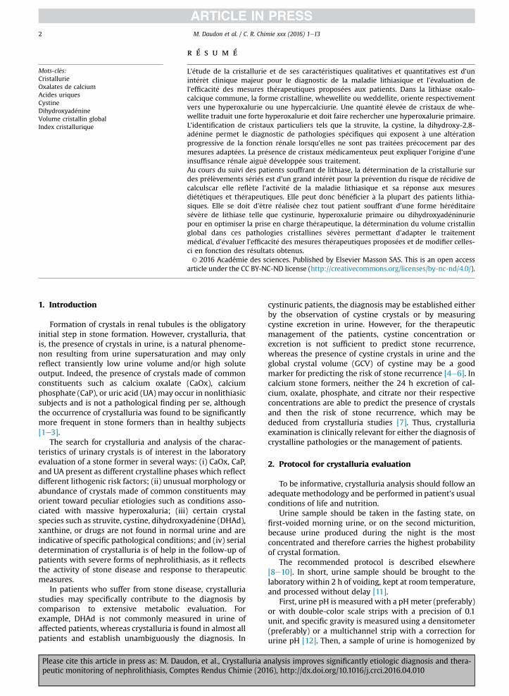
a b s t r a c t
The search for crystalluria and morphologic analysis of urinary crystals is of interest in thediagnostic evaluation and assessment of efficacy of therapeutic strategies in stone formers.In common calcium oxalate (CaOx) nephrolithiasis, identification of the monohydrate ordihydrate form of CaOx orients toward hyperoxaluria or hypercalciuria as the mainlithogenic mechanism, respectively. An unusual high abundance of crystals exclusivelymade of CaOx monohydrate is highly suggestive of primary hyperoxaluria, the most severeof all types of renal stone diseases. The identification of crystal species such as struvite,cystine, 2,8-dihydroxyadenine, which are not found in normal urine, is indicative of spe-cific pathological conditions, namely, infection stones and hereditary diseases such ascystinuria and dihydroxyadeninuria. Such diseases expose to progressive loss of renalfunction unless they are prevented by specific therapeutic measures. The presence ofcrystals made of drugs may entail the risk of kidney dysfunction because of crystallizationof the drug or its metabolites.Serial determination of crystalluria, which reflects the activity of stone disease and itsresponse to therapeutic measures, is of major interest in the follow-up of patients sufferingfrom nephrolithiasis. It should be more largely used in clinical practice in most stoneformers, still more for the surveillance of patients affected by severe, hereditary renalstone diseases such as primary hyperoxaluria, cystinuria, and dihydroxyadeninuria, wheredetermination of the global crystal volume allows assessing the efficacy of therapeuticmeasures and optimizing the medical management of the patients.© 2016 Académie des sciences. Published by Elsevier Masson SAS. This is an open access
article under the CC BY-NC-ND license (http://creativecommons.org/licenses/by-nc-nd/4.0/).
vice des Explorations Fonctionnelles, 4, rue de la Chine, 75970 Paris cedex 20, France.Daudon).
ed by Elsevier Masson SAS. This is an open access article under the CC BY-NC-ND license (http://
on, et al., Crystalluria analysis improves significantly etiologic diagnosis and thera-ptes Rendus Chimie (2016), http://dx.doi.org/10.1016/j.crci.2016.04.010
Mots-cl�es:Cristallurie
Oxalates de calciumAcides uriquesCystineDihydroxyad�enineVolume cristallin globalIndex cristallurique2
Please cite this article in press as: M. Daudpeutic monitoring of nephrolithiasis, Com
M. Daudon et al. / C. R. Chimie xxx (2016) 1e13
r é s u m é
L’�etude de la cristallurie et de ses caract�eristiques qualitatives et quantitatives est d'unint�eret clinique majeur pour le diagnostic de la maladie lithiasique et l’�evaluation del'efficacit�e des mesures th�erapeutiques propos�ees aux patients. Dans la lithiase oxalo-calcique commune, la forme cristalline, whewellite ou weddellite, oriente respectivementvers une hyperoxalurie ou une hypercalciurie. Une quantit�e �elev�ee de cristaux de whe-wellite traduit une forte hyperoxalurie et doit faire rechercher une hyperoxalurie primaire.L'identification de cristaux particuliers tels que la struvite, la cystine, la dihydroxy-2,8-ad�enine permet le diagnostic de pathologies sp�ecifiques qui exposent �a une alt�erationprogressive de la fonction r�enale lorsqu'elles ne sont pas trait�ees pr�ecocement par desmesures adapt�ees. La pr�esence de cristaux m�edicamenteux peut expliquer l'origine d'uneinsuffisance r�enale aigu€e d�evelopp�ee sous traitement.Au cours du suivi des patients souffrant de lithiase, la d�etermination de la cristallurie surdes pr�el�evements s�eri�es est d'un grand int�eret pour la pr�evention du risque de r�ecidive decalculscar elle refl�ete l'activit�e de la maladie lithiasique et sa r�eponse aux mesuresdi�et�etiques et th�erapeutiques. Elle peut donc b�en�eficier �a la plupart des patients lithia-siques. Elle se doit d’etre r�ealis�ee chez tout patient souffrant d'une forme h�er�editaires�ev�ere de lithiase telle que cystinurie, hyperoxalurie primaire ou dihydroxyad�eninuriepour en optimiser la prise en charge th�erapeutique, la d�etermination du volume cristallinglobal dans ces pathologies cristallines s�ev�eres permettant d'adapter le traitementm�edical, d’�evaluer l'efficacit�e des mesures th�erapeutiques propos�ees et de modifier celles-ci en fonction des r�esultats obtenus.© 2016 Académie des sciences. Published by Elsevier Masson SAS. This is an open access
article under the CC BY-NC-ND license (http://creativecommons.org/licenses/by-nc-nd/4.0/).
1. Introduction
Formation of crystals in renal tubules is the obligatoryinitial step in stone formation. However, crystalluria, thatis, the presence of crystals in urine, is a natural phenome-non resulting from urine supersaturation and may onlyreflect transiently low urine volume and/or high soluteoutput. Indeed, the presence of crystals made of commonconstituents such as calcium oxalate (CaOx), calciumphosphate (CaP), or uric acid (UA) may occur in nonlithiasicsubjects and is not a pathological finding per se, althoughthe occurrence of crystalluria was found to be significantlymore frequent in stone formers than in healthy subjects[1e3].
The search for crystalluria and analysis of the charac-teristics of urinary crystals is of interest in the laboratoryevaluation of a stone former in several ways: (i) CaOx, CaP,and UA present as different crystalline phases which reflectdifferent lithogenic risk factors; (ii) unusual morphology orabundance of crystals made of common constituents mayorient toward peculiar etiologies such as conditions asso-ciated with massive hyperoxaluria; (iii) certain crystalspecies such as struvite, cystine, dihydroxyad�enine (DHAd),xanthine, or drugs are not found in normal urine and areindicative of specific pathological conditions; and (iv) serialdetermination of crystalluria is of help in the follow-up ofpatients with severe forms of nephrolithiasis, as it reflectsthe activity of stone disease and response to therapeuticmeasures.
In patients who suffer from stone disease, crystalluriastudies may specifically contribute to the diagnosis bycomparison to extensive metabolic evaluation. Forexample, DHAd is not commonly measured in urine ofaffected patients, whereas crystalluria is found in almost allpatients and establish unambiguously the diagnosis. In
on, et al., Crystalluria aptes Rendus Chimie (20
cystinuric patients, the diagnosis may be established eitherby the observation of cystine crystals or by measuringcystine excretion in urine. However, for the therapeuticmanagement of the patients, cystine concentration orexcretion is not sufficient to predict stone recurrence,whereas the presence of cystine crystals in urine and theglobal crystal volume (GCV) of cystine may be a goodmarker for predicting the risk of stone recurrence [4e6]. Incalcium stone formers, neither the 24 h excretion of cal-cium, oxalate, phosphate, and citrate nor their respectiveconcentrations are able to predict the presence of crystalsand then the risk of stone recurrence, which may bededuced from crystalluria studies [7]. Thus, crystalluriaexamination is clinically relevant for either the diagnosis ofcrystalline pathologies or the management of patients.
2. Protocol for crystalluria evaluation
To be informative, crystalluria analysis should follow anadequate methodology and be performed in patient's usualconditions of life and nutrition.
Urine sample should be taken in the fasting state, onfirst-voided morning urine, or on the second micturition,because urine produced during the night is the mostconcentrated and therefore carries the highest probabilityof crystal formation.
The recommended protocol is described elsewhere[8e10]. In short, urine sample should be brought to thelaboratory within 2 h of voiding, kept at room temperature,and processed without delay [11].
First, urine pH is measured with a pHmeter (preferably)or with double-color scale strips with a precision of 0.1unit, and specific gravity is measured using a densitometer(preferably) or a multichannel strip with a correction forurine pH [12]. Then, a sample of urine is homogenized by
nalysis improves significantly etiologic diagnosis and thera-16), http://dx.doi.org/10.1016/j.crci.2016.04.010

Fig. 2. Hexagonal crystal of caoxite (calcium oxalate trihydrate).
M. Daudon et al. / C. R. Chimie xxx (2016) 1e13 3
gentle shaking (not centrifuged), homogenized mixture isput in a Malassez cell, and is examined by polarized lightmicroscopy. Microscopic examination includes urinecytology, and comprehensive crystalluria evaluation isbased on the identification of all crystal species, numera-tion and measure of the size of crystals and aggregates and,if relevant, the assessment of crystal volume. Fouriertransform infrared spectroscopy is used whenever neededto confirm the nature of unusual crystals.
Statistical comparisons regarding biochemistry of urinewith or without crystals (ANOVA) and the distribution ofcrystals as a function of urine pH classes (chi-square test)were performed with the NCSS statistical package (J. Hintz,Gainesville, FL). A P value less than 0.05 was considered asstatistically significant.
3. Nature and morphology of urinary crystals andcorrelations with etiology
Urinary crystals have a great variety of morphologies.Several textbooks dealing with the urinary sediment orcrystal analysis may help to identify metabolic or drugcrystals [13e15]. From a clinical point of view, crystals arereflecting supersaturation of urine regarding one or severalchemical species. In our experience, correlations betweencrystalluria and etiology of nephrolithiasis have beenestablished from microscopic examination of approxi-mately 25,000 first morning urine samples from 3000 pa-tients for whom urine biochemistry parameters weredetermined in the same samples and full etiological eval-uation was available. In addition to the chemical nature ofcommon forms of nephrolithiasis, identification of thecrystalline forms of CaOx, CaP, or UA crystals providesimportant information that may orient toward specificpathophysiological conditions, some corresponding to se-vere forms of nephrolithiasis.
3.1. Common types of urinary crystals
3.1.1. Calcium oxalatesCrystalluria made of CaOx crystals is the commonest
form of crystalluria found in stone formers. The maindeterminant of CaOx crystal formation is urine supersatu-ration owing to excessive concentration of calcium andoxalate ions, reflected by their molar product (pCaOx). A
Fig. 1. (a) Typical oval crystals of whewellite with a depressed
Please cite this article in press as: M. Daudon, et al., Crystalluria apeutic monitoring of nephrolithiasis, Comptes Rendus Chimie (20
very low concentration of low molecular weight inhibitorssuch as citrate and magnesium ions may also induce crys-talluria [16,17]. However, CaOx crystals do not come undera homogeneous category, as they present commonly in twodifferent forms, CaOx monohydrate (COM, or whewellite)and CaOx dihydrate (COD, or weddellite) according to therespective concentrations of Ca and Ox ions (Fig. 1). A thirdform, namely CaOx trihydrate or caoxite may be scarcelyfound in urine (Fig. 2).
Simultaneous determination of calcium and oxalateconcentrations and search for crystalluria in 21,220 urinesamples from calcium stone formers allowed to definethreshold values for the risk of CaOx crystal formation [18].Thus, the concentration of oxalate ions associated with theformation of CaOx crystals, determined as the inflexion ofthe curve plotting the percentage of urine samples exhib-iting the presence of CaOx crystals versus the concentrationof oxalate, was 0.3 mmol/l (Fig. 3).
The concentration of calcium ions associated with theformation of CaOx crystals, determined as the inflexion ofthe curve plotting the percentage of urine samples exhib-iting the presence of CaOx crystals versus the concentrationof calcium, was 3.8 mmol/l (Fig. 4).
core. (b) Bipyramidal (octahedral) crystals of weddellite.
nalysis improves significantly etiologic diagnosis and thera-16), http://dx.doi.org/10.1016/j.crci.2016.04.010
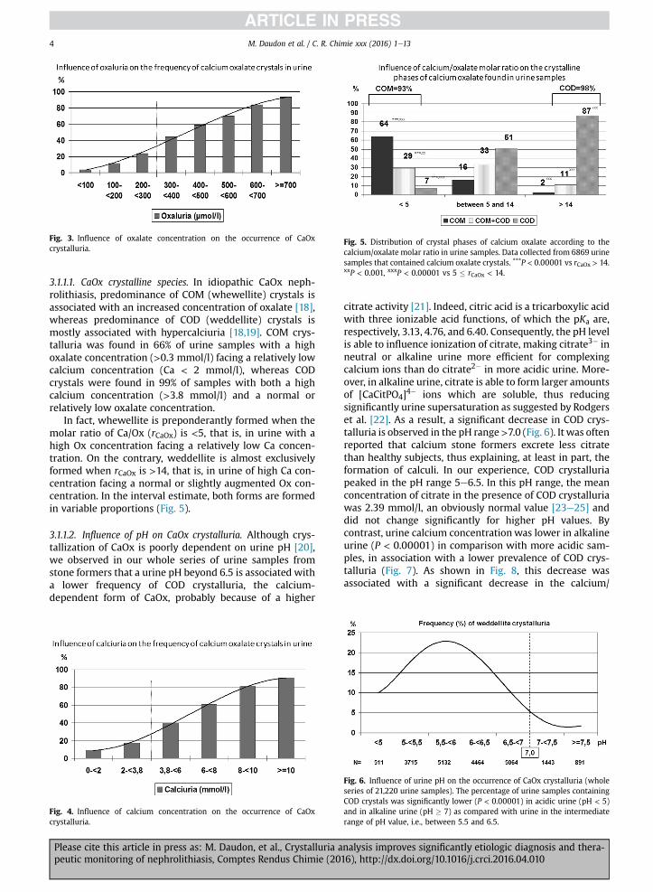
Fig. 3. Influence of oxalate concentration on the occurrence of CaOxcrystalluria.
Fig. 5. Distribution of crystal phases of calcium oxalate according to thecalcium/oxalate molar ratio in urine samples. Data collected from 6869 urinesamples that contained calcium oxalate crystals. ***P < 0.00001 vs rCaOx > 14.xxP < 0.001, xxxP < 0.00001 vs 5 � rCaOx < 14.
M. Daudon et al. / C. R. Chimie xxx (2016) 1e134
3.1.1.1. CaOx crystalline species. In idiopathic CaOx neph-rolithiasis, predominance of COM (whewellite) crystals isassociated with an increased concentration of oxalate [18],whereas predominance of COD (weddellite) crystals ismostly associated with hypercalciuria [18,19]. COM crys-talluria was found in 66% of urine samples with a highoxalate concentration (>0.3 mmol/l) facing a relatively lowcalcium concentration (Ca < 2 mmol/l), whereas CODcrystals were found in 99% of samples with both a highcalcium concentration (>3.8 mmol/l) and a normal orrelatively low oxalate concentration.
In fact, whewellite is preponderantly formed when themolar ratio of Ca/Ox (rCaOx) is <5, that is, in urine with ahigh Ox concentration facing a relatively low Ca concen-tration. On the contrary, weddellite is almost exclusivelyformed when rCaOx is >14, that is, in urine of high Ca con-centration facing a normal or slightly augmented Ox con-centration. In the interval estimate, both forms are formedin variable proportions (Fig. 5).
3.1.1.2. Influence of pH on CaOx crystalluria. Although crys-tallization of CaOx is poorly dependent on urine pH [20],we observed in our whole series of urine samples fromstone formers that a urine pH beyond 6.5 is associated witha lower frequency of COD crystalluria, the calcium-dependent form of CaOx, probably because of a higher
Fig. 4. Influence of calcium concentration on the occurrence of CaOxcrystalluria.
Please cite this article in press as: M. Daudon, et al., Crystalluria apeutic monitoring of nephrolithiasis, Comptes Rendus Chimie (20
citrate activity [21]. Indeed, citric acid is a tricarboxylic acidwith three ionizable acid functions, of which the pKa are,respectively, 3.13, 4.76, and 6.40. Consequently, the pH levelis able to influence ionization of citrate, making citrate3� inneutral or alkaline urine more efficient for complexingcalcium ions than do citrate2� in more acidic urine. More-over, in alkaline urine, citrate is able to form larger amountsof [CaCitPO4]4� ions which are soluble, thus reducingsignificantly urine supersaturation as suggested by Rodgerset al. [22]. As a result, a significant decrease in COD crys-talluria is observed in the pH range>7.0 (Fig. 6). It was oftenreported that calcium stone formers excrete less citratethan healthy subjects, thus explaining, at least in part, theformation of calculi. In our experience, COD crystalluriapeaked in the pH range 5e6.5. In this pH range, the meanconcentration of citrate in the presence of COD crystalluriawas 2.39 mmol/l, an obviously normal value [23e25] anddid not change significantly for higher pH values. Bycontrast, urine calcium concentrationwas lower in alkalineurine (P < 0.00001) in comparison with more acidic sam-ples, in association with a lower prevalence of COD crys-talluria (Fig. 7). As shown in Fig. 8, this decrease wasassociated with a significant decrease in the calcium/
Fig. 6. Influence of urine pH on the occurrence of CaOx crystalluria (wholeseries of 21,220 urine samples). The percentage of urine samples containingCOD crystals was significantly lower (P < 0.00001) in acidic urine (pH < 5)and in alkaline urine (pH � 7) as compared with urine in the intermediaterange of pH value, i.e., between 5.5 and 6.5.
nalysis improves significantly etiologic diagnosis and thera-16), http://dx.doi.org/10.1016/j.crci.2016.04.010
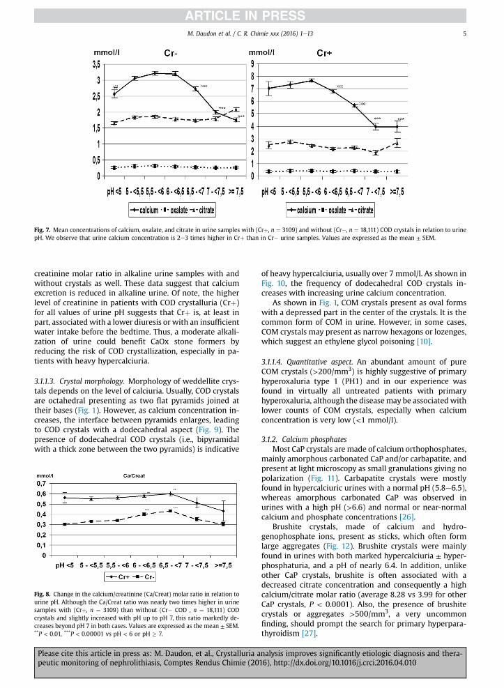
Fig. 7. Mean concentrations of calcium, oxalate, and citrate in urine samples with (Crþ, n ¼ 3109) and without (Cr�, n ¼ 18,111) COD crystals in relation to urinepH. We observe that urine calcium concentration is 2e3 times higher in Crþ than in Cr� urine samples. Values are expressed as the mean ± SEM.
M. Daudon et al. / C. R. Chimie xxx (2016) 1e13 5
creatinine molar ratio in alkaline urine samples with andwithout crystals as well. These data suggest that calciumexcretion is reduced in alkaline urine. Of note, the higherlevel of creatinine in patients with COD crystalluria (Crþ)for all values of urine pH suggests that Crþ is, at least inpart, associatedwith a lower diuresis or with an insufficientwater intake before the bedtime. Thus, a moderate alkali-zation of urine could benefit CaOx stone formers byreducing the risk of COD crystallization, especially in pa-tients with heavy hypercalciuria.
3.1.1.3. Crystal morphology. Morphology of weddellite crys-tals depends on the level of calciuria. Usually, COD crystalsare octahedral presenting as two flat pyramids joined attheir bases (Fig. 1). However, as calcium concentration in-creases, the interface between pyramids enlarges, leadingto COD crystals with a dodecahedral aspect (Fig. 9). Thepresence of dodecahedral COD crystals (i.e., bipyramidalwith a thick zone between the two pyramids) is indicative
Fig. 8. Change in the calcium/creatinine (Ca/Creat) molar ratio in relation tourine pH. Although the Ca/Creat ratio was nearly two times higher in urinesamples with (Crþ, n ¼ 3109) than without (Cr� COD , n ¼ 18,111) CODcrystals and slightly increased with pH up to pH 7, this ratio markedly de-creases beyond pH 7 in both cases. Values are expressed as the mean ± SEM.**P < 0.01, ***P < 0.00001 vs pH < 6 or pH � 7.
Please cite this article in press as: M. Daudon, et al., Crystalluria apeutic monitoring of nephrolithiasis, Comptes Rendus Chimie (20
of heavy hypercalciuria, usually over 7 mmol/l. As shown inFig. 10, the frequency of dodecahedral COD crystals in-creases with increasing urine calcium concentration.
As shown in Fig. 1, COM crystals present as oval formswith a depressed part in the center of the crystals. It is thecommon form of COM in urine. However, in some cases,COM crystals may present as narrow hexagons or lozenges,which suggest an ethylene glycol poisoning [10].
3.1.1.4. Quantitative aspect. An abundant amount of pureCOM crystals (>200/mm3) is highly suggestive of primaryhyperoxaluria type 1 (PH1) and in our experience wasfound in virtually all untreated patients with primaryhyperoxaluria, although the diseasemay be associatedwithlower counts of COM crystals, especially when calciumconcentration is very low (<1 mmol/l).
3.1.2. Calcium phosphatesMost CaP crystals are made of calcium orthophosphates,
mainly amorphous carbonated CaP and/or carbapatite, andpresent at light microscopy as small granulations giving nopolarization (Fig. 11). Carbapatite crystals were mostlyfound in hypercalciuric urines with a normal pH (5.8e6.5),whereas amorphous carbonated CaP was observed inurines with a high pH (>6.6) and normal or near-normalcalcium and phosphate concentrations [26].
Brushite crystals, made of calcium and hydro-genophosphate ions, present as sticks, which often formlarge aggregates (Fig. 12). Brushite crystals were mainlyfound in urines with both marked hypercalciuria ± hyper-phosphaturia, and a pH of nearly 6.4. In addition, unlikeother CaP crystals, brushite is often associated with adecreased citrate concentration and consequently a highcalcium/citrate molar ratio (average 8.28 vs 3.99 for otherCaP crystals, P < 0.0001). Also, the presence of brushitecrystals or aggregates >500/mm3, a very uncommonfinding, should prompt the search for primary hyperpara-thyroidism [27].
nalysis improves significantly etiologic diagnosis and thera-16), http://dx.doi.org/10.1016/j.crci.2016.04.010

Fig. 9. Two morphological aspects of dodecahedral crystals of weddellite: (a) polarizing microscopy; (b) light microscopy.
M. Daudon et al. / C. R. Chimie xxx (2016) 1e136
All types of CaP crystals are pH-dependent and form inurines whose pH is commonly >6.4 (Fig. 13).
3.1.3. Uric acidUA crystals form in acidic urine. UA dihydrate is the
commonest form. It has the lowest crystallization pH (5.25)and presents with various morphologies, typically as hex-agonal or diamond-shaped crystals, characteristically hav-ing a polychrome aspect in polarized light (Fig. 14).Crystalluria made of UA dihydrate is suggestive of defectiverenal ammoniagenesis as observed in the metabolic syn-drome or type 2 diabetes [28]. Anhydrous UA and UAmonohydrate are less frequently observed and also form inacidic urine, whereas amorphous UA (Fig. 15) precipitatesare found in urine with a high UA concentration inmoderately acidic pH.
Anhydrous UA presents as large polygonal crystals witha monochrome aspect in polarized light.
As shown in Fig. 16, UA dihydrate forms mainly in veryacidic urine, whereas amorphous UA mainly depends onthe concentration of urate ions in urine.
3.1.4. Urate saltsThe most frequent urate form is anhydrous ammonium
hydrogen urate. Its crystals form in alkaline urine or in the
Fig. 10. Occurrence (%) of dodecahedral crystals of weddellite as a functionof calcium concentration in urine.
Please cite this article in press as: M. Daudon, et al., Crystalluria apeutic monitoring of nephrolithiasis, Comptes Rendus Chimie (20
pH range of 6.3e7, depending on the associated metabolicfactors, and they exhibit various morphologies (Fig. 17).
Unlike ammonium urate crystals, crystals made of uratesalts such as sodium hydrogen urate, potassium quad-riurate, calcium hydrogen urate, or magnesium hydrogenurate are very uncommon. All of these species are observedin neutral or alkaline urine.
3.1.5. StruviteCrystals of magnesium ammonium phosphate (stru-
vite) form in strongly alkaline urine. Struvite crystalstypically have a “coffin-lid” aspect (Fig. 18), but they alsopresent as other morphologies (Fig. 19). The presence ofstruvite crystals, even in small amounts, in a markedlyalkaline urine (pH > 7) is indicative of urinary tractinfection (UTI) with urease-producing microorganisms,usually Proteus mirabilis or certain strands of Escherichiacoli, Klebsiella pneumoniae, Staphylococcus aureus, Pseu-domonas aeruginosa, or less frequently, Corynebacteriumurealyticum, responsible for encrusted cystitis and/orpyelitis [29]. Finding of struvite crystals even in theabsence of overt UTI should prompt careful bacteriolog-ical urine analysis to identify the responsible microor-ganism, including culture on special media if a usual
Fig. 11. Agglomerate of small grains and irregular translucent plaque ofamorphous carbonated calcium phosphate.
nalysis improves significantly etiologic diagnosis and thera-16), http://dx.doi.org/10.1016/j.crci.2016.04.010

Fig. 12. (a) Rod-shaped crystals of brushite. (b) Aggregate of brushite crystals.
Fig. 13. Influence of urine pH on calcium phosphate crystallization(n ¼ 2800 urine samples).
M. Daudon et al. / C. R. Chimie xxx (2016) 1e13 7
urease-producing microorganism is not found in routineanalysis.
3.2. Infrequent types of urinary crystals
Other crystals are much less frequently found in urine.They reflect the presence of rare hereditary stone diseasesor the renal elimination of drugs.
3.2.1. Hereditary stone diseasesCystine crystals present as hexagonal slides often
forming macles of large dimensions (Fig. 20). They are
Fig. 14. Two morphological aspects of uric acid dihydrate crystals: (a) polychromaticnew crystals at the surface of the initial crystal; (b) polychromatic hexagonal cryst
Please cite this article in press as: M. Daudon, et al., Crystalluria apeutic monitoring of nephrolithiasis, Comptes Rendus Chimie (20
pathognomonic of proximal tubular defects because ofmutations of the genes coding for membrane transportersof dibasic amino acids [30].
2,8-Dihydroxyadenine crystals present as small roundcrystals (about 5 mm in diameter) showing a characteristiccentral black cross (“Maltese cross” aspect) at polarizingmicroscopy (Fig. 21). They are found in patients with anadenine phosphoribosyltransferase deficiency [31,32] andmay be responsible for progressive interstitial crystalinfiltration leading to end-stage renal failure [33e35].
Xanthine crystals present as refringent granulations orsticks without a characteristic aspect, and Fourier trans-form infrared analysis is required for accurate identifica-tion. Xanthine crystals are observed in two differentpathological conditions: (i) inherited xanthine dehydro-genase deficiency, and (ii) long-term treatment with allo-purinol in patients with hereditary hypoxanthine guaninephosphoribosyltransferase deficiency.
Tyrosine, leucine, or potassium orotate crystals are veryinfrequent. They reflect very rare metabolic diseases.
3.2.2. Drug-induced crystalluriasA few drugs, mainly antimicrobial or antiviral agents,
may crystallize in urine when used at high dose for severaldays and, sometimes, for long periods [36]. They may beresponsible for stone formation or sometimes heavytubular precipitation and acute anuric renal failure[37e43].
diamond-shaped crystal seen at polarizing microscopy. Note the initiation ofals.
nalysis improves significantly etiologic diagnosis and thera-16), http://dx.doi.org/10.1016/j.crci.2016.04.010
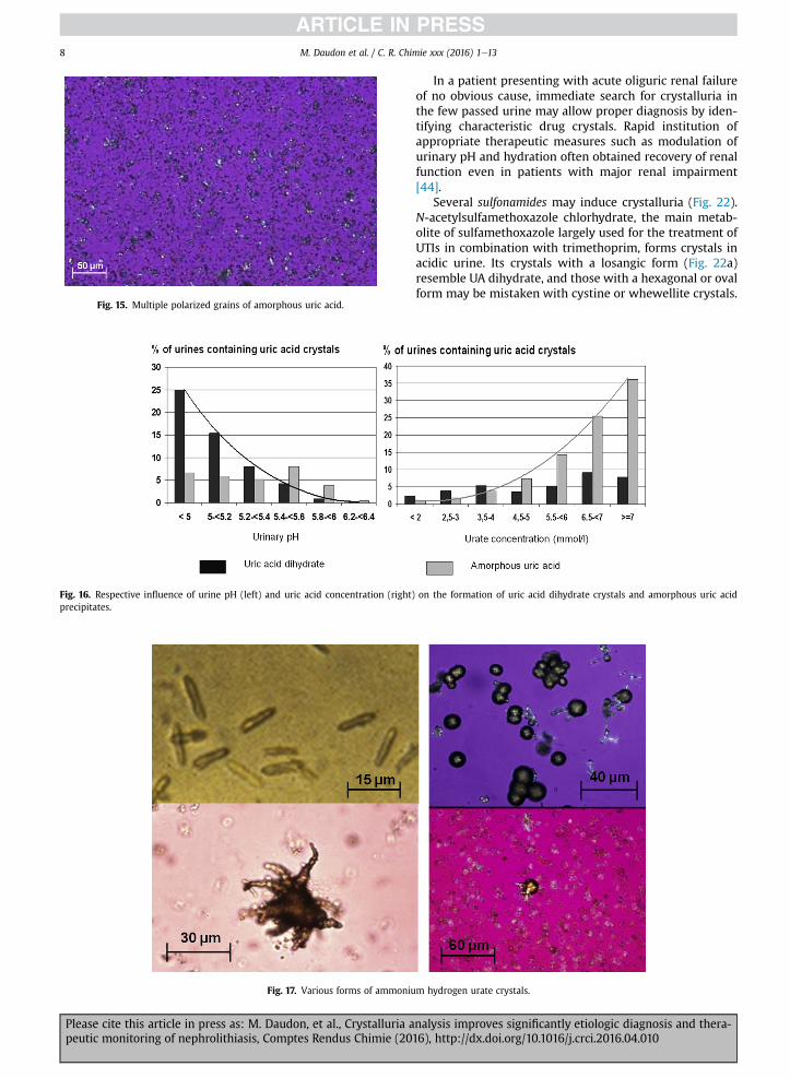
Fig. 15. Multiple polarized grains of amorphous uric acid.
Fig. 16. Respective influence of urine pH (left) and uric acid concentration (rightprecipitates.
Fig. 17. Various forms of ammoniu
M. Daudon et al. / C. R. Chimie xxx (2016) 1e138
Please cite this article in press as: M. Daudon, et al., Crystalluria apeutic monitoring of nephrolithiasis, Comptes Rendus Chimie (20
In a patient presenting with acute oliguric renal failureof no obvious cause, immediate search for crystalluria inthe few passed urine may allow proper diagnosis by iden-tifying characteristic drug crystals. Rapid institution ofappropriate therapeutic measures such as modulation ofurinary pH and hydration often obtained recovery of renalfunction even in patients with major renal impairment[44].
Several sulfonamides may induce crystalluria (Fig. 22).N-acetylsulfamethoxazole chlorhydrate, the main metab-olite of sulfamethoxazole largely used for the treatment ofUTIs in combination with trimethoprim, forms crystals inacidic urine. Its crystals with a losangic form (Fig. 22a)resemble UA dihydrate, and those with a hexagonal or ovalform may be mistaken with cystine or whewellite crystals.
) on the formation of uric acid dihydrate crystals and amorphous uric acid
m hydrogen urate crystals.
nalysis improves significantly etiologic diagnosis and thera-16), http://dx.doi.org/10.1016/j.crci.2016.04.010
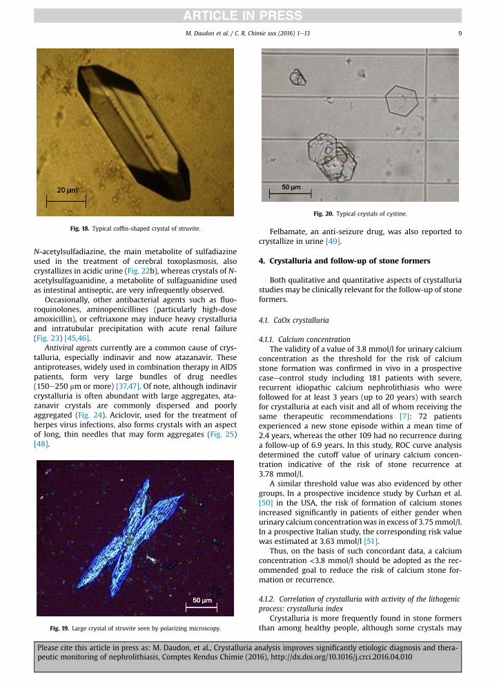
Fig. 18. Typical coffin-shaped crystal of struvite.
Fig. 20. Typical crystals of cystine.
M. Daudon et al. / C. R. Chimie xxx (2016) 1e13 9
N-acetylsulfadiazine, the main metabolite of sulfadiazineused in the treatment of cerebral toxoplasmosis, alsocrystallizes in acidic urine (Fig. 22b), whereas crystals of N-acetylsulfaguanidine, a metabolite of sulfaguanidine usedas intestinal antiseptic, are very infrequently observed.
Occasionally, other antibacterial agents such as fluo-roquinolones, aminopenicillines (particularly high-doseamoxicillin), or ceftriaxone may induce heavy crystalluriaand intratubular precipitation with acute renal failure(Fig. 23) [45,46].
Antiviral agents currently are a common cause of crys-talluria, especially indinavir and now atazanavir. Theseantiproteases, widely used in combination therapy in AIDSpatients, form very large bundles of drug needles(150e250 mm or more) [37,47]. Of note, although indinavircrystalluria is often abundant with large aggregates, ata-zanavir crystals are commonly dispersed and poorlyaggregated (Fig. 24). Aciclovir, used for the treatment ofherpes virus infections, also forms crystals with an aspectof long, thin needles that may form aggregates (Fig. 25)[48].
Fig. 19. Large crystal of struvite seen by polarizing microscopy.
Please cite this article in press as: M. Daudon, et al., Crystalluria apeutic monitoring of nephrolithiasis, Comptes Rendus Chimie (20
Felbamate, an anti-seizure drug, was also reported tocrystallize in urine [49].
4. Crystalluria and follow-up of stone formers
Both qualitative and quantitative aspects of crystalluriastudies may be clinically relevant for the follow-up of stoneformers.
4.1. CaOx crystalluria
4.1.1. Calcium concentrationThe validity of a value of 3.8 mmol/l for urinary calcium
concentration as the threshold for the risk of calciumstone formation was confirmed in vivo in a prospectivecaseecontrol study including 181 patients with severe,recurrent idiopathic calcium nephrolithiasis who werefollowed for at least 3 years (up to 20 years) with searchfor crystalluria at each visit and all of whom receiving thesame therapeutic recommendations [7]: 72 patientsexperienced a new stone episode within a mean time of2.4 years, whereas the other 109 had no recurrence duringa follow-up of 6.9 years. In this study, ROC curve analysisdetermined the cutoff value of urinary calcium concen-tration indicative of the risk of stone recurrence at3.78 mmol/l.
A similar threshold value was also evidenced by othergroups. In a prospective incidence study by Curhan et al.[50] in the USA, the risk of formation of calcium stonesincreased significantly in patients of either gender whenurinary calcium concentrationwas in excess of 3.75mmol/l.In a prospective Italian study, the corresponding risk valuewas estimated at 3.63 mmol/l [51].
Thus, on the basis of such concordant data, a calciumconcentration <3.8 mmol/l should be adopted as the rec-ommended goal to reduce the risk of calcium stone for-mation or recurrence.
4.1.2. Correlation of crystalluria with activity of the lithogenicprocess: crystalluria index
Crystalluria is more frequently found in stone formersthan among healthy people, although some crystals may
nalysis improves significantly etiologic diagnosis and thera-16), http://dx.doi.org/10.1016/j.crci.2016.04.010

Fig. 21. Small spherical crystals of dihydroxyadenine: (a) light microscopy; (b) polarizing microscopy. Note the typical aspect under polarized light showing thatall crystals exhibit a Maltese cross.
Fig. 22. (a) Crystals of N-acetylsulfamethoxazole hydrochloride resembling uric acid dehydrate crystals (light microscopy). (b) Aggregated crystals of N-ace-tylsulfadiazine (polarizing microscopy).
M. Daudon et al. / C. R. Chimie xxx (2016) 1e1310
transiently be found in urines of nonstone forming persons,especially during the hours after a meal.
As there is a necessary relationship between precipita-tion of urinary crystals and formation of stones, wereasoned that the risk of stone formation would increasewith an increased frequency of crystalluria. By evaluatingthe presence or absence of crystals in first morning urinesamples at each visit in 204 calcium stone formers pro-spectively followed at our institution for a median durationof 7 years (5e15 years), we observed sustained crystalluria
Fig. 23. Isolated or aggregated large needle-shaped crystals of amoxycillintrihydrate (polarizing microscopy).
Please cite this article in press as: M. Daudon, et al., Crystalluria apeutic monitoring of nephrolithiasis, Comptes Rendus Chimie (20
to be indicative of persistent stone disease activity andhighly predictive of the recurrence of stones. The presenceof crystals in 50% or more of urine samples was associatedwith stone recurrence in 87% of cases, whereas stonerecurrence was observed in only 9% of patients with lessfrequent crystalluria (Fig. 26). In another study, we foundthat around 90% of recurrent stone formers exhibitedcrystals in at least 50% of urine samples [52].
Accordingly, we proposed a “crystalluria index” definedas the ratio of the number of urine samples containingcrystals to the total number of examined samples in a givenpatient, with a crystalluria index �0.50 as the thresholdvalue indicative of persistent lithogenic activity and risk ofstone recurrence [7]. In clinical practice, the interval of timebetween two successive urine sample examinations shouldbe about 6 months (or less in the case of an active neph-rolithiasis) for an optimized follow-up of the patients. Ac-cording to the ROC curve analysis, the cutoff value of acrystalluria index for the risk of developing stone recur-rence was 0.50, and multivariate analysis by the Cox modelidentified a crystalluria index of 0.50 as the most powerfulindependent predicting parameter, with a hazard ratio of16.8 [7].
4.2. Quantitative aspects: GCV
Crystalluria abundance is usually evaluated by simplycounting the number of crystals per cubic millimeter. It
nalysis improves significantly etiologic diagnosis and thera-16), http://dx.doi.org/10.1016/j.crci.2016.04.010

Fig. 24. Crystals of antiprotease drugs used to treat HIV patients. (a) and (b) Aggregates of indinavir monohydrate crystals. (c) and (d) Needle-shaped crystals ofatazanavir. The crystals are commonly associated with white cells (arrow).
Fig. 25. Needle-shaped crystals of aciclovir (polarizing microscopy).
Fig. 26. Relation between the occurrence of crystalluria in serial urinesamples and the risk of stone recurrence in stone formers.NRSF ¼ nonrecurrent stone formers, RSF ¼ recurrent stone formers over aperiod of 7 years of follow-up.
M. Daudon et al. / C. R. Chimie xxx (2016) 1e13 11
Please cite this article in press as: M. Daudon, et al., Crystalluria apeutic monitoring of nephrolithiasis, Comptes Rendus Chimie (20
may be evaluated more precisely by determining their totalvolume per cubic millimeter. Such quantification is espe-cially useful for the management of severe forms ofnephrolithiasis.
Disappearance of crystalluria is the best indicator ofwell-controlled lithogenic activity. This result is generallyreached in patients with common forms of calcium or UAnephrolithiasis. However, total disappearance of crys-talluria is a difficult goal to achieve in genetic diseases, suchas primary hyperoxaluria and cystinuria, which have a veryactive, permanent crystallization process. In these severediseases, a partial decrease in the amount of crystals pre-sent in urine samples guided by the determination of GCVis a more attainable goal often sufficient to reduce stoneformation.
Determination of the global volume occupied in urineby a given crystal species per volume unit provides themost precise estimation of the abundance of crystalluria.
The GCV (expressed as mm3/mm3) is calculated as theproduct of the number of crystals per cubic millimetermultiplied by the average size of crystals in micrometersand by a numeric factor taking into account the geometricform of crystals specific for each species [10].
Formulae proposed for the calculation of GCV of crys-tallurias of whewellite, weddellite, cystine, or dihydrox-yadenine are presented in Figs. 27 and 28.
By comparing clinical outcomes and variations in crystalvolume in serial urine samples, we could determine thethresholds of GCV associated with stone recurrence in PH1and cystinuria, thus defining the desirable goals to achieve.
In PH1 patients, a crystal volume <500 mm3/mm3 wasassociated with a lowered risk of tubular plugging withCOM crystals in the days following combined liverekidneytransplantation, a crucial period when considerableamounts of CaOx stored in bones are excreted through thekidneys after restoration of renal function, with the risk ofmassive tubular obstruction and loss of the kidney trans-plant [53]. By closely monitoring CaOx crystal volume, early
nalysis improves significantly etiologic diagnosis and thera-16), http://dx.doi.org/10.1016/j.crci.2016.04.010
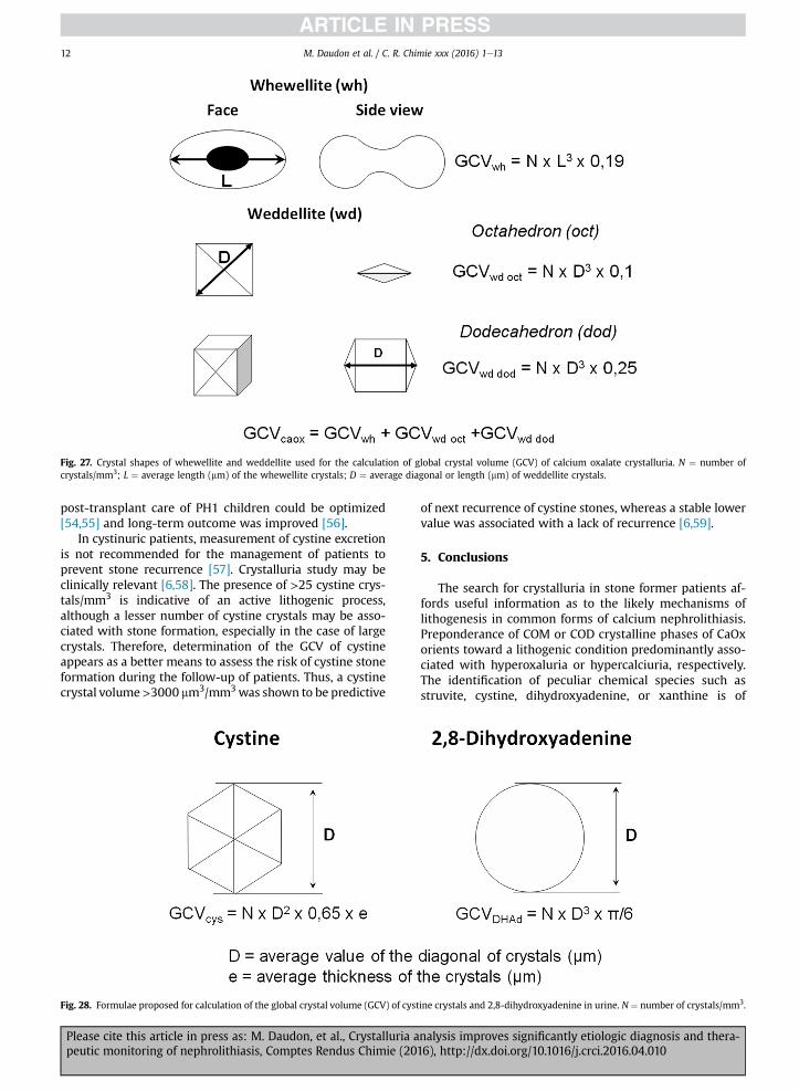
Fig. 27. Crystal shapes of whewellite and weddellite used for the calculation of global crystal volume (GCV) of calcium oxalate crystalluria. N ¼ number ofcrystals/mm3; L ¼ average length (mm) of the whewellite crystals; D ¼ average diagonal or length (mm) of weddellite crystals.
M. Daudon et al. / C. R. Chimie xxx (2016) 1e1312
post-transplant care of PH1 children could be optimized[54,55] and long-term outcome was improved [56].
In cystinuric patients, measurement of cystine excretionis not recommended for the management of patients toprevent stone recurrence [57]. Crystalluria study may beclinically relevant [6,58]. The presence of >25 cystine crys-tals/mm3 is indicative of an active lithogenic process,although a lesser number of cystine crystals may be asso-ciated with stone formation, especially in the case of largecrystals. Therefore, determination of the GCV of cystineappears as a better means to assess the risk of cystine stoneformation during the follow-up of patients. Thus, a cystinecrystal volume>3000 mm3/mm3was shown to be predictive
Fig. 28. Formulae proposed for calculation of the global crystal volume (GCV) of cyst
Please cite this article in press as: M. Daudon, et al., Crystalluria apeutic monitoring of nephrolithiasis, Comptes Rendus Chimie (20
of next recurrence of cystine stones, whereas a stable lowervalue was associated with a lack of recurrence [6,59].
5. Conclusions
The search for crystalluria in stone former patients af-fords useful information as to the likely mechanisms oflithogenesis in common forms of calcium nephrolithiasis.Preponderance of COM or COD crystalline phases of CaOxorients toward a lithogenic condition predominantly asso-ciated with hyperoxaluria or hypercalciuria, respectively.The identification of peculiar chemical species such asstruvite, cystine, dihydroxyadenine, or xanthine is of
ine crystals and 2,8-dihydroxyadenine in urine. N ¼ number of crystals/mm3.
nalysis improves significantly etiologic diagnosis and thera-16), http://dx.doi.org/10.1016/j.crci.2016.04.010
M. Daudon et al. / C. R. Chimie xxx (2016) 1e13 13
considerable value for the diagnosis of infection stones andof genetic diseases such as cystinuria and dihydrox-yadeninuria, which require specific therapy. Determinationof the GCV allows to assess the efficacy of therapeuticmeasures in severe genetic diseases such as primaryhyperoxaluria and cystinuria. Crystalluria index was clearlyshown as the best marker for predicting stone recurrence.In addition, other criteria such as the size of COD crystals orthe presence of heterogeneous nucleation process may berelevant for identifying particular metabolic disorders inurine or the risk of recurrence in some patients [60].
In all types of renal stone disease, especially in the mostsevere forms, serial search for crystalluria appears as avaluable tool for identifying patients with active crystalformation and at risk of forming kidney stones, thereforeallowing to timely institute or modify therapeutic mea-sures to prevent stone recurrence.
Of note, crystalluria was rarely studied in normal sub-jects [1]. Thus, the interest of studying crystalluria to pre-dict a first stone formation in healthy subjects was neverassessed. However, it was recently found that high values ofrisk indices such as the Tiselius index, reflecting high urineconcentration of lithogenic factors, were associated with ahigh occurrence of CaOx crystalluria in healthy subjects,suggesting that crystalluria study could be, in stone formersand in healthy subjects as well, an interesting tool fordetecting a possible risk of stone formation [61]. Thesepreliminary results require further investigations to bevalidated on a larger series of normal subjects.
Conflicts of interest
The authors have no conflicts of interest to declare.
References
[1] P.G. Werness, J.H. Bergert, L.H. Smith, J. Cryst. Growth 53 (1981) 166.[2] H.V. Nguyen, M. Daudon, R.J. R�eveillaud, P. Jungers, N�ephrologie 8
(1987) 65.[3] M. Daudon, P. Jungers, B. Lacour, Ann. Biol. Clin. 62 (2004) 379.[4] Y. Nakagawa, J.R. Asplin, D.S. Goldfarb, J.H. Parks, F.L. Coe, J. Urol. 164
(2000) 1481.[5] C.Y.C. Pak, C.J. Fuller, J. Urol. 129 (1983) 1066.[6] M. Daudon, F. Cohen-Solal, F. Barbey, M.-F. Gagnadoux,
B. Knebelmann, P. Jungers, Urol. Res. 31 (2003) 207.[7] M. Daudon, C. Hennequin, G. Boujelben, B. Lacour, P. Jungers, Kidney
Int. 67 (2005) 1934.[8] C.A. Bader, A. Chevalier, C. Hennequin, P. Jungers, M. Daudon,
Scanning Microsc. 8 (1994) 215.[9] M. Daudon, P. Jungers, Nephron Physiol. 98 (2004) 31.
[10] M. Daudon (Ed.), EMC e N�ephrologie, vol.10, Elsevier Masson, Paris,2013, p. 15 (Article 18-026-C-50).
[11] J.S. Elliot, I.N. Rabinowitz, J. Urol. 123 (1980) 324.[12] C. Hennequin, M. Daudon, T. Phung, B. Lacour, P. Jungers, Presse
M�ed. 24 (1995) 1559.[13] L. Graff, A Handbook of Routine Urinalysis, Lippincott Company,
Toronto, 1982.[14] G.B. Fogazzi, The Urinary Sediment. An Integrated View, 3rd ed.,
Elsevier, Milan, 2010.[15] M. Daudon, O. Traxer, P. Jungers, Lithiase urinaire, 2e �ed., M�edecine-
Sciences, Lavoisier, Paris, 2012, p. 730.[16] D.J. Kok, S.E. Papapoulos, O.L. Bijvoet, Lancet 1 (1986) 1056.[17] H.G. Tiselius, Urol. Int. 47 (1991) 255.[18] M. Daudon, Mod�eles de cristallisation, in: P. Jungers, M. Daudon,
A. Le Duc (Eds.), Lithiase Urinaire, Flammarion M�edecine-Sciences,Paris, 1989, p. 158.
Please cite this article in press as: M. Daudon, et al., Crystalluria apeutic monitoring of nephrolithiasis, Comptes Rendus Chimie (20
[19] J.R. Asplin, J. Lingeman, R. Kahnoski, H. Mardis, J.H. Parks, F.L. Coe, J.Urol. 159 (1998) 664.
[20] H. Verplaetse, R.M.H. Verbeeck, H. Minnaert, W. Osterlinck, Eur.Urol. 11 (1985) 44.
[21] R.J. Unwin, G. Capasso, D.G. Shirley, Nephron Physiol. 98 (2004) 15.[22] A. Rodgers, S. Allie-Hamdulay, G. Jackson, Nephrol. Dial. Transpl. 21
(2006) 361.[23] S.G. Welshman, G. McGeown, Br. J. Urol. 48 (1976) 7.[24] M. Menon, C.J. Mahle, J. Urol. 129 (1983) 1158.[25] M. Nikkila, T. Koivula, H. Jokela, Eur. Urol. 16 (1989) 382.[26] M. Daudon, H. Bouzidi, D. Bazin, Urol. Res. 38 (2010) 459.[27] H. Bouzidi, D. de Brauwere, M. Daudon, Nephrol. Dial. Transpl. 26
(2011) 565.[28] M. Mbarki, J. Jabrane, A. Oussama, M. Daudon, Progr. Urol. 5 (2005)
420.[29] P. M�eria, A. Desgrippes, C. Arfi, A. Le Duc, J. Urol. 160 (1998) 3.[30] H. Bouzidi, M. Daudon, Ann. Biol. Clin. 65 (2007) 473.[31] H.A. Simmonds, K.J. Van Acker, J.S. Cameron, W. Snedden, Biochem.
J. 157 (1976) 485.[32] G. Boll�ee, C. Dollinger, L. Boutaud, D. Guillemot, A. Bensman,
J. Harambat, et al., J. Am. Soc. Nephrol. 21 (2010) 679.[33] P. Stratta, G.B. Fogazzi, C. Canavese, A. Airoldi, R. Fenoglio,
C. Bozzola, et al., Am. J. Kidney Dis. 56 (2010) 585.[34] H. Bouzidi, B. Lacour, M. Daudon, Ann. Biol. Clin. 65 (2007) 585.[35] M. Hoffmann, A. Talaska, J.P. Bocquet, H. Le Monies de Sagazan,
M. Daudon, N�ephrologie 25 (2004) 297.[36] M. Daudon, P. Jungers, Drugs 64 (2004) 245.[37] M. Daudon, L. Est�epa, J.-P. Viard, D. Joly, P. Jungers, Lancet 349
(1997) 1294.[38] H. Hanabusa, H. Tagami, H. Hataya, N. Engl. J. Med. 340 (1999) 392.[39] H.R. Chang, P.M. Pella, N. Engl. J. Med. 355 (2006) 2158.[40] H. Izzedine, M.B. M'Rad, A. Bardier, M. Daudon, D. Salmon, AIDS 21
(2007) 2357.[41] L.A. Farina, J. Palou Redorta, G. Chechile Toniolo, Arch. Esp. Urol. 48
(1995) 418.[42] M. Crespo, C. Quereda, J. Pascual, et al., Clin. Nephrol. 54 (2000) 68.[43] H. Izzedine, M.A. Valantin, M. Daudon, H. Ait Mohand, F. Caby,
C. Katlama, AIDS 21 (2007) 1992 (letter).[44] G. Zanetta, L. Maurice-Estepa, C. Mousson, E. Justrabo, M. Daudon,
G. Rifle, Y. Tanfer, Transplantation 67 (1999) 1376.[45] G.B. Fogazzi, M. Cantu, L. Saglimbeni, M. Daudon, Nephrol. Dial.
Transpl. 18 (2003) 212.[46] G. Fogazzi, G. Garigali, C. Brambilla, M. Daudon, Nephrol. Dial.
Transpl. 21 (2006) 2982.[47] D. Viglietti, J. Verine, N. De Castro, A. Scemla, M. Daudon, D. Glotz,
�E. Pillebout, Antivir. Ther. 16 (2011) 119.[48] B.N. Becker, P. Fall, C. Hall, D. Milam, J. Leonard, A. Glick,
G. Schulman, Am. J. Kidney Dis. 22 (1993) 611.[49] X. Parent, F. Schieffer, Ann. Biol. Clin. (Paris) 68 (2010) 609.[50] G.C. Curhan, W.C. Willett, F.E. Speizer, M.J. Stampfer, Kidney Int. 59
(2001) 2290.[51] M. Cirillo, D. Stellato, P. Panarelli, M. Laurenzi, N.G. De Santo, Kidney
Int. 63 (2003) 2200.[52] M. Daudon, F. Cohen-Solal, P. Jungers, in: D.J. Kok, H.C. Romjin,
P.C.M.S. Verhagen, C.F. Verkoelen (Eds.), Eurolithiasis, Proc. 9thEuropean Symposium on Urolithiasis, Shaker Publishing, Maas-tricht, The Netherlands, 2001, p. 261.
[53] P. Cochat, S.A. Hulton, C. Acquaviva, C.J. Danpure, M. Daudon, M. DeMarchi, et al., Nephrol. Dial. Transpl. 27 (2012) 1729.
[54] M. Daudon, L. Priqueler, P. Jouvet, M. Labrunie, M. Broyer,M.F. Gagnadoux, P. Jungers, in: P. Jungers, M. Daudon (Eds.), RenalStone Disease. Crystallization Process, Pathophysiology, MetabolicDisorders and Prevention, Elsevier, Paris, 1997, p. 53.
[55] P. Jouvet, L. Priqueler, M.F. Gagnadoux, D. Jan, A. Beringer, F. Lacaille,et al., Kidney Int. 53 (1998) 1412.
[56] M.-F. Gagnadoux, F. Lacaille, P. Niaudet, Y. Revillon, P. Jouvet, D. Jan,et al., Pediatr. Nephrol. 16 (2001) 946.
[57] N. Sumorok, D.S. Goldfarb, Curr. Opin. Nephrol. Hypertens. 22(2013) 427.
[58] K.A. Wong, C. Pardy, S. Pillay, T. Athanasiou, G. Rottenberg,M. Bultitude, A. Chandra, K. Thomas, J. Endourol. 30 (2016) 609.
[59] M. Daudon, L. Estepa, M.F. Gagnadoux, B. Lacour, P. Jungers, in:L. Borghi, T. Meschi, A. Briganti, T. Schianchi, A. Novarini (Eds.),Kidney Stones, Editoriale Bios, Cosenza, 1999, p. 531.
[60] M. Daudon, N�ephrol Th�er 11 (2015) 174.[61] V. de La Gu�eronni�ere, L. Le Bellego, I. Buendia Jimenez, O. Dohein,
I. Tack, M. Daudon, Arch. Ital. Urol. Androl. 83 (2011) 43e50.
nalysis improves significantly etiologic diagnosis and thera-16), http://dx.doi.org/10.1016/j.crci.2016.04.010