Omega-3 fatty acid therapy dose-dependently and significantly decreased triglycerides and improved...
7
Omega-3 fatty acid therapy dose-dependently and significantly decreased triglycerides and improved flow-mediated dilation, however, did not significantly improve insulin sensitivity in patients with hypertriglyceridemia Pyung Chun Oh a,b , Kwang Kon Koh a,b, ⁎, Ichiro Sakuma c , Soo Lim d , Yonghee Lee e , Seungik Lee a,b , Kyounghoon Lee a,b , Seung Hwan Han a,b , Eak Kyun Shin a,b a Cardiology, Gachon University Gil Medical Center, Incheon, Republic of Korea b Gachon Cardiovascular Research Institute, Incheon, Republic of Korea c Cardiovascular Medicine, Hokko Memorial Clinic, Sapporo, Japan d Division of Endocrinology, Seoul National University Bundang Hospital, Seoul National University College of Medicine, Seongnam, Republic of Korea e Department of Statistics, University of Seoul, Seoul, Republic of Korea abstract article info Article history: Received 26 March 2014 Received in revised form 16 May 2014 Accepted 24 July 2014 Available online 7 August 2014 Keywords: Omega-3 fatty acids Atherosclerosis Insulin resistance Hypertriglyceridemia Background: Experimental studies demonstrate that higher intake of omega-3 fatty acids (n−3 FA) improves in- sulin sensitivity, however, we reported that n−3 FA 2 g therapy, most commonly used dosage did not signifi- cantly improve insulin sensitivity despite reducing triglycerides by 21% in patients. Therefore, we investigated the effects of different dosages of n−3 FA in patients with hypertriglyceridemia. Methods: This was a randomized, single-blind, placebo-controlled, parallel study. Age, sex, and body mass index were matched among groups. All patients were recommended to maintain a low fat diet. Forty-four patients (about 18 had metabolic syndrome/type 2 diabetes mellitus) in each group were given placebo, n −3 FA 1 (O1), 2 (O2), or 4 g (O4), respectively daily for 2 months. Results: n−3 FA therapy dose-dependently and significantly decreased triglycerides and triglycerides/HDL cho- lesterol and improved flow-mediated dilation, compared with placebo (by ANOVA). However, each n −3 FA therapy did not significantly decrease high-sensitivity C-reactive protein and fibrinogen, compared with placebo. O1 significantly increased insulin levels and decreased insulin sensitivity (determined by QUICKI) and O2 signif- icantly decreased plasma adiponectin levels relative to baseline measurements. Of note, when compared with placebo, each n−3 FA therapy did not significantly change insulin, glucose, adiponectin, glycated hemoglobin levels and insulin sensitivity (by ANOVA). We observed similar results in a subgroup of patients with the meta- bolic syndrome. Conclusions: n −3 FA therapy dose-dependently and significantly decreased triglycerides and improved flow- mediated dilation. Nonetheless, n−3 FA therapy did not significantly improve acute-phase reactants and insulin sensitivity in patients with hypertriglyceridemia, regardless of dosages. © 2014 Published by Elsevier Ireland Ltd. 1. Introduction Epidemiological and clinical evidences suggest a significant inverse association between long-term intake of omega-3 fatty acids, especially eicosapentaenoic acid (EPA) and docosahexaenoic acid (DHA), and mortality associated with coronary artery disease [1–3]. Thus, consump- tion of fish or fish-oil may help prevent adverse consequences of coronary artery disease, especially fatal myocardial infarction and sud- den cardiac death. Consumption of omega-3 fatty acids causes improve- ment in many relevant cardiovascular biomarkers including those represented by hypertriglyceridemia [4], vascular dysfunction [5,6], and inflammation [6]. However, recently, the reported beneficial effects of omega-3 fatty acids remain debated. Indeed, a recent meta-analysis stated that omega-3 fatty acids may protect against vascular disease, but the evidence is not clear-cut, and any benefits are certainly not as great as previously believed [7]. Endothelial dysfunction associated with metabolic syndrome and other insulin resistant states is characterized by impaired nitric oxide (NO) bioavailability and release from endothelium [8–10]. This reduces International Journal of Cardiology 176 (2014) 696–702 ⁎ Corresponding author at: Cardiology, Gachon University Gil Medical Center, 1198 Kuwol-dong, Namdong-gu, Incheon 405-760, Republic of Korea. Tel.: +82 32 460-3683; fax: +82 32 469 1906. E-mail address: [email protected] (K.K. Koh). http://dx.doi.org/10.1016/j.ijcard.2014.07.075 0167-5273/© 2014 Published by Elsevier Ireland Ltd. Contents lists available at ScienceDirect International Journal of Cardiology journal homepage: www.elsevier.com/locate/ijcard
-
Upload
independent -
Category
Documents
-
view
2 -
download
0
Transcript of Omega-3 fatty acid therapy dose-dependently and significantly decreased triglycerides and improved...

International Journal of Cardiology 176 (2014) 696–702
Contents lists available at ScienceDirect
International Journal of Cardiology
j ourna l homepage: www.e lsev ie r .com/ locate / i j ca rd
Omega-3 fatty acid therapy dose-dependently and significantlydecreased triglycerides and improved flow-mediated dilation, however,did not significantly improve insulin sensitivity in patientswith hypertriglyceridemia
Pyung Chun Oh a,b, Kwang Kon Koh a,b,⁎, Ichiro Sakuma c, Soo Lim d, Yonghee Lee e, Seungik Lee a,b,Kyounghoon Lee a,b, Seung Hwan Han a,b, Eak Kyun Shin a,b
a Cardiology, Gachon University Gil Medical Center, Incheon, Republic of Koreab Gachon Cardiovascular Research Institute, Incheon, Republic of Koreac Cardiovascular Medicine, Hokko Memorial Clinic, Sapporo, Japand Division of Endocrinology, Seoul National University Bundang Hospital, Seoul National University College of Medicine, Seongnam, Republic of Koreae Department of Statistics, University of Seoul, Seoul, Republic of Korea
⁎ Corresponding author at: Cardiology, Gachon UniveKuwol-dong, Namdong-gu, Incheon 405-760, Republic offax: +82 32 469 1906.
E-mail address: [email protected] (K.K. Koh).
http://dx.doi.org/10.1016/j.ijcard.2014.07.0750167-5273/© 2014 Published by Elsevier Ireland Ltd.
a b s t r a c t
a r t i c l e i n f oArticle history:
Received 26 March 2014Received in revised form 16 May 2014Accepted 24 July 2014Available online 7 August 2014Keywords:Omega-3 fatty acidsAtherosclerosisInsulin resistanceHypertriglyceridemia
Background: Experimental studies demonstrate that higher intake of omega-3 fatty acids (n−3 FA) improves in-sulin sensitivity, however, we reported that n−3 FA 2 g therapy, most commonly used dosage did not signifi-cantly improve insulin sensitivity despite reducing triglycerides by 21% in patients. Therefore, we investigatedthe effects of different dosages of n−3 FA in patients with hypertriglyceridemia.Methods: This was a randomized, single-blind, placebo-controlled, parallel study. Age, sex, and body mass indexwere matched among groups. All patients were recommended to maintain a low fat diet. Forty-four patients(about 18 had metabolic syndrome/type 2 diabetes mellitus) in each group were given placebo, n−3 FA 1(O1), 2 (O2), or 4 g (O4), respectively daily for 2 months.Results: n−3 FA therapy dose-dependently and significantly decreased triglycerides and triglycerides/HDL cho-lesterol and improved flow-mediated dilation, compared with placebo (by ANOVA). However, each n−3 FA
therapy did not significantly decrease high-sensitivity C-reactive protein and fibrinogen, comparedwith placebo.O1 significantly increased insulin levels and decreased insulin sensitivity (determined by QUICKI) and O2 signif-icantly decreased plasma adiponectin levels relative to baseline measurements. Of note, when compared withplacebo, each n−3 FA therapy did not significantly change insulin, glucose, adiponectin, glycated hemoglobinlevels and insulin sensitivity (by ANOVA). We observed similar results in a subgroup of patients with the meta-bolic syndrome.Conclusions: n−3 FA therapy dose-dependently and significantly decreased triglycerides and improved flow-mediated dilation. Nonetheless, n−3 FA therapy did not significantly improve acute-phase reactants and insulinsensitivity in patients with hypertriglyceridemia, regardless of dosages.© 2014 Published by Elsevier Ireland Ltd.
1. Introduction
Epidemiological and clinical evidences suggest a significant inverseassociation between long-term intake of omega-3 fatty acids, especiallyeicosapentaenoic acid (EPA) and docosahexaenoic acid (DHA), andmortality associatedwith coronary artery disease [1–3]. Thus, consump-tion of fish or fish-oil may help prevent adverse consequences of
rsity Gil Medical Center, 1198Korea. Tel.: +82 32 460-3683;
coronary artery disease, especially fatal myocardial infarction and sud-den cardiac death. Consumption of omega-3 fatty acids causes improve-ment in many relevant cardiovascular biomarkers including thoserepresented by hypertriglyceridemia [4], vascular dysfunction [5,6],and inflammation [6]. However, recently, the reported beneficial effectsof omega-3 fatty acids remain debated. Indeed, a recent meta-analysisstated that omega-3 fatty acids may protect against vascular disease,but the evidence is not clear-cut, and any benefits are certainly not asgreat as previously believed [7].
Endothelial dysfunction associated with metabolic syndrome andother insulin resistant states is characterized by impaired nitric oxide(NO) bioavailability and release from endothelium [8–10]. This reduces
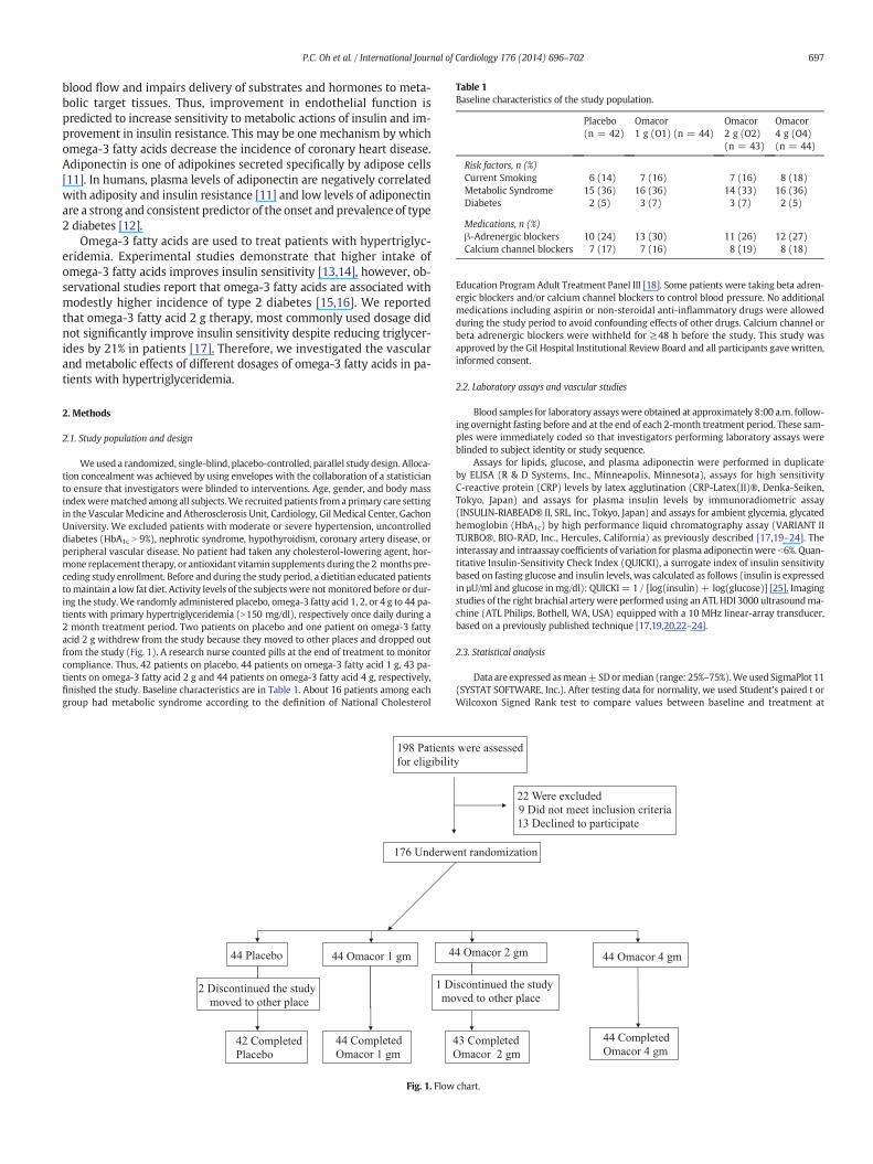
Table 1Baseline characteristics of the study population.
Placebo(n = 42)
Omacor1 g (O1) (n = 44)
Omacor2 g (O2)(n = 43)
Omacor4 g (O4)(n = 44)
Risk factors, n (%)Current Smoking 6 (14) 7 (16) 7 (16) 8 (18)Metabolic Syndrome 15 (36) 16 (36) 14 (33) 16 (36)Diabetes 2 (5) 3 (7) 3 (7) 2 (5)
Medications, n (%)β-Adrenergic blockers 10 (24) 13 (30) 11 (26) 12 (27)Calcium channel blockers 7 (17) 7 (16) 8 (19) 8 (18)
697P.C. Oh et al. / International Journal of Cardiology 176 (2014) 696–702
blood flow and impairs delivery of substrates and hormones to meta-bolic target tissues. Thus, improvement in endothelial function ispredicted to increase sensitivity to metabolic actions of insulin and im-provement in insulin resistance. This may be one mechanism by whichomega-3 fatty acids decrease the incidence of coronary heart disease.Adiponectin is one of adipokines secreted specifically by adipose cells[11]. In humans, plasma levels of adiponectin are negatively correlatedwith adiposity and insulin resistance [11] and low levels of adiponectinare a strong and consistent predictor of the onset and prevalence of type2 diabetes [12].
Omega-3 fatty acids are used to treat patients with hypertriglyc-eridemia. Experimental studies demonstrate that higher intake ofomega-3 fatty acids improves insulin sensitivity [13,14], however, ob-servational studies report that omega-3 fatty acids are associated withmodestly higher incidence of type 2 diabetes [15,16]. We reportedthat omega-3 fatty acid 2 g therapy, most commonly used dosage didnot significantly improve insulin sensitivity despite reducing triglycer-ides by 21% in patients [17]. Therefore, we investigated the vascularand metabolic effects of different dosages of omega-3 fatty acids in pa-tients with hypertriglyceridemia.
2. Methods
2.1. Study population and design
Weused a randomized, single-blind, placebo-controlled, parallel study design. Alloca-tion concealment was achieved by using envelopes with the collaboration of a statisticianto ensure that investigators were blinded to interventions. Age, gender, and body massindexwerematched among all subjects.We recruited patients froma primary care settingin the Vascular Medicine and Atherosclerosis Unit, Cardiology, Gil Medical Center, GachonUniversity. We excluded patients with moderate or severe hypertension, uncontrolleddiabetes (HbA1c N 9%), nephrotic syndrome, hypothyroidism, coronary artery disease, orperipheral vascular disease. No patient had taken any cholesterol-lowering agent, hor-mone replacement therapy, or antioxidant vitamin supplements during the 2 months pre-ceding study enrollment. Before and during the study period, a dietitian educated patientstomaintain a low fat diet. Activity levels of the subjects were notmonitored before or dur-ing the study.We randomly administered placebo, omega-3 fatty acid 1, 2, or 4 g to 44 pa-tients with primary hypertriglyceridemia (N150 mg/dl), respectively once daily during a2 month treatment period. Two patients on placebo and one patient on omega-3 fattyacid 2 g withdrew from the study because they moved to other places and dropped outfrom the study (Fig. 1). A research nurse counted pills at the end of treatment to monitorcompliance. Thus, 42 patients on placebo, 44 patients on omega-3 fatty acid 1 g, 43 pa-tients on omega-3 fatty acid 2 g and 44 patients on omega-3 fatty acid 4 g, respectively,finished the study. Baseline characteristics are in Table 1. About 16 patients among eachgroup had metabolic syndrome according to the definition of National Cholesterol
2 Discontinued the study
moved to other place
176 Underw
198 Patients
for eligibilit
44 Placebo
42 Completed
Placebo
44 Completed
Omacor 1 gm
44 Omacor 1 gm 4
1 D
mo
Fig. 1. Flow
Education Program Adult Treatment Panel III [18]. Some patients were taking beta adren-ergic blockers and/or calcium channel blockers to control blood pressure. No additionalmedications including aspirin or non-steroidal anti-inflammatory drugs were allowedduring the study period to avoid confounding effects of other drugs. Calcium channel orbeta adrenergic blockers were withheld for ≥48 h before the study. This study wasapproved by the Gil Hospital Institutional Review Board and all participants gave written,informed consent.
2.2. Laboratory assays and vascular studies
Blood samples for laboratory assayswere obtained at approximately 8:00 a.m. follow-ing overnight fasting before and at the end of each 2-month treatment period. These sam-ples were immediately coded so that investigators performing laboratory assays wereblinded to subject identity or study sequence.
Assays for lipids, glucose, and plasma adiponectin were performed in duplicateby ELISA (R & D Systems, Inc., Minneapolis, Minnesota), assays for high sensitivityC-reactive protein (CRP) levels by latex agglutination (CRP-Latex(II)®, Denka-Seiken,Tokyo, Japan) and assays for plasma insulin levels by immunoradiometric assay(INSULIN-RIABEAD® II, SRL, Inc., Tokyo, Japan) and assays for ambient glycemia, glycatedhemoglobin (HbA1c) by high performance liquid chromatography assay (VARIANT IITURBO®, BIO-RAD, Inc., Hercules, California) as previously described [17,19–24]. Theinterassay and intraassay coefficients of variation for plasma adiponectinwere b6%. Quan-titative Insulin-Sensitivity Check Index (QUICKI), a surrogate index of insulin sensitivitybased on fasting glucose and insulin levels, was calculated as follows (insulin is expressedin µU/ml and glucose inmg/dl): QUICKI= 1 / [log(insulin)+ log(glucose)] [25]. Imagingstudies of the right brachial arterywere performed using an ATL HDI 3000 ultrasoundma-chine (ATL Philips, Bothell, WA, USA) equipped with a 10 MHz linear-array transducer,based on a previously published technique [17,19,20,22–24].
2.3. Statistical analysis
Data are expressed asmean±SDormedian (range: 25%–75%).We used SigmaPlot 11(SYSTAT SOFTWARE, Inc.). After testing data for normality, we used Student's paired t orWilcoxon Signed Rank test to compare values between baseline and treatment at
ent randomization
were assessed
y
22 Were excluded
9 Did not meet inclusion criteria
13 Declined to participate
43 Completed
Omacor 2 gm
4 Omacor 2 gm
iscontinued the study
ved to other place
44 Omacor 4 gm
44 Completed
Omacor 4 gm
chart.

698 P.C. Oh et al. / International Journal of Cardiology 176 (2014) 696–702
2 months, as reported in Tables 2 and3.We used oneway analysis of variance (ANOVA) orKruskal–Wallis ANOVA on Ranks to compare baseline or treatment effects amongtreatment groups. Post-hoc comparisons between different treatment pairs were madeusing the Student–Newman–Keuls multiple comparison procedures or Dunn's method.Pearson or Spearman correlation coefficient analysis was used to assess associations be-tweenmeasured parameters, as reported in Tables 2 and 3.We calculated that 38 subjectswould provide 80% power for detecting an absolute increase of 1.6% or greater in flow-mediated dilation of the brachial artery between baseline and omega-3 fatty acid 2 g,with α = 0.05 based on our previous studies [17]. The comparison of endothelium-dependent dilation was prospectively designated as the primary end-point of the study.All other comparisons were considered secondary. P b 0.05 was considered to representstatistical significance.
3. Results
There were no significant differences between groups for any of thebaseline measurements (Tables 2 and 3).
3.1. Effects on lipids
Placebo treatment significantly reduced triglycerides (TG) andTG/high-density lipoprotein (HDL) cholesterol ratio from baseline.Omega-3 fatty acid treatment dose-dependently and significantlyreduced TG and TG/HDL cholesterol ratio from baseline. Effects ofomega-3 fatty acids on TG levels were significant when comparedwith placebo treatment (P b 0.05 by ANOVA; Fig. 2). Omega-3 fattyacid 2 g and 4 g treatment significantly reduced apolipoprotein AI andnon-HDL cholesterol from baseline, respectively. However, omega-3fatty acid treatment did not significantly change other lipoproteins in-cluding total cholesterol, non-HDL cholesterol and HDL cholesterolfrom baseline. Effects of omega-3 fatty acids on these were not signifi-cant when compared with placebo treatment.
Table 2Effects of placebo or Omacor on lipids and endocrine parameters in patients with hypertriglyc
Placebo (n = 42) Omacor 1 g (O1) (n = 44)
Baseline Treatment Baseline Treatment
Age 54 ± 9 55 ± 9Sex (M:F) 23:19 22:22BMI 26.50 ± 2.72 26.45 ± 2.71 26.32 ± 3.20 26.35 ± 3.2Lipids (mg/dl)
Total cholesterol 201 ± 29 196 ± 31 197 ± 29 193 ± 32Triglycerides 281 ± 63 247 ± 102⁎ 286 ± 73 229 ± 99⁎
LDL cholesterol 111 ± 34 109 ± 34 109 ± 32 110 ± 33Apo B 107 ± 20 107 ± 23 105 ± 19 103 ± 19HDL cholesterol 42 ± 8 43 ± 7 41 ± 8 43 ± 9Apo A-I 131 ± 15 133 ± 17 128 ± 16 130 ± 17TG/HDL ratio 7.0 ± 2.4 5.9 ± 2.9⁎ 7.3 ± 2.6 5.6 ± 2.8‡
Non-HDL 159 ± 27 153 ± 29 157 ± 27 150 ± 31Vasomotor
FMD (%) 5.98 ± 1.42 6.31 ± 1.56‡ 6.04 ± 1.52 7.61 ± 1.68NTG (%) 16.23 ± 3.19 16.59 ± 3.26 16.25 ± 3.34 16.61 ± 4.1
InflammationhsCRP (mg/l) 1.15
(0.50–1.75)0.95(0.48–1.93)
1.05(0.43–1.68)
0.65(0.33–1.38)
Fibrinogen (mg/dl) 355 ± 69 348 ± 83 353 ± 65 348 ± 62Insulin resistance
ADP (μg/ml) 2.6 (1.9–3.8) 2.7 (1.9–4.1) 2.9 (1.5–4.2) 2.3 (1.7–4.1)Insulin (μU/ml) 7.4 (5.5–13.7) 9.3 (5.8–13.5) 7.3 (5.1–11.1) 9.7 (5.9–13.8Glucose (mg/dl) 101 ± 15 101 ± 13 98 ± 15 99 ± 14QUICKI 0.35 ± 0.03 0.34 ± 0.04 0.35 ± 0.03 0.34 ± 0.03HbA1c (%) 5.97 ± 0.65 5.94 ± 0.63 5.93 ± 0.62 5.97 ± 0.55
Data are expressed as means ± SD or median.There were no significant differences among each baseline value.⁎P b 0.05, +P b 0.01, ‡P b 0.001 for comparison with each baseline value.†P b 0.05 for comparison with the value after therapy with placebo.Global ANOVA indicates group differences.FMD = flow-mediated dilation, NTG = nitroglycerin-induced dilation, HbA1c = glycated hemQuantitative Insulin-Sensitivity Check Index (QUICKI) = 1 / [log (insulin) + log (glucose)] [2
3.2. Effects on vasomotor function, high sensitivity C-reactive protein,and fibrinogen
Placebo treatment significantly improved flow-mediated dilatorresponse to hyperemia (FMD) relative to baseline measurements.Omega-3 fatty acid treatment dose-dependently and significantly im-proved FMD after 2 months of therapy when compared with baseline(P b 0.001 by paired t-test) or when compared with placebo treatment(P b 0.001 by ANOVA; Fig. 2). Brachial artery dilator responses to nitro-glycerin were not significantly different between any of the therapies.Placebo and omega-3 fatty acid treatment did not significantly changehigh sensitivity CRP and fibrinogen levels relative to baseline measure-ments except omega-3 fatty acid 4 g increasing fibrinogen levels.
3.3. Effects on adiponectin, glycated hemoglobin, and insulin resistance
Placebo and omega-3 fatty acid treatment did not significantlychange insulin or glucose levels from baseline except omega-3 fattyacid 1 g increasing insulin levels. However, the effects of omega-3fatty acid treatment on fasting insulin and glucose levels were not sig-nificant when compared with placebo treatment (Fig. 3). We observedsignificant inverse correlations between baseline adiponectin and base-line insulin levels (r = −0.338, P = 0.028 before placebo) and signifi-cant correlations between baseline adiponectin levels and baselineQUICKI (r = 0.451, P = 0.003 before placebo; r = 0.314, P = 0.038before omega-3 fatty acid 1 g).
Placebo and omega-3 fatty acids did not significantly change plasmaadiponectin levels, insulin sensitivity (determined by QUICKI), or HbA1c
levels relative to baseline measurements except omega-3 fatty acid 2 gdecreasing adiponectin levels and 1 g decreasing insulin sensitivity.However, the effects of omega-3 fatty acid treatment on these werenot significant when compared with placebo treatment (Fig. 4).
eridemia.
Omacor 2 g (O2) (n = 43) Omacor 4 g (O4) (n = 44) GlobalANOVA
Baseline Treatment Baseline Treatment
54 ± 9 55 ± 8 0.94423:20 23:21
0 26.51 ± 2.63 26.43 ± 2.61 26.18 ± 3.21 26.09 ± 3.19 0.959
195 ± 31 194 ± 31 198 ± 30 188 ± 32 0.403267 ± 118 203 ± 106+ 287 ± 73 191 ± 117‡† 0.021109 ± 33 113 ± 32 110 ± 33 109 ± 33 0.743107 ± 22 108 ± 20 107 ± 21 103 ± 18 0.35843 ± 7 42 ± 8 40 ± 7 40 ± 8 0.050132 ± 17 126 ± 14⁎ 128 ± 17 125 ± 17 0.0966.4 ± 2.9 5.2 ± 3.1‡ 7.3 ± 2.4 5.0 ± 3.2‡ 0.152153 ± 29 153 ± 29 157 ± 28 148 ± 29⁎ 0.263
‡† 5.82 ± 1.54 7.64 ± 1.74‡† 6.03 ± 1.39 8.37 ± 1.51‡† b0.0014 16.44 ± 3.18 16.78 ± 3.83 16.05 ± 2.96 16.45 ± 4.11 0.989
1.00(0.50–2.10)
0.80(0.40–1.70)
1.05(0.53–1.70)
0.85(0.60–1.68)
0.401
354 ± 80 359 ± 70 349 ± 61 370 ± 60⁎ 0.070
2.6 (1.8–4.5) 2.3 (1.7–4.3)⁎ 2.6 (1.5–3.6) 2.2 (1.6–3.5) 0.375)⁎ 10.1 (6.4–12.8) 7.8 (6.0–12.0) 7.3 (4.2–10.2) 8.5 (5.6–11.1) 0.068
102 ± 18 99 ± 12 96 ± 15 98 ± 11 0.397⁎ 0.34 ± 0.03 0.35 ± 0.03 0.36 ± 0.03 0.35 ± 0.03 0.222
5.94 ± 0.63 5.98 ± 0.61 5.95 ± 0.33 6.01 ± 0.51 0.389
oglobin, ADP = adiponectin.5].

Table 3Effects of placebo or Omacor on lipids and endocrine parameters in patients with hypertriglyceridemia and metabolic syndrome/type 2 diabetes.
Placebo (n = 17) Omacor 1 g (O1) (n = 19) Omacor 2 g (O2) (n = 17) Omacor 4 g (O4) (n = 18) GlobalANOVA
Baseline Treatment Baseline Treatment Baseline Treatment Baseline Treatment
Age 53 ± 6 54 ± 10 55 ± 8 54 ± 9 0.877Sex (M:F) 9:8 9:10 8:9 8:10BMI 26.41 ± 2.68 26.42 ± 2.72 26.12 ± 3.05 26.06 ± 2.98 25.89 ± 2.40 25.82 ± 2.44 26.18 ± 2.99 26.06 ± 2.90 0.627Lipids (mg/dl)
Total cholesterol 206 ± 37 200 ± 38 193 ± 28 190 ± 36 192 ± 38 196 ± 33 194 ± 28 186 ± 32 0.432Triglycerides 268 ± 66 243 ± 104 274 ± 70 227 ± 82⁎ 231 ± 119 182 ± 135⁎ 276 ± 56 175 ± 80‡ 0.055LDL cholesterol 110 ± 42 107 ± 42 105 ± 30 108 ± 35 111 ± 39 114 ± 39 112 ± 33 113 ± 31 0.904Apo B 106 ± 23 110 ± 29 105 ± 23 101 ± 23 107 ± 29 107 ± 17 107 ± 22 105 ± 21 0.576HDL cholesterol 42 ± 7 43 ± 8 42 ± 9 44 ± 10 43 ± 8 42 ± 8 40 ± 9 40 ± 8 0.445Apo A-I 136 ± 15 135 ± 18 129 ± 17 134 ± 18 130 ± 13 126 ± 11 127 ± 20 123 ± 18 0.204TG/HDL ratio 6.7 ± 2.6 5.9 ± 3.1 6.8 ± 2.1 5.4 ± 2.5⁎ 5.5 ± 3.1 4.3 ± 2.8+ 7.1 ± 2.0 4.5 ± 2.1‡ 0.220Non-HDL 164 ± 36 157 ± 35 152 ± 27 146 ± 35 149 ± 36 154 ± 29 154 ± 27 146 ± 28 0.222
VasomotorFMD (%) 6.03 ± 1.60 6.37 ± 1.81+ 5.76 ± 1.30 7.31 ± 1.30‡† 5.62 ± 1.72 7.39 ± 1.98‡† 5.68 ± 1.11 8.32 ± 1.25‡† b0.001NTG (%) 16.16 ± 3.37 16.35 ± 3.43 15.94 ± 3.32 15.97 ± 4.18 15.98 ± 3.36 15.94 ± 4.40 16.31 ± 3.39 17.25 ± 4.55 0.323
InflammationhsCRP (mg/l) 0.90
(0.30–1.75)0.70(0.40–1.25)
1.00(0.40–1.70)
0.50(0.30–1.00)
1.10(0.80–2.30)
1.00(0.45–2.55)
1.15(0.60–1.75)
0.85(0.70–1.43)
0.668
Fibrinogen (mg/dl) 336 ± 68 331 ± 72 343 ± 69 331 ± 49 357 ± 77 366 ± 64 361 ± 56 391 ± 60 0.179Insulin resistance
ADP (μg/ml) 3.0 (2.0–4.1) 2.9 (1.8–3.9) 2.1 (1.3–4.0) 2.2 (1.6–3.7) 3.4 (1.4–4.5) 3.1 (1.7–4.2) 2.5 (1.4–3.7) 2.3 (1.6–3.6) 0.656Insulin (μU/ml) 10.1 (5.5–15.3) 8.6 (5.2–12.4) 6.6 (4.0–8.7) 8.9 (5.1–14.7) 8.3 (6.5–13.1) 7.7 (6.3–13.1) 7.4 (4.6–10.7) 8.6 (5.9–10.7) 0.636Glucose (mg/dl) 103 ± 19 105 ± 15 95 ± 10 96 ± 13 103 ± 13 104 ± 15 97 ± 10 94 ± 8 0.063QUICKI 0.34 ± 0.03 0.34 ± 0.03 0.36 ± 0.04 0.35 ± 0.04 0.34 ± 0.04 0.34 ± 0.03 0.35 ± 0.04 0.35 ± 0.03 0.793HbA1c (%) 5.95 ± 0.49 5.95 ± 0.55 5.79 ± 0.35 5.87 ± 0.40 6.12 ± 0.72 6.16 ± 0.73 5.96 ± 0.30 6.01 ± 0.43 0.649
Data are expressed as means ± SD or median.There were no significant differences among each baseline value.⁎P b 0.05, +P b 0.01, ‡P b 0.001 for comparison with each baseline value.†P b 0.05 for comparison with the value after therapy with placebo.Global ANOVA indicates group differences.HbA1c = glycated hemoglobin, ADP = adiponectin.Quantitative Insulin-Sensitivity Check Index (QUICKI) = 1 / [log (insulin) + log (glucose)] [25].
699P.C. Oh et al. / International Journal of Cardiology 176 (2014) 696–702
We investigated whether changes in percent flow-mediated dilatorresponse to hyperemia, plasma levels of adiponectin, insulin, insulin re-sistance, or HbA1c were related to changes in lipoprotein levels. Therewere no significant correlations between changes in these parametersand changes in lipoprotein levels following any of the therapies. Further,there were no significant correlations between percent changes inadiponectin levels and percent changes in insulin or percent changesin QUICKI following any of the therapies.
3.4. Effects of therapies in patients with metabolic syndrome/type 2Diabetes mellitus
We analyzed patients with metabolic syndrome/type 2 diabetesmellitus, as reported in Table 3. Overall, compared with the effects of
-40
-30
-20
-10
0
10
Pl O1 O20
20
40
60
Pl O1
%Change in FMD%Change in Triglycerides
P=0.021by ANOVA
O4
P<0.001by ANOVA
O2 O4
Fig. 2. Omega-3 fatty acid treatment (O1, O2, or O4), dose-dependently and significantlyreduced triglyceride levels and improved flow-mediated dilation (FMD) after 2 monthsof therapy when compared with baseline (P b 0.001 by paired t-test) or when comparedwith placebo treatment (P = 0.021 and P b 0.001 by ANOVA, respectively). Standarderror of the mean is identified by the bars.
each therapy in 44 hypertriglyceridemic patients, we observed similarresults in patients with metabolic syndrome/type 2 diabetes mellitus.Omega-3 fatty acid treatment dose-dependently and significantlyreduced TG and TG/HDL cholesterol ratio and improved FMD after2 months of therapy when compared with baseline or when comparedwith placebo treatment. However, omega-3 fatty acid treatment did notsignificantly change high sensitivity CRP, fibrinogen, plasma adiponectinlevels, insulin sensitivity (determined by QUICKI), or HbA1c levels relativeto baseline measurements. Of note, the effects of omega-3 fatty acidtreatment on these were not significant when compared with placebotreatment.
4. Discussion
Weobserved that omega-3 fatty acid therapy dose-dependently andsignificantly decreased triglycerides and triglycerides/HDL cholesterol
-6
-4
-2
0
2
4
6
8
10
Pl O1 O2-30
-20
-10
0
10
20
30
Pl O1
%Change in Insulin%Change in Glucose
P=0.397by ANOVA
O4
P=0.068by ANOVA on Ranks
O2 O4
Fig. 3. The effects of omega-3 fatty acid treatment on fasting insulin and glucose levelswere not significant when compared with placebo treatment. Median values are used in% change in insulin. Standard error of the mean is identified by the bars.

-12
-10
-8
-6
-4
-2
0
2
Pl O1 O2-5-4-3-2-1012345
Pl O1
%Change in QUICKI%Change in Adiponectin
P=0.375by ANOVA on Ranks
O4
P=0.222by ANOVA
O2 O4
-2
-1
0
1
2
3
Pl O1 O2-5-4-3-2-1012345
Pl O1
%Change in QUICKI%Change in HbA1C
P=0.389by ANOVA
O4
P=0.222by ANOVA
O2 O4
Fig. 4. The effects of omega-3 fatty acid treatment on plasma adiponectin levels, insulinsensitivity (determined by QUICKI), or HbA1c levels were not significant when comparedwith placebo treatment. Median values are used in % change in adiponectin. Standarderror of the mean is identified by the bars.
700 P.C. Oh et al. / International Journal of Cardiology 176 (2014) 696–702
and improved flow-mediated dilation, compared with placebo. None-theless, when compared with placebo, omega-3 fatty acid therapy didnot significantly change other lipoproteins such as non-HDL cholesteroland HDL cholesterol, high-sensitivity C-reactive protein, fibrinogen,insulin, glucose, adiponectin, glycated hemoglobin levels and insulinsensitivity in patients with hypertriglyceridemia, regardless of dosages.We observed similar results in a subgroup of patients with themetabol-ic syndrome/type 2 diabetes mellitus.
Several studies reported that omega-3 fatty acids improve flow-mediated arterial dilation [26–28]. This effect of omega-3 fatty acidson endothelial functionmight be supported by experimental evidences.In the rat fed menhaden oil-rich diets, aortic NO production wasincreased [29]. EPA also enhanced NO production in cultured humanendothelial cells [30] and induced Ca2+-independent activationand translocation of endothelial NO synthase to the cytosol andendothelium-dependent vasorelaxation [31]. In addition, DHA de-creased cytokine-induced expression of endothelial leukocyte adhesionmolecules and secretion of IL-6 and IL-8 in cultured endothelial cell [32].However, it remains unclear whether its favorable vasomotor functionor anti-inflammatory effects translate to improve insulin sensitivity inpatients. For example, our group has demonstrated that statins do notimprove but worsen insulin sensitivity in patients despite of improvingflow-mediated arterial dilation significantly [19,21,23]. Therefore, anovel point of our current study is to investigate vascular andmetabolicphenotype of different dosages of omega-3 fatty acids in patients.
Non-HDL cholesterol is an important one of residual risk factors [33]and has predictive value of cardiovascular events [34,35]. Acute phase re-actants such as C-reactive protein have also predictive value of cardiovas-cular events. However, in the current study, omega-3 fatty acid therapydid not significantly decrease non-HDL cholesterol and HDL cholesterol,high-sensitivity C-reactive protein, fibrinogen, compared with placebo.
When we observed that omega-3 fatty acid 2 g therapy did not sig-nificantly improve insulin sensitivity in patients despite reduction oftriglycerides and improvement of flow-mediated dilation [17], some
argued high dose may improve [13,14,36]. Therefore, we investigatedthe effects of low-to-high dose omega-3 fatty acids with a powercalculation.
Adiponectin is an adipose-derived factor that augments and mimicsbothmetabolic and vascular actions of insulin [11]. Adiponectin directlystimulates nitric oxide production from endothelium via activation ofAMP-activated protein kinase and nitric oxide synthase [37]. Therefore,increasing adiponectin levels is predicted to improve both insulin sensi-tivity and endothelial function bymultiplemechanisms [11]. Regulationof metabolic homeostasis and hemodynamic homeostasis may becoupled by vascular actions of insulin to stimulate production of nitricoxide. Thus, improvements in endothelial functionmay increase insulinsensitivity while increased insulin sensitivity may improve endothelialfunction [8,9,38].
QUICKI is a reliable surrogate index for insulin sensitivity that has anespecially excellent correlation with the reference standard glucoseclamp method in insulin resistant subjects with type II diabetes or obe-sity [25]. In addition, test characteristics of QUICKI including coefficientof variation and discriminant ratio are significantly better than othersimple surrogate indexes and comparable to those of the glucoseclamp [39]. A large meta-analysis of insulin resistant subjects demon-strates that QUICKI is among the best surrogate indexes in terms of pre-dictive power for the onset of diabetes [40]. Becausemeasures of insulinresistance were considered secondary in the current study, we usedQUICKI to assess insulin sensitivity instead of the reference standardeuglycemic glucose clamp technique. Thus, QUICKI is the most exten-sively validated and accurate surrogate index of insulin sensitivity cur-rently available in humans.
The results of experimental and clinical studies with fish oil andomega-3 fatty acids are controversial. Dietary fish oil increased serumtotal adiponectin levels in a dietary model of insulin resistance inducedby long-term sucrose-rich diet in rats [41]. Experimental studies havedemonstrated that dietary fish oils and omega-3 fatty acids increasetotal adiponectin levels [41–44]. Indeed, one study demonstrated thatthe G protein-coupled receptor GPR120 is a receptor for omega-3 fattyacids on primary intraperitoneal macrophages and monocytic RAW264.7 cells and further, activation of GPR120 by omega-3 fatty acidsinhibited multiple inflammation cascades in macrophages and reversesinsulin resistance in obese mice although this study did not measureadiponectin and acute phase reactant. Since chronic macrophage-mediated tissue inflammation is a keymechanism for insulin resistancein obesity, they fed obese wild type and GPR120 knockout mice a high-fat diet with or without omega-3 fatty acid supplementation. Theomega-3 fatty acid treatment inhibited inflammation by observingexpression of tumor necrosis factor-α, interleukin-6, and monocytechemoattractant protein-1 and enhanced systemic insulin sensitivityin WT mice by increasing glucose transport and translocation ofGLUT4 and enhancing glucose uptake, but was without effect inGPR120 knockout mice [45].
However, other studies are different. EPA significantly decreasedadiponectin gene expression and protein secretion in primary culturedrat adipocytes [46]. Omega-3 fatty acids did not significantly increaseplasma or high-molecular weight adiponectin levels in overweight-to-moderately obese healthy people [47]. DHA supplementation did notchange fasting or postprandial insulin and glucose concentrations andinsulin sensitivity, determined by insulin and homeostasis model as-sessment of insulin resistance (HOMA-IR) in hypertriglyceridemicmen [48]. In a meta-analysis of 18 randomized clinical trials, omega-3fatty acids had no effects on insulin resistance compared to placebo[49]. Instead, some observational studies reported that omega-3 fattyacid or fish consumption was associated with modestly higher inci-dence of type 2 diabetes [15,16]. In two meta-analyses, fish oil con-sumption had no overall effects on fasting glucose or HbA1c in patientswith type 2 diabetes [50,51]. Overall, it seems that omega-3 fatty acidshave no overall effects or increase insulin resistance or diabetes risk,but further investigation is needed.

701P.C. Oh et al. / International Journal of Cardiology 176 (2014) 696–702
Metabolic syndrome is associated with atherosclerotic and cardio-vascular disease. Patients with metabolic syndrome comprise one ofthe largest groups of individuals with dyslipidemia and insulin resis-tance. In the present study, we observed similar results in a subgroupof patients with the metabolic syndrome/type 2 diabetes mellitus.
In addition to epidemiologic studies, recent clinical studies demon-strate that omega-3 fatty acids decreased admission to hospital for car-diovascular reasons and mortality in patients with heart failure [52] orEPA decreased major coronary events, especially non-fatal coronaryevents, but not sudden cardiac death and coronary death in hypercho-lesterolemic patients [53]. By contrast, recently published OMEGA andAlpha Omega trial report that low doses of omega-3 fatty acids failedto reduce the rate of major cardiovascular events [54,55].
In summary, omega-3 fatty acid therapy dose-dependently and sig-nificantly decreased triglycerides and improved flow-mediated dilation.Nonetheless, omega-3 fatty acid therapy did not significantly improveacute-phase reactants and insulin sensitivity in patients with hypertri-glyceridemia, regardless of dosages.
Conflict of interest
None.
Acknowledgment
This study was partly supported by grants from established investi-gator award, Gachon University Gil Medical Center (K.K. Koh).
References
[1] Dietary supplementation with n−3 polyunsaturated fatty acids and vitamin E aftermyocardial infarction: results of the GISSI-Prevenzione trial: Gruppo Italiano per loStudio della Sopravvivenza nell'Infarto Miocardico. Lancet 1999;354:447–55.
[2] Marchioli R, Barzi F, Bomba E, et al. GISSI-Prevenzione investigators. Early protectionagainst sudden death by n−3 polyunsaturated fatty acids aftermyocardial infarction:time-course analysis of the results of the Gruppo Italiano per lo Studio dellaSopravvivenza nell'Infarto Miocardico (GISSI)-Prevenzione. Circulation 2002;105:1897–903.
[3] Streppel MT, OckeMC, Boshuizen HC, Kok FJ, Kromhout D. Long-term fish consump-tion and n−3 fatty acid intake in relation to (sudden) coronary heart disease death:the Zutphen study. Eur Heart J 2008;29:2024–30.
[4] Bays HE, Tighe AP, Sadovsky R, Davidson MH. Prescription omega-3 fatty acids andtheir lipid effects: physiologic mechanisms of action and clinical implications. ExpertRev Cardiovasc Ther 2008;6:391–409.
[5] Mori TA, Watts GF, Burke V, Hilme E, Puddey IB, Beilin LJ. Differential effects ofeicosapentaenoic acid and docosahexaenoic acid on vascular reactivity of the fore-arm microcirculation in hyperlipidemic, overweight men. Circulation 2000;102:1264–9.
[6] Stirban A, Nandrean S, Gotting C, et al. Effects of n−3 fatty acids on macro- andmicrovascular function in subjects with type 2 diabetes mellitus. Am J Clin Nutr2010;91:808–13.
[7] Kotwal S, Jun M, Sullivan D, Perkovic V, Neal B. Omega 3 Fatty acids and cardiovas-cular outcomes: systematic review and meta-analysis. Circ Cardiovasc Qual Out-comes 2012;5:808–18.
[8] Kim J, Montagnani M, Koh KK, Quon MJ. Reciprocal relationships between insulinresistance and endothelial dysfunction: molecular and pathophysiological mecha-nisms. Circulation 2006;113:1888–904.
[9] Han SH, Quon MJ, Koh KK. Reciprocal relationships between abnormal metabolicparameters and endothelial dysfunction. Curr Opin Lipidol 2007;18:58–65.
[10] Jee SH, Jo J. Linkage of epidemiologic evidence with the clinical aspects of metabolicsyndrome. Korean Circ J 2012;42:371–8.
[11] Han SH, QuonMJ, Kim JA, Koh KK. Adiponectin and cardiovascular disease: responseto therapeutic interventions. J Am Coll Cardiol 2007;49:531–8.
[12] Lim S, Quon MJ, Koh KK. Modulation of adiponectin as a potential therapeutic strat-egy. Atherosclerosis 2014;233:721–8.
[13] HolnessMJ, Smith ND, Greenwood GK, SugdenMC. Acute omega-3 fatty acid enrich-ment selectively reverses high-saturated fat feeding-induced insulin hypersecretionbut does not improve peripheral insulin resistance. Diabetes 2004;53:S166–71[Suppl.].
[14] Svegliati-Baroni G, Candelaresi C, Saccomanno S, et al. A model of insulin resistanceand nonalcoholic steatohepatitis in rats: role of peroxisome proliferator-activatedreceptor alpha and n−3 polyunsaturated fatty acid treatment on liver injury. Am JPathol 2006;169:846–60.
[15] Kaushik M, Mozaffarian D, Spiegelman D, Manson JE, Willett WC, Hu FB. Long-chainomega-3 fatty acids, fish intake, and the risk of type 2 diabetes mellitus. Am J ClinNutr 2009;90:613–20.
[16] Djousse L, Gaziano JM, Buring JE, Lee IM. Dietary omega-3 fatty acids and fishconsumption and risk of type 2 diabetes. Am J Clin Nutr 2011;93:143–50.
[17] Koh KK, Quon MJ, Shin KC, et al. Significant differential effects of omega-3 fatty acidsand fenofibrate in patients with hypertriglyceridemia. Atherosclerosis 2012;220:537–44.
[18] Third Report of the National Cholesterol Education Program (NCEP) Expert Panelon Detection, Evaluation, and Treatment of High Blood Cholesterol in Adults(Adult Treatment Panel III) final report. Circulation 2002;106:3143–421.
[19] Koh KK, QuonMJ, Han SH, et al. Additive beneficial effects of losartan combinedwithsimvastatin in the treatment of hypercholesterolemic, hypertensive patients.Circulation 2004;110:3687–92.
[20] Koh KK, Han SH, Quon MJ, Ahn JY, Shin EK. Beneficial effects of fenofibrate to im-prove endothelial dysfunction and raise adiponectin levels in patients with primaryhypertriglyceridemia. Diabetes Care 2005;28:1419–24.
[21] Koh KK, Quon MJ, Han SH, Lee Y, Kim SJ, Shin EK. Atorvastatin causes insulin resis-tance and increases ambient glycemia in hypercholesterolemic patients. J Am CollCardiol 2010;55:1209–16.
[22] Koh KK, Quon MJ, Lim S, et al. Effects of fenofibrate therapy on circulatingadipocytokines in patients with primary hypertriglyceridemia. Atherosclerosis2011;214:144–7.
[23] Koh KK, Quon MJ, Sakuma I, et al. Differential metabolic effects of rosuvastatin andpravastatin in hypercholesterolemic patients. Int J Cardiol 2013;166:509–15.
[24] Koh KK, Lim S, Choi H, et al. Combination pravastatin and valsartan treatment hasadditive beneficial effects to simultaneously improve both metabolic and cardiovas-cular phenotypes beyond that of monotherapy with either drug in patients withprimary hypercholesterolemia. Diabetes 2013;62:3547–52.
[25] Katz A, Nambi SS, Mather K, et al. Quantitative insulin sensitivity check index: a sim-ple, accurate method for assessing insulin sensitivity in humans. J Clin EndocrinolMetab 2000;85:2402–10.
[26] Goodfellow J, BellamyMF, RamseyMW, Jones CJ, LewisMJ. Dietary supplementationwith marine omega-3 fatty acids improve systemic large artery endothelial functionin subjects with hypercholesterolemia. J Am Coll Cardiol 2000;35:265–70.
[27] Engler MM, Engler MB, Malloy M, et al. Docosahexaenoic acid restores endothelialfunction in children with hyperlipidemia: results from the EARLY study. Int J ClinPharmacol Ther 2004;42:672–9.
[28] Rizza S, Tesauro M, Cardillo C, et al. Fish oil supplementation improves endothelialfunction in normoglycemic offspring of patients with type 2 diabetes. Atherosclero-sis 2009;206:569–74.
[29] Lopez D, Moller M, Denicola A, et al. Long-chain n−3 polyunsaturated fatty acidfrom fish oil modulates aortic nitric oxide and tocopherol status in the rat. Br JNutr 2008;100:767–75.
[30] Okuda Y, Kawashima K, Sawada T, et al. Eicosapentaenoic acid enhances nitric oxideproduction by cultured human endothelial cells. Biochem Biophys Res Commun1997;232:487–91.
[31] Omura M, Kobayashi S, Mizukami Y, et al. Eicosapentaenoic acid (EPA) inducesCa(2+)-independent activation and translocation of endothelial nitric oxidesynthase and endothelium-dependent vasorelaxation. FEBS Lett 2001;487:361–6.
[32] De Caterina R, Cybulsky MI, Clinton SK, Gimbrone Jr MA, Libby P. The omega-3 fattyacid docosahexaenoate reduces cytokine-induced expression of proatherogenic andproinflammatory proteins in human endothelial cells. Arterioscler Thromb 1994;14:1829–36.
[33] Lim S, Park YM, Sakuma I, Koh KK. How to control residual cardiovascular riskdespite statin treatment: focusing on HDL-cholesterol. Int J Cardiol 2013;166:8–14.
[34] Emerging Risk Factors CollaborationDi Angelantonio E, Sarwar N, et al. Major lipids,apolipoproteins, and risk of vascular disease. JAMA 2009;302:1993–2000.
[35] Ahmed K, Jeong MH, Chakraborty R, et al. Clinical impact of non-high densitylipoprotein-cholesterol and apolipoprotein B on clinical outcomes in metabolic syn-drome patients with acutemyocardial infarction undergoing percutaneous coronaryintervention. Korean Circ J 2012;42:319328.
[36] Eckel RH. The fish oil story remains fish. Circulation 2010;122:2110–2.[37] Chen H, Montagnani M, Funahashi T, Shimomura I, QuonMJ. Adiponectin stimulates
production of nitric oxide in vascular endothelial cells. J Biol Chem 2003;278:45021–6.
[38] Kim J, Koh KK, Quon MJ. The union of vascular and metabolic actions of insulin insickness and in health. Arterioscler Thromb Vasc Biol 2005;25:889–91.
[39] Mather KJ, Hunt AE, Steinberg HO, et al. Repeatability characteristics of simple indi-ces of insulin resistance: implications for research applications. J Clin EndocrinolMetab 2001;86:5457–64.
[40] Hanley AJ, Williams K, Gonzalez C, et al. Prediction of type 2 diabetes using simplemeasures of insulin resistance: combined results from the San Antonio HeartStudy, the Mexico City Diabetes Study, and the Insulin Resistance AtherosclerosisStudy. Diabetes 2003;52:463–9.
[41] Rossi AS, Lombardo YB, Lacorte JM, et al. Dietary fish oil positively regulates plasmaleptin and adiponectin levels in sucrose-fed, insulin-resistant rats. Am J PhysiolRegul Integr Comp Physiol 2005;289:R486–94.
[42] Neschen S, Morino K, Rossbacher JC, et al. Fish oil regulates adiponectin secretion bya peroxisome proliferator-activated receptor-gamma-dependent mechanism inmice. Diabetes 2006;55:924–8.
[43] Duda MK, O'Shea KM, Lei B, et al. Dietary supplementation with omega-3 PUFAincreases adiponectin and attenuates ventricular remodeling and dysfunction withpressure overload. Cardiovasc Res 2007;76:303–10.
[44] Bueno AA, Oyama LM, de Oliveira C, et al. Effects of different fatty acids and dietarylipids on adiponectin gene expression in 3T3-L1 cells and C57BL/6J mice adiposetissue. Pflugers Arch 2008;455:701–9.
[45] Oh DY, Talukdar S, Bae EJ, et al. GPR120 is an omega-3 fatty acid receptor mediatingpotent anti-inflammatory and insulin-sensitizing effects. Cell 2010;142:687–98.

702 P.C. Oh et al. / International Journal of Cardiology 176 (2014) 696–702
[46] Lorente-Cebrian S, Perez-Matute P, Martinez JA, Marti A, Moreno-Aliaga MJ. Effectsof eicosapentaenoic acid (EPA) on adiponectin gene expression and secretion inprimary cultured rat adipocytes. J Physiol Biochem 2006;62:61–9.
[47] Kratz M, Swarbrick MM, Callahan HS, Matthys CC, Havel PJ, Weigle DS. Effect ofdietary n−3 polyunsaturated fatty acids on plasma total and high-molecular-weight adiponectin concentrations in overweight to moderately obese men andwomen. Am J Clin Nutr 2008;87:347–53.
[48] Kelley DS, Adkins Y, Woodhouse LR, Swislocki A, Mackey BE, Siegel D.Docosahexaenoic acid supplementation improved lipocentric but not glucocentricmarkers of insulin sensitivity in hypertriglyceridemic men. Metab Syndr RelatDisord 2012;10:32–8.
[49] Akinkuolie AO, Ngwa JS, Meigs JB, Djousse L. Omega-3 polyunsaturated fatty acidand insulin sensitivity: a meta-analysis of randomized controlled trials. Clin Nutr2011;30:702–7.
[50] Montori VM, Farmer A, Wollan PC, Dinneen SF. Fish oil supplementation in type 2diabetes: a quantitative systematic review. Diabetes Care 2000;23:1407–15.
[51] Hartweg J, Perera R, Montori V, Dinneen S, Neil HA, Farmer A. Omega-3 polyunsatu-rated fatty acids (PUFA) for type 2 diabetes mellitus. Cochrane Database Syst Rev2008:CD003205.
[52] Tavazzi L, Maggioni AP, Marchioli R, et al. Gissi-HF investigators. Effect of n−3polyunsaturated fatty acids in patients with chronic heart failure (the GISSI-HFtrial): a randomised, double-blind, placebo-controlled trial. Lancet 2008;372:1223–30.
[53] Yokoyama M, Origasa H, Matsuzaki M, et al. Japan EPA lipid intervention study(JELIS) investigators. Effects of eicosapentaenoic acid on major coronary events inhypercholesterolaemic patients (JELIS): a randomised open-label, blinded endpointanalysis. Lancet 2007;369:1090–8.
[54] Rauch B, Schiele R, Schneider S, et al. OMEGA, a randomized, placebo-controlled trialto test the effect of highly purified omega-3 fatty acids on top of modern guidelineadjusted therapy after myocardial infarction. Circulation 2010;122:2152–9.
[55] Kromhout D, Giltay E, Geleijnse JM. Alpha omega trial group. n−3 fatty acids andcardiovascular events after myocardial infarction. N Engl J Med 2010;363:2015–26.