Nifedipine Treatment Improves Muscle Function in Mdx Mice
10
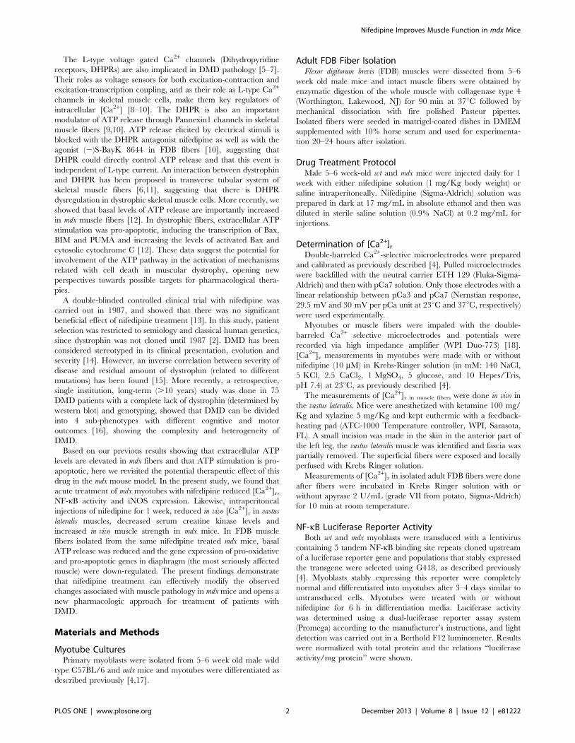
Nifedipine Treatment Reduces Resting Calcium Concentration, Oxidative and Apoptotic Gene Expression, and Improves Muscle Function in Dystrophic mdx Mice Francisco Altamirano 1,2. , Denisse Valladares 1. , Carlos Henrı´quez-Olguı´n 1 , Mariana Casas 1,4 , Jose R. Lo ´ pez 2,3 , Paul D. Allen 2,3 , Enrique Jaimovich 1 * 1 Centro de Estudios Moleculares de la Ce ´ lula, Instituto de Ciencias Biome ´ dicas, Facultad de Medicina, Universidad de Chile, Santiago, Chile, 2 Department of Molecular Biosciences, School of Veterinary Medicine, University of California Davis, Davis, California, United States of America, 3 Department of Anesthesiology, Perioperative and Pain Medicine, Brigham & Women’s Hospital, Harvard Medical School, Boston, Massachusetts, United States of America, 4 Programa de Fisiologı ´a y Biofı ´sica, Instituto de Ciencias Biome ´ dicas, Facultad de Medicina, Universidad de Chile, Santiago, Chile Abstract Duchenne Muscular Dystrophy (DMD) is a recessive X-linked genetic disease, caused by mutations in the gene encoding dystrophin. DMD is characterized in humans and in mdx mice by a severe and progressive destruction of muscle fibers, inflammation, oxidative/nitrosative stress, and cell death. In mdx muscle fibers, we have shown that basal ATP release is increased and that extracellular ATP stimulation is pro-apoptotic. In normal fibers, depolarization-induced ATP release is blocked by nifedipine, leading us to study the potential therapeutic effect of nifedipine in mdx muscles and its relation with extracellular ATP signaling. Acute exposure to nifedipine (10 mM) decreased [Ca 2+ ] r , NF-kB activity and iNOS expression in mdx myotubes. In addition, 6-week-old mdx mice were treated with daily intraperitoneal injections of nifedipine, 1 mg/Kg for 1 week. This treatment lowered the [Ca 2+ ] r measured in vivo in the mdx vastus lateralis. We demonstrated that extracellular ATP levels were higher in adult mdx flexor digitorum brevis (FDB) fibers and can be significantly reduced after 1 week of treatment with nifedipine. Interestingly, acute treatment of mdx FDB fibers with apyrase, an enzyme that completely degrades extracellular ATP to AMP, reduced [Ca 2+ ] r to a similar extent as was seen in FDB fibers after 1-week of nifedipine treatment. Moreover, we demonstrated that nifedipine treatment reduced mRNA levels of pro-oxidative/ nitrosative (iNOS and gp91 phox /p47 phox NOX2 subunits) and pro-apoptotic (Bax) genes in mdx diaphragm muscles and lowered serum creatine kinase (CK) levels. In addition, nifedipine treatment increased muscle strength assessed by the inverted grip-hanging test and exercise tolerance measured with forced swimming test in mdx mice. We hypothesize that nifedipine reduces basal ATP release, thereby decreasing purinergic receptor activation, which in turn reduces [Ca 2+ ] r in mdx skeletal muscle cells. The results in this work open new perspectives towards possible targets for pharmacological approaches to treat DMD. Citation: Altamirano F, Valladares D, Henrı ´quez-Olguı ´n C, Casas M, Lo ´ pez JR, et al. (2013) Nifedipine Treatment Reduces Resting Calcium Concentration, Oxidative and Apoptotic Gene Expression, and Improves Muscle Function in Dystrophic mdx Mice. PLoS ONE 8(12): e81222. doi:10.1371/journal.pone.0081222 Editor: James M. Ervasti, University of Minnesota, United States of America Received June 26, 2013; Accepted October 9, 2013; Published December 9, 2013 Copyright: ß 2013 Altamirano et al. This is an open-access article distributed under the terms of the Creative Commons Attribution License, which permits unrestricted use, distribution, and reproduction in any medium, provided the original author and source are credited. Funding: Fondo de Investigacio ´ n Avanzado en Areas Prioritarias 15010006, Fondo Nacional de Desarrollo Cientı ´fico y Tecnolo ´ gico 1110467, ACT1111 and AFM14562 (EJ, MC), National Institutes of Health AR43140, AR052534 (PDA and JRL), Program U-INICIA VID 2011, grant U-INICIA 02/12M; University of Chile (MC) supported this work. FA and DV were recipients of a doctoral fellowship and a doctoral thesis support grants AT-24100066 (FA), AT-24110211 (DV) from Comisio ´n Nacional de Ciencia y Tecnologı ´a (CONICYT). FA thanks Vicerrectorı ´a Asuntos Acade ´ micos and MECESUP UCH0714 (Universidad de Chile) and REDES 120003 (CONICYT) for travel support. The funders had no role in study design, data collection and analysis, decision to publish, or preparation of the manuscript. Competing Interests: The authors have declared that no competing interests exist. * E-mail: [email protected] . These authors contributed equally to this work. Introduction Duchenne muscular dystrophy (DMD) is a severe neuromus- cular disorder characterized by the absence of the dystrophin protein and is the most common form of muscular dystrophy with a frequency of 1 in 3500 male births [1]. DMD is caused by a recessive X-linked mutation in dystrophin gene located to Xp21 [2]. Usually patients lose the ability to walk between 6–12 years old due to severe muscle damage and contractures that results in wheelchair dependence [1]. Since DMD is a progressive disease, most patients die in their twenties due either to respiratory failure or cardiac dysfunction [3]. Resting intracellular calcium concentration ([Ca 2+ ] r ) is elevated in myotubes from mdx mice, a murine model for DMD [4]. This increased [Ca 2+ ] r depends on sarcolemmal Ca 2+ entry, as well as Ca 2+ leak from sarcoplasmatic reticulum (SR), through type 1 ryanodine receptors (RyR1) and inositol tri-phosphate receptors (IP 3 R). In turn, elevated [Ca 2+ ] r modulates NF-kB activation, leading to an up-regulation of inducible nitric oxide synthase (iNOS) in dystrophic myotubes [4]. PLOS ONE | www.plosone.org 1 December 2013 | Volume 8 | Issue 12 | e81222
-
Upload
independent -
Category
Documents
-
view
0 -
download
0
Transcript of Nifedipine Treatment Improves Muscle Function in Mdx Mice

Nifedipine Treatment Reduces Resting CalciumConcentration, Oxidative and Apoptotic GeneExpression, and Improves Muscle Function in Dystrophicmdx MiceFrancisco Altamirano1,2., Denisse Valladares1., Carlos Henrıquez-Olguın1, Mariana Casas1,4,
Jose R. Lopez2,3, Paul D. Allen2,3, Enrique Jaimovich1*
1 Centro de Estudios Moleculares de la Celula, Instituto de Ciencias Biomedicas, Facultad de Medicina, Universidad de Chile, Santiago, Chile, 2 Department of Molecular
Biosciences, School of Veterinary Medicine, University of California Davis, Davis, California, United States of America, 3 Department of Anesthesiology, Perioperative and
Pain Medicine, Brigham & Women’s Hospital, Harvard Medical School, Boston, Massachusetts, United States of America, 4 Programa de Fisiologıa y Biofısica, Instituto de
Ciencias Biomedicas, Facultad de Medicina, Universidad de Chile, Santiago, Chile
Abstract
Duchenne Muscular Dystrophy (DMD) is a recessive X-linked genetic disease, caused by mutations in the gene encodingdystrophin. DMD is characterized in humans and in mdx mice by a severe and progressive destruction of muscle fibers,inflammation, oxidative/nitrosative stress, and cell death. In mdx muscle fibers, we have shown that basal ATP release isincreased and that extracellular ATP stimulation is pro-apoptotic. In normal fibers, depolarization-induced ATP release isblocked by nifedipine, leading us to study the potential therapeutic effect of nifedipine in mdx muscles and its relation withextracellular ATP signaling. Acute exposure to nifedipine (10 mM) decreased [Ca2+]r, NF-kB activity and iNOS expression inmdx myotubes. In addition, 6-week-old mdx mice were treated with daily intraperitoneal injections of nifedipine, 1 mg/Kgfor 1 week. This treatment lowered the [Ca2+]r measured in vivo in the mdx vastus lateralis. We demonstrated thatextracellular ATP levels were higher in adult mdx flexor digitorum brevis (FDB) fibers and can be significantly reduced after 1week of treatment with nifedipine. Interestingly, acute treatment of mdx FDB fibers with apyrase, an enzyme thatcompletely degrades extracellular ATP to AMP, reduced [Ca2+]r to a similar extent as was seen in FDB fibers after 1-week ofnifedipine treatment. Moreover, we demonstrated that nifedipine treatment reduced mRNA levels of pro-oxidative/nitrosative (iNOS and gp91phox/p47phox NOX2 subunits) and pro-apoptotic (Bax) genes in mdx diaphragm muscles andlowered serum creatine kinase (CK) levels. In addition, nifedipine treatment increased muscle strength assessed by theinverted grip-hanging test and exercise tolerance measured with forced swimming test in mdx mice. We hypothesize thatnifedipine reduces basal ATP release, thereby decreasing purinergic receptor activation, which in turn reduces [Ca2+]r in mdxskeletal muscle cells. The results in this work open new perspectives towards possible targets for pharmacologicalapproaches to treat DMD.
Citation: Altamirano F, Valladares D, Henrıquez-Olguın C, Casas M, Lopez JR, et al. (2013) Nifedipine Treatment Reduces Resting Calcium Concentration, Oxidativeand Apoptotic Gene Expression, and Improves Muscle Function in Dystrophic mdx Mice. PLoS ONE 8(12): e81222. doi:10.1371/journal.pone.0081222
Editor: James M. Ervasti, University of Minnesota, United States of America
Received June 26, 2013; Accepted October 9, 2013; Published December 9, 2013
Copyright: � 2013 Altamirano et al. This is an open-access article distributed under the terms of the Creative Commons Attribution License, which permitsunrestricted use, distribution, and reproduction in any medium, provided the original author and source are credited.
Funding: Fondo de Investigacion Avanzado en Areas Prioritarias 15010006, Fondo Nacional de Desarrollo Cientıfico y Tecnologico 1110467, ACT1111 andAFM14562 (EJ, MC), National Institutes of Health AR43140, AR052534 (PDA and JRL), Program U-INICIA VID 2011, grant U-INICIA 02/12M; University of Chile (MC)supported this work. FA and DV were recipients of a doctoral fellowship and a doctoral thesis support grants AT-24100066 (FA), AT-24110211 (DV) from ComisionNacional de Ciencia y Tecnologıa (CONICYT). FA thanks Vicerrectorıa Asuntos Academicos and MECESUP UCH0714 (Universidad de Chile) and REDES 120003(CONICYT) for travel support. The funders had no role in study design, data collection and analysis, decision to publish, or preparation of the manuscript.
Competing Interests: The authors have declared that no competing interests exist.
* E-mail: [email protected]
. These authors contributed equally to this work.
Introduction
Duchenne muscular dystrophy (DMD) is a severe neuromus-
cular disorder characterized by the absence of the dystrophin
protein and is the most common form of muscular dystrophy with
a frequency of 1 in 3500 male births [1]. DMD is caused by a
recessive X-linked mutation in dystrophin gene located to Xp21
[2]. Usually patients lose the ability to walk between 6–12 years
old due to severe muscle damage and contractures that results in
wheelchair dependence [1]. Since DMD is a progressive disease,
most patients die in their twenties due either to respiratory failure
or cardiac dysfunction [3].
Resting intracellular calcium concentration ([Ca2+]r) is elevated
in myotubes from mdx mice, a murine model for DMD [4]. This
increased [Ca2+]r depends on sarcolemmal Ca2+ entry, as well as
Ca2+ leak from sarcoplasmatic reticulum (SR), through type 1
ryanodine receptors (RyR1) and inositol tri-phosphate receptors
(IP3R). In turn, elevated [Ca2+]r modulates NF-kB activation,
leading to an up-regulation of inducible nitric oxide synthase
(iNOS) in dystrophic myotubes [4].
PLOS ONE | www.plosone.org 1 December 2013 | Volume 8 | Issue 12 | e81222
The L-type voltage gated Ca2+ channels (Dihydropyridine
receptors, DHPRs) are also implicated in DMD pathology [5–7].
Their roles as voltage sensors for both excitation-contraction and
excitation-transcription coupling, and as their role as L-type Ca2+
channels in skeletal muscle cells, make them key regulators of
intracellular [Ca2+] [8–10]. The DHPR is also an important
modulator of ATP release through Pannexin1 channels in skeletal
muscle fibers [9,10]. ATP release elicited by electrical stimuli is
blocked with the DHPR antagonist nifedipine as well as with the
agonist (2)S-BayK 8644 in FDB fibers [10], suggesting that
DHPR could directly control ATP release and that this event is
independent of L-type current. An interaction between dystrophin
and DHPR has been proposed in transverse tubular system of
skeletal muscle fibers [6,11], suggesting that there is DHPR
dysregulation in dystrophic skeletal muscle cells. More recently, we
showed that basal levels of ATP release are importantly increased
in mdx muscle fibers [12]. In dystrophic fibers, extracellular ATP
stimulation was pro-apoptotic, inducing the transcription of Bax,
BIM and PUMA and increasing the levels of activated Bax and
cytosolic cytochrome C [12]. These data suggest the potential for
involvement of the ATP pathway in the activation of mechanisms
related with cell death in muscular dystrophy, opening new
perspectives towards possible targets for pharmacological thera-
pies.
A double-blinded controlled clinical trial with nifedipine was
carried out in 1987, and showed that there was no significant
beneficial effect of nifedipine treatment [13]. In this study, patient
selection was restricted to semiology and classical human genetics,
since dystrophin was not cloned until 1987 [2]. DMD has been
considered stereotyped in its clinical presentation, evolution and
severity [14]. However, an inverse correlation between severity of
disease and residual amount of dystrophin (related to different
mutations) has been found [15]. More recently, a retrospective,
single institution, long-term (.10 years) study was done in 75
DMD patients with a complete lack of dystrophin (determined by
western blot) and genotyping, showed that DMD can be divided
into 4 sub-phenotypes with different cognitive and motor
outcomes [16], showing the complexity and heterogeneity of
DMD.
Based on our previous results showing that extracellular ATP
levels are elevated in mdx fibers and that ATP stimulation is pro-
apoptotic, here we revisited the potential therapeutic effect of this
drug in the mdx mouse model. In the present study, we found that
acute treatment of mdx myotubes with nifedipine reduced [Ca2+]r,
NF-kB activity and iNOS expression. Likewise, intraperitoneal
injections of nifedipine for 1 week, reduced in vivo [Ca2+]r in vastus
lateralis muscles, decreased serum creatine kinase levels and
increased in vivo muscle strength in mdx mice. In FDB muscle
fibers isolated from the same nifedipine treated mdx mice, basal
ATP release was reduced and the gene expression of pro-oxidative
and pro-apoptotic genes in diaphragm (the most seriously affected
muscle) were down-regulated. The present findings demonstrate
that nifedipine treatment can effectively modify the observed
changes associated with muscle pathology in mdx mice and opens a
new pharmacologic approach for treatment of patients with
DMD.
Materials and Methods
Myotube CulturesPrimary myoblasts were isolated from 5–6 week old male wild
type C57BL/6 and mdx mice and myotubes were differentiated as
described previously [4,17].
Adult FDB Fiber IsolationFlexor digitorum brevis (FDB) muscles were dissected from 5–6
week old male mice and intact muscle fibers were obtained by
enzymatic digestion of the whole muscle with collagenase type 4
(Worthington, Lakewood, NJ) for 90 min at 37uC followed by
mechanical dissociation with fire polished Pasteur pipettes.
Isolated fibers were seeded in matrigel-coated dishes in DMEM
supplemented with 10% horse serum and used for experimenta-
tion 20–24 hours after isolation.
Drug Treatment ProtocolMale 5–6 week-old wt and mdx mice were injected daily for 1
week with either nifedipine solution (1 mg/Kg body weight) or
saline intraperitoneally. Nifedipine (Sigma-Aldrich) solution was
prepared in dark at 17 mg/mL in absolute ethanol and then was
diluted in sterile saline solution (0.9% NaCl) at 0.2 mg/mL for
injections.
Determination of [Ca2+]r
Double-barreled Ca2+-selective microelectrodes were prepared
and calibrated as previously described [4]. Pulled microelectrodes
were backfilled with the neutral carrier ETH 129 (Fluka-Sigma-
Aldrich) and then with pCa7 solution. Only those electrodes with a
linear relationship between pCa3 and pCa7 (Nernstian response,
29.5 mV and 30 mV per pCa unit at 23uC and 37uC, respectively)
were used experimentally.
Myotubes or muscle fibers were impaled with the double-
barreled Ca2+ selective microelectrodes and potentials were
recorded via high impedance amplifier (WPI Duo-773) [18].
[Ca2+]r measurements in myotubes were made with or without
nifedipine (10 mM) in Krebs-Ringer solution (in mM: 140 NaCl,
5 KCl, 2.5 CaCl2, 1 MgSO4, 5 glucose, and 10 Hepes/Tris,
pH 7.4) at 23uC, as previously described [4].
The measurements of [Ca2+]r in muscle fibers were done in vivo in
the vastus lateralis. Mice were anesthetized with ketamine 100 mg/
Kg and xylazine 5 mg/Kg and kept euthermic with a feedback-
heating pad (ATC-1000 Temperature controller, WPI, Sarasota,
FL). A small incision was made in the skin in the anterior part of
the left leg, the vastus lateralis muscle was identified and fascia was
partially removed. The superficial fibers were exposed and locally
perfused with Krebs Ringer solution.
Measurements of [Ca2+]r in isolated adult FDB fibers were done
after fibers were incubated in Krebs Ringer solution with or
without apyrase 2 U/mL (grade VII from potato, Sigma-Aldrich)
for 10 min at room temperature.
NF-kB Luciferase Reporter ActivityBoth wt and mdx myoblasts were transduced with a lentivirus
containing 5 tandem NF-kB binding site repeats cloned upstream
of a luciferase reporter gene and populations that stably expressed
the transgene were selected using G418, as described previously
[4]. Myoblasts stably expressing this reporter were completely
normal and differentiated into myotubes after 3–4 days similar to
untransduced cells. Myotubes were treated with or without
nifedipine for 6 h in differentiation media. Luciferase activity
was determined using a dual-luciferase reporter assay system
(Promega) according to the manufacturer’s instructions, and light
detection was carried out in a Berthold F12 luminometer. Results
were normalized with total protein and the relations ‘‘luciferase
activity/mg protein’’ were shown.
Nifedipine Improves Muscle Function in mdx Mice
PLOS ONE | www.plosone.org 2 December 2013 | Volume 8 | Issue 12 | e81222

mRNA QuantitationTotal RNA was isolated from myotubes or diaphragm muscles
from both the nifedipine- or saline-treated groups with TRIzolHreagent (Invitrogen) according to the manufacturer’s protocol.
cDNA was prepared by reverse transcription (RT) reaction of 1 mg
of total RNA using random primers. Real time PCR was
performed as previously described [4] using the following primers:
bax 59-GCTGACATGTTTGCTGATGG-39 and 59-GAT-
CAGCTCGGGCACTTTAG-39 bim 59-CGACAGTCTCAG-
GAGGAACC-39 and 59-CATTTGCAAACACCCTCCTT-39
gp91phox 59-TCACATCCTCTACCAAAACC-39 and 59-
CCTTTATTTTTCCCCATTCT-39 p47phox 59-AGAACA-
GAGTCATCCCACAC-39 and 59-GCTACGTTATTCTTGC-
CATC-39 iNOS 59-CAGCTCAAGAGCCAGAAACG-39 and 59-
TTACTCAGTGCCAGAAGCTG-39 gapdh 59-CTCATGACCA-
CAGTCCATGC-39 and 59-TTCAGCTCTGGGATGACCTT-
39 18S rRNA 59-GGGCCCGAAGCGTTTACTTT-39 and 59-
TTGCGCCGGTCCAAGAATTT-39.
ATP Detection Using a Luciferin/Luciferase AssayFDB fibers from nifedipine- and saline-treated mice were
prepared as described above. Because media replacement causes a
mechanical stimulus that itself induces ATP release [19,20], we
measured ATP release for up to 9 min beginning 30 min after
media change. ATP concentrations were measured with the
CellTiter-GloH Luminescent Cell Viability Assay (Promega,
Madison, WI, USA) as reported [9]. Data were calculated as
pmol extracellular ATP/mg total RNA. Normalization by total
RNA instead of total protein was chosen because fibers were
seeded on a Matrigel-coated dish (containing a large amount of
protein), which may affect the protein determination associated
only to fibers.
Serum Creatine Kinase DeterminationsBlood samples were obtained by cardiac puncture in anesthe-
tized mice. Blood was collected in a sterile test tube, allowed to clot
on ice for 30 min and then centrifuged at 3000 rpm for 10
minutes. Creatine kinase (CK) levels were determined using the
UV-kinetic method (Teco Diagnostics) according to the manufac-
turer instructions. DAbs/min were used to calculate CK enzymatic
activity and the results were expressed as International Kilo Units
per liter (KUI/L).
Inverted Grid-hanging TestMuscle strength of mouse limbs was tested with an inverted
grid-hanging test [21]. Mice were placed individually on the center
of a 21621 cm wire grid (wire width <0.1 cm and spacing
0.5 cm), mounted 35 cm above a table. After gently inverting the
grid, the mouse hanging time was recorded (grip latency). This
procedure was repeated three times and the average hanging time
values were calculated for each mouse.
Forced Swimming TestA 1-liter beaker (11 cm diameter and 15 cm height) filled with
water (23uC) was used as swimming pool to assess the exercise
tolerance of saline- and nifedipine-treated mice [22]. First, a
weight (10% of their body weight) was attached to the tail of a
mouse, which was then gently placed in the water, and the time at
which the mouse was unable to maintain complete buoyancy was
recorded. At this time the mice were immediately removed from
the swimming pool, dried gently with paper towels and returned to
their cages.
Statistical AnalysisData of n experiments were expressed as mean 6 S.E.M. The
significance of difference among treatments was evaluated using a
two-tailed t test for unpaired data or ANOVA- followed by Tukey’s t-
test. A P value ,0.05 was considered statistically significant.
Ethics ApprovalAll procedures for animal experimentation were done in
accordance with guidelines approved by the Bioethical Committee
at the Facultad de Medicina, Universidad de Chile and the
IACUCs at Harvard Medical School and University of California
at Davis.
Results
Nifedipine Reduces [Ca2+]r in Dystrophic mdx MyotubesMyotubes were incubated in Krebs Ringer solution with or
without nifedipine (10 mM) for 10 min and [Ca2+]r was measured
with Ca2+ selective microelectrodes, in both wt and mdx myotubes.
[Ca2+]r observed in mdx myotubes was significantly higher
compared to wt myotubes (31568 vs 11262 nM P,0.001)
(Figure 1). There was a significant reduction in the [Ca2+]r in
nifedipine-treated mdx myotubes compared with untreated mdx
myotubes (254612, P,0.001). Nifedipine treatment did not
modify [Ca2+]r in wt myotubes (11961 nM, P.0.05).
NF-kB Activity and iNOS Expression were Reduced AfterNifedipine Incubation
We previously reported that the increases in NF-kB activity and
iNOS expression are modulated by [Ca2+]r in mdx myotubes [4].
To study the effect of nifedipine treatment in NF-kB activity and
iNOS expression, we used a NF-kB luciferase reporter and real
Figure 1. Nifedipine incubation reduces [Ca2+]r in mdx myo-tubes. Myotubes were incubated with 10 mM Nifedipine for 10 min atroom temperature in Krebs Ringer solution and [Ca2+]r was measuredusing double-barreled Ca2+ selective microelectrodes. Data are ex-pressed as mean 6 S.E.M. Wt (n = 10), mdx (n = 27), Wt+Nife (n = 10) andmdx+Nife (n = 10). ***P,0.001, ANOVA-Tukey’s.doi:10.1371/journal.pone.0081222.g001
Nifedipine Improves Muscle Function in mdx Mice
PLOS ONE | www.plosone.org 3 December 2013 | Volume 8 | Issue 12 | e81222
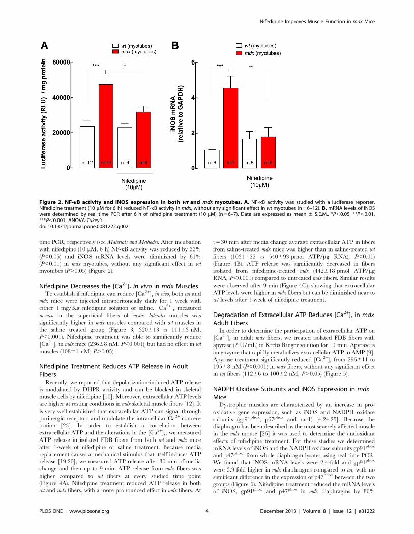
time PCR, respectively (see Materials and Methods). After incubation
with nifedipine (10 mM, 6 h) NF-kB activity was reduced by 33%
(P,0.05) and iNOS mRNA levels were diminished by 61%
(P,0.01) in mdx myotubes, without any significant effect in wt
myotubes (P.0.05) (Figure 2).
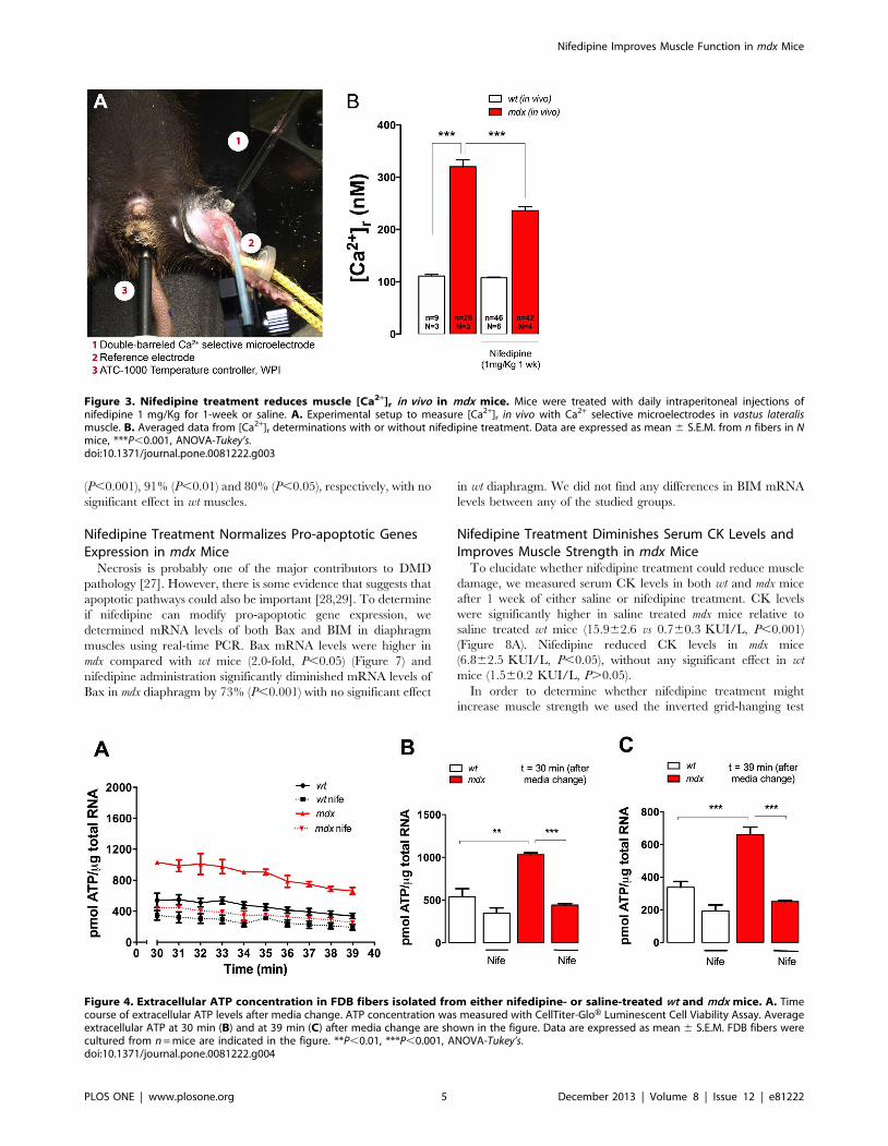
Nifedipine Decreases the [Ca2+]r in vivo in mdx MusclesTo establish if nifedipine can reduce [Ca2+]r in vivo, both wt and
mdx mice were injected intraperitoneally daily for 1 week with
either 1 mg/Kg nifedipine solution or saline. [Ca2+]r measured
in vivo in the superficial fibers of vastus lateralis muscles was
significantly higher in mdx muscles compared with wt muscles in
the saline treated group (Figure 3, 320613 vs 11163 nM,
P,0.001). Nifedipine treatment was able to significantly reduce
[Ca2+]r in mdx mice (23668 nM, P,0.001), but had no effect in wt
muscles (10861 nM, P.0.05).
Nifedipine Treatment Reduces ATP Release in AdultFibers
Recently, we reported that depolarization-induced ATP release
is modulated by DHPR activity and can be blocked in skeletal
muscle cells by nifedipine [10]. Moreover, extracellular ATP levels
are higher at resting conditions in mdx skeletal muscle fibers [12]. It
is very well established that extracellular ATP can signal through
purinergic receptors and modulate the intracellular Ca2+ concen-
tration [23]. In order to establish a correlation between
extracellular ATP and the alterations in the [Ca2+]r, we measured
ATP release in isolated FDB fibers from both wt and mdx mice
after 1-week of nifedipine or saline treatment. Because media
replacement causes a mechanical stimulus that itself induces ATP
release [19,20], we measured ATP release after 30 min of media
change and then up to 9 min. ATP release from mdx fibers was
higher compared to wt fibers at every studied time point
(Figure 4A). Nifedipine treatment reduced ATP release in both
wt and mdx fibers, with a more pronounced effect in mdx fibers. At
t = 30 min after media change average extracellular ATP in fibers
from saline-treated mdx mice was higher than in saline-treated wt
fibers (1031622 vs 540693 pmol ATP/mg RNA), P,0.01)
(Figure 4B). ATP release was significantly decreased in fibers
isolated from nifedipine-treated mdx (442618 pmol ATP/mg
RNA, P,0.001) compared to untreated mdx fibers. Similar results
were observed after 9 min (Figure 4C), showing that extracellular
ATP levels were higher in mdx fibers but can be diminished near to
wt levels after 1-week of nifedipine treatment.
Degradation of Extracellular ATP Reduces [Ca2+]r in mdxAdult Fibers
In order to determine the participation of extracellular ATP on
[Ca2+]r in adult mdx fibers, we treated isolated FDB fibers with
apyrase (2 U/mL) in Krebs Ringer solution for 10 min. Apyrase is
an enzyme that rapidly metabolizes extracellular ATP to AMP [9].
Apyrase treatment significantly reduced [Ca2+]r from 296611 to
19568 nM (P,0.001) in mdx fibers, without any significant effect
in wt fibers (11266 to 10062 nM, P.0.05) (Figure 5).
NADPH Oxidase Subunits and iNOS Expression in mdxMice
Dystrophic muscles are characterized by an increase in pro-
oxidative gene expression, such as iNOS and NADPH oxidase
subunits (gp91phox, p67phox and rac1) [4,24,25]. Because the
diaphragm has been described as the most severely affected muscle
in the mdx mouse [26] it was used to determine the antioxidant
effects of nifedipine treatment. For these studies we determined
mRNA levels of iNOS and the NADPH oxidase subunits gp91phox
and p47phox, from whole diaphragm lysates using real time PCR.
We found that iNOS mRNA levels were 2.4-fold and gp91phox
were 3.9-fold higher in mdx diaphragms compared to wt, with no
significant difference in the expression of p47phox between the two
groups (Figure 6). Nifedipine treatment reduced the mRNA levels
of iNOS, gp91phox and p47phox in mdx diaphragms by 86%
Figure 2. NF-kB activity and iNOS expression in both wt and mdx myotubes. A. NF-kB activity was studied with a luciferase reporter.Nifedipine treatment (10 mM for 6 h) reduced NF-kB activity in mdx, without any significant effect in wt myotubes (n = 6–12). B. mRNA levels of iNOSwere determined by real time PCR after 6 h of nifedipine treatment (10 mM) (n = 6–7). Data are expressed as mean 6 S.E.M., *P,0.05, **P,0.01,***P,0.001, ANOVA-Tukey’s.doi:10.1371/journal.pone.0081222.g002
Nifedipine Improves Muscle Function in mdx Mice
PLOS ONE | www.plosone.org 4 December 2013 | Volume 8 | Issue 12 | e81222
(P,0.001), 91% (P,0.01) and 80% (P,0.05), respectively, with no
significant effect in wt muscles.
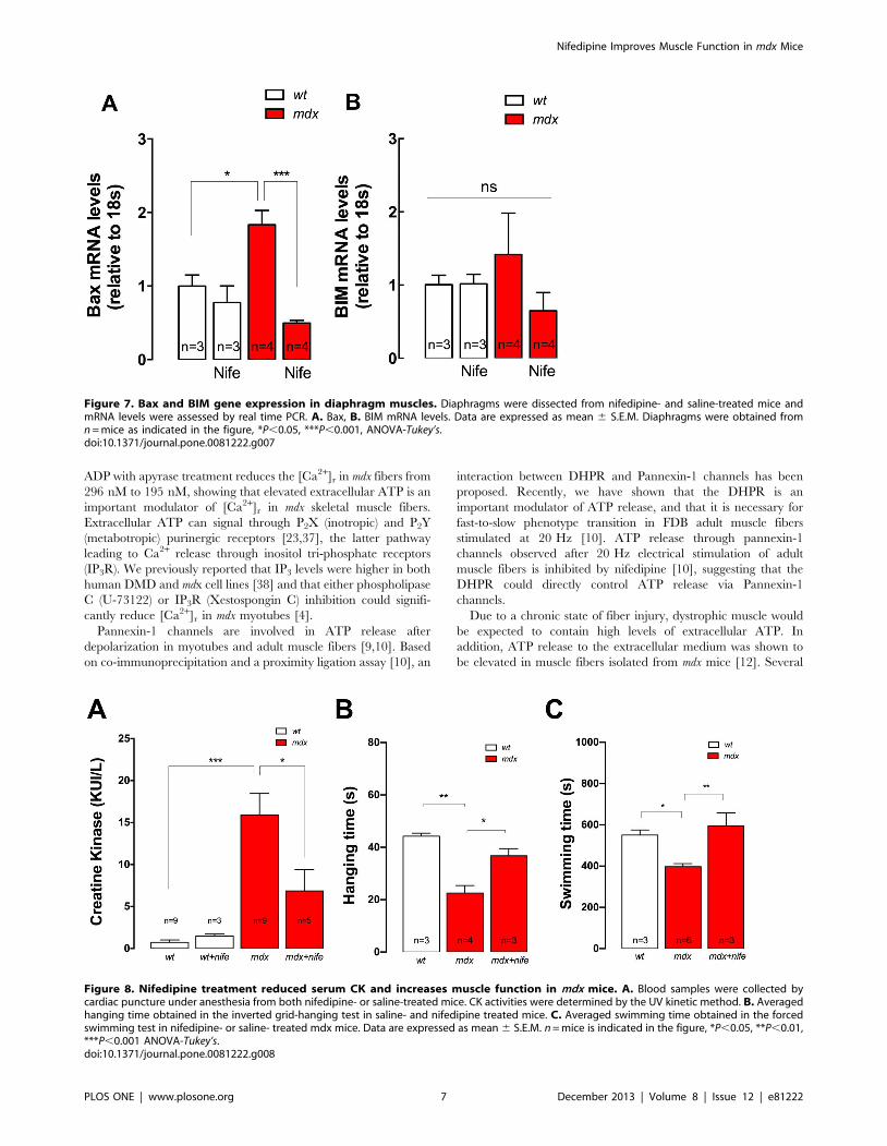
Nifedipine Treatment Normalizes Pro-apoptotic GenesExpression in mdx Mice
Necrosis is probably one of the major contributors to DMD
pathology [27]. However, there is some evidence that suggests that
apoptotic pathways could also be important [28,29]. To determine
if nifedipine can modify pro-apoptotic gene expression, we
determined mRNA levels of both Bax and BIM in diaphragm
muscles using real-time PCR. Bax mRNA levels were higher in
mdx compared with wt mice (2.0-fold, P,0.05) (Figure 7) and
nifedipine administration significantly diminished mRNA levels of
Bax in mdx diaphragm by 73% (P,0.001) with no significant effect
in wt diaphragm. We did not find any differences in BIM mRNA
levels between any of the studied groups.
Nifedipine Treatment Diminishes Serum CK Levels andImproves Muscle Strength in mdx Mice
To elucidate whether nifedipine treatment could reduce muscle
damage, we measured serum CK levels in both wt and mdx mice
after 1 week of either saline or nifedipine treatment. CK levels
were significantly higher in saline treated mdx mice relative to
saline treated wt mice (15.962.6 vs 0.760.3 KUI/L, P,0.001)
(Figure 8A). Nifedipine reduced CK levels in mdx mice
(6.862.5 KUI/L, P,0.05), without any significant effect in wt
mice (1.560.2 KUI/L, P.0.05).
In order to determine whether nifedipine treatment might
increase muscle strength we used the inverted grid-hanging test
Figure 3. Nifedipine treatment reduces muscle [Ca2+]r in vivo in mdx mice. Mice were treated with daily intraperitoneal injections ofnifedipine 1 mg/Kg for 1-week or saline. A. Experimental setup to measure [Ca2+]r in vivo with Ca2+ selective microelectrodes in vastus lateralismuscle. B. Averaged data from [Ca2+]r determinations with or without nifedipine treatment. Data are expressed as mean 6 S.E.M. from n fibers in Nmice, ***P,0.001, ANOVA-Tukey’s.doi:10.1371/journal.pone.0081222.g003
Figure 4. Extracellular ATP concentration in FDB fibers isolated from either nifedipine- or saline-treated wt and mdx mice. A. Timecourse of extracellular ATP levels after media change. ATP concentration was measured with CellTiter-GloH Luminescent Cell Viability Assay. Averageextracellular ATP at 30 min (B) and at 39 min (C) after media change are shown in the figure. Data are expressed as mean 6 S.E.M. FDB fibers werecultured from n = mice are indicated in the figure. **P,0.01, ***P,0.001, ANOVA-Tukey’s.doi:10.1371/journal.pone.0081222.g004
Nifedipine Improves Muscle Function in mdx Mice
PLOS ONE | www.plosone.org 5 December 2013 | Volume 8 | Issue 12 | e81222

and the forced swimming test. Mice were treated daily with
nifedipine (1 mg/Kg) or saline for 1 week, and the functional test
was performed one day after cessation of treatment. Saline treated
mdx mice had a significantly shorter hanging time compared to wt
mice (2363 s vs 4461 s, P,0.01 Figure 8B). Nifedipine treatment
increased the mdx hanging time by 61% (3763 s, P,0.05
compared to saline-treated mice (Figure 8B).
Similar results were observed using the forced swimming test.
Under this experimental condition, wt swimming time was
significantly higher compared to saline-treated mdx mice
(550625 s vs 398615 s, P,0.05, Figure 8C). In this case
nifedipine treatment for 1 week normalized the swimming time
in mdx mice increasing it to 594664 s (P,0.01 compared to saline-
treated mice, Figure 8C).
Discussion
Our data show that [Ca2+]r was elevated in mdx myotubes and
to a similar extent in adult skeletal muscle fibers from mdx mice.
Acute exposure of mdx myotubes to nifedipine decreased [Ca2+]r,
NF-kB activity and iNOS expression. Likewise, in mdx mice,
nifedipine treatment for 1-week lowered in vivo [Ca2+]r in the vastus
lateralis, reduced ATP release in FDB fibers, diminished mRNA
levels of pro-oxidative/apoptotic genes in the diaphragm, lowered
serum CK levels and improved muscle function as assessed by
both inverted grid-hanging test and forced swimming test.
Although still controversial, it is generally accepted that
dystrophic skeletal muscle cells have elevated [Ca2+]r [4,30–32].
Our data support these findings showing that [Ca2+]r was elevated
in mdx myotubes and to a similar extent in skeletal muscles from
mdx mice, measured either in isolated FDB fibers or determined
in vivo, in anesthetized mice. Moreover, we showed that either
acute nifedipine treatment of myotubes or chronic nifedipine
treatment of mice for 1-week reduced [Ca2+]r. Nifedipine is known
as specific inhibitor of the DHPR [33]. In addition to its role as an
L-type Ca2+ channel the DHPR operates as voltage sensor for
both excitation-contraction and excitation-transcription coupling
in adult muscle fibers [8–10]. Dystrophic mdx skeletal muscle cells
have a dysregulated excitation-contraction coupling as evidenced
by reduced calcium transients evoked by single action potentials
compared with wt fibers [34,35]. This has been used to explain the
muscle weakness observed in DMD patients. On the other hand,
there is a controversy related to whether there are alterations of L-
type Ca2+ currents in mdx muscles, with some studies showing
normal maximum currents [5] while others show decreased
maximum current [6,7]. No differences have been found in charge
movement between normal and dystrophic skeletal muscle fibers
[5,36]. In addition, a leftward-shift of the voltage dependence of L-
type Ca2+ current has been described [5].
Our data showed that extracellular ATP levels were higher in
mdx fibers compared to wt fibers. Chronic nifedipine treatment
reduces ATP release in adult mdx FDB fibers to a level similar to wt
fibers. Moreover, acute enzymatic ablation of extracellular ATP-
Figure 5. Apyrase treatment reduces [Ca2+]r in FDB adult fibers.Adult fibers were isolated from wt and mdx mice and incubated inKrebs Ringer solution with or without apyrase (2 U/mL) for 10 min atroom temperature. [Ca2+]r was determined by double-barreled Ca2+
selective microelectrodes. Data are expressed as mean 6 S.E.M. from nfibers (indicated in the figure) from three different cultures. n.s, nosignificant difference, ***P,0.001, ANOVA-Tukey’s.doi:10.1371/journal.pone.0081222.g005
Figure 6. iNOS and NADPH oxidase subunits gene expression in diaphragm muscles. Diaphragms were dissected from nifedipine- andsaline-treated mice and mRNA levels were assessed by real time PCR. A. iNOS, B., gp91phox and C. p47phox expression. Data are expressed as mean 6S.E.M. Diaphragms were obtained from n = mice as indicated in the figure. *P,0.05, **P,0.01, ***P,0.001, ANOVA-Tukey’s.doi:10.1371/journal.pone.0081222.g006
Nifedipine Improves Muscle Function in mdx Mice
PLOS ONE | www.plosone.org 6 December 2013 | Volume 8 | Issue 12 | e81222
ADP with apyrase treatment reduces the [Ca2+]r in mdx fibers from
296 nM to 195 nM, showing that elevated extracellular ATP is an
important modulator of [Ca2+]r in mdx skeletal muscle fibers.
Extracellular ATP can signal through P2X (inotropic) and P2Y
(metabotropic) purinergic receptors [23,37], the latter pathway
leading to Ca2+ release through inositol tri-phosphate receptors
(IP3R). We previously reported that IP3 levels were higher in both
human DMD and mdx cell lines [38] and that either phospholipase
C (U-73122) or IP3R (Xestospongin C) inhibition could signifi-
cantly reduce [Ca2+]r in mdx myotubes [4].
Pannexin-1 channels are involved in ATP release after
depolarization in myotubes and adult muscle fibers [9,10]. Based
on co-immunoprecipitation and a proximity ligation assay [10], an
interaction between DHPR and Pannexin-1 channels has been
proposed. Recently, we have shown that the DHPR is an
important modulator of ATP release, and that it is necessary for
fast-to-slow phenotype transition in FDB adult muscle fibers
stimulated at 20 Hz [10]. ATP release through pannexin-1
channels observed after 20 Hz electrical stimulation of adult
muscle fibers is inhibited by nifedipine [10], suggesting that the
DHPR could directly control ATP release via Pannexin-1
channels.
Due to a chronic state of fiber injury, dystrophic muscle would
be expected to contain high levels of extracellular ATP. In
addition, ATP release to the extracellular medium was shown to
be elevated in muscle fibers isolated from mdx mice [12]. Several
Figure 7. Bax and BIM gene expression in diaphragm muscles. Diaphragms were dissected from nifedipine- and saline-treated mice andmRNA levels were assessed by real time PCR. A. Bax, B. BIM mRNA levels. Data are expressed as mean 6 S.E.M. Diaphragms were obtained fromn = mice as indicated in the figure, *P,0.05, ***P,0.001, ANOVA-Tukey’s.doi:10.1371/journal.pone.0081222.g007
Figure 8. Nifedipine treatment reduced serum CK and increases muscle function in mdx mice. A. Blood samples were collected bycardiac puncture under anesthesia from both nifedipine- or saline-treated mice. CK activities were determined by the UV kinetic method. B. Averagedhanging time obtained in the inverted grid-hanging test in saline- and nifedipine treated mice. C. Averaged swimming time obtained in the forcedswimming test in nifedipine- or saline- treated mdx mice. Data are expressed as mean 6 S.E.M. n = mice is indicated in the figure, *P,0.05, **P,0.01,***P,0.001 ANOVA-Tukey’s.doi:10.1371/journal.pone.0081222.g008
Nifedipine Improves Muscle Function in mdx Mice
PLOS ONE | www.plosone.org 7 December 2013 | Volume 8 | Issue 12 | e81222

alterations have been found in ATP signaling in dystrophic skeletal
muscle cells. In an immortalized myoblast cell line derived from
mdx mouse, addition of exogenous ATP to the media induced a
large increase in cytosolic Ca2+ concentration compared with its wt
counterpart [39]. This increased susceptibility to ATP was
associated with changes in expression and function of P2X
channels and to pathogenic Ca2+ entry in dystrophic muscles.
Furthermore, enhanced expression of the P2X4 and P2X7
receptors have been related to macrophage invasion in mdx mice
[40].
Here we determined the expression of iNOS and NADPH
oxidase subunits in diaphragm, because this muscle has been
described as the most severely affected muscle in the mdx mouse
[26]. Nifedipine reduced iNOS, gp91phox and p47phox mRNA
levels in mdx diaphragm after 1-week of treatment. We previously
demonstrated that iNOS expression was higher in mdx myotubes
which we attributed to an up-regulated NF-kB activity mediated
by the elevated [Ca2+]r [4]. Nifedipine treatment reduced iNOS
expression in diaphragm, likely due to the same mechanism.
Recently, it has been shown that dihydropyridines have anti-
inflammatory and anti-oxidant effects. Nifedipine caused concen-
tration-dependent inhibitory effects on sarcolemmal lipid perox-
idation in vitro [41]. Toma et al, 2011 suggest that amlodipine, a
third generation dihydropyridine L-type calcium channel blocker,
may improve endothelial dysfunction in diabetics through anti-
oxidant and anti-inflammatory mechanisms. The authors stimu-
lated human endothelial cells with irreversible glycated low-density
lipoproteins (AGE-LDL), as an in vitro model mimicking the
diabetic condition. Their results show that amlodipine reduced the
expression of NADPH oxidase subunits (p22phox and NOX4) and
iNOS, and diminished oxidative/nitrosative stress. Moreover,
amlodipine had anti-inflammatory effects reducing the activation
of MCP-1, VCAM-1, p38 MAPK and NF-kB [42]. In another
study, it has been shown that amlodipine (3 mg/kg/day) decrease
the expression of NADPH subunits (p47phox and rac1), NADPH
activity and the expression of inflammatory factor in atheroscle-
rotic lesions [43].
Necrosis is probably the major contributor to DMD pathology
[27]. However, there is some evidence that suggests that apoptotic
pathways could also be important [28,29]. We observed that
nifedipine reduced significantly Bax expression in mdx diaphragms,
suggestive of a protective anti-apoptotic effect. Bax was abun-
dantly expressed in the mdx masseter muscles compared to wt
muscles at 3 weeks after birth [29]. We have demonstrated that
exogenous ATP increases mRNA levels of several pro-apoptotic
genes in skeletal muscle fibers, including Bax, BIM and PUMA.
Moreover, ATP induced Bax activation and cytochrome C release
in mdx fibers, which is related with apoptotic cell death [12].
A previous double-blind controlled clinical trial with nifedipine
did not demonstrate a beneficial therapeutic response on mean
muscle strength, joint contractures, time-functional tests, pulmo-
nary function, or creatine kinase levels in 105 patients between 3–
Figure 9. Proposed model for ATP-mediated effects in dystrophic skeletal muscle. In dystrophic skeletal muscle fibers there is an increasein basal ATP release, through Pannexin1 channels that is modulated by the DHPR [12]. Extracellular ATP increases the [Ca2+]r in dystrophic musclefibers through the activation of purinergic receptors (P2X, inotropic and P2Y, metabotropic). This leads to the expression of pro-apoptotic and pro-inflammatory genes, increasing the muscle damage observed in dystrophic skeletal muscle cells. Nifedipine treatment reduces the basal ATP releaseand reduces [Ca2+]r, resulting in less pro-inflammatory and pro-apoptotic gene expression and subsequently reduces muscle damage as indicated bya decrease in blood CK and an increase in muscle function assessed by the inverted grid-hanging test and the force swimming test.doi:10.1371/journal.pone.0081222.g009
Nifedipine Improves Muscle Function in mdx Mice
PLOS ONE | www.plosone.org 8 December 2013 | Volume 8 | Issue 12 | e81222

27 years of age at the end of the study who were presumed to have
DMD [13]. Patients were given nifedipine 0.75 mg/kg/day in
three divided oral doses for 6 months and then 1.5–2 mg/Kg/day
for the final 12 months of the study [13]. A total of 97 patients
completed the trial. The power of the study to detect a difference
between groups was calculated using the decrease of average
muscle strength over time as the primary outcome measure.
Average muscle strength score were calculated with a modified
manual testing scale (MMT) [14] and the study had a power
greater that 0.99 to detect a slowing of the illness to 25% of its
original progression. The diagnostic criteria were based on
semiology and classical human genetics, however three patients
classified as Becker dystrophy were included in the study.
Moreover the one patient aged 27 years old was considered to
be unusual for DMD [44]. The data presented showed that the
average muscle strength score as slightly improved without any
significant difference. The trial used an unpaired analysis and it
may have been more appropriate to use a paired analysis so that
before and after changes in individuals could be compared [44].
As we mentioned before, there is an inverse correlation between
severity of disease and dystrophin expression [15] and even
patients with a complete lack of dystrophin DMD can be divided
into 4 sub-phenotypes with different cognitive and motor
outcomes [16]. This shows the high variability in the natural
history of DMD. Phenotypic variations have shown to compro-
mise results of clinical trials [45]. Desguerre et al, suggest that
trials, which are in danger of being inconclusive due to lack of
precise knowledge of DMD’s natural history, would strongly
benefit from accurate selection of clinically homogeneous patient
subsets [16].
High CK levels observed in DMD patients are attributed to an
enhanced membrane permeability and muscle damage [46]. In
our study, nifedipine treatment significantly reduced CK levels in
mdx mice after 1-week, but did not normalize it. These data are
supported by a study using verapamil, another L-type calcium
channel blocker, which showed a significant reduction in the
release of CK and LDH from diaphragm and gastrocnemius
muscles [47]. In the previous nifedipine clinical trial, authors did
not observe any significant difference in averaged CK levels after
treatment, reporting that patients from nifedipine group had an
average CK level of 2681 U/liter at the start and a value of
2045 U/liter at the end [13]. High levels of CK in DMD are well-
known, and after a rise in infancy, they remain at high levels with
an abrupt decrease at around 10 years old due to loss of muscle
mass [48]. Thus averaged data from a broad age group could
easily miss a significant difference when and if one existed.
Reduction of [Ca2+]r and in the expression of pro-oxidative/
apoptotic proteins, could be beneficial to dystrophic muscles and
may explain a reduction in muscle damage that a decrease in
serum CK would indicate. Moreover, an increase in the hanging
time and normalization of the forced swimming test were observed
in nifedipine-treated mdx mice, demonstrating an increase in
motor function, probably due to reduced muscle damage or
increased regeneration. In summary, these results provide further
evidence that [Ca2+]r is elevated in mdx muscles and this can be
modulated in vivo by nifedipine administration through a reduction
in basal ATP release from dystrophic fibers. Moreover, nifedipine
treatment reduced pro-oxidative/apoptotic gene expression with
the end result being less muscle damage as evidenced by a
significant reduction of serum CK and an increased muscle
strength in mdx mice (Figure 9). Our study used daily intraper-
itoneally doses of nifedipine (1 mg/Kg) for 1 week as compared to
the clinical study, which used it for a longer time period via the
oral route of administration. Moreover, our study was carried out
in 5–6 week old mdx mice (necrosis/regeneration stage) compared
to the clinical study that included patients at different stages of the
disease (3–27 years old). Longer studies need to be carried out to
assess the positive effects and pitfalls of nifedipine treatment studies
in mdx mice. However, our results strongly suggest that blockers of
the ATP signaling pathway should be tested in DMD patients, as
they might be promising in palliating the disease and prolonging
muscle function.
Author Contributions
Conceived and designed the experiments: FA DV JRL PDA EJ. Performed
the experiments: FA DV CHO JRL. Analyzed the data: FA DV CHO MC
EJ. Contributed reagents/materials/analysis tools: PDA EJ. Wrote the
paper: FA MC PDA EJ.
References
1. Blake DJ, Weir A, Newey SE, Davies KE (2002) Function and genetics of
dystrophin and dystrophin-related proteins in muscle. Physiol Rev 82: 291–329.
2. Koenig M, Hoffman EP, Bertelson CJ, Monaco AP, Feener C, et al. (1987)
Complete cloning of the Duchenne muscular dystrophy (DMD) cDNA and
preliminary genomic organization of the DMD gene in normal and affected
individuals. Cell 50: 509–517.
3. Emery AE (2002) The muscular dystrophies. Lancet 359: 687–695.
4. Altamirano F, Lopez JR, Henriquez C, Molinski T, Allen PD, et al. (2012)
Increased resting intracellular calcium modulates NF-kappaB-dependent
inducible nitric-oxide synthase gene expression in dystrophic mdx skeletal
myotubes. J Biol Chem 287: 20876–20887.
5. Collet C, Csernoch L, Jacquemond V (2003) Intramembrane charge movement
and L-type calcium current in skeletal muscle fibers isolated from control and
mdx mice. Biophys J 84: 251–265.
6. Friedrich O, von Wegner F, Chamberlain JS, Fink RH, Rohrbach P (2008) L-
type Ca2+ channel function is linked to dystrophin expression in mammalian
muscle. PLoS One 3: e1762.
7. Imbert N, Vandebrouck C, Duport G, Raymond G, Hassoni AA, et al. (2001)
Calcium currents and transients in co-cultured contracting normal and
Duchenne muscular dystrophy human myotubes. J Physiol 534: 343–355.
8. Tanabe T, Beam KG, Powell JA, Numa S (1988) Restoration of excitation-
contraction coupling and slow calcium current in dysgenic muscle by
dihydropyridine receptor complementary DNA. Nature 336: 134–139.
9. Buvinic S, Almarza G, Bustamante M, Casas M, Lopez J, et al. (2009) ATP
released by electrical stimuli elicits calcium transients and gene expression in
skeletal muscle. J Biol Chem 284: 34490–34505.
10. Jorquera G, Altamirano F, Contreras-Ferrat A, Almarza G, Buvinic S, et al.
(2013) Cav1.1 controls frequency-dependent events regulating adult skeletal
muscle plasticity. J Cell Sci.
11. Knudson CM, Hoffman EP, Kahl SD, Kunkel LM, Campbell KP (1988)
Evidence for the association of dystrophin with the transverse tubular system in
skeletal muscle. J Biol Chem 263: 8480–8484.
12. Valladares D, Almarza G, Contreras A, Pavez M, Buvinic S, et al. (2013)
Electrical stimuli are anti-apoptotic in skeletal muscle via extracellular ATP.
Alterations of this signal in mdx mice is a likely cause of dystrophy. PLoS One.
In Press.
13. Moxley RT 3rd, Brooke MH, Fenichel GM, Mendell JR, Griggs RC, et al.
(1987) Clinical investigation in Duchenne dystrophy. VI. Double-blind
controlled trial of nifedipine. Muscle Nerve 10: 22–33.
14. Brooke MH, Fenichel GM, Griggs RC, Mendell JR, Moxley R, et al. (1983)
Clinical investigation in Duchenne dystrophy: 2. Determination of the ‘‘power’’
of therapeutic trials based on the natural history. Muscle Nerve 6: 91–103.
15. Nicholson LV, Johnson MA, Bushby KM, Gardner-Medwin D, Curtis A, et al.
(1993) Integrated study of 100 patients with Xp21 linked muscular dystrophy
using clinical, genetic, immunochemical, and histopathological data. Part 3.
Differential diagnosis and prognosis. J Med Genet 30: 745–751.
16. Desguerre I, Christov C, Mayer M, Zeller R, Becane HM, et al. (2009) Clinical
heterogeneity of duchenne muscular dystrophy (DMD): definition of sub-
phenotypes and predictive criteria by long-term follow-up. PLoS One 4: e4347.
17. Casas M, Altamirano F, Jaimovich E (2012) Measurement of calcium release due
to inositol trisphosphate receptors in skeletal muscle. Methods Mol Biol 798:
383–393.
Nifedipine Improves Muscle Function in mdx Mice
PLOS ONE | www.plosone.org 9 December 2013 | Volume 8 | Issue 12 | e81222
18. Eltit JM, Ding X, Pessah IN, Allen PD, Lopez JR (2012) Nonspecific
sarcolemmal cation channels are critical for the pathogenesis of malignant
hyperthermia. FASEB J.
19. Ho CL, Yang CY, Lin WJ, Lin CH (2013) Ecto-Nucleoside Triphosphate
Diphosphohydrolase 2 Modulates Local ATP-Induced Calcium Signaling in
Human HaCaT Keratinocytes. PLoS One 8: e57666.
20. Yoshida H, Kobayashi D, Ohkubo S, Nakahata N (2006) ATP stimulates
interleukin-6 production via P2Y receptors in human HaCaT keratinocytes.
Eur J Pharmacol 540: 1–9.
21. Kaja S, van de Ven RC, van Dijk JG, Verschuuren JJ, Arahata K, et al. (2007)
Severely impaired neuromuscular synaptic transmission causes muscle weakness
in the Cacna1a-mutant mouse rolling Nagoya. Eur J Neurosci 25: 2009–2020.
22. Razani B, Wang XB, Engelman JA, Battista M, Lagaud G, et al. (2002)
Caveolin-2-deficient mice show evidence of severe pulmonary dysfunction
without disruption of caveolae. Mol Cell Biol 22: 2329–2344.
23. Burnstock G (2006) Purinergic P2 receptors as targets for novel analgesics.
Pharmacol Ther 110: 433–454.
24. Bellinger AM, Reiken S, Carlson C, Mongillo M, Liu X, et al. (2009)
Hypernitrosylated ryanodine receptor calcium release channels are leaky in
dystrophic muscle. Nat Med 15: 325–330.
25. Whitehead NP, Yeung EW, Froehner SC, Allen DG (2010) Skeletal muscle
NADPH oxidase is increased and triggers stretch-induced damage in the mdx
mouse. PLoS One 5: e15354.
26. Stedman HH, Sweeney HL, Shrager JB, Maguire HC, Panettieri RA, et al.
(1991) The mdx mouse diaphragm reproduces the degenerative changes of
Duchenne muscular dystrophy. Nature 352: 536–539.
27. Miller JB, Girgenrath M (2006) The role of apoptosis in neuromuscular diseases
and prospects for anti-apoptosis therapy. Trends Mol Med 12: 279–286.
28. Tews DS, Goebel HH (1997) DNA-fragmentation and expression of apoptosis-
related proteins in muscular dystrophies. Neuropathol Appl Neurobiol 23: 331–
338.
29. Honda A, Abe S, Hiroki E, Honda H, Iwanuma O, et al. (2007) Activation of
caspase 3, 9, 12, and Bax in masseter muscle of mdx mice during necrosis.
J Muscle Res Cell Motil 28: 243–247.
30. Allen DG, Gervasio OL, Yeung EW, Whitehead NP (2010) Calcium and the
damage pathways in muscular dystrophy. Can J Physiol Pharmacol 88: 83–91.
31. Lopez JR, Briceno LE, Sanchez V, Horvart D (1987) Myoplasmic (Ca2+) in
Duchenne muscular dystrophy patients. Acta Cient Venez 38: 503–504.
32. Turner PR, Westwood T, Regen CM, Steinhardt RA (1988) Increased protein
degradation results from elevated free calcium levels found in muscle from mdx
mice. Nature 335: 735–738.
33. Neuhaus R, Rosenthal R, Luttgau HC (1990) The effects of dihydropyridine
derivatives on force and Ca2+ current in frog skeletal muscle fibres. J Physiol
427: 187–209.
34. Capote J, DiFranco M, Vergara JL (2010) Excitation-contraction coupling
alterations in mdx and utrophin/dystrophin double knockout mice: acomparative study. Am J Physiol Cell Physiol 298: C1077–1086.
35. Woods CE, Novo D, DiFranco M, Vergara JL (2004) The action potential-
evoked sarcoplasmic reticulum calcium release is impaired in mdx mouse musclefibres. J Physiol 557: 59–75.
36. Hollingworth S, Marshall MW, Robson E (1990) Excitation contractioncoupling in normal and mdx mice. Muscle Nerve 13: 16–20.
37. Abbracchio MP, Burnstock G, Boeynaems JM, Barnard EA, Boyer JL, et al.
(2006) International Union of Pharmacology LVIII: update on the P2Y Gprotein-coupled nucleotide receptors: from molecular mechanisms and patho-
physiology to therapy. Pharmacol Rev 58: 281–341.38. Liberona JL, Powell JA, Shenoi S, Petherbridge L, Caviedes R, et al. (1998)
Differences in both inositol 1,4,5-trisphosphate mass and inositol 1,4,5-trispho-sphate receptors between normal and dystrophic skeletal muscle cell lines.
Muscle Nerve 21: 902–909.
39. Yeung D, Zablocki K, Lien CF, Jiang T, Arkle S, et al. (2006) Increasedsusceptibility to ATP via alteration of P2X receptor function in dystrophic mdx
mouse muscle cells. FASEB J 20: 610–620.40. Yeung D, Kharidia R, Brown SC, Gorecki DC (2004) Enhanced expression of
the P2X4 receptor in Duchenne muscular dystrophy correlates with macrophage
invasion. Neurobiol Dis 15: 212–220.41. Mak IT, Weglicki WB (1990) Comparative antioxidant activities of propranolol,
nifedipine, verapamil, and diltiazem against sarcolemmal membrane lipidperoxidation. Circ Res 66: 1449–1452.
42. Toma L, Stancu CS, Sanda GM, Sima AV (2011) Anti-oxidant and anti-inflammatory mechanisms of amlodipine action to improve endothelial cell
dysfunction induced by irreversibly glycated LDL. Biochem Biophys Res
Commun 411: 202–207.43. Yoshii T, Iwai M, Li Z, Chen R, Ide A, et al. (2006) Regression of atherosclerosis
by amlodipine via anti-inflammatory and anti-oxidative stress actions. HypertensRes 29: 457–466.
44. Phillips MF, Quinlivan R (2008) Calcium antagonists for Duchenne muscular
dystrophy. Cochrane Database Syst Rev: CD004571.45. Escolar DM, Buyse G, Henricson E, Leshner R, Florence J, et al. (2005) CINRG
randomized controlled trial of creatine and glutamine in Duchenne musculardystrophy. Ann Neurol 58: 151–155.
46. Ozawa E, Hagiwara Y, Yoshida M (1999) Creatine kinase, cell membrane andDuchenne muscular dystrophy. Mol Cell Biochem 190: 143–151.
47. Niebroj-Dobosz I, Lukasiuk M (1996) Release of Intracellular Enzymes from
Skeletal Muscles and Diaphragm in Mdx Mice. European Journal ofTranslational Myology 6: 377–383.
48. Konagaya M, Takayanagi T (1986) Regularity in the change of serum creatinekinase level in Duchenne muscular dystrophy. A study with long-term follow-up
cases. Jpn J Med 25: 2–8.
Nifedipine Improves Muscle Function in mdx Mice
PLOS ONE | www.plosone.org 10 December 2013 | Volume 8 | Issue 12 | e81222