Metabotropic Glutamate Receptor II in the Brains of Parkinsonian Patients
9
Copyright @ 2009 by the American Association of Neuropathologists, Inc. Unauthorized reproduction of this article is prohibited. ORIGINAL ARTICLE Metabotropic Glutamate Receptor II in the Brains of Parkinsonian Patients Pershia Samadi, PhD, Alex Rajput, MD, FRCPC, Fre ´de ´ric Calon, PhD, Laurent Gre ´goire, BSc, Oleh Hornykiewicz, MD, Ali H. Rajput, MBBS, FRCPC, and The `re `se Di Paolo, PhD Abstract Modulation of basal ganglia group II metabotropic glutamate re- ceptors (mGluR2/3) is a potential therapeutic alternative to levodopa in Parkinson disease (PD). We used receptor-binding autoradiog- raphy of the mGluR2/3-selective radioligand [ 3 H]LY341495 in post- mortem brain specimens from PD patients (n = 14) and controls (n = 11) to investigate possible contributions of changes in ligand binding of this receptor to levodopa-associated motor complications experienced premortem in PD patients. The PD patients included those with and without histories of dyskinesias and those with and without ‘‘wearing off,’’ which is defined as a reduced period of benefit from levodopa. Specific binding of [ 3 H]LY341495 to mGluR2/3 in the basal ganglia was higher in the caudate nucleus than the putamen and lower by approximately half in the external and internal globus pallidus (GPi) in controls. [ 3 H]LY341495-specific binding was reduced in the caudate and GPi in patients without wearing-off (j22% caudate, j30% GPi), compared with controls and with patients who had experienced wearing-off; there were no differences among PD patients with or without dyskinesias. These data suggest that an adaptive downregulation of mGluR2/3 in PD patients without wearing-off may compensate for increased gluta- mate. They indicate a key role for mGluR2/3 in control of movement and the potential for mGluR2/3-targeted drugs in the management of wearing-off fluctuations in PD. Key Words: Dyskinesias, mGluR2/3, Parkinson disease, Wearing- off. INTRODUCTION Parkinson disease (PD) is a progressive neurodegener- ative disorder characterized by tremor, rigidity, bradykinesia, and instability in postural reflexes that is primarily attributed to loss of dopamine neurons in the substantia nigra compacta (1). Although levodopa remains the gold standard for symp- tomatic treatment of PD (2), various complications including motor fluctuations such as ‘‘wearing-off’’ and ‘‘on-off’’ phe- nomena and abnormal involuntary movements (i.e. levodopa- induced dyskinesias [LIDs]) limit the quality of life in PD patients. Recent studies have reported that 38% to 50% of patients develop motor complications within 2 years, and 50% to 80% of patients develop motor complications within 5 to 10 years of therapy (3, 4). ‘‘Wearing-off’’ is defined as a reduced duration of benefit from an individual levodopa dose and a recurrence of parkinsonian symptoms before the nor- mal dosing schedule of levodopa and is the first type of motor fluctuation to develop within 5 to 6 months of therapy (5). A large body of evidence suggests that increased glutamatergic transmission in the basal ganglia motor circuit plays a critical role in the clinical manifestations of PD and in levodopa-induced motor complications (6Y8). In addition to the fast synaptic transmission mediated by ionotropic glu- tamate receptors, metabotropic glutamate receptors (mGluRs) are of particular interest because of their abundance in the basal ganglia and their ability to respond to low concentra- tions of glutamate to modulate neuronal excitability (9, 10). The mGluRs constitute a family of 8 receptor subtypes that are classified into 3 groups based on the homology of the amino acid sequence, signal transduction pathway, and recep- tor pharmacology (11). Group I receptors (mGluR1 and mGluR5) are coupled to activation of phospholipase C and activate protein kinase C. Group II (mGluR2 and mGluR3) and group III (mGluR4, mGluR6, mGluR7, and mGluR8) receptors are negatively coupled to adenylyl cyclase and inhibit adenosine 3c,5c-cyclic monophosphate (cAMP) for- mation. Group II mGluRs are also linked to the rapid regu- lation of various voltage-dependent Ca +2 and K + channels (11). In the striatum, mGluRII are located presynaptically on excitatory corticostriatal terminals, cholinergic interneuron terminals, and GABAergic medium spiny output neurons in rat, primate, and human striatum (10, 12Y16). Group II mGluRs are also localized in the external and internal seg- ments of globus pallidus (GPe and GPi, respectively) and presynaptically on subthalamic nucleus (STN) terminals in the substantia nigra reticulata (SNr) (10, 14, 15, 17). Whereas mGluR3 is widely distributed in neuronal and glial cells, mGluR2 is only expressed in neurons (11, 18, 19). Large differences in mRNA expression between mGluR2 and mGluR3 have been identified in rat brain (12). J Neuropathol Exp Neurol Volume 68, Number 4, April 2009 374 J Neuropathol Exp Neurol Copyright Ó 2009 by the American Association of Neuropathologists, Inc. Vol. 68, No. 4 April 2009 pp. 374Y382 From the Molecular Endocrinology and Oncology Research Centre, Laval University Medical Centre, Quebec, Canada (PS, FC, LG, TDP); Faculty of Pharmacy, Laval University, Quebec, Canada (PS, FC, TDP); Division of Neurology, University of Saskatchewan, Royal University Hospital, Saskatoon, Canada (AR, OH, AHR, TDP); and Institute for Brain Research, Faculty of Medicine, University of Vienna, Vienna, Austria (OH). Send correspondence and reprint requests to: The `re `se Di Paolo, PhD, Molecular Endocrinology and Oncology Research Centre, Laval Uni- versity Medical Centre, 2705 Laurier Blvd, Que ´bec, Canada G1V 4G2; E-mail: [email protected] This work was supported by a grant from the Canadian Institute of Health Research (CIHR) (to T.D.P. and A.R.). P.S. held a postdoctoral fellowship from CIHR-Rx&D.
-
Upload
independent -
Category
Documents
-
view
0 -
download
0
Transcript of Metabotropic Glutamate Receptor II in the Brains of Parkinsonian Patients

Copyright @ 2009 by the American Association of Neuropathologists, Inc. Unauthorized reproduction of this article is prohibited.
ORIGINAL ARTICLE
Metabotropic Glutamate Receptor II in the Brainsof Parkinsonian Patients
Pershia Samadi, PhD, Alex Rajput, MD, FRCPC, Frederic Calon, PhD, Laurent Gregoire, BSc,Oleh Hornykiewicz, MD, Ali H. Rajput, MBBS, FRCPC, and Therese Di Paolo, PhD
AbstractModulation of basal ganglia group II metabotropic glutamate re-
ceptors (mGluR2/3) is a potential therapeutic alternative to levodopain Parkinson disease (PD). We used receptor-binding autoradiog-raphy of the mGluR2/3-selective radioligand [3H]LY341495 in post-mortem brain specimens from PD patients (n = 14) and controls(n = 11) to investigate possible contributions of changes in ligandbinding of this receptor to levodopa-associated motor complicationsexperienced premortem in PD patients. The PD patients includedthose with and without histories of dyskinesias and those with andwithout ‘‘wearing off,’’ which is defined as a reduced period ofbenefit from levodopa. Specific binding of [3H]LY341495 tomGluR2/3 in the basal ganglia was higher in the caudate nucleusthan the putamen and lower by approximately half in the external andinternal globus pallidus (GPi) in controls. [3H]LY341495-specificbinding was reduced in the caudate and GPi in patients withoutwearing-off (j22% caudate, j30% GPi), compared with controlsand with patients who had experienced wearing-off; there were nodifferences among PD patients with or without dyskinesias. Thesedata suggest that an adaptive downregulation of mGluR2/3 in PDpatients without wearing-off may compensate for increased gluta-mate. They indicate a key role for mGluR2/3 in control of movementand the potential for mGluR2/3-targeted drugs in the management ofwearing-off fluctuations in PD.
Key Words: Dyskinesias, mGluR2/3, Parkinson disease, Wearing-off.
INTRODUCTIONParkinson disease (PD) is a progressive neurodegener-
ative disorder characterized by tremor, rigidity, bradykinesia,and instability in postural reflexes that is primarily attributedto loss of dopamine neurons in the substantia nigra compacta
(1). Although levodopa remains the gold standard for symp-tomatic treatment of PD (2), various complications includingmotor fluctuations such as ‘‘wearing-off’’ and ‘‘on-off’’ phe-nomena and abnormal involuntary movements (i.e. levodopa-induced dyskinesias [LIDs]) limit the quality of life in PDpatients. Recent studies have reported that 38% to 50% ofpatients develop motor complications within 2 years, and50% to 80% of patients develop motor complications within5 to 10 years of therapy (3, 4). ‘‘Wearing-off’’ is defined as areduced duration of benefit from an individual levodopa doseand a recurrence of parkinsonian symptoms before the nor-mal dosing schedule of levodopa and is the first type of motorfluctuation to develop within 5 to 6 months of therapy (5).
A large body of evidence suggests that increasedglutamatergic transmission in the basal ganglia motor circuitplays a critical role in the clinical manifestations of PD and inlevodopa-induced motor complications (6Y8). In addition tothe fast synaptic transmission mediated by ionotropic glu-tamate receptors, metabotropic glutamate receptors (mGluRs)are of particular interest because of their abundance in thebasal ganglia and their ability to respond to low concentra-tions of glutamate to modulate neuronal excitability (9, 10).The mGluRs constitute a family of 8 receptor subtypes thatare classified into 3 groups based on the homology of theamino acid sequence, signal transduction pathway, and recep-tor pharmacology (11). Group I receptors (mGluR1 andmGluR5) are coupled to activation of phospholipase C andactivate protein kinase C. Group II (mGluR2 and mGluR3)and group III (mGluR4, mGluR6, mGluR7, and mGluR8)receptors are negatively coupled to adenylyl cyclase andinhibit adenosine 3c,5c-cyclic monophosphate (cAMP) for-mation. Group II mGluRs are also linked to the rapid regu-lation of various voltage-dependent Ca+2 and K+ channels(11). In the striatum, mGluRII are located presynaptically onexcitatory corticostriatal terminals, cholinergic interneuronterminals, and GABAergic medium spiny output neurons inrat, primate, and human striatum (10, 12Y16). Group IImGluRs are also localized in the external and internal seg-ments of globus pallidus (GPe and GPi, respectively) andpresynaptically on subthalamic nucleus (STN) terminals inthe substantia nigra reticulata (SNr) (10, 14, 15, 17). WhereasmGluR3 is widely distributed in neuronal and glial cells,mGluR2 is only expressed in neurons (11, 18, 19). Largedifferences in mRNA expression between mGluR2 andmGluR3 have been identified in rat brain (12).
J Neuropathol Exp Neurol � Volume 68, Number 4, April 2009374
J Neuropathol Exp NeurolCopyright � 2009 by the American Association of Neuropathologists, Inc.
Vol. 68, No. 4April 2009
pp. 374Y382
From the Molecular Endocrinology and Oncology Research Centre, LavalUniversity Medical Centre, Quebec, Canada (PS, FC, LG, TDP); Faculty ofPharmacy, Laval University, Quebec, Canada (PS, FC, TDP); Division ofNeurology, University of Saskatchewan, Royal University Hospital,Saskatoon, Canada (AR, OH, AHR, TDP); and Institute for Brain Research,Faculty of Medicine, University of Vienna, Vienna, Austria (OH).
Send correspondence and reprint requests to: Therese Di Paolo, PhD,Molecular Endocrinology and Oncology Research Centre, Laval Uni-versity Medical Centre, 2705 Laurier Blvd, Quebec, Canada G1V 4G2;E-mail: [email protected]
This work was supported by a grant from the Canadian Institute of HealthResearch (CIHR) (to T.D.P. and A.R.). P.S. held a postdoctoral fellowshipfrom CIHR-Rx&D.
Copyright @ 2009 by the American Association of Neuropathologists, Inc. Unauthorized reproduction of this article is prohibited.
There are close interactions between mGluR2/3 anddopaminergic systems both presynaptically and postsynapti-cally (20, 21), but the precise role of mGluR2/3 in basalganglia functions is not well known; available data are incon-clusive regarding the effect of selective activation or block-ade of mGluR2/3 in the regulation of movement in animalmodels of PD. In rats, selective mGluR2/3 agonists producedantiparkinsonian-like effects in reserpine-induced akinesia(22) and haloperidol-induced catalepsy (17), but intrastriataladministration of an mGluR2/3 agonist did not reduceparkinsonian-like symptoms (23). Group II mGluR antago-nists also increase basal locomotor activity (21), and additiveeffects of an mGluR2/3 antagonist with the dopamine agonistapomorphine and with L-Dopa have also been reported inreserpine and unilateral 6-hydroxydopamineYlesioned ratmodels (24). Results from unilateral 6-hydroxydopamineYlesioned rat models of PD are varied with increased (25) or nochange (16) in striatal mGluR2/3 expression and evendecreased striatal and pallidal mGluR3 mRNA expression(26). In monkeys, specific binding to mGluR2/3 receptors inthe basal ganglia remained unchanged by the 1-methyl-4-phenyl-1,2,3,6-tetrahydropyridine (MPTP) lesion and L-Dopatreatment inducing dyskinesias (27).
This study was undertaken to determine whether adap-tive changes in the basal ganglia mGluRII are associated withthe development of motor complications in levodopa-treatedparkinsonian patients. We report that patients with historiesof the absence of wearing-off phenomenon but not LIDs havea reduction of [3H]LY341495-specific binding to mGluR2/3in the caudate nucleus and GPi compared with controls and
with levodopa-treated PD patients with histories of wearing-off.
MATERIALS AND METHODS
PatientsThe patients studied were the same as those previously
investigated (6, 28Y30). These patients were selected from alarge prospective study on levodopa-induced motor compli-cations, evaluated by the same neurologist (A.H.R.) at 6- to12-month intervals (31). The clinical profiles of the PDpatients, the age and mode of onset, severity of the disease,drug treatment, response to treatment, and motor complica-tions (LIDs, wearing-off) were entered prospectively aftereach clinical assessment (31). All motor complications aftereach levodopa dose are defined based on information re-ported by the family, another observer, or the family physi-cian and confirmed by the neurologist during a personalinterview with the patient or family, as well as evidence ofthese motor abnormalities at the time of neurological exami-nation. Levodopa-induced dyskinesias are defined as biphasicor peak-dose choreic/dystonic abnormal involuntary move-ments; ‘‘wearing-off’’ is defined as a predictable decline inmotor benefit at the end of the dose in a patient withpreviously stable response (31). The patients were analyzedin relation to the presence (n = 7) or absence (n = 7) ofdyskinesias, and the presence (n = 6) or absence (n = 8) ofwearing-off (Table 1). These groups were not statisticallydifferent with respect to sex, age of death, brain pH, age ofPD onset, duration of PD, duration of levodopa use, average
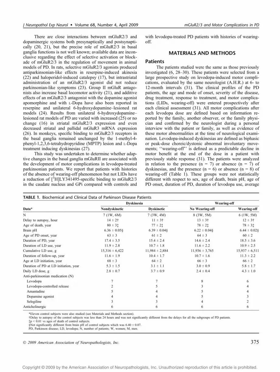
TABLE 1. Biochemical and Clinical Data of Parkinson Disease PatientsDyskinesia Wearing-off
Data* Nondyskinetic Dyskinetic No Wearing-off Wearing-off
N 7 (1W, 6M) 7 (3W, 4M) 8 (3W, 5M) 6 (1W, 5M)
Delay to autopsy, hour 14 T 2† 11 T 3† 13 T 3† 12 T 3†
Age of death, year 80 T 3‡ 77 T 2‡ 78 T 2‡ 78 T 3‡
Brain pH 6.36 T 0.05§ 6.39 T 0.04§ 6.22 T 0.04§ 6.44 T 0.02§
Age of PD onset, year 63 T 3 61 T 2 64 T 3 60 T 2
Duration of PD, year 17.4 T 3.5 15.4 T 2.4 14.6 T 2.4 18.5 T 3.6
Duration of LD use, year 11.9 T 2.8 10.7 T 1.8 11.6 T 2.2 10.9 T 2.5
Cumulative LD use, g 15,316 T 6,422 11,984 T 2,884 11,936 T 3,765 15,937 T 6,511
Duration of follow-up, year 11.6 T 1.9 10.4 T 1.7 10.7 T 1.6 11.3 T 2.2
Age at LD initiation, year 68 T 3 64 T 2 66 T 3 66 T 2
Duration of PD at LD initiation, year 5.3 T 1.5 3.1 T 1.1 3.0 T 0.9 5.8 T 1.7
Daily LD dose, g 2.8 T 0.7 3.7 T 0.9 2.4 T 0.4 4.3 T 1.0
Anti-parkinsonian medication (N)
Levodopa 7 7 8 6
Levodopa-controlled release 2 5 3 4
Amantadine 5 4 5 4
Dopamine agonist 4 4 5 3
Selegiline 3 3 4 2
Anticholinergic 6 6 6 6
*Eleven control subjects were also studied (see Materials and Methods section).†Delay to autopsy of the control subjects was less than 24 hours and was not significantly different from the delays for all the subgroups of PD patients.‡p G 0.01 vs ages of death of control subjects.§Not significantly different from brain pH of control subjects which was 6.40 T 0.07.PD, Parkinson disease; LD, levodopa; N, number of patients; W, women; M, men.
J Neuropathol Exp Neurol � Volume 68, Number 4, April 2009 mGluR2/3 and Motor Complications in PD
� 2009 American Association of Neuropathologists, Inc. 375

Copyright @ 2009 by the American Association of Neuropathologists, Inc. Unauthorized reproduction of this article is prohibited.
daily dose of levodopa, cumulative levodopa dose, durationof follow-up, and age at levodopa initiation (table and pre-vious publications [6, 28Y30]). The patient medical recordswere rechecked to determine the time of the last levodopaand/or dopamine agonist treatment. Two patients had re-ceived levodopa shortly (3 hours) before death but not theothers; for 1 patient, this information was not available.
Eleven control patients (2 women and 9 men) were alsostudied. Delay to autopsy of the control subjects was lessthan 24 hours and was not significantly different from thedelays for the PD patients (Table 1). The age of death ofcontrol subjects was 68 T 3 years (p G 0.01 vs age of death ofPD patients).
Autopsy and Handling of Brain MaterialAll autopsies of the 14 PD patients and the 11 control
patients (2 from Douglas Hospital Research Center brainbank, Montreal, Canada) who died with no neurological dis-orders were performed within 24 hours of death. The brainswere immediately divided into halves; one half was frozen atj80-C for biochemical studies and the other half was fixedin formalin for histological studies. These half brains werechosen at random for biochemical or histological studies. ThePD diagnosis was based on marked neuronal loss in thesubstantia nigra compacta, the presence of Lewy body in-clusions, and the absence of other pathological changes thatmight have accounted for parkinsonian symptoms (31). Thefrozen half brain was cut by hand in the frontal plane into 2-to 3-mm-thick slices.
Tissue PreparationSmall punches of the cerebral cortex were used to de-
termine tissue pH as an assessment of its preservation (32).Brain slices were cut into coronal sections (20 Km) on acryostat (j18-C), thaw-mounted onto Superfrost plus 75 �50-mm slides (Fisher; Nepean, Ontario, Canada), desiccatedovernight at 4-C, and stored at j80-C until they wereassayed. Small extracts of putamen were dissected, stored atj80-C, and processed for measurements of biogenic amineconcentrations.
Measures of the Extent of DopamineDenervation
The extent of striatal dopamine denervation was assessedby measurement of dopamine concentrations and dopaminetransporterYspecific binding. Biogenic amine concentrationswere assayed by high-performance liquid chromatographywith electrochemical detection, and the dopamine transporterwas labeled with [125I]RTI-121 (3Aa-(4-125I-iodophenyl)tropane-2A-carboxylic acid isopropyl ester), as previously re-ported (29).
[3H]LY341495 AutoradiographyGroup II mGluRs receptor-specific binding was eval-
uated using [3H]LY341495 (34.61 Ci/mmol) (Tocris Cookson,Ltd, Bristol, UK) binding by an adaptation of previously pub-lished methods used on rat brain (33). Sections were pre-incubated for 30 minutes in ice-cold phosphate-bromide buffer(10 mM potassium phosphate buffer with 100 mM potassium
bromide), pH 7.6. After drying, the sections were incubated for90 minutes at room temperature with 5 nM [3H]LY341495 inphosphate-bromide buffer. Nonspecific binding was deter-mined in the presence of 1 mM L-glutamic acid (ResearchBiomedical International, Natick, MD) in the buffer solution.After 2 washes for 30 seconds in ice-cold phosphate-bromidebuffer, the sections were rinsed briefly (30 seconds) in ice-colddistilled water. Finally, the slide-mounted tissue sections weredried overnight at room temperature and exposed to 3H-sensitive films (Kodak, Rochester, NY; along with standards[3H]-microscales; Amersham, Buckinghamshire, UK) for 14days at room temperature.
Data Analysis and StatisticsIntensities of autoradiographic labelingwith [3H]LY341495
were quantified on x-ray films by a Power Macintosh G4connected to a video camera (XC-77; Sony, Toronto, Canada)and a constant illumination light table using computerizeddensitometry (National Institutes of Health image v.1.63).Optical gray densities were transformed into nanocuries permilligram of tissue equivalent using a standard curve gen-erated with [3H]-standards. The results were then convertedinto femtomoles per milligram of tissue using the specificactivity of the radioligand. The caudate nucleus and theputamen were quantified in 4 subregions along a medial-lateral and dorsoventral axis. The GPe and GPi were dividedalong a dorsoventral axis. Nonspecific binding was measuredin the same brain regions on adjacent sections and thensubtracted from total binding. Control subjects (n = 11) wereused for the distribution analyses of mGluR2/3 in the basalganglia and were compared with paired t-tests. Statisticalcomparisons of all other data were performed by analysis ofvariance, followed by post hoc pairwise comparisons withFisher probability of least significant difference test. Allcontrol (n = 11) and PD patients (n = 14) were compared first.Comparisons were then performed between controls, PDpatients who had developed LIDs, and PD patients who hadnot developed LIDs. Finally, comparisons were made amongcontrols, PD patients with wearing-off, and PD patientswithout wearing-off. A value of p = 0.05 or less wasconsidered significant.
RESULTSBiogenic amine concentrations measured by high-per-
formance liquid chromatography in the same brain specimenswere previously reported (29). There was a marked decreaseof dopamine (j98.8%), homovanillic acid (HVA; j78.3%),3-methoxytyramine (j91.4%), and to a lesser extent 5-hydroxytyramine (j48.5%) and 5-hydroxy-indolacetic acid(j44.8%) concentrations in the putamen of all PD patientscompared with controls; there were no difference among thesubgroups of PD patients. The loss of striatal dopaminergicinnervation was also evaluated previously with [125I]RTI-121Yspecific binding to the dopamine transporter (29). Therewas a marked decrease of dopamine transporter in the cau-date nucleus (j72%) and putamen (j92%) of all PD patientscompared with controls, without difference among PD patientsub-groups.
Samadi et al J Neuropathol Exp Neurol � Volume 68, Number 4, April 2009
� 2009 American Association of Neuropathologists, Inc.376

Copyright @ 2009 by the American Association of Neuropathologists, Inc. Unauthorized reproduction of this article is prohibited.
Representative autoradiograms of [3H]LY341495 bind-ing in controls and different groups of PD patients andnonspecific binding are shown in Figure 1. There was higher[3H]LY341495-specific binding in the caudate nucleus (30%)of control subjects than in the putamen (df = 10, t = 8.1,p G 0.0001); this was approximately twice the binding mea-sured in the globus pallidus (caudate vs GPe: df = 10, t = 20.1,p G 0.0001; caudate vs GPi: df = 10, t = 23.1, p G 0.0001;putamen vs GPe: df = 10, t =11.9, p G 0.0001); putamen vsGPi: df = 10, t =13.0, p G 0.0001) (Figs. 1, 2). There was asmall (12%) but significantly higher [3H]LY341495-specificbinding in the GPe compared with the GPi (df = 10, t = 2.9,p = 0.0164). Division of these brain areas in their dorsal,ventral, lateral, and medial subregions showed smaller dif-ferences (10% or less).
Lesion and treatment effects were similar in all sub-regions of the caudate nucleus, putamen, and GPe and GPi;therefore, the values of the subregions were grouped in thesubsequent analyses. [3H]LY341495-specific binding of the 2patients who had received levodopa shortly before death wasnot different from that of the other PD patients.The loss ofdopaminergic innervation was associated with a significantreduction of [3H]LY341495-specific binding in the caudatenucleus of all PD patients (j13%) compared with controls(F1,23 =5.01, p = 0.04; Fig. 3). When PD patients were ana-lyzed according to the presence or absence of dyskinesias, nosignificant effect on [3H]LY341495-specific binding was ob-served in the caudate nucleus (F2,22 = 2.44, p = 0.11; Fig. 3).Binding was, however, reduced significantly in the caudate ofPD patients who had not developed wearing-off complications(j20% to j22%) compared with patients with wearing-off(p G 0.01) and controls (F2,22 = 6.93, p = 0.005), respectively(Fig. 3).
[3H]LY341495-specific binding was not different in theputamen of all PD patients compared with controls (F1,23 =0.28, p = 0.60; Fig. 3). Moreover, this binding was notaltered in the putamen when patients with PD were groupedaccording to the development or absence of dyskinesias orwearing-off (F2,22 = 1.18, p = 0.33; F2,22 = 1.75, p = 0.20,respectively) (Fig. 3).
[3H]LY341495-specific binding was not changed in theGPe of all PD patients (F1,23 = 1.90, p = 0.18) or differentgroups of patients who had or had not developed dyskinesiasor wearing-off (F2,22 = 1.01, p = 0.38; F2,22 =1.17, p = 0.33,respectively) (Fig. 4). By contrast, binding was reduced sig-nificantly (j25%) in the GPi of all PD patients comparedwith controls (F1,23 = 5.86, p = 0.02; Fig. 4). This reductiondid not reach significance in the GPi of patients with orwithout dyskinesias (F2,22 = 2.82, p = 0.08), but it was lessin patients who had not developed wearing-off (j30%)
FIGURE 1. Representative autoradiograms of [3H]LY341495 binding in postmortem human coronal brain sections at the 14.6-mmlevel (34) showing a control subject and levodopa-treated parkinsonian patients who had or had not developed motorcomplications; an example of nonspecific binding in the presence of 1 mM L-glutamic acid is also shown. Cd, caudate nucleus;Put, putamen; GPe, external globus pallidus; GPi, internal globus pallidus.
FIGURE 2. [3H]LY341495 binding in the caudate nucleus,putamen, and globus pallidus (external [GPe] and internal[GPi]) of postmortem human controls. Values are presented infemtomoles per milligram (fmol/mg) of tissue as mean T SEM.Higher [3H]LY341495-specific binding was observed in thecaudate nucleus compared with the putamen, and both ofthese bindings were twice those measured in the GPe or GPi.In the globus pallidus, [3H]LY341495-specific binding washigher in the GPe compared with the GPi. ****p G 0.0001 vscaudate nucleus; LLLLp G 0.0001 vs putamen; ++p = 0.0164 vsGPe. DL, dorsolateral; DM, dorsomedial; VL, ventrolateral; VM,ventromedial; D, dorsal; V, ventral.
J Neuropathol Exp Neurol � Volume 68, Number 4, April 2009 mGluR2/3 and Motor Complications in PD
� 2009 American Association of Neuropathologists, Inc. 377

Copyright @ 2009 by the American Association of Neuropathologists, Inc. Unauthorized reproduction of this article is prohibited.
fluctuations compared with control subjects (F2,22 = 3.34, p =0.05; Fig. 4).
DISCUSSIONThe present study is the first to show the following: 1)
[3H]LY341495-specific binding to mGluR2/3 in normalcontrols is higher in the caudate nucleus than in the putamenand that both are approximately twice as high as in the globuspallidus, with the former more in the external than internalpart; 2) there is lower [3H]LY341495-specific binding in thecaudate nucleus and GPi of patients who had not had wearing-off compared with controls and to PD patients who had
developed wearing-off; and 3) there is similar [3H]LY341495-specific binding in the caudate nucleus, putamen, and GPeand GPi of PD patients who did or did not have dyskinesias.
The distribution of [3H]LY341495-specific bindingobserved in human control subjects is similar to that observedin monkeys (27). As in MPTP-treated monkeys (27),[3H]LY341495 specific binding was not different in dyskine-tic and nondyskinetic subjects and controls. The MPTP mon-key experiments were not designed to study wearing-off, andit was not observed in these monkeys. There was, however adecrease of [3H]LY341495-specific binding compared withcontrols in the striatum and globus pallidus of MPTP mon-keys treated with levodopa and cabergoline, a long-acting
FIGURE 3. [3H]LY341495 binding in the caudate nucleus and putamen of postmortem control and levodopa-treated parkinsonianpatients. Individual values and means are in femtomoles per milligram (fmol/mg) of tissue. Significantly less [3H]LY341495 bindingwas observed in the caudate nucleus but not putamen of Parkinson disease (PD) patients compared with controls (j13%). Nodifferences in [3H]LY341495-specific binding were observed in the caudate and putamen of PD patients who had or had notdeveloped dyskinesias. There was less binding in the caudate nucleus of PD patients who had not developed wearing-off motorcomplications compared with controls (j22%) and to PD patients who had wearing-off fluctuations (j20%). *p G 0.05 and***p G 0.005 vs controls; LLp G 0.01 vs PD wearing-off.
Samadi et al J Neuropathol Exp Neurol � Volume 68, Number 4, April 2009
� 2009 American Association of Neuropathologists, Inc.378

Copyright @ 2009 by the American Association of Neuropathologists, Inc. Unauthorized reproduction of this article is prohibited.
dopamine agonist that provides more constant dopaminergicstimulation. This is analogous to the present results in whichPD patients who had not had wearing-off had lower caudatenucleus and GPi [3H]LY341495-specific binding; this motorcomplication is associated with fluctuation of dopaminergicstimulation.
The similar extensive degrees of reduction of striataldopamine and dopamine metabolites concentrations and thedopamine transporter of the different groups of PD patients(29) precludes differences in striatal denervation as an expla-nation for motor fluctuations in these patients. The increaseof dopamine turnover after nigrostriatal damage (HVA/dopamine) was, however, higher in the putamen of patientswith wearing-off than those without wearing-off (29). Ahigher HVA/dopamine ratio was also reported in the putamen
of PD patients with wearing-off compared with patients withdyskinesias or patients without motor complications (35).The association between wearing-off fluctuations and in-creased dopamine turnover after nigrostriatal damage wasobserved by positron emission tomography (36). Hence, lossof presynaptic dopamine storage capacity and rapid metab-olism of dopamine at striatal synapses may play a relevantrole in levodopa-related wearing-off in PD patients (29, 35,36). This raises the question as to whether the decrease ofspecific binding to mGluR2/3 in the caudate nucleus is asso-ciated with changes in dopamine turnover. Although theHVA/dopamine ratio was not measured in the caudate ofthese patients, the fact that the specific binding to mGluR2/3is similar in the caudate nucleus of control subjects and PDpatients with wearing-off fluctuations makes this possibility
FIGURE 4. [3H]LY341495-specific binding in the external and internal segments of the globus pallidus (external [GPe] and internal[GPi]) of control and levodopa-treated parkinsonian patients. Individual values and means are in femtomoles per milligram(fmol/mg) of tissue. No difference in [3H]LY341495-specific binding was observed in the GPe of Parkinson disease (PD) patients whohad or had not experienced motor complications; binding was less in the GPi of PD patients (j25%) compared with controls. Thisreduction was significant in the GPi of patients who had not developed wearing-off (j30%). *p G 0.05 and **p G 0.01 vs controls.
J Neuropathol Exp Neurol � Volume 68, Number 4, April 2009 mGluR2/3 and Motor Complications in PD
� 2009 American Association of Neuropathologists, Inc. 379

Copyright @ 2009 by the American Association of Neuropathologists, Inc. Unauthorized reproduction of this article is prohibited.
unlikely. Moreover, wearing-off is also seen with dopamineagonists (which are not stored in presynaptic terminals) (4).No clear correlation was found in positron emission tomog-raphy studies of human PD subjects between the degree ofdamage in dopamine nigrostriatal neurons and motor fluctua-tions (37).
Our results suggest that in addition to presynaptic in-ability to buffer levodopa variations, alterations in group IImGluRs responsiveness could also play an important role inthe development of motor fluctuations. The mechanisms bywhich the reduction of group II mGluRs is involved in pre-vention of motor fluctuations in PD patients are not known; itmay have a protective role or there may not be a cause/effectrelationship and be secondary to the numerous glutamatergicchanges caused by the disease and its treatments.
The differential effect of mGluR2/3 on D1 and D2 do-pamine receptors (21) and expression of the latter receptors inthe anatomically distinct direct and indirect pathways, re-spectively (38), suggest that a reduction of mGluR2/3 mightcontribute to restoring the balance between the striatal outputpathways and preventing motor fluctuations in levodopa-treated PD patients. Moreover, activation of mGluR2/3 ex-pressed presynaptically and postsynaptically on striatal cho-linergic interneurons can also decrease striatal acetylcholinerelease (13, 39). Interestingly, the increase of acetylcholinerelease by the mGluR2/3 antagonist, LY341495, was abol-ished in dopamine-depleted striatum (40). In PD patientswithout motor fluctuations, the reduction of mGluR2/3 mightcontribute to normalization of disrupted striatal cholinergictransmission. Facilitation of striatal acetylcholine release byN-methyl D-aspartate receptors (40) and increased specificbinding to striatal N-methyl D-aspartate receptors of PDpatients with wearing-off (6) corroborate this hypothesis.
Why was the reduction of specific binding to mGluR2/3 more pronounced in the caudate nucleus than in the puta-men? The caudate nucleus plays a central role in learning asimple stimulus-response association; this is impaired inparkinsonian patients in the ‘‘off’’ state and reverts to normalin ‘‘on’’ patients treated with levodopa (41, 42). Modifica-tions of dopamine activity in associative territories located inthe caudate nucleus could affect attention-deficit behavior(43). Because activation of group II mGluRs induces cog-nitive and memory deficits (44), the more pronounced re-duction of mGluR2/3 receptor binding in the caudate nucleusmay act as a compensatory mechanism to prevent motorfluctuations in PD patients.
The striatal medium spiny neurons project to the outputnuclei of the basal ganglia, SNr, and GPi monosynapticallyor polysynaptically through the GPe and the STN (38, 45).Group II mGluRs are localized presynaptically on STNefferent terminals in the GPi and SNr where they depressglutamate release (17, 46) and postsynaptically in the basalganglia nuclei (14) where they can regulate neuronal excit-ability via modulation of ion channel functions (47). Hence,reduction of mGluR2/3 in the GPi of PD patients withoutwearing-off fluctuations may contribute to restoring the nor-mal GPi neuronal discharge patterns. Moreover, the GPesends inhibitory projections to the GPi, and the GPe-GPibalance is important in normal neuronal activity of the output
basal ganglia circuitry (48). Reduction of functional inhib-itory tone of mGluR2/3 on GPe efferent terminals in the GPi,by disinhibition of GPe, could reduce GPi neuronal activityleading to more normalize motor function and improvementof motor fluctuations. Activation of mGluR2/3 to maintain aconstant inhibitory tone is hypothesized to suppress locomo-tor output (49).
In the presence of sufficient intracellular Ca2+, en-hanced production of cAMP can be induced by increase ofadenosine release after activation of mGluR3 localized inastrocytes (50). This potentiation of cAMP levels is inhibitedby adenosine deaminase and adenosine A2A receptor antag-onist (50). According to antiparkinsonian effect of A2A re-ceptor antagonist confirmed by behavioral studies in animalmodels and parkinsonian patients (51, 52), it might besuggested that a reduction of striatal mGluR2/3 may repre-sent a decrease of mGluR3 in astrocytes as a compensatorymechanism to prevent A2A receptor activation and subse-quent motor fluctuations. Further investigations are requiredto elucidate the mechanisms involved in neuron-glial cell in-teraction in normal and pathological conditions. Our bindingassay was based on Wright et al (33), who suggested thatunder their binding conditions, [3H]LY341495 may bindpredominantly to a mGluR3 receptor population in the ratforebrain. Based on the similar distribution pattern ofmGluR2 and mGluR3 in preterminal and terminal portionsof the axons (19, 53) and stronger expression of mGluR3 inthe globus pallidus than mGluR2 (19), it might be speculatedthat in the striatum and especially in the globus pallidus, ourspecific binding was mainly to an mGluR3 receptor pop-ulation. According to the strong labeling of mGluR2 receptorin the caudate and putamen (53), however, we could not ruleout the striatal [3H]LY341495-specific binding to mGluR2.The development of subtype-selective radioligands for groupII mGluRs is required to decipher mGluR2 and mGluR3receptor contributions.
Recent studies reported an earlier and higher incidenceof motor and nonmotor symptoms of wearing-off, particu-larly in the earlier stages of the PD compared with the olderliterature (3, 54, 55). The present study is the first to provideevidence of a decrease of [3H]LY341495-specific binding tomGluR2/3 in the caudate nucleus and GPi of PD patientswithout wearing-off motor fluctuations. Our results, togetherwith other studies (29, 35, 56), suggest that in addition topresynaptic inability to buffer levodopa variations, alterationsin group II mGluR responsiveness may play an importantrole in the development of motor fluctuations. Increased glu-tamatergic transmission in PD patients may lead to a compen-satory adaptive downregulation of mGluR2/3 in PD patientswithout wearing-off, and this adaptation may be absent inpatients with this motor complication. These results indicatethat pharmacological modulation of mGluR2/3 activity mighthelp to manage levodopa-induced wearing-off fluctuations inpatients with PD.
REFERENCES1. Wichmann T, DeLong MR. Functional neuroanatomy of the basal gan-
glia in Parkinson’s disease. Adv Neurol 2003;91:9Y182. Mercuri NB, Bernardi G. The Fmagic_ of L-Dopa: Why is it the gold stan-
dard Parkinson’s disease therapy? Trends Pharmacol Sci 2005;26:341Y44
Samadi et al J Neuropathol Exp Neurol � Volume 68, Number 4, April 2009
� 2009 American Association of Neuropathologists, Inc.380

Copyright @ 2009 by the American Association of Neuropathologists, Inc. Unauthorized reproduction of this article is prohibited.
3. Stacy M, Hauser R, Oertel W, et al. End-of-dose wearing off in Parkinsondisease: A 9-question survey assessment. Clin Neuropharmacol 2006;29:312Y21
4. Parkinson Study Group. Pramipexole vs levodopa as initial treatment forParkinson disease: A randomized controlled trial. Parkinson Study Group.JAMA 2000;284:1931Y38
5. Fahn S, Oakes D, Shoulson I, et al. Levodopa and the progression ofParkinson’s disease. N Engl J Med 2004;351:2498Y508
6. Calon F, Rajput AH, Hornykiewicz O, Bedard PJ, Di Paolo T.Levodopa-induced motor complications are associated with alterationsof glutamate receptors in Parkinson’s disease. Neurobiol Dis 2003;14:404Y16
7. Calabresi P, Centonze D, Bernardi G. Electrophysiology of dopamine innormal and denervated striatal neurons. Trends Neurosci 2000;23:S57Y63
8. Gubellini P, Eusebio A, Oueslati A, Melon C, Kerkerian-Le Goff L,Salin P. Chronic high-frequency stimulation of the subthalamic nucleusand L-DOPA treatment in experimental parkinsonism: Effects on motorbehaviour and striatal glutamate transmission. Eur J Neurosci 2006;24:1802Y14
9. Marino MJ, Awad H, Poisik O, Wittmann M, Conn PJ. Localization andphysiological roles of metabotropic glutamate receptors in the direct andindirect pathways of the basal ganglia. Amino Acids 2002;23:185Y91
10. Conn PJ, Battaglia G, Marino MJ, Nicoletti F. Metabotropic glutamatereceptors in the basal ganglia motor circuit. Nat Rev Neurosci 2005;6:787Y98
11. Ferraguti F, Shigemoto R. Metabotropic glutamate receptors. Cell TissueRes 2006;326:483Y504
12. Richards G, Messer J, Malherbe P, et al. Distribution and abundance ofmetabotropic glutamate receptor subtype 2 in rat brain revealed by[3H]LY354740 binding in vitro and quantitative radioautography:Correlation with the sites of synthesis, expression, and agonist stimula-tion of [35S]GTPgammas binding. J Comp Neurol 2005;487:15Y27
13. Pisani A, Bonsi P, Catania MV, et al. Metabotropic glutamate 2 recep-tors modulate synaptic inputs and calcium signals in striatal cholinergicinterneurons. J Neurosci 2002;22:6176Y85
14. Phillips T, Rees S, Augood S, et al. Localization of metabotropic gluta-mate receptor type 2 in the human brain. Neuroscience 2000;95:1139Y56
15. Smith Y, Charara A, Paquet M, et al. Ionotropic and metabotropic GABAand glutamate receptors in primate basal ganglia. J Chem Neuroanat2001;22:13Y42
16. Testa CM, Friberg IK, Weiss SW, Standaert DG. Immunohistochemicallocalization of metabotropic glutamate receptors mGluR1a and mGluR2/3in the rat basal ganglia. J Comp Neurol 1998;390:5Y19
17. Bradley SR, Marino MJ, Wittmann M, et al. Activation of group IImetabotropic glutamate receptors inhibits synaptic excitation of thesubstantia nigra pars reticulata. J Neurosci 2000;20:3085Y94
18. Mudo G, Trovato-Salinaro A, Caniglia G, Cheng Q, Condorelli DF.Cellular localization of mGluR3 and mGluR5 mRNAs in normal andinjured rat brain. Brain Res 2007;1149:1Y13
19. Tamaru Y, Nomura S, Mizuno N, Shigemoto R. Distribution of meta-botropic glutamate receptor mGluR3 in the mouse CNS: Differentiallocation relative to pre- and postsynaptic sites. Neuroscience 2001;106:481Y503
20. Morishima Y, Miyakawa T, Furuyashiki T, Tanaka Y, Mizuma H,Nakanishi S. Enhanced cocaine responsiveness and impaired motor co-ordination in metabotropic glutamate receptor subtype 2 knockout mice.Proc Natl Acad Sci U S A 2005;102:4170Y75
21. David HN, Abraini JH. Differential modulation of the D1-like andD2-like dopamine receptor-induced locomotor responses by group IImetabotropic glutamate receptors in the rat nucleus accumbens. Neuro-pharmacology 2001;41:454Y63
22. Dawson L, Chadha A, Megalou M, Duty S. The group II metabotropicglutamate receptor agonist, DCG-IV, alleviates akinesia followingintranigral or intraventricular administration in the reserpine-treated rat.Br J Pharmacol 2000;129:541Y46
23. Ossowska K, Konieczny J, Wardas J, et al. An influence of ligands ofmetabotropic glutamate receptor subtypes on parkinsonian-like symp-toms and the striatopallidal pathway in rats. Amino Acids 2007;32:179Y88
24. Feeley Kearney JA, Albin RL. mGluRs: A target for pharmacotherapy inParkinson disease. Exp Neurol 2003;184(Suppl 1):S30Y6
25. Picconi B, Pisani A, Centonze D, et al. Striatal metabotropic glutamatereceptor function following experimental parkinsonism and chroniclevodopa treatment. Brain 2002;125:2635Y45
26. Messenger MJ, Dawson LG, Duty S. Changes in metabotropic glutamatereceptor 1Y8 gene expression in the rodent basal ganglia motor loopfollowing lesion of the nigrostriatal tract. Neuropharmacology 2002;43:261Y71
27. Samadi P, Gregoire L, Morissette M, et al. Basal ganglia group II meta-botropic glutamate receptors specific binding in non-human primate modelof L-DopaYinduced dyskinesias. Neuropharmacology 2008;54:258Y68
28. Calon F, Dridi M, Hornykiewicz O, Bedard PJ, Rajput AH, Di Paolo T.Increased adenosine A2A receptors in the brain of Parkinson’s diseasepatients with dyskinesias. Brain 2004;127:1075Y84
29. Calon F, Morissette M, Rajput AH, Hornykiewicz O, Bedard PJ,Di Paolo T. Changes of GABA receptors and dopamine turnover in thepostmortem brains of parkinsonians with levodopa-induced motor com-plications. Mov Disord 2003;18:241Y53
30. Calon F, Birdi S, Rajput AH, Hornykiewicz O, Bedard PJ, Di Paolo T.Increase of preproenkephalin mRNA levels in the putamen of Parkinsondisease patients with levodopa-induced dyskinesias. J Neuropathol ExpNeurol 2002;61:186Y96
31. Rajput AH, Fenton ME, Birdi S, et al. Clinical-pathological study oflevodopa complications. Mov Disord 2002;17:289Y96
32. Kingsbury AE, Foster OJ, Nisbet AP, et al. Tissue pH as an indicator ofmRNA preservation in human post-mortem brain. Brain Res Mol BrainRes 1995;28:311Y18
33. Wright RA, Arnold MB, Wheeler WJ, Ornstein PL, Schoepp DD.[3H]LY341495 binding to group II metabotropic glutamate receptors inrat brain. J Pharmacol Exp Ther 2001;298:453Y60
34. Mai JK, Assheuer J, Paxinos G. Atlas of the Human Brain. San Diego,CA: Academic Press, 1997:193
35. Rajput AH, Fenton ME, Di Paolo T, Sitte H, Pifl C, Hornykiewicz O.Human brain dopamine metabolism in levodopa-induced dyskinesia andwearing-off. Parkinsonism Relat Disord 2004;10:221Y26
36. de la Fuente-Fernandez R, Lu JQ, Sossi V, et al. Biochemical variations inthe synaptic level of dopamine precede motor fluctuations in Parkinson’sdisease: PET evidence of increased dopamine turnover. Ann Neurol 2001;49:298Y303
37. Snow BJ, Tooyama I, McGeer EG, et al. Human positron emission to-mographic [18F]fluorodopa studies correlate with dopamine cell countsand levels. Ann Neurol 1993;34:324Y30
38. Nadjar A, Brotchie JM, Guigoni C, et al. Phenotype of striatofugal me-dium spiny neurons in parkinsonian and dyskinetic nonhuman primates:A call for a reappraisal of the functional organization of the basal gan-glia. J Neurosci 2006;26:8653Y61
39. Bonsi P, Cuomo D, Picconi B, et al. Striatal metabotropic glutamatereceptors as a target for pharmacotherapy in Parkinson’s disease. AminoAcids 2007;32:189Y95
40. Mela F, Marti M, Fiorentini C, Missale C, Morari M. Group-IImetabotropic glutamate receptors negatively modulate NMDA trans-mission at striatal cholinergic terminals: Role of P/Q-type high voltageactivated Ca++ channels and endogenous dopamine. Mol Cell Neurosci2006;31:284Y92
41. Shohamy D, Myers CE, Grossman S, Sage J, Gluck MA. The role ofdopamine in cognitive sequence learning: Evidence from Parkinson’sdisease. Behav Brain Res 2005;156:191Y99
42. Seger CA, Cincotta CM. The roles of the caudate nucleus in humanclassification learning. J Neurosci 2005;25:2941Y51
43. Grabli D, McCairn K, Hirsch EC, et al. Behavioural disorders inducedby external globus pallidus dysfunction in primates: I. Behaviouralstudy. Brain 2004;127:2039Y54
44. Higgins GA, Ballard TM, Kew JN, et al. Pharmacological manipulationof mGlu2 receptors influences cognitive performance in the rodent.Neuropharmacology 2004;46:907Y17
45. Levesque M, Parent A. The striatofugal fiber system in primates: Areevaluation of its organization based on single-axon tracing studies.Proc Natl Acad Sci U S A 2005;102:11888Y93
46. Poisik O, Raju DV, Verreault M, et al. Metabotropic glutamate receptor2 modulates excitatory synaptic transmission in the rat globus pallidus.Neuropharmacology 2005;49(Suppl 1):57Y69
47. Cho K, Bashir ZI. Cooperation between mGlu receptors: A depressingmechanism? Trends Neurosci 2002;25:405Y11
J Neuropathol Exp Neurol � Volume 68, Number 4, April 2009 mGluR2/3 and Motor Complications in PD
� 2009 American Association of Neuropathologists, Inc. 381

Copyright @ 2009 by the American Association of Neuropathologists, Inc. Unauthorized reproduction of this article is prohibited.
48. Heimer G, Rivlin-Etzion M, Bar-Gad I, Goldberg JA, Haber SN,Bergman H. Dopamine replacement therapy does not restore the full spec-trum of normal pallidal activity in the 1-methyl-4-phenyl-1,2,3,6-tetra-hydropyridine primate model of Parkinsonism. J Neurosci 2006;26:8101Y14
49. O’Neill MF, Heron-Maxwell C, Conway MW, Monn JA, Ornstein P.Group II metabotropic glutamate receptor antagonists LY341495 andLY366457 increase locomotor activity in mice. Neuropharmacology2003;45:565Y74
50. Moldrich RX, Aprico K, Diwakarla S, O’Shea RD, Beart PM. AstrocytemGlu(2/3)-mediated cAMP potentiation is calcium sensitive: Studies inmurine neuronal and astrocyte cultures. Neuropharmacology 2002;43:189Y203
51. Grondin R, Bedard PJ, Hadj Tahar A, Gregoire L, Mori A, Kase H.Antiparkinsonian effect of a new selective adenosine A2A receptorantagonist in MPTP-treated monkeys. Neurology 1999;52:1673Y77
52. Chase TN, Bibbiani F, Bara-Jimenez W, Dimitrova T, Oh-Lee JD.Translating A2A antagonist KW6002 from animal models to parkinso-nian patients. Neurology 2003;61:S107Y11
53. Ohishi H, Neki A, Mizuno N. Distribution of a metabotropic glutamatereceptor, mGluR2, in the central nervous system of the rat and mouse:An immunohistochemical study with a monoclonal antibody. NeurosciRes 1998;30:65Y82
54. Hauser RA. Long-term care of Parkinson’s disease. Strategies for man-aging ‘‘wearing off’’ symptom re-emergence and dyskinesias. Geriatrics2006;61:14Y20
55. Stocchi F, Vacca L, Ruggieri S, Olanow CW. Intermittent vs continuouslevodopa administration in patients with advanced Parkinson disease: Aclinical and pharmacokinetic study. Arch Neurol 2005;62:905Y10
56. Stocchi F. The levodopa wearing-off phenomenon in Parkinson’s disease:Pharmacokinetic considerations. Expert Opin Pharmacother 2006;7:1399Y407
Samadi et al J Neuropathol Exp Neurol � Volume 68, Number 4, April 2009
� 2009 American Association of Neuropathologists, Inc.382