Metabotropic Glutamate Receptors 1 and 5 Differentially Regulate CA1 Pyramidal Cell Function
10
Metabotropic Glutamate Receptors 1 and 5 Differentially Regulate CA1 Pyramidal Cell Function Guido Mannaioni, 1 Michael J. Marino, 1 Ornella Valenti, 1,2 Stephen F. Traynelis, 1 and P. Jeffrey Conn 3 1 Department of Pharmacology, Emory University School of Medicine, Atlanta, Georgia 30322, 2 Postdoctoral Program of Preclinical and Clinical Pharmacology, University of Catania, 95125 Catania, Italy, and 3 Department of Pharmacology, Neuroscience Division, Merck Research Laboratories, West Point, Pennsylvania 19486 The activation of group I metabotropic glutamate receptors (mGluRs) produces a variety of actions that lead to alterations in excitability and synaptic transmission in the CA1 region of the hippocampus. The group I mGluRs, mGluR1 and mGluR5, are activated selectively by (S)-3,5-dihydroxyphenylglycine (DHPG). To identify which of these mGluR subtypes are respon- sible for the various actions of DHPG in area CA1, we took advantage of two novel subtype-selective antagonists. (S)-()- -amino-a-methylbenzeneacetic acid (LY367385) is a potent competitive antagonist that is selective for mGluR1, whereas 2-methyl-6-(phenylethynyl)-pyridine (MPEP) is a potent non- competitive antagonist that is selective for mGluR5. The use of these compounds in experiments with whole-cell patch-clamp recording and Ca 2 -imaging techniques revealed that each group I mGluR subtype plays distinct roles in regulating the function of CA1 pyramidal neurons. The block of mGluR1 by LY367385 suppressed the DHPG-induced increase in intracel- lular Ca 2 concentration ([Ca 2 ] i ) and the direct depolarization of CA1 hippocampal neurons. In addition, the increase in the frequency of spontaneous IPSCs (sIPSCs) caused by the DHPG-induced depolarization of inhibitory interneurons also was blocked by LY367385, as was the DHPG-induced inhibi- tion of transmission at the Schaffer collateral3CA1 synapse. On the other hand, the block of mGluR5 by MPEP antagonized the DHPG-induced suppression of the Ca 2 -activated potas- sium current (I AHP ) and potentiation of the NMDA receptor. Finally, antagonism of the DHPG-induced suppression of evoked IPSCs required the blockade of both mGluR1 and mGluR5. These data suggest that mGluR1 and mGluR5 play distinct roles in the regulation of the excitability of hippocampal CA1 pyramidal neurons. Key words: mGluR; mGluR1; mGluR5; (S)-3,5-dihydroxy- phenylglycine (DHPG); (S)-()--amino-a-methylbenzeneacetic acid (LY367385); 2-methyl-6-(phenylethynyl)-pyridine (MPEP); I AHP ; IPSC; EPSC; hippocampus Metabotropic glutamate receptors (mGluRs) have been impli- cated in a number of physiological and pathological responses to glutamate in CA1 hippocampal region. These include the modu- lation of neuronal excitability and synaptic transmission (for review, see Anwyl, 1999) as well as the induction of long-term potentiation (Bashir et al., 1993), generation of epileptiform ac- tivity (Aronica et al., 1997; Merlin, 1999; Galoyan and Merlin, 2000), and postischemic injury (Bond et al., 1999, 2000; Pellegrini-Giampietro et al., 1999). The family of mGluRs is composed of three distinct groups that are based on sequence homology, pharmacology, and coupling to second messenger sys- tems (for review, see Nakanishi, 1992; Schoepp and Conn, 1993; Pin and Duvoisin, 1995). Although all three groups of mGluRs play roles in regulating hippocampal function (Anwyl, 1999), group I mGluRs are especially important for the regulation of pyramidal cell excitability. The group I mGluRs include mGluR1 and mGluR5, both of which are activated selectively by ( S)-3,5- dihydroxyphenylglycine (DHPG), and couple to G q and to the activation of phosphoinositide hydrolysis. The activation of group I mGluRs has a number of direct excitatory effects on CA1 pyramidal cells, including depolariza- tion and increased cell firing (Charpak et al., 1990; Desai and Conn, 1991; Pedarzani and Storm, 1993; Davies et al., 1995; Gereau and Conn, 1995b; Mannaioni et al., 1999). These effects have been ascribed to the activation of C a 2 -activated and C a 2 - independent cationic conductances (Crepel et al., 1994; Gueri- neau et al., 1995) and the inhibition of at least four different K currents. These include the AHP current (Charpak et al., 1990; Desai and Conn, 1991), a leak current (Guerineau et al., 1994), the M current (Charpak et al., 1990), and a voltage-dependent slow-inactivating current (Luthi et al., 1996). In addition, the activation of group I mGluRs increases CA1 pyramidal cell ex- citability by decreasing GABA-mediated inhibition (Desai and Conn, 1991; Gereau and Conn, 1995a; Fitzsimonds and Dichter, 1996). Immunocytochemistry studies reveal that mGluR5 is localized in CA1 pyramidal cells, whereas mGluR1a is not detectable (Baude et al., 1993; Romano et al., 1995). Therefore, mGluR5 is thought to play a predominant role in regulating CA1 pyramidal cells. However, although mGluR5 is the most abundant group I mGluR in CA1 pyramidal cells, these cells also express mGluR1 mRNA (Shigemoto et al., 1992; Berthele et al., 1998), and a recent immunohistochemical study with antibodies that react with all splice forms of mGluR1 revealed low levels of mGluR1 im- munoreactivity in this region (Ferraguti et al., 1998). Therefore, Received Feb. 12, 2001; revised May 15, 2001; accepted May 31, 2001. This work was supported by the National Institutes of Health and by the National Alliance for Research on Schizophrenia and Depression. We thank Dr. Gianmaria Maccaferri for critical reading and comments on this manuscript. Correspondence should be addressed to Dr. Jeffrey Conn, Senior Director, Neuroscience Division, Department of Pharmacology, Merck Research Laborato- ries, Merck & Company, Incorporated, 770 Sumneytown Pike, P.O. Box 4, WP 46-300, West Point, PA 19486-0004. E-mail: [email protected]. Copyright © 2001 Society for Neuroscience 0270-6474/01/215925-10$15.00/0 The Journal of Neuroscience, August 15, 2001, 21(16):5925–5934
Transcript of Metabotropic Glutamate Receptors 1 and 5 Differentially Regulate CA1 Pyramidal Cell Function
Metabotropic Glutamate Receptors 1 and 5 Differentially RegulateCA1 Pyramidal Cell Function
Guido Mannaioni,1 Michael J. Marino,1 Ornella Valenti,1,2 Stephen F. Traynelis,1 and P. Jeffrey Conn3
1Department of Pharmacology, Emory University School of Medicine, Atlanta, Georgia 30322, 2Postdoctoral Program ofPreclinical and Clinical Pharmacology, University of Catania, 95125 Catania, Italy, and 3Department of Pharmacology,Neuroscience Division, Merck Research Laboratories, West Point, Pennsylvania 19486
The activation of group I metabotropic glutamate receptors(mGluRs) produces a variety of actions that lead to alterationsin excitability and synaptic transmission in the CA1 region of thehippocampus. The group I mGluRs, mGluR1 and mGluR5, areactivated selectively by (S)-3,5-dihydroxyphenylglycine(DHPG). To identify which of these mGluR subtypes are respon-sible for the various actions of DHPG in area CA1, we tookadvantage of two novel subtype-selective antagonists. (S)-(�)-�-amino-a-methylbenzeneacetic acid (LY367385) is a potentcompetitive antagonist that is selective for mGluR1, whereas2-methyl-6-(phenylethynyl)-pyridine (MPEP) is a potent non-competitive antagonist that is selective for mGluR5. The use ofthese compounds in experiments with whole-cell patch-clamprecording and Ca2�-imaging techniques revealed that eachgroup I mGluR subtype plays distinct roles in regulating thefunction of CA1 pyramidal neurons. The block of mGluR1 byLY367385 suppressed the DHPG-induced increase in intracel-lular Ca2� concentration ([Ca2�]i ) and the direct depolarization
of CA1 hippocampal neurons. In addition, the increase in thefrequency of spontaneous IPSCs (sIPSCs) caused by theDHPG-induced depolarization of inhibitory interneurons alsowas blocked by LY367385, as was the DHPG-induced inhibi-tion of transmission at the Schaffer collateral3CA1 synapse.On the other hand, the block of mGluR5 by MPEP antagonizedthe DHPG-induced suppression of the Ca2�-activated potas-sium current (IAHP) and potentiation of the NMDA receptor.Finally, antagonism of the DHPG-induced suppression ofevoked IPSCs required the blockade of both mGluR1 andmGluR5. These data suggest that mGluR1 and mGluR5 playdistinct roles in the regulation of the excitability of hippocampalCA1 pyramidal neurons.
Key words: mGluR; mGluR1; mGluR5; (S)-3,5-dihydroxy-phenylglycine (DHPG); (S)-(�)-�-amino-a-methylbenzeneaceticacid (LY367385); 2-methyl-6-(phenylethynyl)-pyridine (MPEP);IAHP; IPSC; EPSC; hippocampus
Metabotropic glutamate receptors (mGluRs) have been impli-cated in a number of physiological and pathological responses toglutamate in CA1 hippocampal region. These include the modu-lation of neuronal excitability and synaptic transmission (forreview, see Anwyl, 1999) as well as the induction of long-termpotentiation (Bashir et al., 1993), generation of epileptiform ac-tivity (Aronica et al., 1997; Merlin, 1999; Galoyan and Merlin,2000), and postischemic injury (Bond et al., 1999, 2000;Pellegrini-Giampietro et al., 1999). The family of mGluRs iscomposed of three distinct groups that are based on sequencehomology, pharmacology, and coupling to second messenger sys-tems (for review, see Nakanishi, 1992; Schoepp and Conn, 1993;Pin and Duvoisin, 1995). Although all three groups of mGluRsplay roles in regulating hippocampal function (Anwyl, 1999),group I mGluRs are especially important for the regulation ofpyramidal cell excitability. The group I mGluRs include mGluR1and mGluR5, both of which are activated selectively by (S)-3,5-dihydroxyphenylglycine (DHPG), and couple to Gq and to theactivation of phosphoinositide hydrolysis.
The activation of group I mGluRs has a number of directexcitatory effects on CA1 pyramidal cells, including depolariza-tion and increased cell firing (Charpak et al., 1990; Desai andConn, 1991; Pedarzani and Storm, 1993; Davies et al., 1995;Gereau and Conn, 1995b; Mannaioni et al., 1999). These effectshave been ascribed to the activation of Ca2�-activated and Ca2�-independent cationic conductances (Crepel et al., 1994; Gueri-neau et al., 1995) and the inhibition of at least four different K�
currents. These include the AHP current (Charpak et al., 1990;Desai and Conn, 1991), a leak current (Guerineau et al., 1994),the M current (Charpak et al., 1990), and a voltage-dependentslow-inactivating current (Luthi et al., 1996). In addition, theactivation of group I mGluRs increases CA1 pyramidal cell ex-citability by decreasing GABA-mediated inhibition (Desai andConn, 1991; Gereau and Conn, 1995a; Fitzsimonds and Dichter,1996).
Immunocytochemistry studies reveal that mGluR5 is localizedin CA1 pyramidal cells, whereas mGluR1a is not detectable(Baude et al., 1993; Romano et al., 1995). Therefore, mGluR5 isthought to play a predominant role in regulating CA1 pyramidalcells. However, although mGluR5 is the most abundant group ImGluR in CA1 pyramidal cells, these cells also express mGluR1mRNA (Shigemoto et al., 1992; Berthele et al., 1998), and arecent immunohistochemical study with antibodies that react withall splice forms of mGluR1 revealed low levels of mGluR1 im-munoreactivity in this region (Ferraguti et al., 1998). Therefore,
Received Feb. 12, 2001; revised May 15, 2001; accepted May 31, 2001.This work was supported by the National Institutes of Health and by the National
Alliance for Research on Schizophrenia and Depression. We thank Dr. GianmariaMaccaferri for critical reading and comments on this manuscript.
Correspondence should be addressed to Dr. Jeffrey Conn, Senior Director,Neuroscience Division, Department of Pharmacology, Merck Research Laborato-ries, Merck & Company, Incorporated, 770 Sumneytown Pike, P.O. Box 4, WP46-300, West Point, PA 19486-0004. E-mail: [email protected] © 2001 Society for Neuroscience 0270-6474/01/215925-10$15.00/0
The Journal of Neuroscience, August 15, 2001, 21(16):5925–5934
it is possible that both mGluR1 and mGluR5 participate inregulating CA1 pyramidal cell function.
We have used new pharmacological tools to determine thephysiological roles of mGluR1 and mGluR5 in CA1 pyramidalcells. We report that both mGluR1 and mGluR5 participate inregulating synaptic transmission and pyramidal cell excitability inhippocampal area CA1. However, each of these group I mGluRsubtypes has distinct physiological roles and shows little overlap.Thus, although mGluR1 and mGluR5 are highly homologous anddisplay similar effector coupling, these receptors are functionallydistinct when present in a single neuronal population.
MATERIALS AND METHODSMaterials. ( S)-3,5-dihydroxyphenylglycine (DHPG), 6-cyano-7-nitro-quinoxaline-2,3-dione (CNQX), D(�)-2-amino-5-phosphonopentanoicacid (D-AP5), ( S)-(�)-�-amino-a-methylbenzeneacetic acid (LY367385),and 2-methyl-6-(phenylethynyl)-pyridine (MPEP) were obtained fromTocris (Balwin, MO). QX 314 was purchased from Alomone Laborato-ries (Jerusalem, Israel). The cell-impermeant fluo-3 pentapotassium saltwas obtained from Molecular Probes (Eugene, OR). All other materialswere obtained from Sigma (St. Louis, MO).
Electrophysiolog ical recording from rat hippocampal slices. Preparationof hippocampal slices was performed as described previously (Marino etal., 1998). Young rats [Sprague Dawley, age postnatal day (P) P15–P20]were anesthetized deeply with isoflurane and decapitated. The brain wasremoved rapidly and submerged in an ice-cold artificial CSF (aCSF) ofthe following composition (in mM): 130 NaCl, 24 NaHCO3, 3.5 KCl, 1.25NaH2PO4, 1 CaCl2, 3 MgCl, and 10 glucose saturated with 95% O2/5%CO2, pH 7.4. The hemisected brain was glued onto the stage of avibrating microtome (Vibratome series 1000, Ted Pella, Redding, CA),and sections of 300 �m thickness were cut and stored in an incubationchamber at room temperature for �1 hr before use. The Emory Univer-sity Institutional Animal Care and Use Committee approved allprocedures.
Conventional blind and visually guided whole-cell patch recordingswere obtained from CA1 pyramidal neurons both in voltage-clamp and incurrent-clamp configuration with an Axopatch 200A (Axon Instruments,Foster City, CA) and a pipette resistance of 5–7 M�. The solution usedto fill the electrodes and the holding current in the voltage-clamp con-figuration varied with the different experimental approaches that wereused as described below. The standard recording solution was composedof (in mM) 130 NaCl, 24 NaHCO3, 3.5 KCl, 1.25 NaH2PO4, 1.5 CaCl2,1.5 MgCl and 10 glucose saturated with 95% O2/5% CO2, pH 7.4. Allneurons included in this study had a resting membrane potential below�55 mV (�58 � 1.1; n � 125) and an access resistance in the range of10–20 M� that showed only minimal variations during the recordingsthat were included in this study. Records were filtered at 5 kHz anddigitized at 20 kHz with a Digidata 1200 analog-to-digital board. All datawere acquired, stored, and analyzed on a PC with the pClamp and Originsoftware (Axon Instruments and Microcal Software, Northampton, MA,respectively) and the Mini Analysis Program (Synaptosoft, Leonia, NJ).In all of the experiments the drugs were administered by addition to thesuperfusing medium and were applied for a sufficient period to allow fortheir full equilibration. All of the data were collected at room temper-ature (23–26°C).
Imaging of fluo-3 fluorescence. In the calcium-imaging experiments theelectrodes were filled with (in mM) 140 K-gluconate, 10 HEPES, 7 NaCl,4 Mg-ATP, 0.3 Na3-GTP, and 100 �M fluo-3. To eliminate synapticactivity, we added 1 �M tetrodotoxin (TTX) to the aCSF. Throughoutthe experiments 15 msec, �5 mV voltage steps were applied at 30 secintervals from a holding potential of �70 mV to monitor the holdingcurrent, series resistance, and membrane input resistance continuously.After entering whole-cell mode, the cells were maintained for �20 minto allow for filling with fluo-3 before image acquisition. After the base-line period, DHPG was applied, and images were acquired every 5 secafter a 25 msec exposure to 450–490 nm light. Fluorescence was re-corded through a bandpass filter (500–550 nm) with a Princeton Micro-max camera (Trenton, NJ). Fluorescence intensity was measured in cellbodies by using the Axon Imaging Workbench program (v2.2.1; AxonInstruments) and expressed as F/Fo, where Fo is the fluorescence inten-sity before drug treatment. Increases in fluorescence �1.2-fold wereconsidered to be real changes. Baseline fluorescence values possessed apeak F/Fo ratio of 1.01 � 0.01 over a typical experiment (see Fig. 1C,
filled bar). Peaks of F/Fo ratio and of �I holding during the DHPGapplication were used as a measure of the actions of DHPG to removethe variability associated with the time required for DHPG to reach cellsat different depths in the slices (see Fig. 1C,D).
Current-clamp experiments. Electrodes were filled with (in mM) 140K-gluconate, 10 HEPES, 7 NaCl, 4 Mg-ATP, and 0.3 Na3-GTP. StandardaCSF was used with the addition of 1 �M TTX.
I–V relationship. Electrodes were filled with (in mM) 140 K-gluconate,10 HEPES, 7 NaCl, 4 Mg-ATP, and 0.3 Na3-GTP. Standard aCSF wasused with the addition of (in �M) 10 MPEP, 1 TTX, 10 bicuculline, 25CNQX, and 50 AP-5. Depolarizing pulses (�10 mV amplitude, 750 mseclong) were applied periodically to monitor membrane conductance, anda chart recorder was used to monitor the holding current. The I–Vrelationship was assessed by ramping the membrane potential from �10to �130 mV (20 mV/sec) before drug application and at the time ofmaximal DHPG-induced inward current. Voltage-dependent calciumcurrents were inactivated by holding the membrane potential at �10 mVfor 1 sec before initiating the ramp.
Ca 2�-activated potassium current (IAHP) measurements. For measure-ment of IAHP the aCSF contained 0.5 �M TTX, 1 mM tetraethylammo-nium (TEA), 10 �M bicuculline, and 2.5 mM CaCl2. Electrodes werefilled with (in mM) 140 K-methylsulfate, 10 HEPES, 2 Na2-ATP, 3MgCl2, and 0.4 Na3-GTP. IAHP was elicited once every 60 sec by applying100–200 msec depolarizing steps to �60 and to �70 mV from a holdingpotential of �50 mV. This depolarizing step elicits a robust, unclampedCa 2� action current (Pedarzani and Storm, 1993; Stocker et al., 1999),and IAHP was measured as the outward tail current that immediatelyfollowed the depolarizing step. The access resistance and the amplitudeand time course of the Ca 2� current during the step were monitoredcontinuously and showed only minimal variations during the recordingsthat were included in this study.
NMDA-evoked currents. Measurement of NMDA-evoked currents wasperformed as described previously (Marino et al., 1998; Alagarsamy etal., 1999). NMDA (100 �M) was applied directly above the recording siteby a modified U-tube application system. NMDA-evoked currents wererecorded from a holding potential of �60 mV in slices bathed in aCSFcontaining 0.5 �M TTX. The percentage of potentiation was defined byusing the ratio of maximum current amplitude during DHPG application(Imax) to the average current amplitude of three trials immediatelypreceding the drug application (Ibase) in the equation: % potentiation �[((Imax/Ibase) � 1) � 100]. Patch solution was identical to the K-gluconatesolution described above except that the K-gluconate was replaced withCs-methanesulfonate.
Measurement of IPSCs. For recordings of monosynaptic IPSCs andspontaneous IPSCs, electrodes were filled with (in mM) 120 CsCl, 10HEPES, 4 Mg-ATP, 0.3 Na3-GTP, and 5 QX 314. IPSCs were evokedfrom a holding potential of �70 mV by stimulation at a frequency of 0.1Hz through a bipolar stimulating electrode (240 �m spacing; FHC,Bowdoinham, ME). Stimulating electrodes were placed within 100 �m ofthe patched cell. The ionotropic glutamate receptor blockers AP-5 (50�M) and CNQX (25 �M) were included in all of the IPSC studies.Because of the high intracellular chloride current, IPSCs were inwardcurrents at �70 mV.
Spontaneous IPSCs were detected automatically. Both the frequencyand the peak amplitude of detected events were analyzed. The GABAAreceptor blocker bicuculline (10 �M) was added routinely at the end ofexperiments to verify that both the evoked IPSCs and the spontaneousIPSCs were abolished completely, confirming that they were GABAAreceptor-mediated.
Measurement of EPSCs and paired pulse facilitation recording. For therecording of evoked EPSCs and paired pulse facilitation, electrodes werefilled with (in mM) 140 K-gluconate, 10 HEPES, 7 NaCl, 4 Mg-ATP, and0.3 Na3-GTP. Pairs of EPSCs were evoked from a holding potential of�60 mV by stimulation at a frequency of 0.06 Hz with an interpulseinterval of 100 msec. Stimuli were delivered through a bipolar stimulatingelectrode (240 �m spacing; FHC) placed in the stratum radiatum within100 �m of the patched cell. Standard aCSF was used with the addition ofbicuculline (10 �M); a preventive cut between areas CA3 and CA1 wasmade to avoid recurrent excitation. The AMPA receptor blocker CNQX(25 �M) was added routinely at the end of the experiments to verify thatthe evoked EPSCs were abolished completely, confirming that they wereAMPA receptor-mediated.
Statistical analysis. All numerical data are expressed as means � SEM.Data were analyzed statistically by paired or unpaired Student’s t test or
5926 J. Neurosci., August 15, 2001, 21(16):5925–5934 Mannaioni et al. • Group I mGluRs and CA1 Pyramidal Function
by the Kolmogorov–Smirnov test. A value of p � 0.05 was consideredstatistically significant.
RESULTSThe highly selective, noncompetitive mGluR5 antagonist MPEPhas been shown previously to inhibit recombinant mGluR5 withan IC50 value of 39 nM with no detectable effect on mGluR1-mediated responses at much higher concentrations (Gasparini etal., 1999). The highly selective, competitive mGluR1 antagonistLY367385 has been found to block responses selectively to re-combinant mGluR1a with an IC50 value of 8.8 �M and with nodetectable effect on mGluR5-mediated responses (Clark et al.,1997). Therefore, these compounds were used to determine whichgroup I mGluR underlies the calcium responses and direct exci-tatory effects of DHPG onto CA1 hippocampal pyramidal neu-rons, as well as DHPG-induced modulation of the inhibitorytransmission in CA1 hippocampal area.
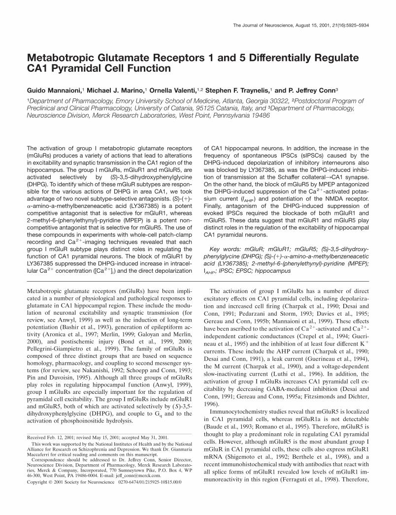
mGluR1 mediates the DHPG-induced increasein [Ca2�]i and depolarization in CA1hippocampal neuronsA common response to the activation of group I mGluRs that hasbeen observed in both recombinant and native systems is therelease of calcium from internal stores. Consistent with this andwith previous studies in hippocampal pyramidal cells (Abe et al.,1992; Pozzo-Miller et al., 1996; Bianchi et al., 1999), DHPGinduced a robust increase in fluorescence in CA1 pyramidal cellsthat were loaded with the calcium-sensitive dye fluo-3. DHPG (30�M) elicited a robust increase in fluo-3 fluorescence in both thesoma (Fig. 1A,C, lef t panels, D) and the dendrites (data notshown) of all of the cells that were examined, suggesting that thistreatment increases intracellular [Ca2�]. Also consistent withprevious studies (Guerineau et al., 1995), simultaneous voltage-
clamp recordings in the same neurons revealed a DHPG-inducedinward current in CA1 pyramidal cells (Fig. 1B, lef t panel, E).
Surprisingly, neither of these effects of DHPG was blocked bymaximal concentrations of the mGluR5-selective antagonistMPEP (10 �M) (Fig. 1A–C, middle panels, D,E), suggesting thatmGluR5 plays little or no role in these group I-mediated responses.In contrast, the mGluR1-selective antagonist LY367385 (100 �M)completely blocked the DHPG-induced increase in the fluo-3 flu-orescence (Fig. 1A,C, right panels, D) and significantly reduced theDHPG-induced inward current (Fig. 1B, right panel, E).
The DHPG-induced inward current underlies the previouslydescribed 1-aminocyclopentane-1,3-dicarboxylic acid (ACPD)and DHPG-induced depolarization of CA1 pyramidal neuronsrecorded in current-clamp mode (Desai and Conn, 1991; Gereauand Conn, 1995b; Mannaioni et al., 1999). Figure 1F summarizesthe depolarizing effect of 30 �M DHPG application (�Vm of 4.2 �0.3 mV; n � 18) on CA1 pyramidal cells. Consistent with thevoltage-clamp studies, DHPG-induced depolarization was notblocked by 10 �M MPEP (�Vm of 4.6 � 1.1 mV; n � 10) (Fig. 1F,open bar). Because the slight DHPG-induced inward current thatremained in the presence of 100 �M LY367385 could be sufficientfor a substantial depolarization, we slightly increased the concen-tration of this competitive mGluR1 antagonist used in the current-clamp experiments. Consistent with the voltage-clamp studies, theDHPG-induced depolarization was blocked completely by 300 �M
LY367385 (�Vm of 1.20 � 0.1 mV; n � 5) (Fig. 1F).In other brain regions, group I mGluR activation depolarizes
neurons by modulating a variety of conductances. Notably, inhippocampal area CA3 DHPG depolarizes neurons by the inhi-bition of a leak potassium conductance (Guerineau et al., 1994) orby an increase in a nonselective cationic conductance (Guerineauet al., 1995). The DHPG-induced inward current was associated
Figure 1. mGluR1 mediates the DHPG-induced increase in [Ca 2�]i and inwardcurrents simultaneously recorded fromCA1 hippocampal neurons. A, B, Timecourse of the effect of 30 �M DHPG alone(lef t) and in the presence of 10 �M MPEP(middle) or 100 �M LY367385 (right) onfluo-3 fluorescence and on holding cur-rent obtained in four different CA1 pyra-midal neurons. Each symbol represents adifferent cell. Antagonists and 1 �M TTXwere applied for 10 min before the record-ings were started. The horizontal bars fordrug application in A also apply to B. C, Atypical example of mGluR-mediated in-crease in somatic fluo-3 fluorescence mea-sured in three different CA1 pyramidalneurons. Shown are DHPG alone (lef t) orin the presence of 10 �M MPEP (middle)or 100 �M LY367385 (right). D–F, Bargraphs expressing the highest values ofF/Fo ratio of fluo-3 fluorescence (D), peakinward current (E), and peak depolariza-tion (F) induced by DHPG applicationalone or in the presence of 10 �M MPEPor 100–300 �M LY367385. Values are themeans � SEM; n � 4 in D and E and isindicated in parentheses in F. *p 0.05;**p 0.01; Student’s t test.
Mannaioni et al. • Group I mGluRs and CA1 Pyramidal Function J. Neurosci., August 15, 2001, 21(16):5925–5934 5927
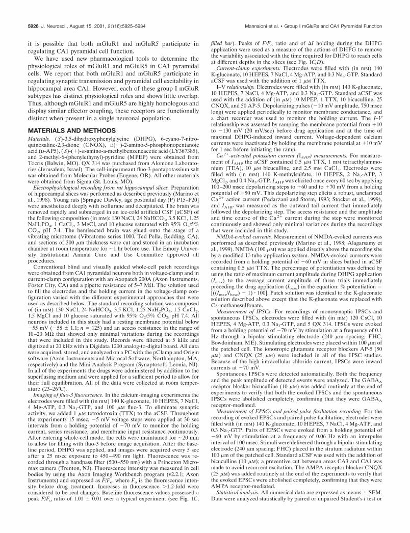
with an increase in input resistance (�Rin 9.3 � 1.8 M�; n � 4)(Fig. 2A). This suggests that an inhibition of a leak potassiumconductance is the most likely mechanism underlying this effect.We therefore examined the current–voltage relationship of themGluR1-mediated inward current. Application of DHPG (30�M) in the presence of the mGluR5 blocker MPEP (10 �M)induced a change in the slope of the whole-cell current–voltagerelationship (Fig. 2B). Subtracting the predrug I–V trace from thetrace in the presence of DHPG reveals a near-linear I–V relation-ship for the DHPG-induced current that reverses near the calcu-lated potassium equilibrium potential in three of the four cellsthat were tested (�104.5 � 8 mV; n � 3) (Fig. 2B, inset),suggesting that the activation of mGluR1 depolarizes CA1 pyra-midal cells by an inhibition of a leak potassium conductance.
Taken together, these data suggest that the DHPG-inducedincrease in intracellular calcium concentration and the DHPG-induced inward current and depolarization in CA1 hippocampalneurons are mediated by mGluR1 and not by mGluR5.
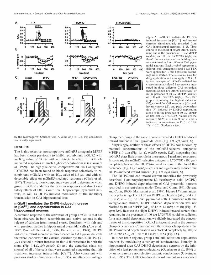
mGluR5 mediates the DHPG-induced suppression ofIAHP and potentiation of NMDA receptor currents inCA1 pyramidal neuronsThe finding that DHPG-induced calcium transients and depolar-ization of CA1 pyramidal cells is mediated by mGluR1 alone wassurprising in light of previous studies that revealed that mGluR5is expressed abundantly in these neurons. However, DHPG in-duces a number of other physiological responses in these cells,some of which could be mediated by mGluR5. Two of the mostprominent effects of DHPG in these cells are the inhibition ofIAHP and the potentiation of NMDA receptor currents. Thus, wedetermined the effects of the mGluR1 and mGluR5-selectiveantagonists on each of these responses to DHPG. As reportedpreviously (Gereau and Conn, 1995b), DHPG (30 �M) sup-pressed IAHP in a reversible manner (73.2 � 3% of control) (Fig.
3A,C). Application of the selective mGluR5 antagonist MPEP(10 �M) completely blocked the DHPG-induced suppression ofIAHP (Fig. 3B), whereas the selective mGluR1 antagonistLY367385 (100 �M) had no effect (Fig. 3C). Neither of theantagonists had an effect on the amplitude of IAHP when appliedalone.
We also investigated which group I mGluR subtype mediatesthe DHPG-induced potentiation of NMDA-induced currents.Consistent with previous studies (Aniksztejn et al., 1991, 1992;Fitzjohn et al., 1996; Pisani et al., 1997), the bath application ofDHPG (30 �M) increased the peak amplitude of the currentinduced by fast local application of NMDA (100 �M) (Fig. 4A, toppanels). The potentiation of NMDA receptor currents was inhib-ited by preincubation with MPEP (Fig. 4A–C), but not byLY367385 (Fig. 4C).
Figure 2. DHPG-induced inward current and depolarization in CA1hippocampal neurons most likely are mediated by decreasing a leakpotassium conductance. A, The mGluR1-mediated inward current ob-served in CA1 pyramidal neurons is associated with a decrease in mem-brane conductance (top vs bottom trace). B, This decrease in membraneconductance is evident in the whole-cell current–voltage relation. Theinset shows the subtraction of the currents that reveals a linear I–Vrelationship, which is inward at normal resting potentials and reversesnear the predicted potassium equilibrium potential. Axis titles in B applyin the inset. The figure is representative of the results that were observedin three of four cells.
Figure 3. The DHPG-induced suppression of IAHP in CA1 pyramidalneurons is mediated by mGluR5. A, Time course from a single experi-ment showing the suppression of IAHP induced by the application of 30�M DHPG. Letters indicate the time of the corresponding traces that areshown at the top. B, The 10 min preapplication of 10 �M MPEP antago-nizes the DHPG-induced suppression of IAHP. C, Bar graph summarizingmean data showing that the DHPG-induced suppression of IAHP isblocked selectively by MPEP but is not altered by preincubation with themGluR1-selective antagonist LY367385 (100 �M). The number of cellsthat have been tested is in parentheses; values are the means � SEM of thedata, expressed as a percentage of control IAHP peak values. *p 0.05versus control; Student’s t test.
5928 J. Neurosci., August 15, 2001, 21(16):5925–5934 Mannaioni et al. • Group I mGluRs and CA1 Pyramidal Function
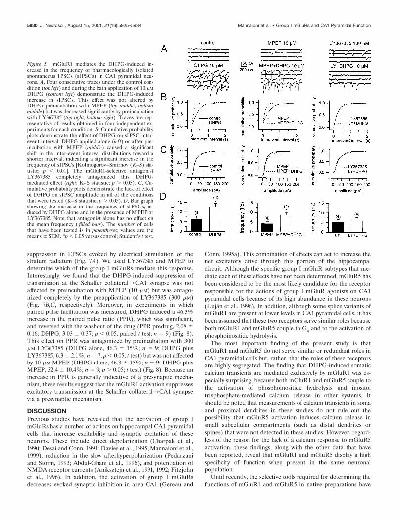
The DHPG-induced increase in the frequency ofspontaneous IPSCs is mediated by mGluR1In addition to its direct actions on CA1 pyramidal cells, theactivation of group I mGluRs impacts CA1 pyramidal cell excit-ability indirectly by regulating GABA-mediated inhibitory trans-mission. Two distinct effects of group I mGluR activation oninhibitory transmission can be seen in recordings from CA1pyramidal cells. First, group I mGluR agonists induce depolar-ization and increase firing of inhibitory interneurons (McBain etal., 1994; van Hooft et al., 2000), leading to a robust increase inspontaneous IPSCs. Second, group I mGluR agonists reduce theamplitude of evoked monosynaptic IPSCs (Desai et al., 1994;Gereau and Conn, 1995a). Activation of group I mGluRs doesnot alter the amplitude of miniature IPSCs, suggesting that thereduction of evoked IPSCs is mediated by a presynaptic action.However, analysis of the effect of mGluR activation on monosyn-apticIPSCs is complicated by the high frequency of DHPG-inducedspontaneous IPSCs. Thus, we took advantage of the mGluR1 and
mGluR5-selective antagonists to determine whether we coulddifferentiate between these two actions of DHPG.
Figure 5A, lef t, shows examples of sIPSCs recorded undercontrol conditions and during the bath application of 10 �M
DHPG. Similar to results reported previously in area CA3 (Milesand Poncer, 1993) and in rat frontal cortex (Chu and Hablitz,1998), the application of DHPG (10 �M) increased the frequencyof sIPSCs in all of the cells that were tested (n � 4). These effectswere reversible on drug washout from the recording chamber(data not shown). Preincubation with the mGluR5-selective an-tagonist MPEP (10 �M) did not block the DHPG-induced in-crease in sIPSC frequency (Fig. 5A, middle), whereas preincuba-tion with the selective mGluR1 antagonist LY367385 (100 �M)completely blocked this effect (Fig. 5A, right). DHPG produced aleftward shift of the cumulative probability distributions of sIPSCinter-event intervals (Fig. 5B, lef t), indicating a DHPG-inducedincrease in the frequency of sIPSCs. This effect was blocked byLY367385, but not by MPEP (Fig. 5B, right and middle, respec-tively). The average frequency was calculated for each cell, andthe means of these values are shown in Figure 5D. Application of10 �M DHPG induced a significant increase in mean frequency(Fig. 5D, lef t). Preincubation with either MPEP or LY367385alone did not change the mean frequency of sIPSCs significantly;however, LY367385 blocked the DHPG-induced increase insIPSC frequency, whereas MPEP was without effect (Fig. 5D,right and middle, respectively). In contrast, the application of 10�M DHPG alone or in combination with the subtype-selectiveantagonists produced no significant effect on sIPSC amplitude(Fig. 5C, lef t, middle, right). This is consistent with the previouslyreported lack of effect of DHPG on mIPSC amplitude (Gereauand Conn, 1995a) and suggests that DHPG does not alterpostsynaptic GABAA receptor currents.
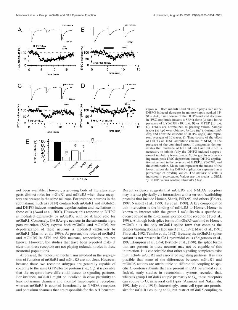
Activation of both mGluR1 and mGluR5 reducesevoked monosynaptic IPSCsThe activation of group I mGluRs also has been demonstrated toinduce a reduction in evoked monosynaptic IPSCs (Desai et al.,1994; Gereau and Conn, 1995a). Consistent with this, the bathapplication of DHPG (10 �M) produced a reversible depressionof IPSCs by 80 � 4% (means � SEM) of baseline amplitude (Fig.6A,E) that was antagonized only partially by preincubation withLY367385 (100 �M; 48 � 9% of baseline amplitude; means �SEM) (Fig. 6B) or MPEP (10 �M; 54 � 10% of baseline ampli-tude; means � SEM) (Fig. 6C). However, preincubation with acombination of LY367685 (100 �M) and MPEP (10 �M) fullyblocked the DHPG-induced suppression of evoked IPSCs (7 �2% of baseline amplitude) (Fig. 6D,E). These data suggest thatthe group I mGluR-induced suppression of monosynaptic IPSCsis mediated by both mGluR1 and mGluR5. Therefore, DHPGinduces an increase in sIPSC frequency by the activation ofmGluR1 and an inhibition of evoked IPSCs by activation of bothmGluR1 and mGluR5.
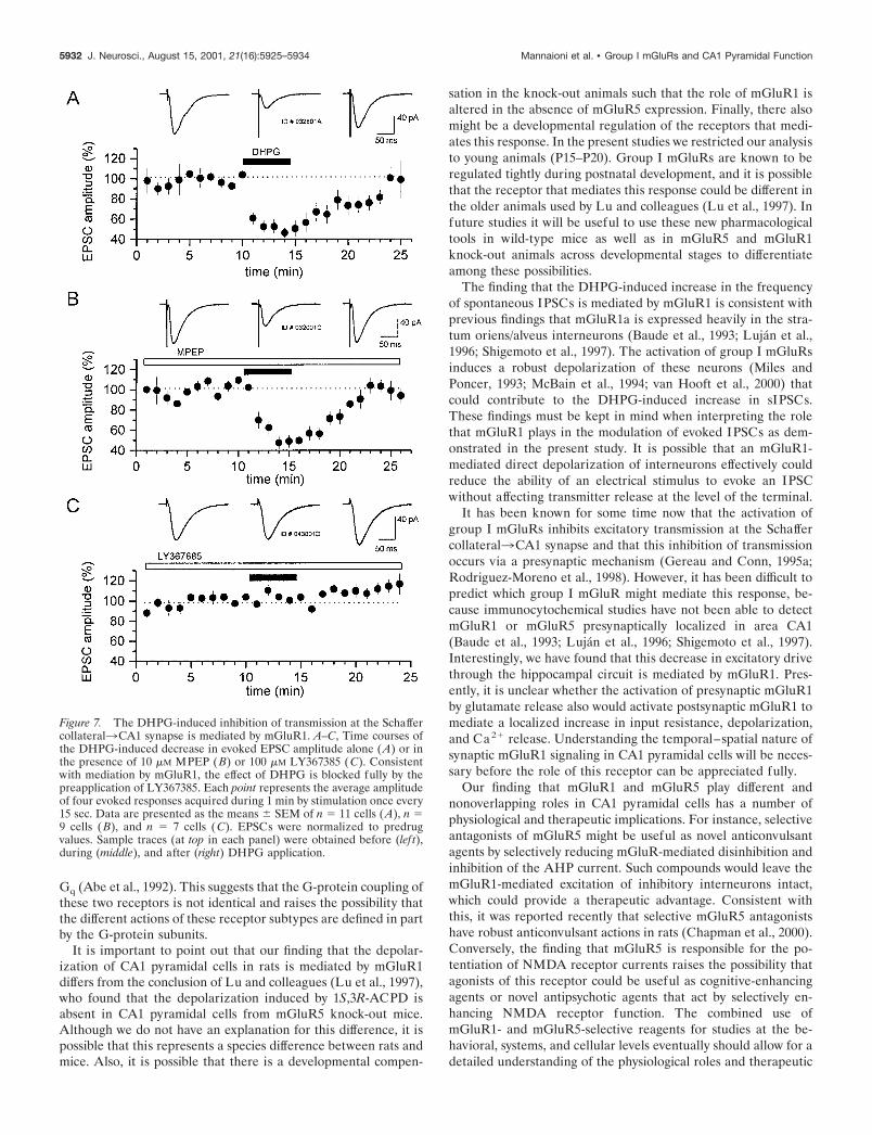
Activation of mGluR1 reduces evoked EPSCs via apresynaptic mechanismPrevious studies have demonstrated that multiple mGluRs mod-ulate excitatory synaptic transmission. In particular, the groupI-selective agonist DHPG has been shown to induce a presynap-tically mediated depression of excitatory transmission at theSchaffer collateral3CA1 synapse (Gereau and Conn, 1995a;Rodriguez-Moreno et al., 1998). Consistent with these previousreports, 30 �M DHPG induced a 54 � 5% (means � SEM)
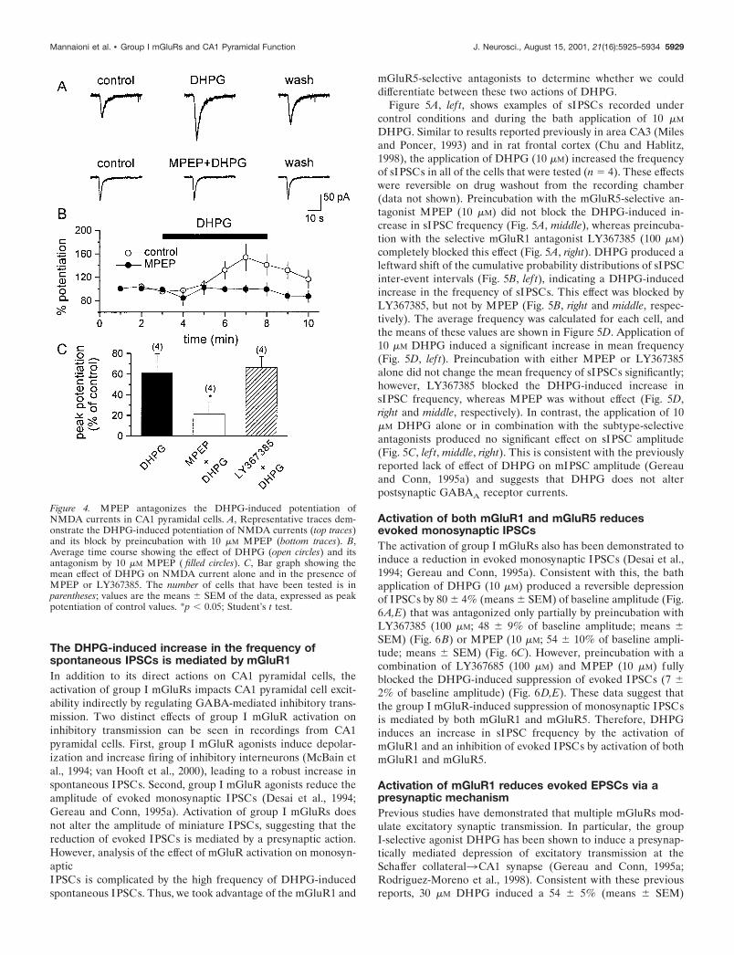
Figure 4. MPEP antagonizes the DHPG-induced potentiation ofNMDA currents in CA1 pyramidal cells. A, Representative traces dem-onstrate the DHPG-induced potentiation of NMDA currents (top traces)and its block by preincubation with 10 �M MPEP (bottom traces). B,Average time course showing the effect of DHPG (open circles) and itsantagonism by 10 �M MPEP ( filled circles). C, Bar graph showing themean effect of DHPG on NMDA current alone and in the presence ofMPEP or LY367385. The number of cells that have been tested is inparentheses; values are the means � SEM of the data, expressed as peakpotentiation of control values. *p 0.05; Student’s t test.
Mannaioni et al. • Group I mGluRs and CA1 Pyramidal Function J. Neurosci., August 15, 2001, 21(16):5925–5934 5929
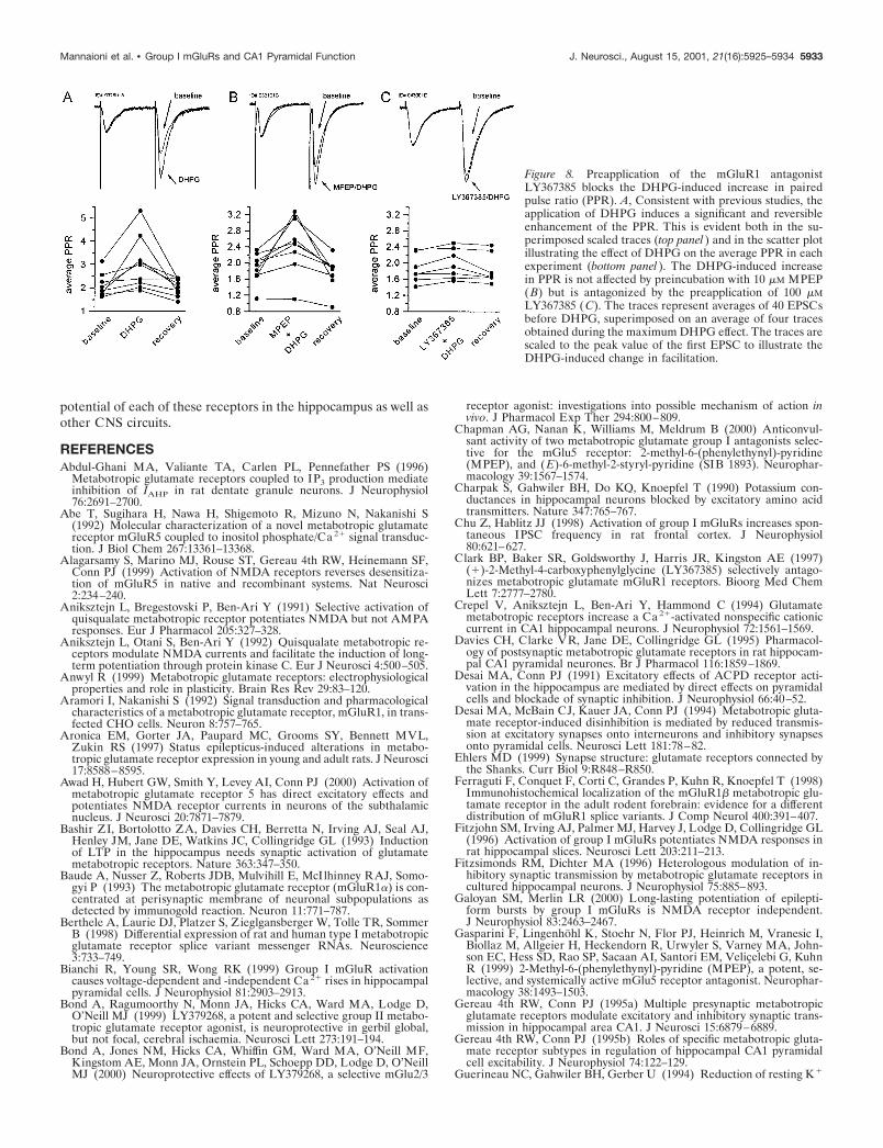
suppression in EPSCs evoked by electrical stimulation of thestratum radiatum (Fig. 7A). We used LY367385 and MPEP todetermine which of the group I mGluRs mediate this response.Interestingly, we found that the DHPG-induced suppression oftransmission at the Schaffer collateral3CA1 synapse was notaffected by preincubation with MPEP (10 �M) but was antago-nized completely by the preapplication of LY367385 (300 �M)(Fig. 7B,C, respectively). Moreover, in experiments in whichpaired pulse facilitation was measured, DHPG induced a 46.3%increase in the paired pulse ratio (PPR), which was significant,and reversed with the washout of the drug (PPR predrug, 2.08 �0.16; DHPG, 3.03 � 0.37; p 0.05, paired t test; n � 9) (Fig. 8).This effect on PPR was antagonized by preincubation with 300�M LY367385 (DHPG alone, 46.3 � 15%; n � 9; DHPG plusLY367385, 6.3 � 2.1%; n � 7; p 0.05; t test) but was not affectedby 10 �M MPEP (DHPG alone, 46.3 � 15%; n � 9; DHPG plusMPEP, 32.4 � 10.4%; n � 9; p � 0.05; t test) (Fig. 8). Because anincrease in PPR is generally indicative of a presynaptic mecha-nism, these results suggest that the mGluR1 activation suppressesexcitatory transmission at the Schaffer collateral3CA1 synapsevia a presynaptic mechanism.
DISCUSSIONPrevious studies have revealed that the activation of group ImGluRs has a number of actions on hippocampal CA1 pyramidalcells that increase excitability and synaptic excitation of theseneurons. These include direct depolarization (Charpak et al.,1990; Desai and Conn, 1991; Davies et al., 1995; Mannaioni et al.,1999), reduction in the slow afterhyperpolarization (Pedarzaniand Storm, 1993; Abdul-Ghani et al., 1996), and potentiation ofNMDA receptor currents (Aniksztejn et al., 1991, 1992; Fitzjohnet al., 1996). In addition, the activation of group I mGluRsdecreases evoked synaptic inhibition in area CA1 (Gereau and
Conn, 1995a). This combination of effects can act to increase thenet excitatory drive through this portion of the hippocampalcircuit. Although the specific group I mGluR subtypes that me-diate each of these effects have not been determined, mGluR5 hasbeen considered to be the most likely candidate for the receptorresponsible for the actions of group I mGluR agonists on CA1pyramidal cells because of its high abundance in these neurons(Lujan et al., 1996). In addition, although some splice variants ofmGluR1 are present at lower levels in CA1 pyramidal cells, it hasbeen assumed that these two receptors serve similar roles becauseboth mGluR1 and mGluR5 couple to Gq and to the activation ofphosphoinositide hydrolysis.
The most important finding of the present study is thatmGluR1 and mGluR5 do not serve similar or redundant roles inCA1 pyramidal cells but, rather, that the roles of these receptorsare highly segregated. The finding that DHPG-induced somaticcalcium transients are mediated exclusively by mGluR1 was es-pecially surprising, because both mGluR1 and mGluR5 couple tothe activation of phosphoinositide hydrolysis and inositoltrisphosphate-mediated calcium release in other systems. Itshould be noted that measurements of calcium transients in somaand proximal dendrites in these studies do not rule out thepossibility that mGluR5 activation induces calcium release insmall subcellular compartments (such as distal dendrites orspines) that were not detected in these studies. However, regard-less of the reason for the lack of a calcium response to mGluR5activation, these findings, along with the other data that havebeen reported, reveal that mGluR1 and mGluR5 display a highspecificity of function when present in the same neuronalpopulation.
Until recently, the selective tools required for determining thefunctions of mGluR1 and mGluR5 in native preparations have
Figure 5. mGluR1 mediates the DHPG-induced in-crease in the frequency of pharmacologically isolatedspontaneous IPSCs (sIPSCs) in CA1 pyramidal neu-rons. A, Four consecutive traces under the control con-dition (top lef t) and during the bath application of 10 �MDHPG (bottom lef t) demonstrate the DHPG-inducedincrease in sIPSCs. This effect was not altered byDHPG preincubation with MPEP (top middle, bottommiddle) but was decreased significantly by preincubationwith LY367385 (top right, bottom right). Traces are rep-resentative of results obtained in four independent ex-periments for each condition. B, Cumulative probabilityplots demonstrate the effect of DHPG on sIPSC inter-event interval. DHPG applied alone (lef t) or after pre-incubation with MPEP (middle) caused a significantshift in the inter-event interval distributions toward ashorter interval, indicating a significant increase in thefrequency of sIPSCs [Kolmogorov–Smirnov ( K–S) sta-tistic; p 0.01]. The mGluR1-selective antagonistLY367385 completely antagonized this DHPG-mediated effect (right; K–S statistic; p � 0.05). C, Cu-mulative probability plots demonstrate the lack of effectof DHPG on sIPSC amplitude in all of the conditionsthat were tested (K–S statistic; p � 0.05). D, Bar graphshowing the increase in the frequency of sIPSCs, in-duced by DHPG alone and in the presence of MPEP orLY367385. Note that antagonist alone has no effect onthe mean frequency ( filled bars). The number of cellsthat have been tested is in parentheses; values are themeans � SEM. *p 0.05 versus control; Student’s t test.
5930 J. Neurosci., August 15, 2001, 21(16):5925–5934 Mannaioni et al. • Group I mGluRs and CA1 Pyramidal Function
not been available. However, a growing body of literature sug-gests distinct roles for mGluR1 and mGluR5 when these recep-tors are present in the same neurons. For instance, neurons in thesubthalamic nucleus (STN) contain both mGluR1 and mGluR5,and DHPG induces membrane depolarization and oscillations inthese cells (Awad et al., 2000). However, this response to DHPGis mediated exclusively by mGluR5, with no defined role formGluR1. Conversely, GABAergic neurons in the substantia nigrapars reticulata (SNr) express both mGluR1 and mGluR5, butdepolarization of these neurons is mediated exclusively bymGluR1 (Marino et al., 1999). At present, the roles of mGluR1and mGluR5 in STN and SNr neurons, respectively, are notknown. However, the studies that have been reported make itclear that these receptors are not playing redundant roles in theseneuronal populations.
At present, the molecular mechanisms involved in the segrega-tion of function of mGluR1 and mGluR5 are not clear. However,because these two receptor subtypes are generally capable ofcoupling to the same GTP effector proteins (i.e., Gq), it is possiblethat the receptors have differential access to signaling partners.For instance, mGluR1 might be localized in close proximity toleak potassium channels and inositol trisphosphate receptors,whereas mGluR5 is coupled functionally to NMDA receptorsand potassium channels that are responsible for the AHP current.
Recent evidence suggests that mGluR5 and NMDA receptorsmay interact physically via interactions with a series of scaffoldingproteins that include Homer, Shank, PSD-95, and others (Ehlers,1999; Naisbitt et al., 1999; Tu et al., 1999). A key component ofthis interaction is the binding of mGluR5 to Homer. Homer isknown to interact with the group I mGluRs via a specific se-quence found in the C-terminal portion of the receptor (Tu et al.,1998). Although both splice forms of mGluR5 can bind to Homer,mGluR1a is the only mGluR1 splice form that contains theHomer binding domain (Houamed et al., 1991; Masu et al., 1991;Pin et al., 1992; Tanabe et al., 1992). Because the mGluR1a splicevariant is not present in CA1 pyramidal cells (Shigemoto et al.,1992; Hampson et al., 1994; Berthele et al., 1998), the splice formsthat are present in these neurons may not be capable of thisinteraction. It is conceivable that other signaling complexes existthat include mGluR1 and associated signaling partners. It is alsopossible that some of the differences between mGluR1 andmGluR5 actions are attributable to differential coupling to spe-cific G-protein subunits that are present in CA1 pyramidal cells.Indeed, early studies in recombinant systems revealed that,whereas group I mGluRs couple primarily to Gq, these receptorscan couple to Gs in several cell types (Aramori and Nakanishi,1992; Joly et al., 1995). Interestingly, some cell types are permis-sive for mGluR1 coupling to Gs but restrict mGluR5 coupling to
Figure 6. Both mGluR1 and mGluR5 play a role in theDHPG-induced decrease in monosynaptic evoked IP-SCs. A–C, Time course of the DHPG-induced decreasein IPSC amplitude (means � SEM) alone (A) and in thepresence of LY367385 (100 �M; B) or MPEP (10 �M;C). IPSCs are normalized to predrug values. Sampletraces (at top) were obtained before (lef t), during (mid-dle), and after the washout of DHPG (right) and repre-sent averages of 10 traces. D, Time course of the effectof DHPG on IPSC amplitude (means � SEM) in thepresence of the combined group I antagonists demon-strates that blockade of both mGluR1 and mGluR5 isnecessary to inhibit fully the DHPG-induced suppres-sion of inhibitory transmission. E, Bar graphs represent-ing mean peak IPSC depression during DHPG applica-tion alone and in the presence of MPEP, LY367385, andthe combination. Mean data represent the means of thelowest values during DHPG application expressed as apercentage of predrug values. The number of cells isindicated in parentheses. Values are the means � SEM.*p 0.05 versus control; Student’s t test.
Mannaioni et al. • Group I mGluRs and CA1 Pyramidal Function J. Neurosci., August 15, 2001, 21(16):5925–5934 5931
Gq (Abe et al., 1992). This suggests that the G-protein coupling ofthese two receptors is not identical and raises the possibility thatthe different actions of these receptor subtypes are defined in partby the G-protein subunits.
It is important to point out that our finding that the depolar-ization of CA1 pyramidal cells in rats is mediated by mGluR1differs from the conclusion of Lu and colleagues (Lu et al., 1997),who found that the depolarization induced by 1S,3R-ACPD isabsent in CA1 pyramidal cells from mGluR5 knock-out mice.Although we do not have an explanation for this difference, it ispossible that this represents a species difference between rats andmice. Also, it is possible that there is a developmental compen-
sation in the knock-out animals such that the role of mGluR1 isaltered in the absence of mGluR5 expression. Finally, there alsomight be a developmental regulation of the receptors that medi-ates this response. In the present studies we restricted our analysisto young animals (P15–P20). Group I mGluRs are known to beregulated tightly during postnatal development, and it is possiblethat the receptor that mediates this response could be different inthe older animals used by Lu and colleagues (Lu et al., 1997). Infuture studies it will be useful to use these new pharmacologicaltools in wild-type mice as well as in mGluR5 and mGluR1knock-out animals across developmental stages to differentiateamong these possibilities.
The finding that the DHPG-induced increase in the frequencyof spontaneous IPSCs is mediated by mGluR1 is consistent withprevious findings that mGluR1a is expressed heavily in the stra-tum oriens/alveus interneurons (Baude et al., 1993; Lujan et al.,1996; Shigemoto et al., 1997). The activation of group I mGluRsinduces a robust depolarization of these neurons (Miles andPoncer, 1993; McBain et al., 1994; van Hooft et al., 2000) thatcould contribute to the DHPG-induced increase in sIPSCs.These findings must be kept in mind when interpreting the rolethat mGluR1 plays in the modulation of evoked IPSCs as dem-onstrated in the present study. It is possible that an mGluR1-mediated direct depolarization of interneurons effectively couldreduce the ability of an electrical stimulus to evoke an IPSCwithout affecting transmitter release at the level of the terminal.
It has been known for some time now that the activation ofgroup I mGluRs inhibits excitatory transmission at the Schaffercollateral3CA1 synapse and that this inhibition of transmissionoccurs via a presynaptic mechanism (Gereau and Conn, 1995a;Rodriguez-Moreno et al., 1998). However, it has been difficult topredict which group I mGluR might mediate this response, be-cause immunocytochemical studies have not been able to detectmGluR1 or mGluR5 presynaptically localized in area CA1(Baude et al., 1993; Lujan et al., 1996; Shigemoto et al., 1997).Interestingly, we have found that this decrease in excitatory drivethrough the hippocampal circuit is mediated by mGluR1. Pres-ently, it is unclear whether the activation of presynaptic mGluR1by glutamate release also would activate postsynaptic mGluR1 tomediate a localized increase in input resistance, depolarization,and Ca2� release. Understanding the temporal–spatial nature ofsynaptic mGluR1 signaling in CA1 pyramidal cells will be neces-sary before the role of this receptor can be appreciated fully.
Our finding that mGluR1 and mGluR5 play different andnonoverlapping roles in CA1 pyramidal cells has a number ofphysiological and therapeutic implications. For instance, selectiveantagonists of mGluR5 might be useful as novel anticonvulsantagents by selectively reducing mGluR-mediated disinhibition andinhibition of the AHP current. Such compounds would leave themGluR1-mediated excitation of inhibitory interneurons intact,which could provide a therapeutic advantage. Consistent withthis, it was reported recently that selective mGluR5 antagonistshave robust anticonvulsant actions in rats (Chapman et al., 2000).Conversely, the finding that mGluR5 is responsible for the po-tentiation of NMDA receptor currents raises the possibility thatagonists of this receptor could be useful as cognitive-enhancingagents or novel antipsychotic agents that act by selectively en-hancing NMDA receptor function. The combined use ofmGluR1- and mGluR5-selective reagents for studies at the be-havioral, systems, and cellular levels eventually should allow for adetailed understanding of the physiological roles and therapeutic
Figure 7. The DHPG-induced inhibition of transmission at the Schaffercollateral3CA1 synapse is mediated by mGluR1. A–C, Time courses ofthe DHPG-induced decrease in evoked EPSC amplitude alone (A) or inthe presence of 10 �M MPEP (B) or 100 �M LY367385 (C). Consistentwith mediation by mGluR1, the effect of DHPG is blocked fully by thepreapplication of LY367385. Each point represents the average amplitudeof four evoked responses acquired during 1 min by stimulation once every15 sec. Data are presented as the means � SEM of n � 11 cells (A), n �9 cells (B), and n � 7 cells (C). EPSCs were normalized to predrugvalues. Sample traces (at top in each panel) were obtained before (lef t),during (middle), and after (right) DHPG application.
5932 J. Neurosci., August 15, 2001, 21(16):5925–5934 Mannaioni et al. • Group I mGluRs and CA1 Pyramidal Function
potential of each of these receptors in the hippocampus as well asother CNS circuits.
REFERENCESAbdul-Ghani MA, Valiante TA, Carlen PL, Pennefather PS (1996)
Metabotropic glutamate receptors coupled to IP3 production mediateinhibition of IAHP in rat dentate granule neurons. J Neurophysiol76:2691–2700.
Abe T, Sugihara H, Nawa H, Shigemoto R, Mizuno N, Nakanishi S(1992) Molecular characterization of a novel metabotropic glutamatereceptor mGluR5 coupled to inositol phosphate/Ca 2� signal transduc-tion. J Biol Chem 267:13361–13368.
Alagarsamy S, Marino MJ, Rouse ST, Gereau 4th RW, Heinemann SF,Conn PJ (1999) Activation of NMDA receptors reverses desensitiza-tion of mGluR5 in native and recombinant systems. Nat Neurosci2:234–240.
Aniksztejn L, Bregestovski P, Ben-Ari Y (1991) Selective activation ofquisqualate metabotropic receptor potentiates NMDA but not AMPAresponses. Eur J Pharmacol 205:327–328.
Aniksztejn L, Otani S, Ben-Ari Y (1992) Quisqualate metabotropic re-ceptors modulate NMDA currents and facilitate the induction of long-term potentiation through protein kinase C. Eur J Neurosci 4:500–505.
Anwyl R (1999) Metabotropic glutamate receptors: electrophysiologicalproperties and role in plasticity. Brain Res Rev 29:83–120.
Aramori I, Nakanishi S (1992) Signal transduction and pharmacologicalcharacteristics of a metabotropic glutamate receptor, mGluR1, in trans-fected CHO cells. Neuron 8:757–765.
Aronica EM, Gorter JA, Paupard MC, Grooms SY, Bennett MVL,Zukin RS (1997) Status epilepticus-induced alterations in metabo-tropic glutamate receptor expression in young and adult rats. J Neurosci17:8588–8595.
Awad H, Hubert GW, Smith Y, Levey AI, Conn PJ (2000) Activation ofmetabotropic glutamate receptor 5 has direct excitatory effects andpotentiates NMDA receptor currents in neurons of the subthalamicnucleus. J Neurosci 20:7871–7879.
Bashir ZI, Bortolotto ZA, Davies CH, Berretta N, Irving AJ, Seal AJ,Henley JM, Jane DE, Watkins JC, Collingridge GL (1993) Inductionof LTP in the hippocampus needs synaptic activation of glutamatemetabotropic receptors. Nature 363:347–350.
Baude A, Nusser Z, Roberts JDB, Mulvihill E, McIlhinney RAJ, Somo-gyi P (1993) The metabotropic glutamate receptor (mGluR1�) is con-centrated at perisynaptic membrane of neuronal subpopulations asdetected by immunogold reaction. Neuron 11:771–787.
Berthele A, Laurie DJ, Platzer S, Zieglgansberger W, Tolle TR, SommerB (1998) Differential expression of rat and human type I metabotropicglutamate receptor splice variant messenger RNAs. Neuroscience3:733–749.
Bianchi R, Young SR, Wong RK (1999) Group I mGluR activationcauses voltage-dependent and -independent Ca 2� rises in hippocampalpyramidal cells. J Neurophysiol 81:2903–2913.
Bond A, Ragumoorthy N, Monn JA, Hicks CA, Ward MA, Lodge D,O’Neill MJ (1999) LY379268, a potent and selective group II metabo-tropic glutamate receptor agonist, is neuroprotective in gerbil global,but not focal, cerebral ischaemia. Neurosci Lett 273:191–194.
Bond A, Jones NM, Hicks CA, Whiffin GM, Ward MA, O’Neill MF,Kingstom AE, Monn JA, Ornstein PL, Schoepp DD, Lodge D, O’NeillMJ (2000) Neuroprotective effects of LY379268, a selective mGlu2/3
receptor agonist: investigations into possible mechanism of action invivo. J Pharmacol Exp Ther 294:800–809.
Chapman AG, Nanan K, Williams M, Meldrum B (2000) Anticonvul-sant activity of two metabotropic glutamate group I antagonists selec-tive for the mGlu5 receptor: 2-methyl-6-(phenylethynyl)-pyridine(MPEP), and ( E)-6-methyl-2-styryl-pyridine (SIB 1893). Neurophar-macology 39:1567–1574.
Charpak S, Gahwiler BH, Do KQ, Knoepfel T (1990) Potassium con-ductances in hippocampal neurons blocked by excitatory amino acidtransmitters. Nature 347:765–767.
Chu Z, Hablitz JJ (1998) Activation of group I mGluRs increases spon-taneous IPSC frequency in rat frontal cortex. J Neurophysiol80:621–627.
Clark BP, Baker SR, Goldsworthy J, Harris JR, Kingston AE (1997)(�)-2-Methyl-4-carboxyphenylglycine (LY367385) selectively antago-nizes metabotropic glutamate mGluR1 receptors. Bioorg Med ChemLett 7:2777–2780.
Crepel V, Aniksztejn L, Ben-Ari Y, Hammond C (1994) Glutamatemetabotropic receptors increase a Ca 2�-activated nonspecific cationiccurrent in CA1 hippocampal neurons. J Neurophysiol 72:1561–1569.
Davies CH, Clarke VR, Jane DE, Collingridge GL (1995) Pharmacol-ogy of postsynaptic metabotropic glutamate receptors in rat hippocam-pal CA1 pyramidal neurones. Br J Pharmacol 116:1859–1869.
Desai MA, Conn PJ (1991) Excitatory effects of ACPD receptor acti-vation in the hippocampus are mediated by direct effects on pyramidalcells and blockade of synaptic inhibition. J Neurophysiol 66:40–52.
Desai MA, McBain CJ, Kauer JA, Conn PJ (1994) Metabotropic gluta-mate receptor-induced disinhibition is mediated by reduced transmis-sion at excitatory synapses onto interneurons and inhibitory synapsesonto pyramidal cells. Neurosci Lett 181:78–82.
Ehlers MD (1999) Synapse structure: glutamate receptors connected bythe Shanks. Curr Biol 9:R848–R850.
Ferraguti F, Conquet F, Corti C, Grandes P, Kuhn R, Knoepfel T (1998)Immunohistochemical localization of the mGluR1� metabotropic glu-tamate receptor in the adult rodent forebrain: evidence for a differentdistribution of mGluR1 splice variants. J Comp Neurol 400:391–407.
Fitzjohn SM, Irving AJ, Palmer MJ, Harvey J, Lodge D, Collingridge GL(1996) Activation of group I mGluRs potentiates NMDA responses inrat hippocampal slices. Neurosci Lett 203:211–213.
Fitzsimonds RM, Dichter MA (1996) Heterologous modulation of in-hibitory synaptic transmission by metabotropic glutamate receptors incultured hippocampal neurons. J Neurophysiol 75:885–893.
Galoyan SM, Merlin LR (2000) Long-lasting potentiation of epilepti-form bursts by group I mGluRs is NMDA receptor independent.J Neurophysiol 83:2463–2467.
Gasparini F, Lingenhohl K, Stoehr N, Flor PJ, Heinrich M, Vranesic I,Biollaz M, Allgeier H, Heckendorn R, Urwyler S, Varney MA, John-son EC, Hess SD, Rao SP, Sacaan AI, Santori EM, Velicelebi G, KuhnR (1999) 2-Methyl-6-(phenylethynyl)-pyridine (MPEP), a potent, se-lective, and systemically active mGlu5 receptor antagonist. Neurophar-macology 38:1493–1503.
Gereau 4th RW, Conn PJ (1995a) Multiple presynaptic metabotropicglutamate receptors modulate excitatory and inhibitory synaptic trans-mission in hippocampal area CA1. J Neurosci 15:6879–6889.
Gereau 4th RW, Conn PJ (1995b) Roles of specific metabotropic gluta-mate receptor subtypes in regulation of hippocampal CA1 pyramidalcell excitability. J Neurophysiol 74:122–129.
Guerineau NC, Gahwiler BH, Gerber U (1994) Reduction of resting K �
Figure 8. Preapplication of the mGluR1 antagonistLY367385 blocks the DHPG-induced increase in pairedpulse ratio (PPR). A, Consistent with previous studies, theapplication of DHPG induces a significant and reversibleenhancement of the PPR. This is evident both in the su-perimposed scaled traces (top panel ) and in the scatter plotillustrating the effect of DHPG on the average PPR in eachexperiment (bottom panel ). The DHPG-induced increasein PPR is not affected by preincubation with 10 �M MPEP(B) but is antagonized by the preapplication of 100 �MLY367385 (C). The traces represent averages of 40 EPSCsbefore DHPG, superimposed on an average of four tracesobtained during the maximum DHPG effect. The traces arescaled to the peak value of the first EPSC to illustrate theDHPG-induced change in facilitation.
Mannaioni et al. • Group I mGluRs and CA1 Pyramidal Function J. Neurosci., August 15, 2001, 21(16):5925–5934 5933
current by metabotropic glutamate and muscarinic receptors in rat CA3cells: mediation by G-proteins. J Physiol (Lond) 474:27–33.
Guerineau NC, Bossu JL, Gahwiler BH, Gerber U (1995) Activation ofa nonselective cationic conductance by metabotropic glutamatergic andmuscarinic agonists in CA3 pyramidal neurons of the rat hippocampus.J Neurosci 15:4395–4407.
Hampson DR, Theriault E, Huang XP, Kristensen P, Pickering DS,Franck JE, Mulvihill ER (1994) Characterization of two alternativelyspliced forms of a metabotropic glutamate receptor in the centralnervous system of the rat. Neuroscience 60:325–336.
Houamed KM, Kuijper JL, Gilbert TL, Haldeman BA, O’Hara PJ,Mulvihill ER, Almers W, Hagen FS (1991) Cloning, expression, andgene structure of a G-protein-coupled glutamate receptor from ratbrain. Science 252:1318–1321.
Joly C, Gomeza J, Brabet I, Curry K, Bockaert J, Pin J-P (1995) Molec-ular, functional, and pharmacological characterization of the metabo-tropic glutamate receptor type 5 splice variants: comparison withmGluR1. J Neurosci 15:3970–3981.
Lu YM, Jia Z, Janus C, Henderson JT, Gerlai R, Wojtowicz JM, RoderJ (1997) Mice lacking metabotropic glutamate receptor 5 show im-paired learning and reduced CA1 long-term potentiation (LTP) butnormal CA3 LTP. J Neurosci 17:5196–5205.
Lujan R, Nusser Z, Roberts JC, Shigemoto R, Somogyi P (1996) Peri-synaptic location of metabotropic glutamate receptors mGluR1 andmGluR5 on dendrites and dendritic spines in the rat hippocampus. EurJ Neurosci 8:1488–1500.
Luthi A, Gahwiler BH, Gerber U (1996) A slowly inactivating potassiumcurrent in CA3 pyramidal cells of rat hippocampus in vitro. J Neurosci16:586–594.
Mannaioni G, Attucci S, Missanelli A, Pellicciari R, Corradetti R, MoroniF (1999) Biochemical and electrophysiological studies on (S)-(�)-2-(3-carboxybicyclo[1.1.1]pentyl)-glycine (CBPG), a novel mGlu5 recep-tor agonist endowed with mGlu1 receptor antagonist activity. Neuro-pharmacology 38:917–926.
Marino MJ, Rouse ST, Levey A, Potter LT, Conn PJ (1998) Activationof the genetically defined m1 muscarinic receptor potentiates N-methyl-D-aspartate (NMDA) receptor currents in hippocampal pyramidalcells. Proc Natl Acad Sci USA 95:11465–11470.
Marino MJ, Risso Bradley S, Wittmann M, Conn PJ (1999) Directexcitation of GABAergic projection neurons of the rat substantia nigrapars reticulata by activation of the mGluR1 metabotropic glutamatereceptor. Soc Neurosci Abstr 25:446.
Masu M, Tanabe Y, Tsuchida K, Shigemoto R, Nakanishi S (1991)Sequence and expression of a metabotropic glutamate receptor. Nature349:760–765.
McBain CJ, DiChiara TJ, Kauer JA (1994) Activation of metabotropicglutamate receptors differentially affects two classes of hippocampalinterneurons and potentiates excitatory synaptic transmission. J Neu-rosci 14:4433–4445.
Merlin LR (1999) Group I mGluR-mediated silent induction of long-lasting epileptiform discharges. J Neurophysiol 82:1078–1081.
Miles R, Poncer JC (1993) Metabotropic glutamate receptors mediate apost-tetanic excitation of guinea-pig hippocampal inhibitory neurones.J Physiol (Lond) 463:461–473.
Naisbitt S, Kim E, Tu JC, Xiao B, Sala C, Valtschanoff J, Weinberg RJ,Worley PF, Sheng M (1999) Shank, a novel family of postsynaptic
density proteins that binds to the NMDA receptor/PSD-95/GKAPcomplex and cortactin. Neuron 23:569–582.
Nakanishi S (1992) Molecular diversity of glutamate receptors and im-plications for brain function. Science 258:597–603.
Pedarzani P, Storm JF (1993) PKA mediates the effects of monoaminetransmitters on the K � current underlying the slow spike frequencyadaptation in hippocampal neurons. Neuron 11:1023–1035.
Pellegrini-Giampietro DE, Cozzi A, Peruginelli F, Leonardi P, Meli E,Pellicciari R, Moroni F (1999) 1-Aminoindan-1,5-dicarboxylic acidand (S)-(�)-2-(3-carboxybicyclo[1.1.1]pentyl)-glycine, two mGlu1receptor-preferring antagonists, reduce neuronal death in in vitro and invivo models of cerebral ischemia. Eur J Neurosci 11:3637–3647.
Pin J-P, Duvoisin R (1995) The metabotropic glutamate receptors:structure and functions. Neuropharmacology 34:1–26.
Pin J-P, Waeber C, Prezeau L, Bockaert J, Heinemann SF (1992) Alter-native splicing generates metabotropic glutamate receptors inducingdifferent patterns of calcium release in Xenopus oocytes. Proc NatlAcad Sci USA 89:10331–10335.
Pisani A, Calabresi P, Centonze D, Bernardi G (1997) Enhancement ofNMDA responses by group I metabotropic glutamate receptor activa-tion in striatal neurones. Br J Pharmacol 120:1007–1014.
Pozzo-Miller LD, Petrozzino JJ, Golarai G, Connor JA (1996) Ca 2�
release from intracellular stores induced by afferent stimulation of CA3pyramidal neurons in hippocampal slices. J Neurophysiol 76:554–562.
Rodriguez-Moreno A, Sistiaga A, Lerma J, Sanchez-Prieto J (1998)Switch from facilitation to inhibition of excitatory synaptic transmissionby group I mGluR desensitization. Neuron 21:1477–1486.
Romano C, Sesma MA, McDonald CT, O’Malley K, van den Pol AN,Olney JW (1995) Distribution of metabotropic glutamate receptormGluR5 immunoreactivity in rat brain. J Comp Neurol 355:455–469.
Schoepp DD, Conn PJ (1993) Metabotropic glutamate receptors inbrain function and pathology. Trends Pharmacol Sci 14:13–20.
Shigemoto R, Nakanishi S, Mizuno N (1992) Distribution of the mRNAfor a metabotropic glutamate receptor (mGluR1) in the central nervoussystem: an in situ hybridization study in adult and developing rat.J Comp Neurol 322:121–135.
Shigemoto R, Kinoshita A, Wada E, Nomura S, Ohishi H, Takada M,Flor PJ, Neki A, Abe T, Nakanishi S, Mizuno N (1997) Differentialpresynaptic localization of metabotropic glutamate receptor subtypesin the rat hippocampus. J Neurosci 17:7503–7522.
Stocker M, Krause M, Pedarzani P (1999) An apamin-insensitive Ca 2�-activated K � current in hippocampal pyramidal neurons. Proc NatlAcad Sci USA 96:4662–4667.
Tanabe Y, Masu M, Ishii T, Shigemoto R, Nakanishi S (1992) A familyof metabotropic glutamate receptors. Neuron 8:169–179.
Tu JC, Xiao B, Yuan JP, Lanahan AA, Leoffert K, Li M, Linden DJ,Worley PF (1998) Homer binds a novel proline-rich motif and linksgroup I metabotropic glutamate receptors with IP3 receptors. Neuron21:717–726.
Tu JC, Xiao B, Naisbitt S, Yuan JP, Petralia RS, Brakeman P, Doan A,Aakalu VK, Lanahan AA, Sheng M, Worley PF (1999) Coupling ofmGluR/Homer and PSD-95 complexes by the Shank family of postsyn-aptic density proteins. Neuron 23:583–592.
van Hooft JA, Giuffrida R, Blatow M, Monyer H (2000) Differentialexpression of group I metabotropic glutamate receptors in functionallydistinct hippocampal interneurons. J Neurosci 20:3544–3551.
5934 J. Neurosci., August 15, 2001, 21(16):5925–5934 Mannaioni et al. • Group I mGluRs and CA1 Pyramidal Function