Generation, description and storage of dendritic morphology data
Upload
independentCategory
view
2download
0
Development/Plasticity/Repair
Calsyntenin-1 Regulates Targeting of Dendritic NMDAReceptors and Dendritic Spine Maturation in CA1Hippocampal Pyramidal Cells during Postnatal Development
Jeanne Ster,1 Martin Steuble,2 Clara Orlando,1 Tu-My Diep,2 Alexander Akhmedov,2 Olivier Raineteau,1
Vincent Pernet,1 Peter Sonderegger,2 and Urs Gerber1
1Brain Research Institute and 2Department of Biochemistry, University of Zurich, CH-8057 Zurich, Switzerland
Calsyntenin-1 is a transmembrane cargo-docking protein important for kinesin-1-mediated fast transport of membrane-boundorganelles that exhibits peak expression levels at postnatal day 7. However, its neuronal function during postnatal developmentremains unknown. We generated a knock-out mouse to characterize calsyntenin-1 function in juvenile mice. In the absence ofcalsyntenin-1, synaptic transmission was depressed. To address the mechanism, evoked EPSPs were analyzed revealing a greaterproportion of synaptic GluN2B subunit-containing receptors typical for less mature synapses. This imbalance was due to adisruption in calsyntenin-1-mediated dendritic transport of NMDA receptor subunits. As a consequence of increased expressionof GluN2B subunits, NMDA receptor-dependent LTP was enhanced at Schaffer collateral–CA1 pyramidal cell synapses. Interest-ingly, these defects were accompanied by a decrease in dendritic arborization and increased proportions of immature filopodia-like dendritic protrusions at the expense of thin-type dendritic spines in CA1 pyramidal cells. Thus, these results highlight a keyrole for calsyntenin-1 in the transport of NMDA receptors to synaptic targets, which is necessary for the maturation of neuronalcircuits during early development.
Key words: CA1 pyramidal cells; calsyntenin-1; GluN2B subunit; NMDA receptors; receptor targeting; spine maturation
IntroductionThe calsyntenins (calsyntenin-1, calsyntenin-2, and calsyntenin-3)are a family of neuronally expressed proteins that belong to thecadherin superfamily of cell adhesion molecules (Vogt et al.,2001). In situ hybridization in the mouse brain reveals a distinctexpression pattern for each form of calsyntenin. In the hip-pocampus, pyramidal neurons of the CA1 region and granulecells of the dentate gyrus express predominantly calsyntenin-1,while pyramidal neurons of the CA3 region express similar levelsof all three calsyntenins (Hintsch et al., 2002). The cytosolic seg-ment of the calsyntenins interacts directly with the light chain ofkinesin-1 via the conserved L/M-E/D-W-D-D-S motif (Konecnaet al., 2006; Araki et al., 2007). Through this interaction, calsyn-tenins play an essential role in kinesin-1-dependent fast vesicular
transport (Konecna et al., 2006). Using organelle immuno-isolation and proteomics, we demonstrated that calsyntenin-1organelles contain components characteristic of vesicles of endo-somal pathways (Steuble et al., 2010). Axons of hippocampalneurons transport at least two distinct, nonoverlapping calsyntenin-1-containing cargo packages: one characterized as early endosomal,amyloid precursor protein (APP) positive, the other as recycling-endosomal, APP negative (Ludwig et al., 2009). Calsyntenin-1 is alsoessential for the segregation and concentration of molecular cargo atexport domains of the trans-Golgi network (TGN). For example,calsyntenin-1 is involved in the exit of APP from the TGN (Ludwig etal., 2009) and its subsequent anterograde axonal transport (Ludwiget al., 2009; Steuble et al., 2010, 2012; Vagnoni et al., 2012). Evidencefor a synaptic role of calsyntenins was provided by a study in which amutation in a nematode ortholog of calsyntenin resulted in impairedassociative learning (Hoerndli et al., 2009), and genetic linkage stud-ies indicate that calsyntenin-2 is involved in episodic memory per-formance in human subjects (Papassotiropoulos et al., 2006). In thisrespect, the strong immunoreactivity for calsyntenin-1 in the graymatter (Konecna et al., 2006) suggests a role for calsyntenin-1 notonly in axonal but also in dendritic vesicular transport.
To characterize potential synaptic functions of calsyntenin-1,we compared responses in wild-type (WT) and a calsyntenin-1knock-out (Cst-1 KO) mouse that we generated. Our data indi-cate that calsyntenin-1 regulates the timely development of syn-apses and dendritic spines by ensuring rapid trafficking ofNMDA receptor subunits to synapses.
Received Jan. 13, 2014; revised May 2, 2014; accepted May 19, 2014.Author contributions: J.S., P.S., and U.G. designed research; J.S., M.S., C.O., T.-M.D., A.A., O.R., and V.P. performed
research; J.S., M.S., C.O., V.P., and P.S. analyzed data; J.S., P.S., and U.G. wrote the paper.This work was funded by the Swiss National Science Foundation, the Swiss Foundation for Excellence and Talent
in Biomedical Research, and the Novartis Foundation for Medical Biological Research. We are grateful for experttechnical assistance from F. David, D. Gockeritz-Dujmovic, H. Kasper, B. Kunz, S. Giger, and P. Morciano. We thankMitsuo Tagaya for kindly providing anti-Rip11. We thank Oliver Weinmann and Serguei Kozlov for advice andtechnical help; Philipp Schatzle for help with genotyping; and Beat Gahwiler, Bjorn Kampa, and Fabienne Loup forcritically reading this manuscript.
The authors declare no competing financial interests.Correspondence should be addressed to Urs Gerber, Brain Research Institute, University of Zurich, Winterthur-
erstrasse 190, CH-8057 Zurich, Switzerland. E-mail: [email protected]:10.1523/JNEUROSCI.0144-14.2014
Copyright © 2014 the authors 0270-6474/14/348716-12$15.00/0
8716 • The Journal of Neuroscience, June 25, 2014 • 34(26):8716 – 8727

Materials and MethodsGeneration and characterization of Cst-1 KO mice. A lambda library ofgenomic fragments from a C57BL/6 mouse strain was screened with a32P-labeled hybridization probe comprising the cytoplasmic domain ofcalsyntenin-1. Of the several overlapping fragments identified, an 18.9 kbfragment containing exons 17, 18, and 19, covering the coding sequencesfor the transmembrane and cytoplasmic domains, was chosen for gener-ation of the targeting construct. The left arm of homology was isolated asa 6.8 kb long EcoRI/SalI restriction fragment. The loxP site was excised asSmaI/HindIII from pBS KS loxPE1 and inserted as the orphan loxP siteinto the SmaI site within intron 16 of the left arm. The right arm ofhomology consisted of a 6.3 kb SalI fragment, which was subcloned intoSalI/XhoI digested pBC KS(–) (Stratagene). The floxed neo cassette wascloned as a blunted 2.2 kb BamHI/XhoI restriction fragment from pSK-loxPneoloxP into the blunted SfiI site 200 bp downstream of the last exonof calsyntenin-1. This intermediate was digested with ClaI (in vectorbackbone), filled, and redigested with SalI to enable the insertion of thepreviously prepared left arm construct as a DraI/SalI fragment. The neg-ative selection marker, in the form of a 1.2 kb NotI/Bsp120I fragmentfrom p#8DT�, was inserted into NotI of the vector backbone. Vectorlinearization was done by double digestion with NotI/AatII. The result-ing targeting construct consisted of two long arms of homology, with anorphan loxP site upstream of exon 17 and a loxP-flanked neo cassettedownstream of the last exon.
The linearized targeting vector was electroporated into B6III embry-onic stem cells (from the C57BL/6 mouse line) and clones were selectedpositively for neomycin resistance with G418 (Geneticin; Life Technolo-gies). Clones were screened for homologous recombination by Southernblot analysis using EcoRI digestion and a combination of PCR-generated800 pb hybridization probes with (1) a short sequence upstream of theleft arm of homology in intron 11, (2) the neo cassette, and (3) a sequencedownstream of exon 19 within the right arm of homology. Embryonicstem (ES) cells with appropriate targeting were injected into CB.20 blas-tocysts. Resulting chimeric offspring had a white background (CB.20host) with black chimerism. When bred with C57BL/6 mice, the ES cell-derived offspring were black. The LoxP-flanked region including exons17–19 was deleted by breeding these conditional Cst-1 KO mice with atransgenic mouse expressing Cre recombinase under the control of theubiquitously active EIIa promoter (EIIa-Cre mice). Interbreeding ofthese first generation progeny resulted in efficient germline transmissionof the deletion to subsequent generations.
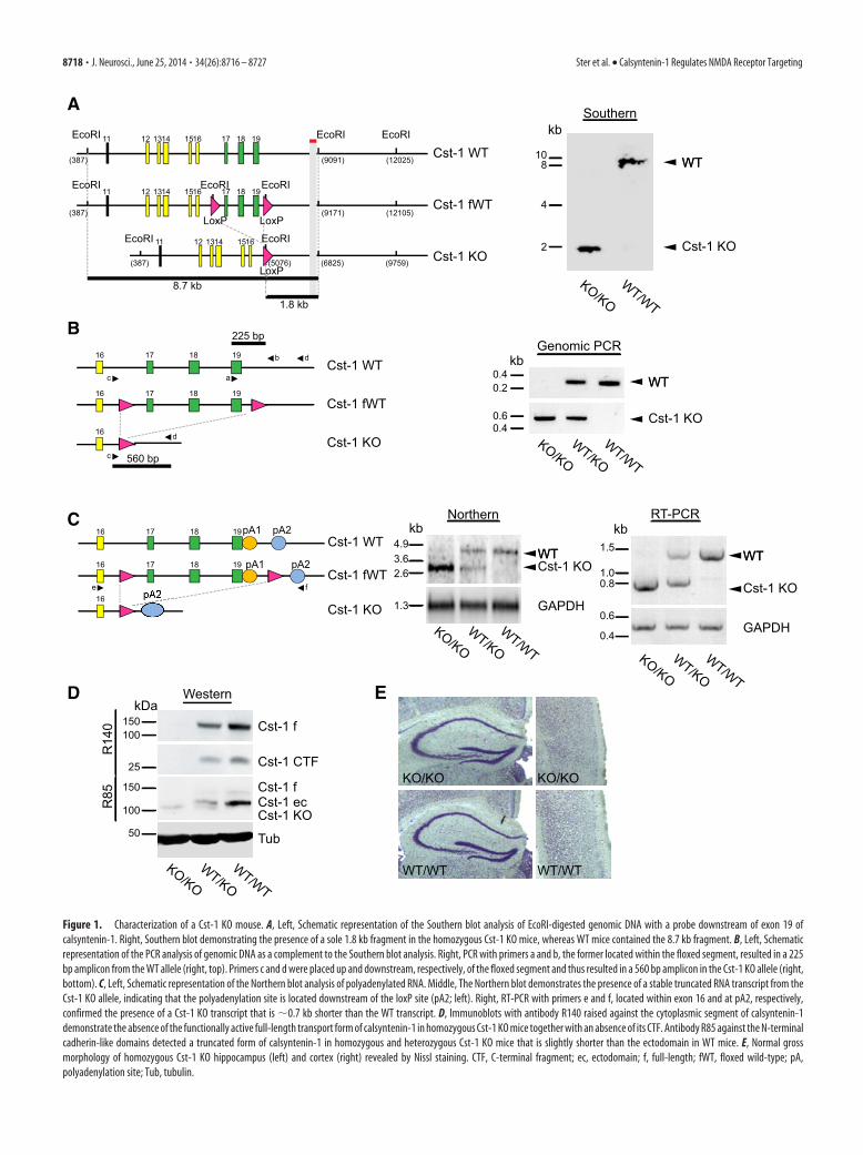
Progeny were screened by Southern hybridization and genomic PCRfor successful deletion and germline transmission. Southern blot analysisconfirmed that the 8.7 kb EcoRI-fragment spanning this genomic regionin WT mice was replaced by a 1.8 kb fragment, attributable to the pres-ence of an EcoRI site in the sole loxP site remaining after Cre/loxP-baseddeletion of the loxP-flanked region (see Fig. 1A). Successful deletion ofthe loxP-flanked segment comprising the transmembrane and cytoplas-mic domains was confirmed by PCR of genomic DNA of WT, heterozy-gous, and homozygous Cst-1 KO mice. Only WT and heterozygous Cst-1KO mice showed the 225 bp amplicon, because one of the primers waslocated within exon 19 that was deleted in the Cst-1 KO allele (Fig. 1B). Afurther round of PCR with another pair of primers, located up and down-stream of the segment to be deleted, resulted in the expected truncated560 bp amplicon, thus confirming successful deletion. Northern blots ofisolated polyadenylated mRNA from brain homogenates showed a trun-cated transcript in mice containing the Cst-1 KO allele, indicating a stabletranscript with the polyadenylation site located downstream of the stillpresent sole loxP site (Fig. 1C). RT-PCR demonstrated a truncation by�0.7 kb, which corresponds to the length of the coding sequences ofexons 17–19 (Fig. 1C).
In all experiments we compared transgenic mice (n � 70) with non-transgenic littermates (n � 88) of either sex.
Tissue preparation. Acute slices were prepared from juvenile mice(P15–P21) and adult mice (2 months old) following a protocol approvedby the Veterinary Department of the Canton of Zurich. Mice were de-capitated, and brains quickly removed and chilled in cold artificial CSF(ACSF) containing the following (in mM): 125 NaCl, 2.5 KCl, 1.25
NaH2PO4, 25 NaCHCO3, 1 MgCl2, 2 CaCl2, and 10 glucose, pH 7.4,equilibrated with 95% O2 and 5% CO2. Acute 350-�m-thick slices wereprepared with a vibratome (HM 650V, Microm International) in ice-cold ACSF. Sections were incubated in ACSF at 34°C for 20 min and thenkept at room temperature for at least 1 h before recording.
Electrophysiology. Acute slices were transferred to a recording chambermounted on an upright microscope (Axioskop FS1; Zeiss) and super-fused with ACSF at 27°C. A monopolar electrode was placed in the Schaf-fer collaterals, and stimulation was applied at 0.033 Hz with stimulusintensity ranging from 10 to 60 �A, yielding evoked field EPSPs (fEPSPs)of 0.2–1 V. Baseline fEPSPs were recorded for a minimum of 20 min. LTPwas induced by stimulation with 100 Hz for one train or three trains of 1 sduration separated by 20 s (high-frequency stimulation; HFS), four stim-uli at 100 Hz for 10 bursts separated by 200 ms (theta-burst stimulation;TBS). For LTD Schaffer collaterals were stimulated at 1 Hz for 15 min(low-frequency stimulation; LFS). Data were analyzed by measuring theslope of individual fEPSPs.
Whole-cell patch-clamp recordings were obtained from CA1 pyrami-dal cells using an Axopatch 200B amplifier (Molecular Devices). Patchrecording pipettes (4 –5 M�) were filled with intracellular solution con-taining the following (in mM): 120 CsMeSO4, 10 CsCl2, 10 HEPES, 2MgCl2, 4 NaATP, 0.4 NaGTP, 5 Na-creatine, and 10 EGTA, pH 7.2.Biocytin was added to the patch solution at a concentration of 2 �g/ml.
EPSCs were evoked by electrical stimulation of Schaffer collateral/commissural axons using a bipolar-stimulating electrode placed in thestratum radiatum of CA1 (0.033 Hz stimulation frequency). AMPA-mediated EPSCs and NMDA-mediated EPSCs were obtained at �70 mVand �30 mV, respectively, in the presence of picrotoxin (50 �M).
Evoked current and spontaneous activities were analyzed offline usingpClamp 10 (Molecular Devices). All data are reported as the mean �SEM. Statistical comparisons were made using two-tailed unpaired or pairedStudent’s t tests. For comparisons of cumulative distributions of amplitudeand interevent interval of EPSCs we used the Kolmogorov–Smirnov test. Allcompounds were bath applied 20 min before induction of LTP.
Spine imaging and analysis. Slices fixed with 4% paraformaldehydewere extensively washed in 0.1 M PB. After blocking in 0.1 M PB supple-mented with 0.4% Triton X-100 and 10% inactivated horse serum for 2 hat room temperature, slices were incubated with Streptavidin Alexa Fluor488 (4 �g/ml, Invitrogen) for 2 h at room temperature. Slices were coun-terstained with DAPI (Invitrogen), washed in 0.1 M PB, and mountedonto gelatin-coated slides with Vectashield-mounting medium (Reacto-lab) to preserve fluorescent labeling. Images of CA1 apical dendrites inthe stratum radiatum were acquired using a Leica TCS SPE II confocalmicroscope. Dendritic spines were visualized by using the 488 nm laserline, with a 63�/1.4 oil objective and an additional magnification of 8�.Stacks of images (512 � 512 pixels) were collected with a z-step size of 0.2�m. Images were deconvolved using Huygens Software (Scientific Vol-ume Imaging) and analyzed with ImageJ (http://rsb.info.nih.gov/ij/). Allexperiments were done blind with respect to the experimental condition.Spine density was measured on 3D projections and defined as the num-ber of spines per 10 �m dendritic length. Spine length was measured asthe distance from the outer edge of the dendritic shaft to the tip of thespine. Spine head diameter was estimated as the length of the longest linethrough the spine head orthogonal to the neck of the spine. Spine mor-phology was estimated according to the following parameters: mush-room (M), head diameter exceeding 0.5 �m; thin (T), 0.2 head � 0.5�m in diameter and a maximum length at least twice as large as the headdiameter; filopodia-like (F), head � 0.2 �m and not clearly distinguish-able from the neck; other (O), 0.2 head � 0.5 �m in diameter and amaximum length less than twice the head diameter (Harris et al., 1992;Kim et al., 2008).
Subcellular fractionation and immunoprecipitation from mouse brain.The V1 membrane fraction was prepared from P7 mouse brains by dif-ferential centrifugation as previously described (Konecna et al., 2006;Steuble et al., 2010). Washed V1 pellets were resuspended in immuno-precipitation (IP) buffer (PBS, 320 mM sucrose, and 5 mM EDTA, pH 7.4)and stirred for 1 h at 4°C. For immunoblotting, 2 mg magnetic Dyna-beads M-280 protein A (Invitrogen) was incubated with 10 �g IgG for 40min and washed four times in IP buffer. V1 inputs were adjusted to �0.7
Ster et al. • Calsyntenin-1 Regulates NMDA Receptor Targeting J. Neurosci., June 25, 2014 • 34(26):8716 – 8727 • 8717
EcoRI
EcoRI
11 12 1314 1516 17 18 19 EcoRI EcoRI
11 12 1314 1516 17 18 19
11 12 1314 1516EcoRI
EcoRI EcoRI
EcoRI
(5076) (6825) (9759)(387)
(9171) (12105)(387)
(9091) (12025)(387)
LoxPLoxP
LoxP8.7 kb
1.8 kb
ooxxPP
6
LL
EEE
Cst-1 WT
Cst-1 fWT
Cst-1 KO
WTWT
Cst-1 KO
108
4
2
kbSouthernA
C
E
B
D
WT/WT
KO/KO
c a
b d16 17 18 19
16 17 18 19
c
16 d
225 bp
560 bp
Cst-1 WT
Cst-1 fWT
Cst-1 KO
WTWT
Cst-1 KO
Genomic PCR
WT/WT
KO/KOWT/KO
0.40.2
kb
0.60.4
16 17 18 19
e f
16 17 18 19
16
Cst-1 WT
Cst-1 fWT
Cst-1 KO
pA1 pA2
pA1 pA2
4.93.62.6
1.3
WT/WT
KO/KOWT/KO
kbNorthern
WTWT
GAPDH
Cst-1 KOWTWT
GAPDH
Cst-1 KO
WT/WT
KO/KOWT/KO
1.5
1.00.8
0.6
kb
0.4
RT-PCR
Cst-1 f
Cst-1 CTF
Cst-1 fCst-1 ecCst-1 KO
Tub
WT/WT
KO/KOWT/KO
Western
150
150
kDa
100
50
100
25
R14
0R
85
KO/KO KO/KO
WT/WTWT/WT
Figure 1. Characterization of a Cst-1 KO mouse. A, Left, Schematic representation of the Southern blot analysis of EcoRI-digested genomic DNA with a probe downstream of exon 19 ofcalsyntenin-1. Right, Southern blot demonstrating the presence of a sole 1.8 kb fragment in the homozygous Cst-1 KO mice, whereas WT mice contained the 8.7 kb fragment. B, Left, Schematicrepresentation of the PCR analysis of genomic DNA as a complement to the Southern blot analysis. Right, PCR with primers a and b, the former located within the floxed segment, resulted in a 225bp amplicon from the WT allele (right, top). Primers c and d were placed up and downstream, respectively, of the floxed segment and thus resulted in a 560 bp amplicon in the Cst-1 KO allele (right,bottom). C, Left, Schematic representation of the Northern blot analysis of polyadenylated RNA. Middle, The Northern blot demonstrates the presence of a stable truncated RNA transcript from theCst-1 KO allele, indicating that the polyadenylation site is located downstream of the loxP site (pA2; left). Right, RT-PCR with primers e and f, located within exon 16 and at pA2, respectively,confirmed the presence of a Cst-1 KO transcript that is �0.7 kb shorter than the WT transcript. D, Immunoblots with antibody R140 raised against the cytoplasmic segment of calsyntenin-1demonstrate the absence of the functionally active full-length transport form of calsyntenin-1 in homozygous Cst-1 KO mice together with an absence of its CTF. Antibody R85 against the N-terminalcadherin-like domains detected a truncated form of calsyntenin-1 in homozygous and heterozygous Cst-1 KO mice that is slightly shorter than the ectodomain in WT mice. E, Normal grossmorphology of homozygous Cst-1 KO hippocampus (left) and cortex (right) revealed by Nissl staining. CTF, C-terminal fragment; ec, ectodomain; f, full-length; fWT, floxed wild-type; pA,polyadenylation site; Tub, tubulin.
8718 • J. Neurosci., June 25, 2014 • 34(26):8716 – 8727 Ster et al. • Calsyntenin-1 Regulates NMDA Receptor Targeting

mg/ml with IP buffer and incubated with beads precoated with the re-spective immunoprecipitating antibody for 1 h at 4°C. Beads withimmuno-isolated organelles were washed 10 times with 1 ml IP buffer, oncewith 20 mM Tris-HCl, pH 7.4. Immuno-isolated organelles were solubilizedunder mild detergent conditions (50 �l 20 mM Tris-HCl, pH 7.4, 0.1% (v/v)Triton X-100) for 30 min at 25°C. Forty microliters of the respective eluateswere resolved together with 10 �g V1 input on 4–12% Bis-Tris gels (Invit-rogen). Immunoblotting was performed as described below.
Western blots. At P17, mice were killed by cervical dislocation andhippocampi were quickly dissected, flash frozen in liquid nitrogen, andstored at �80°C before extraction in 300 �l of RIPA buffer (10 mM
Tris-HCl, 1% Nonidet 40, 150 mM NaCl, 0.1% SDS, and 1% deoxy-cholate, pH 7.4) containing protease inhibitors (Complete mini; RocheDiagnostics). The samples were homogenized and kept for 60 min on ice.After centrifugation for 15 min at 15,000 � g, 4°C, the supernatant wascollected in new Eppendorf tubes. Hippocampal proteins (20 �g) wereresolved by electrophoresis on a 4 –12% polyacrylamide gel and trans-ferred to nitrocellulose membranes. The nitrocellulose membranes werethen incubated in a blocking solution containing 5% BSA in TBST 0.2%(Tris-base 0.1 M and 0.2% Tween 20, pH 7.4) for 1 h at room temperatureand incubated with primary antibodies overnight at 4°C. Primary anti-bodies were rabbit anti-GluN2A (1:1000; Millipore Bioscience ResearchReagents, AB1555), rabbit anti-GluN2B (1:500; Millipore BioscienceResearch Reagents, AB1557), rabbit anti-GluA1 (1:5000; Millipore, #05-855), mouse anti-PSD95 (1:5000; Millipore, MABN68), rabbit anti-GRIP1 (1:500; Sigma-Aldrich, #AV32496), mouse anti synaptotagmin(1:1000; BD Transduction Laboratories, # S39520), mouse anti-VAMP2(1:5000; Synaptic Systems, #104211), mouse anti-Rab5 (1:100; SantaCruz Biotechnology, # sc-46692), rabbit anti-Rab4 (1:200, Santa CruzBiotechnology, #sc-28569), mouse anti-Rab11(1:2000; TransductionLaboratories, #610656), rabbit anti-syntaxin6 (1:1000; Synaptic Systems,#110062), mouse anti-transferrin receptor (1:1000; Invitrogen, #13-68000), anti-Rip11 (1:2000, provided by Mitsuo Tagaya, Tokyo), mouseanti-N-cadherin (1:2500; Transduction Laboratories, #C70320), andmouse anti-�-actin (1:5000; Sigma-Aldrich, #A5326). After three washesin TBST, a horseradish peroxidase-conjugated anti-rabbit antibody wasadded to the blots (Pierce Biotechnology). Protein signals were detected byapplying SuperSignal West Pico Chemiluminescent Substrate (Pierce) andby exposing the blots in a Stella detector. Protein quantity was measured bydensitometry with NIH software and normalized to GAPDH values.
Immunocytochemistry. Primary dissociated hippocampal cultures wereprepared from embryonic day 18 mice as described previously (Kaechand Banker, 2006). For immunofluorescence analysis, neurons werefixed in 4% paraformaldehyde, 4% sucrose in PBS, pH 7.4, for 10 min,washed, and permeabilized for 1 h in 10% FCS, 0.1% glycine, and 0.1%saponin in PBS. Cells were incubated with primary (polyclonal rabbitantibody R140 was generated against murine calsyntenin-1, monoclonalmouse anti-GluN2A from Millipore Corporation, monoclonal mouseanti-APP from Millipore Bioscience Research Reagents, anti-EEA1 fromSanta Cruz Biotechnology, monoclonal mouse anti-transferrin recep-tor from Invitrogen) in blocking solution and secondary antibodies(Cy3-conjugated and Alexa 488-conjugated antibodies) in blockingsolution plus 2% NGS. Confocal images were acquired with a TCSSP2 AOBS spectral confocal microscope (Leica Microsystems). Im-ages were adjusted only for brightness and contrast using AdobePhotoshop 12.
ResultsGeneration and characterization of a Cst-1 KO mouseWe generated a Cst-1 KO by introducing loxP sites upstream ofexon 17 and downstream of exon 19 of the calsyntenin-1 gene todelete the transmembrane and cytosolic segments of calsyntenin-1(see Materials and Methods). The C-terminal cytosolic segment isessential for vesicular docking to kinesin-1 motors. Immunoblotanalysis of whole-brain homogenates from Cst-1 KO mice withR140, an antibody raised against the cytoplasmic segment ofcalsyntenin-1, demonstrated the absence of the full-length,transportation-competent form of calsyntenin-1, as well as the
C-terminal cytosolic segment (Fig. 1D). As expected for the ex-pression of a truncated mRNA transcript, antibody R85 raisedagainst the N-terminal cadherin-like domains of calsyntenin-1revealed the presence of truncated calsyntenin-1 that is slightlyshorter than the calsyntenin-1 ectodomain, the predominantform of calsyntenin-1 that accumulates in WT mice after jux-tamembrane cleavage in the extracellular compartment. Becauseof the absence of the transmembrane and cytosolic segment, thisfragment lacks the kinesin-docking function of full-lengthcalsyntenin-1. Therefore, it is unlikely that this truncated form ofcalsyntenin-1 exhibits residual calsyntenin-1-dependent func-tions for vesicular transport in KO mice. Moreover, a dominant-negative effect of this truncated calsyntenin-1 can be excludedwith high probability, because of its close structural homology tothe released ectodomain of calsyntenin-1 found at markedlyhigher levels in WT mice. Cst-1 KO mice were viable and fertile,without an overt morphological or behavioral phenotype, and theirweight was comparable to WT littermates (P17: WT � 7.1 � 0.39 g,n � 14; Cst-1 KO � 6.9 � 0.33 g, n � 14, p 0.6; P48: WT � 20.6 �0.5 g, n � 31; Cst-1 KO � 19.8 � 0.8 g, n � 18, p 0.3). Investiga-
N-Cad
Rip11
TfR
Stx6
Rab11
Rab4
Rab5
GluA1/A2
VAMP2
PSD95
GRIP1
Sytg
β-actin
WT/WT
KO/KOWT/WT
KO/KO
GluN1
GluN2A
GluN2B
Adult Mice Juvenile Mice
Figure 2. Western blot characterization of the adult and the juvenile Cst-1 KO mouse. Each30 �g of brain homogenates from WT and Cst-1 KO mice was analyzed by immunoblotting withthe indicated antibodies. Sytg, synaptotagmin; Stx6, syntaxin 6; TfR, transferrin receptor;N-Cad, N-cadherin.
Ster et al. • Calsyntenin-1 Regulates NMDA Receptor Targeting J. Neurosci., June 25, 2014 • 34(26):8716 – 8727 • 8719

tion of brain sections by in situ hybridiza-tion and Nissl staining did not reveal grossmorphological alterations (Fig. 1E). More-over, in adults the absence of calsyntenin-1did not affect the expression of an extendedseries of synaptic proteins as shown in West-ern blots of brain homogenates from WTand Cst-1 KO (Fig. 2). In the juvenile KOmouse, most synaptic proteins were simi-larly unchanged (Fig. 2), except for NMDAreceptors as described below.
Basal excitatory synaptic transmissionis decreased in juvenile Cst-1 KO miceThe preferential localization of cal-syntenin-1 in the CA1 area of the hip-pocampus (Hintsch et al., 2002) in apattern indicating dendritic expression(Konecna et al., 2006) suggests a functionat inputs to CA1 pyramidal cells. Becausethe expression levels of calsyntenin-1 arehighest in juvenile mice, we comparedspontaneous EPSCs in acute hippocampalslices prepared at P15–P21 from Cst-1 KOand WT mice. Analysis of recordings fromCA1 pyramidal cells (Fig. 3A) showed thatthe amplitudes of spontaneous EPSCswere similar in slices prepared from juve-nile (P15–P21) WT and Cst-1 KO mice(WT � �14.6 � 1.1 pA, Cst-1 KO ��15.5 � 1 pA, p 0.05; Fig. 3B,C). How-ever, spontaneous EPSC frequency wasmarkedly decreased in Cst-1 KO com-pared with WT mice (WT � 0.9 � 0.3 Hz,Cst-1 KO � 0.3 � 0.1 Hz, p 0.01; Fig.3B,C). The input/output curve obtainedfrom field recordings revealed a signifi-cant decrease in EPSP slope in Cst-1 KOcompared with WT mice (Fig. 3D,E). Torule out a bias in the placement of thestimulating electrode, the magnitude ofthe EPSP was replotted as a function of thefiber volley at a constant amplitude of�0.2 mV, which confirmed a decrease insynaptic transmission in the Cst-1 KO mice (KO � �0.11 � 0.02;WT � �0.23 � 0.03, p 0.05; Fig. 3F). A reduction in synaptictransmission can reflect either a presynaptic process in whichvesicular release is diminished, or a postsynaptic effect in whichthe number of synapses is reduced or fewer mature or functionalsynapses are present. To distinguish between these two possibil-ities we assessed parameters for presynaptic release of neu-rotransmitter. First we examined paired-pulse facilitation (PPF),which was not significantly different at pulse intervals rangingfrom 50 to 500 ms (Fig. 4A,B). Short-term plasticity induced bystimulation with three stimuli at 100 Hz was also not different inCst-1 KO compared with WT slices (12–15 min: WT � 106 �6%, Cst-1 KO � 107 � 3.3%, p 0.1; Fig. 4C,D). These resultsindicate that presynaptic release properties are not modified inthe hippocampus of Cst-1 KO mice. A further possibility ac-counting for decreased synaptic transmission could be a decreasein dendritic spine density. However, the number of spines perunit length was not changed in CA1 pyramidal cells from Cst-1KO compared with WT dendrites (Fig. 4E).
Involvement of calsyntenin-1 in dendritic transportFurther evidence for a postsynaptic mechanism mediating theobserved reduction in synaptic transmission was the presenceof calsyntenin-1-positive puncta along all dendrites as visual-ized with R140, an antibody against the cytosolic fragment ofcalsyntenin-1 (Fig. 5A). A well established function ofcalsyntenin-1 is as a docking protein that directly binds to thelight chain of kinesin-1 for fast axonal transport (Konecna etal., 2006; Steuble et al., 2012; Vagnoni et al., 2012). We there-fore checked for a similar role in dendrites. Indeed, the con-siderable degree of colocalization of dendritic calsyntenin-1puncta with both the transferrin receptor (TfR), a marker forrecycling endosomes, as well as with APP, which is carried inearly endosomal calsyntenin-1 vesicles (Steuble et al., 2012),indicates that dendritic calsyntenin-1-associated vesicles areinvolved in transport of both early and recycling endosomalorganelles (Fig. 5 A, B). Importantly, dendritic calsyntenin-1also colocalizes with NMDA receptor subunits (Fig. 5C,D).
Cst-1 KO
AWT
WT Cst-1 KO
EP
SC
s am
plitu
de (p
A)
-5
0
-10
-20
-156 8
EPSCs amplitude (pA)
WTCst-1 KO
-60-40-200 -80
1
0.5
0Cum
ulat
ive
prob
abili
ty
B
1
0.5
04000 8000 12000 160000
Cum
ulat
ive
prob
abili
ty
EPSCs inter-event interval (msec)
WTCst-1 KO
C
6
EP
SC
s fre
quen
cy (H
z)
0
1
0.5
**
8
WTCst-1 KO
0.1 s20 pA
D E
0.5 mV
5 ms
WT
Cst-1 KO
0.4
0.2
0.6
020 40 60
***
*
Stimulus intensity (μA)fE
PS
P sl
ope
(mV
/ms)
WT (n = 13)Cst-1 KO (n = 15)
0.2mV2 ms
WTCst-1 KO *
F
7
8
7
8
-0.1
0
-0.2
-0.3
Fiber volley (mV)
fEPSP slope (mV/ms)
Figure 3. Excitatory synaptic transmission is reduced in juvenile Cst-1 KO mice. A, The frequency of spontaneous EPSCsrecorded in CA1 pyramidal cells voltage clamped at �70 mV is reduced in Cst-1 KO versus WT slices in P15–P21 mice. B, Bargraphs showing no significant change in amplitude but a marked decrease in frequency of spontaneous EPSCs in Cst-1 KOmice. C, Pooled data for EPSCs represented as cumulative distributions of EPSC amplitudes and interevent intervals. D,Evoked synaptic activity induced by Schaffer collateral stimulation is decreased in field recordings of CA1 pyramidal cellsfrom Cst-1 KO mice. E, Input– output relation comparing CA1 field responses in WT and Cst-1 KO mice F, Superimposedtraces scaled to the amplitude of the fiber volley. Data normalized to the amplitude of the input volley reveal a significantdecrease in the fEPSP.
8720 • J. Neurosci., June 25, 2014 • 34(26):8716 – 8727 Ster et al. • Calsyntenin-1 Regulates NMDA Receptor Targeting

Enhanced synaptic expression of GluN2B in Cst-1 KO miceIf calsyntenin-1 contributes to dendritic trafficking of proteins,the reduction in excitatory transmission that we observed in theCst-1 KO mouse may arise from disrupted targeting of glutama-tergic receptors. We therefore assessed AMPA and NMDAreceptor-mediated responses by comparing their ratios in WTversus Cst-1 CA1 pyramidal cells. Schaffer collaterals were stim-ulated to evoke EPSCs in CA1 pyramidal cells voltage clamped at�70 mV, where extracellular magnesium (1 mM) blocks mostNMDA current, to measure AMPA responses and then at � 30mV, where the NMDA receptor-mediated component is appar-ent (Fig. 6A). We also took advantage of the much faster decaykinetics of AMPA versus NMDA receptor currents (Fig. 6A, ar-rows) to calculate the proportion of the current mediated by eachreceptor channel. Our data show that the AMPA/NMDA re-sponse ratio was lower in Cst-1 KO than in WT cells (WT ��1.2 � 0.6, Cst-1 KO � �0.4 � 0.2, p 0.05; Fig. 6A). Begin-ning in the second postnatal week, there is a switch from predom-inantly GluN2B subunit-containing NMDA receptors toincreasing expression of GluN2A subunit-containing NMDA re-ceptors (Monyer et al., 1994; Sheng et al., 1994). We thereforeexamined in greater detail the evoked response mediated byNMDA receptors. Our recordings show that the NMDA compo-nent of the current had slower decay kinetics in the Cst-1 KOversus the WT neurons (WT tau � 89 � 12 ms, Cst-1 KO tau �147 � 19 ms, p 0.05; Fig. 6B), which may reflect a high expres-sion of NMDA receptors containing the GluN2B subunit(Monyer et al., 1994; Vicini et al., 1998; Tovar et al., 2000). Incontrast, the decay kinetics of AMPA receptor-mediated EPSCwas not modified (WT tau � 19.2 � 4.5 ms, Cst-1 KO tau �15.4 � 2.4 ms, p 0.3; Fig. 6B). To determine whether the slowerNMDA receptor kinetics in the Cst-1 KO was indeed due to a
greater GluN2B-mediated component,we compared the effects of a GluN2A sub-unit antagonist, NVP-AAM (0.1 �M; Au-berson et al., 2002; Bartlett et al., 2007),and a GluN2B subunit antagonist, Ro25–6981 (3 �M; Fischer et al., 1997) on evokedNMDA EPSCs at �30 mV in the presenceof NBQX (25 �M) and picrotoxin (50�M). The GluN2B subunit antagonistinduced a greater suppression of EPSCsin Cst-1 KO neurons whereas theGluN2A subunit antagonist was more ef-fective in reducing EPSCs in WT neurons(Fig. 6C). Thus, at Cst-1 KO synapses theproportion of GluN2B subunit-containingreceptors was increased (WT � 20.6 �6.9; Cst-1 KO � 40.6 � 3.7; p 0.05; Fig.6C), whereas the proportion of GluN2A-containing receptors was decreased(WT � 68.9 � 5.8; Cst-1 KO � 44.5 �8.2; p 0.05; Fig. 6C).
Western blots of whole hippocampifrom P17 mice also showed that GluN2Blevels are higher in Cst-1 KO comparedwith WT mice (ratio GluN2B/GAPDH:WT � 100 � 7.1, Cst-1 KO � 127.8 � 3.4,p � 0.0246; Fig. 6D,E). No difference intotal tissue GluN2A levels was observed(ratio GluN2A/GAPDH: WT � 100 �9.7, Cst-1 KO � 107.4 � 11.4, p 0.05).As calsyntenin-1 is associated with trans-
port vesicles, we were interested in whether these vesicles maycontain NMDA receptors. Calsyntenin-1-positive endosomeswere immuno-isolated from whole brains of juvenile mice usingthe R140 antibody against the cytosolic segment of calsyntenin-1.Western blots revealed the presence of the GluN1 subunit as wellas both of the main hippocampal subtypes of the GluN2 subunit,GluN2A and GluN2B in calsyntenin-1 vesicles, which corre-spond to early and recycling endosomes on the basis of the mark-ers Stx13, TfR, and Rab11 (Fig. 6F). Thus, the above results areconsistent with a role for calsyntenin-1 as a transporter of NMDAreceptor subunits to dendritic targets.
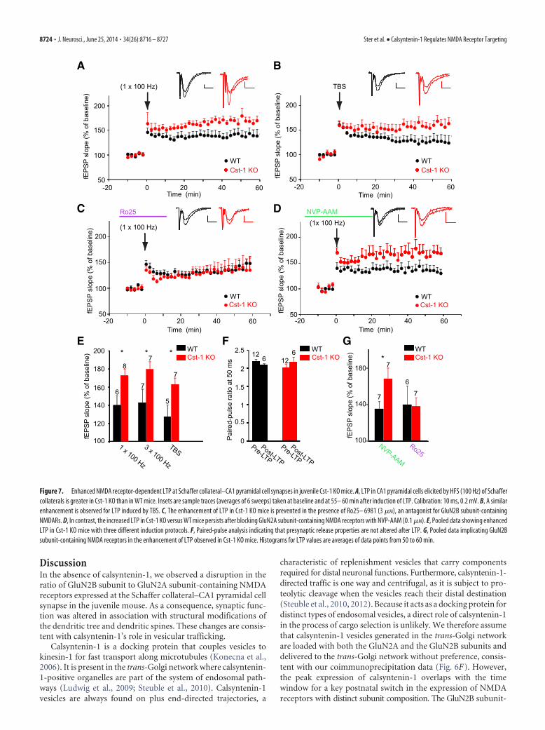
NMDA receptor-dependent LTP is increased in juvenile Cst-1KO miceAs GluN2A and GluN2B subunit-containing NMDA receptorsplay distinct roles in LTP (Shipton and Paulsen, 2014), we exam-ined whether the increase in GluN2B in Cst-1 KO mice affectssynaptic plasticity. We found that LTP induced at Schaffer collat-eral–CA1 pyramidal cell synapses by HFS with one train or threetrains at 100 Hz was significantly increased in slices from Cst-1KO mice (1 � 100 Hz: WT � 139.3 � 10.9%, Cst-1 KO � 172 �7.4%, p 0.05; 3 � 100 Hz: WT � 142.4 � 14.7%, Cst-1 KO �178.9 � 8.5%, p 0.05; Fig. 7A,E). This form of LTP is NMDAreceptor dependent as it was blocked by the specific antagonistD-AP5 (50 �M): WT control � 142.4 � 14.7%, WT D-AP5 �107.2 � 6.3%, p 0.05. We also induced LTP with a TBS, whichhas been proposed as a more physiological protocol to studysynaptic plasticity (Staubli and Lynch, 1987). Again, LTP inducedby TBS was enhanced in Cst-1 KO compared with WT mice (TBS:WT � 126.4 � 13.5%, Cst-1 KO � 162 � 7.7%, p 0.05; Fig.7B,E). Moreover, PPF did not change after LTP induction, bothin WT and in Cst-1 KO slices (Fig. 7F). Although in the hip-
WTCst-1 KO160
120
100
140
fEP
SP
slop
e (%
of b
asel
ine)
0-3 min 12-15 min
7
6
76WTCst-1 KO
STP (3 stim at 100 Hz)
0-10 20Time (min)
10
200
100
50
150
fEP
SP
slop
e (%
of b
asel
ine)
A B
ISI (ms)
Pai
red-
puls
e ra
tio 2
0.5
0
1
2.5
1.5
200 4000
WTCst-1 KO
Scaled
10 ms0.2 mV
C E
15
5
10
20
25
30
WTCst-1 KO
Num
ber o
f spi
nes
/10
μm
0
D
Figure 4. Presynaptic function is not changed in CA1 pyramidal cells from juvenile Cst-1 KO mice. A, Field recordings show thatPPF at 50 ms is not altered in Cst-1 KO mice. B, Pooled data for a range of paired-pulse inter stimulation intervals (ISI). C, Short-termsynaptic potentiation is not affected in Cst-1 KO mice. D, Pooled data showing pronounced short-term plasticity during the first 3min after stimulation, which decrease toward baseline after 15 min. E, Dendritic spine density is unchanged in Cst-1 KO mice.
Ster et al. • Calsyntenin-1 Regulates NMDA Receptor Targeting J. Neurosci., June 25, 2014 • 34(26):8716 – 8727 • 8721
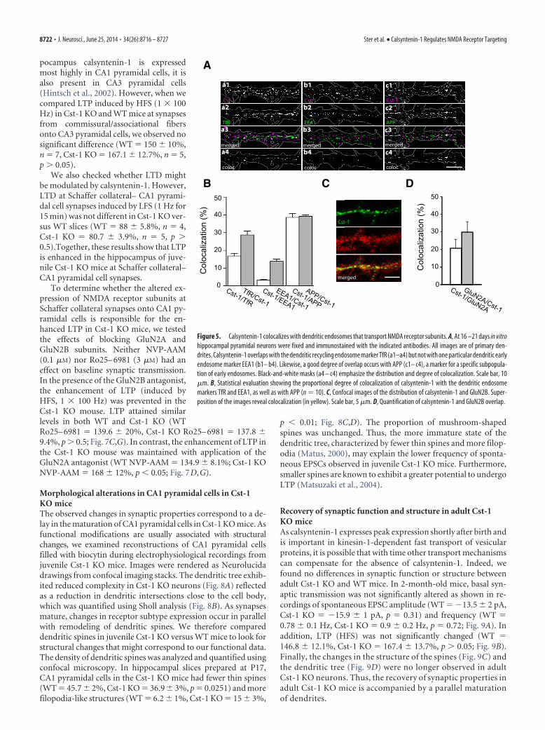
pocampus calsyntenin-1 is expressedmost highly in CA1 pyramidal cells, it isalso present in CA3 pyramidal cells(Hintsch et al., 2002). However, when wecompared LTP induced by HFS (1 � 100Hz) in Cst-1 KO and WT mice at synapsesfrom commissural/associational fibersonto CA3 pyramidal cells, we observed nosignificant difference (WT � 150 � 10%,n � 7, Cst-1 KO � 167.1 � 12.7%, n � 5,p 0.05).
We also checked whether LTD mightbe modulated by calsyntenin-1. However,LTD at Schaffer collateral– CA1 pyrami-dal cell synapses induced by LFS (1 Hz for15 min) was not different in Cst-1 KO ver-sus WT slices (WT � 88 � 5.8%, n � 4,Cst-1 KO � 80.7 � 3.9%, n � 5, p 0.5).Together, these results show that LTPis enhanced in the hippocampus of juve-nile Cst-1 KO mice at Schaffer collateral–CA1 pyramidal cell synapses.
To determine whether the altered ex-pression of NMDA receptor subunits atSchaffer collateral synapses onto CA1 py-ramidal cells is responsible for the en-hanced LTP in Cst-1 KO mice, we testedthe effects of blocking GluN2A andGluN2B subunits. Neither NVP-AAM(0.1 �M) nor Ro25– 6981 (3 �M) had aneffect on baseline synaptic transmission.In the presence of the GluN2B antagonist,the enhancement of LTP (induced byHFS, 1 � 100 Hz) was prevented in theCst-1 KO mouse. LTP attained similarlevels in both WT and Cst-1 KO (WTRo25– 6981 � 139.6 � 20%, Cst-1 KO Ro25– 6981 � 137.8 �9.4%, p 0.5; Fig. 7C,G). In contrast, the enhancement of LTP inthe Cst-1 KO mouse was maintained with application of theGluN2A antagonist (WT NVP-AAM � 134.9 � 8.1%; Cst-1 KONVP-AAM � 168 � 12%, p 0.05; Fig. 7D,G).
Morphological alterations in CA1 pyramidal cells in Cst-1KO miceThe observed changes in synaptic properties correspond to a de-lay in the maturation of CA1 pyramidal cells in Cst-1 KO mice. Asfunctional modifications are usually associated with structuralchanges, we examined reconstructions of CA1 pyramidal cellsfilled with biocytin during electrophysiological recordings fromjuvenile Cst-1 KO mice. Images were rendered as Neurolucidadrawings from confocal imaging stacks. The dendritic tree exhib-ited reduced complexity in Cst-1 KO neurons (Fig. 8A) reflectedas a reduction in dendritic intersections close to the cell body,which was quantified using Sholl analysis (Fig. 8B). As synapsesmature, changes in receptor subtype expression occur in parallelwith remodeling of dendritic spines. We therefore compareddendritic spines in juvenile Cst-1 KO versus WT mice to look forstructural changes that might correspond to our functional data.The density of dendritic spines was analyzed and quantified usingconfocal microscopy. In hippocampal slices prepared at P17,CA1 pyramidal cells in the Cst-1 KO mice had fewer thin spines(WT � 45.7 � 2%, Cst-1 KO � 36.9 � 3%, p � 0.0251) and morefilopodia-like structures (WT � 6.2 � 1%, Cst-1 KO � 15 � 3%,
p 0.01; Fig. 8C,D). The proportion of mushroom-shapedspines was unchanged. Thus, the more immature state of thedendritic tree, characterized by fewer thin spines and more filop-odia (Matus, 2000), may explain the lower frequency of sponta-neous EPSCs observed in juvenile Cst-1 KO mice. Furthermore,smaller spines are known to exhibit a greater potential to undergoLTP (Matsuzaki et al., 2004).
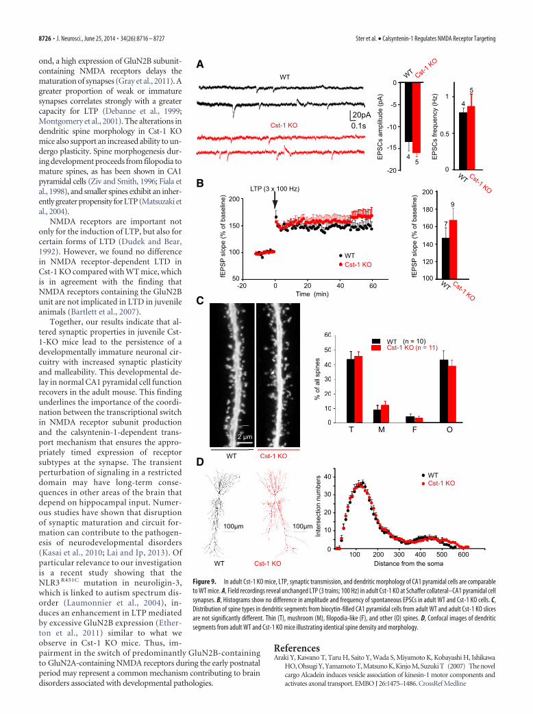
Recovery of synaptic function and structure in adult Cst-1KO miceAs calsyntenin-1 expresses peak expression shortly after birth andis important in kinesin-1-dependent fast transport of vesicularproteins, it is possible that with time other transport mechanismscan compensate for the absence of calsyntenin-1. Indeed, wefound no differences in synaptic function or structure betweenadult Cst-1 KO and WT mice. In 2-month-old mice, basal syn-aptic transmission was not significantly altered as shown in re-cordings of spontaneous EPSC amplitude (WT � �13.5 � 2 pA,Cst-1 KO � �15.9 � 1 pA, p � 0.31) and frequency (WT �0.78 � 0.1 Hz, Cst-1 KO � 0.9 � 0.2 Hz, p � 0.72; Fig. 9A). Inaddition, LTP (HFS) was not significantly changed (WT �146.8 � 12.1%, Cst-1 KO � 167.4 � 13.7%, p 0.05; Fig. 9B).Finally, the changes in the structure of the spines (Fig. 9C) andthe dendritic tree (Fig. 9D) were no longer observed in adultCst-1 KO neurons. Thus, the recovery of synaptic properties inadult Cst-1 KO mice is accompanied by a parallel maturationof dendrites.
a1
Cst-1a2
TfRa3
mergeda4
coloc
Cst-1
b1
EEA1
b2
merged
b3
coloc
b4
Cst-1
c1
APP
c2
merged
c3
coloc
c4
A
0
Col
ocal
izat
ion
(%)
B50
40
30
20
10
Cst-1/TfR
TfR/Cst-1
Cst-1/EEA1
EEA1/Cst-1
Cst-1/APP
APP/Cst-1
C50
40
30
20
10
0
Col
ocal
izat
ion
(%)
Cst-1/GluN2A
GluN2A/Cst-1
Cst-1
GluN2A
merged
D
Figure 5. Calsyntenin-1 colocalizes with dendritic endosomes that transport NMDA receptor subunits. A, At 16 –21 days in vitrohippocampal pyramidal neurons were fixed and immunostained with the indicated antibodies. All images are of primary den-drites. Calsyntenin-1 overlaps with the dendritic recycling endosome marker TfR (a1–a4) but not with one particular dendritic earlyendosome marker EEA1 (b1– b4). Likewise, a good degree of overlap occurs with APP (c1– c4), a marker for a specific subpopula-tion of early endosomes. Black-and-white masks (a4 – c4) emphasize the distribution and degree of colocalization. Scale bar, 10�m. B, Statistical evaluation showing the proportional degree of colocalization of calsyntenin-1 with the dendritic endosomemarkers TfR and EEA1, as well as with APP (n � 10). C, Confocal images of the distribution of calsyntenin-1 and GluN2B. Super-position of the images reveal colocalization (in yellow). Scale bar, 5 �m. D, Quantification of calsyntenin-1 and GluN2B overlap.
8722 • J. Neurosci., June 25, 2014 • 34(26):8716 – 8727 Ster et al. • Calsyntenin-1 Regulates NMDA Receptor Targeting

A
C
-0.5
0
-1
WT Cst-1 KO
-2
-1.5Rat
io A
MPA
/NM
DA
6
8
*
WT Cst-1 KO
20 ms
50 pA
100 pA
WTCst-1 KO
-70 mV
30 mV
AMPA-R (scaled)
NMDA-R (scaled)
120
160
80
0
40
WT Cst-1 KO
*
6
8
NM
DA
R T
au d
ecay
(ms)
B
AMPA
NMDA
NMDA
AMPA
0.05 sec50pA
100
40
0
80*
7
860
207
8
*
% (N
MD
A-s
ubun
it)Antagonist GluN2B
6
820
0
30
10
AM
PAR
Tau
dec
ay (m
s)
WT Cst-1 KO
120
40
0
80
Pro
tein
leve
ls (%
of W
T)
3
3
*3
3
GluN2A
GluN2B
GluN2A220
GluN2B220kDa
WT KOD E
GluN2A
GluN2B
GAPDH45
30
GAPDH45
30
GluN2A
GluN2B
GluN1
TfR
Stx13
Rab11
IgG-IP (c
ontrol)
R140-IP
Input: 10 μg V1
F
Antagonist GluN2B+antagonist GluN2A
Antagonist GluN2B
Antagonist GluN2B+antagonist GluN2A
Figure 6. Levels of GluN2B subunit-containing NMDA receptors are increased in juvenile Cst-1 KO mice. A, Representative averaged traces of evoked AMPA and NMDA receptor-mediated responses recorded in the same CA1 pyramidal cell at �70 mV (inward currents) and at �30 mV (outward currents) from a WT and Cst-1 KO mouse. Note that traces are alignedhorizontally. Histograms for the ratio of the AMPA to NMDA receptor-mediated response. To minimize the contribution from AMPA receptors, the NMDA receptor-mediated current wasmeasured 80 ms after stimulation. B, Scaling of traces reveals a slowing in the decay kinetics of the NMDA but not the AMPA receptor-mediated responses in Cst-1 KO mice. Histogramsshow pooled data. C, Pharmacologically isolated NMDA EPSCs exposed to NMDA receptor subunit-specific antagonists shows control current, current remaining after application of anGluN2B subunit antagonist (Ro25– 6981; 3 �M) and residual current in the presence of antagonists for both GluN2A (NVP-AAM; 0.1 �M) and GluN2B (Ro25– 6981) subunits. Histogramsshow pooled data. D, Protein levels for GluN2B and for GluN2A quantified by determining the ratio of GluN2B/GAPDH and GluN2A/GAPDH in hippocampal tissue extracts. E, Quantificationshows a significant increase in GluN2B but not GluN2A. F, Western blots of recycling endosomes, identified by Rab11 co-IP, from whole brains of WT juvenile mice (n � 3). Note the strongco-IP for NMDA receptors in the R140-enriched fraction, a marker for calsyntenin-1-positive recycling endosomes. Recycling endosomes are also positive for syntaxin-13 (Stx13), ageneral marker for endosomes.
Ster et al. • Calsyntenin-1 Regulates NMDA Receptor Targeting J. Neurosci., June 25, 2014 • 34(26):8716 – 8727 • 8723
DiscussionIn the absence of calsyntenin-1, we observed a disruption in theratio of GluN2B subunit to GluN2A subunit-containing NMDAreceptors expressed at the Schaffer collateral–CA1 pyramidal cellsynapse in the juvenile mouse. As a consequence, synaptic func-tion was altered in association with structural modifications ofthe dendritic tree and dendritic spines. These changes are consis-tent with calsyntenin-1’s role in vesicular trafficking.
Calsyntenin-1 is a docking protein that couples vesicles tokinesin-1 for fast transport along microtubules (Konecna et al.,2006). It is present in the trans-Golgi network where calsyntenin-1-positive organelles are part of the system of endosomal path-ways (Ludwig et al., 2009; Steuble et al., 2010). Calsyntenin-1vesicles are always found on plus end-directed trajectories, a
characteristic of replenishment vesicles that carry componentsrequired for distal neuronal functions. Furthermore, calsyntenin-1-directed traffic is one way and centrifugal, as it is subject to pro-teolytic cleavage when the vesicles reach their distal destination(Steuble et al., 2010, 2012). Because it acts as a docking protein fordistinct types of endosomal vesicles, a direct role of calsyntenin-1in the process of cargo selection is unlikely. We therefore assumethat calsyntenin-1 vesicles generated in the trans-Golgi networkare loaded with both the GluN2A and the GluN2B subunits anddelivered to the trans-Golgi network without preference, consis-tent with our coimmunoprecipitation data (Fig. 6F). However,the peak expression of calsyntenin-1 overlaps with the timewindow for a key postnatal switch in the expression of NMDAreceptors with distinct subunit composition. The GluN2B subunit-
A
C
(1 x 100 Hz)
WTCst-1 KO
160
120
100
140
200
180
fEP
SP
slop
e (%
of b
asel
ine) * * *
1 x 100 Hz
3 x 100 Hz
TBS
7
5
78
6
7
WTCst-1 KO
E
Time (min)
fEP
SP
slop
e (%
of b
asel
ine)
0-20
200
100
50
150
20 40 60
TBS
WTCst-1 KO
fEP
SP
slop
e (%
of b
asel
ine)
0-20
200
100
50
150
20 40 60Time (min)
B
D
0-20 20 40Time (min)
fEP
SP
slop
e (%
of b
asel
ine)
60
200
100
50
150
Ro25
(1 x 100 Hz)
WTCst-1 KO
G
Ro25
100
140
180*
fEP
SP
slop
e (%
of b
asel
ine)
NVP-AAM
WTCst-1 KO
0-20 20 40Time (min)
fEP
SP
slop
e (%
of b
asel
ine)
60
200
100
50
150
(1x 100 Hz)NVP-AAM
WTCst-1 KO
12 6 126
2
0.5
0
1
2.5
1.5
Post-LTP
Pre-LTP
Post-LTP
Pre-LTP
Pai
red-
puls
e ra
tio a
t 50
ms
WTCst-1 KO
F
75
7
6
7
Figure 7. Enhanced NMDA receptor-dependent LTP at Schaffer collateral–CA1 pyramidal cell synapses in juvenile Cst-1 KO mice. A, LTP in CA1 pyramidal cells elicited by HFS (100 Hz) of Schaffercollaterals is greater in Cst-1 KO than in WT mice. Insets are sample traces (averages of 6 sweeps) taken at baseline and at 55– 60 min after induction of LTP. Calibration: 10 ms, 0.2 mV. B, A similarenhancement is observed for LTP induced by TBS. C, The enhancement of LTP in Cst-1 KO mice is prevented in the presence of Ro25– 6981 (3 �M), an antagonist for GluN2B subunit-containingNMDARs. D, In contrast, the increased LTP in Cst-1 KO versus WT mice persists after blocking GluN2A subunit-containing NMDA receptors with NVP-AAM (0.1 �M). E, Pooled data showing enhancedLTP in Cst-1 KO mice with three different induction protocols. F, Paired-pulse analysis indicating that presynaptic release properties are not altered after LTP. G, Pooled data implicating GluN2Bsubunit-containing NMDA receptors in the enhancement of LTP observed in Cst-1 KO mice. Histograms for LTP values are averages of data points from 50 to 60 min.
8724 • J. Neurosci., June 25, 2014 • 34(26):8716 – 8727 Ster et al. • Calsyntenin-1 Regulates NMDA Receptor Targeting

containing NMDA receptors prevail early during developmentwhereas levels of the GluN2A subunit-containing NMDA recep-tor begin to rise in the second postnatal week. The switch fromGluN2B to GluN2A subunits is initiated at the transcriptionallevel by the increased synthesis of mRNA encoding GluN2A sub-units at the expense of mRNA encoding GluN2B subunits. Aftertranslation in the endoplasmic reticulum, the new GluN2A sub-units need to be transported to their synaptic destination throughthe Golgi complex and along the dendrites. Based on its essentialrole for fast, microtubule-dependent transport of vesicular cargofrom the Golgi complex to the dendrites, calsyntenin-1 allows forthe rapid implementation of the switch from GluN2B to GluN2Asubunits at the synaptic level. Without calsyntenin-1 the tran-scriptionally controlled switch from the production of GluN2Bto GluN2A subunits is not faithfully reflected in a timely mannerat synapses. As a result, synapses of young Cst-1 KO mice exhibitincreased levels of GluN2B subunits, the default subunit in earlydevelopment. Conversely, for dendritically transported proteinsthat do not undergo a developmental switch, such as AMPA re-ceptors, we found no evidence for changes in synaptic expression(Fig. 4A,B). As calsyntenin-1 is also expressed before the NMDAreceptor subunit switch occurs, one might expect additional deficitsthat impact GluN2B trafficking. However, the GluN2B KO mouse is
neonatally lethal (Kutsuwada et al., 1996),suggesting that additional mechanismsmust be present to traffic these subunits. In-deed, KIF17, which is also a kinesin motorprotein, is one example of a protein criticalfor the specific transport of GluN2B to thesynapse (Yin et al., 2011). In the adult, we nolonger observed a difference in synapticfunction between Cst-1 KO and WT mice.The fact that the GluN2A subunit is not re-quired for survival may explain the lackof an additional fast transport system forthis subunit. Thus, without calsyntenin-1,GluN2A and GluN2B subunit-containingNMDA receptors still reach their synapticdestination via other calsyntenin-negativetransport vesicles, albeit with a delay andpossibly with some losses due to partially in-correct targeting.
Our data point to a postsynaptic site ofaction for calsyntenin-1 in its regulation ofsynaptic function. Accordingly, colocaliza-tion experiments showed that calsyntenin-1,as well as NMDA receptor subunits, arepresent in dendrites (Fig. 5). Moreover, inthe absence of calsyntenin-1, postsynapticAMPA/NMDA receptor ratios were al-tered as was the expression of NMDAreceptor subtypes. NMDA receptor-de-pendent LTP, which is considered to in-volve mainly postsynaptic mechanisms,was enhanced. The delay in maturationof functional synaptic properties wasalso accompanied by structural changes inthe postsynaptic elements. The dendriticspines exhibited altered profiles and thedendritic arborization was less developed.In contrast, presynaptic release propertieswere not affected in the Cst-1 KO mouse.In this respect it is of interest that the differ-
entiation of presynaptic terminals also does not depend oncalsyntenin-1, but rather requires calsyntenin-3 (Pettem et al., 2013).Furthermore, this study showed that calsyntenin-3, which has muchlower affinity for kinesin-1 than calsyntenin-1, is essential for syn-apse development through its interaction with �-neurexin.
Surprisingly, deletion of calsyntenin-1 resulted in one of the veryfew known examples where lack of a synaptic protein enhances LTP.As reported previously, GluN2B-containing NMDA receptors me-diate EPSCs with greater amplitude and duration comparedwithGluN2A-containing receptors (Monyer et al., 1994; Vicini et al.,1998; Tovar et al., 2000; Barria and Malinow, 2002). Two distinctmechanisms may account for the enhanced LTP mediated byGluN2B subunit-containing NMDA receptors. First, the slower ki-netics of GluN2B-mediated EPSPs allows more calcium influx, andsignificantly enhances LTP, as shown previously in studies in theCA1 region of the hippocampus in juvenile animals (Ito et al., 1996;Kohr et al., 2003). This process depends on a high-affinity interac-tion between CaMKII and the C-terminal segment of the GluN2Bsubunit, whereas native GluN2A does not bind to CaMKII (Barriaand Malinow, 2005). Preventing this switch from GluN2B toGluN2A subunit-containing NMDA receptors leads to excessivespine motility and synaptogenesis (Gambrill and Barria, 2011). Sec-
C D
30
10
0
20
% o
f all
spin
es
T M
40
50
60
F O
*
**
WTCst-1 KO
(n = 25)(n = 16)
WT Cst-1 KO
2 μm
1 μm
Cst-1 KO
A B
WT
50 μm 50 μm
15
5
10
20
25
30
0300100 200 400 500
WTCst-1 KO
Inte
rsec
tion
num
bers
Distance from the soma
Figure 8. The spine-type distribution is altered in CA1 pyramidal cells from juvenile Cst-1 KO mice. A, Neurolucida reconstructionsrevealingdecreasedbranchingintheproximaldendritictreeofaCA1pyramidalcell fromaCst-1KOmouse.B,Shollanalysiscomparingdatafrom five WT and six Cst-1 KO mice. C, Dendritic segments of biocytin-filled CA1 pyramidal cells from a WT and a Cst-1 KO mice. Highermagnification insets provide examples of filopodial spines. D, Proportions of thin (T), mushroom (M), filopodia-like (F), and other (O) spinesshowing fewer thin spines but more filopodial structures in Cst-1 KO compared with WT dendrites.
Ster et al. • Calsyntenin-1 Regulates NMDA Receptor Targeting J. Neurosci., June 25, 2014 • 34(26):8716 – 8727 • 8725
ond, a high expression of GluN2B subunit-containing NMDA receptors delays thematuration of synapses (Gray et al., 2011). Agreater proportion of weak or immaturesynapses correlates strongly with a greatercapacity for LTP (Debanne et al., 1999;Montgomery et al., 2001). The alterations indendritic spine morphology in Cst-1 KOmice also support an increased ability to un-dergo plasticity. Spine morphogenesis dur-ing development proceeds from filopodia tomature spines, as has been shown in CA1pyramidal cells (Ziv and Smith, 1996; Fiala etal., 1998), and smaller spines exhibit an inher-ently greater propensity for LTP (Matsuzaki etal., 2004).
NMDA receptors are important notonly for the induction of LTP, but also forcertain forms of LTD (Dudek and Bear,1992). However, we found no differencein NMDA receptor-dependent LTD inCst-1 KO compared with WT mice, whichis in agreement with the finding thatNMDA receptors containing the GluN2Bunit are not implicated in LTD in juvenileanimals (Bartlett et al., 2007).
Together, our results indicate that al-tered synaptic properties in juvenile Cst-1-KO mice lead to the persistence of adevelopmentally immature neuronal cir-cuitry with increased synaptic plasticityand malleability. This developmental de-lay in normal CA1 pyramidal cell functionrecovers in the adult mouse. This findingunderlines the importance of the coordi-nation between the transcriptional switchin NMDA receptor subunit productionand the calsyntenin-1-dependent trans-port mechanism that ensures the appro-priately timed expression of receptorsubtypes at the synapse. The transientperturbation of signaling in a restricteddomain may have long-term conse-quences in other areas of the brain thatdepend on hippocampal input. Numer-ous studies have shown that disruptionof synaptic maturation and circuit for-mation can contribute to the pathogen-esis of neurodevelopmental disorders(Kasai et al., 2010; Lai and Ip, 2013). Ofparticular relevance to our investigationis a recent study showing that theNLR3 R451C mutation in neuroligin-3,which is linked to autism spectrum dis-order (Laumonnier et al., 2004), in-duces an enhancement in LTP mediatedby excessive GluN2B expression (Ether-ton et al., 2011) similar to what weobserve in Cst-1 KO mice. Thus, im-pairment in the switch of predominantly GluN2B-containingto GluN2A-containing NMDA receptors during the early postnatalperiod may represent a common mechanism contributing to braindisorders associated with developmental pathologies.
ReferencesAraki Y, Kawano T, Taru H, Saito Y, Wada S, Miyamoto K, Kobayashi H, Ishikawa
HO, Ohsugi Y, Yamamoto T, Matsuno K, Kinjo M, Suzuki T (2007) The novelcargo Alcadein induces vesicle association of kinesin-1 motor components andactivates axonal transport. EMBO J 26:1475–1486. CrossRef Medline
A
B
Time (min)
fEP
SP
slop
e (%
of b
asel
ine)
9
LTP (3 x 100 Hz)
WTCst-1 KO
0-20
200
100
50
150
20 40 60
160
120
100
140
200
180
7
EP
SC
s fre
quen
cy (H
z)
0
1
0.5
4
5
-5
0
-10
-20
-154
5
EP
SC
s am
plitu
de (p
A)
D
fEP
SP
slop
e (%
of b
asel
ine)
5 μm
30
10
0
20% o
f all
spin
es
T M
40
50
60
F O
WTCst-1 KO
(n = 10)(n = 11)
WT Cst-1 KO
WT Cst-1 KO
C
WT Cst-1 KO
WT Cst-1 KO
5 μm
30
10
0
20% o
f all
spin
es
Tμμ
M
40
50
60WTCst-1 K
(n
Cst-1 KO
2 μm
30
10
0
20
40
300100 200 400 500
WTCst-1 KO
Inte
rsec
tion
num
bers
Distance from the soma600
Cst-1 KO WT
20pA0.1sCst-1 KO
WT
100μm 100μm
Figure 9. In adult Cst-1 KO mice, LTP, synaptic transmission, and dendritic morphology of CA1 pyramidal cells are comparableto WT mice. A, Field recordings reveal unchanged LTP (3 trains; 100 Hz) in adult Cst-1 KO at Schaffer collateral–CA1 pyramidal cellsynapses. B, Histograms show no difference in amplitude and frequency of spontaneous EPSCs in adult WT and Cst-1 KO cells. C,Distribution of spine types in dendritic segments from biocytin-filled CA1 pyramidal cells from adult WT and adult Cst-1 KO slicesare not significantly different. Thin (T), mushroom (M), filopodia-like (F), and other (O) spines. D, Confocal images of dendriticsegments from adult WT and Cst-1 KO mice illustrating identical spine density and morphology.
8726 • J. Neurosci., June 25, 2014 • 34(26):8716 – 8727 Ster et al. • Calsyntenin-1 Regulates NMDA Receptor Targeting

Auberson YP, Allgeier H, Bischoff S, Lingenhoehl K, Moretti R, Schmutz M (2002)5-Phosphonomethylquinoxalinediones as competitive NMDA receptor antago-nists with a preference for the human 1A/2A, rather than 1A/2B receptor com-position. Bioorg Med Chem Lett 12:1099–1102. CrossRef Medline
Barria A, Malinow R (2002) Subunit-specific NMDA receptor trafficking tosynapses. Neuron 35:345–353. CrossRef Medline
Barria A, Malinow R (2005) NMDA receptor subunit composition controlssynaptic plasticity by regulating binding to CaMKII. Neuron 48:289 –301.CrossRef Medline
Bartlett TE, Bannister NJ, Collett VJ, Dargan SL, Massey PV, Bortolotto ZA,Fitzjohn SM, Bashir ZI, Collingridge GL, Lodge D (2007) Differentialroles of NR2A and NR2B-containing NMDA receptors in LTP and LTDin the CA1 region of two-week old rat hippocampus. Neuropharmacol-ogy 52:60 –70. CrossRef Medline
Debanne D, Gahwiler BH, Thompson SM (1999) Heterogeneity of synapticplasticity at unitary CA3-CA1 and CA3-CA3 connections in rat hip-pocampal slice cultures. J Neurosci 19:10664 –10671. Medline
Dudek SM, Bear MF (1992) Homosynaptic long-term depression in areaCA1 of hippocampus and effects of N-methyl-D-aspartate receptorblockade. Proc Natl Acad Sci U S A 89:4363– 4367. CrossRef Medline
Etherton M, Foldy C, Sharma M, Tabuchi K, Liu X, Shamloo M, Malenka RC,Sudhof TC (2011) Autism-linked neuroligin-3 R451C mutation differ-entially alters hippocampal and cortical synaptic function. Proc Natl AcadSci U S A 108:13764 –13769. CrossRef Medline
Fiala JC, Feinberg M, Popov V, Harris KM (1998) Synaptogenesis via den-dritic filopodia in developing hippocampal area CA1. J Neurosci 18:8900 – 8911. Medline
Fischer G, Mutel V, Trube G, Malherbe P, Kew JN, Mohacsi E, Heitz MP,Kemp JA (1997) Ro 25– 6981, a highly potent and selective blocker ofN-methyl-D-aspartate receptors containing the NR2B subunit. Charac-terization in vitro. J Pharmacol Exp Ther 283:1285–1292. Medline
Gambrill AC, Barria A (2011) NMDA receptor subunit composition con-trols synaptogenesis and synapse stabilization. Proc Natl Acad Sci U S A108:5855–5860. CrossRef Medline
Gray JA, Shi Y, Usui H, During MJ, Sakimura K, Nicoll RA (2011) Distinctmodes of AMPA receptor suppression at developing synapses by GluN2Aand GluN2B: single-cell NMDA receptor subunit deletion in vivo. Neu-ron 71:1085–1101. CrossRef Medline
Harris KM, Jensen FE, Tsao B (1992) Three-dimensional structure of den-dritic spines and synapses in rat hippocampus (CA1) at postnatal day 15and adult ages: implications for the maturation of synaptic physiologyand long-term potentiation. J Neurosci 12:2685–2705. Medline
Hintsch G, Zurlinden A, Meskenaite V, Steuble M, Fink-Widmer K, Kinter J,Sonderegger P (2002) The calsyntenins–a family of postsynaptic mem-brane proteins with distinct neuronal expression patterns. Mol Cell Neu-rosci 21:393– 409. CrossRef Medline
Hoerndli FJ, Walser M, Frohli Hoier E, de Quervain D, Papassotiropoulos A,Hajnal A (2009) A conserved function of C. elegans CASY-1 calsynteninin associative learning. PLoS One 4:e4880. CrossRef Medline
Ito I, Sakimura K, Mishina M, Sugiyama H (1996) Age-dependent reduc-tion of hippocampal LTP in mice lacking N-methyl-D-aspartate receptorepsilon 1 subunit. Neurosci Lett 203:69 –71. CrossRef Medline
Kaech S, Banker G (2006) Culturing hippocampal neurons. Nat Protoc1:2406 –2415. CrossRef Medline
Kasai H, Fukuda M, Watanabe S, Hayashi-Takagi A, Noguchi J (2010) Struc-tural dynamics of dendritic spines in memory and cognition. TrendsNeurosci 33:121–129. CrossRef Medline
Kim BG, Dai HN, McAtee M, Bregman BS (2008) Modulation of dendriticspine remodeling in the motor cortex following spinal cord injury: effects ofenvironmental enrichment and combinatorial treatment with transplantsand neurotrophin-3. J Comp Neurol 508:473–486. CrossRef Medline
Kohr G, Jensen V, Koester HJ, Mihaljevic AL, Utvik JK, Kvello A, OttersenOP, Seeburg PH, Sprengel R, Hvalby Ø (2003) Intracellular domains ofNMDA receptor subtypes are determinants for long-term potentiationinduction. J Neurosci 23:10791–10799. Medline
Konecna A, Frischknecht R, Kinter J, Ludwig A, Steuble M, Meskenaite V,Indermuhle M, Engel M, Cen C, Mateos JM, Streit P, Sonderegger P(2006) Calsyntenin-1 docks vesicular cargo to kinesin-1. Mol Biol Cell17:3651–3663. CrossRef Medline
Kutsuwada T, Sakimura K, Manabe T, Takayama C, Katakura N, Kushiya E,Natsume R, Watanabe M, Inoue Y, Yagi T, Aizawa S, Arakawa M, Taka-
hashi T, Nakamura Y, Mori H, Mishina M (1996) Impairment of suck-ling response, trigeminal neuronal pattern formation, and hippocampalLTD in NMDA receptor epsilon 2 subunit mutant mice. Neuron 16:333–344. CrossRef Medline
Lai KO, Ip NY (2013) Structural plasticity of dendritic spines: the underlyingmechanisms and its dysregulation in brain disorders. Biochim BiophysActa 1832:2257–2263. CrossRef Medline
Laumonnier F, Bonnet-Brilhault F, Gomot M, Blanc R, David A, MoizardMP, Raynaud M, Ronce N, Lemonnier E, Calvas P, Laudier B, Chelly J,Fryns JP, Ropers HH, Hamel BC, Andres C, Barthelemy C, Moraine C,Briault S (2004) X-linked mental retardation and autism are associatedwith a mutation in the NLGN4 gene, a member of the neuroligin family.Am J Hum Genet 74:552–557. CrossRef Medline
Ludwig A, Blume J, Diep TM, Yuan J, Mateos JM, Leuthauser K, Steuble M,Streit P, Sonderegger P (2009) Calsyntenins mediate TGN exit of APP ina kinesin-1-dependent manner. Traffic 10:572–589. CrossRef Medline
Matsuzaki M, Honkura N, Ellis-Davies GC, Kasai H (2004) Structural basisof long-term potentiation in single dendritic spines. Nature 429:761–766.CrossRef Medline
Matus A (2000) Actin-based plasticity in dendritic spines. Science 290:754 –758. CrossRef Medline
Montgomery JM, Pavlidis P, Madison DV (2001) Pair recordings reveal all-silent synaptic connections and the postsynaptic expression of long-termpotentiation. Neuron 29:691–701. CrossRef Medline
Monyer H, Burnashev N, Laurie DJ, Sakmann B, Seeburg PH (1994) Devel-opmental and regional expression in the rat brain and functional proper-ties of four NMDA receptors. Neuron 12:529 –540. CrossRef Medline
Papassotiropoulos A, Stephan DA, Huentelman MJ, Hoerndli FJ, Craig DW,Pearson JV, Huynh KD, Brunner F, Corneveaux J, Osborne D, WollmerMA, Aerni A, Coluccia D, Hanggi J, Mondadori CR, Buchmann A,Reiman EM, Caselli RJ, Henke K, de Quervain DJ (2006) Common Ki-bra alleles are associated with human memory performance. Science 314:475– 478. CrossRef Medline
Pettem KL, Yokomaku D, Luo L, Linhoff MW, Prasad T, Connor SA, SiddiquiTJ, Kawabe H, Chen F, Zhang L, Rudenko G, Wang YT, Brose N, CraigAM (2013) The specific alpha-neurexin interactor calsyntenin-3 pro-motes excitatory and inhibitory synapse development. Neuron 80:113–128. CrossRef Medline
Sheng M, Cummings J, Roldan LA, Jan YN, Jan LY (1994) Changing subunitcomposition of heteromeric NMDA receptors during development of ratcortex. Nature 368:144 –147. CrossRef Medline
Shipton OA, Paulsen O (2014) GluN2A and GluN2B subunit-containingNMDA receptors in hippocampal plasticity. Philos Trans R Soc Lond BBiol Sci 369:20130163. CrossRef Medline
Staubli U, Lynch G (1987) Stable hippocampal long-term potentiation elicited by‘theta’ pattern stimulation. Brain Res 435:227–234. CrossRef Medline
Steuble M, Gerrits B, Ludwig A, Mateos JM, Diep TM, Tagaya M, Stephan A,Schatzle P, Kunz B, Streit P, Sonderegger P (2010) Molecular characteriza-tion of a trafficking organelle: dissecting the axonal paths of calsyntenin-1transport vesicles. Proteomics 10:3775–3788. CrossRef Medline
Steuble M, Diep TM, Schatzle P, Ludwig A, Tagaya M, Kunz B, Sonderegger P(2012) Calsyntenin-1 shelters APP from proteolytic processing duringanterograde axonal transport. Biol Open 1:761–774. CrossRef Medline
Tovar KR, Sprouffske K, Westbrook GL (2000) Fast NMDA receptor-mediated synaptic currents in neurons from mice lacking the epsilon2(NR2B) subunit. J Neurophysiol 83:616 – 620. Medline
Vagnoni A, Perkinton MS, Gray EH, Francis PT, Noble W, Miller CC (2012)Calsyntenin-1 mediates axonal transport of the amyloid precursor protein andregulates Abeta production. Hum Mol Genet 21:2845–2854. CrossRef Medline
Vicini S, Wang JF, Li JH, Zhu WJ, Wang YH, Luo JH, Wolfe BB, Grayson DR(1998) Functional and pharmacological differences between recombinantN-methyl-D-aspartate receptors. J Neurophysiol 79:555–566. Medline
Vogt L, Schrimpf SP, Meskenaite V, Frischknecht R, Kinter J, Leone DP,Ziegler U, Sonderegger P (2001) Calsyntenin-1, a proteolytically pro-cessed postsynaptic membrane protein with a cytoplasmic calcium-binding domain. Mol Cell Neurosci 17:151–166. CrossRef Medline
Yin X, Takei Y, Kido MA, Hirokawa N (2011) Molecular motor KIF17 isfundamental for memory and learning via differential support of synapticNR2A/2B levels. Neuron 70:310 –325. CrossRef Medline
Ziv NE, Smith SJ (1996) Evidence for a role of dendritic filopodia in synap-togenesis and spine formation. Neuron 17:91–102. CrossRef Medline
Ster et al. • Calsyntenin-1 Regulates NMDA Receptor Targeting J. Neurosci., June 25, 2014 • 34(26):8716 – 8727 • 8727
Copyright © 2022 FDOKUMEN