Cell permeable BH3-peptides overcome the cytoprotective effect of Bcl-2 and Bcl-XL
Upload
khangminh22Category
view
0download
0
of July 12, 2022.This information is current as
VivoBcl-2 Controls Dendritic Cell Longevity In
Adam Nopora and Thomas Brocker
http://www.jimmunol.org/content/169/6/3006doi: 10.4049/jimmunol.169.6.3006
2002; 169:3006-3014; ;J Immunol
Referenceshttp://www.jimmunol.org/content/169/6/3006.full#ref-list-1
, 24 of which you can access for free at: cites 45 articlesThis article
average*
4 weeks from acceptance to publicationFast Publication! •
Every submission reviewed by practicing scientistsNo Triage! •
from submission to initial decisionRapid Reviews! 30 days* •
Submit online. ?The JIWhy
Subscriptionhttp://jimmunol.org/subscription
is online at: The Journal of ImmunologyInformation about subscribing to
Permissionshttp://www.aai.org/About/Publications/JI/copyright.htmlSubmit copyright permission requests at:
Email Alertshttp://jimmunol.org/alertsReceive free email-alerts when new articles cite this article. Sign up at:
Print ISSN: 0022-1767 Online ISSN: 1550-6606. Immunologists All rights reserved.Copyright © 2002 by The American Association of1451 Rockville Pike, Suite 650, Rockville, MD 20852The American Association of Immunologists, Inc.,
is published twice each month byThe Journal of Immunology
by guest on July 12, 2022http://w
ww
.jimm
unol.org/D
ownloaded from
by guest on July 12, 2022
http://ww
w.jim
munol.org/
Dow
nloaded from

Bcl-2 Controls Dendritic Cell Longevity In Vivo1
Adam Nopora and Thomas Brocker2
Dendritic cells (DC) were found to down-regulate Bcl-2 protein upon maturation in vivo. Because Bcl-2 has been shown to exertanti-apoptotic functions, down-regulation of Bcl-2 could be a mechanism by which DC longevity is controlled. To dysregulate thispotential control system and to study the role of Bcl-2 in DC, we expressed human Bcl-2 under control of the murine CD11c-promoter as a transgene specifically in DC and show that DC frequencies and numbers increase in transgenic mice. In vivobromodeoxyuridin, as well as adoptive, DC transfer studies show that the relative turnover/survival of mature Bcl-2 transgenicDC is increased. This had a direct impact on CD4� T cell, as well as humoral immune, responses, which were elevated intransgenic animals. When Bcl-2 transgenic DC were used as DC vaccines, they induced 2- to 3-fold greater expansion of Ag-specificCTL, and stronger in vivo cytotoxicity. Overall, these data indicate that down-regulation of Bcl-2 controls DC longevity, which inturn directly regulates immune responses and the efficacy of DC when used as vaccines. The Journal of Immunology, 2002, 169:3006–3014.
D endritic cells (DC)3 are professional APC capable of ini-tiating specific immune responses against invasivepathogens. Their crucial functions are uptake and pro-
cessing of Ag, subsequent activation-induced migration to lym-phoid organs, and priming of Ag-specific naive T cells (1), activ-ities that seem to be spatially and temporally segregated. Althoughimmature DC in nonlymphoid organs have a high capacity forAg-processing combined with low immunostimulatory potency,mature DC in lymphoid organs display lowered Ag uptake but areable to prime T cells with maximal efficiency (1). Changes in thefunction and developmental status of DC are induced by inflam-matory stimuli like cytokines and bacterial products or by thecross-linking of DC surface molecules. After reception of suchsignals, the process of DC maturation is accomplished within 24 hand is irreversible (2). A recent study using bromodeoxyuridin(BrdU) labeling in vivo describes that resident DC of the spleenare replaced within 3–4 days (3), suggesting a short DC lifespanregulated by programmed apoptotic cell death (2). Differential ex-pression studies with microarray technology by Granucci et al. (4,5) describe a very rapid down-regulation of the anti-apoptoticBcl-2 (5), as well as modulation of other life cycle and apoptosisregulators (4), upon induction of DC maturation. The loss of Bcl-2expression in mature DC could represent a mechanism by whichthe immune system restricts, in an autoregulatory fashion, the pres-ence of immunostimulatory Ag-laden mature DC in the T cellareas of lymphoid organs. Bcl-2 has been described as a criticaldeterminant of the lifespan of hemopoietic cells, antagonizing ap-
optotic cell death by altering the transmembrane conductance inmitochondria, retarding cell cycle entry and negatively regulatingthe activation of caspases (6, 7). Studies in transgenic mice over-expressing Bcl-2 in B or T lymphocytes demonstrated that Bcl-2can antagonize cell death induced by several, but not all, signaltransduction routes (Ref. 8; for review, see Refs. 9, 10, and 11).Cell surface cross-linking of molecules of the TNFR family alsomodulates the levels of expression of Bcl-2 or Bcl-2-related pro-teins (12, 13). In vitro engagement of CD40 on bone marrow-derived human DC has been shown to interfere with apoptosis byup-regulating expression of Bcl-2 (12). In contrast, treatment ofbone marrow-derived mouse DC with TNF-related activation-in-duced cytokine (TRANCE) or CD40 ligand (CD40L) prolongssurvival by up-regulating Bcl-xL, but not Bcl-2 (14, 15).
To investigate the role of Bcl-2 in DC homeostasis, turnover,and function in vivo, we expressed the human Bcl-2 (hBcl-2) geneunder control of the DC-specific murine CD11c promoter in trans-genic mice. In this study, we describe the consequences of thisexpression and demonstrate that Bcl-2 substantially prolongs thelifespan of mature DC in vitro as well as in vivo. As a directconsequence, we find higher numbers of DC in lymphoid organsand elevated T cell and humoral responses in immunized animals.When Bcl-2 transgenic DC are used as Ag-pulsed DC vaccines,they induce CTL activation in vivo more efficiently than normalDC. Our findings indicate that the abundance and longevity of DCis directly regulated by Bcl-2 and that DC homeostasis and naturalturnover regulate immune responses in vivo.
Materials and MethodsGeneration of transgenic construct and mice
The cDNA encoding for hBcl-2 was excised by restriction digestion with Hin-dIII from pIC19-Bcl-2, a previously published vector obtained from A.Strasser (Walter and Eliza Hall Institute of Medical Research, Melbourne,Australia) (16, 17). After blunt ending with Klenow fragment, the Bcl-2 cDNAwas ligated into the blunt-ended EcoRI site of the previously describedCD11c-pDOI-5 vector (18). The orientation of the cDNA was controlled byrestriction digestion and DNA sequence analysis. The linearized transgenicconstruct devoid of vector sequences was injected into fertilized oozytes from(BDF1 � BDF1)F1 mice and transgenic offspring were initially identified bySouthern blotting. We obtained three founders with similar copy numbers (ap-proximately four copies) and identical transgene expression patterns. We back-crossed founder line 19, referred to in this study as CD11c-Bcl-2 mice, towardC57BL/6 for 10 generations. OT-1 mice (19) and rat insulin promoter (RIP)-OVAlow (20) mice have been described previously.
Institute for Immunology, Ludwig-Maximilians University, Munich, Germany
Received for publication June 3, 2002. Accepted for publication July 12, 2002.
The costs of publication of this article were defrayed in part by the payment of pagecharges. This article must therefore be hereby marked advertisement in accordancewith 18 U.S.C. Section 1734 solely to indicate this fact.1 This work was supported by Deutsche Forschungsgemeinschaft Grants Br 1889-1and Br 1889-3 (to T.B.).2 Address correspondence and reprint requests to Dr. Thomas Brocker, Institute forImmunology, Ludwig-Maximilians University, Goethestrasse 31, 80336 Munich,Germany. E-mail address: [email protected] Abbreviations used in this paper: DC, dendritic cell; BrdU, bromodeoxyuridin;TRANCE, TNF-related activation-induced cytokine; WT, wild type; CMFDA,5-chloromethyl-fluorescein diacetate; LN, lymph node; NIP, 4-hydroxy-5-iodo-3-ni-trophenyl; RIP, rat insulin promoter; MHC-II, MHC class II; MFI, mean fluorescenceintensity; hBcl-2, human Bcl-2; LCMV, lymphocytic choriomeningitis virus.
The Journal of Immunology
Copyright © 2002 by The American Association of Immunologists, Inc. 0022-1767/02/$02.00
by guest on July 12, 2022http://w
ww
.jimm
unol.org/D
ownloaded from
mAbs and reagents
The mAbs specific for CD4, CD8, V�5.1/5.2 TCR, V�8.1/8.2 TCR, Va11TCR, I-E, I-Ab, CD11c, CD19, hBcl-2, isotype control MOPC.-21, andB220 were purchased from BD PharMingen (San Diego, CA). Single cellpreparation, staining, and FACS analysis were done following standardprocedures. Rabbit anti-prohibitin Ab (21) was a gift of Dr. M. Reth (MaxPlanck Institute for Immunobiology, Freiburg, Germany).
Generation of DC from bone marrow cultures and enrichmentof DC
Total bone marrow was seeded in 90-mm tissue culture-treated Petri dishesat 5 � 105 cells/ml in 10 ml culture medium containing 25 ng/ml GM-CSF.Maximal yield of DC was obtained between days 7 and 9 of culture. Forisolation of DC, cell suspensions of cultured DC or total splenocytes werestained with a biotinylated CD11c-specific mAb and magnetically sepa-rated with streptavidin-MACS microbeads (Miltenyi Biotec, BergischGladbach, Germany) according to the supplier’s instructions. The purityof DC obtained by this method was controlled by flow cytometry andwas usually �90%. For DC vaccination experiments, CD11c-Bcl-2 orwild-type (WT)-cultured DC were pulsed with 20 �g/ml OVA-peptideSIINFEKL (Neosystem, Strasbourg, France) for 2 h and washed exten-sively before immunization.
Western blot analysis
DC from GM-CSF cultures or spleens were isolated with magnetic beadsas described above and lysed in 500 �l Nonidet P-40 buffer (1% IgepalCA-630 (Nonidet P-40), 137 mM NaCl, 50 mM Tris-HCl, pH 7.8, 2 mMEDTA, pH 8, 1 mM PMSF) per 107 cells for 15 min on ice. The detergent-soluble fraction was obtained by centrifugation for 10 min at 14,000 � g.Aliquots corresponding to 5 � 105 cells were suspended in 25 �l Laemmli-loading buffer, and proteins were separated by SDS-PAGE (12%) accord-ing to Laemmli (22). After transfer to nitrocellulose, proteins were visu-alized with primary and secondary HRP-labeled Abs and a luminescentsubstrate (ECL; Amersham, Piscataway, NJ).
Labeling and migration of DCs
To analyze the migratory capacities of bone marrow-derived DCs, culturedcells from CD11c-Bcl-2 or WT mice were labeled with 5-chloromethyl-fluorescein diacetate (CMFDA) according to the manufacturer’s instruc-tions (Molecular Probes, Eugene, OR) as published previously (15).Briefly, DCs were incubated for 10 min at 37°C in CMFDA (5 �M in PBS)and then washed twice. The DCs were counted, and injected s.c. in bothlower hind legs (3 � 105 per leg). The draining popliteal lymph nodes(LNs) were harvested at various time points after injection and digestedwith 400 U/ml collagenase (Sigma-Aldrich, St. Louis, MO); total LN cellswere counted and then stained with PE-conjugated anti-CD11c mAb. Aftergating on live cells, migrated DC were detected as CD11c-positive andFL-1 (CMFDA) high and cells.
BrdU labeling and analysis
Mice were injected i.p. at day 0 i.p. with 1 mg BrdU dissolved in H2O and,during the pulse period, received drinking water containing 1 mg/ml BrdU.At the indicated timepoint, splenocytes were harvested and stained forextracellular surface molecules. Following fixation, staining for intranu-clear BrdU incorporation was performed with a BrdU staining kit accord-ing to manufacturer’s instructions (BD Biosciences, San Diego, CA).
T cell proliferation analysis
Mice were immunized with 100 �g OVA (Sigma-Aldrich) in CFA s.c. atthe tail base. Five days later, cell suspensions of draining sacral and in-guinal LNs were prepared by passing LNs through a nylon mesh. LN cells(5 � 105/well) were cultured in the presence of varying concentrations ofOVA in complete culture medium (IMDM, 10% FCS) for 96 h. [3H]Thy-midine (1 �Ci/well) was present for the last 8 h. Cells were harvested andtheir radioactivity content was measured with a betaplate system (1205;Wallac, Turku, Finland).
4-Hydroxy-5-iodo-3-nitrophenyl (NIP)-specific Ig responses
Mice were immunized s.c. at day 0 with 100 �g NIP-OVA (a kind giftfrom Dr. A. Rolink, University of Basel, Basel, Switzerland) in a 1:1 CFAemulsion and boosted s.c. at day 21 with 100 �g NIP-OVA in IFA; serumwas analyzed at the timepoints indicated. ELISA plates were coated with2.5 mg/ml NIP-BSA (kind gift of Dr. A. Rolink) in 0.02 M NaCl at 4°C for12 h. Plates were washed extensively with PBS, and dilutions of sera (in
PBS, 4% BSA, 0.2% Tween 20) were transferred to the coated plates andincubated for 2 h at room temperature. After five washes with PBS, thealkaline phosphatase-conjugated second-step reagent (goat anti-mouseIgG; Southern Biotechnology Associates, Birmingham, AL) was added andincubated for 2 h at room temperature. After washing, the alkaline phos-phatase substrate p-nitrophenyl phosphate was added according to manu-facturer’s instructions (N-2765; Sigma-Aldrich), and the coloration wasquantified at 405 nm.
Adoptive T cell transfer and DC vaccination
Suspensions of spleen and LNs from OT-1 or LCMV-TCR transgenic micewere prepared, and percentages of transgenic T cells were determined byFACS analysis. A suspension of 2.5 � 106 transgenic T cells was injectedi.v. into the lateral tail vein (day 1). Recipient mice were immunized withOVA- or gp33-pulsed DC (3 � 106 DC/mouse on day 0), and subsequentT cell expansion was monitored by flow cytometry of Ficoll-purified bloodlymphocytes using TCR�- and �-specific mAb that recognize the trans-genic TCR. For determination of diabetes induction, 5 � 105 OT-1 cellswere injected i.v. into RIP-OVAlow-recipient mice. T cell expansion wasdetermined as above and the DIABUR test (Roche, Switzerland) was per-formed to determine the urine glucose content of the vaccinated animals.
ResultsDown-regulation of Bcl-2 expression in mature DC
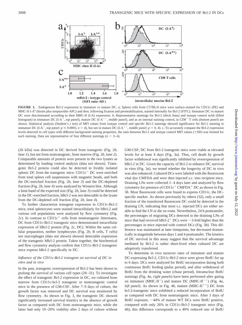
Differential analysis of mRNA from immature and mature DC hasdemonstrated that Bcl-2 expression is rapidly down-regulatedupon induction of DC maturation by an inflammatory stimulus(Ref. 5 and unpublished observations). To investigate whether in-tracellular Bcl-2 protein levels reflect this decrease in transcription,we performed intracellular stainings for murine Bcl-2 (Fig. 1a).Spleen cell suspensions from C57BL/6 mice were stained withmAb specific for CD11c and MHC class II (MHC-II; I-A) as anindicator of DC maturation. Mature DC were identified as CD11c�
MHC-II (I-A)�� cells, whereas immature DC were defined asCD11c� MHC-II (I-A)� (Fig. 1a, rectangular gates). A compari-son of intracellular staining for mouse Bcl-2 in these two popula-tions revealed that mature CD11c� MHC-II (I-A)�� DC expresssignificantly lower Bcl-2 levels than immature CD11c� MHC-II(I-A)� DC (Fig. 1b, top and middle panels; isotype control back-ground excluded). Because the specific Bcl-2 stainings in DC wererelatively weak, we analyzed CD8� T cells within the same spleencell suspensions as a control; CD8� T cells have been shown toexpress high levels of Bcl-2 (23), and they demonstrate low back-ground staining with the isotype control mAb (Fig. 1b, CD8� Tcells). Background (isotype control) staining of DC increased withmaturation (Fig. 1b); to quantify murine Bcl-2 expression levelsand accurately compare the different DC populations, the ratio be-tween the mean fluorescence intensities (MFI) of murine Bcl-2staining and isotype control staining was calculated for each pop-ulation. These “corrected” Bcl-2 expression levels (Fig. 1c) clearlydemonstrate the relative down-regulation of Bcl-2 protein in ma-ture DC as compared with immature DC.
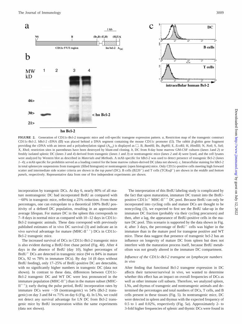
CD11c-Bcl-2 transgene expression
To compensate for the down-regulation of endogenous mouseBcl-2 in DC, we generated mice that express hBcl-2 as a transgeneunder the control of the mouse CD11c promoter. This promoterregion, which regulates the expression of hBcl-2 cDNA in thecontext of a mini exon derived from rabbit �-globin (Ref. 24; Fig.2a), has been used to drive the DC-specific expression of varioustransgenes in vivo (Refs. 18 and 25). To monitor transgene ex-pression in DC from transgenic mice, we prepared DC from bonemarrow progenitors cultured in the presence of GM-CSF (26, 27).After 8 days of culture, when most DC displayed a mature phe-notype as determined by high MHC-II and B7.2 expression levels(data not shown), DC were lysed and submitted to Western blotanalysis (Fig. 2b). The presence of the transgenic hBcl-2 protein
3007The Journal of Immunology
by guest on July 12, 2022http://w
ww
.jimm
unol.org/D
ownloaded from
(26 kDa) was detected in DC derived from transgenic (Fig. 2b,lane 1), but not from nontransgenic, bone marrow (Fig. 2b, lane 2).Comparable amounts of protein were present in the two lysates asdetermined by loading control analysis (data not shown). Trans-genic Bcl-2 protein could also be detected in freshly isolatedsplenic DC from the transgenic mice. CD11c� DC were enrichedfrom total spleen cell suspensions with magnetic beads, and boththe DC-enriched fraction (Fig. 2b, lane 3) and the DC-depletedfraction (Fig. 2b, lane 4) were analyzed by Western blot. Althougha faint band of the expected size (Fig. 2b, lane 3) could be detectedin the DC-enriched fraction, hBcl-2 was not detectable in the lysatefrom the DC-depleted cell fraction (Fig. 2b, lane 4).
To further characterize transgene expression in CD11c-Bcl-2mice, total splenocytes were stained intracellularly for hBcl-2 andvarious cell populations were analyzed by flow cytometry (Fig.2c). In contrast to CD11c� cells from nontransgenic littermates,DC from CD11c-Bcl-2 transgenic mice demonstrated intracellularexpression of hBcl-2 protein (Fig. 2c, DC). Within the same cel-lular preparation, neither lymphocytes (Fig. 2b, B cells, T cells)nor macrophages (data not shown) showed detectable expressionof the transgenic hBcl-2 protein. Taken together, the biochemicaland flow cytometry analysis confirm that CD11c-Bcl-2 transgenicmice express hBcl-2 specifically in DC.
Influence of the CD11c-Bcl-2 transgene on survival of DC invitro and in vivo
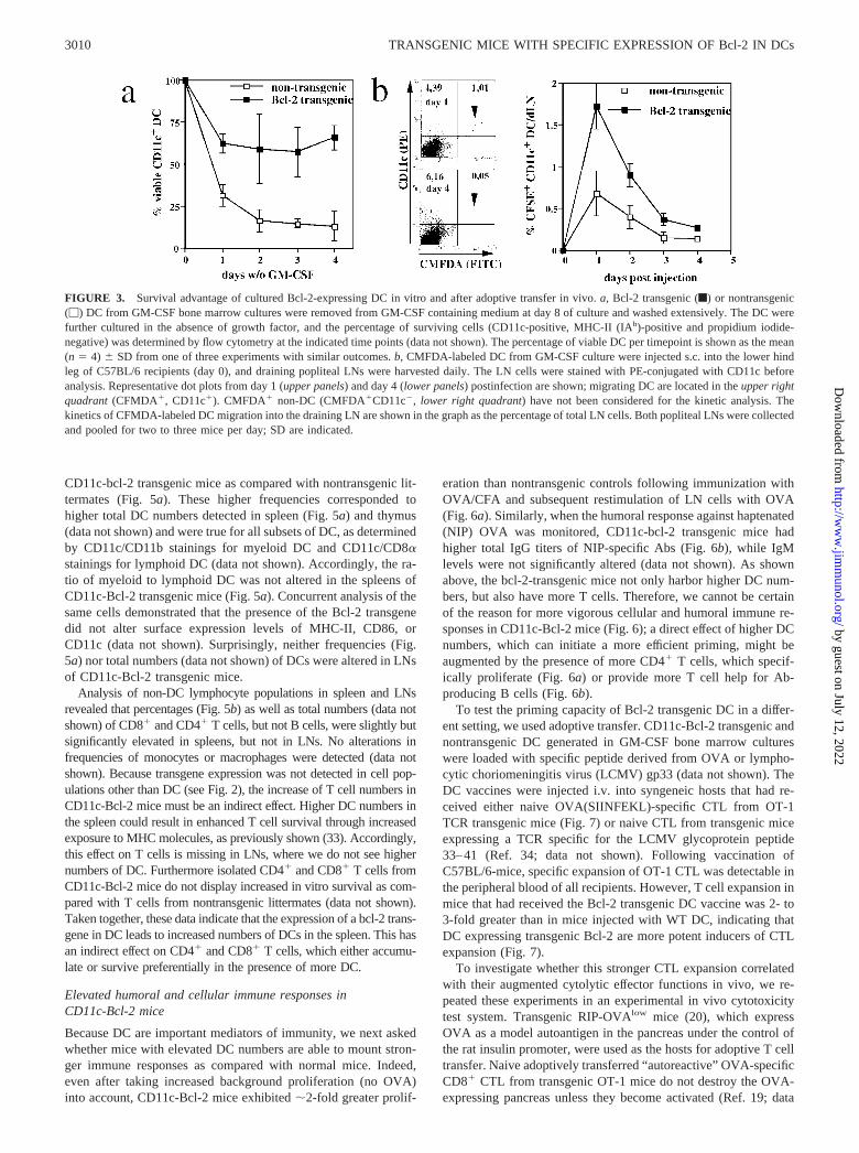
In the past, transgenic overexpression of Bcl-2 has been shown toprolong the survival of various cell types (28–31). To investigatethe effect of transgenic Bcl-2 expression in DC, we cultured bonemarrow from CD11c-bcl-2 transgenic or nontransgenic controlmice in the presence of GM-CSF. After 7–9 days of culture, thegrowth factor was removed and DC survival was monitored byflow cytometry. As shown in Fig. 3, the transgenic DC showedsignificantly increased survival kinetics in the absence of growthfactor as compared with DC from nontransgenic mice, while thelatter had only 10–20% viability after 2 days of culture without
GM-CSF; DC from Bcl-2 transgenic mice were viable at elevatedlevels for at least 4 days (Fig. 3a). Thus, cell death by growthfactor withdrawal was significantly inhibited by overexpression ofhBcl-2 in DC. Given the capacity of Bcl-2 to enhance DC survivalin vitro (Fig. 3a), we tested whether the longevity of DC in vivowas also enhanced. Cultured DCs were labeled with the fluorescentvital dye CMFDA and were then injected s.c. into recipient mice.Draining LNs were collected 1–4 days later and analyzed by flowcytometry for presence of CD11c� CMFDA� DC as shown in Fig.3b. Most fluorescent cells were found to express CD11c, the DC-specific marker. As shown previously by others (32), only a smallfraction of the transferred fluorescent DC could be detected in thedraining LN, indicating that most s.c. injected DCs are either un-able to find the LN or die on route. Nevertheless, 24 h posttransfer,the percentages of migrating DCs detected in the draining LNs ofmice that had received hBcl-2� DCs were �3-fold higher than thepercentages in mice injected with control DCs (Fig. 3b). This dif-ference was maintained at later timepoints, but decreased dramat-ically in magnitude between days 1 and 4 posttransfer. The kineticsof DC survival in this assay suggest that the survival advantagemediated by Bcl-2 is rather short-lived when cultured DC areadoptively transferred.
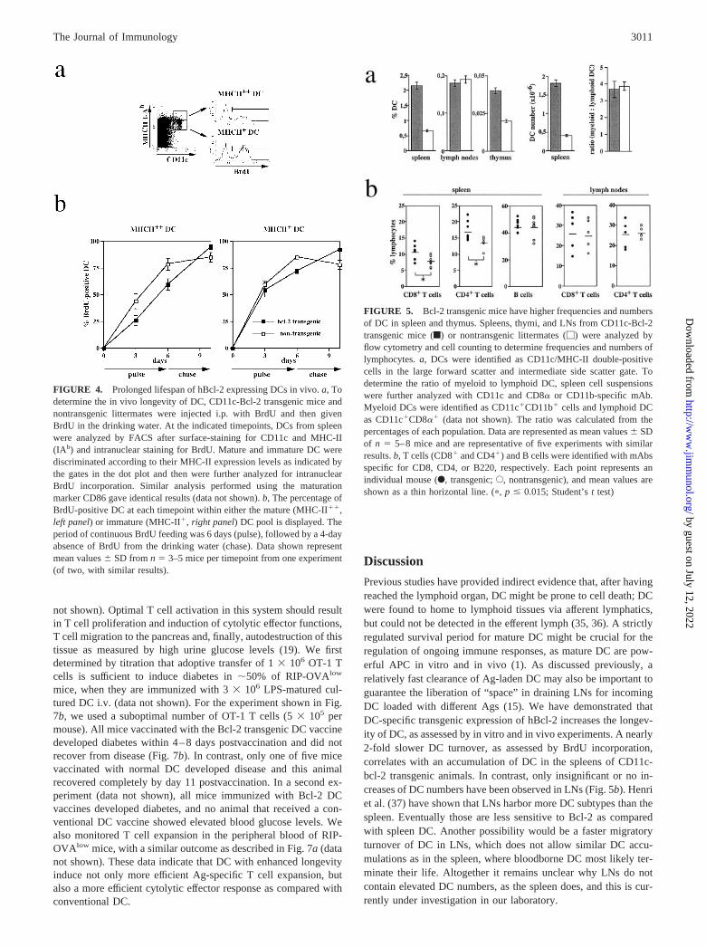
To determine in vivo turnover rates of immature and matureDC-expressing Bcl-2, CD11c-Bcl-2 mice were given BrdU for upto 6 days. DCs were analyzed for BrdU incorporation during bothcontinuous BrdU feeding (pulse period), and after withdrawal ofBrdU from the drinking water (chase period). Intranuclear BrdUstainings (Fig. 4a, right panels) have been performed after gatingon immature (MHC-II�) and mature DC (MHC-II��) (Fig. 4a,left panel). As shown in Fig. 4b, mature (MHC-II��) DC frombcl-2-transgenic mice exhibited a reduced incorporation of BrdUas compared with DC from nontransgenic mice. After 3 days ofBrdU exposure, �44% of mature WT DCs were BrdU positive,compared with only 26% in CD11c-Bcl-2 transgenic mice (Fig.4b); this difference corresponds to a 40% reduced rate of BrdU
FIGURE 1. Endogenous Bcl-2 expression in immature vs mature DC. a, Spleen cells from C57BL/6 mice were surface-stained for CD11c (PE) andMHC-II I-Ab (biotin plus streptavidin-APC) and then, following fixation and permeabilization, stained internally for Bcl-2 (FITC). Immature DC vs matureDC were discriminated according to their MHC-II (I-A) expression. b, Representative stainings for Bcl-2 (thick lines) and isotype control mAb (filledhistogram) in immature DC (I-A�, top panel), mature DC (I-A��, middle panel), and as an internal staining control, in CD8� T cells (bottom panel) areshown. Statistical analysis (Student’s t test) of MFI values from isotype control and specific Bcl-2 stainings showed significance for Bcl-2 staining inimmature DC (I-A�, top panel; p � 0.0005, n � 4), but not in mature DC (I-A��, middle panel; p � 0, 4). c, To accurately compare the Bcl-2 expressionlevels detected in cell types with different background staining properties, the ratio between Bcl-2 and isotype control MFI values (�SD) was formed foreach staining. Data are representative of four different stainings (n � 3–4).
3008 TRANSGENIC MICE WITH SPECIFIC EXPRESSION OF Bcl-2 IN DCs
by guest on July 12, 2022http://w
ww
.jimm
unol.org/D
ownloaded from
incorporation by transgenic DCs. At day 6, nearly 80% of all ma-ture nontransgenic DC had incorporated BrdU as compared with�60% in transgenic mice, reflecting a 25% reduction. From thesepercentages, one can extrapolate to a theoretical 100% BrdU pos-itivity of a defined DC population, resulting in an approximateaverage lifespan. For mature DC in the spleen this corresponds to7–8 days in normal mice as compared with 10–12 days in CD11c-Bcl-2 transgenic animals. These data correspond with previouslypublished estimates of in vivo DC survival (3) and indicate an invivo survival advantage for mature (MHC-II��) DCs in CD11c-Bcl-2 transgenic mice.
The increased survival of DCs in CD11c-Bcl-2 transgenic miceis also evident during a BrdU-free chase period (Fig. 4b). After 4days in the absence of BrdU (day 10), higher percentages ofBrdU� DCs are detected in transgenic mice (94 vs 84% in matureDCs, 92 vs 78% in immature DCs). By day 14 (8 days withoutBrdU feeding), only 17–25% of BrdU-positive DC are detectable,with no significantly higher numbers in transgenic DC (data notshown). In contrast to these data, differences between CD11c-Bcl-2 transgenic DC and WT DC were less pronounced in theimmature population (MHC-II�) than in the mature subset (MHC-II��); early during the pulse period, BrdU incorporation rates byimmature DCs were �59 (nontransgenic) vs 54% (Bcl-2 trans-genic) on day 3 and 84 vs 71% on day 6 (Fig. 4). In LNs, we couldnot detect any survival advantage for LN DC from Bcl-2 trans-genic mice by BrdU incorporation within the same experiments(data not shown).
The interpretation of this BrdU labeling study is complicated bythe fact that upon maturation, immature DC transit into the BrdU-positive CD11c� MHC-II�� DC pool. Because BrdU can only beincorporated into cycling cells and mature DCs are thought to benoncycling (3), we expected to first see the BrdU take-up in theimmature DC fraction (probably via their cycling precursors) andthen, after a lag, the appearance of BrdU-positive cells in the ma-ture DC pool. This scenario is supported by the data shown in Fig.4; after 3 days, the percentage of BrdU� cells was higher in theimmature than in the mature pool for transgene positive and WTmice. These data suggest that presence of transgenic bcl-2 has aninfluence on longevity of mature DC from spleen but does notinterfere with the maturation process itself, because BrdU metab-olism was not greatly altered in the immature DC compartment.
Influence of the CD11c-Bcl-2 transgene on lymphocyte numbersin vivo
After finding that functional Bcl-2 transgene expression in DCaffects their turnover/survival in vivo, we wanted to determinewhether this effect has an impact on overall frequencies of DCs aswell as other immune cell subsets. Therefore, we analyzed spleen,LNs, and thymus of transgenic and nontransgenic animals and de-termined the percentages and total numbers of DCs, T cells, and Bcells present in these tissues (Fig. 5). In nontransgenic mice, DCwere detected in spleen and thymus with the expected frequency of0.5 to 1 and 0.02%, respectively (Fig. 5a). Approximately 2- to3-fold higher frequencies of splenic and thymic DCs were found in
FIGURE 2. Generation of CD11c-Bcl-2 transgenic mice and cell-specific transgene expression pattern. a, Restriction map of the transgenic constructCD11c-Bcl-2. hBcl-2 cDNA (o) was placed behind a DNA segment containing the mouse CD11c promoter (u). The rabbit �-globin gene fragmentproviding the cDNA with an intron and a polyadenylation signal (A(n)) is displayed as �. B, BamHI; Bs, BspHI; E, EcoRI; H, HindIII; N, NotI; S, SstI;X, XhoI; restriction sites in parentheses have been destroyed by blunt-end cloning. b, DC from 8-day bone marrow GM-CSF cultures (lanes 1and 2) orfreshly isolated splenic DC (lanes 3 and 4) derived from transgenic (lanes 1 and 3) or nontransgenic mice (lanes 2 and 4) were lysed, and the cell lysateswere analyzed by Western blot as described in Materials and Methods. A mAb specific for hBcl-2 was used to detect presence of transgenic Bcl-2 (lanes1–4); a mAb specific for prohibitin served as a loading control for the bone marrow culture-derived DC (data not shown). c, Intracellular staining for hBcl-2in total splenocyte suspensions from transgenic (filled histogram) or nontransgenic (open histogram) mice. Only CD11c-positive cells meeting high forwardscatter and intermediate side scatter criteria are shown in the top panel (DC). B cells (B220�) and T cells (TCR���) are shown in the middle and bottompanels, respectively. Representative data from one of five independent experiments are shown.
3009The Journal of Immunology
by guest on July 12, 2022http://w
ww
.jimm
unol.org/D
ownloaded from
CD11c-bcl-2 transgenic mice as compared with nontransgenic lit-termates (Fig. 5a). These higher frequencies corresponded tohigher total DC numbers detected in spleen (Fig. 5a) and thymus(data not shown) and were true for all subsets of DC, as determinedby CD11c/CD11b stainings for myeloid DC and CD11c/CD8�stainings for lymphoid DC (data not shown). Accordingly, the ra-tio of myeloid to lymphoid DC was not altered in the spleens ofCD11c-Bcl-2 transgenic mice (Fig. 5a). Concurrent analysis of thesame cells demonstrated that the presence of the Bcl-2 transgenedid not alter surface expression levels of MHC-II, CD86, orCD11c (data not shown). Surprisingly, neither frequencies (Fig.5a) nor total numbers (data not shown) of DCs were altered in LNsof CD11c-Bcl-2 transgenic mice.
Analysis of non-DC lymphocyte populations in spleen and LNsrevealed that percentages (Fig. 5b) as well as total numbers (data notshown) of CD8� and CD4� T cells, but not B cells, were slightly butsignificantly elevated in spleens, but not in LNs. No alterations infrequencies of monocytes or macrophages were detected (data notshown). Because transgene expression was not detected in cell pop-ulations other than DC (see Fig. 2), the increase of T cell numbers inCD11c-Bcl-2 mice must be an indirect effect. Higher DC numbers inthe spleen could result in enhanced T cell survival through increasedexposure to MHC molecules, as previously shown (33). Accordingly,this effect on T cells is missing in LNs, where we do not see highernumbers of DC. Furthermore isolated CD4� and CD8� T cells fromCD11c-Bcl-2 mice do not display increased in vitro survival as com-pared with T cells from nontransgenic littermates (data not shown).Taken together, these data indicate that the expression of a bcl-2 trans-gene in DC leads to increased numbers of DCs in the spleen. This hasan indirect effect on CD4� and CD8� T cells, which either accumu-late or survive preferentially in the presence of more DC.
Elevated humoral and cellular immune responses inCD11c-Bcl-2 mice
Because DC are important mediators of immunity, we next askedwhether mice with elevated DC numbers are able to mount stron-ger immune responses as compared with normal mice. Indeed,even after taking increased background proliferation (no OVA)into account, CD11c-Bcl-2 mice exhibited �2-fold greater prolif-
eration than nontransgenic controls following immunization withOVA/CFA and subsequent restimulation of LN cells with OVA(Fig. 6a). Similarly, when the humoral response against haptenated(NIP) OVA was monitored, CD11c-bcl-2 transgenic mice hadhigher total IgG titers of NIP-specific Abs (Fig. 6b), while IgMlevels were not significantly altered (data not shown). As shownabove, the bcl-2-transgenic mice not only harbor higher DC num-bers, but also have more T cells. Therefore, we cannot be certainof the reason for more vigorous cellular and humoral immune re-sponses in CD11c-Bcl-2 mice (Fig. 6); a direct effect of higher DCnumbers, which can initiate a more efficient priming, might beaugmented by the presence of more CD4� T cells, which specif-ically proliferate (Fig. 6a) or provide more T cell help for Ab-producing B cells (Fig. 6b).
To test the priming capacity of Bcl-2 transgenic DC in a differ-ent setting, we used adoptive transfer. CD11c-Bcl-2 transgenic andnontransgenic DC generated in GM-CSF bone marrow cultureswere loaded with specific peptide derived from OVA or lympho-cytic choriomeningitis virus (LCMV) gp33 (data not shown). TheDC vaccines were injected i.v. into syngeneic hosts that had re-ceived either naive OVA(SIINFEKL)-specific CTL from OT-1TCR transgenic mice (Fig. 7) or naive CTL from transgenic miceexpressing a TCR specific for the LCMV glycoprotein peptide33–41 (Ref. 34; data not shown). Following vaccination ofC57BL/6-mice, specific expansion of OT-1 CTL was detectable inthe peripheral blood of all recipients. However, T cell expansion inmice that had received the Bcl-2 transgenic DC vaccine was 2- to3-fold greater than in mice injected with WT DC, indicating thatDC expressing transgenic Bcl-2 are more potent inducers of CTLexpansion (Fig. 7).
To investigate whether this stronger CTL expansion correlatedwith their augmented cytolytic effector functions in vivo, we re-peated these experiments in an experimental in vivo cytotoxicitytest system. Transgenic RIP-OVAlow mice (20), which expressOVA as a model autoantigen in the pancreas under the control ofthe rat insulin promoter, were used as the hosts for adoptive T celltransfer. Naive adoptively transferred “autoreactive” OVA-specificCD8� CTL from transgenic OT-1 mice do not destroy the OVA-expressing pancreas unless they become activated (Ref. 19; data
FIGURE 3. Survival advantage of cultured Bcl-2-expressing DC in vitro and after adoptive transfer in vivo. a, Bcl-2 transgenic (f) or nontransgenic(�) DC from GM-CSF bone marrow cultures were removed from GM-CSF containing medium at day 8 of culture and washed extensively. The DC werefurther cultured in the absence of growth factor, and the percentage of surviving cells (CD11c-positive, MHC-II (IAb)-positive and propidium iodide-negative) was determined by flow cytometry at the indicated time points (data not shown). The percentage of viable DC per timepoint is shown as the mean(n � 4) � SD from one of three experiments with similar outcomes. b, CMFDA-labeled DC from GM-CSF culture were injected s.c. into the lower hindleg of C57BL/6 recipients (day 0), and draining popliteal LNs were harvested daily. The LN cells were stained with PE-conjugated with CD11c beforeanalysis. Representative dot plots from day 1 (upper panels) and day 4 (lower panels) postinfection are shown; migrating DC are located in the upper rightquadrant (CFMDA�, CD11c�). CMFDA� non-DC (CMFDA�CD11c�, lower right quadrant) have not been considered for the kinetic analysis. Thekinetics of CFMDA-labeled DC migration into the draining LN are shown in the graph as the percentage of total LN cells. Both popliteal LNs were collectedand pooled for two to three mice per day; SD are indicated.
3010 TRANSGENIC MICE WITH SPECIFIC EXPRESSION OF Bcl-2 IN DCs
by guest on July 12, 2022http://w
ww
.jimm
unol.org/D
ownloaded from
not shown). Optimal T cell activation in this system should resultin T cell proliferation and induction of cytolytic effector functions,T cell migration to the pancreas and, finally, autodestruction of thistissue as measured by high urine glucose levels (19). We firstdetermined by titration that adoptive transfer of 1 � 106 OT-1 Tcells is sufficient to induce diabetes in �50% of RIP-OVAlow
mice, when they are immunized with 3 � 106 LPS-matured cul-tured DC i.v. (data not shown). For the experiment shown in Fig.7b, we used a suboptimal number of OT-1 T cells (5 � 105 permouse). All mice vaccinated with the Bcl-2 transgenic DC vaccinedeveloped diabetes within 4–8 days postvaccination and did notrecover from disease (Fig. 7b). In contrast, only one of five micevaccinated with normal DC developed disease and this animalrecovered completely by day 11 postvaccination. In a second ex-periment (data not shown), all mice immunized with Bcl-2 DCvaccines developed diabetes, and no animal that received a con-ventional DC vaccine showed elevated blood glucose levels. Wealso monitored T cell expansion in the peripheral blood of RIP-OVAlow mice, with a similar outcome as described in Fig. 7a (datanot shown). These data indicate that DC with enhanced longevityinduce not only more efficient Ag-specific T cell expansion, butalso a more efficient cytolytic effector response as compared withconventional DC.
Discussion
Previous studies have provided indirect evidence that, after havingreached the lymphoid organ, DC might be prone to cell death; DCwere found to home to lymphoid tissues via afferent lymphatics,but could not be detected in the efferent lymph (35, 36). A strictlyregulated survival period for mature DC might be crucial for theregulation of ongoing immune responses, as mature DC are pow-erful APC in vitro and in vivo (1). As discussed previously, arelatively fast clearance of Ag-laden DC may also be important toguarantee the liberation of “space” in draining LNs for incomingDC loaded with different Ags (15). We have demonstrated thatDC-specific transgenic expression of hBcl-2 increases the longev-ity of DC, as assessed by in vitro and in vivo experiments. A nearly2-fold slower DC turnover, as assessed by BrdU incorporation,correlates with an accumulation of DC in the spleens of CD11c-bcl-2 transgenic animals. In contrast, only insignificant or no in-creases of DC numbers have been observed in LNs (Fig. 5b). Henriet al. (37) have shown that LNs harbor more DC subtypes than thespleen. Eventually those are less sensitive to Bcl-2 as comparedwith spleen DC. Another possibility would be a faster migratoryturnover of DC in LNs, which does not allow similar DC accu-mulations as in the spleen, where bloodborne DC most likely ter-minate their life. Altogether it remains unclear why LNs do notcontain elevated DC numbers, as the spleen does, and this is cur-rently under investigation in our laboratory.
FIGURE 4. Prolonged lifespan of hBcl-2 expressing DCs in vivo. a, Todetermine the in vivo longevity of DC, CD11c-Bcl-2 transgenic mice andnontransgenic littermates were injected i.p. with BrdU and then givenBrdU in the drinking water. At the indicated timepoints, DCs from spleenwere analyzed by FACS after surface-staining for CD11c and MHC-II(IAb) and intranuclear staining for BrdU. Mature and immature DC werediscriminated according to their MHC-II expression levels as indicated bythe gates in the dot plot and then were further analyzed for intranuclearBrdU incorporation. Similar analysis performed using the maturationmarker CD86 gave identical results (data not shown). b, The percentage ofBrdU-positive DC at each timepoint within either the mature (MHC-II��,left panel) or immature (MHC-II�, right panel) DC pool is displayed. Theperiod of continuous BrdU feeding was 6 days (pulse), followed by a 4-dayabsence of BrdU from the drinking water (chase). Data shown representmean values � SD from n � 3–5 mice per timepoint from one experiment(of two, with similar results).
FIGURE 5. Bcl-2 transgenic mice have higher frequencies and numbersof DC in spleen and thymus. Spleens, thymi, and LNs from CD11c-Bcl-2transgenic mice (f) or nontransgenic littermates (�) were analyzed byflow cytometry and cell counting to determine frequencies and numbers oflymphocytes. a, DCs were identified as CD11c/MHC-II double-positivecells in the large forward scatter and intermediate side scatter gate. Todetermine the ratio of myeloid to lymphoid DC, spleen cell suspensionswere further analyzed with CD11c and CD8� or CD11b-specific mAb.Myeloid DCs were identified as CD11c�CD11b� cells and lymphoid DCas CD11c�CD8�� (data not shown). The ratio was calculated from thepercentages of each population. Data are represented as mean values � SDof n � 5–8 mice and are representative of five experiments with similarresults. b, T cells (CD8� and CD4�) and B cells were identified with mAbsspecific for CD8, CD4, or B220, respectively. Each point represents anindividual mouse (F, transgenic; E, nontransgenic), and mean values areshown as a thin horizontal line. (�, p � 0.015; Student’s t test)
3011The Journal of Immunology
by guest on July 12, 2022http://w
ww
.jimm
unol.org/D
ownloaded from
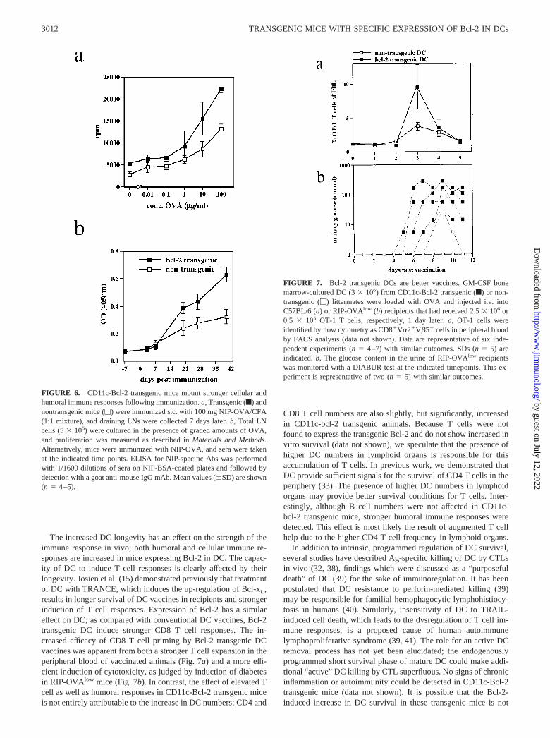
The increased DC longevity has an effect on the strength of theimmune response in vivo; both humoral and cellular immune re-sponses are increased in mice expressing Bcl-2 in DC. The capac-ity of DC to induce T cell responses is clearly affected by theirlongevity. Josien et al. (15) demonstrated previously that treatmentof DC with TRANCE, which induces the up-regulation of Bcl-xL,results in longer survival of DC vaccines in recipients and strongerinduction of T cell responses. Expression of Bcl-2 has a similareffect on DC; as compared with conventional DC vaccines, Bcl-2transgenic DC induce stronger CD8 T cell responses. The in-creased efficacy of CD8 T cell priming by Bcl-2 transgenic DCvaccines was apparent from both a stronger T cell expansion in theperipheral blood of vaccinated animals (Fig. 7a) and a more effi-cient induction of cytotoxicity, as judged by induction of diabetesin RIP-OVAlow mice (Fig. 7b). In contrast, the effect of elevated Tcell as well as humoral responses in CD11c-Bcl-2 transgenic miceis not entirely attributable to the increase in DC numbers; CD4 and
CD8 T cell numbers are also slightly, but significantly, increasedin CD11c-bcl-2 transgenic animals. Because T cells were notfound to express the transgenic Bcl-2 and do not show increased invitro survival (data not shown), we speculate that the presence ofhigher DC numbers in lymphoid organs is responsible for thisaccumulation of T cells. In previous work, we demonstrated thatDC provide sufficient signals for the survival of CD4 T cells in theperiphery (33). The presence of higher DC numbers in lymphoidorgans may provide better survival conditions for T cells. Inter-estingly, although B cell numbers were not affected in CD11c-bcl-2 transgenic mice, stronger humoral immune responses weredetected. This effect is most likely the result of augmented T cellhelp due to the higher CD4 T cell frequency in lymphoid organs.
In addition to intrinsic, programmed regulation of DC survival,several studies have described Ag-specific killing of DC by CTLsin vivo (32, 38), findings which were discussed as a “purposefuldeath” of DC (39) for the sake of immunoregulation. It has beenpostulated that DC resistance to perforin-mediated killing (39)may be responsible for familial hemophagocytic lymphohistiocy-tosis in humans (40). Similarly, insensitivity of DC to TRAIL-induced cell death, which leads to the dysregulation of T cell im-mune responses, is a proposed cause of human autoimmunelymphoproliferative syndrome (39, 41). The role for an active DCremoval process has not yet been elucidated; the endogenouslyprogrammed short survival phase of mature DC could make addi-tional “active” DC killing by CTL superfluous. No signs of chronicinflammation or autoimmunity could be detected in CD11c-Bcl-2transgenic mice (data not shown). It is possible that the Bcl-2-induced increase in DC survival in these transgenic mice is not
FIGURE 6. CD11c-Bcl-2 transgenic mice mount stronger cellular andhumoral immune responses following immunization. a, Transgenic (f) andnontransgenic mice (�) were immunized s.c. with 100 mg NIP-OVA/CFA(1:1 mixture), and draining LNs were collected 7 days later. b, Total LNcells (5 � 105) were cultured in the presence of graded amounts of OVA,and proliferation was measured as described in Materials and Methods.Alternatively, mice were immunized with NIP-OVA, and sera were takenat the indicated time points. ELISA for NIP-specific Abs was performedwith 1/1600 dilutions of sera on NIP-BSA-coated plates and followed bydetection with a goat anti-mouse IgG mAb. Mean values (�SD) are shown(n � 4–5).
FIGURE 7. Bcl-2 transgenic DCs are better vaccines. GM-CSF bonemarrow-cultured DC (3 � 106) from CD11c-Bcl-2 transgenic (f) or non-transgenic (�) littermates were loaded with OVA and injected i.v. intoC57BL/6 (a) or RIP-OVAlow (b) recipients that had received 2.5 � 106 or0.5 � 105 OT-1 T cells, respectively, 1 day later. a, OT-1 cells wereidentified by flow cytometry as CD8�V�2�V�5� cells in peripheral bloodby FACS analysis (data not shown). Data are representative of six inde-pendent experiments (n � 4–7) with similar outcomes. SDs (n � 5) areindicated. b, The glucose content in the urine of RIP-OVAlow recipientswas monitored with a DIABUR test at the indicated timepoints. This ex-periment is representative of two (n � 5) with similar outcomes.
3012 TRANSGENIC MICE WITH SPECIFIC EXPRESSION OF Bcl-2 IN DCs
by guest on July 12, 2022http://w
ww
.jimm
unol.org/D
ownloaded from
dramatic enough to induce an autoimmune status. Alternatively,the hypothesis that DC longevity plays a central role in immuno-regulation may be incorrect.
DC survival is controlled by more than one mechanism. Up-regulation of the Bcl-xL, for example, is induced by inflammatorysignals such as TNF-� and LPS, which have been reported toprotect DC from apoptosis via the Fas-mediated pathway (42), aswell as anti-CD40 and TRANCE. Treatment of DC with TRANCEhas an even greater effect on survival than the induced expressionof Bcl-2; while increased survival of CD11c-bcl-2 transgenic DCvaccines could be detected in the draining LNs until, but not laterthan, 24 h postimmunization (Fig. 3b), TRANCE-treated DC vac-cines were reported to survive much longer posttransfer (15). Stim-ulation of human DC by CD154, IL-12, or IL-15 has also beenreported to result in increased Bcl-xL levels and resistance of the DCto apoptosis (43). In contrast, neither Bcl-xL nor Bcl-2 seem to beinvolved in CpG-DNA mediated DC survival, where signaling viaToll-like receptor 9 on murine DCs leads to the activation of phospha-ditylinositide-3�-OH kinase and, most likely, the inhibition ofcaspase-3 activation (44). Long-term DC cultures from TNFR1-defi-cient mice also demonstrate protection from apoptosis that is inde-pendent from Bcl-2 (45). Therefore, signaling via Bcl-2 is only onemechanism among several dedicated to DC survival. Our results showthat enhanced transgenic expression of Bcl-2 in mature DC in vivoregulates their survival and longevity. Therefore, we conclude thatBcl-2 down-regulation is a means to restrict DC longevity in thesteady state, i.e., in the absence of inflammatory stimuli.
AcknowledgmentsWe thank A. Strasser for the hBcl-2 cDNA, U. Muller (Basel Institute forImmunology) for making of the transgenic mice, M. Riedinger (Basel In-stitute for Immunology, Basel, Switzerland) for cloning of the transgenicconstruct, H. Pircher (Freiburg, Germany) for performing the T cell trans-fer experiments with LCMV-specific T cells, and A. Rolink (Basel, Swit-zerland) for the kind gift of NIP-OVA and NIP-BSA. We also thank A.Kamath (Compton, U.K.) and K. Shortman (Melbourne, Australia) for ad-vice with the BrdU staining of DC and C. Kurts (Aachen) for providing uswith RIP-OVAlow mice. We thank K. Kerksiek for critically reading themanuscript and helpful suggestions and G. Riethmuller for general support.
References1. Banchereau, J., and R. M. Steinman. 1998. Dendritic cells and the control of
immunity. Nature 392:245.2. Winzler, C., P. Rovere, M. Rescigno, F. Granucci, G. Penna, L. Adorini,
V. S. Zimmermann, J. Davoust, and P. Ricciardi-Castagnoli. 1997. Maturationstages of mouse dendritic cells in growth factor-dependent long-term cultures.J. Exp. Med. 185:317.
3. Kamath, A. T., J. Pooley, M. A. O’Keeffe, D. Vremec, Y. Zhan, A. M. Lew, A.D’Amico, L. Wu, D. F. Tough, and K. Shortman. 2000. The development, mat-uration, and turnover rate of mouse spleen dendritic cell populations. J. Immunol.165:6762.
4. Granucci, F., C. Vizzardelli, E. Virzi, M. Rescigno, and P. Ricciardi-Castagnoli.2001. Transcriptional reprogramming of dendritic cells by differentiation stimuli.Eur. J. Immunol. 31:2539.
5. Granucci, F., C. Vizzardelli, N. Pavelka, S. Feau, M. Persico, E. Virzi,M. Rescigno, G. Moro, and P. Ricciardi-Castagnoli. 2001. Inducible IL-2 pro-duction by dendritic cells revealed by global gene expression analysis. Nat. Im-munol. 2:882.
6. Chinnaiyan, A. M., K. Orth, K. O’Rourke, H. Duan, G. G. Poirier, andV. M. Dixit. 1996. Molecular ordering of the cell death pathway: Bcl-2 andBcl-xL function upstream of the CED-3-like apoptotic proteases. J. Biol. Chem.271:4573.
7. Minn, A. J., P. Velez, S. L. Schendel, H. Liang, S. W. Muchmore, S. W. Fesik,M. Fill, and C. B. Thompson. 1997. Bcl-xL forms an ion channel in synthetic lipidmembranes. Nature 385:353.
8. McDonnell, T. J., N. Deane, F. M. Platt, G. Nunez, U. Jaeger, J. P. McKearn, andS. J. Korsmeyer. 1989. bcl-2-immunoglobulin transgenic mice demonstrate ex-tended B cell survival and follicular lymphoproliferation. Cell 57:79.
9. Strasser, A. 1995. Life and death during lymphocyte development and function:evidence for two distinct killing mechanisms. Curr. Opin. Immunol. 7:228.
10. Korsmeyer, S. J. 1995. Regulators of cell death. Trends Genet. 11:101.
11. Cory, S. 1995. Regulation of lymphocyte survival by the bcl-2 gene family. Annu.Rev. Immunol. 13:513.
12. Bjorck, P., J. Banchereau, and L. Flores-Romo. 1997. CD40 ligation counteractsFas-induced apoptosis of human dendritic cells. Int. Immunol. 9:365.
13. Wang, Z., J. G. Karras, R. G. Howard, and T. L. Rothstein. 1995. Induction ofBcl-x by CD40 engagement rescues sIg-induced apoptosis in murine B cells.J. Immunol. 155:3722.
14. Wong, B. R., R. Josien, S. Y. Lee, B. Sauter, H. L. Li, R. M. Steinman, andY. Choi. 1997. TRANCE (tumor necrosis factor (TNF)-related activation-in-duced cytokine), a new TNF family member predominantly expressed in T cells,is a dendritic cell-specific survival factor. J. Exp. Med. 186:2075.
15. Josien, R., H. L. Li, E. Ingulli, S. Sarma, B. R. Wong, M. Vologodskaia,R. M. Steinman, and Y. Choi. 2000. TRANCE, a tumor necrosis factor familymember, enhances the longevity and adjuvant properties of dendritic cells in vivo.J. Exp. Med. 191:495.
16. Cleary, M. L., S. D. Smith, and J. Sklar. 1986. Cloning and structural analysis ofcDNAs for bcl-2 and a hybrid bcl-2/immunoglobulin transcript resulting from thet(14;18) translocation. Cell 47:19.
17. Vaux, D. L., S. Cory, and J. M. Adams. 1988. bcl-2 gene promotes haemopoieticcell survival and cooperates with c-myc to immortalize pre-B cells. Nature 335:440.
18. Brocker, T., M. Riedinger, and K. Karjalainen. 1997. Targeted expression ofmajor histocompatibility complex (MHC) class II molecules demonstrates thatdendritic cells can induce negative but not positive selection of thymocytes invivo. J. Exp. Med. 185:541.
19. Kurts, C., H. Kosaka, F. R. Carbone, J. F. Miller, and W. R. Heath. 1997. ClassI-restricted cross-presentation of exogenous self-antigens leads to deletion of au-toreactive CD8� T cells. J. Exp. Med. 186:239.
20. Blanas, E., F. R. Carbone, J. Allison, J. Miller, and W. R. Heath. 1996. Inductionof autoimmune diabetes by oral administration of autoantigen. Science 274:1707.
21. Terashima, M., K. M. Kim, T. Adachi, P. J. Nielsen, M. Reth, G. Kohler, andM. C. Lamers. 1994. The IgM antigen receptor of B lymphocytes is associatedwith prohibitin and a prohibitin-related protein. EMBO J. 13:3782.
22. Laemmli, U. K. 1970. Cleavage of structural proteins during the assembly of thehead of bacteriophage T4. Nature 227:680.
23. Zhang, X., H. Fujii, H. Kishimoto, E. LeRoy, C. D. Surh, and J. Sprent. 2002.Aging leads to disturbed homeostasis of memory phenotype CD8� cells. J. Exp.Med. 195:283.
24. Kouskoff, V., H. J. Fehling, M. Lemeur, C. Benoist, and D. Mathis. 1993. Avector driving the expression of foreign cDNAs in the MHC class II- positivecells of transgenic mice. J. Immunol. Methods 166:287.
25. Brocker, T., A. Gulbranson-Judge, S. Flynn, M. Riedinger, C. Raykundalia, andP. Lane. 1999. CD4 T cell traffic control: in vivo evidence that ligation of OX40on CD4 T cells by OX40-ligand expressed on dendritic cells leads to the accu-mulation of CD4 T cells in B follicles. Eur. J. Immunol. 29:1610.
26. Inaba, K., M. Inaba, N. Romani, H. Aya, M. Deguchi, S. Ikehara, S. Muramatsu,and R. M. Steinman. 1992. Generation of large numbers of dendritic cells frommouse bone marrow cultures supplemented with granulocyte/macrophagecolony-stimulating factor. J. Exp. Med. 176:1693.
27. Inaba, K., M. Inaba, M. Naito, and R. M. Steinman. 1993. Dendritic cell pro-genitors phagocytose particulates, including bacillus Calmette-Guerin organisms,and sensitize mice to mycobacterial antigens in vivo. J. Exp. Med. 178:479.
28. Strasser, A., A. W. Harris, and S. Cory. 1991. bcl-2 transgene inhibits T cell deathand perturbs thymic self-censorship. Cell 67:889.
29. Strasser, A., A. W. Harris, H. von Boehmer, and S. Cory. 1994. Positive andnegative selection of T cells in T-cell receptor transgenic mice expressing a bcl-2transgene. Proc. Natl. Acad. Sci. USA 91:1376.
30. Lang, J., B. Arnold, G. Hammerling, A. W. Harris, S. Korsmeyer, D. Russell,A. Strasser, and D. Nemazee. 1997. Enforced Bcl-2 expression inhibits antigen-mediated clonal elimination of peripheral B cells in an antigen dose-dependentmanner and promotes receptor editing in autoreactive, immature B cells. J. Exp.Med. 186:1513.
31. Smith, K. G., A. Light, L. A. O’Reilly, S. M. Ang, A. Strasser, and D. Tarlinton.2000. bcl-2 transgene expression inhibits apoptosis in the germinal center andreveals differences in the selection of memory B cells and bone marrow antibody-forming cells. J. Exp. Med. 191:475.
32. Ingulli, E., A. Mondino, A. Khoruts, and M. K. Jenkins. 1997. In vivo detectionof dendritic cell antigen presentation to CD4� T cells. J. Exp. Med. 185:2133.
33. Brocker, T. 1997. Survival of mature CD4 T lymphocytes is dependent on majorhistocompatibility complex class II-expressing dendritic cells. J. Exp. Med. 186:1223.
34. Kyburz, D., P. Aichele, D. E. Speiser, H. Hengartner, R. M. Zinkernagel, andH. Pircher. 1993. T cell immunity after a viral infection versus T cell toleranceinduced by soluble viral peptides. Eur. J. Immunol. 23:1956.
35. Fossum, S. 1988. Lymph-borne dendritic leucocytes do not recirculate, but enter thelymph node paracortex to become interdigitating cells. Scand. J. Immunol. 27:97.
3013The Journal of Immunology
by guest on July 12, 2022http://w
ww
.jimm
unol.org/D
ownloaded from
36. Pugh, C. W., G. G. MacPherson, and H. W. Steer. 1983. Characterization ofnonlymphoid cells derived from rat peripheral lymph. J. Exp. Med. 157:1758.
37. Henri, S., D. Vremec, A. Kamath, J. Waithman, S. Williams, C. Benoist,K. Burnham, S. Saeland, E. Handman, and K. Shortman. 2001. The dendritic cellpopulations of mouse lymph nodes. J. Immunol. 167:741.
38. Hermans, I. F., D. S. Ritchie, J. Yang, J. M. Roberts, and F. Ronchese. 2000.CD8� T cell-dependent elimination of dendritic cells in vivo limits the inductionof antitumor immunity. J. Immunol. 164:3095.
39. Ronchese, F., and I. F. Hermans. 2001. Killing of dendritic cells: a life cut shortor a purposeful death? J. Exp. Med. 194:F23.
40. Stepp, S. E., R. Dufourcq-Lagelouse, F. Le Deist, S. Bhawan, S. Certain,P. A. Mathew, J. I. Henter, M. Bennett, A. Fischer, G. de Saint Basile, andV. Kumar. 1999. Perforin gene defects in familial hemophagocytic lymphohis-tiocytosis. Science 286:1957.
41. Wang, J., L. Zheng, A. Lobito, F. K. Chan, J. Dale, M. Sneller, X. Yao,J. M. Puck, S. E. Straus, and M. J. Lenardo. 1999. Inherited human caspase 10
mutations underlie defective lymphocyte and dendritic cell apoptosis in autoim-mune lymphoproliferative syndrome type II. Cell 98:47.
42. Lundqvist, A., T. Nagata, R. Kiessling, and P. Pisa. 2002. Mature dendritic cellsare protected from Fas/CD95-mediated apoptosis by upregulation of Bcl-xL. Can-cer Immunol. Immunother. 51:139.
43. Pirtskhalaishvili, G., G. V. Shurin, C. Esche, Q. Cai, R. R. Salup, S. N. Bykovs-kaia, M. T. Lotze, and M. R. Shurin. 2000. Cytokine-mediated protection ofhuman dendritic cells from prostate cancer-induced apoptosis is regulated by theBcl-2 family of proteins. Br. J. Cancer 83:506.
44. Park, Y., S. W. Lee, and Y. C. Sung. 2002. Cutting edge: CpG DNA inhibitsdendritic cell apoptosis by up-regulating cellular inhibitor of apoptosis proteinsthrough the phosphatidylinositide-3�-OH kinase pathway. J. Immunol. 168:5.
45. Funk, J. O., H. Walczak, C. Voigtlander, S. Berchtold, T. Baumeister, P. Rauch,S. Rossner, A. Steinkasserer, G. Schuler, and M. B. Lutz. 2000. Cutting edge:resistance to apoptosis and continuous proliferation of dendritic cells deficient forTNF receptor-1. J. Immunol. 165:4792.
3014 TRANSGENIC MICE WITH SPECIFIC EXPRESSION OF Bcl-2 IN DCs
by guest on July 12, 2022http://w
ww
.jimm
unol.org/D
ownloaded from
Copyright © 2022 FDOKUMEN