Essential role for the BH3-only protein Bim but redundant roles for Bax, Bcl-2, and Bcl-w in the...
31
doi:10.1182/blood-2002-07-2132 Prepublished online November 14, 2002; Andreas Villunger, Clare Scott, Philippe Bouillet and Andreas Strasser Bcl-2, and Bcl-w in the control of granulocyte survival Essential role for the BH3-only protein Bim, but redundant roles for Bax, (972 articles) Phagocytes (1086 articles) Gene Expression (746 articles) Apoptosis Articles on similar topics can be found in the following Blood collections http://bloodjournal.hematologylibrary.org/site/misc/rights.xhtml#repub_requests Information about reproducing this article in parts or in its entirety may be found online at: http://bloodjournal.hematologylibrary.org/site/misc/rights.xhtml#reprints Information about ordering reprints may be found online at: http://bloodjournal.hematologylibrary.org/site/subscriptions/index.xhtml Information about subscriptions and ASH membership may be found online at: digital object identifier (DOIs) and date of initial publication. the indexed by PubMed from initial publication. Citations to Advance online articles must include final publication). Advance online articles are citable and establish publication priority; they are appeared in the paper journal (edited, typeset versions may be posted when available prior to Advance online articles have been peer reviewed and accepted for publication but have not yet Copyright 2011 by The American Society of Hematology; all rights reserved. 20036. the American Society of Hematology, 2021 L St, NW, Suite 900, Washington DC Blood (print ISSN 0006-4971, online ISSN 1528-0020), is published weekly by For personal use only. by guest on January 1, 2012. bloodjournal.hematologylibrary.org From
Transcript of Essential role for the BH3-only protein Bim but redundant roles for Bax, Bcl-2, and Bcl-w in the...

doi:10.1182/blood-2002-07-2132Prepublished online November 14, 2002;
Andreas Villunger, Clare Scott, Philippe Bouillet and Andreas Strasser Bcl-2, and Bcl-w in the control of granulocyte survivalEssential role for the BH3-only protein Bim, but redundant roles for Bax,
(972 articles)Phagocytes � (1086 articles)Gene Expression �
(746 articles)Apoptosis �Articles on similar topics can be found in the following Blood collections
http://bloodjournal.hematologylibrary.org/site/misc/rights.xhtml#repub_requestsInformation about reproducing this article in parts or in its entirety may be found online at:
http://bloodjournal.hematologylibrary.org/site/misc/rights.xhtml#reprintsInformation about ordering reprints may be found online at:
http://bloodjournal.hematologylibrary.org/site/subscriptions/index.xhtmlInformation about subscriptions and ASH membership may be found online at:
digital object identifier (DOIs) and date of initial publication. theindexed by PubMed from initial publication. Citations to Advance online articles must include
final publication). Advance online articles are citable and establish publication priority; they areappeared in the paper journal (edited, typeset versions may be posted when available prior to Advance online articles have been peer reviewed and accepted for publication but have not yet
Copyright 2011 by The American Society of Hematology; all rights reserved.20036.the American Society of Hematology, 2021 L St, NW, Suite 900, Washington DC Blood (print ISSN 0006-4971, online ISSN 1528-0020), is published weekly by
For personal use only. by guest on January 1, 2012. bloodjournal.hematologylibrary.orgFrom

Phagocytes 1
Essential Role for the BH3-only Protein Bim, but Redundant Roles for Bax,
Bcl-2 and Bcl-w in the Control of Granulocyte Survival
Andreas Villunger1,3, Clare Scott1, Philippe Bouillet1 and Andreas Strasser2
The Walter and Eliza Hall Institute of Medical Research, Melbourne, Australia
2corresponding author: Andreas Strasser The Walter and Eliza Hall Institute of Medical Research Post Office Royal Melbourne Hospital Vic, 3050, Australia Phone +61-3-9345-2624 Fax: +61-3-9347-0852 Email: [email protected] Support: This work was supported by fellowships from the Human Frontiers Science Program
(HFSP), the Leukemia Research Foundation, the NHMRC and RACP and grants from the
NHMRC (Canberra, Reg. Key 973002), the Dr Josef Steiner Cancer Research Foundation (Bern),
the National Cancer Institute (CA43540 and CA80188) and the Leukemia and Lymphoma
Society of America (Grant 7015-02).
Running title: Regulation of granulocyte survival by the Bcl-2 family
Word count: summary 228, manuscript 5012
Key words: apoptosis, Bcl-2 family, BH3-only proteins, Fas (APO-1/CD95), knock-out and
transgenic mice
3current address: Institute for Pathophysiology, Innsbruck University Medical School, Fritz-Pregl
Strasse 3, A-6020 Innsbruck, Austria
Copyright (c) 2002 American Society of Hematology
Blood First Edition Paper, prepublished online November 14, 2002; DOI 10.1182/blood-2002-07-2132 For personal use only. by guest on January 1, 2012. bloodjournal.hematologylibrary.orgFrom

Phagocytes 2
Abstract
Programmed cell death of granulocytes is one of the mechanisms that limit inflammatory
responses. Members of the Bcl-2 protein family are essential regulators of apoptosis induced by
growth factor withdrawal or cytotoxic stress. We have used gene-targeted and transgenic mice to
investigate the roles of the pro-survival molecules Bcl-2 and Bcl-w and their pro-apoptotic
relatives Bax and Bim in spontaneous and stress-induced apoptosis of granulocytes from bone
marrow or the peritoneum. Bim-deficiency, like Bcl-2 overexpression, rendered granulocytes
resistant to cytokine withdrawal and cytotoxic drugs but absence of Bax alone had no protective
effect. Loss of Bcl-2 or Bcl-w did not increase the sensitivity of granulocytes to any of these
apoptotic stimuli but Bcl-2 was essential for the in vitro survival of myeloid progenitors under
conditions of cytokine-withdrawal where cell death was mediated, in part, by Bim.
G-CSF, a key survival factor for granulocytes, enhanced viability of cells lacking bcl-2, bcl-w,
bax or bim, indicating that none of these genes alone is the essential target of this cytokine’s pro-
survival function. Expression analysis of pro-apoptotic Bcl-2 family members in granulocytes
revealed that the BH3-only protein Bmf is induced upon cytokine-withdrawal. These results
indicate that the BH3-only protein Bim and possibly also Bmf are critical initiators of
spontaneous and drug-induced apoptosis of granulocytes whereas Bcl-2, Bcl-w and Bax act in a
redundant manner in regulating granulocyte survival and death, respectively.
For personal use only. by guest on January 1, 2012. bloodjournal.hematologylibrary.orgFrom
Phagocytes 3
Introduction
In the bone marrow extra-cellular regulatory factors, such as G-CSF and GM-CSF, promote
production and differentiation of myeloid progenitors into mature granulocytes or monocytes 1.
Mature granulocytes enter the blood stream where they have a short lifespan unless they are
stimulated by inflammatory cytokines 2. Activated granulocytes have the ability to ingest bacteria
and infiltrate tissues. Inflammatory responses are kept in check, at least in part, by the apoptotic
death of granulocytes, followed by their engulfment by macrophages to avoid release of
histotoxic substances 3. Life and death of granulocytes must be tightly regulated because
excessive granulocyte apoptosis increases susceptibility to bacterial infections 2,4, whereas
prolonged granulocyte survival is associated with inflammatory diseases 5 and may predispose to
leukemogenesis 6.
Considerable insight into the control of programmed cell death (apoptosis) has emerged from
genetic and biochemical analyses in mammals and C. elegans 7. The effector phase of apoptosis
requires aspartate specific cysteine proteases, termed caspases. Caspases are synthesized as
zymogens with low enzymatic activity, and to become functional, must be cleaved at caspase
recognition sites, either by adaptor protein-induced auto-catalysis or by already active caspases 7.
Mammals have two distinct apoptosis signaling pathways for activating caspases. One is initiated
when ligation of death receptors (members of the tumor necrosis factor receptor (TNF-R) family
with an intracellular death domain) causes formation of a death inducing signaling complex
(DISC) in which FADD adaptor proteins promote oligomerization and autocatalytic activation of
caspase-8 7. The other pathway is triggered by growth factor deprivation or various stress
conditions and is regulated by the interplay of pro- and anti-apoptotic members of the Bcl-2
family 7.
The mechanisms that control the granulocyte lifespan are still unclear, but Bcl-2-regulated
apoptosis signaling and death receptor signaling have both been implicated in this process. The
expression and function of several cell death regulators can be regulated by pro-inflammatory
cytokines, such as G-CSF, GM-CSF, or by bacterial products (e.g. LPS) 8-10. Bcl-2 mRNA and
For personal use only. by guest on January 1, 2012. bloodjournal.hematologylibrary.orgFrom

Phagocytes 4
protein were reported to be expressed at low levels in mouse granulocytes 6,11 but seem to be
barely detectable in granulocytes form human blood 8,12. We and others have previously shown
that expression of a bcl-2 transgene protects granulocytes from spontaneous and stress-induced
cell death in culture but does not render them resistant to Fas ligand (FasL) 11,13. The pro-
survival Bcl-2 homologue Bcl-w is expressed at readily detectable levels in myeloid cells 11,14.
When overexpressed, Bcl-w has similar effects to Bcl-2 and protects hematopoietic cell lines
against apoptosis induced by cytokine withdrawal or drug treatment 15. Analysis of mice lacking
bcl-w demonstrated that it is required for spermatogenesis but dispensable for the development of
other cell types including hematopoietic ones 16. Expression of the Bcl-2 family members Mcl-1
and A1 can be increased in myeloid cells by stimulation with G-CSF or GM-CSF 8,17.
Granulocytes from mice lacking one of the genes for A1, A1a, undergo abnormally accelerated
spontaneous death in culture 18. In contrast, mice lacking the pro-apoptotic BH3-only Bcl-2
family member Bim have an approximately two-fold increase in granulocytes 19, and
granulocytes from patients with certain inflammatory diseases were reported to have abnormally
low levels of Bax 5. Bim is sequestered to microtubules in healthy cells and released in reponse to
certain apoptotic stimuli, such as the anit-cancer drug taxol allowing its binding to and
inactivation of Bcl-2 like molecules at inner membranes and mitochondria 20. Transfection
experiments indicate that Bim acts upstream of Bax and Bak 21.
In order to define the role of individual Bcl-2 family members in granulocyte survival we
investigated the impact of absence of the pro-survival molecules bcl-2 and bcl-w or loss of their
pro-apoptotic relatives bax and bim on granulocyte survival. As a control, we also studied the
effects caused by Bcl-2 over-expression or absence of the death receptor Fas/APO-1/CD95. Our
results indicate that Bim and possibly other BH3-only proteins play an essential role in
programmed death of granulocytes whereas Bcl-2, Bcl-w and Bax have redundant functions in
the control of granulocyte apoptosis.
For personal use only. by guest on January 1, 2012. bloodjournal.hematologylibrary.orgFrom

Phagocytes 5
Methods
Mice
The generation and genotyping of the bim-deficient mice (266/266del) 19, bcl-w-deficient mice 16
and vav-bcl-2-69 transgenic mice 22, expressing a human bcl-2 c-DNA under control of the vav
promoter at high levels in all hematopoetic cell types, have been described. Fas-deficient lpr
mutant mice were provided by our Institute’s breeding facility. The bax+/- mice 23 were purchased
from The Jackson Laboratory and bcl-2+/- mice were a kind gift of Dr. D. Loh 24. All mouse
strains were on an inbred C57BL/6J genetic background or had been backcrossed for more than 8
generations onto this background. Since bcl-2-/- mice do not survive beyond 6 weeks after birth
due to severe polycystic kidney disease 24,25, we generated chimeric mice by hematopoietic
reconstitution of lethally irradiated (2 x 550 rad, 3 h interval) C57BL/6-Ly5.1 mice with 2 x 10⁶
fetal liver cells from E14 bcl-2-/- or (as a control) from wt embryos produced from intercrosses of
bcl-2+/- (C57BL/6-Ly5.2) animals. Granulocytes from chimeric animals were analysed 10-16
weeks after reconstitution.
Cell culture and reagents
Granulocytes were isolated from the bone marrow of untreated mice or were collected by lavage
from the peritoneal cavity of mice that had been injected i.p. 3 h earlier with 2 mL of a 0.5%
casein/PBS solution. Purified granulocytes were cultured in the high glucose version of
Dulbecco's modified Eagle's (DME) medium supplemented with 13 µM folic acid, 250 µM
L-asparagine, 50 µM 2-mercaptoethanol and 10% FCS (TRACE). Recombinant human G-CSF
(AMRAD) was used at 100 U/mL. FLAG epitope-tagged FasL (Alexis) was used at 1-100 ng/mL
together with cross-linking M2 anti-FLAG antibody (Sigma) at 1 µg/mL. Ionomycin (Sigma) was
used at 200 ng/mL, VP-16 (David Bull Laboratories) was used at 10 or 20 µg/mL and Taxol
(Sigma/Aldrich) at 1 µM.
For personal use only. by guest on January 1, 2012. bloodjournal.hematologylibrary.orgFrom

Phagocytes 6
Immunoblotting
Western blotting was performed as previously described 11. Membranes were probed with rabbit
polyclonal anti-Bim and anti-Bad antibodies (Stressgen) or Bmf (Alexis), mouse monoclonal
antibodies to Bid (Transduction Laboratories) and goat-polyclonal anti-Noxa (St. Cruz, SC19). A
rabbit anti-Puma antiserum was generated by immunizing New Zealand White rabbits with a
human GST-PUMA fusion protein. Membranes were probed with rabbit polyclonal anti-Bim
antibodies (Stressgen) or Bmf 26 (Alexis) or mouse monoclonal antibodies to Bid (Transduction
Laboratories). Horseradish peroxidase (HRP)-conjugated sheep-anti rabbit or rabbit anti-mouse Ig
antibodies (Silenus) served as secondary reagents and the enhanced chemiluminescence (ECL)
system was used for detection. To demonstrate equal protein loading, membranes were probed
with a mouse monoclonal antibody to heat-shock protein Hsp70 (a gift from Dr. R. Anderson,
Peter MacCallum Cancer Institute, Melbourne). Membranes were stripped prior reprobing in
100mM 2-mercaptoethanol, 2% SDS, 62.5mM Tris-HCl pH6.7 for 30min at 55oC.
Cell sorting and immunofluorescence analysis
Granulocytes were isolated by staining cell suspensions from bone marrow or peritoneal lavage
with FITC- or Cy5-conjugated rat anti-Gr-1 mAb RB6-8C5 (2 µg/mL) in PBS/10%FCS for 30 min
on ice and cell sorting on a MoFlo (Cytomation). Gating on the basis of forward (FSC) and side
(SSC) light scatter and staining with the vital dye propidium iodide (1 µg/mL) was used to exclude
dead cells. Only Gr-1high cells with large FSC/SSC profile were collected to minimize
contamination with macrophages. In the case of bcl-2-/- granulocytes, which were isolated from
chimeric mice generated by reconstitution of lethally irradiated C57BL/6-Ly5.1 mice with fetal
liver cells from C57BL/6-Ly5.2 E14 bcl-2-/- embryos, recipient-derived cells were excluded by
staining with FITC-conjugated rat anti-Ly5.1 mAb (A201.7). Alternatively, granulocytes were
sorted by excluding cells stained with FITC labeled RA3-6B2 anti-B220, T24.3.2.1 anti-Thy-1,
53.9.2 anti-Ter 119 and anti-F4/80. Sorted granulocytes were > 98% positive for Gr-1 and Mac-1
For personal use only. by guest on January 1, 2012. bloodjournal.hematologylibrary.orgFrom

Phagocytes 7
but negative for the B cell marker B220, the T cell marker Thy-1, the erythroid cell marker TER-
119 and the macrophage marker F4/80.
Absolute numbers of T cells, B cells, macrophages and granulocytes in blood, bone marrow or
spleen were determined by cell counting and flow cytometric analysis of cells stained with the
following surface marker-specific monoclonal antibodies: RA3-6B2 anti-B220, T24.3.2.1 anti-
Thy-1, RB6-8C5 anti-Gr-1 or MI/70 anti-Mac-1. In the indicated mice reconstituted with bcl-2-/- or
wt stem cells mice (see above), host and donor derived cells were distinguished by staining with a
FITC-conjugated mAb recognizing Ly5.2 (5.430-15.2), and generally > 95% of the myeloid cells
were donor derived in these chimeric mice.
Cell death assays
The percentage of viable cells in culture was determined by staining with 2 µg/mL propidium
iodide plus FITC-coupled annexin-V and analyzing the samples in a FACScan (Becton
Dickinson). Alternatively, cells were stained with trypan blue (0.1% in PBS) and analyzed in a
hemocytometer.
RT-PCR analysis
Total RNA was isolated from granulocytes derived from the bone marrow of wt mice
immediately after cell sorting or after culture in the presence or absence of G-CSF (100 U/mL)
using Trizol reagents (GibcoBRL) according to the manufacturer’s recommendations. An
amount of 250 ng of total RNA was transcribed into cDNA using AMV-reverse transcriptase and
oligo dT primers (Roche). Five-fold dilutions of cDNA templates (1:1, 1:5, 1:25) were amplified
by PCR using Taq polymerase and oligos specific for the BH3-only genes bim, bid, bad, blk,
hrk/DP5, bmf, puma/bbc3 and noxa. Cycling conditions were chosen as follows: 94oC/2min, 5
cycles 94oC/15 sec., 55oC/30sec., 72oC/30sec., followed by 25 cycles 94oC/15sec., 55oC+0.2oC
per cycle/30 sec., 72oC/30sec. and a final synthesis step for 10min at 72oC. Identity of PCR
For personal use only. by guest on January 1, 2012. bloodjournal.hematologylibrary.orgFrom

Phagocytes 8
products was confirmed by Southern blotting and sequence specific internal oligonucleotides. blk
fwd. 5’ATGTCGGAGGCGAGACTTATG3’, rev. 5’CCCTGCCCCAGCTGCACTTCAC TG3’,
int. 5’CACTCAG GCGCCAGGAGTCAAGAC3’; hrk/dp5 fwd. 5’CCGGA CCGAGCAAC
AGGTTAGC3’, rev. 5’GCTTCGGCCCAGTCCCCTCTAC3’, int. 5’CTCTCCCCTGC
CACCCTAGACATTACG3’; bim fwd. 5’TTGCCATCGTC GCCGTCAC3’, rev.
5’CAGTTGTAAGATAACCATTTGAGGGTGG3’, int. 5’CAGCTCCTGTGCAATCCGTATC
TC3’; bid fwd. 5’ATGGACTCTGAGG3’, rev. 5’TTAGTCCATCTCGTTTCTAACCAAG3’,
int. 5’CAGTGTGGGCTGGATGTTG TGG3’; bad fwd. 5’TTCC AGATCCCAGAGTTTG3’,
rev. 5’GGAGATCACTG GGAGGGGGTGG3’; int. 5’GCG CCTCCATGATGACTGTTGG3’;
noxa fwd. 5’CGTCGGAACGCGCCAGTGAACCC3’, rev. 5’TCCTTCCTGGGAGGTCCC
TTCTTGC3’, int. 5’AACCCGGGTGCCAGCAGACTTG3’, puma/bbc3 fwd. 5’CCTCAGCC
CTCCCTGTCACCAG3’ rev. 5’CCGCCGCTCGTACTGCGCGTTG3’, int. 5’CGGCGGATGG
CGGACGACCTC3’; bmf fwd. 5’CCCTTGGGGAGCA GCCCCCTG3’ rev. 5’CAAGAC
AGTATCTGTCCTCCCAGA C3’, int. 5’CATACGCAACAACACCAGCAG3’. PCR products
were separated on 2% agarose gels in TBE buffer, transferred onto nylon membranes over night
in 0.4M NaOH. Membranes were washed in 2xSSC for 5 min, pre-hybridised at 42oC in
CHURCH buffer and hybridized over night with 2x10⁶ dpm/mL internal oligonucleotide labelled
with 32P γATP and T4 polynucleotide kinase. Filters were washed in 40mM Na2HPO4/1% SDS at
42oC for 20 min, dried and exposed to X-ray film (Kodak).
Progenitor cell assays
Assays for bone marrow progenitor cells in bone marrow from mice reconstituted with fetal liver
cells from wt or bcl-2-/- mice or wt, bim-/- and vav-bcl-2 transgenic mice were performed by
culturing 2.5 x 10⁴ nucleated cells in 0.3% semisolid agar (1 mL) in Dulbecco's modified Eagle's
medium (DMEM) supplemented with 20% newborn calf serum (Hyclone). Cells were plated in
triplicate or quadruplicate and stimulated by a combination of mSCF (produced by expression in
For personal use only. by guest on January 1, 2012. bloodjournal.hematologylibrary.orgFrom

Phagocytes 9
Pichia pastoris) at 100 ng/mL and mIL-3 (Peprotech, Rocky Hill NJ) at 2500 U/mL. Cytokines
were added at the time of plating or after a delay of 12, 18, 24, 48 or 72 hours. After an additional
7 days of incubation at 37O C, colonies were counted under a microscope, fixed, stained and
identified as described before 22. The number of colonies that grew when cytokines were added at
the time of plating was considered 100% to allow calculation of relative colony survival of the
cells that had been starved of cytokines for variable amounts of time.
Statistical analysis
Statistical analysis was performed by using the Fischer protected least significant difference
(PLSD) test and Stat-view 4.1 software. P-values of < 0.04 were considered to indicate
statistically significant differences.
Results
Bim is an essential inducer of programmed death of granulocytes
Granulocytes were isolated by immunofluorescent staining with surface marker-specific
antibodies and flow cytometric sorting from the bone marrow of wt mice, chimeric mice lacking
Bcl-2 in the hematopoietic system (see Materials and Methods), mice deficient for Bcl-w, Bax,
the BH3-only protein Bim, functional Fas/APO-1/CD95 (lpr mutant mice) or from animals
expressing a bcl-2 transgene in all hematopoietic cells 22. Sorted cells were cultured in simple
medium or treated with the cytotoxic drugs VP-16 (10µg/mL), taxol (1µM), the calcium
ionophore ionomycin (250ng/mL) or stimulated with Fas ligand (FasL) at 100ng/mL. After 24,
48 or 72 h in culture, cell survival was determined by staining with Annexin V-FITC plus
propidium iodide followed by flow cytometric analysis. Absence of the BH3-only protein Bim or
Bcl-2 over-expression protected granulocytes from spontaneous or drug-induced cell death (Figs.
1a,c). For example, after 72 hours in culture on average only 19% wt granulocytes remained
alive, whereas 41% bim-/- and 69% of vav-bcl-2 trangenic cells had survived (Fig. 1a). In
contrast, loss of Bim or Bcl-2 over-expression had no impact on FasL-induced killing (Fig. 1c),
For personal use only. by guest on January 1, 2012. bloodjournal.hematologylibrary.orgFrom

Phagocytes 10
whereas Fas-deficient lpr mutant granulocytes were resistant to FasL but died normally in
response to cytokine withdrawal or treatment with cytotoxic drugs and ionomycin (Figs.1b, d).
Absence of the pro-survival proteins Bcl-2, Bcl-w or its pro-apoptotic relative Bax did not
increase the sensitivity of granulocytes to any of these death stimuli (Figs. 1b, d).
Recruitment to a site of inflammation has been reported to alter the responsiveness of
granulocytes to certain apoptotic stimuli 27,28. We therefore challenged wt, bcl-2-/- hematopoietic
chimeric animals, bcl-w-/-, bax-/-, bim-/- and vav-bcl-2 transgenic mice by intraperitoneal injection
of casein and 3 hours later harvested their peritoneal fluid to purify the mobilized granulocytes by
immunofluorescent staining and cell sorting. Sorted cells were cultured in simple medium or
treated with the cytotoxic drug VP-16 (20µg/mL) or FasL (100ng/mL). Mobilized wt
granulocytes underwent spontaneous death in culture more rapidly than bone marrow derived wt
granulocytes (compare Fig. 1a with Fig. 1e). Chemotherapeutic drug treatment or Fas activation
therefore could only marginally accelerate apoptosis of mobilized granulocytes in culture. As
shown for the bone marrow-derived granulocytes (Fig 1a), absence of Bim or Bcl-2 over-
expression delayed spontaneous as well as drug-induced cell death, whereas Bax-deficiency had
no protective effect (Figs. 1e,g,i). After 24 hours culture in simple medium, only about 20% of wt
granulocytes survived, but on average 59% bim-/- and 69% of the vav-bcl-2 transgenic cells
retained viability (Fig 1e). Loss of Bim or Bcl-2 over-expression had no impact on FasL-induced
killing (Fig. 1g). Mobilized granulocytes lacking Bcl-2 or Bcl-w did not differ from wt cells in
their responsiveness to apoptotic stimuli (Figs. 1f,h,j), whereas these granulocytes from lpr
mutant mice were resistant to FasL but otherwise indistinguishable from wt cells in their response
to all other apoptotic stimuli (Figs. 1f,h,j).
For personal use only. by guest on January 1, 2012. bloodjournal.hematologylibrary.orgFrom

Phagocytes 11
F ig ure 1 Villun ger et a l.
h
10 0
10
10 0
100h 24 h 48 h 72 h 0h 24 h 48 h 72 h
a b
lprbcl-2-/-
wt
bcl-w-/-
wtbim-/-vav-bcl-2bax-/-
c d80
60
40
20
0
80
60
40
20
0un tre ated VP -16 Tax ol ion om y cinFasL un tre ated VP -16 Tax ol ion om y cinFasL
wt
bim-/-vav-bcl-2
bax-/- lpr
bcl-2-/-wt
bcl-w-/-
48h48h
1
10
10 0
0h 16 h 24 h 36 h
e
wtbim-/-vav-bcl-2bax-/-
8h1
10
10 0
lprbcl-2-/-
wt
bcl-w-/-
f
0h 16 h 24 h 36 h8hg
8h80
60
40
20
0
10 0
u n tre ated F asL VP -16i j
u n tre ated F asL VP -16
16h
10
20
30
0
lpr
bcl-2-/-wt
bcl-w-/-
wt
bim-/-vav-bcl-2
bax-/-
8h
u ntre ated FasL VP -16
80
60
40
20
0
10 0
lpr
bcl-2-/-wt
bcl-w-/-
wt
bim-/-vav-bcl-2
bax-/-
u ntre ated F asL VP -16
16h80
60
40
20
0
10 0
Figure 1: Bim-deficiency and bcl-2 transgene expression delay spontaneous, chemotherapeutic drug- and stress-induced apoptosis of granulocytes. Granulocytes from bone marrow (a-d) of wt, vav-bcl-2 transgenic, bim-/-, bax-/-, bcl-w-/-, lpr mice and from bcl-2-/- reconstituted mice (see Materials and Methods) were sorted by flow cytometry and cultured in simple medium. (a,b) Survival under conditions of cytokine-withdrawal was determined after 24, 48 and 72h by staining with Annexin V plus propidium iodide and flow cytometric analysis. (c,d) Granulocytes of the indicated genotypes were either cultured in simple
For personal use only. by guest on January 1, 2012. bloodjournal.hematologylibrary.orgFrom

Phagocytes 12
medium, the presence of FasL (100 ng/mL) multimerized with M2 anti-FLAG mAb (1 µg/mL), the cytotoxic drugs VP-16 (10 µg/mL), Taxol (1 µM) or the calc ium ionophore ionomycin (200 ng/mL). Survival was assessed after 48h incubation as mentioned in (a). Data shown represent arithmetic means + SD of 4 independent experiments. Analysis was performed in duplicate on 4-8 animals of each genotype. Analysis of mobilized granulocytes from the peritoneal cavity (e-f). Mice of the genotypes indicated in (a) were injected i.p. with 0.5% casein in PBS (2mL). Peritoneal exudate cells were harvested by lavage 3h later and granulocytes were sorted by flow cytometry as indicated in (a). (e,f) Survival under conditions of cytokine-withdrawal was determined after 16, 24 and 48h as described in (a). (g,h) Granulocytes of the indicated genotypes were cultured in simple medium or treated with FasL (100 ng/mL) multimerized with M2 anti-FLAG mAb (1 µg/mL) or the cytotoxic drug VP-16 (20 µg/mL). Survival was assessed after 16h incubation as described in (a). Data shown represent arithmetic means + SD of 2-3 independent experiments performed in duplicate on 2-5 animals of each genotype.
Bcl-2 influences the sensitivity of early myeloid progenitors to cytokine-withdrawal
The role of Bcl-2 in determining the lifespan of cells from different hematopoietic lineages in
vivo was examined by comparing the numbers of lymphoid and myeloid cells between lethally
irradiated mice that had been reconstituted with wt or bcl-2-/- fetal liver-derived stem cells. In
agreement with previous findings 29, mice reconstituted with bcl-2-/- stem cells had considerably
lower numbers of B and T cells in bone marrow, peripheral blood and spleen compared to
animals reconstituted with wt stem cells (Figs. 2a,b and data not shown). The bcl-2-/-
reconstituted mice (analyzed 8-16 weeks after reconstitution, >95% donor-derived myeloid cells)
also had abnormally low numbers of macrophages in spleen (p.0006) and peripheral blood
(p.0001) but roughly normal numbers of granulocytes (Figs. 2c,d).
We also investigated the influence of Bcl-2-deficiency on the survival of early myeloid
progenitors under conditions of cytokine deprivation and compared it to the effect caused by
Bim-deficiency or expression of a bcl-2 transgene. Bone marrow derived myeloid progenitors
from normal mice, animals lacking Bcl-2, Bim, or fromvav-bcl-2 transgenic mice were grown in
soft agar. Stem cell factor (SCF) and interleukin-3 (IL-3), cytokines that promote proliferation
and differentiation of myeloid progenitors, were added either at the time of plating or after a
delay of 12, 18, 24, 48 and 72h. When cultured from the start of the experiment in the presence of
cytokines, bone marrow cells from bcl-2-/- reconstituted mice produced significantly fewer
For personal use only. by guest on January 1, 2012. bloodjournal.hematologylibrary.orgFrom

Phagocytes 13
myeloid colonies compared to bone marrow cells from wild-type reconstituted mice (bcl-2-/- 35+9
vs. wt 58+8 colonies/25000 nucleated bone marrow cells; p.0006). Moreover, when cytokine
addition was delayed, even by a short time, only few or no colonies arose from bcl-2 deficient
bone marrow whereas wild type progenitors could still form significant numbers of colonies. For
example, growth factor deprivation for 12 hours killed more than 90% of bcl-2-/- colony forming
cells but only 25% of the wt cells (Fig. 2e). As previously described 22, expression of a bcl-2
transgene rendered colony forming cells refractory to cytokine withdrawal and absence of Bim
had a smaller but reproducible protective effect (Fig. 2f). Staining of colonies with hematoxylin
after fixation and examination by light microscopy revealed that the relative frequencies of
granulocyte, granulocyte/macrophage, macrophage, blast and megakaryocytic colonies were
similar in cultures of bone marrow from wt, bim-/-as well as bcl-2-/- or wt chimeric mice (data not
shown).
For personal use only. by guest on January 1, 2012. bloodjournal.hematologylibrary.orgFrom
Phagocytes 14
0
.2
.4
.6
.8
1
1.2
1.4
1.6
0
1
2
3
4
5
6
7
8
9
Gr-1+
Gr-1-/Mac-1
+
B220+ 0
20
40
60
80
100
120
140
160
0
2
4
6
8
10
12
14
16
Thy1 +B220+Thy1 +
Gr-1 + Gr-1 -/Mac-1 +
F ig u re 2
a b
c d
Villunger et a l.
1 2 18 2 4 48 7 20
100
1 0
1
wtwt recbcl-2-/- rec
delay of cytokine addition (h)
wtvav-bcl-2bim-/-
delay of cytokine addition (h)
100
1 0
1
e
f
0 722 4 48
bcl-2-/- recwt rec
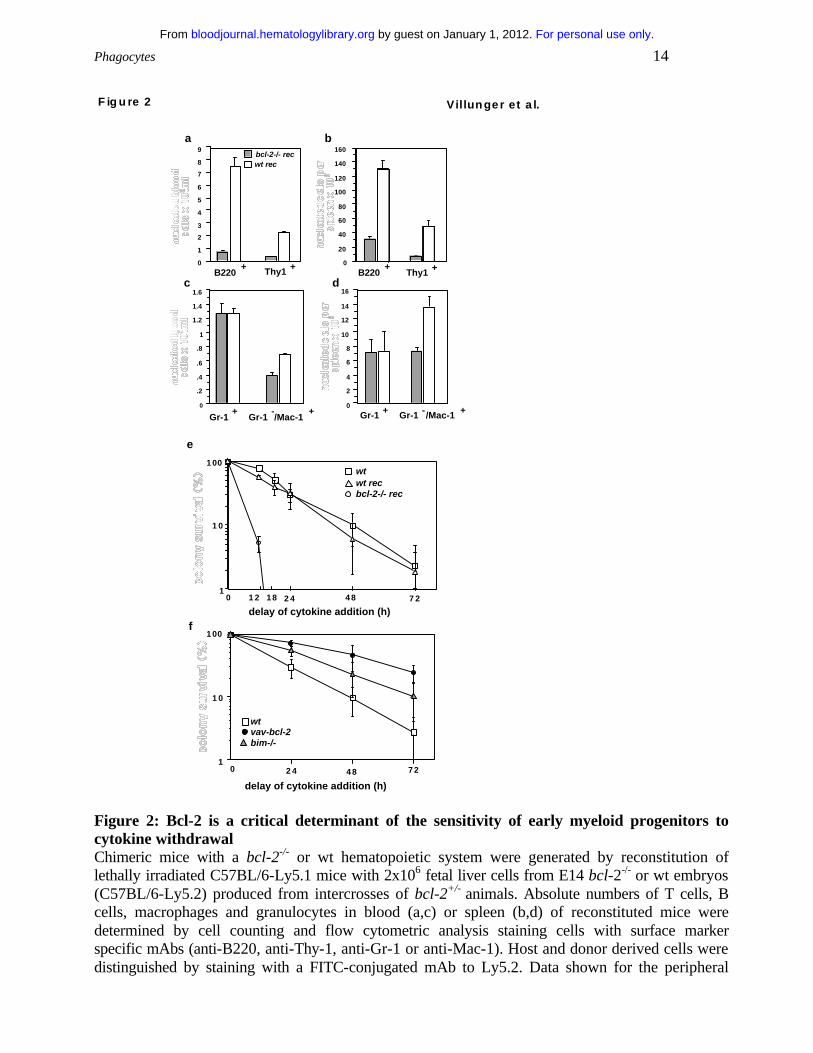
Figure 2: Bcl-2 is a critical determinant of the sensitivity of early myeloid progenitors to cytokine withdrawal Chimeric mice with a bcl-2-/- or wt hematopoietic system were generated by reconstitution of lethally irradiated C57BL/6-Ly5.1 mice with 2x10⁶ fetal liver cells from E14 bcl-2-/- or wt embryos (C57BL/6-Ly5.2) produced from intercrosses of bcl-2+/- animals. Absolute numbers of T cells, B cells, macrophages and granulocytes in blood (a,c) or spleen (b,d) of reconstituted mice were determined by cell counting and flow cytometric analysis staining cells with surface marker specific mAbs (anti-B220, anti-Thy-1, anti-Gr-1 or anti-Mac-1). Host and donor derived cells were distinguished by staining with a FITC-conjugated mAb to Ly5.2. Data shown for the peripheral
For personal use only. by guest on January 1, 2012. bloodjournal.hematologylibrary.orgFrom

Phagocytes 15
blood represent arithmetic means + SE of 9 bcl2-/- reconstituted and 5 wt reconstituted animals that were analysed 10 weeks after fetal liver stem cell transplantation. Data shown for the spleen represent arithmetic means + SE of 6 bcl2-/- and 4 wt reconstituted animals analysed 10-16 weeks after reconstitution. (e,f) Survival analysis of bone marrow derived myeloid progenitors. Cells (2.5x10⁴) from bone marrow of (e) wt mice, lethally irradiated wt mice reconstituted with either wt or bcl-2-/- fetal liver stem cells and from bone marrow of (f) wt, bim-/- and bcl-2 transgenic mice were cultured in 0.3% agar in triplicate or quadruplicate and stimulated with mSCF (100 ng/mL) and mIL-3 (2500 U/mL). Cytokines were added at the time of plating or after a delay of 12, 18, 24, 48 or 72 h. After 7 days of stimulation with cytokines at 37oC, colonies were counted using a dissection microscope. Data shown represent arithmetic means + SD of 2-3 independent experiments performed using 4-6 animals of each genotype.
Expression of the BH3-only protein Bmf increases in cultured granulocytes
The effector molecules of the anti-apoptotic response mediated by G-CSF in granulocytes are not
clearly defined. We analyzed whether G-CSF receptor stimulation could modulate the expression
of BH3-only proteins in granulocytes and tested whether loss of Bcl-2, Bcl-w, Bax or Bim or
over-expression of Bcl-2 had an effect on the ability of G-CSF to promote granulocyte survival in
culture. Granulocytes from the bone marrow or mobilized granulocytes from the peritoneal cavity
of casein injected wt mice or chimeric mice reconstituted with a bcl-2-/- hemopoietic system, vav-
bcl-2 transgenic mice or from animals lacking Bim, Bax or Bcl-w were cultured in the presence
or absence of G-CSF for 24 h or 72 h and cell survival was assessed by propidium iodide staining
with or without addition of Annexin V and flow cytometric analysis. Both methods gave
comparable results and data of both types of experiments have therefore been pooled (Figs. 3a,b).
Granulocytes lacking Bcl-2, Bcl-w or Bax responded to G-CSF like normal cells and survival of
bone marrow derived bim-/- granulocytes, which are partially protected from cytokine withdrawal,
could still be augmented by G-CSF treatment (p.0114)(Figs. 3a,b). These results demonstrate that
none of these molecules alone is the key target for the pro-survival effect of G-CSF. Culturing of
bone marrow derived granulocytes expressing a bcl-2 transgene with G-CSF did not provide any
additional survival benefit over a period of seven days (Fig. 3c) demonstrating that Bcl-2 can
efficiently compensate for G-CSF receptor signaling.
For personal use only. by guest on January 1, 2012. bloodjournal.hematologylibrary.orgFrom

Phagocytes 16
bo ne m arrow
un treated G-C SF
periton eu m
24h
un treated G-C SF
wt
bax-/-
bcl-2-/-
bcl-w-/-
a
b
bim-/-
vav-bcl-2 tg
F ig u re 3 Villun ger e t a l.
10
100
13d 7d
c
5d0d
wt
wt + G-CSF
vav-bcl-2 tg
vav-bcl-2 tg+ G-CSF
bo ne m arrow
72h
0
100
80
60
40
20
100
80
60
40
20
0
Figure 3: Influence of G-CSF on granulocyte survival Granulocytes from mice of the indicated genotypes were isolated from (a) bone marrow or (b) the peritoneal cavity after injection with casein. Granulocytes were cultured in the absence or presence of rhG-CSF (100 U/mL). Cell survival was assessed after 72h (bone marrow-derived granulocytes) or 24h (mobilized peritoneal granulocytes) by staining with propidium iodide plus or minus Annexin V staining and flow cytometric analysis. Both methods gave comparable results and data were therefore pooled. Bars represent means of + SD 3-6 independent experiments performed in duplicate on 4-8 animals of each genotype.
Bim-deficient granulocytes were partially protected from cytokine withdrawal-induced apoptosis
in culture and bone marrow derived bim-/- granulocytes still responded with enhanced survival to
For personal use only. by guest on January 1, 2012. bloodjournal.hematologylibrary.orgFrom

Phagocytes 17
G-CSF (Fig. 3a). Expression of a bcl-2 transgene protected granulocytes from cytokine
withdrawal-induced death more potently than loss of Bim (Fig. 1a). Thus, G-CSF may confer its
pro-survival effect in part via regulation of Bim expression or function and perhaps also through
inhibitory effects on other pro-apoptotic Bcl-2 family members. We therefore analyzed
expression of BH3-only genes at the mRNA and protein level in bone marrow derived as well as
mobilized peritoneal granulocytes that were freshly isolated or cultured in the presence or
absence of G-CSF. Semi-quantitative RT-PCR and Southern blotting demonstrated mRNA
expression for the BH3-only genes bad, bid, bim (primers were designed to amplify all four
known BH3-containing splice variants), blk, bmf, noxa and PUMA/bbc3 but not hrk/DP5, which
has previously been reported to be expressed exclusively in neuronal cells 30.
Expression levels of the mRNA of these genes did not change significantly over time, whether
cells were cultured in the presence or absence of G-CSF (Fig. 4a,b). Moreover the basal
expression of these genes did not differ markedly between resting and activated granulocytes
(Fig. 4b). The bim mRNA levels appeared to be increased under conditions of growth-factor
deprivation in mobilized but not in bone marrow-derived granulocytes (Fig. 4b). This was not
reflected by an increase in Bim protein level (data not shown), but interestingly, loss of Bim
protects activated granulocytes from spontaneous death more potently than bone marrow derived
resting granulocytes (Fig. 1a vs. 1e). Expression of blk mRNA was found to be very low in all
granulocyte populations tested (data not shown).
For personal use only. by guest on January 1, 2012. bloodjournal.hematologylibrary.orgFrom
Phagocytes 18
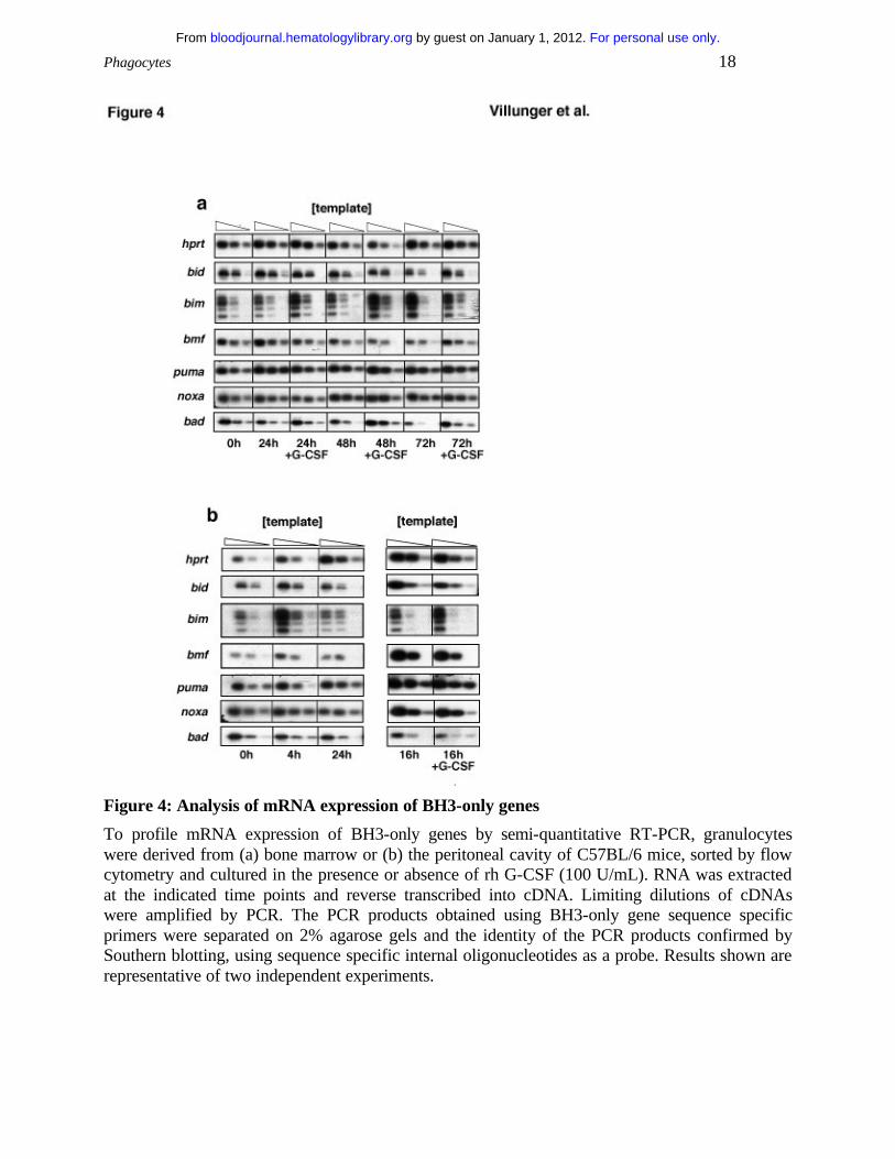
Figure 4: Analysis of mRNA expression of BH3-only genes
To profile mRNA expression of BH3-only genes by semi-quantitative RT-PCR, granulocytes were derived from (a) bone marrow or (b) the peritoneal cavity of C57BL/6 mice, sorted by flow cytometry and cultured in the presence or absence of rh G-CSF (100 U/mL). RNA was extracted at the indicated time points and reverse transcribed into cDNA. Limiting dilutions of cDNAs were amplified by PCR. The PCR products obtained using BH3-only gene sequence specific primers were separated on 2% agarose gels and the identity of the PCR products confirmed by Southern blotting, using sequence specific internal oligonucleotides as a probe. Results shown are representative of two independent experiments.
For personal use only. by guest on January 1, 2012. bloodjournal.hematologylibrary.orgFrom

Phagocytes 19
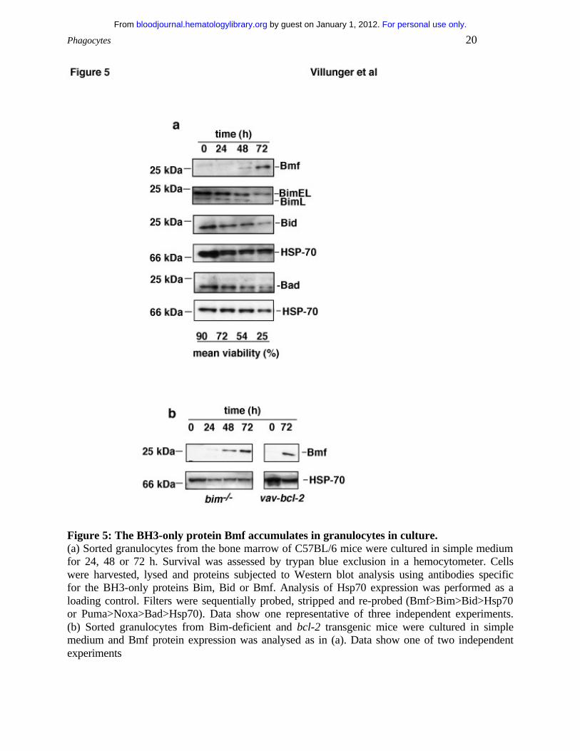
Surprisingly, protein expression analysis by Western blotting of lysates from sorted granulocytes
undergoing spontaneous apoptosis in culture revealed that the BH3-only protein Bmf increased
substantial whereas levels of Bim, Bad or Bid remained unchanged (Fig. 5a). Expression of the
proteins PUMA/bbc3 or Noxa were undetectable under these conditions (data not shown).
Accumulation of Bmf was also observed in cultured granulocytes from bim-/- orbcl-2 transgenic
mice, indicating that this accumulation is not merely a consequence of cell death (Fig. 5b). Bmf
protein levels did not increase in thymocytes undergoing cytokine-withdrawal induced death,
indicating that this increase may be specific for granulocytes (data not shown).
For personal use only. by guest on January 1, 2012. bloodjournal.hematologylibrary.orgFrom
Phagocytes 20
Figure 5: The BH3-only protein Bmf accumulates in granulocytes in culture. (a) Sorted granulocytes from the bone marrow of C57BL/6 mice were cultured in simple medium for 24, 48 or 72 h. Survival was assessed by trypan blue exclusion in a hemocytometer. Cells were harvested, lysed and proteins subjected to Western blot analysis using antibodies specific for the BH3-only proteins Bim, Bid or Bmf. Analysis of Hsp70 expression was performed as a loading control. Filters were sequentially probed, stripped and re-probed (Bmf>Bim>Bid>Hsp70 or Puma>Noxa>Bad>Hsp70). Data show one representative of three independent experiments. (b) Sorted granulocytes from Bim-deficient and bcl-2 transgenic mice were cultured in simple medium and Bmf protein expression was analysed as in (a). Data show one of two independent experiments
For personal use only. by guest on January 1, 2012. bloodjournal.hematologylibrary.orgFrom

Phagocytes 21
Discussion
We investigated the role of several pro- and anti-apoptotic Bcl-2 family members on the survival
of bone marrow-derived and mobilized peritoneal granulocytes We observed that absence of the
BH3-only protein Bim, but not loss of pro-apoptotic Bax, rendered granulocytes refractory to
apoptosis induced by cytokine-withdrawal (Figs. 1a,e), chemotherapeutic drugs or the calcium
ionophore ionomycin, the latter only inducing cell death in mouse but not human granulocytes 31
(Figs. 1c,g,i). In contrast, killing by FasL was not impaired in the absence of Bim or Bax (Figs.
1c,g,i). Our results are consistent with the previous observations that bim-/- thymocytes 19,32 as
well as bcl-2 transgenic lymphocytes 33 and granulocytes 11 are resistant to cell death induced by
cytokine withdrawal or cytotoxic drugs but remain normally sensitive to FasL. The resistance of
Bim-deficient granulocytes to spontaneous death in culture (Figs. 1a,e) is consistent with
abnormal granulocyte accumulation observed in bim-/- mice 19 and indicates that this is due to a
cell intrinsic defect and not a consequence of increased cytokine levels or a phagocytosis defect
in the bim-/- animals. Bim-deficient granulocytes are less resistant to spontaneous death in culture
than those over-expressing Bcl-2 (Figs. 1a,e) indicating that Bcl-2 inhibitable death inducers in
addition to Bim are involved in programmed death of granulocytes (see below). Bax expression
levels have been reported to be abnormally low in granulocytes from patients with certain
inflammatory diseases, such as cystic fibrosis or acute pneumonia 5. Our findings, however,
suggest that absence of Bax alone is not sufficient to extend granulocyte survival (Figs. 1a,e), and
this is consistent with the observation that bax-/- mice have no obvious increase in granulocytes
(34 and our data not shown). In contrast, bax-/-/bak-/- double deficient animals have an abnormal
accumulation of granulocytes 34, indicating that these two multi-BH domain pro-apoptotic Bcl-2
family members have overlapping functions in programmed death of myeloid cells, probably
acting downstream of BH3-only proteins.
Loss of Bcl-2 or Bcl-w does not sensitize granulocytes from bone marrow or peritoneal cavity to
spontaneous (Figs. 1b,f) or stress-induced apoptosis (Figs. 1d,h,j) but Bcl-2 over-expression
prolongs their survival. This indicates that Bcl-2 and Bcl-w act in a redundant manner in
For personal use only. by guest on January 1, 2012. bloodjournal.hematologylibrary.orgFrom

Phagocytes 22
regulating granulocyte survival or that they may even be dispensable. Mice lacking A1a show a
small but significant acceleration of spontaneous granulocyte apoptosis in culture 18. Since mice
have three closely related A1 genes, all of which appear to be expressed in granulocytes 18, it is
possible that combined loss of all A1 genes would evoke extensive granulocyte apoptosis and
severe neutropenia. Another pro-survival Bcl-2 family member, Mcl-1, is induced in
granulocytes by cytokine receptor stimulation 8, and expression of a Mcl-1 transgene inhibits
apoptosis of hematopoietic cells 35. The early embryonic lethality of Mcl-1-deficient mice
precluded investigation of the function of this protein in myeloid cells 36, but it is possible that a
critical role in granulocyte survival will emerge from studies with mice in which the gene can be
deleted selectively in this cell type.
Since loss of Bcl-2 did not influence the survival of mature granulocytes in culture (Fig. 1) but
expression of a bcl-2 transgene was reported to protect granulocytes as well as myeloid
precursors against growth factor withdrawal in vitro 22, we assessed whether absence of Bcl-2 has
an impact on the survival of early myeloid progenitors and compared this effect with the one
caused by absence of the BH3-only protein Bim. Analysis of wt animals reconstituted with a Bcl-
2 deficient hemopoietic system confirmed previous observations of strongly reduced numbers of
lymphocytes in all hemopoietic compartments analyzed (i.e. spleen, bone marrow and peripheral
blood) 29, but also indicates a previously unrecognized role for Bcl-2 in the survival of mature
macrophages which were significantly reduced in numbers in peripheral blood and spleen when
compared with animals reconstituted with wt bone marrow (Figs. 2a-d). Although granulocyte
numbers were comparable in wt and bcl-2-/- reconstituted animals, analysis of the colony forming
potential of early myeloid progenitors in soft agar revealed that Bcl-2 is essential for the survival
of all types of myeloid precursors under conditions of cytokine withdrawal (Fig. 2e). Absence of
Bim had a less pronounced but reproducible protective effect on the survival of cytokine-
deprived myeloid progenitors (Fig. 2f). The frequency of Bim-deficient myeloid precursors was
comparable to that found in wt bone marrow. Expression of a bcl-2 transgene proved most potent
in protecting myeloid progenitors from the effects of delayed cytokine addition (Fig. 2f and 22). In
For personal use only. by guest on January 1, 2012. bloodjournal.hematologylibrary.orgFrom

Phagocytes 23
agreement with previous results, the frequency of bcl-2 transgenic myeloid precursors was
comparable to that found in wt bone marrow with the exception of a previously described minor
reduction of macrophage colonies 22. These observations are in line with data demonstrating that
Bcl-2 protects hematopoietic stem cells and increases their number and repopulation potential in
vivo 37. Furthermore, our findings are consistent with the notion that bcl-2-/- stem cells perform
poorly in serial transplantation assays 29 and demonstrate that although Bcl-2 is not essential for
the survival of mature granulocytes, it is required to protect myeloid progenitors under conditions
of cytokine withdrawal. Collectively, these results indicate that cytokine deprivation–induced
death of myeloid progenitors is regulated by Bcl-2 and that Bim contributes to this process,
although to a lower degree than in mature granulocytes (see also Figs. 1a, e).
G-CSF promotes maturation and proliferation of granulocytes in the bone marrow through
activation of signal transducer and activator of transcription (STAT) 3 and STAT 5 as well as
mitogen activated protein kinase (MAPK) 38. Little, however, is known about the mechanism by
which G-CSF regulates granulocyte survival. Downregulation of Bax expression levels 5 as well
as the prevention of a conformational change, required to allow Bax to insert into inner
membranes and subsequent caspase-3 activation 39 were reported to contribute to the pro-survival
effect of G-CSF. Our experiments analyzing the ability of G-CSF to promote survival of
granulocytes lacking Bim, Bax, Bcl-2 or Bcl-w indicate that none of these molecules alone is the
key target for the pro-survival effect of G-CSF. In cells expressing a bcl-2 transgene G-CSF
failed to further enhance survival, demonstrating that Bcl-2 can efficiently compensate for G-CSF
receptor signaling in these cells (Figs. 4 a,b). This indicates that Bcl-2-like pro-survival
molecules, such as Mcl-1 or A1, may be the key targets of the anti-apoptotic signals induced by
G-CSF. Alternatively, since absence of Bim partially protects granulocytes under conditions of
growth factor-withdrawal (Figs. 1a, e), G-CSF may modulate the function and/or expression
levels of BH3-only proteins. We found no evidence that G-CSF would modulate mRNA
expression levels of BH3-only proteins in granulocytes derived form bone marrow or the
peritoneal cavity under conditions of growth factor withdrawal (Figs. 4 a,b).
For personal use only. by guest on January 1, 2012. bloodjournal.hematologylibrary.orgFrom

Phagocytes 24
Analyzing protein expression levels of BH3-only proteins we observed accumulation of Bmf, in
cytokine-deprived bone marrow derived cells (Fig. 5a). This accumulation was also observed in
granulocytes form Bim-deficient or bcl-2 transgenic animals (Fig. 5b) which indicates that this
accumulation is not merely a consequence of cell death, but may actively contribute to apoptosis
of granulocytes. The pro-apoptotic activity of Bim and Bmf can be controlled post-translationally
by sequestration to distinct subcellular compartments 20,26. This does, however, not exclude that
transcriptional control can also regulate their pro-apoptotic function. Upon cytokine-deprivation,
Bim expression is induced by the fork-head related transcription factor FKHR-L1 in the BaF3 B
cell line 40 and by AP-1 in neuronal cells 41,42. However, since bmf mRNA levels did not increase
upon cytokine-deprivation (Figs. 4a,b), Bmf accumulation appears to be due to a post-
translational mechanism, perhaps caused by enhanced protein stability.
In conclusion, our results demonstrate that the BH3-only protein Bim and possibly also its
relative Bmf are key initiators of growth factor withdrawal and stress-induced apoptosis of
granulocytes, whereas the function of the pro-apoptotic multi BH-domain protein Bax appears to
be redundant. To allow efficient production but rapid turnover of granulocytes, the pro-survival
function of Bcl-2 or Bcl-w may be more prominent in progenitors than in mature cells.
Alternatively, Bcl-2 and Bcl-w or other more distantly related Bcl-2-like pro-survival molecules,
such as Mcl-1 or A1, may act in a redundant manner in mature granulocytes.
Acknowledgments
We thank Drs. A. Harris, J. Adams and S. Cory for supplying vav-bcl-2 transgenic and bcl-w-/-
mice and Dr. D. Loh for bcl-2+/- mice. We are grateful to S. Mifsud and Drs. D. Metcalf and N.
Nicola for gifts of cytokines and Dr. R. Anderson for anti-HSP-70 antibody. We thank C.
Tilbrook and A. Naughton for mouse care and injections and Dr F. Battye, V. Lapatis, J. Chang,
C. Tarlinton and D. Kaminaris for cell sorting.
For personal use only. by guest on January 1, 2012. bloodjournal.hematologylibrary.orgFrom

Phagocytes 25
References
1. Metcalf D. The molecular control of granulocytes and macrophages. Ciba Foundation
Symposium. 1997;204:40-50
2. Basu S, Hodgson G, Katz M, Dunn AR. Evaluation of role of G-CSF in the
production, survival, and release of neutrophils from bone marrow into circulation. Blood.
2002;100:854-861
3. Savill J, Fadok V, Henson P, Haslett C. Phagocyte recognition of cells undergoing
apoptosis`. Immunol Today. 1993;14:131-136
4. Lieschke GJ, Grail D, Hodgson G, Metcalf D, Stanley E, Cheers C, Fowler KJ, Basu
S, Zhan YF, Dunn AR. Mice lacking granulocyte colony-stimulating factor have chronic
neutropenia, granulocyte and macrophage progenitor cell deficiency, and impaired neutrophil
mobilization. Blood. 1994;84:1737-1746
5. Dibbert B, Weber M, Nikolaizik WH, Vogt P, Schoni MH, Blaser K, Simon HU.
Cytokine-mediated Bax deficiency and consequent delayed neutrophil apoptosis: a general
mechanism to accumulate effector cells in inflammation. Proc Natl Acad Sci USA.
1999;96:13330-13335
6. Passegue E, Jochum W, Schorpp-Kistner M, Mohle-Steinlein U, Wagner EF. Chronic
myeloid leukemia with increased granulocyte progenitors in mice lacking JunB expression in the
myeloid lineage. Cell. 2001;104:21-32
7. Strasser A, O'Connor L, Dixit VM. Apoptosis signaling. Annu Rev Biochem.
2000;69:217-245
For personal use only. by guest on January 1, 2012. bloodjournal.hematologylibrary.orgFrom

Phagocytes 26
8. Moulding DA, Quayle JA, Hart CA, Edwards SW. Mcl-1 expression in human
neutrophils: regulation by cytokines and correlation with cell survival. Blood. 1998;92:2495-
2502
9. Orlofsky A, Somogyi RD, Weiss LM, Prystowsky MB. The murine antiapoptotic
protein A1 is induced in inflammatory macrophages and constitutively expressed in neutrophils. J
Immunol. 1999;163:412-419
10. Lin EY, Orlofsky A, Wang H-G, Reed JC, Prystowsky MB. A1, a bcl-2 family
member, prolongs cell survival and permits myeloid differentiation. Blood. 1996;87:983-992
11. Villunger A, O’Reilly LA, Holler N, Adams JM, Strasser A. Fas ligand, Bcl-2, G-
CSF and p38 MAPK: regulators of distinct cell death and survival pathways in granulocytes. J
Exp Med. 2000;192:647-657
12. Iwai K, Miyawaki T, Takizawa T, Konno A, Ohta K, Yachie A, Seki H, Taniguchi N.
Differential expression of bcl-2 and susceptibility to anti-Fas-mediated cell death in peripheral
blood lymphocytes, monocytes, and neutrophils. Blood. 1994;84:1201-1208
13. Lagasse E, Weissman IL. bcl-2 inhibits apoptosis of neutrophils but not their
engulfment by macrophages. J Exp Med. 1994;179:1047-1052
14. O'Reilly LA, Print C, Hausmann G, Moriishi K, Cory S, Huang DCS, Strasser A.
Tissue expression and subcellular localization of the pro-survival molecule Bcl-w. Cell Death
Differ. 2001;8:486-494
15. Gibson L, Holmgreen S, Huang DCS, Bernard O, Copeland NG, Jenkins NA,
Sutherland GR, Baker E, Adams JM, Cory S. bcl-w, a novel member of the bcl-2 family,
promotes cell survival. Oncogene. 1996;13:665-675
16. Print CG, Loveland KL, Gibson L, Meehan T, Stylianou A, Wreford N, de Kretser D,
Metcalf D, Köntgen F, Adams JM, Cory S. Apoptosis regulator Bcl-w is essential for
For personal use only. by guest on January 1, 2012. bloodjournal.hematologylibrary.orgFrom

Phagocytes 27
spermatogenesis but appears otherwise redundant. Proc Natl Acad Sci USA. 1998;95:12424-
12431
17. Chuang PI, Yee E, Karsan A, Winn RK, Harlan JM. A1 is a constitutive and
inducible Bcl-2 homologue in mature human neutrophils. Biochem Biophys Res Commun.
1998;249:361-365
18. Hamasaki A, Sendo F, Nakayama K, Ishida N, Negishi I, Nakayama K-I,
Hatakeyama S. Accelerated neutrophil apoptosis in mice lacking A1-a, a subtype of the bcl-2-
related A1 gene. J Exp Med. 1998;188:1985-1992
19. Bouillet P, Metcalf D, Huang DCS, Tarlinton DM, Kay TWH, Köntgen F, Adams
JM, Strasser A. Proapoptotic Bcl-2 relative Bim required for certain apoptotic responses,
leukocyte homeostasis, and to preclude autoimmunity. Science. 1999;286:1735-1738
20. Puthalakath H, Huang DCS, O'Reilly LA, King SM, Strasser A. The pro-apoptotic
activity of the Bcl-2 family member Bim is regulated by interaction with the dynein motor
complex. Mol Cell. 1999;3:287-296
21. Zong WX, Lindsten T, Ross AJ, MacGregor GR, Thompson CB. BH3-only proteins
that bind pro-survival Bcl-2 family members fail to induce apoptosis in the absence of Bax and
Bak. Genes Dev. 2001;15:1481-1486
22. Ogilvy S, Metcalf D, Print CG, Bath ML, Harris AW, Adams JM. Constitutive bcl-2
expression throughout the hematopoietic compartment affects multiple lineages and enhances
progenitor cell survival. Proc Natl Acad Sci USA. 1999;96:14943-14948
23. Knudson CM, Tung KSK, Tourtellotte WG, Brown GAJ, Korsmeyer SJ. Bax-
deficient mice with lymphoid hyperplasia and male germ cell death. Science. 1995;270:96-99
For personal use only. by guest on January 1, 2012. bloodjournal.hematologylibrary.orgFrom

Phagocytes 28
24. Nakayama K, Nakayama K-I, Negishi I, Kuida K, Sawa H, Loh DY. Targeted
disruption of bcl-2αβ in mice: occurrence of gray hair, polycystic kidney disease, and
lymphocytopenia. Proc Natl Acad Sci USA. 1994;91:3700-3704
25. Veis DJ, Sorenson CM, Shutter JR, Korsmeyer SJ. Bcl-2-deficient mice demonstrate
fulminant lymphoid apoptosis, polycystic kidneys, and hypopigmented hair. Cell. 1993;75:229-
240
26. Puthalakath H, Villunger A, O'Reilly LA, Beaumont JG, Coultas L, Cheney RE,
Huang DCS, Strasser A. Bmf: a pro-apoptotic BH3-only protein regulated by interaction with the
myosin V actin motor complex, activated by anoikis. Science. 2001;293:1829-1832
27. Liles WC, Kiener PA, Ledbetter JA, Aruffo A, Klebanoff SJ. Differential expression
of Fas (CD95) and Fas ligand on normal human phagocytes: implications for the regulation of
apoptosis in neutrophils. J Exp Med. 1996;184:429-440
28. Tortorella C, Piazzolla G, Spaccavento F, Pece S, Jirillo E, Antonaci S. Spontaneous
and Fas-induced apoptotic cell death in aged neutrophils. J Clin Immunol. 1998;18:321-329
29. Matsuzaki Y, Nakayama K-I, Nakayama K, Tomita T, Isoda M, Loh DY, Nakauchi
H. Role of bcl-2 in the development of lymphoid cells from the hematopoietic stem cell. Blood.
1997;89:853-862
30. Imaizumi K, Tsuda M, Imai Y, Wanaka A, Takagi T, Tohyama M. Molecular cloning
of a novel polypeptide, DP5, induced during programmed neuronal death. J Biol Chem.
1997;272:18842-18848
31. Whyte MK, Hardwick SJ, Meagher LC, Savill JS, Haslett C. Transient elevations of
cytosolic free calcium retard subsequent apoptosis in neutrophils in vitro. J Clin Invest.
1993;92:446-455
For personal use only. by guest on January 1, 2012. bloodjournal.hematologylibrary.orgFrom
Phagocytes 29
32. Bouillet P, Purton JF, Godfrey DI, Zhang L-C, Coultas L, Puthalakath H, Pellegrini
M, Cory S, Adams JM, Strasser A. BH3-only Bcl-2 family member Bim is required for apoptosis
of autoreactive thymocytes. Nature. 2002;415:922-926
33. Strasser A, Harris AW, Huang DCS, Krammer PH, Cory S. Bcl-2 and Fas/APO-1
regulate distinct pathways to lymphocyte apoptosis. EMBO J. 1995;14:6136-6147
34. Lindsten T, Ross AJ, King A, Zong W, Rathmell JC, Shiels HA, Ulrich E, Waymire
KG, Mahar P, Frauwirth K, Chen Y, Wei M, Eng VM, Adelman DM, Simon MC, Ma A, Golden
JA, Evan G, Korsmeyer SJ, MacGregor GR, Thompson CB. The combined functions of
proapoptotic Bcl-2 family members Bak and Bax are essential for normal development of
multiple tissues. Mol Cell. 2000;6:1389-1399.
35. Zhou P, Qian L, Kozopas KM, Craig RW. Mcl-1, a Bcl-2 family member, delays the
death of hematopoietic cells under a variety of apoptosis-inducing conditions. Blood.
1997;89:630-643
36. Rinkenberger JL, Horning S, Klocke B, Roth K, Korsmeyer SJ. Mcl-1 deficiency
results in peri-implantation embryonic lethality. Genes Dev. 2000;14:23-27
37. Domen J, Cheshier SH, Weissman IL. The role of apoptosis in the regulation of
hematopoietic stem cells: overexpression of Bcl-2 increases both their number and repopulation
potential. J Exp Med. 2000;191:253-264
38. Coffer PJ, Koenderman L, de Groot RP. The role of STATs in myeloid differentiation
and leukemia. Oncogene. 2000;19:2511-2522
39. Maianski NA, Mul FP, van Buul JD, Roos D, Kuijpers TW. Granulocyte colony-
stimulating factor inhibits the mitochondria- dependent activation of caspase-3 in neutrophils.
Blood. 2002;99:672-679
For personal use only. by guest on January 1, 2012. bloodjournal.hematologylibrary.orgFrom

Phagocytes 30
40. Dijkers PF, Medemadagger RH, Lammers JJ, Koenderman L, Coffer PJ. Expression
of the pro-apoptotic Bcl-2 family member Bim is regulated by the forkhead transcription factor
FKHR-L1. Curr Biol. 2000;10:1201-1204.
41. Putcha GV, Moulder KL, Golden JP, Bouillet P, Adams JM, Strasser A, Johnson
EMJ. Induction of Bim, a proapoptotic BH3-only Bcl-2 family member, is critical for neuronal
apoptosis. Neuron. 2001;29:615-628
42. Whitfield J, Neame SJ, Paquet L, Bernard O, Ham J. Dominant-negative c-Jun
promotes neuronal survival by reducing BIM expression and inhibiting mitochondrial
cytochrome c release. Neuron. 2001;29:629-643.
For personal use only. by guest on January 1, 2012. bloodjournal.hematologylibrary.orgFrom