BH3 Mimetic ABT-737 and a Proteasome Inhibitor Synergistically Kill Melanomas through Noxa-Dependent...
8
BH3 Mimetic ABT-737 and a Proteasome Inhibitor Synergistically Kill Melanomas through Noxa-Dependent Apoptosis Leslie A. Miller 1 , Nathaniel B. Goldstein 1 , Widya U. Johannes 1 , Christine H. Walton 1 , Mayumi Fujita 1 , David A. Norris 1,2,3 and Yiqun G. Shellman 1,3 The Bcl-2 family is important in modulating sensitivity to anticancer drugs in many cancers, including melanomas. The BH3 mimetic ABT-737 is a potent small molecule inhibitor of the anti-apoptotic proteins Bcl-2/ Bcl-X L /Bcl-w. In this report, we examined whether ABT-737 is effective in killing melanoma cells in combination with the proteasome inhibitor MG-132, and further evaluated the mechanisms of action. Viability, morphological, and Annexin V apoptosis assays showed that ABT-737 alone exhibited little cytotoxicity, yet it displayed strong synergistic lethality when combined with MG-132. In addition, the detection of Bax/Bak activation indicated that the combination treatment synergistically induced mitochondria-mediated apoptosis. Furthermore, mechanistic analysis revealed that this combination treatment induced expression of the pro-apoptotic protein Noxa- and caspase-dependent degradation of the anti-apoptotic protein, Mcl-1. Finally, siRNA-mediated inhibition of Mcl-1 expression significantly increased sensitivity to ABT-737 in these cells, and knocking down Noxa expression protected the cells from cytotoxicity induced by the combination treatment. These findings demonstrate that ABT-737 combined with MG-132 synergistically induced Noxa-dependent mitochondrial-mediated apoptosis. In summary, this study indicates promising therapeutic potential of targeting anti-apoptotic Bcl-2 family members in treating melanoma, and it validates rational molecular approaches that target anti-apoptotic defenses when developing cancer treatments. Journal of Investigative Dermatology (2009) 129, 964–971; doi:10.1038/jid.2008.327; published online 6 November 2008 INTRODUCTION Malignant melanoma is largely unresponsive to existing therapies and has a very poor prognosis (Buzaid, 2004; Cummins et al., 2006; Gogas et al., 2007). In addition, there has been little progress over the past 30 years in medical treatment of this disease. Thus, the paucity of effective treatments for malignant melanoma is a pressing issue in medicine. The recent use of proteasome inhibition in clinical practice to induce apoptosis in cancer has produced interesting results. However, proteasome inhibition alone is neither complete nor sustained because of the need to limit secondary toxicity (Adams, 2004). In addition, pharmacokinetic analyses have indicated that proteasome inhibitors are rapidly inactivated by hepatic detoxification (Schwartz and Davidson, 2004). There- fore, compounds that synergize with proteasome inhibitors to lower required dosages and/or to minimize the time delay from proteasome inhibition to the activation of the apoptotic machinery could have important clinical implications. Apoptosis is one of the main pathways contributing to the cytotoxic effects of conventional cancer treatments, and functional apoptotic pathways are required for the success of most treatments. During melanoma’s progression, malignant cells become ‘bullet proof’ against a variety of chemother- apeutic drugs by exploiting their intrinsic apoptotic defenses and by reprogramming their proliferation and survival path- ways (Soengas and Lowe, 2003). The Bcl-2 family of proteins is crucial in regulating apoptosis (see review Thomadaki et al., 2006), especially mitochondria- dependent apoptotic cell death. This family can be divided into three groups: anti-apoptotic proteins, including proteins such as Bcl-2, Bcl-X L , Bcl-w, and Mcl-1; multi-domain pro-apoptotic proteins Bax and Bak; and pro-apoptotic BH3-only proteins, including Noxa, Bim, Bid, Bad, Bmf, and Bik. Molecular-targeted therapy holds the promise of providing new and more effective treatment options with minimal toxicity (Weinstein and Joe, 2006). Compounds have been developed to target activated kinases or receptors (Weinstein and Joe, 2006), but there are also many advantages for ORIGINAL ARTICLE 964 Journal of Investigative Dermatology & 2009 The Society for Investigative Dermatology Received 11 April 2008; revised 28 July 2008; accepted 29 August 2008; published online 6 November 2008 1 Department of Dermatology, School of Medicine, University of Colorado Denver, Aurora, Colorado, USA and 2 Department of Veterans Affairs Medical Center, Dermatology Section, Denver, Colorado, USA Correspondence: Dr Yiqun G. Shellman, Department of Dermatology, School of Medicine, University of Colorado Denver, Mail Stop #8127, PO Box 6511, Aurora, Colorado 80010, USA. E-mail: [email protected] 3 These authors contributed equally to this project. Abbreviations: ANOVA, analysis of variance; EB/AO, ethidium bromide and acridine orange; MTS, a tetrazolium compound [3-(4,5-dimethylthiazol-2-yl)- 5-(3-carboxymethoxy-phenyl)-2 (4-sulfophenyl)-2H-tetrazolium], inner salt; SiRNA, short interfering RNA
Transcript of BH3 Mimetic ABT-737 and a Proteasome Inhibitor Synergistically Kill Melanomas through Noxa-Dependent...
BH3 Mimetic ABT-737 and a Proteasome InhibitorSynergistically Kill Melanomas throughNoxa-Dependent ApoptosisLeslie A. Miller1, Nathaniel B. Goldstein1, Widya U. Johannes1, Christine H. Walton1, Mayumi Fujita1,David A. Norris1,2,3 and Yiqun G. Shellman1,3
The Bcl-2 family is important in modulating sensitivity to anticancer drugs in many cancers, includingmelanomas. The BH3 mimetic ABT-737 is a potent small molecule inhibitor of the anti-apoptotic proteins Bcl-2/Bcl-XL/Bcl-w. In this report, we examined whether ABT-737 is effective in killing melanoma cells in combinationwith the proteasome inhibitor MG-132, and further evaluated the mechanisms of action. Viability,morphological, and Annexin V apoptosis assays showed that ABT-737 alone exhibited little cytotoxicity, yet itdisplayed strong synergistic lethality when combined with MG-132. In addition, the detection of Bax/Bakactivation indicated that the combination treatment synergistically induced mitochondria-mediated apoptosis.Furthermore, mechanistic analysis revealed that this combination treatment induced expression of thepro-apoptotic protein Noxa- and caspase-dependent degradation of the anti-apoptotic protein, Mcl-1. Finally,siRNA-mediated inhibition of Mcl-1 expression significantly increased sensitivity to ABT-737 in these cells, andknocking down Noxa expression protected the cells from cytotoxicity induced by the combination treatment.These findings demonstrate that ABT-737 combined with MG-132 synergistically induced Noxa-dependentmitochondrial-mediated apoptosis. In summary, this study indicates promising therapeutic potential oftargeting anti-apoptotic Bcl-2 family members in treating melanoma, and it validates rational molecularapproaches that target anti-apoptotic defenses when developing cancer treatments.
Journal of Investigative Dermatology (2009) 129, 964–971; doi:10.1038/jid.2008.327; published online 6 November 2008
INTRODUCTIONMalignant melanoma is largely unresponsive to existingtherapies and has a very poor prognosis (Buzaid, 2004;Cummins et al., 2006; Gogas et al., 2007). In addition, therehas been little progress over the past 30 years in medicaltreatment of this disease. Thus, the paucity of effective treatmentsfor malignant melanoma is a pressing issue in medicine.
The recent use of proteasome inhibition in clinical practiceto induce apoptosis in cancer has produced interesting results.However, proteasome inhibition alone is neither complete norsustained because of the need to limit secondary toxicity(Adams, 2004). In addition, pharmacokinetic analyses have
indicated that proteasome inhibitors are rapidly inactivated byhepatic detoxification (Schwartz and Davidson, 2004). There-fore, compounds that synergize with proteasome inhibitors tolower required dosages and/or to minimize the time delay fromproteasome inhibition to the activation of the apoptoticmachinery could have important clinical implications.
Apoptosis is one of the main pathways contributing to thecytotoxic effects of conventional cancer treatments, andfunctional apoptotic pathways are required for the success ofmost treatments. During melanoma’s progression, malignantcells become ‘bullet proof’ against a variety of chemother-apeutic drugs by exploiting their intrinsic apoptotic defensesand by reprogramming their proliferation and survival path-ways (Soengas and Lowe, 2003).
The Bcl-2 family of proteins is crucial in regulating apoptosis(see review Thomadaki et al., 2006), especially mitochondria-dependent apoptotic cell death. This family can be divided intothree groups: anti-apoptotic proteins, including proteins such asBcl-2, Bcl-XL, Bcl-w, and Mcl-1; multi-domain pro-apoptoticproteins Bax and Bak; and pro-apoptotic BH3-only proteins,including Noxa, Bim, Bid, Bad, Bmf, and Bik.
Molecular-targeted therapy holds the promise of providingnew and more effective treatment options with minimaltoxicity (Weinstein and Joe, 2006). Compounds have beendeveloped to target activated kinases or receptors (Weinsteinand Joe, 2006), but there are also many advantages for
ORIGINAL ARTICLE
964 Journal of Investigative Dermatology & 2009 The Society for Investigative Dermatology
Received 11 April 2008; revised 28 July 2008; accepted 29 August 2008;published online 6 November 2008
1Department of Dermatology, School of Medicine, University of ColoradoDenver, Aurora, Colorado, USA and 2Department of Veterans Affairs MedicalCenter, Dermatology Section, Denver, Colorado, USA
Correspondence: Dr Yiqun G. Shellman, Department of Dermatology, Schoolof Medicine, University of Colorado Denver, Mail Stop #8127, PO Box 6511,Aurora, Colorado 80010, USA.E-mail: [email protected]
3These authors contributed equally to this project.
Abbreviations: ANOVA, analysis of variance; EB/AO, ethidium bromide andacridine orange; MTS, a tetrazolium compound [3-(4,5-dimethylthiazol-2-yl)-5-(3-carboxymethoxy-phenyl)-2 (4-sulfophenyl)-2H-tetrazolium], inner salt;SiRNA, short interfering RNA
targeting the defective apoptotic machinery in cancer cells:(1) Even though different gene mutations exist in differenttypes of cancers, downstream pathways that include anti-apoptotic defenses are very similar. (2) Abnormally regulatedapoptotic signaling is one of the hallmarks of cancer. (3)Finding molecular targets in anti-apoptotic defenses willprovide molecular targets for cancer therapy that differ fromtargeting kinases or receptors.
ABT-737 (developed by Abbott Laboratories) is apotent small molecule inhibitor of the anti-apoptotic proteinsBcl-2, Bcl-XL, and Bcl-w with an affinity 2–3 orders ofmagnitude more potent than any previously reportedcompounds (Letai, 2005; Oltersdorf et al., 2005). It acts likea BH3—only protein to antagonize anti-apoptotic Bcl-2family members, thereby diminishing their ability to inhibitapoptosis (Oltersdorf et al., 2005). Many teams have recentlyreported the high efficacy of ABT-737 either as a single agentor as a chemo-potentiator in combination with otherchemotherapeutic agents to treat multiple types of cancers,including lymphoma, small-cell lung carcinoma, variousleukemias, etc. (Adams et al., 2005; Oltersdorf et al., 2005;Certo et al., 2006; Konopleva et al., 2006; Shoemaker et al.,2006; van Delft et al., 2006; Chauhan et al., 2007; Chenet al., 2007a, b; Kang et al., 2007; Kohl et al., 2007).
In this study, we explored whether ABT-737 is effectivein treating human melanoma cell lines and whether itpotentiates killing of melanoma cells by the proteasomeinhibitor, MG-132. We found that the two drugs actedsynergistically to activate Noxa-dependent mitochondrial-mediated apoptosis. Mechanistic analysis suggests that thiscombination increases pro-apoptotic protein Noxa expres-sion, neutralizes all main anti-apoptotic Bcl-2 proteins, andinduces accelerated Noxa-mediated synergistic killing.
RESULTSABT-737 synergistically killed human melanoma cells whencombined with MG-132
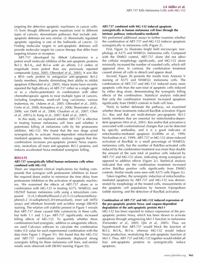
There are important clinical implications for finding com-pounds that synergize with proteasome inhibitors to lowerthe required doses and/or to minimize the time delay fromproteasome inhibition to the activation of apoptotic machin-ery. We examined the effects of ABT-737 alone or incombination with MG-132 in treating A375, WM852c andHS294T human melanoma cells using a tetrazolium com-pound [3-(4,5-dimethylthiazol-2-yl)-5-(3-carboxymethoxy-phenyl)-2 (4-sulfophenyl)-2H-tetrazolium], inner salt (MTS)assays and ethidium bromide and acridine orange (EB/AO)staining. The relative cell viability present in Figure 1 revealsthat ABT-737 alone caused little cytotoxicity in these cells,but both 1.1 and 3.3 mM ABT-737 significantly increasedkilling effects of MG-132. To quantify whether thesecombinations had synergistic, additive or antagonistic effects,we used Calcusyn software to calculate the combinationindex (CI) value for each experimental combination with thedata from Figure 1 (Figure S1). We found that the MG-132and ABT-737 combination treatments displayed strongsynergistic killing for these melanoma cell lines, and similarresults were observed with EB/AO staining (Figure S2).
ABT-737 combined with MG-132 induced apoptosissynergistically in human melanoma cell lines through theintrinsic pathway (mitochondria mediated)
We performed additional assays to further examine whetherthe combination of ABT-737 and MG-132 induces apoptosissynergistically in melanoma cells (Figure 2).
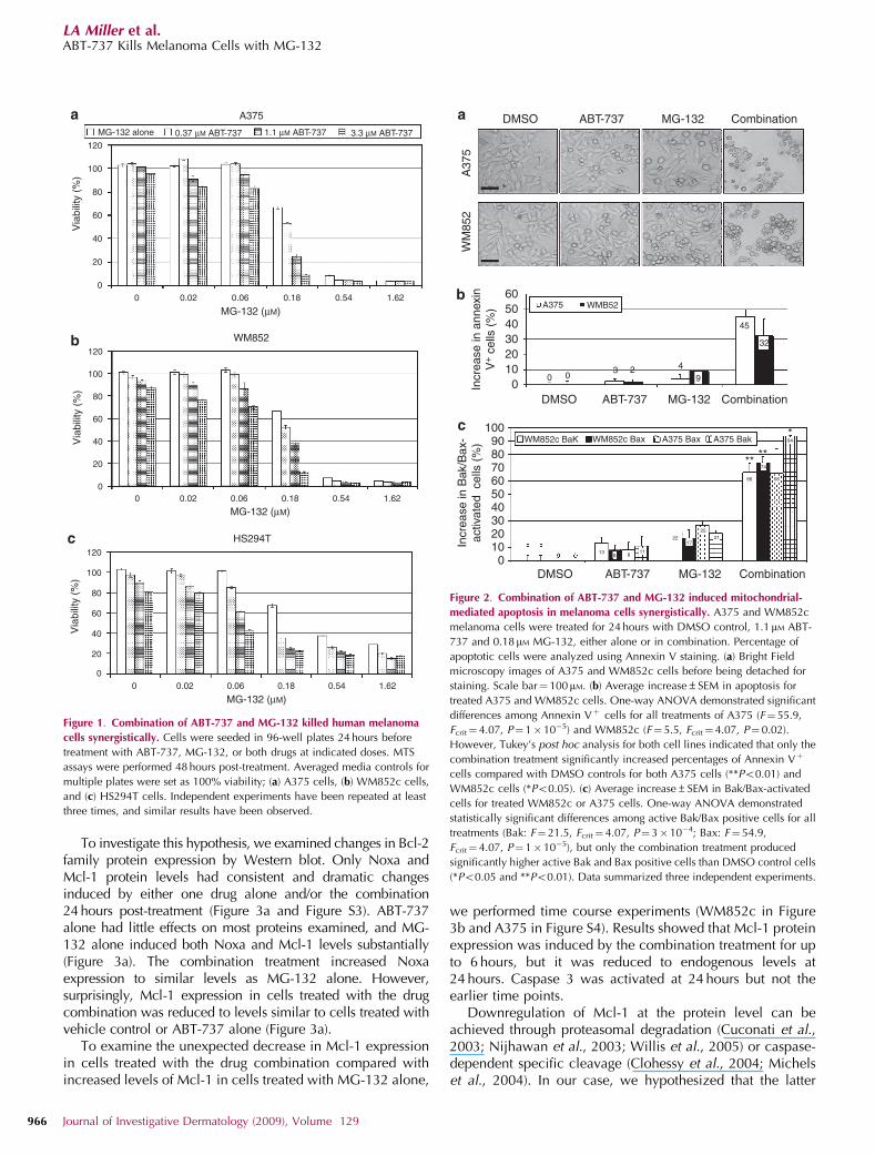
First, Figure 2a illustrates bright field microscopic mor-phology of A375 and WM852c melanoma cells. Comparedwith the DMSO control, ABT-737 alone did not affectthe cellular morphology significantly, and MG-132 aloneminimally increased the number of rounded cells, which stillappeared alive. In contrast, the combination treatmentcaused almost all cells to detach and appear dead.
Second, Figure 2b presents the results from Annexin Vstaining of A375 and WM852c melanoma cells. Thecombination of ABT-737 and MG-132 induced many moreapoptotic cells than the sum total of apoptotic cells inducedby either drug alone, demonstrating the synergistic killingeffects of the combination. Statistical analysis indicatedthat only the combination treatment increased apoptosissignificantly from DMSO controls in both cell lines.
Third, to further delineate the pathway, we examinedwhether these treatments induced Bax/Bak activation (Figure2c). Bax and Bak are multi-domain pro-apoptotic Bcl-2family members that are essential for mitochondria-depen-dent apoptosis (Wei et al., 2001). Bax and/or Bak activation isassociated with conformational changes that can be detectedby specific antibodies, and it is a good indicator ofmitochondria-mediated apoptosis (Griffiths et al., 1999;Nechushtan et al., 1999). ABT-737 or MG-132 alone inducedactivation of Bax/Bak in a small percentage of WM852cmelanoma cells, but the number of Bak/Bax-activated cellsinduced by the combination treatment was more than doublethe amount of the sum total of positive cells induced byABT-737 and MG-132 alone, indicating strong synergism asopposed to additive effects (Figure 2c). Statistical analysisindicated that only the combination treatment increasedactive Bak/Bax positive cells significantly from DMSOcontrols. Similar results were seen with A375 cells (Figure 2c).
Taken together, the synergistic induction of mitochondria-mediated apoptosis by ABT-737 and MG-132 was demon-strated by morphology of the treated cells, measurements ofthe apoptotic cell populations by Annexin V/propidiumiodide staining, and the detection of Bax/Bak activation.
Combination of ABT-737 and MG-132 induced expression ofthe pro-apoptotic protein Noxa- and caspase-dependentdegradation of the anti-apoptotic protein Mcl-1
MG-132 has been reported to induce expression of the pro-apoptotic protein Noxa, which has been shown to activateapoptosis through antagonizing Mcl-1 function in melanoma(Fernandez et al., 2005; Qin et al., 2005). Thus, wehypothesized that ABT-737 would block the function ofBcl-2, Bcl-XL, Bcl-w, whereas MG-132 would induceNoxa production, neutralizing the anti-apoptotic function ofMcl-1. Thus, ABT-737 and MG-132 together would inhibit allfour anti-apoptotic proteins to synergistically induceapoptosis.
www.jidonline.org 965
LA Miller et al.ABT-737 Kills Melanoma Cells with MG-132
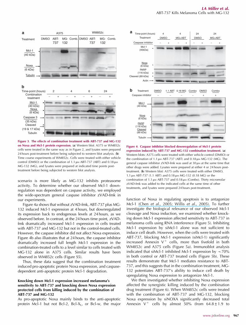
To investigate this hypothesis, we examined changes in Bcl-2family protein expression by Western blot. Only Noxa andMcl-1 protein levels had consistent and dramatic changesinduced by either one drug alone and/or the combination24 hours post-treatment (Figure 3a and Figure S3). ABT-737alone had little effects on most proteins examined, and MG-132 alone induced both Noxa and Mcl-1 levels substantially(Figure 3a). The combination treatment increased Noxaexpression to similar levels as MG-132 alone. However,surprisingly, Mcl-1 expression in cells treated with the drugcombination was reduced to levels similar to cells treated withvehicle control or ABT-737 alone (Figure 3a).
To examine the unexpected decrease in Mcl-1 expressionin cells treated with the drug combination compared withincreased levels of Mcl-1 in cells treated with MG-132 alone,
we performed time course experiments (WM852c in Figure3b and A375 in Figure S4). Results showed that Mcl-1 proteinexpression was induced by the combination treatment for upto 6 hours, but it was reduced to endogenous levels at24 hours. Caspase 3 was activated at 24 hours but not theearlier time points.
Downregulation of Mcl-1 at the protein level can beachieved through proteasomal degradation (Cuconati et al.,2003; Nijhawan et al., 2003; Willis et al., 2005) or caspase-dependent specific cleavage (Clohessy et al., 2004; Michelset al., 2004). In our case, we hypothesized that the latter
A375
120
100
80
60
40
20
0
120
100
80
60
40
20
0
120
100
80
60
40
20
0
MG-132 (μM)
MG-132 (μM)
MG-132 (μM)
HS294T
WM852
0 0.06 0.18 0.54 1.620.02
0 0.06 0.18 0.54 1.620.02
0 0.06 0.18 0.54 1.620.02
Via
bilit
y (%
)V
iabi
lity
(%)
Via
bilit
y (%
)
3.3 μM ABT-7370.37 μM ABT-737MG-132 alone 1.1 μM ABT-737
Figure 1. Combination of ABT-737 and MG-132 killed human melanoma
cells synergistically. Cells were seeded in 96-well plates 24 hours before
treatment with ABT-737, MG-132, or both drugs at indicated doses. MTS
assays were performed 48 hours post-treatment. Averaged media controls for
multiple plates were set as 100% viability; (a) A375 cells, (b) WM852c cells,
and (c) HS294T cells. Independent experiments have been repeated at least
three times, and similar results have been observed.
DMSO ABT-737 MG-132 Combination
A37
5W
M85
2
6050403020100In
crea
se in
ann
exin
V+ c
ells
(%
)
DMSO ABT-737 MG-132 Combination
45
32
9423
00
WM852c BaK WM852c Bax A375 BakA375 Bax*
****
1009080706050403020100
Incr
ease
in B
ak/B
ax-
activ
ated
cel
ls (
%)
DMSO ABT-737 MG-132 Combination
A375 WMB52
Figure 2. Combination of ABT-737 and MG-132 induced mitochondrial-
mediated apoptosis in melanoma cells synergistically. A375 and WM852c
melanoma cells were treated for 24 hours with DMSO control, 1.1 mM ABT-
737 and 0.18mM MG-132, either alone or in combination. Percentage of
apoptotic cells were analyzed using Annexin V staining. (a) Bright Field
microscopy images of A375 and WM852c cells before being detached for
staining. Scale bar¼ 100mM. (b) Average increase±SEM in apoptosis for
treated A375 and WM852c cells. One-way ANOVA demonstrated significant
differences among Annexin Vþ cells for all treatments of A375 (F¼55.9,
Fcrit¼ 4.07, P¼ 1�10�5) and WM852c (F¼ 5.5, Fcrit¼4.07, P¼ 0.02).
However, Tukey’s post hoc analysis for both cell lines indicated that only the
combination treatment significantly increased percentages of Annexin Vþ
cells compared with DMSO controls for both A375 cells (**Po0.01) and
WM852c cells (*Po0.05). (c) Average increase±SEM in Bak/Bax-activated
cells for treated WM852c or A375 cells. One-way ANOVA demonstrated
statistically significant differences among active Bak/Bax positive cells for all
treatments (Bak: F¼ 21.5, Fcrit¼ 4.07, P¼ 3�10�4; Bax: F¼ 54.9,
Fcrit¼ 4.07, P¼ 1�10�5), but only the combination treatment produced
significantly higher active Bak and Bax positive cells than DMSO control cells
(*Po0.05 and **Po0.01). Data summarized three independent experiments.
966 Journal of Investigative Dermatology (2009), Volume 129
LA Miller et al.ABT-737 Kills Melanoma Cells with MG-132
scenario is more likely as MG-132 inhibits proteasomeactivity. To determine whether our observed Mcl-1 down-regulation was dependent on caspase activity, we employedthe wide-spectrum general caspase inhibitor zVAD-fmk inour experiments.
Figure 4a shows that without zVAD-fmk, ABT-737 plus MG-132 induced Mcl-1 expression at 4 hours, but downregulatedits expression back to endogenous levels at 24 hours, as weobserved before. In contrast, at the 24 hours time point, zVAD-fmk dramatically increased Mcl-1 expression in cells treatedwith ABT-737 and MG-132 but not in the control-treated cells.However, the caspase inhibitor did not affect Noxa expression.Figure 4b also illustrates that at 24 hours, the caspase inhibitordramatically increased full length Mcl-1 expression in thecombination-treated cells to a level similar to cells treated withMG-132 alone in A375 cells. Similar results have beenobserved in WM852c cells (Figure S5).
Thus, these data suggest that the combination treatmentinduced pro-apoptotic protein Noxa expression, and caspase-dependent anti-apoptotic protein Mcl-1 degradation.
Knocking down Mcl-1 expression increased melanoma’ssensitivity to ABT-737 and knocking down Noxa expressionprotected cells from killing induced by the combination ofABT-737 and MG-132As pro-apoptotic Noxa mainly binds to the anti-apoptoticprotein Mcl-1 but not Bcl-2, Bcl-XL, or Bcl-w, the major
function of Noxa in regulating apoptosis is to antagonizeMcl-1 (Chen et al., 2005; Willis et al., 2005). To furtherinvestigate the biological relevance of our observed Mcl-1cleavage and Noxa induction, we examined whether knock-ing down Mcl-1 expression affected sensitivity to ABT-737 inmelanoma cells using RNA interference (Figure 5). InhibitingMcl-1 expression by siMcl-1 alone was not sufficient toinduce cell death. However, when the cells were treated withABT-737, blocking Mcl-1 expression (siMcl-1) significantlyincreased Annexin Vþ cells, more than fivefold in bothWM852c and A375 cells (Figure 5a). Immunoblot analysisindicated that siMcl-1 inhibited Mcl-1 expression by B70%in both control or ABT-737 treated cells (Figure 5b). Theseresults demonstrate that Mcl-1 mediates resistance to ABT-737, and this suggests that in the combination treatment, MG-132 potentiates ABT-737’s ability to induce cell death byupregulating Noxa expression to antagonize Mcl-1.
We then investigated whether inhibiting Noxa expressionaffected the synergistic killing induced by the combinationdrug treatment (Figure 6). When WM852c cells were treatedwith the combination of ABT-737 and MG-132, blockingNoxa expression by siNOXA significantly decreased totalAnnexin Vþ cells by almost 50% (from 64.8±1.9 to
A375
737 132737 132
WM852c
Treatment
McI-1
Noxa
Tubulin
(40 kDa)
(6 kDa)
McI-1
Time-point (hours) 2
− + + + +− − −
4 6 24Combination
treatment
Noxa
Tubulin(19 & 17 kDa)
Caspase 3(35 kDa)Cleaved
(40 kDa)
(6 kDa)
DMSO DMSO ABT- MG- Comb.ABT- MG- Comb.
Figure 3. The effects of combination treatment with ABT-737 and MG-132
on Noxa and Mcl-1 protein expression. (a) Western blot: A375 or WM852c
cells were treated in the same way as in Figure 2, and lysates were prepared
24 hours post-treatment before being subjected to western blot analysis. (b)
Time course experiments of WM852c. Cells were treated with either vehicle
control (DMSO) or the combination of 1.1 mM ABT-737 (ABT) and 0.18mM
MG-132 (MG), and lysates were prepared at indicated time points post-
treatment before being subjected to western blot analysis.
Time-point (hours)
Treatment
Treatment
Caspase inhibitor
Caspase inhibitor
4 4
DMSO DMSO
DMSO
MG+ABT MG+ABT
− + − + − + − +
Mcl-1(40kDa)
Noxa(6 kDa)
PARP(116 kDa)
Cleaved (89 kDa)
Actin
Tubulin
Cleaved(89 kDa)
PARP(116 kDa)
Noxa(6 kDa)
Mcl-1(40 kDa)
1.1 ABT 0.18 MG Combo ComboDMSO
++−−−−
24 24
Figure 4. Caspase inhibitor blocked downregulation of Mcl-1 protein
expression induced by ABT-737 and MG-132 combination treatment. (a)
Western blots: A375 cells were treated with either vehicle control (DMSO) or
the combination of 1.1mM ABT-737 (ABT) and 0.18mM MG-132 (MG). The
general caspase inhibitor zVAD-fmk was used at 30 mM at the same time that
other drugs were added. Lysates were prepared at either 4 or 24 hours post-
treatment. (b) Western blot: A375 cells were treated with either DMSO,
1.1mM ABT-737 (1.1 ABT) and 0.18 mM MG-132 (0.18 MG) or the
combination of 1.1 mM ABT-737 and 0.18mM (Combo). Thirty micromolar
zVAD-fmk was added to the indicated cells at the same time of other
treatments, and lysates were prepared 24 hours post-treatment.
www.jidonline.org 967
LA Miller et al.ABT-737 Kills Melanoma Cells with MG-132
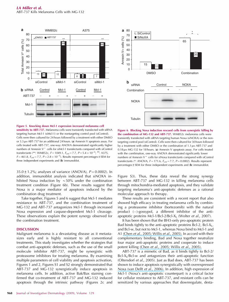
35.0±1.2%; analyses of variance (ANOVA), P¼0.0002). Inaddition, immunoblot analysis indicated that siNOXA in-hibited Noxa induction by B50% under the combinationtreatment condition (Figure 6b). These results suggest thatNoxa is a major mediator of apoptosis induced by thecombination drug treatment.
Take together, Figures 5 and 6 suggest that Mcl-1 mediatesresistance to ABT-737, and the combination treatment ofMG-132 and ABT-737 antagonizes Mcl-1 through increasedNoxa expression and caspase-dependent Mcl-1 cleavage.These observations explain the potent synergy observed forthe combination treatment.
DISCUSSIONMalignant melanoma is a devastating disease as it metasta-sizes early and is highly resistant to all conventionaltreatments. This study investigates whether the strategies thatcombat anti-apoptotic defenses, such as the use of the smallmolecule inhibitor ABT-737, might be synergistic withproteasome inhibitors for treating melanoma. By examiningmultiple parameters of cell viability and apoptosis activation,Figures 1 and 2, Figures S1 and S2 strongly demonstrated thatABT-737 and MG-132 synergistically induce apoptosis inmelanoma cells. In addition, active Bak/Bax staining con-firmed that co-treatment of ABT-737 and MG-132 inducedapoptosis through the intrinsic pathway (Figures 2c and
Figure S3). Thus, these data reveal the strong synergybetween ABT-737 and MG-132 in killing melanoma cellsthrough mitochondria-mediated apoptosis, and they validatetargeting melanoma’s anti-apoptotic defenses as a rationalmolecular approach to therapy.
These results are consistent with a recent report that alsoshowed high efficacy in treating melanoma cells by combin-ing a proteasome inhibitor (bortezomib) with the naturalproduct (�)-gossypol, a different inhibitor of the anti-apoptotic proteins Mcl-1/Bcl-2/Bcl-XL (Wolter et al., 2007).
It has been shown that the BH3-only pro-apoptotic proteinBad binds tightly to the anti-apoptotic proteins Bcl-2, Bcl-XL,and Bcl-w, but not to Mcl-1, whereas Noxa bind to Mcl-1 andA1 (Chen et al., 2005; Willis et al., 2005). In accord with theircomplementary binding, Bad and Noxa together neutralizefour major anti-apoptotic proteins and cooperate to inducepotent killing (Chen et al., 2005; Willis et al., 2005).
ABT-737 is a mimetic of Bad, as it binds tightly to Bcl-2/Bcl-XL/Bcl-w and antagonizes their anti-apoptotic function(Oltersdorf et al., 2005). Just as Bad does, ABT-737 has beenshown to induce apoptosis synergistically with overexpressedNoxa (van Delft et al., 2006). In addition, high expression ofMcl-1 (Noxa’s anti-apoptotic counterpart) is a critical factorfor cellular resistance to ABT-737, and resistant cells can besensitized by various approaches that downregulate, desta-
Mcl-1 Mcl-1
Mcl-1
13 10 128 8 7
6668
DMSO ABT-737
siRNA
ABT-737
Tubulin
ControlControl
− − − −+ + + +
** **
Tota
l Ann
exin
V+ c
ells
(%
)
WM852c
siControl siControlsiMcl-1 siMcl-1
A37580
70
60
50
40
30
20
10
0
Figure 5. Knocking down Mcl-1 expression increased melanoma cell
sensitivity to ABT-737. Melanoma cells were transiently transfected with siRNA
targeting human Mcl-1 (siMcl-1) or the nontargeting control pool (siControl).
Cells were then cultured for 24 hours followed by a treatment with either DMSO
or 3.3mM ABT-737 for an additional 24 hours. (a) Annexin V apoptosis assay. For
cells treated with ABT-737, one-way ANOVA demonstrated significantly higher
numbers of Annexin Vþ cells for siMcl-1 transfectants compared with siControl
transfectants (**: WM852c, F¼1049.1, Fcrit¼7.7, P¼5.4� 10�6; ##: A375,
F¼ 461.8, Fcrit¼ 7.7, P¼2.8� 10�5). Results represent percentages±SEM for
three independent experiments and (b) immunoblot.
Ann
exin
V+
cel
ls (
%)
80
70
60
50
40
30
20
10
0
1511
65
35
Combination
DMSO Combination
SiControlSiNoXA
Control
− + − +
NOXA
NOXA
siRNA
Tubulin
*
Figure 6. Blocking Noxa induction rescued cells from synergistic killing by
the combination of MG-132 and ABT-737. WM852c melanoma cells were
transiently transfected with siRNA targeting human Noxa (siNOXA) or the non-
targeting control pool (siControl). Cells were then cultured for 30hours followed
by a treatment with either DMSO or the combination of 1.1mM ABT-737 and
0.18mM MG-132 for 18hours. (a) Annexin V apoptosis assay. For cells treated
with the combination, one-way ANOVA demonstrated significantly lower
numbers of Annexin Vþ cells for siNoxa transfectants compared with siControl
transfectants (*: ANOVA, F¼ 171.6, Fcrit¼7.7, P¼ 0.0002). Results represent
percentages±SEM for three independent experiments and (b) immunoblot.
968 Journal of Investigative Dermatology (2009), Volume 129
LA Miller et al.ABT-737 Kills Melanoma Cells with MG-132
bilize, or inactivate Mcl-1 (Konopleva et al., 2006; van Delftet al., 2006; Chen et al., 2007a, b). We found that knockingdown Mcl-1 expression significantly increased melanomacell sensitivity to ABT-737 (Figure 5), indicating that Mcl-1 isthe main barrier for ABT-737-induced cell death.
In melanoma, proteasome inhibitors such as MG-132 andbortezomib have been reported to induce high levels of Noxaexpression and lead to Noxa-mediated apoptosis (Fernandezet al., 2005; Qin et al., 2005). We hypothesized that ABT-737would be synergistic with proteasome inhibitor such as MG-132 in killing melanoma cells, as ABT-737 blocks thefunction of Bcl-2, Bcl-XL, Bcl-w, whereas MG-132 wouldinduce Noxa production, neutralizing the anti-apoptoticfunction of Mcl-1. Thus, ABT-737 and MG-132 togetherwould inhibit four major anti-apoptotic proteins to synergis-tically induce apoptosis.
We found that the combination of ABT-737 and MG-132indeed induced high levels of Noxa expression, similar toMG-132 treatment alone (Figures 3 and Figure S4a). We alsodemonstrated that the combination induced Noxa-dependentapoptosis (Figure 6), even though Noxa induction alone wasnot sufficient to induce apoptosis effectively in these cells(see MG-132 treatment alone in Figures 1 and 2). Thus, oneof the mechanisms for potent killing synergy between ABT-737 and MG-132 is that the combination acts similarly tooverexpressing Bad plus Noxa. As a result, the combinationtreatment effectively neutralizes all four main anti-apoptoticproteins, therefore promoting cooperation to induce potentkilling in melanoma.
Interestingly, proteasome inhibitors also have been reportedto induce accumulation of the anti-apoptotic protein Mcl-1(Fernandez et al., 2005; Qin et al., 2005), and reducing Mcl-1levels (using siRNA, UV light, or fludarabine) synergizes incausing proteasome-inhibitor-induced cell death in melanomacells (Fernandez et al., 2005; Qin et al., 2005; Gomez-Bougieet al., 2007). Even partial downregulation of Mcl-1 causes atwo-fold increase in proteasome-inhibitor-induced melanomakilling (Qin et al., 2006), and more efficient reduction of Mcl-1expression dramatically increases and accelerates bortezomib-induced cell death (Wolter et al., 2007).
Although we also observed substantial Mcl-1 accumula-tion up to at least 24 hours in cells treated with MG-132 aloneand at early time points of combination treatment, full lengthMcl-1 was quickly degraded in the combination treatment at24 hours (Figures 3 and Figure S4). This rapid destruction ofMcl-1 might also contribute to potent synergy; likely becauseABT-737 plus MG-132 simultaneously triggers Noxa produc-tion (by MG-132) and reduces Mcl-1 levels (by ABT-737 plusMG-132).
Rapid degradation of Mcl-1 can be induced by certaincytotoxic triggers, including UV irradiation (Nijhawan et al.,2003; Willis et al., 2005), DNA damage by viral infection(Cuconati et al., 2003) and detachment (Woods et al., 2007).Further, destruction of Mcl-1 is essential for apoptosisinduced by these stimuli (Cuconati et al., 2003; Nijhawanet al., 2003; Willis et al., 2005; Woods et al., 2007), andelimination of Mcl-1 may help to ensure irreversiblecommitment to apoptosis.
In our case, the combination of ABT-737 and MG-132induced Mcl-1 degradation swiftly in melanoma cells, whichmay be another reason for the potent killing synergy seen inthe combination of these two compounds. This rapiddestruction of Mcl-1 might help overcome cellular resistanceto both MG-132 and ABT-737. As shown in Figure 4, the wide-spectrum caspase inhibitor zVAD-fmk almost completelyblocked Mcl-1 degradation induced by co-treatment withABT-737 and MG-132. This demonstrates that Mcl-1 degrada-tion in the combination treatment is mainly caspase-depen-dent, although this might only be a by-product of the apoptoticcascade. However, studies have shown that caspase-depen-dent Mcl-1 cleavage can be induced by various stimuli,including the proteasome inhibitor bortezomib, for example,in multiple myeloma (Clohessy et al., 2004; Michels et al.,2004; Gomez-Bougie et al., 2007). Further, caspase-depen-dent Mcl-1 cleavage has been shown not only to abrogateMcl-1’s anti-apoptotic function, but also to generate pro-apoptotic Mcl-1-truncated proteins which enhance the deathsignal (Clohessy et al., 2004; Michels et al., 2004). Moreover,a recent study demonstrated that cleavage-resistant Mcl-1potently inhibits mitochondrial-mediated apoptosis (Chenet al., 2007a, b). Thus, we propose that caspase-dependentMcl-1 cleavage is also a part of a positive feedback loop in ourstudy, which accelerates and synergizes the killing effects ofABT-737 combined with MG-132 in treating melanoma.
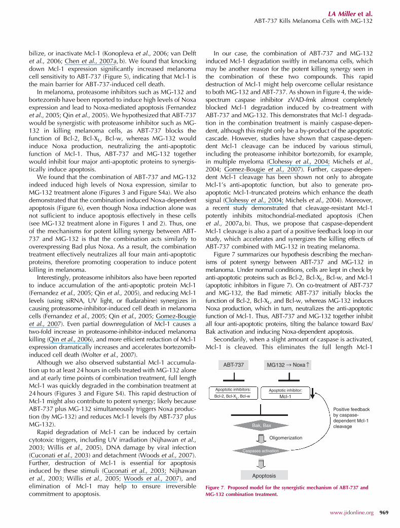
Figure 7 summarizes our hypothesis describing the mechan-isms of potent synergy between ABT-737 and MG-132 inmelanoma. Under normal conditions, cells are kept in check byanti-apoptotic proteins such as Bcl-2, Bcl-XL, Bcl-w, and Mcl-1(apoptotic inhibitors in Figure 7). On co-treatment of ABT-737and MG-132, the Bad mimetic ABT-737 initially blocks thefunction of Bcl-2, Bcl-XL, and Bcl-w, whereas MG-132 inducesNoxa production, which in turn, neutralizes the anti-apoptoticfunction of Mcl-1. Thus, ABT-737 and MG-132 together inhibitall four anti-apoptotic proteins, tilting the balance toward Bax/Bak activation and inducing Noxa-dependent apoptosis.
Secondarily, when a slight amount of caspase is activated,Mcl-1 is cleaved. This eliminates the full length Mcl-1
MG132ABT-737
Apoptotic inhibitors:
BcI-2, BcI-XL, BcI-w McI-1Apoptotic inhibitor:
Positive feedbackby caspase-dependent McI-1cleavage
Apoptosis
Caspases activation
Oligomerization
Bak, Bax
Noxa ↑
↑
Figure 7. Proposed model for the synergistic mechanism of ABT-737 and
MG-132 combination treatment.
www.jidonline.org 969
LA Miller et al.ABT-737 Kills Melanoma Cells with MG-132
(induced by MG-132) quickly, and generates pro-apoptoticMcl-1 cleavage products that synergize even further in thedeath signal. Therefore, caspase-dependent Mcl-1 cleavageproduces a positive feedback signal for apoptosis induced bythe combination of ABT-737 and MG-132.
In conclusion, this study demonstrates that ABT-737lowers the apoptotic threshold for the proteasome inhibitorMG-132 in melanoma to induce Noxa-dependent mitochon-drial-mediated apoptosis very effectively in melanoma cells.These effects are probably due to both drugs’ combinedability to neutralize multiple anti-apoptotic proteins andinduce caspase-dependent Mcl-1 cleavage to create apositive feedback death signal. These results not only providescientific basis for further animal and clinical studies toevaluate the efficacy of this combination in treatingmelanomas, but also implicate the importance of testingadditional drug combinations that simultaneously targetmultiple anti-apoptotic defenses.
MATERIALS AND METHODSCell lines and culture conditionsA375, an advanced vertical growth phase human melanoma cell
line, was obtained from ATCC (Manassas, VA). WM852c and
HS294T, two metastatic human melanoma cell lines, were kindly
provided by Dr. Meenhard Herlyn (Wistar Institute, Philadelphia,
PA). Cells were maintained in RPMI1640 (Invitrogen, Grand Island,
NY) with 10% fetal bovine serum (Gemini Bio-Products Inc., West
Sacramento, CA). A375 has a BRAFV600E mutation and no common
mutations in NRAS (exon 1 or 2), and WM852c has an NRASQ61R
mutation but no BRAF mutation (exon 11 or 15).
ReagentsMG-132 and z-VAD-FMK were purchased from EMD Biosciences
(San Diego, CA) and R&D Systems (Minneapolis, MN), respectively.
ABT-737 was kindly provided by Abbott Laboratories (Abbott Park, IL).
Cell titer 96 aqueous one solution cell proliferation assay forquantification of cell viability (MTS assay)
The reagents were obtained from Promega (Madison, WI), and pro-
cedures were followed as previously described (Shellman et al., 2005).
Measurement of apoptosis using Annexin V staining
The Annexin V–fluorescein isothiocyanate Apoptosis Detection Kit
(BD Biosciences, San Jose, CA) was used according to the
manufacturer’s protocol. Cells were analyzed by flow cytometry
using a Beckman Coulter FC500 with CXP software (Hialeah, FL) in
the University of Colorado Cancer Center Flow Cytometry Core.
Immunoblot
Cells, both floating and adherent, were harvested with 1� Laemmli
sample buffer (Bio-Rad, Hercules, CA). Samples were used in the
standard western blot analysis protocol as described previously (Ruth
et al., 2006). Blots were developed with horseradish peroxidase
substrate (West Pico or Femto solutions, Pierce, Rockford, IL) for
5 minutes at room temperature, and analyzed using a Chemi-doc
chemiluminescence detector (Bio-Rad). The following antibodies
were used at the suggested dilution from the manufacturers: caspase
3, poly (ADP-ribose) polymerase, Bax, and tubulin a/b from Cell
Signaling Technology (Danvers, MA), Noxa, anti-actin mouse
monoclonal antibody and horseradish peroxidase-conjugated goat
anti-mouse immunoglobulin M from EMD Biosciences Inc., Bad,
Bcl-x, Mcl-1, and horseradish peroxidase-conjugated goat anti-
rabbit immunoglobulin G from BD Biosciences, Bim from Chemicon
(Temecula, CA), Bcl-2 from Dako (Glostrup, Denmark), and horse-
radish peroxidase-conjugated goat anti-mouse IgG from Jackson
Immuno-Research (West Grove, PA).
Measurement of mitochondrial-mediated apoptosis with activeBax/Bak staining
Cells were stained with a protocol modified as previously described
(Qin et al., 2006). Cells were detached and fixed in 2%
formaldehyde (PolySciences Inc., Warrington, PA) (10 minutes,
room temperature), and then washed with FACS buffer (phosphate-
buffered saline solution containing 5% fetal bovine serum and
0.02% sodium azide) three times. Cells were permeabilized
briefly with 0.03% saponin/FACS buffer before incubated with
primary antibody in the same buffer (30 minutes, room temperature).
Mouse anti-active-human-Bax antibody (BD Pharmingen,
San Jose, CA) at a 1:100 dilution, and mouse anti-active-human-
Bak antibody (EMD Biosciences) was used at a 1:250 dilution.
Fluorescein isothiocyanate-conjugated goat anti-mouse antibody
(Jackson ImmunoResearch, West Grove, PA) were used in 0.3%
saponin/FACS buffer at a 1:750 dilution (30 minutes in the dark,
room temperature). FACS buffer was used as the washing solution for
three times after incubation with each antibody. Stained cells were
analyzed by flow cytometry in the University of Colorado Cancer
Center Flow Cytometry Core. Average percent increase in Bak/Bax
activated cell was calculated as the percent increase of active Bakþ /
Baxþ cells in treated conditions over the DMSO controls.
RNA interference
ON-TARGETplus SMARTpools of siRNA were purchased from
Dharmacon Inc. (Lafayette, CO), targeting human Noxa (PMAIP1,
NM_021127), human Mcl-1 (MCL1, NM_182763), and the non-
targeting control pool. siRNA was introduced into cells using an
Amaxa Nucleofector II device (Amaxa Inc., Gaithersburg, MD). One
million cells and 2.5 mM siRNA were used with Amaxa’s Reagent V
and program X-005 for each reaction.
Cells were nucleofected with indicated siRNA and cultured for
indicated amount of time. Then all medium was removed and fresh
medium containing indicated compounds for indicated time before
the cells were harvested for analysis.
Statistical analysisStatistical analyses were performed similarly for both Annexin V
staining and active Bak/Bax staining. One-way ANOVA were
performed to compare group means, and Tukey’s post hoc tests
were used to compare treated samples to DMSO controls.
CONFLICT OF INTERESTThe authors state no conflict of interest.
ACKNOWLEDGMENTSThis work was supported in part by NIAMS grant R01AR26427-18 (D.A.N.)and merit grant PN: 0012 (D.A.N.) from the Department of Veterans Affairs,Veterans Health Administration, Office of Biomedical Laboratory Researchand Development. We thank Abbott Laboratories (Saul Rosenberg, Steven
970 Journal of Investigative Dermatology (2009), Volume 129
LA Miller et al.ABT-737 Kills Melanoma Cells with MG-132
Elmore and colleagues) for providing ABT-737 compound. We also thankKaren Helm and Christine Childs at the University of Colorado Cancer CenterFlow Cytometry Core (supported by NIH grant P30 CA 046934) for theirexpert technical assistance, and Ian Maxwell for his critique of the paper.
SUPPLEMENTARY MATERIAL
Supplemental Results, Material and Methods
Figure S1. Combination of ABT-737 and MG-132 killed melanoma cellssynergistically demonstrated by Combination Index values.
Figure S2. Combination of ABT-737 and MG-132 killed melanoma cellssynergistically as analyzed by EB/AO staining.
Figure S3. The effects of combination treatment with ABT-737 and MG-132on Bcl-2 family member protein expression.
Figure S4. The effects of combination treatment with ABT-737 and MG-132on Noxa and Mcl-1 protein expression.
Figure S5. Caspase inhibitor zVAD-fmk blocked downregulation of Mcl-1protein expression induced by ABT-737 and MG-132 combination treatmentin WM852c cells.
REFERENCES
Adams J (2004) The development of proteasome inhibitors as anticancerdrugs. Cancer Cell 5:417–21
Adams JM, Huang DC, Strasser A, Willis S, Chen L, Wei A et al. (2005)Subversion of the Bcl-2 life/death switch in cancer development andtherapy. Cold Spring Harb Symp Quant Biol 70:469–77
Buzaid AC (2004) Management of metastatic cutaneous melanoma.Oncology (Williston Park) 18:1443–50; discussion 1457–9
Certo M, Del Gaizo Moore V, Nishino M, Wei G, Korsmeyer S, Armstrong SAet al. (2006) Mitochondria primed by death signals determine cellularaddiction to antiapoptotic BCL-2 family members. Cancer Cell 9:351–65
Chauhan D, Velankar M, Brahmandam M, Hideshima T, Podar K, RichardsonP et al. (2007) A novel Bcl-2/Bcl-X(L)/Bcl-w inhibitor ABT-737 as therapyin multiple myeloma. Oncogene 26:2374–80
Chen L, Willis SN, Wei A, Smith BJ, Fletcher JI, Hinds MG et al. (2005)Differential targeting of prosurvival Bcl-2 proteins by their BH3-onlyligands allows complementary apoptotic function. Mol Cell 17:393–403
Chen M, Guerrero AD, Huang L, Shabier Z, Pan M, Tan TH et al. (2007a)Caspase-9-induced mitochondrial disruption through cleavage of anti-apoptotic BCL-2 family members. J Biol Chem 282:33888–95
Chen S, Dai Y, Harada H, Dent P, Grant S (2007b) Mcl-1 down-regulationpotentiates ABT-737 lethality by cooperatively inducing Bak activationand Bax translocation. Cancer Res 67:782–91
Clohessy JG, Zhuang J, Brady HJ (2004) Characterisation of Mcl-1 cleavageduring apoptosis of haematopoietic cells. Br J Haematol 125:655–65
Cuconati A, Mukherjee C, Perez D, White E (2003) DNA damage responseand MCL-1 destruction initiate apoptosis in adenovirus-infected cells.Genes Dev 17:2922–32
Cummins DL, Cummins JM, Pantle H, Silverman MA, Leonard AL, ChanmugamA (2006) Cutaneous malignant melanoma. Mayo Clin Proc 81:500–7
Fernandez Y, Verhaegen M, Miller TP, Rush JL, Steiner P, Opipari AW Jr et al.(2005) Differential regulation of noxa in normal melanocytes andmelanoma cells by proteasome inhibition: therapeutic implications.Cancer Res 65:6294–304
Gogas HJ, Kirkwood JM, Sondak VK (2007) Chemotherapy for metastaticmelanoma: time for a change? Cancer 109:455–64
Gomez-Bougie P, Wuilleme-Toumi S, Menoret E, Trichet V, Robillard N,Philippe M et al. (2007) Noxa up-regulation and Mcl-1 cleavage areassociated to apoptosis induction by bortezomib in multiple myeloma.Cancer Res 67:5418–24
Griffiths GJ, Dubrez L, Morgan CP, Jones NA, Whitehouse J, Corfe BM et al.(1999) Cell damage-induced conformational changes of the pro-apoptotic protein Bak in vivo precede the onset of apoptosis. J CellBiol 144:903–14
Kang MH, Kang YH, Szymanska B, Wilczynska-Kalak U, Sheard MA, HarnedT et al. (2007) Activity of vincristine, L-ASP, and dexamethasone against
acute lymphoblastic leukemia is enhanced by the BH3-mimeticABT-737 in vitro and in vivo. Blood 110:2057–66
Kohl TM, Hellinger C, Ahmed F, Buske C, Hiddemann W, Bohlander SK et al.(2007) BH3 mimetic ABT-737 neutralizes resistance to FLT3 inhibitortreatment mediated by FLT3-independent expression of BCL2 in primaryAML blasts. Leukemia 21:1763–72
Konopleva M, Contractor R, Tsao T, Samudio I, Ruvolo PP, Kitada S et al.(2006) Mechanisms of apoptosis sensitivity and resistance to the BH3mimetic ABT-737 in acute myeloid leukemia. Cancer Cell 10:375–88
Letai A (2005) BCL-2: found bound and drugged!. Trends Mol Med 11:442–4
Michels J, O’Neill JW, Dallman CL, Mouzakiti A, Habens F, Brimmell M et al.(2004) Mcl-1 is required for Akata6 B-lymphoma cell survival and isconverted to a cell death molecule by efficient caspase-mediatedcleavage. Oncogene 23:4818–27
Nechushtan A, Smith CL, Hsu YT, Youle RJ (1999) Conformation of the BaxC-terminus regulates subcellular location and cell death. EMBO J18:2330–41
Nijhawan D, Fang M, Traer E, Zhong Q, Gao W, Du F et al. (2003)Elimination of Mcl-1 is required for the initiation of apoptosis followingultraviolet irradiation. Genes Dev 17:1475–86
Oltersdorf T, Elmore SW, Shoemaker AR, Armstrong RC, Augeri DJ, Belli BAet al. (2005) An inhibitor of Bcl-2 family proteins induces regression ofsolid tumours. Nature 435:677–81
Qin JZ, Xin H, Sitailo LA, Denning MF, Nickoloff BJ (2006) Enhanced killingof melanoma cells by simultaneously targeting Mcl-1 and NOXA. CancerRes 66:9636–45
Qin JZ, Ziffra J, Stennett L, Bodner B, Bonish BK, Chaturvedi V et al. (2005)Proteasome inhibitors trigger NOXA-mediated apoptosis in melanomaand myeloma cells. Cancer Res 65:6282–93
Ruth MC, Xu Y, Maxwell IH, Ahn NG, Norris DA, Shellman YG (2006) RhoCpromotes human melanoma invasion in a PI3K/Akt-dependent pathway.J Invest Dermatol 126:862–8
Schwartz R, Davidson T (2004) Pharmacology, pharmacokinetics, andpractical applications of bortezomib. Oncology (Williston Park)18(Suppl 11):14–21
Shellman YG, Ribble D, Miller L, Gendall J, Vanbuskirk K, Kelly D et al.(2005) Lovastatin-induced apoptosis in human melanoma cell lines.Melanoma Res 15:83–9
Shoemaker AR, Oleksijew A, Bauch J, Belli BA, Borre T, Bruncko M et al.(2006) A small-molecule inhibitor of Bcl-XL potentiates the activity ofcytotoxic drugs in vitro and in vivo. Cancer Res 66:8731–39
Soengas MS, Lowe SW (2003) Apoptosis and melanoma chemoresistance.Oncogene 22:3138–51
Thomadaki H, Scorilas A, Hindmarsh JT (2006) BCL2 family of apoptosis-related genes: functions and clinical implications in cancer. Crit Rev ClinLab Sci 43:1–67
van Delft MF, Wei AH, Mason KD, Vandenberg CJ, Chen L, Czabotar PE et al.(2006) The BH3 mimetic ABT-737 targets selective Bcl-2 proteins andefficiently induces apoptosis via Bak/Bax if Mcl-1 is neutralized. CancerCell 10:389–99
Wei MC, Zong WX, Cheng EH, Lindsten T, Panoutsakopoulou V, Ross AJ et al.(2001) Proapoptotic BAX and BAK: a requisite gateway to mitochondrialdysfunction and death. Science 292:727–30
Weinstein IB, Joe AK (2006) Mechanisms of disease: oncogene addiction—arationale for molecular targeting in cancer therapy. Nat Clin Pract Oncol3:448–57
Willis SN, Chen L, Dewson G, Wei A, Naik E, Fletcher JI et al. (2005)Proapoptotic Bak is sequestered by Mcl-1 and Bcl-xL, but not Bcl-2, untildisplaced by BH3-only proteins. Genes Dev 19:1294–305
Wolter KG, Verhaegen M, Fernandez Y, Nikolovska-Coleska Z, Riblett M,Martin de la Vega C et al. (2007) Therapeutic window for melanomatreatment provided by selective effects of the proteasome on Bcl-2proteins. Cell Death Differ 14:1605–1616
Woods NT, Yamaguchi H, Lee FY, Bhalla KN, Wang HG (2007) Anoikis,initiated by Mcl-1 degradation and Bim induction, is deregulated duringoncogenesis. Cancer Res 67:10744–52
www.jidonline.org 971
LA Miller et al.ABT-737 Kills Melanoma Cells with MG-132