diTFPP, a Phenoxyphenol, Sensitizes Hepatocellular ... - MDPI

IntroductionThe growth and metastasis of mela-noma together with its resistance totherapy present major obstacles tomost conventional therapies. CREB, c-Jun, ATF1, ATF2, Stat3, and NF-κBare among transcription factors shownto play an important role in the courseof melanoma development and pro-gression (1–3). ATF2 has been found toplay an important role in melanoma’s
proliferation (4) and resistance to treat-ment (1, 5). Attenuating the activitiesof ATF2 appears to be sufficient to sen-sitize melanoma to treatment (5, 6).
ATF2 is a member of the ATF/CREBprotein family of basic-region leucinezipper (bZIP) proteins (7), which areinvolved in the response to stress (8).Transcriptionally active ATF2 recog-nizes and binds specific ATF/CREmotifs as a homo- or a heterodimerform (7, 8). Stimulation of ATF2 tran-scriptional activity can result from itsphosphorylation by the stress kinasesp38 or JNK (9), as well as from its inter-action with any of several transcriptionfactors, including c-Jun (10), NF-κB(11), and retinoblastoma protein (12).ATF2 has been implicated in the regu-lation of a wide set of genes that playroles in the regulation of cell growth,differentiation, immune response, andapoptosis, including c-Jun (8), TNF-α(13), TGF-β (12), cyclin A (14), and E-selectin (11). ATF2 has also beenimplicated in cellular proliferation in
vitro and tumor formation in vivothrough its cooperation with v-Jun (15).Similarly, c-Jun–ATF2 dimers have beenimplicated in oncogenesis (16).
Hypophosphorylated or transcrip-tionally inactive forms of ATF2 reduceTNF-α expression, resulting in sensiti-zation of melanoma to treatment viaincreased apoptosis (5, 17). Screening ofATF2-driven peptides identified aminoacids 50–100 (ATF250–100) as capable ofsensitizing cultured melanoma cells toapoptosis induced by chemotherapeu-tic drugs, ribotoxic agents, or inhibitorsof stress kinases (6). ATF250–100 containsthe phosphoacceptor sites for JNK orp38, the binding domain for JNK (18),and has been implicated in p300-dependent transcriptional activation(19). Here we demonstrate using in vivomodels that the ATF50–100 peptide effi-ciently inhibits growth and metastasisof melanoma and sensitizes humanand mouse melanoma tumors to treat-ment and points to the mechanismunderlying ATF2 peptide’s ability toelicit these effects.
MethodsCells. SW1 and B16F10 mousemelanoma cells were maintained inDMEM supplemented with 10% FBS,L-glutamine, and antibiotics.
Constructs. ATF2 peptides werecloned in frame into a hemagglutin-penetratin (HA-penetratin) pcDNA3vector (6). ATF2 peptide, correspon-ding to amino acids 50–100 ofhuman ATF2, shares 100% homolo-gy with the murine sequence (corre-sponding amino acids in the mouseATF2 cDNA are 33–82). Jun2-luciferase (Jun2-luc) and TRE-lucconstructs were previously described(8, 10). The ATF250–100 peptide was
The Journal of Clinical Investigation | September 2002 | Volume 110 | Number 5 643
An ATF2-derived peptide sensitizesmelanomas to apoptosis and inhibits their growth and metastasis
Anindita Bhoumik,1 Tian-Gui Huang,2 Vladimir Ivanov,1 Lisa Gangi,3 Rui F. Qiao,1
Savio L.C. Woo,2 Shu-Hsia Chen,2 and Ze’ev Ronai1
1Ruttenberg Cancer Center, and 2The Carl C. Icahn Institute for Gene Therapy and Molecular Medicine, Mount Sinai School of Medicine, New York, New York, USA3Laboratory of Molecular Technology, National Cancer Institute–Science Applications International Corporation,Frederick, Maryland, USA
Melanomas are among the aggressive tumor types because of their notoriousresistance to treatment and their high capacity to metastasize. ATF2 is amongtranscription factors implicated in the progression of melanoma and its resist-ance to treatment. Here we demonstrate that the expression of a peptide span-ning amino acids 50–100 of ATF2 (ATF250–100) reduces ATF2 transcriptionalactivities while increasing the expression and activity of c-Jun. Altering the bal-ance of Jun/ATF2 transcriptional activities sensitized melanoma cells to apop-tosis, an effect that could be attenuated by inhibiting c-Jun. Inhibition of ATF2via RNA interference likewise increased c-Jun expression and primed melanomacells to undergo apoptosis. Growth and metastasis of SW1 and B16F10 mousemelanomas were inhibited by ATF250–100 to varying degrees up to a completeregression, depending on the mode (inducible, constitutive, or adenoviral deliv-ery) of its expression. Thus, by attenuating ATF2 and inducing c-Jun activity,ATF250–100 inhibits melanoma growth and metastasis.
This article was published online in advance of the print edition.The date of publication is available from the JCI website, http://www.jci.org.
J. Clin. Invest. 110:643–650 (2002). doi:10.1172/JCI200216081.
Rapid Publication
Received for publication June 5, 2002, and acceptedin revised form July 9, 2002.
Address correspondence to: Ze’ev Ronai, The Ruttenberg Cancer Center, Mount SinaiSchool of Medicine, One Gustave L. LevyPlace, Box 1130, New York, New York 10029,USA. Phone: (212) 659-5571; Fax: (212) 849-2425; E-mail: [email protected] of interest: No conflict of interesthas been declared.Nonstandard abbreviations used: luciferase(luc); wild-type (WT); Fas ligand (FasL); TNF-related apoptosis inducing ligand (TRAIL);TRAIL receptor 1 (TRAIL-R1); β-galactosidase(β-gal); hemagglutin (HA); small interferingRNAs (RNAs).

cloned in frame with HA and pene-tratin into the SpeI and NotI sites inthe PadL1-RSV-BPA vector (20). Forthe doxycycline-inducible system theATF250–100 peptide was cloned inframe with HA and penetratin intoSacII and XbaI sites of the PUHD10-3 vector (21). The PeF1PrtTA vector(22) places the rtTA gene under tran-scriptional control of the human EF-1α promoter. Replication-deficientadenovirus expressing ATF250–100
was generated by subcloning the
ATF2 peptide cDNA into the corre-sponding adenovirus vector as previ-ously described (20).
Cell culture and derivation of stable cellline. Selection of clones stably express-ing the rtTA construct was carried outin SW1 cells in the presence of 600µg/ml of G418. After the selection peri-od, we picked and expanded 12 singlecolonies and analyzed them for activityby assaying for luciferase activity (22).The clone that exhibited eightfoldinduction upon addition of doxycycline
was chosen. The positive clone, whichexpresses the rtTA cDNA, was cotrans-fected with PUHD10-3 ATF250–100 andpBabe Puro, and single clones wereselected with 2 µg/ml of Puromycin.
Transcriptional analysis. Transienttransfection of different reporter con-structs (0.5 µg) with expression vectorsand pCMV-βgal (0.1 µg) into 5 × 10⁵
melanoma cells was performed usingLipofectamine (Life Technologies Inc.,Carlsbad, California, USA). Luciferaseactivity was determined as previouslydescribed (23).
RNA interference. RNAs of 21nucleotides (24), designed to targetmouse ATF2 within nucleotides119–135 (sense: 5′-AGCACGUAAU-GACAGUGUCTT-3′; antisense: 5′- GAC-ACUGUCAUUACGUGCUTT-3′), were syn-thesized, deprotected, and HPLC-purified (Midland Certified ReagentCo., Midland, Texas, USA). For anneal-ing of small interfering RNAs (siRNAs),20 µM single strands were incubatedin annealing buffer (100 mM potassi-um acetate, 30 mM HEPES-KOH atpH 7.4, 2 mM magnesium acetate) for1 minute at 90°C followed by 1 hourat 37°C. siRNAs were transfected intoSW1 cells (2.4 µg siRNA duplex per six-well plate) using Lipofectamine (LifeTechnologies Inc.). siRNAs of RNF5, aRING finger protein important incytoskeleton organization (our unpub-lished studies), were used as control.
Treatment and apoptosis studies. Cellswere exposed to concentrations of thechemicals (10 µM UCN-01-7-hydrox-ystaurosporine [UCN-01], 500 µg/mlneocarzinostatin [NCS], 10 µg/ml ani-somycin) or of the pharmacologicalinhibitors (50 µM LY294002, aninhibitor of phosphatidylinositol [PI]3-kinase; 50 µM PD98059, an inhibitorof mitogen-activated protein kinase[MAPK]; 50 µM AG490, an inhibitor ofJAK; 10 µM SB203580, an inhibitor ofp38/JNK) for 36 hours; then FACSanalysis was carried out to measure thehypodiploid cell populations. Apopto-sis was assessed by quantifying the per-centage of hypodiploid nuclei under-going DNA fragmentation to the leftof the diploid G0/1 peak (17).
Western blot analysis and immunohisto-chemistry. Cell lysates (50–100 µg pro-tein) were resolved on 10% SDS-PAGE, transferred to nitrocellulose,
644 The Journal of Clinical Investigation | September 2002 | Volume 110 | Number 5
Figure 1(a) Expression of the ATF250–100 peptide in SW1 cells. Mouse melanoma tumor-derived cells(SW1) were transfected with control or an HA-tagged ATF250–100 construct and clones wereexamined by immunohistochemistry using anti-HA antibodies. (b) Decreased ATF2 bindingoligo bearing Jun2 motif in SW1 cells expressing ATF250–100 peptide. Nuclear proteins wereincubated with biotinylated oligonucleotides bearing Jun2 motif. Oligo-bound proteins werecaptured on avidin-coated beads; then nonspecific bound proteins were removed by exten-sive washes, and bound material was subjected to analysis via Western blotting using anti-bodies to ATF2 and CREB. (c) Decreased Jun2-luc activity in SW1 mouse melanoma tumorsexpressing ATF250–100 peptide. SW1 cells that constitutively express control or ATF250–100 weretransfected with the Jun2-luc and β-galactosidase (β-gal) constructs to monitor transcrip-tional activity of ATF2 and c-Jun. Proteins were assayed for β-gal activity and luciferase activ-ity. Values depict relative luciferase activity normalized with respect to transfection efficiencybased on β-gal assays. (d) Jun2-luc activity in Jun-null and WT cells. Shown is a Jun2-luc analy-sis similar to that shown in c except that the cell types used were Jun+/+ or null mouse fibrob-lasts. (e) TRE-luc activities in mouse fibroblast cells. Analysis of TRE-luc activities was carriedout as indicated in c using mouse fibroblast cells that carry WT Jun. (f) SW1 cells expressingATF2 peptide exhibit increased TRE-luc activity. Control and ATF2 peptide–expressing SW1cells were transfected with the TRE-luc and β-gal constructs, and degree of TRE-mediated tran-scriptional activity was measured. Values were normalized per β-gal activity. DAPI, 4,6-diamino-2 phenylindole dilactate; UVC, ultraviolet-c radiation; neo, neomycin.

and processed according to the stan-dard protocols. The antibodies usedwere polyclonal anti-ATF2, phospho-ATF2, c-Jun, phospho–c-Jun (NEB),and anti-HA (BaBco). The primaryantibodies were used at dilutions of1:1,000 to 1:3,000. The secondaryantibodies were anti-rabbit or anti-mouse IgG conjugated to horseradishperoxidase (dilution 1:5,000). Signalswere detected using ECL (AmershamLife Sciences Inc., New Wark, New Jer-sey, USA). Immunoprecipitation wascarried out by standard methods.
Tumor growth and metastasis in vivo.SW1 cells that express control orATF250–100 peptide were trypsinized,resuspended in PBS, and injected sub-cutaneously (1 × 10⁶) into 6- to 7-week-old mice in the lower flank. Tumorgrowth was monitored every 2 days.SW1 cells expressing the tet-inducibleATF250–100 peptide were injected sub-cutaneously (1 × 10⁶). When tumorsreached a size of about 50 mm3, themice received doxycycline in theirdrinking water (500 µg/ml). At the endof the experiment, the tumors wereexcised and weighed. To detectmetastatic lesions, the lungs as well asother organs were subjected tohistopathological examination. In thecase of adenoviral injection, SW1 cellswere injected subcutaneously to
groups of 12 mice per experimentalcondition, and tumors reached the sizeof 40 mm3 before virus control, solu-tion control, or virus carrying the ATF2peptide was first injected (1.5 × 10¹⁰
virus particles per intratumoral injec-tion). A second injection took place 4days later. Tumors were measured forup to 3 weeks. C57BL/6 mice (12 miceper group in one set of experimentsand 6 mice per group in a secondexperiment) were injected with B16F10cells (2 × 10⁵, subcutaneously), andtumors grew to the size of 40 mm3
before injections of adenovirus bearingthe ATF2 or control construct were ini-tiated. Virus injection (1.5 × 10¹⁰ virusparticles per injection) took place threetimes at the time points indicated in the figures.
Histology analysis and immunohisto-chemistry. Tissue samples were fixed informalin and embedded in paraffin.Hematoxylin and eosin staining,TUNEL staining, and immunohisto-chemistry for HA were performed aspreviously described (6).
ResultsThe ATF250–100 peptide alters transcrip-tional activities of ATF2 and c-Jun. SW1cells, derived from mouse melanoma,which is highly tumorigenic andmetastatic (25), were transfected with
ATF250–100–expressing vector, and cellpopulations that exhibited constitu-tive expression of this peptide or con-trol vector were selected (Figure 1a).Binding of ATF2 to oligonucleotidebearing Jun2 motif was reduced innuclear extracts prepared fromATF250–100–expressing SW1 cells,whereas there was no effect on thebinding of CREB (Figure 1b). Analysisof the Jun2-luc reporter gene revealeda fivefold decrease in basal as well asultraviolet-inducible levels of Jun2-dependent transcriptional activities inSW1 cells that constitutively expressthe ATF250–100 peptide (Figure 1c).These findings suggest thatATF250–100 interferes with endogenousATF2 and/or c-Jun transcriptionalactivities. Expression of the ATF250–100
peptide also decreased basal as well asultraviolet-inducible levels of Jun2-lucactivities in wild-type (WT) as well asin Jun-null fibroblasts, similar to whatwas observed in the SW1 melanomacells (Figure 1d).
Whereas the oligonucleotides bear-ing Jun2 motif primarily bindATF2/Jun heterodimers, the TREmotif interacts with c-Jun/c-Fos as wellas with other members of the AP1 fam-ily (8, 10, 26). Analysis of TRE-luc inATF2 peptide–expressing fibroblastsrevealed a remarkable increase in the
The Journal of Clinical Investigation | September 2000 | Volume 110 | Number 5 645
Figure 2(a) Expression of ATF250–100 increases expression of c-Jun. Protein extracts prepared from indicated cells were subjected to immunoprecipita-tion with antibodies to c-Jun followed by immunoblot analysis using antibodies to phosphorylated c-Jun or control non-phosphoantibodies.Parallel analysis was carried out using antibodies to ATF2, c-Fos, and β-actin. Slow-migrating bands in ATF2 Western blots are likely to rep-resent covalently modified forms of ATF2. (b) ATF2-siRNA alters c-Jun and ATF2 expression and the activity of Jun2-luc. SW1 cells were cotrans-fected with ATF2-siRNA and Jun2-luc as well as β-gal constructs. A portion of the same transfectants was taken for analysis of ATF2 and c-Jun expression to confirm inhibition of ATF2 by siRNA (lower panels). Luciferase assays carried out to monitor changes in TRE-mediatedtranscription are indicated following their normalization to β-gal. For the three experiments shown, P = 0.0167. (c) ATF2-siRNA alters c-Junand ATF2 expression and the activity of TRE-luc. Experiment was performed as indicated in panel b, except that TRE-luc was used. For thethree experiments shown, P = 0.0027. P-ATF2, phospho-ATF2; P-c-Jun, phospho-c-Jun.

activities of TRE-luc (Figure 1e). Simi-larly, expression of the ATF2 peptideled to a marked increase in basal TRE-luc activities in the SW1 cells (Figure1f). These data suggest that expressionof the ATF2 peptide suffices to increasetranscriptional activities of the c-Jun orJun family members that recognize theAP1 target sequence.
The ATF250–100 peptide or inhibition ofATF2 increases c-Jun expression and activi-ty. Expression of ATF250–100 did notalter the phosphorylation or expressionof endogenous ATF2 (Figure 2a). Incontrast, ATF250–100–expressing cellsexhibit a noticeable increase in the levelof c-Jun protein (Figure 2a), which coin-cides with increased TRE-mediatedtranscription (Figure 1, e and f). Todetermine the possible mechanism
underlying the marked increase inexpression and activity of c-Jun, we haveassessed whether inhibition of ATF2expression per se (which would result indecreased ATF2-mediated transcriptionas seen in ATF2 peptide–expressingcells; Figure 1, c and d) would suffice toincrease c-Jun expression and activities.Using ATF2-siRNA oligonucleotides,we have generated a transient ATF2-null environment in the SW1 mela-noma cells. Transfection of ATF2-siRNA resulted in efficient inhibition ofATF2 and an increase in c-Jun RNAtranscripts as revealed by RT-PCR (datanot shown). SW1 cells transfected withATF2-siRNA also exhibited increased c-Jun and decreased ATF2 expression atthe protein levels (Figure 2, b and c).These data suggest that inhibition of
ATF2 expression suffices to increase c-Jun transcription and expression in themelanoma cells studied here, similar tothe effect of the ATF2 peptide used inthe present studies.
We next assessed whether the increasein c-Jun expression would also bereflected in its transcriptional activities.Measurement of Jun2-luc in ATF2-siRNA–expressing cells revealed inhibi-tion of Jun2-mediated transcriptionthat coincided with the degree ofdecreased ATF2 and increased c-Junexpression (Figure 2b). In contrast toinhibition of Jun2-luc activity, expres-sion of ATF2-siRNA caused a notice-able increase in TRE-mediated tran-scription (Figure 2c).
These data indicate that the inhibi-tion of ATF2 expression suffices to
646 The Journal of Clinical Investigation | September 2002 | Volume 110 | Number 5
Figure 3(a) Sensitization of ATF250–100 peptide–expressing SW1 melanoma cells to apoptosis. SW1 cells expressing the ATF250–100 peptide or controlpeptide were treated with the indicated drugs, followed by FACS analysis to measure the hypodiploid cell populations. (b) Sensitization ofSW1 melanoma to apoptosis by inhibitors of specific signaling pathways. SW1 cells expressing the ATF250–100 peptide were treated with thepharmacological inhibitors indicated, followed by FACS analysis to reveal the percentage of apoptotic cells. (c) Inhibition of TRAIL abolishessensitivity of ATF250–100–expressing SW1 cells to anisomycin-induced apoptosis. Control and ATF250–100–expressing SW1 cells were pretreatedwith neutralizing antibodies to FasL, TRAIL, or TNF. Twenty-four hours later, cells were subjected to anisomycin (Aniso) treatment, and degreeof apoptosis was monitored via FACS analysis. (d) ATF250–100–expressing cells exhibit an increase in expression of TRAIL receptor. Western blotanalysis using antibodies to TRAIL or TRAIL receptor 1 was performed on extracts prepared from the indicated cells. (e) Dominant negativec-Jun construct attenuates the sensitivity of SW1 cells to anisomycin-induced apoptosis. Control cells and SW1 cells expressing ATF2 peptidewere transfected with a dominant negative form of c-Jun (TAM67), and cells were subjected to anisomycin treatment. Degree of apoptosiswas monitored via FACS analysis. (f) ATF2-siRNA sensitizes SW1 cells to apoptosis. Cotransfection of ATF2-siRNA and green fluorescent pro-tein was followed by treatment of cells with anisomycin and analysis of green fluorescent protein–positive cells for apoptosis via FACS. For thethree experiments shown, P = 0.0039. UCN-01, UCN-01-7-hydroxystaurosporine; NCS, neocarzinostatin.
increase c-Jun expression, resulting inelevated TRE-based transcription whileattenuating Jun2-based transcription.As these changes resemble the effectsupon the expression of ATF250–100 pep-tide, these findings suggest that theprimary mechanism underlying thechanges monitored in the melanomacells upon expression of the ATF250–100
peptide could be attributed to the inhi-bition of ATF2 activity. Neither JNKnor p38 was found to be capable ofphosphorylating the ATF250–100 pep-tide in vitro (data not shown). Thissuggests that inhibition of ATF2 activ-ities may be mediated by the peptide’sability to interfere with ATF2 bindingand/or assembly of the transcriptioninitiation complex.
Using GST-ATF250–100, we foundthat the ATF2 peptide binds to ATF2but not to c-Jun. Further, JNK was alsobound to the GST-ATF250–100 peptide(data not shown), which is in line withformer studies indicating that JNKassociation with ATF2 requires aminoacids 40–60 of ATF2 (18) and criticalresidues within the 51–100 peptide(Nic Jones, personal communication).
Mouse melanoma cells expressing theATF250–100 peptide are sensitized to TNF-related apoptosis inducing ligand–mediatedapoptosis. Expression of the ATF250–100
peptide in SW1 cells resulted in a pro-found degree of apoptosis (6- to 14-fold increase) after treatment withchemotherapeutic, ribotoxic, orradiomimetic drugs (Figure 3a). Simi-larly, ATF250–100–expressing cells exhib-ited a marked increase (four- to ten-fold) in the degree of apoptosisfollowing treatment with inhibitors ofJAK, MAPK, and PI 3-kinase signalingcascades (Figure 3b). These observa-tions indicate that expression of theATF2 peptide suffices to sensitizemelanoma cells to treatments that oth-erwise do not affect this tumor type.
To identify the apoptotic pathwaythat sensitized ATF2 peptide–express-ing SW1 cells to treatment, these cellswere treated with antibodies that neu-tralize TNF-related apoptosis induc-ing ligand (TRAIL), Fas ligand (FasL),or TNF-α, respectively, thereby inacti-vating the corresponding death path-ways. Only the neutralization ofTRAIL reduced (from 22% to 2%) ani-
somycin-induced apoptosis of ATF2peptide–expressing SW1 cells (Figure3c). Treatment of SW1 cells withTRAIL in the presence of cyclohex-imide led to a marked increase indegree of apoptosis of cells thatexpress the ATF2 peptide (data notshown). These results demonstratethat expression of the ATF2 peptidechanged the apoptotic cascade towardthe TRAIL pathway. Western blotanalysis did not reveal changes in thelevel of TRAIL but identified increasein the expression of TRAIL receptor 1(TRAIL-R1) in SW1 cells that expressATF250–100 peptide (Figure 3d), provid-ing a plausible explanation for theirgreater sensitivity to apoptosis uponvarious stimuli.
To directly assess whether elevated c-Jun expression and activities are theprimary cause of melanoma sensitiza-tion to treatment, we used a dominantnegative form of c-Jun (TAM67) (27).Forced expression of TAM67 abol-ished ATF250–100–expressing SW1 cells’ability to undergo apoptosis inresponse to anisomycin treatment(Figure 3e). These results provide
The Journal of Clinical Investigation | September 2000 | Volume 110 | Number 5 647
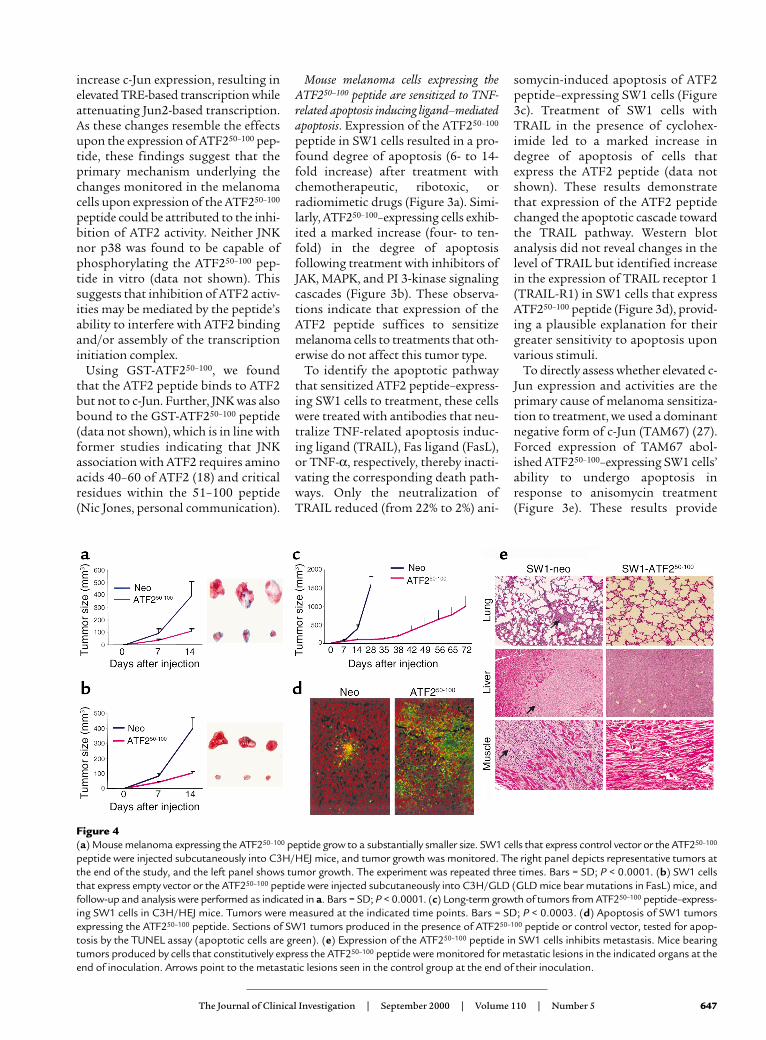
Figure 4(a) Mouse melanoma expressing the ATF250–100 peptide grow to a substantially smaller size. SW1 cells that express control vector or the ATF250–100
peptide were injected subcutaneously into C3H/HEJ mice, and tumor growth was monitored. The right panel depicts representative tumors atthe end of the study, and the left panel shows tumor growth. The experiment was repeated three times. Bars = SD; P < 0.0001. (b) SW1 cellsthat express empty vector or the ATF250–100 peptide were injected subcutaneously into C3H/GLD (GLD mice bear mutations in FasL) mice, andfollow-up and analysis were performed as indicated in a. Bars = SD; P < 0.0001. (c) Long-term growth of tumors from ATF250–100 peptide–express-ing SW1 cells in C3H/HEJ mice. Tumors were measured at the indicated time points. Bars = SD; P < 0.0003. (d) Apoptosis of SW1 tumorsexpressing the ATF250–100 peptide. Sections of SW1 tumors produced in the presence of ATF250–100 peptide or control vector, tested for apop-tosis by the TUNEL assay (apoptotic cells are green). (e) Expression of the ATF250–100 peptide in SW1 cells inhibits metastasis. Mice bearingtumors produced by cells that constitutively express the ATF250–100 peptide were monitored for metastatic lesions in the indicated organs at theend of inoculation. Arrows point to the metastatic lesions seen in the control group at the end of their inoculation.
direct evidence for the role of c-Jun inmediating the sensitization ofmelanoma cells to apoptosis followingexpression of the ATF250–100 peptide.To further assess the role of ATF2 ver-sus c-Jun in the sensitization ofmelanoma cells to treatment, we mon-itored sensitivity to apoptosis of cellsthat were transfected with ATF2-siRNA. Transient null ATF2 environ-ment generated upon transfection ofthe ATF2-siRNA was sufficient to sen-sitize the SW1 melanoma cells toapoptosis in response to anisomycintreatment (Figure 3f). The degree ofapoptosis seen in cells transfected withATF2-siRNA was threefold higherthan that seen using siRNA of RNF5,
a RING finger protein involved incytoskeleton organization (ourunpublished data), which was usedhere as a control. These findings indi-cate that changes elicited upon inhibi-tion of ATF2 expression in themelanoma cells are sufficient to causetheir sensitization to apoptosis upontreatment, as has been observed withthe ATF2 peptide–expressing cells.
Expression of ATF250–100 in SW1 mousemelanoma cells causes marked reduction intheir growth and inhibits their metastaticcapacity. Subcutaneous injection ofSW1 cells expressing a control con-struct resulted in rapid growth pro-ducing 400-mm3 tumors within 2weeks. Tumors produced by SW1 cells
that constitutively express theATF250–100 peptide were much smaller(90 mm3; Figure 4a). The expression ofthe ATF250–100 peptide was as efficientin blocking growth of SW1 tumors inFasL-deficient GLD mice (Figure 4b),suggesting that the sensitization of theATF250–100–expressing tumors toapoptosis is not dependent on theexogenous source of FasL.
While the parental cells producedtumors weighing about 1 g within 28days after inoculation, it took 72 days(three times longer) for ATF250–100–expressing cells to produce similar-sized tumors (Figure 4c). The strongestinhibition of growth was observed dur-ing the first 35 days after inoculation,with somewhat increased growth ratethereafter. The partial loss of growthinhibition could be attributed to aselective loss of the ATF250–100 peptidedue to lack of selective pressure (tomaintain drug resistance) and lack ofgenomic integration of the plasmidvector. While Western blot analysisconfirmed expression of ATF2 peptide,immunohistochemistry analysis re-vealed focal expression (data notshown), which coincided with foci ofapoptotic cells as seen via the TUNELassay (Figure 4d), thereby confirmingpartial expression of the ATF2 peptideat this stage.
SW1 cells are prone to metastasizeinto multiple organs, resulting in mul-tiple lesions as early as 4 weeks follow-ing injection (25); however, no metasta-sis was seen in experiments whereATF250–100–expressing SW1 cells weremonitored for up to 72 days (Figure 4e).
Inducible expression of the ATF250–100
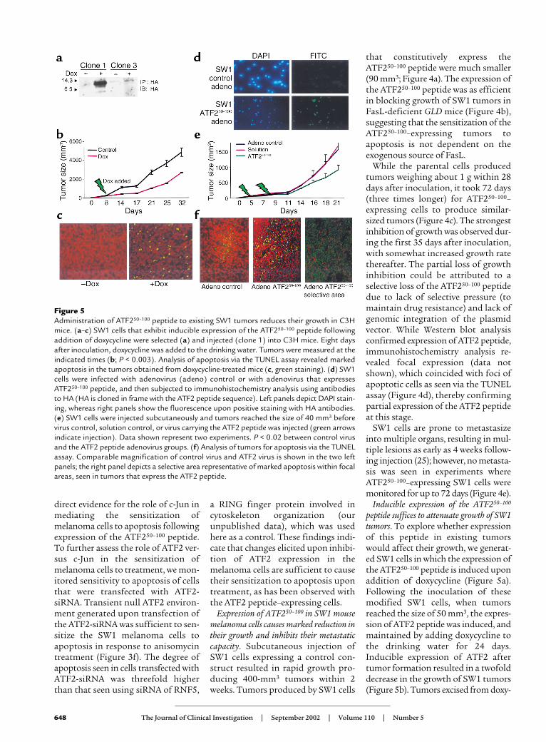
peptide suffices to attenuate growth of SW1tumors. To explore whether expressionof this peptide in existing tumorswould affect their growth, we generat-ed SW1 cells in which the expression ofthe ATF250–100 peptide is induced uponaddition of doxycycline (Figure 5a).Following the inoculation of thesemodified SW1 cells, when tumorsreached the size of 50 mm3, the expres-sion of ATF2 peptide was induced, andmaintained by adding doxycycline tothe drinking water for 24 days.Inducible expression of ATF2 aftertumor formation resulted in a twofolddecrease in the growth of SW1 tumors(Figure 5b). Tumors excised from doxy-
648 The Journal of Clinical Investigation | September 2002 | Volume 110 | Number 5
Figure 5Administration of ATF250–100 peptide to existing SW1 tumors reduces their growth in C3Hmice. (a–c) SW1 cells that exhibit inducible expression of the ATF250–100 peptide followingaddition of doxycycline were selected (a) and injected (clone 1) into C3H mice. Eight daysafter inoculation, doxycycline was added to the drinking water. Tumors were measured at theindicated times (b; P < 0.003). Analysis of apoptosis via the TUNEL assay revealed markedapoptosis in the tumors obtained from doxycycline-treated mice (c, green staining). (d) SW1cells were infected with adenovirus (adeno) control or with adenovirus that expressesATF250–100 peptide, and then subjected to immunohistochemistry analysis using antibodiesto HA (HA is cloned in frame with the ATF2 peptide sequence). Left panels depict DAPI stain-ing, whereas right panels show the fluorescence upon positive staining with HA antibodies.(e) SW1 cells were injected subcutaneously and tumors reached the size of 40 mm3 beforevirus control, solution control, or virus carrying the ATF2 peptide was injected (green arrowsindicate injection). Data shown represent two experiments. P < 0.02 between control virusand the ATF2 peptide adenovirus groups. (f) Analysis of tumors for apoptosis via the TUNELassay. Comparable magnification of control virus and ATF2 virus is shown in the two leftpanels; the right panel depicts a selective area representative of marked apoptosis within focalareas, seen in tumors that express the ATF2 peptide.
cycline-fed mice exhibited an increasein degree of apoptosis, albeit sporadic(Figure 5c), which coincides with par-tial expression of the ATF250–100 pep-tide as revealed by immunohistochem-istry of the tumors (not shown).
In a parallel approach, the cDNA ofthe ATF2 peptide was cloned into anadenovirus vector and administered toSW1 tumors (40 mm3 in size) via directintratumoral injection. Expression ofadenovirus encoding the ATF250–100
peptide was confirmed via immunoflu-orescence (Figure 5d). A marked reduc-tion (∼50%) in tumor growth wasobserved shortly after the second aden-oviral injection (Figure 5e). Inhibitionof tumor growth coincided with elevat-ed degree of apoptosis, which was non-homogenous (Figure 5f). These datasuggest that partial expression of theATF250–100 peptide in already developedmelanoma suffices to elicit efficientreduction of subsequent tumor growth.
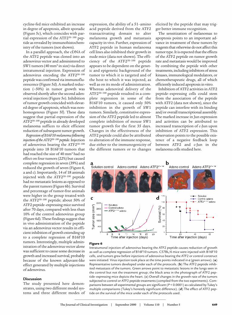
Regression of B16F10 melanoma followinginjection of the ATF250–100 peptide. Injectionof adenovirus bearing the ATF250–100
peptide into 18 B16F10 tumors thathad reached the size of 40 mm3 had noeffect on four tumors (22%) but causedcomplete regression in seven (39%) andreduced the growth of seven (Figure 6,a and c). Importantly, 14 of 18 animalsinjected with the ATF250–100 peptidehad no metastatic lesions as opposed tothe parent tumors (Figure 6b). Survivaland percentage of tumor-free animalswere higher in the group treated withthe ATF250–100 peptide; about 50% ofATF2 peptide–expressing mice survivedafter 70 days, compared with less than10% of the control adenovirus group(Figure 6d). These findings suggest thatin vivo administration of the peptidevia an adenovirus vector results in effi-cient inhibition of growth extending upto a complete regression of B16F10tumors. Interestingly, multiple admin-istration of the adenovirus vector alonewas sufficient to cause some decrease ingrowth and increased survival, probablybecause of the known adjuvant-likeeffect generated by multiple injectionsof adenovirus.
DiscussionThe study presented here demon-strates, using two different model sys-tems and three different modes of
expression, the ability of a 51–aminoacid peptide derived from the ATF2transactivating domain to altermelanoma growth and metastasiscapacity in vivo. Further, expression ofATF2 peptide in human melanomacell lines also inhibited their growth innude mice (data not shown). The effi-ciency of the ATF250–100 peptideappears to be dependent on the genet-ic and epigenetic background of thetumor to which it is targeted and ofthe host to which it was injected, aswell as on its mode of administration.Whereas adenoviral delivery of theATF250–100 peptide resulted in a com-plete regression in some of theB16F10 tumors, it caused only 50%inhibition in the growth of SW1tumors. Similarly, constitutive expres-sion of the ATF2 peptide led to almostcomplete inhibition of mouse SW1tumor growth for the first 35 days.Changes in the effectiveness of theATF2 peptide could also be attributedto alteration of the immune response,due either to the immunogenicity ofthe different tumors or to changes
elicited by the peptide that may trig-ger better immune recognition.
The sensitization of melanomas toapoptosis points to an important ad-vance in treatment of these tumors byreagents that otherwise do not affect thistumor type. It is expected that the effectsof the ATF2 peptide on tumor growthrate and metastasis would be improvedby combining the peptide with othertreatments, including inhibitors of stresskinases, immunological modulators, orchemotherapeutic drugs, all of whichefficiently induced apoptosis in vitro.
Inhibition of ATF2 activities in ATF2peptide–expressing cells could stemfrom the association of the peptidewith ATF2 (data not shown), since thepeptide can interfere with its bindingand/or overall transcriptional assembly.The marked increase in Jun expressionand activities can be attributed toincreased transcription of c-Jun uponinhibition of ATF2 expression. Thisobservation points to the possible exis-tence of a negative feedback loopbetween ATF2 and c-Jun in themelanoma cells studied here.
The Journal of Clinical Investigation | September 2000 | Volume 110 | Number 5 649
Figure 6Intratumoral injection of adenovirus bearing the ATF2 peptide causes reduction of growthas well as complete regression of B16F10 tumors. C57BL/6 mice were injected with B16F10cells, and tumors grew before injections of adenovirus bearing the ATF2 or control constructwere initiated. Virus injection took place at the time points indicated in c (green arrows). (a)Representative tumors developed under each of the protocols. (b) The ATF2 peptide inhib-ited metastasis of the tumors. Green arrows point to metastatic lesions in the lungs seen inthe control but not the treatment group; the black area in the photograph of ATF2 pep-tide–expressing mice depicts the heart. (c) Overall changes in the growth rate of the tumorssubjected to control or ATF2 peptide treatments (compiled from the two experiments). Com-parisons between all experimental groups are significant (P < 0.0001) as calculated by Tukey’smultiple comparisons (Tukey’s honestly significant difference). (d) The effect of ATF2 pep-tide on the survival of the mice under each of the protocols used.

Decreased ATF2 transcriptionalactivities together with a concomitantincrease in c-Jun transcriptional activi-ties is likely to be the primary mecha-nism underlying the observed effectsupon the expression of the ATF250–100
peptide. This conclusion is supportedby an independent set of experimentswhere siRNA used to inhibit ATF2transcription resulted in increased c-Jun expression, transcription, andsensitization to apoptosis. The abilityto abolish the sensitization ofATF250–100–expressing SW1 cells to ani-somycin-induced apoptosis uponexpression of a potent c-Jun dominantnegative construct supports the centralrole of c-Jun, in conjunction withdecreased ATF2 transcriptional activi-ties, in the sensitization of melanomacells to apoptosis. Of interest, increasein expression of TRAIL-R1 in ATF2peptide–expressing cells could also beattributed to c-Jun, which was recentlyshown to positively regulate TRAIL-Rpromoter (28). The ability of c-Jun toinduce apoptosis has been document-ed in several systems (29–31).
Support for the possible antionco-genic activities of c-Jun comes from theindependent finding that c-Jun is capa-ble of inhibiting oncogene-dependentin vitro transformation of primary ratembryo fibroblasts (32). Our findingsare in agreement with these studies, asthey point to the central role of c-Jun insensitization of melanoma to apopto-sis in conjunction with decreased ATF2transcriptional activities. Accordingly,our findings suggest that the onco-genic activities of c-Jun may depend onits cooperation with other transcrip-tion factors, as demonstrated here forATF2. In the absence of such coopera-tion there is a switch in the profile of c-Jun–regulated genes, as reflected inexpression profiling analysis, whichrevealed an increase in TNF-α and adecrease in growth-, Fas-, and IFN-γ–related genes (data not shown).
Independent support for the role ofATF2 in proliferation of melanomaswas recently provided in an HGF-based melanoma model (4). Overall,our results highlight the ability to“reprogram” c-Jun/ATF2 transcrip-tional activities via a 51–amino acidATF2-driven peptide, resulting in the
sensitization of melanoma to apopto-sis and in the inhibition of the growthand metastasis of this otherwiseaggressive tumor type.
AcknowledgmentsWe thank M. Herlyn and O. Fodstadfor providing the melanoma cell lines,Ron Wisdom for the Jun-null cells,and Raul Gopalkrishnan and PaulFisher for the doxycycline-inducibleconstructs. We also thank LillianaOssowski for critical reading, andJohn Mandeli for biostatistics analy-sis. We thank Nic Jones and membersof the Ronai laboratory for discus-sions. Support by National CancerInstitute grant CA-59008 and CA-78419 (to Z. Ronai) and by the SharpFoundation (to Z. Ronai and S.-H.Chen) is gratefully acknowledged.
1. Yang, Y.M., Dolan, L.R., and Ronai, Z. 1996.Expression of dominant negative CREB reducesresistance to radiation of human melanoma cells.Oncogene. 12:2223–2233.
2. Jean, D., Harbison, M., McConkey, D.J., Ronai, Z.,and Bar-Eli, M. 1998. CREB and its associated pro-teins act as survival factors for human melanomacells. J. Biol. Chem. 273:24884–24890.
3. Yang, J., and Richmond, A. 2001. ConstitutiveIkappaB kinase activity correlates with nuclear fac-tor-kappaB activation in human melanoma cells.Cancer Res. 61:4901–4909.
4. Recio, J.A., and Merlino, G. 2002. Hepatocytegrowth factor/scatter factor activates proliferationin melanoma cells through p38 MAPK, ATF-2 andcyclin D1. Oncogene. 21:1000–1008.
5. Ronai, Z., et al. 1998. ATF2 confers radiation resist-ance to human melanoma cells. Oncogene.16:523–531.
6. Bhoumik, A., Ivanov, V.N., and Ronai, Z. 2001.Activating transcription factor 2-derived peptidesalter resistance of human tumor cell lines to ultra-violet irradiation and chemical treatment. Clin.Cancer Res. 7:331–342.
7. Hai, T.W., Liu, F., Coukos, W.J., and Green, M.R.1989. Transcription factor ATF cDNA clones: anextensive family of leucine zipper proteins able toselectively form DNA-binding heterodimers. GenesDev. 3:2083–2090.
8. van Dam, H., et al. 1995. ATF-2 is preferentiallyactivated by stress-activated protein kinases tomediate c-jun induction in response to genotoxicagents. EMBO J. 14:1798–1811.
9. Gupta, S., Campbell, D., Derijard, B., and Davis,R.J. 1995. Transcription factor ATF2 regulation bythe JNK signal transduction pathway. Science.267:389–393.
10. van Dam, H., et al. 1993. Heterodimer formationof cJun and ATF-2 is responsible for induction ofc-jun by the 243 amino acid adenovirus E1A pro-tein. EMBO J. 12:479–487.
11. Kaszubska, W., et al. 1993. Cyclic AMP-independ-ent ATF family members interact with NF-kappaB and function in the activation of the E-selectinpromoter in response to cytokines. Mol. Cell. Biol.13:7180–7190.
12. Kim, S.J., et al. 1992. Retinoblastoma gene productactivates expression of the human TGF-beta 2gene through transcription factor ATF-2. Nature.358:331–334.
13. Tsai, E.Y., Jain, J., Pesavento, P.A., Rao, A., andGoldfeld, A.E. 1996. Tumor necrosis factor alphagene regulation in activated T cells involves ATF-2/Jun and NFATp. Mol. Cell. Biol. 16:459–467.
14. Shimizu, M., et al. 1998. Activation of the rat cyclinA promoter by ATF2 and Jun family members andits suppression by ATF4. Exp. Cell Res. 239:93–103.
15. Huguier, S., Baguet, J., Perez, S., van Dam, H., andCastellazzi, M. 1998. Transcription factor ATF2cooperates with v-Jun to promote growth factor-independent proliferation in vitro and tumor for-mation in vivo. Mol. Cell. Biol. 18:7020–7029.
16. van Dam, H., and Castellazzi, M. 2001. Distinctroles of Jun. Fos and Jun: ATF dimers in oncogen-esis. Oncogene. 20:2453–2464.
17. Ivanov, V.N., and Ronai, Z. 1999. Down-regulationof tumor necrosis factor alpha expression by acti-vating transcription factor 2 increases UVC-induced apoptosis of late-stage melanoma cells. J. Biol. Chem. 274:14079–14089.
18. Fuchs, S.Y., Dolan, L.R., Davis, R., and Ronai, Z.1996. Phosphorylation dependent targeting of c-jun ubiquitination by Jun N-kinase. Oncogene.13:1531–1535.
19. Duyndam, M.C., et al. 1999. The N-terminal trans-activation domain of ATF2 is a target for the co-operative activation of the c-jun promoter by p300and 12S E1A. Oncogene. 18:2311–2321.
20. Chen, S.H., Shine, H.D., Goodman, J.C., Gross-man, R.G., and Woo, S.L. 1994. Gene therapy forbrain tumors: regression of experimental gliomasby adenovirus-mediated gene transfer in vivo. Proc.Natl. Acad. Sci. USA. 91:3054–3057.
21. Gossen, M., and Bujard, H. 1992. Tight control ofgenen expression in mammalian cells by tetracy-cline-responsive promoters. Proc. Natl. Acad. Sci.USA. 89:5547–5551.
22. Gopalkrishnan, R.V., Christiansen, K.A., Golden-stein, N.I., DePinho, R.A., and Fisher, P.B. 1999.Use of the human EF-1alpha promoter for expres-sion can significantly increase success in estab-lishing stable cell lines with consistent expression:a study using the tetracycline-inducible system in human cancer cells. Nucleic Acids Res.27:4775–4782.
23. Ivanov, V.N., et al. 2001. Cooperation betweenSTAT3 and c-jun suppresses Fas transcription.Mol. Cell. 7:517–528.
24. Elbashir, S.M., et al. 2001. Duplexes of 21-nucleotide RNAs mediate RNA interference in cul-tured mammalian cells. Nature. 411:494–498.
25. Owen-Schaub, L.B., van Golen, K., Hill, L., andPrice, J.E. 1998. Fas and Fas ligand interactionssuppress melanoma lung metastasis. J. Exp. Med.188:1717–1723.
26. van Dam, H., et al. 1998. Autocrine growth andanchorage independence: two complementingJun-controlled genetic programs of cellular trans-formation. Genes Dev. 12:1227–1239.
27. Brown, P.H., Chen, T.K., and Birrer, M.J. 1994.Mechanism of action of a dominant-negativemutant of c-Jun. Oncogene. 3:791–799.
28. Guan, B., Yue, P., Lotan, R., and Sun, S.-Y. 2002. Evi-dence that the human death receptor 4 is regulatedby activator protein 1. Oncogene. 21:3121–3129.
29. Bossy-Wetzel, E., Bakiri, L., and Yaniv, M. 1997.Induction of apoptosis by the transcription factorc-Jun. EMBO J. 16:1695–1709.
30. Behrens, A., Sibilia, M., and Wagner, E.F. 1999.Amino-terminal phosphorylation of c-Jun regu-lates stress-induced apoptosis and cellular prolif-eration. Nat. Genet. 3:326–329.
31. Yoshida, K., et al. 2002. Amino-terminal phospho-rylation of c-Jun regulates apoptosis in the retinalganglion cells by optic nerve transection. Invest.Ophthalmol. Vis. Sci. 43:1631–1635.
32. Ginsberg, D., Hirai, S.I., Pinhasi-Kimhi, O., Yaniv,M., and Oren, M. 1991. Transfected mouse c-Juncan inhibit transformation of primary rat embryofibroblasts. Oncogene. 4:669–672.
650 The Journal of Clinical Investigation | September 2002 | Volume 110 | Number 5
Copyright © 2022 FDOKUMEN