MicroRNA Profiles Discriminate among Colon Cancer Metastasis
Upload
independentCategory
view
0download
0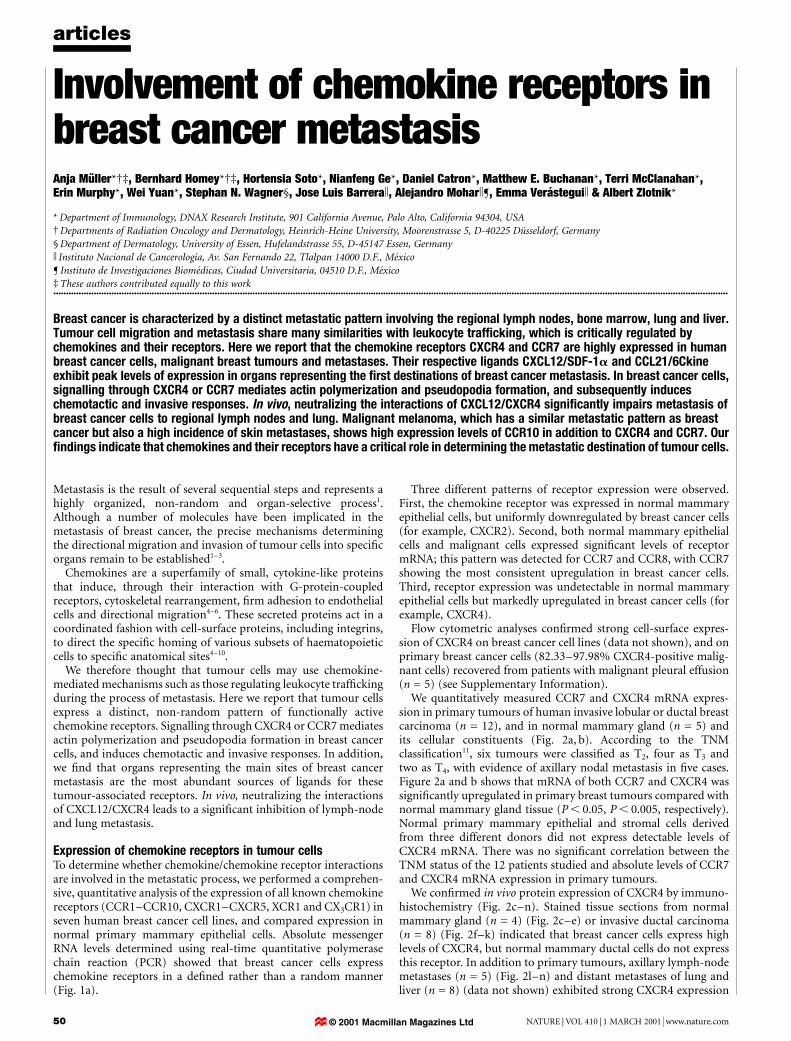
Involvement of chemokine receptors inbreast cancer metastasisAnja MuÈ ller*²³, Bernhard Homey*²³, Hortensia Soto*, Nianfeng Ge*, Daniel Catron*, Matthew E. Buchanan*, Terri McClanahan*,Erin Murphy*, Wei Yuan*, Stephan N. Wagner§, Jose Luis Barrerak, Alejandro Mohark¶, Emma VeraÂsteguik & Albert Zlotnik*
* Department of Immunology, DNAX Research Institute, 901 California Avenue, Palo Alto, California 94304, USA² Departments of Radiation Oncology and Dermatology, Heinrich-Heine University, Moorenstrasse 5, D-40225 DuÈsseldorf, Germany§ Department of Dermatology, University of Essen, Hufelandstrasse 55, D-45147 Essen, Germany
k Instituto Nacional de CancerologõÂa, Av. San Fernando 22, Tlalpan 14000 D.F., MeÂxico
¶ Instituto de Investigaciones BiomeÂdicas, Ciudad Universitaria, 04510 D.F., MeÂxico³ These authors contributed equally to this work............................................................................................................................................................................................................................................................................
Breast cancer is characterized by a distinct metastatic pattern involving the regional lymph nodes, bone marrow, lung and liver.Tumour cell migration and metastasis share many similarities with leukocyte traf®cking, which is critically regulated bychemokines and their receptors. Here we report that the chemokine receptors CXCR4 and CCR7 are highly expressed in humanbreast cancer cells, malignant breast tumours and metastases. Their respective ligands CXCL12/SDF-1a and CCL21/6Ckineexhibit peak levels of expression in organs representing the ®rst destinations of breast cancer metastasis. In breast cancer cells,signalling through CXCR4 or CCR7 mediates actin polymerization and pseudopodia formation, and subsequently induceschemotactic and invasive responses. In vivo, neutralizing the interactions of CXCL12/CXCR4 signi®cantly impairs metastasis ofbreast cancer cells to regional lymph nodes and lung. Malignant melanoma, which has a similar metastatic pattern as breastcancer but also a high incidence of skin metastases, shows high expression levels of CCR10 in addition to CXCR4 and CCR7. Our®ndings indicate that chemokines and their receptors have a critical role in determining the metastatic destination of tumour cells.
Metastasis is the result of several sequential steps and represents ahighly organized, non-random and organ-selective process1.Although a number of molecules have been implicated in themetastasis of breast cancer, the precise mechanisms determiningthe directional migration and invasion of tumour cells into speci®corgans remain to be established1±3.
Chemokines are a superfamily of small, cytokine-like proteinsthat induce, through their interaction with G-protein-coupledreceptors, cytoskeletal rearrangement, ®rm adhesion to endothelialcells and directional migration4±6. These secreted proteins act in acoordinated fashion with cell-surface proteins, including integrins,to direct the speci®c homing of various subsets of haematopoieticcells to speci®c anatomical sites4±10.
We therefore thought that tumour cells may use chemokine-mediated mechanisms such as those regulating leukocyte traf®ckingduring the process of metastasis. Here we report that tumour cellsexpress a distinct, non-random pattern of functionally activechemokine receptors. Signalling through CXCR4 or CCR7 mediatesactin polymerization and pseudopodia formation in breast cancercells, and induces chemotactic and invasive responses. In addition,we ®nd that organs representing the main sites of breast cancermetastasis are the most abundant sources of ligands for thesetumour-associated receptors. In vivo, neutralizing the interactionsof CXCL12/CXCR4 leads to a signi®cant inhibition of lymph-nodeand lung metastasis.
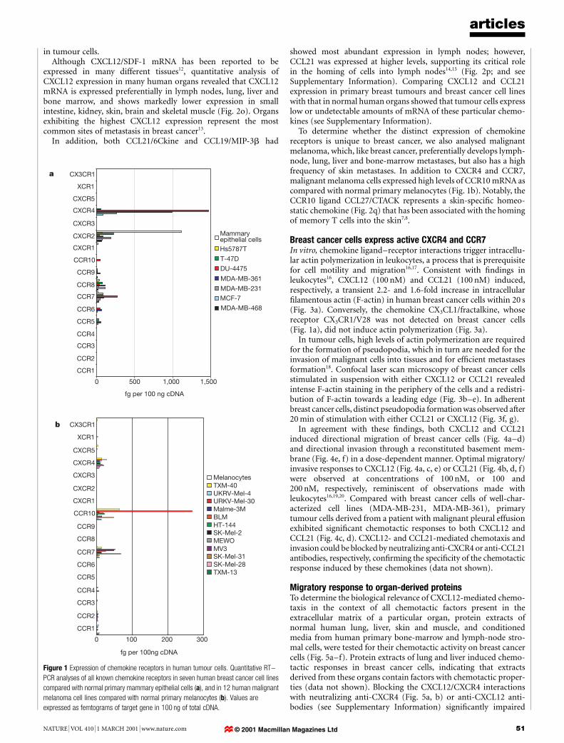
Expression of chemokine receptors in tumour cellsTo determine whether chemokine/chemokine receptor interactionsare involved in the metastatic process, we performed a comprehen-sive, quantitative analysis of the expression of all known chemokinereceptors (CCR1±CCR10, CXCR1±CXCR5, XCR1 and CX3CR1) inseven human breast cancer cell lines, and compared expression innormal primary mammary epithelial cells. Absolute messengerRNA levels determined using real-time quantitative polymerasechain reaction (PCR) showed that breast cancer cells expresschemokine receptors in a de®ned rather than a random manner(Fig. 1a).
Three different patterns of receptor expression were observed.First, the chemokine receptor was expressed in normal mammaryepithelial cells, but uniformly downregulated by breast cancer cells(for example, CXCR2). Second, both normal mammary epithelialcells and malignant cells expressed signi®cant levels of receptormRNA; this pattern was detected for CCR7 and CCR8, with CCR7showing the most consistent upregulation in breast cancer cells.Third, receptor expression was undetectable in normal mammaryepithelial cells but markedly upregulated in breast cancer cells (forexample, CXCR4).
Flow cytometric analyses con®rmed strong cell-surface expres-sion of CXCR4 on breast cancer cell lines (data not shown), and onprimary breast cancer cells (82.33±97.98% CXCR4-positive malig-nant cells) recovered from patients with malignant pleural effusion(n = 5) (see Supplementary Information).
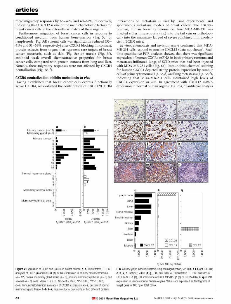
We quantitatively measured CCR7 and CXCR4 mRNA expres-sion in primary tumours of human invasive lobular or ductal breastcarcinoma (n = 12), and in normal mammary gland (n = 5) andits cellular constituents (Fig. 2a, b). According to the TNMclassi®cation11, six tumours were classi®ed as T2, four as T3 andtwo as T4, with evidence of axillary nodal metastasis in ®ve cases.Figure 2a and b shows that mRNA of both CCR7 and CXCR4 wassigni®cantly upregulated in primary breast tumours compared withnormal mammary gland tissue (P , 0.05, P , 0.005, respectively).Normal primary mammary epithelial and stromal cells derivedfrom three different donors did not express detectable levels ofCXCR4 mRNA. There was no signi®cant correlation between theTNM status of the 12 patients studied and absolute levels of CCR7and CXCR4 mRNA expression in primary tumours.
We con®rmed in vivo protein expression of CXCR4 by immuno-histochemistry (Fig. 2c±n). Stained tissue sections from normalmammary gland (n = 4) (Fig. 2c±e) or invasive ductal carcinoma(n = 8) (Fig. 2f±k) indicated that breast cancer cells express highlevels of CXCR4, but normal mammary ductal cells do not expressthis receptor. In addition to primary tumours, axillary lymph-nodemetastases (n = 5) (Fig. 2l±n) and distant metastases of lung andliver (n = 8) (data not shown) exhibited strong CXCR4 expression
articles
50 NATURE | VOL 410 | 1 MARCH 2001 | www.nature.com© 2001 Macmillan Magazines Ltd
in tumour cells.Although CXCL12/SDF-1 mRNA has been reported to be
expressed in many different tissues12, quantitative analysis ofCXCL12 expression in many human organs revealed that CXCL12mRNA is expressed preferentially in lymph nodes, lung, liver andbone marrow, and shows markedly lower expression in smallintestine, kidney, skin, brain and skeletal muscle (Fig. 2o). Organsexhibiting the highest CXCL12 expression represent the mostcommon sites of metastasis in breast cancer13.
In addition, both CCL21/6Ckine and CCL19/MIP-3b had
showed most abundant expression in lymph nodes; however,CCL21 was expressed at higher levels, supporting its critical rolein the homing of cells into lymph nodes14,15 (Fig. 2p; and seeSupplementary Information). Comparing CXCL12 and CCL21expression in primary breast tumours and breast cancer cell lineswith that in normal human organs showed that tumour cells expresslow or undetectable amounts of mRNA of these particular chemo-kines (see Supplementary Information).
To determine whether the distinct expression of chemokinereceptors is unique to breast cancer, we also analysed malignantmelanoma, which, like breast cancer, preferentially develops lymph-node, lung, liver and bone-marrow metastases, but also has a highfrequency of skin metastases. In addition to CXCR4 and CCR7,malignant melanoma cells expressed high levels of CCR10 mRNA ascompared with normal primary melanocytes (Fig. 1b). Notably, theCCR10 ligand CCL27/CTACK represents a skin-speci®c homeo-static chemokine (Fig. 2q) that has been associated with the homingof memory T cells into the skin7,8.
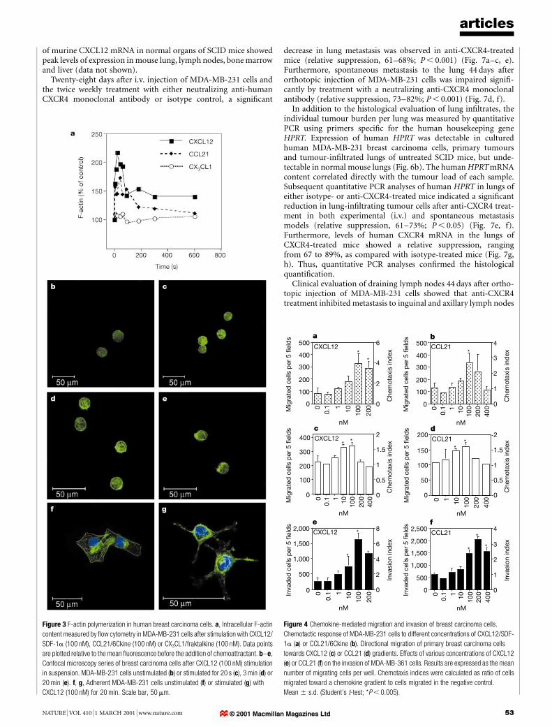
Breast cancer cells express active CXCR4 and CCR7In vitro, chemokine ligand±receptor interactions trigger intracellu-lar actin polymerization in leukocytes, a process that is prerequisitefor cell motility and migration16,17. Consistent with ®ndings inleukocytes16, CXCL12 (100 nM) and CCL21 (100 nM) induced,respectively, a transient 2.2- and 1.6-fold increase in intracellular®lamentous actin (F-actin) in human breast cancer cells within 20 s(Fig. 3a). Conversely, the chemokine CX3CL1/fractalkine, whosereceptor CX3CR1/V28 was not detected on breast cancer cells(Fig. 1a), did not induce actin polymerization (Fig. 3a).
In tumour cells, high levels of actin polymerization are requiredfor the formation of pseudopodia, which in turn are needed for theinvasion of malignant cells into tissues and for ef®cient metastasesformation18. Confocal laser scan microscopy of breast cancer cellsstimulated in suspension with either CXCL12 or CCL21 revealedintense F-actin staining in the periphery of the cells and a redistri-bution of F-actin towards a leading edge (Fig. 3b±e). In adherentbreast cancer cells, distinct pseudopodia formation was observed after20 min of stimulation with either CCL21 or CXCL12 (Fig. 3f, g).
In agreement with these ®ndings, both CXCL12 and CCL21induced directional migration of breast cancer cells (Fig. 4a±d)and directional invasion through a reconstituted basement mem-brane (Fig. 4e, f) in a dose-dependent manner. Optimal migratory/invasive responses to CXCL12 (Fig. 4a, c, e) or CCL21 (Fig. 4b, d, f)were observed at concentrations of 100 nM, or 100 and200 nM, respectively, reminiscent of observations made withleukocytes16,19,20. Compared with breast cancer cells of well-char-acterized cell lines (MDA-MB-231, MDA-MB-361), primarytumour cells derived from a patient with malignant pleural effusionexhibited signi®cant chemotactic responses to both CXCL12 andCCL21 (Fig. 4c, d). CXCL12- and CCL21-mediated chemotaxis andinvasion could be blocked by neutralizing anti-CXCR4 or anti-CCL21antibodies, respectively, con®rming the speci®city of the chemotacticresponse induced by these chemokines (data not shown).
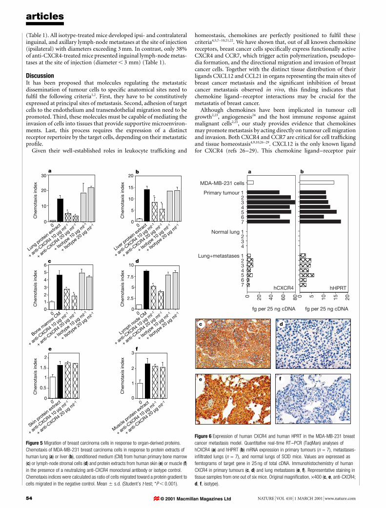
Migratory response to organ-derived proteinsTo determine the biological relevance of CXCL12-mediated chemo-taxis in the context of all chemotactic factors present in theextracellular matrix of a particular organ, protein extracts ofnormal human lung, liver, skin and muscle, and conditionedmedia from human primary bone-marrow and lymph-node stro-mal cells, were tested for their chemotactic activity on breast cancercells (Fig. 5a±f). Protein extracts of lung and liver induced chemo-tactic responses in breast cancer cells, indicating that extractsderived from these organs contain factors with chemotactic proper-ties (data not shown). Blocking the CXCL12/CXCR4 interactionswith neutralizing anti-CXCR4 (Fig. 5a, b) or anti-CXCL12 anti-bodies (see Supplementary Information) signi®cantly impaired
articles
NATURE | VOL 410 | 1 MARCH 2001 | www.nature.com 51
a CX3CR1
XCR1
CXCR5
CXCR4
CXCR3
CXCR2
CXCR1
CCR10
CCR9
CCR8
CCR7
CCR6
CCR5
CCR4
CCR3
CCR2
CCR1
0 500 1,500
fg per 100 ng cDNA
1,000
Mammaryepithelial cellsHs5787T
T-47D
DU-4475
MDA-MB-361
MDA-MB-231MCF-7
MDA-MB-468
0 100 200 300
CCR1
CCR2
CCR3
CCR4
CCR5
CCR6
CCR7
CCR8
CCR9
CCR10
CXCR1
CXCR2
CXCR3
CXCR4
CXCR5
XCR1
CX3CR1
fg per 100ng cDNA
MelanocytesTXM-40UKRV-Mel-4URKV-Mel-30Malme-3MBLMHT-144SK-Mel-2MEWOMV3SK-Mel-31SK-Mel-28TXM-13
b
Figure 1 Expression of chemokine receptors in human tumour cells. Quantitative RT±
PCR analyses of all known chemokine receptors in seven human breast cancer cell lines
compared with normal primary mammary epithelial cells (a), and in 12 human malignant
melanoma cell lines compared with normal primary melanocytes (b). Values are
expressed as femtograms of target gene in 100 ng of total cDNA.
© 2001 Macmillan Magazines Ltd
these migratory responses by 63±76% and 60±62%, respectively,indicating that CXCL12 is one of the main chemotactic factors forbreast cancer cells in the extracellular matrix of these organs.
Furthermore, migration of breast cancer cells in response toconditioned medium from human bone-marrow (Fig. 5c) orlymph-node (Fig. 5d) stromal cells was signi®cantly reduced (53±61% and 51±54%, respectively) after CXCR4 blocking. In contrast,protein extracts from organs that represent rare targets of breastcancer metastasis, such as skin (Fig. 5e) or muscle (Fig. 5f),exhibited weak overall chemoattractive properties for breastcancer cells, compared with protein extracts from lung and liver.Notably, these migratory responses were not affected by CXCR4neutralization (Fig. 5e, f).
CXCR4-neutralization inhibits metastasis in vivoHaving established that breast cancer cells express functionallyactive CXCR4, we evaluated the contribution of CXCL12/CXCR4
interactions on metastasis in vivo by using experimental andspontaneous metastasis models of breast cancer. The CXCR4-positive, human breast carcinoma cell line MDA-MB-231 wasinjected either intravenously (i.v.) into the tail vein or orthotopi-cally into the mammary fat pad of severe combined immunode®-cient (SCID) mice.
In vitro, chemotaxis and invasion assays con®rmed that MDA-MB-231 cells respond to murine CXCL12 (data not shown). Real-time quantitative PCR analyses showed that there was signi®cantexpression of human CXCR4 mRNA in both primary tumours andmetastases-in®ltrated lungs of SCID mice that had been injectedwith MDA-MB-231 cells (Fig. 6a). Immunohistochemical stainingfor human CXCR4 depicted strong protein expression by tumourcells of primary tumours (Fig. 6c, d) and lung metastases (Fig. 6e, f),indicating that MDA-MB-231 cells maintained high levels ofCXCR4 expression in vivo. In agreement with CXCL12 mRNAexpression in normal human organs (Fig. 2o), quantitative analysis
articles
52 NATURE | VOL 410 | 1 MARCH 2001 | www.nature.com
Figure 2 Expression of CCR7 and CXCR4 in breast cancer. a, b, Quantitative RT±PCR
analyses of CCR7 (a) and CXCR4 (b) mRNA expression in primary breast carcinoma
(n = 12), normal mammary gland tissue (n = 5), primary mammary epithelial (n = 3) and
stromal (n = 3) cells. Mean 6 s.e.m. (Student's t-test; *P , 0.05, **P , 0.005).
c±n, Immunohistochemical evaluation of CXCR4 expression. c±e, Section of normal
mammary gland tissue. f±h, i±k, Invasive ductal carcinoma of two different patients.
l±n, Axillary lymph-node metastasis. Original magni®cation, ´200 (c, f, i, l, anti-CXCR4;
e, h, k, n, isotype); ´400 (d, g, j, m, anti-CXCR4). Quantitative RT±PCR analyses of
CXCL12/SDF-1 (o), CCL21/6Ckine and CCL19/MIP-3b (p) or CCL27/CTACK (q) mRNA
expression in various normal human organs. Values are expressed as femtograms of
target gene in 100 ng of total cDNA.
© 2001 Macmillan Magazines Ltd
of murine CXCL12 mRNA in normal organs of SCID mice showedpeak levels of expression in mouse lung, lymph nodes, bone marrowand liver (data not shown).
Twenty-eight days after i.v. injection of MDA-MB-231 cells andthe twice weekly treatment with either neutralizing anti-humanCXCR4 monoclonal antibody or isotype control, a signi®cant
decrease in lung metastasis was observed in anti-CXCR4-treatedmice (relative suppression, 61±68%; P , 0.001) (Fig. 7a±c, e).Furthermore, spontaneous metastasis to the lung 44 days afterorthotopic injection of MDA-MB-231 cells was impaired signi®-cantly by treatment with a neutralizing anti-CXCR4 monoclonalantibody (relative suppression, 73±82%; P , 0.001) (Fig. 7d, f).
In addition to the histological evaluation of lung in®ltrates, theindividual tumour burden per lung was measured by quantitativePCR using primers speci®c for the human housekeeping geneHPRT. Expression of human HPRT was detectable in culturedhuman MDA-MB-231 breast carcinoma cells, primary tumoursand tumour-in®ltrated lungs of untreated SCID mice, but unde-tectable in normal mouse lungs (Fig. 6b). The human HPRTmRNAcontent correlated directly with the tumour load of each sample.Subsequent quantitative PCR analyses of human HPRT in lungs ofeither isotype- or anti-CXCR4-treated mice indicated a signi®cantreduction in lung-in®ltrating tumour cells after anti-CXCR4 treat-ment in both experimental (i.v.) and spontaneous metastasismodels (relative suppression, 61±73%; P , 0.05) (Fig. 7e, f).Furthermore, levels of human CXCR4 mRNA in the lungs ofCXCR4-treated mice showed a relative suppression, rangingfrom 67 to 89%, as compared with isotype-treated mice (Fig. 7g,h). Thus, quantitative PCR analyses con®rmed the histologicalquanti®cation.
Clinical evaluation of draining lymph nodes 44 days after ortho-topic injection of MDA-MB-231 cells showed that anti-CXCR4treatment inhibited metastasis to inguinal and axillary lymph nodes
articles
NATURE | VOL 410 | 1 MARCH 2001 | www.nature.com 53
Figure 3 F-actin polymerization in human breast carcinoma cells. a, Intracellular F-actin
content measured by ¯ow cytometry in MDA-MB-231 cells after stimulation with CXCL12/
SDF-1a (100 nM), CCL21/6Ckine (100 nM) or CX3CL1/fraktalkine (100 nM). Data points
are plotted relative to the mean ¯uorescence before the addition of chemoattractant. b±e,
Confocal microscopy series of breast carcinoma cells after CXCL12 (100 nM) stimulation
in suspension. MDA-MB-231 cells unstimulated (b) or stimulated for 20 s (c), 3 min (d) or
20 min (e). f, g, Adherent MDA-MB-231 cells unstimulated (f) or stimulated (g) with
CXCL12 (100 nM) for 20 min. Scale bar, 50 mm.
e
0
500
1,000
1,500
2,000 CXCL12
0
2
4
6
8
0
200
100
10
1
0.1In
vad
ed c
ells
per
5 fi
eld
s
Inva
sion
ind
ex
nM
*
*
0
100
200
300
400
500CXCL12
a
Mig
rate
d c
ells
per
5 fi
eld
s
0
200
100
101
0.1 0
2
4
6
Che
mot
axis
ind
ex**
nMc
* *CXCL12
nM
0
200
100101
0.1
400Mig
rate
d c
ells
per
5 fi
eld
s
Mig
rate
d c
ells
per
5 fi
eld
s
Che
mot
axis
ind
ex
0
100
200
300
400
0
0.5
1
1.5
2
f
0
200
100
1010.
1
400In
vad
ed c
ells
per
5 fi
eld
s
Inva
sion
ind
ex
0
500
1,000
1,500
2,000
2,500CCL21
0
1
2
3
4*
**
nM
0
100
200
300
400
500 CCL21
0
200
100
1010.
1
400Mig
rate
d c
ells
per
5 fi
eld
s b
0
1
2
3
4
Che
mot
axis
ind
ex*
nMd
**CCL21
nM
0
200
100
101
400
Che
mot
axis
ind
ex
0
50
100
150
200
0
0.5
1
1.5
2
*
Figure 4 Chemokine-mediated migration and invasion of breast carcinoma cells.
Chemotactic response of MDA-MB-231 cells to different concentrations of CXCL12/SDF-
1a (a) or CCL21/6Ckine (b). Directional migration of primary breast carcinoma cells
towards CXCL12 (c) or CCL21 (d) gradients. Effects of various concentrations of CXCL12
(e) or CCL21 (f) on the invasion of MDA-MB-361 cells. Results are expressed as the mean
number of migrating cells per well. Chemotaxis indices were calculated as ratio of cells
migrated toward a chemokine gradient to cells migrated in the negative control.
Mean 6 s.d. (Student's t-test; *P , 0.005).
© 2001 Macmillan Magazines Ltd
(Table 1). All isotype-treated mice developed ipsi- and contralateralinguinal, and axillary lymph-node metastases at the site of injection(ipsilateral) with diameters exceeding 3 mm. In contrast, only 38%of anti-CXCR4-treated mice presented inguinal lymph-node metas-tases at the site of injection (diameter , 3 mm) (Table 1).
DiscussionIt has been proposed that molecules regulating the metastaticdissemination of tumour cells to speci®c anatomical sites need toful®l the following criteria1,2. First, they have to be constitutivelyexpressed at principal sites of metastasis. Second, adhesion of targetcells to the endothelium and transendothelial migration need to bepromoted. Third, these molecules must be capable of mediating theinvasion of cells into tissues that provide supportive microenviron-ments. Last, this process requires the expression of a distinctreceptor repertoire by the target cells, depending on their metastaticpro®le.
Given their well-established roles in leukocyte traf®cking and
homeostasis, chemokines are perfectly positioned to ful®l thesecriteria4,5,7±10,21,22. We have shown that, out of all known chemokinereceptors, breast cancer cells speci®cally express functionally activeCXCR4 and CCR7, which trigger actin polymerization, pseudopo-dia formation, and the directional migration and invasion of breastcancer cells. Together with the distinct tissue distribution of theirligands CXCL12 and CCL21 in organs representing the main sites ofbreast cancer metastasis and the signi®cant inhibition of breastcancer metastasis observed in vivo, this ®nding indicates thatchemokine ligand±receptor interactions may be crucial for themetastatis of breast cancer.
Although chemokines have been implicated in tumour cellgrowth2,23, angiogenesis24 and the host immune response againstmalignant cells2,25, our study provides evidence that chemokinesmay promote metastasis by acting directly on tumour cell migrationand invasion. Both CXCR4 and CCR7 are critical for cell traf®ckingand tissue homeostasis4,9,10,26±29. CXCL12 is the only known ligandfor CXCR4 (refs 26±29). This chemokine ligand±receptor pair
articles
54 NATURE | VOL 410 | 1 MARCH 2001 | www.nature.com
Lung protein extract0
+ anti-CXCR4 10 µg m
l–1
+ anti-
CXCR4 20 µg ml–1
+ Isotyp
e 10 µg ml–1
+ Isotyp
e 20 µg ml–1
Liver p
rotein extract0
+ anti-CXCR4 10 µg m
l–1
+ anti-CXCR4 20 µg m
l–1
+ Isotyp
e 10 µg ml–1
+ Isotyp
e 20 µg ml–1
Bone marro
w CM0
+ anti-CXCR4 10 µg m
l–1
+ anti-
CXCR4 20 µg ml–1
Skin protein extr
act 0
+ anti-CXCR4 10 µg m
l–1
+ anti-CXCR4 20 µg m
l–1
Muscle protein extr
act 0
+ anti-CXCR4 10 µg m
l–1
+ anti-CXCR4 20 µg m
l–1
+ Isotyp
e 10 µg ml–1
+ Isotyp
e 20 µg ml–1
Lymph node C
M 0
+ anti-CXCR4 10 µg m
l–1
+ anti-CXCR4 20 µg m
l–1
+ Isotyp
e 10 µg ml–1
+ Isotyp
e 20 µg ml–1
**
0
10
20
30
0
0.5
1
1.5
2
Che
mot
axis
ind
ex
e
0
1
2
3
Che
mot
axis
ind
ex
f
Che
mot
axis
ind
ex
**
0
1
2
3
4
5
6c
*
*
Che
mot
axis
ind
ex
0
2.5
5
7.5
10d
Che
mot
axis
ind
ex
a
*
0
5
10
15
20
Che
mot
axis
ind
ex
*
b
Figure 5 Migration of breast carcinoma cells in response to organ-derived proteins.
Chemotaxis of MDA-MB-231 breast carcinoma cells in response to protein extracts of
human lung (a) or liver (b), conditioned medium (CM) from human primary bone marrow
(c) or lymph-node stromal cells (d) and protein extracts from human skin (e) or muscle (f)
in the presence of a neutralizing anti-CXCR4 monoclonal antibody or isotype control.
Chemotaxis indices were calculated as ratio of cells migrated toward a protein gradient to
cells migrated in the negative control. Mean 6 s.d. (Student's t-test; *P , 0.001).
c d
e
fg per 25 ng cDNAfg per 25 ng cDNA
0 20 40 60 80
765432
Lung+metastases 1
432
Normal lung 1
765432
Primary tumour 1
MDA-MB-231 cells
0 5 10
b
hHPRT
15 20
a
hCXCR4
f
Figure 6 Expression of human CXCR4 and human HPRT in the MDA-MB-231 breast
cancer metastasis model. Quantitative real-time RT±PCR (TaqMan) analyses of
hCXCR4 (a) and hHPRT (b) mRNA expression in primary tumours (n = 7), metastases-
in®ltrated lungs (n = 7), and normal lungs of SCID mice. Values are expressed as
femtograms of target gene in 25 ng of total cDNA. Immunohistochemistry of human
CXCR4 in primary tumours (c, d) and lung metastases (e, f). Representative staining in
tissue samples from one out of six mice. Original magni®cation, ´400 (c, e, anti-CXCR4;
d, f, isotype).
© 2001 Macmillan Magazines Ltd

exhibits unparalleled chemotactic ef®cacy for leukocytes in vitroand is a highly potent chemoattractant in vivo16,19,26±29. BothCXCR4- and CXCL12-de®cient mice display perinatal lethalityowing to profound defects in embryonic development of thehaematopoietic, cardiovascular and nervous systems26±29. Thesephenotypic changes are mediated by the disrupted migration ofembryonic progenitor cells into appropriate microenvironments,suggesting that CXCL12/CXCR4 interactions are vital for themigration of haematopoietic and non-haematopoietic cells in vivo.
Furthermore, in vivo studies using neutralizing antibodies toCXCR4 implicate this receptor in the homing and repopulation ofhuman stem cells into the bone marrow of SCID mice9. As expres-sion of CXCR4 by breast cancer cells is related to ef®cient, CXCL12-induced migration and invasion of these cells both in vitro and invivo, it is conceivable that CXCL12/CXCR4 interactions may con-tribute to bone-marrow in®ltration by breast cancer cells. The
preferential expression of CXCL12 in lung, liver and lymph nodealso suggests a role for this chemokine in the tropism of breastcancer cells for these anatomical sites.
Data point to the crucial role of CCL21 and its receptor CCR7 inthe homing of lymphocytes into secondary lymphoid organs. Anatural mutation in mice, designated plt (for paucity of lymph-nodeT cells) that results in the loss of one of the forms of mCCL21 (refs14, 15, 30), and targeted disruption of the CCR7 gene10 causeimpaired homing of naive T cells to secondary lymphoid organs.Thus, the abundant expression of the homeostatic chemokineCCL21 in lymph nodes makes it a likely candidate to attractCCR7-positive tumour cells. In fact, CXCL12 and CCL21 mayhave synergistic effects, as both chemokines are highly expressedin lymph nodes and mediate chemotactic and invasive responses ofbreast cancer cells in vitro.
Our ®ndings are probably not unique to breast cancer. Othertumour entities of haematopoietic and non-haematopoietic origin,including acute myeloid and lymphoblastic leukaemia31, chroniclymphocytic leukaemia17,32, non-Hodgkin B-cell lymphoma33 andpancreatic cancer34, express functionally active chemokine receptorsthat mediate tumour cell migration in vitro. Our results in breastcancer and malignant melanoma suggest that malignant cells, ingeneral, express distinct and non-random patterns of chemokinereceptors. Furthermore, the association of CCR10 expression bymalignant melanoma cells with the skin-speci®c expression of itsligand CCL27/CTACK7,8 and the high incidence of skin metastasesin this malignant disease support the involvement of chemokinereceptors in metastasis.
Currently, intense efforts are underway to identify small-mole-cule antagonists for many chemokine receptors35. We propose thatsmall molecule antagonists of chemokine receptors, such as CXCR4,may be useful to interfere with tumour progression and metastasisin tumour patients. M
MethodsCell lines
The following human breast cancer and melanoma cell lines were obtained from theATCC: DU-4475, MCF-7, T-47D, Hs578T, MDA-MB-468, MDA-MB-361, MDA-MB-231,SK-Mel-2, SK-Mel-28, SK-Mel-31, HT-144 and Malme-3M. MV3 melanoma cells were agift from D. J. Ruiter, Nijmegen; TXM-14 and TXM-30 cells were obtained from I. J. Fidler,Houston; UKRV-Mel-4 and UKRV-Mel-30 melanoma cells were a gift fromD. Schadendorf, Mannheim. Human primary mammary epithelial and stromal cells, andhuman primary melanocytes and bone-marrow stromal cells were obtained fromClonetics (San Diego, CA). Human primary lymph-node stromal cells were cultured fromnormal human lymph-node tissue as described33.
Real-time quantitative PCR
Total RNA from cells or homogenized tissue samples was extracted and reverse transcribedas described8. Complementary DNA was quantitatively analysed for the expression ofhuman chemokines and chemokine receptors by the ¯uorogenic 59-nuclease PCR assay36
as reported8. Speci®c primers and probes were obtained from Applied Biosystems. Gene-speci®c PCR products were continuously measured by means of an ABI PRISM 7700Sequence Detection System (Applied Biosystems) during 40 cycles. For metastasisquanti®cation, speci®c primers for human HPRT which do not cross-react with its mousecounterpart were designed (forward, 59-TTCCTTGGTCAGGCAGTATAATCC-39; reverse,59-AGTCTGGCTTATATCCAACACTTCG-39). 18S ribosomal RNA was used fornormalization.
Tissue samples and immunohistochemistry
Tissue samples of primary tumours from untreated patients (n = 12) with invasive ductal
articles
NATURE | VOL 410 | 1 MARCH 2001 | www.nature.com 55
0
2.5
5
7.5
10
*
61%
0
1
2
3
4
5
**
67%
0
0.2
0.4
0.6
0.8
1
**
c68%
e
g
Met
asta
sis
ind
exp
er lu
nghH
PR
T (fg
) per
lung
hCX
CR
4 (fg
) per
lung
0
1
2
3
4
5
*
73%
0
2
4
6
8
**
89%
0
100
200
300
400
500
**
d 82%
f
h
hHP
RT
(fg) p
er lu
nghC
XC
R4
(fg) p
er lu
ng
Isotype(n=8)
anti-CXCR4(n=8)
Isotype(n=8)
anti-CXCR4(n=8)
Isotype(n=8)
anti-CXCR4(n=8)
Isotype(n=8)
anti-CXCR4(n=8)
Isotype(n=8)
anti-CXCR4(n=8)
Isotype(n=8)
anti-CXCR4(n=8)
Met
asta
ses
num
ber
per
lung
a b
Figure 7 Effect of CXCR4-neutralization on metastasis in vivo. a, b, Haematoxylin and
eosin staining of lungs from SCID mice injected with human MDA-MB-231 breast
carcinoma cells and treated with either isotype control (a) or anti-hCXCR4 monoclonal
antibody (b). Original magni®cation, ´100. c, Lung colony formation after i.v. injection of
MDA-MB-231 cells in isotype- or anti-CXCR4-treated mice. Representative data of one
out of two experiments. d, Spontaneous lung metastasis after orthotopic injection of MDA-
MB-231 cells. e, f, Metastases quanti®cation by quantitative RT±PCR analyses in
contralateral lungs. Expression of hHPRT mRNA after i.v. (e) or orthotopic (f) injection.
Expression of hCXCR4 mRNA after i.v. (g) or orthotopic (h) injection. Values are expressed
as femtograms of target gene in 25 ng of total cDNA. Mean 6 s.e.m. (Student's t-test;
*P , 0.05, **P , 0.001). Percentages indicate relative suppression compared with
isotype.
Table 1 Frequency of lymph-node metastases
Mice* Ipsilateralinguinal LN
Contralateralinguinal LN
Axillary LN
Isotype (n = 8) 8/8 (.3 mm) 8/8 (.3 mm) 8/8 (.3 mm)Anti-CXCR4 (n = 8) 3/8 (,3 mm) 0/8 0/8.............................................................................................................................................................................
* MDA-MB-231 breast carcinoma cells (107) were injected into the inguinal mammary fat pad ofSCID mice, which were treated with anti-CXCR4 monoclonal antibody or isotype control. Regionallymph-node (LN) metastasis was evaluated on day 44.
© 2001 Macmillan Magazines Ltd

(n = 9) or lobular (n = 3) breast carcinoma were taken, with informed consent, fromeither diagnostic biopsies or after lumpectomy/mastectomy. Histopathological diag-nosis was con®rmed for each specimen. Tissue samples of normal human organs wereobtained from the National Disease Research Interchange. RNA of various normalhuman organs was obtained from Clontech. The study was approved by the local ethicscommittee.
Primary tumours and metastases were routinely ®xed with formalin and embedded inparaf®n. Antigen retrieval was performed and sections were stained with either anti-human CXCR4 monoclonal antibody (12G5, IgG2a) or IgG2a isotype (R&D Systems) usinga standard indirect avidin±biotin horseradish peroxidase method. Colour was developedwith diaminobenzidine (DAB) and sections were counterstained with haematoxylin. Forstaining human CXCR4 in paraf®n-embedded or cryo-preserved tissue samples oftumour-bearing SCID mice, 3-amino-9-ethylcarbazole (AEC) was used for colourdevelopment.
Actin polymerization assay and microscopy
Actin polymerization was tested as described17,37. Breast cancer cells were incubated eitherwith CXCL12/SDF-1a or CCL21/6Ckine (R&D Systems) at concentrations shown to beoptimal in chemotaxis experiments, or with CX3CL1/fractalkine as negative control. At theindicated time points, cells were ®xed, permeabilized and stained in a solution containingparaformaldehyde, lysophosphatidylcholine and ¯uorescein isothiocyanate (FITC)-labelled phalloidin. Fixed cells were subjected to ¯ow cytometry or analysed by confocalmicroscopy (Leica DMR). For experiments with adherent cells, breast cancer cells werepre-seeded and incubated with CXCL12, CCL21 or assay buffer (DMEM/0.1% bovineserum albumin (BSA)/ 12 mM HEPES) for 20 min. Cells were stained with phalloidin and49,6-diamidino-2-phenylindole dihydrochloride (DAPI) (Molecular Probes).
Chemotaxis and chemoinvasion assays
Migration and invasion was assayed in 24-well cell-culture chambers using inserts with8-mm pore membranes as described38. Membranes were pre-coated with ®bronectin (2.5±7.5 mg ml-1) for chemotaxis or Matrigel (28 mg per insert) and ®bronectin for invasionstudies. Breast cancer cells were resuspended in chemotaxis buffer (DMEM/ 0.1% BSA/12 mM HEPES) at 2 or 4 ´ 104 cells per ml. After incubation for 6 or 24 h for chemotaxis orchemoinvasion assays, respectively, cells on the lower surface of the membrane werestained and counted under a light microscope in at least ®ve different ®elds (originalmagni®cation, ´200). Assays were performed in triplicates. Chemokinesis was tested incheckerboard assays and was uniformly negative for both CXCL12 and CCL21.Proteins from homogenized normal human lung, liver, skin and muscle were extracted inTris-HCl and protease inhibitor as described39. Conditioned media from human primarybone-marrow or lymph-node stromal cells were generated as described16,17,33.
For neutralization studies, cells were pre-incubated with various concentrations ofeither anti-human CXCR4 monoclonal antibody (44717.111, IgG2b), anti-humanCXCL12 polyclonal antibody (goat IgG) or anti-human CCL21 polyclonal antibody (goatIgG) (all R&D Systems).
In vivo metastasis studies
CB-17 SCID mice (Taconic Farms, Germantown, NY) were injected with MDA-MB-231breast carcinoma cells either i.v. (106 cells) into the tail vein, orthotopically into theinguinal mammary fat pad (107 cells). Mice were treated with intraperitoneal injections ofa neutralizing anti-human CXCR4 antibody (44717.111, IgG2b) or control IgG2b twiceweekly (1 mg per injection). No cytotoxicity for either the anti-human CXCR4 antibody orcontrol IgG2b could be detected in in vitro assays. For studies of experimental metastasis,lungs were collected on day 28 and ®xed or snap frozen for immunohistochemistry andRNA extraction.
Micro-metastasis was quanti®ed by counting the total tissue area per lung section (D1)and micro-metastasis present in the same area (D2) using a 21-mm2 reference grid. Themetastatic index was calculated by the ratio D2/D1. For spontaneous metastasis experi-ments, primary tumours and organs were collected on day 44 after injection. The numberof lung colonies was counted by light microscopy (magni®cation, ´40). The anti-humanCXCR4 monoclonal antibody showed no cross-reactivity with murine CXCR4.
Received 15 May 2000; accepted 17 January 2001.
1. Nicolson, G. L. Paracrine and autocrine growth mechanisms in tumor metastasis to speci®c sites
with particular emphasis on brain and lung metastasis. Cancer Metastasis Rev. 12, 325±343
(1993).
2. Wang, J. M., Deng, X., Gong, W. & Su, S. Chemokines and their role in tumor growth and metastasis.
J. Immunol. Methods 220, 1±17 (1998).
3. Yeatman, T. J. & Nicolson, G. L. Molecular basis of tumor progression: mechanisms of organ-speci®c
tumor metastasis. Semin. Surg. Oncol. 9, 256±263 (1993).
4. Zlotnik, A. & Yoshie, O. Chemokines: a new classi®cation system and their role in immunity.
Immunity 12, 121±127 (2000).
5. Campbell, J. J. & Butcher, E. C. Chemokines in tissue-speci®c and microenvironment-speci®c
lymphocyte homing. Curr. Opin. Immunol. 12, 336±341 (2000).
6. Butcher, E. C., Williams, M., Youngman, K., Rott, L. & Briskin, M. Lymphocyte traf®cking and
regional immunity. Adv. Immunol. 72, 209±253 (1999).
7. Morales, J. et al. CTACK, a skin-associated chemokine that preferentially attracts skin-homing
memory T cells. Proc. Natl Acad. Sci. USA 96, 14470±14475 (1999).
8. Homey, B. et al. Cutting edge: the orphan chemokine receptor G protein-coupled receptor-2 (GPR-2,
CCR10) binds the skin-associated chemokine CCL27 (CTACK/ALP/ILC). J. Immunol. 164, 3465±
3470 (2000).
9. Peled, A. et al. Dependence of human stem cell engraftment and repopulation of NOD/SCID mice on
CXCR4. Science 283, 845±848 (1999).
10. Forster, R. et al. CCR7 coordinates the primary immune response by establishing functional
microenvironments in secondary lymphoid organs. Cell 99, 23±33 (1999).
11. Beahrs, O. H. in Manual for Staging of Cancer 4th edn (JB Lippincott, Philadelphia, 1992).
12. Tashiro, K. et al. Signal sequence trap: a cloning strategy for secreted proteins and type I membrane
proteins. Science 261, 600±603 (1993).
13. Bruce, J., Carter, D. C. & Fraser, J. Patterns of recurrent disease in breast cancer. Lancet 1, 433±435
(1970).
14. Gunn, M. D. et al. A chemokine expressed in lymphoid high endothelial venules promotes the
adhesion and chemotaxis of naive T lymphocytes. Proc. Natl Acad. Sci. USA 95, 258±263 (1998).
15. Gunn, M. D. et al. Mice lacking expression of secondary lymphoid organ chemokine have defects in
lymphocyte homing and dendritic cell localization. J. Exp. Med. 189, 451±460 (1999).
16. Bleul, C. C., Fuhlbrigge, R. C., Casasnovas, J. M., Aiuti, A. & Springer, T. A. A highly ef®cacious
lymphocyte chemoattractant, stromal cell-derived factor 1 (SDF-1). J. Exp. Med. 184, 1101±1109
(1996).
17. Burger, J. A., Burger, M. & Kipps, T. J. Chronic lymphocytic leukemia B cells express functional
CXCR4 chemokine receptors that mediate spontaneous migration beneath bone marrow stromal
cells. Blood 94, 3658±3667 (1999).
18. Verschueren, H., Van der Taelen, I., Dewit, J., De Braekeleer, J. & De Baetselier, P. Metastatic
competence of BW5147 T-lymphoma cell lines is correlated with in vitro invasiveness, motility and
F-actin content. J. Leukoc. Biol. 55, 552±556 (1994).
19. Bleul, C. C. et al. The lymphocyte chemoattractant SDF-1 is a ligand for LESTR/fusin and blocks
HIV-1 entry. Nature 382, 829±833 (1996).
20. Hromas, R. et al. Isolation and characterization of Exodus-2, a novel C-C chemokine with a unique
37-amino acid carboxyl-terminal extension. J. Immunol. 159, 2554±2558 (1997).
21. Hedrick, J. A. & Zlotnik, A. Identi®cation and characterization of a novel beta chemokine containing
six conserved cysteines. J. Immunol. 159, 1589±1593 (1997).
22. Campbell, J. J. et al. Chemokines and the arrest of lymphocytes rolling under ¯ow conditions. Science
279, 381±384 (1998).
23. Luboshits, G. et al. Elevated expression of the CC chemokine regulated on activation, normal T
cell expressed and secreted (RANTES) in advanced breast carcinoma. Cancer Res. 59, 4681±4687
(1999).
24. Belperio, J. A. et al. CXC chemokines in angiogenesis. J. Leukoc. Biol. 68, 1±8 (2000).
25. Vicari, A. P. et al. Antitumor effects of the mouse chemokine 6Ckine/SLC through angiostatic and
immunological mechanisms. J. Immunol. 165, 1992±2000 (2000).
26. Ma, Q. et al. Impaired B-lymphopoiesis, myelopoiesis, and derailed cerebellar neuron migration in
CXCR4- and SDF-1-de®cient mice. Proc. Natl Acad. Sci. USA 95, 9448±9453 (1998).
27. Tachibana, K. et al. The chemokine receptor CXCR4 is essential for vascularization of the gastro-
intestinal tract. Nature 393, 591±594 (1998).
28. Nagasawa, T. et al. Defects of B-cell lymphopoiesis and bone-marrow myelopoiesis in mice lacking the
CXC chemokine PBSF/SDF-1. Nature 382, 635±638 (1996).
29. Zou, Y. R., Kottmann, A. H., Kuroda, M., Taniuchi, I. & Littman, D. R. Function of the
chemokine receptor CXCR4 in haematopoiesis and in cerebellar development. Nature 393, 595±
599 (1998).
30. Vassileva, G. et al. The reduced expression of 6Ckine in the plt mouse results from the deletion of one
of two 6Ckine genes. J. Exp. Med. 190, 1183±1188 (1999).
31. Mohle, R. et al. Functional response of leukaemic blasts to stromal cell-derived factor-1 correlates with
preferential expression of the chemokine receptor CXCR4 in acute myelomonocytic and lympho-
blastic leukaemia. Br. J. Haematol. 110, 563±572 (2000).
32. Mohle, R., Failenschmid, C., Bautz, F. & Kanz, L. Overexpression of the chemokine receptor CXCR4 in
B cell chronic lymphocytic leukemia is associated with increased functional response to stromal cell-
derived factor-1 (SDF-1). Leukemia 13, 1954±1959 (1999).
33. Arai, J., Yasukawa, M., Yakushijin, Y., Miyazaki, T. & Fujita, S. Stromal cells in lymph nodes attract
B-lymphoma cells via production of stromal cell-derived factor-1. Eur. J. Haematol. 64, 323±332
(2000).
34. Koshiba, T. et al. Expression of stromal cell-derived factor 1 and CXCR4 ligand receptor system in
pancreatic cancer: a possible role for tumor progression. Clin. Cancer Res. 6, 3530±3535 (2000).
35. Hendrix, C. W. et al. Pharmacokinetics and safety of AMD-3100, a novel antagonist of the CXCR-4
chemokine receptor, in human volunteers. Antimicrob. Agents Chemother. 44, 1667±1673 (2000).
36. Holland, P. M., Abramson, R. D., Watson, R. & Gelfand, D. H. Detection of speci®c polymerase chain
reaction product by utilizing the 59!39 exonuclease activity of Thermus aquaticus DNA polymerase.
Proc. Natl Acad. Sci. USA 88, 7276±7280 (1991).
37. Howard, T. H. & Meyer, W. H. Chemotactic peptide modulation of actin assembly and locomotion in
neutrophils. J. Cell Biol. 98, 1265±1271 (1984).
38. Saiki, I., Murata, J., Nakajima, M., Tokura, S. & Azuma, I. Inhibition by sulfated chitin derivatives of
invasion through extracellular matrix and enzymatic degradation by metastatic melanoma cells.
Cancer Res. 50, 3631±3637 (1990).
39. Hujanen, E. S. & Terranova, V. P. Migration of tumor cells to organ-derived chemoattractants. Cancer
Res. 45, 3517±3521 (1985).
Supplementary information is available on Nature's World-Wide web site(http://www.nature.com) or as paper copy from the London editorial of®ce of Nature.
Acknowledgements
We thank R. M. Barcenas for technical assistance; S. Ulloa and K. Smith for helpprocuring tissue samples; and D. J. Ruiter, I. J. Fidler and D. Schadendorf for themelanoma cell lines. We are grateful to R. de Waal Malefyt for establishing the quantitativeRT±PCR at the DNAX Research Institute, and H. Kanzler, T. Hauser and L. Bakker forcritical comments on the manuscript. B.H. was supported by a grant from the DeutscheForschungsgemeinschaft. DNAX Research Institute is supported by the Schering-PloughCorporation.
Correspondence and requests for materials should be addressed to A.Z.(e-mail: [email protected]).
articles
56 NATURE | VOL 410 | 1 MARCH 2001 | www.nature.com© 2001 Macmillan Magazines Ltd
Copyright © 2022 FDOKUMEN