Peptide-based Antifungal Therapies against Emerging Infections
Upload
khangminh22Category
view
0download
0
This is a repository copy of Bone metastasis : mechanisms, therapies and biomarkers.
White Rose Research Online URL for this paper:https://eprints.whiterose.ac.uk/170088/
Version: Accepted Version
Article:
Clezardin, P. orcid.org/0000-0003-0149-4463, Coleman, R., Puppo, M. et al. (5 more authors) (2021) Bone metastasis : mechanisms, therapies and biomarkers. Physiological Reviews, 101 (3). pp. 797-855. ISSN 0031-9333
https://doi.org/10.1152/physrev.00012.2019
© 2020 Physiological Reviews. This is an author-produced version of a paper subsequently published in Physiological Reviews. Uploaded in accordance with the publisher's self-archiving policy.
[email protected]://eprints.whiterose.ac.uk/
Reuse
Items deposited in White Rose Research Online are protected by copyright, with all rights reserved unless indicated otherwise. They may be downloaded and/or printed for private study, or other acts as permitted by national copyright laws. The publisher or other rights holders may allow further reproduction and re-use of the full text version. This is indicated by the licence information on the White Rose Research Online record for the item.
Takedown
If you consider content in White Rose Research Online to be in breach of UK law, please notify us by emailing [email protected] including the URL of the record and the reason for the withdrawal request.

1
Bone Metastasis: mechanisms, therapies and biomarkers. 1
Philippe Clézardin,1,2 Rob Coleman,2 Margherita Puppo,2 Penelope Ottewell,2 Edith Bonnelye,1 Frédéric 2
Paycha,3 Cyrille B. Confavreux,1,4 Ingunn Holen.2 3
4
1 INSERM, Research Unit UMR_S1033, LyOS, Faculty of Medicine Lyon-Est, University of Lyon 1, 5
Lyon, France. 6
2 Department of Oncology and Metabolism, University of Sheffield, Sheffield, UK. 7
3 Service de Médecine Nucléaire, Hôpital Lariboisière, Paris, France. 8
4 Service de Rhumatologie Sud, CEMOS - Centre Expert des Métastases Osseuses, Centre Hospitalier 9
Lyon Sud, Hospices Civils de Lyon, Lyon, France. 10
11
Abbreviated title: bone metastasis 12
Key words: metastatic niche, dormancy, invasion, aerobic glycolysis, immunosurveillance, 13
osteomimicry, osteoclast, osteoblast, osteocyte, bisphosphonate, denosumab, non-coding RNA 14
15
Corresponding author: 16
Prof Philippe Clézardin 17
INSERM, UMR_S1033, UFR de Médecine Lyon-Est, 7 Rue Guillaume Paradin, 18
69372 Lyon cedex 08, France 19
(Tel.: +33 478785737; e-mail: [email protected]) 20
Department of Oncology and Metabolism, The Medical School, Beech Hill Road, Sheffield S10 2RX, UK 21
(Tel.: +44 1142229242; e-mail: [email protected]) 22
23
Downloaded from journals.physiology.org/journal/physrev at INSERM (193.054.110.061) on December 29, 2020.

2
I. Introduction ........................................................................................................................... 5 24
II. Bone metastasis incidence and consequences for bone health ...................................... 6 25 A. Common ground - bone metastasis in different cancer types ....................................... 6 26 B. Cancer-related skeletal complications ........................................................................... 8 27
III. Taking over the neighborhood - Bone colonization by tumor cells .................................. 9 28 A. Preparing the soil – the concept of the metastatic niche ............................................... 10 29 B. Mechanisms of tumor cell extravasation and homing to the bone marrow .................... 13 30 1.31
............................................................................................................................... Tumo32 r cell extravasation ................................................................................................ 13 33
2.34 ............................................................................................................................... Homi35 ng .......................................................................................................................... 15 36
C. Hiding in plain sight – Tumor cell dormancy and dormant cell reactivation 37 mechanisms in bone marrow niches ............................................................................. 17 38 1. Tumor cell interactions with cells from bone marrow niches .................................. 18 39 2. Tumor cell dormancy ............................................................................................. 22 40 3. Dormant cell reactivation ....................................................................................... 24 41
IV. Fitting in - Adaptation of tumor cells to the bone marrow microenvironment ................. 25 42 V. Disrupting the balance - Tumor-induced bone destruction ............................................... 28 43
A. Factors promoting osteoclast-mediated bone resorption .............................................. 28 44 B. Factors suppressing osteoblast-mediated bone formation ............................................ 30 45 C. Osteocytes – silent partners with a role to play .............................................................. 32 46 D. The fertile soil - contribution of the bone matrix ............................................................. 35 47
VI. Too much of a good thing - tumor-derived factors regulating osteosclerosis ................ 37 48 A. Factors promoting osteoblast-mediated bone formation ............................................... 37 49 B. Factors suppressing osteoclast-mediated bone resorption ........................................... 40 50
VII. Contribution of bone marrow cells to tumor development – multiple 51 interactions beyond the vicious cycle ................................................................................. 41 52
A. The immune cells of the bone microenvironment .......................................................... 42 53 1. Immune cells inhibiting local tumor growth in the bone microenvironment ............. 42 54 2. Immunosuppressive cells promoting local tumor growth in the bone 55
microenvironment ................................................................................................... 44 56 B. Nerve cells .................................................................................................................... 50 57 C. Adipocytes .................................................................................................................... 52 58
Downloaded from journals.physiology.org/journal/physrev at INSERM (193.054.110.061) on December 29, 2020.

3
VIII. Fueling expansion - Reprogramming energy metabolism 59 to facilitate bone metastasis progression ........................................................................... 56 60
IX. Blocking bone deconstruction - Current therapies for the treatment of 61 bone metastasis .................................................................................................................... 58 62 A. Inhibiting bone resorption by targeting osteoclasts ....................................................... 60 63
1. Bisphosphonates ................................................................................................... 60 64 2. Anti-RANKL: denosumab ...................................................................................... 66 65 3. Novel antiresorptive agents ................................................................................... 70 66
B. Promoting bone formation by targeting osteoblasts ...................................................... 76 67 1. Agents blocking WNT inhibitors ............................................................................. 76 68 2. Endothelin-1 receptor inhibitors ............................................................................. 76 69 3. Androgen inhibitors ............................................................................................... 77 70 4. Activin A inhibitors ................................................................................................. 77 71
C. Targeting the bone matrix and the microenvironment ................................................... 78 72 1. Bone targeted radiopharmaceuticals ..................................................................... 78 73 2. Agents targeting nerve- or bone-derived growth factors ........................................ 80 74 X. The value of bone turnover biomarkers in bone metastasis ............................................. 80 75
A. Bone formation markers ................................................................................................. 81 76
B. Bone resorption markers ............................................................................................... 83 77
C. Insights from markers not associated with bone turnover ............................................. 86 78
XI. Conclusion ............................................................................................................................. 87 79
XII. References ............................................................................................................................. 89 80 81 82
Downloaded from journals.physiology.org/journal/physrev at INSERM (193.054.110.061) on December 29, 2020.

4
83
Abstract: 84
Skeletal metastases are frequent complications of many cancers, causing bone complications 85
(fractures, bone pain, disability), which negatively affect the patient’s quality of life. Here, we first 86
discuss the burden of skeletal complications in cancer bone metastasis. We then describe the 87
pathophysiology of bone metastasis. Bone metastasis is a multistage process; long before the 88
development of clinically detectable metastases, circulating tumor cells settle and enter a dormant state 89
in normal vascular and endosteal niches present in the bone marrow, which provide immediate 90
attachment and shelter, and only become active years later as they proliferate and alter the functions of 91
bone-resorbing (osteoclasts) and bone-forming (osteoblasts) cells, promoting skeletal destruction. The 92
molecular mechanisms involved in mediating each of these steps are described and we also explain 93
how tumor cells interact with a myriad of interconnected cell populations in the bone marrow, including a 94
rich vascular network, immune cells, adipocytes and nerves. We discuss metabolic programs that tumor 95
cells could engage with to specifically grow in bone. We also describe the progress and future directions 96
of existing bone-targeted agents and report emerging therapies that have arisen from recent advances 97
in our understanding of the pathophysiology of bone metastases. Finally, we discuss the value of bone 98
turnover biomarkers in detection and monitoring of progression and therapeutic effects in patients with 99
bone metastasis. 100
101
Downloaded from journals.physiology.org/journal/physrev at INSERM (193.054.110.061) on December 29, 2020.

5
I. INTRODUCTION 102
During metastatic dissemination, cancer cells from the primary tumor must first undergo epithelial-103
to-mesenchymal transition (EMT) to invade the surrounding tissue and enter the microvasculature 104
(intravasation) of the blood and/or lymphatic systems (49, 268). Once in the bloodstream, cancer cells 105
may disseminate to distant organs, exit from blood vessels (extravasation) and settle in the foreign 106
microenvironment where they enter a dormant state or proliferate to subsequently form macroscopic 107
secondary tumors (metastases) (49). It has been estimated that only 0.02% of cancer cells entering the 108
circulation produce clinically detectable metastases (217). Metastasis formation is therefore a highly 109
inefficient process. However, when metastases do occur, they are responsible for 90% of cancer-110
associated mortality (49). There is therefore an urgent need to increase our understanding of the 111
cellular and molecular mechanisms associated with metastasis formation, in order to develop therapies 112
that will improve patient outcome. 113
Bone metastases occur in more than 1.5 million patients with cancer worldwide (361). They are 114
frequent complications of many cancers but are especially common from tumors arising in the breast 115
and prostate. Weakened bones due to skeletal metastases can lead to occurrence of skeletal-related 116
events, such as fractures, spinal cord compression, bone pain and disability, contributing substantially 117
to both morbidity and mortality in patients with advanced cancer (150, 361). In adults, the bone mass is 118
maintained by continuously shaping and reshaping the overall bone structure through a process called 119
bone remodeling, which is a balance between the resorption of mineralized bone by bone-resorbing 120
cells (osteoclasts) and formation of new bone by bone-forming cells (osteoblasts) (76). Bone remodeling 121
is tightly regulated by systemic and local factors in order to maintain this balance at its physiological 122
steady state (76, 150). The late Greg Mundy pioneered the field of cancer and bone, demonstrating that 123
skeletal-related complications associated with bone metastasis were a consequence of a distortion in 124
bone remodeling caused by interactions between cancer cells and cells within the bone 125
microenvironment (236). 126
Downloaded from journals.physiology.org/journal/physrev at INSERM (193.054.110.061) on December 29, 2020.

6
In this review, we provide a broad overview of the current understanding of cancer-associated 127
bone metastasis. We first review the incidence of bone metastasis in different cancer types and discuss 128
the burden of skeletal complications in cancer bone metastasis. Current knowledge of the 129
pathophysiology of bone metastasis is then described in detail. Bone metastasis is a stepwise sequence 130
of events that include tumor cell colonization of the bone marrow, adaptation to the microenvironment, 131
construction of a cancer niche, disruption of normal bone homeostasis through tumor cell interactions 132
with bone cells (osteoclasts, osteoblasts and osteocytes, the latter being osteoblasts that have 133
undergone a dramatic morphological transformation into stellate cells) and the release of signals from 134
the resorbed bone matrix that promote skeletal tumor growth. We describe molecular mechanisms that 135
are involved in mediating each of these steps and explain how bone marrow cells (e.g. immune cells, 136
endothelial cells, adipocytes, and nerve cells) contribute to tumor development through multiple 137
interactions. We also highlight metabolic adaptations of cancer cells that facilitate tumor progression in 138
bone. Finally, we review current and future therapies for the treatment and prevention of bone 139
metastasis and discuss the clinical utility of bone turnover biomarkers to predict the risk of disease 140
relapse in patients with cancer. Given the vast collection of literature existing on the pathophysiology of 141
bone metastasis we focus here on cellular and molecular mechanisms that are the most relevant to 142
human cancer. However, it is important to note that we also cover emerging research areas where 143
many mechanisms are derived from model systems, which still remain to be validated in human 144
systems but could ultimately yield clues for better understanding and prevention of bone metastases. 145
146
II. BONE METASTASIS INCIDENCE AND CONSEQUENCES FOR BONE HEALTH 147
A. Common Ground – Bone Metastasis in Different Cancer Types 148
Bone is one of the most common sites for metastasis in cancer. Much of the work performed 149
to describe the natural history of bone metastases is based on autopsy studies and large case series 150
Downloaded from journals.physiology.org/journal/physrev at INSERM (193.054.110.061) on December 29, 2020.

7
from single institutions conducted several decades ago. Although bone is a frequent location for 151
metastases from many malignancies, there are specific types of cancers that have a predilection for 152
metastasis to the skeleton (150, 361). In particular, bone metastases are frequent complications of 153
breast (especially estrogen receptor positive) and prostate cancer. In their retrospective study, Coleman 154
and Rubens found in breast cancer a bone metastasis incidence of around 70% (68). These findings 155
were consistent with the post-mortem examination from Galasko (115), who reported bone metastasis 156
incidences of 73% and 68% of bone metastasis in breast and prostate cancer, respectively. Autopsies 157
allowed the identification of a second group of osteophilic tumors with a postmortem prevalence of bone 158
metastases of 60% in thyroid cancer, 30-40% in lung cancer, 40% in bladder cancer, 20-25% in renal 159
cancer and 14-45% in melanoma (65). Apart from osteoblastic bone metastases in prostate cancer, 160
bone metastases from other cancers are mainly osteolytic or a mix of lytic and blastic changes to the 161
bone structure (Figure 1). 162
With the exception of a few relatively rare malignancies such as high-grade lymphoma or 163
germ cell tumors affecting bone, metastatic bone disease is currently incurable. However, for many 164
patients the median prognosis after development of bone metastasis is measurable in years, especially 165
in those patients with metastatic breast or prostate cancers or multiple myeloma who, with modern 166
treatment approaches, can often be expected to survive more than 5 years after bone involvement is 167
diagnosed (65). Furthermore, new drugs, such as tyrosine kinase inhibitors and immune checkpoint 168
inhibitors, have prolonged primary disease control in patients considerably, resulting in longer survival 169
and consequently living long enough for bone metastasis to become clinically relevant (338). Thus, the 170
epidemiology of bone metastases is evolving. In the coming years we may therefore expect an onset of 171
bone metastases in patients who would have never developed clinically detectable bone metastases 172
some years ago because they would have died from their cancer at a time when they only had (sub-173
clinical) bone marrow micrometastases. As a result, the prevalence of bone metastasis is increasing 174
Downloaded from journals.physiology.org/journal/physrev at INSERM (193.054.110.061) on December 29, 2020.

8
and, in many cancers, the dominant site of disease requires specialist expertise and multidisciplinary 175
management (72). 176
177
B. Cancer-Related Skeletal Complications 178
Bone metastases may be identified when asymptomatic through imaging tests such are 179
computerized tomography (CT), 18F-fluorodeoxyglucose (FDG)-positron emission tomography (PET) 180
scanning, or radionuclide bone scanning. However, most patients with bone metastasis present with 181
bone pain (150, 236). Usually the onset of pain is insidious, may be localized or multifocal, and is often 182
confused with benign causes such as osteoarthritis. With time the pain typically worsens and becomes 183
persistent, frequently reaching a severe level that may not be relieved by opioids (150, 236). 184
Bone metastasis is associated with impaired quality of life, reduced physical function and 185
loss of autonomy (68, 150). Because of the proximity between bone and neurological structures (spinal 186
cord and nerve roots), bone metastasis often causes neurological pain, such as paresthesia and tingling 187
or burning sensations induced by epiduritis (68). Fractures are major complications of bone metastases 188
and are commonly a result of osteolytic lesions in the vertebrae and weight-bearing bones, such as the 189
proximal femur (68). The humerus is also at risk because of the forces applied through the arm use in 190
daily life. Once pathological fractures have occurred, bone healing is compromised, and surgical 191
intervention often required. Pathological fractures can be devastating complication for cancer patients, 192
typically worsening their quality of life and increasing mortality (68, 150). 193
Hypercalcemia is an important metabolic complication of bone metastases (337). Symptoms 194
include a wide spectrum of presentations from subtle changes in mood and gastrointestinal symptoms 195
of nausea and constipation to a life-threatening state with vomiting and dehydration, acute renal 196
insufficiency, disordered consciousness and ultimately coma (337). In bone metastases, hypercalcemia 197
usually results from increased osteoclastic bone resorption but may be exacerbated by the 198
Downloaded from journals.physiology.org/journal/physrev at INSERM (193.054.110.061) on December 29, 2020.

9
paraneoplastic secretion of parathyroid hormone-related peptide (PTHrP) or an abnormal activation of 199
25-OH vitamin D (345). The use of systemic anti-resorptive drugs has considerably reduced the number 200
of patients with hypercalcemia (61, 345). 201
Most clinical studies use the composite endpoint Skeletal-Related Events (SREs) to establish 202
the efficacy of systemic anti-resorptive drugs (61, 345). SREs are defined as pathologic fractures, spinal 203
cord compression and the requirement for radiation therapy and/or surgery to bone; episodes of 204
hypercalcemia may also be considered within the definition (220). Early placebo-controlled 205
bisphosphonate clinical trials estimated that 50 to 56% of patients with bone metastases from solid 206
tumors suffer from at least one SRE during follow-up on standard anti-cancer treatments without the 207
addition of a bone targeted treatment (157, 279). SREs can occur quite early and indeed can be the 208
presenting event in a patient with bone metastasis. In these trials, the median time to occurrence of the 209
first SRE ranged from 5 to 7 months (157, 279). Moreover, patients with a first SRE are at increased risk 210
for subsequent events, strengthening the importance of primary and secondary SRE prevention in 211
cancer patients with bone metastases (68, 72). In addition to reducing a patient’s quality of life and 212
social and functional independence, the management of SREs consumes considerable health care 213
resources (68, 72). 214
Besides analgesics and anticancer treatments, bone metastases benefit from systemic anti-215
resorptive treatments (bisphosphonate or denosumab) and local treatments such as radiotherapy, 216
surgery or interventional radiology (cementoplasty, radiofrequency, ablation, cryotherapy). Optimal care 217
should be discussed in a Bone Metastasis Multidisciplinary Board in order to reach a personalized 218
strategy for every patient (72). Bone-targeted agents such as bisphosphonates and denosumab have 219
been shown to be very effective in preventing and reducing SREs and are now the standard of care for 220
the treatment of patients with malignant bone disease (see sections IX-A.1 and A.2 for further 221
discussion). 222
223
Downloaded from journals.physiology.org/journal/physrev at INSERM (193.054.110.061) on December 29, 2020.

10
III. TAKING OVER THE NEIGHBORHOOD - BONE COLONIZATION BY TUMOR 224
CELLS 225
Bone colonization by tumor cells is a stepwise sequence of events that include (i) the formation of 226
a pre-metastatic niche in the bone marrow to attract circulating tumor cells, (ii) the extravasation of 227
these tumor cells from the circulation and homing to the pre-metastatic niche, and (iii), following tumor 228
cell engraftment, the evolution of this pre-metastatic niche into a metastatic niche, the latter being 229
conducive to the survival of these tumor cells. Each of these events is discussed below (Figure 2). 230
A. Preparing the Soil – the Concept of the Premetastatic Niche 231
The concept of premetastatic niche was first described by Dr Lyden and colleagues showing that 232
vascular endothelial growth factor (VEGF)-A and placental growth factor (PIGF) secreted from primary 233
tumors mobilize bone marrow-derived VEGF receptor 1 (VEGFR-1)-positive hematopoietic cells to the 234
lungs before the arrival of tumor cells (169). Furthermore, an upregulation of fibronectin in resident 235
fibroblasts at these premetastatic sites subsequently supports adhesion of VEGFR-1-positive cells 236
(169). This localized accumulation of bone marrow-derived hematopoietic cells and stromal fibronectin 237
creates docking sites for the future engraftment of tumor cells in lungs (169). Since then, many other 238
tumor-derived factors, including cytokines, chemokines, extracellular matrix components, small 239
noncoding RNAs and tumor-shed extracellular vesicles have been shown to act as systemic signals that 240
trigger the formation of premetastastic niches in lung, liver or lymph nodes in different preclinical models 241
(149, 261). Clinical evidence for the existence of premetastatic tissues comes from patients with 242
meningioma (a benign brain tumor) who later progress with tumor-to-meningioma metastasis of breast, 243
lung or renal cancer (88, 264, 270). It is suggested that the presence of pro-inflammatory macrophages 244
and the high microvascular density in meningioma contribute to metastasis formation (88). Similarly, the 245
existence of a premetastatic tissue in sentinel lymph nodes resected from patients with solid tumors has 246
been reported (302). Thus, there is preclinical and clinical evidence that primary tumors may remotely 247
induce the formation of a permissive environment within distant organs for future metastasis. 248
Downloaded from journals.physiology.org/journal/physrev at INSERM (193.054.110.061) on December 29, 2020.

11
Multiple molecular mechanisms involved in the formation of a premetastatic niche in bone have 249
been described (98, 137, 151, 216, 247, 248, 250, 251, 261, 271, 327, 336, 341, 371). Some of them 250
already exist in the normal bone marrow (216, 248, 250, 251), whereas others are initiated by systemic 251
signals coming from primary tumors (98, 137, 151, 327, 336, 341, 371). For example, interleukin (IL)-6 252
secreted from senescent osteoblasts promotes osteoclast-mediated bone resorption that, in turn, 253
increases tumor cell seeding and subsequent breast cancer bone metastasis formation in animals 254
(216). Similarly, in the absence of estrogen or androgen, osteoclast activity and bone resorption are 255
increased, which leads to the release of bone-derived factors from resorbed bone that shape a 256
favorable environment for tumor cells to survive and grow (248-250). Additionally, soluble factors 257
secreted from primary tumors can target stromal and/or bone cells to support future metastatic 258
colonization in the bone marrow. For example, in breast cancer models, tumor-derived IL-1β drives 259
bone metastasis formation in vivo (151, 336). Blocking IL-1β activity with the anti-IL-1 receptor 260
antagonist Anakinra or the IL-1β specific antibody Canakinumab inhibits tumor cell dissemination from 261
the primary site into the circulation and blocks spontaneous formation of metastases to human bone 262
implants in treated mice, compared to the placebo-treated group (336). Hypoxia-induced lysyl oxidase 263
(LOX) can be secreted from primary tumors into the circulation from which LOX primes distant organs 264
for metastatic colonization, including bone (73, 98, 263, 274). The primary function of LOX is to drive 265
collagen crosslinking and extracellular matrix stiffness (7). In bone, tumor-derived LOX cooperates with 266
receptor activator of nuclear factor kappa-B (RANK) ligand (RANKL) to accelerate osteoclastic bone 267
resorption, and the formation of premetastatic osteolytic lesions in animal models of breast or colon 268
cancer (73, 274, 335). A function-blocking antibody (AB0023) directed against LOXL2 (another member 269
of the LOX family) inhibits breast cancer bone metastasis formation in animals (10), suggesting that 270
LOXL2 could also contribute to the formation of a pre-metastatic niche in the bone marrow. Phase II 271
clinical trials with simtuzumab, a humanized anti-LOXL2 monoclonal antibody, showed however that the 272
addition of this antibody to current treatments of patients with metastatic pancreatic or colorectal cancer 273
Downloaded from journals.physiology.org/journal/physrev at INSERM (193.054.110.061) on December 29, 2020.

12
does not improve clinical outcomes (ClinicalTrials.gov identifiers NCT01472198 and NCT01479465, 274
respectively). Conversely, the anti-IL-1β antibody canakinumab has been shown to significantly reduce 275
the incidence of lung cancer and lung cancer mortality in patients with atherosclerosis (ClinicalTrials.gov 276
identifier NCT01327846). It remains to be established whether blocking IL-1β, LOX or LOXL2 impedes 277
progression of bone metastasis in breast cancer patients. 278
It also appears that factors contained within cancer cell-derived exosomes can influence bone cell 279
activity before tumor cells arrive at this site. Exosomes are small extracellular vesicles (30-120 nm) 280
containing DNA, RNA [mRNA, miRNA and other noncoding RNAs], lipids and proteins that are released 281
by all types of cells and taken up by recipient cells (368). For example, exosomal amphiregulin secreted 282
by non-small cell lung carcinoma (NSCLC) cells or amphiregulin-containing exosomes released in 283
plasma of NSCLC patients promote the differentiation of human peripheral blood monocytes into 284
osteoclasts (327). In melanoma, the transfer of the MET oncoprotein from tumor-shed exosomes to 285
bone marrow progenitor cells can reprogram these cells towards a prometastatic phenotype in lungs 286
and bone in vivo (261). Similar findings were reported with tumor-derived exosomal miRNAs (miR-21, 287
miR-141, miR-192, and miR-940) (22, 137, 341, 371, 376). In particular, exosomal miR-141 and miR-288
940 produced by prostate cancer cells promote osteoblast differentiation and proliferation, facilitating 289
the formation of bone metastases with an osteoblastic phenotype in mouse models (137, 376). MiRNAs 290
mainly act as negative regulators of gene expression (14). In this respect, tumor-derived exosomal miR-291
141 promotes osteoblast differentiation by inhibiting DLC1 mRNA expression that, in turn, leads to 292
p38MAPKinase activation and increased osteoprotegerin (OPG) expression in osteoblasts (376). 293
Tumor-derived exosomal miR-940 promotes osteogenic differentiation of mesenchymal stem cells by 294
directly inhibiting ARHGAP1 (Rho GTPase Activating Protein 1) and FAM134A (Family with Sequence 295
Similarity 134 Member A) mRNA expression (137). 296
Overall, these experimental findings strongly suggest that, in addition to molecular mechanisms 297
already existing in the normal bone marrow, primary tumors can also remotely control the formation of a 298
Downloaded from journals.physiology.org/journal/physrev at INSERM (193.054.110.061) on December 29, 2020.

13
premetastatic niche through the release of systemic factors that induce a distortion in bone remodeling. 299
Research designed to determine the mechanisms by which primary tumors promote the formation of 300
pre-metastatic niches in bone is still in its infancy and further investigations using in vivo model systems 301
are required to gain a more comprehensive understanding of this process. As tumor cell dissemination 302
into bone is believed to be an early process, likely to occur before the clinical detection of primary 303
tumors, the detection of these molecules in the primary tumor and/or blood may provide useful 304
biomarkers to predict future relapse in bone. Further clinical trials are needed to test this hypothesis. 305
306
The premetastatic niche: current understandings & open questions 307
• Preclinical and clinical studies support the existence of premetastatic tissues for future 308
metastasis. 309
• The general applicability of these mechanisms associated with the formation of a premetastatic 310
niche remains to be validated in vivo for other model systems and for other cancer types. 311
• Beside the observation that primary tumors can generate systemic changes that modify the 312
bone microenvironment, there is also some preclinical evidence suggesting that bone may 313
remotely control growth of primary tumors at distant sites (85, 97, 166, 257). These latter 314
observations are intriguing and clearly deserve further study. 315
316 B. Mechanisms of Tumor Cell Extravasation and Homing to the Bone Marrow 317
In response to pro-migratory and pro-inflammatory molecules produced by the pre-metastatic 318
niche, circulating tumor cells (CTCs) cross the endothelial cell barrier and basement membrane of blood 319
vessels (a process called extravasation) in order to home in the newly invaded parenchyma where they 320
interact with specific extracellular matrix components that facilitate their survival. 321
1. Tumor cell extravasation 322
Downloaded from journals.physiology.org/journal/physrev at INSERM (193.054.110.061) on December 29, 2020.

14
In the bone marrow, the vascular endothelium that constitutes blood vessels (called sinusoids) is 323
predominantly discontinuous and fenestrated, which facilitates the traffic of hematopoietic stem cells 324
(HSCs) (241). Therefore, the sinusoids are likely to be more permissive to CTCs, suggesting there is a 325
limited requirement of extravasation mechanisms for tumor cells to invade the bone marrow (241, 273). 326
Indeed, tumor cells hijack molecular mechanisms that are used by HSCs. In particular, E-selectin 327
(Endothelial selectin) and CXCL-12 are constitutively expressed on sinusoidal endothelial cells, aiding 328
the homing of HSCs in the bone marrow (301, 316). Similarly, E-selectin- and CXCL-12-expressing 329
bone marrow endothelial cells mediate attachment of breast and prostate cancer cells through 330
interaction with E-selectin ligands and CXCR-4, respectively (267, 273). Using high-resolution real-time 331
fluorescence microscopy to track breast cancer cell migration in the calvarial bone marrow in vivo, Price 332
et al. (267) showed that 2 hours after intracardiac injection, tumor cells interacted with endothelial cells 333
in sinusoidal vascular and perisinusoidal vascular regions where expression of E-selectin and CXCL-12 334
is high. Of special interest, the preventive treatment of mice with a selective inhibitor of E-selectin, 335
before tumor cell injection, substantially blocked tumor cell interaction with E-selectin-expressing 336
endothelial cells, whereas pretreatment of animals with a small molecule inhibitor of CXCR-4 337
(AMD3100) did not inhibit breast cancer cell homing to the bone marrow in vivo (267). By contrast, 338
AMD3100 treatment of mice after tumor cell engraftment forced breast cancer cells residing in 339
perivascular niches to migrate from the bone marrow into the peripheral circulation. Overall, these 340
findings demonstrate that E-selectin is critical for allowing breast cancer cells to extravasate in the bone 341
marrow, whereas CXCR-4/CXCL-12 maintains tumor cells in the perivascular environment and controls 342
their exit from the bone marrow (267). The CXCR-4/CXCL-12 axis is the most well-described and 343
prominent mechanism involved in regulating tumor cell entry in the bone marrow environment (235). 344
However, it should be noted that, not all breast cancers that metastasize to bone express CXCR4 (243), 345
and other chemokines produced by the bone microenvironment (CXCL-5, CXCL-10, CXCL-13, CX3CL-346
1, CCL-2) have also been implicated in mediating tumor cell colonization in the bone marrow (158, 159, 347
Downloaded from journals.physiology.org/journal/physrev at INSERM (193.054.110.061) on December 29, 2020.

15
193, 213, 232, 275, 300) (Table 1). However, correlations between expression of these chemokines 348
and relapse in bone, in clinical samples, remains to be established. 349
Another factor that has been implicated in tumor cell extravasation is the cytokine IL-1β whose 350
expression in breast cancer cell lines and primary breast carcinomas is strongly associated with bone 351
metastasis (151, 336). IL-1β drives metastasis by inducing epithelial-to-mesenchymal transition and 352
increasing dissemination of breast cancer cells into the circulation (151, 336). Once in bone, IL-1β 353
facilitates adhesion of CTCs to sinusoidal endothelial cells by inducing the expression of vascular cell 354
adhesion molecules [intercellular adhesion molecule 1 (ICAM-1), vascular cell adhesion molecule 1 355
(VCAM-1), and E-selectin] (273). Then, IL-1β stimulates expansion of the metastatic niche, increasing 356
proliferation of blood vessels and osteoblasts, thereby promoting tumor cell extravasation and 357
metastatic outgrowth of tumor cells that disseminated in this site (273, 336). Interestingly, IL-1β-358
expressing E0771 primary breast tumors spontaneously metastasize to bones in IL-1β-knockout 359
animals and to an extent similar to that observed in normal mice, indicating that tumor-derived IL-1β 360
rather than IL-1β from the bone marrow microenvironment promotes bone metastasis formation (336). 361
Although these mechanisms have all been identified in mouse models it is likely that these, at least in 362
part, explain why increased IL-1β in primary breast tumors associates with recurrence in bone in cancer 363
patients (151, 336). 364
2. Tumor cell homing 365
Bone extracellular matrix proteins (e.g., type I collagen, tenascin C, periostin, fibronectin, 366
SIBLINGs) by binding to tumor cell surface integrins play an important role in mediating tumor cell 367
attachment to the bone matrix (17, 57, 221, 255, 260, 285, 311, 317, 333). In particular, type I collagen, 368
which is the most abundant protein among bone extracellular matrix components, mediates attachment 369
of human prostate cancer cells through binding tumor cell α2β1 integrin (311). Fibronectin in the bone 370
marrow mediates survival of human triple negative breast cancer cells by binding to tumor cell surface 371
Downloaded from journals.physiology.org/journal/physrev at INSERM (193.054.110.061) on December 29, 2020.

16
α5β1 integrin (255). Tenascin C is a hexameric protein that fosters the early colonization of prostate 372
cancer cells in the bone marrow through binding tumor cell surface α9β1 integrin (285). Like tenascin 373
C, periostin is produced by stromal cells and mediates tumor cell adhesion by binding tumor αvβ3 374
integrin (221). As such, integrins have been therefore considered as attractive drug targets. For 375
example, αvβ3 integrin recognizes an Arg-Gly-Asp (RGD) peptide motif expressed by extracellular 376
matrix proteins and the treatment of animals with RGD-based peptide antagonists (PSK104, S247, 377
GLPG0187) of αvβ3 integrin suppresses breast and prostate cancer bone metastasis formation, 378
supporting the crucial role played by this integrin in bone metastasis formation (136, 303, 344, 388). 379
However, despite these encouraging experimental studies, ensuing clinical trials that used integrin 380
antagonists have been mainly unsuccessful (132). There may be a need to develop alternative 381
strategies that target specific integrin signaling pathways promoting tumor cell survival and drug 382
resistance (132). Integrins are also emerging as valuable cancer imaging probes probes to identify bone 383
metastases in clinical studies. For example, integrin αvβ3 is highly expressed in osteotropic tumor cells 384
and osteoclasts and, using PET/CT imaging, this property has been used to show that an RGD peptide-385
containing αvβ3 integrin tracer (99mTc-3P-RGD2) is superior to 99mTc-bisphosphonates to detect 386
osteolytic bone metastases in patients with advanced lung cancer (297). Similarly, PET/CT imaging with 387
a tracer targeting gastrin-releasing peptide receptor and integrin αvβ3 (68Ga-BBN-RGD) shows a 388
significant uptake in bone metastases from patients with advanced breast cancer (384). 389
RANK and RANKL have an established role in regulating bone remodeling (183). RANKL secreted 390
by osteoblasts and osteocytes binds to its receptor RANK on osteoclast precursors, leading to the 391
formation of mature osteoclasts and osteoclast-mediated bone resorption (183). Interestingly, tumor 392
cells can also express RANK in breast, prostate, and lung carcinomas and RANKL triggers in vitro 393
migration of RANK-expressing breast and prostate cancer and melanoma cells (21, 164, 325, 346). In 394
the case of breast cancer, a high RANK expression in hormone receptor-negative primary tumors is 395
associated with poor relapse-free survival and high risk of bone metastasis in patients (272, 286, 347). It 396
Downloaded from journals.physiology.org/journal/physrev at INSERM (193.054.110.061) on December 29, 2020.

17
has been therefore suggested that RANK-expressing cancer cells may be specifically attracted to bone 397
where high local concentrations of RANKL exist (346). This contention was supported by the 398
observation that inhibition of RANK/RANKL signaling by soluble decoy receptor OPG, which binds to 399
RANKL, reduces skeletal tumor burden and bone metastasis in a melanoma model that does not 400
activate osteoclasts, whereas metastasis in other organs (ovaries, brain, adrenal glands) remain 401
unchanged (164). However, administration of OPG to a mouse model of breast cancer did not reduce 402
the number of tumor cells that disseminated in bone (249). In clinical studies, the RANKL inhibitor 403
denosumab, when given in patients with early-stage breast cancer or non-metastatic castration-resistant 404
prostate cancer (CRPC), had no effect on disease recurrence in either pre- or postmenopausal women 405
with breast cancer (63) and only modestly increased bone metastasis-free survival in CRPC patients 406
(306), thereby suggesting that RANK/RANKL does not play a major role in tumor cell colonization in 407
bone. 408
Tumor cell extravasation and homing: current understandings & open questions 409
• Recent studies on the bone microvasculature in mouse models have shown that there are 410
particular vessel subtypes within the same vascular bed, termed H (CD31hiEndomucinhi) and L 411
(CD31loEndomucinlo) vessels, which have different characteristics (182). It will be of particular 412
interest to determine whether disseminated tumor cells preferentially associate with a particular 413
vessel subtype. 414
• Results obtained with PET/CT imaging using integrin-binding tracers to detect skeletal lesions 415
and evaluate treatment response in patients with advanced cancer and bone metastases are 416
very encouraging and deserve further investigations. 417
418
C. Hiding in Plain Sight –Tumor Cell Dormancy and Dormant Cell Reactivation 419
Mechanisms in Bone Marrow Niches 420
Downloaded from journals.physiology.org/journal/physrev at INSERM (193.054.110.061) on December 29, 2020.

18
Entering a foreign environment, such as the bone marrow, poses tumor cells with numerous 421
challenges regarding survival and proliferation. It is hypothesized that tumor cells in the bone marrow 422
compete with HSCs for occupancy in the vascular and endosteal niches. Once in the niche, tumor cells 423
can enter a dormant state as a mechanism to help them survive until environmental conditions are 424
sufficiently permissive for proliferation and tumor outgrowth (298, 309). This hypothesis of tumor 425
dormancy is supported by an extensive body of clinical research. First, long before the development of 426
clinically detectable metastases, tumor cells disseminate to the bone marrow, only becoming active 427
following years or decades after primary tumor diagnosis (247). Second, disseminated tumor cells 428
(DTCs) are present in the bone marrow of patients with various types of cancer and are predictive of 429
future relapse, yet some of these cancer types will never develop clinically detectable bone metastases 430
(205). Lastly, for those cancer types that have a high propensity to develop overt bone metastases, 431
such as breast and prostate cancers, the rate of detection of DTCs in the bone marrow is higher than 432
the proportion of patients who subsequently develop skeletal lesions, suggesting the bone marrow 433
microenvironment influences DTC fate (32, 228). Thus, these clinical observations provide strong 434
evidence of tumor dormancy in the bone marrow. However, DTCs isolated from the bone marrow of 435
non-metastatic patients with cancer (breast, prostate, melanoma) fail to generate tumor xenografts in 436
immunodeficient mice (364). Consequently, only tumor cell lines derived from overt metastases from 437
patients with advanced cancer (breast, prostate) have been used in animal models to study tumor 438
dormancy in bone (31, 46, 117, 119, 163, 177, 267, 320, 381, 382). The use of these metastatic cell 439
lines, therefore, poses a major limitation as metastatic cancer cells and DTCs have different genotypic 440
and phenotypic traits (309). Determining the different mechanisms controlling the ability of tumor cells to 441
seed in the bone marrow and those responsible for metastatic outgrowth may require the development 442
of more clinically relevant models. Yet, tumor dormancy is an emerging research area and we believe 443
that unravelling molecular mechanisms associated with tumor dormancy using these existing preclinical 444
models may still help us better understand the earliest stages that precede the clinical development of 445
Downloaded from journals.physiology.org/journal/physrev at INSERM (193.054.110.061) on December 29, 2020.

19
bone metastases. Below we describe our current understanding of the mechanisms involved in 446
mediating tumor cell interactions with cells from bone marrow niches (Figure 2) and then detail 447
molecular signaling mechanisms proposed to keep these tumor cells in a dormant state (Figure 3). 448
Finally, we describe how osteoclast-mediated bone resorption creates an environment that promotes 449
dormant cell reactivation. 450
1. Tumor cells interactions with cells from bone marrow niches 451
The vascular niche surrounds E-selectin-expressing endothelial cells that form bone marrow 452
sinusoids, and is made up of perivascular cells expressing high levels of CXCL-12 [called CXCL-12-453
abundant reticular (CAR) cells], leptin receptor (Lepr)-expressing perivascular stromal cells, and 454
mesenchymal stem cells (MSCs) (301, 316). This vascular niche regulates HSC quiescence and the 455
supply of lineage-committed progenitors (301). Real-time in vivo microscopy of bone marrow sinusoids 456
in a breast cancer xenograft model has revealed opposing roles of E-selectin and CXCL-12 in tumor cell 457
trafficking (267). Whereas E-selectin interactions are critical for allowing breast cancer cell entry into the 458
bone marrow, CXCL-12/CXCR-4 interactions maintain breast cancer cells dormant in the vascular niche 459
(267). Additional mechanisms are involved in maintaining tumor cells dormant in the vascular niche. For 460
example, endothelium-derived extracellular matrix protein thrombospondin-1 (TSP-1) induces sustained 461
dormancy of breast cancer cells in vivo (119). Conversely, MSCs with both endothelial and pericytic cell 462
surface markers prevent the homing of breast and prostate cancer cells to the bone marrow (282). In 463
model systems, the tumor-suppressive nature of the vascular endothelium is lost when endothelial cells 464
start sprouting, which is characterized by reduced TSP-1 expression and enhanced expression of pro-465
metastatic factors (periostin, tenascin, fibronectin) that promote tumor outgrowth (119). Interestingly, 466
immunohistochemical analysis of the bone marrow from breast cancer patients with micrometastatic 467
disease shows that dormant (Ki67-negative) breast cancer cells are preferentially localized in 468
perisinusoidal, CXCL12-rich vascular regions (267). By contrast, proliferative (Ki67-positive) breast 469
cancer cells in bone marrow biopsies from patients with macrometastatic disease are frequently 470
Downloaded from journals.physiology.org/journal/physrev at INSERM (193.054.110.061) on December 29, 2020.

20
observed adjacent to the bone surface (267). This observation (267) is in agreement with the fact that 471
calcium levels, which are high at the endosteal mineral surface, can promote breast cancer cell 472
proliferation and bone metastasis formation in animals (354). Thus, it appears that the vascular niche 473
provides a microenvironment supportive of dormancy at least in breast cancer. 474
As the name suggests, the endosteal niche is localized at the inner surface of the bone cavity in 475
the endosteum, and is primarily made up of undifferentiated osteoblastic cells, such as spindle-shaped 476
N-cadherin+/CD45- osteoblast (SNO) cells (138). Mature osteoblasts are short-lived cells and, as such, 477
they are unlikely to be part of the endosteal niche (76, 256). Beside SNO cells, CAR cells are also 478
present and are proposed to maintain the quiescent HSC pool through CXCL-12/CXCR-4 interactions 479
(316). The disruption of this connection using CXCR4 antagonists, in mouse models, results in 480
increased mobilization of HSCs from the bone marrow into the circulation (316). Osteoclasts are 481
dispensable for HSC maintenance in the endosteal niche and may function as negative regulators in the 482
hematopoietic system (231). With regard to the homing of tumor cells to the endosteal niche, ER-483
negative (but not ER-positive) breast cancer cells compete with HSC to interact with SNO cells through 484
a specific Jagged-Notch2 interaction that mediates tumor cell dormancy both in vitro and in vivo (46). It 485
must be pointed out however that these particular in vivo experiments were conducted using intratibial 486
tumor cell inoculation, thereby bypassing the blood circulation, which impedes breast cancer cells from 487
homing to the vascular niche. Other studies, using more clinically relevant mouse models in which 488
tumor cells were disseminated into the bone via intra-arterial injection, reported that SNO cells support 489
survival of ER-positive breast cancer cells through specific N-cadherin/E-cadherin interactions and 490
connexin-43 (Cx43) gap junctions that trigger pro-survival mTOR signaling and calcium signaling 491
pathways in tumor cells, respectively, hence promoting micrometastatic progression (354, 355). In 492
addition, independently of the hormone receptor status or breast cancer subtype, CXCL-12 triggers 493
activation of a Src-dependent AKT signaling pathway by binding to CXCR-4, enhancing the survival of 494
breast cancer cells in the bone marrow (387). Of note, Werner-Klein et al. (364) performed single-cell 495
Downloaded from journals.physiology.org/journal/physrev at INSERM (193.054.110.061) on December 29, 2020.

21
RNA-sequencing analysis of DTCs isolated from the bone marrow of non-metastatic breast cancer 496
patients (n=30 DTCs; 21 patients) and found that mRNA expression of the IL-6 signal transducing unit 497
gp130 (IL6ST) is strongly enriched in these cells, whereas the mRNA of the IL-6 binding receptor alpha 498
chain CD126 (IL6RA) is absent. In the absence of CD126, the IL-6 signaling pathway can be activated 499
in trans through the binding of IL-6 to the soluble form of CD126 (sIL6RA) prior to binding to gp130. 500
Both IL-6 and sIL6RA are abundant in the bone marrow, and IL-6 trans-signaling through the PI3K/AKT 501
pathway can be activated in tumor cells (364). However, the endosteal niche renders DTCs 502
unresponsive to IL-6 trans-signaling (364). Interestingly, genetic analysis of DTCs revealed that only 503
4.4% (3/68) of nonmetastatic breast cancer patients harbored mutations in the gene for PI3K (PIK3CA), 504
whereas 34.3% (23/67) of metastatic breast cancer patients displayed PIK3CA mutations, indicating 505
that DTCs may undergo further selection to become more independent from their microenvironment 506
during cancer progression. Overall, these results strongly indicate that the endosteal niche provides 507
breast cancer cells with an environment supporting their survival, outgrowth and/or enabling tumor cells 508
to acquire genetic alterations (e.g., PIK3CA mutation) that render them more autonomous (354, 355, 509
364, 387). 510
In prostate cancer, CXCR-4/CXCL-12 and Annexin 2 (ANXA2)/CXCL12 interactions also play a 511
crucial role in the recruitment of tumor cells in the endosteal niche (167, 298, 318, 357). The targeting of 512
CXCR4 in model systems results in increased numbers of prostate cancer cells in the circulation, 513
supporting the notion that these tumor cells inhabit this endosteal niche (298, 357). The current 514
hypothesis is that prostate cancer cells compete with HSCs for space in the endosteal niche (298). 515
However, as opposed to breast cancer, it seems that prostate cancer cells homing in the endosteal 516
niche may benefit from this supportive environment for maintenance of dormancy but not tumor 517
outgrowth (42, 298, 320, 357). Notably, growth arrest-specific 6 (GAS6) is an osteoblast-derived ligand 518
of the MER, TYRO3 and AXL tyrosine kinase receptors that has been shown to induce tumor dormancy 519
in prostate cancer (320). When prostate cancer cells bind to osteoblastic cells in the endosteal niche, 520
Downloaded from journals.physiology.org/journal/physrev at INSERM (193.054.110.061) on December 29, 2020.

22
they increase their expression level of AXL and consequently GAS6 inhibits tumor cell proliferation by 521
binding to AXL (320). Similarly, high MER expression levels in prostate cancer cells are associated with 522
tumor dormancy in the bone marrow (42). By contrast, when TYRO3 expression levels exceed AXL 523
levels, prostate cancer cells exhibit rapid growth (320). Thus, a balance between expression levels of 524
TYRO3 and AXL/MER may regulate prostate cancer cell dormancy in the endosteal niche. A similar role 525
for AXL in promoting dormancy in models of multiple myeloma has been reported (173). Overall, the 526
relative contribution of these niches/molecules to tumour cell dormancy in these various bone metastatic 527
cancers has yet to be validated in clinical samples. 528
The bone microenvironment is also an immune privileged site, offering protection of HSCs from 529
environmental insults and the resulting immune response. High resolution in vivo imaging shows co-530
localization of HSC and regulatory T cells (Treg) on endosteal surfaces in the trabecular bone marrow 531
areas in mice (111). Treg cells are known to be potent immune suppressors. In addition to vascular and 532
endosteal niches, it has been therefore proposed that Treg cells helped create an immune niche 533
supporting stem cell function whilst providing sanctuary from immune attack (111). The bone marrow 534
also contains very high numbers of myeloid-derived suppressor cells (MDSCs) (254, 370). MDSCs 535
suppress anti-cancer immune activity by inhibiting NK and CD8+ T cells (254, 370). Thus, this type of 536
protected environment would clearly also benefit resident tumor cells, preventing their elimination and 537
promoting their survival in bone. In addition, bone marrow mesenchymal stem cells also promote tumor 538
cell dormancy (247). See section VII for further discussion on the contribution of immune cells to tumor 539
development. 540
541
2. Tumor cell dormancy 542
Tumor cell dormancy is defined as the arrest in the cell cycle (also known as mitotic or cellular 543
dormancy). A second mode of dormancy refers to tumor mass dormancy of micrometastases where 544
Downloaded from journals.physiology.org/journal/physrev at INSERM (193.054.110.061) on December 29, 2020.

23
there is a balance between cell proliferation and cell death, the latter is widely believed to be due to 545
immune surveillance and/or lack of blood supply (309). The signaling pathways through which tumor 546
mass dormancy is controlled are largely unknown, mostly because of the lack of appropriate animal 547
models that reproduce tumor dormancy in bone. Thus, although these two modes of dormancy coexist 548
in the bone marrow, we have concentrated here on molecular mechanisms that regulate tumor cell 549
dormancy in laboratory models. 550
In breast cancer, tumor cell dormancy appears to be determined by a balance between the 551
activities of activated protein kinases ERK1/2 and p38, where a switch towards ERK1/2 phosphorylation 552
favors proliferation whereas activation of p38 leads to quiescence (309). Mitogen- and stress-activated 553
kinase 1 (MSK1) is a downstream effector of the p38 and ERK1/2 signaling pathways (117). Using 554
experimental models of ER-positive human breast cancer (T47D, ZR-75) in which tumor cells form 555
latent micrometastatic bone lesions in vivo, Gawrzak and colleagues (117) showed that p38 depletion in 556
ER-positive human breast cancer cells decreases MSK1 expression. In turn, MSK1 depletion increases 557
the capacity of poorly metastatic ER-positive breast cancer cells to form overt metastasis in animals 558
(117). Thus, MSK1 is a dormancy enforcer and a negative regulator of metastasis initiation. 559
Another signal that regulates breast cancer dormancy in the bone marrow is leukemia inhibitory 560
factor (LIF) (163). By binding to LIF receptor (LIFR), LIF negatively regulates STAT3 (signal transducer 561
and activator 3) in breast cancer cells. The loss of LIFR or STAT3 enables otherwise quiescent human 562
MCF-7 breast cancer cells to proliferate and specifically metastasise to bone (163). Indeed, LIFR 563
expression levels in primary tumor of breast cancer patients who are predicted to relapse in bone are 564
significantly lower compared with those with a good prognosis (163), further supporting the observation 565
that LIFR signaling mediates tumor cell dormancy in animal models of bone metastasis. 566
In prostate cancer, bone morphogenetic protein (BMP)-7 secreted from bone marrow stromal cells 567
promotes dormancy of prostate cancer stem-like cells, and an inverse correlation between expression of 568
the BMP7 receptor BMPR2 and occurrence of bone metastasis is found in patients with prostate cancer 569
Downloaded from journals.physiology.org/journal/physrev at INSERM (193.054.110.061) on December 29, 2020.

24
(177). By binding to BMPR2, BMP7 induces the quiescence of prostate cancer stem-like cells through 570
p38 activation and increased expression of the cell cycle inhibitor p21 (177). BMP7 also inhibits breast 571
cancer stem cell population and reduces bone metastasis formation in animals (41). 572
Bone-derived growth factors TGFβ1 and TGFβ2 exhibit competing functions on the behavior of 573
tumor cells in the bone marrow (309). TGFβ2 promotes tumor cell dormancy, whereas TGFβ1 switches 574
off dormancy, leading to rapid tumor growth in vivo (309). In a head and neck squamous cell carcinoma 575
model of bone metastasis, TGFβ2 (but not TGFβ1) activates p38, which up-regulates the metastasis 576
suppressor gene DEC2 (31). In turn, DEC2 induces p27 and down-regulates cyclin-dependent kinase 4 577
(CDK4), leading to tumor cell quiescence (31). In model systems of prostate cancer bone metastasis, 578
TGFβ2 induces dormancy through p38 activation and AXL/GAS-6 expression (381, 382). 579
Due to the diversity of the molecular mechanisms that regulate tumor cell dormancy in 580
laboratory models, these processes are difficult to validate in clinical samples. However, future reserch 581
will establish if targeting key drivers of dormancy can be used as a method of retaining tumor cells in 582
this state indefinitely, thereby preventing metastatic outgrowth and symptomatic disease. 583
3. Dormant cell reactivation 584
Bone resorption likely creates an environment that promotes tumor cell reactivation. Intravital 585
imaging of the bone microenvironment in murine models of multiple myeloma has shown that tumor 586
cells colonizing endosteal niches are in a dormant state (189). However, these tumor cells are reactived 587
and released from the endosteal niche upon treatment of tumor-bearing animals with a soluble form of 588
RANKL that stimulates osteoclast-mediated resorption (189). By contrast, sRANKL treatment has no 589
effect on tumor cells colonizing soft tissue sites (189). Androgen deprivation by orchidectomy stimulates 590
bone turnover of castrated animals bearing disseminated hormone-insensitive prostate cancer cells in 591
the bones, thereby also increasing the incidence of overt bone metastasis in these animals (251). A 592
similar effect was reported in an animal model of breast cancer, where ovariectomy-induced bone loss 593
Downloaded from journals.physiology.org/journal/physrev at INSERM (193.054.110.061) on December 29, 2020.

25
triggered growth of disseminated hormone-insensitive breast cancer cells in bone (249,250). Thus, 594
osteoclast-mediated bone resorption plays an important role at an early stage in the establishment of 595
bone metastasis. This contention is also supported by experiments conducted in a mouse model of 596
indolent breast cancer bone metastasis, showing that VCAM-1 overexpression in tumor cells promotes 597
the recruitment of osteoclast precursors by binding to osteoclast integrin α4β1, leading to osteoclast 598
formation and osteoclast-mediated bone resorption (214). In turn, bone-derived growth factorsTGFβ1 599
released from resorbed bone switches off dormancy, leading to rapid tumor growth in vivo (309). 600
Furthermore, PTHrP expressed by tumor cells can act in autocrine fashion by reducing pro-dormancy 601
LIFR gene expression (163), suggesting that PTHrP also plays a role in promoting tumor cell exit from 602
dormancy. Thus, changes to the bone environment in favor of bone resorption are sufficient to trigger 603
dormant cell reactivation. This idea is supported by the fact that bisphosphonates, by decreasing bone 604
resorption, improve elimination of DTCs in the bone marrow of breast cancer patients with a minimal 605
residual disease (307), and reduce development of bone metastases when given as a neoadjuvant 606
treatment (66, 123). 607
608
Niches, tumor cell dormancy and reactivation: current understandings & open questions 609
• Experimental and clinical studies support the notion that vascular and endosteal niches can be 610
hijacked by arriving tumor cells to provide immediate shelter, thereby preventing their 611
elimination and promoting their survival in the bone marrow. Other existing niches, such as the 612
immune niche, may also support tumor cell survival in the bone marrow. However, many 613
aspects of the interplay between these niches and tumor cells remain elusive. The use of 614
clinically applicable imaging technologies such as PET and SPECT with niche-specific tracers 615
and single cell-omics techniques will certainly help to understand the dynamics of tumor-niche 616
interactions in the future. 617
Downloaded from journals.physiology.org/journal/physrev at INSERM (193.054.110.061) on December 29, 2020.

26
• The diversity of the molecular mechanisms associated with tumor dormancy in the bone 618
marrow niches represent both a challenge and an opportunity for therapeutic targeting. How to 619
avoid unwanted effects on normal homeostasis while disrupting interactions that maintain tumor 620
cells in these bone marrow niches remains an open question. 621
622
IV. FITTING IN - ADAPTATION OF TUMOR CELLS TO THE BONE MARROW 623
MICROENVIRONMENT 624
During the time tumor cells are resident in the bone marrow, exiting and re-entering a dormant 625
state, they rewire their biology to meet the demands of the tissue colonized, thus modifying their primary 626
properties in order to adopt a genetic phenotype similar to bone cells that, in turn, facilitates their 627
survival in the bone microenvironment (16, 291). This process is called osteomimicry (178). 628
Immunohistochemical analysis of human clinical samples in breast and prostate carcinomas clearly 629
shows that cancer cells metastatic to the bone highly express bone proteins in situ (16, 55, 108, 196, 630
353). In particular, paired immunochemistry on human primary breast tumor samples and matched liver, 631
lung or bone metastases showed that only bone metastatic tumor cells express bone proteins such as 632
cathepsin K (CTSK), osteonectin, cadherin-11 (CDH-11), and Cx-43, which are normally expressed by 633
osteoblasts or osteoclasts (16, 108, 196). 634
The functions of these osteomimicry genes have been studied in animal models of bone 635
metastasis. CDH-11 mediates interactions of breast and prostate cancer cells with osteoblasts in vitro, 636
and its silencing in tumor cells greatly reduces bone metastasis formation in vivo (55, 154, 323). Breast 637
cancer cells can get calcium from the osteogenic niche through Cx43 gap junctions that facilitate 638
calcium influx from osteogenic cells to breast cancer cells and, in turn, calcium promotes tumor cell 639
proliferation (354). Similarly, Cx43 overexpression in human LNCaP prostate cancer cells enhanced 640
their capability to induce bone destruction in vivo following intratibial tumor cell injection, and moderately 641
augmented tumor cell proliferation in vitro, when tumor cells were cocultured with osteoblasts (184). 642
Downloaded from journals.physiology.org/journal/physrev at INSERM (193.054.110.061) on December 29, 2020.

27
Another example of osteomimicry is the expression of transcription factor RUNX2 (a master regulator of 643
osteoblast differentiation) in osteotropic tumor cells. The disruption of RUNX2 expression in breast 644
cancer cells abolishes their ability to form osteolytic lesions in vivo (266). RUNX2 in osteotropic breast 645
cancer cells promotes expression of metastasis-related factors [MMP-9, MMP-13, VEGF, osteopontin, 646
bone sialoprotein (BSP), ITGA5] and bone-resorbing factors (PTH-rP, IL-8), thereby explaining why 647
RUNX2 inhibition in tumor cells decreases skeletal tumor burden and osteolysis (200, 266). Forkhead 648
box F2 (FOXF2) is another example of master transcription factor that mediates epithelial-to-649
osteomimicry transition, increasing the tendency for breast cancer cells to metastasise to bone. In 650
particular, the orthotopic implantation of murine 4T1 breast cancer cells overexpressing FOXF2 or the 651
intracardiac inoculation of FOXF2-overexpressing human MDA-MB-231 breast cancer cells enhances 652
the formation of osteolytic bone metastases in animals (358). Mechanistically, FOXF2 directly 653
upregulates CTSK that, in turn, increases breast cancer cell invasion (358). Interestingly, high 654
expression levels of transcription factors FOXF2 and RUNX2 in primary mammary carcinomas correlate 655
with bone-specific metastasis in patients with breast cancer (200, 358). 656
It is highly likely that miRNA dysregulation in tumor cells contributes to osteomimicry (36). For 657
instance, the downregulation of miR-30, miR-135, and miR-203 enhances abnormal expression of 658
osteoblast-specific genes (CDH-11, RUNX2, SOST, ITGA5, BSP, OPN), which endows tumor cells with 659
full competence for survival in the bone marrow (75, 320). Other genes associated with osteomimicry 660
(DKK-1), osteoclastogenesis (IL-8, IL-11) and tumour cell invasiveness (CTGF, ITGA5, ITGB3) are 661
direct targets for repression by miR-30 family members, these miR-30s being downregulated in 662
osteotropic breast cancer cells (75). Conversely, miR-218 is highly expressed in human MDA-MB-231 663
breast cancer cells and acts as a promoter of bone metastasis formation through stimulation of the 664
expression of metastasis-related genes (CXCR-4, BSP and OP) that are associated with osteomimicry 665
and production of the bone-resorbing factor PTH-rP (321). 666
667
Downloaded from journals.physiology.org/journal/physrev at INSERM (193.054.110.061) on December 29, 2020.

28
Osteomimicry: current understandings & open questions 668
• In situ expression of bone proteins in tumor cells from human bone metastasis specimens 669
unequivocally establishes osteomimicry as a process occurring during the development of bone 670
metastases in patients with advanced breast or prostate cancer. 671
• Experimentally, RUNX2, FOXF2 and some miRNAs (miR-30, miR-135, miR-203, and miR-218) 672
function as master regulators of osteomimicry. 673
• The importance of osteotropic factors as potential biomarkers for the prediction of bone 674
metastasis risk and/or response to bone-targeted agents remains to be investigated. 675
676
V. DISRUPTING THE BALANCE - TUMOR-INDUCED BONE DESTRUCTION 677
The radiographic appearance of bone metastases ranges from typically destructive (osteolytic) to 678
mostly bone-forming (osteoblastic) with most tumors demonstrating a mixture of lesions (Figure 1). 679
There is always an imbalance between bone formation and bone resorption during the development of 680
bone metastases. Therefore, predominantly osteolytic lesions are associated with high osteoclast 681
activity and reduced osteoblast activity, whereas predominantly osteoblastic lesions have a high 682
osteoblast activity and variable, but also often increased, osteoclast activity (35, 67). 683
The different molecular mechanisms associated with the formation of osteolytic lesions are 684
described below (Figure 4), whereas tumor-derived factors governing the formation of osteoblastic 685
lesions are described in the next section. 686
A. Factors Promoting Osteoclast-Mediated Bone Resorption 687
Several factors secreted by tumor cells stimulate osteoclast activity and bone resorption (PTHrP, 688
lysophosphatidic acid, macrophage-stimulating protein, prostaglandin E2, IL-8, IL-11, MMP-1, CCN3, 689
granulocyte macrophage-colony stimulating factor) (18, 26, 126, 252, 329, 361, 362). Among them, 690
Downloaded from journals.physiology.org/journal/physrev at INSERM (193.054.110.061) on December 29, 2020.

29
PTHrP was the first to be recognized as involved in malignant osteolysis (126, 265). Using 691
immunohistochemistry in a retrospective series of 31 human breast cancer metastasis specimens, 692
PTHrP has been shown to be expressed in 92% of bone metastases (12 out of 13 samples) and 17% of 693
metastases to non-bone sites (3 out of 18 samples) (265). Early investigations showed that preventive 694
treatment of animals with a neutralizing antibody against PTHrP reduced the development of osteolytic 695
lesions caused by human MDA-MB-231 breast cancer cells (126). PTHrP binds to the type 1 696
parathyroid hormone receptor (PTHR1), a seven-transmembrane G protein-coupled receptor expressed 697
by osteoblast, which stimulates the expression of RANKL. In turn, RANKL binds to its receptor RANK on 698
osteoclast precursors, leading to the formation of new osteoclasts and therefore enhanced bone 699
resorption (126, 329). Moreover, tumor-derived PTHrP inhibits OPG production, thus promoting bone 700
metastasis (329). The production of PTHrP by tumor cells is induced by transcription factors RUNX2 701
and Gli2. RUNX2 is upregulated in osteotropic breast cancer cells and directly activates the Indian 702
Hedgehog (IHH) pathway characterized by the upregulation of the Gli family of zinc finger transcription 703
factors (Gli1, Gli2 and Gli3) (266). TGFβ released from resorbed bone also induces Gli2 expression in 704
tumor cells (3). In turn, Gli2 (but not Gli1 and Gli3) induces PTHrP expression in bone metastatic human 705
breast cancer cells and osteolysis in tumor-bearing animals (314). As a result, the blockade of the 706
RUNX2-IHH pathway in MDA-MB-231 breast cancer cells by Runx2 short hairpin RNA inhibition 707
prevents the osteolytic disease in bone metastatic animals (266). Likewise, the transcription factor MAF 708
mediates breast cancer bone metastasis through the control of many factors including PTHrP (259). 709
Interestingly, MAF expression in primary mammary tumors has been shown to predict treatment 710
outomes of the bisphosphonate zoledronic acid in reducing the incidence of bone metastases in early-711
stage breast cancer (64). See section IX for further discussion. 712
Hypoxia also induces PTHrP expression and secretion by tumor cells through a HIF-dependent 713
mechanism (222). Although bone is highly vascularized, the absolute oxygen tension in the bone 714
marrow is quite low, and there is a moderate oxygen gradient between the peri-sinusoidal regions, 715
Downloaded from journals.physiology.org/journal/physrev at INSERM (193.054.110.061) on December 29, 2020.

30
which have the lowest levels of oxygen tension (9.9 mmHg), and the endosteal region (13.5 mmHg), 716
which is perfused with small arteries (313). Thus, tumor cells experience hypoxic conditions in the bone 717
marrow. Moreover, tumor cells are also susceptible to hypoxia as they grow in the bone marrow, which 718
is caused by reduced vascular supplies of oxygen and nutrients. The role of HIF-1α in bone metastasis 719
formation has been therefore tested experimentally (146). The extent of bone destruction and 720
vascularisation of bone metastases in animals injected with MDA-MB-231 cells overexpressing an 721
active form of HIF-1α was significantly increased compared to mock-transfected cells (146). HIF-1α 722
also directly regulates the expression of transcription factor TWIST in human breast cancer cells (374), 723
and TWIST overexpression in osteotropic breast cancer cells promotes bone metastasis formation 724
through a mechanism dependent of miR-10b, facilitating tumor cell invasion and cancer-induced bone 725
destruction (74). 726
Platelet-derived lysophosphatidic acid (LPA) supports progression of osteolytic bone metastases in 727
breast cancer (26,27). By binding to its receptor LPA1 at the tumor cell surface, LPA promotes tumor 728
cell proliferation through the stimulation of a Pi3K/ZEB1/miR-21-dependent pathway (284). LPA also 729
induces the production of interleukins IL-6 and IL-8 by human breast cancer cells, which then stimulate 730
osteoclast-mediated bone resorption (26,27). Pharmacological inhibition of LPA action on its receptor, 731
using a LPA1 antagonist, substantially reduces progression of osteolytic bone metastases caused by 732
MDA-MB-231/B02 breast cancer cells in immunodeficient animals (27). Likewise, the treatment of 733
immunocompetent animals with a LPA1 antagonist inhibits spontaneous dissemination of murine 4T1 734
breast cancer cells in distant organs (lungs, liver) with no effect on primary tumor size (225). 735
Macrophage-stimulating protein (MSP) is produced by tumor cells in breast cancer (362). It binds 736
to the RON receptor tyrosine kinase, which is expressed by osteoclasts but not osteoblasts, and 737
stimulates osteoclast survival and activity (but not osteoclast differentiation) through a RANK-738
independent, Src phosphorylation-dependent pathway (4).The intratibial injection of MSP-expressing 739
breast cancer cells in syngeneic wild-type mice causes a profound osteolysis (4). Moreover, the 740
Downloaded from journals.physiology.org/journal/physrev at INSERM (193.054.110.061) on December 29, 2020.

31
therapeutic targeting of RON with tyrosine kinase inhibitor BMS-777607/ASLAN002 inhibits the 741
formation of osteolytic lesions in tumor-bearing animals (4) and reduces bone resorption in 742
postmenopausal women with advanced cancer (phase-I trial; ClinicalTrials.gov identifier 743
NCT01721148). 744
745
B. Factors Suppressing Osteoblast-Mediated Bone Formation 746
Tumor cells not only stimulate osteoclast activity, but also inhibit osteoblast activity, thereby 747
worsening the imbalance between bone formation and bone resorption, and promoting bone destruction 748
(361). Main factors produced by tumor cells that have been shown to suppress osteoblast differentiation 749
include activin A, the BMP inhibitor noggin, dickkopf-1 (DKK-1), and sclerostin (SOST-1) (198, 293, 326, 750
330, 395, 396). 751
Activin A is a member of the TGF-β superfamily of growth factors. It binds to activin type IIA 752
(ActRIIA) or type IIB (ActRIIB) receptors and induces the recruitment and phosphorylation of an activin 753
type I receptor (ActRIB), which then phosphorylates Smad2 and Smad3 intracellular signaling proteins 754
(198). In multiple myeloma, it has been reported that activin A secreted by plasma cells inhibits 755
osteoblast differentiation via Smad2-dependent downregulation of DLX (distal-less homeobox)-5 (198). 756
In breast and prostate cancer, activin A might modulate, via Smad signaling, the expression of pro-757
osteoclastic factors (IL-11, CTGF, MMP-1) (198). Interestingly, in animal models of breast cancer bone 758
metastasis and of multiple myeloma with osteolytic lesions, the treatment of mice with a soluble activin 759
receptor type IIA fusion protein (ActRIIA.muFc) blocks bone destruction (51). Specifically, ActRIIA.muFc 760
stimulates osteoblastogenesis and promotes bone formation in tumor-bearing animals, thereby 761
preventing cancer cell-induced suppression of bone formation (51). 762
Noggin is a BMP antagonist encoded by NOG. In breast cancer, NOG mRNA expression levels are 763
significantly upregulated in bone metastatic lesions, compared to that observed in brain, lung and liver 764
Downloaded from journals.physiology.org/journal/physrev at INSERM (193.054.110.061) on December 29, 2020.

32
lesions (326). The silencing of NOG in osteotropic breast cancer cell lines substantially reduces bone 765
metastasis formation in animals (326). Similarly, NOG is expressed in human prostate cancer cells that 766
metastasize to bone and cause osteolytic lesions in animals (293). Tumor-derived noggin interferes with 767
physiologic bone coupling by inhibiting bone formation, which thereby prevents repair of osteolytic 768
lesions generated by an excess of osteoclast-mediated bone resorption (293). 769
DKK-1 and SOST-1 are two Wnt (Wingless/int) protein antagonists. WNT agonists promote 770
osteoblast proliferation by binding to a receptor complex consisting of a member of the Frizzled 771
transmembrane receptor family and either LRP (low-density lipoprotein receptor-related protein) 5 or 772
LRP 6 (8). Both DKK-1 and SOST-1 exhibit redundant functions by blocking LRP5/6 binding to WNTs, 773
thereby inhibiting WNT signaling (8). High circulating levels of DKK-1 were first reported in multiple 774
myeloma patients with osteolytic lesions (332). Multipe myeloma cells express DKK-1 and the blockade 775
of DKK-1 using neutralizing antibodies results in a decrease of both osteolysis and skeletal tumour 776
growth in murine models of multiple myeloma (76, 372). DKK-1 is also expressed in breast, lung and 777
prostate cancers (39, 76, 130, 131). DKK-1 knockdown in breast and prostate cancer cell lines 778
decreases bone metastasis formation, while DKK-1 overexpression increases bone metastasis and 779
bone destruction in vivo (330, 396). Mechanistically, tumor-derived DKK-1 promotes osteolysis in animal 780
models of multiple myeloma and breast cancer bone metastasis and decreases the formation of 781
osteoblastic lesions in a model of prostate cancer bone metastasis by silencing canonical WNT 782
signaling of osteoblasts (76, 330, 396). 783
With regard to SOST-1, this WNT inhibitor is expressed in human primary breast tumors and 784
breast cancer cell lines, especially those that are hormone unresponsive (143, 395). An anti-SOST 785
antibody was shown to decrease the extent of osteolytic lesions in mouse models of MDA-MB-231 786
breast cancer bone metastasis (143, 395). As previously reported for DKK-1 in breast cancer (396), 787
SOST-1 promotes cancer-induced bone destruction by silencing canonical WNT signaling of 788
osteoblasts (143, 395). Furthermore, a treatment with an anti-SOST antibody also protects tumor-789
Downloaded from journals.physiology.org/journal/physrev at INSERM (193.054.110.061) on December 29, 2020.

33
bearing animals from cancer-induced muscle weakness, which is a debilitating event that can be 790
associated with bone metastases in breast cancer patients (143). Plasma cells in multiple myeloma do 791
not express SOST-1 (227). SOST-1 is however produced by osteocytes and treatment of animals with 792
an anti-SOST antibody reduces osteolytic lesions induced by multiple myeloma, thereby preventing 793
bone destruction (227). 794
795
C. Osteocytes – Silent Partners with a Role to Play 796
Osteocytes are terminally differentiated osteoblast lineage cells that reside in lacunae within the 797
mineralized bone matrix (6, 80). Osteocytes are by far the most abundant cells of the bone. They are 798
stellate cells that communicate with their environment via cytoplasmic projections termed dendrites. 799
Dendrites of osteocytes form Cx43-dependent gap junctions with dendrites of neighbouring osteocytes 800
as well as osteoblasts on the bone surface and cells in the bone marrow and vascular space, which 801
results in the formation of a communication network in the bone matrix (80). Osteocytes modulate bone 802
turnover by regulating osteoblast and osteoclast functions through the secretion of RANKL, SOST and 803
DKK-1, and control calcium homeostasis through remodeling of the osteocytic perilacunar matrix (6, 804
80). They act as mechanosensors to control responses to mechanical loading of the skeleton (6, 80). 805
Moreover, osteocytes regulate phosphate homeostasis through secretion into the circulation of 806
fibroblast growth factor (FGF)-23 (80). 807
The contribution of osteocytes to bone metastasis is only beginning to be uncovered. This may be 808
explained by the fact that studying osteocytes remains very challenging due to their location within the 809
mineralized bone matrix. Methods used to isolate osteocytes from the bone matrix remain difficult and 810
the phenotype of isolated osteocytes is not necessarily maintained in vitro (80). For example, human 811
primary osteocytic cells in 2D culture do not express SOST-1, DKK-1 and FGF-23 (54). Thus, in vitro 812
methods that are used to isolate and culture osteocytes may be a limitation to the study of osteocyte 813
Downloaded from journals.physiology.org/journal/physrev at INSERM (193.054.110.061) on December 29, 2020.

34
functions such as in bone metastasis. Despite the technical challenges, it has been shown that tumor 814
growth in bone induces pressure due to the lack of expansible space, suggesting that physical forces 815
might modulate mechanotransduction properties of osteocytes (310). Indeed, application of hydrostatic 816
pressure to cultures of MLO-Y4 osteocytic cells stimulated the secretion of factors associated with 817
enhanced survival and invasion of prostate cancer cells in vitro (310). Osteocyte-derived CCL-5 and 818
MMPs were among these factors promoting prostate cancer cell invasion in vitro (310). Whether these 819
osteocyte-derived factors promote tumor cell invasion in vivo remains to be determined. Tissue 820
engineered 3D bone models formed by primary human osteocytes have facilitated investigation into 821
osteocyte functions (54). For example, primary human osteocytes in a 3D-culture system produce FGF-822
23, SOST-1, and DKK-1 as opposed to osteocytes in 2D culture (54). Using a 3D model, it has been 823
shown that primary human prostate cancer cells induce a significant increase in the expression of FGF-824
23, RANKL and, to a lower extent, DKK-1 in primary human osteocytes, whereas SOST-1 expression is 825
drastically decreased when compared to that observed with osteocytes in the absence of tumor cells 826
(54). The authors suggested that the greater decrease in SOST-1 could favor the formation of 827
osteoblastic lesions (54). It is however unclear how SOST-1 expression in osteocytes is downregulated 828
by prostate cancer cells. These experimental findings are in contrast with the observation that high 829
circulating levels of SOST-1 are found in prostate cancer patients with osteoblastic bone metastases 830
(6). Further studies are therefore required to better understand the contribution of osteocytic-derived 831
SOST-1 in prostate cancer bone metastasis. 832
In an in vivo model of bone disease caused by human JJN3 multiple myeloma cells, it has been 833
shown that osteocytic dendrites were in direct contact with JJN3 cells in the bone marrow, leading to 834
increases in osteocyte apoptosis and osteocytic RANKL and SOST production (83). In vitro cocultures 835
between osteocyte-like MLO-A5 cells and JJN3 myeloma cells showed that cell-to-cell contact activated 836
bidirectional Notch signaling in osteocytes and multiple myeloma cells, which increased multiple 837
myeloma cell proliferation and induced osteocyte apoptosis. In turn, the induction of apoptosis promoted 838
Downloaded from journals.physiology.org/journal/physrev at INSERM (193.054.110.061) on December 29, 2020.

35
osteocytic RANKL secretion, which then stimulated osteoclast formation (83). Thus, interactions 839
between osteocytes and multiple myeloma cells generate a microenvironment supportive of increased 840
tumor growth and bone destruction (83). These findings are in agreement with the fact that treatment of 841
animals with an anti-SOST antibody prevents bone destruction in different preclinical models of multiple 842
myeloma bone disease (5TGM1, 5T2MM, and MM1.5) (227). 843
In a breast cancer animal model, Cx43 hemichannels in osteocytes have been shown to play a 844
critical role in the suppression of bone metastasis (393). Specifically, Cx43 osteocyte-specific knockout 845
mice and osteocyte-specific ∆130-136 transgenic mice with impaired Cx43 gap junctions and 846
hemichannels showed increased tumor growth after intra-tibial injection of Py8119 mouse mammary 847
carcinoma cells (393). Additionally, R76W transgenic mice with functional hemichannels but not gap 848
junctions in osteocytes did not display a significant difference (393). Cx43 gap junctions mediate 849
communication between adjacent cells, whereas Cx43 hemichannels serve as a portal for the exit of 850
molecules in the extracellular microenvironment (80). Zhou and colleagues (393) established a specific 851
role for osteocytic Cx43 hemichannels in suppressing breast cancer growth and bone metastasis, 852
whereas osteocytic Cx43 gap junctions did not play such a role. In agreement with this observation 853
(393), ATP is released from osteocytes through Cx43 hemichannels and exerts inhibitory effects on 854
breast cancer cell migration in vitro and tumor growth in vivo (392). However, these findings are 855
contrary to the pro-metastatic role of Cx43 gap junctions between osteoblasts and breast cancer cells, 856
which promote progression of osteolytic lesions in animals (354). 857
Overall, these findings strongly suggest that osteocytes have a role to play in the development of 858
bone metastases. However, a lot of uncertainties remain as to whether osteocytes have bone 859
metastasis suppressor or promoter activities, and whether this activity depends on the cancer cell type 860
that metastasizes to bone. Further studies are therefore warranted to investigate the contribution of 861
osteocytes in bone metastasis formation. 862
863
Downloaded from journals.physiology.org/journal/physrev at INSERM (193.054.110.061) on December 29, 2020.

36
D. The Fertile Soil - Contribution of the Bone Matrix 864
Bone is a riche source of growth factors, including TGFβ, IGFs and PDGF (platelet-derived 865
growth factor) (361). For example, while there is no difference in bone marrow TGF-β levels between 866
healthy controls and castration-resistant prostate cancer patients without bone metastases, patients 867
with bone metastases have aberrantly high levels of TGF-β (161). Indeed, when released from the 868
resorbed bone matrix, TGFβ acts on tumor cells, via SMAD- and COX2-dependent signaling pathways, 869
and stimulates the expression of factors such as Gli2, PTHrP, the Notch ligand Jagged-1, IL-11 and 870
PGE2 (3, 168, 295, 361). Jagged-expressing tumor cells are capable of directly activating osteoclasts 871
by activating the Notch signaling pathway and the therapeutic targeting of Jagged-1 with a monoclonal 872
antibody inhibits bone metastasis formation in animals (295, 390). In addition, TGFβ released from the 873
bone matrix during bone destruction contributes to muscle weakness by decreasing Ca2+-induced 874
muscle force production (359). Bone-derived IGF-I stimulates growth of breast cancer cells via 875
activation of the IGF type I receptor (IGF-IR)/Akt/NFkB pathway, and IGFR-IR was found to be elevated 876
in 13/15 cases of bone metastases obtained from patients with a range of tumor types, supporting a role 877
for the IGF axis in development of human disease (147). Similarly, bone-derived IGF-II stimulates 878
skeletal outgrowth of prostate cancer cells in vivo (174). Bone-derived PDGF activates the Akt/PKB 879
survival pathway in osteotropic breast and prostate cancer cells, as PDGF receptors in tumor cells 880
growing in bone are highly expressed compared to nonmetastatic cancer cells (89, 199). Thus, 881
evidence from both model systems and clinical samples support that bone-derived growth factors 882
contribute to bone metastasis formation by promoting skeletal tumor outgrowth. To date, therapeutic 883
targeting of growth factors has so far not resulted in patient benefit however trials including anti-growth 884
factor agents as part of combination therapy for patients with bone metastases are ongoing. For 885
example, the XENERA-1 trial (ClinicalTrials.gov Identifier: NCT03659136) aims to assess the antitumor 886
activity of xentuzamab, a monoclonal antibody that binds both IGF-I and IGF-II and inhibits the binding 887
Downloaded from journals.physiology.org/journal/physrev at INSERM (193.054.110.061) on December 29, 2020.

37
of these ligands to IGF-R, in patients with ER+/HER2- advanced or metastatic breast cancer and bone 888
metastases. 889
Bone is a mineralized tissue, rich in calcium. We previously discussed the contribution of calcium 890
from the osteogenic niche that facilitates tumor cell proliferation through Cx43 gap junctions (354). 891
However, calcium is also released from bone during osteoclastic resorption. It binds on tumor cells 892
(breast, prostate, renal cell carcinoma) via a calcium-sensing receptor (CaSR) and promotes tumor cell 893
proliferation and migration (29, 110, 354). Additionally, calcium stimulates the secretion of PTHrP and 894
epiregulin by tumor cells (29, 361). PTH-rP promotes osteoclast-mediated bone resorption and 895
epiregulin decreases OPG expression in osteoblasts, thereby both contributing to the progression of 896
osteolytic lesions (29, 361). 897
898
Osteolysis: current understandings & open questions 899
• Understanding the cancer-associated mechanisms that stimulate osteoclast-mediated bone 900
resorption has led to the development of anti-resorptive pharmaceutical agents that have 901
become established as a valuable additional approach to the treatment of bone metastases in 902
patients with advanced cancer. 903
• The observation that tumor cells not only stimulate osteoclast activity, but also inhibit osteoblast 904
activity, suggests that stimulating osteoblastic bone formation to promote bone repair could be 905
a novel alternative approach to treat malignant skeletal lesions. 906
• The contribution of osteocytes to bone metastasis is only beginning to be uncovered. A better 907
understanding of the interplay between osteocytes and tumor cells will represent an opportunity 908
for therapeutic targeting. 909
910
Downloaded from journals.physiology.org/journal/physrev at INSERM (193.054.110.061) on December 29, 2020.

38
VI. TOO MUCH OF A GOOD THING - TUMOR-DERIVED FACTORS REGULATING 911 OSTEOSCLEROSIS 912
A number of molecular mechanisms responsible for the formation of osteoblastic lesions have 913
been identified (Figure 5), described in the following sections. 914
A. Factors Promoting Osteoblast-Mediated Bone Formation 915
Tumor cells in the bone environment secrete factors that activate osteoblasts, leading to the 916
formation of skeletal lesions with extensive new bone deposition(osteosclerosis) (210, 248). Among 917
them, endothelin-1 (ET-1) was recognized as a major mediator of osteosclerosis; it stimulates 918
osteoblast proliferation and inhibits osteoclast activity and motility (2, 53, 210, 378). In this respect, 919
prostate cancer patients with bone metastases have far higher circulating levels of ET-1, compared to 920
those with localized cancer (276). Interestingly, TMPRSS2-ERG is the most frequent fusion gene 921
expressed in prostate cancer, it is associated with cancer progression, and its expression in PC3c-T1E4 922
prostate cancer cells has been demonstrated to promote ET-1 expression and formation of osteoblastic 923
lesions in animals (84). Human ZR-75-1 breast cancer cells that produce ET-1 stimulate new bone 924
formation and osteoblast proliferation in organ cultures, and osteoblastic metastases in animals (378). 925
Stimulatory effects of ET-1 on osteoblasts are mediated by two receptors, ETAR and ETBR, which 926
activate similar signaling pathways and down-regulate expression of the Wnt signaling inhibitor DKK-1 927
(276). Osteoblast proliferation and bone metastasis are both inhibited by ETAR antagonists atrasentan 928
and zibotentan, as well as by the dual ETAR and ETBR antagonist bosentan, highlighting the prominent 929
role played by ET-1 in the formation of osteoblastic lesions in preclinical settings (276, 378). Despite 930
this, both atrasentan and zibotentan have failed to show benefit in CRPC patients with bone metastases 931
(47, 240). 932
Breast and prostate cancer cells can produce BMPs, such as BMP-2, BMP-4 or BMP-6, which 933
facilitate the development of osteoblastic bone metastates by stimulating tumor growth and 934
osteogenesis (77, 78, 171, 195). In this respect, prostate cancer cell-derived BMP-4 mediates 935
Downloaded from journals.physiology.org/journal/physrev at INSERM (193.054.110.061) on December 29, 2020.

39
conversion of endothelial cells into osteoblasts, thereby promoting aberrant bone formation (204). 936
Analyses of human samples of prostate cancer bone metastases confirmed the presence of cells co-937
expressing endothelial and osteoblastic markers (Tie-2 and osteocalcin, respectively), which together 938
with the detection of increased expression of BMP-4 in bone metastases compared to that of primary 939
prostate tumors, support the hypothesis that endothelial-to-osteoblast conversion could also take place 940
in human disease (204). Overall, these findings (77, 78, 171, 195, 204) illustrate how the dysregulation 941
of BMPs can have deleterious effects on the bone microenvironment. Furthermore, the BMP inhibitor 942
noggin is also secreted by tumor cells and it is the balance between BMPs and noggin that determines, 943
at least in part, the phenotype of breast and prostate cancer bone metastases (293). 944
Prostate cancer cells secrete multiple WNT agonists, including canonical WNTs 3A, 7B and 945
10B, which, by binding to LRP5/6, are known mediators of osteoblast differentiation and mineralization 946
(129, 202, 210, 237). However, the WNT antagonist DKK-1 is also secreted by prostate cancer cells 947
and, as aforementioned for BMPs and noggin (293), it is the relative expression levels of WNT agonists 948
and DKK-1 that determine the phenotype of skeletal lesions (130). For example, C4-2B prostate cancer 949
cells express the WNT agonists WNT7A and WNT8B, but not DKK-1, and they induce mixed 950
osteoblastic/osteolytic lesions in animals (130). DKK-1 overexpression in C4-2B cells antagonizes WNT 951
functions, which leads to the suppression of WNT signaling in osteoblasts and results in the formation of 952
highly osteolytic lesions in animals (130). Among the many proteins downstream of WNT, autocrine 953
WNTs induce BMP-4 and BMP-6 expression in prostate cancer cells that, in turn, promotes osteoblast 954
differentiation (77, 195). WNT expression in tumor cells is itself regulated by many factors. For example, 955
T-box family transcription factor TBX2 is overexpressed in human prostate cancer specimens and bone 956
metastases from xenograft mouse models of human prostate cancer (239). It promotes transcription of 957
WNT3A in prostate cancer cells and the blockade of WNT3A with neutralizing antibodies dramatically 958
reduces experimental bone metastasis formation (239). Similarly, WNT5A and WNT7B are targets for 959
the transcription factor ERRα (“Estrogen Receptor Related Receptor alpha”) and the androgen receptor 960
Downloaded from journals.physiology.org/journal/physrev at INSERM (193.054.110.061) on December 29, 2020.

40
(AR), respectively, which are both highly expressed in castration-resistant prostate cancer cells, and 961
they promote tumor growth and development of osteoblastic lesions in animals (108, 391). Thus, there 962
is evidence that WNT signaling is central to osteoblast-stimulatory activity of metastatic prostate cancer, 963
however therapeutic targeting of this pathway is in its infancy (237). 964
PTHrP can be actively involved in the progression of osteoblastic lesions in prostate cancer by 965
enhancing proliferation of bone marrow stromal cells and early osteoblast differentiation (203). 966
Moreover, prostate-specific antigen (PSA), a serine protease expressed by prostate cancer cells and a 967
well-known marker of cancer progresssion, can cleave IGF binding protein (IGFBP)-5, rendering IGF-I 968
available to bind to its receptor and stimulate osteoblast proliferation (219). PSA also enhances the 969
bioavailability of TGF-β in the bone microenvironment (379). Like PSA, production of urokinase-type 970
plasminogen activator (uPA) by prostate cancer cells can increase IGF-I and TGF-β bioavailibility to the 971
bone microenvironment (113). 972
Several other osteoblast-regulatory factors expressed by tumor cells have been identified. These 973
include growth factors [PDGF BB (377), FGFs (FGF-8, FGF-9) (201, 342) and VEGF (176)], 974
adrenomedullin (299), TGFβ-regulated gene PMEPA1 (107) and prostatic acid phosphatase (PAP) 975
(188). Prostate cancer cells can also secrete neuropeptides, such as substance P (124) and Sema3A 976
(112), which stimulate osteoblast differentiation. 977
978
B. Factors Suppressing Osteoclast-Mediated Bone Resorption 979
Tumor cells that induce osteoblastic lesions not only stimulate osteoblast activity but may 980
sometimes also inhibit osteoclast activity. Among the osteoclast inhibitors produced by cancer cells are 981
ET-1 and OPG (210). Patients with metastatic prostate cancer have high circulating levels of ET-1 and 982
OPG (90, 210, 383). Tumor-derived ET-1 directly inhibits osteoclast-mediated bone resorption by 983
binding to the surface of osteoclasts via membrane receptors ETA and ETB (2, 53). Tumor-derived 984
Downloaded from journals.physiology.org/journal/physrev at INSERM (193.054.110.061) on December 29, 2020.

41
OPG inhibits osteoclast differentiation by binding to RANKL, thereby preventing its interaction with 985
RANK (183). The overexpression of OPG in human C4-2 prostate cancer cells protects these cells from 986
TRAIL (tumor necrosis factor-related apoptosis-inducing ligand)-induced apoptosis, decreases 987
osteoclast formation and promotes the formation of osteoblastic lesions in animals (70). OPG 988
expression in prostate cancer cells is regulated by factors such as PAP, a tumor-derived acid 989
phosphatase that promotes osteoblast differentiation and bone mineralization (175, 188). PAP 990
knockdown in pro-osteoblastic VCAP prostate cancer cells decreases OPG while increasing 991
RANK/RANKL expression (175). Conversely, PAP overexpression in pro-osteolytic PC3M prostate 992
cancer cells has the inverse effect, increasing OPG while decreasing RANK/RANKL expression (175). 993
The transcription factor ERRα induces OPG expression in MDA-MB-231-B02 breast cancer cells, 994
thereby inhibiting osteoclast differentiation (109). Tumor-derived PSA also stimulates OPG production 995
and inhibits RANKL expression in osteoblasts (379). BMP-2 enhances Wnt/beta-catenin-dependent 996
transcriptional activation of the OPG promoter in osteoblasts (287). Thus, tumor-derived ET-1 and OPG 997
produced by both tumor cells and osteoblasts, by means of their ability to inhibit osteoclast activity, 998
contribute substantially to the formation of osteosclerostic bone metastases in model systems. When it 999
comes to human disease, evidence that tumor-derived OPG contributes to the pathology of bone 1000
lesions is limited, hampered by the lack of bone metastases available for research. A meta-analysis 1001
found that in studies of prostate cancer, patients with bone metastases had higher levels of serum OPG 1002
compared to patients with metastases in other sites or healthy control (383). However, as this study did 1003
not include any information about the lesion types of the patients (sclerotic/lytic/mixed) it was not 1004
possible to link elevated serum OPG levels to decreased osteoclast activity and a shift in the balance 1005
towards osteoblastic bone lesions. 1006
1007
Osteosclerosis: current understandings & open questions 1008
Downloaded from journals.physiology.org/journal/physrev at INSERM (193.054.110.061) on December 29, 2020.

42
• Despite elevated levels of the osteoblast stimulating factor ET-1 in patients with bone 1009
metastases and supportive data from a number of in vitro and in vivo model systems, drugs 1010
targeting ET receptors have failed to provide benefit in trials of CRPC. 1011
• WNT signalling regulates osteoblast differentiation and is activated in human prostate cancer, 1012
however the precise role of WNT family members in development and progression of the 1013
disease remains to be established. 1014
• Studies that combine detailed characterisation of bone lesions with paired measurements of 1015
factors modifying bone turnover in serum and/or bone marrow are required to provide evidence 1016
of which molecules are the most promising therapeutic targets in metastatic prostate cancer. 1017
1018
VII. CONTRIBUTION OF BONE MARROW CELLS TO TUMOR DEVELOPMENT – 1019
MULTIPLE INTERACTIONS BEYOND THE VICIOUS CYCLE 1020
In addition to the main cell types responsible for bone remodeling described above (osteoblasts, 1021
osteocytes and osteoclasts), the bone microenvironment includes a myriad of interconnected cell 1022
populations, including a rich vascular network, immune cells, adipocytes, nerve cells, and 1023
megakaryocytes. Tumor cells arriving in this environment are proposed to utilize the mechanisms that 1024
regulate normal physiological processes in order to avoid immune surveillance and establish cellular 1025
interactions that support their expansion to overt metastases. As described in earlier sections, 1026
endothelial cells contribute in tumor cell extravasation, tumor cell dormancy and formation of 1027
osteoblastic lesions (119, 204, 267, 273). The role of platelets in stimulating bone metastasis formation 1028
has also been described above (26, 27, 190, 191). With regard to megakaryocytes, the platelet-1029
producing cells, little is known about their role in bone metastasis, with both promoting and inhibitory 1030
roles having been reported (191, 223). In the following sections we chose to cover some of the key 1031
discoveries linking immune cells, nerve cells and adipocytes to the development of bone metastases in 1032
solid tumors. 1033
Downloaded from journals.physiology.org/journal/physrev at INSERM (193.054.110.061) on December 29, 2020.

43
A. The Immune Cells of the Bone Microenvironment 1034
It has long been clear that the immune system plays an integral part of both normal bone 1035
homeostasis, as well as in a number of pathologies associated with bone loss, mainly through the link 1036
with inflammation. Combining the bone biology and immunology research fields to increase our 1037
understanding of their close connection has resulted in the new discipline of “osteoimmunology” (245). 1038
Initially focused on the bone-destructive effects of immune infiltrates through stimulation of osteoclasts 1039
by pro-inflammatory cytokines, research is expanding to other areas, including bone metastasis (253, 1040
370). Although the inflammatory response undoubtedly contributes to the extent and severity of cancer-1041
induced bone disease, as covered in the following sections, evidence from model systems support that 1042
immune cells may also affect tumor cell colonization and progression in bone (Figure 6). 1043
1044
1. Immune cells inhibiting local tumor growth in the bone microenvironment. 1045
CD8+ T cells 1046
CD8+ T cells are central players in controlling infections and cancer, recognised as one of the most 1047
important immune cells associated with tumour destruction (253). By cross-presenting tumor antigens, 1048
dendritic cells (DCs) activate CD8+ T cells. In turn, tumor-specific cytotoxic CD8+ T cells participate in 1049
the killing of antigen-positive tumor cells (45). The anti-tumor effects of tumor-specific cytotoxic CD8+ T 1050
cells is dependent on their ability to produce interferon (IFN)-γ. Pioneering work by the Faccio group has 1051
demonstrated that activation of CD8+ T cells reduces bone metastasis formation in animals, whereas 1052
depletion of CD8+ T cells enhances it (386). Specifically, using phospholipase C gamma (PLCγ) 2-/- 1053
mice, which have broadly compromised immune responses and are osteopetrotic due to reduced 1054
osteoclast number and functionality, Zhang and colleagues (386) reported an unexpected increased 1055
tumor growth in bone despite osteoclast dysfunction. This was found to be due to a defective anti-tumor 1056
T cell response in tumor-bearing PLCγ2-/- mice. Similar experiments were then conducted in Lyn-/- mice, 1057
which have enhanced T-cell responses and decreased bone mass due to high number of osteoclasts. 1058
Downloaded from journals.physiology.org/journal/physrev at INSERM (193.054.110.061) on December 29, 2020.

44
Lyn-/- mice had a reduced bone tumor burden despite osteolysis (386). Importantly, injection of antigen-1059
specific wild-type cytotoxic CD8+ T cells in PLCγ2-/- mice or depletion of CD8+ T cells in Lyn-/- mice 1060
normalized tumor growth in bone, regardless of osteoclast activity (386). This study is important in that it 1061
used both genetic and pharmacological approaches to demonstrate that the extent of tumor growth in 1062
bone is not only linked to the level of osteoclast activity as stipulated by the vicious cycle of cancer-1063
induced bone destruction. In addition, these findings demonstrate that CD8+ T cells have the potential to 1064
act as regulators of tumor growth in bone. This contention is supported by the observation that 1065
transcription factor ERRα in murine 4T1 breast cancer cells inhibits the progression of bone metastases 1066
by increasing the recruitment of CD8+ T cells in the bone marrow (28). However, this remains to be 1067
established for human cancers where our capacity to identify T cell subsets in bone metastatic foci is 1068
limited. 1069
1070
Natural killer (NK) cells 1071
Mature NK cells represent 1% of the lymphocyte population in bone, which is the primary site of murine 1072
NK cell development (253). In contrast, human NK cells are shown to differentiate from precursors and 1073
located in the secondary lymphoid organs like spleen and lymph nodes, and single cell RNA sequencing 1074
of NK cells isolated from both blood and bone marrow of healthy donors has revealed the presence of 1075
multiple heterogenous subsets with potentially different functions (373). 1076
NK cells are involved in the nonspecific elimination of tumor cells through the production of IFNγ, 1077
release of cytolytic granules or TRAIL/FASL-induced apoptosis (253). Pathways of IFN induction are 1078
regulated by IFN regulatory factors (IRF3, IRF5 and IRF7) and NFκB (20). Bidwell and colleagues (20) 1079
found that irf7 expression was suppressed in murine 4T1.2 mouse breast cancer cells isolated from 1080
bone metastases, compared to those of matched primary mammary tumors. Enforced expression of Irf7 1081
in bone metastatic 4T1.2 cells restored an antimetastatic immune response in immunocompetent tumor-1082
bearing animals (20). Conversely, the inoculation of lrf7-overexpressing 4T1.2 cells to mice deficient in 1083
Downloaded from journals.physiology.org/journal/physrev at INSERM (193.054.110.061) on December 29, 2020.

45
NK and CD8+ T cell responses led to accelerated development of bone metastases, compared to 1084
immunocompetent mice (20). Similarly, the impairment of NK-cell-mediated anti-tumour immunity with a 1085
JAK/STAT inhibitor enhanced skeletal tumor burden in preclinical models of breast cancer metastasis 1086
(25). Taken together, these data indicated that NK cells (and CD8+ T cells), through the production of 1087
IFN-γ, contributed to the suppression of bone metastasis and that NK cells are potential therapeutic 1088
targets in this setting. The clinical relevance of these findings was confirmed in over 800 patients in 1089
whom high expression of Irf7-regulated genes in primary tumors was associated with prolonged bone 1090
metastasis-free survival (20). A comprehensive review describing how NK cells may control metastasis 1091
points out that NK cells appear to have a particular role in reducing metastatic dissemination and 1092
speculates that this may be due to their ability to eliminate tumor cells that escape the 1093
immunosuppressive microenvironment of the primary tumor (211). It also includes an overview of 1094
clinical trials with immunotherapy agents boosting NK cell effector functions, the outcomes of which will 1095
provide important information about the impact of NK cells in metastatic disease, including in skeletal 1096
metastasis. 1097
1098
2. Immunosuppressive cells promoting local tumor growth in the bone 1099 microenvironment 1100 1101 Myeloid Derived Suppressor Cells (MDSCs) 1102
MDSCs describe a heterogeneous collective of immature progenitor populations for the myeloid cells, 1103
for which a variety of roles in tumor progression have been reported (172). Bone marrow accumulation 1104
of MDSCs is found in many cancer types, indicating pathological disruption of myeloid cell maturation 1105
(114). An important role of MDSCs in the metastatic process is their immune-suppressive functions, 1106
which include induction of oxidative stress, interference with lymphocyte trafficking and expansion of 1107
Treg cells (172). MDSCs are proposed to increase the number of Treg cells and modify tumor growth in 1108
bone independent of osteoclast activation through modification of T cell responses (45). For instance, 1109
PLCγ2-/- mice are osteopetrotic due to reduced osteoclast number and functionality (45). Interestingly, 1110
Downloaded from journals.physiology.org/journal/physrev at INSERM (193.054.110.061) on December 29, 2020.

46
despite osteoclast dysfunction, tumor growth in bone of PLCγ2-/- mice was significantly higher than that 1111
observed in their wild-type counterparts due to an aberrant increased percentage of MDSCs in the bone 1112
marrow that, in turn, inhibited anti-tumor T cell response in tumor-bearing PLCγ2-/- mice (45, 386). 1113
1114
Few studies have investigated the potential role of MDSCs in human bone metastases, but a recent 1115
report compared polymorphonuclear (PN-) MDSC distribution in primary prostate tumours (n=90) and 1116
their corresponding lymph node metastases (n=37) to that of bone metastases (n=35) (363). PN-1117
MDSCs were found to mainly infiltrate the stroma (rather than the epithelial areas), and that this was 1118
more prominent in the metastases compared to the primary tumour. The authors propose that this 1119
stromal location would facilitate better suppression of infiltrating T cells by the PN-MDSCs and that the 1120
high levels of CXCL5 in bone may drive MDSC infiltration and ultimately metastatic progression (363). 1121
1122
A combination of the CXCR4 antagonist AMD3465 and the IDO1 inhibitor D1MT has been shown to 1123
delay the progression of breast cancer bone metastases in mice through activation of CD8+ T-cells and 1124
inhibition of Treg cells and MDSCs, supporting that suppression of MDSCs could potentially reduce 1125
metastatic progression in bone (385). 1126
1127
MDSCs isolated from tumor bearing mice have been also shown to be able to differentiate into 1128
functional osteoclasts in vitro and in vivo (81, 288). Interestingly, only MDSCs from mice with confirmed 1129
tumor growth in bone had osteoclastic potential, whereas those isolated from mice with peripheral 1130
tumors or control mice did not (288). The increased understanding of the inter-connectivity between 1131
cells residing in the bone marrow has resulted in studies exploring the effects of anti-resorptive agents 1132
beyond their traditional osteoclast targets. For example, in mouse models, a single, clinically relevant 1133
dose of the osteoclast inhibitor zoledronic acid has widespread effects on a number of cell types in the 1134
bone marrow, including hematopoietic stem cells, myeloid-biased progenitor cells and lymphoid-biased 1135
Downloaded from journals.physiology.org/journal/physrev at INSERM (193.054.110.061) on December 29, 2020.

47
cells (340). Importantly, bone marrow cells isolated from zoledronic acid treated animals, but not from 1136
control, were able to suppress tumor growth in vivo when co-injected with tumor cells, supporting the 1137
finding that anti-resorptive agents could support the generation of tumor-suppressing myeloid cells 1138
(340). In follow-up studies, these findings were confirmed, demonstrating that even a single dose of 1139
zoledronic acid skews myeloid progenitor cells to enter the macrophage, rather than the osteoclast 1140
lineage (339). This exemplifies the potential for unexpected (both beneficial and harmful) effects of anti-1141
cancer therapies on bone marrow cell populations with implications for tumour progression, generally 1142
not considered when assessing the clinical benefits of cancer treatment. As the anti-resorptive 1143
bisphosphonates are increasingly used as adjuvant therapies in post-menopausal breast cancer without 1144
the precise mechanism conveying their positive effects on survival (66), it will be interesting to see if 1145
additional patient benefit could be linked to effects of these agents on a range of bone marrow cell 1146
populations. 1147
1148
Macrophages 1149
Macrophages develop from circulating monocytes within tissues and are heterogeneous and highly 1150
plastic cells, which can polarize into pro- or anti-inflammatory sub-types (M1 and M2, respectively) 1151
depending on signaling cues (45, 118). However, there are many other discrete sub-populations across 1152
the M1/M2 spectrum determined by the location and activation status of macrophages (45, 118). 1153
Macrophages are consistently found in bone metastases from patients with prostate cancer (369). In 1154
breast cancer, tumor-associated macrophages are also significantly increased in bone metastases 1155
compared to matched primary mammary tumors (394). Experimentally, tumor-associated macrophages 1156
were found to promote breast and lung cancer bone metastasis formation (102, 148). Additionally, a 1157
population of specialist osteal tissue macrophages termed ‘osteomacs’, whose normal function is to 1158
regulate osteoblast differentiation (50), were found to facilitate formation of osteoblastic lesions in an 1159
animal model of prostate cancer (369). Nonetheless, their specific role in bone metastasis has proven 1160
Downloaded from journals.physiology.org/journal/physrev at INSERM (193.054.110.061) on December 29, 2020.

48
elusive, in part because of ablation techniques that remove a number of myeloid related populations, 1161
including the closely related osteoclast precursors that are established as major drivers of bone 1162
metastasis. For example, treatment of animals with clodronate-encapsulated liposomes markedly 1163
reduced the number of monocytes in peripheral blood, and the formation of bone metastasis when 1164
HARA-B lung cancer cells were injected intracardiacally to mice (148). However, in this study, 1165
clodronate-encapsulated liposomes not only reduced macrophages within tumors, but also osteoclasts 1166
in metastastic bone lesions, thereby explaining the reduction of bone destruction (148). Similarly, the 1167
use of an anti-mouse CD115 monoclonal antibody, which specifically targets monocytic cells, inhibited 1168
breast cancer bone metastasis formation in animals by blocking osteoclast activity (102). Beside the use 1169
of techniques that can directly interfere with osteoclast function (102, 148), tumor cells can recruit 1170
macrophages and osteoclasts through the same mechanism of action. For example, breast cancer-1171
derived CCL2, which is a ligand for the chemokine receptor CCR2 expressed by myelomonocytic 1172
progenitors such as macrophages and osteoclast precursors, can stimulate through the same molecular 1173
mechanism the migration of macrophages in lung parenchyma and the differentiation of osteoclasts in 1174
the bone marrow, which ultimately aids metastasis to lungs and bone (214). Similar findings were 1175
reported with CCL2 produced by human prostate cancer cells, which promoted recruitment of 1176
macrophages within subcutaneous tumor xenografts and osteoclast-mediated bone destruction in 1177
animals bearing bone metastases (232). Thus, despite some suggestions that macrophages and 1178
osteomacs can contribute to bone metastasis (45, 369), further studies, including of samples human 1179
bone metastases, are needed to establish the precise mechanisms that could regulate these highly 1180
adaptable cells in the context of tumour growth in bone. 1181
1182
1183
Dendritic cells 1184
Downloaded from journals.physiology.org/journal/physrev at INSERM (193.054.110.061) on December 29, 2020.

49
Dendritic cells are specialised antigen-presenting cells that are derived from hematopoietic bone 1185
marrow progenitor cells that differentiate into 2 subsets: conventional or myeloid dendritic cells (mDCs, 1186
similar to monocytes, produce IL-12) and plasmacytoid dendritic cells (pDCs, resembling plasma cells, 1187
produce IFN-α). These cells are responsible for presentation of antigens on their surface to induce T 1188
cell activation and prime CD8+ T cells (253). However, DCs in tumors can have limited antigen-1189
presenting function, thereby affecting the generation of anti-tumor immune responses (218). 1190
Additionally, DCs in cancer may exhibit immunosuppressive properties under certain circumstances 1191
(218). For example, using different syngeneic breast cancer models, Sawant and colleagues (289) 1192
observed an increased number of pDCs with increased bone metastasis in animals. Conversely, 1193
depletion of pDCs following treatment of animals with PDCA1 antibody prevented breast cancer bone 1194
metastasis formation (289). Furthermore, isolated CD8+ T cells from pDC-depleted mice exhibited 1195
enhanced cytotoxic activity compared to those from untreated animals, indicating that in bone-1196
metastatic disease pDCs exhibit immunosuppressive properties on CD8+ T cells (289). Thus, there is 1197
some evidence to support that pDCs may be critical regulators of bone metastasis, however this is one 1198
of the least investigated immune cell types in this context, with a paucity of informative clinical studies to 1199
provide solid data to allow a conclusion regarding their importance to be drawn at this point. 1200
1201 Regulatory T (Treg) cells) 1202
Treg cells are potent immune suppressors, impairing CD8+ cell proliferation (45). The role of Treg cells 1203
in bone remodeling has not been extensively studied, with a few examples of inhibition of osteoclast 1204
maturation by Treg cells due to their production of IL-10, IL-4 and TGFβ (30). In prostate cancer, Treg 1205
cells are significantly increased in the bone marrow of patients with bone metastasis compared to those 1206
without (389). Furthermore, the intravenous injection of activated Treg cells to immunodeficient 1207
NOD/SCID mice bearing human PC3 prostate cancer skeletal lesions leads to a reduction of bone 1208
destruction, due to the osteoclast-inhibitory effect of Treg cells (389). 1209
Downloaded from journals.physiology.org/journal/physrev at INSERM (193.054.110.061) on December 29, 2020.

50
A study by Jiao et al (161) has shed light on why a combination of anti-CTLA-4 (ipilimumab) and anti-1210
PD-1 (nivolumab) checkpoint inhibitors that reduce primary prostate tumour growth in patients are 1211
largely ineffective in reducing bone metastatic disease. The study compared levels of TGFβ in bone 1212
from patients with and without bone metastases and mapped T cell subsets in primary tumors and bone 1213
metastases from patients treated with ipilimumab. Results showed that the increased levels of TGFβ 1214
released during cancer-induced bone resorption caused helper T cells to polarise into Th17 CD4 cells 1215
instead of the Th1 CD4 effector cells required to trigger an anti-tumour immune response (161). 1216
Combining anti-TGFβ and anti-CTLA-4 therapy in a mouse model resulted in reduced bone metastasis, 1217
a strategy that the researchers now will take forward in clinical trials of patients with metastatic prostate 1218
cancer (161). This study (161) is an example of ‘reverse translation’, where a clinical observation is 1219
explored in model systems to identify mechanisms responsible for the observed effects and how they 1220
can be overcome. It also highlights that the specific immune tumor environment in bone presents a 1221
particular challenge when considering immunotherapy approaches in patients with bone metastases. 1222
1223
Immune cells and bone metastasis: current understandings & open questions 1224
• Depletion of CD8+ T cells results in increased bone metastasis in model systems, but direct 1225
evidence for a role of CD8+ T cells in development of human skeletal metastases is missing. 1226
• The anti-metastatic functions of NK cells are not yet established in human disease, but 1227
evidence from models of bone metastasis support their role in suppressing tumour growth. 1228
• Myeloid Derived Suppressor Cells (MDSCs) have been identified in bone metastases in model 1229
systems and in human samples, where they are proposed to increase Tregs and inhibit immune 1230
elimination of tumour cells. 1231
• Macrophages are highly plastic cells with context-specific functions, their role in the different 1232
stages of human bone metastasis remains to be defined. 1233
Downloaded from journals.physiology.org/journal/physrev at INSERM (193.054.110.061) on December 29, 2020.

51
• Plasmacytoid dendritic cells (pDCs) may be critical regulators of bone metastasis in model 1234
systems, however there is a paucity of informative clinical studies to draw any conclusion at this 1235
point. 1236
• The specific immune tumour environment in bone presents a particular challenge when 1237
considering immunotherapy approaches in patients with bone metastases. Differences between 1238
murine and human immune cell populations must be considered when translating findings from 1239
in vivo model systems to human disease. 1240
1241
B. Nerve cells 1242
Bone is highly innervated, and a recent extensive review describes how bone homeostasis is 1243
influenced by sympathetic, parasympathetic, and sensory nerves (95). How bone remodelling is 1244
regulated by nerve cells is illustrated by the example of norepinephrine, released by sympathetic 1245
nerves, which activates β2-adrenergic receptors expressed by osteoblasts, stimulating synthesis of 1246
RANKL that in turn modifies both osteoblast and osteoclast activity (95). 1247
As a cancer diagnosis is associated with increased levels of stress and depression, a number of 1248
studies have investigated whether stress-induced activation of the sympathetic nervous system (SNS) 1249
results in increased bone metastasis. In a model of learned helplessness (chronic immobilization 1250
stress), intracardiac injection of MDA-MB-231 breast cancer cells in mice that had undergone 2 weeks 1251
of SNS activation resulted in increased number of bone metastatic foci associated with larger lytic bone 1252
lesions, compared to control (44). Similar results were obtained when injecting MDA-MB-231 breast 1253
cancer cells in mice pre-treated for 3 weeks with isoproterenol (ISO), which is used as a surrogate of 1254
SNS activation through stimulation of the β2-adrenergic receptor (44, 234). Importantly, these effects 1255
observed in mice under chronic stress or treated with ISO were inhibited by administration of the β2-1256
blocker isopropranolol (44, 234), supporting that sympathetic nerve activity mediates the ‘pro-metastatic’ 1257
effect of chronic stress. Furthermore, breast cancer bone metastasis in animals that are under chronic 1258
Downloaded from journals.physiology.org/journal/physrev at INSERM (193.054.110.061) on December 29, 2020.

52
stress is inhibited by knocking down RANK in MDA-MB-231 cells (44). Mechanistically, it is proposed 1259
that stress-induced activation of the β2-adrenergic receptor in osteoblasts stimulates RANKL production 1260
that, in turn, promotes MDA-MB-231 cell migration to bone in a RANK-dependent manner (44). ISO pre-1261
treatment of bone marrow stromal cells also induce the expression of pro-inflammatory cytokines (IL-1β, 1262
IL-6) that increase the expression of E- and P-selectins by endothelial cells and the subsequent 1263
adhesion of MDA-MB-231 breast cancer cells to these cells under static and dynamic conditions in vitro 1264
(56). It has also been suggested that DTCs residing in endosteal niches could be affected by 1265
norepinephrine (82). Specifically, Decker et al. (82) found that the binding of norepinephrine to β2-1266
adrenergic receptors affects osteoblasts in vitro through downregulation of the dormancy-inducing 1267
molecule GAS6, thereby re-activating proliferation of dormant disseminated prostate cancer cells that 1268
interacted with these osteoblasts. Taken together, these experimental data (44, 56, 82) suggest that 1269
stress-induced activation of the SNS prior to tumor cells arriving in bone alters the microenvironment to 1270
become more supportive of tumor outgrowth. Whether this also applies in human disease is difficult to 1271
investigate. A study including over 100,000 women in UK did not find any evidence of increased risk of 1272
breast cancer in those who reported high levels of stress (292). The current experimental evidence for 1273
neuronal involvement therefore relates to progression of established disease, and great care should be 1274
taken not to suggest that patients are in any way responsible for their disease progression through their 1275
ability to manage the inevitable stress associated with a cancer diagnosis and treatment. 1276
Once bone metastases are established and progressing, their interaction with the nervous system 1277
is obvious. Pain is one of the most common and difficult to treat complications associated with skeletal 1278
metastases (229). Tumor cells, their associated stromal cells and osteoclasts can generate pain by 1279
releasing algogenic substances including protons (create acidosis), bradykinin, endothelins, 1280
prostaglandins, proteases, and tyrosine kinase activators such as nerve growth factor (NGF) (100). 1281
Sensory fibers in the bone marrow express acid-sensing nociceptor TRPV1 (transient receptor potential 1282
vanilloid 1) and the NGF tyrosine kinase receptor type 1 (TrkA) (23, 351). In murine models of intra-1283
Downloaded from journals.physiology.org/journal/physrev at INSERM (193.054.110.061) on December 29, 2020.

53
femoral or intra-tibial injection of NGF-expressing breast, prostate or Lewis lung cancer cells, tumour 1284
growth in bone is associated with induced sprouting of sensory nerve fibers and lytic bone lesions (23, 1285
162, 226, 351). SNS activation and bone pain (as judged by hyperalgesia and flinching) caused by 1286
Lewis lung cancer cells are substantially reduced in TRPV1-/- mice compared to wild-type animals with 1287
comparable tumor burden (351). Similarly, systemic administration of a neutralizing anti-NGF antibody 1288
to animals bearing breast or prostate cancer bone metastasis reduces ectopic nerve fiber sprouting and 1289
attenuates nociceptive behaviors (spontaneous guarding and flinching) (23,162, 226). Overall these 1290
findings suggest that targeting NGF and/or TRPV1 are potential strategies to treat bone pain. 1291
1292
Nerve cells and bone metastasis: current understandings & open questions 1293
• Bone homeostasis is influenced by sympathetic, parasympathetic, and sensory nerves. These 1294
interactions are affected by numerous factors released by tumor cells. 1295
• In model systems, stress-induced activation of the sympathetic nervous system alters the bone 1296
microenvironment to become more supportive of tumor outgrowth. However, there is no 1297
evidence that it also applies in human disease. 1298
1299
C. Adipocytes 1300
The adipose content of the bone marrow increases with age, obesity levels and metabolic 1301
conditions (134, 230). A large body of research underpins the current view that bone marrow fat is a 1302
hormone-sensitive endocrine tissue with the capacity to modify bone mass, and hence could contribute 1303
to skeletal tumour growth through a range of mechanisms, including provision of energy and pro-1304
survival factors for tumor cells (134). However, the majority of these studies are from model systems, 1305
often involving injection of large numbers of tumor cells directly into bone of immunocompromised mice 1306
fed a high fat diet. Due to suitable clinical material being difficult to obtain, the relevance of the 1307
Downloaded from journals.physiology.org/journal/physrev at INSERM (193.054.110.061) on December 29, 2020.

54
information generated in model systems described in this section remains to be validated in studies of 1308
human samples. 1309
Adipocytes can be drivers of chronic inflammation, resulting in immune cell infiltration and release 1310
of high levels of pro-inflammatory cytokines, including CXCL-1, CXCL-2, IL-1β, IL-6 and TNFα, 1311
molecules known to stimulate bone resorption and bone metastasis (134, 135, 140, 151). Using an in 1312
vivo diet-induced obesity model, Herroon et al. (142) have demonstrated a direct link between 1313
adipocytes and prostate cancer growth in bone. Specifically, the intratibial injection of PC3 prostate 1314
cancer cells into high-fat-diet-fed mice led to larger tumors than those observed in mice on normal diet 1315
(142). In vitro, prostate cancer cells exposed to lipids supplied by bone marrow adipocytes displayed 1316
increased invasive and proliferative capacity compared to control, which was associated with induction 1317
of lipid chaperone FABP4 (fatty acid binding protein 4) and IL-1β in tumor cells (142). Although FABP4 1318
is known for its expression in adipocytes, it was also expressed by PC3 cells co-cultured with 1319
adipocytes, and its inhibition with a selective inhibitor (BMS309403) blocked PC3 cell invasion in vitro 1320
(142). Immunohistochemical staining showed that in the small number of human prostate cancer bone 1321
metastasis samples analysed (n=5), FABP4 positivity was more pronounced compared to benign 1322
prostate lesions and primary tumor tissues (142). The authors acknowledge that further studies are 1323
however required to establish whether FABP4 acts as a mediator between adipocytes and tumor cells 1324
to stimulate tumor growth in human bone metastases. 1325
In addition to FABP4, the expression of oxidative stress enzyme HO-1 (heme oxygenase 1) was 1326
also found to be significantly upregulated in prostate cancer bone metastases from high-fat-diet-fed 1327
mice (141). In vitro, bone marrow adipocytes induced the upregulation of HO-1 in prostate cancer cells, 1328
whereas, in vivo, HO-1 overexpression in human prostate cancer cells promoted skeletal tumor growth 1329
and osteolysis (141). Importantly, a link to human prostate cancer was established by analysis of five 1330
datasets of patients with metastatic disease (n=89), showing significant upregulation of HO-1 in 1331
metastatic foci (including in bone) compared to primary tumors. HO-1 expression was identified by 1332
Downloaded from journals.physiology.org/journal/physrev at INSERM (193.054.110.061) on December 29, 2020.

55
immunohistochemical staining in two samples from prostate cancer bone metastasis biopsies, providing 1333
some limited support that HO-1 is associated with tumor progression in bone, which requires validation 1334
in a larger sample set. 1335
Bone marrow adipocytes also promote metabolic reprogramming of prostate cancer cells in bone 1336
towards a glycolytic phenotype (87) (see section VII for further discussion). Additional studies utilizing 1337
these diet-induced models of increased bone marrow adiposity demonstrated that the adipose-derived 1338
chemokines CXCL1 and CXCL2 cause accelerated osteoclastogenesis in vitro, leading to enhanced 1339
prostate cancer-associated bone degradation in vivo (135) (Table 1). 1340
Adipocytes have also been linked to bone metastasis in a number of cancer types other than 1341
prostate cancer. In multiple myeloma, bone marrow adipocytes support tumor cell proliferation and 1342
migration in vitro through mechanisms that are, at least in part, mediated by leptin (43). Furthermore, 1343
myeloma cells promote MSC differentiation into adipocytes at the expense of osteoblasts by inhibiting 1344
expression of the ubiquitin ligase MURF1, thereby suppressing osteoblast-mediated bone formation in 1345
tumor-bearing animals and in cells from patients with myeloma (208). In a model of breast cancer bone 1346
metastasis, using conditioned medium generated from cultured adult human bone fragments, migration 1347
of MDA-MB-231 cells was found to correlate with increasing levels of the adipokine leptin and IL-1β 1348
(328). Direct co-cultures demonstrated high numbers of breast cancer cells associated with the marrow 1349
adipose tissue within the bone fragments, supporting that tumour cells colonise areas of bone with high 1350
adiposity (328). Similar findings were reported from studies of melanoma models of bone metastasis, 1351
showing that melanoma cells were located in close proximity to adipocytes when colonising bone (52, 1352
356). Furthermore, the intra-tibial injection of B16F10 melanoma cells resulted in larger tumors and 1353
increased osteoclast numbers in mice fed a high-fat diet, compared to that observed in mice fed a 1354
normal diet (52). In agreement with these findings, melanoma cell-adipocyte co-culture experiments 1355
showed an increase in pro-inflammatory and pro-osteoclastic production of cytokines and chemokines 1356
(IL-6, IL-1β, CXCL-1, CXCL-2, and CXCL-5) by melanoma cells (52). Wang and colleagues (356) 1357
Downloaded from journals.physiology.org/journal/physrev at INSERM (193.054.110.061) on December 29, 2020.

56
reported that following intra-cardiac injection of B16F10 melanoma cells into mice, regardless of diet, 1358
there was a transient, highly significant increase in bone marrow adipocity and serum leptin levels 1359
compared to that of age-matched controls. In vitro, conditioned medium from melanoma cells promoted 1360
differentiation of adipocytes; conversely melanoma cell proliferation was stimulated by exposure to 1361
adipocyte-conditioned medium. The authors conclude that adipocytes may contribute to the lytic bone 1362
disease caused by melanoma cells (356). However, as tumours grow and induce lytic bone disease the 1363
number of adipocytes is rapidly reduced (356). This decrease in the number of adipocytes may result 1364
from a lipolysis that fuels tumor cells with adipocyte-derived fatty acids, thereby promoting tumor growth 1365
(140). 1366
Collectively, the experimental studies described above represent the increasing volume of data 1367
providing a strong link between bone marrow adipocytes and the progression of bone metastasis, 1368
however evidence from human disease to support this remains surprisingly limited. A relatively small 1369
retrospective study of 2,731 patients with early breast cancer found no significant link between body 1370
mass index and the subsequent pattern of metastasis, but in agreement with other reports, obese and 1371
owerweight patients had significantly shorter survival compared to the normal weight group (375). As 1372
levels of obesity are rising, including in young people, there are concerns that we are facing an increase 1373
in almost all cancers, as well as the potential for more aggressive metastatic disease, including in bone, 1374
driven by some of the mechanisms described in this section. 1375
1376
Adipocytes and bone metastasis: current understandings & open questions 1377
• Bone marrow fat is a hormone-sensitive endocrine tissue with the potential to modify bone 1378
mass and to influence skeletal tumour growth through provision of energy and tumour cell 1379
survival factors, as well as through generation of a pro-inflammatory environment. 1380
Downloaded from journals.physiology.org/journal/physrev at INSERM (193.054.110.061) on December 29, 2020.

57
• Solid evidence from clinical studies for a link between obesity and bone metastasis is lacking, 1381
this important area should be the focus of retrospective analyses of large datasets that include 1382
detailed information about patient BMI and metastatic sites. 1383
1384
VIII. FUELING EXPANSION - REPROGRAMMING ENERGY METABOLISM TO 1385 FACILITATE BONE METASTASIS PROGRESSION 1386
Metastatic tumor cells colonizing distant organs must rewire their biology in order to grow in the 1387
colonized organ (1,133, 271, 291). Most cancer cells use glycolysis (an oxygen-independent metabolic 1388
pathway) for glucose metabolism even when oxygen is sufficient (133). This phenomenon is called “the 1389
Warburg effect” or “aerobic glycolysis”. Consequently, glucose is utilized for ATP generation through 1390
lactate production and via the pentose phosphate pathway (PPP) for nucleotide synthesis that is 1391
essential for cell proliferation (Figure 7). Not only does the Warburg effect allow cells to maintain ATP 1392
levels but also reduce oxidative stress and generation of ROS, enabling cancer cells to survive at the 1393
metastatic site (291). The existence of these metabolic adaptation mechanisms in cancer cells has been 1394
observed in situ in patients with bone metastasis (262), as visualized by 18F-FDG-PET scanning of 1395
breast cancer bone metastases (Figure 8). 1396
In order to increase glucose uptake, cancer cells upregulate glucose transporters, notably glucose 1397
transporter 1 (GLUT1), phosphoglycerate kinase and lactate dehydrogenase A (133). These enzymes 1398
of the glycolytic pathway, including phosphoglycerate kinase and PPP-associated proteins, such as 6-1399
phosphogluconolactonase, were observed to be highly expressed in osteotropic breast cancer cells and 1400
bone metastases from patients with breast cancer (48). Moreover, osteotropic MDA-MB-231 breast 1401
cancer cells produce large amounts of lactate, compared to non-osteotropic ones (197). Lactate is 1402
released from MDA-MB-231 cells by monocarboxylate transporter 4 (MCT4) and uptaken by osteoclasts 1403
through the transporter MCT1, which then fuels their oxidative metabolism and promotes osteoclast-1404
mediated bone resorption (197) (Figure 7). These experimental findings are supported by 1405
immunohistochemical analysis of a small number of human breast cancer bone metastasis specimens 1406
Downloaded from journals.physiology.org/journal/physrev at INSERM (193.054.110.061) on December 29, 2020.

58
(n = 4) showing a strong staining for MCT4 in tumor cells at the bone metastatic site (197). In prostate 1407
cancer bone metastasis, bone marrow adipocytes promote aerobic glycolysis in tumor cells in vitro and 1408
in vivo by up-regulating HIF-1α (87). In turn, HIF-1α stimulates the expression of proteins involved in 1409
glucose uptake, such as GLUT1, and glycolytic genes, such as phosphoglycerate kinase and lactate 1410
dehydrogenase A (87). Thus, aerobic glycolysis seems to be key in supporting skeletal tumor growth 1411
and osteolysis, at least experimentally. This metabolic adaptation system of cancer cells in bone also 1412
occurs in breast cancer patients with bone metastasis (262), as judged by FDG-PET (Figure 8). 1413
Although some clinical studies suggest that FDG has a higher sensitivity for detecting bone metastasis 1414
than primary lesions in prostate cancer, FDG remains however of limited use in this cancer type when 1415
compared to imaging agent 18F-fluorocholine (FCH), thereby suggesting prostate cancer cells also use 1416
nonglucose metabolic pathways to thrive in colonized organs (352). 1417
Beside aerobic glycolysis, cancer cells in bone can use additional sources of energy. In the 1418
previous section we have discussed the contribution of bone marrow adipocytes, which can provide free 1419
fatty acids as an energy source for tumor cell survival and growth. In addition, the cell membrane 1420
phospholipid choline can be abnormally metabolized and internalized in tumor cells overexpressing 1421
choline kinase (352). This abnormal regulation of the phospholipid metabolism has been observed in 1422
situ in cancer cells metastatic to bone (290, 348), as visualized by FCH-PET scanning of prostate 1423
cancer bone metastases (Figure 8). Autophagy could be another potential source of energy for tumor 1424
cells in bone (233). For example, the small-GTPase Rab5a and Runx2 stimulate autophagosome 1425
trafficking in human osteotropic metastatic breast cancer cells (224, 324). However, further studies are 1426
clearly needed to understand how autophagy facilitates bone metastasis formation. Of note, cancer 1427
cells transport a significant portion of glucose-derived pyruvate into mitochondria where it serves as an 1428
anaplerotic substrate to replenish tricarboxylic acid (TCA) cycle intermediates used for the biosynthesis 1429
of fatty acids and cholesterol as well as protein acetylation (1) (Figure 7). In this respect, there is 1430
experimental evidence that this mitochondrial metabolism can be used as a source of energy for 1431
Downloaded from journals.physiology.org/journal/physrev at INSERM (193.054.110.061) on December 29, 2020.

59
prostate cancer cells in bone (331). High levels of cholesterol in human prostate cancer bone 1432
metastasis specimens were observed, compared to normal bone (331). In addition, 1433
immunohistochemistry shows intense staining of the low-density lipoprotein receptor and variable levels 1434
of the scavenger receptor class B type 1 and 3-hydroxy-3-methylglutaryl-coenzyme reductase in 1435
prostate cancer cells that are metastatic to bone, thereby indicating possibilities for influx and de novo 1436
synthesis of cholesterol (331). 1437
Taken together, these studies provide evidence that aerobic glycolysis and/or abnormal 1438
phospholipid and mitochondrial metabolism in tumor cells may contribute to skeletal tumor burden and 1439
bone destruction in vivo. 1440
1441
Reprogramming energy metabolism: facts & open questions 1442
• There is substantial evidence from model systems and human studies that energy metabolism 1443
is disrupted to favour glycolysis in breast cancer bone metastases. However, further work is 1444
required to validate the importance of aerobic glycolysis in other model systems of cancer and 1445
bone metastasis, and in patients with advanced cancer and bone metastasis other than breast 1446
cancer. 1447
• The importance of other mechanisms (autophagy, increased cholesterol synthesis) and the 1448
potential for therapeutic targeting of energy metabolism to inhibit bone metastasis needs to be 1449
established in model systems in order to determine the implications for human disease. 1450
1451
IX. BLOCKING BONE DECONSTRUCTION - CURRENT THERAPIES FOR THE 1452 TREATMENT OF BONE METASTASIS 1453
In general, the treatment of bone metastases is aimed at palliating morbidity associated with 1454
skeletal lesions. It cures only rarely (e.g., in lymphoma) and treatment varies depending on the tumor 1455
type. The treatment of bone metastasis includes external beam radiotherapy, systemic therapy with 1456
Downloaded from journals.physiology.org/journal/physrev at INSERM (193.054.110.061) on December 29, 2020.

60
cytotoxic antineoplastic drugs (chemotherapy) and endocrine agents, targeted therapies and targeted 1457
radionucleotide therapy. In addition, orthopedic intervention may be necessary for impending 1458
pathological fractures. Optimal management of skeletal metastases requires a multimodality approach 1459
that involves the combined expertise of medical and radiation oncologists, interventional radiologists, 1460
nuclear medicine and orthopedic oncologists, general physicians, palliative medicine specialists and the 1461
symptom control team (69, 72). Treatment decisions depend on whether the bone disease is localized 1462
or widespread, the presence or absence of extra-skeletal metastases, and the nature of the underlying 1463
malignancy. Systemic therapy for bone metastases can be directed against the tumor cell to reduce 1464
both cell proliferation and, in consequence, the production of cytokines and growth factors influencing 1465
bone cell function. Alternatively, systemic treatment is directed toward blocking the effect of these 1466
substances on host cells. Chemotherapy, biologically targeted agents, and endocrine treatments have 1467
direct antitumor effects, whereas bone targeted agents (BTA) such as the bisphosphonates and 1468
denosumab are effective by preventing host cells (primarily osteoclasts) from reacting to tumor 1469
products. 1470
In the past two decades BTA have become established as a valuable additional approach to the 1471
range of current treatments (60, 345). Biochemical data indicate that osteoclast-mediated bone 1472
resorption is of importance not only in osteolytic bone metastases such as in breast and lung cancer but 1473
also in prostate cancer osteoblastic lesions, with values of resorption markers in the latter at least as 1474
high as those seen in breast cancer and other solid tumors (see section X for further discussion). As a 1475
result, the osteoclast is a key therapeutic target for skeletal metastases irrespective of the tissue of 1476
origin. 1477
BTA provide an additional treatment approach for the relief of bone pain across a range of tumor 1478
types, with effects that seem to be independent of the nature of the underlying tumor or radiographic 1479
appearance of metastases (367). 1480
Downloaded from journals.physiology.org/journal/physrev at INSERM (193.054.110.061) on December 29, 2020.

61
Approved BTA for use in oncology include the bisphosphonates, the RANK ligand inhibitor 1481
denosumab and bone seeking radionuclides including radium-223, strontium-89 and samarium-153. 1482
As our understanding of the signaling mechanisms between bone cells and tumor cells increases, 1483
several new, targeted agents have entered clinical development. These agents include inhibitors of 1484
cathepsin K and Src kinase (both key regulators of osteoclast function), mammalian target of rapamycin 1485
(mTOR) inhibitors such as everolimus, endothelin antagonists such as atrasentan, several agents 1486
targeting TGF beta and various anabolic agents including inhibitors of the WNT signaling pathway. 1487
Below, we describe the progress and future directions of existing bone-targeted therapies and 1488
report emerging therapies that have arisen from advances in our understanding of the biology of bone 1489
metastases (Figure 9). 1490
1491 A. Inhibiting Bone Resorption by Targeting Osteoclasts 1492
1. Bisphosphonates 1493
The bisphosphonates are pyrophosphate analogs, characterized by a P-C-P–containing central 1494
structure rather than the P-O-P of pyrophosphate and a variable R′ chain that determines the relative 1495
potency, adverse effects, and precise mechanism of action (58). The P-C-P backbone renders 1496
bisphosphonates resistant to phosphatase activity and promotes binding to the mineralized bone matrix. 1497
The absorption of oral bisphosphonates from the gut is poor, variable, and dramatically inhibited by food 1498
intake. After intravenous administration of a bisphosphonate, the kidney rapidly excretes approximately 1499
25% to 40% of the absorbed dose and the remainder binds avidly to exposed bone around resorbing 1500
osteoclasts, leading to high local concentrations of bisphosphonate in the resorption lacunae. 1501
During bone resorption, bisphosphonates are internalized by the osteoclast, where they cause 1502
disruption of several biochemical processes involved in osteoclast function, ultimately leading to 1503
apoptotic cell death. Nitrogen-containing bisphosphonates inhibit farnesyl pyrophosphate (FPP) 1504
synthase within the mevalonate pathway that is responsible for events that catalyse post-translational 1505
Downloaded from journals.physiology.org/journal/physrev at INSERM (193.054.110.061) on December 29, 2020.

62
modification of a number of proteins, including the small guanosine triphosphatases such as Ras and 1506
Rho (58). Bisphosphonates that do not contain nitrogen, such as clodronate, induce osteoclast 1507
apoptosis through the generation of cytotoxic adenosine triphosphate analogs. The biological half-life of 1508
bisphosphonates is long with effects after a single dose still detectable several years later (34, 58). 1509
Based on the results of large randomized controlled trials (see below), BTA have become the 1510
standard of care for the treatment and prevention of skeletal complications associated with bone 1511
metastases in patients with solid tumors (60, 345). The primary end point of these studies was the 1512
influence of bone-targeted treatment on the number of patients experiencing SREs, as well as the time 1513
to the first SRE and the rate of SREs as determined by either a simple annual rate or more complex 1514
multiple event analysis techniques. 1515
1516
Bisphosphonates for metastatic bone disease 1517
The greatest experience with BTAs from the use of bisphosphonates in the management of bone 1518
metastases from breast cancer, where the value of the agents is undisputed. Placebo controlled 1519
randomized trials have shown that both the oral agents, clodronate and ibandronate, and the 1520
intravenous formulations of pamidronate, ibandronate and zoledronic acid all have useful clinical 1521
efficacy. 1522
Pamidronate was the first intravenous bisphosphonate to be systematically evaluated and has 1523
clinically important efficacy on skeletal morbidity, quality of life, pain and analgesic use in patients with 1524
breast cancer (153, 207). Zoledronic acid is the most potent bisphosphonate available and has been the 1525
bisphosphonate of choice in most clinical settings and health care systems around the world for more 1526
than a decade. In a placebo-controlled trial of zoledronic acid, the percentage of patients with at least 1527
one SRE after one year was significantly reduced from 50% in the placebo group to 30% with zoledronic 1528
acid (P = .003) (179). In comparison with placebo, zoledronic acid also significantly delayed the time to 1529
first SRE and reduced the overall risk of SREs by 41% (179). Zoledronic acid is somewhat more 1530
Downloaded from journals.physiology.org/journal/physrev at INSERM (193.054.110.061) on December 29, 2020.

63
effective than pamidronate and oral ibandronate in preventing SREs (9, 278). In a multiple event 1531
analysis, zoledronic acid reduced the risk of developing skeletal complications by an additional 20% 1532
compared with pamidronate (P = .025) (277). In a randomized comparison of oral ibandronate and 1533
intravenous zoledronic acid, the two agents had broadly similar activity although ibandronate did not 1534
meet the strict statistical criteria for non-inferiority defined in the study protocol (9). 1535
Bisphosphonates have been shown to reduce bone pain and biochemical markers of bone 1536
resorption in patients with osteoblastic bone lesions that are associated with advanced prostate cancer. 1537
Despite somewhat disappointing preliminary results with other bisphosphonates, zoledronic acid was 1538
investigated in patients with CRPC and bone metastases and showed that the increased potency of this 1539
compound translated into improved clinical benefit (283). Treatment with zoledronic acid reduced the 1540
overall risk of skeletal complications by 36% and extended the time to first skeletal complication by 1541
more than 4 months. Bone pain was also reduced at all time points (283). 1542
The pathophysiology of bone metastases is broadly similar in all tumor types, and BTA could thus 1543
be expected to be of value in preventing skeletal morbidity across the range of tumors involving the 1544
skeleton, especially if metastatic bone disease was a patient's dominant site of disease. As part of the 1545
development program for zoledronic acid, a phase-III randomized, placebo-controlled trial was 1546
performed in patients with bone metastases from a wide range of solid tumours other than breast or 1547
prostate cancer; more than half of the persons recruited had lung cancer (279, 280). Zoledronic acid 1548
significantly reduced the proportion of patients with at least one SRE, almost doubled the time to the 1549
first SRE compared with placebo and reduced the overall risk for SRE(s) by about 30% compared with 1550
placebo. 1551
There remains uncertainty regarding the most appropriate duration and schedule of treatment. 1552
Bone targeted therapy should certainly not be stopped following the development of a first skeletal 1553
related event whilst on treatment; this should not be considered a failure of treatment, as the trials 1554
demonstrate a significant reduction in second and subsequent complications with continued treatment. 1555
Downloaded from journals.physiology.org/journal/physrev at INSERM (193.054.110.061) on December 29, 2020.

64
Several trials have investigated the schedule of bisphosphonate treatment and suggested that 1556
the efficacy of 3 monthly and monthly administration of zoledronic acid is similar. For example, the 1557
CALGB 70604 (Alliance) trial, randomized patients with bone metastases from a range of different 1558
primary tumor types to zoledronic acid on a monthly or three-monthly schedule from the outset of 1559
treatment for two years (145). This study demonstrated non-inferiority of less frequent administration; in 1560
both arms, 29% of patients developed ≥1 SRE and suggests that three monthly administration of 1561
zoledronic acid is a reasonable choice for most patients (145). 1562
1563
1564
Bisphosphonates for prevention of cancer treatment induced bone loss 1565
There are now increasing numbers of long-term survivors from cancer who have received 1566
combination chemotherapy, radiotherapy, and hormonal cancer treatments. Many of these survivors are 1567
at increased risk of osteoporosis, largely because of the endocrine changes induced by treatment. 1568
Cancer treatment–induced bone loss (CTIBL) is a particularly important long-term problem for women 1569
with breast cancer and men receiving androgen deprivation therapy (ADT). For example, the fracture 1570
incidence in women with breast cancer on an aromatase inhibitor was found to be around 18–20% after 1571
5 years follow-up suggesting that about one in five women on an aromatase inhibitor without a bone 1572
protective agent will sustain a fracture (121). 1573
More than half of cases of prostate cancer occur in men over the age of 70 and thus many men 1574
with prostate cancer are at risk of osteoporosis, exacerbated by the ADT many will receive as treatment 1575
for their cancer. ADT reduces serum concentrations of testosterone to less than 5% of normal level and 1576
estrogen to less than 20% of normal level with consequent adverse effects on bone turnover and an 1577
increase in fracture rate, as clearly demonstrated by large retrospective epidemiological studies (296). 1578
In addition, ADT affects muscle mass, making falls more likely. 1579
The effects of both bisphosphonates and denosumab (see next section) on CTIBL have been 1580
Downloaded from journals.physiology.org/journal/physrev at INSERM (193.054.110.061) on December 29, 2020.

65
studied in multiple randomized clinical trials. These studies have used dosing regimens that are similar, 1581
but not necessarily identical to those used for the treatment of age-related osteoporosis. In breast 1582
cancer, zoledronic acid is the most comprehensively studied bisphosphonate. In premenopausal 1583
women, zoledronic acid (4 mg IV every 6 months) prevented the bone loss associated with ovarian 1584
function suppression (OFS) and tamoxifen or an aromatase inhibitor whereas in the control group 1585
reductions in BMD at 3 years were around 5% and 10% with OFS plus tamoxifen and anastrazole 1586
respectively (123). In postmenopausal women, three companion trials (Z-FAST, ZO-FAST, E-ZO-FAST) 1587
compared the efficacy of a similar dosing schedule of zoledronic acid given either in conjunction with 1588
initiation of an aromatase inhibitor (immediate group), or if required due to a decline in BMD during 1589
adjuvant aromatase inhibitor therapy to a T-score<–2.0 at any site or a non-traumatic fracture (delayed 1590
group). At 5 years all three trials reported similar results with 7-9% and 4-6% differences in lumbar spine 1591
and hip BMD respectively between the two treatment arms in favor of zoledronic acid (38, 62, 209). 1592
None of these studies were designed to show a significant difference in fracture incidence between the 1593
treatment arms. Nevertheless, the BMD effects are similar to those seen in trials performed in 1594
postmenopausal osteoporosis in which bisphosphonates confer a relative risk reduction (RRR) of 45% 1595
for vertebral fractures and approximately 16% RRR for non-vertebral fractures (94). 1596
Several other randomized clinical trials have investigated the efficacy of oral bisphosphonates for 1597
preventing CTIBL in breast cancer (127). The numbers of patients included in each study is somewhat 1598
less than for the zoledronic acid studies and thus, unlike for other forms of osteoporosis, the evidence 1599
for efficacy of oral bisphosphonates in this specific setting is less robust. Indirect cross trial comparisons 1600
suggest the increases in BMD with oral regimens are somewhat less than with zoledronic acid or 1601
denosumab, especially in younger women receiving ovarian function suppression (OFS) or experiencing 1602
chemotherapy induced menopause. Again, none of the trials with oral agents were designed to assess 1603
reliably the impact of oral bisphosphonates on fracture risk. 1604
Alendronate, risedronate, pamidronate, and zoledronic acid have all been shown to prevent loss 1605
Downloaded from journals.physiology.org/journal/physrev at INSERM (193.054.110.061) on December 29, 2020.

66
in BMD in patients with prostate cancer but the studies have been small and, while the preservation of 1606
BMD would argue for a favorable effect on fractures, the magnitude of effect cannot be reliably 1607
assessed (144). 1608
Overall, current guidelines for preventing CTIBL suggest that patients having adjuvant endocrine 1609
treatment should be managed according to risk of fracture (60, 127). Patients with a T-score of greater 1610
than -2 and no additional risk factors for fracture are advised to exercise and receive calcium and 1611
vitamin D, with risks and BMD monitored every one-two years. If the T-score is less than -2, or there are 1612
two or more risk factors for fracture, patients should receive the same advice and supplements plus a 1613
bisphosphonate or denosumab (see next section). Guidelines recommend continuing anti-resorptive 1614
therapy for as long as the patient is receiving endocrine treatment. 1615
1616
Disease modifying effects of bisphosphonate treatments 1617
The potential benefits of bone-targeted treatments on the clinical course of breast cancer in terms 1618
of prevention of recurrence and death from breast cancer have been an area of intense study over the 1619
past 20 years. In breast cancer patients with no sign of distant metastases, but having a minimal 1620
residual disease in the bone marrow, CTIBL leads to the release of bone-derived growth factors from 1621
resorbed bone that, in turn, may activate DTCs from a dormant to a proliferative state and trigger bone 1622
relapses. Of note, adjuvant zoledronic acid treatment of patients with early breast cancer improves 1623
elimination of DTCs (307). Bisphosphonates exert, at least experimentally, a variety of direct and 1624
indirect anticancer activities (58, 312). Moreover, bisphosphonates, by decreasing bone resorption, may 1625
also make the bone microenvironment less hospitable for tumor cells, thereby explaining the elimination 1626
of DTCs. These findings suggested a greater role for the use of bisphosphonates than has previously 1627
been considered. Individual trials provided varying results that suggested benefits were restricted to 1628
women who had low levels of reproductive hormones due to either natural age-related menopause or 1629
ovarian function suppression. This hypothesis was confirmed by the Early Breast Cancer Trialists’ 1630
Downloaded from journals.physiology.org/journal/physrev at INSERM (193.054.110.061) on December 29, 2020.

67
Collaborative Group (EBCTCG) meta-analysis of individual patient data from >18,000 breast cancer 1631
patients included in randomized trials of adjuvant bisphosphonates. The meta-analysis showed that 1632
adjuvant bisphosphonates (intravenous zoledronic acid, oral clodronate and oral ibandronate) only 1633
reduced breast cancer recurrences and breast cancer deaths in postmenopausal women (93). Overall, 1634
across all age and menopausal groups, despite a reduction in bone metastases, adjuvant use of a 1635
bisphosphonate had no significant effect on breast cancer recurrence (rate ratio (RR)=0.94) and the 1636
effect on breast cancer mortality, though statistically significant, was small (RR=0.91) (93). However, in 1637
postmenopausal women or those receiving ovarian suppression with goserelin, clinically important 1638
benefits were seen with statistically significant improvements in overall breast cancer recurrence 1639
(RR=0.86), distant recurrence at any site (RR=0.82), bone recurrence (RR=0.72) and breast cancer-1640
specific mortality (RR=0.82) (123). This equates to prevention of more that 1 in 6 breast cancer deaths 1641
at 10 years. Several international guidelines now recommend the use of adjuvant bisphosphonates in 1642
postmenopausal early breast cancer, especially for those at moderate to high for recurrence (86, 128). 1643
Understanding why the benefits of adjuvant bisphosphonates appear restricted to 1644
postmenopausal women is a priority area for further research. There does not appear to be a link 1645
between bone resorption rates and treatment efficacy (33). On the other hand, more detailed evaluation 1646
on the primary tumor may help identify patients who will benefit from an adjuvant bisphosphonate. For 1647
example, a study demonstrated that patients with a MAF negative tumour (79% of all patients), 1648
evaluated using a FISH assay for the transcription factor, had significantly improved survival at 10 years 1649
and a lower relapse rate with the use of adjuvant zoledronic acid (64). On the other hand, in the 21% of 1650
women with tumors that over-express MAF no benefits were seen in this subset of patients treated with 1651
an adjuvant bisphosphonate and, in younger patients, disease outcomes were significantly worse (64). 1652
For reasons that remain unclear, the disease modifying effects of bisphosphonates in 1653
postmenopausal breast cancer have not been seen in men with prostate cancer treated with ADT. In the 1654
randomized controlled STAMPEDE trial, the addition of zoledronic acid alone or in combination with 1655
Downloaded from journals.physiology.org/journal/physrev at INSERM (193.054.110.061) on December 29, 2020.

68
docetaxel chemotherapy to ADT for men with advanced prostate cancer did not improve survival, 1656
despite extending the time to first skeletal complication (157). By contrast, docetaxel showed evidence 1657
of survival improvement when combined to ADT (157). 1658
1659
2. Anti-RANKL antibody: Denosumab 1660
Therapeutic candidates to inhibit RANK/RANKL interaction were developed. Fusion proteins were 1661
initially engineered. These are recombinant proteins comprising the Fc portion of human IgG1 fused with 1662
the N-terminal ligand binding cysteine-rich domain (CRD) of OPG (OPG-Fc/AMGN-007 and Fc-OPG) or the 1663
four extracellular CRDs of RANK (RANK-Fc) (183). Both Fc-OPG and RANK-Fc potently inhibit bone 1664
resorption in preclinical models of osteoporosis and of cancer and bone metastasis (183, 312). However, 1665
following repeat dosing of human RANK-Fc in primates, autoantibodies were detected (183). This 1666
highlighted the potential risk of an immune response against endogeneous RANK or OPG in patients, when 1667
using RANK-Fc or AMGN-007, respectively. An anti-RANKL antibody approach was therefore preferred, 1668
which led to the development of denosumab. 1669
Denosumab is a fully human, synthetic antibody that binds to RANKL with high affinity, thereby 1670
preventing its interaction with RANK in a way similar to that of OPG (183). Denosumab is administered 1671
by subcutaneous injection. The biological half-life of denosumab is only weeks compared to the months 1672
or years seen with bisphosphonates (34). Rebound osteolysis may occur following discontinuation of 1673
denosumab with accelerated bone loss and, in a few patients, an increased incidence of vertebral 1674
fractures 12-36 months after treatment cessation (334). 1675
1676
Denosumab for metastatic bone disease 1677
Denosumab has been shown to be superior to zoledronic acid for the prevention of SREs from 1678
breast cancer. 2046 patients were randomly assigned to receive four weekly subcutaneous injections of 1679
denosumab (120 mg) or intravenous zoledronic acid (4 mg), with supplements of calcium and vitamin D. 1680
Downloaded from journals.physiology.org/journal/physrev at INSERM (193.054.110.061) on December 29, 2020.

69
Denosumab was statistically superior to zoledronic acid in delaying the first SRE (315). Overall, 1681
denosumab treatment delayed the occurrence of all types of SREs. However, no differences in survival 1682
or investigator-reported disease progression were found between the two treatment groups. 1683
Denosumab was investigated in patients with CRPC and bone metastases and showed that in a 1684
randomized trial versus zoledronic acid, this compound was statistically superior to the bisphosphonate 1685
in delaying the first SRE and the overall risk of SREs (105). 1686
Denosumab has also been studied in advanced solid tumors other than breast and prostate 1687
cancers. Non-inferiority to zoledronic acid was demonstrated with a trend to better control of skeletal 1688
morbidity (139). 1689
1690
Denosumab for prevention of cancer treatment induced bone loss 1691
Denosumab is the only agent to have a specific license for CTIBL following large randomised 1692
trials in postmenopausal women with breast cancer receiving an aromatase inhibitor and in men with 1693
prostate cancer receiving ADT (121, 305). In both studies, fracture incidence was the primary endpoint. 1694
The ABCSG-18 trial compared adjuvant denosumab (60 mg by subcutaneous injection given twice a 1695
year) with placebo (both with calcium and Vitamin D supplements) in 3425 postmenopausal women 1696
receiving adjuvant aromatase inhibitor treatment (121). Women treated with denosumab had a 50% 1697
(95% CI 39–65%, p<0.0001) risk reduction for any clinical fracture. The fracture risk reduction appeared 1698
to be irrespective of age and baseline BMD. Furthermore, the disease-free survival (secondary 1699
endpoint) was significantly improved in the denosumab group (HR=0.82, 95% CI 0.69 – 0.98, p = 0.026) 1700
compared to placebo group (122). In a placebo-controlled trial of denosumab in 1468 men receiving 1701
ADT for non-metastatic prostate cancer, 36 months of denosumab treatment was associated with a 1702
significantly reduced incidence of new vertebral fractures (1.5% with denosumab vs. 3.9% with placebo; 1703
relative risk [RR] 0.38; 95% CI 0.19-0.78). BMD increased from baseline at all sites in the denosumab 1704
group, whereas it declined in the placebo group (305). 1705
Downloaded from journals.physiology.org/journal/physrev at INSERM (193.054.110.061) on December 29, 2020.

70
As aforementioned for bisphosphonates, current guidelines for preventing CTIBL recommend 1706
continuing anti-resorptive therapy for as long as the patient is receiving endocrine treatment. Patients 1707
treated with denosumab may need additional bone protection with a bisphosphonate when denosumab 1708
is discontinued to prevent rebound osteolysis and the increased risk of multiple vertebral fractures 1709
associated with treatment withdrawal (334). 1710
1711
Disease modifying effects of denosumab treatment 1712
The disease modifying effects of denosumab have also been assessed in early breast cancer but 1713
this agent, at least when given in the intensive schedule selected in the adjuvant D-CARE study, had no 1714
effect on disease recurrence in either pre- or postmenopausal women (63). The osteoporosis schedule 1715
of denosumab has a beneficial effect on the underlying disease, as observed in the ABCSG-18 study 1716
(121,122). However, no survival benefits have been yet seen and the agent is therefore only 1717
recommended for fracture prevention (122). Of note, the presence of RANK-positive CTCs in the 1718
bloodstream of metastatic breast cancer patients (n = 20/42) is associated with a better response to 1719
denosumab therapy with respect to time to first SRE [HR=0.25 (0.1 – 10.62), P = 0.0012)], compared to 1720
metastatic patients with RANK-negative CTCs (n = 22/42) (254). It would be interesting to determine if 1721
RANK expression in CTCs could help identify high-risk early-stage breast cancer patients who might 1722
benefit from adjuvant denosumab. Similarly, a post-hoc analysis of the D-CARE trial will be conducted 1723
to determine if the expression of transcription factor MAF in primary tumors will help clarify the potential 1724
anticancer mechanism of denosumab (122) (see section X-C for further discussion). 1725
Denosumab may have some disease modifying effects in prostate cancer: 1432 men with non-1726
metastatic CRPC who were at high risk for bone metastasis by virtue of either a PSA of ≥8.0 ng/mL 1727
and/or PSA doubling time ≤10.0 months were randomized to receive monthly denosumab, 120 mg, or 1728
placebo in addition to continuation of ADT. Denosumab significantly increased bone metastasis-free 1729
survival by a median of 4.2 months compared with placebo and delayed the time to symptomatic first 1730
Downloaded from journals.physiology.org/journal/physrev at INSERM (193.054.110.061) on December 29, 2020.

71
bone metastases (306). However, this effect on the disease process did not translate into an 1731
improvement in overall survival and, in light of the relatively high cumulative incidence of ONJ (4% at 3 1732
years), the marginal benefits were not considered sufficient to change clinical practice. 1733
The RANK/RANKL pathway has been shown to play a crucial role in the initiation and 1734
progression of inherited breast cancer caused by mutation in the tumor-suppressor gene breast cancer 1735
1 (BRCA1) (269). BRCA1 mutation carriers have a greater propensity to generate cancer stem cells, 1736
whose expansion is RANKL/RANK dependent, and denosumab inhibits the expansion of these cancer 1737
stem cells in vitro (269). Therefore, these findings strongly suggest that targeting the RANK/RANKL 1738
pathway could be beneficial for the prevention of breast cancer in BRCA1 mutation carriers. Two pilot 1739
studies are currently evaluating the biological effects of denosumab on Ki67 proliferation index (primary 1740
endpoint) in normal breast and fallopian tube fimbrial tissues from BRCA1 and BRCA2 mutation carriers 1741
(ACTRN12614000694617 and ClinicalTrials.gov NCT03382574 studies). Furthermore, a randomized, 1742
double-blind, placebo-controlled, multi-center, international phase 3 study will determine if denosumab 1743
can prevent breast cancer development in women carrying a BRCA1 germline mutation (ABCSG-50, 1744
EudraCT number 2017-002505-35; estimated number of subjects to be enrolled in the study: 2,918). 1745
1746
3. Novel antiresorptive agents 1747
LGR4/RANKL and small-molecule RANKL inhibitors 1748
The leucine-rich repeat-containing G-protein-coupled receptor 4 (LGR4) has recently been 1749
identified as a RANKL receptor that negatively regulates osteoclast differentiation (215). LGR4 1750
competes with RANK to bind RANKL and suppresses canonical RANK signaling during osteoclast 1751
differentiation (215). A soluble LGR4 extracellular domain (ECD), which binds to RANKL, was examined 1752
in animal models of osteoporosis. LGR4-ECD notably increased bone mass and inhibits osteoclast 1753
differentiation in vivo (215). Interestingly, LGR4-ECD had little physiological effect on osteoclast 1754
Downloaded from journals.physiology.org/journal/physrev at INSERM (193.054.110.061) on December 29, 2020.

72
differentiation in normal mice, which suggests that LGR4-ECD could antagonize excessive RANKL in 1755
benign and malignant osteoclast-related diseases with few side effects. 1756
The efficacy of a small-molecule RANKL inhibitor (AS2676293) has been tested in an animal 1757
model of bone metastasis (238). Oral administration of AS2676293 to animals inhibits formation of 1758
osteolytic lesions caused by MDA-MB-231 breast cancer cells. In vitro, AS2676293 inhibits 1759
osteoclastogenesis. 1760
These antiresorptive agents are still at a preclinical stage of development. 1761
Cathepsin K inhibitors 1762
Cathepsin K is a lysosomal cysteine protease highly expressed in osteoclasts, which degrades 1763
collagen during bone resorption. Cathepsin K inhibitors (AFG-495, L-235) reduce bone destruction and 1764
skeletal tumor burden in animal models of breast cancer bone metastasis (92, 196), providing a direct 1765
proof for causal role of cathepsin K in bone metastasis formation. Additionally, metastatic tumor cells in 1766
bone express cathepsin K and AFG-495 dose-dependently inhibits breast cancer cell invasion in vitro, 1767
but not tumor growth in vivo. These results suggest that cathepsin K inhibitors in the treatment of bone 1768
metastasis could potentially have a dual effect, inhibiting both osteoclast-mediated bone resorption and, 1769
to a less extent, tumor burden (196). Interestingly, a phase II trial in women with breast cancer and bone 1770
metastases showed that cathepsin K inhibitor odanacatib (which is structurally related to L-235) 1771
successfully reduced circulating levels of bone resorption markers after 4 weeks of treatment (160). 1772
Similarly, a phase-II trial in postmenopausal women with osteoporosis showed that odanacatib therapy 1773
was effective at inhibiting bone resorption and increasing bone mineral density (186). A large phase III 1774
trial, Long-Term Odanacatib Fracture Trial (LOFT), enrolling 16,713 participants with osteoporosis from 1775
387 centres was therefore conducted (24). However, development of the agent has been discontinued 1776
due to possible cardiovascular adverse events. A phase III trial assessing the efficacy of odanacatib in 1777
Downloaded from journals.physiology.org/journal/physrev at INSERM (193.054.110.061) on December 29, 2020.

73
reducing the risk of bone metastasis in women with breast cancer (ClinicalTrials.gov identifier 1778
NCT00692458) has also been withdrawn for undisclosed commercial reasons. 1779
1780
Mammalian target of rapamycin (mTOR) inhibitors 1781
In bone physiology, RANKL and M-CSF promote osteoclast survival by signalling through 1782
mTOR (19). Rapamycin and everolimus (a rapamycin analog) are both mTOR inhibitors that block 1783
osteoclast differentiation in vitro and suppress bone resorption in an animal model of bone loss caused 1784
by ovariectomy (19, 36). In addition to their antiresorptive effects, mTOR inhibitors exhibit anti-cancer 1785
effects. Everolimus and temsirolimus were the first mTOR inhibitors to be approved in the treatment of 1786
advanced breast and renal cell cancers. Interestingly, everolimus and temsirolimus inhibit skeletal tumor 1787
burden and osteolysis in animal models of bone metastasis caused by breast and renal cell carcinoma, 1788
respectively (36, 343). Everolimus combined with aromatase inhibitor exemestane has been approved 1789
for patients with advanced hormone receptor-positive/HER2-negative breast cancer who progress on 1790
prior nonsteroidal aromatase inhibitor therapy with either letrozole or anastrozole (BOLERO-2 study). In 1791
this study, the progression-free survival was significantly improved in the everolimus plus exemestane 1792
arm, compared to the placebo plus exemestane arm (11). Because exemestane is known to increase 1793
bone turnover, an exploratory analysis in the BOLERO-2 study has been conducted in patients with or 1794
without bone-only disease (120, 152). Compared to placebo plus exemestane, everolimus plus 1795
exemestane increased the median progression-free survival (5.3 and 12.9 months, respectively), 1796
regardless of bisphosphonate use and presence of bone metastases at baseline, indicating a 67% 1797
reduction in the risk of progression (120, 152). In addition, bone marker levels increased with 1798
exemestane monotherapy, but decreased when used in combination therapy with everolimus (120). 1799
Overall, these results suggest that everolimus plus exemestane decreases disease progression in the 1800
bone, probably by suppressing increased bone turnover observed with exemestane monotherapy in 1801
addition to the greater antitumor effects of the combination. 1802
Downloaded from journals.physiology.org/journal/physrev at INSERM (193.054.110.061) on December 29, 2020.

74
Src non-receptor tyrosine kinase inhibitors 1803
Src is one of eleven members of a family of non-receptor tyrosine kinases that interact with 1804
several protein-tyrosine kinase receptors, G-protein-coupled receptors and integrins, which are 1805
expressed at the plasma membrane (281). c-Src plays multiple roles in regulating cell proliferation, 1806
survival, adhesion, migration, invasion, metastasis, and angiogenesis (281). Although Src is 1807
ubiquitously expressed in vertebrate cells, much higher protein levels are found in osteoclasts, platelets 1808
and neurons than most other cells. Of note, the most noticeable phenotype of Src knock-out mice is 1809
osteopetrosis (enhanced bone mass) as a result of osteoclast dysfunction (281). In fact, following 1810
integrin αvβ3 activation, Src phosphorylation regulates the organization of osteoclast’s actin 1811
cytoskeleton, which enables the osteoclast to attach and spread to bone and optimally resorb bone. 1812
Additionally, following RANK/RANKL interaction, Src activation triggers signaling through the 1813
PI3K/AKT/mTOR pathway and promotes osteoclast survival (312). As exemplified in experimental bone 1814
metastasis, the injection of cancer cells in Src knock-out mice shows that these animals are protected 1815
from tumor-associated bone destruction because Src-defective osteoclasts do not resorb bone (361). 1816
Thus, Src plays a central role in osteoclast function. In addition, elevated expression and activity of c-1817
Src have been reported in a variety of cancers. Three Src inhibitors (dasatinib, saracatinib and 1818
bosutinib) underwent clinical studies in patients with cancer and (bone) metastases (312). To date 1819
clinical development has yielded disappointing results in the setting of solid tumors and bone 1820
metastases. 1821
RON receptor tyrosine kinase inhibitor. 1822
RON is a receptor tyrosine kinase receptor expressed by osteoclasts (4). Tumor-derived MSP 1823
promotes the formation of osteolytic lesions in an animal model of breast cancer bone metastasis, 1824
whose extent is inhibited upon treatment of metastatic animals with RON tyrosine kinase inhibitor BMS-1825
777607/ASLAN002 (4). A phase-I trial in postmenopausal women with advanced cancer shows that 1826
Downloaded from journals.physiology.org/journal/physrev at INSERM (193.054.110.061) on December 29, 2020.

75
BMS-777607/ASLAN002 reduced bone resorption after 4 weeks of treatment (ClinicalTrials.gov 1827
identifier NCT01721148). 1828
Dual c-MET and VEGFR2 receptor tyrosine kinase inhibitor (cabozantinib) 1829
Receptor tyrosine kinases c-MET and vascular endothelial growth factor (VEGF) receptor 2 1830
(VEGFR2) and their respective ligands, hepatocyte growth factor (HGF) and VEGF, are expressed by 1831
both osteoblasts and osteoclasts, enabling the activation of autocrine and paracrine HGF/c-MET and 1832
VEGF/VEGFR2 signaling pathways for the regulation of osteoblast and osteoclast activities (194). 1833
Additionally, c-MET and VEGFR signaling facilitates tumor progression through the activation of multiple 1834
pathways (PI3K/AKT/mTOR, SRC, MAPKinases) (194). Dual tyrosine kinase inhibitors that target both 1835
VEGFR2 and c-MET (cabozantinib and TAS-115) were therefore developed and investigated in pre-1836
clinical and clinical settings (194). 1837
Cabozantinib inhibits human osteoclast differentiation and osteoclast-mediated bone resorption 1838
and increases OPG production by human osteoblasts in vitro (104). In animal models of prostate 1839
cancer, cabozantinib decreased skeletal tumor burden and formation of osteoblastic lesions, indicating 1840
that this compound suppresses bone metastasis formation, at least in part, through inhibition of bone 1841
remodeling (79, 192). Similarly, TAS-115 inhibits human PC-3 prostate cancer bone metastasis 1842
formation and suppresses bone destruction (360). The effect of cabozantinib was therefore investigated 1843
in the treatment of prostate cancer patients with bone metastasis. 1844
In phase-II studies, cabozantinib treatment of metastatic CRPC patients with bone metastases 1845
showed a remarkable 68% rate of normalization of bone scans and suggested disease benefits with 1846
prolongation of progression-free survival (194). However in the phase-III trial COMET-1, comparing 1847
cabozantinib with prednisone in patients with metastatic CRPC and bone metastases following prior 1848
treatment with docetaxel and either abiraterone or enzalutamide, although cabozantinib improved 1849
progression-free survival and time to first SRE, it failed to improve overall survival, the primary end point 1850
of the trial (304). In the light of these data, further development of cabozantinib in prostate cancer has 1851
Downloaded from journals.physiology.org/journal/physrev at INSERM (193.054.110.061) on December 29, 2020.

76
been halted and a second trial (COMET-2) comparing cabozantinib with mitoxantrone plus prednisone 1852
in a similar patient population as COMET-1 closed prematurely. It is likely that the dramatic effects on 1853
bone scan appearances seen in the initial studies reflected the direct effects of cabozantinib on bone 1854
cell function and skeletal blood flow rather than effects on the underlying malignant disease. 1855
Cabozantinib is approved for the treatment of patients with advanced renal cell carcinoma after 1856
previous antiangiogenic therapy on the basis of significant improvements in progression-free survival 1857
and overall survival when compared with everolimus (phase-3 METEOR trial). Pre-specified analyses of 1858
progression-free survival and overall survival were conducted in a sub-group of patients with bone 1859
metastasis from the METEOR trial (99). Compared to everolimus, cabozantinib treatment was 1860
associated with a significant improvement of progression-free survival and overall survival in patients 1861
with bone metastases, indicating it is a good treatment option for these patients. 1862
miR-34a mimic (MRX34) 1863
MiR-34a is a critical suppressor of osteoclastogenesis and bone resorption by directly targeting 1864
the pro-osteoclastic factor Tgif2 (transforming growth factor-β-induced factor 2) (181). Its expression is 1865
therefore downregulated during osteoclast differentiation. The pharmacological administration of a 1866
miR-34a mimic delivered in nanoparticles (whose aim is to replenish the lost miRNA expression) can 1867
attenuate bone metastases in animals bearing breast or skin tumours (181). A phase I, open-label, 1868
multicenter, dose-escalation study investigated the safety, pharmacokinetics and pharmacodynamics 1869
of a miR-34a mimic (MRX34) encapsulated in lipid nanoparticles, in patients with unresectable primary 1870
liver cancer or advanced or metastatic cancer with or without liver involvement or hematologic 1871
malignancies (15). However, the clinical trial was terminated prematurely due to cases of immune-1872
related serious adverse events (ClinicalTrials.gov identifier NCT01829971). 1873
BET inhibitor 1874
Downloaded from journals.physiology.org/journal/physrev at INSERM (193.054.110.061) on December 29, 2020.

77
The bromodomain and extraterminal (BET) protein family (BRD2, BRD3, BRD4 and BRDT) is an 1875
important class of chromatin readers, regulating chromatin accessibility to transcription factors and RNA 1876
polymerase. JQ1, a thienotriazolo-1,4-diazapine that binds selectively to BET bromodomain proteins, 1877
inhibits osteoclast differentiation by interfering with BRD4-dependent RANKL activation of NFATC1 1878
transcription (185). Moreover, JQ1 inhibits bone resorption in experimental models of malignant 1879
osteolytic lesions and osteoporosis (12, 185). JQ1 is still at a preclinical stage of development. 1880
Dock5 inhibitor 1881
Dock5 (Dedicator of cytokinesis 5), a guanine nucleotide exchange factor for the small GTPase 1882
Rac, participates to the formation of the sealing zone in osteoclasts. C21, a chemical inhibitor of Dock5, 1883
reduces osteoclast-mediated bone resorption in vitro and blocks bone destruction in a melanoma model 1884
of bone metastasis in vivo (349). C21 is still at a preclinical stage of development. 1885
Jagged/Notch inhibitor 1886
In bone metastasis, tumor-derived Jagged1 activate Notch signaling in osteoclast precursors, 1887
promoting osteoclast differentiation and bone resorption (295). Tumor-derived Jaddged1 also engages 1888
Notch signaling in osteoblasts, stimulating IL-6 production. In turn, IL-6 secreted from osteoblasts 1889
stimulates tumor growth (295). Therefore, a fully human monoclonal antibody (15D11) against Jagged-1 1890
has been developed. 15D11 inhibits bone metastasis formation in animals and sensitizes bone 1891
metastases to chemotherapy (390). 15D11 is still at a preclinical stage of development. 1892
1893
B. Promoting Bone Formation by Targeting Osteoblasts 1894
1. Agents blocking WNT inhibitors 1895
Anti-DKK1 (BHQ880 and DKN-01) and anti-SOST (blosozumab, BPS804 and romosozumab) 1896
antibodies have been developed to block the inhibitory effect of Wnt antagonists DKK-1 and SOST on 1897
osteoblast-mediated bone formation (71, 170, 227, 395). BHQ880 and DKN-01 are in phase I/II clinical 1898
Downloaded from journals.physiology.org/journal/physrev at INSERM (193.054.110.061) on December 29, 2020.

78
trials for patients with multiple myeloma and other solid tumors, such as cholangiocarcinoma, 1899
esophageal cancer and gastric cancer, whereas romosozumab, blosozumab and BPS804 are in phase 1900
II/III clinical trials for osteoporosis (170). In a phase III trial romosozumab decreased the risk of vertebral 1901
fractures in postmenopausal women with osteoporosis (36% lower risk with romosozumab than 1902
placebo) (71). Experimentally, an anti-SOST antibody decreased the extent of osteolytic lesions in a 1903
mouse model of breast cancer bone metastasis and multiple myeloma (227, 395). However, up to know, 1904
there is no clinical study investigating the effect of anti-SOST antibodies in cancer-induced bone 1905
diseases. 1906
Of note, the inhibition of one of these two WNT inhibitors (DKK1 and SOST) may engender a 1907
compensatory response in order to return to a steady state (106). By contrast, a bispecific antibody 1908
targeting SOST and DKK-1 (Hetero-DS) leads to synergistic bone formation in rodents and non-human 1909
primates (106). Thus, Hetero-DS could have a valuable role in increasing bone mass and improving 1910
healing of lytic bone lesions associated with bone metastasis. 1911
2. Endothelin-1 receptor inhibitors 1912
Preclinical studies have uncovered a prominent role for ET-1 in the formation of osteosclerostic 1913
lesions (276, 378). However, phase 3 trials of the inhibitors, atrasentan and zibotentan in combination 1914
with docetaxel failed to improve overall survival in patients with metastatic castration-resistant prostate 1915
cancer compared with docetaxel alone and this treatment approach seems unlikely to reach the clinic 1916
(47, 240). 1917
3. Androgen Inhibitors 1918
Abiraterone acetate is an orally administered selective androgen biosynthesis inhibitor derived from 1919
the structure of pregnenolone. It is an irreversible inhibitor of cytochrome CYP17A, resulting in virtually 1920
undetectable serum and intratumoral androgen levels (244). Abiraterone was evaluated in 1921
chemotherapy-naïve and chemotherapy-treated men with metastatic castration-resistant prostate 1922
cancer (COU-AA-301 and COU-AA-302 trials). The data from these two phase-III trials showed that 1923
Downloaded from journals.physiology.org/journal/physrev at INSERM (193.054.110.061) on December 29, 2020.

79
abiraterone treatment significantly improves overall survival and skeletal outcomes (delay of 1924
symptomatic progression and reduction of time to first SRE) (116). These benefits of abiraterone 1925
treatment on metastatic bone disease may not only be related to a systemic control of the disease but 1926
also associated with a direct effect in the bone. Indeed, abiraterone exhibits direct bone anabolic and 1927
anti-resorptive effects (156). 1928
Another promising inhibitor is the androgen receptor antagonist enzalutamide. Enzalutamide was 1929
evaluated in chemotherapy-naïve and chemotherapy-treated men with metastatic castration-resistant 1930
prostate cancer patients (13, 116). In these two phase-III trials (AFFIRM and PREVAIL), enzalutamide 1931
significantly decreased the risk of death and improved skeletal outcomes (time to first SREs and 1932
radiographic progression, respectively). 1933
However, despite approval of abiraterone and enzalutamide in metastatic castration-resistant 1934
prostate cancer, virtually all patients eventually acquire secondary resistance. One plausible explanation 1935
for resistance may involve the presence of the androgen-receptor isoform encoded by splice variant 7 1936
(AR.V7), which lacks the ligand-binding domain, but remains constitutively active as a transcription 1937
factor (5). 1938
4. Activin A inhibitors 1939
Activin A binds to activin type IIA (ActRIIA) or type IIB (ActRIIB) receptors and induces the 1940
recruitment and phosphorylation of an activin type I receptor (ActRIB), which then phosphorylates 1941
Smad2 and Smad3 intracellular signaling proteins (312). The treatment of non-human primates with a 1942
soluble chimeric protein composed of the extracellular domain of ActRIIA fused to human IgG-Fc 1943
(sotatercept, formerly called ACE-011) increased bone volume by decreasing bone resorption and 1944
increasing bone formation (212). In animal models of multiple myeloma with osteolytic lesions, the 1945
treatment of mice with a soluble activin receptor type IIA fusion protein (ActRIIA.muFc) blocked bone 1946
destruction (51). Specifically, ActRIIA.muFc treatment significantly stimulated osteoblastogenesis, 1947
prevented myeloma-induced suppression of bone formation, blocked the development of osteolytic 1948
Downloaded from journals.physiology.org/journal/physrev at INSERM (193.054.110.061) on December 29, 2020.

80
bone lesions and increased survival (51). In the clinic, sotatercept improved bone mineral density and 1949
bone formation in multiple myeloma patients (312). These pre-clinical and clinical findings suggest that 1950
stimulating osteoblastic bone formation to facilitate bone repair might be an alternative or additional 1951
therapeutic approach to the use of antiresorptive agents to treat osteolytic lesions. Although higher 1952
serum levels of activin A were reported in breast or prostate cancer patients with bone metastases, 1953
compared with those of patients without bone metastases, there are currently no ongoing trials in breast 1954
or prostate cancer with bone metastases (312). 1955
1956
C. Targeting the Bone Matrix and the Microenvironment 1957
1. Bone targeted radiopharmaceuticals 1958
The therapeutic use of radioactive-labeled tracer molecules is currently an area of considerable 1959
interest and research. Targeted radiotherapy has potential advantages over external beam radiotherapy 1960
in that the radiation dose may be delivered more specifically to the tumor and normal tissues may 1961
partially be spared unnecessary irradiation (242). Theoretically, it should also be possible to administer 1962
high doses of radiation to the tumor on a recurrent basis if necessary. 1963
The α-emitting radiopharmaceutical 223radium dichloride (radium-223, a calcium-mimetic 1964
radioisotope) and the β-emitting radiopharmaceuticals 89strontium (strontium-89, a calcium-mimetic 1965
radioisotope) and ethylene diamine tetramethylene phosphonate-153samarium (samarium-153, a 1966
bisphosphonate-conjugated radioisotope) bind to bone mineral and preferentially to newly formed bone 1967
matrix, such as areas of osteosclerostic bone metastatic lesions (59, 312). These radiopharmaceuticals 1968
emit radiation causing DNA damage and cell death. Radium-223 almost exclusively produces alpha 1969
particles that produce a high-linear energy transfer (LET) with ultra-short penetration (< 100 μm; 2-10 1970
cell diameters) resulting in a highly localized antitumor effect on adjacent bone metastases while limiting 1971
damage to the surrounding normal tissue (242). In contrast to radium-223, strontium-89 and samarium-1972
Downloaded from journals.physiology.org/journal/physrev at INSERM (193.054.110.061) on December 29, 2020.

81
153 have a low-LET with a penetration range of 3 to 8 millimetres, which results in considerably more 1973
dose to normal tissues, notably the bone marrow and this limits the use of these agents and the ability 1974
to combine with other treatments (59, 312). 1975
Strontium-89 and samarium-153 are approved for palliation of bone pain (103), but only 1976
occasionally used and it is the bone-seeking, alpha particle emitting, radium-223 that is of most 1977
relevance to current practice. Radium-223 is now approved for the treatment of bone metastases from 1978
CRPC following a placebo-controlled randomized phase-III trial (ALSYMPCA) in which radium-223 1979
increased the survival of patients by 3.6 months and further reduced skeletal morbidity over and above 1980
a bisphosphonate (258). Treatment was well tolerated and improved quality of life with no significant 1981
long-term toxicities identified (258, 348). Radium-223 has subsequently been studied earlier in the 1982
course of metastatic prostate cancer and in combination with other therapies. However, a double-1983
blinded, placebo-controlled randomized phase-III trial (ERA 223; NCT02043678) investigating the 1984
efficacy and safety of radium-223 and abiraterone versus abiraterone alone in chemotherapy-naïve 1985
CRPC patients with bone metastases showed that the combination of radium-223 and abiraterone did 1986
not improve either disease or skeletal outcomes compared with abiraterone alone. Furthermore, more 1987
bone fractures were observed in the combined treatment arm, particularly in patients not receiving 1988
concomitant antiresorptive agents (zoledronic acid, denosumab). In breast cancer, experimental 1989
findings showed that radium-223 inhibits skeletal tumor burden and bone destruction in a mouse model 1990
of breast cancer bone metastasis (319). A phase IIa study was conducted in breast cancer patients with 1991
bone-dominant disease. Radium-223 induced metabolic changes, as judged by a 25% decrease of 18F-1992
fluorodeoxyglucose uptake in osteosclerostic lesions using positron emission tomography and 1993
computed tomography (59). 1994
1995 2. Agents targeting nerve- or bone-derived growth factors. 1996
The release of algogenic factors by cancer/stromal cells and osteoclasts can induce sensitization 1997
and activation of sensory fibers that innervate the bone. Bisphosphonates and denosumab have been 1998
Downloaded from journals.physiology.org/journal/physrev at INSERM (193.054.110.061) on December 29, 2020.

82
approved for the treatment of bone pain (100). A phase II study evaluated the safety and efficacy of the 1999
anti-NGF antibody tanezumab in patients with painful bone metastases taking daily opioids (308). The 2000
data are encouraging and suggest that tanezumab treatment results in sustained analgesic 2001
improvements. 2002
The TGFβ signaling pathway plays a critical and dual role in cancer progression. Several inhibitors 2003
of TGFβ signaling, such as neutralizing antibodies, antisense oligonucleotides, and receptor kinase 2004
inhibitors, have been developed and shown to have inhibitory effects on bone metastases in animal 2005
models (40, 101). 2006
2007 X. THE VALUE OF BONE TURNOVER BIOMARKERS IN BONE METASTASIS 2008 2009 As previously discussed in section I-C, clinical presentations of bone metastases are highly 2010
diverse, and many locations remain asymptomatic. Nowadays, plain radiography is insufficient to 2011
correctly identify bone metastases since more than 50% of an affected bone is required to be detected. 2012
As a consequence, bone metastases are often diagnosed at the time symptoms occur, increasing the 2013
risk of developing SREs, which significantly impair patients’ quality of life (64). 2014
In adults, the bone mass is maintained by a continuous bone remodeling, which is a balance 2015
between osteoclast-mediated bone resorption and osteoblast-mediated bone formation (76). The 2016
turnover between resorption and formation leads to the release of bone-derived molecules that are 2017
amenable to measurement in blood and urine (350). Some of these molecules have been used as 2018
biochemical biomarkers of bone turnover, reflecting ongoing rates of bone formation or bone resorption 2019
(Table 2). Since cancer cells cause a distortion of bone turnover, several clinical studies examined 2020
whether variations in the expression levels of these bone biomarkers in blood or urine were associated 2021
with malignant bone disease progression (61, 90). However, at present, the high inter- and intra-2022
individual variability represents a limitation to the routine use of these biomarkers (90). Clinical 2023
Downloaded from journals.physiology.org/journal/physrev at INSERM (193.054.110.061) on December 29, 2020.

83
applications of these biomarkers for the detection and monitoring of bone metastasis and response to 2024
antiresorptive therapies have recently been reviewed (90) and are outlined in the following sections. 2025
2026
A. Bone Formation Markers 2027
Biochemical markers of bone formation include bone alkaline phosphatase (BALP), which is an 2028
enzyme localized at the plasma membrane of osteoblasts, and procollagen I carboxyl-terminal and 2029
amino-terminal propeptides (PICP and PINP, respectively), which are cleaved during the processing of 2030
type I collagen (Table 2) (350). Their clinical applications in the management of malignant bone 2031
diseases are summarized below. 2032
Diagnosis of bone metastasis 2033
In breast and prostate cancer, serum concentrations of PINP were found significantly increased in 2034
patients with bone metastases (90). In a retrospective analysis, PINP was measured in the serum of 2035
prostate cancer patients with different clinical outcomes (N0/M0: no metastases, N1/M0: lymph node 2036
metastases only, and M1: bone metastases) (180). Increased PINP levels in the M1 group were 2037
detectable 8 months before the first positive bone scintigraph (180). PICP and BALP were also 2038
investigated in the prostate cancer setting. In particular, serum BALP concentrations significantly 2039
correlated with the extent of bone involvement (90). Furthermore, a meta-analysis including 19 trials and 2040
3,628 patients with solid tumors showed that serum BALP levels in patients with bone metastases were 2041
2.9-fold higher (P < 0.05) than in patients without bone lesions (91). Another meta-analysis in lung 2042
cancer including 16 trials and 1,720 patients with or without bone metastases showed that high 2043
concentrations of BALP were also associated with bone metastasis (155). 2044
Prognosis of bone metastasis 2045
Downloaded from journals.physiology.org/journal/physrev at INSERM (193.054.110.061) on December 29, 2020.

84
BALP levels were assessed in patients with bone metastases from breast cancer (n = 1,648), 2046
castration-resistant prostate cancer (n = 643) and lung cancer and other solid tumors (n = 773) treated 2047
with a bisphosphonate (zoledronic acid or pamidronate). High serum levels of BALP at baseline and on-2048
study were associated with increased risks of SREs, disease progression and death in patients who did 2049
not receive bisphosphonate therapy (61, 90). Similar findings were reported in a retrospective analysis 2050
involving 5,543 patients who received zoledronic acid or denosumab for bone metastasis treatment 2051
(206). Furthermore, after 3 months of treatment with either denosumab or zoledronic acid, patients with 2052
serum BALP levels ≥ median at month 3 had significantly reduced overall survival compared with those 2053
who had serum BALP levels < median (HR = 2.44; P < 0.0001) (206). 2054
The prognosis value of PINP for bone metastasis was investigated in breast cancer (33). 2055
Specifically, PINP was measured at baseline in the serum from 872 patients from a large randomized 2056
trial of adjuvant zoledronic acid (AZURE) in early breast cancer (33). High baseline PINP was 2057
prognostic for future bone recurrence at any time (P < 0.006), but was not predictive for distant 2058
metastasis taken as a whole, demonstrating the bone metastasis specificity of PINP (33). 2059
Response to bone-targeted therapies 2060
The predictive value of BALP and PICP has been evaluated in castration-resistant prostate cancer 2061
patients with bone metastases (n = 778) treated on a placebo-controlled phase III trial of docetaxel with 2062
or without atrasentan (SWOG S0421) (187). High baseline serum levels of BALP and PICP were 2063
associated with poor overall survival (HR = 1.23; P < 0.001 and HR = 1.38; P < 0.001, respectively). 2064
Increasing BALP and PICP levels by week 9 of therapy were associated with a significantly increased 2065
risk of death (HR = 1.28; P < 0.001 and HR = 1.35; P < 0.001, respectively). For patients with the 2066
highest biomarker levels (upper 25th percentile), improved survival was observed in the atrasentan arm 2067
compared with placebo arm (HR = 0.65; P < 0.04 and HR = 0.61; P < 0.02 for BALP and PICP, 2068
respectively). 2069
Downloaded from journals.physiology.org/journal/physrev at INSERM (193.054.110.061) on December 29, 2020.

85
2070
B. Bone Resorption Markers 2071
Biochemical markers of bone resorption include (1) the carboxyterminal telopeptide of type I 2072
collagen (ICTP), which is a degradation product of mature type I collagen cleaved by MMP, (2) C- and 2073
N-telopeptides (CTX and NTX, respectively), which are proteolytic fragments generated by cathepsin K 2074
cleavage of type I collagen, (3) pyridinoline (PYD) and deoxypyridinoline (DPD), which are nonreducible 2075
pyridinium cross-links present in the mature form of type I collagen, and (4) tartrate resistant acid 2076
phosphatase 5b (TRACP), which is an osteoclast-derived enzyme (Table 2) (350). Additional potential 2077
bone resorption markers are RANKL and OPG, the RANK‐L/OPG ratio being used to estimate the 2078
osteolysis rate, and miRNAs (Table 2), the latter being significantly upregulated in the serum of patients 2079
with osteoporotic fractures and breast cancer patients with osteolytic bone metastases (96, 294). BSP 2080
and osteopontin, which are osteoblast-derived bone matrix components, have been also investigated as 2081
potential bone markers associated with osteolysis (165). ICTP, TRACP, serum CTX, and urinary NTX 2082
are the most common resorption markers used in clinical practice. Clinical applications for these bone 2083
resorption markers in the management of malignant bone diseases are summarized below. 2084
Diagnosis of bone metastasis 2085
Serum concentrations of TRACP were found increased in patients with bone metastases from 2086
breast cancer, but not lung cancer (90, 155). In prostate cancer, high NTX and CTX levels were 2087
associated with bone metastases (35). In lung cancer, a meta-analysis including 16 trials and 1,720 2088
patients with or without bone metastasis showed that high concentrations of NTX and ICTP (but not 2089
CTX) were associated with bone metastasis (155). 2090
Increased serum levels of BSP and OPN have been associated with bone metastases from breast, 2091
lung and prostate cancer (165). However, these proteins are also expressed by tumor cells, suggesting 2092
they can be considered tumor markers rather than bone biomarkers (165). 2093
Downloaded from journals.physiology.org/journal/physrev at INSERM (193.054.110.061) on December 29, 2020.

86
Increased serum levels of RANKL and/or OPG have been associated with bone metastases from 2094
prostate cancer (165). Similarly, the RANKL/OPG ratio is increased in severe osteolysis associated with 2095
primary bone tumors and bone metastasis from lung, renal and breast cancer (125). However, the low 2096
sensitivity of the assays to measure circulating levels of RANKL and OPG and the observation that 2097
RANK, RANKL and OPG are expressed in a wide variety of different cell types including tumors cells 2098
have so far limited the routine measurement of these molecules (165, 183). 2099
Circulating miRNAs originating from tissues, being remarkably stable in blood, may be able to 2100
serve as biomarkers. For example, Seeliger et al. (294) identified 5 miRNAs (miR-21, miR-23a, miR-25, 2101
miR-100 and miR-125b) that were upregulated in both the serum and the bone tissue of osteoporotic 2102
patients with bone fractures. Although these miRNAs are not bone tissue-specific, they have been 2103
reported to play a role in bone remodeling when they are expressed by osteoblasts (294). Similarly, Ell 2104
et al. (96) identified a series of 4 miRNAs (miR-16, miR-211, miR-378 and Let-7a) that were specifically 2105
upregulated during osteoclast differentiation. The authors then thought to investigate this series of 2106
miRNAs as potential biomarkers for osteolytic bone metastases. They found that miR-16 and miR-378 2107
were consistently increased in the serum from breast cancer patients with bone metastases (n = 38), 2108
compared to healthy female donors (n = 21) (96). 2109
Prognosis of bone metastasis 2110
NTX levels were assessed in patients with bone metastases from breast cancer (n = 1,648), 2111
castration-resistant prostate cancer (n = 643) and lung cancer and other solid tumors (n = 773) treated 2112
with a bisphosphonate (zoledronic acid or pamidronate). High serum levels of NTX at baseline and on-2113
study were associated with increased risks of SREs, disease progression and death in patients who did 2114
not receive bisphosphonate therapy (61, 90). Similar findings were reported in a retrospective analysis 2115
involving 5,543 patients who received zoledronic acid or anti-RANKL antibody denosumab for bone 2116
metastasis treatment (206). Furthermore, after 3 months of treatment with either denosumab or 2117
Downloaded from journals.physiology.org/journal/physrev at INSERM (193.054.110.061) on December 29, 2020.

87
zoledronic acid, patients with urinary NTX levels ≥ median at month 3 had significantly reduced overall 2118
survival compared with those who had urinary NTX levels < median (HR = 1.85; P < 0.0001) (206). 2119
The prognosis value of CTX and ICTP for bone metastasis was investigated in breast cancer (33). 2120
These bone resorption markers were measured at baseline in the serum from 872 patients from a large 2121
randomized trial of adjuvant zoledronic acid (AZURE) in early breast cancer (33). High baseline CTX or 2122
ICTP was prognostic for future bone recurrence at any time (P < 0.009 and 0.008, respectively), but 2123
were not predictive for overall distant recurrence, demonstrating the bone metastasis specificity of CTX 2124
and ICTP (33). 2125
Response to bone-targeted therapies 2126
The value of measuring NTX levels to assess response to bisphosphonate therapy was 2127
investigated by exploring databases from phase III trials of zoledronic acid in solid tumors and multiple 2128
myeloma. The analysis revealed that patients with high NTX levels at baseline that normalize during 2129
zoledronic acid treatment have improved survival as compared to patients with persistent elevated NTX 2130
levels (61). 2131
The predictive value of NTX and PYD has been evaluated in castration-resistant prostate cancer 2132
patients with bone metastases (n = 778) treated on a placebo-controlled phase III trial of docetaxel with 2133
or without atrasentan (SWOG S0421) (187). As for bone formation markers BALP and PICP, high 2134
baseline levels of NTX and PYD were associated with poor overall survival (HR = 1.40; P < 0.001 and 2135
HR = 1.52; P < 0.001, respectively). Increasing bone resorption marker levels by week 9 of therapy 2136
were associated with a significantly increased risk of death (HR = 1.36; P = 0.002 and HR = 1.36; P = 2137
0.002 for NTX and PYD, respectively). In contrast to what was observed for patients with the highest 2138
bone formation marker levels, there was however no survival benefit from atrasentan when using NTX 2139
or PYD in the upper 25th percentile. Nonetheless, when combining all four biomarkers in the highest 2140
quartile, there was clear evidence that patients had a survival benefit from atrasentan (HR = 0.33; 2141
median survival = 13 [atrasentan] vs 5 months [placebo]; P = .005) (187). 2142
Downloaded from journals.physiology.org/journal/physrev at INSERM (193.054.110.061) on December 29, 2020.

88
2143
C. Insights from markers not associated with bone turnover 2144
Westbrook and colleagues (365) have identified two proteins [macrophage-capping protein (CAPG) 2145
and PDZ domain-containing protein (GIPC1)] from proteomic analysis of osteotropic human breast 2146
cancer cell lines whose expression in tumor cells was subsequently validated by immunohistochemistry 2147
using tumor tissue microarrays (TMAs) from breast cancer patients (n = 364) of the AZURE trial. Clinical 2148
validation of these two markers showed that patients who did not receive a bisphosphonate therapy 2149
were more likely to develop a first distant recurrence in bone (HR = 4.5; P <0.001) and die (HR = 1.8; P 2150
= 0.045) if CAPG and GIPC1 were highly expressed in the primary tumor. Moreover, patients with high 2151
expression of CAPG and GIPC1 had a 10-fold increase in treatment benefit, compared with patients on 2152
standard therapy (365). 2153
Dedicator of cytokinesis protein 4 (DOCK4) is another protein specifically expressed in osteotropic 2154
tumor cells (366). DOCK4 expression in primary tumors was validated by immunohistochemistry, using 2155
TMAs from breast cancer patients (n = 345) of the AZURE trial (366). Adjusted Cox regression analyses 2156
showed that patients who did not receive a bisphosphonate therapy were more likely to develop a first 2157
distant recurrence in bone (HR = 2.13; P = 0.034) if DOCK4 was highly expressed in the primary tumor 2158
(366). 2159
MAF is a transcription factor of the AP-1 family shown to mediate breast cancer bone metastasis 2160
(260). The value of MAF expression in primary tumors to predict the treatment outcomes of adjuvant 2161
zoledronic acid in breast cancer patients from the AZURE trial (n = 1,739) has been investigated (64). In 2162
patients with MAF-negative tumors (79% of all patients), there was a lower relapse rate with the use of 2163
zoledronic acid (HR = 0.74), but not in patients who had MAF-positive tumors (64). Additionally, MAF 2164
positivity was associated with increased extraskeletal recurrence in the zoledronic acid group (HR = 2165
6.92) (64). Data from ABCSG-18 trial are also being used in a post-hoc analysis addressing MAF to 2166
help clarify the anticancer mechanism of denosumab (122). 2167
Downloaded from journals.physiology.org/journal/physrev at INSERM (193.054.110.061) on December 29, 2020.

89
2168
XI. CONCLUSION 2169
Bone is one of the most common sites for metastasis, especially from breast, prostate and lung 2170
cancer. These skeletal metastases contribute substantially to morbidity and mortality in patients with 2171
advanced cancer. It is therefore essential to better understand the pathophysiology of bone metastasis 2172
in order to improve therapies for the treatment and prevention of bone metastasis and predict the risk of 2173
disease relapse. In this review, we described the importance of the systemic effect of primary tumors in 2174
preparing a pre-metastatic niche to facilitate the arrival of tumor cells in the bone marrow and we 2175
highlighted the prominence of metastatic niches in mediating dormancy of tumor cells. We also 2176
discussed the key role of the environment for reactivation of dormant tumor cells, which then undergo 2177
further selection to acquire a full complement of metastasis-colonization functions that dormant tumor 2178
cells did not express before. We also explained how, at a later stage, tumor cells induce osteolytic or 2179
osteoblastic lesions. These findings provide the rationale for the use of bone-targeted agents such as 2180
the bisphosphonates, the RANK ligand inhibitor denosumab and bone seeking radiopharmaceuticals. 2181
However, as our understanding of the signaling mechanisms between tumor cells and cells in the bone 2182
marrow microenvironment increases, several new, targeted agents have entered clinical development. 2183
They could be used in combination with anti-resorptive agents to efficiently block the development of 2184
skeletal lesions. Another attractive avenue of research would be to reconstruct bone lesions by restoring 2185
osteoblast anabolic functions. Finally, we showed that bone markers potentially provide important 2186
insight for predicting the risk of disease relapse in patients with cancer and evaluating a patient’s risk of 2187
worsening skeletal health. 2188
Downloaded from journals.physiology.org/journal/physrev at INSERM (193.054.110.061) on December 29, 2020.

90
ACKNOWLEDGEMENT 2189
The authors gratefully acknowledge Jean Paul Roux (INSERM UMR1033, Lyon, France) for providing 2190
photographs of bone metastases. Figures were created using adapted images from Servier Medical Art 2191
by Servier, licensed under a Creative Commons Attribution 3.0 unported license (available at 2192
http://smart.servier.com). 2193
2194
GRANTS 2195
P. Clézardin is funded by Inserm, Inserm Cancer, the University of Lyon, the French National Cancer 2196
Institute (INCa; grant n°: PLBIO14-164 and PLBIO14-213), “Appel à Projets LIA/LEA 2016” (grant n°: 2197
ASC17018CSA), Weston Park Cancer Charity (grant n°: CA163) and the LabEX DEVweCAN (ANR-10-2198
LABX-61) from Université de Lyon, within the program "Investissements d'Avenir" (ANR-11-IDEX-0007) 2199
operated by the French National Research Agency (ANR). P.D. Ottewell is funded by Research Council 2200
UK (grant no MR/P000096/1) and Weston Park Cancer Charity (grant nos CA 142 and CA175). 2201
2202
Downloaded from journals.physiology.org/journal/physrev at INSERM (193.054.110.061) on December 29, 2020.

91
XII. REFERENCES 2203
1. Ahn CS, Metallo CM. Mitochondria as biosynthetic factories for cancer proliferation. Cancer 2204
Metab. 3:1, 2015. doi: 10.1186/s40170-015-0128-2. 2205
2. Alam AS, Gallagher A, Shankar V, Ghatei MA, Datta HK, Huang CL, Moonga BS, Chambers 2206
TJ, Bloom SR, Zaidi M. Endothelin inhibits osteoclastic bone resorption by a direct effect on 2207
cell motility: implications for the vascular control of bone resorption. Endocrinology. 130:3617-2208
3624, 1992. 2209
3. Alexaki VI, Javelaud D, Van Kempen LC, Mohammad KS, Dennler S, Luciani F, Hoek KS, 2210
Juàrez P, Goydos JS, Fournier PJ, Sibon C, Bertolotto C, Verrecchia F, Saule S, Delmas V, 2211
Ballotti R, Larue L, Saiag P, Guise TA, Mauviel A. GLI2-mediated melanoma invasion and 2212
metastasis. J Natl Cancer Inst. 102:1148-1159, 2010. doi: 10.1093/jnci/djq257. 2213
4. Andrade K, Fornetti J, Zhao L, Miller SC, Randall RL, Anderson N, Waltz SE, McHale M, Welm 2214
AL. RON kinase: A target for treatment of cancer-induced bone destruction and osteoporosis. 2215
Sci Transl Med. 9. pii: eaai9338, 2017. doi: 10.1126/scitranslmed.aai9338. 2216
5. Antonarakis ES, Lu C, Luber B, Wang H, Chen Y, Zhu Y, Silberstein JL, Taylor MN, Maughan 2217
BL, Denmeade SR, Pienta KJ, Paller CJ, Carducci MA, Eisenberger MA, Luo J. Clinical 2218
Significance of Androgen Receptor Splice Variant-7 mRNA Detection in Circulating Tumor 2219
Cells of Men With Metastatic Castration-Resistant Prostate Cancer Treated With First- and 2220
Second-Line Abiraterone and Enzalutamide. J Clin Oncol. 35:2149-2156, 2017. doi: 2221
10.1200/JCO.2016.70.1961. 2222
6. Atkinson EG, Delgado-Calle J. The Emerging Role of Osteocytes in Cancer in Bone. JBMR 2223
Plus 3:e10186, 2019. doi: 10.1002/jbm4.10186. 2224
7. Barker HE, Cox TR, Erler JT. The rationale for targeting the LOX family in cancer. Nat Rev 2225
Cancer 12: 540-552, 2012. doi: 10.1038/nrc3319. 2226
Downloaded from journals.physiology.org/journal/physrev at INSERM (193.054.110.061) on December 29, 2020.

92
8. Baron R, Kneissel M. WNT signaling in bone homeostasis and disease: from human mutations 2227
to treatments. Nat Med. 19:179-192, 2013. doi: 10.1038/nm.3074. 2228
9. Barrett-Lee P, Casbard A, Abraham J, Hood K, Coleman R, Simmonds P, Timmins H, 2229
Wheatley D, Grieve R, Griffiths G, Murray N. Oral ibandronic acid versus intravenous 2230
zoledronic acid in treatment of bone metastases from breast cancer: a randomised, open label, 2231
non-inferiority phase 3 trial. Lancet Oncol. 15:114-122, 2014. doi: 10.1016/S1470-2232
2045(13)70539-4. 2233
10. Barry-Hamilton V, Spangler R, Marshall D, McCauley S, Rodriguez HM, Oyasu M, Mikels A, 2234
Vaysberg M, Ghermazien H, Wai C, Garcia CA, Velayo AC, Jorgensen B, Biermann D, Tsai D, 2235
Green J, Zaffryar-Eilot S, Holzer A, Ogg S, Thai D, Neufeld G, Van Vlasselaer P, Smith V. 2236
Allosteric inhibition of lysyl oxidase-like-2 impedes the development of a pathologic 2237
microenvironment. Nat Med 16: 1009-1017, 2010. doi: 10.1038/nm.2208. 2238
11. Baselga J, Campone M, Piccart M, Burris HA 3rd, Rugo HS, Sahmoud T, Noguchi S, Gnant M, 2239
Pritchard KI, Lebrun F, Beck JT, Ito Y, Yardley D, Deleu I, Perez A, Bachelot T, Vittori L, Xu Z, 2240
Mukhopadhyay P, Lebwohl D, Hortobagyi GN. Everolimus in postmenopausal hormone-2241
receptor-positive advanced breast cancer. N Engl J Med. 366:520-529, 2012. doi: 2242
10.1056/NEJMoa1109653. 2243
12. Baud'huin M, Lamoureux F, Jacques C, Rodriguez Calleja L, Quillard T, Charrier C, Amiaud J, 2244
Berreur M, Brounais-LeRoyer B, Owen R, Reilly GC, Bradner JE, Heymann D, Ory B. Inhibition 2245
of BET proteins and epigenetic signaling as a potential treatment for osteoporosis. Bone. 2246
94:10-21, 2017. doi: 10.1016/j.bone.2016.09.020. 2247
13. Beer TM, Armstrong AJ, Rathkopf DE, Loriot Y, Sternberg CN, Higano CS, Iversen P, 2248
Bhattacharya S, Carles J, Chowdhury S, Davis ID, de Bono JS, Evans CP, Fizazi K, Joshua 2249
AM, Kim CS, Kimura G, Mainwaring P, Mansbach H, Miller K, Noonberg SB, Perabo F, Phung 2250
D, Saad F, Scher HI, Taplin ME, Venner PM, Tombal B; PREVAIL Investigators. Enzalutamide 2251
Downloaded from journals.physiology.org/journal/physrev at INSERM (193.054.110.061) on December 29, 2020.

93
in metastatic prostate cancer before chemotherapy. N Engl J Med. 371:424-433, 2014. doi: 2252
10.1056/NEJMoa1405095. 2253
14. Beermann J, Piccoli MT, Viereck J, Thum T. Non-coding RNAs in development and disease: 2254
background, mechanisms, and therapeutic approaches. Physiol Rev 96: 1297-1325, 2016. doi: 2255
10.1152/physrev.00041.2015. 2256
15. Beg MS, Brenner AJ, Sachdev J, Borad M, Kang YK, Stoudemire J, Smith S, Bader AG, Kim 2257
S, Hong DS. Phase I study of MRX34, a liposomal miR-34a mimic, administered twice weekly 2258
in patients with advanced solid tumors. Invest New Drugs. 35:180-188, 2017. doi: 2259
10.1007/s10637-016-0407-y. 2260
16. Bellahcène A, Bachelier R, Detry C, Lidereau R, Clézardin P, Castronovo V. Transcriptome 2261
analysis reveals an osteoblast-like phenotype for human osteotropic breast cancer cells. 2262
Breast Cancer Res Treat 101: 135-148, 2007. doi: 10.1007/s10549-006-9279-8. 2263
17. Bellahcène A, Castronovo V, Ogbureke KU, Fisher LW, Fedarko NS. Small integrin-binding 2264
ligand N-linked glycoproteins (SIBLINGs): multifunctional proteins in cancer. Nat Rev Cancer 2265
8: 212-226, 2008. doi: 10.1038/nrc2345. 2266
18. Bendre MS, Gaddy-Kurten D, Mon-Foote T, Akel NS, Skinner RA, Nicholas RW, Suva LJ. 2267
Expression of interleukin 8 and not parathyroid hormone-related protein by human breast 2268
cancer cells correlates with bone metastasis in vivo. Cancer Res. 62: 5571-5579, 2002. 2269
19. Bertoldo F, Silvestris F, Ibrahim T, Cognetti F, Generali D, Ripamonti CI, Amadori D, Colleoni 2270
MA, Conte P, Del Mastro L, De Placido S, Ortega C, Santini D. Targeting bone metastatic 2271
cancer: Role of the mTOR pathway. Biochim Biophys Acta 1845:248-254, 2014. doi: 2272
10.1016/j.bbcan.2014.01.009. 2273
20. Bidwell BN, Slaney CY, Withana NP, Forster S, Cao Y, Loi S, Andrews D, Mikeska T, Mangan 2274
NE, Samarajiwa SA, de Weerd NA, Gould J, Argani P, Möller A, Smyth MJ, Anderson RL, 2275
Downloaded from journals.physiology.org/journal/physrev at INSERM (193.054.110.061) on December 29, 2020.

94
Hertzog PJ, Parker BS. Silencing of Irf7 pathways in breast cancer cells promotes bone 2276
metastasis through immune escape. Nat Med. 18:1224-1231, 2012. doi: 10.1038/nm.2830. 2277
21. Blake ML, Tometsko M, Miller R, Jones JC, Dougall WC. RANK expression on breast cancer 2278
cells promotes skeletal metastasis. Clin Exp Metastasis 31:233-245, 2014. doi: 2279
10.1007/s10585-013-9624-3. 2280
22. Bliss SA, Sinha G, Sandiford OA, Williams LM, Engelberth DJ, Guiro K, Isenalumhe LL, Greco 2281
SJ, Ayer S, Bryan M, Kumar R, Ponzio NM, Rameshwar P. Mesenchymal Stem Cell-Derived 2282
Exosomes Stimulate Cycling Quiescence and Early Breast Cancer Dormancy in Bone Marrow. 2283
Cancer Res 76: 5832-5844, 2016. doi: 10.1158/0008-5472.CAN-16-1092. 2284
23. Bloom AP, Jimenez-Andrade JM, Taylor RN, Castañeda-Corral G, Kaczmarska MJ, Freeman 2285
KT, Coughlin KA, Ghilardi JR, Kuskowski MA, Mantyh PW. Breast cancer-induced bone 2286
remodeling, skeletal pain, and sprouting of sensory nerve fibers. J Pain. 12:698-711, 2011. doi: 2287
10.1016/j.jpain.2010.12.016. 2288
24. Bone HG, Dempster DW, Eisman JA, Greenspan SL, McClung MR, Nakamura T, Papapoulos 2289
S, Shih WJ, Rybak-Feiglin A, Santora AC, Verbruggen N, Leung AT, Lombardi A. Odanacatib 2290
for the treatment of postmenopausal osteoporosis: development history and design and 2291
participant characteristics of LOFT, the Long-Term Odanacatib Fracture Trial. Osteoporos Int. 2292
26:699-712, 2015. doi: 10.1007/s00198-014-2944-6. 2293
25. Bottos A, Gotthardt D, Gill JW, Gattelli A, Frei A, Tzankov A, Sexl V, Wodnar-Filipowicz A, 2294
Hynes NE. Decreased NK-cell tumour immunosurveillance consequent to JAK inhibition 2295
enhances metastasis in breast cancer models. Nat Commun. 7:12258, 2016. doi: 2296
10.1038/ncomms12258. 2297
26. Boucharaba A, Serre CM, Grès S, Saulnier-Blache JS, Bordet JC, Guglielmi J, Clézardin P, 2298
Peyruchaud O. Platelet-derived lysophosphatidic acid supports the progression of osteolytic 2299
bone metastases in breast cancer. J Clin Invest 114; 1714-1725, 2004. doi: 10.1172/JCI22123. 2300
Downloaded from journals.physiology.org/journal/physrev at INSERM (193.054.110.061) on December 29, 2020.

95
27. Boucharaba A, Serre CM, Guglielmi J, Bordet JC, Clézardin P, Peyruchaud O. The type 1 2301
lysophosphatidic acid receptor is a target for therapy in bone metastases. Proc Natl Acad Sci 2302
USA 103; 9643-9648, 2006. doi: 10.1073/pnas.0600979103. 2303
28. Bouchet M, Lainé A, Boyault C, Proponnet-Guerault M, Meugnier E, Bouazza L, Kan CWS, 2304
Geraci S, El-Moghrabi S, Hernandez-Vargas H, Benetollo C, Yoshiko Y, Duterque-Coquillaud 2305
M, Clézardin P, Marie JC, Bonnelye E. ERRα Expression in Bone Metastases Leads to an 2306
Exacerbated Antitumor Immune Response. Cancer Res 80; 2914-2926, 2020. doi: 2307
10.1158/0008-5472.CAN-19-3584. 2308
29. Boudot C, Hénaut L, Thiem U, Geraci S, Galante M, Saldanha P, Saidak Z, Six I, Clézardin P, 2309
Kamel S, Mentaverri R. Overexpression of a functional calcium-sensing receptor dramatically 2310
increases osteolytic potential of MDA-MB-231 cells in a mouse model of bone metastasis 2311
through epiregulin-mediated osteoprotegerin downregulation. Oncotarget 8:56460-56472, 2312
2017. doi: 10.18632/oncotarget.16999. 2313
30. Bozec A, Zaiss MM. T regulatory cells in bone remodelling. Curr Osteoporos Rep 15:121-125, 2314
2017. doi: 10.1007/s11914-017-0356-1. 2315
31. Bragado P, Estrada Y, Parikh F, Krause S, Capobianco C, Farina HG, Schewe DM, Aguirre-2316
Ghiso JA. TGF-β2 dictates disseminated tumour cell fate in target organs through TGF-β-RIII 2317
and p38α/β signalling. Nat Cell Biol 15: 1351-1361, 2013. doi: 10.1038/ncb2861. 2318
32. Braun S, Vogl FD, Naume B, Janni W, Osborne MP, Coombes RC, Schlimok G, Diel IJ, 2319
Gerber B, Gebauer G, Pierga JY, Marth C, Oruzio D, Wiedswang G, Solomayer EF, Kundt G, 2320
Strobl B, Fehm T, Wong GY, Bliss J, Vincent-Salomon A, Pantel K. A pooled analysis of bone 2321
marrow micrometastasis in breast cancer. N Engl J Med. 353:793-802, 2005. 2322
doi:10.1056/NEJMoa050434. 2323
33. Brown J, Rathbone E, Hinsley S, Gregory W, Gossiel F, Marshall H, Burkinshaw R, Shulver H, 2324
Thandar H, Bertelli G, Maccon K, Bowman A, Hanby A, Bell R, Cameron D, Coleman R. 2325
Downloaded from journals.physiology.org/journal/physrev at INSERM (193.054.110.061) on December 29, 2020.

96
Associations Between Serum Bone Biomarkers in Early Breast Cancer and Development of 2326
Bone Metastasis: Results From the AZURE (BIG01/04) Trial. J Natl Cancer Inst. 110:871-879, 2327
2018. doi: 10.1093/jnci/djx280. 2328
34. Brown JE, Coleman RE. Denosumab in patients with cancer-a surgical strike against the 2329
osteoclast. Nat Rev Clin Oncol. 9:110-118, 2012. doi: 10.1038/nrclinonc.2011.197. 2330
35. Brown JE, Cook RJ, Major P, Lipton A, Saad F, Smith M, Lee KA, Zheng M, Hei YJ, Coleman 2331
RE. Bone turnover markers as predictors of skeletal complications in prostate cancer, lung 2332
cancer, and other solid tumors. J Natl Cancer Inst. 97:59-69, 2005. DOI: 10.1093/jnci/dji002. 2333
36. Browne AJ, Kubasch ML, Göbel A, Hadji P, Chen D, Rauner M, Stölzel F, Hofbauer LC, 2334
Rachner TD. Concurrent antitumor and bone-protective effects of everolimus in osteotropic 2335
breast cancer. Breast Cancer Res. 19:92, 2017. doi: 10.1186/s13058-017-0885-7. 2336
37. Browne G, Taipaleenmäki H, Stein GS, Stein JL, Lian JB. MicroRNAs in the control of 2337
metastatic bone disease. Trends Endocrinol Metab 25: 320-327, 2014. doi: 2338
10.1016/j.tem.2014.03.014. 2339
38. Brufsky AM, Harker WG, Beck JT, Bosserman L, Vogel C, Seidler C, Jin L, Warsi G, Argonza-2340
Aviles E, Hohneker J, Ericson SG, Perez EA. Final 5-year results of Z-FAST trial: adjuvant 2341
zoledronic acid maintains bone mass in postmenopausal breast cancer patients receiving 2342
letrozole. Cancer. 118:1192-1201, 2012. doi: 10.1002/cncr.26313. 2343
39. Bu G, Lu W, Liu CC, Selander K, Yoneda T, Hall C, Keller ET, Li Y. Breast cancer-derived 2344
Dickkopf1 inhibits osteoblast differentiation and osteoprotegerin expression: implication for 2345
breast cancer osteolytic bone metastases. Int J Cancer. 123:1034-1042, 2008. doi: 2346
10.1002/ijc.23625. 2347
40. Buijs JT, Stayrook KR, Guise TA. The role of TGF-β in bone metastasis: novel therapeutic 2348
perspectives. Bonekey Rep. 1:96, 2012. doi: 10.1038/bonekey.2012.96. 2349
Downloaded from journals.physiology.org/journal/physrev at INSERM (193.054.110.061) on December 29, 2020.

97
41. Buijs JT, van der Horst G, van den Hoogen C, Cheung H, de Rooij B, Kroon J, Petersen M, 2350
van Overveld PG, Pelger RC, van der Pluijm G. The BMP2/7 heterodimer inhibits the human 2351
breast cancer stem cell subpopulation and bone metastases formation. Oncogene 31: 2164-2352
2174, 2012. doi: 10.1038/onc.2011.400. 2353
42. Cackowski FC, Eber MR, Rhee J, Decker AM, Yumoto K, Berry JE, Lee E, Shiozawa Y, Jung 2354
Y, Aguirre-Ghiso JA, Taichman RS. Mer Tyrosine Kinase Regulates Disseminated Prostate 2355
Cancer Cellular Dormancy. J Cell Biochem 118: 891-902, 2017. doi: 10.1002/jcb.25768. 2356
43. Caers J, Deleu S, Belaid Z, De Raeve H, Van Valckenborgh E, De Bruyne E, Defresne MP, 2357
Van Riet I, Van Camp B, Vanderkerken K. Neighboring adipocytes participate in the bone 2358
marrow microenvironment of multiple myeloma cells. Leukemia. 21:1580-1584, 2007. doi: 2359
10.1038/sj.leu.2404658 2360
44. Campbell JP, Karolak MR, Ma Y, Perrien DS, Masood-Campbell SK, Penner NL, Munoz SA, 2361
Zijlstra A, Yang X, Sterling JA, Elefteriou F. Stimulation of host bone marrow stromal cells by 2362
sympathetic nerves promotes breast cancer bone metastasis in mice. PLoS Biol. 2363
10:e1001363, 2012. doi: 10.1371/journal.pbio.1001363. 2364
45. Capietto AH, Faccio R. Immune regulation of bone metastasis. Bonekey Rep. 3:600, 2014. doi: 2365
10.1038/bonekey.2014.95. 2366
46. Capulli M, Hristova D, Valbret Z, Carys K, Arjan R, Maurizi A, Masedu F, Cappariello A, Rucci 2367
N, Teti A. Notch2 pathway mediates breast cancer cellular dormancy and mobilisation in bone 2368
and contributes to haematopoietic stem cell mimicry. Br J Cancer. 121:157-171, 2019. doi: 2369
10.1038/s41416-019-0501-y. 2370
47. Carducci MA, Saad F, Abrahamsson PA, Dearnaley DP, Schulman CC, North SA, Sleep DJ, 2371
Isaacson JD, Nelson JB. A phase 3 randomized controlled trial of the efficacy and safety of 2372
atrasentan in men with metastatic hormone-refractory prostate cancer.; Atrasentan Phase III 2373
Study Group Institutions. Cancer. 110:1959-66, 2007. doi: 10.1002/cncr.22996. 2374
Downloaded from journals.physiology.org/journal/physrev at INSERM (193.054.110.061) on December 29, 2020.

98
48. Cha YJ, Jung WH, Koo JS. Differential Site-Based Expression of Pentose Phosphate 2375
Pathway-Related Proteins among Breast Cancer Metastases. Dis Markers 2017:7062517, 2376
2017. doi: 10.1155/2017/7062517. 2377
49. Chaffer CL, San Juan BP, Lim E, Weinberg RA. EMT, cell plasticity and metastasis. Cancer 2378
Metastasis Rev 35: 645-654, 2016. doi: 10.1007/s10555-016-9648-7. 2379
50. Chang MK, Raggatt LJ, Alexander KA, Kuliwaba JS, Fazzalari NL, Schroder K, Maylin ER, 2380
Ripoll VM, Hume DA, Pettit AR. Osteal tissue macrophages are intercalated throughout human 2381
and mouse bone lining tissues and regulate osteoblast function in vitro and in vivo. J 2382
Immunol.181:1232-1244, 2008. 2383
51. Chantry AD, Heath D, Mulivor AW, Pearsall S, Baud'huin M, Coulton L, Evans H, Abdul N, 2384
Werner ED, Bouxsein ML, Key ML, Seehra J, Arnett TR, Vanderkerken K, Croucher P. 2385
Inhibiting activin-A signaling stimulates bone formation and prevents cancer-induced bone 2386
destruction in vivo. J Bone Miner Res. 25:2633-2646, 2010. doi: 10.1002/jbmr.142. 2387
52. Chen GL, Luo Y, Eriksson D, Meng X, Qian C, Bäuerle T, Chen XX, Schett G, Bozec A. High 2388
fat diet increases melanoma cell growth in the bone marrow by inducing osteopontin and 2389
interleukin 6. Oncotarget. 7:26653-26669, 2016. doi: 10.18632/oncotarget.8474. 2390
53. Chiao JW, Moonga BS, Yang YM, Kancherla R, Mittelman A, Wu-Wong JR, Ahmed T. 2391
Endothelin-1 from prostate cancer cells is enhanced by bone contact which blocks osteoclastic 2392
bone resorption. Br J Cancer 83:360-365, 2000. doi: 10.1054/bjoc.2000.1261. 2393
54. Choudhary S, Ramasundaram P, Dziopa E, Mannion C, Kissin Y, Tricoli L, Albanese C, Lee 2394
W, Zilberberg J. Human ex vivo 3D bone model recapitulates osteocyte response to metastatic 2395
prostate cancer. Sci Rep. 8:17975, 2018. doi: 10.1038/s41598-018-36424-x. 2396
55. Chu K, Cheng CJ, Ye X, Lee YC, Zurita AJ, Chen DT, Yu-Lee LY, Zhang S, Yeh ET, Hu MC, 2397
Logothetis CJ, Lin SH. Cadherin-11 promotes the metastasis of prostate cancer cells to bone. 2398
Mol Cancer Res 6:1259-67, 2008. doi: 10.1158/1541-7786.MCR-08-0077. 2399
Downloaded from journals.physiology.org/journal/physrev at INSERM (193.054.110.061) on December 29, 2020.

99
56. Clément-Demange L, Mulcrone PL, Tabarestani TQ, Sterling JA, Elefteriou F. β2ARs 2400
stimulation in osteoblasts promotes breast cancer cell adhesion to bone marrow endothelial 2401
cells in an IL-1β and selectin-dependent manner. J Bone Oncol. 13:1-10, 2018. doi: 2402
10.1016/j.jbo.2018.09.002. 2403
57. Clézardin P. Integrins in bone metastasis formation and potential therapeutic implications. Curr 2404
Cancer Drug Targets 9: 801-806, 2009. DOI: 10.2174/156800909789760348. 2405
58. Clézardin P, Benzaïd I, Croucher PI. Bisphosphonates in preclinical bone oncology. Bone. 2406
49:66-70, 2011. doi: 10.1016/j.bone.2010.11.017. 2407
59. Coleman R. Treatment of Metastatic Bone Disease and the Emerging Role of Radium-223. 2408
Semin Nucl Med. 46:99-104, 2016. doi: 10.1053/j.semnuclmed.2015.10.012. 2409
60. Coleman R, Body JJ, Aapro M, Hadji P, Herrstedt J. Bone health in cancer patients: ESMO 2410
Clinical Practice Guidelines. Ann Oncol. 25 Suppl 3, iii124-iii137, 2014. 2411
61. Coleman R, Costa L, Saad F, Cook R, Hadji P, Terpos E, Garnero P, Brown J, Body JJ, Smith 2412
M, Lee KA, Major P, Dimopoulos M, Lipton A. Consensus on the utility of bone markers in the 2413
malignant bone disease setting. Crit Rev Oncol Hematol. 80:411-432, 2011. doi: 2414
10.1016/j.critrevonc.2011.02.005. 2415
62. Coleman R, de Boer R, Eidtmann H, Llombart A, Davidson N, Neven P, von Minckwitz G, 2416
Sleeboom HP, Forbes J, Barrios C, Frassoldati A, Campbell I, Paija O, Martin N, Modi A, 2417
Bundred N. Zoledronic acid (zoledronate) for postmenopausal women with early breast cancer 2418
receiving adjuvant letrozole (ZO-FAST study): final 60-month results. Ann Oncol. 24:398-405, 2419
2013. doi: 10.1093/annonc/mds277. 2420
63. Coleman R, Finkelstein DM, Barrios C, Martin M, Iwata H, Hegg R, Glaspy J, Periañez AM, 2421
Tonkin K, Deleu I, Sohn J, Crown J, Delaloge S, Dai T, Zhou Y, Jandial D, Chan A. Adjuvant 2422
denosumab in early breast cancer (D-CARE): an internatinal, multicentre, randomised, 2423
controlled, phase 3 trial. Lancet Oncol. 21:60-72, 2020. doi: 10.1016/S1470-2045(19)30687-4. 2424
Downloaded from journals.physiology.org/journal/physrev at INSERM (193.054.110.061) on December 29, 2020.

100
64. Coleman R, Hall A, Albanell J, Hanby A, Bell R, Cameron D, Dodwell D, Marshall H, Jean-2425
Mairet J, Tercero JC, Rojo F, Gregory W, Gomis RR. Effect of MAF amplification on treatment 2426
outcomes with adjuvant zoledronic acid in early breast cancer: a secondary analysis of the 2427
international, open-label, randomised, controlled, phase 3 AZURE (BIG 01/04) trial. Lancet 2428
Oncol. 18:1543-1552, 2017. doi: 10.1016/S1470-2045(17)30603-4. 2429
65. Coleman RE. Clinical features of metastatic bone disease and risk of skeletal morbidity. Clin 2430
Cancer Res. 12:6243s-6249s, 2006. doi:10.1158/1078-0432.CCR-06-0931. 2431
66. Coleman RE, Collinson M, Gregory W, Marshall H, Bell R, Dodwell D, Keane M, Gil M, Barrett-2432
Lee P, Ritchie D, Bowman A, Liversedge V, De Boer RH, Passos-Coelho JL, O'Reilly S, 2433
Bertelli G, Joffe J, Brown JE, Wilson C, Tercero JC, Jean-Mairet J, Gomis R, Cameron D. 2434
Benefits and risks of adjuvant treatment with zoledronic acid in stage II/III breast cancer. 10 2435
years follow-up of the AZURE randomized clinical trial (BIG 01/04). J Bone Oncol. 13:123-135, 2436
2018. doi: 10.1016/j.jbo.2018.09.008. 2437
67. Coleman RE, Major P, Lipton A, Brown JE, Lee KA, Smith M, Saad F, Zheng M, Hei YJ, 2438
Seaman J, Cook R. Predictive value of bone resorption and formation markers in cancer 2439
patients with bone metastases receiving the bisphosphonate zoledronic acid. J Clin Oncol. 2440
23:4925-4935, 2005. doi: 10.1200/JCO.2005.06.091. 2441
68. Coleman RE, Rubens RD. The clinical course of bone metastases from breast cancer. Br J 2442
Cancer. 55:61–66, 1987. doi: 10.1038/bjc.1987.13. 2443
69. Confavreux CB, Pialat JB, Bellière A, Brevet M, Decroisette C, Tescaru A, Wegrzyn J, Barrey 2444
C, Mornex F, Souquet PJ, Girard N. Bone metastases from lung cancer: A paradigm for 2445
multidisciplinary onco-rheumatology management. Joint Bone Spine. pii: S1297-2446
319X(18)30048-4, 2018. doi: 10.1016/j.jbspin.2018.03.005. 2447
Downloaded from journals.physiology.org/journal/physrev at INSERM (193.054.110.061) on December 29, 2020.

101
70. Corey E, Brown LG, Kiefer JA, Quinn JE, Pitts TE, Blair JM, Vessella RL. Osteoprotegerin in 2448
prostate cancer bone metastasis. Cancer Res. 65:1710-1718, 2005. doi: 10.1158/0008-2449
5472.CAN-04-2033. 2450
71. Cosman F, Crittenden DB, Grauer A. Romosozumab Treatment in Postmenopausal 2451
Osteoporosis. N Engl J Med. 376:396-397, 2017. doi: 10.1056/NEJMc1615367. 2452
72. Costa L, Badia X, Chow E, Lipton A, Wardley A. Impact of skeletal complications on patients’ 2453
quality of life, mobility, and functional independence. Support Care Cancer 16:879–889, 2008. 2454
doi:10.1007/s00520-008-0418-0. 2455
73. Cox TR, Rumney RMH, Schoof EM, Perryman L, Høye AM, Agrawal A, Bird D, Latif NA, 2456
Forrest H, Evans HR, Huggins ID, Lang G, Linding R, Gartland A, Erler JT. The hypoxic cancer 2457
secretome induces pre-metastatic bone lesions through lysyl oxidase. Nature 522: 106-110, 2458
2015. doi: 10.1038/nature14492. 2459
74. Croset M, Goehrig D, Frackowiak A, Bonnelye E, Ansieau S, Puisieux A, Clézardin P. TWIST1 2460
expression in breast cancer cells facilitates bone metastasis formation. J Bone Miner Res. 2461
29:1886-1899, 2014. doi: 10.1002/jbmr.2215. 2462
75. Croset M, Pantano F, Kan CWS, Bonnelye E, Descotes F, Alix-Panabières C, Lecellier CH, 2463
Bachelier R, Allioli N, Hong SS, Bartkowiak K, Pantel K, Clézardin P. MicroRNA-30 family 2464
members inhibit breast cancer invasion, osteomimicry, and bone destruction by directly 2465
targeting multiple bone metastasis-associated genes. Cancer Res 78: 5260-5273, 2018. doi: 2466
10.1158/0008-5472.CAN-17-3058. 2467
76. Croucher PI, McDonald MM, Martin TJ. Bone metastasis: the importance of the 2468
neighbourhood. Nat Rev Cancer 16: 373-386, 2016. doi: 10.1038/nrc.2016.44. 2469
77. Dai J, Hall CL, Escara-Wilke J, Mizokami A, Keller JM, Keller ET. Prostate cancer induces 2470
bone metastasis through Wnt-induced bone morphogenetic protein-dependent and 2471
Downloaded from journals.physiology.org/journal/physrev at INSERM (193.054.110.061) on December 29, 2020.

102
independent mechanisms. Cancer Res 68: 5785-5794, 2008. doi: 10.1158/0008-5472.CAN-2472
07-6541. 2473
78. Dai J, Keller J, Zhang J, Lu Y, Yao Z, Keller ET. Bone morphogenetic protein-6 promotes 2474
osteoblastic prostate cancer bone metastases through a dual mechanism. Cancer Res 2475
65:8274-8285, 2005. doi: 10.1158/0008-5472.CAN-05-1891. 2476
79. Dai J, Zhang H, Karatsinides A, Keller JM, Kozloff KM, Aftab DT, Schimmoller F, Keller ET. 2477
Cabozantinib inhibits prostate cancer growth and prevents tumor-induced bone lesions. Clin 2478
Cancer Res. 20:617-630, 2014. doi: 10.1158/1078-0432.CCR-13-0839. 2479
80. Dallas SL, Prideaux M, Bonewald LF. The osteocyte: an endocrine cell ... and more. Endocr 2480
Rev. 34:658-690, 2013. doi: 10.1210/er.2012-1026. 2481
81. Danilin S, Merkel AR, Johnson JR, Johnson RW, Edwards JR, Sterling JA. Myeloid-derived 2482
suppressor cells expand during breast cancer progression and promote tumor-induced bone 2483
destruction. Oncoimmunology. 1:1484-1494, 2012. doi: 10.4161/onci.21990. 2484
82. Decker AM, Jung Y, Cackowski FC, Yumoto K, Wang J, Taichman RS. Sympathetic signaling 2485
re-activates proliferation of dormant disseminated prostate cancer cells in the bone marrow. 2486
Mol Cancer Res. 15:1644-1655, 2017. doi: 10.1158/1541-7786.MCR-17-0132. 2487
83. Delgado-Calle J, Anderson J, Cregor MD, Hiasa M, Chirgwin JM, Carlesso N, Yoneda T, 2488
Mohammad KS, Plotkin LI, Roodman GD, Bellido T. Bidirectional Notch Signaling and 2489
Osteocyte-Derived Factors in the Bone Marrow Microenvironment Promote Tumor Cell 2490
Proliferation and Bone Destruction in Multiple Myeloma. Cancer Res. 76:1089-1100, 2016. doi: 2491
10.1158/0008-5472.CAN-15-1703. 2492
84. Delliaux C, Tian TV, Bouchet M, Fradet A, Vanpouille N, Flourens A, Deplus R, Villers A, Leroy 2493
X, Clézardin P, de Launoit Y, Bonnelye E, Duterque-Coquillaud M. TMPRSS2:ERG gene 2494
fusion expression regulates bone markers and enhances the osteoblastic phenotype of 2495
Downloaded from journals.physiology.org/journal/physrev at INSERM (193.054.110.061) on December 29, 2020.

103
prostate cancer bone metastases. Cancer Lett. 438:32-43, 2018. doi: 2496
10.1016/j.canlet.2018.08.027. 2497
85. Devignes CS, Aslan Y, Brenot A, Devillers A, Schepers K, Fabre S, Chou J, Casbon AJ, Werb 2498
Z, Provot S. HIF signaling in osteoblast-lineage cells promotes systemic breast cancer growth 2499
and metastasis in mice. Proc Natl Acad Sci U S A 115: E992-E1001, 2018. doi: 2500
10.1073/pnas.1718009115. 2501
86. Dhesy-Thind S, Fletcher GG, Blanchette PS, Clemons MJ, Dillmon MS, Frank ES, Gandhi S, 2502
Gupta R, Mates M, Moy B, Vandenberg T, Van Poznak CH. Use of Adjuvant Bisphosphonates 2503
and Other Bone-Modifying Agents in Breast Cancer: A Cancer Care Ontario and American 2504
Society of Clinical Oncology Clinical Practice Guideline. J Clin Oncol. 35:2062-2081, 2017. doi: 2505
10.1200/JCO.2016.70.7257. 2506
87. Diedrich JD, Rajagurubandara E, Herroon MK, Mahapatra G, Hüttemann M, Podgorski I. Bone 2507
marrow adipocytes promote the Warburg phenotype in metastatic prostate tumors via HIF-1α 2508
activation. Oncotarget. 7:64854-64877, 2016. doi: 10.18632/oncotarget.11712. 2509
88. Dietterle J, Frydrychowicz C, Müller W, Hoffmann KT, Jähne K, Meixensberger J. Tumor-to-2510
Tumor Metastasis of Multiple Meningiomas and Clear Cell Renal Cell Carcinoma Metastasis as 2511
First Clinical Appearance of Kidney Cancer: A Case Report and Analysis. J Neurol Surg Rep. 2512
81(1):e10-e14, 2020. doi: 10.1055/s-0040-1708846. 2513
89. Dolloff NG, Shulby SS, Nelson AV, Stearns ME, Johannes GJ, Thomas JD, Meucci O, Fatatis 2514
A. Bone-metastatic potential of human prostate cancer cells correlates with Akt/PKB activation 2515
by alpha platelet-derived growth factor receptor. Oncogene. 24:6848-6854, 2015. doi: 2516
10.1038/sj.onc.1208815. 2517
90. D'Oronzo S, Brown J, Coleman R. The role of biomarkers in the management of bone-homing 2518
malignancies. J Bone Oncol. 9:1-9, 2017. doi: 10.1016/j.jbo.2017.09.001. 2519
Downloaded from journals.physiology.org/journal/physrev at INSERM (193.054.110.061) on December 29, 2020.

104
91. Du WX, Duan SF, Chen JJ, Huang JF, Yin LM, Tong PJ. Serum bone-specific alkaline 2520
phosphatase as a biomarker for osseous metastases in patients with malignant carcinomas: a 2521
systematic review and meta-analysis. J Cancer Res Ther. 10 Suppl:C140-133, 2014. doi: 2522
10.4103/0973-1482.145842. 2523
92. Duong LT, Wesolowski GA, Leung P, Oballa R, Pickarski M. Efficacy of a cathepsin K inhibitor 2524
in a preclinical model for prevention and treatment of breast cancer bone metastasis. Mol 2525
Cancer Ther. 13:2898-2909, 2014. doi: 10.1158/1535-7163.MCT-14-0253. 2526
93. Early Breast Cancer Trialists Cooperative Group. Adjuvant bisphosphonate treatment in early 2527
breast cancer: meta-analyses of individual patient data from randomized trials. Lancet. 386: 2528
1353-1361, 2015. doi: 10.1016/S0140-6736(15)60908-4. 2529
94. Eastell R, O'Neill TW, Hofbauer LC, Langdahl B, Reid IR, Gold DT, Cummings SR. 2530
Postmenopausal osteoporosis. Nat Rev Dis Primers. 2:16069, 2016. doi: 2531
10.1038/nrdp.2016.69. 2532
95. Elefteriou F. Impact of the Autonomic Nervous System on the Skeleton. Physiol Rev. 98: 1083-2533
1112, 2018. doi: 10.1152/physrev.00014.2017. 2534
96. Ell B, Mercatali L, Ibrahim T, Campbell N, Schwarzenbach H, Pantel K, Amadori D, Kang Y. 2535
Tumor-induced osteoclast miRNA changes as regulators and biomarkers of osteolytic bone 2536
metastasis. Cancer Cell. 24: 542-556, 2013. doi: 10.1016/j.ccr.2013.09.008. 2537
97. Engblom C, Pfirschke C, Zilionis R, Da Silva Martins J, Bos SA, Courties G, Rickelt S, Severe 2538
N, Baryawno N, Faget J, Savova V, Zemmour D, Kline J, Siwicki M, Garris C, Pucci F, Liao 2539
HW, Lin YJ, Newton A, Yaghi OK, Iwamoto Y, Tricot B, Wojtkiewicz GR, Nahrendorf M, 2540
Cortez-Retamozo V, Meylan E, Hynes RO, Demay M, Klein A, Bredella MA, Scadden DT, 2541
Weissleder R, Pittet MJ. Osteoblasts remotely supply lung tumors with cancer-promoting 2542
SiglecFhigh neutrophils. Science 358, 2017. pii: eaal5081. doi: 10.1126/science.aal5081. 2543
Downloaded from journals.physiology.org/journal/physrev at INSERM (193.054.110.061) on December 29, 2020.

105
98. Erler JT, Bennewith KL, Cox TR, Lang G, Bird D, Koong A, Le QT, Giaccia AJ. Hypoxia-2544
induced lysyl oxidase is a critical mediator of bone marrow cell recruitment to form the 2545
premetastatic niche. Cancer Cell. 15: 35-44, 2009. doi: 10.1016/j.ccr.2008.11.012. 2546
99. Escudier B, Powles T, Motzer RJ, Olencki T, Arén Frontera O, Oudard S, Rolland F, Tomczak 2547
P, Castellano D, Appleman LJ, Drabkin H, Vaena D, Milwee S, Youkstetter J, Lougheed JC, 2548
Bracarda S, Choueiri TK. Cabozantinib, a New Standard of Care for Patients With Advanced 2549
Renal Cell Carcinoma and Bone Metastases? Subgroup Analysis of the METEOR Trial. J Clin 2550
Oncol. 36:765-772, 2018. doi: 10.1200/JCO.2017.74.7352. 2551
100. Falk S, Bannister K, Dickenson AH. Cancer pain physiology. Br J Pain. 8:154-162, 2014. doi: 2552
10.1177/2049463714545136. 2553
101. Fang Y, Chen Y, Yu L, Zheng C, Qi Y, Li Z, Yang Z, Zhang Y, Shi T, Luo J, Liu M. Inhibition of 2554
breast cancer metastases by a novel inhibitor of TGFbeta receptor 1. J Natl Cancer Inst 105: 2555
47-58, 2013. doi: 10.1093/jnci/djs485. 2556
102. Fend L, Accart N, Kintz J, Cochin S, Reymann C, Le Pogam F, Marchand JB, Menguy T, Slos 2557
P, Rooke R, Fournel S, Bonnefoy JY, Préville X, Haegel H. Therapeutic effects of anti-CD115 2558
monoclonal antibody in mouse cancer models through dual inhibition of tumor-associated 2559
macrophages and osteoclasts. PLoS One. 8:e73310, 2013. doi: 2560
10.1371/journal.pone.0073310. 2561
103. Finlay IG, Mason MD, Shelley M. Radioisotopes for the palliation of metastatic bone cancer: a 2562
systematic review. Lancet Oncol. 6: 392-400, 2005. doi: 10.1016/S1470-2045(05)70206-0. 2563
104. Fioramonti M, Santini D, Iuliani M, Ribelli G, Manca P, Papapietro N, Spiezia F, Vincenzi B, 2564
Denaro V, Russo A, Tonini G, Pantano F. Cabozantinib targets bone microenvironment 2565
modulating human osteoclast and osteoblast functions. Oncotarget. 8:20113-20121, 2017. doi: 2566
10.18632/oncotarget.15390. 2567
Downloaded from journals.physiology.org/journal/physrev at INSERM (193.054.110.061) on December 29, 2020.

106
105. Fizazi K, Carducci M, Smith M, Damião R, Brown J, Karsh L, Milecki P, Shore N, Rader M, 2568
Wang H, Jiang Q, Tadros S, Dansey R, Goessl C. Denosumab versus zoledronic acid for 2569
treatment of bone metastases in men with castration-resistant prostate cancer: a randomised, 2570
double-blind study. Lancet. 377:813-822, 2011. doi: 10.1016/S0140-6736(10)62344-6. 2571
106. Florio M, Gunasekaran K, Stolina M, Li X, Liu L, Tipton B, Salimi-Moosavi H, Asuncion FJ, Li 2572
C, Sun B, Tan HL, Zhang L, Han CY, Case R, Duguay AN, Grisanti M, Stevens J, Pretorius 2573
JK, Pacheco E, Jones H, Chen Q, Soriano BD, Wen J, Heron B, Jacobsen FW, Brisan E, 2574
Richards WG, Ke HZ, Ominsky MS. A bispecific antibody targeting sclerostin and DKK-1 2575
promotes bone mass accrual and fracture repair. Nat Commun. 7:11505, 2016. doi: 2576
10.1038/ncomms11505. 2577
107. Fournier PG, Juárez P, Jiang G, Clines GA, Niewolna M, Kim HS, Walton HW, Peng XH, Liu Y, 2578
Mohammad KS, Wells CD, Chirgwin JM, Guise TA. The TGF-β signaling regulator PMEPA1 2579
suppresses prostate cancer metastases to bone. Cancer Cell 27:809-821, 2015. doi: 2580
10.1016/j.ccell.2015.04.009. 2581
108. Fradet A, Bouchet M, Delliaux C, Gervais M, Kan C, Benetollo C, Pantano F, Vargas G, 2582
Bouazza L, Croset M, Bala Y, Leroy X, Rosol TJ, Rieusset J, Bellahcène A, Castronovo V, 2583
Aubin JE, Clézardin P, Duterque-Coquillaud M, Bonnelye E. Estrogen related receptor alpha in 2584
castration-resistant prostate cancer cells promotes tumor progression in bone. Oncotarget 7: 2585
77071-77086, 2016. doi: 10.18632/oncotarget.12787. 2586
109. Fradet A, Sorel H, Bouazza L, Goehrig D, Dépalle B, Bellahcène A, Castronovo V, Follet H, 2587
Descotes F, Aubin JE, Clézardin P, Bonnelye E. Dual function of ERRα in breast cancer and 2588
bone metastasis formation: implication of VEGF and osteoprotegerin. Cancer Res 71: 5728-2589
5738, 2011. doi: 10.1158/0008-5472.CAN-11-1431. 2590
110. Frees S, Breuksch I, Haber T, Bauer HK, Chavez-Munoz C, Raven P, Moskalev I, D Costa N, 2591
Tan Z, Daugaard M, Thüroff JW, Haferkamp A, Prawitt D, So A, Brenner W. Calcium-sensing 2592
Downloaded from journals.physiology.org/journal/physrev at INSERM (193.054.110.061) on December 29, 2020.

107
receptor (CaSR) promotes development of bone metastasis in renal cell carcinoma. 2593
Oncotarget. 9:15766-15779, 2018. doi: 10.18632/oncotarget.24607. 2594
111. Fujisaki J, Wu J, Carlson AL, Silberstein L, Putheti P, Larocca R, Gao W, Saito TI, Lo Celso C, 2595
Tsuyuzaki H, Sato T, Côté D, Sykes M, Strom TB, Scadden DT, Lin CP. In vivo imaging of 2596
Treg cells providing immune privilege to the haematopoietic stem-cell niche. Nature 474: 216-2597
219, 2011. doi: 10.1038/nature10160. 2598
112. Fukuda T, Takeda S, Xu R, Ochi H, Sunamura S, Sato T, Shibata S, Yoshida Y, Gu Z, Kimura 2599
A, Ma C, Xu C, Bando W, Fujita K, Shinomiya K, Hirai T, Asou Y, Enomoto M, Okano H, 2600
Okawa A, Itoh H. Sema3A regulates bone-mass accrual through sensory innervations. Nature 2601
497: 490-493, 2013. doi: 10.1038/nature12115. 2602
113. Furlan F, Galbiati C, Jorgensen NR, Jensen JE, Mrak E, Rubinacci A, Talotta F, Verde P, Blasi 2603
F. Urokinase plasminogen activator receptor affects bone homeostasis by regulating 2604
osteoblast and osteoclast function. J Bone Miner Res 22:1387-96, 2007. doi: 2605
10.1359/jbmr.070516. 2606
114. Gabrilovich DI, Nagaraj S. Myeloid-derived suppressor cells as regulators of the immune 2607
system. Nat Rev Immunol. 9: 162-174, 2009. doi: 10.1038/nri2506. 2608
115. Galasko CS. Monitoring of bone metastases. Schweiz Med Wochenschr. 111:1873-1875, 2609
1981. 2610
116. Gartrell BA, Saad F. Managing bone metastases and reducing skeletal related events in 2611
prostate cancer. Nat Rev Clin Oncol. 11:335-345, 2014. doi: 10.1038/nrclinonc.2014.70. 2612
117. Gawrzak S, Rinaldi L, Gregorio S, Arenas EJ, Salvador F, Urosevic J, Figueras-Puig C, Rojo 2613
F, Del Barco Barrantes I, Cejalvo JM, Palafox M, Guiu M, Berenguer-Llergo A, Symeonidi A, 2614
Bellmunt A, Kalafatovic D, Arnal-Estapé A, Fernández E, Müllauer B, Groeneveld R, 2615
Slobodnyuk K, Stephan-Otto Attolini C, Saura C, Arribas J, Cortes J, Rovira A, Muñoz M, Lluch 2616
A, Serra V, Albanell J, Prat A, Nebreda AR, Benitah SA, Gomis RR. MSK1 regulates luminal 2617
Downloaded from journals.physiology.org/journal/physrev at INSERM (193.054.110.061) on December 29, 2020.

108
cell differentiation and metastatic dormancy in ER+ breast cancer. Nat Cell Biol. 20: 211-221, 2618
2018. doi: 10.1038/s41556-017-0021-z. 2619
118. Geissmann F, Manz MG, Jung S, Sieweke MH, Merad M, Ley K. Development of monocytes, 2620
macrophages, and dendritic cells. Science. 327: 656-61, 2010. doi: 10.1126/science. 2621
119. Ghajar CM, Peinado H, Mori H, Matei IR, Evason KJ, Brazier H, Almeida D, Koller A, Hajjar 2622
KA, Stainier DY, Chen EI, Lyden D, Bissell MJ. The perivascular niche regulates breast tumour 2623
dormancy. Nat Cell Biol 15: 807-817, 2013. doi: 10.1038/ncb2767. 2624
120. Gnant M, Baselga J, Rugo HS, Noguchi S, Burris HA, Piccart M, Hortobagyi GN, Eakle J, 2625
Mukai H, Iwata H, Geberth M, Hart LL, Hadji P, El-Hashimy M, Rao S, Taran T, Sahmoud T, 2626
Lebwohl D, Campone M, Pritchard KI. Effect of everolimus on bone marker levels and 2627
progressive disease in bone in BOLERO-2. J Natl Cancer Inst 105(9):654-663, 2013. doi: 2628
10.1093/jnci/djt026. 2629
121. Gnant M, Pfeiler G, Dubsky PC, Hubalek M, Greil R, Jakesz R, Wette V, Balic M, Haslbauer F, 2630
Melbinger E, Bjelic-Radisic V, Artner-Matuschek S, Fitzal F, Marth C, Sevelda P, Mlineritsch B, 2631
Steger GG, Manfreda D, Exner R, Egle D, Bergh J, Kainberger F, Talbot S, Warner D, Fesl C, 2632
Singer CF; Austrian Breast and Colorectal Cancer Study Group. Adjuvant denosumab in 2633
breast cancer (ABCSG-18): a multicentre, randomised, double-blind, placebo-controlled trial. 2634
Lancet. 386:433-443, 2015. doi: 10.1016/S0140-6736(15)60995-3. 2635
122. Gnant M, Pfeiler G, Steger GG, Egle D, Greil R, Fitzal F, Wette V, Balic M, Haslbauer F, 2636
Melbinger-Zeinitzer E, Bjelic-Radisic V, Jakesz R, Marth C, Sevelda P, Mlineritsch B, Exner R, 2637
Fesl C, Frantal S, Singer CF, on behalf of Austrian Breast and Colorectal Cancer Study Group. 2638
Adjuvant denosumab in postmenopausal patients with hormone receptor-positive breast 2639
cancer (ABCSG-18): disease-free survival results from a randomized, double-blind, placebo-2640
controlled, phase 3 trial. Lancet Oncol. 20: 339-351, 2019. doi: 10.1016/S1470-2641
2045(18)30862-3. 2642
Downloaded from journals.physiology.org/journal/physrev at INSERM (193.054.110.061) on December 29, 2020.

109
123. Gnant MF, Mlineritsch B, Luschin-Ebengreuth G, Grampp S, Kaessmann H, Schmid M, 2643
Menzel C, Piswanger-Soelkner JC, Galid A, Mittlboeck M, Hausmaninger H, Jakesz R; 2644
Austrian Breast and Colorectal Cancer Study Group. Zoledronic acid prevents cancer 2645
treatment-induced bone loss in premenopausal women receiving adjuvant endocrine therapy 2646
for hormone-responsive breast cancer: a report from the Austrian Breast and Colorectal 2647
Cancer Study Group. J Clin Oncol. 25:820-828, 2007. doi: 10.1200/JCO.2005.02.7102. 2648
124. Goto T, Nakao K, Gunjigake KK, Kido MA, Kobayashi S, Tanaka T. Substance P stimulates 2649
late-stage rat osteoblastic bone formation through neurokinin-1 receptors. Neuropeptides 41: 2650
25-31, 2007. doi: 10.1016/j.npep.2006.11.002. 2651
125. Grimaud E, Soubigou L, Couillaud S, Coipeau P, Moreau A,Passuti N, Gouin F, Redini F, 2652
Heymann D. Receptor activator of nuclear factor kappaB ligand (RANKL)/osteoprotegerin 2653
(OPG) ratio is increased in severe osteolysis. Am. J Pathol. 163: 2021-2031, 2003. doi: 2654
10.1016/s0002-9440(10)63560-2. 2655
126. Guise TA, Yin JJ, Taylor SD, Kumagai Y, Dallas M, Boyce BF, Yoneda T, Mundy GR. 2656
Evidence for a causal role of parathyroid hormone-related protein in the pathogenesis of 2657
human breast cancer-mediated osteolysis. J Clin Invest. 98:1544-1549, 1996. DOI: 2658
10.1172/JCI118947. 2659
127. Hadji P, Aapro MS, Body JJ, Gnant M, Brandi ML, Reginster JY, Zillikens MC, Glüer CC, de 2660
Villiers T, Baber R, Roodman GD, Cooper C, Langdahl B, Palacios S, Kanis J, Al-Daghri N, 2661
Nogues X, Eriksen EF, Kurth A, Rizzoli R, Coleman RE. Management of Aromatase Inhibitor-2662
Associated Bone Loss (AIBL) in postmenopausal women with hormone sensitive breast 2663
cancer: Joint position statement of the IOF, CABS, ECTS, IEG, ESCEO IMS, and SIOG. J 2664
Bone Oncol. 7:1-12, 2017. doi: 10.1016/j.jbo.2017.03.001. 2665
128. Hadji P, Coleman RE, Wilson C, Powles TJ, Clézardin P, Aapro M, Costa L, Body JJ, 2666
Markopoulos C, Santini D, Diel I, Di Leo A, Cameron D, Dodwell D, Smith I, Gnant M, Gray R, 2667
Downloaded from journals.physiology.org/journal/physrev at INSERM (193.054.110.061) on December 29, 2020.

110
Harbeck N, Thurlimann B, Untch M, Cortes J, Martin M, Albert US, Conte PF, Ejlertsen B, 2668
Bergh J, Kaufmann M, Holen I. Adjuvant bisphosphonates in early breast cancer: consensus 2669
guidance for clinical practice from a European Panel. Ann Oncol. 27:379-390, 2016. doi: 2670
10.1093/annonc/mdv617. 2671
129. Hall CL, Bafico A, Dai J, Aaronson SA, Keller ET. Prostate cancer cells promote osteoblastic 2672
bone metastases through Wnts. Cancer Res. 65:7554-7560, 2005. doi: 10.1158/0008-2673
5472.CAN-05-1317. 2674
130. Hall CL, Daignault SD, Shah RB, Pienta KJ, Keller ET. Dickkopf-1 expression increases early 2675
in prostate cancer development and decreases during progression from primary tumor to 2676
metastasis. Prostate. 68:1396-1404, 2008. doi: 10.1002/pros.20805. 2677
131. Hall CL, Zhang H, Baile S, Ljungman M, Kuhstoss S, Keller ET. p21CIP-1/WAF-1 induction is 2678
required to inhibit prostate cancer growth elicited by deficient expression of the Wnt inhibitor 2679
Dickkopf-1. Cancer Res. 70:9916-9926, 2010. doi: 10.1158/0008-5472.CAN-10-0440. 2680
132. Hamidi H, Ivaska J. Every step of the way: integrins in cancer progression and metastasis. Nat 2681
Rev Cancer 2018. doi: 10.1038/s41568-018-0038-z. 2682
133. Hanahan D, Weinberg RA. Hallmarks of cancer: the next generation. Cell 144: 646-674, 2011. 2683
doi: 10.1016/j.cell.2011.02.013. 2684
134. Hardaway AL, Herroon MK, Rajagurubandara E, Podgorski I. Bone marrow fat: linking 2685
adipocyte-induced inflammation with skeletal metastases. Cancer Metastasis Rev. 33:527-453, 2686
2014. doi: 10.1007/s10555-013-9484-y. 2687
135. Hardaway AL, Herroon MK, Rajagurubandara E, Podgorski I. Marrow adipocyte-derived 2688
CXCL1 and CXCL2 contribute to osteolysis in metastatic prostate cancer. Clin Exp Metastasis. 2689
32:353-368, 2015. doi: 10.1007/s10585-015-9714-5. 2690
136. Harms JF, Welch DR, Samant RS, Shevde LA, Miele ME, Babu GR, Goldberg SF, Gilman VR, 2691
Sosnowski DM, Campo DA, Gay CV, Budgeon LR, Mercer R, Jewell J, Mastro AM, Donahue 2692
Downloaded from journals.physiology.org/journal/physrev at INSERM (193.054.110.061) on December 29, 2020.

111
HJ, Erin N, Debies MT, Meehan WJ, Jones AL, Mbalaviele G, Nickols A, Christensen ND, 2693
Melly R, Beck LN, Kent J, Rader RK, Kotyk JJ, Pagel MD, Westlin WF, Griggs DW. A small 2694
molecule antagonist of the alpha(v)beta3 integrin suppresses MDA-MB-435 skeletal 2695
metastasis. Clin Exp Metastasis 21:119-128, 2004. 2696
137. Hashimoto K, Ochi H, Sunamura S, Kosaka N, Mabuchi Y, Fukuda T, Yao K, Kanda H, Ae K, 2697
Okawa A, Akazawa C, Ochiya T, Futakuchi M, Takeda S, Sato S. Cancer-secreted hsa-miR-2698
940 induces an osteoblastic phenotype in the bone metastatic microenvironment via targeting 2699
ARHGAP1 and FAM134A. Proc Natl Acad Sci USA 115: 2204-2209, 2018. doi: 2700
10.1073/pnas.1717363115. 2701
138. Haug JS, He XC, Grindley JC, Wunderlich JP, Gaudenz K, Ross JT, Paulson A, Wagner KP, 2702
Xie Y, Zhu R, Yin T, Perry JM, Hembree MJ, Redenbaugh EP, Radice GL, Seidel C, Li L. N-2703
cadherin expression level distinguishes reserved versus primed states of hematopoietic stem 2704
cells. Cell Stem Cell 2:367-379, 2008. doi: 10.1016/j.stem.2008.01.017. 2705
139. Henry DH, Costa L, Goldwasser F, Hirsh V, Hungria V, Prausova J, Scagliotti GV, Sleeboom 2706
H, Spencer A, Vadhan-Raj S, von Moos R, Willenbacher W, Woll PJ, Wang J, Jiang Q, Jun S, 2707
Dansey R, Yeh H. Randomized, double-blind study of denosumab versus zoledronic acid in 2708
the treatment of bone metastases in patients with advanced cancer (excluding breast and 2709
prostate cancer) or multiple myeloma. J Clin Oncol. 29:1125-1132, 2011. doi: 2710
10.1200/JCO.2010.31.3304. 2711
140. Herroon MK, Diedrich JD, Rajagurubandara E, Martin C, Maddipati KR, Kim S, Heath EI, 2712
Granneman J, Podgorski I. Prostate Tumor Cell-Derived IL1β Induces an Inflammatory 2713
Phenotype in Bone Marrow Adipocytes and Reduces Sensitivity to Docetaxel via Lipolysis-2714
Dependent Mechanisms. Mol Cancer Res. 17:2508-2521, 2019. doi: 10.1158/1541-2715
7786.MCR-19-0540. 2716
Downloaded from journals.physiology.org/journal/physrev at INSERM (193.054.110.061) on December 29, 2020.
112
141. Herroon MK, Rajagurubandara E, Diedrich JD, Heath EI, Podgorski I. Adipocyte-activated 2717
oxidative and ER stress pathways promote tumor survival in bone via upregulation of Heme 2718
Oxygenase 1 and Survivin. Sci Rep. 8:40, 2018. doi: 10.1038/s41598-017-17800-5. 2719
142. Herroon MK, Rajagurubandara E, Hardaway AL, Powell K, Turchick A, Feldmann D, Podgorski 2720
I. Bone marrow adipocytes promote tumor growth in bone via FABP4-dependent mechanisms. 2721
Oncotarget. 4:2108-2123, 2013. doi: 10.18632/oncotarget.1482. 2722
143. Hesse E, Schröder S, Brandt D, Pamperin J, Saito H, Taipaleenmäki H. Sclerostin inhibition 2723
alleviates breast cancer-induced bone metastases and muscle weakness. JCI Insight. pii: 2724
125543, 2019. doi: 10.1172/jci.insight.125543. 2725
144. Higano CS. Understanding treatments for bone loss and bone metastases in patients with 2726
prostate cancer: a practical review and guide for the clinician. Urol Clin North Am. 3: 331-352, 2727
2004. 10.1016/j.ucl.2004.01.001. 2728
145. Himelstein AL, Foster JC, Khatcheressian JL, Roberts JD, Seisler DK, Novotny PJ, Qin R, Go 2729
RS, Grubbs SS, O'Connor T, Velasco MR Jr, Weckstein D, O'Mara A, Loprinzi CL, Shapiro CL. 2730
Effect of Longer-Interval vs Standard Dosing of Zoledronic Acid on Skeletal Events in Patients 2731
With Bone Metastases: A Randomized Clinical Trial. JAMA. 317:48-58, 2017. doi: 2732
10.1001/jama.2016.19425. 2733
146. Hiraga T, Kizaka-Kondoh S, Hirota K, Hiraoka M, Yoneda T. Hypoxia and hypoxia-inducible 2734
factor-1 expression enhance osteolytic bone metastases of breast cancer. Cancer Res. 67: 2735
4157-4163, 2007. doi: 10.1158/0008-5472.CAN-06-2355. 2736
147. Hiraga T, Myoui A, Hashimoto N, Sasaki A, Hata K, Morita Y, Yoshikawa H, Rosen CJ, Mundy 2737
GR, Yoneda T. Bone-derived IGF mediates crosstalk between bone and breast cancer cells in 2738
bony metastases. Cancer Res. 72:4238-4249, 2012. doi: 10.1158/0008-5472.CAN-11-3061. 2739
148. Hiraoka K, Zenmyo M, Watari K, Iguchi H, Fotovati A, Kimura YN, Hosoi F, Shoda T, Nagata 2740
K, Osada H, Ono M, Kuwano M. Inhibition of bone and muscle metastases of lung cancer cells 2741
Downloaded from journals.physiology.org/journal/physrev at INSERM (193.054.110.061) on December 29, 2020.

113
by a decrease in the number of monocytes/ macrophages. Cancer Sci. 99:1595-602, 2008. 2742
doi: 10.1111/j.1349-7006.2008.00880.x. 2743
149. Hiratsuka S, Watanabe A, Aburatani H, Maru Y. Tumour-mediated upregulation of 2744
chemoattractants and recruitment of myeloid cells predetermines lung metastasis. Nat Cell 2745
Biol. 8:1369-1375, 2006. doi: 10.1038/ncb1507. 2746
150. Hofbauer LC, Rachner TD, Coleman RE, Jakob F. Endocrine aspects of bone metastases. 2747
Lancet Diabetes Endocrinol. 2:500-512, 2014. doi: 10.1016/S2213-8587(13)70203-1. 2748
151. Holen I, Lefley DV, Francis SE, Rennicks S, Bradbury S, Coleman RE, Ottewell P. IL-1 drives 2749
breast cancer growth and bone metastasis in vivo. Oncotarget 7: 75571-75584, 2016. doi: 2750
10.18632/oncotarget.12289. 2751
152. Hortobagyi GN. Everolimus plus exemestane for the treatment of advanced breast cancer: a 2752
review of subanalyses from BOLERO-2. Neoplasia 17:279-288, 2015. doi: 2753
10.1016/j.neo.2015.01.005. 2754
153. Hortobagyi GN, Theriault RL, Porter L, Blayney D, Lipton A, Sinoff C, Wheeler H, Simeone JF, 2755
Seaman J, Knight RD. Efficacy of pamidronate in reducing skeletal complications in patients 2756
with breast cancer and lytic bone metastases. Protocol 19 Aredia Breast Cancer Study Group. 2757
N Engl J Med. 335:1785-1791, 1996. doi: 10.1056/NEJM199612123352401. 2758
154. Huang CF, Lira C, Chu K, Bilen MA, Lee YC, Ye X, Kim SM, Ortiz A, Wu FL, Logothetis CJ, 2759
Yu-Lee LY, Lin SH. Cadherin-11 increases migration and invasion of prostate cancer cells and 2760
enhances their interaction with osteoblasts. Cancer Res 70: 4580-4589, 2010. doi: 2761
10.1158/0008-5472.CAN-09-3016. 2762
155. Huang J, Gu T, Ying J. A meta-analysis survey of appropriate bone turnover markers in the 2763
detection of bone metastasis in lung cancer. Int J Clin Oncol. 22:1015-1025, 2017. doi: 2764
10.1007/s10147-017-1159-1. 2765
Downloaded from journals.physiology.org/journal/physrev at INSERM (193.054.110.061) on December 29, 2020.

114
156. Iuliani M, Pantano F, Buttigliero C, Fioramonti M, Bertaglia V, Vincenzi B, Zoccoli A, Ribelli G, 2766
Tucci M, Vignani F, Berruti A, Scagliotti GV, Tonini G, Santini D. Biological and clinical effects 2767
of abiraterone on anti-resorptive and anabolic activity in bone microenvironment. Oncotarget. 2768
6:12520-12528, 2015. doi: 10.18632/oncotarget.3724. 2769
157. James ND, Sydes MR, Clarke NW, Mason MD, Dearnaley DP, Spears MR, Ritchie AW, Parker 2770
CC, Russell JM, Attard G, de Bono J, Cross W, Jones RJ, Thalmann G, Amos C, Matheson D, 2771
Millman R, Alzouebi M, Beesley S, Birtle AJ, Brock S, Cathomas R, Chakraborti P, Chowdhury 2772
S, Cook A, Elliott T, Gale J, Gibbs S, Graham JD, Hetherington J, Hughes R, Laing R, 2773
McKinna F, McLaren DB, O'Sullivan JM, Parikh O, Peedell C, Protheroe A, Robinson AJ, 2774
Srihari N, Srinivasan R, Staffurth J, Sundar S, Tolan S, Tsang D, Wagstaff J, Parmar MK; 2775
STAMPEDE investigators. Addition of docetaxel, zoledronic acid, or both to first-line long-term 2776
hormone therapy in prostate cancer (STAMPEDE): survival results from an adaptive, multiarm, 2777
multistage, platform randomised controlled trial. Lancet. 387:1163-1177, 2016. doi: 2778
10.1016/S0140-6736(15)01037-5. 2779
158. Jamieson WL, Shimizu S, D'Ambrosio JA, Meucci O, Fatatis A. CX3CR1 is expressed by 2780
prostate epithelial cells and androgens regulate the levels of CX3CL1/fractalkine in the bone 2781
marrow: potential role in prostate cancer bone tropism. Cancer Res. 68:1715-22, 2008. doi: 2782
10.1158/0008-5472.CAN-07-1315. 2783
159. Jamieson-Gladney WL, Zhang Y, Fong AM, Meucci O, Fatatis A. The chemokine receptor 2784
CX₃CR1 is directly involved in the arrest of breast cancer cells to the skeleton. Breast Cancer 2785
Res. 13:R91, 2011. doi: 10.1186/bcr3016. 2786
160. Jensen AB, Wynne C, Ramirez G, He W, Song Y, Berd Y, Wang H, Mehta A, Lombardi A. The 2787
cathepsin K inhibitor odanacatib suppresses bone resorption in women with breast cancer and 2788
established bone metastases: results of a 4-week, double-blind, randomized, controlled trial. 2789
Clin Breast Cancer. 10:452-458, 2010. doi: 10.3816/CBC.2010.n.059. 2790
Downloaded from journals.physiology.org/journal/physrev at INSERM (193.054.110.061) on December 29, 2020.

115
161. Jiao S, Subudhi SK, Aparicio A, Ge Z, Guan B, Miura Y, Sharma P. Differences in Tumor 2791
Microenvironment Dictate T Helper Lineage Polarization and Response to Immune Checkpoint 2792
Therapy. Cell. 179:1177-1190.e13, 2019. doi: 10.1016/j.cell.2019.10.029. 2793
162. Jimenez-Andrade JM, Ghilardi JR, Castañeda-Corral G, Kuskowski MA, Mantyh PW. 2794
Preventive or late administration of anti-NGF therapy attenuates tumor-induced nerve 2795
sprouting, neuroma formation, and cancer pain. Pain. 152:2564-2574, 2011. doi: 2796
10.1016/j.pain.2011.07.020. 2797
163. Johnson RW, Finger EC, Olcina MM, Vilalta M, Aguilera T, Miao Y, Merkel AR, Johnson JR, 2798
Sterling JA, Wu JY, Giaccia AJ. Induction of LIFR confers a dormancy phenotype in breast 2799
cancer cells disseminated to the bone marrow. Nat Cell Biol 18: 1078-1089, 2016. doi: 2800
10.1038/ncb3408. 2801
164. Jones DH, Nakashima T, Sanchez OH, Kozieradzki I, Komarova SV, Sarosi I, Morony S, Rubin 2802
E, Sarao R, Hojilla CV, Komnenovic V, Kong YY, Schreiber M, Dixon SJ, Sims SM, Khokha R, 2803
Wada T, Penninger JM. Regulation of cancer cell migration and bone metastasis by RANKL. 2804
Nature 440: 692-696, 2006. doi: 10.1038/nature04524. 2805
165. Jung K, Lein M. Bone turnover markers in serum and urine as diagnostic, prognostic and 2806
monitoring biomarkers of bone metastasis. Biochim Biophys Acta. 1846:425-438, 2014. doi: 2807
10.1016/j.bbcan.2014.09.001. 2808
166. Jung Y, Kim JK, Shiozawa Y, Wang J, Mishra A, Joseph J, Berry JE, McGee S, Lee E, Sun H, 2809
Wang J, Jin T, Zhang H, Dai J, Krebsbach PH, Keller ET, Pienta KJ, Taichman RS. 2810
Recruitment of mesenchymal stem cells into prostate tumours promotes metastasis. Nat 2811
Commun. 4:1795, 2013. doi: 10.1038/ncomms2766. 2812
167. Jung Y, Wang J, Lee E, McGee S, Berry JE, Yumoto K, Dai J, Keller ET, Shiozawa Y, 2813
Taichman RS. Annexin 2-CXCL12 interactions regulate metastatic cell targeting and growth in 2814
the bone marrow. Mol Cancer Res. 13:197-207, 2015. doi: 10.1158/1541-7786.MCR-14-0118. 2815
Downloaded from journals.physiology.org/journal/physrev at INSERM (193.054.110.061) on December 29, 2020.

116
168. Kakonen SM, Selander KS, Chirgwin JM, Yin JJ, Burns S, Rankin WA, Grubbs BG, Dallas M, 2816
Cui Y, Guise TA. Transforming growth factor-beta stimulates parathyroid hormone-related 2817
protein and osteolytic metastases via Smad and mitogen-activated protein kinase signaling 2818
pathways. J Biol Chem 277:24571-24578, 2002. doi: 10.1074/jbc.M202561200. 2819
169. Kaplan RN, Riba RD, Zacharoulis S, Bramley AH, Vincent L, Costa C, MacDonald DD, Jin DK, 2820
Shido K, Kerns SA, Zhu Z, Hicklin D, Wu Y, Port JL, Altorki N, Port ER, Ruggero D, Shmelkov 2821
SV, Jensen KK, Rafii S, Lyden D. VEGFR1-positive haematopoietic bone marrow progenitors 2822
initiate the pre-metastatic niche. Nature. 438:820-7, 2005. doi: 10.1038/nature04186. 2823
170. Katoh M, Katoh M. Molecular genetics and targeted therapy of WNT-related human diseases. 2824
Int J Mol Med 40 :587-606, 2017. doi: 10.3892/ijmm.2017.3071. 2825
171. Katsuno Y, Hanyu A, Kanda H, Ishikawa Y, Akiyama F, Iwase T, Ogata E, Ehata S, Miyazono 2826
K, Imamura T. Bone morphogenetic protein signaling enhances invasion and bone metastasis 2827
of breast cancer cells through Smad pathway. Oncogene. 27:6322-6333, 2008. doi: 2828
10.1038/onc.2008.232. 2829
172. Keskinov AA, Shurin MR. Myeloid regulatory cells in tumor spreading and metastasis. 2830
Immunobiology 220: 236-242, 2015. doi: 10.1016/j.imbio. 2831
173. Khoo WH, Ledergor G, Weiner A, Roden DL, Terry RL, McDonald MM, Chai RC, De Veirman 2832
K, Owen KL, Opperman KS, Vandyke K, Clark JR, Seckinger A, Kovacic N, Nguyen A, 2833
Mohanty ST, Pettitt JA, Xiao Y, Corr AP, Seeliger C, Novotny M, Lasken RS, Nguyen TV, 2834
Oyajobi BO, Aftab D, Swarbrick A, Parker B, Hewett DR, Hose D, Vanderkerken K, Zannettino 2835
ACW, Amit I, Phan TG, Croucher PI. A niche-dependent myeloid transcriptome signature 2836
defines dormant myeloma cells. Blood 134:30-43, 2019. doi: 10.1182/blood.2018880930. 2837
174. Kimura T, Kuwata T, Ashimine S, Yamazaki M, Yamauchi C, Nagai K, Ikehara A, Feng Y, 2838
Dimitrov DS, Saito S, Ochiai A. Targeting of bone-derived insulin-like growth factor-II by a 2839
Downloaded from journals.physiology.org/journal/physrev at INSERM (193.054.110.061) on December 29, 2020.

117
human neutralizing antibody suppresses the growth of prostate cancer cells in a human bone 2840
environment. Clin Cancer Res. 16:121-129, 2010. doi: 10.1158/1078-0432.CCR-09-0982. 2841
175. Kirschenbaum A, Izadmehr S, Yao S, O'Connor-Chapman KL, Huang A, Gregoriades EM, 2842
Yakar S, Levine AC. Prostatic Acid Phosphatase Alters the RANKL/OPG System and Induces 2843
Osteoblastic Prostate Cancer Bone Metastases. Endocrinology 157:4526-4533, 2016. doi: 2844
10.1210/en.2016-1606. 2845
176. Kitagawa Y, Dai J, Zhang J, Keller JM, Nor J, Yao Z, Keller ET. Vascular endothelial growth 2846
factor contributes to prostate cancer-mediated osteoblastic activity. Cancer Res 65:10921-2847
10929, 2005. doi: 10.1158/0008-5472.CAN-05-1809. 2848
177. Kobayashi A, Okuda H, Xing F, Pandey PR, Watabe M, Hirota S, Pai SK, Liu W, Fukuda K, 2849
Chambers C, Wilber A, Watabe K. Bone morphogenetic protein 7 in dormancy and metastasis 2850
of prostate cancer stem-like cells in bone. J Exp Med 208: 2641-2655, 2011. doi: 2851
10.1084/jem.20110840. 2852
178. Koeneman KS, Yeung F, Chung LW. Osteomimetic properties of prostate cancer cells: a 2853
hypothesis supporting the predilection of prostate cancer metastasis and growth in the bone 2854
environment. Prostate 39:246-261, 1999. doi: 10.1002/(sici)1097-2855
0045(19990601)39:4<246::aid-pros5>3.0.co;2-u. 2856
179. Kohno N, Aogi K, Minami H, Nakamura S, Asaga T, Iino Y, Watanabe T, Goessl C, Ohashi Y, 2857
Takashima S. Zoledronic acid significantly reduces skeletal complications compared with 2858
placebo in Japanese women with bone metastases from breast cancer: a randomized, 2859
placebo-controlled trial. J Clin Oncol. 23:3314-3321, 2005. doi: 10.1200/JCO.2005.05.116. 2860
180. Koopmans N, de Jong IJ, Breeuwsma AJ, van der Veer E. Serum bone turnover markers 2861
(PINP and ICTP) for the early detection of bone metastases in patients with prostate cancer: a 2862
longitudinal approach. J Urol. 178: 849-853, 2007. doi: 10.1016/j.juro.2007.05.029. 2863
Downloaded from journals.physiology.org/journal/physrev at INSERM (193.054.110.061) on December 29, 2020.

118
181. Krzeszinski JY, Wei W, Huynh H, Jin Z, Wang X, Chang TC, Xie XJ, He L, Mangala LS, Lopez-2864
Berestein G, Sood AK, Mendell JT, Wan Y. miR-34a blocks osteoporosis and bone metastasis 2865
by inhibiting osteoclastogenesis and Tgif2. Nature. 512:431-435, 2014. doi: 2866
10.1038/nature13375. 2867
182. Kusumbe AP, Ramasamy SK, Adams RH. Coupling of angiogenesis and osteogenesis by a 2868
specific vessel subtype in bone. Nature 507:323-328, 2014. doi: 10.1038/nature13145. 2869
183. Lacey DL, Boyle WJ, Simonet WS, Kostenuik PJ, Dougall WC, Sullivan JK, San Martin J, 2870
Dansey R. Bench to bedside: elucidation of the OPG-RANK-RANKL pathway and the 2871
development of denosumab. Nat Rev Drug Discov 11: 401-419, 2012. doi: 10.1038/nrd3705. 2872
184. Lamiche C, Clarhaut J, Strale PO, Crespin S, Pedretti N, Bernard FX, Naus CC, Chen VC, 2873
Foster LJ, Defamie N, Mesnil M, Debiais F, Cronier L. The gap junction protein Cx43 is 2874
involved in the bone-targeted metastatic behaviour of human prostate cancer cells. Clin Exp 2875
Metastasis. 29:111-122, 2012. doi: 10.1007/s10585-011-9434-4. 2876
185. Lamoureux F, Baud'huin M, Rodriguez Calleja L, Jacques C, Berreur M, Rédini F, Lecanda F, 2877
Bradner JE, Heymann D, Ory B. Selective inhibition of BET bromodomain epigenetic signalling 2878
interferes with the bone-associated tumour vicious cycle. Nat Commun. 5:3511, 2014. doi: 2879
10.1038/ncomms4511. 2880
186. Langdahl B, Binkley N, Bone H, Gilchrist N, Resch H, Rodriguez Portales J, Denker A, 2881
Lombardi A, Le Bailly De Tilleghem C, Dasilva C, Rosenberg E, Leung A. Odanacatib in the 2882
treatment of postmenopausal women with low bone mineral density: five years of continued 2883
therapy in a phase 2 study. J Bone Miner Res. 27:2251-2258, 2012. doi: 10.1002/jbmr.1695. 2884
187. Lara PN Jr, Ely B, Quinn DI, Mack PC, Tangen C, Gertz E, Twardowski PW, Goldkorn A, 2885
Hussain M, Vogelzang NJ, Thompson IM, Van Loan MD. Serum biomarkers of bone 2886
metabolism in castration-resistant prostate cancer patients with skeletal metastases: results 2887
from SWOG 0421. J Natl Cancer Inst. 106:dju013, 2014. doi: 10.1093/jnci/dju013. 2888
Downloaded from journals.physiology.org/journal/physrev at INSERM (193.054.110.061) on December 29, 2020.

119
188. Larson SR, Chin J, Zhang X, Brown LG, Coleman IM, Lakely B, Tenniswood M, Corey E, 2889
Nelson PS, Vessella RL, Morrissey C. Prostate cancer derived prostatic acid phosphatase 2890
promotes an osteoblastic response in the bone microenvironment. Clin Exp Metastasis 31: 2891
247-256, 2014. doi: 10.1007/s10585-013-9625-2. 2892
189. Lawson MA, McDonald MM, Kovacic N, Hua Khoo W, Terry RL, Down J, Kaplan W, Paton-2893
Hough J, Fellows C, Pettitt JA, Neil Dear T, Van Valckenborgh E, Baldock PA, Rogers MJ, 2894
Eaton CL, Vanderkerken K, Pettit AR, Quinn JM, Zannettino AC, Phan TG, Croucher PI. 2895
Osteoclasts control reactivation of dormant myeloma cells by remodelling the endosteal niche. 2896
Nat Commun 6:8983, 2015. doi: 10.1038/ncomms9983. 2897
190. Leblanc R, Lee SC, David M, Bordet JC, Norman DD, Patil R, Miller D, Sahay D, Ribeiro J, 2898
Clézardin P, Tigyi GJ, Peyruchaud O. Interaction of platelet-derived autotaxin with tumor 2899
integrin αVβ3 controls metastasis of breast cancer cells to bone. Blood 124:3141-3150, 2014. 2900
doi: 10.1182/blood-2014-04-56868. 2901
191. Leblanc R, Peyruchaud O. Metastasis: new functional implications of platelets and 2902
megakaryocytes. Blood 128:24-31, 2016. doi: 10.1182/blood-2016-01-636399. 2903
192. Lee C, Whang YM, Campbell P, Mulcrone PL, Elefteriou F, Cho SW, Park SI. Dual targeting c-2904
met and VEGFR2 in osteoblasts suppresses growth and osteolysis of prostate cancer bone 2905
metastasis. Cancer Lett. 414:205-213, 2018. doi: 10.1016/j.canlet.2017.11.016. 2906
193. Lee JH, Kim HN, Kim KO, Jin WJ, Lee S, Kim HH, Ha H, Lee ZH. CXCL10 promotes osteolytic 2907
bone metastasis by enhancing cancer outgrowth and osteoclastogenesis. Cancer Res 72: 2908
3175-3186, 2012. doi: 10.1158/0008-5472.CAN-12-0481. 2909
194. Lee RJ, Smith MR. Targeting MET and vascular endothelial growth factor receptor signaling in 2910
castration-resistant prostate cancer. Cancer J. 19:90-98, 2013. doi: 2911
10.1097/PPO.0b013e318281e280. 2912
Downloaded from journals.physiology.org/journal/physrev at INSERM (193.054.110.061) on December 29, 2020.

120
195. Lee YC, Cheng CJ, Bilen MA, Lu JF, Satcher RL, Yu-Lee LY, Gallick GE, Maity SN, Lin SH. 2913
BMP4 promotes prostate tumor growth in bone through osteogenesis. Cancer Res 71: 5194-2914
5203, 2011. doi: 10.1158/0008-5472.CAN-10-4374. 2915
196. Le Gall C, Bellahcène A, Bonnelye E, Gasser JA, Castronovo V, Green J, Zimmermann J, 2916
Clézardin P. A cathepsin K inhibitor reduces breast cancer induced osteolysis and skeletal 2917
tumor burden. Cancer Res. 67:9894-9902, 2007. doi: 10.1158/0008-5472.CAN-06-3940. 2918
197. Lemma S, Di Pompo G, Porporato PE, Sboarina M, Russell S, Gillies RJ, Baldini N, Sonveaux 2919
P, Avnet S. MDA-MB-231 breast cancer cells fuel osteoclast metabolism and activity: A new 2920
rationale for the pathogenesis of osteolytic bone metastases. Biochim Biophys Acta Mol Basis 2921
Dis. 1863:3254-3264, 2017. doi: 10.1016/j.bbadis.2017.08.030. 2922
198. Leto G. Activin A and bone metastasis. J Cell Physiol. 225:302-309, 2010. doi: 2923
10.1002/jcp.22272. 2924
199. Lev DC, Kim SJ, Onn A, Stone V, Nam DH, Yazici S, Fidler IJ, Price JE. Inhibition of platelet-2925
derived growth factor receptor signaling restricts the growth of human breast cancer in the 2926
bone of nude mice. Clin Cancer Res. 11:306-314, 2005. 2927
200. Li XQ, Lu JT, Tan CC, Wang QS, Feng YM. RUNX2 promotes breast cancer bone metastasis 2928
by increasing integrin α5-mediated colonization. Cancer Lett 380: 78-86, 2016. doi: 2929
10.1016/j.canlet.2016.06.007. 2930
201. Li ZG, Mathew P, Yang J, Starbuck MW, Zurita AJ, Liu J, Sikes C, Multani AS, Efstathiou E, 2931
Lopez A, Wang J, Fanning TV, Prieto VG, Kundra V, Vazquez ES, Troncoso P, Raymond AK, 2932
Logothetis CJ, Lin SH, Maity S, Navone NM. Androgen receptor-negative human prostate 2933
cancer cells induce osteogenesis in mice through FGF9-mediated mechanisms. J Clin Invest 2934
118: 2697-2710, 2008. doi: 10.1172/JCI33093. 2935
202. Li ZG, Yang J, Vazquez ES, Rose D, Vakar-Lopez F, Mathew P, Lopez A, Logothetis CJ, Lin 2936
SH, Navone NM. Low-density lipoprotein receptor-related protein 5 (LRP5) mediates the 2937
Downloaded from journals.physiology.org/journal/physrev at INSERM (193.054.110.061) on December 29, 2020.

121
prostate cancer-induced formation of new bone. Oncogene 27:596-603, 2008. doi: 2938
10.1038/sj.onc.1210694. 2939
203. Liao J, Li X, Koh AJ, Berry JE, Thudi N, Rosol TJ, Pienta KJ, McCauley LK. Tumor expressed 2940
PTHrP facilitates prostate cancer-induced osteoblastic lesions. Int J Cancer 123: 2267-78, 2941
2008. doi: 10.1002/ijc.23602. 2942
204. Lin SC, Lee YC, Yu G, Cheng CJ, Zhou X, Chu K, Murshed M, Le NT, Baseler L, Abe JI, 2943
Fujiwara K, deCrombrugghe B, Logothetis CJ, Gallick GE, Yu-Lee LY, Maity SN, Lin SH. 2944
Endothelial-to-osteoblast conversion generates osteoblastic metastasis of prostate cancer. 2945
Dev Cell 41: 467-480, 2017. doi: 10.1016/j.devcel.2017.05.005. 2946
205. Lindemann F, Schlimok G, Dirschedl P, Witte J, Riethmüller G. Prognostic significance of 2947
micrometastatic tumour cells in bone marrow of colorectal cancer patients. Lancet. 340:685-2948
689, 1992. doi: 10.1016/0140-6736(92)92230-d. 2949
206. Lipton A, Smith MR, Fizazi K, Stopeck AT, Henry D, Brown JE, Shore ND, Saad F, Spencer A, 2950
Zhu L, Warner DJ. Changes in Bone Turnover Marker Levels and Clinical Outcomes in 2951
Patients with Advanced Cancer and Bone Metastases Treated with Bone Antiresorptive 2952
Agents. Clin Cancer Res. 22:5713-5721, 2016. doi: 10.1158/1078-0432.CCR-15-3086. 2953
207. Lipton A, Theriault RL, Hortobagyi GN, Simeone J, Knight RD, Mellars K, Reitsma DJ, 2954
Heffernan M, Seaman JJ. Pamidronate prevents skeletal complications and is effective 2955
palliative treatment in women with breast carcinoma and osteolytic bone metastases: long term 2956
follow-up of two randomized, placebo-controlled trials. Cancer. 88:1082-1090, 2000. doi: 2957
10.1002/(sici)1097-0142(20000301)88:5<1082::aid-cncr20>3.0.co;2-z. 2958
208. Liu Z, Liu H, He J, Lin P, Tong Q, Yang J. Myeloma cells shift osteoblastogenesis to 2959
adipogenesis by inhibiting the ubiquitin ligase MURF1 in mesenchymal stem cells. Sci Signal. 2960
13:eaay8203, 2020. doi: 10.1126/scisignal.aay8203. 2961
209. Llombart A, Frassoldati A, Paija O, Sleeboom HP, Jerusalem G, Mebis J, Deleu I, Miller J, 2962
Schenk N, Neven P. Immediate Administration of Zoledronic Acid Reduces Aromatase 2963
Downloaded from journals.physiology.org/journal/physrev at INSERM (193.054.110.061) on December 29, 2020.

122
Inhibitor-Associated Bone Loss in Postmenopausal Women With Early Breast Cancer: 12-2964
month analysis of the E-ZO-FAST trial. Clin Breast Cancer. 12:40-48, 2012. doi: 2965
10.1016/j.clbc.2011.08.002. 2966
210. Logothetis CJ, Lin SH. Osteoblasts in prostate cancer metastasis to bone. Nat Rev Cancer 2967
5:21-28, 2005. doi: 10.1038/nrc1528. 2968
211. López-Soto A, Gonzalez S, Smyth MJ, Galluzzi L. Control of Metastasis by NK Cells. Cancer 2969
Cell. 32:135-154, 2017. doi:10.1016/j.ccell.2017.06.009 2970
212. Lotinun S, Pearsall RS, Davies MV, Marvell TH, Monnell TE, Ucran J, Fajardo RJ, Kumar R, 2971
Underwood KW, Seehra J, Bouxsein ML, Baron R. A soluble activin receptor Type IIA fusion 2972
protein (ACE-011) increases bone mass via a dual anabolic-antiresorptive effect in 2973
Cynomolgus monkeys. Bone. 46:1082-1088, 2010. doi: 10.1016/j.bone.2010.01.370. 2974
213. Lu X, Kang Y. Chemokine (C-C motif) ligand 2 engages CCR2+ stromal cells of monocytic 2975
origin to promote breast cancer metastasis to lung and bone. J Biol Chem. 284:29087-29096, 2976
2009. doi: 10.1074/jbc.M109.035899. 2977
214. Lu X, Mu E, Wei Y, Riethdorf S, Yang Q, Yuan M, Yan J, Hua Y, Tiede BJ, Lu X, Haffty BG, 2978
Pantel K, Massagué J, Kang Y. VCAM-1 promotes osteolytic expansion of indolent bone 2979
micrometastasis of breast cancer by engaging α4β1-positive osteoclast progenitors. Cancer 2980
Cell 20: 701-714, 2011. doi: 10.1016/j.ccr.2011.11.002. 2981
215. Luo J, Yang Z, Ma Y, Yue Z, Lin H, Qu G, Huang J, Dai W, Li C, Zheng C, Xu L, Chen H, 2982
Wang J, Li D, Siwko S, Penninger JM, Ning G, Xiao J, Liu M. LGR4 is a receptor for RANKL 2983
and negatively regulates osteoclast differentiation and bone resorption. Nat Med. 22:539-546, 2984
2016. doi: 10.1038/nm.4076. 2985
216. Luo X, Fu Y, Loza AJ, Murali B, Leahy KM, Ruhland MK, Gang M, Su X, Zamani A, Shi Y, 2986
Lavine KJ, Ornitz DM, Weilbaecher KN, Long F, Novack DV, Faccio R, Longmore GD, Stewart 2987
Downloaded from journals.physiology.org/journal/physrev at INSERM (193.054.110.061) on December 29, 2020.

123
SA. Stromal-Initiated Changes in the Bone Promote Metastatic Niche Development. Cell Rep. 2988
14:82-92, 2016. doi: 10.1016/j.celrep.2015.12.016. 2989
217. Luzzi KJ, MacDonald IC, Schmidt EE, Kerkvliet N, Morris VL, Chambers AF, Groom AC. 2990
Multistep nature of metastatic inefficiency: dormancy of solitary cells after successful 2991
extravasation and limited survival of early micrometastases. Am J Pathol. 153:865-873, 1998. 2992
10.1016/S0002-9440(10)65628-3. 2993
218. Ma Y, Shurin GV, Peiyuan Z, Shurin MR. Dendritic cells in the cancer microenvironment. J 2994
Cancer. 4:36-44, 2013. doi: 10.7150/jca.5046. 2995
219. Maeda H, Yonou H, Yano K, Ishii G, Saito S, Ochiai A. Prostate-specific antigen enhances 2996
bioavailability of insulin-like growth factor by degrading insulin-like growth factor binding protein 2997
5. Biochem Biophys Res Commun 381: 311-316, 2009. doi: 10.1016/j.bbrc.2009.01.096. 2998
220. Major PP, Cook RJ, Chen BL, Zheng M. Survival-adjusted multiple-event analysis for the 2999
evaluation of treatment effects of zoledronic Acid in patients with bone metastases from solid 3000
tumors. Support Cancer Ther. 2:234-240, 2005. doi: 10.3816/SCT.2005.n.017. 3001
221. Malanchi I, Santamaria-Martínez A, Susanto E, Peng H, Lehr HA, Delaloye JF, Huelsken J. 3002
Interactions between cancer stem cells and their niche govern metastatic colonization. Nature 3003
481: 85-89, 2011. doi: 10.1038/nature10694. 3004
222. Manisterski M, Golan M, Amir S, Weisman Y, Mabjeesh NJ. Hypoxia induces PTHrP gene 3005
transcription in human cancer cells through the HIF-2α. Cell Cycle. 9:3723-3729, 2010. 3006
223. Maroni P. Megakaryocytes in Bone Metastasis: Protection or Progression? Cells. 8: 2019. pii: 3007
E134. doi: 10.3390/cells8020134. 3008
224. Maroni P, Bendinelli P, Matteucci E, Locatelli A, Nakamura T, Scita G, Desiderio MA. 3009
Osteolytic bone metastasis is hampered by impinging on the interplay among autophagy, 3010
anoikis and ossification. Cell Death Dis 5:e1005, 2014. doi: 10.1038/cddis.2013.465. 3011
Downloaded from journals.physiology.org/journal/physrev at INSERM (193.054.110.061) on December 29, 2020.

124
225. Marshall JC, Collins JW, Nakayama J, Horak CE, Liewehr DJ, Steinberg SM, Albaugh M, 3012
Vidal-Vanaclocha F, Palmieri D, Barbier M, Murone M, Steeg PS. Effect of inhibition of the 3013
lysophosphatidic acid receptor 1 on metastasis and metastatic dormancy in breast cancer. J 3014
Natl Cancer Inst 104: 1306-1319, 2012. doi: 10.1093/jnci/djs319. 3015
226. McCaffrey G, Thompson ML, Majuta L, Fealk MN, Chartier S, Longo G, Mantyh PW. NGF 3016
blockade at early times during bone cancer development attenuates bone destruction and 3017
increases limb use. Cancer Res. 74:7014-7023, 2014. doi: 10.1158/0008-5472.CAN-14-1220. 3018
227. McDonald MM, Reagan MR, Youlten SE, Mohanty ST, Seckinger A, Terry RL, Pettitt JA, Simic 3019
MK, Cheng TL, Morse A, Le LMT, Abi-Hanna D, Kramer I, Falank C, Fairfield H, Ghobrial IM, 3020
Baldock PA, Little DG, Kneissel M, Vanderkerken K, Bassett JHD, Williams GR, Oyajobi BO, 3021
Hose D, Phan TG, Croucher PI. Inhibiting the osteocyte-specific protein sclerostin increases 3022
bone mass and fracture resistance in multiple myeloma. Blood. 129:3452-3464, 2017. doi: 3023
10.1182/blood-2017-03-773341. 3024
228. Melchior SW, Corey E, Ellis WJ, Ross AA, Layton TJ, Oswin MM, Lange PH, Vessella RL. 3025
Early tumor cell dissemination in patients with clinically localized carcinoma of the prostate. 3026
Clin Cancer Res. 3:249-256, 1997. 3027
229. Mercadante S, Arcuri E. Breakthrough pain in cancer patients: pathophysiology and treatment. 3028
Cancer Treat Rev. 24:425-432, 1998. doi: 10.1016/s0305-7372(98)90005-6. 3029
230. Meunier P, Aaron J, Edouard C, Vignon G. Osteoporosis and the replacement of cell 3030
populations of the marrow by adipose tissue. A quantitative study of 84 iliac bone biopsies. 3031
Clin Orthop Relat Res. 80:147-154, 1971. doi: 10.1097/00003086-197110000-00021. 3032
231. Miyamoto K, Yoshida S, Kawasumi M, Hashimoto K, Kimura T, Sato Y, Kobayashi T, Miyauchi 3033
Y, Hoshi H, Iwasaki R, Miyamoto H, Hao W, Morioka H, Chiba K, Kobayashi T, Yasuda H, 3034
Penninger JM, Toyama Y, Suda T, Miyamoto T. Osteoclasts are dispensable for hematopoietic 3035
Downloaded from journals.physiology.org/journal/physrev at INSERM (193.054.110.061) on December 29, 2020.

125
stem cell maintenance and mobilization. J Exp Med 208: 2175-2181, 2011. doi: 3036
10.1084/jem.20101890. 3037
232. Mizutani K, Sud S, McGregor NA, Martinovski G, Rice BT, Craig MJ, Varsos ZS, Roca H, 3038
Pienta KJ. The chemokine CCL2 increases prostate tumor growth and bone metastasis 3039
through macrophage and osteoclast recruitment. Neoplasia. 11:1235-1242, 2009. doi: 3040
10.1593/neo.09988. 3041
233. Mowers EE, Sharifi MN, Macleod KF. Autophagy in cancer metastasis. Oncogene. 2017 3042
36:1619-1630, 2017. doi: 10.1038/onc.2016.333. 3043
234. Mulcrone PL, Campbell JP, Clément-Demange L, Anbinder AL, Merkel AR, Brekken RA, 3044
Sterling JA, Elefteriou F. Skeletal Colonization by Breast Cancer Cells Is Stimulated by an 3045
Osteoblast and β2AR-Dependent Neo-Angiogenic Switch. J Bone Miner Res. 32:1442-1454, 3046
2017. doi: 10.1002/jbmr.3133. 3047
235. Müller A, Homey B, Soto H, Ge N, Catron D, Buchanan ME, McClanahan T, Murphy E, Yuan 3048
W, Wagner SN, Barrera JL, Mohar A, Verástegui E, Zlotnik A. Involvement of chemokine 3049
receptors in breast cancer metastasis. Nature 410: 50-56, 2001. doi: 10.1038/35065016. 3050
236. Mundy GR. Metastasis to bone: causes, consequences and therapeutic opportunities. Nat Rev 3051
Cancer. 2:584-593, 2002. DOI: 10.1038/nrc867. 3052
237. Murillo-Garzón V, Kypta R. WNT signalling in prostate cancer. Nat Rev Urol. 14:683-696, 3053
2017. doi: 10.1038/nrurol.2017.144. 3054
238. Nakai Y, Okamoto K, Terashima A, Ehata S, Nishida J, Imamura T, Ono T, Takayanagi H. 3055
Efficacy of an orally active small-molecule inhibitor of RANKL in bone metastasis. Bone Res. 3056
7:1, 2019. doi: 10.1038/s41413-018-0036-5. 3057
239. Nandana S, Tripathi M, Duan P, Chu CY, Mishra R, Liu C, Jin R, Yamashita H, Zayzafoon M, 3058
Bhowmick NA, Zhau HE, Matusik RJ, Chung LW. Bone Metastasis of Prostate Cancer Can Be 3059
Downloaded from journals.physiology.org/journal/physrev at INSERM (193.054.110.061) on December 29, 2020.

126
Therapeutically Targeted at the TBX2-WNT Signaling Axis. Cancer Res 77:1331-1344, 2017. 3060
doi: 10.1158/0008-5472.CAN-16-0497. 3061
240. Nelson JB, Fizazi K, Miller K, Higano C, Moul JW, Akaza H, Morris T, McIntosh S, Pemberton 3062
K, Gleave M. Phase 3, randomized, placebo-controlled study of zibotentan (ZD4054) in 3063
patients with castration-resistant prostate cancer metastatic to bone. Cancer. 118:5709-18, 3064
2012. doi: 10.1002/cncr.27674. 3065
241. Nguyen DX, Bos PD, Massagué J. Metastasis: from dissemination to organ-specific 3066
colonization. Nat Rev Cancer 9: 274-284, 2009. doi: 10.1038/nrc2622. 3067
242. Nilsson S, Larsen RH, Fossa SD. First clinical experience with a-emitting radium-223 in the 3068
treatment of skeletal metastases. Clin Cancer Res. 11:4451-4459, 2005. 3069
243. Nutter F, Holen I, Brown HK, Cross SS, Evans CA, Walker M, Coleman RE, Westbrook JA, 3070
Selby PJ, Brown JE, Ottewell PD. Different molecular profiles are associated with breast 3071
cancer cell homing compared with colonisation of bone: evidence using a novel bone-seeking 3072
cell line. Endocr Relat Cancer. 21:327-341, 2014. doi: 10.1530/ERC-13-0158. 3073
244. O'Donnell A, Judson I, Dowsett M, Raynaud F, Dearnaley D, Mason M, Harland S, Robbins A, 3074
Halbert G, Nutley B, Jarman M. Hormonal impact of the 17alpha-hydroxylase/C(17,20)-lyase 3075
inhibitor abiraterone acetate (CB7630) in patients with prostate cancer. Br J Cancer. 90:2317-3076
2325, 2004. doi: 10.1038/sj.bjc.6601879. 3077
245. Okamoto K, Nakashima T, Shinohara M, Negishi-Koga T, Komatsu N, Terashima A, Sawa S, 3078
Nitta T, Takayanagi H. Osteoimmunology: The Conceptual Framework Unifying the Immune 3079
and Skeletal Systems. Physiol Rev 97: 1295-1349, 2017. doi: 10.1152/physrev.00036. 3080
246. Ono M, Kosaka N, Tominaga N, Yoshioka Y, Takeshita F, Takahashi RU, Yoshida M, Tsuda 3081
H, Tamura K, Ochiya T. Exosomes from bone marrow mesenchymal stem cells contain a 3082
microRNA that promotes dormancy in metastatic breast cancer cells. Sci Signal. 7:ra63, 2014. 3083
doi: 10.1126/scisignal.2005231. 3084
Downloaded from journals.physiology.org/journal/physrev at INSERM (193.054.110.061) on December 29, 2020.

127
247. Oskarsson T, Batlle E, Massagué J. Metastatic stem cells: sources, niches, and vital 3085
pathways. Cell Stem Cell 14: 306-321, 2014. doi: 10.1016/j.stem.2014.02.002. 3086
248. Ottewell PD. The role of osteoblasts in bone metastasis. J Bone Oncol 5: 124-127, 2016. doi: 3087
10.1016/j.jbo.2016.03.007. 3088
249. Ottewell PD, Wang N, Brown HK, Fowles CA, Croucher PI, Eaton CL, Holen I. OPG-Fc inhibits 3089
ovariectomy-induced growth of disseminated breast cancer cells in bone. Int J Cancer 3090
137:968-977, 2015. doi: 10.1002/ijc.29439. 3091
250. Ottewell PD, Wang N, Brown HK, Reeves KJ, Fowles CA, Croucher PI, Eaton CL, Holen I. 3092
Zoledronic acid has differential antitumor activity in the pre- and postmenopausal bone 3093
microenvironment in vivo. Clin Cancer Res 20: 2922-2932, 2014. doi: 10.1158/1078-3094
0432.CCR-13-1246. 3095
251. Ottewell PD, Wang N, Meek J, Fowles CA, Croucher PI, Eaton CL, Holen I. Castration-induced 3096
bone loss triggers growth of disseminated prostate cancer cells in bone. Endocr Relat Cancer 3097
21: 769-781, 2014. doi: 10.1530/ERC-14-0199. 3098
252. Ouellet V, Tiedemann K, Mourskaia A, Fong JE, Tran-Thanh D, Amir E, Clemons M, Perbal B, 3099
Komarova SV, Siegel PM. CCN3 impairs osteoblast and stimulates osteoclast differentiation to 3100
favor breast cancer metastasis to bone. Am J Pathol 178: 2377-2388, 2011. doi: 3101
10.1016/j.ajpath.2011.01.033. 3102
253. Owen KL, Parker BS. Beyond the vicious cycle: The role of innate osteoimmunity, automimicry 3103
and tumor-inherent changes in dictating bone metastasis. Mol Immunol. 110: 57-68, 2019. doi: 3104
10.1016/j.molimm.2017.11.023. 3105
254. Pantano F, Rossi E, Iuliani M, Facchinetti A, Simonetti S, Ribelli G, Zoccoli A, Vincenzi B, 3106
Tonini G, Zamarchi R, Santini D. Dynamic changes of Receptor activator of nuclear factor-κB 3107
expression in Circulating Tumor Cells during Denosumab predict treatment effectiveness in 3108
Metastatic Breast Cancer. Sci Rep. 10:1288, 2020. doi: 10.1038/s41598-020-58339-2. 3109
Downloaded from journals.physiology.org/journal/physrev at INSERM (193.054.110.061) on December 29, 2020.

128
255. Pantano F, Croset M, Driouch K, Bednarz-Knoll N, Iuliani M, Ribelli G, Bonnelye E, Wilkman H, 3110
Geraci S, Bonin F, Simonetti S, Vincenzi B, Hong SS, Sousa S, Pantel K, Tonini G, Santini D, 3111
Clézardin P. Integrin alpha-5 in human breast cancer is a mediator of bone metastasis and a 3112
therapeutic target for the treatment of osteolytic lesions. Oncogene (accepted for publication). 3113
256. Parfitt AM, Mundy GR, Roodman GD, Hughes DE, Boyce BF. A new model for the regulation 3114
of bone resorption with particular reference to the effects of bisphosphonates. J Bone Min Res. 3115
11:150-159, 1996. 3116
257. Park SI, Lee C, Sadler WD, Koh AJ, Jones J, Seo JW, Soki FN, Cho SW, Daignault SD, 3117
McCauley LK. Parathyroid hormone-related protein drives a CD11b+Gr1+ cell-mediated 3118
positive feedback loop to support prostate cancer growth. Cancer Res. 73:6574-6583, 2013. 3119
doi: 10.1158/0008-5472.CAN-12-4692. 3120
258. Parker C, Nilsson S, Heinrich D, Helle SI, O'Sullivan JM, Fosså SD, Chodacki A, Wiechno P, 3121
Logue J, Seke M, Widmark A, Johannessen DC, Hoskin P, Bottomley D, James ND, Solberg 3122
A, Syndikus I, Kliment J, Wedel S, Boehmer S, Dall'Oglio M, Franzén L, Coleman R, 3123
Vogelzang NJ, O'Bryan-Tear CG, Staudacher K, Garcia-Vargas J, Shan M, Bruland ØS, Sartor 3124
O; ALSYMPCA Investigators. Alpha emitter radium-223 and survival in metastatic prostate 3125
cancer. N Engl J Med. 369:213-223, 2013. doi: 10.1056/NEJMoa1213755. 3126
259. Pavlovic M, Arnal-Estapé A, Rojo F, Bellmunt A, Tarragona M, Guiu M, Planet E, Garcia-3127
Albéniz X, Morales M, Urosevic J, Gawrzak S, Rovira A, Prat A, Nonell L, Lluch A, Jean-Mairet 3128
J, Coleman R, Albanell J, Gomis RR. Enhanced MAF Oncogene Expression and Breast 3129
Cancer Bone Metastasis. J Natl Cancer Inst. 107:djv256, 2015. doi: 10.1093/jnci/djv256. 3130
260. Pécheur I, Peyruchaud O, Serre CM, Guglielmi J, Voland C, Bourre F, Margue C, Cohen-Solal 3131
M, Buffet A, Kieffer N, Clézardin P. Integrin alpha(v)beta3 expression confers on tumor cells a 3132
greater propensity to metastasize to bone. FASEB J 16:1266-1268, 2002. doi: 10.1096/fj.01-3133
0911fje. 3134
Downloaded from journals.physiology.org/journal/physrev at INSERM (193.054.110.061) on December 29, 2020.

129
261. Peinado H, Zhang H, Matei IR, Costa-Silva B, Hoshino A, Rodrigues G, Psaila B, Kaplan RN, 3135
Bromberg JF, Kang Y, Bissell MJ, Cox TR, Giaccia AJ, Erler JT, Hiratsuka S, Ghajar CM, 3136
Lyden D. Pre-metastatic niches: organ-specific homes for metastases. Nat Rev Cancer 17: 3137
302-317, 2017. doi: 10.1038/nrc.2017.6. 3138
262. Peterson LM, O'Sullivan J, Wu QV, Novakova-Jiresova A, Jenkins I, Lee JH, Shields A, 3139
Montgomery S, Linden HM, Gralow JR, Gadi VK, Muzi M, Kinahan PE, Mankoff DA, Specht 3140
JM. Prospective study of serial 18F-FDG PET and 18F-fluoride (18F-NaF) PET to predict time 3141
to skeletal related events, time-to-progression, and survival in patients with bone-dominant 3142
metastatic breast cancer. J Nucl Med 59: 1823-1830, 2018.doi: 10.2967/jnumed.118.211102. 3143
263. Pez F, Dayan F, Durivault J, Kaniewski B, Aimond G, Le Provost GS, Deux B, Clézardin P, 3144
Sommer P, Pouysségur J, Reynaud C. The HIF-1-inducible lysyl oxidase activates HIF-1 via 3145
the Akt pathway in a positive regulation loop and synergizes with HIF-1 in promoting tumor cell 3146
growth. Cancer Res 71: 1647-1657, 2011. doi: 10.1158/0008-5472.CAN-10-1516. 3147
264. Pham JT, Kim RC, Nguyen A, Bota D, Kong XT, Vadera S, Hsu F, Carrillo JA. Intracranial 3148
meningioma with carcinoma tumor-to-tumor metastasis: two case reports. CNS Oncol. 3149
7:CNS09, 2018. doi: 10.2217/cns-2017-0022. 3150
265. Powell GJ, Southby J, Danks JA, Stillwell RG, Hayman JA, Henderson MA, Bennett RC, Martin 3151
TJ. Localization of parathyroid hormone-related protein in breast cancer metastases: increased 3152
incidence in bone compared with other sites. Cancer Res 51:3059-3061, 1991. 3153
266. Pratap J, Lian JB, Stein GS. Metastatic bone disease: role of transcription factors and future 3154
targets. Bone 48: 30-36, 2011. doi: 10.1016/j.bone.2010.05.035. 3155
267. Price TT, Burness ML, Sivan A, Warner MJ, Cheng R, Lee CH, Olivere L, Comatas K, Magnani 3156
J, Kim Lyerly H, Cheng Q, McCall CM, Sipkins DA. Dormant breast cancer micrometastases 3157
reside in specific bone marrow niches that regulate their transit to and from bone. Sci Transl 3158
Med 8: 340ra73, 2016. doi: 10.1126/scitranslmed.aad4059. 3159
Downloaded from journals.physiology.org/journal/physrev at INSERM (193.054.110.061) on December 29, 2020.

130
268. Puisieux A, Brabletz T, Caramel J. Oncogenic roles of EMT-inducing transcription factors. Nat 3160
Cell Biol 16:488-494, 2014. doi: 10.1038/ncb2976. 3161
269. Rao S, Cronin SJF, Sigl V, Penninger JM. RANKL and RANK: From Mammalian Physiology to 3162
Cancer Treatment. Trends Cell Biol. 28:213-223, 2018. doi: 10.1016/j.tcb.2017.11.001. 3163
270. Ravnik J, Ravnik M, Bunc G, Glumbic I, Tobi-Veres E, Velnar T. Metastasis of an occult 3164
pulmonary carcinoma into meningioma: a case report. World J Surg Oncol 13:292, 2015. doi: 3165
10.1186/s12957-015-0714-3 3166
271. Reagan MR, Rosen CJ. Navigating the bone marrow niche: translational insights and cancer-3167
driven dysfunction. Nature Rev Rheum 12: 154-168, 2016. doi: 10.1038/nrrheum.2015.160. 3168
272. Reyes ME, Fujii T, Branstetter D, Krishnamurthy S, Masuda H, Wang X, Reuben JM, 3169
Woodward WA, Edwards BJ, Hortobagyi GN, Tripathy D, Dougall WC, Eckhardt BL, Ueno NT. 3170
Poor prognosis of patients with triple-negative breast cancer can be stratified by RANK and 3171
RANKL dual expression. Breast Cancer Res Treat 164: 57-67, 2017. doi: 10.1007/s10549-3172
017-4233-5. 3173
273. Reymond N, d'Água BB, Ridley AJ. Crossing the endothelial barrier during metastasis. Nat 3174
Rev Cancer 13: 858-870, 2013. doi: 10.1038/nrc3628. 3175
274. Reynaud C, Ferreras L, Di Mauro P, Kan C, Croset M, Bonnelye E, Pez F, Thomas C, Aimond 3176
G, Karnoub AE, Brevet M, Clézardin P. Lysyl Oxidase Is a Strong Determinant of Tumor Cell 3177
Colonization in Bone. Cancer Res 77: 268-278, 2017. doi: 10.1158/0008-5472.CAN-15-2621. 3178
275. Romero-Moreno R, Curtis KJ, Coughlin TR, Miranda-Vergara MC, Dutta S, Natarajan A, 3179
Facchine BA, Jackson KM, Nystrom L, Li J, Kaliney W, Niebur GL, Littlepage LE. The 3180
CXCL5/CXCR2 axis is sufficient to promote breast cancer colonization during bone 3181
metastasis. Nat Commun 10: 4404, 2019. doi: 10.1038/s41467-019-12108-6. 3182
276. Rosanò L, Spinella F, Bagnato A. Endothelin 1 in cancer: biological implications and 3183
therapeutic opportunities. Nat Rev Cancer. 13:637-651, 2013. doi: 10.1038/nrc3546. 3184
Downloaded from journals.physiology.org/journal/physrev at INSERM (193.054.110.061) on December 29, 2020.

131
277. Rosen LS, Gordon D, Kaminski M, Howell A, Belch A, Mackey J, Apffelstaedt J, Hussein MA, 3185
Coleman RE, Reitsma DJ, Chen BL, Seaman JJ. Long-term efficacy and safety of zoledronic 3186
acid compared with pamidronate disodium in the treatment of skeletal complications in patients 3187
with advanced multiple myeloma or breast carcinoma: a randomized, double-blind, multicenter, 3188
comparative trial. Cancer. 98:1735-1744, 2003. doi: 10.1002/cncr.11701. 3189
278. Rosen LS, Gordon D, Kaminski M, Howell A, Belch A, Mackey J, Apffelstaedt J, Hussein M, 3190
Coleman RE, Reitsma DJ, Seaman JJ, Chen BL, Ambros Y. Zoledronic acid versus 3191
pamidronate in the treatment of skeletal metastases in patients with breast cancer or osteolytic 3192
lesions of multiple myeloma: a phase III, double-blind, comparative trial. Cancer J. 7:377-387, 3193
2001. 3194
279. Rosen LS, Gordon D, Tchekmedyian NS, Yanagihara R, Hirsh V, Krzakowski M, Pawlicki M, 3195
De Souza P, Zheng M, Urbanowitz G, Reitsma D, Seaman J. Long-term efficacy and safety of 3196
zoledronic acid in the treatment of skeletal metastases in patients with nonsmall cell lung 3197
carcinoma and other solid tumors: a randomized, Phase III, double-blind, placebo-controlled 3198
trial. Cancer. 100:2613-2621, 2004. doi: 10.1002/cncr.20308. 3199
280. Rosen LS, Gordon D, Tchekmedyian S, Yanagihara R, Hirsh V, Krzakowski M, Pawlicki M, de 3200
Souza P, Zheng M, Urbanowitz G, Reitsma D, Seaman JJ. Zoledronic acid versus placebo in 3201
the treatment of skeletal metastases in patients with lung cancer and other solid tumors: a 3202
phase III, double-blind, randomized trial--the Zoledronic Acid Lung Cancer and Other Solid 3203
Tumors Study Group. J Clin Oncol. 21:3150-3157, 2003. DOI: 10.1200/JCO.2003.04.105. 3204
281. Roskoski R Jr. Src protein-tyrosine kinase structure, mechanism, and small molecule 3205
inhibitors. Pharmacol Res. 94:9-25, 2015. doi: 10.1016/j.phrs.2015.01.003. 3206
282. Rossnagl S, Ghura H, Groth C, Altrock E, Jakob F, Schott S, Wimberger P, Link T, Kuhlmann 3207
JD, Stenzl A, Hennenlotter J, Todenhöfer T, Rojewski M, Bieback K, Nakchbandi IA. A 3208
Downloaded from journals.physiology.org/journal/physrev at INSERM (193.054.110.061) on December 29, 2020.

132
Subpopulation of Stromal Cells Controls Cancer Cell Homing to the Bone Marrow. Cancer Res 3209
78: 129-142, 2018. doi: 10.1158/0008-5472.CAN-16-3507. 3210
283. Saad F, Gleason DM, Murray R, Tchekmedyian S, Venner P, Lacombe L, Chin JL, Vinholes 3211
JJ, Goas JA, Zheng M; Zoledronic Acid Prostate Cancer Study Group. Long-term efficacy of 3212
zoledronic acid for the prevention of skeletal complications in patients with metastatic 3213
hormone-refractory prostate cancer. J Natl Cancer Inst. 96:879-882, 2004. doi: 3214
10.1093/jnci/djh141. 3215
284. Sahay D, Leblanc R, Grunewald TG, Ambatipudi S, Ribeiro J, Clézardin P, Peyruchaud O. The 3216
LPA1/ZEB1/miR-21-activation pathway regulates metastasis in basal breast cancer. 3217
Oncotarget 6:20604-20620, 2015. doi: 10.18632/oncotarget.3774. 3218
285. San Martin R, Pathak R, Jain A, Jung SY, Hilsenbeck SG, Piña-Barba MC, Sikora AG, Pienta 3219
KJ, Rowley DR. Tenascin-C and Integrin α9 Mediate Interactions of Prostate Cancer with the 3220
Bone Microenvironment. Cancer Res 77: 5977-5988, 2017. doi: 10.1158/0008-5472.CAN-17-3221
0064. 3222
286. Santini D, Schiavon G, Vincenzi B, Gaeta L, Pantano F, Russo A, Ortega C, Porta C, Galluzzo 3223
S, Armento G, La Verde N, Caroti C, Treilleux I, Ruggiero A, Perrone G, Addeo R, Clézardin P, 3224
Muda AO, Tonini G. Receptor activator of NF-kB (RANK) expression in primary tumors 3225
associates with bone metastasis occurrence in breast cancer patients. PLoS One 6:e19234, 3226
2011. doi: 10.1371/journal.pone.0019234. 3227
287. Sato MM, Nakashima A, Nashimoto M, Yawaka Y, Tamura M. Bone morphogenetic protein-2 3228
enhances Wnt/beta-catenin signaling-induced osteoprotegerin expression. Genes Cells 14: 3229
141-153, 2009. doi: 10.1111/j.1365-2443.2008.01258.x. 3230
288. Sawant A, Deshane J, Jules J, Lee CM, Harris BA, Feng X, Ponnazhagan S. Myeloid-derived 3231
suppressor cells function as novel osteoclast progenitors enhancing bone loss in breast 3232
cancer. Cancer Res 73: 672-82, 2013. doi: 10.1158/0008-5472. 3233
Downloaded from journals.physiology.org/journal/physrev at INSERM (193.054.110.061) on December 29, 2020.

133
289. Sawant A, Hensel JA, Chanda D, Harris BA, Siegal GP, Maheshwari A, Ponnazhagan S. 3234
Depletion of plasmacytoid dendritic cells inhibits tumor growth and prevents bone metastasis 3235
of breast cancer cells. J Immunol. 189:4258-4265, 2012. doi: 10.4049/jimmunol.1101855. 3236
290. Scalzi P, Baiocco C, Genovese S, Trevisan A, Sirotova Z, Poti C. Evaluation of bone 3237
metastases by 18F-choline PET/CT in a patient with castration-resistant prostate cancer 3238
treated with radium-223. Urologia. 84:61-64, 2017. doi: 10.5301/uro.5000206. 3239
291. Schild T, Low V, Blenis J, Gomes AP. Unique Metabolic Adaptations Dictate Distal Organ-3240
Specific Metastatic Colonization. Cancer Cell. 33:347-354, 2018. doi: 3241
10.1016/j.ccell.2018.02.001. 3242
292. Schoemaker MJ, Jones ME, Wright LB, Griffin J2, McFadden E, Ashworth A, Swerdlow AJ. 3243
Psychological stress, adverse life events and breast cancer incidence: a cohort investigation in 3244
106,000 women in the United Kingdom. Breast Cancer Res. 18:72, 2016. doi: 3245
10.1186/s13058-016-0733-1. 3246
293. Secondini C, Wetterwald A, Schwaninger R, Thalmann GN, Cecchini MG. The role of the BMP 3247
signaling antagonist noggin in the development of prostate cancer osteolytic bone metastasis. 3248
PLoS One. 6:e16078, 2011. doi: 10.1371/journal.pone.0016078. 3249
294. Seeliger C, Karpinski K, Haug AT, Vester H, Schmitt A, Bauer JS, van Griensven M. Five freely 3250
circulating miRNAs and bone tissue miRNAs are associated with osteoporotic fractures. J 3251
Bone Miner Res. 29:1718-1728, 2014. doi: 10.1002/jbmr.2175. 3252
295. Sethi N, Dai X, Winter CG, Kang Y. Tumor-derived JAGGED1 promotes osteolytic bone 3253
metastasis of breast cancer by engaging notch signaling in bone cells. Cancer Cell. 19: 192-3254
205, 2011. doi: 10.1016/j.ccr.2010.12.022. 3255
296. Shahinian VB, Kuo YF, Freeman JL, Goodwin JS. Risk of fracture after androgen deprivation 3256
for prostate cancer. N Engl J Med. 352:154-164, 2005. doi: 10.1056/NEJMoa041943. 3257
Downloaded from journals.physiology.org/journal/physrev at INSERM (193.054.110.061) on December 29, 2020.

134
297. Shao G, Gu W, Guo M, Zang S, Fu J, Liu S, Wang F, Wang Z. Clinical study of (99m)Tc-3P-3258
RGD2 peptide imaging in osteolytic bone metastasis. Oncotarget. 8:75587-75596, 2017. doi: 3259
10.18632/oncotarget.17486. 3260
298. Shiozawa Y, Pedersen EA, Havens AM, Jung Y, Mishra A, Joseph J, Kim JK, Patel LR, Ying 3261
C, Ziegler AM, Pienta MJ, Song J, Wang J, Loberg RD, Krebsbach PH, Pienta KJ, Taichman 3262
RS. Human prostate cancer metastases target the hematopoietic stem cell niche to establish 3263
footholds in mouse bone marrow. J Clin Invest 121: 1298-1312, 2011. doi: 10.1172/JCI43414. 3264
299. Siclari VA, Mohammad KS, Tompkins DR, Davis H, McKenna CR, Peng X, Wessner LL, 3265
Niewolna M, Guise TA, Suvannasankha A, Chirgwin JM. Tumor-expressed adrenomedullin 3266
accelerates breast cancer bone metastasis. Breast Cancer Res. 16: 458, 2014. doi: 3267
10.1186/s13058-014-0458-y. 3268
300. Singh S, Singh R, Sharma PK, Singh UP, Rai SN, Chung LW, Cooper CR, Novakovic KR, 3269
Grizzle WE, Lillard JW Jr. Serum CXCL13 positively correlates with prostatic disease, prostate-3270
specific antigen and mediates prostate cancer cell invasion, integrin clustering and cell 3271
adhesion. Cancer Lett. 283:29-35, 2009. doi: 10.1016/j.canlet.2009.03.022. 3272
301. Sipkins DA, Wei X, Wu JW, Runnels JM, Côté D, Means TK, Luster AD, Scadden DT, Lin CP. 3273
In vivo imaging of specialized bone marrow endothelial microdomains for tumour engraftment. 3274
Nature 435: 969-973, 2005. doi: 10.1038/nature03703. 3275
302. Sleeman JP. The lymph node pre-metastatic niche. J Mol Med (Berl). 93:1173-84, 2015. doi: 3276
10.1007/s00109-015-1351-6. 3277
303. Sloan EK, Pouliot N, Stanley KL, Chia J, Moseley JM, Hards DK, Anderson RL. Tumor-specific 3278
expression of alphavbeta3 integrin promotes spontaneous metastasis of breast cancer to 3279
bone. Breast Cancer Res 8: R20, 2006. doi: 10.1186/bcr1398. 3280
304. Smith M, De Bono J, Sternberg C, Le Moulec S, Oudard S, De Giorgi U, Krainer M, Bergman 3281
A, Hoelzer W, De Wit R, Bögemann M, Saad F, Cruciani G, Thiery-Vuillemin A, Feyerabend S, 3282
Downloaded from journals.physiology.org/journal/physrev at INSERM (193.054.110.061) on December 29, 2020.

135
Miller K, Houédé N, Hussain S, Lam E, Polikoff J, Stenzl A, Mainwaring P, Ramies D, Hessel 3283
C, Weitzman A, Fizazi K. Phase III Study of Cabozantinib in Previously Treated Metastatic 3284
Castration-Resistant Prostate Cancer: COMET-1. J Clin Oncol. 34:3005-3013, 2016. doi: 3285
10.1200/JCO.2015.65.5597. 3286
305. Smith MR, Egerdie B, Hernández Toriz N, Feldman R, Tammela TL, Saad F, Heracek J, 3287
Szwedowski M, Ke C, Kupic A, Leder BZ, Goessl C; Denosumab HALT Prostate Cancer Study 3288
Group. Denosumab in men receiving androgen-deprivation therapy for prostate cancer. N Engl 3289
J Med. 361:745-755, 2009. doi: 10.1056/NEJMoa0809003. 3290
306. Smith MR, Saad F, Coleman R, Shore N, Fizazi K, Tombal B, Miller K, Sieber P, Karsh L, 3291
Damião R, Tammela TL, Egerdie B, Van Poppel H, Chin J, Morote J, Gómez-Veiga F, 3292
Borkowski T, Ye Z, Kupic A, Dansey R, Goessl C. Denosumab and bone-metastasis-free 3293
survival in men with castration-resistant prostate cancer: results of a phase 3, randomised, 3294
placebo-controlled trial. Lancet. 379:39-46, 2012. doi: 10.1016/S0140-6736(11)61226-9. 3295
307. Solomayer EF, Gebauer G, Hirnle P, Janni W, Lück HJ, Becker S, Huober J, Krämer B, 3296
Wackwitz B, Wallwiener D, Fehm T. Influence of zoledronic acid on disseminated tumor cells in 3297
primary breast cancer patients. Ann Oncol. 23:2271-2277, 2012. doi: 10.1093/annonc/mdr612. 3298
308. Sopata M, Katz N, Carey W, Smith MD, Keller D, Verburg KM, West CR, Wolfram G, Brown 3299
MT. Efficacy and safety of tanezumab in the treatment of pain from bone metastases. Pain. 3300
156:1703-1713, 2015. doi: 10.1097/j.pain.0000000000000211. 3301
309. Sosa MS, Bragado P, Aguirre-Ghiso JA. Mechanisms of disseminated cancer cell dormancy: 3302
an awakening field. Nat Rev Cancer 14:611-622, 2014. doi: 10.1038/nrc3793. 3303
310. Sottnik JL, Dai J, Zhang H, Campbell B, Keller ET. Tumor-induced pressure in the bone 3304
microenvironment causes osteocytes to promote the growth of prostate cancer bone 3305
metastases. Cancer Res. 75:2151-2158, 2015. doi: 10.1158/0008-5472.CAN-14-2493. 3306
Downloaded from journals.physiology.org/journal/physrev at INSERM (193.054.110.061) on December 29, 2020.

136
311. Sottnik JL, Daignault-Newton S, Zhang X, Morrissey C, Hussain MH, Keller ET, Hall CL. 3307
Integrin alpha2beta 1 (α2β1) promotes prostate cancer skeletal metastasis. Clin Exp 3308
Metastasis 30:569-578, 2013. doi: 10.1007/s10585-012-9561-6. 3309
312. Sousa S, Clézardin P. Bone-Targeted Therapies in Cancer-Induced Bone Disease. Calcif 3310
Tissue Int. 102:227-250, 2018. doi: 10.1007/s00223-017-0353-5. 3311
313. Spencer JA, Ferraro F, Roussakis E, Klein A, Wu J, Runnels JM, Zaher W, Mortensen LJ, Alt 3312
C, Turcotte R, Yusuf R, Côté D, Vinogradov SA, Scadden DT, Lin CP. Direct measurement of 3313
local oxygen concentration in the bone marrow of live animals. Nature. 508:269-273, 2014. doi: 3314
10.1038/nature13034. 3315
314. Sterling JA, Oyajobi BO, Grubbs B, Padalecki SS, Munoz SA, Gupta A, Story B, Zhao M, 3316
Mundy GR. The hedgehog signaling molecule Gli2 induces parathyroid hormone-related 3317
peptide expression and osteolysis in metastatic human breast cancer cells. Cancer Res. 3318
66:7548-7553, 2006. doi: 10.1158/0008-5472.CAN-06-0452. 3319
315. Stopeck AT, Lipton A, Body JJ, Steger GG, Tonkin K, de Boer RH, Lichinitser M, Fujiwara Y, 3320
Yardley DA, Viniegra M, Fan M, Jiang Q, Dansey R, Jun S, Braun A. Denosumab compared 3321
with zoledronic acid for the treatment of bone metastases in patients with advanced breast 3322
cancer: a randomized, double-blind study. J Clin Oncol. 28:5132-5139, 2010. doi: 3323
10.1200/JCO.2010.29.7101. 3324
316. Sugiyama T, H. Kohara, M. Noda, T. Nagasawa. Maintenance of the hematopoietic stem cell 3325
pool by CXCL12-CXCR4 chemokine signaling in bone marrow stromal cell niches Immunity 25: 3326
977-988, 2006. doi: 10.1016/j.immuni.2006.10.016. 3327
317. Sun YX, Fang M, Wang J, Cooper CR, Pienta KJ, Taichman RS. Expression and activation of 3328
alpha v beta 3 integrins by SDF-1/CXC12 increases the aggressiveness of prostate cancer 3329
cells. Prostate 67: 61-73, 2007. doi: 10.1002/pros.20500. 3330
Downloaded from journals.physiology.org/journal/physrev at INSERM (193.054.110.061) on December 29, 2020.

137
318. Sun YX, Schneider A, Jung Y, Wang J, Dai J, Wang J, Cook K, Osman NI, Koh-Paige AJ, 3331
Shim H, Pienta KJ, Keller ET, McCauley LK, Taichman RS. Skeletal localization and 3332
neutralization of the SDF-1(CXCL12)/CXCR4 axis blocks prostate cancer metastasis and 3333
growth in osseous sites in vivo. J Bone Miner Res 20: 318-329, 2005. doi: 3334
10.1359/JBMR.041109. 3335
319. Suominen MI, Fagerlund KM, Rissanen JP, Konkol YM, Morko JP, Peng Z, Alhoniemi EJ, 3336
Laine SK, Corey E, Mumberg D, Ziegelbauer K, Käkönen SM, Halleen JM, Vessella RL, 3337
Scholz A. Radium-223 Inhibits Osseous Prostate Cancer Growth by Dual Targeting of Cancer 3338
Cells and Bone Microenvironment in Mouse Models. Clin Cancer Res. 23:4335-4346, 2017. 3339
doi: 10.1158/1078-0432.CCR-16-2955. 3340
320. Taichman RS, Patel LR, Bedenis R, Wang J, Weidner S, Schumann T, Yumoto K, Berry JE, 3341
Shiozawa Y, Pienta KJ. GAS6 receptor status is associated with dormancy and bone 3342
metastatic tumor formation. PLoS One 8:e61873, 2013. doi: 10.1371/journal.pone.0061873. 3343
321. Taipaleenmäki H, Browne G, Akech J, Zustin J, van Wijnen AJ, Stein JL, Hesse E, Stein GS, 3344
Lian JB. Targeting of Runx2 by miR-135 and miR-203 impairs progression of breast cancer 3345
and metastatic bone disease. Cancer Res 75:1433-1444, 2015. doi: 10.1158/0008-5472.CAN-3346
14-1026. 3347
322. Taipaleenmäki H, Farina NH, van Wijnen AJ, Stein JL, Hesse E, Stein GS, Lian JB. 3348
Antagonizing miR-218-5p attenuates Wnt signaling and reduces metastatic bone disease of 3349
triple negative breast cancer cells. Oncotarget 7: 79032-79046, 2016. doi: 3350
10.18632/oncotarget.12593. 3351
323. Tamura D, Hiraga T, Myoui A, Yoshikawa H, Yoneda T. Cadherin-11-mediated interactions 3352
with bone marrow stromal/osteoblastic cells support selective colonization of breast cancer 3353
cells in bone. Int J Oncol 33: 17-24, 2008. 3354
Downloaded from journals.physiology.org/journal/physrev at INSERM (193.054.110.061) on December 29, 2020.

138
324. Tandon M, Othman AH, Ashok V, Stein GS, Pratap J. The role of Runx2 in facilitating 3355
autophagy in metastatic breast cancer cells. J Cell Physiol. 233:559-571, 2018. doi: 3356
10.1002/jcp.25916. 3357
325. Tang ZN, Zhang F, Tang P, Qi XW, Jiang J. Hypoxia induces RANK and RANKL expression 3358
by activating HIF-1α in breast cancer cells. Biochem Biophys Res Commun. 408:411-416, 3359
2011. doi: 10.1016/j.bbrc.2011.04.035. 3360
326. Tarragona M, Pavlovic M, Arnal-Estapé A, Urosevic J, Morales M, Guiu M, Planet E, 3361
González-Suárez E, Gomis RR. Identification of NOG as a specific breast cancer bone 3362
metastasis-supporting gene. J Biol Chem. 287(25):21346-21355, 2012. doi: 3363
10.1074/jbc.M112.355834. 3364
327. Taverna S, Pucci M, Giallombardo M, Di Bella MA, Santarpia M, Reclusa P, Gil-Bazo I, Rolfo 3365
C, Alessandro R. Amphiregulin contained in NSCLC-exosomes induces osteoclast 3366
differentiation through the activation of EGFR pathway. Sci Rep. 7:3170, 2017. doi: 3367
10.1038/s41598-017-03460-y. 3368
328. Templeton ZS, Lie WR, Wang W, Rosenberg-Hasson Y, Alluri RV, Tamaresis JS, Bachmann 3369
MH, Lee K, Maloney WJ, Contag CH, King BL. Breast Cancer Cell Colonization of the Human 3370
Bone Marrow Adipose Tissue Niche. Neoplasia. 17:849-861, 2015. doi: 3371
10.1016/j.neo.2015.11.005. 3372
329. Thomas RJ, Guise TA, Yin JJ, Elliott J, Horwood NJ, Martin TJ, Gillespie MT. Breast cancer 3373
cells interact with osteoblasts to support osteoclast formation. Endocrinology 140: 4451-4458, 3374
1999. doi: 10.1210/endo.140.10.7037. 3375
330. Thudi NK, Martin CK, Murahari S, Shu ST, Lanigan LG, Werbeck JL, Keller ET, McCauley LK, 3376
Pinzone JJ, Rosol TJ. Dickkopf-1 (DKK-1) stimulated prostate cancer growth and metastasis 3377
and inhibited bone formation in osteoblastic bone metastases. Prostate. 71:615-625, 2011. doi: 3378
10.1002/pros.21277. 3379
Downloaded from journals.physiology.org/journal/physrev at INSERM (193.054.110.061) on December 29, 2020.

139
331. Thysell E, Surowiec I, Hörnberg E, Crnalic S, Widmark A, Johansson AI, Stattin P, Bergh A, 3380
Moritz T, Antti H, Wikström P. Metabolomic characterization of human prostate cancer bone 3381
metastases reveals increased levels of cholesterol. PLoS One. 5:e14175, 2010. doi: 3382
10.1371/journal.pone.0014175. 3383
332. Tian E, Zhan F, Walker R, Rasmussen E, Ma Y, Barlogie B, Shaughnessy JD Jr. The role of 3384
the Wnt-signaling antagonist DKK1 in the development of osteolytic lesions in multiple 3385
myeloma. N Engl J Med. 349:2483-2494, 2003. doi: 10.1056/NEJMoa030847. 3386
333. Trotter TN, Yang Y. Matricellular proteins as regulators of cancer metastasis to bone. Matrix 3387
Biol 52-54:301-314, 2016. doi: 10.1016/j.matbio.2016.01.006. 3388
334. Tsourdi E, Langdahl B, Cohen-Solal M, Aubry-Rozier B, Eriksen EF, Guañabens N, 3389
Obermayer-Pietsch B, Ralston SH, Eastell R, Zillikens MC. Discontinuation of Denosumab 3390
therapy for osteoporosis: A systematic review and position statement by ECTS. Bone 105:11-3391
17, 2017. doi: 10.1016/j.bone.2017.08.003. 3392
335. Tsukasaki M, Hamada K, Okamoto K, Nagashima K, Terashima A, Komatsu N, Win SJ, 3393
Okamura T, Nitta T, Yasuda H, Penninger JM, Takayanagi H. LOX Fails to Substitute for 3394
RANKL in Osteoclastogenesis. J Bone Miner Res 32:434-439, 2017. doi: 10.1002/jbmr.2990. 3395
336. Tulotta C, Lefley DV, Freeman K, Gregory WM, Hanby AM, Heath PR, Nutter-Howard F, 3396
Wilkinson JM, Spicer-Hadlington AR, Liu X, Bradbury S, Hambley L, Cookson V, Allocca G, 3397
Kruithof-de-Julio M, Coleman RE, Brown JE, Holen I, Ottewell PD. Endogenous production of 3398
IL-1B by breast cancer cells drives metastasis and colonisation of the bone microenvironment. 3399
Clin Cancer Res. 25:2769-2782, 2019. doi: 10.1158/1078-0432.CCR-18-2202. 3400
337. Turner JJO. Hypercalcaemia - presentation and management. Clin Med (Lond). 17:270-273, 3401
2017. doi: 10.7861/clinmedicine. 3402
338. Turpin A, Duterque-Coquillaud M, Vieillard MH. Bone Metastasis: Current State of Play. Transl 3403
Oncol. 13: 308-320, 2019. doi: 10.1016/j.tranon.2019.10.012. 3404
Downloaded from journals.physiology.org/journal/physrev at INSERM (193.054.110.061) on December 29, 2020.

140
339. Ubellacker JM, Baryawno N, Severe N, DeCristo MJ, Sceneay J, Hutchinson JN, Haider MT, 3405
Rhee CS, Qin Y, Gregory WM, Garrido-Castro AC, Holen I, Brown JE, Coleman RE, Scadden 3406
DT, McAllister SS. Modulating bone marrow hematopoietic lineage potential to prevent bone 3407
metastasis in breast cancer. Cancer Res. 78:5300-5314, 2018. doi: 10.1158/0008-5472.CAN-3408
18-0548. 3409
340. Ubellacker JM, Haider MT, DeCristo MJ, Allocca G, Brown NJ, Silver DP, Holen I, McAllister 3410
SS. Zoledronic acid alters hematopoiesis and generates breast tumor-suppressive bone 3411
marrow cells. Breast Cancer Res 19:23, 2017. doi: 10.1186/s13058-017-0815-8. 3412
341. Valencia K, Luis-Ravelo D, Bovy N, Antón I, Martínez-Canarias S, Zandueta C, Ormazábal C, 3413
Struman I, Tabruyn S, Rebmann V, De Las Rivas J, Guruceaga E, Bandrés E, Lecanda F. 3414
miRNA cargo within exosome-like vesicle transfer influences metastatic bone colonization. Mol 3415
Oncol. 8:689-703, 2014. doi: 10.1016/j.molonc.2014.01.012. 3416
342. Valta MP, Tuomela J, Bjartell A, Valve E, Väänänen HK, Härkönen P. FGF-8 is involved in 3417
bone metastasis of prostate cancer. Int J Cancer 123: 22-31, 2008. doi: 10.1002/ijc.23422. 3418
343. Valta MP, Zhao H, Ingels A, Thong AE, Nolley R, Saar M, Peehl DM. Development of a 3419
realistic in vivo bone metastasis model of human renal cell carcinoma. Clin Exp Metastasis. 3420
31:573-584, 2014. 3421
344. van der Horst G, van den Hoogen C, Buijs JT, Cheung H, Bloys H, Pelger RC, Lorenzon G, 3422
Heckmann B, Feyen J, Pujuguet P, Blanque R, Clément-Lacroix P, van der Pluijm G. 3423
Targeting of α(v)-integrins in stem/progenitor cells and supportive microenvironment impairs 3424
bone metastasis in human prostate cancer. Neoplasia 13: 516-525, 2011. 3425
345. Van Poznak C, Somerfield MR, Barlow WE, Biermann JS, Bosserman LD, Clemons MJ, 3426
Dhesy-Thind SK, Dillmon MS, Eisen A, Frank ES, Jagsi R, Jimenez R, Theriault RL, 3427
Vandenberg TA, Yee GC, Moy B. Role of Bone-Modifying Agents in Metastatic Breast Cancer: 3428
Downloaded from journals.physiology.org/journal/physrev at INSERM (193.054.110.061) on December 29, 2020.

141
An American Society of Clinical Oncology-Cancer Care Ontario Focused Guideline Update. J 3429
Clin Oncol. 35:3978-3986, 2017. doi: 10.1200/JCO.2017.75.4614. 3430
346. Vargas G, Bouchet M, Bouazza L, Reboul P, Boyault C, Gervais M, Kan C, Benetollo C, 3431
Brevet M, Croset M, Mazel M, Cayrefourcq L, Geraci S, Vacher S, Pantano F, Filipits M, 3432
Driouch K, Bieche I, Gnant M, Jacot W, Aubin JE, Duterque-Coquillaud M, Alix-Panabières C, 3433
Clézardin P, Bonnelye E. ERRα promotes breast cancer cell dissemination to bone by 3434
increasing RANK expression in primary breast tumors. Oncogene. 38:950-964, 2019. doi: 3435
10.1038/s41388-018-0579-3. 3436
347. Vidula N, Yau C, Li J, Esserman LJ, Rugo HS. Receptor activator of nuclear factor kappa B 3437
(RANK) expression in primary breast cancer correlates with recurrence-free survival and 3438
development of bone metastases in I-SPY1 (CALGB 150007/150012; ACRIN 6657). Breast 3439
Cancer Res Treat 165: 129-138, 2017. doi: 10.1007/s10549-017-4318-1. 3440
348. Vija Racaru L, Sinigaglia M, Kanoun S, Ben Bouallègue F, Tal I, Brillouet S, Bauriaud-Mallet 3441
M, Zerdoud S, Dierickx L, Vallot D, Caselles O, Gabiache E, Pascal P, Courbon F. Fluorine-3442
18-fluorocholine PET/CT parameters predictive for hematological toxicity to radium-223 3443
therapy in castrate-resistant prostate cancer patients with bone metastases: a pilot study. Nucl 3444
Med Commun. 39:672-679, 2018. doi: 10.1097/MNM.0000000000000850. 3445
349. Vives V, Cres G, Richard C, Busson M, Ferrandez Y, Planson AG, Zeghouf M, Cherfils J, 3446
Malaval L, Blangy A. Pharmacological inhibition of Dock5 prevents osteolysis by affecting 3447
osteoclast podosome organization while preserving bone formation. Nat Commun. 6:6218, 3448
2015. doi: 10.1038/ncomms7218. 3449
350. Voorzanger-Rousselot N, Garnero P. Biochemical markers in oncology. Part I: molecular 3450
basis. Part II: clinical uses. Cancer Treat Rev. 33:230-283, 2007. doi: 3451
10.1016/j.ctrv.2007.01.008. 3452
Downloaded from journals.physiology.org/journal/physrev at INSERM (193.054.110.061) on December 29, 2020.

142
351. Wakabayashi H, Wakisaka S, Hiraga T, Hata K, Nishimura R, Tominaga M, Yoneda T. 3453
Decreased sensory nerve excitation and bone pain associated with mouse Lewis lung cancer 3454
in TRPV1-deficient mice. J Bone Miner Metab.36:274-285, 2018. doi: 10.1007/s00774-017-3455
0842-7. 3456
352. Wallitt KL, Khan SR, Dubash S, Tam HH, Khan S, Barwick TD. Clinical PET Imaging in 3457
Prostate Cancer. Radiographics. 37:1512-1536, 2017. doi: 10.1148/rg.2017170035. 3458
353. Waltregny D, Bellahcène A, Van Riet I, Fisher LW, Young M, Fernandez P, Dewé W, de Leval 3459
J, Castronovo V. Prognostic value of bone sialoprotein expression in clinically localized human 3460
prostate cancer. J Natl Cancer Inst. 90:1000-1008, 1998. doi: 10.1093/jnci/90.13.1000 3461
354. Wang H, Tian L, Liu J, Goldstein A, Bado I, Zhang W, Arenkiel BR, Li Z, Yang M, Du S, Zhao 3462
H, Rowley DR, Wong STC, Gugala Z, Zhang XH. The Osteogenic Niche Is a Calcium 3463
Reservoir of Bone Micrometastases and Confers Unexpected Therapeutic Vulnerability. 3464
Cancer Cell. 34:823-839, 2018.e7. doi: 10.1016/j.ccell.2018.10.002. 3465
355. Wang H, Yu C, Gao X, Welte T, Muscarella AM, Tian L, Zhao H, Zhao Z, Du S, Tao J, Lee B, 3466
Westbrook TF, Wong ST, Jin X, Rosen JM, Osborne CK, Zhang XH. The osteogenic niche 3467
promotes early-stage bone colonization of disseminated breast cancer cells. Cancer Cell 3468
27:193-210, 2015. doi: 10.1016/j.ccell.2014.11.017. 3469
356. Wang J, Chen GL, Cao S, Zhao MC, Liu YQ, Chen XX, Qian C. Adipogenic niches for 3470
melanoma cell colonization and growth in bone marrow. Lab Invest. 97:737-745, 2017. doi: 3471
10.1038/labinvest.2017.14. 3472
357. Wang N, Docherty FE, Brown HK, Reeves KJ, Fowles AC, Ottewell PD, Dear TN, Holen I, 3473
Croucher PI, Eaton CL. Prostate cancer cells preferentially home to osteoblast-rich areas in 3474
the early stages of bone metastasis: evidence from in vivo models. J Bone Miner Res 29:2688-3475
2696, 2014. doi: 10.1002/jbmr.2300. 3476
Downloaded from journals.physiology.org/journal/physrev at INSERM (193.054.110.061) on December 29, 2020.

143
358. Wang S, Li GX, Tan CC, He R, Kang LJ, Lu JT, Li XQ, Wang QS, Liu PF, Zhai QL, Feng YM. 3477
FOXF2 reprograms breast cancer cells into bone metastasis seeds. Nat Commun. 10:2707, 3478
2019. doi: 10.1038/s41467-019-10379-7. 3479
359. Waning DL, Mohammad KS, Reiken S, Xie W, Andersson DC, John S, Chiechi A, Wright LE, 3480
Umanskaya A, Niewolna M, Trivedi T, Charkhzarrin S, Khatiwada P, Wronska A, Haynes A, 3481
Benassi MS, Witzmann FA, Zhen G, Wang X, Cao X, Roodman GD, Marks AR, Guise TA. 3482
Excess TGF-β mediates muscle weakness associated with bone metastases in mice. Nat Med. 3483
21:1262-1271, 2015. doi: 10.1038/nm.3961. 3484
360. Watanabe K, Hirata M, Tominari T, Matsumoto C, Fujita H, Yonekura K, Murphy G, Nagase H, 3485
Miyaura C, Inada M. The MET/Vascular Endothelial Growth Factor Receptor (VEGFR)-3486
targeted Tyrosine Kinase Inhibitor Also Attenuates FMS-dependent Osteoclast Differentiation 3487
and Bone Destruction Induced by Prostate Cancer. J Biol Chem. 291:20891-20899, 2016. 3488
361. Weilbaecher KN, Guise TA, McCauley LK. Cancer to bone: a fatal attraction. Nat Rev Cancer 3489
11: 411-425, 2011. doi: 10.1038/nrc3055. 3490
362. Welm AL, Sneddon JB, Taylor C, Nuyten DS, van de Vijver MJ, Hasegawa BH, Bishop JM. 3491
The macrophage-stimulating protein pathway promotes metastasis in a mouse model for 3492
breast cancer and predicts poor prognosis in humans. Proc Natl Acad Sci U S A. 104:7570-3493
7575, 2007. 3494
363. Wen J, Huang G, Liu S, Wan J, Wang X, Zhu Y, Kaliney W, Zhang C, Cheng L, Wen X, Lu X. 3495
Polymorphonuclear MDSCs are enriched in the stroma and expanded in metastases of 3496
prostate cancer. J Pathol Clin Res. 6:171-177, 2020. doi: 10.1002/cjp2.160. 3497
364. Werner-Klein M, Grujovic A, Irlbeck C, Obradović M, Hoffmann M, Koerkel-Qu H, Lu X, 3498
Treitschke S, Köstler C, Botteron C, Weidele K, Werno C, Polzer B, Kirsch S, Gužvić M, 3499
Warfsmann J, Honarnejad K, Czyz Z, Feliciello G, Blochberger I, Grunewald S, Schneider E, 3500
Haunschild G, Patwary N, Guetter S, Huber S, Rack B, Harbeck N, Buchholz S, Rümmele P, 3501
Downloaded from journals.physiology.org/journal/physrev at INSERM (193.054.110.061) on December 29, 2020.

144
Heine N, Rose-John S, Klein CA. Interleukin-6 trans-signaling is a candidate mechanism to 3502
drive progression of human DCCs during clinical latency. Nat Commun 11:4977, 2020. doi: 3503
10.1038/s41467-020-18701-4. 3504
365. Westbrook JA, Cairns DA, Peng J, Speirs V, Hanby AM, Holen I, Wood SL, Ottewell PD, 3505
Marshall H, Banks RE, Selby PJ, Coleman RE, Brown JE. CAPG and GIPC1: Breast Cancer 3506
Biomarkers for Bone Metastasis Development and Treatment. J Natl Cancer Inst. 108(4), 3507
2016. doi: 10.1093/jnci/djv360. 3508
366. Westbrook JA, Wood SL, Cairns DA, McMahon K, Gahlaut R, Thygesen H, Shires M, Roberts 3509
S, Marshall H, Oliva MR, Dunning MJ, Hanby AM, Selby PJ, Speirs V, Mavria G, Coleman RE, 3510
Brown JE. Identification and validation of DOCK4 as a potential biomarker for risk of bone 3511
metastasis development in patients with early breast cancer. J Pathol. 247:381-391, 2019. doi: 3512
10.1002/path.5197. 3513
367. Wong R, Wiffen PJ. Bisphosphonates for the relief of pain secondary to bone metastases. 3514
Cochrane Database Syst Rev. CD002068, 2002. 3515
368. Wortzel I, Dror S, Kenific CM, Lyden D. Exosome-Mediated Metastasis: Communication from a 3516
Distance. Dev Cell. 49:347-360, 2019. doi: 10.1016/j.devcel.2019.04.011. 3517
369. Wu AC, He Y, Broomfield A, Paatan NJ, Harrington BS, Tseng HW, Beaven EA, Kiernan DM, 3518
Swindle P, Clubb AB, Levesque JP, Winkler IG, Ling MT, Srinivasan B, Hooper JD, Pettit AR. 3519
CD169(+) macrophages mediate pathological formation of woven bone in skeletal lesions of 3520
prostate cancer. J Pathol. 239:218-230, 2016. doi: 10.1002/path.4718. 3521
370. Xiang L, Gilkes DM. The Contribution of the Immune System in Bone Metastasis 3522
Pathogenesis. Int J Mol Sci. 20(4). pii: E999, 2019. doi: 10.3390/ijms20040999. 3523
371. Xu Z, Liu X, Wang H, Li J, Dai L, Li J, Dong C. Lung adenocarcinoma cell-derived exosomal 3524
miR-21 facilitates osteoclastogenesis. Gene 666: 116-122, 2018. doi: 3525
10.1016/j.gene.2018.05.008. 3526
Downloaded from journals.physiology.org/journal/physrev at INSERM (193.054.110.061) on December 29, 2020.

145
372. Yaccoby S, Ling W, Zhan F, Walker R, Barlogie B, Shaughnessy JD Jr. Antibody-based 3527
inhibition of DKK1 suppresses tumor-induced bone resorption and multiple myeloma growth in 3528
vivo. Blood. 109:2106-2111, 2007. doi: 10.1182/blood-2006-09-047712. 3529
373. Yang C, Siebert JR, Burns R, Gerbec ZJ, Bonacci B, Rymaszewski A, Rau M, Riese MJ, Rao 3530
S, Carlson KS, Routes JM, Verbsky JW, Thakar MS, Malarkannan S. Heterogeneity of human 3531
bone marrow and blood natural killer cells defined by single-cell transcriptome. Nat Commun. 3532
10:3931, 2019. doi: 10.1038/s41467-019-11947-7. 3533
374. Yang MH, Wu MZ, Chiou SH, Chen PM, Chang SY, Liu CJ, Teng SC, Wu KJ. Direct regulation 3534
of TWIST by HIF-1alpha promotes metastasis. Nat Cell Biol. 10:295-305, 2008. doi: 3535
10.1038/ncb1691. 3536
375. Yazici O, Aksoy S, Sendur MA, Babacan T, Ozdemir N, Ozisik Y, Zengin N, Altundag K. The 3537
effect of obesity on recurrence pattern in early breast cancer patients. J BUON. 20:954-962, 3538
2015. 3539
376. Ye Y, Li SL, Ma YY, Diao YJ, Yang L, Su MQ, Li Z, Ji Y, Wang J, Lei L, Fan WX, Li LX, Xu Y, 3540
Hao XK. Exosomal miR-141-3p regulates osteoblast activity to promote the osteoblastic 3541
metastasis of prostate cancer. Oncotarget 8: 94834-94849, 2017. doi: 3542
10.18632/oncotarget.22014. 3543
377. Yi B, Williams PJ, Niewolna M, Wang Y, Yoneda T. Tumor-derived platelet-derived growth 3544
factor-BB plays a critical role in osteosclerotic bone metastasis in an animal model of human 3545
breast cancer. Cancer Res 62: 917-923, 2002. 3546
378. Yin JJ, Mohammad KS, Käkönen SM, Harris S, Wu-Wong JR, Wessale JL, Padley RJ, Garrett 3547
IR, Chirgwin JM, Guise TA. A causal role for endothelin-1 in the pathogenesis of osteoblastic 3548
bone metastases. Proc Natl Acad Sci USA 100: 10954-10959, 2003. doi: 3549
10.1073/pnas.1830978100. 3550
Downloaded from journals.physiology.org/journal/physrev at INSERM (193.054.110.061) on December 29, 2020.

146
379. Yonou H, Aoyagi Y, Kanomata N, Kamijo T, Oda T, Yokose T, Hasebe T, Nagai K, Hatano T, 3551
Ogawa Y, Ochiai A. Prostate-specific antigen induces osteoplastic changes by an autonomous 3552
mechanism. Biochem Biophys Res Commun 289: 1082-1087, 2001. doi: 3553
10.1006/bbrc.2001.6129. 3554
380. Yonou H, Horiguchi Y, Ohno Y, Namiki K, Yoshioka K, Ohori M, Hatano T, Tachibana M. 3555
Prostate-specific antigen stimulates osteoprotegerin production and inhibits receptor activator 3556
of nuclear factor-kappaB ligand expression by human osteoblasts. Prostate 67:840-848, 2007. 3557
doi: 10.1002/pros.20574. 3558
381. Yu-Lee LY, Yu G, Lee YC, Lin SC, Pan J, Pan T, Yu KJ, Liu B, Creighton CJ, Rodriguez-3559
Canales J, Villalobos PA, Wistuba II, de Nadal E, Posas F, Gallick GE, Lin SH. Osteoblast-3560
Secreted Factors Mediate Dormancy of Metastatic Prostate Cancer in the Bone via Activation 3561
of the TGFβRIII-p38MAPK-pS249/T252RB Pathway. Cancer Res. 78: 2911-2924, 2018. doi: 3562
10.1158/0008-5472.CAN-17-1051. 3563
382. Yumoto K, Eber MR, Wang J, Cackowski FC, Decker AM, Lee E, Nobre AR, Aguirre-Ghiso JA, 3564
Jung Y, Taichman RS. Axl is required for TGF-β2-induced dormancy of prostate cancer cells in 3565
the bone marrow. Sci Rep. 6:36520, 2016. doi: 10.1038/srep36520. 3566
383. Zang L, Ma M, Hu J, Qiu H, Huang B, Chu T. The effects of lung and prostate cancer bone 3567
metastasis on serum osteoprotegerin levels: a meta-analysis. Sci Rep 5:18324, 2015. doi: 3568
10.1038/srep18324. 3569
384. Zhang J, Mao F, Niu G, Peng L, Lang L, Li F, Ying H, Wu H, Pan B, Zhu Z, Chen X. (68)Ga-3570
BBN-RGD PET/CT for GRPR and Integrin α(v)β(3) Imaging in Patients with Breast Cancer. 3571
Theranostics. 8:1121-1130, 2018. doi: 10.7150/thno.22601. 3572
385. Zhang J, Pang Y, Xie T, Zhu L. CXCR4 antagonism in combination with IDO1 inhibition 3573
weakens immune suppression and inhibits tumor growth in mouse breast cancer bone 3574
metastases. Onco Targets Ther. 12:4985-4992, 2019. doi:10.2147/OTT.S200643 3575
Downloaded from journals.physiology.org/journal/physrev at INSERM (193.054.110.061) on December 29, 2020.

147
386. Zhang K, Kim S, Cremasco V, Hirbe AC, Collins L, Piwnica-Worms D, Novack DV, 3576
Weilbaecher K, Faccio R. CD8+ T cells regulate bone tumor burden independent of osteoclast 3577
resorption. Cancer Res. 71:4799-4808, 2011. doi: 10.1158/0008-5472.CAN-10-3922. 3578
387. Zhang XH, Wang Q, Gerald W, Hudis CA, Norton L, Smid M, Foekens JA, Massagué J. Latent 3579
bone metastasis in breast cancer tied to Src-dependent survival signals. Cancer Cell. 16: 67-3580
78, 2009. doi: 10.1016/j.ccr.2009.05.017. 3581
388. Zhao Y, Bachelier R, Treilleux I, Pujuguet P, Peyruchaud O, Baron R, Clément-Lacroix P, 3582
Clézardin P. Tumor alphavbeta3 integrin is a therapeutic target for breast cancer bone 3583
metastases. Cancer Res 67: 5821-5830, 2007. DOI: 10.1158/0008-5472.CAN-06-4499. 3584
389. Zhao E, Wang L, Dai J, Kryczek I, Wei S, Vatan L, Altuwaijri S, Sparwasser T, Wang G, Keller 3585
ET, Zou W. Regulatory T cells in the bone marrow microenvironment in patients with prostate 3586
cancer. Oncoimmunology. 1:152-161, 2012. doi: 10.4161/onci.1.2.18480. 3587
390. Zheng H, Bae Y, Kasimir-Bauer S, Tang R, Chen J, Ren G, Yuan M, Esposito M, Li W, Wei Y, 3588
Shen M, Zhang L, Tupitsyn N, Pantel K, King C, Sun J, Moriguchi J, Jun HT, Coxon A, Lee B, 3589
Kang Y. Therapeutic Antibody Targeting Tumor- and Osteoblastic Niche-Derived Jagged1 3590
Sensitizes Bone Metastasis to Chemotherapy. Cancer Cell. 32:731-747, 2017. doi: 3591
10.1016/j.ccell.2017.11.002. 3592
391. Zheng D, Decker KF, Zhou T, Chen J, Qi Z, Jacobs K, Weilbaecher KN, Corey E, Long F, Jia 3593
L. Role of WNT7B-induced noncanonical pathway in advanced prostate cancer. Mol Cancer 3594
Res 11: 482-493, 2013. doi: 10.1158/1541-7786.MCR-12-0520. 3595
392. Zhou JZ, Riquelme MA, Gao X, Ellies LG, Sun LZ, Jiang JX. Differential impact of adenosine 3596
nucleotides released by osteocytes on breast cancer growth and bone metastasis. Oncogene 3597
34: 1831-42, 2015. doi: 10.1038/onc.2014.113. 3598
Downloaded from journals.physiology.org/journal/physrev at INSERM (193.054.110.061) on December 29, 2020.

148
393. Zhou JZ, Riquelme MA, Gu S, Kar R, Gao X, Sun L, Jiang JX. Osteocytic connexin 3599
hemichannels suppress breast cancer growth and bone metastasis. Oncogene. 35:5597-5607, 3600
2016. doi: 10.1038/onc.2016.101. 3601
394. Zhu L, Narloch JL, Onkar S, Joy M, Broadwater G, Luedke C, Hall A, Kim R, Pogue-Geile K, 3602
Sammons S, Nayyar N, Chukwueke U, Brastianos PK, Anders CK, Soloff AC, Vignali DAA, 3603
Tseng GC, Emens LA, Lucas PC, Blackwell KL, Oesterreich S, Lee AV. Metastatic breast 3604
cancers have reduced immune cell recruitment but harbor increased macrophages relative to 3605
their matched primary tumors. J Immunother Cancer. 7:265, 2019. doi: 10.1186/s40425-019-3606
0755-1. 3607
395. Zhu M, Liu C, Li S, Zhang S, Yao Q, Song Q. Sclerostin induced tumor growth, bone 3608
metastasis and osteolysis in breast cancer. Sci Rep. 7:11399, 2017. doi: 10.1038/s41598-017-3609
11913-7. 3610
396. Zhuang X, Zhang H, Li X, Li X, Cong M, Peng F, Yu J, Zhang X, Yang Q, Hu G. Differential 3611
effects on lung and bone metastasis of breast cancer by Wnt signalling inhibitor DKK1. Nat 3612
Cell Biol 19:1274-1285, 2017. doi: 10.1038/ncb3613. 3613
3614 3615
Downloaded from journals.physiology.org/journal/physrev at INSERM (193.054.110.061) on December 29, 2020.

149
Figure legends: 3616
Figure 1: Patterns of bone metastases from solid tumors ranging from mostly destructive (osteolytic) to 3617
mostly bone-forming (osteoblastic). Representative radiographs and histology of bone metastases with 3618
osteolytic (white arrow) or osteoblastic (black arrow) lesions are shown. For bone tumor sections, 3619
mineralized bone is stained green, whereas bone marrow and tumor cells (*) are stained red. Of note, in 3620
osteoblastic lesions, extensive new woven bone (stained dark red) can be observed, leading to the 3621
formation of new trabecular bone that fills the bone marrow cavity (white arrow). 3622
3623
Figure 2: Bone colonization by tumor cells is a stepwise sequence of events that include (i) the 3624
formation of a pre-metastatic niche to attract circulating tumor cells (CTCs) in bone, (ii) the 3625
extravasation and homing of CTCs within the pre-metastatic niche where they bind to bone extracellular 3626
matrix proteins, and (iii) the maintenance of tumor cells in the vascular niche and the osteoblastic niche 3627
where tumor cells become quiescent through specific adhesive interactions with host cells. ANXA2: 3628
annexin A2; AXL: tyrosine kinase receptor; CAR cells: CXCL-12-abundant reticular cells; CDH: 3629
cadherin; Cx43: connexin 43; CXCL: chemokine; CXCR: chemokine receptor; ECM: extracellular matrix; 3630
GAS-6: growth arrest-specific 6; IL-1: interleukin-1; IL-6: interleukin-6; LOX: lysyl oxidase; RANK-L: 3631
receptor activator of nuclear factor kappa-B (RANK) ligand; SNO: spindle-shaped N-cadherin+ 3632
osteoblast; TSP-1: thrombospondin-1. 3633
3634
Figure 3: The fate of bone-resident tumor cells is determined by a balance between activities of multiple 3635
activated protein kinases (MAPK) ERK1/2 and p38, where a switch towards ERK1/2 phosphorylation 3636
favors proliferation whereas activation of p38 leads to quiescence. This balance between ERK1/2 and 3637
p38 activities is governed by several factors that either promote dormancy (boxes in green), helping 3638
tumor cells to survive in the vascular and osteoblastic niches, or enhance tumor cell reactivation and 3639
proliferation (boxes in red). However, proliferative tumor cells become vulnerable to immune 3640
Downloaded from journals.physiology.org/journal/physrev at INSERM (193.054.110.061) on December 29, 2020.

150
surveillance, leading to tumor cell killing by CD8+ T cells and NK cells. The bone microenvironment also 3641
contains immunosuppressive cells (MDSCs, Treg, pDC) that help tumor cells to escape from adaptive 3642
immunity. AXL: tyrosine kinase receptor; BMP-7: bone morphogenetic protein-7; C CAR cells: CXCL-3643
12-abundant reticular cells; CXCL-12: chemokine;: GAS-6: growth arrest-specific 6; IFN-γ: interferon 3644
γ; LIF: leukemia inhibitory factor; MDSCs: myeloid-derived suppressor cells; MSK1: mitogen- and 3645
stress-activated kinase 1; NK cell: natural killer cell; POSTN: periostin; pDC: plasmacytoid dendritic cell; 3646
PTHrP: parathyroid hormone-related peptide; RANK-L: receptor activator of nuclear factor kappa-B 3647
(RANK) ligand; SNO: spindle-shaped N-cadherin+ osteoblast; TGFβ: transforming growth factor beta; 3648
Treg: regulatory T cells; TSP-1: thrombospondin-1; TYRO3: tyrosine kinase receptor; VCAM-1: vascular 3649
cell adhesion protein 1; VEGF: vascular endothelial growth factor. 3650
3651
Figure 4: Mechanisms governing the formation of osteolytic bone metastases. Several factors secreted 3652
by tumor cells enhance osteoclast-mediated bone resorption, either directly (e.g., IL-8) or indirectly 3653
(e.g., PTHrP, IL-6) via stimulation of RANK-L secretion and inhibition of OPG production by osteoblasts. 3654
In turn, the binding of RANK-L to RANK on osteoclast precursors leads to the formation of new 3655
osteoclasts. LPA by binding to its receptor LPA1 at the tumor cell surface promotes tumor cell 3656
proliferation and the production of IL-6 and IL-8, further enhancing osteoclast-mediated bone resorption. 3657
In addition, tumor-derived LOX and IL-1beta accelerate RANKL-induced osteoclastogenesis. 3658
Consequently, growth factors (TGFβ, IGFs, PDGF) and calcium are released from the resorbed bone 3659
matrix. TGFβ acts on tumor cells and stimulates the expression of factors such as PTHrP and Notch 3660
ligand Jagged-1. In turn, Jagged/Notch signaling promotes osteoclast differentiation. IGFs and calcium 3661
promote tumor cell proliferation. Calcium also stimulates the secretion of PTHrP and epiregulin by tumor 3662
cells. Tumor-derived epiregulin decreases OPG expression in osteoblasts. Thus, there is a vicious cycle 3663
where tumor cells stimulate bone destruction and factors released from resorbed bone stimulate tumor 3664
growth. This cycle is enhanced by the secretion of tumor-derived factors (DKK-1, SOST-1, noggin, 3665
Downloaded from journals.physiology.org/journal/physrev at INSERM (193.054.110.061) on December 29, 2020.

151
activin A) that inhibit osteoblast activity, thereby worsening the imbalance between bone formation and 3666
bone resorption, and promoting bone destruction. 3667
DKK-1: dickkopf-1; IGF: insulin-like growth factor; IL-6: interleukin-6; LOX: lysyl oxidase; LPA: 3668
lysophosphatidic acid; OPG: osteoprotegerin; PDGF: platelet-derived growth factor; PTHrP: parathyroid 3669
hormone-related peptide; RANK-L: receptor activator of nuclear factor kappa-B (RANK) ligand; SOST-1: 3670
sclerostin; TGFβ: transforming growth factor beta. 3671
3672
Figure 5: Mechanisms governing the formation of osteoblastic bone metastases. Several factors 3673
secreted by tumor cells directly enhance osteoblast differentiation (ET-1, BMP-2, BMP-6, Wnts). BMP-4 3674
mediates conversion of endothelial cells into osteoblasts. The stimulation of osteoblast differentiation is 3675
associated with increased OPG production, whereas RANK-L secretion is decreased. Tumor cells also 3676
produce OPG. Tumor-derived ET-1 directly acts onto mature osteoclasts to inhibit osteoclast activity. 3677
Therefore, there is a strong imbalance between bone formation and bone resorption, leading to aberrant 3678
bone formation. In addition, tumor-derived PSA and uPA increase the bioavailability of tumor growth-3679
promoting factors to the bone microenvironment, such as IGF-I and TGF-β. 3680
BMP: bone morphogenetic protein; ET-1: endothelin-1; IGF: insulin-like growth factor; OPG: 3681
osteoprotegerin; PSA: prostate specific antigen; TGFβ: transforming growth factor beta; uPA: 3682
urokinase. 3683
3684
Figure 6: Contribution of immune cells to bone metastasis formation. The innate and adaptive immune 3685
cells in the bone tissue microenvironment harbor both tumor-promoting and tumor-suppressing 3686
activities. CD8+ T cells and natural killer (NK) cells eliminate tumor cells through the production of 3687
interferon (IFN)-γ or TRAIL/FASL-induced apoptosis. However, these tumor cells may escape to the 3688
cytotoxic activity of immune cells (e.g., CD8+ T cells), by inducing the recruitment of myeloid derived 3689
suppressor cells (MDSC), plasmacytoid dendritic cells (pDC) and regulatory T cells (Treg) that induce 3690
Downloaded from journals.physiology.org/journal/physrev at INSERM (193.054.110.061) on December 29, 2020.

152
an immunosuppressive state within the bone tissue microenvironment. Beside tumor-suppressing 3691
activities, MDSCs can differentiate into functional osteoclasts. Furthermore, tumor-associated 3692
macrophages and a population of specialist osteal tissue macrophages termed osteomacs facilitate 3693
bone metastasis formation. RANK-L: receptor activator of nuclear factor kappa-B (RANK) ligand. 3694
3695
Figure 7: Metabolic pathways associated with bone metastasis progression. In order to increase 3696
glucose uptake, cancer cells up-regulate glucose transporters, notably glucose transporter 1 (GLUT1). 3697
Glucose is then utilized for ATP generation through lactate production (aerobic glycolysis), via glucose-3698
6-phosphate (G6P) and the pentose phosphate pathway (PPP) for nucleotide synthesis and through the 3699
tricarboxylic acid (TCA) cycle for lipid biosynthesis and protein acetylation. Lactate is released from 3700
tumor cells by monocarboxylate transporter 4 (MCT4) and then uptaken by osteoclasts through the 3701
transporter MCT1. Lactate stimulates osteoclast-mediated bone resorption, whereas fatty acids, 3702
cholesterol and nucleotides stimulate tumor cell proliferation. 3703
3704
Figure 8: (A) Clinical presentation of a solitary asymptomatic bone metastasis in a 54-year-old patient 3705
with breast cancer. A hot spot localized on L1 left pedicle (black arrow) was initially detected using 3706
technetium-99m (99m Tc)-bisphosphonate (BP) planar whole-body scintigraphy. Further analysis was 3707
conducted using fluorodeoxyglucose (FDG) positron emission tomography (PET) imaging. FDG/PET 3708
imaging displayed a hypermetabolic focus (white arrow) congruent to 99mTc-BP scintigraphy. (B) 3709
Clinical presentation of a solitary asymptomatic bone metastasis in a 67-year-old patient with prostate 3710
cancer. A hot spot localized on L3 vertebral body (black arrow) was initially detected using 99mTc-BP 3711
planar whole-body scintigraphy. Further analysis was conducted using fluorocholine (FCH)/PET 3712
imaging, displaying a hypermetabolic focus (white arrow) on L3, which was congruent to 99mTc-BP 3713
scintigraphy. 3714
3715
Downloaded from journals.physiology.org/journal/physrev at INSERM (193.054.110.061) on December 29, 2020.

153
Figure 9: Current and emerging bone-targeted therapies. Summary of cellular and molecular targets 3716
and corresponding bone-targeted agents that are approved by the FDA and EMA for use in oncology or 3717
evaluated in phase trials. NCT: ClinicalTrials.gov identifier (ID) number. 3718
Downloaded from journals.physiology.org/journal/physrev at INSERM (193.054.110.061) on December 29, 2020.

Osteolytic lesionOsteoblastic lesion
*
*
Figure 1Downloaded from journals.physiology.org/journal/physrev at INSERM (193.054.110.061) on December 29, 2020.

Primary
tumor
Premetastatic niche Tumor cell extravasation
and homing
Maintenance within the
bone marrow niches
PROGRESSION OF BONE COLONIZATION
Bone
LOX
Exosomes
miRNAs
IL-1beta
Endothelial
cell
Osteoblast
Bone
Extracellular
Matrix Proteins
Blood vessel
Bone
CXCL-12
RANK-L
VASCULAR NICHE
ENDOSTEAL NICHE
CDH1/CDH2
Cx43/Cx43
CXCR-4/CXCL-12
ANXA2/CXCL-12
GAS6/AXL
CAR cell
Tumor
cell
Quiescent
Tumor
cell
ECM
SNO cell
IL-6
Figure 2
CXCL-12
/CXCR-4
TSP-1
E-selectin
CXCR-4
RANK
IL-1beta
Downloaded from journals.physiology.org/journal/physrev at INSERM (193.054.110.061) on December 29, 2020.

Endothelial Cell
SNO Cell
ENDOSTEAL NICHE
VASCULAR NICHE
Tumor Dormancy
p38 MAPK
ERK 1/2
Tumor Reactivation
p38 MAPK
ERK 1/2
MDSCsTreg
pDC
IMMUNE NICHE
CD8+ T cellNK cell
IL-12, IFN-gamma
Immune Cell
Tumor Cells
Figure 3
CAR cell
TGF-beta2LIF, BMP-7
MSK1, GAS6/AXL
TGF-beta1RANKL, VCAM-1
GAS6/TYRO3, PTH-rP
TSP-1CXCR-4/CXCL-12
VEGFTGF-beta1, POSTN
Downloaded from journals.physiology.org/journal/physrev at INSERM (193.054.110.061) on December 29, 2020.
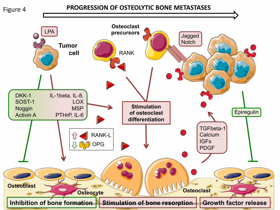
OsteoblastOsteoclast
Stimulation of bone resorptionInhibition of bone formation
RANK
Osteoclastprecursors
Tumor
cell
LPA
Stimulation of osteoclast
differentiation
RANK-L
OPG
TGFbeta-1Calcium
IGFs
PDGF
JaggedNotch
Epiregulin
PROGRESSION OF OSTEOLYTIC BONE METASTASES
Growth factor release
Osteocyte
Figure 4
IL-1beta, IL-8, LOX
MSP
PTHrP, IL-6
DKK-1SOST-1
Noggin
Activin A
Downloaded from journals.physiology.org/journal/physrev at INSERM (193.054.110.061) on December 29, 2020.

Inhibition of osteoclastdifferentiation
Inhibition of bone resorptionStimulation of bone formation
Osteoclast
Tumor cell
Osteoclastprecursor
BMP-4
Endothelial cell
Osteoblasts
Metastaticoutgrowth
PSA
uPA
IGFBP Latent
TGFbeta
TGFbetaIGF ET-1
OPG
RANK-L
ET-1
BMP-2
BMP-6
Wnts
PTHrP
PDGF
…..
RANK
PROGRESSION OF OSTEOBLASTIC BONE METASTASES
Release of growth factors
Osteocyte
Figure 5
ET-1
Downloaded from journals.physiology.org/journal/physrev at INSERM (193.054.110.061) on December 29, 2020.

CD8+
T cell
NK cell
Osteomacs
MDSC
Treg
pDC
Tumor Cells
Stimulation of osteoclast differentiation
Osteoclast Osteoclast precursor
Macrophage
RANKL
Suppression of CD8+ T cells
Osteoblast
IFN-gamma
IFN-gammaTRAIL/Fas-L
anti-tumor immune
response
Differentiationinto osteoclast
Bone tissue microenvironmentDownloaded from journals.physiology.org/journal/physrev at INSERM (193.054.110.061) on December 29, 2020.

Bone
resorption
Osteoclast
MCT-1
Tumor cell
proliferation
Tumorcell
Glucose
Glucose G6P Pyruvate Lactate
MCT-4
GLUT-1
PPP
Ribose
nucleotides
Glycolysis
Fatty acids
CholesterolProtein acetylation
TCA cycle
Cytoplasm
Lactate
Figure 7Downloaded from journals.physiology.org/journal/physrev at INSERM (193.054.110.061) on December 29, 2020.

A
B
BR
EA
ST
CA
NC
ER
PR
OS
TAT
E C
AN
CE
R
(99mTc)-BP
scintigraphy
PET/Fluorodeoxyglucose
PET/Fluorocholine
L1
L3
(99mTc)-BP
scintigraphy
Downloaded from journals.physiology.org/journal/physrev at INSERM (193.054.110.061) on December 29, 2020.

Bone matrix
Osteoclast
Osteoblast
Nerve cell
Molecular target
Calcium
TGFb
FPPS
RANKL
mTOR
VEGFR, c-MET
RON
Phase / Trial ID (NCT)
NGF
Drug
Androgen receptor
CYP17A
Activin A
DKK-1
Bisphosphonates, Radium-223,
Samarium-223, Strontium-89
Galinusertib
Bisphosphonates
Denosumab
Everolimus
Cabozantinib
BMS777607/ASLAN002
Approved
Approved
Approved
Approved
I / NCT01721148, NCT00605618
Approved
II / NCT02452008
Enzalutamide
Abiraterone
Sotatercept
BHQ880
Tanezumab
Approved
Approved
II / NCT01562405, NCT00747123
II / NCT01302886, NCT01337752,
NCT00741377
III / NCT00545129, NCT02609828
Matrix, Cell
Downloaded from journals.physiology.org/journal/physrev at INSERM (193.054.110.061) on December 29, 2020.

Table 1. Chemokines and their receptors involved in the formation of bone metastasis.
Chemokine
receptor
Chemokine
Ligand
Tumor
type
Function in bone metastasis Reference
CXCR-2 CXCL-1,
CXCL-2
Prostate Marrow adipocyte-derived CXCL-1
and CXCL-2 contribute to osteolysis
in metastatic prostate cancer
135
CXCR-2 CXCL-5 Breast The CXCR-2/CXCL-5 axis promotes
tumor cell colonization in the bone
marrow
275
CXCR-2 CXCL-8 (IL8) Breast Tumor-derived IL8 contributes to
osteoclastic bone destruction in
bone metastasis
18, 26, 27, 361
CXCR-3 CXCL-10 Breast,
melanoma
CXCL-10 facilitates trafficking of
CXCR-3-expressing cancer cells to
bone
193
CXCR-4 CXCL-12 Breast,
prostate
The CXCR-4/CXCL-12 axis
regulates tumor cell entry in the
bone marrow
235, 267, 273
CXCR-5 CXCL-13 Prostate CXCL-13 mediates attachment of
CXCR-5-expressing tumor cells to
bone marrow endothelial cells in
vitro
300
CX3CR-1 CX3CL-1 Breast,
prostate
CX3CR-1 and CX3CL-1 interactions
promote tumor cell extravasation in
the bone marrow
158,159
CCR-2 CCL-2 Breast,
prostate
CCL2-expressing tumor cells
engage CCR-2-expressing
macrophages and pre-osteoclasts to
facilitate colonization in bone
213, 232
Downloaded from journals.physiology.org/journal/physrev at INSERM (193.054.110.061) on December 29, 2020.

Table 2. Potential clinical utility of bone turnover biomarkers.
Biomarker Abbreviation Clinical application Reference
Bone formation marker
Bone alkaline phosphatase BALP Diagnosis of bone metastasis in solid tumors. Prognosis of bone
metastasis in solid tumors. Risk of skeletal related events.
Prognosis during anti-resorptive therapy. Prediction of response to
treatment.
61, 67, 90, 91,
155, 187, 206
procollagen I carboxyl-terminal propeptide PICP Diagnosis of bone metastasis in prostate cancer. Prediction of
response to atrasentan in prostate cancer.
187
Procollagen I amino-terminal propeptide PINP Diagnosis of bone metastasis in breast and prostate cancer. 35, 90, 180
Bone resorption marker
C-telopeptide CTX Diagnosis of bone metastasis in prostate cancer. Prognosis of
bone metastasis in breast cancer.
33, 35
N-telopeptide NTX Diagnosis of bone metastasis in prostate and lung cancer.
Prognosis of bone metastasis in solid tumors. Risk of skeletal
related events. Prognosis during anti-resorptive therapy. Prediction
35, 61, 67,90,
155, 187, 206
Downloaded from journals.physiology.org/journal/physrev at INSERM (193.054.110.061) on December 29, 2020.

of response to treatment.
carboxyterminal telopeptide of type I
collagen
ICTP Diagnosis of bone metastasis in lung cancer. Prognosis of bone
metastasis in breast cancer.
33, 155
Tartrate resistant acid phosphatase 5b TRACP Diagnosis of bone metastasis in breast cancer. 90, 155
pyridinoline PYD Prediction of response to atrasentan in prostate cancer. 186
Receptor activator of nuclear factor κB-
ligand/osteoprotegerin
RANKL/OPG Diagnosis of bone metastasis in solid tumors 125, 165, 183
microRNAs miRNAs Diagnosis of bone metastasis in breast cancer. 96, 294
Downloaded from journals.physiology.org/journal/physrev at INSERM (193.054.110.061) on December 29, 2020.

Downloaded from journals.physiology.org/journal/physrev at INSERM (193.054.110.061) on December 29, 2020.
Copyright © 2022 FDOKUMEN