102 - J Le Galliot - Psicoanalisis y lenguajes literarios - Caps I II y II (17 Copias)
METHOD
Translation, stability, and resistance to decapping of mRNAs
containing caps substituted in the triphosphate chainwith BH3, Se, and NH
WEI SU,1 SERGEY SLEPENKOV,1 EWA GRUDZIEN-NOGALSKA,1 JOANNA KOWALSKA,2 MARTA KULIS,2
JOANNA ZUBEREK,2 MACIEJ LUKASZEWICZ,2 EDWARD DARZYNKIEWICZ,2 JACEK JEMIELITY,2
and ROBERT E. RHOADS1
1Department of Biochemistry and Molecular Biology, Louisiana State University Health Sciences Center, Shreveport, Louisiana 71130-3932, USA2Division of Biophysics, Institute of Experimental Physics, Faculty of Physics, University of Warsaw, Warsaw 02-089, Poland
ABSTRACT
Decapping is an essential step in multiple pathways of mRNA degradation. Previously, we synthesized mRNAs containing capsthat were resistant to decapping, both to dissect the various pathways for mRNA degradation and to stabilize mRNA for moresustained protein expression. mRNAs containing an a-b CH2 group are resistant to in vitro cleavage by the decapping enzymehDcp2 but poorly translated. mRNAs containing an S substitution at the b-phosphate are well translated but only partiallyresistant to hDcp2. We now describe seven new cap analogs substituted at the b-phosphate with BH3 or Se, or substituted ateither the a-b or b-g O with NH. The analogs differ in affinity for eIF4E and efficiency of in vitro incorporation into mRNA byT7 RNA polymerase. Luciferase mRNAs capped with these analogs differ in resistance to hDcp2 hydrolysis in vitro, translationalefficiency in rabbit reticulocyte lysate and in HeLa cells, and stability in HeLa cells. Whereas mRNAs capped with m2
7,29-OGppSpGwere previously found to have the most favorable properties of translational efficiency and stability in mammalian cells, mRNAscapped with m7GppBH3pm7G are translated with the same efficiency but are more stable. Interestingly, some mRNAs exhibit a lagof up to 60 min before undergoing first-order decay (t1/2 ffi 25 min). Only mRNAs that are efficiently capped, resistant todecapping in vitro, and actively translated have long lag phases.
Keywords: boranophosphate cap analogs; phosphoroselenoate cap analogs; imidodiphosphate cap analogs; translationalefficiency; mRNA stability; in vitro transcription
INTRODUCTION
mRNA turnover is a highly regulated determinant in theoverall control of eukaryotic gene expression (Parker andSong 2004). The presence of a 59-terminal cap and a39-terminal poly(A) tract influences both the stability andtranslational efficiency of mRNA. After removal of thepoly(A) tract (deadenylation), mRNA is degraded by twogeneral pathways, decapping followed by 59/39 hydrolysisby the exonuclease Xrn1, and 39/59 hydrolysis by theexosome (Mukherjee et al. 2002; Wang et al. 2002). Inaddition to these two canonical pathways, mRNAs con-taining premature termination codons are degraded by thenonsense-mediated mRNA decay pathway (Maquat and
Gong 2009). Such nonsense-containing transcripts are de-graded either by deadenylation-independent decappingmediated by the protein Upf1 (Johansson et al. 2007) orby accelerated deadenylation and 39/59 exonucleolyticdigestion (Mitchell and Tollervey 2003). Another special-ized decay pathway exists for mRNAs containing AU-richsequence elements (AREs) in their 39-UTRs, which encodeproteins regulating either cell growth or the response toexternal factors such as microorganisms or inflammatorystimuli (Barreau et al. 2006). Degradation of mRNAs canalso be initiated by sequence-specific endonucleases (Dodsonand Shapiro 2002) or miRNAs (Franks and Lykke-Andersen2008). Histone mRNAs, which contain a conserved 39-terminal stem–loop (SL) rather than a poly(A) tract, aredegraded by another specialized pathway that is initiatedupon inhibition of DNA replication or at the end of S phase(Marzluff et al. 2008). Instead of deadenylation, the initialstep in histone mRNA decay is the addition of 39-terminaluridine residues (Mullen and Marzluff 2008).
Reprint requests to: Robert E. Rhoads, Department of Biochemistryand Molecular Biology, Louisiana State University Health Sciences Center,Shreveport, LA 71130-3932, USA; e-mail: [email protected].
Article published online ahead of print. Article and publication date areat http://www.rnajournal.org/cgi/doi/10.1261/rna.2430711.
978 RNA (2011), 17:978–988. Published by Cold Spring Harbor Laboratory Press. Copyright � 2011 RNA Society.

Decapping of mRNA is an essential step in all of thesepathways. This involves removal of the 7-methylguanosine-containing cap, which both shuts down translation andconstitutes the first irreversible step in mRNA degradation.There are two types of decapping enzymes in eukaryoticcells, DcpS and the Dcp1-Dcp2 complex. DcpS is a scaven-ger decapping enzyme and acts on the short cappedoligonucleotides remaining after 39/59 hydrolysis by theexosome, cleaving the cap between the b- and g-phos-phates to produce m7GMP (Liu et al. 2002). Dcp1 andDcp2 are the central components of an RNA-dependentdecapping complex. Dcp2 belongs to the Nudix family ofhydrolases (Piccirillo et al. 2003; She et al. 2006). InSaccharomyces cerevisiae, Dcp1 and Dcp2 directly interactto form a stable holoenzyme in which Dcp2 is the catalyticsubunit and Dcp1 plays a stimulatory role (Steiger et al.2003). Evidence has also been presented for an interactionbetween human Dcp1 and Dcp2 (Lykke-Andersen 2002;van Dijk et al. 2002). The proteins Edc1,Edc2, Edc3, Dhh1, and the Lsm1–7protein complex stimulate decappingby Dcp2 in S. cerevisiae and mammals(Coller and Parker 2004). Edc4 (alsoknown as Hedls and Ge-1) also acts asa positive effector of Dcp2 in mammals(Fenger-Gron et al. 2005). Decappingby Dcp2 is negatively regulated byeukaryotic initiation factor 4E (eIF4E),the translational cap-binding protein, aswell as by the 39-terminal poly(A) tract(Li et al. 2008). Biochemical studies ofthe human decapping protein (hDcp2)indicate that it requires both an m7G-containing cap and an RNA body of atleast 25 nt for full enzymatic activityand cleaves between the a- and b-phos-phate moieties to produce m7GDP(Lykke-Andersen 2002; van Dijk et al.2002; Wang et al. 2002; Piccirillo et al.2003).
In previous studies, we sought todevelop modified cap structures that,when incorporated into mRNAs, wouldbe resistant to hydrolysis by hDcp2 andthereby would stabilize mRNA against59/39 degradation (Grudzien et al.2006; Grudzien-Nogalska et al. 2007).Such modified cap analogs could po-tentially serve two purposes: to facilitatedissection of the various steps of mRNAdegradation and to boost production ofproteins encoded by exogenously intro-duced mRNAs. The new cap analogshad substitutions in the triphosphatechain intended to prevent hydrolysis by
decapping enzymes, but they also had substitutions ateither the C29 or C39 positions of m7Guo (for example,compounds 2 and 3 in Fig. 1, respectively), to ensure theyare incorporated into RNA by T7 RNA polymerase exclu-sively in the correct orientation (Stepinski et al. 2001;Jemielity et al. 2003). We found that m2
7,39-OGppCH2pG(4), which has a methylene group replacing the a-b bridgingO, was resistant to hDcp2 hydrolysis in vitro and increasedmRNA stability in cultured cells (Grudzien et al. 2006). How-ever, the affinity of m2
7,39-OGppCH2pG (4) for eIF4E wasonly 60% that of the parent compound, m2
7,39-OGpppG (3)(Kalek et al. 2005; Grudzien et al. 2006). We thereforedesigned a new series of cap analogs that contained aphosphorothioate moiety at either the a, b, or g position ofthe triphosphate chain (Grudzien-Nogalska et al. 2007).Each analog contains a stereogenic P-center and is thereforeobtained as a mixture of two diastereomers that can beresolved by reverse-phase HPLC (termed D1 and D2 based
FIGURE 1. Structures of cap analogs used in this study. D1 and D2 refer to the twodiastereoisomers produced by substitution of a non-bridging oxygen atom on the b-phos-phate. The cleavage sites for two decapping enzymes, DcpS and Dcp1/Dcp2, are indicated.
Cap analogs substituted with BH3, Se, and NH
www.rnajournal.org 979
on their elution order). Luciferase mRNA containingm2
7,29-OGppSpG, D2 (5) was more stable and was translatedmore efficiently in cultured HC11 cells than mRNAcontaining the parent compound m2
7,29-OGpppG (2)(Grudzien-Nogalska et al. 2007).
In the current study, we tested the resistance of mRNAcapped with m2
7,29-OGppSpG, D2 (5) to hydrolysis byhDcp2 under more rigorous conditions and found that itwas only partially resistant. We therefore attempted todevelop new cap analogs that conferred to mRNA both thefavorable stability of m2
7,39-OGppCH2pG (4) and the favor-able translation of m2
7,29-OGppSpG, D2 (5). Rather than theS substitution for a b non-bridging O of m2
7,29-OGppSpG (5),we made substitutions with BH3 (m2
7,29-OGppBH3pG, D1 [8]and m2
7,29-OGppBH3pG, D2 [9]) and Se (m27,29-OGppSepG,
D1 [6] and m27,29-OGppSepG, D2 [7]). Rather than the CH2
substitution for the a-b bridging O of m27,39-OGppCH2pG
(4), we made a substitution with NH (m27,29-OGppNHpG
[11]). Two related compounds, m7GppBH3pm7G (10) andm2
7,29-OGpNHppG (12), were also synthesized. The sevennew analogs were tested for in vitro and in vivo properties.Compared with the most favorable cap analog developed todate, m2
7,29-OGppSpG, D2 (5), capping efficiency was betterfor two analogs (compounds 11 and 12), the same for one(10), and worse for four (6–9). mRNAs capped with theseven new analogs differed in resistance to in vitro digestionwith hDcp2, but importantly, three of them (9, 10, and 11),were more resistant than m2
7,29-OGppSpG, D2 (5). mRNAscapped with the new analogs had translational efficienciesin HeLa cells that, compared with m2
7,29-OGppSpG, D2(5)-capped mRNA, were either the same (7 and 10) orlower (6, 8, 9, 11, and 12). Similarly, translation in rabbitreticulocyte lysate was either the same (9, 10) or worse (6,7, 8, 11, and 12). Finally, the stabilities of differently cappedmRNAs in HeLa cells were either greater (compound 10),the same (8 and 9), or less (6, 7, and 11) than mRNAcapped with m2
7,29-OGppSpG, D2 (5).
RESULTS
mRNA capped by m27,29-OGppSpG, D2 (5) is only
partially resistant to hDcp2
mRNA containing m27,29-OGppSpG, D2 (5) is resistant to in
vitro decapping by hDcp2 compared to the parent com-pound m2
7,29-OGpppG (2) (Grudzien-Nogalska et al. 2007),but when we tested it under more rigorous conditions, wefound it could still be partially decapped. Decapping wasmeasured by synthesizing RNAs with T7 polymerase inthe presence of a cap dinucleotide and [a-32P]GTP, in-cubating with recombinant hDcp2, and separating theproducts on an RNA sequencing gel (Fig. 2A). CappedRNAs (m2
7,29-OGpppGp*GpNp. . ., where p* represents 32P)migrate slower than decapped RNAs (pGp*GpNp. . .), whichmigrate the same as uncapped RNAs (ppp*Gp*GpNp. . .)
(lane 31). Bands detected by autoradiography were cut outand quantified by Cerenkov radiation. After 15 min, RNAcapped with m2
7,29-OGpppG (2) was completely decapped(lanes 1–3), whereas RNA capped with m2
7,29-OGppSpG, D2(5) was decapped 12 6 3% (Fig. 2A, lanes 4–6; Table 1),indicating partial but not complete resistance to decapping.To confirm this result, capped RNAs were incubated withhDcp2 for 60 min, resulting in 20 6 1% decapping of RNAcontaining m2
7,29-OGppSpG, D2 (5) (Fig. 2C, lanes 21–25;Fig. 2E, squares).
Development of novel borano-, seleno-,and imido-substituted cap analogs
hDcp2 is thought to cleave the cap structure between thea- and b-phosphate moieties, based on the observationsthat m7GDP is released (Lykke-Andersen 2002; van Dijket al. 2002; Wang et al. 2002) and that cleavage is blockedor retarded by substitution of the a-b bridging O with CH2
(Grudzien et al. 2006) or the b non-bridging O with S(Grudzien-Nogalska et al. 2007). However, as noted above,mRNAs capped with m2
7,39-OGppCH2pG (4) are poorlytranslated (Grudzien et al. 2006), and mRNAs capped withm2
7,29-OGppSpG, D2 (5) are not completely resistant tohDcp2 hydrolysis (Fig. 2). We therefore sought new capanalogs that would confer both of these favorable proper-ties to mRNA: high translational efficiency and highstability. Our previous studies showed that modificationsat a non-bridging O on the b-phosphate (Grudzien-Nogalska et al. 2007) or bridging O between a- andb-phosphates (Grudzien et al. 2006) rendered mRNA withthe most resistance to hDcp2. Accordingly, we focused oncap analogs with substitutions at these positions (Fig. 1,compounds 6–11). The m2
7,29-OGppSepG diastereoisomers(6 and 7) have a Se-for-O substitution, forming a phosphoro-selenoate group at the b position. The m2
7,29-OGppBH3pGdiastereoisomers (8 and 9) have a BH3-for-O substitution,forming a boranophosphate group at the b position.m7GppBH3pm7G (10) is cap analog containing two identi-cal nucleoside moieties and a boranophosphate group atthe b position, so it is not synthesized as two diastereo-mers. m2
7,29-OGppNHpG (11) has an NH-for-O substi-tution at the a-b bridge, forming an imidodiphosphategroup. An analog with the NH group replacing the b-gbridging O, m2
7,29-OGpNHppG (12), was also included forcomparison.
Fluorescence quenching measurements indicated that allseven new cap analogs have either the same or higher affinityfor murine eIF4E compared with the parent compound,m2
7,29-OGpppG (2) (Table 1, compounds 6–12). Previousstudies (Kowalska et al. 2009) had shown that severalphosphorothioate isomers of the cap dinucleotide had thehighest affinity for eIF4E of all triphosphate analogs testedto date. Two of the new analogs, m2
7,29-OGppSepG, D1 (6)and m2
7,29-OGppBH3pG, D1 (8), also had very high affinities
Su et al.
980 RNA, Vol. 17, No. 5
for eIF4E (Table 1), comparable to that of the homologousphosphorothioate m2
7,29-OGppSpG, D1 (Kowalska et al.2008). For all three substitutions for the b non-bridgingO (S, Se, and BH3), the D1 diastereomer had nearly twice
the affinity for eIF4E than the D2diastereomer. An analog with a CH2
substitution for the a-b bridging O,m2
7,39-OGppCH2pG (4), binds eIF4E withlower affinity than the parent compound(Table 1; Kalek et al. 2005), but sub-stitution of the same O with NH pro-duces an analog, m2
7,29-OGppNHpG (11),with an affinity for eIF4E that is compa-rable to the parent compound (Table 1).
The new cap analogs differ in theirability to serve as substrates for T7RNA polymerase
Capping efficiency reflects the degreeto which a dinucleotide cap analog willbe recognized as GTP by T7 RNApolymerase during in vitro transcrip-tion. We measured capping efficiencyby separation on RNA sequencing gelsand autoradiography (Fig. 2A). Laneslabeled 0 min (lanes 1,4,7, etc.) were notincubated with hDcp2. The ratio of theradioactivity in these lanes of the upperband [m2
7,29-OGppXpGp*(Np)46N, where‘‘X’’ represents a modification] to thesum of the radioactivity in the upperplus lower band [pppGp*(Np)46N] rep-resents capping efficiency. This is sum-marized for all new analogs in Table 1.Under the conditions used here (10:1ratio of cap analog to GTP in the T7polymerase reaction), we observed 84 6
1% capping for the parent compound,m2
7,29-OGpppG (2). Higher capping ef-ficiency can be obtained by using higherratios of cap analog to GTP. The cap-ping efficiency for m2
7,29-OGppNHpG(11) and m2
7,29-OGpNHppG (12) wasslightly better than for the parent com-pound, followed in decreasing order bym2
7,39-OGppCH2pG (4), m27,29-OGppSpG,
D2 (5), m7GppBH3pm7G (10), m27,29-O
GppBH3pG, D2 (9), m27,29-OGppBH3pG,
D1 (8), m27,29-OGppSepG, D1 (6), and
m27,29-OGppSepG, D2 (7) (Fig. 2A; Table 1).
RNAs capped with BH3- andNH-containing analogs are resistantto decapping by hDcp2 in vitro
Transcripts synthesized in the presence of nine different capanalogs plus one uncapped transcript were incubated withhDcp2 for various times and subjected to PAGE (Fig. 2A).Transcripts capped with m2
7,39-OGppCH2pG (4) were
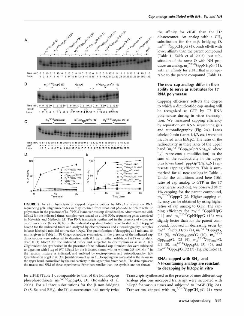
FIGURE 2. In vitro hydrolysis of capped oligonucleotides by hDcp2 analyzed on RNAsequencing gels. Oligonucleotides were synthesized from Nco1-cut pluc-A60 template with T7polymerase in the presence of [a-32P]GTP and various cap dinucleotides. After treatment withhDcp2 for the indicated times, samples were loaded on a 10% RNA sequencing gel as describedin Materials and Methods. (A) Ten RNA transcripts synthesized in the presence of either nocap dinucleotide (lanes 31,32) or the indicated cap dinucleotide were treated with 0.6 mg ofhDcp2 for the indicated times and analyzed by electrophoresis and autoradiography. Samplesin lanes labeled 0 min did not receive hDcp2. The quantification of decapping at 5 min and 15min is given in Table 1. (B) Oligonucleotides synthesized in the presence of the indicated capdinucleotides were subjected to digestion with 0.4 mg of either wild-type (WT) or catalyticdead (CD) hDcp2 for the indicated times and subjected to electrophoresis as in A. (C)Oligonucleotides synthesized in the presence of the indicated cap dinucleotides were subjectedto digestion with 1 mg of WT hDcp2 for the indicated times, with or without 0.5 mM Mn2+ inthe reaction mixture as indicated, and analyzed by electrophoresis and autoradiography. (D)Quantification of gel in B. (E) Quantification of gel in C. Decapping was calculated as the % loss inthe upper band, normalized by the radioactivity in the upper plus lower bands. The data representthe means and SEM of three experiments. Error bars smaller than the symbols are not shown.
Cap analogs substituted with BH3, Se, and NH
www.rnajournal.org 981
completely resistant to hDcp2 (Fig. 2A, lanes 7–9), inagreement with our previous study in which SAX chroma-tography was used to measure decapping (Grudzien et al.2006). Transcripts capped with m2
7,29-OGppSepG, D1 (6)(lanes 10–12), m2
7,29-OGppSepG, D2 (7) (lanes 13–15), andm2
7,29-OGppBH3pG, D1 (8) (lanes 16–18) were intermediatein their susceptibility to hDcp2. By contrast, transcriptscapped with m2
7,29-OGppBH3pG, D2 (9) (lanes 19–21),m7GppBH3pm7G (10) (lanes 22–24), and m2
7,29-OGppNHpG(11) (lanes 25–27) were highly resistant to hDcp2. Theaverage results for three experiments are presented in Table 1.
To confirm that the decapping activity observed in Figure2A was due to hDcp2 and not to a contaminating bacterialpyrophosphatase, we used a variant of hDcp2 in which Glu-147 and Glu-148 are both changed to Gln, producinga catalytically dead (CD) enzyme (Wang et al. 2002). Underconditions where RNA containing m2
7,29-OGpppG (2) wascompletely decapped by wild-type (WT) hDcp2 (Fig. 2B,lanes 1–4; Fig. 2D, closed circles), there was no decapping byCD hDcp2 (Fig. 2B, lanes 5–8; Fig. 2D, open circles). RNAwith an unmethylated cap was inefficiently cleaved by WThDcp2 (Fig. 2B, lanes 9–12; Fig. 2D, filled diamonds; 9.0 6
0.5% by 60 min), in agreement with published findings (vanDijk et al. 2002; Wang et al. 2002), but was not cleaved at allby CD hDcp2 (Fig. 2B, lanes 13–16; Fig. 2D, open di-amonds). RNA capped with m7GppBH3pm7G (10) was notdetectably decapped by either enzyme preparation (Fig. 2B,lanes 17–24; Fig. 2D, triangles).
The original in vitro decapping assay conditions reportedfor hDcp2 utilized Mg2+ as the sole divalent cation (Lykke-Andersen, 2002; van Dijk et al. 2002; Wang et al. 2002), but
subsequently it was shown that bothhDcp2 (Piccirillo et al. 2003) andyeast Dcp2p (Steiger et al. 2003) areconsiderably more active if Mn2+ isadded, although there is no evidencethat the enzyme utilizes Mn2+ invivo. Previous studies from ourown laboratory used 2 mM Mg2+ asthe sole divalent cation to demon-strate cleavage by hDcp2 of RNAcapped with m2
7,39-OGpppG (3) andresistance of RNA capped withm2
7,39-OGppCH2pG (4) (Grudzienet al. 2006), but subsequently we used2 mM Mg2+ plus 0.5 mM Mn2+ toshow resistance to of mRNA cap-ped with m2
7,2-OGppSpG, D2 (5)(Grudzien-Nogalska et al. 2007). Toensure that the resistance of mRNAcapped with m2
7,29-OGppSpG, D2 (5)and m7GppBH3pm7G (10) to hDcp2is not a Mn2+-induced artifact, wecarried out the decapping assay underboth conditions: with Mg2+ as the
sole divalent cation and with 2 mM Mg2+ plus 0.5 mMMn2+ (Fig. 2C,E). The results confirmed that hDcp2 is muchmore active against RNA capped with m2
7,29-OGpppG (2) inthe presence of Mn2+ (Fig. 2E, filled versus open circles).Importantly, the same order of resistance to decapping isseen under both conditions. For instance, at the 30-min timepoint, the % decapping in Mg2+ plus Mn2+ is 92.8 6 0.3for RNA capped with m2
7,29-OGpppG (2), 15.3 6 2.6 form2
7,29-OGppSpG, D2 (5), and 5.5 6 1.9 for m7GppBH3pm7G(10), whereas with Mg2+ alone, it is 24.0 6 1.7 form2
7,29-OGpppG (2), 4.9 6 1.9 for m27,29-OGppSpG, D2
(5), and 1.5 6 0.3 for m7GppBH3pm7G (10). Thus, we con-clude that the resistance to in vitro decapping of thesephosphate chain-modified cap structures is not a Mn2+-induced artifact.
Cap analogs substituted with BH3 stabilize mRNAsin HeLa cells
We next tested how the new cap analogs would affect mRNAstability when incorporated into RNAs and delivered intomammalian cells. Luciferase mRNAs were synthesized invitro with various cap analogs at the 59-termini and 60-ntpoly(A) tracts at the 39-termini. The mRNAs were intro-duced into HeLa cells by nucleoporation, the cells wereincubated for various times up to 4 h, and total RNA wasrecovered. Luciferase mRNA levels were measured byqRT-PCR (Fig. 3). Interestingly, we observed a two-phasedecay pattern for all mRNAs except those capped withm2
7,29-OGppSepG, D1 (6) and m27,29-OGppSepG, D2 (7); there
was a lag phase of various durations lasting as long as
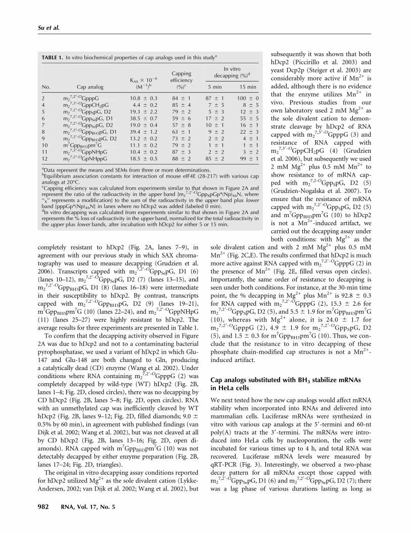
TABLE 1. In vitro biochemical properties of cap analogs used in this studya
No. Cap analogKAS 3 10�6
(M�1)b
Cappingefficiency
(%)c
In vitrodecapping (%)d
5 min 15 min
2 m27,29-OGpppG 10.8 6 0.3 84 6 1 87 6 1 100 6 0
4 m27,39-OGppCH2pG 4.4 6 0.2 85 6 4 7 6 5 8 6 5
5 m27,29-OGppSpG, D2 19.3 6 2.2 79 6 2 5 6 3 12 6 3
6 m27,29-OGppSepG, D1 38.5 6 0.7 59 6 6 17 6 2 55 6 5
7 m27,29-OGppSepG, D2 19.0 6 0.4 57 6 8 10 6 1 16 6 1
8 m27,29-OGppBH3pG, D1 39.4 6 1.2 63 6 1 9 6 2 22 6 3
9 m27,29-OGppBH3pG, D2 13.2 6 0.2 73 6 2 2 6 2 4 6 1
10 m7GppBH3pm7G 11.1 6 0.2 79 6 2 1 6 1 1 6 111 m2
7,29-OGppNHpG 10.4 6 0.2 87 6 3 2 6 2 3 6 212 m2
7,29-OGpNHppG 18.5 6 0.5 88 6 2 85 6 2 99 6 1
aData represent the means and SEMs from three or more determinations.bEquilibrium association constants for interaction of mouse eIF4E (28-217) with various capanalogs at 20°C.cCapping efficiency was calculated from experiments similar to that shown in Figure 2A andrepresent the ratio of the radioactivity in the upper band [m2
7,29-OGppXpGp*(Np)46N, where‘‘X’’ represents a modification] to the sum of the radioactivity in the upper band plus lowerband [pppGp*(Np)46N] in lanes where no hDcp2 was added (labeled 0 min).dIn vitro decapping was calculated from experiments similar to that shown in Figure 2A andrepresents the % loss of radioactivity in the upper band, normalized for the total radioactivity inthe upper plus lower bands, after incubation with hDcp2 for either 5 or 15 min.
Su et al.
982 RNA, Vol. 17, No. 5

60 min, followed by first-order decay with approximatelythe same t1/2 for all mRNAs. This type of experiment wasconducted three or more times for each of the sevennew cap analogs, using as controls m2
7,29-OGpppG (2),m2
7,29-OGppSpG, D2 (5), and m27,39-OGppCH2pG (4) (Table
2). mRNAs containing different cap analogs had similart1/2 values for the first-order decay phase (21–32 min) butvaried significantly in the length of lag phase (0–51 min).
mRNAs capped with Se- and BH3-containing analogsare efficiently translated in HeLa cells and rabbitreticulocyte lysate
Since mRNAs capped with several of the new analogs(m2
7,29-OGppBH3pG, D2 [9], m7GppBH3pm7G [10], andm2
7,29-OGppNHpG [11]) are more resistant to hDcp2hydrolysis in vitro than m2
7,29-OGppSpG, D2 (5), we
determined their translational properties. Translationalefficiency in cultured mammalian cells can be determinedby introducing luciferase mRNA by nucleoporation, re-moving aliquots of the cells after various times, andmeasuring the accumulation of luciferase (Grudzien et al.2006; Grudzien-Nogalska et al. 2007). The concentration ofluciferase mRNA is measured in the same cells by qRT-PCR, and the rate of luciferase accumulation per unit ofmRNA represents translational efficiency. Figure 4A showsthat luciferase accumulation begins after a z15 min lag,which is required for synthesis of the enzyme, and continueslinearly for z1 h, after which it slows dramatically (upperpanel). However, when the luciferase value is normalized forthe amount of luciferase mRNA still remaining at each timepoint, the rate of luciferase accumulation is linear for atleast 3 h (lower panel). Thus, the translational efficiency ofluciferase mRNA remains constant over this period eventhough the amount of luciferase mRNA decreases.
We chose to measure translational efficiency of mRNAscapped with the new analogs over the first 60 min afternucleoporation to minimize errors caused by mRNA decay(Fig. 4B). This type of experiment was performed three ormore times for each of the seven new analogs plus threecontrols (Table 2). mRNAs capped with two of the new capanalogs, m2
7,29-OGppSepG, D2 (7) and m7GppBH3pm7G (10),were translated 1.4- and 1.7-fold more efficiently than mRNAcapped with the parent compound, m2
7,29-OGpppG (2).mRNAs capped with m2
7,29-OGppBH3pG, D2 (9) were trans-lated with similar efficiency as the parent mRNA, whereasfour others (m2
7,29-OGppSepG, D1 [6], m27,29-OGppBH3pG,
D1 [8], m27,29-OGppNHpG [11], and m2
7,29-OGpNHppG[12]) were translated less efficiently.
Measuring translational efficiency in mammalian cellshas the advantage of being more physiologically relevantthan in vitro translation systems but the disadvantage thatthe amount of mRNA introduced by nucleoporation mustbe independently measured, thereby introducing experi-mental error. When mRNA is introduced by nucleopora-tion, there is also the possibility that some of it may bepresent in a form or location that is inaccessible to thetranslational machinery, although we previously showedthat the majority of exogenously introduced mRNA is inpolysomes (Grudzien et al. 2006; Grudzien-Nogalska et al.2007). Both of these potential problems, as well as ex-perimental error arising from differential degradation rates,are avoided by using an in vitro translation system. Wetherefore measured the translational efficiency of mRNAscapped with the seven new analogs in a rabbit reticulocytelysate system (Table 2). Although there were quantitativedifferences between in vitro and in vivo translational effi-ciencies, most of the analogs had qualitatively similar effectsin the two systems. For instance, mRNAs capped withm2
7,29-OGppSepG, D2 (7) and m7GppBH3pm7G (10) weremore efficiently translated than the control mRNA in bothsystems. Similarly, mRNAs capped with m2
7,29-OGppNHpG
FIGURE 3. Decay of luciferase mRNA terminated by various capanalogs in HeLa cells. Luciferase mRNAs were synthesized in thepresence of the indicated cap analogs and delivered into HeLa cellsby nucleoporation as described in Materials and Methods. Cells werelysed at the indicated times and luciferase mRNA was measured byqRT-PCR. Data in panels A–E are plotted as a percentage of theluciferase mRNA present immediately after nucleoporation. In panelsB–E, there is a lag before the initiation of rapid decay. The data for thepost-lag period were fit to single-exponential function and the t½ ofcalculated as described in Materials and Methods. Vertical dashedlines mark the boundary between lag phase and first-order decayphase. The data for each transcript represent a single experiment. Theerror bars represent duplicate luciferase mRNA determinations. Thevalues for replicate experiments are given in Table 2.
Cap analogs substituted with BH3, Se, and NH
www.rnajournal.org 983

(11) and m27,29-OGpNHppG (12) were less efficiently trans-
lated than the control mRNA in both systems. How-ever, mRNAs capped with m2
7,29-OGppSepG, D1 (6) andm2
7,29-OGppBH3pG, D1 (8) were translated somewhat betterthan the control mRNA in vitro but worse in vivo. Anothertrend observed in both systems is that the mRNAs cappedwith the D2 diastereomers were translated more efficientlythan those capped with the D1 diastereomers, despite thehigher affinity of the latter for eIF4E (Table 1).
DISCUSSION
Resistance of cap analogs substitutedat the b-phosphate to hDcp2 hydrolysis
The new cap analogs with modifications at the b-phosphateposition were designed to both provide resistance to hDcp2and enhance translational efficiency. Nucleotide analogswith substitution of S, BH3, or Se for the non-bridging O inphosphate moieties share some chemical and biochemicalproperties such as polarity, net charge at physiological pH,and resistance to nucleases but also differ with respect toP-X bond length, van der Waals radius, and affinity of theX group for various divalent or other metal cations. S andSe, due to their larger atomic size, are more polarizable andprefer interactions with softer metal ions such as Zn2+,
Mn2+, and Cd2+ compared to Mg2+
and Ca2+, which are preferred by O.The BH3 group is a very poor nucle-ophile because it lacks a lone electronpair and therefore does not acceptH-bonds or coordinate metal ions (Liet al. 2007). It has also been reportedthat oligonucleotides containingboranophosphate modifications canbe more resistant to enzymatic cleav-age than their corresponding phos-phorothioates (Sergueev and Shaw1998). Enzymatic resistance of nucle-otides and nucleic acids contain-ing phosphorothioate, phosphoro-selenoate, and boranophosphatemodifications are thought to resultfrom changes in affinities to metalions and phosphate group geometry.
Like other Nudix-domain hydro-lases, the catalytic center of Dcp2contains three conserved Glu resi-dues that coordinate a divalent cat-ion responsible for activation ofa water molecule to attack the phos-phate moiety (She et al. 2006). Theresistance of mRNA capped withm2
7,29-OGppSpG, D2 (5) to hDcp2may be due to poor interaction of S
with Mg2+ or Mn2+ in the enzyme’s active site or todisruption of enzyme-substrate complex geometry becauseof the longer P-S bond and larger S atom. The even higherresistance of BH3-containing analogs may be explained bythe poor ability of BH3 to bind metal ions or accept H-bondscompared to S and Se. It is surprising that m7GppBH3pm7G(10) is less susceptible to hDcp2 than either m2
7,29-OGppBH3pG,D1 (8) or m2
7,29-OGppBH3pG, D2 (9). m7GppBH3pm7G (10)is not diastereomeric, but after incorporation into mRNA,it exists as a mixture of diastereomers analogous to theD1 and D2 diastereomers of m2
7,29-OGppBH3pG (8 and 9,respectively). (This is because the first phosphodiester bondcatalyzed by T7 RNA polymerase could be formed with the39-OH of either m7Guo moiety of m7GppBH3pm7G [10].)Consequently, the enzymatic resistance of mRNA preparedwith m7GppBH3pm7G would be expected to be intermediatebetween that observed for m2
7,29-OGppBH3pG, D1 (8) andm2
7,29-OGppBH3pG, D2 (9). The fact that it is greater suggeststhat the second m7Guo moiety of m7GppBH3pm7G (10)makes an additional contribution to enzymatic resistance.
Resistance of cap analogs substituted at the a-bbridge to hDcp2 hydrolysis
The imidodiphosphate (pNHp) and methylenebisphos-phonate (pCH2p) bridging modifications are both known
TABLE 2. In vivo biochemical properties of cap analogs used in this studya
No. Cap analog
mRNA decay (min) In vivotranslationalefficiencyd
In vitrotranslationalefficiencyfLag phaseb Half-lifec
2 m27,29-OGpppG 9 6 3 23 6 5 1.0 6 0 ND
3 m27,39-OGpppG ND ND NDe 1.0 6 0.1
4 m27,39-OGppCH2pG 19 6 4 24 6 3 NDe NDe
5 m27,29-OGppSpG, D2 38 6 8 25 6 1 1.8 6 0.3 1.6 6 0.2
6 m27,29-OGppSepG, D1 no lag 32 6 3 0.6 6 0.1 1.2 6 0.1
7 m27,29-OGppSepG, D2 no lag 24 6 6 1.4 6 0.5 1.3 6 0.1
8 m27,29-OGppBH3pG, D1 25 6 5 21 6 4 0.5 6 0 1.2 6 0.2
9 m27,29-OGppBH3pG, D2 38 6 4 23 6 2 1.0 6 0.2 1.9 6 0.1
10 m7GppBH3pm7G 51 6 4 31 6 9 1.7 6 0.2 1.5 6 0.111 m2
7,29-OGppNHpG 18 6 4 26 6 6 0.5 6 0 0.8 6 0.112 m2
7,29-OGpNHppG 16 6 4 23 6 4 0.5 6 0.2 0.7 6 0.1
aData represent the means and SEMs from three or more determinations.bThe length of the lag phase in HeLa cells for mRNAs containing the indicated cap analogs wasdetermined from experiments similar to that shown in Figure 3 and corresponds to the timeperiod before rapid first-order decay begins (before the dashed line in Fig. 3).cThe half-lives in HeLa cells of mRNAs containing the indicated cap analogs was determinedfrom experiments similar to that shown in Figure 3 from the points corresponding to first-orderdecay (after the dashed line in Fig. 3) where t1/2 = ln2/k and k is defined in Equation 1.dTranslational efficiency in HeLa cells was determined from experiments similar to those shownin Figure 4B and represent the rate of luciferase accumulation, normalized by luciferasemRNA, for mRNAs capped with the indicated analogs relative to that of mRNA capped withm2
7,29-OGpppG (2).eNot determined in the current study but previously reported (Grudzien et al. 2006).fTranslational efficiency in a rabbit reticulocyte lysate system represents the rate of luciferaseaccumulation from mRNA containing the indicated cap analogs compared to control mRNAcapped with m2
7,39-OGpppG (3). The latter does not differ significantly from mRNA cappedwith m2
7,29-OGpppG (2) (Jemielity et al. 2003).
Su et al.
984 RNA, Vol. 17, No. 5

for their resistance to enzymatic cleavage, but unlike thenon-bridging modifications discussed above, their affinitiesfor Mg2+ are higher than those of the parent compounds(Yount et al. 1971). The pNHp moiety is more similar topyrophosphate than pCH2p in terms of bond length,geometry, and acid-base properties (Yount et al. 1971).Unlike the CH2 group, the NH group has a lone electronpair and can therefore participate in H-bonding, which isparticularly important for binding to eIF4E (Tomoo et al.2003). This is consistent with the fact that NH-containinganalogs bind eIF4E with affinities similar to those of theparent compounds (Table 1), whereas the CH2-containinganalogs bind eIF4E weaker (Kalek et al. 2005). The mechanismof resistance of NH- and CH2-containing analogs is different
from that of non-bridging b-phosphate-substituted analogsand arises from the high chemical stability of P-C-P and P-N-Pbonds compared to P-O-P. Thus, we observed complete re-sistance to decapping with both of these analogs (Table 1).
Translational properties of mRNAs containingthe new cap analogs
For a cap analog to be incorporated into an mRNA thathas both high translational efficiency and high resistanceto decapping, the analog must have favorable interac-tions within the active sites of three structurally un-related proteins: T7 RNA polymerase (Steitz 2009), eIF4E(Marcotrigiano et al. 1997; Matsuo et al. 1997; Tomoo et al.2003), and hDcp2 (Deshmukh et al. 2008). Our resultssuggest that these interactions differ among the seven newcap analogs. mRNAs capped with compounds havingBH3 in the b position (m2
7,29-OGppBH3pG, D2 [9] andm7GppBH3pm7G [10]) are highly resistant to decapping,but the capping efficiency, stability, and translationalefficiency of the latter is higher. m2
7,29-OGppNHpG (11)-capped RNAs are also completely resistant to in vitrohydrolysis by hDcp2 but are translated less efficiently thanthe control mRNA both in vitro and in vivo. mRNAcapped with m2
7,29-OGppSepG, D2 (7) is translated moreefficiently than the control mRNA despite the fact that its% capping is only 57%, suggesting that it would have aneven higher translational efficiency if the % capping couldbe increased. Surprisingly, even though m2
7,29-OGppNHpG(11) is similar to the parent m2
7,29-OGpppG (2) with respectto bond length, geometry, and acid-base properties, it doesnot enhance translational efficiency or increase the lengthof the lag phase, even though it is completely resistant tohDcp2 in vitro.
Relationship of the lag in mRNA degradationto translation and decapping
Some insight into the differences among these mRNAscomes from correlations among resistance to hDcp2 invitro, translational efficiency, and length of the lagphase. Transcripts capped with m2
7,29-OGppSpG, D2 (5),m2
7,29-OGppBH3pG, D2 (9), and m7GppBH3pm7G (10) areresistant to hDcp2, are translated with the same or higherefficiency than the control mRNA, and exhibit a longlag phase. On the other hand, RNAs capped withm2
7,39-OGppCH2pG (4) and m27,29-OGppNHpG (11) are
resistant to hDcp2, are translated poorly, and exhibit a shortlag phase. Thus, resistance to hDcp2 hydrolysis alonecannot account for a long lag phase and efficient trans-lation. We propose that the lag is partly a function ofmRNA recognition by eIF4E and recruitment to polysomes,which are determinants of the rate of both deadenylationand decapping. During translation, the cap is protectedfrom decapping by association with eIF4E (Schwartz and
FIGURE 4. Translational efficiencies in HeLa cells of mRNAs con-taining various caps. (A) Upper panel: Luciferase activity wasmeasured at the indicated times after introduction of luciferasemRNA capped with m2
7,29-OGpppG (2) into HeLa cells by nucleo-poration. Lower panel: Luciferase activity values in the upper panelwere divided by the amount of luciferase mRNA at each time point asmeasured by qRT-PCR. (B) Luciferase activity for mRNAs synthesizedin the presence of the indicated cap analogs was measured at theindicated times after nucleoporation. Data were normalized for theamount of luciferase mRNA as in A. The data for each transcriptrepresent a single experiment. The error bars represent duplicateluciferase activity determinations. The values for replicate experimentsare given in Table 2.
Cap analogs substituted with BH3, Se, and NH
www.rnajournal.org 985

Parker 2000). PABP binds to poly(A) and protects mRNAagainst exonucleolytic degradation (Sachs 1993; Coller et al.1998; Wang et al. 1999). PABP also binds to the N terminusof eIF4G (Tarun and Sachs 1996) and stabilizes the eIF4G-eIF4E complex (Amrani et al. 2008). We speculate thatmRNAs that are poorly recognized by the translationalmachinery, such as those capped with m2
7,39-OGppCH2pG(4) or m2
7,29-OGppNHpG (11), enter the 39/59 degrada-tion pathway immediately, exhibiting a short lag, even ifthe cap is resistant to hDcp2. mRNAs that are well rec-ognized by the translational machinery, such as thosecapped with m2
7,29-OGpppG (2), are protected fromdecapping because Dcp2 and eIF4E compete for bindingto the cap; these mRNAs exhibit an intermediate lag phase,even though the cap is susceptible to cleavage by hDcp2.When there is both efficient recognition by the translationalmachinery and resistance to decapping, such as for mRNAscapped with m2
7,29-OGppSpG, D2 (5), m27,29-OGppBH3pG,
D2 (9), or m7GppBH3pm7G (10), the lag is the longest,presumably because both deadenylation and decapping areretarded. The absence of a lag for mRNAs capped withm2
7,29-OGppSepG, D2 (7) is likely due to the fact that thisanalog is poorly recognized by T7 polymerase, resulting inthe lowest capping efficiency of all the new analogs; nearlyhalf of the mRNA in this preparation is uncapped (Table 1).
Utility of cleavage-resistant cap analogs
The new cap analogs differ from each other and frompreviously described analogs in numerous properties thatmake them potentially useful for a variety of applications.For example, structural studies of the catalytic site ofhDcp2 and related enzymes could be carried out witha series of analogs that are completely resistant to cleavagebut differ in molecular properties (m2
7,39-OGppCH2pG [4],m2
7,29-OGppBH3pG, D2 [9], and m27,29-OGppNHpG [11]).
Some have very high affinity for eIF4E and may pointthe way to new anticancer drugs based on inhibition ofcap-dependent translation (m2
7,29-OGppSepG, D1 [6] andm2
7,29-OGppBH3pG, D1 [8]). Some confer high transla-tional efficiency in vivo, in vitro, or both and should beuseful for enhanced protein production. Efficiently trans-lated mRNAs that differ in decapping rate (e.g., mRNAscapped with m2
7,29-OGpppG [2], m27,29-OGppSpG, D2 [5],
m27,29-OGppSepG, D2 [7], and m7GppBH3pm7G [10])
should be useful for the study of mRNA turnover. Long-lived and efficiently translated mRNAs also have a promis-ing role is in cancer immunotherapy, where an in vitro–synthesized mRNA encoding a tumor antigen is used toelicit an immune response in dendritic cells (Weide et al.2008). Vaccination with such mRNAs has already enteredhuman clinical testing. Recently, capping with the D1diastereomer of m2
7,29-OGppSpG was shown to enhanceRNA stability and translational efficiency in immaturedendritic cells (Kuhn et al. 2010). In vivo delivery of an
antigen encoded by this mRNA led to increased proteinexpression and enhanced priming and expansion of naıveantigen–specific-T–cells in mice. Further advances in thedevelopment of mRNAs that are both stable and transla-tionally efficient should aid this powerful approach toinduce therapeutic immune responses.
MATERIALS AND METHODS
Materials
All common reagents were of analytical grade. Oligodeoxynucleo-tides were synthesized by MWG/Operon (Huntsville, AL). Liter-ature citations for synthesis of all cap analogs are given in Figure 1.The structures and homogeneities of all compounds were con-firmed by mass spectrometry, 1H NMR, 31P NMR and reverse-phase HPLC. The concentrations of cap analog solutions weredetermined by UV absorbance at pH 7.0 using the extinctioncoefficient e255nm = 22.6 3 103 M�1cm�1.
In vitro synthesis of RNAs
The DNA template for T7 RNA polymerase used to make cappedRNAs for in vitro decapping assays was the plasmid pluc-A60(Grudzien et al. 2006) digested with NcoI, which yields a cappedRNA corresponding to the first 48 nt of firefly luciferase mRNA.Capped oligoribonucleotides were synthesized in the presence ofvarious cap analogs and 10 mCi/mL [a-32P]GTP (ICN) in a 50-mLreaction for 1 h as previously described (Jemielity et al. 2003)except that the T7 RNA polymerase was prepared in ourlaboratory (Davanloo et al. 1984). Reaction mixtures were ex-tracted with phenol and chloroform, and RNAs were separatedfrom unincorporated nucleotides with NucAway spin columns(Ambion). The concentrations of RNAs were determined bymeasuring Cerenkov radiation in a scintillation counter (Beckman)and calculating from the specific radioactivity of the precursorGTP. Full-length luciferase mRNA capped with various analogs wassynthesized as described above except that pluc-A60 was digestedwith HpaI, the reaction mixture was 200 mL, no radioactivity wasincluded, and the incubation time was 2 h. Reaction mixtureswere treated with 10 units of RQ1 RNase-Free DNase (Promega)at 37°C for 20 min. The RNAs were purified with an E.Z.N.A.Total RNA Miniprep Kit (Omega Bio-Tek). RNA concentrationwas determined by UV absorbance at 260 nm, and RNA integritywas verified by electrophoresis on 1.2% agarose gels containingformaldehyde.
Decapping assays
GST-hDcp2 and a variant of GST-hDcp2 in which Glu-147 andGlu-148 is replaced with Gln to produce a catalytically inactiveenzyme were expressed in BL21(DE3)pLysS Escherichia coli cellsfrom plasmids pGEX-hDcp2 and pGEX hDcp2Q147/8, respectively(Wang et al. 2002), which were generously donated by MegerdichKiledjian, Rutgers University. The enzymes were expressed (Wanget al. 2002) and purified (Zhao et al. 2003) as previously described.Capped 32P-labled oligonucleotides were subjected to digestionwith either wild-type or catalytically inactive GST-hDcp2 at 37°C
Su et al.
986 RNA, Vol. 17, No. 5
for various times. The conditions were the same as describedpreviously (Grudzien-Nogalska et al. 2007) except that spermidinewas omitted. In Figure 2C, Mn2+ was omitted from somereactions as indicated. Reactions were stopped by adding twovolumes of Precipitation/Inactivation buffer (Ambion), and RNAswere precipitated at �20°C for 20 min and collected by centrifu-gation at 13,000 3 g at 4°C for 20 min. Precipitated samples wereresuspended in 5 mL of Sequencing Gel Loading Buffer (Ambion)and denatured at 95°C for 5 min. RNA sequencing gels (10%polyacrylamide) were run at 45–70 W for z3 h on a Base RunnerNucleic Acid Sequencer apparatus (International Biotechnologies)(Slatko and Albright 1992). Gels were fixed in 5% acetic acid, 5%methanol for 10–15 min, dried onto Whatman 3MM filter paper(Fisher Scientific), and exposed to Blue X-ray film (Kodak).Radioactivity in individual bands was quantified by twomethods: analyzing scanned film using ImageQuant TL program(GE Health Care, version 7.0), and cutting them out anddetermining Cenrenkov radiation.
Cell culture and nucleoporation of mRNA
HeLa cells were grown in DMEM high glucose medium (Hyclone)containing 10% fetal bovine serum (Atlanta Biologicals) and 13
penicillin-streptomycin antibiotics (Mediatech). Cells were seeded1 d prior to nucleoporation and detached from plates bytreatment with 0.05% trypsin and 2 mM EDTA when theyreached z60% confluency. Cells were counted, incubated in freshmedia at 37°C for 30 min, and subjected to nucleoporation witha Nucleofector II (Lonza), following the manufacture’s protocol.One microgram of RNA was introduced into 106 cells inNucleoporation Solution R by using program I-13.
Measurement of translational efficiency and mRNAdecay in HeLa cells
For assay of in vivo translational efficiency, aliquots of 0.5 3 106
cells were shaken in 1.5-mL Eppendorf tubes at 37°C for varioustimes after nucleoporation. Total protein was extracted and lucif-erase activity was measured as described previously (Grudzien-Nogalska et al. 2007). For measurement of mRNA stability overperiods <1 h, cells were shaken in Eppendorf tubes as describedabove. For periods >1 h, cells were plated onto 35-mm cell culturedishes and incubated at 37°C in 5% CO2. The extraction of totalRNA and measurement of luciferase mRNA levels by quantitativeRT-PCR (qRT-PCR) were performed as described previously(Grudzien-Nogalska et al. 2007). The amount of luciferase mRNAat different time points was expressed as a percentage of the mRNAat time zero. Data were plotted versus time of incubation afternucleoporation and fitted to a single-exponential function:
AðtÞ= DA½expð�ktÞ�+ AN ð1Þ
where k, DA, and AN are the observed first-order rate constant formRNA decay, amplitude, and final value of mRNA, respectively.KaleidaGraph (Synergy Software, version 3.06) was used fornonlinear least-squares fitting of decay data and determinationof standard errors for parameters obtained from the fits. The t1/2
of luciferase mRNAs was calculated using the equation t1/2 =ln2/k. When a lag in decay of mRNA was observed, only the datapoints occurring after the lag were fit to Equation 1.
Other methods
Mouse eIF4E (residues 28–217) was expressed in E. coli (Zubereket al. 2003), reconstituted from inclusion bodies, and purifiedby ion-exchange chromatography on a HiTrap SP column(Marcotrigiano et al. 1997). Fluorescence time-synchronizedtitrations were performed as previously described (Niedzwieckaet al. 2002). In vitro translational efficiency was measured ina rabbit reticulocyte lysate system (Jemielity et al. 2003).
ACKNOWLEDGMENTS
This work was supported by grants from the National Institutes ofHealth (R01GM20818 to R.E.R.), the Polish Ministry of Scienceand Higher Education (N N204 089438 to J.J., N N301 096339 toE.D.), and the Howard Hughes Medical Institute (55005604 toE.D.). The authors are grateful to Megerditch Kiledjian (RutgersUniversity) for the pGEX-hDcp2 and pGEX hDcp2Q147/8 plas-mids, Eric First (LSUHSC-S) for assistance with RNA sequencinggels, and the Research Core Facility of the Louisiana StateUniversity Health Sciences Center, Shreveport, for instrumentation.
Received August 22, 2010; accepted February 15, 2011.
REFERENCES
Amrani N, Ghosh S, Mangus DA, Jacobson A. 2008. Translationfactors promote the formation of two states of the closed-loopmRNP. Nature 453: 1276–1280.
Barreau C, Paillard L, Osborne HB. 2006. AU-rich elements andassociated factors: are there unifying principles? Nucleic Acids Res33: 7138–7150.
Coller J, Parker R. 2004. Eukaryotic mRNA decapping. Annu RevBiochem 73: 861–890.
Coller JM, Gray NK, Wickens MP. 1998. mRNA stabilization bypoly(A) binding protein is independent of poly(A) and requirestranslation. Genes Dev 12: 3226–3235.
Darzynkiewicz E, Stepinski J, Tahara SM, Stolarski R, Ekiel I, HaberD, Neuvonen K, Lehikoinen P, Labadi I, Lonnberg H. 1990.Synthesis, conformation and hydrolytic stability of 1,P3-dinucleo-side triphosphates related to mRNA 59-cap, and comparativekinetic studies on their nucleoside and nucleoside monophosphateanalogs. Nucleosides Nucleotides 9: 599–618.
Davanloo P, Rosenberg AH, Dunn JJ, Studier FW. 1984. Cloning andexpression of the gene for bacteriophage T7 RNA polymerase. ProcNatl Acad Sci 81: 2035–2039.
Deshmukh MV, Jones BN, Quang-Dang DU, Flinders J, Floor SN,Kim C, Jemielity J, Kalek M, Darzynkiewicz E, Gross JD. 2008.mRNA decapping is promoted by an RNA-binding channel inDcp2. Mol Cell 29: 324–336.
Dodson RE, Shapiro DJ. 2002. Regulation of pathways of mRNAdestabilization and stabilization. Prog Nucleic Acid Res Mol Biol 72:129–164.
Fenger-Gron M, Fillman C, Norrild B, Lykke-Andersen J. 2005.Multiple processing body factors and the ARE binding proteinTTP activate mRNA decapping. Mol Cell 20: 905–915.
Franks TM, Lykke-Andersen J. 2008. The control of mRNA decappingand P-body formation. Mol Cell 32: 605–615.
Grudzien E, Kalek M, Jemielity J, Darzynkiewicz E, Rhoads RE. 2006.Differential inhibition of mRNA degradation pathways by novelcap analogs. J Biol Chem 281: 1857–1867.
Grudzien-Nogalska E, Jemielity J, Kowalska J, Darzynkiewicz E,Rhoads RE. 2007. Phosphorothioate cap analogs stabilize mRNAand increase translational efficiency in mammalian cells. RNA 13:1745–1755.
Cap analogs substituted with BH3, Se, and NH
www.rnajournal.org 987

Jemielity J, Fowler T, Zuberek J, Stepinski J, Lewdorowicz M,Niedzwiecka A, Stolarski R, Darzynkiewicz E, Rhoads RE. 2003.Novel ‘‘anti-reverse’’ cap analogues with superior translationalproperties. RNA 9: 1108–1122.
Johansson MJO, He F, Spatrick P, Li C, Jacobson A. 2007. Associationof yeast Upf1p with direct substrates of the NMD pathway. ProcNatl Acad Sci 104: 20872–20877.
Kalek M, Jemielity J, Grudzien E, Zuberek J, Bojarska E, Cohen LS,Stepinski J, Stolarski R, Davis RE, Rhoads RE, et al. 2005. Synthesisand biochemical properties of novel mRNA 59 cap analogsresistant to enzymatic hydrolysis. Nucleosides Nucleotides NucleicAcids 24: 615–621.
Kowalska J, Lewdorowicz M, Zuberek J, Grudzien-Nogalska E,Bojarska E, Stepinski J, Rhoads RE, Darzynkiewicz E, Davis RE,Jemielity J. 2008. Synthesis and characterization of mRNA capanalogs containing phosphorothioate substitutions that bindtightly to eIF4E and are resistant to the decapping pyrophospha-tase DcpS. RNA 14: 1119–1131.
Kowalska J, Lukaszewicz M, Zuberek J, Darzynkiewicz E, Jemielity J.2009. Phosphoroselenoate dinucleotides for modification ofmRNA 59 end. ChemBioChem 10: 2469–2473.
Kuhn AN, Diken M, Kreiter S, Selmi A, Kowalska J, Jemielity J,Darzynkiewicz E, Huber C, Tureci O, Sahin U. 2010. Phosphoro-thioate cap analogs increase stability and translational efficiency ofRNA vaccines in immature dendritic cells and induce superiorimmune responses in vivo. Gene Ther 17: 961–971.
Li P, Sergueeva ZA, Dobrikov M, Shaw BR. 2007. Nucleoside andoligonucleoside boranophosphates: chemistry and properties.Chem Rev 107: 4746–4796.
Li Y, Song M-G, Kiledjian M. 2008. Transcript-specific decapping andregulated stability by the human Dcp2 decapping protein. Mol CellBiol 28: 939–948.
Liu H, Rodgers ND, Jiao X, Kiledjian M. 2002. The scavenger mRNAdecapping enzyme DcpS is a member of the HIT family ofpyrophosphatases. EMBO J 21: 4699–4708.
Lykke-Andersen J. 2002. Identification of a human decapping com-plex associated with hUpf proteins in nonsense-mediated decay.Mol Cell Biol 22: 8114–8121.
Maquat LE, Gong C. 2009. Gene expression networks: competingmRNA decay pathways in mammalian cells. Biochem Soc Trans 37:1287–1292.
Marcotrigiano J, Gingras A-C, Sonenberg N, Burley SK. 1997.Cocrystal structure of the messenger RNA 59 cap-binding protein(eIF4E) bound to 7-methyl-GDP. Cell 89: 951–961.
Marzluff WF, Wagner EJ, Duronio RJ. 2008. Metabolism andregulation of canonical histone mRNAs: life without a poly(A)tail. Nat Rev Genet 9: 843–854.
Matsuo H, Li H, McGuire AM, Fletcher CM, Gingras A-C, SonenbergN, Wagner G. 1997. Structure of translation factor eIF4E bound tom7GDP and interaction with 4E-binding protein. Nat Struct Biol 4:717–724.
Mitchell P, Tollervey D. 2003. An NMD pathway in yeast involvingaccelerated deadenylation and exosome-mediated 39/59 degra-dation. Mol Cell 11: 1405–1413.
Mukherjee D, Gao M, O’Connor JP, Raijmakers R, Pruijn G, Lutz CS,Wilusz J. 2002. The mammalian exosome mediates the efficientdegradation of mRNAs that contain AU-rich elements. EMBO J21: 165–174.
Mullen TE, Marzluff WF. 2008. Degradation of histone mRNArequires oligouridylation followed by decapping and simultaneousdegradation of the mRNA both 59 to 39 and 39 to 59. Genes Dev 22:50–65.
Niedzwiecka A, Marcotrigiano J, Stepinski J, Jankowska-Anyszka M,Wyslouch-Cieszynska A, Dadlez M, Gingras A-C, Mak P,Darzynkiewicz E, Sonenberg N, et al. 2002. Biophysical studiesof eIF4E cap-binding protein: recognition of mRNA 59 cap struc-
ture and synthetic fragments of eIF4G and 4E-BP1 proteins. J MolBiol 319: 615–635.
Parker R, Song H. 2004. The enzymes and control of eukaryoticmRNA turnover. Nat Struct Mol Biol 11: 121–127.
Piccirillo C, Khanna R, Kiledjian M. 2003. Functional characterizationof the mammalian mRNA decapping enzyme hDcp2. RNA 9:1138–1147.
Sachs AB. 1993. Messenger RNA degradation in eukaryotes. Cell 74:413–421.
Schwartz DC, Parker R. 2000. mRNA decapping in yeast requiresdissociation of the cap binding protein, eukaryotic translationinitiation factor 4E. Mol Cell Biol 20: 7933–7942.
Sergueev DS, Shaw BR. 1998. H-Phosphonate approach forsolid-phase synthesis of oligodeoxyribonucleoside boranophos-phates and their characterization. J Am Chem Soc 120: 9417–9427.
She M, Decker CJ, Chen N, Tumati S, Parker R, Song H. 2006. Crystalstructure and functional analysis of Dcp2p from Schizosaccharomycespombe. Nat Struct Mol Biol 13: 63–70.
Slatko BE, Albright LM. 1992. Denaturing gel electrophoresis forsequencing. In Current protocols in molecular biology, pp. 7.6.1–7.6.13. John Wiley & Sons, Inc., NJ.
Steiger M, Carr-Schmid A, Schwartz DC, Kiledjian M, Parker R. 2003.Analysis of recombinant yeast decapping enzyme. RNA 9: 231–238.
Steitz TA. 2009. The structural changes of T7 RNA polymerase fromtranscription initiation to elongation. Curr Opin Struct Biol 19:683–690.
Stepinski J, Waddell C, Stolarski R, Darzynkiewicz E, Rhoads RE.2001. Synthesis and properties of mRNAs containing the novel‘‘anti-reverse’’ cap analogues 7-methyl(39-O-methyl)GpppG and7-methyl(39-deoxy)GpppG. RNA 7: 1486–1495.
Tarun SZ, Sachs AB. 1996. Association of the yeast poly(A) tailbinding protein with translation initiation factor eIF-4G. EMBO J15: 7168–7177.
Tomoo K, Shen X, Okabe K, Nozoe Y, Fukuhara S, Morino S,Sasaki M, Taniguchi T, Miyagawa H, Kitamura K, et al. 2003.Structural feature of human factor 4E, studied by X-ray crystalanalysis and molecular dynamics simulations. J Mol Biol 328:365–383.
van Dijk E, Cougot N, Meyer S, Babajko S, Wahle E, Seraphin B.2002. Human Dcp2: a catalytically active mRNA decappingenzyme located in specific cytoplasmic structures. EMBO J 21:6915–6924.
Wang Z, Day N, Trifillis P, Kiledjian M. 1999. An mRNA stabilitycomplex functions with poly(A)-binding protein to stabilizemRNA in vitro. Mol Cell Biol 19: 4552–4560.
Wang Z, Jiao X, Carr-Schmid A, Kiledjian M. 2002. The hDcp2protein is a mammalian mRNA decapping enzyme. Proc Natl AcadSci 99: 12663–12668.
Weide B, Garbe C, Rammensee HG, Pascolo S. 2008. Plasmid DNA-and messenger RNA-based anti-cancer vaccination. Immunol Lett115: 33–42.
Yount RG, Babcock D, Ballantyne W, Ojala D. 1971. Adenylylimidodiphosphate, an adenosine triphosphate analog containinga P-N-P linkage. Biochemistry 10: 2484–2499.
Zhao X, Lamphear BJ, Xiong D, Knowlton K, Rhoads RE. 2003.Protection of cap-dependent protein synthesis in vivo and invitro with an eIF4G-1 variant highly resistant to cleavage byCoxsackievirus 2A protease. J Biol Chem 278: 4449–4457.
Zuberek J, Wyslouch-Cieszynska A, Niedzwiecka A, Dadlez M,Stepinski J, Augustyniak W, Gingras A-C, Zhang Z, BurleySK, Sonenberg N, et al. 2003. Phosphorylation of eIF4E attenuatesits interaction with mRNA cap analogs by electrostatic repusion:Intein-mediated protein ligation strategy to obtain phosphorylatedprotein. RNA 9: 52–61.
Su et al.
988 RNA, Vol. 17, No. 5
Copyright © 2022 FDOKUMEN