Protein modification and replicative senescence of WI38 human embryonic fibroblasts

Matters Arising
The BH3-Only Protein Bid Is Dispensablefor DNA Damage- and Replicative Stress-Induced Apoptosis or Cell-Cycle ArrestThomas Kaufmann,1 Lin Tai,1 Paul G. Ekert,1,2 David C.S. Huang,1 Fiona Norris,3 Ralph K. Lindemann,4
Ricky W. Johnstone,4 Vishva M. Dixit,5 and Andreas Strasser1,*1The Walter and Eliza Hall Institute of Medical Research, Parkville 3050, Australia2Children’s Cancer Centre, Murdoch Children’s Research Institute, Royal Children’s Hospital, Parkville 3052, Australia3Cytogenetics Laboratory, VCGS Pathology, Parkville 3052, Australia4Peter MacCallum Cancer Centre, Melbourne 3000, Australia5Genentech Inc., South San Francisco, CA 94080-4990, USA
*Correspondence: [email protected] 10.1016/j.cell.2007.03.017
SUMMARY
Bid, a caspase-activated proapoptotic BH3-only protein, is essential for Fas-induced hepa-tocyte destruction. Recent studies published inCell produced conflicting results, indicatingthat loss of Bid either protects or enhances ap-optosis induced by DNA damage or replicativestress. To resolve this controversy, we gener-ated novel Bid-deficient mice on an inbredC57BL/6 background and removed the drug-selection cassette from the targeted locus.Nine distinct cell types from these Bid-deficientmice underwent cell-cycle arrest and apoptosisin a manner indistinguishable from control WTcells in response to DNA damage or replicativestress. Moreover, we found that even cells fromthe original Bid-deficient mice responded nor-mally to these stimuli, indicating that differ-ences in genetic background or the presence ofa strong promoter within the targeted locus areunlikely to explain the differences between ourresults and those reported previously. We con-clude that Bid has no role in DNA damage- orreplicative stress-induced apoptosis or cell-cycle arrest.
INTRODUCTION
The Bcl-2 protein family comprises evolutionarily con-
served regulators of developmentally programmed cell
death and stress-induced apoptosis (Adams and Cory,
1998; Hengartner, 2000). In vertebrates this family con-
tains three structurally and functionally distinct sub-
groups. Bcl-2, Bcl-xL, Bcl-w, A1, and Mcl-1 share up to
four regions of homology (Bcl-2 homology or BH do-
mains), and all of these proteins are critical for cell survival.
The Bax/Bak-like proteins have three BH regions and ex-
hibit surprisingly extensive structural similarity to their pro-
survival relatives, although they are required for mitochon-
drial release of apoptogenic molecules (e.g., cytochrome
c), subsequent activation of the caspase cascade, and
cell killing (Green and Reed, 1998).
The so-called BH3-only proteins (Bad, Bik/Blk/Nbk,
Hrk/DP5, Bid, Bim/Bod, Bmf, Noxa, and Puma/Bbc3) all
bear only the BH3 domain, and they are essential for initi-
ation of apoptosis (Huang and Strasser, 2000). BH3-only
proteins can directly interact with Bcl-2-like prosurvival
proteins: Bim and Puma bind to all of them with high affin-
ity, whereas all other BH3-only proteins exhibit more se-
lective interactions (Chen et al., 2005; Kuwana et al.,
2005). It has been proposed that BH3-only proteins acti-
vate Bax/Bak indirectly by unleashing them from their se-
questration by prosurvival Bcl-2 family members (Willis
et al., 2005), but direct interactions between BH3-only
proteins (at least certain ones) and Bax/Bak-like proteins
have also been postulated (Letai et al., 2002). Develop-
mental cues or stress signals can activate BH3-only pro-
teins through a range of transcriptional or posttransla-
tional processes (Puthalakath and Strasser, 2002).
Experiments with gene-targeted mice have identified
key functions of individual BH3-only proteins (Huang and
Strasser, 2000). For example, Bim is critical for cytokine
deprivation-induced apoptosis (Bouillet et al., 1999), dele-
tion of autoreactive lymphocytes (Bouillet et al., 2002;
Enders et al., 2003), and shutdown of T cell immune re-
sponses (Hildeman et al., 2002; Pellegrini et al., 2003).
Puma is also involved in cytokine deprivation-induced ap-
optosis (Ekert et al., 2006) and, with Noxa, is needed for
DNA damage-induced apoptosis mediated by the tumor
suppressor p53 (Jeffers et al., 2003; Shibue et al., 2003;
Villunger et al., 2003).
Bid is an unusual BH3-only protein (Wang et al., 1996)
because it is activated by caspase-mediated proteolysis
(Li et al., 1998; Luo et al., 1998). This cleavage allows
Cell 129, 423–433, April 20, 2007 ª2007 Elsevier Inc. 423

posttranslational N-myristoylation of Bid, thereby facilitat-
ing its translocation to the mitochondrial outer membrane,
where it initiates apoptosis signaling (Zha et al., 2000). The
essential functions of Bid are still not fully identified. Stud-
ies with knockout mice have shown that Bid is required for
fatal hepatocyte apoptosis induced by injection of agonis-
tic antibodies to the death receptor Fas (APO-1/CD95; Yin
et al., 1999). Recent studies on the role of Bid in cellular re-
sponses to DNA damage produced curiously contradic-
tory results: some reports indicated that Bid promotes
cell killing (Kamer et al., 2005; Sax et al., 2002), whereas
others concluded that Bid is critical for cell survival (Zinkel
et al., 2005). Our analysis of independently generated
bid�/� mice on an inbred C57BL/6 background plus ex-
periments with the originally reported Bid-deficient ani-
mals revealed that Bid is dispensable for DNA damage-
induced apoptosis and cell-cycle arrest.
RESULTS
Generation of Bid-Deficient Mice on an Inbred
C57BL/6 Genetic Background
To examine the essential functions of Bid, we generated
mice lacking this proapoptotic BH3-only protein. To ex-
clude any confounding effects caused by background
genes, these animals were produced on an inbred
C57BL/6 genetic background using C57BL/6-derived em-
bryonic stem (ES) cells. The introduced neo cassette was
designed to target exons 1, 2, and part of exon 3 of bid, in-
cluding the region encoding the crucial BH3 domain
(Figure 1A). Neomycin-resistant ES cells were selected
and analyzed by Southern blotting (Figure 1B). Expected
changes in restriction fragments were detected with 50
and 30 probes in three ES cell clones (clones 7B6, 23B5,
and 24F7). These ES cell clones were used to generate
chimeric mice and then to establish the bid[neo] line.
The pGKneo cassette, flanked by loxP sites, was excised
by crossing bid[neo] heterozygous males to Cre-recombi-
nase transgenic females, and deletion in the offspring was
assessed by PCR analysis of DNA from tail biopsies.
Mouse genotyping using a three-primer PCR strategy
confirmed that homozygous mutant mice could be gener-
ated (Figure 1C). Western blotting using two different
commercially available Bid-specific antibodies failed to
detect any Bid protein in various cells or tissues from mice
homozygous for the bid mutation, proving that a null allele
had been generated (Figure 1D). In our hands, these com-
mercially available antibodies to Bid also detected several
additional proteins and failed to recognize the active form
of Bid, the truncated p15 tBid (Figure 1D). We therefore
generated several new rat monoclonal antibodies with
high specificity for both full-length Bid and truncated tBid
(Figure 1E). Western blotting using these antibodies con-
firmed that our mutant mice lack Bid protein (Figure 1E).
To further prove the absence of functional Bid protein,
we injected Bid-deficient mice with an agonistic anti-Fas
antibody (Jo2). All WT mice succumbed to fatal hepatitis
within 3–4 hr of anti-Fas antibody treatment, suffering from
424 Cell 129, 423–433, April 20, 2007 ª2007 Elsevier Inc.
severe liver destruction, which caused abnormally high
serum levels of the liver enzymes alanine aminotransfer-
ase (ALT) and aspartate aminotransferase (AST; Figures
1F and 1G). In contrast, all bid�/�mice survived this treat-
ment, and, at the time when WT mice were moribund and
even 1 or 5 days after injection, they had normal (or near
normal) serum levels of ALT and AST and normal liver
architecture (Figures 1F and 1G). These results confirm
previous studies that Bid is essential for anti-Fas anti-
body-induced hepatocyte apoptosis in vivo (Yin et al.,
1999) and provide further evidence that our gene-targeted
mice are Bid deficient.
Bid-Deficient Mice Develop and Age Normally and
Have No Increased Incidence of Tumor Development
Intercrosses of bid+/� mice produced bid�/� offspring at
the expected Mendelian ratio. The bid�/� mice appeared
normal and had normal body weight and organ size, and
both females and males were fertile. Furthermore, cell
subset composition and total cellularity of thymus, spleen,
and lymph nodes were comparable to WT control mice
(see Figures S1A–S1D; shown in the Supplemental Data
section). It has previously been reported that the originally
described Bid-deficient mice (here termed bidneo/neo to
distinguish them from our novel bid�/� mice described in
Figure 1) spontaneously develop myeloid leukemia start-
ing at �12 months of age (Zinkel et al., 2003). Amongst
21 of our aged (14–21 months) bid�/� mice as well as in
8 of the originally described Bid-deficient (bidneo/neo)
mice (aged 18–24 months) we found no abnormal blood
counts (Figure S2). Furthermore, so far only one of our
bid�/� mice has died (male, 18.4 months), with enlarged
spleen and liver and elevated lymphocyte and neutrophil
counts in peripheral blood. This is not significantly differ-
ent from the incidence of this disease in control WT
C57BL/6 animals (Zinkel et al., 2003).
Bid-Deficient Hemopoietic Cells and Fibroblasts
Are Normally Sensitive to DNA Damage
and Other Apoptotic Stimuli
Paradoxically, it has been reported that loss of Bid either
reduces (Kamer et al., 2005; Sax et al., 2002) or increases
(Zinkel et al., 2005) DNA damage-induced apoptosis in
lymphoid cells, myeloid progenitors, and fibroblasts. In
an attempt to resolve this controversy, we compared the
responses of FACS-sorted WT and bid�/� lymphocyte
subsets to a range of DNA-damaging agents. After g-irra-
diation or treatment with etoposide, immature CD4+8+
thymocytes, mature CD4+8� or CD4�8+ resting T cells
from lymph nodes, activated T cells, B220+sIg�c-Kit�
pre-B cells from bone marrow, and B220+ mature B cells
from lymph nodes of bid�/�mice all died at a rate indistin-
guishable from their WT counterparts (Figures 2A and 3A).
Similarily, when we analyzed the response to DNA-dam-
age or replicative stress of lymphocyte subsets derived
from the original Bid-deficient (bidneo/neo) mice, we were
unable to detect any significant difference in survival when
compared to their matching C57BL/6 WT control cells

Figure 1. Generation of Bid-Deficient
Mice and Demonstration that these Ani-
mals Are Resistant to Anti-Fas Anti-
body-Induced Fatal Hepatitis
(A) Schematic representation of the mouse bid
locus, the targeting vector, and the knockout
allele (abbreviations: H indicates HindIII, S indi-
cates SacII, and N indicates NheI).
(B) Southern blot analysis of restriction-
enzyme-digested genomic DNA from selected
electroporated C57BL/6-derived ES cell clones.
(C) Three-primer PCR analysis of genomic DNA
to determine mouse genotypes.
(D) Western blot analysis using a commercially
available anti-Bid monoclonal antibody (BD
Pharmingen) and proving the absence of de-
tectable levels of Bid protein in extracts from
thymocytes of bid�/� mice. Asterisk denotes
Ig light chain. As a loading control, the mem-
brane was reprobed with an antibody to b-actin.
(E) Western blot analysis with two newly devel-
oped Bid-specific rat monoclonal antibodies
(clones 2D1 and 8C3). Note the specificity of
both antibodies for both full-length as well as
truncated tBid. In order to induce Bid cleavage,
total (unsorted) thymocytes or purified CD4+8+
thymocytes were incubated for 2 hr with re-
combinant FLAG-tagged FasL (100 ng/ml,
crosslinked with anti-FLAG antibody; lanes 4
and 6). As a loading control, membranes were
probed with an antibody to b-actin.
(F) Bid-deficient mice, WT littermate controls,
and Fas mutant lpr mice were injected i.v. with
either carrier (PBS) or a lethal dose (0.25 mg/g
body weight) of agonistic anti-Fas antibody
(clone Jo2). All mice were sacrificed after
200 min, then bled, and their sera were ana-
lyzed for the liver enzymes ALT and AST. Each
circle represents a single mouse. Numbers (n)
of mice analyzed are indicated.
(G) Histological examination of liver sections
from WT or bid�/�mice injected 200 min earlier
with Jo2 anti-Fas antibody (bars = 50 mm). Pic-
tures shown are representative of the analysis
of at least three mice for each treatment and
genotype.
(Figures 2B, 4A, and 4B). Moreover, when WT and bid�/�
mice were subjected to a single dose of g-radiation
(2.5 Gy), we observed similar reduction of all hemopoietic
cell subsets analyzed in spleen, thymus, lymph nodes,
and peripheral blood after 20 hr (Figure S1). In addition,
we found that purified lymphocyte subpopulations from
bid�/� mice are normally sensitive in culture to cytotoxic
stimuli that do not induce DNA damage, such as treatment
with dexamethasone, PMA, ionomycin, or FasL (Figure S3).
We also generated clonal lines of E1a/ras-transformed
mouse embryo fibroblasts (MEF) from three independent
WT and five independent bid�/� embryos (derived from
two independent litters) as well as from three independent
WT and three independent bidneo/neo embryos (derived
from the original Bid-deficient mice) but found no differ-
ence in their survival after exposure to g- or UVC-radiation
or treatment with etoposide (Figures 3B and 4C). More-
over, Bid-deficient fibroblasts immortalized with another
oncoprotein, SV40 large T antigen, were found to be
normally sensitive to apoptosis induced by UV-radiation,
g-radiation, or treatment with etoposide (data not shown),
in contrast to previous reports (Kamer et al., 2005; Sax
et al., 2002).
Zinkel et al. (2005) reported abnormally high susceptibil-
ity of immortalized bid�/� myeloid progenitors to replica-
tive stress-inducing drugs, such as mitomycin C or hy-
droxyurea, but normal sensitivity to g- or UVC-radiation
and slight resistance to etoposide treatment. We gener-
ated HoxB8-immortalized IL-3-dependent clonal myeloid
progenitor cell lines (FDM) from fetal-liver-derived
Cell 129, 423–433, April 20, 2007 ª2007 Elsevier Inc. 425

Figure 2. Bid-Deficient Lymphocytes Are Normally Sensitive to DNA Damage-Induced Apoptosis In Vitro
FACS-sorted lymphocyte subsets (CD4+8+ immature thymocytes, CD4+8� and CD4�8+ mature T cells from lymph nodes, B220+sIg�c-Kit� pre-B
cells from bone marrow, and B220+sIgMloDhi mature B cells from lymph nodes) of the newly generated bid�/� mice and WT littermates (A) or of
the original Bid-deficient (bidneo/neo) mice (backcrossed for >10 generations onto the C57BL/6 background) and C57BL/6 WT control mice (B)
were treated with the indicated doses of etoposide or g-radiation. Percentages of viable cells were determined at the indicated time points by staining
with PI plus AnnexinV-FITC followed by flow cytometric analysis. Data represent means ±SD from cells of 3–6 independent mice of each genotype.
hemopoietic progenitors of three independent WT and
three bid�/� mice and tested their response to a broad
range of DNA damage- or replicative stress-inducing
426 Cell 129, 423–433, April 20, 2007 ª2007 Elsevier Inc.
drugs. Although we found substantial variation in survival
between independent FDM clones of the same genotype,
there was no significant difference between bid�/� and

Figure 3. Bid Is Dispensable for DNA
Damage- and Replicative Stress-Induced
Apoptosis of Diverse Cell Types In Vitro
(A) bid�/� and WT activated T cells were de-
prived of IL-2, g-irradiated, or treated with eto-
poside. Percentages of viable (PI-negative)
cells were assessed as indicated in Figure 2.
Data represent means ±SD of three indepen-
dent experiments.
(B) E1a/ras-transformed MEF were exposed to
etoposide, g-radiation, or UVC-radiation, and
percentages of viable (PI-negative) cells were
assessed over time by flow cytometry. Data
represent means ±SD of three independent
WT and five independent bid�/� clones.
(C) Immortalized myeloid progenitor cells
(FDM) generated from bid�/� or WT fetal-
liver-derived hemopoietic progenitors were
exposed to the indicated doses of etoposide,
g-radiation, or the replicative stress-inducing
drugs aphidicolin, hydroxyurea, or mitomycin
C. Cell viability was measured as indicated in
Figure 2. Data represent means ±SD of three
independent clones per genotype.
(D) The same cells as in (C) were treated for 24 hr
with the indicated drugs at the same concentra-
tions as in (C). Nuclear DNA was then stained
with PI (using hypotonic buffer), and cells with
a subG1 DNA content enumerated by flow
cytometry (n = 5 for WT; n = 4 for bid�/�).
(E) bid�/� or WT FDM cells were exposed to mi-
tomycin C or left untreated for 24 hr. The cells
were arrested, stained, and scored for chroma-
tid breaks and triradial figures. No radial figures
were recorded (not shown). Twenty-five cells
were analyzed per independent, clonal FDM
cell line (n = 3 for WT; n = 4 for bid�/�). Data
are represented as numbers of chromatid
breaks per cell.
WT FDM in their response to mitomycin C, etoposide,
g-radiation, hydroxyurea, or aphidicolin (Figures 3C and
3D). Furthermore, bid�/� FDM cells did not show an ab-
normal increase in chromatid breaks after treatment with
mitomycin C (Figure 3E). In conclusion, in our analysis of
nine different cell types we found no evidence for an oblig-
atory role for Bid in DNA damage-induced apoptosis.
Bid Is Not Required for DNA Damage-Induced Cell-
Cycle Arrest or Mitogen-Induced Cell Proliferation
It has been reported that Bid-deficient cells have a defect
in S or G2/M phase cell-cycle arrest upon treatment with
DNA damaging or replicative stress-inducing drugs
(Kamer et al., 2005; Zinkel et al., 2005). Our analysis using
at least three independent lines of each genotype showed
that primary (Figure S4) as well as E1a/ras-transformed
MEF (Figure 5A) and FDM (Figure 5B) from our bid�/�
mice, as well as from bidneo/neo mice (derived from the
original Bid-deficient mice), arrested in the S or G2/M
phase after DNA damage (etoposide or g-radiation) or rep-
licative stress (mitomycin C, aphidicolin, or hydroxyurea)
to the same extent as the corresponding cells from WT
animals. Furthermore, WT and bid�/� E1a/ras-transformed
MEF formed similar numbers of colonies after exposure to
graded doses of g-radiation (Figure 6A), indicating that
loss of Bid does not enhance clonogenic survival after
DNA damage, in contrast to a previous study (Kamer
et al., 2005).
Bai et al. (2005) reported that bid�/� splenic T cells stim-
ulated in vitro with mitogenic anti-CD3 antibodies prolifer-
ated more slowly than WT T cells. After in vitro stimulation
of purified T cells with anti-CD3 antibodies, anti-CD3
plus anti-CD28 antibodies, or concanavalin A (ConA) we
found a similar increase in the numbers of bid�/� and
WT T cells (Figure 6B). Collectively, these results indicate
that Bid does not play an essential role in DNA damage-
induced cell-cycle arrest or mitogen-induced cell
proliferation.
Bid Is Localized in the Cytosol of Untreated Cells and
Does Not Translocate to the Nucleus upon Exposure
to DNA Damage or Replicative Stress
It has previously been reported that upon exposure to
DNA damage or replicative stress-inducing drugs, Bid
Cell 129, 423–433, April 20, 2007 ª2007 Elsevier Inc. 427
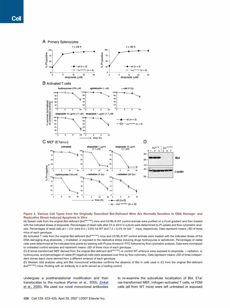
Figure 4. Various Cell Types from the Originally Described Bid-Deficient Mice Are Normally Sensitive to DNA Damage- and
Replicative Stress-Induced Apoptosis In Vitro
(A) Spleen cells from the original Bid-deficient (bidneo/neo) mice and C57BL/6 WT control animals were purified on a Ficoll gradient and then treated
with the indicated doses of etoposide. Percentages of dead cells after 24 or 40 hr in culture were determined by PI uptake and flow cytometric anal-
ysis. Percentages of dead cells at t = 0 hr were 9.4 ± 0.6% for WT and 7.3 ± 0.4% for bid�/�mice, respectively. Data represent means ±SD of three
mice of each genotype.
(B) Activated T cells from the original Bid-deficient (bidneo/neo) mice and C57BL/6 WT control animals were treated with the indicated doses of the
DNA-damaging drug etoposide, g-irradiated, or exposed to the replicative stress inducing drugs hydroxyurea or aphidicolin. Percentages of viable
cells were determined at the indicated time points by staining with PI plus AnnexinV-FITC followed by flow cytometric analysis. Data were normalized
to untreated control samples and represent means ±SD of three mice of each genotype.
(C) E1a/ras-transformed MEF derived from the original Bid-deficient (bidneo/neo) or control WT embryos were exposed to etoposide, g-radiation, or
hydroxyurea, and percentages of viable (PI negative) cells were assessed over time by flow cytometry. Data represent means ±SD of three indepen-
dent clones (each clone derived from a different embryo) of each genotype.
(D) Western blot analysis using anti-Bid monoclonal antibodies confirms the absence of Bid in cells used in (C) from the original Bid-deficient
(bidneo/neo) mice. Probing with an antibody to b-actin served as a loading control.
undergoes a posttranslational modification and then
translocates to the nucleus (Kamer et al., 2005; Zinkel
et al., 2005). We used our novel monoclonal antibodies
428 Cell 129, 423–433, April 20, 2007 ª2007 Elsevier Inc.
to re-examine the subcellular localization of Bid. E1a/
ras-transformed MEF, mitogen-activated T cells, or FDM
cells (all from WT mice) were left untreated or exposed

Figure 5. Bid-Deficient Cells Undergo Normal Cell-Cycle Arrest after Exposure to DNA Damage or Replicative Stress
(A) Independent clones of WT and bid�/� or WT and bidneo/neo (derived from original Bid-deficient mice) E1a/ras-transformed MEF were left untreated,
g-irradiated (g-rad., 5 Gy), or treated for 2 hr with etoposide (etop., 10 mg/ml = 17 mM), mitomycin C (mito C, 100 nM), aphidicolin (aphid., 5 mM), or
hydroxyurea (HU, 100 mM) followed by 8 hr of culture in drug-free medium. The cells were then lysed in hypotonic PI buffer, and the cell-cycle profiles
analyzed by flow cytometry.
(B) Independent clones of WT and bid�/� or WT and bidneo/neo (derived from original Bid-deficient mice) FDM were left untreated, g-irradiated (g-rad.,
1.25 or 5 Gy), or treated for 24 hr with etoposide (etop., 1 mg/ml = 1.7 mM), aphidicolin (aphid., 0.1 mM), hydroxyurea (HU, 100 mM), or mitomycin C (mito
C, 100 nM). Nuclear DNA was then stained with PI (using hypotonic buffer), and the cell cycle profiles were analyzed by flow cytometry. Data in (A) and
(B) represent means ±SD of three independent clones (from three independent embryos) for each genotype.
to etoposide, g-radiation, or hydroxyurea. Subcellular
fractionation followed by SDS-PAGE and western blotting
detected Bid exclusively in the cytosol of untreated as
well as stressed cells (Figure 7). These results demon-
strate that Bid resides in the cytosol of healthy cells and
cells that have sustained DNA damage or replicative
stress.
DISCUSSION
In order to analyze the function of the BH3-only protein Bid
and to resolve some of the discrepancies in the published
data, we generated Bid-deficient mice on a pure C57BL/6
genetic background. These mice are viable, were born
at an expected Mendelian frequency, and show no
Cell 129, 423–433, April 20, 2007 ª2007 Elsevier Inc. 429

Figure 6. Loss of Bid Does Not Affect
Clonogenic Death of g-Irradiated MEF
or Mitogen-Induced Proliferation of T
Lymphocytes
(A) bid�/� or WT E1a/ras-transformed MEF
were seeded at 1 3 103 cells per well (6-well
plate) the day prior to g-irradiation with the indi-
cated doses. After 7 days, the cells were fixed
and stained with crystal violet, and the number
of colonies per well was counted. Data are
presented as percentage of surviving colony-
forming cells compared to the nonirradiated
control cultures giving means ±SD of three in-
dependent clonal E1a/ras-transformed MEF
(from three independent embryos) of each ge-
notype. Only one well from a 6-well plate is
shown in the illustrative photograph.
(B) Purified bid�/� and control (WT) T lympho-
cytes were activated with anti-CD3 antibody,
anti-CD3 plus anti-CD28 antibodies, or ConA.
The relative cell size (forward light scatter:
FSC) of activated T cells was determined at
the indicated time points in a FACScan and is
represented as mean FSC. The viable number
of activated T cells was measured by PI stain-
ing and flow cytometry using FACS reference
beads. Data are represented as fold-increase
of viable cells, giving means ±SD from T cells
of three independent mice of each genotype.
phenotypic or morphological abnormalities, consistent
with a previous report examining (young) Bid-deficient
(bidneo/neo) mice generated on a C57BL/6/Sv129 mixed
Figure 7. Bid Is a Cytoplasmic Protein and Does Not Translo-
cate into the Nucleus after DNA Damage or Replicative Stress
E1a/ras-transformed MEF (A), FDM cells (B), or activated T cells (C; all
from WT C57BL/6 mice) were left untreated or were treated for 1 hr
with etoposide (etop., 100 mM = 58.8 mg/ml) or hydroxyurea (HU,
1 mM) or were g-irradiated (g-rad., 20 Gy) and then cultured for 1 hr.
Nuclear (N) and cytoplasmic (C) fractions were prepared and equal
amounts per fraction separated on SDS-PAGE gels, then western blot-
ted and probed for Bid as well as for the cytosolic protein HSP70 and
the nuclear protein PARP, both of which served as controls for the pu-
rity of the subcellular fractions.
430 Cell 129, 423–433, April 20, 2007 ª2007 Elsevier Inc.
genetic background (Yin et al., 1999). These latter mice
are, however, reported to spontaneously develop myeloid
leukemia as they age, starting from �12–15 months (Zin-
kel et al., 2003). So far (14–21 months) we have not ob-
served an abnormally increased incidence of leukemias
or other tumors in our bid�/� mice or, indeed, in the orig-
inal Bid-deficient mice backcrossed for >12 generations
onto the C57BL/6 background, but it will be important to
monitor this further as they continue to age. However, un-
like Zinkel et al. (2005), we did not find evidence for in-
creased genomic instability of bid-deficient immortalized
FDM after treatment with mitomycin C, a defect that
they postulated might be responsible for leukemogenesis
in their animals. Moreover, in contrast to loss of the BH3-
only protein Bim (Egle et al., 2004), loss of Bid did not
accelerate lymphomagenesis in Em-myc transgenic mice
(R.W.J. and R.K.L., unpublished data), and in contrast to
bim (Tagawa et al., 2005), no mutations in bid have so
far been found in hemopoietic or other neoplasms. These
observations do not support a critical role for Bid in tumor
suppression.
Is Bid Critical for the Cellular Response
to DNA Damage?
Two groups recently reported a critical role for Bid in the
DNA-damage response, with Bid being a target of the
DNA-damage-activated kinase ATM (Kamer et al., 2005;
Zinkel et al., 2005). Paradoxically, one group found that
loss of Bid enhances cell killing (Zinkel et al., 2005),

whereas the other found the opposite (Kamer et al., 2005).
In contrast to Zinkel et al. (2005), we did not observe in-
creased sensitivity of FDM to DNA-damaging agents, mi-
tomycin C, hydroxyurea, or aphidicolin, nor did we gain
evidence for a role for Bid in cell-cycle arrest. Although
we did observe a large variation between independent
FDM clones derived from different embryos of the same
genotype, there was no significant difference between
the average sensitivity of the WT and Bid-deficient FDM,
indicating the epigenetic variation was much greater
than the variation that could be attributed to the presence
or absence of Bid. In contrast to Zinkel et al. (2005), Kamer
et al. (2005) reported abnormally increased survival of Bid-
deficient (bidneo/neo) MEF and spleen cells after UV- or
g-irradiation or exposure to etoposide or cisplatin, as
well as a failure of these cells to arrest in cell cycle after
such treatment. Our analyses of nine distinct cell types, in-
cluding primary and immortalized MEF, immortalized my-
eloid progenitors, primary T and B cell subsets, as well as
activated T cells, done using the same treatments as re-
ported before, showed no significant difference between
our bid�/� and WT cells in cell survival or cell-cycle arrest.
How can these differences between our results and earlier
findings (Kamer et al., 2005; Zinkel et al., 2005) be
reconciled? It is important to note that although they had
reportedly (Kamer et al., 2005; Zinkel et al., 2005) been
backcrossed for 12 generations onto the C57BL/6
background, the previously generated bid�/� mice still
contained the pgk-neo cassette (Yin et al., 1999) used
for ES cell selection, which may have affected expression
of linked genes (Koyasu et al., 1994). It is therefore possi-
ble that abnormalities in cellular responses to DNA dam-
age or replicative stress, as well as the genetic instability
and leukemogenesis seen in the original bidneo/neo mice,
are not a direct consequence of loss of Bid, but instead
are due to the altered expression of other (unknown)
genes. To test this hypothesis, we analyzed splenocytes,
thymocytes, mature B and T cells, activated T cells, E1a/
ras-transformed MEF, and FDM cells derived from the
original Bid-deficient (bidneo/neo) mice (Yin et al., 1999)
for their responses to DNA damage and replicative stress.
We were, however, unable to detect any significant differ-
ence in apoptotic response or cell-cycle arrest between
cells derived from the bidneo/neo mice (backcrossed to
C57BL/6 for >10 generations) and C57BL/6 control cells.
Hence, we believe that there is no significant difference
between the two Bid-deficient mouse lines and WT control
animals in response to DNA-damage or replicative stress.
This emphasizes the importance of using significant num-
bers of truly independent cell lines and primary cells (from
several independent mice of each genotype) to control for
experimental variability to allow generalization of impor-
tant findings. In contrast to the previous studies (Kamer
et al., 2005; Zinkel et al., 2005), we found no evidence
for a nuclear localization of Bid or a translocation of Bid
into the nucleus after DNA damage or replicative stress.
Because Bid-deficient cells undergo normal apoptosis
and cell-cycle arrest in response to DNA damage, it ap-
pears unlikely that the previously reported ATM-mediated
phosphorylation of Bid (Kamer et al., 2005; Zinkel et al.,
2005) has a significant role in the cellular response to
DNA damage. Collectively, our results do not support an
essential role for Bid in DNA damage- or replicative
stress-induced apoptosis and cell-cycle arrest or mito-
gen-induced cell proliferation.
EXPERIMENTAL PROCEDURES
Mice
Bid-deficient mice were generated by electroporation of C57BL/6-
derived ES cells (Bruce4) with a targeting vector replacing an
�4.5 kb region of the bid locus containing exons 1 and 2 plus part of
exon 3, including the crucial BH3 domain. Neomycin-resistant ES cells
were selected and analyzed by Southern blotting using a 50 external
probe, which recognized a 6.5 kb HinDIII WT and a 13 kb mutant
HinDIII fragment, as well as a 30 external probe, which recognized a
9 kb HinDIII WT and a 13 kb mutant HinDIII fragment. ES clones with
homologous recombination were injected into BALB/c blastocysts,
and chimeric males with a large extent of black coat were selected
for breeding with C57BL/6 females. Black offspring with germline
transmission of the ES cells were selected to establish the bid[neo]
line. Mouse genotypes were determined using a three-primer PCR
approach (with one pGK neo-specific primer) on genomic DNA from
tail biopsies and confirmed by western blotting. The loxP-flanked
pGK neo cassette was deleted by crossing bid[neo] heterozygous
males with Cre-recombinase transgenic (deleter) females (kept on a
C57BL/6 background; Schwenk et al., 1995), and deletion was con-
firmed by PCR. The generation of the original Bid-deficient mice was
previously described (Yin et al., 1999). All experiments with mice
were performed according to the guidelines of the animal ethics com-
mittees of the Melbourne Health Research Directorate or the Peter
MacCallum Cancer Institute.
Materials and Cell Culture
Aphidicolin (A0781), mitomycin C (M4287), and hydroxyurea (H8627)
were purchased from SIGMA. Lymphocyte subpopulations (CD4+8+
immature thymocytes, CD4+8� and CD4�8+ mature T cells and
B220+sIgMloDhi mature B cells from lymph nodes, B220+sIg�c-Kit�
preB cells, and B220+sIgMhiDlo immature B cells from bone marrow)
were FACS sorted as previously described (Strasser et al., 1991)
from 8- to 12-week-old bid�/� mice and WT littermates. Primary
MEF were prepared from E14.5-old embryos and were used at pas-
sage 1 or 2 for experiments. E1a/ras-transformed MEF were generated
by retroviral transduction with E1a.hygro and ras.puro expression con-
structs (gift from S. Lowe, Cold Spring Harbor Labs). Infected cells
were selected with hygromycin (100 mg/ml) and puromycin (3 mg/ml)
for 3 days, and the transformed cells were allowed to grow for several
generations prior to any experiments.
Activated T cells were prepared by stimulating splenocytes for
3 days at a starting density of �1 3 106 cells/ml with 2 mg/ml ConA
(SIGMA, C-0412) and 100 U/ml IL-2. Cells were then thoroughly
washed to remove ConA, diluted between 1:2 to 1:8, and expanded
for a further 2 days in the presence of IL-2. Cells were then stained
with FITC-conjugated antibodies to CD19 (B-cells), Ter119 (red blood
cells), Mac-1 (macrophages/granulocytes), and Gr-1 (granulocytes) as
well as PI (dead cells), and FITC�PI� cells were sorted in a MoFlo
(Cytomation) or FACS DIVA (Becton Dickinson), resulting in a highly
purified population of activated T cells (routinely >95% pure).
All primary lymphocytes and primary as well as E1a/ras-transformed
MEF were cultured in the high-glucose version of DMEM (Dulbecco’s
Modified Eagle Medium), supplemented with 10% fetal calf serum,
100 mM Asparagine, and 50 mM b-mercaptoethanol. IL-3-dependent
FDM were generated as previously described (Ekert et al., 2004) by
Cell 129, 423–433, April 20, 2007 ª2007 Elsevier Inc. 431

coculturing E14.5 fetal liver cells with fibroblasts expressing a HoxB8
retrovirus in the low-glucose version of DMEM, supplemented with
10% fetal calf serum and 1000 U/ml IL-3.
Western Blotting
Cell lysates were prepared in a buffer containing 20 mM Tris/HCl pH
7.4, 135 mM NaCl; 1.5 mM MgCl2; 1 mM EGTA; 1% Triton X-100;
10% glycerol; 500 mg/ml Pefabloc (AEBSF); 1 mg/ml each of Leupeptin,
Aprotinin, and Pepstatin; 100 mg/ml soybean trypsin-inhibitor; 2 mg/ml
E64; 50 mM NaF; and 2 mM Na3VO4. Proteins in cell lysates were size
separated on precast 4%–20% SDS-PAGE gradient gels (Invitrogen).
Two commercial antibodies to Bid (BD Pharmingen; mouse monoclo-
nal anti-Bid cat# 611866 and rabbit polyclonal anti-Bid cat# 559681)
and two newly developed rat anti-Bid monoclonal antibodies (clones
2D1 and 8C3; generated using human tBid as an immunogen) were
used to prove that a bid null allele had been generated. Probing with a
mouse monoclonal antibody to b-actin (SIGMA, AC-40) served as a
loading control. For subcellular fractionation, cells were washed and
incubated for 5 min on ice in a hypotonic lysis (HL) buffer (25 mM
HEPES pH 7.4, 2 mM EGTA, 2 mM MgCl2, and 2 mM DTT) containing
protease and phosphatase inhibitors (see above) prior to mechanical
lysis by gently passing the cells through 26.5 gauge needles. Nuclei
were spun at 500 3 g (5 min, 4�C), washed once in HL buffer, and
spun again to obtain the nuclear fraction. The nuclear pellets were
lysed in a 1% Triton X-100-containing buffer as described above.
The postnuclear supernatant was spun at 13,000 rpm in a microcentri-
fuge (15 min, 4�C), and the supernatant was collected as the cytoplas-
mic fraction. Equal amounts of cytoplasmic and nuclear fractions
were separated on 12% or 4%–20% SDS-PAGE gels and probed
with monoclonal antibodies to Bid (rat monoclonal, clone 2D1),
Hsp70 (mouse monoclonal N6; gift from Dr. R. Anderson, Peter
MacCallum Cancer Research Centre, Melbourne, Australia), or PARP
(Alexis).
Cell Survival and Cell-Cycle Analysis
Viability of hemopoietic cells was assessed in a FACScan by exclusion
of PI and absence of cell-surface staining with FITC-labeled AnnexinV.
Viability of E1a/ras-transformed MEF was determined by PI exclusion
only. For cell-cycle analysis cells were stained in ‘‘hypotonic PI buffer’’
(0.1% Na3-citrate, 0.1% Triton X-100, 50 mg/ml PI, and 25 mg/ml RNase
A) at a density of�1 3 106 cells/ml for at least 30 min on ice. Cells were
then analyzed in a FACScan at a flow rate of <200 events/second, and
PI staining intensity was plotted on a linear scale (FL3). Cell-cycle pro-
files were analyzed using the Weasel 1.2.2 software.
Chromosome Damage in Response to Mitomycin C Treatment
WT and bid�/� FDM cells (1–2 3 106/ml) were exposed for 24 hr to 0.03
or 0.05 mg/ml mitomycin C. A control group of WT and bid�/� cells
without addition of mitomycin C was included in the analysis. After
24 hr the cells were arrested in metaphase and harvested using stan-
dard cytogenetic protocols prior to being stained with a 7% Leishmans
solution. The efficacy of the mitomycin C was established using a sister
chromatid exchange (SCE) assay, routinely used. Twenty-five cells per
independent clonal FDM cell line (n = 3 for WT; n = 4 for bid�/�) were
scored for chromatid breaks and triradial figures. The number of
breaks per cell was recorded; no radial figures were observed in any
of the cultures. The scoring was performed in a blinded fashion.
Colony-Forming Assay
One thousand E1a/ras-transformed MEF (three independent clones
per genotype per experiment) were seeded in 6-well plates and g-irra-
diated the following day at doses ranging from 0 to 8 Gy and then in-
cubated for an additional 7 days. Culture wells were then washed twice
in PBS, fixed for 15 min in ice-cold 70% ethanol, and stained in a 0.5%
crystal violet solution. The number of colonies was counted by visual
inspection, and the percentage of clonogenic cells was obtained
from the ratio [colonies at dose (x Gy)/colonies at dose (0 Gy)] 3 100.
432 Cell 129, 423–433, April 20, 2007 ª2007 Elsevier Inc.
T Cell Proliferation Assay
FACS-sorted splenic T cells (2.5 3 104 /well) were seeded in 96-well
plates that had either been precoated with mitogenic anti-CD3 anti-
body (145-2C11, 10 mg/ml) or anti-CD3 plus anti-CD28 antibody
(37N51, 10 mg/ml) or that contained 2 mg/ml ConA, all in the presence
of 100 U/ml IL-2. The number of viable (PI-negative) cells at various
time points, including t0, was determined by adding a known number
of flow cytometry reference beads (PeakFlow, Molecular Probes,
P14828) plus 2 mg/ml PI to each well prior to FACS analysis. Calcula-
tion of viable cell number per well was obtained from the ratio of beads
to viable (PI-negative) cells. Fold increase in viable cell number was ob-
tained from the ratio viable cells t(x)/viable cells t(0).
Supplemental Data
Supplemental Data include four figures and can be found with this
article online at http://www.cell.com/cgi/content/full/129/2/423/DC1/.
ACKNOWLEDGMENTS
We thank N. Iannarella and G. Siciliano for animal care; Dr. F. Battye
and C. Tarlinton, V. Milovac, C. Young, and J. Garbe for cell sorting;
M. Robati and B. Helbert for genotyping; the Biochemistry Department
of the Royal Melbourne Hospital for ALT/AST measurements; J. Corbin
for blood-count analysis; L. Hills and J. Hammer for expert technical
assistance; Dr. R. Kluck for recombinant tBid; and Drs. J. Adams,
S. Cory, P. Bouillet, D. Vaux, and H. Puthalakath for advice and critical
comments on the manuscript. This work was supported by grants
(programs #257502 and #251608 and project #384404) and fellow-
ships from the NHMRC (Canberra), the NCI (NIH, USA; # CA 80188
and #CA 43540), the Leukemia and Lymphoma Society of America
(SCOR grant #7015), the JDRF/NHMRC, the Cancer Council Victoria,
the Leukemia Foundation of Australia, the Swiss National Science
Foundation, the Roche Research Foundation, the Novartis Foundation
(formerly Ciba-Geigy-Jubilaeumsstiftung; Postdoctoral Fellowships to
T.K.), and by a Pfizer Australia Senior Research Fellowship (R.W.J.).
Received: August 1, 2006
Revised: January 22, 2007
Accepted: March 9, 2007
Published: April 19, 2007
REFERENCES
Adams, J.M., and Cory, S. (1998). The Bcl-2 protein family: arbiters
of cell survival. Science 281, 1322–1326.
Bai, L., Ni, H.M., Chen, X., DiFrancesca, D., and Yin, X.M. (2005).
Deletion of Bid impedes cell proliferation and hepatic carcinogenesis.
Am. J. Pathol. 166, 1523–1532.
Bouillet, P., Metcalf, D., Huang, D.C.S., Tarlinton, D.M., Kay, T.W.H.,
Kontgen, F., Adams, J.M., and Strasser, A. (1999). Proapoptotic
Bcl-2 relative Bim required for certain apoptotic responses, leukocyte
homeostasis, and to preclude autoimmunity. Science 286, 1735–1738.
Bouillet, P., Purton, J.F., Godfrey, D.I., Zhang, L.-C., Coultas, L.,
Puthalakath, H., Pellegrini, M., Cory, S., Adams, J.M., and Strasser,
A. (2002). BH3-only Bcl-2 family member Bim is required for apoptosis
of autoreactive thymocytes. Nature 415, 922–926.
Chen, L., Willis, S.N., Wei, A., Smith, B.J., Fletcher, J.I., Hinds, M.G.,
Colman, P.M., Day, C.L., Adams, J.M., and Huang, D.C.S. (2005).
Differential targeting of pro-survival Bcl-2 proteins by their BH3-only
ligands allows complementary apoptotic function. Mol. Cell 17, 393–
403.
Egle, A., Harris, A.W., Bouillet, P., and Cory, S. (2004). Bim is a suppres-
sor of Myc-induced mouse B cell leukemia. Proc. Natl. Acad. Sci. USA
101, 6164–6169.

Ekert, P.G., Read, S.H., Silke, J., Marsden, V.S., Kaufmann, H.,
Hawkins, C.J., Gerl, R., Kumar, S., and Vaux, D.L. (2004). Apaf-1 and
caspase-9 accelerate apoptosis, but do not determine whether fac-
tor-deprived or drug-treated cells die. J. Cell Biol. 165, 835–842.
Ekert, P.G., Jabbour, A.M., Manoharan, A., Heraud, J.E., Yu, J.,
Pakusch, M., Michalak, E.M., Kelly, P.N., Callus, B., Kiefer, T., et al.
(2006). Cell death provoked by loss of Interleukin-3 signalling is inde-
pendent of Bad, Bim, and PI3 Kinase, but depends in part on Puma.
Blood 108, 1461–1468.
Enders, A., Bouillet, P., Puthalakath, H., Xu, Y., Tarlinton, D.M., and
Strasser, A. (2003). Loss of the pro-apoptotic BH3-only Bcl-2 family
member Bim inhibits BCR stimulation-induced apoptosis and deletion
of autoreative B cells. J. Exp. Med. 198, 1119–1126.
Green, D.R., and Reed, J.C. (1998). Mitochondria and apoptosis.
Science 281, 1309–1311.
Hengartner, M.O. (2000). The biochemistry of apoptosis. Nature 407,
770–776.
Hildeman, D.A., Zhu, Y., Mitchell, T.C., Bouillet, P., Strasser, A.,
Kappler, J., and Marrack, P. (2002). Activated T cell death in vivo
mediated by pro-apoptotic Bcl-2 family member, Bim. Immunity 16,
759–767.
Huang, D.C.S., and Strasser, A. (2000). BH3-only proteins – essential
initiators of apoptotic cell death. Cell 103, 839–842.
Jeffers, J.R., Parganas, E., Lee, Y., Yang, C., Wang, J., Brennan, J.,
MacLean, K.H., Han, J., Chittenden, T., Ihle, J.N., et al. (2003). Puma
is an essential mediator of p53-dependent and -independent apopto-
tic pathways. Cancer Cell 4, 321–328.
Kamer, I., Sarig, R., Zaltsman, Y., Niv, H., Oberkovitz, G., Regev, L.,
Haimovich, G., Lerenthal, Y., Marcellus, R.C., and Gross, A. (2005).
Proapoptotic BID is an ATM effector in the DNA-damage response.
Cell 122, 593–603.
Koyasu, S., Hussey, R.E., Clayton, L.K., Lerner, A., Pedersen, R.,
Delany-Heiken, P., Chau, F., and Reinherz, E.L. (1994). Targeted dis-
ruption within the CD3 zeta/eta/phi/Oct-1 locus in mouse. EMBO J.
13, 784–797.
Kuwana, T., Bouchier-Hayes, L., Chipuk, J.E., Bonzon, C., Sullivan,
B.A., Green, D.R., and Newmeyer, D.D. (2005). BH3 domains of
BH3-only proteins differentially regulate Bax-mediated mitochondrial
membrane permeabilization both directly and indirectly. Mol. Cell 17,
525–535.
Letai, A., Bassik, M., Walensky, L., Sorcinelli, M., Weiler, S., and
Korsmeyer, S. (2002). Distinct BH3 domains either sensitize or activate
mitochondrial apoptosis, serving as prototype cancer therapeutics.
Cancer Cell 2, 183–192.
Li, H., Zhu, H., Xu, C.-J., and Yuan, J. (1998). Cleavage of BID by
caspase 8 mediates the mitochondrial damage in the Fas pathway of
apoptosis. Cell 94, 491–501.
Luo, X., Budlhardjo, I., Zou, H., Slaughter, C., and Wang, X. (1998). Bid,
a Bcl-2 interacting protein, mediates cytochrome c release from mito-
chondria in response to activation of cell surface death receptors. Cell
94, 481–490.
Pellegrini, M., Belz, G., Bouillet, P., and Strasser, A. (2003). Shut down
of an acute T cell immune response to viral infection is mediated by the
pro-apoptotic Bcl-2 homology 3-only protein Bim. Proc. Natl. Acad.
Sci. USA 100, 14175–14180.
Puthalakath, H., and Strasser, A. (2002). Keeping killers on a tight
leash: transcriptional and post-translational control of the pro-apopto-
tic activity of BH3-only proteins. Cell Death Differ. 9, 505–512.
Sax, J.K., Fei, P., Murphy, M.E., Bernhard, E., Korsmeyer, S.J., and
El-Deiry, W.S. (2002). BID regulation by p53 contributes to chemosen-
sitivity. Nat. Cell Biol. 4, 842–849.
Schwenk, F., Baron, U., and Rajewsky, K. (1995). A cre-transgenic
mouse strain for the ubiquitous deletion of loxP-flanked gene seg-
ments including deletion in germ cells. Nucleic Acids Res. 23, 5080–
5081.
Shibue, T., Takeda, K., Oda, E., Tanaka, H., Murasawa, H., Takaoka,
A., Morishita, Y., Akira, S., Taniguchi, T., and Tanaka, N. (2003). Inte-
gral role of Noxa in p53-mediated apoptotic response. Genes Dev.
17, 2233–2238.
Strasser, A., Harris, A.W., and Cory, S. (1991). Bcl-2 transgene inhibits
T cell death and perturbs thymic self-censorship. Cell 67, 889–899.
Tagawa, H., Karnan, S., Suzuki, R., Matsuo, K., Zhang, X., Ota, A.,
Morishima, Y., Nakamura, S., and Seto, M. (2005). Genome-wide ar-
ray-based CGH for mantle cell lymphoma: identification of homozy-
gous deletions of the proapoptotic gene BIM. Oncogene 24, 1348–
1358.
Villunger, A., Michalak, E.M., Coultas, L., Mullauer, F., Bock, G.,
Ausserlechner, M.J., Adams, J.M., and Strasser, A. (2003). p53- and
drug-induced apoptotic responses mediated by BH3-only proteins
Puma and Noxa. Science 302, 1036–1038.
Wang, K., Yin, X.-M., Chao, D.T., Milliman, C.L., and Korsmeyer, S.J.
(1996). BID: a novel BH3 domain-only death agonist. Genes Dev. 10,
2859–2869.
Willis, S.N., Chen, L., Dewson, G., Wei, A., Naik, E., Fletcher, J.I.,
Adams, J.M., and Huang, D.C. (2005). Pro-apoptotic Bak is seques-
tered by Mc1–1 and Bcl-xL, but not Bcl-2, until displaced by BH3-
only proteins. Genes Dev. 19, 1294–1305.
Yin, X.-M., Wang, K., Gross, A., Zhao, Y., Zinkel, S., Klocke, B., Roth,
K.A., and Korsmeyer, S.J. (1999). Bid-deficient mice are resistant to
Fas-induced hepatocellular apoptosis. Nature 400, 886–891.
Zha, J., Weiler, S., Oh, K.J., Wei, M.C., and Korsmeyer, S.J. (2000).
Posttranslational N-myristoylation of BID as a molecular switch for
targeting mitochondria and apoptosis. Science 290, 1761–1765.
Zinkel, S.S., Ong, C.C., Ferguson, D.O., Iwasaki, H., Akashi, K.,
Bronson, R.T., Kutok, J.L., Alt, F.W., and Korsmeyer, S.J. (2003).
Proapoptotic BID is required for myeloid homeostasis and tumor
suppression. Genes Dev. 17, 229–239.
Zinkel, S.S., Hurov, K.E., Ong, C., Abtahi, F.M., Gross, A., and
Korsmeyer, S.J. (2005). A role for proapoptotic BID in the DNA-
damage response. Cell 122, 579–591.
Cell 129, 423–433, April 20, 2007 ª2007 Elsevier Inc. 433
Copyright © 2022 FDOKUMEN