Arf4 Is Required for Mammalian Development but Dispensable for Ciliary Assembly
14
Arf4 Is Required for Mammalian Development but Dispensable for Ciliary Assembly John A. Follit 1 , Jovenal T. San Agustin 1 , Julie A. Jonassen 2 , Tingting Huang 3 , Jaime A. Rivera-Perez 3 , Kimberly D. Tremblay 4 , Gregory J. Pazour 1 * 1 Program in Molecular Medicine, University of Massachusetts Medical School, Biotech II, Worcester, Massachusetts, United States of America, 2 Department of Microbiology and Physiological Systems, University of Massachusetts Medical School, Worcester, Massachusetts, United States of America, 3 Department of Cell and Developmental Biology, University of Massachusetts Medical School, Worcester, Massachusetts, United States of America, 4 Department of Veterinary and Animal Sciences, University of Massachusetts, Amherst, Amherst, Massachusetts, United States of America Abstract The primary cilium is a sensory organelle, defects in which cause a wide range of human diseases including retinal degeneration, polycystic kidney disease and birth defects. The sensory functions of cilia require specific receptors to be targeted to the ciliary subdomain of the plasma membrane. Arf4 has been proposed to sort cargo destined for the cilium at the Golgi complex and deemed a key regulator of ciliary protein trafficking. In this work, we show that Arf4 binds to the ciliary targeting sequence (CTS) of fibrocystin. Knockdown of Arf4 indicates that it is not absolutely required for trafficking of the fibrocystin CTS to cilia as steady-state CTS levels are unaffected. However, we did observe a delay in delivery of newly synthesized CTS from the Golgi complex to the cilium when Arf4 was reduced. Arf4 mutant mice are embryonic lethal and die at mid-gestation shortly after node formation. Nodal cilia appeared normal and functioned properly to break left-right symmetry in Arf4 mutant embryos. At this stage of development Arf4 expression is highest in the visceral endoderm but we did not detect cilia on these cells. In the visceral endoderm, the lack of Arf4 caused defects in cell structure and apical protein localization. This work suggests that while Arf4 is not required for ciliary assembly, it is important for the efficient transport of fibrocystin to cilia, and also plays critical roles in non-ciliary processes. Citation: Follit JA, San Agustin JT, Jonassen JA, Huang T, Rivera-Perez JA, et al. (2014) Arf4 Is Required for Mammalian Development but Dispensable for Ciliary Assembly. PLoS Genet 10(2): e1004170. doi:10.1371/journal.pgen.1004170 Editor: Susan K. Dutcher, Washington University School of Medicine, United States of America Received April 13, 2012; Accepted December 25, 2013; Published February 20, 2014 Copyright: ß 2014 Follit et al. This is an open-access article distributed under the terms of the Creative Commons Attribution License, which permits unrestricted use, distribution, and reproduction in any medium, provided the original author and source are credited. Funding: This work was supported by the National Institutes of Health (GM060992 to GJP; GM87130 and GM94874 to JARP) and the Order of the Eagles (GJP). JAJ is a member of the Harvard Center for PKD Research (P50 DK074030). Core resources supported by the Diabetes Endocrinology Research Center grant DK32520 were also used. The funders had no role in study design, data collection and analysis, decision to publish, or preparation of the manuscript. Competing Interests: The authors have declared that no competing interests exist. * E-mail: [email protected] Introduction Cilia play diverse motility and sensory functions throughout the eukaryotic kingdom, but play especially critical roles in vertebrates where severe defects lead to embryonic lethality while mild defects cause a wide range of syndromes affecting every organ system. Both the motility and sensory functions of cilia are important for health and development, but it is now recognized that sensory defects underlie the most severe maladies affecting humans. The sensory functions of cilia rely on a cell’s ability to target and concentrate a specific set of receptors to the ciliary membrane. While contiguous with the plasma membrane of the cell, the ciliary membrane is a distinct compartment to which the cell targets and concentrates a unique complement of proteins [1,2]. The list of membrane proteins found in the ciliary compartment is constantly growing; among the most studied ciliary proteins are the polycystins and fibrocystin that are defective in human polycystic kidney disease, rhodopsins and opsins that are critical for vision and the patched and smoothened receptors of the hedgehog pathway. The mechanism that targets membrane proteins specifically to the ciliary compartment is an active area of study but very little is definitively known [3]. It appears that ciliary membrane proteins contain cis-acting motifs that cause them to be localized to cilia. We identified one of these ciliary targeting sequences (CTS) in fibrocystin, the gene product of the human autosomal recessive polycystic kidney disease gene (PKHD1) [4–7]. Like many other CTSs, the fibrocystin CTS contains lipid-modified residues that target the protein to lipid rafts, which appears to be part of the ciliary trafficking pathway. We proposed that this sequence might interact with proteins that are important for sorting or transport to the ciliary membrane compartment. In support of this idea, we found that the fibrocystin CTS interacted with Rab8, a protein widely recognized as important to ciliary membrane protein trafficking [7–9]. In the present work we asked if the fibrocystin CTS could interact with Arf4 as work of Deretic and colleagues has shown that this protein interacts with the CTS of opsin and is important for the formation of rhodopsin carrier vesicles at the Golgi complex [10,11]. Arf4 is a small G protein in the Arf subfamily of Ras-related small G proteins. Mice have six members of this family while humans have lost Arf2 and have five members. Arf1 and Arf6 have been best-studied and are thought to organize membrane protein cargos into coated vesicles for transport to specific lipid domains in the cell [12–15]. Arf1 forms coated vesicles at the Golgi complex crucial for trafficking between the ER and Golgi and throughout PLOS Genetics | www.plosgenetics.org 1 February 2014 | Volume 10 | Issue 2 | e1004170
Transcript of Arf4 Is Required for Mammalian Development but Dispensable for Ciliary Assembly
Arf4 Is Required for Mammalian Development butDispensable for Ciliary AssemblyJohn A. Follit1, Jovenal T. San Agustin1, Julie A. Jonassen2, Tingting Huang3, Jaime A. Rivera-Perez3,
Kimberly D. Tremblay4, Gregory J. Pazour1*
1 Program in Molecular Medicine, University of Massachusetts Medical School, Biotech II, Worcester, Massachusetts, United States of America, 2 Department of
Microbiology and Physiological Systems, University of Massachusetts Medical School, Worcester, Massachusetts, United States of America, 3 Department of Cell and
Developmental Biology, University of Massachusetts Medical School, Worcester, Massachusetts, United States of America, 4 Department of Veterinary and Animal
Sciences, University of Massachusetts, Amherst, Amherst, Massachusetts, United States of America
Abstract
The primary cilium is a sensory organelle, defects in which cause a wide range of human diseases including retinaldegeneration, polycystic kidney disease and birth defects. The sensory functions of cilia require specific receptors to betargeted to the ciliary subdomain of the plasma membrane. Arf4 has been proposed to sort cargo destined for the cilium atthe Golgi complex and deemed a key regulator of ciliary protein trafficking. In this work, we show that Arf4 binds to theciliary targeting sequence (CTS) of fibrocystin. Knockdown of Arf4 indicates that it is not absolutely required for trafficking ofthe fibrocystin CTS to cilia as steady-state CTS levels are unaffected. However, we did observe a delay in delivery of newlysynthesized CTS from the Golgi complex to the cilium when Arf4 was reduced. Arf4 mutant mice are embryonic lethal anddie at mid-gestation shortly after node formation. Nodal cilia appeared normal and functioned properly to break left-rightsymmetry in Arf4 mutant embryos. At this stage of development Arf4 expression is highest in the visceral endoderm but wedid not detect cilia on these cells. In the visceral endoderm, the lack of Arf4 caused defects in cell structure and apicalprotein localization. This work suggests that while Arf4 is not required for ciliary assembly, it is important for the efficienttransport of fibrocystin to cilia, and also plays critical roles in non-ciliary processes.
Citation: Follit JA, San Agustin JT, Jonassen JA, Huang T, Rivera-Perez JA, et al. (2014) Arf4 Is Required for Mammalian Development but Dispensable for CiliaryAssembly. PLoS Genet 10(2): e1004170. doi:10.1371/journal.pgen.1004170
Editor: Susan K. Dutcher, Washington University School of Medicine, United States of America
Received April 13, 2012; Accepted December 25, 2013; Published February 20, 2014
Copyright: � 2014 Follit et al. This is an open-access article distributed under the terms of the Creative Commons Attribution License, which permitsunrestricted use, distribution, and reproduction in any medium, provided the original author and source are credited.
Funding: This work was supported by the National Institutes of Health (GM060992 to GJP; GM87130 and GM94874 to JARP) and the Order of the Eagles (GJP).JAJ is a member of the Harvard Center for PKD Research (P50 DK074030). Core resources supported by the Diabetes Endocrinology Research Center grantDK32520 were also used. The funders had no role in study design, data collection and analysis, decision to publish, or preparation of the manuscript.
Competing Interests: The authors have declared that no competing interests exist.
* E-mail: [email protected]
Introduction
Cilia play diverse motility and sensory functions throughout the
eukaryotic kingdom, but play especially critical roles in vertebrates
where severe defects lead to embryonic lethality while mild defects
cause a wide range of syndromes affecting every organ system.
Both the motility and sensory functions of cilia are important for
health and development, but it is now recognized that sensory
defects underlie the most severe maladies affecting humans. The
sensory functions of cilia rely on a cell’s ability to target and
concentrate a specific set of receptors to the ciliary membrane.
While contiguous with the plasma membrane of the cell, the ciliary
membrane is a distinct compartment to which the cell targets and
concentrates a unique complement of proteins [1,2]. The list of
membrane proteins found in the ciliary compartment is constantly
growing; among the most studied ciliary proteins are the
polycystins and fibrocystin that are defective in human polycystic
kidney disease, rhodopsins and opsins that are critical for vision
and the patched and smoothened receptors of the hedgehog
pathway.
The mechanism that targets membrane proteins specifically to
the ciliary compartment is an active area of study but very little is
definitively known [3]. It appears that ciliary membrane proteins
contain cis-acting motifs that cause them to be localized to cilia.
We identified one of these ciliary targeting sequences (CTS) in
fibrocystin, the gene product of the human autosomal recessive
polycystic kidney disease gene (PKHD1) [4–7]. Like many other
CTSs, the fibrocystin CTS contains lipid-modified residues that
target the protein to lipid rafts, which appears to be part of the
ciliary trafficking pathway. We proposed that this sequence might
interact with proteins that are important for sorting or transport to
the ciliary membrane compartment. In support of this idea, we
found that the fibrocystin CTS interacted with Rab8, a protein
widely recognized as important to ciliary membrane protein
trafficking [7–9]. In the present work we asked if the fibrocystin
CTS could interact with Arf4 as work of Deretic and colleagues
has shown that this protein interacts with the CTS of opsin and is
important for the formation of rhodopsin carrier vesicles at the
Golgi complex [10,11].
Arf4 is a small G protein in the Arf subfamily of Ras-related
small G proteins. Mice have six members of this family while
humans have lost Arf2 and have five members. Arf1 and Arf6 have
been best-studied and are thought to organize membrane protein
cargos into coated vesicles for transport to specific lipid domains in
the cell [12–15]. Arf1 forms coated vesicles at the Golgi complex
crucial for trafficking between the ER and Golgi and throughout
PLOS Genetics | www.plosgenetics.org 1 February 2014 | Volume 10 | Issue 2 | e1004170

the cell while Arf6 is thought to operate at the plasma membrane
and regulate endosomal-membrane traffic. Arf4, which was
thought to evolve from an Arf1-like precursor when metazoans
arose, is a relatively unstudied member of the family [13–15]. Arf4
was first proposed to be important for ciliary trafficking when it
was found to interact with the C-terminal tail of rhodopsin where
the CTS is localized [10]. Depletion of Arf4 from an in vitro
budding assay showed that it was important for the formation of
rhodopsin carrier vesicles [11]. The CTSs of rhodopsins are
contained in the last five amino acids (QV[S/A]PA) at the C-
terminal end of the protein. The V and P residues are mutated in
human patients with autosomal dominant retinitis pigmentosa.
These residues were found to be important in an in vitro assay for
the formation of rhodopsin carrier vesicles, thus the sequence has
become known as a VXPX motif. A similar RVXP motif is present
in the CNGB1b subunit of the CNG channel, another ciliary-
localized protein [16]. The VXPX sequence is part of the CTS in
polycystin-1 and polycystin-2 and it is hypothesized that VXPX
motifs function as Arf4 binding sites for transport to the cilium
[12,17,18].
In this work we ask if Arf4 plays a role in trafficking the
fibrocystin CTS to the cilium and probe the function of Arf4 in vivo
by analyzing a mutant mouse. Although the fibrocystin CTS does
not contain a VXPX motif, it does bind to Arf4. Arf4 is not
required for the trafficking of the fibrocystin CTS to cilia, but
knockdown of Arf4 increases the time needed for the protein to
travel from the Golgi complex to the cilium. Deletion of Arf4 in the
mouse does not affect the formation or function of nodal cilia, but
causes embryonic lethality at mid-gestation, probably due to
trafficking defects in the visceral endoderm.
Results
Arf4 Interacts with the Ciliary Targeting Sequence ofFibrocystin
Fibrocystin (polyductin), the human autosomal recessive poly-
cystic kidney disease gene product, is targeted to cilia by an 18-
residue ciliary targeting sequence (CTS) located in the cytoplasmic
C-terminal tail of the protein. Previously we showed that this
sequence interacted with Rab8 and proposed that it may function
by interacting with proteins involved in the sorting and transport
of ciliary membrane proteins [7]. Recent work indicates that Arf4
is required for trafficking of other ciliary proteins including
rhodopsin and the polycystins [10,11,18]. Arf4 is one of six
members of the Arf subfamily of small G proteins. Our previous
analysis showed that in non-ciliated cells, the GFP-CTS localized
to small puncta that appeared to be lipid microdomains rich in
GM1 gangliosides. Interestingly, Arf4 exhibited significant co-
localization with the CTS-GFP in these puncta while the rest of
the Arf family did not (Figure 1A). To determine if this
colocalization represented a physical interaction, we immunopre-
cipitated each of the Arfs and asked if the CTS-GFP was co
precipitated. Arf4 strongly precipitated the CTS, while the other
Arfs either precipitated no CTS (Arf1, 3, 5, 6) or only a small
amount (Arf2) (Figure 1B). Because ARF2 is a pseudogene in
humans we did not pursue the Arf2 interaction any further. The
failure of the CTS to interact with Arf5 suggests that the Arf4
interaction is likely to be specific as Arf4 and Arf5 share over 90%
identity at the amino acid level.
To further characterize the interaction between Arf4 and the
CTS we tested the ability of a series of CTS deletion constructs to
interact with Arf4 in the co-expression immunoprecipitation assay
(Figure 1C). In this assay, the CTS is sufficient for binding as the
3–20 construct was precipitated by Arf4 while the 63–193
construct, which lacks the CTS, was not bound. The 1–19 and
4–20 constructs also interact with Arf4 but do not target to cilia,
indicating the CTS has functions in addition to Arf4 binding.
Next we examined the ability of Arf4 to interact with a series of
alanine-scanning mutations within the CTS (Figure 1D). In
general, the ability of Arf4 to bind the CTS correlated with the
ability of the CTS to traffic to cilia. For example, mutations
affecting the palmitoylated cysteines (CCC.AAA) or conserved
basic residues (KTRK.AAAA) prevent ciliary targeting and
likewise inhibit Arf4 binding. Mutations that exhibit little effect on
the ciliary targeting of the CTS (LV.AA) similarly do not affect
Arf4 binding. However the concordance was not perfect as the
(IKP.AAA) mutation blocked ciliary targeting but had little effect
on Arf4 binding, again supporting the idea that while the CTS is
an Arf4 binding site, this is not its entire function.
Arf4 cycles between GTP and GDP bound states. To explore
the role of this cycling on the ciliary trafficking of the CTS we
measured the effects of constitutively active and dominant negative
Arf4 on ciliary targeting and interaction with the CTS (Figure S1).
Constitutively active Arf4 (Q67L) co-localized with IFT20 at the
Golgi Complex while wild type or dominant negative (T13N,
T48N) Arf4 displayed a punctate distribution in the cytoplasm
(Figure S1A). The mutant forms of Arf4 retained the ability to
bind the CTS (Figure S1E). Expression of dominant negative Arf4
reduced the percent of ciliated cells (Figure S1B) and ciliary length
(Figure S1C). Surprisingly, over-expression of any Arf4 constructs
completely inhibited CTS trafficking to the cilium (Figure S1D).
These data indicate that increases in Arf4 levels prevent ciliary
trafficking of the CTS possibly by sequestering it in the cytoplasm.
Arf4 Is Required for Efficient Delivery of the Fibrocystin CTerminal Tail to the Cilium
Prior studies in frog photoreceptors [10,11] and cultured
mammalian cells [18] suggest that Arf4 functions at the Golgi
complex to direct rhodopsin and polycystin-1 to the cilium. Our
initial immunofluorescence and biochemical results suggested that
Arf4 may also be required for the ciliary targeting of fibrocystin.
To test the role of Arf4 in the trafficking of fibrocystin, we
developed a pulse chase assay to measure its movement through
the endomembrane system and delivery to the cilium. To do this,
Author Summary
Primary cilia are ubiquitous sensory organelles that playvital roles in an ever-growing class of human diseasestermed ciliopathies including obesity, retinal degenerationand polycystic kidney disease. The proper function of theprimary cilium relies on a cell’s ability to target andconcentrate specific receptors to the ciliary membrane – aunique subdomain of the plasma membrane yet little isknown about how receptors are trafficked to the primarycilium. Mutations affecting the ciliary localized receptorfibrocystin (PKHD1) cause autosomal recessive polycystickidney disease, which affects approximately 1:20,000individuals. Previously we identified a motif located inthe cytoplasmic domain of fibrocystin that is required forits ciliary localization. In this work we demonstrate that theciliary targeting sequence (CTS) of fibrocystin interactswith the small G protein Arf4 and this interaction isimportant for the efficient delivery of the CTS to cilia incultured cells. Disruption of Arf4 in mice results in defectsin the non-ciliated visceral endoderm and death at mid-gestation indicating Arf4 has vital functions in addition tociliary protein trafficking.
Arf4 in Mouse Development and Ciliary Assembly
PLOS Genetics | www.plosgenetics.org 2 February 2014 | Volume 10 | Issue 2 | e1004170

Figure 1. Arf4 interacts with the ciliary targeting sequence of fibrocystin. A. Mouse IMCD3 cells co-transfected with FLAG-tagged Arfproteins and GFP-tagged CTS; DAPI (blue) anti-FLAG (red) and GFP (green). Scale bar is 10 mm. Note extensive colocalization of Arf4 and CTS-GFP. B.FLAG IPs from cells shown in A were analyzed by western blot after SDS-PAGE. The top panel shows GFP-CTS expressed in the starting material. Thebottom panel indicates that each of the 6 Arf proteins was precipitated. Only Arf4 brought down significant amounts of GFP-CTS (middle panel). C.Selected CTS deletion constructs [7] were co-expressed with Arf4-FLAG. Following FLAG IP, Arf4 (bottom panel) precipitated the full-lengthintracellular tail of fibrocystin (1–193) and also precipitated truncations including the CTS but not the 63–193 construct, which lacks the CTS.Deletions to the CTS that prevent ciliary trafficking (1–19) and (4–20) also bound Arf4. Ciliary Level is a relative measure of the ability of the fusionprotein to traffic to cilia with the control construct set to 100%. Data are from [7]. D. Alanine scanning mutant CTSs [7] were co-expressed with Arf4-FLAG. After Arf4 precipitation (bottom panel), WT, LV and IKP mutant CTSs were brought down (middle panel). Mutations that completely preventedciliary targeting of the CTS (CCC and KTRK) failed to interact with Arf4. Ciliary Level is as described in C.doi:10.1371/journal.pgen.1004170.g001
Arf4 in Mouse Development and Ciliary Assembly
PLOS Genetics | www.plosgenetics.org 3 February 2014 | Volume 10 | Issue 2 | e1004170
we created a chimeric molecule containing the extracellular
domain of CD8 fused to the C-terminal tail of fibrocystin
(including the transmembrane domain) with a SNAP tag [19] on
the C-Terminal end (Figure 2A) and expressed this in mouse
kidney collecting duct cells (IMCD3). The extracellular domain of
CD8 contains a signal sequence that when combined with the
transmembrane domain of fibrocystin produces a type 1 mem-
brane protein with membrane topology the same as native
fibrocystin except that the large (3,851 residue) extracellular
domain is replaced with the CD8 epitope. The SNAP tag is a
fragment of the DNA repair protein O6-alkylguanine-DNA
alkyltransferase that can be covalently modified with benzyl
guanine derivatives and allows for pulse chase experiments
[20,21]. We developed a protocol to follow the movement of this
chimeric protein through the endo-membrane system. At the
beginning of the experiment all existing SNAP sites are blocked
with a non-fluorescent benzyl-guanine so that only newly
synthesized protein will be labeled by the fluorescent benzyl-
guanine. The newly synthesized SNAP-CTS is first detected in the
endoplasmic reticulum as expected for a trans-membrane recep-
tor. The protein then moves to the Golgi complex where it can be
trapped using a 19uC temperature block [22]. Shifting the cells
back to 37uC allows the accumulated protein to exit the Golgi and
traffic to the cilium where it can be detected within 30 min of
release.
To determine if Arf4 is involved in trafficking of the CTS, cells
expressing the CD8-CTS-SNAP construct were treated with
siRNA to reduce the level of Arf4. Arf4 mRNA level was reduced
greater than 90% as compared to cells treated with a control
scrambled siRNA (Figure 2B). The Arf4 knockdown did not affect
the percent of ciliated cells nor did it affect ciliary length (data not
shown). Arf4 knockdown did not affect the total amount of CD8-
CTS-SNAP in the cilium as measured by CD8 fluorescence
(Figure 2D). To determine if the reduction of Arf4 affected the rate
of delivery to the cilium, we measured the time that it takes for the
CD8-CTS-SNAP construct to move from the Golgi complex to
the cilium by using the pulse chase protocol described above.
Newly synthesized protein was accumulated in the Golgi complex
by a 19uC block and then released by shifting cells to 37uC(Figure 2 C, D). In control cells, after release from the Golgi block
the CD8-SNAP-CTS moves quickly to the cilium and is detectable
at the 1 hr time point with the ciliary level peaking at about 2 hrs
post block (Figure 2C, insets). In contrast, when Arf4 is depleted,
little CD8-CTS-SNAP is detectable in the cilium within the first
two hrs after release from Golgi block but protein is detectable at
4 hrs. These data indicate Arf4 is not absolutely required for the
delivery of the CTS to the cilium but does play a kinetic role in the
steps between the Golgi complex and the cilium.
Arf4 Mutant Mice Are Embryonic LethalOur initial data indicates that Arf4 interacts with the CTS of
fibrocystin and this interaction is required for the efficient delivery
of newly synthesized CTS to the cilium. Data in the literature
suggests thatArf4 is important for the trafficking of rhodopsin and
the polycystins to cilium and it has been suggested that it is a global
regulator of ciliary cargo [10,11,18]. To determine if Arf4 is a
global regulator of ciliary protein trafficking we created an Arf4
genetrap mouse with the prediction that if it plays this role, the
mouse should have ciliopathy phenotypes. Embryonic stem cells
harboring a LacZ insertion in the Arf4 locus just downstream of
exon 3 were obtained from the Sanger Institute and used to create
an Arf4 genetrap mouse (Figure 3). The allele we generated
expresses less than 1% of control Arf4 mRNA and is, at minimum,
a strong hypomorph (Figure 3D).
Mice lacking cilia typically die mid-gestation around day
embryonic day (E) 10 with a failure to undergo embryonic turning
and have severe disruptions in left-right patterning [23]. Similar to
this, the Arf4 mutant mice are embryonic lethal at mid-gestation
with no live embryos detected after day E10.5 (Figure 3E). At E9.5
the mutant embryos were smaller than either wild type or
heterozygous embryos and almost never completed embryonic
turning to assume the characteristic fetal body position that is
observed in almost all wild type and heterozygote embryos by this
time (Figure 3B, C).
To investigate if Arf4 affects ciliary assembly we performed
scanning electron microscopy (SEM) of the node, which is thought
to be the first ciliated structure in the embryo. To preclude
differences caused by a developmental delay in the mutant
embryos, embryos from multiple litters were dissected and those at
the 3–4 somite stages were examined by SEM (Figure 4A). No
differences were observed in either length or number of cilia
present on the mutant nodes as compared to wild type or
heterozygous embryos (Figure 4B, C) indicating Arf4 is not
required for ciliary assembly. Nodal cilia beat to create a leftward
flow required to break the left/right symmetry of the developing
embryo [23]. This leads to asymmetric gene expression patterns
on the left versus right side of the embryos and eventually leads to
the right-right pattern of the abdominal and visceral organs. One
of the earliest physical manifestations is the looping of the heart
tube, which under normal conditions adopts a characteristic D-
loop by day E9.5. The developing heart in Arf4 mutant embryos
always adopts a D-loop indicating the nodal cilia present in the
Arf4 mutant embryos are functional in breaking left/right
symmetry (Figure 4D, E).
Arf4 Expression Is Highest in the Visceral EndodermArf4 mutant mice die between embryonic days 9 and 10. This
embryonic lethality does not appear to be connected to ciliary
dysfunction leaving the cause of death unknown. To identify the
site of pathology, we took advantage of the b-galactosidase
insertion that was used to generate this allele and performed X-
Gal staining to identify the sites of high Arf4 expression (Figure 5).
As expected, no staining was observed in the wild type embryos. In
the mutants and heterozygotes, the majority of the staining was
observed in the visceral yolk sac with only mutants exhibiting label
in the embryo on E8 and E9 (Figure 5). At day 8, the label was
observed in the allantois, paraxial mesoderm and in the forming
definitive endoderm in hindgut region (Figure 5C, D; Figure S2).
By E9.5 the definitive endoderm in the posterior foregut, including
the liver bud was most strongly stained (Figure 5F). The yolk sac
consists of two layers; the outer visceral endoderm is composed of
highly polarized cells covered with microvilli on their apical
surface, while the inner mesoderm gives rise to the developing
blood islands in early development of the circulatory system [24].
The b-galactosidase activity was exclusively found in the visceral
endoderm layer of the yolk sac (Figure 5F, bottom row).
Arf4 Is Required for Visceral Endoderm FunctionAs Arf4 is most highly expressed within the visceral endoderm
during development, we examined this tissue further by immuno-
fluorescence and electron microscopy (Figure 6). To determine if
these tissues were ciliated, we stained yolk sacs and sectioned
embryos for cilia, and imaged by confocal microscopy. We did not
detect cilia on the visceral endoderm at embryonic day 8.5 or 9.5.
However cilia are present on the adjacent inner layer of mesoderm
cells in both wild type and mutant embryos at these stages in
development (Figure 6A, B).
Arf4 in Mouse Development and Ciliary Assembly
PLOS Genetics | www.plosgenetics.org 4 February 2014 | Volume 10 | Issue 2 | e1004170
The visceral endoderm serves as the major secretory and
absorptive tissue of the developing embryo prior to placental
formation [25]. As expected for a highly absorptive tissue, the
visceral endoderm has a well-developed brush border on the apical
surface and large apical vacuoles/lysosomes that facilitate uptake
and breakdown of macronutrients from the maternal blood supply
(Figure 6C). In Arf4 mutants the apical/basolateral polarity
appears intact and the brush border remains, but the microvilli
are less organized and the apical vacuoles are missing, suggesting
that visceral endoderm function may be compromised. In
addition, bulbous misshapen microvilli are often observed along
with small vesicles that are surrounded by a fuzzy coat, which is
not seen on the microvilli (Figure 6C, middle row). The cell-cell
contacts also appear to be compromised as more space is seen
between the mutant cells by TEM; moreover microprojections
that form the interdigitations between the lateral surfaces of
adjacent cells can be observed by SEM on the apical surface of the
mutants but not the controls (Figure 6C, bottom row). It is
interesting to note that the amnion is not detectably altered in the
Arf4 mutants (Figure 6D) indicating that not all mutant cells are
disrupted by the lack of Arf4.
The visceral endoderm carries out its absorptive function by
localizing megalin (Lrp2) and other scavenger receptors on its
apical surface. These receptors bind to substrates such as vitamins,
lipoproteins and signaling molecules (reviewed in [26]), which are
then internalized and transcytosed or broken down in the
lysosome. Trafficking defects within the visceral endoderm result
in embryonic lethality and are often associated with mislocaliza-
tion of megalin [27,28]. At E8.5, megalin is normally concentrated
along the apical surface of the visceral endoderm (Figure 6E). Arf4
mutants have significantly reduced megalin staining at the apical
surface and some of the protein appears in the cytoplasm. This
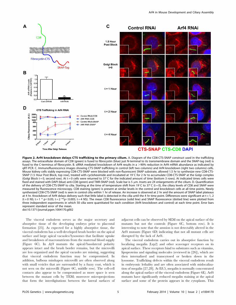
Figure 2. Arf4 knockdown delays CTS trafficking to the primary cilium. A. Diagram of the CD8-CTS-SNAP construct used in the traffickingassays. The extracellular domain of CD8 (green) is fused to fibrocystin (blue) just N-terminal to its transmembrane domain and the SNAP-tag (red) isfused to the C-terminus of fibrocystin. B. siRNA mediated knockdown of Arf4 results in a .90% reduction in Arf4 mRNA abundance as indicated byqRT-PCR. C. Immunofluorescence images showing CTS-SNAP trafficking in control (left two columns) and Arf4 knockdown (right two columns) cells.Mouse kidney cells stably expressing CD8-CTS-SNAP were blocked with non-fluorescent SNAP substrate, allowed 1.5 hr to synthesize new CD8-CTS-SNAP (1.5 Hour Post Block, top row), treated with cycloheximide and incubated at 19uC for 2 hr to accumulate CD8-CTS-SNAP at the Golgi complex(Golgi Block t = 0, second row). At t = 0 cells were returned to 37uC for the indicated amount of time (bottom 3 rows). At indicated times cells werefixed and stained with DAPI (blue) anti-CD8 (green) and TMR-SNAP (red). Scale bar is 5 mm. Insets are 2X enlargements of the cilium. D. Quantificationof the delivery of CD8-CTS-SNAP to cilia. Starting at the time of temperature shift from 19uC to 37uC (t = 0), the ciliary levels of CD8 and SNAP weremeasured by fluorescence microscopy. CD8 staining (green) is present at similar levels in the control and knockdown cells at all time points. Newlysynthesized CD8-CTS-SNAP (red) is seen in control cilia within 1 hr of release. An increase is observed at 2 hr and the amount of SNAP label plateausat 4 hr. Knockdown of Arf4 delays delivery such that little label is detected in the cilia until the 4 hr time point. Differences were significant at t = 1, 2(t = 0 NS; t = 1 * p,0.05; t = 2 **p,0.005; t = 4 NS). The mean CD8 fluorescence (solid line) and SNAP fluorescence (dotted line) were plotted fromthree independent experiments in which 50 cilia were quantitated for each condition (Arf4 knockdown and control) at each time point. Error barsrepresent standard error of the mean.doi:10.1371/journal.pgen.1004170.g002
Arf4 in Mouse Development and Ciliary Assembly
PLOS Genetics | www.plosgenetics.org 5 February 2014 | Volume 10 | Issue 2 | e1004170

suggests megalin trafficking is disrupted in Arf4 mutants and
supports the ultrastructural studies of the visceral endoderm that
indicate nutrient uptake is compromised within this cell layer.
Megalin is a type 1 membrane protein with a large extra cellular
domain, single transmembrane span and a short cytoplasmic C-
terminal tail similar to the structure of fibrocystin. Because of the
similarity in structure and the observation that Arf4 mutant
embryos have reduced megalin on the apical surface of the visceral
endoderm, we asked if Arf4 interacts with megalin. Co-immuno-
precipitation indicates Arf4 interacts with the intracellular domain
of megalin (Figure 6F). Similar to what we observed with
fibrocystin, the highly similar protein Arf5 did not interact with
megalin. These data suggest Arf4 is involved in not just trafficking
of ciliary cargo but also a larger class of trans-membrane receptors
– including megalin.
Discussion
The primary cilium is a sensory organelle and its proper
function relies on the correct complement of receptors localized
specifically in the ciliary membrane. Little is known about how
proteins are sorted to the ciliary compartment but understanding
Figure 3. Arf4 mutant mice are embryonic lethal. A. Schematic of the gene trap allele analyzed and the potential protein. The Arf4 allelecontains a b-galactosidase insertion in exon 3. 89 residues (green in the ribbon diagram) are potentially translated from this allele and may be fusedto the N-terminus of b-galactosidase. B, C. By E9.5, wild type and most heterozygous mice complete embryonic turning and adopt the normal fetalorientation. At this time point, Arf4 mutants are smaller and most have not completed embryonic turning. D. qRT-PCR analysis using primers thatspan the insertion site in intron 3 indicates that mean Arf4 mRNA abundance in the mutant embryos is strongly reduced; error bars depict standarderror of the mean. ***p,0.001. E. Genotype distribution as a function of age (+/+ blue; +/2 green; 2/2 orange; dead embryos are depicted by alighter shade). Arf4 mutant embryos are present until E11.5, however all mutant embryos dissected after E10.5 were dead. Number of embryos (n)analyzed at each time point is given below the graph.doi:10.1371/journal.pgen.1004170.g003
Arf4 in Mouse Development and Ciliary Assembly
PLOS Genetics | www.plosgenetics.org 6 February 2014 | Volume 10 | Issue 2 | e1004170

Figure 4. Arf4 mutant mice have functional nodal cilia. A. SEM of 3–4 somite developmentally matched embryos at 2,5006 (top), 10,0006(middle) and 20,0006 (bottom) magnification showing nodal cilia in wild type (left column) and Arf4 mutant (right column) embryos. B. Nodal cilialength is not significantly affected by the Arf4 mutation. Mean cilia length from 3–4 somite stage embryos is plotted (N = 3 each genotype); error barsare standard error of the mean. C. Total number of nodal cilia is not significantly affected in the Arf4 mutants. Mean number of cilia per node (N = 3for each genotype) is plotted; error bars are standard error of the mean. D. SEM of developing heart tube at E9.5 showing normal heart looping inwild type (left) and Arf4 mutant (right) embryos. E. Heart looping analysis of E9.5 embryos shows that all had normally looped hearts (n = 22, 38, 18 for+/+, +/2, 2/2).doi:10.1371/journal.pgen.1004170.g004
Arf4 in Mouse Development and Ciliary Assembly
PLOS Genetics | www.plosgenetics.org 7 February 2014 | Volume 10 | Issue 2 | e1004170

Arf4 in Mouse Development and Ciliary Assembly
PLOS Genetics | www.plosgenetics.org 8 February 2014 | Volume 10 | Issue 2 | e1004170
this process is critical as defects in the signaling functions of
primary cilia underlie a diverse group of human pathologies
known collectively as ciliopathies. These diseases range from
developmental defects of the brain, heart and other organs to
chronic ailments including retinal degeneration, obesity and
polycystic kidney disease. To study ciliary protein sorting, we
focused on analysis of the trafficking of the transmembrane protein
fibrocystin to the primary cilium. Mutations in the fibrocystin gene
(PKHD1) are responsible for autosomal recessive polycystic kidney
disease, a disorder afflicting approximately 1 in 20,000 individuals
and a cause of significant mortality during the first year of life
[29,30]. The ciliary targeting sequence of fibrocystin is an 18
amino acid sequence contained in the cytoplasmic tail [7]. We had
previously shown that the CTS interacts with the small G protein
Rab8. In this work we studied the interaction of the CTS with
another small G protein, Arf4. Arf proteins group vesicular cargo
and through interactions with coat proteins form transport vesicles
[12–14]. The proposed sorting ability of Arf proteins make them
attractive candidates as specificity factors and recent work suggests
Arf4 is involved in targeting rhodopsin and polycystin-1 to the
cilium [10,11,18]. We found that Arf4 was capable of interacting
with the fibrocystin CTS in a co-immunoprecipitation assay. The
interaction is specific as Arf4 is the only member of this highly
conserved family that precipitated significant amounts of the CTS.
The analysis of deletion and alanine scanning mutants within the
cytoplasmic tail of fibrocystin showed that the Arf4 interaction site
was localized within the CTS. The ability of Arf4 to bind to the
mutated CTSs roughly correlated to the ability of the CTS to
enter the cilium suggesting that the Arf4/CTS interaction was
functionally important.
Using SNAP-tagging technology we found that Arf4 is not
absolutely required for the delivery of the fibrocystin targeting
construct to cilia as the steady state level of the protein was not
affected by knockdown of Arf4. This is in contrast to the reported
effect of knockdown on the trafficking of polycystin-1 where Arf4
knockdown significantly reduced ciliary levels of polycystin-1 [18].
However, in our hands, the delivery of newly synthesized
fibrocystin fusion protein was slower in the knockdown cells
indicating that Arf4 is needed for the efficient delivery to primary
cilia. The delayed, but eventual delivery of the CTS to the cilium
may be a result of residual Arf4 protein (,10% of the mRNA
remains in the knockdown cells) or it may indicate an alternative
pathway to the cilium that does not utilize Arf4. Smoothened
appears to enter the ciliary compartment by first traveling to the
plasma membrane before moving into the cilium [21] and so it is
possible that this route can also be utilized by fibrocystin.
Our work, and work from others, indicates that Arf4 may play a
key role in sorting transmembrane receptors to the cilium. This
suggests that Arf4 may be an important player in human diseases
such as retinal degeneration and polycystic kidney disease. To
better understand the function of Arf4 and its possible role in
ciliopathies, we created an Arf4 mutant mouse. If the primary
function of Arf4 is specific to cilia, we would expect the mutant
mice to exhibit phenotypes in common with established mutations
that affect cilia. Mice with strong defects in ciliary assembly die at
mid-gestation with severe left-right abnormalities while those with
more mild ciliary defects survive longer and display phenotypes
indicative of hedgehog signaling dysfunction. Arf4 mutant mice die
between embryonic days 9 and 10, which is similar to the time
when mice with severe ciliary assembly defects die. However,
ciliary assembly is normal in the embryonic node and the nodal
cilia are functional as all embryos broke left-right symmetry
properly and formed a D-looped heart. The embryonic lethality
but lack of ciliary defects suggested Arf4 might have functions in
addition to the ciliary targeting of transmembrane proteins.
Expression analysis between embryonic day 7 and 10, around the
time the Arf4 mutant embryos were dying, indicated that the major
site of Arf4 expression was in the visceral endoderm. Examination
of the visceral endoderm indicates this tissue is not ciliated at this
time in development although cilia are present on the adjacent
mesoderm. The visceral endoderm is the major secretory and
absorptive tissue of the developing embryo prior to chorioallantoic
placenta formation [24] and defects within this cell layer often
result in embryonic lethality [27,28]. Arf4 mutant embryos have
multiple defects within the visceral endoderm. Ultrastructural
analysis of the visceral endoderm indicates Arf4 mutant embryos
have defects in cell-cell contacts and organization of the brush
border, but most strikingly, they lack the large lysosomes normally
present in healthy tissue. The absence of lysosomes could be a
direct effect of the Arf4 mutation, but is more likely an indirect
consequence of a failure to absorb nutrients from the adjacent
maternal blood supply. A failure to uptake nutrients is consistent
with the observed growth restriction evident by embryonic day 7
and likely accounts for the lethality around day 9. The apical
surface of the visceral endoderm is covered with microvilli that
contain a number of scavenger receptors that bind ligands
including vitamins and lipoproteins required by the developing
embryo. We examined the distribution of one of these receptors,
megalin within the visceral endoderm. Megalin is normally
localized to the apical surface of the developing visceral endoderm
however in Arf4 mutants, megalin fails to localize to the apical
surface. Megalin is a large single span transmembrane receptor
with membrane topology similar to fibrocystin. Co-immunopre-
cipitation assays indicate that Arf4 can interact with megalin
similar to what we observed between Arf4 and fibrocystin. This
interaction and the observed defects in megalin trafficking in the
Arf4 mutant suggest that Arf4 is required to target megalin to the
apical surface. While our work was in progress, another gene trap
allele of Arf4 was generated and used to study dendritic spine
formation in the brain. Similar to our allele, homozygotes were
lost prior to birth but no further analysis was done to characterize
the homozygous phenotypes [31].
RNAi studies suggested that the Arf family of proteins was
highly redundant and it was not predicted that the genetic loss of
any one would have a strong phenotype [32,33]. The observation
that Arf4 null mice die mid gestation indicates that this is not
completely correct. To date, Arf6 is the only other Arf family
member that has been mutated in the mouse. Like Arf4, Arf6 null
mice die during development, although they survive longer than
the Arf4 mice. The major defect in Arf6 null mice was in the liver,
where the lack of Arf6 caused increased rates of apoptosis resulting
in a significantly smaller liver with lethality around embryonic day
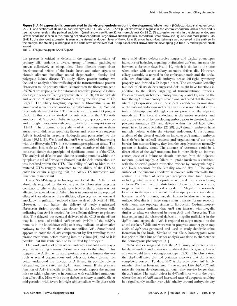
Figure 5. Arf4 expression is concentrated in the visceral endoderm during development. Whole mount b-Galactosidase stained embryos(A, C, E) and sections of stained mutant embryos (B, D, F). On E7 (A, B), Arf4-b-Gal expression is highest in the visceral endoderm (arrow head) and isseen at lower levels in the parietal endoderm (small arrow, see Figure S2 for more planes). On E8 (C, D) expression remains in the visceral endoderm(arrow head) and is seen in the forming definitive endoderm (large arrow) and the paraxial mesoderm (small arrow, see Figure S3 for more planes). OnE9 (E, F), the strongest expression is seen in the visceral endoderm layer of the yolk sac (F, arrow heads) but staining is also observed in the embryo. Inthe embryo, the staining is strongest in the endoderm of the liver bud (F. top panel, small arrow) and the developing gut tube (F, middle panel, smallarrow).doi:10.1371/journal.pgen.1004170.g005
Arf4 in Mouse Development and Ciliary Assembly
PLOS Genetics | www.plosgenetics.org 9 February 2014 | Volume 10 | Issue 2 | e1004170

Arf4 in Mouse Development and Ciliary Assembly
PLOS Genetics | www.plosgenetics.org 10 February 2014 | Volume 10 | Issue 2 | e1004170
15. [34]. The fact that both Arf4 and Arf6 mice survive through
early gestation suggests that neither of these genes are essential
genes at the cellular level, but do play critical functions in
particular cells at particular times in development. In the case of
the Arf4 mouse, the major defect was in the visceral endoderm, a
tissue with a very high rate of internalization and trafficking of
lipid and protein molecules. It is possible that this is the first point
in development that requires this level of internalization and
trafficking. The analysis of a floxed allele will be of interest to
determine if Arf4 is required in adult cells with a high rate of flux
such as the intestine or kidney proximal tubule, or even the rod
and cone photoreceptor cells where a high flux is needed to
maintain the outer segment.
The literature on Arf4 has mostly focused on proposed roles in
trafficking proteins to the ciliary membrane compartment.
However, our finding that the highest level of expression is in
the visceral endoderm, which is not ciliated at the time of high
expression, suggests that Arf4 has functions outside the targeting
of ciliary cargo. This is consistent with recent studies showing a
role for Arf4 in Golgi stress responses [35] and the finding that
class II Arfs (Arf4, Arf5) play roles in the trafficking of dense core
vesicles [36,37] and secretion of Dengue virus particles [33]. In
the case of the dense core vesicle transport, Arf4 and Arf5
interacted with two calcium dependent activator proteins for
secretion (CAPS1 and CAPS2). This interaction was required for
the efficient trafficking of dense core vesicles as knockdown of
either Arf4/5 or CAPS1/2 significantly reduced chromogranin
secretion [36,37]. In the case of Dengue virus production, Arf4
and Arf5 were required for the secretion of subviral particles and
the Arfs were thought to act though an interaction with prM
glycoprotein of the virus. Interestingly, the prM glycoprotein
contains a VXPX motif in the C-terminus similar to the Arf4
binding site in rhodopsin [10]. However mutation of the VXPX
motif did not disrupt interaction with Arf4 indicating that it is not
the binding site [33]. This is consistent with our finding that
Arf4 binds to the CTS of fibrocystin, which does not contain a
VXPX motif and studies of nephrocystin-3, which contains a
VXPX motif, but this motif is not necessary for ciliary targeting
[38].
In conclusion, we have shown that Arf4 plays a role in the
efficient transport of the fibrocystin CTS to the cilium, but it is not
required for ciliary assembly and in the mouse has critical
functions in non-ciliated cells. Thus, our work, and other
published work, suggests that Arf4 function is not restricted to
ciliary assembly but rather plays a broader role in cellular
trafficking.
Materials and Methods
Ethics StatementMouse work was approved by the University of Massachusetts
Medical School IACUC.
Mouse BreedingAn Arf4-mutant ES cell line was obtained from the Sanger
Center and used to generate Arf4Gt(AY0614)Wtsi mutant mice. The
animals used in this study were a mix of 129 and C57Bl6
backgrounds. Embryonic ages were determined by timed mating
with the day of the plug being embryonic day 0.5. Genotyping was
carried out with the following primer pairs: Arf4-1 AGCAGCCT-
CATTGTCCTAGC+Arf4-2 CCTCCCCACAATTCAACAAT
(product size = 189 bp in wildtype) and Geo-3 GATCGGC-
CATTGAACAAGAT+Geo-4 CAATAGCAGCCAGTCCCT-
TC (product size = 280 bp in mutant).
Mammalian Cell CultureIMCD3 (ATCC) were grown in 47.5% DMEM 47.5% F12, 5%
fetal bovine serum, with penicillin and streptomycin at 37uC in 5%
CO2. Cells were transfected by electroporation (Bio-Rad, Hercules
CA). Stable cell lines were selected by supplementing the medium
with 400 mg/ml of G418 (Sigma, St. Louis, MO). Clonal lines
were selected by dilution cloning after drug selection.
Electron MicroscopyFor scanning electron microscopy (SEM), timed pregnant
females were euthanized by approved IACUC protocols, embryos
dissected in DMEM/F12 supplemented with 5% fetal bovine
serum, fixed overnight in 2.5% glutaraldehyde in 0.1M sodium
cacodylate. Fixed embryos were rinsed twice with 0.1M sodium
cacodylate, osmicated in 1% osmium tetroxide, dehydrated in a
graded ethanol series and critical point dried (Autosamdri-815,
Series A, Tousimis Research Corp.). Dried embryos were sputter
coated with iridium to a thickness of 3 nm (Cressington 208 HR
Sputter Coater, Ted Pella, Redding, CA, USA) and examined in a
scanning electron microscope (FEI Quanta 200 FEG SEM) [39].
For comparison of nodal cilia, embryos were developmentally
matched by counting somite number.
For transmission electron microscopy (TEM), samples were fixed,
osmicated and dehydrated as described above. Dehydrated samples
were then infiltrated first with two changes of 100% propylene oxide
and then with a 50%/50% propylene oxide/SPI-Pon 812 resin
mixture. The following day, three changes of fresh 100% SPI-Pon
812 resin were done before the samples were polymerized at 68uC
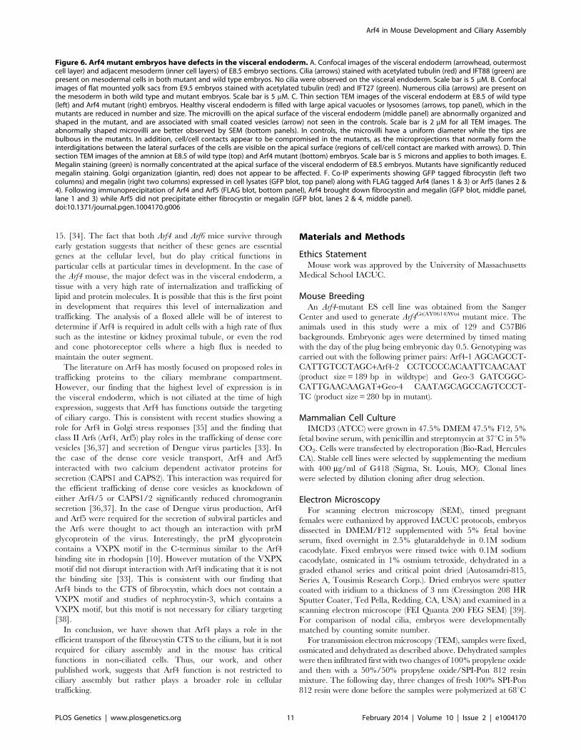
Figure 6. Arf4 mutant embryos have defects in the visceral endoderm. A. Confocal images of the visceral endoderm (arrowhead, outermostcell layer) and adjacent mesoderm (inner cell layers) of E8.5 embryo sections. Cilia (arrows) stained with acetylated tubulin (red) and IFT88 (green) arepresent on mesodermal cells in both mutant and wild type embryos. No cilia were observed on the visceral endoderm. Scale bar is 5 mM. B. Confocalimages of flat mounted yolk sacs from E9.5 embryos stained with acetylated tubulin (red) and IFT27 (green). Numerous cilia (arrows) are present onthe mesoderm in both wild type and mutant embryos. Scale bar is 5 mM. C. Thin section TEM images of the visceral endoderm at E8.5 of wild type(left) and Arf4 mutant (right) embryos. Healthy visceral endoderm is filled with large apical vacuoles or lysosomes (arrows, top panel), which in themutants are reduced in number and size. The microvilli on the apical surface of the visceral endoderm (middle panel) are abnormally organized andshaped in the mutant, and are associated with small coated vesicles (arrow) not seen in the controls. Scale bar is 2 mM for all TEM images. Theabnormally shaped microvilli are better observed by SEM (bottom panels). In controls, the microvilli have a uniform diameter while the tips arebulbous in the mutants. In addition, cell/cell contacts appear to be compromised in the mutants, as the microprojections that normally form theinterdigitations between the lateral surfaces of the cells are visible on the apical surface (regions of cell/cell contact are marked with arrows). D. Thinsection TEM images of the amnion at E8.5 of wild type (top) and Arf4 mutant (bottom) embryos. Scale bar is 5 microns and applies to both images. E.Megalin staining (green) is normally concentrated at the apical surface of the visceral endoderm of E8.5 embryos. Mutants have significantly reducedmegalin staining. Golgi organization (giantin, red) does not appear to be affected. F. Co-IP experiments showing GFP tagged fibrocystin (left twocolumns) and megalin (right two columns) expressed in cell lysates (GFP blot, top panel) along with FLAG tagged Arf4 (lanes 1 & 3) or Arf5 (lanes 2 &4). Following immunoprecipitation of Arf4 and Arf5 (FLAG blot, bottom panel), Arf4 brought down fibrocystin and megalin (GFP blot, middle panel,lane 1 and 3) while Arf5 did not precipitate either fibrocystin or megalin (GFP blot, lanes 2 & 4, middle panel).doi:10.1371/journal.pgen.1004170.g006
Arf4 in Mouse Development and Ciliary Assembly
PLOS Genetics | www.plosgenetics.org 11 February 2014 | Volume 10 | Issue 2 | e1004170
in plastic capsules. The samples were then reoriented and thin
sections were placed on copper support grids and contrasted with
Lead citrate and Uranyl acetate. Sections were examined using a
Phillips CM10 TEM with 80 Kv accelerating voltage, and images
were captured using a Gatan TEM CCD camera.
Immunofluorescence MicroscopyCells for immunofluorescence microscopy were grown,
fixed, and stained as described [40]. For visceral endoderm
immunofluorescence, E8.5 embryos were fixed for 15 minutes
at room temperature with 4% paraformaldehyde in PBS rinsed
twice in PBS, equilibrated in 30% sucrose overnight and
embedded in Tissue Freezing Media (Triangle Biomedical
Sciences). Cryosections (10 mm) were blocked for 1 hour in 1%
bovine serum albumin, incubated with primary antibodies
overnight at 4uC.
Primary antibodies included anti acetylated-tubulin (611B1,
Sigma, St. Louis MO), anti-FLAG (Sigma), anti-MmIFT20, anti-
MmIFT88 [41], MmIFT27 [42], anti-golgin97 (CDF4, Molecular
Probes) anti-BIP (clone 40, BD Transduction Laboratories), anti-
Rab11 (clone 47, BD Transduction Laboratories), anti-TfR (clone
H68.4, Invitrogen), anti-giantin [43] and anti-megalin (P-20,
Santa Cruz Biotechnology).
Widefield images were acquired by an Orca ER camera
(Hamamatsu, Bridgewater, NJ) on a Zeiss Axiovert 200 M
microscope equipped with a Zeiss 1006 plan-Apochromat 1.4
NA objective. Images were captured by Openlab (Improvision,
Waltham, MA) and adjusted for contrast in Adobe Photoshop. If
comparisons are to be made between images, the photos were
taken with identical conditions and manipulated equally. For the
quantification of SNAP and CD8 in the cilia, the length, area, and
average fluorescence intensity of the cilia was measured using the
measurement tools of Openlab. To determine significance of
differences, data from three independent experiments were
subjected to an unpaired Student’s T test. Confocal images were
acquired by a Nikon TE-2000E2 inverted microscope equipped
with a Solamere Technology modified Yokogawa CSU10 spinning
disk confocal scan head. Z-stacks were acquired at 0.2 mm or
0.5 mm intervals and converted to single planes by maximum
projection with MetaMorph software. Bright field images
were acquired using a Zeiss Axioskop 2 Plus equipped with an
Axiocam HRC color digital camera and Axiovision 4.0 acquisition
software.
SNAP Trafficking AssaysThe construct (pJAF270) used for SNAP trafficking assays was
constructed by fusing the extracellular domain of mouse CD8a
[44] to the last 17 extracellular residues of mouse fibrocystin
through the first 27 intracellular residues, the SNAP tag was
cloned onto the c-terminal end of the CTS creating CD8-CTS-
SNAP. Mouse kidney cells stably expressing CD8-CTS-SNAP
were incubated with 0.04 mM cell permeable non-fluorescent BG-
Block (New England Biolabs) for 30 minutes to block all SNAP
epitopes. Following 3 washes with complete growth media cells
were allowed to synthesize new CD8-CTS-SNAP for 1.5 hrs
before the addition of HEPES pH 7.4 to 20 mM and cyclohex-
imide to 150 mg/ml, then shifted to 19uC for two hrs to
accumulate CD8-CTS-SNAP at the Golgi complex. Cells were
returned to 37u and allowed to traffic CD8-CTS-SNAP for the
indicated periods of time before being fixed and stained.
For siRNA knockdown, cells were transfected by RNAiMAXX
(Invitrogen) with SMARTpool siRNA (Dharmacon) targeting Arf4
(L-060271) or a non-targeting control (D-001810) and assayed for
knockdown 48 hours post transfection.
Protein AnalysisFLAG-tagged Arf1-6 (pJAF215, pJAF213, pJAF214, pJAF216,
pJAF210, pJAF211), were constructed by PCR amplifying the
open reading frames and inserting them into p3XFLAG-CMV-14
(Sigma, St. Louis, MO). Point mutations in Arf4 (Arf4T31N = p-
JAF221, Arf4T48N = pJAF222, Arf4Q71L = pJAF223) were gener-
ated by inverse PCR using the Quick Change II site directed
mutagenesis kit (Stratagene) starting from pJAF216. Cells were
transfected with FLAG-tagged Arf and GFP-tagged CTS deletion
and alanine scanning mutants used in [7], CD8-PKHD1
(pJAF268), or CD8-Megalin (pJAF281) and 48 hours later, FLAG
immunoprecipitation was carried out as described in [40].
Beta-galactosidase Staining of Mouse EmbryosEmbryos were fixed in 0.2% glutaraldehyde, 2% formalin,
5 mM EGTA and 2 mM MgCl2 in 0.1M phosphate buffer pH 7.3
for 10 minutes at room temperature then rinsed three times in
wash buffer containing 0.1% sodium deoxycholate, 0.2%
IGEPAL, 2 mM MgCl2 in 0.1M phosphate buffer for 30 minutes
each wash. Fixed embryos were stained overnight at 37uC in
1 mg/ml X-gal, 5 mM potassium ferrocyanide, 5 mM potassium
ferricyanide diluted in wash buffer.
mRNA AnalysisRNA was isolated from individual E9.5 embryos or from
IMCD3 cells using RNeasy kits (Qiagen), including on-column
DNA digestion. First strand cDNA was synthesized from 100–
500 ng of total RNA using a SuperScript II First-Strand Synthesis
System (Invitrogen, Carlsbad, CA) and random hexameric
primers. Quantitative real-time PCR primers were designed to
produce amplicons between 100–150 nucleotides in length, using
the online primer3 web PCR primer tool (http://fokker.wi.mit.
edu/primer3/input.htm) and the IDT Primer Express software
tool (http://www.idtdna.com/Scitools/Applications/Primerquest/).
Primers were synthesized by Integrated DNA Technologies Inc
(Coralville, IA) and are listed in Table 1. qRT-PCR analysis
was performed using an ABI Prism 7500 sequence detection
system (Applied Biosystems, Foster City, CA). Each reaction
contained 5–12.5 ng first strand cDNA, 0.1 mM each specific
forward and reverse primers and 16 Power SYBR Green
(Applied Biosystems, Foster City, CA) in a 15 ml volume. Arf4
mRNA expression was normalized to GAPDH mRNA abun-
dance and compared between mutant and control animals with
an unpaired Student t-test.
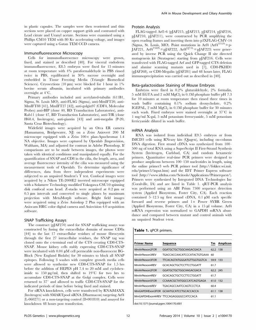
Table 1. qPCR primers.
Primer Name Sequence Tm Amplicon
MmArf4exon2FOR GGATGCTGCTGGCAAGACGACA 62.2 138
MmArf4exon3REV TGACCACCAACATCCCATACTGTGAAA 60
MmArf4exon3FOR TTCACAGTATGGGATGTTGGTGGTCA 59.9 133
MmArf4exon4REV GCACAGCTGCTCCTTCCTGGATT 61.7
MmArf4exon2FOR GGATGCTGCTGGCAAGACGACA 62.2 245
MmArf4exon4REV GCACAGCTGCTCCTTCCTGGATT 61.7
MmArf4exon5FOR CCAAACGCTATGGCCATCAGTGAGA 61.0 132
MmArf4exon6REV TGACAGCCAATCCAGTCCCTCA 60.4
MmGAPDHExon3FOR GCAATGCATCCTGCACCACCA 61.1 138
MmGAPDHExon4rREV TTCCAGAGGGGCCATCCACA 61.1
doi:10.1371/journal.pgen.1004170.t001
Arf4 in Mouse Development and Ciliary Assembly
PLOS Genetics | www.plosgenetics.org 12 February 2014 | Volume 10 | Issue 2 | e1004170
Supporting Information
Figure S1 Arf4 expression inhibits CTS trafficking. A. Mouse
kidney cells stably expressing wild type (top row), dominant negative
(T13N, T48N) and constitutively active (Q67L) Arf4-FLAG (red)
stained with IFT20 (green) and DAPI (blue). Constitutively active
(Q67L) localizes to the Golgi complex while wild type and dominant
negative Arf4 are dispersed in the cytoplasm. B. Expression of
dominant negative (T13N, T48N) Arf4 reduced the percent of
ciliated cells. * p,0.05, ** p,0.01 as compared to IMCD cells not
transfected with an Arf4-FLAG construct C. Dominant negative
Arf4 (T13N, T48N) causes a reduction in ciliary length. * p,0.05,
** p,0.01 as compared to IMCD cells not transfected with an Arf4-
FLAG construct D. Expression of any Arf4 constructs prevents
ciliary trafficking of GFP-CTS. *** p,0.001 as compared to IMCD
cells not transfected with an Arf4-FLAG construct. B–D, error bars
are standard error of the mean. E. Co-immunoprecipitation
experiments demonstrate that GFP-CTS interacts with FLAG-
tagged Arf4 wild type and mutant proteins.
(TIF)
Figure S2 b-galactosidase stained mutant embryo dissected on
embryonic day 7. Embryos were stained with X-gal, embedded in
paraffin, sectioned, dewaxed and photographed. Planes are
organized from proximal to the distal end of the conceptus.
Panels A–C show extra-embryonic and panels D and E embryonic
regions. Small arrows mark staining in the parietal endoderm and
arrow heads mark the visceral endoderm.
(TIF)
Figure S3 b-galactosidase stained mutant embryo dissected on
embryonic day 8. Embryos were stained with X-gal, embedded in
paraffin, sectioned, dewaxed and photographed. Planes are
organized from the proximal to distal regions of the conceptus.
Panels A–C show extra-embryonic and panels D and E embryonic
regions. Arrow heads mark the visceral endoderm. Small arrows
mark staining in the allantois (plane B and C) or paraxial mesoderm
(planes D and E). In plane D, the larger arrow marks the site where
definitive endoderm is forming in the developing hindgut.
(TIF)
Acknowledgments
We thank Drs. S. Jones (Transgenic Mouse Core) G. Hendricks, L.
Strittmatter (Electron Microscopy Core) and P. Furcinitti (Digital Imaging
Core) for assistance during this work. We thank Dr. D. Lambright (UMass
Worcester) for helpful discussion during this project.
Author Contributions
Conceived and designed the experiments: JAF KDT GJP. Performed the
experiments: JAF KDT JAJ JTSA JARP TH GJP. Analyzed the data: JAF
JAJ JARP KDT GJP. Contributed reagents/materials/analysis tools: JAF
JAJ GJP. Wrote the paper: JAF GJP.
References
1. Pazour GJ, Bloodgood RA (2008) Targeting proteins to the ciliary membrane.
Curr Top Dev Biol 85: 115–149.
2. Rohatgi R, Snell WJ (2010) The ciliary membrane. Curr Opin Cell Biol 22:
541–546.
3. Nachury MV, Seeley ES, Jin H (2010) Trafficking to the ciliary membrane: how
to get across the periciliary diffusion barrier? Annu Rev Cell Dev Biol 26: 59–87.
4. Ward CJ, Hogan MC, Rossetti S, Walker D, Sneddon T, et al. (2002) The gene
mutated in autosomal recessive polycystic kidney disease encodes a large,
receptor-like protein. Nat Genet 30: 259–269.
5. Ward CJ, Yuan D, Masyuk TV, Wang X, Punyashthiti R, et al. (2003) Cellular
and subcellular localization of the ARPKD protein; fibrocystin is expressed on
primary cilia. Hum Mol Genet 12: 2703–2710.
6. Onuchic LF, Furu L, Nagasawa Y, Hou X, Eggermann T, et al. (2002) PKHD1,
the polycystic kidney and hepatic disease 1 gene, encodes a novel large protein
containing multiple immunoglobulin-like plexin-transcription-factor domains
and parallel beta-helix 1 repeats. Am J Hum Genet 70: 1305–1317.
7. Follit JA, Li L, Vucica Y, Pazour GJ (2010) The cytoplasmic tail of fibrocystin
contains a ciliary targeting sequence. J Cell Biol 188: 21–28.
8. Nachury MV, Loktev AV, Zhang Q, Westlake CJ, Peranen J, et al. (2007) A core
complex of BBS proteins cooperates with the GTPase Rab8 to promote ciliary
membrane biogenesis. Cell 129: 1201–1213.
9. Yoshimura S, Egerer J, Fuchs E, Haas AK, Barr FA (2007) Functional dissection
of Rab GTPases involved in primary cilium formation. J Cell Biol 178: 363–369.
10. Deretic D, Williams AH, Ransom N, Morel V, Hargrave PA, et al. (2005)
Rhodopsin C terminus, the site of mutations causing retinal disease, regulates
trafficking by binding to ADP-ribosylation factor 4 (ARF4). Proc Natl Acad
Sci U S A 102: 3301–3306.
11. Mazelova J, Astuto-Gribble L, Inoue H, Tam BM, Schonteich E, et al. (2009)
Ciliary targeting motif VxPx directs assembly of a trafficking module through
Arf4. EMBO J 28: 183–192.
12. Donaldson JG, Jackson CL (2011) ARF family G proteins and their regulators:
roles in membrane transport, development and disease. Nat Rev Mol Cell Biol
12: 362–375.
13. Nie Z, Randazzo PA (2006) Arf GAPs and membrane traffic. J Cell Sci 119:
1203–1211.
14. D’Souza-Schorey C, Chavrier P (2006) ARF proteins: roles in membrane traffic
and beyond. Nat Rev Mol Cell Biol 7: 347–358.
15. Gillingham AK, Munro S (2007) The small G proteins of the Arf family and
their regulators. Annu Rev Cell Dev Biol 23: 579–611.
16. Jenkins PM, Hurd TW, Zhang L, McEwen DP, Brown RL, et al. (2006) Ciliary
targeting of olfactory CNG channels requires the CNGB1b subunit and the
kinesin-2 motor protein, KIF17. Curr Biol 16: 1211–1216.
17. Geng L, Okuhara D, Yu Z, Tian X, Cai Y, et al. (2006) Polycystin-2 traffics to
cilia independently of polycystin-1 by using an N-terminal RVxP motif. J Cell
Sci 119: 1383–1395.
18. Ward HH, Brown-Glaberman U, Wang J, Morita Y, Alper SL, et al. (2011) A
conserved signal and GTPase complex are required for the ciliary transport of
polycystin-1. Mol Biol Cell 22: 3289–3305.
19. Keppler A, Gendreizig S, Gronemeyer T, Pick H, Vogel H, et al. (2003) A
general method for the covalent labeling of fusion proteins with small molecules
in vivo. Nat Biotechnol 21: 86–89.
20. Farr GA, Hull M, Mellman I, Caplan MJ (2009) Membrane proteins follow
multiple pathways to the basolateral cell surface in polarized epithelial cells. J Cell
Biol 186: 269–282.
21. Milenkovic L, Scott MP, Rohatgi R (2009) Lateral transport of Smoothened from
the plasma membrane to the membrane of the cilium. J Cell Biol 187: 365–374.
22. Saraste J, Palade GE, Farquhar MG (1986) Temperature-sensitive steps in the
transport of secretory proteins through the Golgi complex in exocrine pancreatic
cells. Proc Natl Acad Sci U S A 83: 6425–6429.
23. Nonaka S, Tanaka Y, Okada Y, Takeda S, Harada A, et al. (1998)
Randomization of left-right asymmetry due to loss of nodal cilia generating
leftward flow of extraembryonic fluid in mice lacking KIF3B motor protein. Cell
95: 829–837.
24. Zohn IE, Sarkar AA (2010) The visceral yolk sac endoderm provides for
absorption of nutrients to the embryo during neurulation. Birth Defects
Res A Clin Mol Teratol 88: 593–600.
25. Bielinska M, Narita N, Wilson DB (1999) Distinct roles for visceral endoderm
during embryonic mouse development. Int J Dev Biol 43: 183–205.
26. May P, Woldt E, Matz RL, Boucher P (2007) The LDL receptor-related protein
(LRP) family: an old family of proteins with new physiological functions. Ann
Med 39: 219–228.
27. Nada S, Hondo A, Kasai A, Koike M, Saito K, et al. (2009) The novel lipid raft
adaptor p18 controls endosome dynamics by anchoring the MEK-ERK
pathway to late endosomes. EMBO J 28: 477–489.
28. Lighthouse JK, Zhang L, Hsieh JC, Rosenquist T, Holdener BC (2011) MESD
is essential for apical localization of megalin/LRP2 in the visceral endoderm.
Dev Dyn 240: 577–588.
29. Pazour GJ (2004) Intraflagellar transport and cilia-dependent renal disease: the
ciliary hypothesis of polycystic kidney disease. J Am Soc Nephrol 15: 2528–2536.
30. Harris PC, Torres VE (2009) Polycystic kidney disease. Annu Rev Med 60: 321–
337.
31. Jain S, Yoon SY, Zhu L, Brodbeck J, Dai J, et al. (2012) Arf4 determines dentate
gyrus-mediated pattern separation by regulating dendritic spine development.
PLoS One 7: e46340.
32. Kahn RA, Volpicelli-Daley L, Bowzard B, Shrivastava-Ranjan P, Li Y, et al.
(2005) Arf family GTPases: roles in membrane traffic and microtubule dynamics.
Biochem Soc Trans 33: 1269–1272.
33. Kudelko M, Brault JB, Kwok K, Li MY, Pardigon N, et al. (2012) Class II ADP-
ribosylation factors are required for efficient secretion of dengue viruses. J Biol
Chem 287: 767–777.
Arf4 in Mouse Development and Ciliary Assembly
PLOS Genetics | www.plosgenetics.org 13 February 2014 | Volume 10 | Issue 2 | e1004170

34. Suzuki T, Kanai Y, Hara T, Sasaki J, Sasaki T, et al. (2006) Crucial role of the
small GTPase ARF6 in hepatic cord formation during liver development. MolCell Biol 26: 6149–6156.
35. Reiling JH, Olive AJ, Sanyal S, Carette JE, Brummelkamp TR, et al. (2013) A
CREB3-ARF4 signalling pathway mediates the response to Golgi stress andsusceptibility to pathogens. Nat Cell Biol 15: 1473–1485.
36. Sadakata T, Sekine Y, Oka M, Itakura M, Takahashi M, et al. (2012) Calcium-dependent activator protein for secretion 2 interacts with the class II ARF
small GTPases and regulates dense-core vesicle trafficking. FEBS J 279: 384–
394.37. Sadakata T, Shinoda Y, Sekine Y, Saruta C, Itakura M, et al. (2010) Interaction
of calcium-dependent activator protein for secretion 1 (CAPS1) with the class IIADP-ribosylation factor small GTPases is required for dense-core vesicle
trafficking in the trans-Golgi network. J Biol Chem 285: 38710–38719.38. Nakata K, Shiba D, Kobayashi D, Yokoyama T (2012) Targeting of Nphp3 to
the primary cilia is controlled by an N-terminal myristoylation site and coiled-
coil domains. Cytoskeleton (Hoboken) 69: 221–234.
39. SanAgustin JT, Follit JA, Hendricks G, Pazour GJ (2009) Scanning electron
microscopy to examine cells and organs. Methods Cell Biol 91: 81–87.40. Follit JA, San Agustin JT, Xu F, Jonassen JA, Samtani R, et al. (2008) The
Golgin GMAP210/TRIP11 anchors IFT20 to the Golgi complex. PLoS Genet
4: e1000315.41. Pazour GJ, Baker SA, Deane JA, Cole DG, Dickert BL, et al. (2002) The
intraflagellar transport protein, IFT88, is essential for vertebrate photoreceptorassembly and maintenance. J Cell Biol 157: 103–113.
42. Keady BT, Samtani R, Tobita K, Tsuchya M, SanAgustin JT, et al. (2012)
IFT25 Links the Signal-Dependent Movement of Hedgehog Components toIntraflagellar Transport. Developmental Cell 22: 940–951.
43. Nozawa K, Casiano CA, Hamel JC, Molinaro C, Fritzler MJ, et al. (2002)Fragmentation of Golgi complex and Golgi autoantigens during apoptosis and
necrosis. Arthritis Res 4: R3.44. Xia H, Hornby ZD, Malenka RC (2001) An ER retention signal explains
differences in surface expression of NMDA and AMPA receptor subunits.
Neuropharmacology 41: 714–723.
Arf4 in Mouse Development and Ciliary Assembly
PLOS Genetics | www.plosgenetics.org 14 February 2014 | Volume 10 | Issue 2 | e1004170