Contributions of Ultrastructural Studies to the Knowledge of ...

Ciliary dysfunction and ultrastructural abnormalities arefeatures of severe asthma
Biju Thomas, MD, Andrew Rutman, CBiol, Robert A. Hirst, PhD, Pranab Haldar, MD, Andrew J. Wardlaw, PhD,
John Bankart, PhD, Christopher E. Brightling, PhD,* and Christopher O’Callaghan, PhD* Leicester, United Kingdom
Background: Epithelial dysfunction has been implicated inasthma pathophysiology, but no studies have directly assessedciliary function in asthma.Objective: To study the ciliary function and epithelialultrastructure of patients with asthma and healthy controls.Methods: We studied ciliary beat frequency and beat pattern byusing digital high-speed video imaging and ultrastructure bytransmission electron microscopy of bronchial epithelial stripsfrom 7 subjects with mild, 7 with moderate, and 19 with severeasthma and 9 healthy controls.Results: The median (interquartile range) ciliary beat frequencywas decreased in moderate (6.5 [4.4-8.5] Hz) and severe asthma(6.7 [6.1-7.6] Hz) compared with controls (10.5 [9.7-11.8] Hz;P < .01). Dyskinesia and immotility indices were higher in severeasthma (65% [43%-75%]; 6.3% [1%-9.5%], respectively)compared with controls (4% [0%-6.7%; 0%, respectively;P < .01). These abnormalities were related to disease severity(ciliary beat frequency, rs 5 –0.68; dyskinesia index, rs 5 0.86;immotility index, rs 5 0.65; P < .0001). The ultrastructure of theepithelium was abnormal in severe asthma with a reduction inciliated cells, an increase in dead cells, and ciliary disorientationcompared with all other groups (P < .05). Compared withpatients with mild asthma and healthy controls, patients withsevere asthma showed increased ciliary depletion, microtubulardefects, mitochondrial damage, and cytoplasmic blebbing(P < .01). All of these changes were related to disease severity.Conclusion: Ciliary dysfunction and ultrastructuralabnormalities are closely related to asthma severity. Ciliarydysfunction is a feature of moderate to severe asthma, andprofound ultrastructural abnormalities are restricted to severedisease. Whether these changes contribute to the development ofsevere asthma phenotype remains to be determined. (J AllergyClin Immunol 2010;126:722-9.)
From the Institute for Lung Health, Department of Infection, Immunity and Inflamma-
tion, University of Leicester.
*Co-senior authors.
C.E.B. obtained support from the Wellcome Trust, Asthma UK, and GlaxoSmithKline,
which funded this study in part. Ciliary function analysis and electron microscopy
analysis were performed in C.O.’s laboratory and were not supported by these sources.
Disclosure of potential conflict of interest: A. J. Wardlaw serves on advisory boards for
GlaxoSmithKline and receives research support from GlaxoSmithKline, Pfizer, and
AstraZeneca. C. E. Brightling serves on advisory boards for GlaxoSmithKline, Astra-
Zeneca, MedImmune, Roche, and Aerovance; receives honoraria from Novartis; and
receives research support from GlaxoSmithKline, AstraZeneca, and MedImmune.
The rest of the authors have declared that they have no conflict of interest.
Received for publication October 19, 2009; revised April 20, 2010; accepted for publica-
tion May 21, 2010.
Available online July 31, 2010.
Reprint requests: Christopher O’Callaghan, PhD, Department of Infection, Immunity and
Inflammation, University of Leicester, PO Box 65, Leicester Royal Infirmary, Leices-
ter, LE2 7LX, United Kingdom. E-mail: [email protected].
0091-6749/$36.00
� 2010 American Academy of Allergy, Asthma & Immunology
doi:10.1016/j.jaci.2010.05.046
722
Key words: Ciliated epithelium, ciliary beat frequency, ciliary beatpattern, ciliary disorientation, ciliary dyskinesia, refractory asthma,severe asthma
Asthma affects approximately 300 million people globally.1
Although the majority of patients have well controlled disease,there remains a subgroup of adults with asthma, accounting forabout 10% of patients with asthma, who continue to have debili-tating chronic and persistent symptoms despite optimal standardasthma treatment.2 This group with severe refractory asthma3 rep-resents those with a high risk of severe exacerbations and asthma-related mortality and accounts for >50% of asthma-related healthcare costs.4,5 There has been extensive research to unravel thecomplex pathophysiology of severe asthma, but it remains uncer-tain what the immunopathologic hallmarks of severe asthmaare.2,5 In this respect, the role of the dysfunctional respiratoryepithelium in severe asthma has been of much recent interest.6,7
The ciliated epithelium that covers the surface of the airwaysforms an immunologically active natural barrier to invasion andinjury by inhaled pathogenic organisms and particulate material.The epithelium is lined by an airway surface liquid and the mucuslayer. The airway surface liquid provides an ideal environment inwhich the cilia beat at a frequency of 11 to 14 Hz. The mucus layerthat lies above the airway surface liquid is cleared from the airwayby the highly coordinated ciliary beating. This process, known asmucociliary clearance,8 is an essential factor in pulmonary de-fense.9 It therefore follows that damage to the respiratory epithe-lium and ciliary dysfunction causes impaired mucociliaryclearance, leading to increased susceptibility to infection andinflammation.
Using an inhaled radio-aerosol technique, mucociliary clear-ance in asthma in the stable state and during exacerbations wasreported to be impaired.10,11 Optimal mucociliary clearance de-pends on the structural and functional integrity of the cilia aswell as the characteristics of the airway surface liquid and mucus.Submucosal gland hypertrophy and goblet cell hyperplasia arewell recognized pathologic features of asthma.12 The asthmaticairway is also characterized by mucus hypersecretion,13 abnormalmucus rheology,12 and tethering of intraluminal mucins to gobletcells in the airway epithelium.14 Although evidence that supportsabnormalities of mucus12-14 and airway surface liquid15 providesa plausible mechanistic explanation for the reduced mucociliaryclearance observed in asthma, to date no studies have assessed cil-iary function directly in asthma or considered its relationship withdisease severity. Indeed, impaired ciliary function would have apronounced effect on mucociliary clearance. In this context, inthis observational study, we aimed to characterize the ciliated res-piratory epithelium from the lower airways of adults with mild,moderate, and severe asthma compared with healthy controls byassessing the function of cilia and detailed ultrastructure of theciliated epithelium.
J ALLERGY CLIN IMMUNOL
VOLUME 126, NUMBER 4
THOMAS ET AL 723
Abbreviations used
FVC: F orced vital capacityGINA: G
lobal Initiative for AsthmaIQR: I
nterquartile rangeMETHODS
SubjectsPatients with mild (n 5 7), moderate (n 5 7), and severe (n 5 19) asthma
were recruited from clinics at Glenfield Hospital, Leicester, United Kingdom,
over a 2-year period (2007-2009). Healthy control subjects (n 5 9) were
recruited from hospital staff and by local advertising. Asthma was diagnosed
on the basis of the presence of clinical features consistent with asthma and
objective measures of airway hyperresponsiveness and variable airflow
obstruction (PC20 in FEV1 of less than 8 mg/mL; increase in FEV1 by at least
15% after the inhalation of 200 mg salbutamol; or variation in peak flow, ex-
pressed as a percentage of the mean, exceeding 20% over a period of 14 days).
Asthma severity was classified as mild, moderate, and severe by using the cur-
rent Global Initiative for Asthma (GINA) guidelines on the basis of the GINA
treatment steps16 (mild asthma, GINA treatment step 1/2; moderate asthma,
GINA treatment step 3; severe asthma, GINA treatment step 4/5). Patients
with severe asthma also met the American Thoracic Society criteria for refrac-
tory asthma.3 At the time of collection of bronchial epithelial samples, patients
with asthma had been free from intercurrent respiratory infections and asthma
exacerbations requiring antibiotics and or rescue use of systemic corticoste-
roids for at least 6 weeks. Healthy subjects had no history of respiratory dis-
ease, normal lung function, and normal PC20. All subjects were current
nonsmokers, and those who did smoke in the past had a smoking history of
less than 10 pack-years. The study protocol was approved by the Leicester-
shire and Rutland regional ethics committee, and written informed consent
was obtained from all subjects.
MeasurementsDemographic and medical details including age, sex, and current medica-
tions were recorded on all subjects. FEV1, forced vital capacity (FVC), and
FEV1/FVC ratio were measured on all subjects with a rolling-seal spirometer
(Vitalograph Ltd, Buckingham, England). The PC20 was computed from the
methacholine dose-response curve (the change in FEV1 in relation to the meth-
acholine concentration) by linear interpolation on a log scale using standard
techniques.17 In patients with asthma, the presence of atopy was determined
by allergen skin prick tests for common aeroallergens including Dermatopha-goides pteronyssinus, dog, cat, and grass pollen. Plasma IgE was measured on
patients with moderate and severe asthma. Patients with asthma had single-
flow exhaled nitric oxide concentration measured at a rate of 50 mL per second
as previously described.18 Induced sputum was obtained from patients with
asthma, and sputum samples were processed as previously described.19 All
subjects underwent flexible bronchoscopy conducted according to the British
Thoracic Society guidelines20 to obtain strips of bronchial epithelium by
brushing the bronchus intermedius.
Ciliary beat frequency and beat patternDetailed methodology is given in this article’s Methods in the Online
Repository at www.jacionline.org. Typically, a strip of bronchial epithelium
obtained by brushing the bronchus contained a row of adjacent bronchial
epithelial cells. Ciliary beat frequency was measured and beat pattern as-
sessed on strips of bronchial epithelium by using a digital high-speed video
microscopy system as described previously with nasal epithelial brushings,
within 2 hours of sample collection.21,22 The high-speed video images were
analyzed in a blind fashion. The images were re-analyzed by a second ob-
server (A.R.) and blinded on a second occasion by the original observer
(B.T.).
The experimental system allowed the ciliary beat pattern to be evaluated in
3 different planes: sideways profile of the ciliated epithelial strip, assessing the
ciliated epithelial strip with cilia beating directly toward the observer, and
assessing the ciliated epithelial strip from directly above.21,22 The path taken
by a cilium during the beat cycle was analyzed frame by frame. This was char-
acterized and compared with the normal beat pattern21 seen on digital high-
speed video analysis. Dyskinesia was defined as an abnormal beat pattern
that included reduced beat amplitude, stiff beat pattern, failure to bend along
the length of the ciliary shaft, a flickering or twitching motion, and static cilia.
The dyskinesia index was calculated as the percentage of dyskinetic cilia
within the sample (number of dyskinetic readings/total number of readings
for sample 3 100). The immotility index23 was calculated as the percentage
of immotile cilia within the sample (number of immotile readings/total num-
ber of readings for sample 3 100).
Transmission electron microscopyThe detailed ultrastructure of the bronchial epithelial strips was studied by
using transmission electron microscopy as described before with nasal
epithelial brushings.24 Detailed methodology is given in the Methods in the
Online Repository. The ciliated epithelium was assessed in a blind fashion
for both epithelial and ciliary ultrastructural changes. Epithelial integrity
was assessed first by assessing the cell type. The number of ciliated cells, un-
ciliated cells, mucus cells, and dead cells were expressed as a percentage of all
cells examined. Disruption and damage to the epithelium was assessed by cal-
culating the percentages of ciliated cells with loss of cilia, cellular projections,
cytoplasmic blebbing, and mitochondrial damage among all cells examined.
Damage to individual cilium was evaluated by examining ciliary ultrastructure
for microtubular and dynein arm defects, and the percentage of cilia with mi-
crotubular or dynein arm defects was calculated. Intracellular ciliary orienta-
tion, defined as the standard deviation of the angles of lines through the central
pair of microtubules of cilia originating from a single ciliated cell, was deter-
mined as described previously.25
Statistical analysisSample size was calculated on the basis of ciliary beat frequency as the
primary outcome measure. Seybold et al26 studied the surface liquid velocity
on freshly excised sheep trachea and found that a 16% increase in ciliary
beat frequency correlates with a 56% increase in the tracheal surface liquid
velocity. Hence, we assumed that an absolute mean difference in ciliary beat
frequency of 2 Hz has potential biological significance. It was estimated
that, to detect a mean difference in ciliary beat frequency of 2 Hz (with a SD
of 1Hz) between 2 groups with a CI of 95% and a power of 80%, a sample
size of 6 (n 5 6) in each group would be required. Statistical analysis was per-
formed by using GraphPad Prism 5 (GraphPad software Inc, Calif) and SAS/
STAT software (SAS Institute Inc, NC). Nonparametric data were described as
medians (interquartile ranges [IQRs]). Groups were initially compared by us-
ing the Kruskal-Wallis test, and post hoc analysis was performed by using the
Dunn method. The Spearman correlation was used to assess the univariable re-
lationship between disease severity and abnormalities of ciliary function and
epithelial ultrastructure. A P value of <.05 was taken as the threshold for sta-
tistical significance in each case. Agreement between the 2 observers was ex-
cellent for measurement of ciliary beat frequency (interclass correlation, 0.94)
as well as dyskinesia index (interclass correlation, 0.93). Repeatability (agree-
ment within observer) was also excellent (interclass correlation was 0.94 for
ciliary beat frequency and 0.99 for dyskinesia index).
RESULTSThe baseline characteristics of the subjects are given in Table I.
Ciliary beat frequency was decreased in moderate to severeasthma. The median (IQR) ciliary beat frequency was decreasedin moderate (6.5 [4.4-8.5] Hz) and severe asthma (6.7 [6.1-7.6]Hz) compared with controls (10.5 [9.7-11.8] Hz; Kruskal-Wallis P < .01; P < .001 between groups; Fig 1, A). Analysis ofciliary beat pattern showed a higher proportion of dyskineticand immotile cilia in moderate to severe asthma. The median

TABLE I. Baseline characteristics of the subjects
Characteristic Control (n 5 9) Mild asthma (n 5 7) Moderate asthma (n 5 7) Severe asthma (n 5 19)
Age (y)* 35 (22.5-52.5) 47 (39-60) 45 (42-56) 42 (38-48)
Sex (no.), male/female 4/5 2/5 3/4 8/11
Age at onset of symptoms (y)* — 39 (20-49) 36 (21-47) 12 (3-28)
Atopy (n) — 4 4 11
Plasma IgE (IU/mL)* — — 71.1 (34.7-406.5) 131 (73.4-356)
FEV1 (L)* 3.7 (3.2-4.1) 2.2 (2.2-2.7) 2.3 (2.0-3.1) 2�4 (1�8-2�9)
FEV1 percentage of predicted value 102 (97-109) 80 (70-92) 77 (71-107) 76 (64-92)�FEV1: FVC ratio (%)* 91 (85-95) 72 (68-77) 71 (69-78) 72 (65-78)�Exhaled nitric oxide (ppb)* — 0.9 (0-4.9) 14 (6.6-25.7) 46�8 (15.9-99)�Sputum measurements*
Eosinophils (%) — 0.5 (0-1.1) 1.8 (1-3.5) 4�0 (0.7-32)
Neutrophils (%) — 32 (13-58) 60 (50-68) 59 (43-86)
PC20 (mg methacholine/mL)* >16 0.6 (0.2-5.8) 0.2 (0.2-0.9) 0�3 (0.06-1.2)
Inhaled steroid dose in mg (BDP
equivalent)*
0 400 (400-800) 800 (800-1000) 1600 (1600-2000)�§
No. of subjects on continuous oral
steroids
0 0 0 7
No. of subjects on long-acting
bronchodilator
0 0 7 19
BDP, Beclomethasone dipropionate; —, not applicable.
*Data expressed as medians (IQRs).
�P < .01 compared with control group.
�P < .01 compared with mild asthma group.
§P < .01 compared with moderate asthma group.
FIG 1. Ciliary beat frequency (CBF, A), dyskinesia index (B), and immotility index (C) of healthy controls and
patients with mild, moderate, and severe asthma.
J ALLERGY CLIN IMMUNOL
OCTOBER 2010
724 THOMAS ET AL
(IQR) dyskinesia index was increased in moderate (42% [35%-56.1%]) and severe asthma (65% [43.1%-74.6%]) comparedwith controls (4% [0%-6.7%]; Kruskal-Wallis P < .05; P < .001between groups; Fig 1, B) and was increased in severe versusmild asthma (13% [9%-22%]; Kruskal-Wallis P < .01; Fig 1,B). The median (IQR) immotility index was increased in severeasthma (6.3% [1.0%-9.5%]) compared with mild asthma (0%)and controls (0%; Kruskal-Wallis P < .05; P < .01 betweengroups; Fig 1, C). All of these abnormalities were related to dis-ease severity (see this article’s Table E1 in the Online Repositoryat www.jacionline.org). The median (IQR) length of cilia was 6(6-6) mm for healthy subjects, 6 (5.5-6.0) mm for patients withmild asthma, 6 (6-6.25) mm for patients with moderate asthma,and 6 (4.5-6.25) mm for patients with severe asthma, and the
difference between the groups was not significant (P 5 .89). Ex-amples of digital high-speed video recordings of ciliated edges ofpatients with severe refractory asthma and healthy controls areprovided in the Online Repository (see this article’s Videos E1and E2 at www.jacionline.org).
Ten of 19 patients with severe asthma had eosinophilic asthma(sputum eosinophils > 3%). In the severe asthma group, themedian (IQR) ciliary beat frequencies of those with eosinophilicasthma and noneosinophilic asthma were 7.3 (6.1-8.1) Hz and 6.3(5.5-6.8) Hz, respectively, and the difference was not statisticallysignificant (P 5 .12). Similarly, there was no significant correla-tion between sputum eosinophilia and ciliary beat frequency(rs 5 0.38; P 5 .11), dyskinesia index (rs 5 –0.13; P 5 .59), orimmotility index (rs 5 0.17; P 5 .48), in the severe asthma group.

TABLE II. Analysis of epithelial ultrastructure by transmission electron microscopyy
Ultrastructural parameter Control (n 5 7) Mild (n 5 6) Moderate (n 5 6) Severe (n 5 12)
Spearman r (between asthma
severity and ultrastructural
features) and P value
Ciliated cells 83.6 (80.6-87.7)* 85.8 (81.4-87.7)* 86.9 (72.7-90.1)* 70.6 (61.3-71.8) –0.62, P 5 .0002
Unciliated cells 11.4 (9.3-15.7) 10.6 (7.7-14.2) 9.1 (7.3-13.1) 14.2 (12.7-15.2) 0.23, P 5 .21
Mucus cells 3.4 (2.9-3.8)** 4.4 (3.7-5.2) 4 (2.6-7.2) 7.9 (5.3-10.2) 0.66, P < .0001
Dead cells 0 (0-1)** 0 (0-0.3)** 0 (0-5.7)* 8.3 (5.8-13.5) 0.71, P < .0001
Dynein arm defects 0.9 (0.6-1.9) 1.6 (1.2-2.8) 2.9 (2.8-3.6) 3.1 (1.3-6.5) 0.45, P 5 .012
Microtubular defects 1.6 (1.2-2.0)** 1.4 (1.2-2.1)** 2.7 (1.6-6.2) 5.7 (2.5-11) 0.69, P < .0001
Ciliary orientation (degrees) 13.5 (12.7-14.1)*** 14 (12.7-14.9)* 14 (12.9-15.9)* 20.7 (17.5-23.2) 0.79, P < .0001
Ciliated cells with loss of cilia 13.5 (9.7-14.9)** 13.9 (10.3-16.9)** 17.5 (11.5-21.8) 39 (29.3-51.5) 0.76, P < .0001
Cells extruding from the surface 12.9 (8.8-18.4)*** 17.7 (13-23.8)** 24.4 (19.1-26.9) 44.7 (36.6-54.8) 0.89, P < .0001
Cells with cytoplasmic blebbing 13.7 (7.7-14.9)** 10.6 (8.9-14.4)*** 19.3 (15.2-25.5) 43.6 (27.3-49.3) 0.77, P < .0001
Cells with mitochondrial damage 8.7 (3.7-11.4)** 9.8 (5.6-13)** 11.7 (9.1-24.0) 35 (31.5-46.4) 0.76, P < .0001
***P < .001.
**P < .01.
*P < .05 compared with severe asthma group.
�Data expressed as median percentages (IQRs). Ciliary orientation is expressed as median degrees (IQRs).
J ALLERGY CLIN IMMUNOL
VOLUME 126, NUMBER 4
THOMAS ET AL 725
We also examined correlation between ciliary functional charac-teristics and duration of asthma, FEV1, FVC, exhaled nitric oxide(eNO), IgE, PC20, and proportion of sputum neutrophils. Wefound that FEV1 and PC20 correlate significantly with ciliarybeat frequency (rs 5 0.32, P < .05; rs 5 0.47, P < .01, respectively)and the dyskinesia index (rs 5 –0.42, P < .01; rs 5 –0.50, P < .01,respectively).
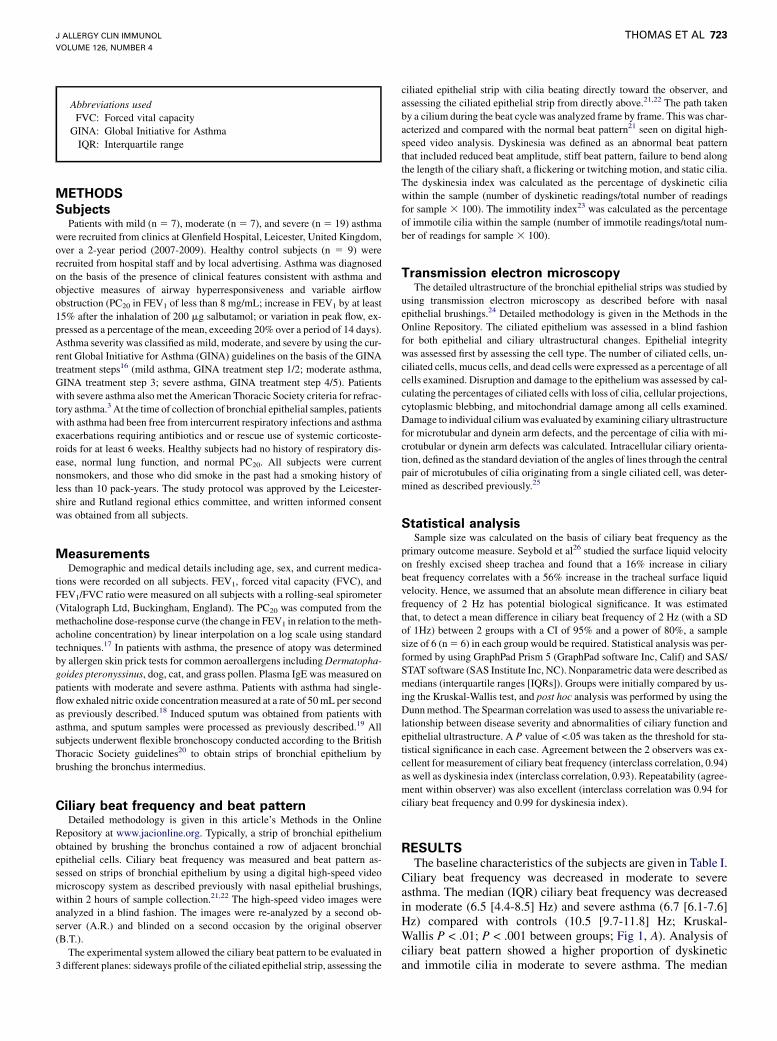
Bronchial epithelium obtained from 2 control subjects, 1 pa-tient with mild asthma, 1 patient with moderate asthma, and 7patients with severe asthma were insufficient for assessment bytransmission electron microscopy. Therefore, transmission elec-tron microscopy was done on samples obtained from 7 controlsubjects, 6 subjects with mild asthma, 6 subjects with moderateasthma, and 12 subjects with severe asthma. The median (IQR) noof epithelial cells per sample studied was 203.5 (162.8-263.3;range, 106-359). Results are summarized in Table II, and exampleelectron micrographs are shown in Figs 2 and 3. The ultrastructureof the ciliated epithelium was abnormal in severe asthma with areduction in ciliated cells, an increase in dead cells, and ciliarydisorientation compared with all of the other groups (P < .05);with ciliary depletion, microtubular defects, mitochondrial dam-age, cytoplasmic blebbing, and loss of epithelial integrity com-pared with patients with mild asthma and healthy controls alone(P < .01); and with an increase in mucus cells compared withhealthy controls only (P < .01). There were no significant differ-ences across groups for the proportion of unciliated cells or dyn-ein arm defects. All of the ultrastructural changes, except theproportion of unciliated cells, were related to disease severity(Table II).
DISCUSSIONThese data provide evidence that ciliary dysfunction and
ultrastructural abnormalities are closely related to asthma sever-ity, with dysfunction a feature of moderate to severe asthma andprofound ultrastructural abnormalities restricted to severe dis-ease. These striking changes, particularly in those subjects withsevere asthma, constitute a phenotype of profound secondaryciliary dysfunction and are likely to have important functionalconsequences.
To our knowledge, this is the first study that has assessed ciliaryfunction in terms of ciliary beat frequency and beat pattern in awell characterized group of patients with asthma of varyingseverity. We used bronchial epithelial brushings rather thanbiopsies because, in our experience, we have found that ciliaryfunction (ciliary beat frequency and beat pattern) studies aretechnically much easier to perform on epithelial brushings, andthis is an accepted method for studying ciliary function.25 Ourstudy is unique in that, in addition to showing a markedly reducedciliary beat frequency, we determined ciliary beat pattern, whichwas also found to be significantly abnormal in patients with mod-erate and severe asthma. This was made possible by the recent ad-vent of high-resolution digital high-speed video imaging21 thatallowed us to assess the precise beat pattern of cilia by viewingthe ciliary beat cycle frame by frame in different planes. Usingthis method, we have previously shown that cilia in certain condi-tions may have a normal beat frequency despite markedly abnor-mal beat pattern,24 and normal ciliary beat frequency does notnecessarily equate to normal ciliary function. The marked reduc-tion in ciliary beat frequency and abnormal beat pattern seen inasthmatic airway epithelium, particularly in those with severe dis-ease, is likely to be multifactorial in causation. Evidence fromprevious studies27,28 suggests that sputum from patients withasthma and products of inflammatory cells such as eosinophil ma-jor basic protein possess ciliostatic activity. Environmental fac-tors such as exposure to irritants or chronic infection may causesecondary ultrastructural defects of the cilia that may affect theciliary beat frequency, as indicated in a number of reports.29
The complex array of inflammatory mediators present in the asth-matic epithelium possesses diverse and opposing effects on cili-ary beat frequency.30 In addition, medications may influenceciliary beat frequency. Indeed, both short-acting and long-actingb2-agonists increase ciliary beat frequency.31 Importantly, inour study, there was no significant correlation between sputum eo-sinophilia and ciliary beat frequency in subjects with severeasthma, and a significantly reduced ciliary beat frequency was ob-served in patients with moderate and severe asthma, despite beingon treatment with long-acting bronchodilators. Therefore, thecause of the dysfunctional ciliated epithelium we observed in se-vere disease remains to be determined. It should also be noted that
FIG 2. Ciliated respiratory epithelium from a healthy control (A) showing normal mitochondria (arrow) and
a patient with severe asthma (B-F) showing abnormal mitochondria (B), projecting cell (C), dead cell (D),
cytoplasmic bleb (E), and disruption of tight junction with separation of cells (F). Internal scale bar 5 2 mm.
J ALLERGY CLIN IMMUNOL
OCTOBER 2010
726 THOMAS ET AL
although the immotile cilia may be abnormal in that they do notcontribute to mucus flow, they may have important mechanosen-sory and chemosensory functions.32 Of particular interest, we ob-served significantly increased intracellular ciliary disorientationin the respiratory epithelium of patients with severe asthma com-pared with healthy controls and patients with mild and moderateasthma. The magnitude of ciliary disorientation seen in patientswith severe asthma is comparable to that previously described
as a possible variant of primary ciliary dyskinesia.25,33 In primaryciliary dyskinesia, the movement of mucus by cilia is negligible.Although we did not study mucociliary clearance in our subjectsand despite the fact that factors such as quantitative and qualita-tive properties of mucus and airway surface liquid also are knownto affect mucociliary clearance, on the basis of availableevidence,26,34 the degree of ciliary dysfunction that we have dem-onstrated in this study is very likely to correlate with the degree of

FIG 3. Cross-sectional image of cilia originating from a single ciliated cell of a healthy control (A) showing
normal ciliary orientation and a patient with severe asthma (B) showing ciliary disorientation. a and b, Dis-
tribution of lines drawn parallel to the central pair of microtubules of cilia originating from a single ciliated
cell. Internal scale bar 5 100 nm.
J ALLERGY CLIN IMMUNOL
VOLUME 126, NUMBER 4
THOMAS ET AL 727
impairment in mucociliary clearance. If the ciliary abnormalitiesseen in the patients with severe asthma we studied were presentthroughout the airways, these patients would have to rely oncough as their main mechanism of mucus clearance. Markedly re-duced mucociliary clearance will predispose these patients to sec-ondary bacterial infection, and indeed, asthma has been shown tobe an independent risk factor for invasive pneumococcal dis-ease.35 The combination of impaired mucociliary clearance andinfection may lead to the development of bronchiectasis. Al-though asthma and bronchiectasis are two diseases with distinctpathophysiologic processes, their coexistence has been reportedin a number of patients, especially in those with severe persistentasthma,36 and despite several caveats, a number of retrospectiveand cross-sectional cohort studies suggest that asthma maya predisposing factor in the development of bronchiectasis.36,37
In this study, all patients with moderate and severe asthma hadhigh-resolution computed tomography of the chest performed,and 4 (21%) patients with severe asthma had evidence ofbronchiectasis.
The ciliary disorientation seen in patients with severe asthma islikely to be a consequence of the chronic inflammation charac-teristic of asthma or chronic infection38 that may occur in somepatients with asthma. Indeed, there is a suggestion that inflamma-tory mediators such as leukotriene D4 may impair ciliary orienta-tion.39 In addition, studies done on quail oviduct and Xenopus
larval skin suggest that ciliary polarity and orientation are influ-enced by the normal development of the apical cytoskeletonand normal tissue patterning.40,41 Thus, recent evidence of aber-rant epithelial repair seen in asthmatic epithelium6,7 could alsoprovide a plausible mechanism contributing to ciliary disorienta-tion seen in asthma.
In this study, we also quantified the ultrastructural abnormal-ities of the respiratory epithelium. Our findings of loss ofepithelial integrity and ultrastructural abnormalities are consis-tent with a number of previous studies showing that asthmaticairway epithelium is structurally abnormal. Evidence supportingepithelial structural damage in asthmatic airways comes largelyfrom earlier studies showing exfoliated epithelial cells in bron-choalveolar lavage samples and marked desquamation, epithelialshedding, and loss of integrity in histopathologic studies onautopsy or endobronchial biopsy specimens.42,43 Despite the con-troversy44 regarding the epithelial abnormalities seen in bronchialbiopsy studies, accumulating evidence reviewed recently stronglysuggests structural abnormalities of the asthmatic epithelium.6,7
Emerging evidence also points toward disruption of epithelialtight junctions and other junctional adhesion proteins in asthmaticairways that are responsible for maintaining the normal structuralintegrity.6,7
One limitation of our study is its cross-sectional design, andtherefore the within-subject repeatability of these changes and the

J ALLERGY CLIN IMMUNOL
OCTOBER 2010
728 THOMAS ET AL
response to interventions and exacerbations are uncertain andneed to be further studied. We restricted our study to epitheliumderived from central airways, and whether our findings extendinto the small airways is unknown. It will be of great interest toinvestigate the ciliary function and epithelial structure in theperipheral airways of patients with asthma. Whether our findingsare specific to asthma or indeed a feature of other airways diseasessuch as chronic obstructive pulmonary disease or chronic coughneeds to be investigated. In spite of these limitations, ourmeasures of ciliary function and epithelial cell morphology arehighly repeatable, and therefore the magnitude of ciliary dys-function in patients with moderate and severe asthma comparedwith that of controls is striking and is very likely to be clinicallyimportant. A further challenge for future studies is to determinewhether our findings reflect intrinsic abnormalities of the asth-matic airway epithelium, effects of chronic inflammation, chronicinfection, aberrant repair mechanisms, effect of medications, or acombination of these factors.
In summary, our study provides evidence for profound ciliarydysfunction in adults with moderate and severe refractory asthmain stable state, in addition to marked epithelial damage restricted tothose with severe disease. The potential direct consequence of thisphenotype of secondary ciliary dyskinesia is reduced mucociliaryclearance and therefore increased susceptibility to infection, andpotentially more prolonged exposure to inhaled particulate pollu-tants and aeroallergens, all of which have been implicated in thepathophysiology of asthma. These findings extend our currentparadigm of severe asthma and present a new therapeutic target.
We are indebted to the subjects who participated in this study, to Mrs
Beverley Hargadon for assistance in the clinical characterization of the
patients, to Dr K. Mutalithas for helping with patient recruitment, to Ms
Gwyneth Williams and Mr Norman Baker for technical assistance in the
laboratory, and to Richard Marshall and Ana de Sousa of GlaxoSmithKline.
Key messages
d Ciliary dysfunction and ultrastructural abnormalities inasthma are related to disease severity.
d Therapy directed at ciliary dysfunction may present anew therapeutic target for asthma.
REFERENCES
1. Partridge M. Examining the unmet need in adults with severe asthma. Eur Respir
Rev 2007;16:67-72.
2. Holgate ST, Polosa R. The mechanisms, diagnosis, and management of severe
asthma in adults. Lancet 2006;368:780-93.
3. Proceedings of the ATS Workshop on Refractory Asthma: current understanding,
recommendations, and unanswered questions. American Thoracic Society. Am J
Respir Crit Care Med 2000;162:2341-51.
4. Barnes PJ. Immunology of asthma and chronic obstructive pulmonary disease. Nat
Rev Immunol 2008;8:183-92.
5. Chanez P, Wenzel SE, Anderson GP, Anto JM, Bel EH, Boulet LP, et al. Severe
asthma in adults: what are the important questions? J Allergy Clin Immunol
2007;119:1337-48.
6. Holgate ST. The airway epithelium is central to the pathogenesis of asthma. Aller-
gol Int 2008;57:1-10.
7. Holgate ST. Epithelium dysfunction in asthma. J Allergy Clin Immunol 2007;120:
1233-44, quiz 1245-1246.
8. Wanner A, Salathe M, O’Riordan TG. Mucociliary clearance in the airways. Am J
Respir Crit Care Med 1996;154:1868-902.
9. Knowles MR, Boucher RC. Mucus clearance as a primary innate defense mecha-
nism for mammalian airways. J Clin Invest 2002;109:571-7.
10. Bateman JR, Pavia D, Sheahan NF, Agnew JE, Clarke SW. Impaired tracheobron-
chial clearance in patients with mild stable asthma. Thorax 1983;38:463-7.
11. Messina MS, O’Riordan TG, Smaldone GC. Changes in mucociliary clearance dur-
ing acute exacerbations of asthma. Am Rev Respir Dis 1991;143:993-7.
12. Ordonez CL, Khashayar R, Wong HH, Ferrando R, Wu R, Hyde DM, et al. Mild
and moderate asthma is associated with airway goblet cell hyperplasia and abnor-
malities in mucin gene expression. Am J Respir Crit Care Med 2001;163:517-23.
13. Rogers DF. Airway mucus hypersecretion in asthma: an undervalued pathology?
Curr Opin Pharmacol 2004;4:241-50.
14. Shimura S, Andoh Y, Haraguchi M, Shirato K. Continuity of airway goblet cells
and intraluminal mucus in the airways of patients with bronchial asthma. Eur
Respir J 1996;9:1395-401.
15. Danahay H, Atherton H, Jones G, Bridges RJ, Poll CT. Interleukin-13 induces a
hypersecretory ion transport phenotype in human bronchial epithelial cells. Am J
Physiol Lung Cell Mol Physiol 2002;282:L226-36.
16. Global strategy for asthma management and prevention: Global Initiative for
Asthma (GINA). 2008. Available at: http://www.ginasthma.org/Guidelineitem.
asp??l152&l251&intId51561. Accessed October 10, 2009.
17. Juniper E, Cockcroft D, Hargreave F. Histamine and methacholine inhalation tests:
tidal breathing method; laboratory procedure and standardization. Lund, Sweden:
Astra Draco; 1994.
18. Kharitonov S, Alving K, Barnes PJ. Exhaled and nasal nitric oxide measurements:
Recommendations. The European Respiratory Society Task Force. Eur Respir J
1997;10:1683-93.
19. Green RH, Brightling CE, McKenna S, Hargadon B, Parker D, Bradding P, et al.
Asthma exacerbations and sputum eosinophil counts: a randomised controlled trial.
Lancet 2002;360:1715-21.
20. British thoracic society guidelines on diagnostic flexible bronchoscopy. Thorax
2001;56(suppl 1):i1-21.
21. Chilvers MA, O’Callaghan C. Analysis of ciliary beat pattern and beat frequency
using digital high speed imaging: comparison with the photomultiplier and photo-
diode methods. Thorax 2000;55:314-7.
22. Chilvers MA, Rutman A, O’Callaghan C. Ciliary beat pattern is associated with
specific ultrastructural defects in primary ciliary dyskinesia. J Allergy Clin Immu-
nol 2003;112:518-24.
23. Greenstone M, Rutman A, Dewar A, Mackay I, Cole PJ. Primary ciliary dyskine-
sia: cytological and clinical features. Q J Med 1988;67:405-23.
24. Chilvers MA, McKean M, Rutman A, Myint BS, Silverman M, O’Callaghan C.
The effects of coronavirus on human nasal ciliated respiratory epithelium. Eur
Respir J 2001;18:965-70.
25. Bush A, Cole P, Hariri M, Mackay I, Phillips G, O’Callaghan C, et al. Pri-
mary ciliary dyskinesia: diagnosis and standards of care. Eur Respir J 1998;
12:982-8.
26. Seybold ZV, Mariassy AT, Stroh D, Kim CS, Gazeroglu H, Wanner A. Mucociliary
interaction in vitro: effects of physiological and inflammatory stimuli. J Appl Phys-
iol 1990;68:1421-6.
27. Dulfano MJ, Luk CK. Sputum and ciliary inhibition in asthma. Thorax 1982;37:
646-51.
28. Hastie AT, Loegering DA, Gleich GJ, Kueppers F. The effect of purified human
eosinophil major basic protein on mammalian ciliary activity. Am Rev Respir
Dis 1987;135:848-53.
29. Smallman LA. Microtubular abnormalities of cilia in acquired pulmonary diseases.
Lancet 1984;1:965-6.
30. Del Donno M, Bittesnich D, Chetta A, Olivieri D, Lopez-Vidriero MT. The effect
of inflammation on mucociliary clearance in asthma: an overview. Chest 2000;118:
1142-9.
31. Hasani A, Toms N, O’Connor J, Dilworth JP, Agnew JE. Effect of salmeterol xi-
nafoate on lung mucociliary clearance in patients with asthma. Respir Med
2003;97:667-71.
32. Bloodgood RA. Sensory reception is an attribute of both primary cilia and motile
cilia. J Cell Sci 2010;123:505-9.
33. Rutman A, Cullinan P, Woodhead M, Cole PJ, Wilson R. Ciliary disorientation: a
possible variant of primary ciliary dyskinesia. Thorax 1993;48:770-1.
34. Yates GT, Wu TY, Johnson RE, Cheung ATW, Frand CL. A theoretical and
experimental study of tracheal muco-ciliary transport. Biorheology 1980;17:
151-62.
35. Talbot TR, Hartert TV, Mitchel E, Halasa NB, Arbogast PG, Poehling KA, et al.
Asthma as a risk factor for invasive pneumococcal disease. N Engl J Med 2005;
352:2082-90.
36. Gupta S, Siddiqui S, Haldar P, Vimal Raj J, Entwisle JJ, Wardlaw AJ, et al. Qual-
itative analysis of high-resolution CT scans in severe asthma. Chest 2009;136:
1521-8.
37. Ip MS, So SY, Lam WK, Yam L, Liong E. High prevalence of asthma in patients
with bronchiectasis in Hong Kong. Eur Respir J 1992;5:418-23.

J ALLERGY CLIN IMMUNOL
VOLUME 126, NUMBER 4
THOMAS ET AL 729
38. Kraft M, Cassell GH, Pak J, Martin RJ. Mycoplasma pneumoniae and Chlamydia
pneumoniae in asthma: effect of clarithromycin. Chest 2002;121:1782-8.
39. Joki S, Saano V, Koskela T, Toskala E, Bray MA, Nuutinen J. Effect of leukotriene
D4 on ciliary activity in human, guinea-pig and rat respiratory mucosa. Pulm Phar-
macol 1996;9:231-8.
40. Boisvieux-Ulrich E, Sandoz D. Determination of ciliary polarity precedes differen-
tiation in the epithelial cells of quail oviduct. Biol Cell 1991;72:3-14.
41. Mitchell B, Jacobs R, Li J, Chien S, Kintner C. A positive feedback mechanism
governs the polarity and motion of motile cilia. Nature 2007;447:97-101.
42. Hogg JC. Pathology of asthma. J Allergy Clin Immunol 1993;92:1-5.
43. Montefort S, Roche WR, Holgate ST. Bronchial epithelial shedding in asthmatics
and non-asthmatics. Respir Med 1993;87(suppl B):9-11.
44. Ordonez C, Ferrando R, Hyde DM, Wong HH, Fahy JV. Epithelial desquamation in
asthma: artifact or pathology? Am J Respir Crit Care Med 2000;162:2324-9.

REFERENCES
E1. Chilvers MA, O’Callaghan C. Analysis of ciliary beat pattern and beat frequency
using digital high speed imaging: comparison with the photomultiplier and pho-
todiode methods. Thorax 2000;55:314-7.
E2. Chilvers MA, Rutman A, O’Callaghan C. Ciliary beat pattern is associated with
specific ultrastructural defects in primary ciliary dyskinesia. J Allergy Clin
Immunol 2003;112:518-24.
E3. Greenstone M, Rutman A, Dewar A, Mackay I, Cole PJ. Primary ciliary dyski-
nesia: cytological and clinical features. Q J Med 1988;67:405-23.
E4. Chilvers MA, McKean M, Rutman A, Myint BS, Silverman M, O’Callaghan C.
The effects of corona virus on human nasal ciliated respiratory epithelium. Eur
Respir J 2001;18:965-70.
E5. Tsang KW, Rutman A, Tanaka E, Lund V, Dewar A, Cole PJ, Wilson R. Interac-
tion of Pseudomonas aeruginosa with human respiratory mucosa in vitro. Eur
Respir J 1994;7:1746-53.
E6. Bush A, Cole P, Hariri M, et al. Primary ciliary dyskinesia: diagnosis and stan-
dards of care. Eur Respir J 1998;12:982-8.
J ALLERGY CLIN IMMUNOL
OCTOBER 2010
729.e1 THOMAS ET AL
METHODS
Ciliary beat frequency and beat patternTypically, a strip of bronchial epithelium obtained by brushing the
bronchus contains a row of adjacent bronchial epithelial cells. Ciliary beat
frequency (CBF) was measured and beat pattern assessed on strips of
bronchial epithelium using a digital high-speed video system, as described
previously with nasal epithelial brushings, within 2 hours of sample
collection.E1,E2 The high-speed video images were analyzed in a blind fashion.
The images were re-analyzed by a second observer (A.R.) and blind on a sec-
ond occasion by the original observer (B.T.).
The sample was placed in Medium 199 (25 mM hydroxyethyl piperazine
ethane sulfonic acid, Earles salt, and L-glutamine [pH 7.3]; Gibco, Leicester,
United Kingdom) that contained antibiotic solution (streptomycin 50 mg�mL–
1 and penicillin 50 mg�mL–1) and analyzed within 2 hours after collection. The
sample was suspended in a chamber created by the separation of a cover slip
and a glass slide by 2 adjacent cover slips. The slide was placed on a heated
stage (378C) of a Leitz Diaplan microscope mounted on an antivibration table
(Wentworth Laboratories Ltd, Sandy, United Kingdom). Ciliated epithelial
strips greater than 50 mm in length and devoid of mucus were observed at
378C by using a 3100 interference contrast lens. They were recorded by using
a digital high-speed video camera (Kodak Ektapro Motion Analyzer; Eastman
Kodak, San Diego, Calif) at a frame rate of 400 frames�s–1. Video sequences
could be recorded and played back at reduced frame rates or frame by frame.
CBF was determined directly from ciliated epithelial strips viewed in a side-
ways profile. Groups of beating cilia were identified, and the number of frames
required to complete 10 beat cycles was recorded. This was converted to CBF
by a simple calculation (CBF 5 [400/number frames for 10 beats] 3 10).E1
The ciliated epithelial strip (>_50 mm) projected onto a high-resolution monitor
was divided into 10 adjacent areas measuring 5 mm each. One reading of CBF
was taken from each area to obtain a total of 10 measurements of CBF along
each ciliated epithelial strip. At least 7 epithelial strips, up to a maximum of 10
strips, were analyzed per subject.
The experimental system allowed the ciliary beat pattern to be evaluated in
3 different planes: sideways profile of the ciliated epithelial strip, assessing the
ciliated epithelial strip with cilia beating directly toward the observer, and
assessing the ciliated epithelial strip from directly above.E1,E2 The path taken
by a cilium during the beat cycle was analyzed frame by frame. This was char-
acterized and compared with the normal beat patternE1 seen on digital high-
speed video analysis. Dyskinesia was defined as an abnormal beat pattern
that included reduced beat amplitude, stiff beat pattern, failure to bend along
the length of the ciliary shaft, a flickering or twitching motion, and static cilia.
The ciliated epithelial strip (>_50 mm) projected onto a high-resolution monitor
was divided into 10 adjacent areas measuring 5 mm each, and the beat pattern
of each 5-mm area was assessed to obtain a total of 10 measurements of beat
pattern per epithelial strip. The dyskinesia index was calculated as the percent-
age of dyskinetic cilia within the sample (number of dyskinetic readings/total
number of readings for sample 3 100). The immotility indexE3 was calculated
as the percentage of immotile cilia within the sample (number of immotile
readings/total number of readings for sample 3 100).
Transmission electron microscopyTransmission electron microscopy was performed as described.E4 Briefly,
on the day of collection, bronchial epithelial samples were fixed in 2.5% glu-
taraldehyde in Sorenson phosphate buffer. After 48 hours, the sample was
postfixed in 1% osmium tetroxide. After rinsing in distilled water, the cells
were embedded in a drop of 2% liquid agar at 458C and allowed to solidify.
This bound the cells together during dehydration and ensured that all the strips
of epithelium and cilia were randomly oriented. This was processed through to
resin by standard transmission electron microscopy techniques. Ultrathin sec-
tions were cut at 70 nm. These were collected on 200 mesh thin-bar copper
grids and stained in 1% uranyl acetate and counterstained in Reynolds lead
phosphate. The sections were then examined by transmission electron micros-
copy. The grids were analyzed by a grid square search pattern so that all the
cells in the sample were analyzed but seen only once. The median (IQR)
number of epithelial cells per sample studied was 203.5 (162.8-263.3; range,
106-359). All the cilia that were, by random chance, captured in cross-section
adequate for dynein arms to be visualized were assessed.
The ciliated epithelium was assessed, in a blind fashion, for both epithelial
and ciliary ultrastructural changes. Epithelial integrity was assessed first by
assessing the cell type. The numbers of ciliated cells, unciliated cells, mucus
cells, and dead cells were expressed as a percentage of all cells examined.
Disruption and damage to the tissue was quantified by using the criteria for
epithelial integrity described by Tsang et al.E5 Percentages of ciliated cells
with loss of cilia, cellular projections (extrusion of cells from the epithelial
edge), cytoplasmic blebbing, and mitochondrial damage (manifested as swell-
ing and disruption of mitochondrial cristae) among all cells examined were
also calculated. Damage to individual cilia was evaluated by examining ciliary
ultrastructure for microtubular and dynein arm defects, and the percentage of
cilia with microtubular or dynein arm defects was calculated. Intracellular cil-
iary orientation, defined as the SD of the angles of lines through the central pair
of microtubules of cilia originating from a single ciliated cell, was determined
as described previously.E6
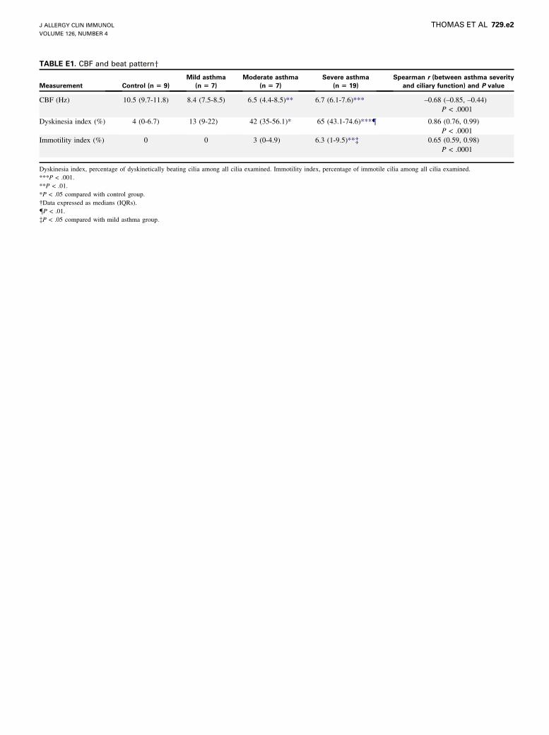
TABLE E1. CBF and beat patterny
Measurement Control (n 5 9)
Mild asthma
(n 5 7)
Moderate asthma
(n 5 7)
Severe asthma
(n 5 19)
Spearman r (between asthma severity
and ciliary function) and P value
CBF (Hz) 10.5 (9.7-11.8) 8.4 (7.5-8.5) 6.5 (4.4-8.5)** 6.7 (6.1-7.6)*** –0.68 (–0.85, –0.44)
P < .0001
Dyskinesia index (%) 4 (0-6.7) 13 (9-22) 42 (35-56.1)* 65 (43.1-74.6)***{ 0.86 (0.76, 0.99)
P < .0001
Immotility index (%) 0 0 3 (0-4.9) 6.3 (1-9.5)**� 0.65 (0.59, 0.98)
P < .0001
Dyskinesia index, percentage of dyskinetically beating cilia among all cilia examined. Immotility index, percentage of immotile cilia among all cilia examined.
***P < .001.
**P < .01.
*P < .05 compared with control group.
�Data expressed as medians (IQRs).
{P < .01.
�P < .05 compared with mild asthma group.
J ALLERGY CLIN IMMUNOL
VOLUME 126, NUMBER 4
THOMAS ET AL 729.e2
Copyright © 2022 FDOKUMEN