A recurrent 15q13.3 microdeletion syndrome associated with mental retardation and seizures

REVIEW Open Access
“Idiopathic” mental retardation and newchromosomal abnormalitiesCinzia Galasso, Adriana Lo-Castro*, Nadia El-Malhany, Paolo Curatolo
Abstract
Mental retardation is a heterogeneous condition, affecting 1-3% of general population. In the last few years, severalemerging clinical entities have been described, due to the advent of newest genetic techniques, such as arrayComparative Genomic Hybridization. The detection of cryptic microdeletion/microduplication abnormalities hasallowed genotype-phenotype correlations, delineating recognizable syndromic conditions that are herein reviewed.With the aim to provide to Paediatricians a combined clinical and genetic approach to the child with cognitiveimpairment, a practical diagnostic algorithm is also illustrated. The use of microarray platforms has further reducedthe percentage of “idiopathic” forms of mental retardation, previously accounted for about half of total cases. Wediscussed the putative pathways at the basis of remaining “pure idiopathic” forms of mental retardation, highlight-ing possible environmental and epigenetic mechanisms as causes of altered cognition.
IntroductionMental retardation (MR) is a variable, heterogeneousmanifestation of central nervous system dysfunctions,occurring in 1-3% of general population [1]. MR repre-sents one of the most frequently diagnosed disablingcondition in our society, and a lifelong disability charac-terized by impairment of cognitive and adaptive skills.The aetiology is very heterogeneous and, unfortu-
nately, in about than one-half of cases the cause of MRis still elusive [2]. Anything that damages and interfereswith the growth and maturation of the brain can lead toMR, and this might happen before, during or after thebirth of the child (complications of pregnancy/birth,toxics, malnutrition, trauma, infections, understimula-tion). Moreover, genetically determined MR aetiology(comprising chromosomal aberrations, single-gene disor-ders, and other genetic conditions) account by itself for17 to 41% of cases, depending of the different techni-ques of analysis [2].Several syndromes (such as Down, Rett syndrome, and
other well known conditions) should be easily suspectedbecause of their association to specific dysmorphisms,behavioural peculiarities, and multiple congenitalabnormalities. However, a consistent percentage of
children with genetic MR do not present a recognizablephenotype striking of a well-recognizable syndrome.With the advent of novel genetic techniques, several
new cryptic chromosomal aberrations have been discov-ered in last few years [3,4], and a consistent number ofMR cases, previously considered “idiopathic” forms, arenow classified as syndromic conditions with clinicalrecognizable phenotypes [5]. Microarrays techniques(such as array-Comparative Genomic Hybridization,array-CGH) revealed submicroscopic aberrations in5-17% of MR patients with normal results from priorconventional cytogenetic testing [6], and higher-densityplatforms (such as Single-Nucleotide Polymorphismarray, SNP array) provided to increase diagnosis inabout 6% of cases evaluated by lower-density oligonu-cleotide arrays [2].Determining a specific etiologic diagnosis is central to
understand the nature of the problem, providinganswers to questions regarding prognosis, recurrencerisks, directing specific therapies, and achieving mean-ingful inclusion of individuals with disability intosociety.
Search strategy and selection criteriaInformation in this review is mainly based on peer-reviewed medical publications of syndromic conditionsfrom 2005 to 2010 (PubMed). Selection criteria are thenovelty and importance of studies, and their relevance
* Correspondence: [email protected] of Neuroscience, Paediatric Neurology Unit, “Tor Vergata”University of Rome, Italy
Galasso et al. Italian Journal of Pediatrics 2010, 36:17http://www.ijponline.net/content/36/1/17 ITALIAN JOURNAL
OF PEDIATRICS
© 2010 Galasso et al; licensee BioMed Central Ltd. This is an Open Access article distributed under the terms of the Creative CommonsAttribution License (http://creativecommons.org/licenses/by/2.0), which permits unrestricted use, distribution, and reproduction inany medium, provided the original work is properly cited.
to Paediatricians. Search terms included “idiopathicmental retardation”, “mental retardation”, “cryptic chro-mosomal abnormalities”, and “array-CGH”. Only articlespublished in English were reviewed. All articles wereread by the authors and references were reviewed toidentify any additional relevant studies.
Clinical ApproachThe clinical approach of a child with MR is a key momentto provide a definitive diagnosis, and requires someexhaustive and comprehensive evaluations of the patient.First of all, a three-generation pedigree should be
done, and a detailed pre-, peri- and postnatal history ismandatory. A dysmorphic child may be at risk from thestress of birth, and later delay may be erroneouslyattributed to birth injury [7]. A careful developmentalhistory, with emphasis on milestones, formal assess-ments and behavior, is also required. Medical recordsshould be sought or requested to validate any diagnosisof malformations. An accurate EEG study and/or brainMRI are sometimes sufficient to suspect several well-known and relatively common disorders (such as Rettsyndrome, Angelman syndrome, neurocutaneous syn-dromes, etc.) [8,9]. The degree of MR is an importantindicator: the so called “chromosomal” phenotype,which is well known to accompany larger aberrations, isfrequently characterized by moderate-severe MR asso-ciated to one or more of major signs, including congeni-tal malformations. The behavioral phenotype is alsodistinctive for several well-known syndromic conditions,such as Williams syndrome, Angelman syndrome, Pra-der-Willi syndrome, and so on [10].Finally, the physical examination of the child is crucial
for a “gestaltic” diagnosis: sometimes the syndromic con-dition can be instantaneously suspected by recognition of“handles”, based on past clinical experience. Hovewer,phenotypic expression among patients with well-recog-nized microdeletion or microduplication syndromes mayvary on the basis of different sizes of genomic alterations,and of individual differences in the rest of the genome.Unfortunately, in many cases the MR is the unique andunspecific sign present in the patient, with lack of majorhallmarks. When present, minor anomalies of the face(such as hypo-hypertelorism, unusual ear conformation,multiple hair whorls, etc.), hands, genitalia, and skinshould be noted and supplemented by objective measure-ments. Abnormalities in head size, growth parameters,and neurologic signs should be carefully investigated.The phenotype can also vary during the time, it
should be useful to collect photos and/or videos ofpatients at different ages, also because the amount ofcontrols in our experience often affects the probabilityto define the etiology.
Genetic ApproachGenetic abnormalities are the most common identifiablecause of unexplained MR [11], but conventional karyo-typing is unable to detect imbalances smaller than about3-5 Mb [12]. Smaller chromosomal abnormalities can beidentified with fluorescent in situ hybridization (FISH)or multiplex ligation-dependent probe amplification(MLPA) techniques, confirming a clinical suspicion ofwell-known microdeletion/microduplication syndromes(i.e. Williams syndrome, Velocardiofacial/DiGeorge syn-drome, etc.) or analysing subtelomeric regions of allchromosomes. The combined analysis of karyotype andsubtelomeric regions, using FISH or other moleculartechniques, have allowed the detection of chromosomeabnormalities in about 5-10% of these patients [11,12].The newer chromosome microarray or comparativegenomic hybridization technique (array-CGH) is an effi-cient manner to approach a case of MR. It does notrequire an expert clinician to suspect a specific diagno-sis, and may cover the entire genome or targets knownpathologic loci in an unique test, identifying deletionsand/or duplications with a higher degree of sensitivity.This new technique has revealed submicroscopic chro-mosome aberrations in MR patients with normal resultsfrom prior cytogenetic analyses with detection rates as5-20% [6,13]. However, array-CGH is incapable ofdetecting balanced rearrangements of chromosomalmaterial (including reciprocal translocations and inver-sions), which are expected to occur in about 0.75% of allMR patients [14]. Moreover, the interpretation of micro-array data in MR is complicated by the discovery ofareas of DNA segment longer than 1 kb, with a variablecopy number compared with a normal reference gen-ome, called Copy Number Variations (CNVs) [15].CNVs are associated to a pathological phenotype whenone or more dosage-sensitive genes inside the rear-ranged region are altered. However, CNVs are not con-sidered pathologic in all cases, because they appear tobe conserved across primate species and may be respon-sible of individual diversity and human evolution. In asingle individual, it is possible to detect even > 1000non-pathological common CNVs [16], needing the com-parison with unaffected control cohorts and parentaltests [17]. The detection of a relatively large, rare, denovo CNV in an affected patient is strongly indicative ofpathological significance, and is present in about 10% ofcases of MR with normal chromosome analysis [1,18].CNVs should be considered causative of the conditionwhen: 1) CNVs overlap with regions known to causewell delineated MR syndromes; 2) CNVs include the cri-tical region of a syndrome or causative genes; 3) thephenotype of the patient is consistent with the syn-drome’s features [2].
Galasso et al. Italian Journal of Pediatrics 2010, 36:17http://www.ijponline.net/content/36/1/17
Page 2 of 8

Combined Diagnostic AlgorithmA possible diagnostic algorithm which can be useful inthe evaluation of a child with unexplained MR and sus-pected genetic condition [8,9] is illustrated in the Figure1. After a detailed anamnesis assessed by three-genera-tions pedigree analysis, a comprehensive cognitive, beha-vioral and physical examination (clinical andinstrumental) is mandatory. Depending on the suspectedor not-suspected diagnosis, the work-up of a patient canfollow two different pathways, guiding to decisionsregarding laboratory testing and imaging studies. On thebasis of the presence of dysmorphic signs and/or majorabnormalities (such as multiple congenital defects),high-resolution karyotype, fragile-X DNA analysis, tar-geted FISH/MLPA, or metabolic tests should be consid-ered. EEG and/or MRI are also useful. Finally, array-CGH is indicated when above-mentioned tests arenegative.When the child has an apparently normal phenotype,
after high-resolution karyotype, fragile-X moleculartests, EEG, and MRI studies, array-CGH analysis should
be directly performed to exclude a cryptic chromosomalaberration.
Chromosomal AbnormalitiesRecent developments in array technology have stronglychanged the genetic approach to MR, combining thewhole-genome analysis of karyotyping technology andthe targeted high-resolution of FISH test. Genomicmicroarrays have a resolution 10-10000 times higherthan that of conventional karyotyping, identifying rare,de novo, submicroscopic interstitial imbalances or CNVsin about 5-20% of cases of idiopathic MR and multiplecongenital abnormalities, depending on the clinicalselection of patients [1,12]. The increased identificationof novel microdeletion/microduplication syndromes isbased on an accurate genotype-phenotype correlation,characterized by the association of similar chromosomalaberrations and overlapped clinical presentationsbetween affected patients.The ability to recognize pathological gestalts and/or
behaviors has already led to a significant improvement
Figure 1 Diagnostic algorithm proposed for unexplained MR cases of suspected genetic origin. Array-CGH = array-Comparative GenomeHybridization; FISH: Fluorescent In Situ Hybridization; Fra-X: Fragile × syndrome molecular analysis; HRK: high-resolution karyotype; microdel/dup:microdeletion/microduplication; MLPA: Multiplex Ligation-dependent Probe Amplification.
Galasso et al. Italian Journal of Pediatrics 2010, 36:17http://www.ijponline.net/content/36/1/17
Page 3 of 8

in the diagnostic yield in patients with MR. Severalexample of relatively common, novel well-delineatedsyndromes are discussed in this review. Salient phenoty-pical traits of each syndrome are also synthesized inAdditional file 1.
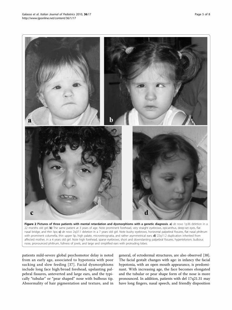
1p36 microdeletion syndromeMonosomy 1p36 is a well-described contiguous genessyndrome, considered as the most common terminaldeletion observed in humans, accounting for 0.5-1.2% ofidiopathic MR [19,20]. Dysmorphisms are very remark-able and distinctive of this condition: microcephaly,large and late closing anterior fontanel, tower skull, pro-minent forehead, straight eyebrows, deep-set eyes, flatnasal bridge with midface hypoplasia, abnormal ears,brachydactyly/camptodactyly, and short feet (Figure 2a,b). Severe hypotonia, seizures, oropharyngeal dysphagia,and heart defects are also common [20]. MR of anydegree, mostly moderate to severe, is present in all indi-viduals, associated to a severe speech impairment andpoor coordinated movements control [19]. SubtelomericFISH analysis, targeted FISH analysis of chromosome 1and/or array-CGH are needed to detect the 1p36 micro-deletion. This syndrome should be considered in ayoung child with above-mentioned dysmorphisms, psy-chomotor delay and hypotonia, in particular when thelanguage is poor or absent, and self-injuring behaviors,stereotypies, and hyperactivity are present.
2q23.1 microdeletion syndromeThis new syndrome has been identified by a-CGH inpatients with severe MR and severe speech impairment,associated with microcephaly, coarse face, short stature,and epilepsy [21]. Frequently, the phenotype of syn-drome includes stereotypic behaviors, altered sleep pat-tern and a broad-based gait, leading to the clinicalimpression of Angelman, Rett or Smith-Magenis syn-dromes [22]. Haploinsufficiency of MBD5 or EPC2genes, included in the deleted genomic region, seems tobe responsible of the typical phenotype [22,23].
2q37 deletion syndromeDel(2q37) syndrome is now a well recognized disease,characterized by facial dysmorphic features (Figure 2c),developmental delay, hypotonia, epilepsy in 25% ofcases, and major anomalies in about 30% [24,25].Psychiatric conditions are frequently associated with
del(2q37). Autism spectrum disorders is present in24-35% of del(2q37) cases, but also severe speech delay,stereotypic movements, aggressive behavior, attention-deficit/hyperactivity disorder (ADHD), and obsessive-compulsive disorder are commonly observed [24]. In achild with MR and these neuropsychiatric disorders, thepresence of facial dysmorphic traits and congenital
defects, variably associated with short stature, obesity,brachydactyly, eczema, and hypotonia, should be consid-ered highly suggestive of del(2q37). High resolution kar-yotype, FISH/MLPA or aCGH analyses are useful fordiagnosis [25].
7q11.3 microduplication syndromeWilliams-Beuren syndrome (WBS), caused by deletionof a 1.4-1.5 Mb region located at 7q11.23, is among themost well-characterized microdeletion syndrome, butthe reciprocal microduplication of this genomic regionis less well described. The clinical phenotype of 7q11.23microduplication seems to vary among patients, rangingfrom mild to severe MR [26]. The neurobehavioral phe-notype is the opposite of WBS: instead of fluent expres-sive language, dup 7q11.23 patients show severe speechdelay and only mildly impaired visuospatial skills [27].Mild facial dysmorphisms (short philtrum, thin lips, andstraight eyebrows), an increased incidence of heartdefects, diaphragmatic hernia, cryptorchidism, and non-specific MRI brain abnormalities should orientate theclinician to make diagnosis [28].
15q13.3 microdeletion syndromeThe overall incidence of this aberration is about 0.3% ofpatients with “idiopathic” MR, considering it comparableto William and Angelman syndromes [29]. MR rangesfrom mild to moderate, and 15q13.3 deletion has alsobeen recently associated to a higher predisposition toautism spectrum disorders, schizophrenia, other psychia-tric disorders, and idiopathic generalized epilepsy orEEG abnormalities [30]. The syndrome has a highlyvariable intra- and inter-familial phenotype, with mildfacial dysmorphisms, including hypertelorism, upslantingpalpebral fissures, prominent philtrum with full evertedlips, and short and/or curved fifth finger and shortfourth metacarpals [31].
16p11.2 microdeletion syndromeMicrodeletion at 16p11.2 has recently been associated withautism in two different studies [32,33], but this syndromeis characterized by a variable phenotype, ranging fromnormal intelligence and mild dysmorphisms to severe cog-nitive impairment and minor/major congenital abnormal-ities. Facial features are characterized by flat andhypotonic facies, deep-set eyes, low-set and posteriorlyrotated ears, and thin upper lip. Frequent ear infections,orofacial clefting, heart defects, and minor hand/footanomalies have been described [34,35]. Expressive lan-guage disorder, dyslexia, and ADHD are also frequent [36].
17q21.31 deletion syndromeThis novel syndrome seems to have a prevalence of 1 in16 000 individuals, and to be underestimated. In all
Galasso et al. Italian Journal of Pediatrics 2010, 36:17http://www.ijponline.net/content/36/1/17
Page 4 of 8
patients mild-severe global psychomotor delay is notedfrom an early age, associated to hypotonia with poorsucking and slow feeding [37]. Facial dysmorphismsinclude long face high/broad forehead, upslanting pal-pebral fissures, anteverted and large ears, and the typi-cally “tubular” or “pear shaped” nose with bulbous tip.Abnormality of hair pigmentation and texture, and in
general, of ectodermal structures, are also observed [38].The facial gestalt changes with age: in infancy the facialhypotonia, with an open mouth appearance, is predomi-nant. With increasing age, the face becomes elongatedand the tubular or pear shape form of the nose is morepronounced. In addition, patients with del 17q21.31 mayhave long fingers, nasal speech, and friendly disposition
Figure 2 Pictures of three patients with mental retardation and dysmorphisms with a genetic diagnosis. a) de novo 1p36 deletion in a22 months old girl; b) The same patient at 3 years of age. Note prominent forehead, very straight eyebrows, epicanthus, deep-set eyes, flatnasal bridge, and thin lips; c) de novo 2q37.1 deletion in a 7 years old girl. Note bushy eyebrows, horizontal palpebral fissures, flat nasal philtrumwith prominent columella, thin upper lip, high palate, microretrognatia, and rather asymmetrical ears; d) 22q11.2 duplication inherited fromaffected mother, in a 4 years old girl. Note high forehead, sparse eyebrows, short and downslanting palpebral fissures, hypertelorism, bulbousnose, pronounced philtrum, fullness of jowls, and large and simplified ears with protruding lobes.
Galasso et al. Italian Journal of Pediatrics 2010, 36:17http://www.ijponline.net/content/36/1/17
Page 5 of 8

[37]. Other common features are cryptorchidism, epi-lepsy, hypermetropia, pectus excavatum, congenitalheart defects, kidney and urologic anomalies, dislocationof the hips, and spinal deformities [38].
22q11.2 microduplicationMicroduplication of 22q11.2 (dup22q11.2) has recentlyemerged as a new chromosomal syndrome (Figure 2c).Dup22q11.2 represents the reciprocal duplication ofregion deleted in Di George/Velocardiofacial syndrome(DG/VCFS), due to misalignment of low-copy repeats inthis band. Although this syndrome shares features withDG/VCFS (heart defects, velopharyngeal insufficiency withor without cleft palate, hypernasal speech, and urogenitalabnormalities), a clear genotype-phenotype correlation isnot yet established [39]. Individuals with dup22q11.2 shownormal intelligence or cognitive impairment of anydegrees. Neurological and psychiatric disorders, includingspeech delay, learning disabilities, motor impairment,aggressiveness, anxiety, depression, autisms, ADHD, oppo-sitional-defiant disorder, obsessive traits, and social inter-action problems are frequently associated [39,40]. Themicroduplication is only detectable by interphase FISH,MLPA or a-CGH analyses, and the genetic test is recom-mended in children with central hypotonia, severe speechdelay, learning disabilities/cognitive impairment, or psy-chiatric disorders including autism [40].
ConclusionsIt is generally assumed that severe forms of MR arethought to be due to larger chromosomal abnormalitiesor defects in single genes, in the majority of casesdetectable with specific genetic tests. Paediatriciansshould be alerted by the presence of MR of unexplainedorigin associated with altered auxological parameters,multiple congenital defects, neurological and psychiatricsigns, and/or minor dysmorphisms. However, many chil-dren who have MR and dysmorphisms often do nothave major malformations, simply having an appearancethat is unusual compared with the general population,and out of keeping with that of unaffected close rela-tives. In particular, mild forms of MR often lack sugges-tive clinical “handles”, resulting from the interaction ofmultiple genes and non-genetic factors [41]. Despite theintroduction of high-resolution platforms has facilitatedthe identification of emerging microdeletion/microdupli-cation syndromes, in mild forms of MR making an etio-logical diagnosis is still very difficult. The quote of“idiopathic” forms of MR account for about half of totalcases, probably due to combination of multigenic andenvironmental causes. The developing brain is moresusceptible to insults by toxic agents than adults,because during the prenatal life several complex stagesof organization and maturation must develop in a tightly
controlled time. Moreover, the blood-brain barrier is notcompletely formed until the sixth month of intrauterinelife, leading the developing brain exposed to toxins [42].In addition, a mutation in a single gene (responsible afor genetic syndrome) may lead to epigenetic dysregula-tion [43], influencing transcription and/or silencingother genes. Epigenetic mechanisms play a central rolein higher-order brain functions, influencing the capacityto modify, reorganize and remodel synaptic plasticityand networks during learning and memory formation,and in response to injury. Several well-known syn-dromes are caused by disrupted epigenetic mechanisms,such as Rett syndrome, and fragile × syndrome [44]. Inthese and others genetic conditions, as well as in envir-onmental MR -associated disorders (e.g. fetal alcoholspectrum disorders or lead exposure) reduced dendriticcomplexity, and significant differences in dendritic spinenumbers and morphological spine types have beenobserved [43,44]. However, epigenetic mechanisms aredynamic and potentially reversible, providing new phar-macological approaches to treat neurodevelopmentaldisorders. DNA-demethylating drugs and HDAC inhibi-tors are two promising examples of targeted epigeneticdrugs [43]. The implementation of the so called “nextgeneration sequencing” technologies (that allow the ana-lysis of whole-genomes, transcriptomes and interac-tomes) could lead to detect single base mutations andstructural variations, further broadening the possibilityof diagnosis in “idiopathic” cases of MR. Understandingthe pathological pathways underlying unexplained formsof MR represent a future challenge to increase both pre-vention and possible therapies.
ConsentWritten consents for publication of patients’ pictureswere obtained from their parents.
Additional file 1: Schematic overview of major features of novelsyndromic conditions discussed in this paper. For a more detaileddescription of each syndrome, refer to the text. (+) present feature; (-)absent feature; (+/-) inconstantly present/rare feature. ADHD: Attention-deficit/hyperactivity disorder; DD: developmental delay; IQ: intelligentquotient; MR: mental retardation.Click here for file[ http://www.biomedcentral.com/content/supplementary/1824-7288-36-17-S1.DOC ]
Authors’ contributionsALC (Medical Doctor) drew the first draft with the assistance andcontribution of NEM (Medical Doctor), and reviewed relevant articles on theliterature under the supervision of CG (Associated Professor of theDepartment of Pediatric Neuroscience Unit) on clinical and neurogeneticalaspects. PC (Director of the Department of Pediatric Neuroscience Unit)proposed and designed the study, and revised the final draft.All authors contributed to the intellectual contents and approved the finalversion.
Galasso et al. Italian Journal of Pediatrics 2010, 36:17http://www.ijponline.net/content/36/1/17
Page 6 of 8

Competing interestsThe authors declare that they have no competing interests.
Received: 28 January 2010 Accepted: 14 February 2010Published: 14 February 2010
References1. Vissers LE, de Vries BB, Veltman JA: Genomic microarrays in mental
retardation: from CNV to gene, from research to diagnosis. J Med Genet2009.
2. Bernardini L, Alesi V, Loddo S, Novelli A, Bottillo I, Battaglia A, Digilio MC,Zampino G, Ertel A, Fortina P, Surrey S, Dallapiccola B: High-resolution SNParrays in mental retardation diagnostics: how much do we gain? Eur JHum Genet 2010, 18:178-185.
3. Shevell MI, Bejjani BA, Srour M, Rorem EA, Hall N, Shaffer LG: Arraycomparative genomic hybridization in global developmental delay. Am JMed Genet 2008, 147B:1101-1108.
4. Shaffer LG, Bejjani BA, Torchia B, Kirkpatrick S, Coppinger J, Ballif BC: Theidentification of microdeletion syndromes and other chromosomeabnormalities: cytogenetic methods of the past, new technologies forthe future. Am J Med Genet 2007, 145C:335-345.
5. Li MM, Andersson HC: Clinical Application of Microarray-Based MolecularCytogenetics: An Emerging New Era of Genomic Medicine. J Pediatr2009, 155:311-317.
6. Liang JS, Shimojima K, Yamamoto T: Application of Array-basedComparative Genome Hybridization in Children with DevelopmentalDelay or Mental Retardation. Pediatr Neonatol 2008, 49:213-217.
7. Hall DMB: Birth asphyxia and cerebral palsy. BMJ 1989, 299:279-282.8. Battaglia A, Bianchini E, Carey JC: Diagnostic yield of the comprehensive
assessment of developmental delay/mental retardation in an institute ofchild neuropsychiatry. Am J Med Genet 1999, 82:60-66.
9. Battaglia A, Carey JC: Diagnostic evaluation of developmental delay/mental retardation: An overview. Am J Med Genet 2003, 117C:3-14.
10. Cassidy SB, Morris CA: Behavioral phenotypes in genetic syndromes:genetic clues to human behavior. Adv Pediatr 2002, 49:59-86.
11. Stankiewicz P, Beaudet AL: Use of array CGH in the evaluation ofdysmorphology, malformations, developmental delay, and idiopathicmental retardation. Curr Opin Genet Dev 2007, 17:182-192.
12. Gijsbers AC, Lew JY, Bosch CA, Schuurs-Hoeijmakers JH, van Haeringen A,den Hollander NS, Kant SG, Bijlsma EK, Breuning MH, Bakker E,Ruivenkamp CA: A new diagnostic workflow for patients with mentalretardation and/or multiple congenital abnormalities: test arrays first. EurJ Hum Genet 2009, 17:1394-1402.
13. Paciorkowski AR, Fang M: Chromosomal microarray interpretation: what isa child neurologist to do?. Pediatr Neurol 2009, 41:391-398.
14. Hochstenbach R, van Binsbergen E, Engelen J, Nieuwint A, Polstra A,Poddighe P, Ruivenkamp C, Sikkema-Raddatz B, Smeets D, Poot M: Arrayanalysis and karyotyping: workflow consequences based on aretrospective study of 36,325 patients with idiopathic developmentaldelay in the Netherlands. Eur J Med Genet 2009, 52:161-169.
15. Webber C, Hehir-Kwa JY, DC Nguyen, de Vries BBA, Veltman JA, Ponting CP:Forging Links between Human Mental Retardation-Associated CNVs andMouse Gene Knockout Models. PLoS Genet 2009, 5:e1000531.
16. Conrad DF, Pinto D, Redon R, Feuk L, Gokcumen O, Zhang Y, Aerts J,Andrews TD, Barnes C, Campbell P, Fitzgerald T, Hu M, Ihm CH,Kristiansson K, Macarthur DG, Macdonald JR, Onyiah I, Pang AW, Robson S,Stirrups K, Valsesia A, Walter K, Wei J, The Wellcome Trust Case ControlConsortium, Tyler-Smith C, Carter NP, Lee C, Scherer SW, Hurles ME: Originsand functional impact of copy number variation in the human genome.Nature 2009.
17. Koolen DA, Pfundt R, de Leeuw N, Hehir-Kwa JY, Nillesen WM, Neefs I,Scheltinga I, Sistermans E, Smeets D, Brunner HG, van Kessel AG,Veltman JA, de Vries BBA: Genomic Microarrays in Mental Retardation: APractical Workflow for Diagnostic Applications. Hum Mutat 2009,30:283-292.
18. Bruno DL, Ganesamoorthy D, Schoumans J, Bankier A, Coman D,Delatycki M, Gardner RJM, Hunter M, James PA, Kannu P, McGillivray G,Pachter N, Peters H, Rieubland C, Savarirayan R, Scheffer IE, Sheffield L,Tan T, White SM, Yeung A, Bowman Z, Ngo C, Choy KW, Cacheux V,
Wong L, Amor DJ, Slater HR: Detection of cryptic pathogenic copynumber variations and constitutional loss of heterozygosity using highresolution SNP microarray analysis in 117 patients referred forcytogenetic analysis and impact on clinical practice. J Med Genet 2009,46:123-131.
19. Gajecka M, Mackay KL, Shaffer LG: Monosomy 1p36 deletion syndrome.Am J Med Gen 2007, 145C:346-356.
20. Battaglia A: Del 1p36 syndrome: a newly emerging clinical entity. BrainDev 2005, 27:358-361.
21. van Bon BW, Koolen DA, Brueton L, McMullan D, Lichtenbelt KD, Adès LC,Peters G, Gibson K, Novara F, Pramparo T, Bernardina BD, Zoccante L,Balottin U, Piazza F, Pecile V, Gasparini P, Guerci V, Kets M, Pfundt R, deBrouwer AP, Veltman JA, de Leeuw N, Wilson M, Antony J, Reitano S,Luciano D, Fichera M, Romano C, Brunner HG, Zuffardi O, de Vries BB: The2q23.1 microdeletion syndrome: clinical and behavioural phenotype. EurJ Hum Genet 2010, 18:163-170.
22. Jaillard S, Dubourg C, Gérard-Blanluet M, Delahaye A, Pasquier L, Dupont C,Henry C, Tabet AC, Lucas J, Aboura A, David V, Benzacken B, Odent S,Pipiras E: 2q23.1 microdeletion identified by array comparative genomichybridisation: an emerging phenotype with Angelman-like features?. JMed Genet 2009, 46:847-855.
23. Williams SR, Mullegama SV, Rosenfeld JA, Dagli AI, Hatchwell E, Allen WP,Williams CA, Elsea SH: Haploinsufficiency of MBD5 associated with asyndrome involving microcephaly, intellectual disabilities, severe speechimpairment, and seizures. Eur J Hum Genet 2009.
24. Falk RE, Casas KA: Chromosome 2q37 deletion: clinical and molecularaspects. Am J Med Genet 2009, 145C:357-371.
25. Galasso C, Lo-Castro A, Lalli C, Nardone AM, Gullotta F, Curatolo P: Deletion2q 37: An identifiable clinical syndrome with mental retardation andautism. J Child Neurol 2008, 23:802-806.
26. Depienne C, Heron D, Betancur C, Benyahia B, Trouillard O, Bouteiller D,Verloes A, LeGuern E, Leboyer M, Brice A: Autism, language delay andmental retardation in a patient with 7q11 duplication. J Med Genet 2007,44:452-458.
27. Gu W, Lupski JR: CNV and nervous system diseases-what’s new?.Cytogenet Genome Res 2008, 123:54-64.
28. Van der Aa N, Rooms L, Vandeweyer G, Van den Ende J, Reyniers E,Fichera M, Romano C, Delle Chiaie B, Mortier G, Menten B, Destrée A,Maystadt I, Männik K, Kurg A, Reimand T, McMullan D, Oley C, Brueton L,Bongers EM, van Bon BW, Pfund R, Jacquemont S, Ferrarini A, Martinet D,Schrander-Stumpel C, Stegmann AP, Frints SG, de Vries BB, Ceulemans B,Kooy RF: Fourteen new cases contribute to the characterization of the7q11.23 microduplication syndrome. Eur J Med Genet 2009, 52:94-100.
29. Slavotinek AM: Novel microdeletion syndromes detected bychromosome microarrays. Hum Genet 2008, 124:1-17.
30. Dibbens LM, Mullen S, Helbig I, Mefford HC, Bayly MA, Bellows S, Leu C,Trucks H, Obermeier T, Wittig M, Franke A, Caglayan H, Yapici Z, EPICUREConsortium, Sander T, Eichler EE, Scheffer IE, Mulley JC, Berkovic SF: Familialand sporadic 15q13.3 microdeletions in idiopathic generalized epilepsy:precedent for disorders with complex inheritance. Hum Mol Genet 2009,18:3626-3631.
31. van Bon BW, Mefford HC, Menten B, Koolen DA, Sharp AJ, Nillesen WM,Innis JW, de Ravel TJ, Mercer CL, Fichera M, Stewart H, Connell LE, Ounap K,Lachlan K, Castle B, van der Aa N, van Ravenswaaij C, Nobrega MA, Serra-Juhé C, Simonic I, de Leeuw N, Pfundt R, Bongers EM, Baker C,Finnemore P, Huang S, Maloney VK, Crolla JA, van Kalmthout M, Elia M,Vandeweyer G, Fryns JP, Janssens S, Foulds N, Reitano S, Smith K, Parkel S,Loeys B, Woods CG, Oostra A, Speleman F, Pereira AC, Kurg A, Willatt L,Knight SJ, Vermeesch JR, Romano C, Barber JC, Mortier G, Pérez-Jurado LA,Kooy F, Brunner HG, Eichler EE, Kleefstra T, de Vries BB: A recurrent 15q13.3microdeletion syndrome associated with mental retardation andseizures. J Med Genet 2009, 46:511-523.
32. Kumar RA, KaraMohamed S, Sudi J, Conrad DF, Brune C, Badner JA,Gilliam TC, Nowak NJ, Cook EH Jr, Dobyns WB, Christian SL: Recurrent16p11.2 microdeletions in autism. Hum Mol Genet 2008, 17:628-638.
33. Weiss LA, Shen Y, Korn JM, Arking DE, Miller DT, Fossdal R, Saemundsen E,Stefansson H, Ferreira MA, Green T, Platt OS, Ruderfer DM, Walsh CA,Altshuler D, Chakravarti A, Tanzi RE, Stefansson K, Santangelo SL, Gusella JF,Sklar P, Wu BL, Daly MJ, Autism Consortium: Association between
Galasso et al. Italian Journal of Pediatrics 2010, 36:17http://www.ijponline.net/content/36/1/17
Page 7 of 8

microdeletion and microduplication at 16p11.2 and autism. N Engl J Med2008, 358:667-675.
34. Battaglia A, Novelli A, Bernardini L, Igliozzi R, Parrini B: Furthercharacterization of the new microdeletion syndrome of 16p11.2-p12.2.Am J Med Genet 2009, 149A:1200-1204.
35. Hempel M, Rivera Brugués N, Wagenstaller J, Lederer G, Weitensteiner A,Seidel H, Meitinger T, Strom TM: Microdeletion syndrome 16p11.2-p12.2:clinical and molecular characterization. Am J Med Genet A 2009,149A:2106-2112.
36. Bijlsma EK, Gijsbers AC, Schuurs-Hoeijmakers JH, van Haeringen A, Fransenvan de Putte DE, Anderlid BM, Lundin J, Lapunzina P, Pérez Jurado LA,Delle Chiaie B, Loeys B, Menten B, Oostra A, Verhelst H, Amor DJ, Bruno DL,van Essen AJ, Hordijk R, Sikkema-Raddatz B, Verbruggen KT, Jongmans MC,Pfundt R, Reeser HM, Breuning MH, Ruivenkamp CA: Extending thephenotype of recurrent rearrangements of 16p11.2: deletions inmentally retarded patients without autism and in normal individuals. EurJ Med Genet 2009, 52:77-87.
37. Koolen DA, Sharp AJ, Hurst JA, Firth HV, Knight SJ, Goldenberg A, Saugier-Veber P, Pfundt R, Vissers LE, Destrée A, Grisart B, Rooms L, Vand der Aa N,Field M, Hackett A, Bell K, Nowaczyk MJ, Mancini GM, Poddighe PJ,Schwartz CE, Rossi E, De Gregori M, Antonacci-Fulton LL, McLellan MD,Garrett JM, Wiechert MA, Miner TL, Crosby S, Ciccone R, Willatt L, Rauch A,Zenker M, Aradhya S, Manning MA, Strom TM, Wagenstaller J, Krepischi-Santos AC, Vianna-Morgante AM, Rosenberg C, Price SM, Stewart H, Shaw-Smith C, Brunner HG, Wilkie AO, Veltman JA, Zuffardi O, Eichler EE, deVries BB: Clinical and molecular delineation of the 17q21.31microdeletion syndrome. J Med Genet 2008, 45:710-720.
38. Tan TY, Aftimos S, Worgan L, Susman R, Wilson M, Ghedia S, Kirk EP,Love D, Ronan A, Darmanian A, Slavotinek A, Hogue J, Moeschler JB,Ozmore J, Widmer R, Bruno D, Savarirayan R, Peters G: Phenotypicexpansion and further characterisation of the 17q21.31 microdeletionsyndrome. J Med Genet 2009, 46:480-489.
39. Portnoï MF: Microduplication 22q11.2: A new chromosomal syndrome.Eur J Med Gen 2009, 52:88-93.
40. Lo-Castro A, Galasso C, Cerminara C, El-Malhany N, Benedetti S,Nardone AM, Curatolo P: Association of syndromic mental retardationand autism with 22q11.2 duplication. Neuropediatrics 2009, 40:137-140.
41. Ropers HH: Genetics of intellectual disability. Curr Opin Genet Dev 2008,18:241-250.
42. Kalia M: Brain development: anatomy, connectivity, adaptive plasticity,and toxicity. Metabolism 2008, 57(Suppl 2):S2-5.
43. Urdinguio RG, Sanchez-Mut JV, Esteller M: Epigenetic mechanisms inneurological diseases: genes, syndromes, and therapies. Lancet Neurol2009, 8:1056-1072.
44. Gräff J, Mansuy IM: Epigenetic dysregulation in cognitive disorders. Eur JNeurosci 2009, 30:1-8.
doi:10.1186/1824-7288-36-17Cite this article as: Galasso et al.: “Idiopathic” mental retardation andnew chromosomal abnormalities. Italian Journal of Pediatrics 2010 36:17.
Submit your next manuscript to BioMed Centraland take full advantage of:
• Convenient online submission
• Thorough peer review
• No space constraints or color figure charges
• Immediate publication on acceptance
• Inclusion in PubMed, CAS, Scopus and Google Scholar
• Research which is freely available for redistribution
Submit your manuscript at www.biomedcentral.com/submit
Galasso et al. Italian Journal of Pediatrics 2010, 36:17http://www.ijponline.net/content/36/1/17
Page 8 of 8
Copyright © 2022 FDOKUMEN